EP1745072B1 - Verfahren zur herstellung loeslicher mhc-proteine - Google Patents
Verfahren zur herstellung loeslicher mhc-proteine Download PDFInfo
- Publication number
- EP1745072B1 EP1745072B1 EP05735130A EP05735130A EP1745072B1 EP 1745072 B1 EP1745072 B1 EP 1745072B1 EP 05735130 A EP05735130 A EP 05735130A EP 05735130 A EP05735130 A EP 05735130A EP 1745072 B1 EP1745072 B1 EP 1745072B1
- Authority
- EP
- European Patent Office
- Prior art keywords
- mhc
- exon
- protein
- recombinant
- cells
- Prior art date
- Legal status (The legal status is an assumption and is not a legal conclusion. Google has not performed a legal analysis and makes no representation as to the accuracy of the status listed.)
- Expired - Lifetime
Links
- 238000004519 manufacturing process Methods 0.000 title claims abstract description 9
- 108090000623 proteins and genes Proteins 0.000 claims abstract description 53
- 102000004169 proteins and genes Human genes 0.000 claims abstract description 47
- 239000000427 antigen Substances 0.000 claims abstract description 15
- 108091007433 antigens Proteins 0.000 claims abstract description 15
- 102000036639 antigens Human genes 0.000 claims abstract description 15
- 230000005714 functional activity Effects 0.000 claims abstract description 4
- 210000004027 cell Anatomy 0.000 claims description 27
- 108090000765 processed proteins & peptides Proteins 0.000 claims description 26
- 238000000034 method Methods 0.000 claims description 21
- 108700028369 Alleles Proteins 0.000 claims description 20
- 238000003752 polymerase chain reaction Methods 0.000 claims description 19
- 239000012528 membrane Substances 0.000 claims description 18
- 239000013612 plasmid Substances 0.000 claims description 15
- 108700024394 Exon Proteins 0.000 claims description 9
- 108020004705 Codon Proteins 0.000 claims description 7
- 125000003275 alpha amino acid group Chemical group 0.000 claims description 7
- 102000004196 processed proteins & peptides Human genes 0.000 claims description 7
- 238000000746 purification Methods 0.000 claims description 7
- 241000894006 Bacteria Species 0.000 claims description 5
- 238000010367 cloning Methods 0.000 claims description 5
- 239000013599 cloning vector Substances 0.000 claims description 5
- 239000013604 expression vector Substances 0.000 claims description 5
- 238000012408 PCR amplification Methods 0.000 claims description 4
- 238000001042 affinity chromatography Methods 0.000 claims description 4
- 238000005227 gel permeation chromatography Methods 0.000 claims description 4
- 238000001890 transfection Methods 0.000 claims description 4
- 108091032973 (ribonucleotides)n+m Proteins 0.000 claims description 3
- 241000588724 Escherichia coli Species 0.000 claims description 3
- 108091092584 GDNA Proteins 0.000 claims description 3
- 102000007056 Recombinant Fusion Proteins Human genes 0.000 claims description 3
- 108010008281 Recombinant Fusion Proteins Proteins 0.000 claims description 3
- 230000000295 complement effect Effects 0.000 claims description 3
- 239000002299 complementary DNA Substances 0.000 claims description 3
- 210000003527 eukaryotic cell Anatomy 0.000 claims description 3
- 238000003306 harvesting Methods 0.000 claims description 3
- 230000003834 intracellular effect Effects 0.000 claims description 3
- 210000000822 natural killer cell Anatomy 0.000 claims description 3
- 108091008874 T cell receptors Proteins 0.000 claims description 2
- 102000016266 T-Cell Antigen Receptors Human genes 0.000 claims description 2
- 230000003321 amplification Effects 0.000 claims description 2
- 238000003199 nucleic acid amplification method Methods 0.000 claims description 2
- 238000010361 transduction Methods 0.000 claims description 2
- 230000026683 transduction Effects 0.000 claims description 2
- 241000282414 Homo sapiens Species 0.000 description 21
- 210000001744 T-lymphocyte Anatomy 0.000 description 12
- 210000004379 membrane Anatomy 0.000 description 12
- 230000000735 allogeneic effect Effects 0.000 description 10
- 108020004414 DNA Proteins 0.000 description 9
- 238000002054 transplantation Methods 0.000 description 9
- 230000001086 cytosolic effect Effects 0.000 description 7
- 206010028980 Neoplasm Diseases 0.000 description 6
- 239000002609 medium Substances 0.000 description 6
- 210000000056 organ Anatomy 0.000 description 6
- 230000008520 organization Effects 0.000 description 6
- 238000011476 stem cell transplantation Methods 0.000 description 6
- 210000004369 blood Anatomy 0.000 description 5
- 239000008280 blood Substances 0.000 description 5
- 238000001514 detection method Methods 0.000 description 5
- 230000014509 gene expression Effects 0.000 description 5
- 208000024908 graft versus host disease Diseases 0.000 description 5
- 239000003446 ligand Substances 0.000 description 5
- 238000012216 screening Methods 0.000 description 5
- 210000000130 stem cell Anatomy 0.000 description 5
- 238000002965 ELISA Methods 0.000 description 4
- 241001295925 Gegenes Species 0.000 description 4
- 208000009329 Graft vs Host Disease Diseases 0.000 description 4
- 150000001413 amino acids Chemical class 0.000 description 4
- 230000008901 benefit Effects 0.000 description 4
- 238000006243 chemical reaction Methods 0.000 description 4
- 238000011161 development Methods 0.000 description 4
- 210000003734 kidney Anatomy 0.000 description 4
- 239000013598 vector Substances 0.000 description 4
- 102100028972 HLA class I histocompatibility antigen, A alpha chain Human genes 0.000 description 3
- 108010075704 HLA-A Antigens Proteins 0.000 description 3
- 108700005089 MHC Class I Genes Proteins 0.000 description 3
- 108700005092 MHC Class II Genes Proteins 0.000 description 3
- 238000004873 anchoring Methods 0.000 description 3
- 238000013459 approach Methods 0.000 description 3
- 102000015736 beta 2-Microglobulin Human genes 0.000 description 3
- 108010081355 beta 2-Microglobulin Proteins 0.000 description 3
- 210000000170 cell membrane Anatomy 0.000 description 3
- 210000002216 heart Anatomy 0.000 description 3
- 210000004185 liver Anatomy 0.000 description 3
- 210000004072 lung Anatomy 0.000 description 3
- 239000002773 nucleotide Substances 0.000 description 3
- 125000003729 nucleotide group Chemical group 0.000 description 3
- 210000000496 pancreas Anatomy 0.000 description 3
- 210000001519 tissue Anatomy 0.000 description 3
- 101100284398 Bos taurus BoLA-DQB gene Proteins 0.000 description 2
- 108091026890 Coding region Proteins 0.000 description 2
- LYCAIKOWRPUZTN-UHFFFAOYSA-N Ethylene glycol Chemical compound OCCO LYCAIKOWRPUZTN-UHFFFAOYSA-N 0.000 description 2
- 241000206602 Eukaryota Species 0.000 description 2
- 101001100327 Homo sapiens RNA-binding protein 45 Proteins 0.000 description 2
- 108091092195 Intron Proteins 0.000 description 2
- 102000043131 MHC class II family Human genes 0.000 description 2
- 108091054438 MHC class II family Proteins 0.000 description 2
- 108091092724 Noncoding DNA Proteins 0.000 description 2
- 102100038823 RNA-binding protein 45 Human genes 0.000 description 2
- 206010052779 Transplant rejections Diseases 0.000 description 2
- 230000001154 acute effect Effects 0.000 description 2
- 230000015572 biosynthetic process Effects 0.000 description 2
- 238000012512 characterization method Methods 0.000 description 2
- 238000011097 chromatography purification Methods 0.000 description 2
- 230000008878 coupling Effects 0.000 description 2
- 238000010168 coupling process Methods 0.000 description 2
- 238000005859 coupling reaction Methods 0.000 description 2
- 230000003013 cytotoxicity Effects 0.000 description 2
- 231100000135 cytotoxicity Toxicity 0.000 description 2
- 239000012634 fragment Substances 0.000 description 2
- 230000004927 fusion Effects 0.000 description 2
- 230000001900 immune effect Effects 0.000 description 2
- 210000000987 immune system Anatomy 0.000 description 2
- 238000009169 immunotherapy Methods 0.000 description 2
- 210000004698 lymphocyte Anatomy 0.000 description 2
- 230000003211 malignant effect Effects 0.000 description 2
- 108020004999 messenger RNA Proteins 0.000 description 2
- 230000000813 microbial effect Effects 0.000 description 2
- 102000054765 polymorphisms of proteins Human genes 0.000 description 2
- 230000004044 response Effects 0.000 description 2
- 239000007787 solid Substances 0.000 description 2
- 239000000243 solution Substances 0.000 description 2
- 239000006228 supernatant Substances 0.000 description 2
- 210000004881 tumor cell Anatomy 0.000 description 2
- 229960005486 vaccine Drugs 0.000 description 2
- 208000023275 Autoimmune disease Diseases 0.000 description 1
- UXVMQQNJUSDDNG-UHFFFAOYSA-L Calcium chloride Chemical compound [Cl-].[Cl-].[Ca+2] UXVMQQNJUSDDNG-UHFFFAOYSA-L 0.000 description 1
- 102000004127 Cytokines Human genes 0.000 description 1
- 108090000695 Cytokines Proteins 0.000 description 1
- 238000011238 DNA vaccination Methods 0.000 description 1
- 102100028976 HLA class I histocompatibility antigen, B alpha chain Human genes 0.000 description 1
- 108010058607 HLA-B Antigens Proteins 0.000 description 1
- 102000006354 HLA-DR Antigens Human genes 0.000 description 1
- 108010058597 HLA-DR Antigens Proteins 0.000 description 1
- 102000008949 Histocompatibility Antigens Class I Human genes 0.000 description 1
- 108091054437 MHC class I family Proteins 0.000 description 1
- 108700018351 Major Histocompatibility Complex Proteins 0.000 description 1
- 206010053159 Organ failure Diseases 0.000 description 1
- 108091005804 Peptidases Proteins 0.000 description 1
- 239000004365 Protease Substances 0.000 description 1
- 108010076504 Protein Sorting Signals Proteins 0.000 description 1
- 102100037486 Reverse transcriptase/ribonuclease H Human genes 0.000 description 1
- 230000005867 T cell response Effects 0.000 description 1
- 208000036142 Viral infection Diseases 0.000 description 1
- 241000700605 Viruses Species 0.000 description 1
- 210000005006 adaptive immune system Anatomy 0.000 description 1
- 230000030741 antigen processing and presentation Effects 0.000 description 1
- 210000000612 antigen-presenting cell Anatomy 0.000 description 1
- 239000007864 aqueous solution Substances 0.000 description 1
- 210000003719 b-lymphocyte Anatomy 0.000 description 1
- 210000004899 c-terminal region Anatomy 0.000 description 1
- 229910001628 calcium chloride Inorganic materials 0.000 description 1
- 239000001110 calcium chloride Substances 0.000 description 1
- 150000001720 carbohydrates Chemical class 0.000 description 1
- 235000014633 carbohydrates Nutrition 0.000 description 1
- 238000004113 cell culture Methods 0.000 description 1
- 239000006143 cell culture medium Substances 0.000 description 1
- 238000012832 cell culture technique Methods 0.000 description 1
- 230000010261 cell growth Effects 0.000 description 1
- 239000003153 chemical reaction reagent Substances 0.000 description 1
- 210000000349 chromosome Anatomy 0.000 description 1
- 230000004186 co-expression Effects 0.000 description 1
- 238000007796 conventional method Methods 0.000 description 1
- 239000013256 coordination polymer Substances 0.000 description 1
- 210000001151 cytotoxic T lymphocyte Anatomy 0.000 description 1
- 210000004443 dendritic cell Anatomy 0.000 description 1
- 230000001419 dependent effect Effects 0.000 description 1
- 201000010099 disease Diseases 0.000 description 1
- 208000037265 diseases, disorders, signs and symptoms Diseases 0.000 description 1
- 230000000694 effects Effects 0.000 description 1
- 238000004520 electroporation Methods 0.000 description 1
- 210000004700 fetal blood Anatomy 0.000 description 1
- 230000002068 genetic effect Effects 0.000 description 1
- 230000013595 glycosylation Effects 0.000 description 1
- 238000006206 glycosylation reaction Methods 0.000 description 1
- 210000002443 helper t lymphocyte Anatomy 0.000 description 1
- 239000012510 hollow fiber Substances 0.000 description 1
- 210000004754 hybrid cell Anatomy 0.000 description 1
- 230000002209 hydrophobic effect Effects 0.000 description 1
- WGCNASOHLSPBMP-UHFFFAOYSA-N hydroxyacetaldehyde Natural products OCC=O WGCNASOHLSPBMP-UHFFFAOYSA-N 0.000 description 1
- 238000000338 in vitro Methods 0.000 description 1
- 239000012678 infectious agent Substances 0.000 description 1
- 208000015181 infectious disease Diseases 0.000 description 1
- 230000002757 inflammatory effect Effects 0.000 description 1
- 230000003993 interaction Effects 0.000 description 1
- 238000011835 investigation Methods 0.000 description 1
- 208000032839 leukemia Diseases 0.000 description 1
- 210000000265 leukocyte Anatomy 0.000 description 1
- 238000001638 lipofection Methods 0.000 description 1
- 230000007774 longterm Effects 0.000 description 1
- 239000006166 lysate Substances 0.000 description 1
- 210000002540 macrophage Anatomy 0.000 description 1
- 238000004949 mass spectrometry Methods 0.000 description 1
- 230000037361 pathway Effects 0.000 description 1
- 239000000047 product Substances 0.000 description 1
- 230000000750 progressive effect Effects 0.000 description 1
- 230000009465 prokaryotic expression Effects 0.000 description 1
- 108020003175 receptors Proteins 0.000 description 1
- 230000011514 reflex Effects 0.000 description 1
- 238000012163 sequencing technique Methods 0.000 description 1
- 238000010561 standard procedure Methods 0.000 description 1
- 230000020382 suppression by virus of host antigen processing and presentation of peptide antigen via MHC class I Effects 0.000 description 1
- 238000003786 synthesis reaction Methods 0.000 description 1
- 230000001225 therapeutic effect Effects 0.000 description 1
- 230000009385 viral infection Effects 0.000 description 1
- 230000003612 virological effect Effects 0.000 description 1
- 238000012800 visualization Methods 0.000 description 1
Images


Classifications
-
- C—CHEMISTRY; METALLURGY
- C07—ORGANIC CHEMISTRY
- C07K—PEPTIDES
- C07K14/00—Peptides having more than 20 amino acids; Gastrins; Somatostatins; Melanotropins; Derivatives thereof
- C07K14/435—Peptides having more than 20 amino acids; Gastrins; Somatostatins; Melanotropins; Derivatives thereof from animals; from humans
- C07K14/705—Receptors; Cell surface antigens; Cell surface determinants
- C07K14/70503—Immunoglobulin superfamily
- C07K14/70539—MHC-molecules, e.g. HLA-molecules
Definitions
- the invention relates to a recombinant, purified MHC protein which has substantially the same conformation, functional activity and binding properties for specific antibodies, peptides, T cell receptors and NK cells and antigens such as the native MHC protein.
- HLA molecules Human leukocyte antigens, or HLA molecules for short, are central to the adaptive immune system. Their function is to present peptides to T lymphocytes at the cell surface.
- the antigen specificity of the T cells depends on the Major Histocompatibility Complex, abbreviated MHC, genotype, which is referred to as MHC restriction.
- MHC Major Histocompatibility Complex
- This MHC-bound specificity forms the basis for the ability of the immune system to distinguish between self and non-self, eg infectious agents, virally infected cells or allografts.
- the genes of the HLA complex are located on the short arm of chromosome 6, 6p21. For historical reasons, a distinction is made between three MHC regions, which are called centromeric class II or class III, and telomeric class I.
- HLA classes I and II are characterized by a tremendous allelic polymorphism which, to the present state of knowledge, includes among others 540 HLA-B alleles and 418 HLA-DRB alleles.
- HLA class I molecules are heterodimeric glycol proteins composed of a heavy, variable ⁇ chain and a light, invariable ⁇ chain, ⁇ 2 microglobulin. Portions of the ⁇ 1 and ⁇ 2 domains together form the peptide-binding region PBR. In this pit-like depression of the platform structure, octa-undecamere fragments of endogenous proteins are noncovalently bound, and to CD8 + cytotoxic T cells presented.
- the ⁇ 1 and ⁇ 2 domains are particularly marked in the PBR region by a pronounced amino acid polymorphism, whereas the variability in the other molecular segments is much lower.
- HLA class II are heterodimeric molecules. These consist of one ⁇ and one ⁇ chain, which differ slightly in molecular weight. In HLA class II, both chains with their ⁇ 1 and ⁇ 1 domains are involved in the formation of the platform structure. In contrast to HLA class I, protein fragments with a length of 15-24 aa, which are taken up via the exogenous antigen presentation pathway, are presented here. HLA class II molecules are expressed on professionally antigen-presenting cells (macrophages, dendritic cells, B lymphocytes) as well as on activated T cells. The presentation of the peptides takes place against CD4 + inflammatory T cells or T helper cells.
- GVHD graft-versus-host disease
- HLA class I mismatches especially for HLA-A and B, proved to be significant risk factors both in GVH and in host-versus-graft (HVG) direction.
- HLA class II in the vast majority of studies, mismatches at the loci in particular, which linked DRB1 and DQB1, which encode the ⁇ -chains of the HLA-DR and DQ characteristics, were associated with an increased risk of developing severe acute GVHD. The importance of mismatches at the DPB1 locus is currently being discussed controversially. With regard to the other HLA class II loci, too little data are currently available for an assessment of their transplantation relevance.
- HLA class I features HLA-A and B, ie low-resolution typing at allele group level, as well as the class II loci DRB1 and DQB1 in allogeneic blood stem cell transplantation of HLA matching donor and recipient, ie high-resolution molecular genetic examination at the allelic level.
- the deadline of July 22, 2003 were worldwide 8575256 potential stem cell donors or umbilical cord blood units are available, this number is growing due to information campaigns and calls for stem cell donation.
- Organ transplantation is now a standard procedure for treating organ failure.
- the most commonly transplanted organs involve the heart, liver, lung, kidney, and pancreas.
- 2003 In the Eurotransplant Association Belgium, Germany, Luxembourg, the Netherlands, Austria, Slovenia, about 3,500 kidneys, 1,100 livers, 400 pancreas, 600 hearts and 300 lungs were transplanted.
- the tissue compatibility between donor and recipient is due to the identity of the HLA system antigens.
- the best results are achieved with complete identity of the HLA antigens between donor and recipient.
- immunological rejection responses to these organs are significantly reduced in their incidence and potency and are better controlled therapeutically when there is the highest possible match for the HLA antigens.
- the immunological rejection risk can be reduced by HLA-matches between patient and donor.
- preformed or post-transplantation donor-specific anti-HLA antibodies in each case lead to a humoral rejection to the loss of function of the transplanted organ.
- the detection of anti-HLA antibodies is a central component of pre-and post-transplantation diagnostics in the context of transplantation of blood stem cells and solid organs. All patients undergoing stem cell transplantation and all patients on the waiting list for solid organ transplantation will always be screened for HLA antibodies prior to transplantation and, depending on the clinical course, after transplantation. In kidney transplant patients, this screening is even conducted on a quarterly basis according to Eurotransplant Foundation guidelines to safely identify HLA antibody-positive patients on waiting lists. In about 20% of the patients HLA antibodies are found, which must be specified for the characterization of non-transplantable HLA antigens in further investigations.
- the methods used for screening and specification are based on cytotoxicity techniques using vital lymphocytes or ELISA techniques using cell line derived natural HLA molecules.
- rhMHC By using recombinant human MHC proteins, rhMHC, these restrictions can be resolved by using a defined antigen per reaction as ELISA or by flow cytometric technique. By using all rhMHC antigens per reaction, an effective antibody screening system can be established. With rhMHC, the problem of the limited availability of rare HLA alleles for screening purposes can still be solved. Recombinant human MHC have recently become available as prokaryotic and eukaryotic proteins that can be prepared by the methods described below.
- the long-term goal of allogeneic blood stem cell transplantation SCT is the establishment of a permanent T cell response against MHC-restricted tumor antigens and the development of tolerance to healthy cells.
- the potential of allogeneic T cells for The control of malignant cell growth is known from the close coupling between graft versus host GvH and graft versus leukemia (GvL) response and has been impressively demonstrated by the reinduction of remission by T cells following stem cell transplantation.
- SNPs single nucleotide polymorphisms
- Virus-specific and the majority of alloreactive T cells recognize MHC molecules in a peptide-dependent manner.
- the identification and cloning Virus- and allopeptide-restricted T lymphocytes are technically very difficult with conventional methods (cytotoxicity or cytokine expression) and are burdened with a very high proportion of unspecific results.
- peptide-selective CD8 + T cells could be detected directly after viral infections as well as in autoimmune diseases and tumor patients.
- tetramers are suitable for the sorting and cloning of allo-restricted CTLs.
- Recombinant human MHC proteins can be produced in prokaryotes or eukaryotes. Production in eukaryotes is either truncated or complete. The truncated variant is expressed as in the prokaryotic expression without the transmembrane section and without the cytoplasmic section. As a result, the recombinant molecule lacks the possibility of anchoring in the cell membrane, so that the protein is secreted by the cells. Such a protein is in the US Laid-Open Publication 2003/0191286 A1 and 2002/0197672 A1 described. The disadvantage of this protein is that the protein lacks the cytosolic tail, although it belongs to the soluble part of the protein.
- the protein In the production of complete recombinant molecules, the protein is expressed in the cell membrane.
- the disadvantage here is that no soluble proteins are produced.
- prokaryotes The production in prokaryotes is carried out as a truncated molecule, the molecule being expressed without the transmembrane section and without the cytoplasmic sections.
- the individual chains are expressed independently in prokaryotes and folded in vitro to a functional MHC molecule.
- the disadvantages of this method are that the folding effort is very high, the proteins lack natural glycosylation, and generally only a single synthetically produced peptide used for folding is presented.
- the object of the invention is therefore to propose an MHC protein and a method for its production which is soluble but nevertheless has the same folding, activity and the epitopes accessible to antibodies outside the membrane, such as the native protein.
- the solubility increases the yield in the production of the recombinant proteins.
- the proteins do not remain in or on the cell membrane but are secreted. This makes them easier to harvest because they only need to be skimmed off the supernatant of the cell culture medium.
- the producing cells are not damaged or destroyed, which is necessary for membrane-bound proteins in order to remove them from or from the membrane. This also improves the yield.
- Their solubility makes the proteins more stable and better in solution store.
- the proteins used as reagents are also soluble. As a result, they are versatile. Further advantages of the protein according to the invention result from the fact that it is not truncated.
- the amino acid sequence is completely cut off from the position which is the beginning of the transmembrane domain.
- the truncation can be accomplished, for example, by a suitable protease. According to the usual method, this is done by PCR amplifying the nucleotide sequence only to the exon, which is located in front of the exon encoding the membrane part. Although this exon is not amplified, but also in the sequence following exons do not encode the intracellular domain of the protein. However, this domain may be important to the function and conformation. It also carries epitopes for anti-MHC specific antibodies. Therefore, it is of considerable advantage if a recombinant MHC protein has this domain.
- the transmembrane domain of the MHC protein provides its anchoring in the membrane and causes the lipophilic properties of the protein to prevent solubility.
- the MHC protein can now be converted into a soluble protein by not having a transmembrane domain. This can be achieved, for example, in the class 1 allele by means of PCR and suitable primers only the allelic sections or exons are amplified, which encode the hydrophilic domains, ie ⁇ 1, ⁇ 2, ⁇ 3 and the tail, while the transmembrane part encoding exon is not amplified. If the uninterrupted base sequence thus amplified is used by suitable methods for expression of the protein, then the C-terminal portion of the protein remains substantially unchanged.
- the protein thus has all domains including the cytosolic Tail except for the transmembrane part on.
- Another possibility is, in principle, to express the entire protein, including the membrane part, but to modify it with regard to its amino acid sequence.
- one or more codons within the exon encoding the membrane part may be altered so that hydrophobic to hydrophilic amino acids are exchanged.
- the replacement of a codon may also be such that the conformation of the transmembrane domain is functionally altered so that it can no longer fulfill its anchoring function and thus the entire protein becomes soluble.
- the exchange of a codon or an amino acid may already be sufficient.
- the recombinant MHC protein has a cytosolic tail, it has approximately the same conformation, functional activity, and epitope structure as the native protein.
- the recombinant MHC protein according to the invention comprises the glycolization of the native MHC protein. Protein bound carbohydrates are essential for the conformation and function of these proteins. Glycolation is also important for the recognition of MHC protein structures by components of the immune system, such as antibodies.
- the recombinant MHC protein can be isolated by purification by affinity chromatography and / or gel chromatography.
- a simple method for affinity chromatographic purification consists in the use of a monoclonal antibody against the tag sequences of the recombinant molecules z. Antis-His, anti-V5.
- the recombinant MHC protein may be a class I or class II HLA protein. As described above, recombinant HLA proteins, human MHC proteins, are urgently needed for the screening of transplant patients.
- the MHC protein can be used to detect the presented peptide by Edman sequencing and mass spectrometry of the eluted peptide. This makes it possible, on the one hand, to detect peptide binding motifs of the relevant HLA allele and, on the other hand, to identify the proteins expressed in the respective cell, which then appear as peptide in the recombinant HLA molecule. On the other hand, the detection of peptide binding motifs may be important for vaccine development.
- detection of peptides from endogenously synthesized proteins of the transfected or transduced cell in tumor cells can provide evidence of relevant tumor antigens.
- detection of peptides from endogenously synthesized proteins of the transfected or transduced cell in naturally or experimentally virus-infected cells can provide evidence of relevant viral antigens that may be important for vaccine development.
- FIG. 1 shows a schematic representation of the genomic organization of HLA class I genes.
- the nucleotide sequence of the gene is shown to the left of 5 'to the right, direction 3'.
- the exons are shown as boxes E1 to E8, wherein the N-terminus begins with E1 and the C-terminus terminates at E8.
- E1 is the signal peptide SP responsible for transport across the membrane following intracellular synthesis of the protein, and E2 and E3 encode the ⁇ 1 and ⁇ 2 domains. These are genetically variable and therefore responsible for the binding of the endogenous peptide.
- the conserved region is formed by the exons E4 to E8.
- E4 encodes the ⁇ 3 domain, E5 the membrane part TM and the exons E6 to E8 CP the cytosolic tail.
- FIG. 2 shows a schematic representation of the genomic organization as in FIG. 2 but from HLA class II genes.
- FIG. 3 shows a schematic representation of the PCR strategy using the example of HLA-A. It is thus shown the essential principle of the invention.
- the figure shows the gene sequence as in FIG. 1 but without introns.
- the coding sequence of exon 1 to exon 4 are amplified by means of PCR with the first initial primer AE1 S and the first end primer AE4AS.
- AE6S_FS contains a 5 'sequence which is complementary to the 3' end of exon 4, so that a fusion sequence can be produced via this sequence.
- the two sequences thus obtained as an uninterrupted sequence are amplified together with AE1S as the initial primer, and AE8AS_WOS as the end primer. This results in the original uninterrupted sequence, which, however, lacks the exon 5.
- the sequence can then be used to produce the almost native protein without membrane part in soluble form.
Landscapes
- Health & Medical Sciences (AREA)
- Chemical & Material Sciences (AREA)
- Life Sciences & Earth Sciences (AREA)
- Immunology (AREA)
- Organic Chemistry (AREA)
- Biochemistry (AREA)
- Medicinal Chemistry (AREA)
- Zoology (AREA)
- Toxicology (AREA)
- Biophysics (AREA)
- General Health & Medical Sciences (AREA)
- Genetics & Genomics (AREA)
- Gastroenterology & Hepatology (AREA)
- Molecular Biology (AREA)
- Proteomics, Peptides & Aminoacids (AREA)
- Cell Biology (AREA)
- Peptides Or Proteins (AREA)
- Preparation Of Compounds By Using Micro-Organisms (AREA)
- Micro-Organisms Or Cultivation Processes Thereof (AREA)
Description
- Die Erfindung betrifft ein rekombinantes, aufgereinigtes MHC-Protein, welches im wesentlichen die gleiche Konformation, funktionelle Aktivität sowie die Bindungseigenschaften für spezifische Antikörper, Peptide, T-Zellrezeptoren und NK-Zellen und Antigene wie das native MHC-Protein aufweist.
- Humane Leukozyten Antigene, oder abgekürzt HLA Moleküle sind innerhalb der adaptiven Immunabwehr von zentraler Bedeutung. Ihre Funktion besteht darin, an der Zelloberfläche Peptide gegenüber T-Lymphozyten zu präsentieren. Die Antigenspezifität der T-Zellen hängt dabei vom Major Histokompatibility Complex, abgekürzt MHC, Genotyp ab, was als MHC Restriktion bezeichnet wird. Diese MHC-gebundene Spezifität stellt die Basis für die Fähigkeit des Immunsystems dar, zwischen Selbst und Nicht-Selbst, z.B. Infektionserregern, viral infizierten Zellen oder Allotransplantaten, zu unterscheiden. Die Gene des HLA Komplexes sind auf dem kurzen Arm von Chromosom 6, 6p21, lokalisiert. Aus historischen Gründen werden drei MHC Regionen unterschieden, die zentromerwärts als Klasse II oder Klasse III sowie telomerwärts als Klasse I bezeichnet werden. Insbesondere HLA Klassen I und II sind durch einen enormen allelischen Polymorphismus gekennzeichnet, welcher nach jetzigem Kenntnisstand unter anderem 540 HLA-B Allele, und 418 HLA-DRB Allele umfasst. Bei HLA Klasse I Molekülen handelt es sich um heterodimere Glykolproteine, welche aus einer schweren, variablen α-Kette und einer leichten, invariablen β-Kette, β2-Mikroglobulin, zusammengesetzt sind. Teile der α1 und α2 Domänen bilden zusammen die Peptid-bindende Region PBR. In dieser grubenartigen Vertiefung der Plattformstruktur werden okta- bis undecamere Fragmente aus endogenen Proteinen nichtkovalent gebunden, und gegenüber CD8+ zytotoxischen T-Zellen präsentiert. Die α1 und α2 Domänen sind besondere im Bereich der PBR durch einen ausgeprägten Aminosäure Polymorphismus gekennzeichnet, wohingegen die Variabilität in den weiteren Molekülabschnitten wesentlich geringer ist.
- Auch bei HLA Klasse II handelt es sich um heterodimere Moleküle. Diese bestehen aus jeweils einer αund einer β Kette, welche sich im Molekulargewicht geringfügig voneinander unterscheiden. Bei HLA Klasse II sind beide Ketten mit ihrer α1 bzw. β1 Domäne gemeinsam an der Bildung der Plattformstruktur beteiligt. Im Gegensatz zu HLA Klasse I werden hier über den exogenen Antigenpräsentationsweg aufgenommene Proteinfragmente mit einer Länge von 15-24 AS präsentiert. HLA Klasse II Moleküle werden auf professionell antigenpräsentierenden Zellen (Makrophagen, dendritische Zellen, B-Lymphozyten) sowie auf aktivierten T-Zellen exprimiert. Die Präsentation der Peptide erfolgt gegenüber CD4+ inflammatorischen T-Zellen oder T-Helferzellen. Die bei HLA Klasse I Molekülen an den α1 und α2 Domänen, und bei HLA Klasse II in den α1 und β1 Domänen vorhandenen Aminosäure Polymorphismen legen das Peptidbindungsverhalten des jeweiligen HLA Moleküls sowie die Konformation der präsentierten Peptide fest.
- Aufgrund der in den industrialisierten Ländern bestehenden geringen durchschnittlichen Familiengröße kann nur bei ca. 30 % der für eine Stammzellentransplantation in Frage kommenden Patienten ein HLA genotypisch identischer Geschwisterspender zur Verfügung gestellt werden. Aus diesem Grunde werden mit steigender Häufigkeit Blutstammzellen von HLA gematchten Fremdspendern transplantiert. Ein wesentliches Problem bei der allogenen Blutstammzellentransplantation stellt die Graft-versus-host Erkrankung (GVHD) dar, bei welcher Donor-Lymphozyten allogene HLA Merkmale im Empfängergewebe erkennen. Der Einfluss von HLA Mismatchen auf das Risiko der Entwicklung einer schweren akuten GVHD oder einer Transplantatabstoßung (Host-versus-graft Reaktion) wurde in einer Reihe von Studien untersucht. In der überwiegenden Mehrzahl der Untersuchungen stellten sich HLA Klasse I Mismatche, insbesondere für HLA-A und B, als deutliche Risikofaktoren sowohl in GVH, als auch in der Host-versus-graft (HVG) Richtung heraus. Hinsichtlich HLA Klasse II waren bei der überwiegenden Mehrzahl der Studien insbesondere Mismatche an den Genorten, welche DRB1 und DQB1, welche die β-Ketten der HLA-DR und DQ Merkmale codieren, mit einem erhöhten Risiko für die Entstehung einer schweren akuten GVHD verbunden. Die Bedeutung von Mismatchen am DPB1 Genort wird derzeit kontrovers diskutiert. Hinsichtlich der übrigen HLA Klassen II Genorte liegen derzeit für eine Bewertung deren Transplantationsrelevanz zuwenig Daten vor. Unter Berücksichtigung der gegenwärtigen Datenlage besteht in Deutschland derzeit der Konsensus, dass bei der allogenen Blutstammzellentransplantation des HLA Matching von Donor und Rezipient die HLA Klasse I Merkmale HLA-A und B, d.h. niedrigauflösende Typisierung auf Allelgruppenebene, sowie die Klasse II Genorte DRB1 und DQB1, d.h. hochauflösende molekulargenetische Untersuchung auf Allelebene umfassen soll. Am Stichtag 22. Juli 2003 standen weltweit
8.575.256 - Die Organtransplantation stellt heute ein standardisiertes Verfahren zur Behandlung von Organversagen dar. Die am häufigsten transplantierten Organe betreffen Herz, Leber, Lunge, Niere und Pankreas. Im Jahr 2003 wurden im Eurotransplantverbund Belgien, Deutschland, Luxemburg, Niederlande, Österreich, Slovenien ca. 3.500 Nieren, 1.100 Lebern, 400 Pankreas, 600 Herzen und 300 Lungen transplantiert.
- Nach gegenwärtigem wissenschaftlichen Kenntnisstand ist die Gewebeverträglichkeit zwischen Spender und Empfänger in der Übereinstimmung der Antigene des HLA-Systems begründet. Für Nierenund Herztransplantationen werden die besten Ergebnisse bei völliger Identität des HLA-Antigene zwischen Spender und Empfänger erzielt. Es besteht kein Zweifel, dass immunologische Abstoßreaktionen gegen diese Organe in ihrer Inzidenz und Stärke deutlich vermindert und therapeutisch besser beherrschbar sind, wenn eine höchstmögliche Übereinstimmung für die HLA-Antigene besteht. Für die Transplantation von Leber, Lunge und Pankreas konnte klinisch bisher nicht nachgewiesen werden, dass durch HLA-Übereinstimmungen zwischen Patienten und Spender das immunologische Abstoßungsrisiko vermindert werden kann. Es konnte jedoch gezeigt werden, dass präformierte oder nach Transplantationen gebildete spenderspezifische anti-HLA-Antikörper in jedem Fall zu einer humoralen Abstoßung bis zum Funktionsverlust des transplantierten Organs führen.
- Die Detektion von anti-HLA Antikörpern ist im Rahmen der Transplantation von Blutstammzellen und soliden Organen ein zentraler Bestandteil der Prä- und Posttransplantations-Diagnostik. Bei allen Patienten zur Stammzellentransplantation und allen Patienten, die auf den Wartelisten zur Transplantation solider Organe geführt werden, erfolgt immer vor Transplantation und in Abhängigkeit vom klinischen Verlauf nach Transplantation ein Screening nach HLA-Antikörpern. Bei Patienten zur Nierentransplantation wird dieses Screening gemäß den Richtlinien der Eurotransplant Foundation sogar quartalsweise durchgeführt, um HLA-Antikörper-positive Patienten auf den Wartelisten sicher zu identifizieren. In ca. 20 % der Patienten werden HLA-Antikörper gefunden, die zur Charakterisierung nicht transplantabler HLA Antigene in weiteren Untersuchungen spezifiziert werden müssen. Die zum Screening und zur Spezifizierung verwendeten Methoden basieren auf Zytotoxizitätstechniken mit vitalen Lymphozyten oder ELISA Techniken mit aus Zelllinien gewonnenen natürlichen HLA Molekülen. Aufgrund der Co-Expression aller HLA Loci sowie dem Kopplungsungleichgewicht zwischen Allelen der verschiedenen HLA Loci ist eine Identifizierung der Antikörperspezifitäten dadurch nur sehr eingeschränkt möglich. Bei hochimmunisierten Patienten ist eine Spezifierung der Antikörper mit diesen Techniken in fast keinem Fall möglich. Aber gerade in diesem Fällen ist die Identifizierung akzeptabler Mismatche, d. h., von HLA Antigenen, gegen die keine Antikörper gebildet worden sind, besonders bedeutsam, da dies häufig die einzige Chance bietet, einen kompatiblen Spender zu ermitteln.
- Durch die Verwendung rekombinanter humaner MHC-Proteine, rhMHC, können diese Restriktionen durch Verwendung eines definierten Antigens pro Reaktion als ELISA oder durch durchflusszytometrische Technik gelöst werden. Durch Verwendung aller rhMHC Antigene pro Reaktion kann zudem ein effektives Antikörper-Screening System etabliert werden. Mit rhMHC kann weiterhin die Problematik der begrenzten Verfügbarkeit seltener HLA-Allele für Screeningzwecke gelöst werden. Rekombinante humane MHC stehen seit kurzem als prokaryote und eukaryote Proteine zur Verfügung, die nach den unten beschriebenen Verfahren hergestellt werden können.
- Das langfristige Ziel einer allogenen Blutstammzellentransplantation SCT ist zum einen die Etablierung einer dauerhaften T-Zellantwort gegen MHC restringierte Tumorantigene und zum anderen die Entstehung einer Toleranz gegenüber gesunden Zellen. Das Potential allogener T-Zellen für die Kontrolle von malignem Zellwachstum ist aus der engen Kopplung zwischen Graft versus Host GvH und Graft versus Leukemia (GvL) Reaktion bekannt und konnte eindrucksvoll durch die Reinduktion von Remissionen durch T-Zellen nach Stammzelltransplantationen gezeigt werden. Unter Berücksichtigung der bislang enttäuschenden Ergebnisse der autologen Immuntherapie maligner Erkrankungen mit einerseits modifizierten Tumorzellen, Tumorlysaten oder Hybridzellen und andererseits Ansätzen zur spezifischen Peptid-, Protein- oder DNA-Vakzinierungen kann vermutet werden, dass eine erfolgreiche Immuntherapie eher im allogenen als im autologen System zu erwarten ist. In die gleiche Richtung weisen die bisherigen Daten zur Identifizierung tumorassoziierter T-Zellepitope. Der allogene Ansatz beinhaltet auf der anderen Seite den wesentlichen Nachteil der Abstoßung und der schweren GvH Reaktion, die einem generellen Einsatz von Stammzellen zur Zeit entgegenstehen. Um den Verlauf eines allogenen Ansatzes therapeutisch gezielter steuern zu können, ist die Charakterisierung von MHC Liganden sowie deren gewebespezifische bzw. tumorspezifische Präsentation ein unumgängliches Ziel. Dazu war der Abschluss des Humanen Genomprojektes ein wesentlicher Meilenstein, der es in zunehmenden Maße ermöglicht, komplette zellspezifische Expressionsprofile zu definieren und der die Basis für das Nachfolgeprojekt der genomweiten Identifizierung von Single Nucleotide Polymorphismen (SNP) liefert. Mit zunehmendem Voranschreiten des SNP Projektes ist unmittelbar eine stetige Identifizierung potentieller allogen relevanter MHC Liganden zu erwarten. Eine vergleichbare Entwicklung ist in der Infektionsbiologie mit fortschreitender Aufklärung mikrobieller Genome und MHC allelspezifischer Peptidbindungsmotive zu erwarten.
- Virus-spezifische und die Mehrzahl alloreaktiver T-Zellen erkennen MHC Moleküle in Peptid-abhängiger Weise. Die identifizierung und Klonierung Virus- und Allopeptid-restringierter T-Lymphozyten ist mit konventionellen Methoden (Zytotoxizität oder Zytokineexpression) technisch sehr schwierig und mit einem sehr hohen Anteil unspezifischer Ergebnisse belastet. Mit Hilfe prokaryonter tetramerer HLA Komplexe konnten Peptid-selektive CD8+ T-Zellen nach viralen Infektionen sowie bei Autoimmunerkrankungen und Tumorpatienten direkt nachgewiesen werden. Neben der direkten Visualisierung konnte gezeigt werden, dass Tetramere zur Sortierung und Klonierung allorestringierter CTLs geeignet sind. Parallel mit der Identifizierung potentieller allogener und mikrobieller MHC Liganden wird der Bedarf nach rhMHC steigen, die mit diesen Liganden bestückt für die Identifizierung targetierbarer T-Zellepitope eingesetzt werden können. Sobald MHC allelspezifische T-Zellepitope identifiziert worden sind, kann die Kaskade diagnostischer und therapeutischer Applikationen einsetzen. Das gleiche gilt für die Detektion von NK-Zellen, deren Rezeptoren Liganden für MHC-Proteine darstellen. Hier ist jedoch noch unklar, ob diese Interaktion Peptid-selektiv oder unabhängig vom präsentierten Peptid erfolgt.
- Rekombinante humane MHC-Proteine können in Prokaryonten oder Eukaryonten hergestellt werden. Die Herstellung in Eukaryonten erfolgt entweder trunkiert oder komplett. Die trunkierte Variante wird wie bei der prokaryonten Expression ohne den transmembranen Abschnitt und ohne den cytoplasmatischen Abschnitt exprimiert. Dadurch fehlt dem rekombinanten Molekül die Möglichkeit der Verankerung in der Zellmembran, so dass das Protein von den Zellen sezerniert wird. Ein solches Protein ist in den
US-Offenlegungsschriften 2003/0191286 A1 und2002/0197672 A1 beschrieben. Bei diesem Protein besteht der Nachteil, dass dem Protein der cytosolische Schwanz fehlt, obwohl er zum löslichen Teil des Proteins gehört. - Bei der Herstellung kompletter rekombinanter Moleküle wird das Protein in der Zellmembran exprimiert. Hierbei liegt der Nachteil aber darin, dass keine löslichen Proteine produziert werden.
- Die Herstellung in Prokaryonten erfolgt als trunkiertes Molekül, wobei das Molekül ohne den transmembranen Abschnitt und ohne die cytoplasmatischen Abschnitte exprimiert wird. Dabei werden die einzelnen Ketten unabhängig in Prokaryonten exprimiert und in vitro zu einem funktionsfähigen MHC Molekül gefaltet. Neben dem Fehlen des cytosolischen Schwanzes bestehen die Nachteile dieser Methode darin, dass der Faltungsaufwand sehr hoch ist, den Proteinen die natürliche Glykosilierung fehlt und in der Regel nur ein einziges synthetisch hergestelltes Peptid, das zur Faltung verwendet wird, präsentiert wird.
- Aufgabe der Erfindung ist es deshalb, ein MHC-Protein und ein Verfahren zu seiner Herstellung vorzuschlagen, welches löslich ist, aber dennoch die gleiche Faltung, Aktivität und die außerhalb der Membran für Antikörper zugänglichen Epitope aufweist, wie das native Protein.
- Eine Lösung dieser Aufgabe sieht überraschenderweise vor, dass das rekombinante Protein löslich und nicht trunkiert, also am Ende abgeschnitten, ist. Hierdurch ergeben sich mehrere Vorteile. Durch die Löslichkeit ist zum einen die Ausbeute bei der Produktion der rekombinanten Proteine höher. Die Proteine verbleiben nicht in oder auf der Zellmembran, sondern werden sezerniert. Dadurch sind sie leichter zu ernten, weil sie nur noch aus dem Überstand des Zellkultivierungsmediums abgeschöpft werden brauchen. Zum anderen werden die produzierenden Zellen nicht beschädigt oder zerstört, was bei membranenständigen Proteinen nötig ist, um diese aus oder von der Membran zu entfernen. Auch hierdurch wird die Ausbeute verbessert. Durch ihre Löslichkeit sind die Proteine in Lösung stabiler und besser aufzubewahren. Da die meisten physiologischen Reaktionen in wässriger Lösung ablaufen, ist es von Vorteil, wenn auch die als Reagenz verwendeten Proteine löslich sind. Hierdurch sind sie vielseitig verwendbar. Weitere Vorteile des erfindungsgemäßen Proteins ergeben sich dadurch, dass es nicht trunkiert ist. Bei herkömmlichen löslichen MHC-Proteinen ist die Aminosäuresequenz ab der Position komplett abgeschnitten, die den Beginn der transmembranen Domäne darstellt. Die Trunkierung kann beispielsweise durch eine geeignete Protease vollzogen werden. Nach dem üblichen Verfahren geschieht dies dadurch, dass mittels PCR die Nucleotidsequenz nur bis zu dem Exon amplifiziert wird, welches vor dem den Membranteil kodierenden Exon liegt. Hierdurch wird dieses Exon zwar nicht amplifiziert, allerdings auch alle in der Sequenz folgenden Exons nicht, die die intrazelluläre Domäne des Proteins kodieren. Diese Domäne kann aber für die Funktion und Konformation wichtig sein. Außerdem ist sie Träger von Epitopen für gegen MHC gerichtete spezifische Antikörper. Daher ist es von erheblichem Vorteil, wenn ein rekombinantes MHC-Protein diese Domäne aufweist.
- Die transmembrane Domäne des MHC-Proteins sorgt für dessen Verankerung in der Membran und verursacht die lipophilen Eigenschaften des Proteins, die eine Löslichkeit verhindern. Das MHC-Protein kann nun dadurch in ein lösliches Protein überführt werden, dass es keine transmembrane Domäne aufweist. Dies kann dadurch erreicht werden, dass beispielsweise beim Klasse 1 Allel mittels PCR und geeigneter Primer nur die Allelabschnitte bzw. Exons amplifiziert werden, welche für die hydrophilen Domänen, also α1, α2, α3 und den Schwanzteil kodieren, während das den transmembranen Teil kodierende Exon nicht amplifiziert wird. Wird die so amplifizierte ununterbrochene Basensequenz mittels geeigneter Methoden zur Expression des Proteins verwendet, dann bleibt der C-terminale Anteil des Proteins im wesentlichen unverändert. Das Protein weist also alle Domänen einschließlich des cytosolischen Schwanzes mit Ausnahme des transmembranen Teils auf. Eine andere Möglichkeit besteht darin, im Prinzip das gesamte Protein einschließlich des Membranteiles zu exprimieren, aber diesen hinsichtlich seiner Aminosäuresequenz zu modifizieren. Beispielweise können ein oder mehrere Codons innerhalb des den Membranteil kodierenden Exons verändert werden, so dass hydrophobe gegen hydrophile Aminosäuren ausgetauscht werden. Der Austausch eines Codons kann auch bezwecken, dass die Konformation der transmembranen Domäne funktionell derart verändert wird, dass sie ihre Verankerungsfunktion nicht mehr erfüllen kann und somit das gesamte Protein löslich wird. Hierzu kann bereits der Austausch eines Codons bzw. einer Aminosäure ausreichend sein.
- Dadurch dass das rekombinante MHC-Protein einen cytosolischen Schwanz aufweist, hat es annähernd die gleiche Konformation, funktionelle Aktivität und Epitop-Struktur wie das native Protein.
- Vorteilhaft ist es, dass das erfindungsgemäße rekombinante MHC-Protein die Glykolisierung des nativen MHC-Proteins aufweist. An Proteine gebundene Kohlenhydrate sind essentiell für die Konformation und Funktion dieser Proteine. Die Glykolisierung ist außerdem wichtig für die Erkennung von MHC-Proteinstrukturen durch Komponenten des Immunsystems, beispielsweise Antikörpern.
- Das rekombinante MHC-Protein lässt sich dadurch isolieren, dass es mittels Affinitätschromatographie und/oder Gelchromatographie aufgereinigt wird. Eine einfache Methode zur affinitätschromatographischen Aufreinigung besteht in der Anwendung eines monoklonalen Antikörpers gegen die tag-Sequenzen der rekombinanten Moleküle z. B. antis-His, anti-V5.
- Das rekombinante MHC-Protein kann ein HLA-Protein der Klasse I oder der Klasse II sein. Wie oben beschrieben wurde, werden rekombinante HLA-Proteine, also humane MHC-Proteine, dringend zum Screening von Transplantationspatienten benötigt.
- Wenn das rekombinante MHC-Protein an seiner Peptid-bindenden Region ein dort gebundenes endogenes Peptid aufweist, hat dies eine Reihe von Vorteilen. Zum einen kann das MHC-Protein zum Nachweis des präsentierten Peptids durch Edman Sequenzierung und Massenspektrometrie des eluierten Peptides verwendet werden. Dadurch lassen sich zum einen Peptidbindungsmotive des betreffenden HLA Allels nachweisen und zum anderen die in der jeweiligen Zelle exprimierten Proteine, die dann als Peptid in dem rekombinanten HLA Molekül erscheinen, identifizieren. Zum anderen kann der Nachweis von Peptidbindungsmotiven für Vakzineentwicklungen bedeutsam sein. Außerdem kann der Nachweis von Peptiden aus endogen synthetisierten Proteinen der transfizierten oder transduzierten Zelle in Tumorzellen Hinweise auf relevante Tumorantigene liefern. Weiterhin kann der Nachweis von Peptiden aus endogen synthetisierten Proteinen der transfizierten oder transduzierten Zelle in natürlicherweise oder experimentell virusinfizierten Zellen Hinweise auf relevante Virusantigene liefern, die für Vakzineentwicklungen bedeutsam sein können.
- Das beschriebene rekombinante MHC-Protein wird im wesentlichen nach folgendem Verfahren hergestellt:
- a) Aufreinigung eines MHC Allels bestehend aus gDNA oder cDNA oder RNA.
- b) PCR (Polymerase Chain Reaction) Amplifikation des MHC Allels von Exon 1 bis Exon 4 mit zwei geeigneten Primern, vorzugsweise mit dem Anfangs-Primer AE1 S und dem End-Primer AE4AS.
- c) PCR Amplifikation eines MHC Allels, vorzugsweise des Allels des ersten oder zweiten Verfahrensschrittes, bis zum Ende der kodierenden Sequenz, die je nach MHC Allel in Exon 7 oder 8 liegt, also von Exon 6 bis Exon 7 oder Exon 8 mit zwei geeigneten Primern, vorzugsweise mit dem Anfangs-Primer AE6S_FS und dem End-Primer AE8AS_WOS, wobei der Anfangs-Primer, vorzugsweise AE6S_FS, eine 5' Sequenz enthält, die zum 3' Ende von Exon 4 komplementär ist, so dass über diese Sequenz eine Fusionssequenz hergestellt werden kann, und wobei der End-Primer, vorzugsweise AE8AS_WOS, kein Stoppcodon enthält. Dadurch können Teile des Vektors 3'wärts vom Insert in das Transcriptat übernommen werden, die für eine tag-Markierung kodieren.
- d) gemeinsame PCR Amplifikation der beiden so erhaltenen Allelabschnitte als eine ununterbrochene Sequenz mittels zweier geeigneter Primer, insbesondere unter Verwendung des Anfangs-Primers aus Verfahrensschritt b, vorzugsweise AE1S, und des End-Primers aus Verfahrensschritt c, vorzugsweise AE8AS_WOS. e) Klonierung der amplifizierten ununterbrochenen Sequenz in einen Klonierungsvektor, der auch als Expressionsvektor geeignet ist z. B. pcDNA3.1, so dass ein Vektor-Insert Konstrukt entsteht, wie z.B. HLADE5pcDNA3.1. Alternativ kann auch ein einfacher Klonierungsvektor verwendet werden und das klonierte Insert später in einen beliebigen Expressionsvektor umkloniert werden. Das klonierte PCR Produkt enthält kein Stoppcodon, so dass auch Teile des Vektors 3'wärts vom Insert, in das Transcriptat übernommen werden können. Dadurch können Sequenzen, die für eine tag-Markierung kodieren und aus dem Vektor stammen, mit übernommen werden. Alternativ könnte der tag, der beliebig gewählt werden kann, auch im Primer AE8AS enthalten sein. In diesem Fall wird der Primer mit einem Stoppcodon 3'wärts vom tag versehen.
- f) Expansion des so erhaltenen Plasmids in geeigneten Prokaryonten, vorzugsweise in kompetenten E. coli, und anschließende kontoaminationsfreie Aufreinigung des Plasmids z. B. mit Endofree Plasmid-Aufreinigungskit, so dass transfektierbare Plasmide gewonnen werden.
- g) Transfektion von eukaryonten Zellen oder Zelllinien, z. B. K562 oder C1 R Zellen, mit dem Plasmid. Die Transfektion kann mit einer beliebigen Methode, z. B. Elektroporation, Lipofektion oder Calciumchlorid-Transfektion erfolgen. Die eukaryonten Zellen können beliebige Zellen sein, die natürlicherweise MHC Moleküle bilden können. Es können auch Zellen verwendet werden, die z.B. wegen fehlender Expression der zweiten Kette des MHC Moleküls bzw. der β2-Microglobulin bei HLA Klasse I, keine MHC Moleküle bilden können. In diesem Fall kann die fehlende Kette in einem weiteren Plasmid oder im gleichen Plasmid wie das beschriebene Insert co-transfiziert werden. Bei MHC Klasse II muss die zweite Kette, die α-Kette bei HLA Klasse II, co-transfiziert werden, da sie ebenfalls in der Membran verankert ist. Die co-transfizierte Kette kann auf die gleiche Weise wie das beschriebene Insert hergestellt werden oder durch Trunkierung der zweiten Kette, so dass nur der extrazelluläre Teil dieser Kette gebildet wird, erfolgen. Bei einer Transduktion wird homolog vorgegangen. Die transfizierten oder transduzierten Zellen bilden dann die rekombinanten MHC Moleküle, die in das umgebende Medium sezerniert werden, da den rekombinanten MHC Klasse I Molekülen der normalerweise durch Exon 5 kodierte transmembrane Abschnitt und den MHC Klasse II Molekülen der normalerweise durch Exon 4 kodierte transmembrane Abschnitt fehlt. Die rekombinanten MHC Moleküle können in einem ELISA nachgewiesen werden. Dazu können monoklonale Antikörper gegen MHC, z. B. w6/32 gegen HLA Klasse I, oder gegen den tag des Proteins, z. B. Anti-His oder Anti-V5 verwendet werden.
- h) Die gentechnisch veränderten Zellen oder Zelllinien können in Zellkulturflaschen und anderen Zellkulturtechniken kultiviert und expandiert werden. Aus dem Überstand können die sezernierten, rekombinanten MHC Moleküle kontinuierlich geerntet werden. Alternativ können die produzierten Zelllinien auch in Hohlfaser-Bioreaktoren, z. B. der Firma Biovest (BioVest International, Inc., 8500 Evergreen Blvd. NW, Minneapolis, MN 55433, USA), kultiviert werden und das Medium kontinuierlich geerntet werden.
- i) Aberntung der von den Zellen sezernierten, rekombinanten MHC-Moleküle.
- j) Die rekombinanten Moleküle können gelchromatographisch und affinitätschromatorgraphisch aus dem geernteten Medium isoliert und gereinigt werden. Eine einfache Methode zur affinitätschromatographischen Aufreinigung besteht in der Anwendung eines monoklonalen Antikörpers gegen die tag-Sequenzen der rekombinanten Moleküle (z. B. antis-His, anti-V5).
- Die beiden folgenden Tabellen zeigen jeweils in Tabelle 1 die genomische Organisation von HLA Klasse I Genen und in Tabelle 2 die genomische Organisation von HLA Klasse II Genen. Hierbei sind jeweils die nicht kodierenden Introns und die kodierenden Exons unter Angabe der Länge der Basenpaare bp und der korrespondierenden Aminosäureposition gezeigt.
Tabelle 1 HLA Klasse I Transkribierte Region (mRNA) Größe (bp) Nichtkodierende Region Größe (bp) Aminosäure Position 5'UT 23 Promotor 200 Exon 1 73 Intron 1 130 -24 - -1 Exon 2 270 Intron 2 241 1 - 90 Exon 3 276 Intron 3 601 91 - 182 Exon 4 276 Intron 4 97 183 - 274 Exon 5 117 Intron 5 438 275 - 313 Exon 6 33 Intron 6 140 314 - 324 Exon 7 48 Intron 7 169 325 - 340 Exon 8 5 341 3'UT 405 Gesamtgröße 1,5 kb 2,0 kb 341 AS Gesamtgröße des 3,5 kb Gens Gesamtgröße desProteins 341 AS Tabelle 2 HLA Klasse II Transkribierte Region (mRNA) Größe (bp) Nichtkodierende Region Größe (bp) Aminosäure Position 5'UT 62 Promotor >200 Exon 1 100 Intron 1 8500 -29 - +4 Exon 2 270 Intron 2 2250-2740 5 - 94 Exon 3 282 Intron 3 700 95 - 188 Exon 4 111 Intron 4 470 189 - 225 Exon 5 24 Intron 5 300-840 226 - 233 Exon 6 14 234 - 238 3'UT >365 Gesamtgröße 1,2 kb 12,4-13,5 kb 238 AA Gesamtgröße des Gens 13.6 - 14.7 kb Gesamtgröße des Proteins 238 AA - Die Erfindung wird in einer bevorzugten Ausführungsform unter Bezugnahme auf eine Zeichnung beispielhaft beschrieben, wobei weitere vorteilhafte Einzelheiten den Figuren der Zeichnung zu entnehmen sind.
- Die Figuren der Zeichnung zeigen im Einzelnen:
- Figur 1:
- Eine schematische Darstellung der genomischen Organisation von HLA Klasse I Genen;
- Figur 2:
- eine schematische Darstellung der genomischen Organisation wie in
Figur 2 , aber von HLA Klasse II Genen und - Figur 3:
- eine schematische Darstellung der PCR Strategie am Beispiel von H LA-A.
-
Figur 1 zeigt eine schematische Darstellung der genomischen Organisation von HLA Klasse I Genen. Hierbei ist die Nucleotidsequenz des Gens links von 5' nach rechts Richtung 3' dargestellt. Die Exons sind als Kästchen E1 bis E8 gezeigt, wobei der N-Terminus mit E1 beginnt und der C-Terminus an E8 endet. E1 ist das Signalpeptid SP, welches nach der intrazellulären Synthese des Proteins für den Transport über die Membran verantwortlich ist und E2 und E3 kodieren die α1 und α2 Domänen. Diese sind genetisch variabel und deshalb für die Bindung des endogenen Peptids verantwortlich. Die konservierte Region wird von den Exons E4 bis E8 gebildet. E 4 kodiert die α3 Domäne, E5 den Membranteil TM und die Exons E6 bis E8 CP den cytosolischen Schwanz. -
Figur 2 zeigt eine schematische Darstellung der genomischen Organisation wie inFigur 2 , aber von HLA Klasse II Genen. -
Figur 3 zeigt eine schematische Darstellung der PCR Strategie am Beispiel von HLA-A. Es wird somit das wesentliche Prinzip der Erfindung dargestellt. Die Figur zeigt die Gensequenz wie inFigur 1 , aber ohne Introns. Zunächst werden mittels PCR mit dem ersten Anfangs-Primer AE1 S und dem ersten End-Primer AE4AS die kodierende Sequenz von Exon 1 bis Exon 4 amplifiziert. Dann wird vom gleichen Allel Exon 6 bis Exon 8 mit dem zweiten Anfangs-Primer AE6S_FS und dem zweiten End-Primer AE8AS_WOS amplifiziert. AE6S_FS enthält dabei eine 5' Sequenz, die zum 3' Ende von Exon 4 komplementär ist, so dass über diese Sequenz eine Fusionssequenz hergestellt werden kann. Die beiden so erhaltenen Sequenzen als eine ununterbrochene Sequenz werden zusammen mit AE1S als Anfangsprimer, und AE8AS_WOS als End-Primer amplifiziert. Es entsteht dadurch wieder die ursprüngliche ununterbrochene Sequenz, der allerdings das Exon 5 fehlt. Mit der Sequenz kann dann das nahezu native Protein ohne Membranteil in löslicher Form hergestellt werden. - Auf diese Weise ist ein lösliches rekombinante MHC Protein vorgeschlagen, das nicht trunkiert ist, und so auf überraschende Weise einen unveränderten C-Terminus aufweist.
-
- <110> Imusyn GmbH & Co. KG
- <120> verfahren zur Herstellung löslicher MHC-Proteine
- <130> 2123
- <140>
DE 10 2004 022 435.8
<141> 2004-05-06 - <160> 15
- <170> PatentIn version 3.1
- <210> 1
<211> 18
<212> DNA
<213> Homo sapiens - <400> 1
gagatggccg tcatggcg 18 - <210> 2
<211> 20
<212> DNA
<213> Homo sapiens - <400> 2
ccatctcagg gtgaggggct 20 - <210> 3
<211> 40
<212> DNA
<213> Homo sapiens - <400> 3
ccctcaccct gagatgggat agaaaaggag ggagctactc 40 - <210> 4
<211> 22
<212> DNA
<213> Homo sapiens - <400> 4 cactttacaa gctgtgagag ac 22
<210> 5
<211> 1098
<212> DNA
<213> Homo sapiens - <400> 5

- <210> 6
<211> 365
<212> PRT
<213> Homo sapiens - <400> 6

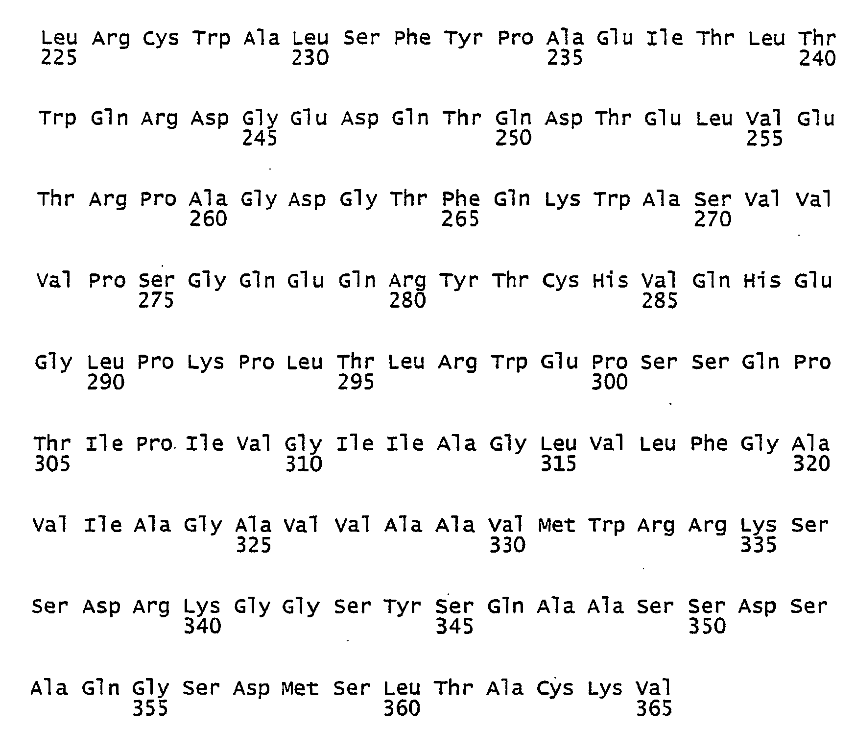
- <210> 7
<211> 341
<212> PRT
<213> Homo sapiens - <400> 7


- <210> 8
<211> 1116
<212> DNA
<213> Homo sapiens - <400> 8

- <210> 9
<211> 135
<212> DNA
<213> Homo sapiens - <400> 9

- <210> 10
<211> 117
<212> DNA
<213> Homo sapiens - <400> 10

- <210> 11
<211> 117 <212> DNA
<213> Homo sapiens - <400> 11

- <210> 12
<211> 371
<212> PRT
<213> Homo sapiens - <400> 12
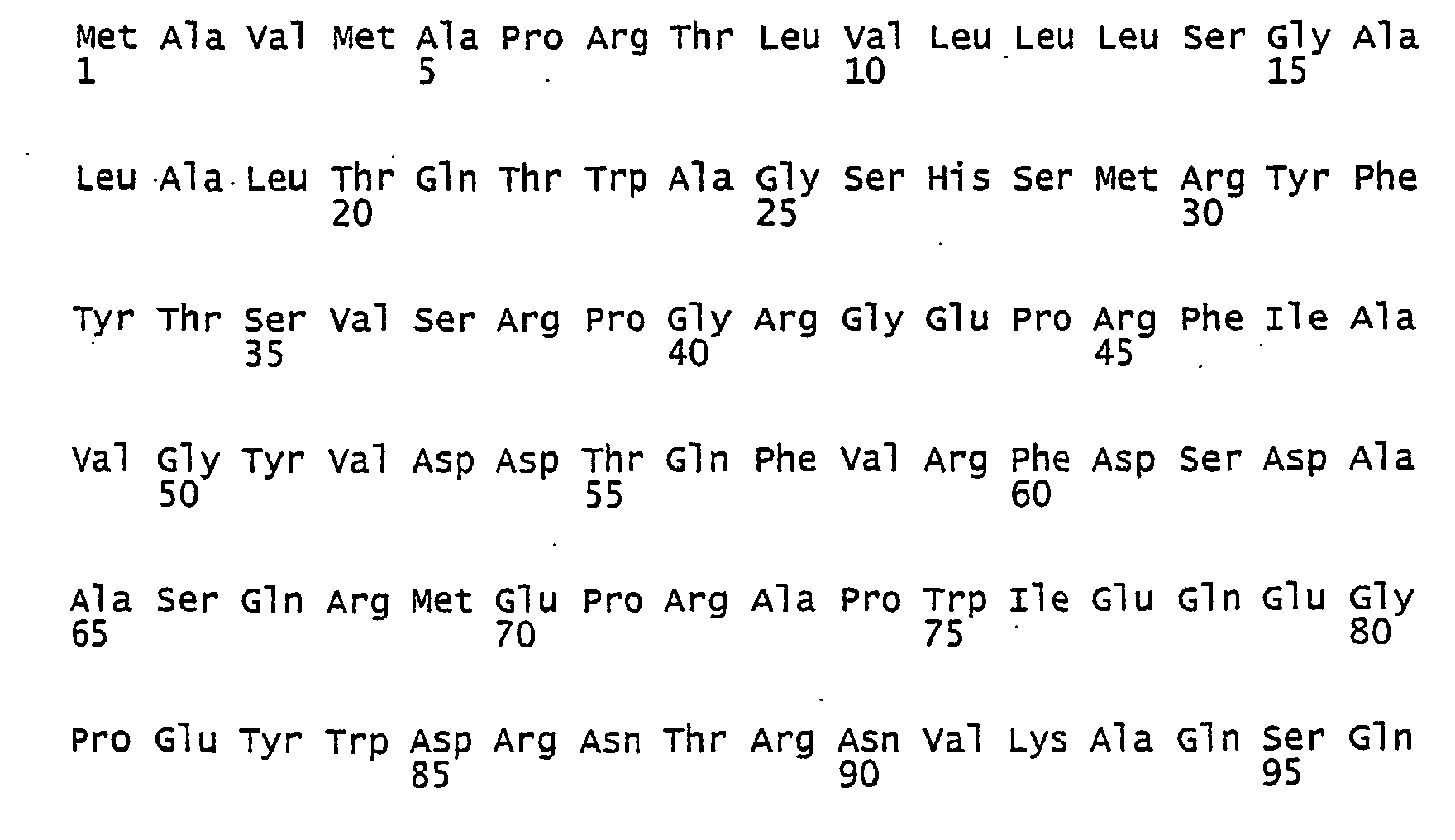


- <210> 13
<211> 24
<212> PRT
<213> Homo sapiens - <400> 13

- <210> 14
<211> 45
<212> PRT
<213> Homo sapiens - <400> 14

- <210> 15
<211> 347
<212> PRT
<213> Homo sapiens - <400> 15


Claims (7)
- Rekombinantes, aufgereinigtes MHC-Protein, welches im Wesentlichen die gleiche Konformation, funktionelle Aktivität sowie die Bindungseigenschaften für spezifische Antikörper, Peptide, T-Zellrezeptoren und NK-Zellen und Antigene wie das native MHC-Protein aufweist, dadurch gekennzeichnet, dass das MHC-Protein keine transmembrane Domäne oder eine transmembrane Domäne aufweist, welche hinsichtlich ihrer Aminosäuresequenz derart modifiziert ist, dass das MHC-Protein löslich wird, und dass das MHC-Protein eine MHC-Protein intrazelluläre Domäne aufweist.
- Rekombinantes MHC-Protein nach Anspruch 1, dadurch gekennzeichnet, dass es die Glykolisierung des nativen MHC-Proteins aufweist.
- Rekombinantes MHC-Protein nach Anspruch 1 oder 2, dadurch gekennzeichnet, dass es mittels Affinitätschromatographie und/oder Gelchromatographie aufgereinigt ist.
- Rekombinantes MHC-Protein nach einem der Ansprüche 1 bis 3, dadurch gekennzeichnet, dass es ein HLA-Protein der Klasse I oder der Klasse II ist.
- Rekombinantes MHC-Protein nach einem der vorhergehenden Ansprüche, dadurch gekennzeichnet, dass es an seiner Peptid-bindenden Region ein dort gebundenes endogenes Peptid aufweist.
- Rekombinantes MHC-Protein nach einem der vorhergehenden Ansprüche, dadurch gekennzeichnet, dass es im Wesentlichen nach folgendem Verfahren hergestellt ist:a) Aufreinigung eines MHC Allels bestehend aus gDNA oder cDNA oder RNA,b) PCR (Polymerase Chain Reaction) Amplifikation des MHC Allels von Exon 1 bis Exon 4 mit zwei geeigneten Primern, nämlich einen Anfangs-Primer und einen End-Primer,c) PCR Amplifikation eines MHC Allels, nämlich des Allels des ersten oder zweiten Verfahrensschrittes, von Exon 6 bis Exon 7 oder Exon 8 mit zwei geeigneten Primern, nämlich einen Anfangs-Primer und einen End-Primer, wobei der Anfangs-Primer eine 5' Sequenz, die zum 3' Ende von Exon 4 komplementär ist, enthält, und wobei der End-Primer kein Stoppcodon enthält,d) gemeinsame PCR Amplifikation der beiden so erhaltenen Allelabschnitte als eine ununterbrochene Sequenz mittels zweier geeigneter Primer, insbesondere unter Verwendung des Anfangs-Primers aus Verfahrensschritt b und des End-Primers aus Verfahrensschritt c,e) Klonierung der amplifizierten ununterbrochenen Sequenz in einen Klonierungsvektor, der auch als Expressionsvektor geeignet ist, oder alternativ Umklonierung des Klonierungsvektors in einen Expressionsvektor,f) Expansion des so erhaltenen Plasmids in geeigneten Prokaryonten, vorzugsweise E. coli, und anschließende Aufreinigung des Plasmids,g) Transfektion oder Transduktion von eukaryonten Zellen oder Zelllinien mit dem so gewonnenen Plasmid,h) Kultivierung und Expansion der Zellen oder Zelllinien mit geeigneten Methoden,i) Aberntung der von den Zellen sezernierten, rekombinanten MHC-Moleküle undj) Aufreinigung der Proteine mittels Affinitätschromatographie und/oder Gelchromatographie.
- Verfahren zur Herstellung eines löslichen rekombinanten Proteins bestehend aus mindestens einer membranständigen und/oder transmembranen Domäne und einer oder mehrerer Domänen außerhalb der Membran, wobei ein das Protein kodierendes Gen jeweils für die Aminosäuresequenz jeder der Domänen kodierende Exons aufweist, dadurch gekennzeichnet, dass mittels PCR durch Wahl geeigneter Primer das oder die in einer Richtung der Sequenz benachbarten Exons amplifiziert werden, welche außerhalb der Membran befindliche Domänen kodieren und sich in genannter Richtung vor dem oder den Exons befinden, welches oder welche die Membran-Domäne kodiert, dann mittels PCR durch Wahl geeigneter Primer die in einer Richtung der Sequenz benachbarten Exons amplifiziert werden, welche außerhalb der Membran befindliche Domänen kodieren und sich in genannter Richtung hinter dem oder den Exons befinden, welches oder welche die Membran-Domäne kodiert, die so amplifizierten Sequenzen mittels PCR und geeigneter Primer, die eine überlappende Sequenz aufweisen, als ununterbrochene Sequenz amplifiziert werden, die ununterbrochene Sequenz , als Klonierungsvektor und/oder Expressionsvektor kloniert und/oder in Zellen expandiert wird, die so modifizierten das Protein ohne Membranteil produzierenden Zellen kultiviert werden und die exprimierten löslichen Proteine abgeernet und mittels üblicher oder sonstiger geeigneter Verfahren aufgereinigt werden.
Applications Claiming Priority (2)
| Application Number | Priority Date | Filing Date | Title |
|---|---|---|---|
| DE102004022435A DE102004022435A1 (de) | 2004-05-06 | 2004-05-06 | Verfahren zur Herstellung löslicher MHC-Proteine |
| PCT/EP2005/004056 WO2005108427A2 (de) | 2004-05-06 | 2005-04-16 | Verfahren zur herstellung löslicher mhc-proteine |
Publications (2)
| Publication Number | Publication Date |
|---|---|
| EP1745072A2 EP1745072A2 (de) | 2007-01-24 |
| EP1745072B1 true EP1745072B1 (de) | 2011-08-24 |
Family
ID=35276195
Family Applications (1)
| Application Number | Title | Priority Date | Filing Date |
|---|---|---|---|
| EP05735130A Expired - Lifetime EP1745072B1 (de) | 2004-05-06 | 2005-04-16 | Verfahren zur herstellung loeslicher mhc-proteine |
Country Status (5)
| Country | Link |
|---|---|
| US (1) | US20080064060A1 (de) |
| EP (1) | EP1745072B1 (de) |
| AT (1) | ATE521637T1 (de) |
| DE (1) | DE102004022435A1 (de) |
| WO (1) | WO2005108427A2 (de) |
Families Citing this family (2)
| Publication number | Priority date | Publication date | Assignee | Title |
|---|---|---|---|---|
| US8236771B2 (en) | 2004-05-18 | 2012-08-07 | National Institute Of Transplantation Foundation | Vectors and methods for long-term immune evasion to prolong transplant viability |
| KR101703540B1 (ko) * | 2012-06-04 | 2017-02-08 | 대한민국 | 3'' 영역의 염기 변이를 통한 전체 단백질의 발현 조절 방법 |
Family Cites Families (4)
| Publication number | Priority date | Publication date | Assignee | Title |
|---|---|---|---|---|
| WO1993010220A1 (en) * | 1991-11-19 | 1993-05-27 | Anergen, Inc. | Soluble mhc molecules and their uses |
| US6090587A (en) * | 1993-10-25 | 2000-07-18 | Corixa Corporation | Prokaryotic expression of MHC proteins |
| JP2004536554A (ja) * | 2000-10-10 | 2004-12-09 | ザ ボード オブ リージェンツ オブ ザ ユニヴァーシティー オブ オクラホマ | Mhc陽性細胞からの比較リガンドマッピング |
| US20040072262A1 (en) * | 2002-10-11 | 2004-04-15 | Montero-Julian Felix A. | Methods and systems for detecting MHC class I binding peptides |
-
2004
- 2004-05-06 DE DE102004022435A patent/DE102004022435A1/de not_active Withdrawn
-
2005
- 2005-04-16 WO PCT/EP2005/004056 patent/WO2005108427A2/de not_active Ceased
- 2005-04-16 US US11/579,705 patent/US20080064060A1/en not_active Abandoned
- 2005-04-16 EP EP05735130A patent/EP1745072B1/de not_active Expired - Lifetime
- 2005-04-16 AT AT05735130T patent/ATE521637T1/de active
Also Published As
| Publication number | Publication date |
|---|---|
| WO2005108427A2 (de) | 2005-11-17 |
| WO2005108427A3 (de) | 2006-04-27 |
| US20080064060A1 (en) | 2008-03-13 |
| ATE521637T1 (de) | 2011-09-15 |
| EP1745072A2 (de) | 2007-01-24 |
| DE102004022435A1 (de) | 2005-12-08 |
Similar Documents
| Publication | Publication Date | Title |
|---|---|---|
| DE69332485T2 (de) | Immunmodulierende peptide | |
| EP2370101B1 (de) | Verwendung von flt3-ligand zur verstärkung von immunreaktionen bei rna-immunisierung | |
| DE10347710B4 (de) | Rekombinante Impfstoffe und deren Verwendung | |
| DE102016115246B3 (de) | Neue t-zellrezeptoren und deren verwendung in immuntherapie | |
| DE102004026135A1 (de) | An MHC-Moleküle bindende Tumor-assoziierte Peptide | |
| DE69935318T2 (de) | Immununterdrückung durch blockierung des t-zellencostimulierungssignales 2 (b7/cd28 interaktion) | |
| DE69929805T2 (de) | Modifiziertes e.coli enterotoxin ii signalpeptid sowie ein mikroorganismus der ein fusionsprotein sowie ein heterologes protein exprimiert | |
| DE60133287T2 (de) | Tumorantigen | |
| DE102005041616B4 (de) | Melanom-assoziierte MHC Klasse I assoziierte Oligopeptide und für diese kodierende Polynukleotide und deren Verwendungen | |
| EP1554307A2 (de) | Il-15 antagonisten | |
| DE69529473T2 (de) | Gezielte t-lymfozyten | |
| EP0816496A2 (de) | Nierenkarzinom-spezifische-T-Zellen | |
| EP1308167A1 (de) | Antigenpräsentierende Vesikel | |
| DE4038189C2 (de) | Humanes, Epididymis-spezifisches Polypeptid und dessen Verwendung zur Therapie und Diagnose männlicher Infertilität | |
| DE10259713A1 (de) | Verfahren zur Expressionsstabilisierung und Verbesserung der spezifischen Effektorfunktion von Einzelketten-Antigenerkennenden genetischen Konstrukten (scARC) und entsprechend mutierten MDM2-Protein spezifischen scT-Zell Rezeptoren | |
| DE102004022435A1 (de) | Verfahren zur Herstellung löslicher MHC-Proteine | |
| EP1363942A2 (de) | Polypeptide eines hdm2-protein spezifischen murinen alpha/beta t-zell rezeptors, diese kodierende nukleinsüren und deren verwendung | |
| DE102004019803A1 (de) | Verfahren zur Herstellung löslicher MHC-Proteine | |
| DE60023891T2 (de) | Peptidverbindung, die von einem verschobenen offenen leserahmen des ice-gens abstammt | |
| WO2002070556A9 (de) | POLYPEPTIDE EINES P53-PROTEIN-SPEZIFISCHEN MURINEN Α/β T-ZELL REZEPTORS, DIESE KODIERENDE NUKLEINSÄUREN UND DEREN VERWENDUNG | |
| EP1220921B1 (de) | Nukleinsäuresequenzen von hyperplasien und tumoren der schilddrüse | |
| DE3781017T2 (de) | Cytotoxische zusammensetzung aus "killer"-zellen. | |
| DE19807265C2 (de) | Adenoviraler Transfervektor für den Gentransport einer DNA-Sequenz | |
| EP1007671A1 (de) | Fanconi-gen ii | |
| DE60021421T2 (de) | APOPTOSE-HEMMENDES POLYPEPTID, FüR DIESES KODIERENDE GENE UND POLYNUKLEOTIDE UND DIESE BEINHALTENDE ZUSAMMENSETZUNGEN |
Legal Events
| Date | Code | Title | Description |
|---|---|---|---|
| PUAI | Public reference made under article 153(3) epc to a published international application that has entered the european phase |
Free format text: ORIGINAL CODE: 0009012 |
|
| 17P | Request for examination filed |
Effective date: 20061206 |
|
| AK | Designated contracting states |
Kind code of ref document: A2 Designated state(s): AT BE BG CH CY CZ DE DK EE ES FI FR GB GR HU IE IS IT LI LT LU MC NL PL PT RO SE SI SK TR |
|
| DAX | Request for extension of the european patent (deleted) | ||
| 17Q | First examination report despatched |
Effective date: 20071213 |
|
| GRAP | Despatch of communication of intention to grant a patent |
Free format text: ORIGINAL CODE: EPIDOSNIGR1 |
|
| GRAS | Grant fee paid |
Free format text: ORIGINAL CODE: EPIDOSNIGR3 |
|
| GRAA | (expected) grant |
Free format text: ORIGINAL CODE: 0009210 |
|
| AK | Designated contracting states |
Kind code of ref document: B1 Designated state(s): AT BE BG CH CY CZ DE DK EE ES FI FR GB GR HU IE IS IT LI LT LU MC NL PL PT RO SE SI SK TR |
|
| REG | Reference to a national code |
Ref country code: GB Ref legal event code: FG4D Free format text: NOT ENGLISH |
|
| REG | Reference to a national code |
Ref country code: CH Ref legal event code: EP |
|
| REG | Reference to a national code |
Ref country code: IE Ref legal event code: FG4D Free format text: LANGUAGE OF EP DOCUMENT: GERMAN |
|
| REG | Reference to a national code |
Ref country code: DE Ref legal event code: R096 Ref document number: 502005011813 Country of ref document: DE Effective date: 20111027 |
|
| REG | Reference to a national code |
Ref country code: NL Ref legal event code: VDEP Effective date: 20110824 |
|
| LTIE | Lt: invalidation of european patent or patent extension |
Effective date: 20110824 |
|
| PG25 | Lapsed in a contracting state [announced via postgrant information from national office to epo] |
Ref country code: SE Free format text: LAPSE BECAUSE OF FAILURE TO SUBMIT A TRANSLATION OF THE DESCRIPTION OR TO PAY THE FEE WITHIN THE PRESCRIBED TIME-LIMIT Effective date: 20110824 Ref country code: NL Free format text: LAPSE BECAUSE OF FAILURE TO SUBMIT A TRANSLATION OF THE DESCRIPTION OR TO PAY THE FEE WITHIN THE PRESCRIBED TIME-LIMIT Effective date: 20110824 Ref country code: FI Free format text: LAPSE BECAUSE OF FAILURE TO SUBMIT A TRANSLATION OF THE DESCRIPTION OR TO PAY THE FEE WITHIN THE PRESCRIBED TIME-LIMIT Effective date: 20110824 Ref country code: LT Free format text: LAPSE BECAUSE OF FAILURE TO SUBMIT A TRANSLATION OF THE DESCRIPTION OR TO PAY THE FEE WITHIN THE PRESCRIBED TIME-LIMIT Effective date: 20110824 Ref country code: IS Free format text: LAPSE BECAUSE OF FAILURE TO SUBMIT A TRANSLATION OF THE DESCRIPTION OR TO PAY THE FEE WITHIN THE PRESCRIBED TIME-LIMIT Effective date: 20111224 Ref country code: PT Free format text: LAPSE BECAUSE OF FAILURE TO SUBMIT A TRANSLATION OF THE DESCRIPTION OR TO PAY THE FEE WITHIN THE PRESCRIBED TIME-LIMIT Effective date: 20111226 |
|
| PG25 | Lapsed in a contracting state [announced via postgrant information from national office to epo] |
Ref country code: SI Free format text: LAPSE BECAUSE OF FAILURE TO SUBMIT A TRANSLATION OF THE DESCRIPTION OR TO PAY THE FEE WITHIN THE PRESCRIBED TIME-LIMIT Effective date: 20110824 Ref country code: CY Free format text: LAPSE BECAUSE OF FAILURE TO SUBMIT A TRANSLATION OF THE DESCRIPTION OR TO PAY THE FEE WITHIN THE PRESCRIBED TIME-LIMIT Effective date: 20110824 Ref country code: GR Free format text: LAPSE BECAUSE OF FAILURE TO SUBMIT A TRANSLATION OF THE DESCRIPTION OR TO PAY THE FEE WITHIN THE PRESCRIBED TIME-LIMIT Effective date: 20111125 Ref country code: PL Free format text: LAPSE BECAUSE OF FAILURE TO SUBMIT A TRANSLATION OF THE DESCRIPTION OR TO PAY THE FEE WITHIN THE PRESCRIBED TIME-LIMIT Effective date: 20110824 |
|
| REG | Reference to a national code |
Ref country code: IE Ref legal event code: FD4D |
|
| PG25 | Lapsed in a contracting state [announced via postgrant information from national office to epo] |
Ref country code: SK Free format text: LAPSE BECAUSE OF FAILURE TO SUBMIT A TRANSLATION OF THE DESCRIPTION OR TO PAY THE FEE WITHIN THE PRESCRIBED TIME-LIMIT Effective date: 20110824 Ref country code: CZ Free format text: LAPSE BECAUSE OF FAILURE TO SUBMIT A TRANSLATION OF THE DESCRIPTION OR TO PAY THE FEE WITHIN THE PRESCRIBED TIME-LIMIT Effective date: 20110824 Ref country code: IE Free format text: LAPSE BECAUSE OF FAILURE TO SUBMIT A TRANSLATION OF THE DESCRIPTION OR TO PAY THE FEE WITHIN THE PRESCRIBED TIME-LIMIT Effective date: 20110824 |
|
| PG25 | Lapsed in a contracting state [announced via postgrant information from national office to epo] |
Ref country code: IT Free format text: LAPSE BECAUSE OF FAILURE TO SUBMIT A TRANSLATION OF THE DESCRIPTION OR TO PAY THE FEE WITHIN THE PRESCRIBED TIME-LIMIT Effective date: 20110824 Ref country code: EE Free format text: LAPSE BECAUSE OF FAILURE TO SUBMIT A TRANSLATION OF THE DESCRIPTION OR TO PAY THE FEE WITHIN THE PRESCRIBED TIME-LIMIT Effective date: 20110824 Ref country code: RO Free format text: LAPSE BECAUSE OF FAILURE TO SUBMIT A TRANSLATION OF THE DESCRIPTION OR TO PAY THE FEE WITHIN THE PRESCRIBED TIME-LIMIT Effective date: 20110824 |
|
| PG25 | Lapsed in a contracting state [announced via postgrant information from national office to epo] |
Ref country code: DK Free format text: LAPSE BECAUSE OF FAILURE TO SUBMIT A TRANSLATION OF THE DESCRIPTION OR TO PAY THE FEE WITHIN THE PRESCRIBED TIME-LIMIT Effective date: 20110824 |
|
| PLBE | No opposition filed within time limit |
Free format text: ORIGINAL CODE: 0009261 |
|
| STAA | Information on the status of an ep patent application or granted ep patent |
Free format text: STATUS: NO OPPOSITION FILED WITHIN TIME LIMIT |
|
| 26N | No opposition filed |
Effective date: 20120525 |
|
| REG | Reference to a national code |
Ref country code: DE Ref legal event code: R097 Ref document number: 502005011813 Country of ref document: DE Effective date: 20120525 |
|
| BERE | Be: lapsed |
Owner name: IMUSYN G.M.B.H. CO. KG Effective date: 20120430 |
|
| PG25 | Lapsed in a contracting state [announced via postgrant information from national office to epo] |
Ref country code: MC Free format text: LAPSE BECAUSE OF NON-PAYMENT OF DUE FEES Effective date: 20120430 |
|
| REG | Reference to a national code |
Ref country code: CH Ref legal event code: PL |
|
| GBPC | Gb: european patent ceased through non-payment of renewal fee |
Effective date: 20120416 |
|
| REG | Reference to a national code |
Ref country code: FR Ref legal event code: ST Effective date: 20121228 |
|
| PG25 | Lapsed in a contracting state [announced via postgrant information from national office to epo] |
Ref country code: BE Free format text: LAPSE BECAUSE OF NON-PAYMENT OF DUE FEES Effective date: 20120430 Ref country code: GB Free format text: LAPSE BECAUSE OF NON-PAYMENT OF DUE FEES Effective date: 20120416 Ref country code: CH Free format text: LAPSE BECAUSE OF NON-PAYMENT OF DUE FEES Effective date: 20120430 Ref country code: LI Free format text: LAPSE BECAUSE OF NON-PAYMENT OF DUE FEES Effective date: 20120430 |
|
| PG25 | Lapsed in a contracting state [announced via postgrant information from national office to epo] |
Ref country code: FR Free format text: LAPSE BECAUSE OF NON-PAYMENT OF DUE FEES Effective date: 20120430 |
|
| PG25 | Lapsed in a contracting state [announced via postgrant information from national office to epo] |
Ref country code: ES Free format text: LAPSE BECAUSE OF FAILURE TO SUBMIT A TRANSLATION OF THE DESCRIPTION OR TO PAY THE FEE WITHIN THE PRESCRIBED TIME-LIMIT Effective date: 20111205 |
|
| REG | Reference to a national code |
Ref country code: AT Ref legal event code: MM01 Ref document number: 521637 Country of ref document: AT Kind code of ref document: T Effective date: 20120416 |
|
| PG25 | Lapsed in a contracting state [announced via postgrant information from national office to epo] |
Ref country code: BG Free format text: LAPSE BECAUSE OF FAILURE TO SUBMIT A TRANSLATION OF THE DESCRIPTION OR TO PAY THE FEE WITHIN THE PRESCRIBED TIME-LIMIT Effective date: 20111124 |
|
| PG25 | Lapsed in a contracting state [announced via postgrant information from national office to epo] |
Ref country code: AT Free format text: LAPSE BECAUSE OF NON-PAYMENT OF DUE FEES Effective date: 20120416 |
|
| PG25 | Lapsed in a contracting state [announced via postgrant information from national office to epo] |
Ref country code: TR Free format text: LAPSE BECAUSE OF FAILURE TO SUBMIT A TRANSLATION OF THE DESCRIPTION OR TO PAY THE FEE WITHIN THE PRESCRIBED TIME-LIMIT Effective date: 20110824 |
|
| PG25 | Lapsed in a contracting state [announced via postgrant information from national office to epo] |
Ref country code: LU Free format text: LAPSE BECAUSE OF NON-PAYMENT OF DUE FEES Effective date: 20120416 |
|
| PG25 | Lapsed in a contracting state [announced via postgrant information from national office to epo] |
Ref country code: HU Free format text: LAPSE BECAUSE OF FAILURE TO SUBMIT A TRANSLATION OF THE DESCRIPTION OR TO PAY THE FEE WITHIN THE PRESCRIBED TIME-LIMIT Effective date: 20050416 |
|
| PGFP | Annual fee paid to national office [announced via postgrant information from national office to epo] |
Ref country code: DE Payment date: 20170607 Year of fee payment: 13 |
|
| REG | Reference to a national code |
Ref country code: DE Ref legal event code: R119 Ref document number: 502005011813 Country of ref document: DE |
|
| PG25 | Lapsed in a contracting state [announced via postgrant information from national office to epo] |
Ref country code: DE Free format text: LAPSE BECAUSE OF NON-PAYMENT OF DUE FEES Effective date: 20181101 |