EP1737489B1 - ErbB2 antagonists for tumor pain therapy - Google Patents
ErbB2 antagonists for tumor pain therapy Download PDFInfo
- Publication number
- EP1737489B1 EP1737489B1 EP05734984A EP05734984A EP1737489B1 EP 1737489 B1 EP1737489 B1 EP 1737489B1 EP 05734984 A EP05734984 A EP 05734984A EP 05734984 A EP05734984 A EP 05734984A EP 1737489 B1 EP1737489 B1 EP 1737489B1
- Authority
- EP
- European Patent Office
- Prior art keywords
- cancer
- antibody
- erbb2
- antibodies
- pain
- Prior art date
- Legal status (The legal status is an assumption and is not a legal conclusion. Google has not performed a legal analysis and makes no representation as to the accuracy of the status listed.)
- Expired - Lifetime
Links
Images


Classifications
-
- C—CHEMISTRY; METALLURGY
- C07—ORGANIC CHEMISTRY
- C07K—PEPTIDES
- C07K16/00—Immunoglobulins [IG], e.g. monoclonal or polyclonal antibodies
- C07K16/18—Immunoglobulins [IG], e.g. monoclonal or polyclonal antibodies against material from animals or humans
- C07K16/28—Immunoglobulins [IG], e.g. monoclonal or polyclonal antibodies against material from animals or humans against receptors, cell surface antigens or cell surface determinants
- C07K16/2863—Immunoglobulins [IG], e.g. monoclonal or polyclonal antibodies against material from animals or humans against receptors, cell surface antigens or cell surface determinants against receptors for growth factors, growth regulators
-
- A—HUMAN NECESSITIES
- A61—MEDICAL OR VETERINARY SCIENCE; HYGIENE
- A61P—SPECIFIC THERAPEUTIC ACTIVITY OF CHEMICAL COMPOUNDS OR MEDICINAL PREPARATIONS
- A61P25/00—Drugs for disorders of the nervous system
- A61P25/04—Centrally acting analgesics, e.g. opioids
-
- A—HUMAN NECESSITIES
- A61—MEDICAL OR VETERINARY SCIENCE; HYGIENE
- A61P—SPECIFIC THERAPEUTIC ACTIVITY OF CHEMICAL COMPOUNDS OR MEDICINAL PREPARATIONS
- A61P29/00—Non-central analgesic, antipyretic or antiinflammatory agents, e.g. antirheumatic agents; Non-steroidal antiinflammatory drugs [NSAID]
-
- A—HUMAN NECESSITIES
- A61—MEDICAL OR VETERINARY SCIENCE; HYGIENE
- A61P—SPECIFIC THERAPEUTIC ACTIVITY OF CHEMICAL COMPOUNDS OR MEDICINAL PREPARATIONS
- A61P35/00—Antineoplastic agents
-
- A—HUMAN NECESSITIES
- A61—MEDICAL OR VETERINARY SCIENCE; HYGIENE
- A61P—SPECIFIC THERAPEUTIC ACTIVITY OF CHEMICAL COMPOUNDS OR MEDICINAL PREPARATIONS
- A61P43/00—Drugs for specific purposes, not provided for in groups A61P1/00-A61P41/00
-
- C—CHEMISTRY; METALLURGY
- C07—ORGANIC CHEMISTRY
- C07K—PEPTIDES
- C07K16/00—Immunoglobulins [IG], e.g. monoclonal or polyclonal antibodies
- C07K16/18—Immunoglobulins [IG], e.g. monoclonal or polyclonal antibodies against material from animals or humans
- C07K16/28—Immunoglobulins [IG], e.g. monoclonal or polyclonal antibodies against material from animals or humans against receptors, cell surface antigens or cell surface determinants
- C07K16/30—Immunoglobulins [IG], e.g. monoclonal or polyclonal antibodies against material from animals or humans against receptors, cell surface antigens or cell surface determinants from tumour cells
-
- C—CHEMISTRY; METALLURGY
- C07—ORGANIC CHEMISTRY
- C07K—PEPTIDES
- C07K16/00—Immunoglobulins [IG], e.g. monoclonal or polyclonal antibodies
- C07K16/18—Immunoglobulins [IG], e.g. monoclonal or polyclonal antibodies against material from animals or humans
- C07K16/32—Immunoglobulins [IG], e.g. monoclonal or polyclonal antibodies against material from animals or humans against translation products of oncogenes
-
- A—HUMAN NECESSITIES
- A61—MEDICAL OR VETERINARY SCIENCE; HYGIENE
- A61K—PREPARATIONS FOR MEDICAL, DENTAL OR TOILETRY PURPOSES
- A61K39/00—Medicinal preparations containing antigens or antibodies
- A61K2039/505—Medicinal preparations containing antigens or antibodies comprising antibodies
-
- C—CHEMISTRY; METALLURGY
- C07—ORGANIC CHEMISTRY
- C07K—PEPTIDES
- C07K2317/00—Immunoglobulins specific features
- C07K2317/20—Immunoglobulins specific features characterized by taxonomic origin
- C07K2317/24—Immunoglobulins specific features characterized by taxonomic origin containing regions, domains or residues from different species, e.g. chimeric, humanized or veneered
-
- C—CHEMISTRY; METALLURGY
- C07—ORGANIC CHEMISTRY
- C07K—PEPTIDES
- C07K2317/00—Immunoglobulins specific features
- C07K2317/50—Immunoglobulins specific features characterized by immunoglobulin fragments
- C07K2317/55—Fab or Fab'
-
- C—CHEMISTRY; METALLURGY
- C07—ORGANIC CHEMISTRY
- C07K—PEPTIDES
- C07K2317/00—Immunoglobulins specific features
- C07K2317/50—Immunoglobulins specific features characterized by immunoglobulin fragments
- C07K2317/56—Immunoglobulins specific features characterized by immunoglobulin fragments variable (Fv) region, i.e. VH and/or VL
- C07K2317/565—Complementarity determining region [CDR]
-
- C—CHEMISTRY; METALLURGY
- C07—ORGANIC CHEMISTRY
- C07K—PEPTIDES
- C07K2317/00—Immunoglobulins specific features
- C07K2317/70—Immunoglobulins specific features characterized by effect upon binding to a cell or to an antigen
- C07K2317/73—Inducing cell death, e.g. apoptosis, necrosis or inhibition of cell proliferation
-
- C—CHEMISTRY; METALLURGY
- C07—ORGANIC CHEMISTRY
- C07K—PEPTIDES
- C07K2317/00—Immunoglobulins specific features
- C07K2317/70—Immunoglobulins specific features characterized by effect upon binding to a cell or to an antigen
- C07K2317/76—Antagonist effect on antigen, e.g. neutralization or inhibition of binding
Definitions
- the present invention concerns ErbB antagonists for treating pain.
- the ErbB family of receptor tyrosine kinases are important mediators of cell growth, differentiation and survival.
- the receptor family includes four distinct members including epidermal growth factor receptor (EGFR or ErbB1), HER2 (ErbB2 or p 185 neu ), HER3 (ErbB3) and HER4 (ErbB4 or tyro2).
- EGFR epidermal growth factor receptor
- HER2 ErbB2 or p 185 neu
- HER3 ErbB3
- HER4 ErbB4 or tyro2
- EGFR encoded by the erb B1 gene
- increased expression of EGFR has been observed in breast, bladder, lung, head, neck and stomach cancer as well as glioblastomas.
- Increased EGFR receptor expression is often associated with increased production of the EGFR ligand, transforming growth factor alpha (TGF- ⁇ ), by the same tumor cells resulting in receptor activation by an autocrine stimulatory pathway.
- TGF- ⁇ transforming growth factor alpha
- Monoclonal antibodies directed against the EGFR or its ligands, TGF- ⁇ and EGF have been evaluated as therapeutic agents in the treatment of such malignancies.
- the second member of the ErbB family, p185 neu was originally identified as the product of the transforming gene from neuroblastomas of chemically treated rats.
- the activated form of the neu proto-oncogene results from a point mutation (valine to glutamic acid) in the transmembrane region of the encoded protein.
- Amplification of the human homolog of neu is observed in breast and ovarian cancers and correlates with a poor prognosis ( Slamon et al., Science, 235:177-182 (1987 ); Slamon et al., Science, 244:707-712 (1989 ); and US Pat No. 4,968,603 ).
- no point mutation analogous to that in the neu proto-oncogene has been reported for human tumors.
- ErbB2 (frequently but not uniformly due to gene amplification) has also been observed in other carcinomas including carcinomas of the stomach, endometrium, salivary gland, lung, kidney, colon, thyroid, pancreas and bladder. See, among others, King et al., Science, 229:974 (1985 ); Yokota et al., Lancet: 1:765-767 (1986 ); Fukushige et al., Mol Cell Biol., 6:955-958 (1986 ); Guerin et al., Oncogene Res., 3:21-31 (1988 ); Cohen et al., Oncogene, 4:81-88 (1989 ); Yonemura et al., Cancer Res., 51:1034 (1991 ); Borst et al., Gynecol.
- Hudziak et al., Mol. Cell. Biol. 9(3):1165-1172 (1989 ) describe the generation of a panel of anti-ErbB2 antibodies which were characterized using the human breast tumor cell line SK-BR-3. Relative cell proliferation of the SK-BR-3 cells following exposure to the antibodies was determined by crystal violet staining of the monolayers after 72 hours. Using this assay, maximum inhibition was obtained with the antibody called 4D5 which inhibited cellular proliferation by 56%. Other antibodies in the panel reduced cellular proliferation to a lesser extent in this assay. The antibody 4D5 was further found to sensitize ErbB2-overexpressing breast tumor cell lines to the cytotoxic effects of TNF- ⁇ See also U.S. Patent No.
- a recombinant humanized version of the murine anti-ErbB2 antibody 4D5 (huMAb4D5-8, rhuMAb HER2 or HERCEPTIN ® ; U.S. Patent No. 5,821,337 ) is clinically active in patients with ErbB2-overexpressing metastatic breast cancers that have received extensive prior anti-cancer therapy ( Baselga et al., J. Clin. Oncol. 14:737-744 (1996 )).
- HERCEPTIN ® received marketing approval from the Food and Drug Administration September 25, 1998 for the treatment of patients with metastatic breast cancer whose tumors overexpress the ErbB2 protein.
- ErbB3 US Pat. Nos. 5,183,884 and 5,480,968 as well as Kraus et al. PNAS (USA) 86:9193-9197 (1989 )
- ErbB4 EP Pat Appln No 599,274 ; Plowman et al., Proc. Natl. Acad. Sci. USA, 90:1746-1750 (1993 ); and Plowman et al., Nature, 366:473-475 (1993 )). Both of these receptors display increased expression on at least some breast cancer cell lines.
- the ErbB receptors are generally found in various combinations in cells and heterodimerization is thought to increase the diversity of cellular responses to a variety of ErbB ligands ( Earp et al. Breast Cancer Research and Treatment 35: 115-132 (1995 )).
- EGFR is bound by six different ligands; epidermal growth factor (EGF), transforming growth factor alpha (TGF- ⁇ ), amphiregulin, heparin binding epidermal growth factor (HB-EGF), betacellulin and epiregulin ( Groenen et al. Growth Factors 11:235-257 (1994 )).
- a family of heregulin proteins resulting from alternative splicing of a single gene are ligands for ErbB3 and ErbB4.
- the heregulin family includes alpha, beta and gamma heregulins ( Holmes et al., Science, 256:1205-1210 (1992 ); U.S. Patent No. 5,641,869 ; and Schaefer et al. Oncogene 15:1385-1394 (1997 )); neu differentiation factors (NDFs), glial growth factors (GGFs); acetylcholine receptor inducing activity (ARIA); and sensory and motor neuron derived factor (SMDF).
- NDFs neu differentiation factors
- GGFs glial growth factors
- ARIA acetylcholine receptor inducing activity
- SMDF sensory and motor neuron derived factor
- EGF and TGF ⁇ do not bind ErbB2, EGF stimulates EGFR and ErbB2 to form a heterodimer, which activates EGFR and results in transphosphorylation of ErbB2 in the heterodimer. Dimerization and/or transphosphorylation appears to activate the ErbB2 tyrosine kinase. See Earp et al., supra.
- ErbB3 is co-expressed with ErbB2
- an active signaling complex is formed and antibodies directed against ErbB2 are capable of disrupting this complex ( Sliwkowski et al., J. Biol. Chem., 269(20):14661-14665 (1994 )).
- ErbB3 for heregulin (HRG) is increased to a higher affinity state when co-expressed with ErbB2.
- HRG heregulin
- Chronic pain is a common symptom of a variety of diseases and pathologic conditions, and includes nociceptive pain (pain caused by an injury to body tissues), neuropathic pain (pain caused by abnormalities in the nerves, spinal cord, or brain), and psychogenic pain (entirely or mostly related to a psychological disorder).
- Nociceptive pain includes somatic pain, which arises from bone, joint, muscle, skin, or connective tissue, and visceral pain, which arises from visceral organs, such as the gastrointestinal tract and the pancreas.
- NSAIDs nonsteroidal antiinflammatory drugs
- Treatment of more severe chronic pain may include opiate and NSAID combinations, such as aspirin and oxycodone (Percodan), acetaminophen and hydrocodone (Vicodin and Lortab).
- Chemotherapy can also cause pain in several ways. Some chemotherapy drugs, referred to as vesicants, can harm tissues if they leak out of the vein. In some instances, chemotherapy causes sores in the mouth (stomatitis) or lining of the intestines (mucositis). Peripheral neuropathy can occur with certain chemotherapy drugs when they are administered long-term in high doses. Radiation treatment can also cause pain because it can affect normal cells that surround the cancerous tumor being treated.
- cancer-related pain is usually managed by opiate analgesics, such as morphine or heroin with the goal to relieve the patient's pain by adjusting the opiate dosage to maintain a pain score of 3 or less on a 10-point visual analog scale.
- This treatment is not optimal.
- Common side effects include drowsiness and constipation.
- patients often experience tolerance and develop a physical dependency on opiate analgesics, which reduces the effectiveness of the pain treatment and raises serious issues of drug dependency.
- withdrawal symptoms may appear, the character and severity of which are dependent upon such factors as the particular opioid being withdrawn, the daily dose of the opioid that is being withdrawn, the duration of opioid treatment, and the condition of the drug-dependent individual. Withdrawal itself is associated with symptoms including severe pain. Often the only effective treatment for cancer-related pain is successful eradication of the tumor.
- Rathore et al., Blood, Vol. 100(11), 2002 , Abstract No. 3684 discloses treatment of patients with prostate cancer with bispecific OKT3 x anti-HER2/neu antibody, describing reduction of narcotic use in patients treated for bone pain.
- US2002/0002276 discloses use of anti-ErbB chimeric heteroadhesin/antagonist antibody for treating peripheral neuropathologies, including cancer-related pain.
- This invention is based, at least in part, on the surprising observation that patients with prostate cancer treated with an ErbB antagonist, namely rhuMAb 2C4, experienced diminished pain or showed reduced analgesia requirement, even where the tumor was progressing. This indicates that rhuMAb 2C4 has analgesic properties.
- the pain is measured by a pain score or quality of life score reflective of pain.
- Pain may, for example, be measured by the McGill Pain Index on a six point scale of 0 to 5. Pain may alternatively be measured by using a visual analog scale of 0-100 reflective of the subjective feeling of pain of the patient.
- the analgesia requirement may be measured using an analgesia score.
- one dose of a non-steroidal analgesic agent corresponds to an analgesia score of 1
- one 10 mg dose of morphine, or an equivalent dose of another opiate analgesic agent corresponds to an analgesia score of 2.
- pain is monitored daily.
- analgesia requirement is monitored daily.
- the cancer can be any kind of cancer, including, for example, breast cancer, ovarian cancer, prostate cancer, pancreatic cancer, squamous cell cancer, lung cancer, cancer of the peritoneum, hepatocellular cancer, gastric cancer, glioblastoma, cervical cancer, liver cancer, bladder cancer, hepatoma, colon cancer, rectal cancer, colorectal cancer, endometrial or uterine carcinoma, salivary gland carcinoma, kidney cancer, vulval cancer, thyroid cancer, hepatic carcinoma, anal carcinoma, penile carcinoma, and head and neck cancer.
- breast cancer breast cancer
- ovarian cancer prostate cancer
- pancreatic cancer squamous cell cancer
- lung cancer cancer of the peritoneum
- hepatocellular cancer gastric cancer
- glioblastoma cervical cancer
- liver cancer liver cancer
- bladder cancer hepatoma
- colon cancer rectal cancer
- colorectal cancer endometrial or uterine carcinoma
- salivary gland carcinoma kidney cancer
- vulval cancer
- the cancer is metastatic cancer.
- the metastasis is soft tissue metastasis.
- the metastasis includes bone metastasis.
- the cancer is prostate cancer, specifically including androgen independent prostate cancer.
- the cancer is prostate cancer
- the patient's PSA shows no reduction during treatment, or becomes elevated during treatment.
- the ErbB2 antagonist preferably is an antibody.
- the antibody can, for example, be a monoclonal antibody that binds an ErbB.
- the antibody blocks ligand activation of an ErbB.
- the antibody blocks formation of an ErbB heterodimer.
- the antibody blocks binding of monoclonal antibody 2C4 to ErbB2.
- the antibody has a biological characteristic of monoclonal antibody 2C4.
- the antibody comprises monoclonal antibody 2C4 or humanized 2C4.
- the antibody may be an antibody fragment, such as, for example, a Fab fragment and may, or may not be conjugated with a cytotoxic agent.
- the pain treated in accordance with the present invention can be acute pain or chronic pain, such as, without limitation., nociceptive pain, neuropathic pain and psychogenic pain, and can be cancer related or not associated with cancer.
- the cancer may, but does not have to, express an ErbB receptor, such as, ErbB2 and/or EGFR.
- the cancer is metastatic cancer, where the metastasis can be soft tissue and/or bone metastasis.
- the cancer is selected from the group consisting of breast cancer, ovarian cancer, prostate cancer, pancreatic cancer, squamous cell cancer, lung cancer, cancer of the peritoneum, hepatocellular cancer, gastric cancer, glioblastoma, cervical cancer, liver cancer, bladder cancer, hepatoma, colon cancer, rectal cancer, colorectal cancer, endometrial or uterine carcinoma, salivary gland carcinoma, kidney cancer, vulval cancer, thyroid cancer, hepatic carcinoma, anal carcinoma, penile carcinoma, and head and neck cancer.
- the cancer is prostate cancer, such as, for example, androgen independent prostate cancer.
- pain is used herein in the broadest sense and refers to all types of pain, including acute and chronic pain, such as nociceptive pain, e.g. somatic pain and visceral pain; neuropathic pain, e.g. centrally generated pain and peripherally generated pain; and psychogenic pain.
- nociceptive pain e.g. somatic pain and visceral pain
- neuropathic pain e.g. centrally generated pain and peripherally generated pain
- psychogenic pain e.g. centrally generated pain and peripherally generated pain
- the term preferably refers to chronic pain, most preferably nociceptive pain, including somatic pain and visceral pain, which can be cancer related, not associated with cancer, or only partially associated with cancer.
- nociceptive pain is used to include all pain caused by injury to body tissues, including, without limitation, by a cut, bruise, bone fracture, crush injury, burn, and the like. This type of pain is typically aching, sharp, or throbbing. Pain receptors for tissue injury (nociceptor) are located mostly in the skin or in the internal organs.
- spontaneous pain is used to refer to pain arising from bone, joint, muscle, skin, or connective tissue. This type of pain is typically aching or throbbing in quality and is well localized.
- visceral pain is used herein to refer to pain arising from visceral organs, such as the gastrointestinal tract and pancreas. Visceral pain includes aching and fairly well localized pain caused by tumor involvement of the organ capsule. Another type of visceral pain, which is typically caused by obstruction of hollow viscus, is characterized by intermittent cramping and poorly localized pain.
- neurode pain is used herein to refer to pain originating from abnormal processing of sensory input by the peripheral or central nervous system.
- analgesia is used to refer to the absence of pain in response to a stimulus that would be normally painful and to a treatment with an analgesic agent.
- analgesic agent and grammatical equivalents thereof refer to agents that are capable of invoking analgesia.
- analgesia requirement of a patient is used herein to refer to the need of administering an analgesic agent (other than an ErbB antagonist) to a patient in order to manage the patient's pain.
- Treatment refers to both therapeutic treatment and prophylactic or preventative measures. Those in need of treatment include those already experiencing pain as well as those in which pain is to be prevented.
- the term "effective amount” refers to an amount of the ErbB antagonist effective to reduce pain, at least to some extent, or to reduce or eliminate the analgesia requirement while maintaining the same or reduced pain score or subjective feeling of pain as experienced under analgesia, prior to the administration of the ErbB antagonist.
- Pain and reduction in pain can be evaluated by using any of the pain score systems well known in the art of pain management and/or a Quality of Life Score system for pain.
- Quality of Life Score for pain can be determined using a visual analog scale of 0-100.
- cancer and “cancerous” refer to or describe the physiological condition in mammals that is typically characterized by unregulated cell growth.
- examples of cancer include, but are not limited to, carcinoma, lymphoma, blastoma, sarcoma, and leukemia or lymphoid malignancies. More particular examples of such cancers include squamous cell cancer (e.g.
- lung cancer including small-cell lung cancer, non-small cell lung cancer, adenocarcinoma of the lung and squamous carcinoma of the lung, cancer of the peritoneum, hepatocellular cancer, gastric or stomach cancer including gastrointestinal cancer, pancreatic cancer, glioblastoma, cervical cancer, ovarian cancer, liver cancer, bladder cancer, hepatoma, breast cancer, colon cancer, rectal cancer, colorectal cancer, endometrial or uterine carcinoma, salivary gland carcinoma, kidney or renal cancer, prostate cancer, vulval cancer, thyroid cancer, hepatic carcinoma, anal carcinoma, penile carcinoma, as well as head and neck cancer.
- lung cancer including small-cell lung cancer, non-small cell lung cancer, adenocarcinoma of the lung and squamous carcinoma of the lung, cancer of the peritoneum, hepatocellular cancer, gastric or stomach cancer including gastrointestinal cancer, pancreatic cancer, glioblastoma, cervical cancer, ovarian cancer, liver cancer,
- PSA prostate-specific antigen in the blood produced by the prostate, as determined by the prostate-specific antigen test. The amount of this antigen increases if the prostate is cancerous, and typically continues to increase as the cancer progresses.
- ErbB is a receptor protein tyrosine kinase which belongs to the ErbB receptor family and includes EGFR, ErbB2, ErbB3 and ErbB4 receptors and other members of this family to be identified in the future.
- the ErbB will generally comprise an extracellular domain, which may bind an ErbB ligand; a lipophilic transmembrane domain; a conserved intracellular tyrosine kinase domain; and a carboxyl-terminal signaling domain harboring several tyrosine residues which can be phosphorylated.
- the ErbB may be a "native sequence" ErbB or an "amino acid sequence variant" thereof.
- the ErbB is native sequence human ErbB.
- ErbB1 refers to EGFR as disclosed, for example, in Carpenter et al. Ann. Rev. Biochem. 56:881-914 (1987 ), including naturally occurring mutant forms thereof (e.g. a deletion mutant EGFR as in Humphrey et al. PNAS (USA) 87:4207-4211 (1990 ); type II EGFR mutant ( US Patent No. 6,455,498 ) etc).
- erbB1 refers to the gene encoding the EGFR protein product.
- ErbB2 and "HER2” are used interchangeably herein and refer to human HER2 protein described, for example, in Semba et al., PNAS (USA) 82:6497-6501 (1985 ) and Yamamoto et al. Nature 319:230-234 (1986 ) (Genebank accession number X03363).
- erb B2 refers to the gene encoding human ErbB2 and "neu " refers to the gene encoding rat p185 neu .
- Preferred ErbB2 is native sequence human ErbB2.
- ErbB3 and HER3 refer to the receptor polypeptide as disclosed, for example, in US Pat. Nos. 5,183,884 and 5,480,968 as well as Kraus et al. PNAS (USA) 86:9193-9197 (1989 ).
- ErbB4 and "HER4" herein refer to the receptor polypeptide as disclosed, for example, in EP Pat Appln No 599,274 ; Plowman et al., Proc. Natl. Acad. Sci. USA, 90:1746-1750 (1993 ); and Plowman et al., Nature, 366:473-475 (1993 ), including isoforms thereof, e.g., as disclosed in WO99/19488, published April 22, 1999 .
- ErbB ligand is meant a polypeptide which binds to and/or activates an ErbB.
- the term includes membrane-bound precursor forms of the ErbB ligand, as well as proteolytically processed soluble forms of the ErbB ligand.
- the ErbB ligand of particular interest herein is a native sequence human ErbB ligand such as epidermal growth factor (EGF) ( Savage et al., J. Biol. Chem.
- TGF- ⁇ transforming growth factor alpha
- amphiregulin also known as schwanoma or keratinocyte autocrine growth factor
- betacellulin Shing et al., Science 259:1604-1607 (1993 ); and Sasada et al. Biochem. Biophys. Res. Commun.
- HB-EGF heparin-binding epidermal growth factor
- epiregulin Toyoda et al., J. Biol. Chem. 270:7495-7500 (1995 ); and Komurasaki et al. Oncogene 15:2841-2848 (1997 )
- a heregulin see below
- neuregulin-2 (NRG-2) ( Carraway et al., Nature 387:512-516 (1997 )
- neuregulin-3 (NRG-3) ( Zhang et al., Proc. Natl. Acad. Sci.
- ErbB ligands which bind EGFR include EGF, TGF- ⁇ , amphiregulin, betacellulin, HB-EGF and epiregulin.
- ErbB ligands which bind ErbB3 include heregulins.
- ErbB ligands capable of binding ErbB4 include betacellulin, epiregulin, HB-EGF, NRG-2, NRG-3, NRG-4 and heregulins.
- Heregulin when used herein refers to a polypeptide encoded by the heregulin gene product as disclosed in U.S. Patent No. 5,641,869 or Marchionni et al., Nature, 362:312-318 (1993 ).
- Examples of heregulins include heregulin- ⁇ , heregulin- ⁇ 1, heregulin- ⁇ 2 and heregulin- ⁇ 3 ( Holmes et al., Science, 256:1205-1210 (1992 ); and U.S. Patent No. 5,641,869 ); neu differentiation factor (NDF) ( Peles et al. Cell 69: 205-216 (1992 )); acetylcholine receptor-inducing activity (ARIA) ( Falls et al.
- GGFs glial growth factors
- SMDF motor neuron derived factor
- SMDF neuron derived factor
- the term includes biologically active fragments and/or amino acid sequence variants of a native sequence HRG polypeptide, such as an EGF-like domain fragment thereof ( e.g . HRG ⁇ 1 177-244 ).
- ErbB hetero-oligomer herein is a noncovalently associated oligomer comprising at least two different ErbBs. Such complexes may form when a cell expressing two or more ErbBs is exposed to an ErbB ligand ( Sliwkowski et al., J. Biol. Chem., 269(20):14661-14665 (1994 )). Examples of such ErbB hetero-oligomers include EGFR-ErbB2, ErbB2-ErbB3 and ErbB3-ErbB4 complexes. Moreover, the ErbB hetero-oligomer may comprise two or more ErbB2 receptors combined with a different ErbB, such as ErbB3, ErbB4 or EGFR.
- hetero-oligomer may be included in the hetero-oligomer.
- a cytokine receptor subunit e.g. gp130
- the patient herein may have been subjected to an assay to determine whether ErbB heterodimers, especially an EGFR-ErbB2 and/or ErbB2-ErbB3 heterodimer are present in cells of the patient, e.g . in diseased tissue therefrom.
- ligand activation of an ErbB is meant signal transduction (e.g . that caused by an intracellular kinase domain of an ErbB phosphorylating tyrosine residues in the ErbB or a substrate polypeptide) mediated by ErbB ligand binding to a ErbB hetero-oligomer comprising the ErbB of interest.
- this will involve binding of an ErbB ligand to an ErbB hetero-oligomer which activates a kinase domain of one or more of the ErbBs in the hetero-oligomer and thereby results in phosphorylation of tyrosine residues in one or more of the ErbBs and/or phosphorylation of tyrosine residues in additional substrate polypeptides(s).
- ErbB activation can be quantified using various tyrosine phosphorylation assays.
- an "ErbB antagonist” is a molecule which blocks (reduces or prevents) a biological activity of one or more ErbB(s). Preferably, the antagonist blocks (reduces or prevents) ligand activation of an ErbB.
- the antagonist will be an antibody, small peptide or non-peptide (organic) molecule, antisense molecule, oligonucleotide decoy, and the like, which inhibits a biological activity of an ErbB receptor.
- an antagonist may bind to or otherwise associate with an ErbB and reduce tyrosine kinase activation thereof.
- ErbB antagonists also include molecules that bind to or associate with ErbB ligands or other members of the ErbB signaling pathway, thereby inhibiting ErbB biological activity.
- the preferred ErbB antagonist is an antibody that binds ErbB2, or EGFR, or a hetero-oligomer (e.g. a heterodimer) comprising ErbB2 and/or EGFR, and blocks ligand activation of an ErbB.
- the most preferred antagonist is rhuMAb 2C4 or a molecule having a biological characteristic of rhuMAb 2C4.
- the antagonist may also be an EGFR-targeted drug and/or a tyrosine kinase inhibitor.
- EGFR-targeted drug refers to a therapeutic agent that binds to EGFR and, optionally, inhibits EGFR activation.
- agents include antibodies and small molecules that bind to EGFR.
- antibodies which bind to EGFR include MAb 579 (ATCC CRL HB 8506), MAb 455 (ATCC CRL HB8507), MAb 225 (ATCC CRL 8508), MAb 528 (ATCC CRL 8509) (see, US Patent No.
- the anti-EGFR antibody may be conjugated with a cytotoxic agent, thus generating an immunoconjugate (see, e.g ., EP659,439A2 , Merck Patent GmbH).
- a cytotoxic agent see, e.g ., EP659,439A2 , Merck Patent GmbH.
- small molecules that bind to EGFR include ZD1839 or Gefitinib (IRESSA®; Astra Zeneca), CP-358,774 (TARCEVA®; Genentech/OSI) and AG1478, AG1571 (SU 5271; Sugen).
- a "tyrosine kinase inhibitor” is a molecule which inhibits to some extent tyrosine kinase activity of a tyrosine kinase such as an ErbB.
- examples of such inhibitors include the EGFR-targeted drugs noted in the preceding paragraph as well as quinazolines such as PD 153035,4-(3-chloroanilino) quinazoline, pyridopyrimidines, pyrimidopyrimidines, pyrrolopyrimidines, such as CGP 59326, CGP 60261 and CGP 62706, and pyrazolopyrimidines, 4-(phenylamino)-7H-pyrrolo[2,3-d] pyrimidines, curcumin (diferuloyl methane, 4,5-bis (4-fluoroanilino)phthalimide), tryphostines containing nitrothiophene moieties; PD-0183805 (Warner-
- a “native sequence” polypeptide is one which has the same amino acid sequence as a polypeptide (e.g., ErbB or ErbB ligand) derived from nature.
- a polypeptide e.g., ErbB or ErbB ligand
- Such native sequence polypeptides can be isolated from nature or can be produced by recombinant or synthetic means.
- a native sequence polypeptide can have the amino acid sequence of naturally occurring human polypeptide, murine polypeptide, or polypeptide from any other mammalian species.
- amino acid sequence variant refers to polypeptides having amino acid sequences that differ to some extent from a native sequence polypeptide. Ordinarily, amino acid sequence variants will possess at least about 70% homology with at least one receptor binding domain of a native ErbB ligand or with at least one ligand binding domain of a native ErbB, and preferably, they will be at least about 80%, more preferably at least about 90% homologous with such receptor or ligand binding domains. The amino acid sequence variants possess substitutions, deletions, and/or insertions at certain positions within the amino acid sequence of the native amino acid sequence.
- Homology is defined as the percentage of residues in the amino acid sequence variant that are identical after aligning the sequences and introducing gaps, if necessary, to achieve the maximum percent homology. Methods and computer programs for the alignment are well known in the art. One such computer program is "Align 2", authored by Genentech, Inc., which was filed with user documentation in the United States Copyright Office, Washington, DC 20559, on December 10, 1991.
- antibody herein is used in the broadest sense and specifically covers intact monoclonal antibodies, polyclonal antibodies, multispecific antibodies (e.g. bispecific antibodies) formed from at least two intact antibodies, and antibody fragments, so long as they exhibit the desired biological activity.
- the term "monoclonal antibody” as used herein refers to an antibody obtained from a population of substantially homogeneous antibodies, i.e. , the individual antibodies comprising the population are identical except for variants that may arise during production of the antibody. Monoclonal antibodies are highly specific, being directed against a single antigenic site. Furthermore, in contrast to polyclonal antibody preparations which include different antibodies directed against different determinants (epitopes), each monoclonal antibody is directed against a single determinant on the antigen. In addition to their specificity, the monoclonal antibodies are advantageous in that they may be synthesized uncontaminated by other antibodies.
- the modifier "monoclonal” indicates the character of the antibody as being obtained from a substantially homogeneous population of antibodies, and is not to be construed as requiring production of the antibody by any particular method.
- the monoclonal antibodies to be used in accordance with the present invention may be made by the hybridoma method first described by Kohler et al., Nature, 256:495 (1975 ), or may be made by recombinant DNA methods (see, e.g., U.S. Patent No. 4,816,567 ).
- the "monoclonal antibodies” may also be isolated from phage antibody libraries using the techniques described in Clackson et al., Nature, 352:624-628 (1991 ) and Marks et al., J. Mol. Biol., 222:581-597 (1991 ), for example.
- the monoclonal antibodies herein specifically include "chimeric" antibodies in which a portion of the heavy and/or light chain is identical with or homologous to corresponding sequences in antibodies derived from a particular species or belonging to a particular antibody class or subclass, while the remainder of the chain(s) is identical with or homologous to corresponding sequences in antibodies derived from another species or belonging to another antibody class or subclass, as well as fragments of such antibodies, so long as they exhibit the desired biological activity ( U.S. Patent No. 4,816,567 ; and Morrison et al., Proc. Natl. Acad. Sci. USA, 81:6851-6855 (1984 )).
- Chimeric antibodies of interest herein include "primatized" antibodies comprising variable domain antigen-binding sequences derived from a non-human primate (e.g . Old World Monkey, Ape etc) and human constant region sequences.
- Antibody fragments comprise a portion of an intact antibody, preferably comprising the antigen-binding or variable region thereof.
- antibody fragments include Fab, Fab', F(ab') 2 , and Fv fragments; diabodies; linear antibodies; single-chain antibody molecules; and multispecific antibodies formed from antibody fragment(s).
- an “intact” antibody is one which comprises an antigen-binding variable region as well as a light chain constant domain (C L ) and heavy chain constant domains, C H 1, C H 2 and C H 3.
- the constant domains may be native sequence constant domains (e.g. human native sequence constant domains) or amino acid sequence variant thereof.
- the intact antibody has one or more effector functions.
- Antibody effector functions refer to those biological activities attributable to the Fc region (a native sequence Fc region or amino acid sequence variant Fc region) of an antibody.
- Examples of antibody effector functions include C1q binding; complement dependent cytotoxicity; Fc receptor binding; antibody-dependent cell-mediated cytotoxicity (ADCC); phagocytosis; down regulation of cell surface receptors (e.g. B cell receptor; BCR), etc.
- intact antibodies can be assigned to different "classes". There are five major classes of intact antibodies: IgA, IgD, IgE, IgG, and IgM, and several of these may be further divided into “subclasses” (isotypes), e.g ., IgG1, IgG2, IgG3, IgG4, IgA, and IgA2.
- the heavy-chain constant domains that correspond to the different classes of antibodies are called ⁇ , ⁇ , ⁇ , ⁇ , and ⁇ , respectively.
- the subunit structures and three-dimensional configurations of different classes of immunoglobulins are well known.
- Antibody-dependent cell-mediated cytotoxicity and “ADCC” refer to a cell-mediated reaction in which nonspecific cytotoxic cells that express Fc receptors (FcRs) (e.g . Natural Killer (NK) cells, neutrophils, and macrophages) recognize bound antibody on a target cell and subsequently cause lysis of the target cell.
- FcRs Fc receptors
- FcR expression on hematopoietic cells in summarized is Table 3 on page 464 of Ravetch and Kinet, Annu. Rev. Immunol 9:457-92 (1991 ).
- ADCC activity of a molecule of interest may be assessed in vitro, such as that described in US Patent No. 5,500,362 or 5,821,337.
- useful effector cells for such assays include peripheral blood mononuclear cells (PBMC) and Natural Killer (NK) cells.
- PBMC peripheral blood mononuclear cells
- NK Natural Killer
- ADCC activity of the molecule of interest may be assessed in vivo, e.g., in a animal model such as that disclosed in Clynes et al. PNAS (USA) 95:652-656 (1998 ).
- Human effector cells are leukocytes which express one or more FcRs and perform effector functions. Preferably, the cells express at least Fc ⁇ RIII and perform ADCC effector function. Examples of human leukocytes which mediate ADCC include peripheral blood mononuclear cells (PBMC), natural killer (NK) cells, monocytes, cytotoxic T cells and neutrophils; with PBMCs and NK cells being preferred.
- PBMC peripheral blood mononuclear cells
- NK natural killer cells
- monocytes cytotoxic T cells and neutrophils
- the effector cells may be isolated from a native source thereof, e.g. from blood or PBMCs as described herein.
- Fc receptor or “FcR” are used to describe a receptor that binds to the Fc region of an antibody.
- the preferred FcR is a native sequence human FcR.
- a preferred FcR is one which binds an IgG antibody (a gamma receptor) and includes receptors of the Fc ⁇ RI, Fc ⁇ RII, and Fc ⁇ RIII subclasses, including allelic variants and alternatively spliced forms of these receptors.
- Fc ⁇ RII receptors include Fc ⁇ RIIA (an “activating receptor") and Fc ⁇ RIIB (an “inhibiting receptor”), which have similar amino acid sequences that differ primarily in the cytoplasmic domains thereof.
- Activating receptor Fc ⁇ RIIA contains an immunoreceptor tyrosine-based activation motif (ITAM) in its cytoplasmic domain.
- Inhibiting receptor Fc ⁇ RIIB contains an immunoreceptor tyrosine-based inhibition motif (ITIM) in its cytoplasmic domain.
- ITAM immunoreceptor tyrosine-based activation motif
- ITIM immunoreceptor tyrosine-based inhibition motif
- FcR FcR
- FcRn neonatal receptor
- “Complement dependent cytotoxicity” or “CDC” refers to the ability of a molecule to lyse a target in the presence of complement.
- the complement activation pathway is initiated by the binding of the first component of the complement system (C1q) to a molecule (e.g. an antibody) complexed with a cognate antigen.
- a CDC assay e.g. as described in Gazzano-Santoro et al., J. Immunol. Methods 202:163 (1996 ), may be performed.
- “Native antibodies” are usually heterotetrameric glycoproteins of about 150,000 daltons, composed of two identical light (L) chains and two identical heavy (H) chains. Each light chain is linked to a heavy chain by one covalent disulfide bond, while the number of disulfide linkages varies among the heavy chains of different immunoglobulin isotypes. Each heavy and light chain also has regularly spaced intrachain disulfide bridges. Each heavy chain has at one end a variable domain (V H ) followed by a number of constant domains. Each light chain has a variable domain at one end (V L ) and a constant domain at its other end. The constant domain of the light chain is aligned with the first constant domain of the heavy chain, and the light-chain variable domain is aligned with the variable domain of the heavy chain. Particular amino acid residues are believed to form an interface between the light chain and heavy chain variable domains.
- variable refers to the fact that certain portions of the variable domains differ extensively in sequence among antibodies and are used in the binding and specificity of each particular antibody for its particular antigen. However, the variability is not evenly distributed throughout the variable domains of antibodies. It is concentrated in three segments called hypervariable regions both in the light chain and the heavy chain variable domains. The more highly conserved portions of variable domains are called the framework regions (FRs).
- the variable domains of native heavy and light chains each comprise four FRs, largely adopting a ⁇ -sheet configuration, connected by three hypervariable regions, which form loops connecting, and in some cases forming part of, the ⁇ -sheet structure.
- the hypervariable regions in each chain are held together in close proximity by the FRs and, with the hypervariable regions from the other chain, contribute to the formation of the antigen-binding site of antibodies (see Kabat et al., Sequences of Proteins of Immunological Interest, 5th Ed. Public Health Service, National Institutes of Health, Bethesda, MD. (1991 )).
- the constant domains are not involved directly in binding an antibody to an antigen, but exhibit various effector functions, such as participation of the antibody in antibody dependent cellular cytotoxicity (ADCC).
- hypervariable region when used herein refers to the amino acid residues of an antibody which are responsible for antigen-binding.
- the hypervariable region generally comprises amino acid residues from a "complementarity determining region" or "CDR" (e.g. residues 24-34 (L1), 50-56 (L2) and 89-97 (L3) in the light chain variable domain and 31-35 (H1), 50-65 (H2) and 95-102 (H3) in the heavy chain variable domain; Kabat et al., Sequences of Proteins of Immunological Interest, 5th Ed. Public Health Service, National Institutes of Health, Bethesda, MD. (1991 )) and/or those residues from a "hypervariable loop" (e.g .
- Papain digestion of antibodies produces two identical antigen-binding fragments, called “Fab” fragments, each with a single antigen-binding site, and a residual "Fc” fragment, whose name reflects its ability to crystallize readily. Pepsin treatment yields an F(ab') 2 fragment that has two antigen-binding sites and is still capable of cross-linking antigen.
- Fv is the minimum antibody fragment which contains a complete antigen-recognition and antigen-binding site. This region consists of a dimer of one heavy chain and one light chain variable domain in tight, non-covalent association. It is in this configuration that the three hypervariable regions of each variable domain interact to define an antigen-binding site on the surface of the V H -V L dimer. Collectively, the six hypervariable regions confer antigen-binding specificity to the antibody. However, even a single variable domain (or half of an Fv comprising only three hypervariable regions specific for an antigen) has the ability to recognize and bind antigen, although at a lower affinity than the entire binding site.
- the Fab fragment also contains the constant domain of the light chain and the first constant domain (CH1) of the heavy chain.
- Fab' fragments differ from Fab fragments by the addition of a few residues at the carboxy terminus of the heavy chain CH1 domain including one or more cysteines from the antibody hinge region.
- Fab'-SH is the designation herein for Fab' in which the cysteine residue(s) of the constant domains bear at least one free thiol group.
- F(ab') 2 antibody fragments originally were produced as pairs of Fab' fragments which have hinge cysteines between them. Other chemical couplings of antibody fragments are also known.
- the "light chains" of antibodies from any vertebrate species can be assigned to one of two clearly distinct types, called kappa ( ⁇ ) and lambda ( ⁇ ), based on the amino acid sequences of their constant domains.
- Single-chain Fv or “scFv” antibody fragments comprise the V H and V L domains of antibody, wherein these domains are present in a single polypeptide chain.
- the Fv polypeptide further comprises a polypeptide linker between the V H and V L domains which enables the scFv to form the desired structure for antigen binding.
- scFv see Plückthun in The Pharmacology of Monoclonal Antibodies, vol. 113, Rosenburg and Moore eds., Springer-Verlag, New York, pp. 269-315 (1994 ).
- Anti-ErbB2 antibody scFv fragments are described in WO93/16185 ; U.S. Patent No. 5,571,894 ; and U.S. Patent No. 5,587,458 .
- diabodies refers to small antibody fragments with two antigen-binding sites, which fragments comprise a variable heavy domain (V H ) connected to a variable light domain (V L ) in the same polypeptide chain (V H - V L ).
- V H variable heavy domain
- V L variable light domain
- the domains are forced to pair with the complementary domains of another chain and create two antigen-binding sites.
- Diabodies are described more fully in, for example, EP 404,097 ; WO 93/11161 ; and Hollinger et al., Proc. Natl. Acad. Sci. USA, 90:6444-6448 (1993 ).
- Humanized forms of non-human (e.g. , rodent) antibodies are chimeric antibodies that contain minimal sequence derived from non-human immunoglobulin.
- humanized antibodies are human immunoglobulins (recipient antibody) in which residues from a hypervariable region of the recipient are replaced by residues from a hypervariable region of a non-human species (donor antibody) such as mouse, rat, rabbit or nonhuman primate having the desired specificity, affinity, and capacity.
- donor antibody such as mouse, rat, rabbit or nonhuman primate having the desired specificity, affinity, and capacity.
- framework region (FR) residues of the human immunoglobulin are replaced by corresponding non-human residues.
- humanized antibodies may comprise residues that are not found in the recipient antibody or in the donor antibody. These modifications are made to further refine antibody performance.
- the humanized antibody will comprise substantially all of at least one, and typically two, variable domains, in which all or substantially all of the hypervariable loops correspond to those of a non-human immunoglobulin and all or substantially all of the FRs are those of a human immunoglobulin sequence.
- the humanized antibody optionally also will comprise at least a portion of an immunoglobulin constant region (Fc), typically that of a human immunoglobulin.
- Fc immunoglobulin constant region
- Humanized anti-ErbB2 antibodies include huMAb4D5-1, huMAb4D5-2, huMAb4D5-3, huMAb4D5-4, huMAb4D5-5, huMAb4D5-6, huMAb4D5-7 and huMAb4D5-8 (HERCEPTIN®) as described in Table 3 of U.S. Patent 5,821,337 ; humanized 520C9 ( WO93/21319 ) and humanized 2C4 antibodies as described hereinbelow.
- an “isolated” antibody is one which has been identified and separated and/or recovered from a component of its natural environment. Contaminant components of its natural environment are materials which would interfere with diagnostic or therapeutic uses for the antibody, and may include enzymes, hormones, and other proteinaceous or nonproteinaceous solutes.
- the antibody will be purified (1) to greater than 95% by weight of antibody as determined by the Lowry method, and most preferably more than 99% by weight, (2) to a degree sufficient to obtain at least 15 residues ofN-terminal or internal amino acid sequence by use of a spinning cup sequenator, or (3) to homogeneity by SDS-PAGE under reducing or nonreducing conditions using Coomassie blue or, preferably, silver stain.
- Isolated antibody includes the antibody in situ within recombinant cells since at least one component of the antibody's natural environment will not be present. Ordinarily, however, isolated antibody will be prepared by at least one purification step.
- an antibody "which binds" an antigen of interest e.g. ErbB2 antigen
- an antigen of interest e.g. ErbB2 antigen
- an antigen of interest e.g. ErbB2 antigen
- an antigen of interest e.g. ErbB2 antigen
- the extent of binding of the antibody to these non-ErbB2 proteins will be less than 10% as determined by fluorescence activated cell sorting (FACS) analysis or radioimmunoprecipitation (RIA).
- FACS fluorescence activated cell sorting
- RIA radioimmunoprecipitation
- the anti-ErbB2 antibody will not significantly cross-react with the rat neu protein, e.g ., as described in Schecter et al. Nature 312:513 (1984 ) and Drebin et al., Nature 312:545-548 (1984 ).
- an antibody which "blocks" ligand activation of an ErbB is one which reduces or prevents such activation as hereinabove defined, wherein the antibody is able to block ligand activation of the ErbB substantially more effectively than monoclonal antibody 4D5, e.g. about as effectively as monoclonal antibodies 7F3 or 2C4 or Fab fragments thereof and preferably about as effectively as monoclonal antibody 2C4 or a Fab fragment thereof.
- the antibody that blocks ligand activation of an ErbB may be one which is about 50-100% more effective than 4D5 at blocking formation of an ErbB hetero-oligomer. Blocking of ligand activation of an ErbB can occur by any means, e.g .
- Examples of antibodies which block ligand activation of an ErbB include monoclonal antibodies 2C4 and 7F3 (which block HRG activation of ErbB2/ErbB3 and ErbB2/ErbB4 hetero-oligomers; and EGF, TGF- ⁇ , amphiregulin, HB-EGF and/or epiregulin activation of an EGFR/ErbB2 hetero-oligomer); and L26, L96 and L288 antibodies ( Klapper et al. Oncogene 14:2099-2109 (1997 )), which block EGF and NDF binding to T47D cells which express EGFR, ErbB2, ErbB3 and ErbB4.
- An antibody having a "biological characteristic" of a designated antibody such as the monoclonal antibody designated 2C4, is one which possesses one or more of the biological characteristics of that antibody which distinguish it from other antibodies that bind to the same antigen (e.g. ErbB2).
- an antibody with a biological characteristic of 2C4 may block HRG activation of an ErbB hetero-oligomer comprising ErbB2 and ErbB3 or ErbB4; block EGF, TGF- ⁇ , HB-EGF, epiregulin and/or amphiregulin activation of an ErbB comprising EGFR and ErbB2; block EGF, TGF- ⁇ and/or HRG mediated activation ofMAPK; and/or bind the same epitope in the extracellular domain of ErbB2 as that bound by 2C4 ( e.g. which blocks binding of monoclonal antibody 2C4 to ErbB2).
- the expression “monoclonal antibody 2C4" refers to an antibody that has antigen binding residues of, or derived from, the murine 2C4 antibody of the Examples below.
- the monoclonal antibody 2C4 may be murine monoclonal antibody 2C4 or a variant thereof, such as humanized antibody 2C4, possessing antigen binding amino acid residues of murine monoclonal antibody 2C4.
- humanized 2C4 antibodies are provided in Example 3 below.
- the expression “rhuMAb 2C4" when used herein refers to an antibody comprising the variable light (V L ) and variable heavy (V H ) sequences of SEQ ID Nos. 3 and 4, respectively, fused to human light and heavy IgG1 (non-A allotype) constant region sequences optionally expressed by a Chinese Hamster Ovary (CHO) cell.
- the term “monoclonal antibody 4D5" refers to an antibody that has antigen binding residues of, or derived from, the murine 4D5 antibody (ATCC CRL 10463).
- the monoclonal antibody 4D5 may be murine monoclonal antibody 4D5 or a variant thereof, such as a humanized 4D5, possessing antigen binding residues of murine monoclonal antibody 4D5.
- Exemplary humanized 4D5 antibodies include huMAb4D5-1, huMAb4D5-2, huMAb4D5-3, huMAb4D5-4, huMAb4D5-5, huMAb4D5-6, huMAb4D5-7 and huMAb4D5-8 (HERCEPTIN®) as in US Patent No. 5,821,337 , with huMAb4D5-8 (HERCEPTIN®) being a preferred humanized 4D5 antibody.
- a “growth inhibitory agent” when used herein refers to a compound or composition which inhibits growth of a cell, especially an ErbB expressing cell either in vitro or in vivo.
- the growth inhibitory agent may be one which significantly reduces the percentage of ErbB expressing cells in S phase.
- growth inhibitory agents include agents that block cell cycle progression (at a place other than S phase), such as agents that induce G1 arrest and M-phase arrest.
- Classical M-phase blockers include the vincas (vincristine and vinblastine), taxanes, and topo II inhibitors such as doxorubicin, epirubicin, daunorubicin, etoposide, and bleomycin.
- DNA alkylating agents such as tamoxifen, prednisone, dacarbazine, mechlorethamine, cisplatin, methotrexate, 5-fluorouracil, and ara-C.
- DNA alkylating agents such as tamoxifen, prednisone, dacarbazine, mechlorethamine, cisplatin, methotrexate, 5-fluorouracil, and ara-C.
- growth inhibitory antibodies are those which bind to ErbB2 and inhibit the growth of cells overexpressing ErbB2.
- Preferred growth inhibitory anti-ErbB2 antibodies inhibit growth of SK-BR-3 breast tumor cells in cell culture by greater than 20%, and preferably greater than 50% ( e.g. from about 50% to about 100%) at an antibody concentration of about 0.5 to 30 ⁇ g/ml, where the growth inhibition is determined six days after exposure of the SK-BR-3 cells to the antibody (see U.S. Patent No. 5,677,171 issued October 14, 1997 ).
- the SK-BR-3 cell growth inhibition assay is described in more detail in that patent and hereinbelow.
- the preferred growth inhibitory antibody is monoclonal antibody 4D5, e.g. , humanized 4D5.
- the cell is generally one which expresses the ErbB2 receptor, especially where the cell overexpresses the ErbB2 receptor.
- the cell is a cancer cell, e.g. a breast, ovarian, stomach, endometrial, salivary gland, lung, kidney, colon, thyroid, pancreatic or bladder cell.
- the cell may be a SK-BR-3, BT474, Calu 3, MDA-MB-453, MDA-MB-361 or SKOV3 cell.
- Cell death in vitro may be determined in the absence of complement and immune effector cells to distinguish cell death induced by antibody-dependent cell-mediated cytotoxicity (ADCC) or complement dependent cytotoxicity (CDC).
- ADCC antibody-dependent cell-mediated cytotoxicity
- CDC complement dependent cytotoxicity
- the assay for cell death may be performed using heat inactivated serum (i.e. in the absence of complement) and in the absence of immune effector cells.
- PI propidium iodide
- trypan blue see Moore et al. Cytotechnology 17:1-11 (1995 )
- 7AAD can be assessed relative to untreated cells.
- Preferred cell death-inducing antibodies are those which induce PI uptake in the PI uptake assay in BT474 cells (see below).
- an antibody which "induces apoptosis” is one which induces programmed cell death as determined by binding of annexin V, fragmentation of DNA, cell shrinkage, dilation of endoplasmic reticulum, cell fragmentation, and/or formation of membrane vesicles (called apoptotic bodies).
- the cell is usually one which overexpresses the ErbB2 receptor.
- the cell is a tumor cell, e.g. a breast, ovarian, stomach, endometrial, salivary gland, lung, kidney, colon, thyroid, pancreatic or bladder cell.
- the cell may be a SK-BR-3, BT474, Calu 3 cell, MDA-MB-453, MDA-MB-361 or SKOV3 cell.
- phosphatidyl serine (PS) translocation can be measured by annexin binding; DNA fragmentation can be evaluated through DNA laddering; and nuclear/chromatin condensation along with DNA fragmentation can be evaluated by any increase in hypodiploid cells.
- the antibody which induces apoptosis is one which results in about 2 to 50 fold, preferably about 5 to 50 fold, and most preferably about 10 to 50 fold, induction of annexin binding relative to untreated cell in an annexin binding assay using BT474 cells (see below).
- the pro-apoptotic antibody will be one which further blocks ErbB ligand activation of an ErbB ( e.g .
- the antibody shares a biological characteristic with monoclonal antibody 2C4.
- the antibody is one which does not significantly block ErbB ligand activation of an ErbB (e.g. 7C2).
- the antibody may be one like 7C2 which, while inducing apoptosis, does not induce a large reduction in the percent of cells in S phase ( e.g . one which only induces about 0-10% reduction in the percent of these cells relative to control).
- the "epitope 2C4" is the region in the extracellular domain of ErbB2 to which the antibody 2C4 binds.
- a routine cross-blocking assay such as that described in Antibodies, A Laboratory Manual, Cold Spring Harbor Laboratory, Ed Harlow and David Lane (1988 ), can be performed.
- epitope mapping can be performed to assess whether the antibody binds to the 2C4 epitope of ErbB2 ( e.g. any one or more residues in the region from about residue 22 to about residue 584 of ErbB2, inclusive; see Figs. 1A-B ).
- the "epitope 4D5" is the region in the extracellular domain of ErbB2 to which the antibody 4D5 (ATCC CRL 10463) binds. This epitope is close to the transmembrane domain of ErbB2.
- a routine cross-blocking assay such as that described in Antibodies, A Laboratory Manual, Cold Spring Harbor Laboratory, Ed Harlow and David Lane (1988 ), can be performed.
- epitope mapping can be performed to assess whether the antibody binds to the 4D5 epitope of ErbB2 ( e.g . any one or more residues in the region from about residue 529 to about residue 625, inclusive; see Figs. 1A-B ).
- epitope 3H4 is the region in the extracellular domain of ErbB2 to which the antibody 3H4 binds. This epitope includes residues from about 541 to about 599, inclusive, in the amino acid sequence of ErbB2 extracellular domain; see Figs. 1A-B .
- the "epitope 7C2/7F3" is the region at the N terminus of the extracellular domain of ErbB2 to which the 7C2 and/or 7F3 antibodies (each deposited with the ATCC, see below) bind.
- a routine cross-blocking assay such as that described in Antibodies, A Laboratory Manual, Cold Spring Harbor Laboratory, Ed Harlow and David Lane (1988 ), can be performed.
- epitope mapping can be performed to establish whether the antibody binds to the 7C2/7F3 epitope on ErbB2 ( e.g . any one or more of residues in the region from about residue 22 to about residue 53 of ErbB2; see Figs. 1A-B ).
- ErbB-expressing cell is one which has ErbB protein present at its cell surface, such that an anti-ErbB2 antibody can bind thereto.
- a cell "characterized by excessive activation" of an ErbB is one in which the extent of ErbB activation therein significantly exceeds the level of activation of that receptor in a normal cell of the same tissue type. Such excessive activation may result from overexpression or amplification of the ErbB and/or greater than normal levels of an ErbB ligand available for activating the ErbB in the cell. Such excessive activation may cause and/or be caused by a diseased state of the cell. In some embodiments, a sample from the patient will be subjected to a diagnostic or prognostic assay to determine whether amplification and/or overexpression of an ErbB is occurring which results in such excessive activation of the ErbB.
- a sample from the patient may be subjected to a diagnostic or prognostic assay to determine whether amplification, overexpression and/or increased proteolytic processing of an ErbB ligand is occurring in the patient which attributes to excessive activation of the receptor.
- a diagnostic or prognostic assay to determine whether amplification, overexpression and/or increased proteolytic processing of an ErbB ligand is occurring in the patient which attributes to excessive activation of the receptor.
- excessive activation of the receptor may result from an autocrine stimulatory pathway.
- the cell may express or overexpress EGFR and also express or overexpress an EGFR ligand (e.g. EGF, TGF- ⁇ , or HB-EGF).
- the cell may express or overexpress ErbB2 and also express or overexpress a heregulin ( e.g . ⁇ -HRG).
- a cell which "overexpresses" an ErbB is one which has significantly higher levels of an ErbB, such as ErbB2, at the cell surface thereof, compared to a normal cell of the same tissue type.
- Such overexpression may be caused by gene amplification or by increased transcription or translation.
- ErbB overexpression may be determined in a diagnostic or prognostic assay by evaluating increased levels of the ErbB protein present on the surface of a cell (e.g. via an immunohistochemistry assay; IHC, immunoenzyme, Western blot, ligand binding, kinase activity). Alternatively, or additionally, one may measure levels of ErbB-encoding nucleic acid in the cell, e.g.
- FISH fluorescent in situ hybridization
- PCR polymerase chain reaction
- RT-PCR real time quantitative PCR
- various in vivo assays are available to the skilled practitioner.
- a detectable label e.g. a radioactive isotope
- a cell which is "not characterized by overexpression of an ErbB" is one which, in a diagnostic assay, does not express higher than normal levels of ErbB compared to a normal cell of the same tissue type.
- a cell which "overexpresses" an ErbB ligand is one which produces significantly higher levels of that ligand compared to a normal cell of the same tissue type. Such overexpression may be caused by gene amplification or by increased transcription or translation. Overexpression of the ErbB ligand may be determined diagnostically by evaluating levels of the ligand (or nucleic acid encoding it) in the patient, e.g . in a biopsy or by various diagnostic assays such as the IHC, immunoenzyme, Western blot, ligand binding, FISH, southern blotting, PCR or in vivo assays described above.
- cytotoxic agent refers to a substance that inhibits or prevents the function of cells and/or causes destruction of cells.
- the term is intended to include radioactive isotopes (e.g. At 211 , I 131 , I 125 , Y 90 , Re 186 , Re 188 , Sm 153 , Bi 212 , P 32 and radioactive isotopes of Lu), chemotherapeutic agents, and toxins such as small molecule toxins or enzymatically active toxins of bacterial, fungal, plant or animal origin, including fragments and/or variants thereof.
- radioactive isotopes e.g. At 211 , I 131 , I 125 , Y 90 , Re 186 , Re 188 , Sm 153 , Bi 212 , P 32 and radioactive isotopes of Lu
- chemotherapeutic agents e.g. At 211 , I 131 , I 125 , Y 90 , Re 186
- chemotherapeutic agent is a chemical compound useful in the treatment of cancer.
- examples of chemotherapeutic agents include alkylating agents such as thiotepa and cyclosphosphamide (CYTOXAN®); alkyl sulfonates such as busulfan, improsulfan and piposulfan; aziridines such as benzodopa, carboquone, meturedopa, and uredopa; ethylenimines and methylamelamines including altretamine, triethylenemelamine, trietylenephosphoramide, triethylenethiophosphaoramide and trimethylolomelamine; nitrogen mustards such as chlorambucil, chlornaphazine, cholophosphamide, estramustine, ifosfamide, mechlorethamine, mechlorethamine oxide hydrochloride, melphalan, novembichin, phenesterine, prednimustine, trofos
- paclitaxel TAXOL ® , Bristol-Myers Squibb Oncology, Princeton, NJ
- docetaxel TAXOTERE®, Rhône-Poulenc Rorer, Antony, France
- chlorambucil gemcitabine
- 6-thioguanine mercaptopurine
- methotrexate platinum analogs such as cisplatin and carboplatin; vinblastine; platinum; etoposide (VP-16); ifosfamide; mitomycin C; mitoxantrone; vincristine; vinorelbine; navelbine; novantrone; teniposide; daunomycin; aminopterin; xeloda; ibandronate; CPT-11; topoisomerase inhibitor RFS 2000; difluoromethylornithine (DMFO); retinoic acid; esperamicins; capecitabine; and pharmaceutically acceptable salts, acids or derivatives of any of the above.
- anti-hormonal agents that act to regulate or inhibit hormone action on cells
- anti-estrogens including for example tamoxifen, raloxifene, aromatase inhibiting 4(5)-imidazoles, 4-hydroxytamoxifen, trioxifene, keoxifene, LY117018, onapristone, and toremifene (Fareston); and anti-androgens such as flutamide, nilutamide, bicalutamide, leuprolide, and goserelin; and pharmaceutically acceptable salts, acids or derivatives of any of the above.
- an "anti-angiogenic agent” refers to a compound which blocks, or interferes with to some degree, the development of blood vessels.
- the anti-angiogenic factor may, for instance, be a small molecule or antibody that binds to a growth factor or growth factor receptor involved in promoting angiogenesis.
- the preferred anti-angiogenic factor herein is an antibody that binds to Vascular Endothelial Growth Factor (VEGF), such as the recombinant humanized anti-VEGF antibody AVASTIN® (Genentech).
- VEGF Vascular Endothelial Growth Factor
- cytokine is a generic term for proteins released by one cell population which act on another cell as intercellular mediators.
- cytokines are lymphokines, monokines, and traditional polypeptide hormones. Included among the cytokines are growth hormone such as human growth hormone, N-methionyl human growth hormone, and bovine growth hormone; parathyroid hormone; thyroxine; insulin; proinsulin; relaxin; prorelaxin; glycoprotein hormones such as follicle stimulating hormone (FSH), thyroid stimulating hormone (TSH), and luteinizing hormone (LH); hepatic growth factor; fibroblast growth factor; prolactin; placental lactogen; tumor necrosis factor- ⁇ and - ⁇ ; mullerian-inhibiting substance; mouse gonadotropin-associated peptide; inhibin; activin; vascular endothelial growth factor; integrin; thrombopoietin (TPO); nerve growth factors such as NGF- ⁇ ; platelet-growth factor;
- package insert is used to refer to instructions customarily included in commercial packages of therapeutic products, that contain information about the indications, usage, dosage, administration, contraindications and/or warnings concerning the use of such therapeutic products.
- a “cardioprotectant” is a compound or composition which prevents or reduces myocardial dysfunction (i.e. cardiomyopathy and/or congestive heart failure) associated with administration of a drug, such as an anthracycline antibiotic and/or an anti-ErbB2 antibody, to a patient.
- the cardioprotectant may, for example, block or reduce a free-radical-mediated cardiotoxic effect and/or prevent or reduce oxidative-stress injury.
- Examples ofcardioprotectants encompassed by the present definition include the iron-chelating agent dexrazoxane (ICRF-187) ( Seifert et al.
- lipid-lowering agent and/or anti-oxidant such as probucol ( Singal et al. J. Mol. Cell Cardiol. 27:loss-1063 (1995 )); amifostine (aminothiol 2-[(3-aminopropyl)amino]ethanethiol-dihydrogen phosphate ester, also called WR-2721, and the dephosphorylated cellular uptake form thereof called WR-1065) and S-3-(3-methylaminopropylamino)propylphosphorothioic acid (WR-151327), see Green et al.
- vitamin E ascorbic acid (vitamin C); free radical scavengers such as oleanolic acid, ursolic acid and N-acetylcysteine (NAC); spin trapping compounds such as alpha-phenyl-tert-butyl nitrone (PBN); ( Paracchini et al., Anticancer Res. 13:1607-1612 (1993 )); selenoorganic compounds such as P251 (Elbesen); and the like.
- vitamin C ascorbic acid
- free radical scavengers such as oleanolic acid, ursolic acid and N-acetylcysteine (NAC)
- spin trapping compounds such as alpha-phenyl-tert-butyl nitrone (PBN); ( Paracchini et al., Anticancer Res. 13:1607-1612 (1993 )); selenoorganic compounds such as P251 (Elbesen); and the like.
- the ErbB (HER) family of transmembrane tyrosine kinase receptors is composed of four members, ErbB1 (HER1, EGFR); ErbB2 (HER2 or p185 neu ), ErbB3 (HER3) and ErbB4 (HER4 or tyro2).
- ErbB2/HER2/p185 neu and ErbB1/EGFR are significantly over-expressed in most epithelial malignancies, including breast cancer, head and neck cancer, stomach cancer, prostate cancer, ovarian cancer, pancreatic cancer, lung cancer bladder, as well as glioblastomas.
- HER2 amplifies the signal provided by other receptors of the HER family by forming heterodimers.
- MAbs monoclonal antibodies
- a humanized anti-ErbB2 MAb transtuzumab, Herceptin®, Genentech, Inc.
- trastuzumab induces HER2 receptor downmodulation and, as a result, inhibits critical signalling pathways (i.e. ras-Raf-MAPK and PI3K/Akt) and blocks cell cycle progression by inducing the formation of p27/Cdk2 complexes.
- Trastuzumab also inhibits HER2 cleavage, preceding antibody-induced receptor downmodulation, which effect might contribute to its antitumor activity in some cancers.
- trastuzumab inhibits angiogenesis and induces antibody-dependent cellular cytotoxicity.
- HER2 is known to form heterodimers with HER1 (EGFR), HER3 or HER4.
- a humanized monoclonal antibody called 2C4 is in clinical development for cancer treatment.
- 2C4 binds to a different epitope of HER2 ectodomain than trastuzumab and sterically hinders HER2 recruitment in heterodimers with other HER receptors. This results in the inhibition of signalling by HER2-based heterodimers both in cells with low and high HER2 expression.
- AHNP Small-molecule anti-HER2/neu peptidomimetics
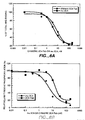
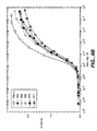
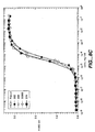
- the present invention is, at least partially, based on the unexpected finding that ErbB2 antagonists exhibit analgesic effects and are, therefore, useful in the management of pain, including chronic pain, such as cancer-related pain.
- prostate cancer patients treated with the humanized anti-ErbB2 antibody 2C4 recorded less pain and reduced analgesia even when their PSA value was not reduced.
- the analgesic effect of the humanized 2C4 antibody was observed during the treatment of liposarcoma.
- the invention relates to pain management using ErbB2 antagonists.
- the invention concerns the management of pain, including acute and chronic pain, cancer related, with ErbB2 antagonists.
- the invention concerns the treatment of any type of pain, including, without limitation, nociceptive pain, somatic pain, visceral pain, neuropathic pain, centrally generated pain (including deafferentiation pain and sympathetically maintained pain), and peripherally generated pain (including painful polyneuropathies and painful mononeuropethies).
- the ErbB antagonists of the present invention are molecules that block (reduce or prevent) a biological activity of ErbB2 Preferably, the antagonist blocks (reduces or prevents) ligand activation of an ErbB.
- the ErbB antagonists of the present invention inhibit a biological activity mediated by an ErbB2 (HER2) receptor or a receptor complex comprising such receptor(s).
- ErbB antagonists herein include, without limitation, polypeptides (including antibodies and antibody fragments), peptides, peptide mimetics, non-peptide small organic molecules, antisense molecules, and oligonucleotide decoy molecules.
- Small organic molecules that compete with ATP binding to the kinase pocket of EGFR are reviewed, for example, by Arteaga CL., Exp. Cell. Res. 284(1):122-30 (2003 ).
- a small molecule EGFR inhibitor, IressaTM ZD1839, gefinitib, Aztra-Zeneca is described, for example, in Kris M et al. JAMA, 290(16):2149-2158 (2003 ).
- Further small molecule EGFR antagonists include CP-358,774 (TARCEVA®; Genentech/OSI) and AG1478, AG1571 (SU 5271; Sugen).
- Anti-EGFR antibodies are also known in the art and include, for example, Erbitux® (IMC-C225, cetuximab, ImClone) a chimeric anti-EGFR MAb, and reshaped human 225 (H225) (see, WO 96/40210 , ImClone Systems Inc.). Further examples of antibodies which bind to EGFR include MAb 579 (ATCC CRL HB 8506), MAb 455 (ATCC CRL HB8507), MAb 225 (ATCC CRL 8508), MAb 528 (ATCC CRL 8509) (see, US Patent No. 4,943, 533, Mendelsohn et al.
- humanized and chimeric antibodies that bind EGFR as described in US Patent No. 5,891,996 and human antibodies that bind EGFR, such as ABX-EGF (see WO98/50433 , Abgenix).
- the anti-EGFR antibody may be conjugated with a cytotoxic agent, thus generating an immunoconjugate (see, e.g ., EP659,439A2 , Merck Patent GmbH).
- CP-654577 a selective inhibitor of ErbB2 relative to EGFR tyrosine kinase, is disclosed, for example, in Barbacci et al., Cancer Res. 63(15):4450-9 (2003 ).
- a small molecule form of anti-ErbB2 peptidomimetic is described by Zhang et al., Drug Nes Perspect 13(6):325-9 (2000 ).
- New agents that target and inhibit a biological activity of ErbB receptors can be identified by methods known in the art.
- the first step in identifying new ErbB antagonists is in vitro screening followed by in vivo assays in an apporpriate animal model and ultimately human clinical trials.
- receptor-binding tests can be performed using ErbB receptors or receptor complexes isolated from their respective native sources, or produced by recombinant DNA technology and/or chemical synthesis.
- the binding affinity of the candidate compounds can be tested by direct binding or by indirect, e.g. competitive, binding.
- competitive binding experiments the concentration of a compound necessary to displace 50% of another compound bound to the receptor (IC 50 ) is usually used as a measure of binding affinity.
- DNA encoding the sequence encoding the target ErbB receptor (e.g. ErbB2 or EGFR) is cloned into an expression vector containing a selectable marker.
- the vector is used to transfect recombinant host cells. Following several rounds of selection stable lines which express the ErbB receptor are identified. New ErbB antagonists can then be identified by virtue of their ability to compete effectively with a known inhibitor of the target ErbB receptor. Binding coefficients can be determined by any known manner, e.g. by Scatchard analysis.
- Binding experiments can also be performed using cells or cell lines known to express the target ErbB receptor.
- the ability of the molecule to block ErbB ligand binding to cells expressing the ErbB may be determined. For example, cells naturally expressing, or transfected to express, ErbBs of the ErbB hetero-oligomer may be incubated with the candidate molecule and then exposed to labeled ErbB ligand. The ability of the candidate molecule to block ligand binding to the ErbB in the ErbB hetero-oligomer may then be evaluated.
- the ability of a candidate molecule to block ErbB ligand-stimulated tyrosine phosphorylation of an ErbB present in an ErbB hetero-oligomer may be assessed.
- cells endogenously expressing the ErbBs or transfected to expressed them may be incubated with the candidate molecule and then assayed for ErbB ligand-dependent tyrosine phosphorylation activity using an anti-phosphotyrosine monoclonal (which is optionally conjugated with a detectable label).
- the kinase receptor activation assay described in U.S. Patent No. 5,766,863 is also available for determining ErbB activation and blocking of that activity by an antagonist.
- MDA-MB-175 cells may be treated with a candidate antagonist and stained with crystal violet. Incubation with a candidate antagonist may show a growth inhibitory effect on this cell line similar to that displayed by monoclonal antibody 2C4. In a further embodiment, exogenous HRG will not significantly reverse this inhibition.
- the antagonist will be able to inhibit cell proliferation of MDA-MB-175 cells to a greater extent than monoclonal antibody 4D5 (and optionally to a greater extent than monoclonal antibody 7F3), both in the presence and absence of exogenous HRG.
- the antagonist may block heregulin dependent association of ErbB2 with ErbB3 in both MCF7 and SK-BR-3 cells as determined in a co-immunoprecipitation experiment such as that described in Example 2 substantially more effectively than monoclonal antibody 4D5, and preferably substantially more effectively than monoclonal antibody 7F3.
- PI PI uptake assay
- an annexin binding assay using BT474 cells or a DNA staining assay using BT 474 cells can be used.
- ErbB2 and EGFR antagonists are identified by correlating EGFR, TGF- ⁇ (a ligand for EGFR), and ErbB2 mRNA expression levels with the results of cytotoxicity assays of the 49000 compounds in the National Cancer Institute (NCI) drug screen database ( Wosikowski et al., J. Natl. Cancer Inst. 89(20):1505-15 (1997 )).
- NCI National Cancer Institute
- the ability of a candidate antagonist to control pain can be tested in in vivo models of pain.
- the analgesic activity of a candidate ErbB antagonist is tested in an animal model used in pain research.
- a suitable animal model of noxious pain is the "tail flick" test ( Bass and Wanderbrook, J. Am. Pharm. Assoc. Sci. Ed. 41:569-70 (1952 ))
- This method is based on measuring pain sensitivity in mice or rats as they respond to the application of heat to a small area of their tails with or without administration of a candidate ErbB antagonist.
- the test is based on measuring the withdrawal time of the tail following application of radiant heat, and can be performed by using commercially available equipment, such as, for example, the Tail Flick Analgesia Meter (SDI, San Diego, CA).
- Another animal model of noxious pain is the "hot plate paw-licking" test.
- animals rats or mice
- the latency to first sign of hind paw licking is taken or jump response is taken as an index of nociceptive threshold.
- the effect of drug administration on this threshold is considered to be an index of analgesic response.
- the route of drug administration may differ but typically is subcutaneous.
- the paw-pressure, or mechanical hyperalgesia, test uses a pressure of increasing intensity applied to a punctiform area on the hindpaw or, less commonly, on the tail.
- the paw or tail is placed between a plane surface and a blunt, plastic-coated point mounted on top of a system of cogwheels, with a cursor that can be displaced along the length of a graduated beam ( Green et al., Br. J. Pharmacol. 6:572-85 (1957 ) for an automated readout.
- the application of increasing pressure is interrupted when the animal removes its tail, an action that is read out as force in grams for the threshold of response.
- Chemical stimuli can also be used to model noxious pain in animals.
- the chemical agents are administered intradermally or intraperitoneally.
- a 0.5 to 15% (generally about 3.5%) solution of formalin is injected into the dorsal or plantar surface of the rat fore- or hindpaw, and produces a painful response of increasing and decreasing intensity for about 60 minutes.
- Typical responses include paw lifting, licking, nibbling or shaking, which can be monitored in the absence and presence of a test substance ( Lariviere and Melzack, Pain 66:271-7 (1996 )).
- Visceral pain is typically tested by intraperitoneal injections of irritants (Writhing Test), such as, for example, acetic acid/ethacrinic acid or phenylbenzoquinone.
- the agents provoke a stereotypical response in rodents, charcterized by abdominal contractions, whol ebody movements, contortions of the abdominal muscles, reduced motor activity and incoordination. These responses are considered indicative of visceral pain associated with visceral chemoreceptors.
- the writhing test has been successfully used to predict effective analgesic doses for treatment of humans ( Dubinsky et al., Agents Actions 20:50-60 (1987 )).
- Inflammatory pain is typically modeled using p-benzoquinone-induced Writhing Test or the carrageean-induced hind paw edema model ( DiRosa et al., J. Pathol. 101:15-29 (1971 )).
- Such responses can be induced, for example, by TNF- ⁇ (typically 100 pg), IFN- ⁇ (typically 100 pg), or IL-1 ⁇ (typically 100 pg) injected intratracheally (i.t.), especially at higher dose (3 g/kg).
- TNF- ⁇ typically 100 pg
- IFN- ⁇ typically 100 pg
- IL-1 ⁇ typically 100 pg
- similar responses can be elicited by i.t.injections of glutamate (20 ⁇ g), N-methyl-D-aspartic acid (50 ng), ⁇ -amino-3-hydrocy-5-methylisoxazole-4-proprionic acid (13 ng) or kainic acid,or by administration fo substrance P or capsaicin.
- CCI Chronic constriction injury
- Candidate agents for the treatment of cancer pain can be tested in the above-listed animals models of acute and chronic pain or inflammatory pain. In addition, there are several animal models available that have been specifically designed to test cancer pain.
- a candidate ErbB antagonist can be tested in a murine model of bone cancer pain, as described in Menendez et al., Brain Res. 969(102):102-9 (2003 ).
- This test investigated the reactivity to noxious heat of C3G/HeJ mice receiving an intratibial (i.t.) injetion of NCTC 2472 cells.
- NCTC2472 cells are able to induce osteosarcoma, and break through bone into soft tissues about two weeks after inoculation, producing a macroscopical increase of the limb size from the fourth week. Thermal reactivity is diminished during the first two weeks after cell implantation, and this hyperalgesia is reversed by the administration of naloxone (10 mg/kg).
- a mouse model of neuropathic cancer pain has been developed by Shimoyama et al., Pain 99(1-2):167-74 (2002 ).
- Meth A sarcoma cells were inoculated to the immediate proximity of the scietic nerve in BALB/c mice. The growing tumor gradually compresses the nerve, thereby causing nerve injury.
- Time courses of thermal hyperalgesia and mechanical sensitivity to von Frey hairs are determined and signs of spontaneous pain are evaluated.
- Pain measurement is essential for assessing analgesia.
- the pain experienced by an individual patient is assessed by using one or more of the known measures of pain, including a Visual Analog Scale (VAS), descriptive scale, numeric scale, Health Assessment Questionare (HAQ) pain index, and the like.
- VAS Visual Analog Scale
- HAQ Health Assessment Questionare
- IMMPACT Initiative on Methods, Measurement, and Pain Assessment in Clinical Trials
- VAS Voice over Sense Pain Symptoms . ⁇ ⁇ ⁇ ⁇ ⁇ ⁇ ⁇ ⁇ ⁇ ⁇ ⁇ ⁇ ⁇ ⁇ ⁇ ⁇ ⁇ ⁇ ⁇ ⁇ ⁇ ⁇ ⁇ ⁇ ⁇ ⁇ ⁇ ⁇ ⁇ ⁇ ⁇ ⁇ ⁇ ⁇ ⁇ ⁇ ⁇ ⁇ ⁇ ⁇ ⁇ ⁇ ⁇ ⁇ ⁇ ⁇ ⁇ ⁇ ⁇ ⁇ ⁇ ⁇ ⁇ ⁇ ⁇ ⁇ ⁇ ⁇ ⁇ ⁇ ⁇ ⁇ ⁇ ⁇ ⁇ ⁇ ⁇ ⁇ ⁇ ⁇ ⁇ ⁇ ⁇ ⁇ ⁇ ⁇ ⁇ ⁇ ⁇ ⁇ ⁇ ⁇ ⁇ ⁇ ⁇ ⁇ ⁇ ⁇ ⁇ ⁇ ⁇ ⁇ ⁇ ⁇ ⁇ ⁇ ⁇ ⁇ ⁇ ⁇ ⁇ ⁇ ⁇ ⁇ ⁇ ⁇ ⁇ ⁇ ⁇ ⁇ ⁇ ⁇ ⁇ ⁇ ⁇ ⁇ ⁇ ⁇ ⁇ ⁇
- the descriptive scale typically includes the following descriptions: no pain, mild pain, moderate pain, severe pain, very severe pain, and worst possible pain.
- the ErbB antagonists of the present invention are anti-ErbB2 antibodies.
- the ErbB2 antigen to be used for production of antibodies may be, e.g ., a soluble form of the extracellular domain of ErbB2 or a portion thereof, containing the desired epitope.
- cells expressing ErbB2 at their cell surface e.g . NIH-3T3 cells transformed to overexpress ErbB2; or a carcinoma cell line such as SK-BR-3 cells, see Stancovski et al. PNAS (USA) 88:8691-8695 (1991 )
- Other forms of ErbB2 useful for generating antibodies will be apparent to those skilled in the art.
- a protein that is immunogenic in the species to be immunized e.g ., keyhole limpet hemocyanin, serum albumin, bovine
- Animals are immunized against the antigen, immunogenic conjugates, or derivatives by combining, e.g .,100 ⁇ g or 5 ⁇ g of the protein or conjugate (for rabbits or mice, respectively) with 3 volumes of Freund's complete adjuvant and injecting the solution intradermally at multiple sites.
- the animals are boosted with 1/5 to 1/10 the original amount ofpeptide or conjugate in Freund's complete adjuvant by subcutaneous injection at multiple sites.
- Seven to 14 days later the animals are bled and the serum is assayed for antibody titer. Animals are boosted until the titer plateaus.
- the animal is boosted with the conjugate of the same antigen, but conjugated to a different protein and/or through a different cross-linking reagent.
- Conjugates also can be made in recombinant cell culture as protein fusions.
- aggregating agents such as alum are suitably used to enhance the immune response.
- Monoclonal antibodies are obtained from a population of substantially homogeneous antibodies, i.e ., the individual antibodies comprising the population are identical except for variants that may arise during the production of the antibody.
- the modifier "monoclonal" indicates the character of the antibody as not being a mixture of discrete antibodies.
- the monoclonal antibodies may be made using the hybridoma method first described by Kohler et al., Nature, 256:495 (1975 ), or may be made by recombinant DNA methods ( U.S. Patent No. 4,816,567 ).
- a mouse or other appropriate host animal such as a hamster
- lymphocytes that produce or are capable of producing antibodies that will specifically bind to the protein used for immunization.
- lymphocytes may be immunized in vitro. Lymphocytes then are fused with myeloma cells using a suitable fusing agent, such as polyethylene glycol, to form a hybridoma cell ( Goding, Monoclonal Antibodies: Principles and Practice, pp.59-103 (Academic Press, 1986 )).
- the hybridoma cells thus prepared are seeded and grown in a suitable culture medium that preferably contains one or more substances that inhibit the growth or survival of the unfused, parental myeloma cells.
- a suitable culture medium that preferably contains one or more substances that inhibit the growth or survival of the unfused, parental myeloma cells.
- the culture medium for the hybridomas typically will include hypoxanthine, aminopterin, and thymidine (HAT medium), which substances prevent the growth of HGPRT-deficient cells.
- Preferred myeloma cells are those that fuse efficiently, support stable high-level production of antibody by the selected antibody-producing cells, and are sensitive to a medium such as HAT medium.
- preferred myeloma cell lines are murine myeloma lines, such as those derived from MOPC-21 and MPC-11 mouse tumors available from the Salk Institute Cell Distribution Center, San Diego, California USA, and SP-2 or X63-Ag8-653 cells available from the American Type Culture Collection, Rockville, Maryland USA.
- Human myeloma and mouse-human heteromyeloma cell lines also have been described for the production of human monoclonal antibodies ( Kozbor, J. Immunol., 133:3001 (1984 ); and Brön et al., Monoclonal Antibody Production Techniques and Applications, pp. 51-63 (Marcel Dekker, Inc., New York, 1987 )).
- Culture medium in which hybridoma cells are growing is assayed for production of monoclonal antibodies directed against the antigen.
- the binding specificity of monoclonal antibodies produced by hybridoma cells is determined by immunoprecipitation or by an in vitro binding assay, such as radioimmunoassay (RIA) or enzyme-linked immunoabsorbent assay (ELISA).
- RIA radioimmunoassay
- ELISA enzyme-linked immunoabsorbent assay
- the binding affinity of the monoclonal antibody can, for example, be determined by the Scatchard analysis of Munson et al., Anal. Biochem., 107:220 (1980 ).
- the clones may be subcloned by limiting dilution procedures and grown by standard methods ( Goding, Monoclonal Antibodies: Principles and Practice, pp.59-103 (Academic Press, 1986 )). Suitable culture media for this purpose include, for example, D-MEM or RPMI-1640 medium.
- the hybridoma cells may be grown in vivo as ascites tumors in an animal.
- the monoclonal antibodies secreted by the subclones are suitably separated from the culture medium, ascites fluid, or serum by conventional antibody purification procedures such as, for example, protein A-Sepharose, hydroxylapatite chromatography, gel electrophoresis, dialysis, or affinity chromatography.
- DNA encoding the monoclonal antibodies is readily isolated and sequenced using conventional procedures (e.g ., by using oligonucleotide probes that are capable of binding specifically to genes encoding the heavy and light chains of murine antibodies).
- the hybridoma cells serve as a preferred source of such DNA.
- the DNA may be placed into expression vectors, which are then transfected into host cells such as E . coli cells, simian COS cells, Chinese Hamster Ovary (CHO) cells, or myeloma cells that do not otherwise produce antibody protein, to obtain the synthesis of monoclonal antibodies in the recombinant host cells.
- monoclonal antibodies or antibody fragments can be isolated from antibody phage libraries generated using the techniques described in McCafferty et al., Nature, 348:552-554 (1990 ). Clackson et al., Nature, 352:624-628 (1991 ) and Marks et al., J. Mol. Biol., 222:581-597 (1991 ) describe the isolation of murine and human antibodies, respectively, using phage libraries.
- the DNA also may be modified, for example, by substituting the coding sequence for human heavy chain and light chain constant domains in place of the homologous murine sequences ( U.S. Patent No. 4,816,567 ; and Morrison, et al., Proc. Natl Acad. Sci. USA, 81:6851 (1984 )), or by covalently joining to the immunoglobulin coding sequence all or part of the coding sequence for a non-immunoglobulin polypeptide.
- non-immunoglobulin polypeptides are substituted for the constant domains of an antibody, or they are substituted for the variable domains of one antigen-combining site of an antibody to create a chimeric bivalent antibody comprising one antigen-combining site having specificity for an antigen and another antigen-combining site having specificity for a different antigen.
- a humanized antibody has one or more amino acid residues introduced into it from a source which is non-human. These non-human amino acid residues are often referred to as "import" residues, which are typically taken from an "import” variable domain.
- Humanization can be essentially performed following the method of Winter and co-workers ( Jones et al., Nature, 321:522-525 (1986 ); Riechmann et al., Nature, 332:323-327 (1988 ); Verhoeyen et al., Science, 239:1534-1536 (1988 )), by substituting hypervariable region sequences for the corresponding sequences of a human antibody.
- humanized antibodies are chimeric antibodies ( U.S. Patent No. 4,816,567 ) wherein substantially less than an intact human variable domain has been substituted by the corresponding sequence from a non-human species.
- humanized antibodies are typically human antibodies in which some hypervariable region residues and possibly some FR residues are substituted by residues from analogous sites in rodent antibodies.
- variable domains both light and heavy
- sequence of the variable domain of a rodent antibody is screened against the entire library of known human variable-domain sequences.
- the human sequence which is closest to that of the rodent is then accepted as the human framework region (FR) for the humanized antibody ( Sims et al., J. Immunol., 151:2296 (1993 ); Chothia et al., J. Mol. Biol., 196:901 (1987 )).
- Another method uses a particular framework region derived from the consensus sequence of all human antibodies of a particular subgroup of light or heavy chains.
- the same framework may be used for several different humanized antibodies ( Carter et al., Proc. Natl. Acad. Sci. USA, 89:4285 (1992 ); Presta et al., J. Immunol., 151:2623 (1993 )).
- humanized antibodies are prepared by a process of analysis of the parental sequences and various conceptual humanized products using three-dimensional models of the parental and humanized sequences.
- Three-dimensional immunoglobulin models are commonly available and are familiar to those skilled in the art.
- Computer programs are available which illustrate and display probable three-dimensional conformational structures of selected candidate immunoglobulin sequences. Inspection of these displays permits analysis of the likely role of the residues in the functioning of the candidate immunoglobulin sequence, i.e ., the analysis of residues that influence the ability of the candidate immunoglobulin to bind its antigen.
- FR residues can be selected and combined from the recipient and import sequences so that the desired antibody characteristic, such as increased affinity for the target antigen(s), is achieved.
- the hypervariable region residues are directly and most substantially involved in influencing antigen binding.
- Example 3 describes production of exemplary humanized anti-ErbB2 antibodies which bind ErbB2 and block ligand activation of an ErbB.
- the humanized antibody of particular interest herein blocks EGF, TGF- ⁇ and/or HRG mediated activation of MAPK essentially as effectively as murine monoclonal antibody 2C4 (or a Fab fragment thereof) and/or binds ErbB2 essentially as effectively as murine monoclonal antibody 2C4 (or a Fab fragment thereof).
- the humanized antibody herein may, for example, comprise nonhuman hypervariable region residues incorporated into a human variable heavy domain and may further comprise a framework region (FR) substitution at a position selected from the group consisting of 69H, 71H and 73H utilizing the variable domain numbering system set forth in Kabat et al., Sequences of Proteins of Immunological Interest, 5th Ed. Public Health Service, National Institutes of Health, Bethesda, MD (1991 ).
- the humanized antibody comprises FR substitutions at two or all ofpositions 69H, 71H and 73H.
- An exemplary humanized antibody of interest herein comprises variable heavy domain complementarity determining residues GFTFTDYTMX (SEQ ID NO:14), where X is preferrably D or S (SEQ ID NO:7); DVNPNSGGSIYNQRFKG (SEQ ID NO:8); and/or NLGPSFYFDY (SEQ ID NO:9), optionally comprising amino acid modifications of those CDR residues, e.g . where the modifications essentially maintain or improve affinity of the antibody.
- the antibody variant of interest may have from about one to about seven or about five amino acid substitutions in the above variable heavy CDR sequences.
- Such antibody variants may be prepared by affinity maturation, e.g ., as described below.
- the most preferred humanized antibody comprises the variable heavy domain amino acid sequence in SEQ ID NO:4.
- the humanized antibody may comprise variable light domain complementarity determining residues KASQDVSIGVA (SEQ ID NO:10); SASYX 1 X 2 X 3 , where X 1 is preferably R or L, X 2 is preferably Y or E, and X 3 is preferably T or S (SEQ ID NO:11); and/or QQYYIYPYT (SEQ ID NO:12), e.g . in addition to those variable heavy domain CDR residues in the preceding paragraph.
- Such humanized antibodies optionally comprise amino acid modifications of the above CDR residues, e.g . where the modifications essentially maintain or improve affinity of the antibody.
- the antibody variant of interest may have from about one to about seven or about five amino acid substitutions in the above variable light CDR sequences.
- Such antibody variants may be prepared by affinity maturation, e.g ., as described below.
- the most preferred humanized antibody comprises the variable light domain amino acid sequence in SEQ ID NO:3.
- the present application also contemplates affinity matured antibodies which bind ErbB2 and block ligand activation of an ErbB.
- the parent antibody may be a human antibody or a humanized antibody, e.g ., one comprising the variable light and/or heavy sequences of SEQ ID Nos. 3 and 4, respectively ( i.e . variant 574).
- the affinity matured antibody preferably binds to ErbB2 receptor with an affinity superior to that of murine 2C4 or variant 574 ( e.g . from about two or about four fold, to about 100 fold or about 1000 fold improved affinity, e.g . as assessed using a ErbB2-extracellular domain (ECD) ELISA).
- ECD ErbB2-extracellular domain
- variable heavy CDR residues for substitution include H28, H30, H34, H35, H64, H96, H99, or combinations of two or more ( e.g . two, three, four, five, six, or seven of these residues).
- variable light CDR residues for alteration include L28, L50, L53, L56, L91, L92, L93, L94, L96, L97 or combinations of two or more ( e.g . two to three, four, five or up to about ten of these residues).
- the humanized antibody or affinity matured antibody may be an antibody fragment, such as a Fab, which is optionally conjugated with one or more cytotoxic agent(s) in order to generate an immunoconjugate.
- the humanized antibody or affinity matured antibody may be an intact antibody, such as an intact IgG1 antibody.
- human antibodies can be generated.
- transgenic animals e.g ., mice
- transgenic animals e.g ., mice
- J H antibody heavy-chain joining region
- phage display technology McCafferty et al., Nature 348:552-553 (1990 )
- V domain gene repertoires from unimmunized donors.
- antibody V domain genes are cloned in-frame into either a major or minor coat protein gene of a filamentous bacteriophage, such as M13 or fd, and displayed as functional antibody fragments on the surface of the phage particle. Because the filamentous particle contains a single-stranded DNA copy of the phage genome, selections based on the functional properties of the antibody also result in selection of the gene encoding the antibody exhibiting those properties.
- the phage mimics some of the properties of the B-cell.
- Phage display can be performed in a variety of formats; for their review see, e.g ., Johnson, Kevin S. and Chiswell, David J., Current Opinion in Structural Biology 3:564-571 (1993 ).
- V-gene segments can be used for phage display. Clackson et al., Nature, 352:624-628 (1991 ) isolated a diverse array of anti-oxazolone antibodies from a small random combinatorial library of V genes derived from the spleens of immunized mice.
- a repertoire of V genes from unimmunized human donors can be constructed and antibodies to a diverse array of antigens (including self-antigens) can be isolated essentially following the techniques described by Marks et al., J. Mol. Biol. 222:581-597 (1991 ), or Griffith et al., EMBO J. 12:725-734 (1993 ). See, also, U.S. Patent Nos. 5,565,332 and 5,573,905 .
- human antibodies may also be generated by in vitro activated B cells (see U.S. Patents 5,567,610 and 5,229,275 ).
- F(ab') 2 fragments can be isolated directly from recombinant host cell culture.
- the antibody of choice is a single chain Fv fragment (scFv). See WO 93/16185 ; U.S. Patent No. 5,571,894 ; and U.S. Patent No. 5,587,458 .
- the antibody fragment may also be a "linear antibody", e.g ., as described in U.S. Patent 5,641,870 for example. Such linear antibody fragments may be monospecific or bispecific.
- Single chain intracellular antibodies (sFv) that bind ErbB2 are described in WO01/56604 and US Patent No. 6,028,059 .
- Bispecific antibodies are antibodies that have binding specificities for at least two different epitopes. Exemplary bispecific antibodies may bind to two different epitopes of the ErbB2 protein. Other such antibodies may combine an ErbB2 binding site with binding site(s) for EGFR, ErbB3 and/or ErbB4. Alternatively, an anti-ErbB2 arm may be combined with an arm which binds to a triggering molecule on a leukocyte such as a T-cell receptor molecule ( e.g .
- Bispecific antibodies may also be used to localize cytotoxic agents to cells which express ErbB2. These antibodies possess an ErbB2-binding arm and an arm which binds the cytotoxic agent (e.g . saporin, anti-interferon- ⁇ , vinca alkaloid, ricin A chain, methotrexate or radioactive isotope hapten). Bispecific antibodies can be prepared as full length antibodies or antibody fragments ( e.g . F(ab') 2 bispecific antibodies).
- WO 96/16673 describes a bispecific anti-ErbB2/anti-Fc ⁇ RIII antibody and U.S. Patent No. 5,837,234 discloses a bispecific anti-ErbB2/anti-Fc ⁇ RI antibody. A bispecific anti-ErbB2/Fc ⁇ antibody is shown in WO98/02463 .
- U.S. Patent No. 5,821,337 teaches a bispecific anti-ErbB2/anti-CD3 antibody.
- bispecific antibodies are known in the art. Traditional production of full length bispecific antibodies is based on the coexpression of two immunoglobulin heavy chain-light chain pairs, where the two chains have different specificities ( Milstein et al., Nature, 305:537-539 (1983 )). Because of the random assortment of immunoglobulin heavy and light chains, these hybridomas (quadromas) produce a potential mixture of 10 different antibody molecules, of which only one has the correct bispecific structure. Purification of the correct molecule, which is usually done by affinity chromatography steps, is rather cumbersome, and the product yields are low. Similar procedures are disclosed in WO 93/08829 , and in Traunecker et al., EMBO J., 10:3655-3659 (1991 ).
- antibody variable domains with the desired binding specificities are fused to immunoglobulin constant domain sequences.
- the fusion preferably is with an immunoglobulin heavy chain constant domain, comprising at least part of the hinge, CH2, and CH3 regions. It is preferred to have the first heavy-chain constant region (CH1) containing the site necessary for light chain binding, present in at least one of the fusions.
- DNAs encoding the immunoglobulin heavy chain fusions and, if desired, the immunoglobulin light chain are inserted into separate expression vectors, and are co-transfected into a suitable host organism.
- the bispecific antibodies are composed of a hybrid immunoglobulin heavy chain with a first binding specificity in one arm, and a hybrid immunoglobulin heavy chain-light chain pair (providing a second binding specificity) in the other arm. It was found that this asymmetric structure facilitates the separation of the desired bispecific compound from unwanted immunoglobulin chain combinations, as the presence of an immunoglobulin light chain in only one half of the bispecific molecule provides for a facile way of separation. This approach is disclosed in WO 94/04690 . For further details of generating bispecific antibodies see, for example, Suresh et al., Methods in Enzymology, 121:210 (1986 ).
- the interface between a pair of antibody molecules can be engineered to maximize the percentage of heterodimers which are recovered from recombinant cell culture.
- the preferred interface comprises at least a part of the C H 3 domain of an antibody constant domain.
- one or more small amino acid side chains from the interface of the first antibody molecule are replaced with larger side chains (e.g . tyrosine or tryptophan).
- Compensatory "cavities" of identical or similar size to the large side chain(s) are created on the interface of the second antibody molecule by replacing large amino acid side chains with smaller ones ( e.g . alanine or threonine). This provides a mechanism for increasing the yield of the heterodimer over other unwanted end-products such as homodimers.
- Bispecific antibodies include cross-linked or "heteroconjugate" antibodies.
- one of the antibodies in the heteroconjugate can be coupled to avidin, the other to biotin.
- Such antibodies have, for example, been proposed to target immune system cells to unwanted cells ( U.S. Patent No. 4,676,980 ), and for treatment of HIV infection ( WO 91/00360 , WO 92/200373 , and EP 03089 ).
- Heteroconjugate antibodies may be made using any convenient cross-linking methods. Suitable cross-linking agents are well known in the art, and are disclosed in U.S. Patent No. 4,676,980 , along with a number of cross-linking techniques.
- bispecific antibodies can be prepared using chemical linkage.
- Brennan et al., Science, 229: 81 (1985 ) describe a procedure wherein intact antibodies are proteolytically cleaved to generate F(ab') 2 fragments. These fragments are reduced in the presence of the dithiol complexing agent sodium arsenite to stabilize vicinal dithiols and prevent intermolecular disulfide formation.
- the Fab' fragments generated are then converted to thionitrobenzoate (TNB) derivatives.
- One of the Fab'-TNB derivatives is then reconverted to the Fab'-thiol by reduction with mercaptoethylamine and is mixed with an equimolar amount of the other Fab'-TNB derivative to form the bispecific antibody.
- the bispecific antibodies produced can be used as agents for the selective immobilization of enzymes.
- bispecific antibodies have been produced using leucine zippers.
- the leucine zipper peptides from the Fos and Jun proteins were linked to the Fab' portions of two different antibodies by gene fusion.
- the antibody homodimers were reduced at the hinge region to form monomers and then re-oxidized to form the antibody heterodimers. This method can also be utilized for the production of antibody homodimers.
- the fragments comprise a heavy-chain variable domain (V H ) connected to a light-chain variable domain (V L ) by a linker which is too short to allow pairing between the two domains on the same chain. Accordingly, the V H and V L domains of one fragment are forced to pair with the complementary V L and V H domains of another fragment, thereby forming two antigen-binding sites.
- V H and V L domains of one fragment are forced to pair with the complementary V L and V H domains of another fragment, thereby forming two antigen-binding sites.
- sFv single-chain Fv
- Antibodies with more than two valencies are contemplated.
- trispecific antibodies can be prepared. Tutt et al. J. Immunol. 147: 60 (1991 ). US Patent No. 6,270,765B1 published August 7, 2001 describes trispecific antibodies that bind ErbB2, EGFR and FcR.
- Amino acid sequence modification(s) of the anti-ErbB2 antibodies described herein are contemplated. For example, it may be desirable to improve the binding affinity and/or other biological properties of the antibody.
- Amino acid sequence variants of the anti-ErbB2 antibody are prepared by introducing appropriate nucleotide changes into the anti-ErbB2 antibody nucleic acid, or by peptide synthesis. Such modifications include, for example, deletions from, and/or insertions into and/or substitutions of, residues within the amino acid sequences of the anti-ErbB2 antibody. Any combination of deletion, insertion, and substitution is made to arrive at the final construct, provided that the final construct possesses the desired characteristics.
- the amino acid changes also may alter post-translational processes of the anti-ErbB2 antibody, such as changing the number or position of glycosylation sites.
- a useful method for identification of certain residues or regions of the anti-ErbB2 antibody that are preferred locations for mutagenesis is called "alanine scanning mutagenesis" as described by Cunningham and Wells Science, 244:1081-1085 (1989 ).
- a residue or group of target residues are identified (e.g ., charged residues such as arg, asp, his, lys, and glu) and replaced by a neutral or negatively charged amino acid (most preferably alanine or polyalanine) to affect the interaction of the amino acids with ErbB2 antigen.
- Those amino acid locations demonstrating functional sensitivity to the substitutions then are refined by introducing further or other variants at, or for, the sites of substitution.
- the site for introducing an amino acid sequence variation is predetermined, the nature of the mutation per se need not be predetermined.
- ala scanning or random mutagenesis is conducted at the target codon or region and the expressed anti-ErbB2 antibody variants are screened for the desired activity.
- Amino acid sequence insertions include amino- and/or carboxyl-terminal fusions ranging in length from one residue to polypeptides containing a hundred or more residues, as well as intrasequence insertions of single or multiple amino acid residues.
- terminal insertions include an anti-ErbB2 antibody with an N-terminal methionyl residue or the antibody fused to a cytotoxic polypeptide.
- Other insertional variants of the anti-ErbB2 antibody molecule include the fusion to the - or C-terminus of the anti-ErbB2 antibody to an enzyme (e.g . for ADEPT) or a polypeptide which increases the serum half-life of the antibody.
- variants are an amino acid substitution variant. These variants have at least one amino acid residue in the anti-ErbB2 antibody molecule replaced by a different residue.
- the sites of greatest interest for substitutional mutagenesis include the hypervariable regions, but FR alterations are also contemplated. Conservative substitutions are shown in Table 1 under the heading of "preferred substitutions". If such substitutions result in a change in biological activity, then more substantial changes, denominated "exemplary substitutions" in Table 1, or as further described below in reference to amino acid classes, may be introduced and the products screened.
- Substantial modifications in the biological properties of the antibody are accomplished by selecting substitutions that differ significantly in their effect on maintaining (a) the structure of the polypeptide backbone in the area of the substitution, for example, as a sheet or helical conformation, (b) the charge or hydrophobicity of the molecule at the target site, or (c) the bulk of the side chain.
- Naturally occurring residues are divided into groups based on common side-chain properties:
- Non-conservative substitutions will entail exchanging a member of one of these classes for another class.
- cysteine residues not involved in maintaining the proper conformation of the anti-ErbB2 antibody also may be substituted, generally with serine, to improve the oxidative stability of the molecule and prevent aberrant crosslinking.
- cysteine bond(s) may be added to the antibody to improve its stability (particularly where the antibody is an antibody fragment such as an Fv fragment).
- a particularly preferred type of substitutional variant involves substituting one or more hypervariable region residues of a parent antibody (e.g . a humanized or human antibody).
- a parent antibody e.g . a humanized or human antibody
- the resulting variant(s) selected for further development will have improved biological properties relative to the parent antibody from which they are generated.
- a convenient way for generating such substitutional variants involves affinity maturation using phage display. Briefly, several hypervariable region sites (e.g . 6-7 sites) are mutated to generate all possible amino substitutions at each site.
- the antibody variants thus generated are displayed in a monovalent fashion from filamentous phage particles as fusions to the gene III product of M13 packaged within each particle. The phage-displayed variants are then screened for their biological activity (e.g .
- alanine scanning mutagenesis can be performed to identify hypervariable region residues contributing significantly to antigen binding.
- Such contact residues and neighboring residues are candidates for substitution according to the techniques elaborated herein.
- Another type of amino acid variant of the antibody alters the original glycosylation pattern of the antibody. By altering is meant deleting one or more carbohydrate moieties found in the antibody, and/or adding one or more glycosylation sites that are not present in the antibody.
- N-linked refers to the attachment of the carbohydrate moiety to the side chain of an asparagine residue.
- the tripeptide sequences asparagine-X-serine and asparagine-X-threonine, where X is any amino acid except proline, are the recognition sequences for enzymatic attachment of the carbohydrate moiety to the asparagine side chain.
- X is any amino acid except proline
- O-linked glycosylation refers to the attachment of one of the sugars N-aceylgalactosamine, galactose, or xylose to a hydroxyamino acid, most commonly serine or threonine, although 5-hydroxyproline or 5-hydroxylysine may also be used.
- glycosylation sites to the antibody is conveniently accomplished by altering the amino acid sequence such that it contains one or more of the above-described tripeptide sequences (for N-linked glycosylation sites).
- the alteration may also be made by the addition of, or substitution by, one or more serine or threonine residues to the sequence of the original antibody (for O-linked glycosylation sites).
- Nucleic acid molecules encoding amino acid sequence variants of the anti-ErbB2 antibody are prepared by a variety of methods known in the art. These methods include, but are not limited to, isolation from a natural source (in the case of naturally occurring amino acid sequence variants) or preparation by oligonucleotide-mediated (or site-directed) mutagenesis, PCR mutagenesis, and cassette mutagenesis of an earlier prepared variant or a non-variant version of the anti-ErbB2 antibody.
- ADCC antigen-dependent cell-mediated cytotoxicity
- CDC complement dependent cytotoxicity
- This may be achieved by introducing one or more amino acid substitutions in an Fc region of the antibody.
- cysteine residue(s) may be introduced in the Fc region, thereby allowing interchain disulfide bond formation in this region.
- the homodimeric antibody thus generated may have improved internalization capability and/or increased complement-mediated cell killing and antibody-dependent cellular cytotoxicity (ADCC). See Caron et al., J. Exp Med. 176:1191-1195 (1992 ) and Shopes, B. J. Immunol.
- Homodimeric antibodies with enhanced activity may also be prepared using heterobifunctional cross-linkers as described in Wolff et al. Cancer Research 53:2560-2565 (1993 ).
- an antibody can be engineered which has dual Fc regions and may thereby have enhanced complement lysis and ADCC capabilities. See Stevenson et al. Anti-Cancer Drug Design 3:219-230 (1989 ).
- Antibody variants with altered Fc region sequence and improved or diminished C1q binding are described in WO99/51642 .
- Antibody variants with altered Fc region sequences and altered FcR binding function are described in WO00/42072 .
- a salvage receptor binding epitope refers to an epitope of the Fc region of an IgG molecule (e.g ., IgG 1 , IgG 2 , IgG 3 , or IgG 4 ) that is responsible for increasing the in vivo serum half-life of the IgG molecule.
- the ability of the antibody to block ErbB ligand binding to cells expressing the ErbB may be determined. For example, cells naturally expressing, or transfected to express, ErbBs of the ErbB hetero-oligomer may be incubated with the antibody and then exposed to labeled ErbB ligand. The ability of the anti-ErbB2 antibody to block ligand binding to the ErbB in the ErbB hetero-oligomer may then be evaluated.
- inhibition of HRG binding to MCF7 breast tumor cell lines by anti-ErbB2 antibodies may be performed using monolayer MCF7 cultures on ice in a 24-well-plate format essentially as described in Example 1 below.
- Anti-ErbB2 monoclonal antibodies may be added to each well and incubated for 30 minutes.
- 125 I-labeled rHRG ⁇ 1 177-224 25 pm
- Dose response curves may be prepared and an IC 50 value may be calculated for the antibody of interest.
- the antibody which blocks ligand activation of an ErbB will have an IC 50 for inhibiting HRG binding to MCF7 cells in this assay of about 50nM, or less, more preferably 10nM or less.
- the IC 50 for inhibiting HRG binding to MCF7 cells in this assay may, for example, be about 100nM or less, more preferably 50nM or less.
- Other, non-antibody, ErbB antagonist candidates can be tested in a similar manner.
- the ability of the anti-ErbB2 antibody to block ErbB ligand-stimulated tyrosine phosphorylation of an ErbB present in an ErbB hetero-oligomer may be assessed.
- cells endogenously expressing the ErbBs or transfected to expressed them may be incubated with the antibody and then assayed for ErbB ligand-dependent tyrosine phosphorylation activity using an anti-phosphotyrosine monoclonal (which is optionally conjugated with a detectable label).
- the kinase receptor activation assay described in U.S. Patent No. 5,766,863 is also available for determining ErbB activation and blocking of that activity by an antibody.
- one may screen for an antibody which inhibits HRG stimulation of p180 tyrosine phosphorylation in MCF7 cells essentially as described in Example 1 below.
- the MCF7 cells may be plated in 24-well plates and monoclonal antibodies to ErbB2 may be added to each well and incubated for 30 minutes at room temperature; then rHRG ⁇ 1 177-244 may be added to each well to a final concentration of 0.2 nM, and the incubation may be continued for 8 minutes. Media may be aspirated from each well, and reactions may be stopped by the addition of 100 ⁇ l of SDS sample buffer (5% SDS, 25 mM DTT, and 25 mM Tris-HCl, pH 6.8).
- SDS sample buffer 5% SDS, 25 mM DTT, and 25 mM Tris-HCl, pH 6.8.
- Each sample (25 ⁇ l) may be electrophoresed on a 4-12% gradient gel (Novex) and then electrophoretically transferred to polyvinylidene difluoride membrane.
- Antiphosphotyrosine (at 1 ⁇ g/ml) immunoblots may be developed, and the intensity of the predominant reactive band at M r ⁇ 180,000 may be quantified by reflectance densitometry.
- the antibody selected will preferably significantly inhibit HRG stimulation of p180 tyrosine phosphorylation to about 0-35% of control in this assay.
- a dose-response curve for inhibition of HRG stimulation of p180 tyrosine phosphorylation as determined by reflectance densitometry may be prepared and an IC 50 for the antibody of interest may be calculated.
- the antibody which blocks ligand activation of an ErbB will have an IC 50 for inhibiting HRG stimulation of p180 tyrosine phosphorylation in this assay of about 50nM or less, more preferably 10nM or less.
- the IC 50 for inhibiting HRG stimulation of p180 tyrosine phosphorylation in this assay may, for example, be about 100nM or less, more preferably 50nM or less.
- MDA-MB-175 cells may also assess the growth inhibitory effects of the antibody on MDA-MB-175 cells, e.g , essentially as described in Schaefer et al. Oncogene 15:1385-1394 (1997 ).
- MDA-MB-175 cells may treated with an anti-ErbB2 monoclonal antibody (10 ⁇ g/mL) for 4 days and stained with crystal violet.
- Incubation with an anti-ErbB2 antibody may show a growth inhibitory effect on this cell line similar to that displayed by monoclonal antibody 2C4.
- exogenous HRG will not significantly reverse this inhibition.
- the antibody will be able to inhibit cell proliferation of MDA-MB-175 cells to a greater extent than monoclonal antibody 4D5 (and optionally to a greater extent than monoclonal antibody 7F3), both in the presence and absence of exogenous HRG.
- the anti-ErbB2 antibody of interest may block heregulin dependent association of ErbB2 with ErbB3 in both MCF7 and SK-BR-3 cells as determined in a co-immunoprecipitation experiment such as that described in Example 2 substantially more effectively than monoclonal antibody 4D5, and preferably substantially more effectively than monoclonal antibody 7F3.
- the growth inhibitory antibody of choice is able to inhibit growth of SK-BR-3 cells in cell culture by about 20-100% and preferably by about 50-100% at an antibody concentration of about 0.5 to 30 ⁇ g/ml.
- the SK-BR-3 assay described in U.S. Patent No. 5,677,171 can be performed. According to this assay, SK-BR-3 cells are grown in a 1:1 mixture of F12 and DMEM medium supplemented with 10% fetal bovine serum, glutamine and penicillin streptomycin.
- the SK-BR-3 cells are plated at 20,000 cells in a 35mm cell culture dish (2mls/35mm dish). 0.5 to 30 ⁇ g/ml of the anti-ErbB2 antibody is added per dish. After six days, the number of cells, compared to untreated cells are counted using an electronic COULTER ⁇ cell counter. Those antibodies which inhibit growth of the SK-BR-3 cells by about 20-100% or about 50-100% may be selected as growth inhibitory antibodies.
- the preferred assay is the PI uptake assay using BT474 cells.
- BT474 cells which can be obtained from the American Type Culture Collection (Rockville, MD)
- D-MEM Dulbecco's Modified Eagle Medium
- Ham's F-12 50:50
- heat-inactivated FBS Hyclone
- 2 mM L-glutamine 2 mM L-glutamine
- the BT474 cells are seeded at a density of 3 x 10 6 per dish in 100 x 20 mm dishes and allowed to attach overnight. The medium is then removed and replaced with fresh medium alone or medium containing 10 ⁇ g/ml of the appropriate monoclonal antibody. The cells are incubated for a 3 day time period. Following each treatment, monolayers are washed with PBS and detached by trypsinization.
- Cells are then centrifuged at 1200rpm for 5 minutes at 4°C, the pellet resuspended in 3 ml ice cold Ca 2+ binding buffer (10 mM Hepes, pH 7.4, 140 mM NaCl, 2.5 mM CaCl 2 ) and aliquoted into 35 mm strainer-capped 12 x 75 tubes (1ml per tube, 3 tubes per treatment group) for removal of cell clumps. Tubes then receive PI (10 ⁇ g/ml). Samples may be analyzed using a FACSCAN® flow cytometer and FACSCONVERT® CellQuest software (Becton Dickinson). Those antibodies which induce statistically significant levels of cell death as determined by PI uptake may be selected as cell death-inducing antibodies.
- an annexin binding assay using BT474 cells is available.
- the BT474 cells are cultured and seeded in dishes as discussed in the preceding paragraph.
- the medium is then removed and replaced with fresh medium alone or medium containing 10 ⁇ g/ml of the monoclonal antibody.
- monolayers are washed with PBS and detached by trypsinization.
- Cells are then centrifuged, resuspended in Ca 2+ binding buffer and aliquoted into tubes as discussed above for the cell death assay. Tubes then receive labeled annexin (e.g. annexin V-FTIC) (1 ⁇ g/ml).
- labeled annexin e.g. annexin V-FTIC
- Samples may be analyzed using a FACSCAN® flow cytometer and FACSCONVERT® CellQuest software (Becton Dickinson). Those antibodies which induce statistically significant levels of annexin binding relative to control are selected as apoptosis-inducing antibodies.
- a DNA staining assay using BT474 cells is available.
- BT474 cells which have been treated with the antibody of interest as described in the preceding two paragraphs are incubated with 9 ⁇ g/ml HOECHST 33342® for 2 hr at 37°C, then analyzed on an EPICS ELITE® flow cytometer (Coulter Corporation) using MODFIT LT® software (Verity Software House).
- Antibodies which induce a change in the percentage of apoptotic cells which is 2 fold or greater (and preferably 3 fold or greater) than untreated cells (up to 100% apoptotic cells) may be selected as pro-apoptotic antibodies using this assay.
- the antibodies identified as antagonists can then be tested in any of the animal models of pain discussed above.
- the ErbB antagonist antibodies of the invention can also be in the form of immunoconjugates comprising an antibody conjugated to a cytotoxic agent such as a chemotherapeutic agent, toxin (e.g . a small molecule toxin or an enzymatically active toxin of bacterial, fungal, plant or animal origin, including fragments and/or variants thereof), or a radioactive isotope (i.e., a radioconjugate).
- a cytotoxic agent such as a chemotherapeutic agent, toxin (e.g . a small molecule toxin or an enzymatically active toxin of bacterial, fungal, plant or animal origin, including fragments and/or variants thereof), or a radioactive isotope (i.e., a radioconjugate).
- Conjugates of an antibody and one or more small molecule toxins such as a calicheamicin, a maytansine ( U.S. Patent No. 5,208,020 ), a trichothene, and CC1065 are also contemplated herein.
- the antibody is conjugated to one or more maytansine molecules (e.g . about 1 to about 10 maytansine molecules per antibody molecule).
- Maytansine may, for example, be converted to May-SS-Me which may be reduced to May-SH3 and reacted with modified antibody ( Chari et al. Cancer Research 52: 127-131 (1992 )) to generate a maytansinoid-antibody immunoconjugate.
- Another immunoconjugate of interest comprises an anti-ErbB2 antibody conjugated to one or more calicheamicin molecules.
- the calicheamicin family of antibiotics are capable of producing double-stranded DNA breaks at sub-picomolar concentrations.
- Structural analogues of calicheamicin which may be used include, but are not limited to, ⁇ 1 I , ⁇ 2 I , ⁇ 3 I , N-acetyl- ⁇ 1 I , PSAG and ⁇ I 1 ( Hinman et al. Cancer Research 53: 3336-3342 (1993 ) and Lode et al. Cancer Research 58: 2925-2928 (1998 )). See, also, US Patent Nos. 5,714,586 ; 5,712,374 ; 5,264,586 ; and 5,773,001 .
- Enzymatically active toxins and fragments thereof which can be used include diphtheria A chain, nonbinding active fragments of diphtheria toxin, exotoxin A chain (from Pseudomonas aeruginosa ), ricin A chain, abrin A chain, modeccin A chain, alpha-sarcin, Aleurites fordii proteins, dianthin proteins, Phytolaca americana proteins (PAPI, PAPII, and PAP-S), momordica charantia inhibitor, curcin, crotin, sapaonaria officinalis inhibitor, gelonin, mitogellin, restrictocin, phenomycin, enomycin and the tricothecenes. See, for example, WO 93/21232 published October 28, 1993 .
- the present invention further contemplates an immunoconjugate formed between an antibody and a compound with nucleolytic activity (e.g. a ribonuclease or a DNA endonuclease such as a deoxyribonuclease; DNase).
- a compound with nucleolytic activity e.g. a ribonuclease or a DNA endonuclease such as a deoxyribonuclease; DNase.
- radioactive isotopes are available for the production of radioconjugated anti-ErbB2 antibodies. Examples include At 211 , I 131 , I 125 , Y 90 , Re 186 , Re 188 , Sm 153 , Bi 212 , P 32 and radioactive isotopes of Lu.
- Conjugates of the antibody and cytotoxic agent may be made using a variety of bifunctional protein coupling agents such as N-succinimidyl-3-(2-pyridyldithiol) propionate (SPDP), succinimidyl-4-(N-maleimidomethyl) cyclohexane-1-carboxylate, iminothiolane (IT), bifunctional derivatives of imidoesters (such as dimethyl adipimidate HCL), active esters (such as disuccinimidyl suberate), aldehydes (such as glutareldehyde), bis-azido compounds (such as bis (p-azidobenzoyl) hexanediamine), bis-diazonium derivatives (such as bis-(p-diazoniumbenzoyl)-ethylenediamine), diisocyanates (such as tolyene 2,6-diisocyanate), and bis-active fluorine compounds (such as 1,5-di
- a ricin immunotoxin can be prepared as described in Vitetta et al. Science 238: 1098 (1987 ).
- Carbon-14-labeled 1-isothiocyanatobenzyl-3-methyldiethylene triaminepentaacetic acid (MX-DTPA) is an exemplary chelating agent for conjugation of radionucleotide to the antibody.
- the linker may be a "cleavable linker" facilitating release of the cytotoxic drug in the cell.
- an acid-labile linker, peptidase-sensitive linker, dimethyl linker or disulfide-containing linker Chari et al. Cancer Research 52: 127-131 (1992 ) may be used.
- a fusion protein comprising the anti-ErbB2 antibody and cytotoxic agent may be made, e.g . by recombinant techniques or peptide synthesis.
- Immunoconjugates of anti-ErbB2 antibody fused to a chemokine e.g . RANTES are described in WO98/33914 ).
- the antibody may be conjugated to a "receptor” (such streptavidin) for utilization in pretargeting wherein the antibody-receptor conjugate is administered to the patient, followed by removal of unbound conjugate from the circulation using a clearing agent and then administration of a "ligand” (e.g . avidin) which is conjugated to a cytotoxic agent (e.g . a radionucleotide).
- a "receptor” such streptavidin
- a ligand e.g . avidin
- cytotoxic agent e.g a radionucleotide
- the antibodies of the present invention may also be used in ADEPT by conjugating the antibody to a prodrug-activating enzyme which converts a prodrug (e.g . a peptidyl chemotherapeutic agent, see WO81/01145 ) to an active drug.
- a prodrug e.g . a peptidyl chemotherapeutic agent, see WO81/01145
- the enzyme component of the immunoconjugate useful for ADEPT includes any enzyme capable of acting on a prodrug in such a way so as to covert it into its more active, cytotoxic form.
- Enzymes that are useful in the method of this invention include, but are not limited to, alkaline phosphatase useful for converting phosphate-containing prodrugs into free drugs; arylsulfatase useful for converting sulfate-containing prodrugs into free drugs; cytosine deaminase useful for converting non-toxic 5-fluorocytosine into the active drug, 5-fluorouracil; proteases, such as serratia protease, thermolysin, subtilisin, carboxypeptidases and cathepsins (such as cathepsins B and L), that are useful for converting peptide-containing prodrugs into free drugs; D-alanylcarboxypeptidases, useful for converting prodrugs that contain D-amino acid substituents; carbohydrate-cleaving enzymes such as ⁇ -galactosidase and neuraminidase useful for converting glycosylated prodrugs into free drugs; ⁇ -lactama
- antibodies with enzymatic activity can be used to convert the prodrugs of the invention into free active drugs (see, e.g ., Massey, Nature 328: 457-458 (1987 )).
- Antibody-abzyme conjugates can be prepared as described herein for delivery of the abzyme to a cell population.
- the enzymes of this invention can be covalently bound to the anti-ErbB2 antibodies by techniques well known in the art such as the use of the heterobifunctional crosslinking reagents discussed above.
- fusion proteins comprising at least the antigen binding region of an antibody of the invention linked to at least a functionally active portion of an enzyme of the invention can be constructed using recombinant DNA techniques well known in the art (see, e.g., Neuberger et al., Nature, 312: 604-608 (1984 ).
- the antibody may be linked to one of a variety of nonproteinaceous polymers, e.g ., polyethylene glycol, polypropylene glycol, polyoxyalkylenes, or copolymers of polyethylene glycol and polypropylene glycol.
- the antibody also may be entrapped in microcapsules prepared, for example, by coacervation techniques or by interfacial polymerization (for example, hydroxymethylcellulose or gelatin-microcapsules and poly-(methylmethacylate) microcapsules, respectively), in colloidal drug delivery systems (for example, liposomes, albumin microspheres, microemulsions, nano-particles and nanocapsules), or in macroemulsions.
- colloidal drug delivery systems for example, liposomes, albumin microspheres, microemulsions, nano-particles and nanocapsules
- the anti-ErbB2 antibodies disclosed herein may also be formulated as immunoliposomes.
- Liposomes containing the antibody are prepared by methods known in the art, such as described in Epstein et al., Proc. Natl. Acad. Sci. USA, 82:3688 (1985 ); Hwang et al., Proc. Natl Acad. Sci. USA, 77:4030 (1980 ); U.S. Pat. Nos. 4,485,045 and 4,544,545 ; and WO97/38731 published October 23, 1997 .
- Liposomes with enhanced circulation time are disclosed in U.S. Patent No. 5,013,556 .
- Particularly useful liposomes can be generated by the reverse phase evaporation method with a lipid composition comprising phosphatidylcholine, cholesterol and PEG-derivatized phosphatidylethanolamine (PEG-PE). Liposomes are extruded through filters of defined pore size to yield liposomes with the desired diameter.
- Fab' fragments of the antibody of the present invention can be conjugated to the liposomes as described in Martin et al. J. Biol. Chem. 257: 286-288 (1982 ) via a disulfide interchange reaction.
- a chemotherapeutic agent is optionally contained within the liposome. See Gabizon et al. J. National Cancer Inst. 81(19)1484 (1989 ).
- Therapeutic formulations of the ErbB antagonists of the present invention can be prepared as pharmaceutical formulations, using standard ingredients and techniques as described, for example, in Remington's Pharmaceutical Sciences 16th edition, Osol, A. Ed. (1980 ).
- anti-ErbB antibodies are prepared for storage by mixing an antagonist having the desired degree of purity with optional pharmaceutically acceptable carriers, excipients or stabilizers, in the form of lyophilized formulations or aqueous solutions.
- Acceptable carriers, excipients, or stabilizers are nontoxic to recipients at the dosages and concentrations employed, and include buffers such as phosphate, citrate, and other organic acids; antioxidants including ascorbic acid and methionine; preservatives (such as octadecyldimethylbenzyl ammonium chloride; hexamethonium chloride; benzalkonium chloride, benzethonium chloride; phenol, butyl or benzyl alcohol; alkyl parabens such as methyl or propyl paraben; catechol; resorcinol; cyclohexanol; 3-pentanol; and m-cresol); low molecular weight (less than about 10 residues) polypeptides; proteins, such as serum albumin, gelatin, or immunoglobulins; hydrophilic polymers such as polyvinylpyrrolidone; amino acids such as glycine, glutamine, asparagine, histidine,
- Zn-protein complexes Zn-protein complexes
- non-ionic surfactants such as TWEEN®, PLUROIVICS® or polyethylene glycol (PEG).
- PEG polyethylene glycol
- the formulation herein may also contain more than one active compound as necessary for the particular indication being treated, preferably those with complementary activities that do not adversely affect each other.
- the composition may further comprise a chemotherapeutic agent, cytotoxic agent, cytokine, growth inhibitory agent, anti-hormonal agent, EGFR-targeted drug, tyrosine kinase inhibitor, immunosuppressive agent, anti-angiogenic agent, and/or cardioprotectant.
- Such molecules are suitably present in combination in amounts that are effective for the purpose intended.
- the active ingredients may also be entrapped in microcapsules prepared, for example, by coacervation techniques or by interfacial polymerization, for example, hydroxymethylcellulose or gelatin-microcapsules and poly-(methylmethacylate) microcapsules, respectively, in colloidal drug delivery systems (for example, liposomes, albumin microspheres, microemulsions, nano-particles and nanocapsules) or in macroemulsions.
- colloidal drug delivery systems for example, liposomes, albumin microspheres, microemulsions, nano-particles and nanocapsules
- Sustained-release preparations may be prepared. Suitable examples of sustained-release preparations include semipermeable matrices of solid hydrophobic polymers containing the antibody, which matrices are in the form of shaped articles, e.g . films, or microcapsules. Examples of sustained-release matrices include polyesters, hydrogels (for example, poly(2-hydroxyethylmethacrylate), or poly(vinylalcohol)), polylactides ( U.S. Pat. No.
- copolymers of L-glutamic acid and ⁇ ethyl-L-glutamate copolymers of L-glutamic acid and ⁇ ethyl-L-glutamate, non-degradable ethylene-vinyl acetate, degradable lactic acid-glycolic acid copolymers such as the LUPRON DEPOT® (injectable microspheres composed of lactic acid-glycolic acid copolymer and leuprolide acetate), and poly-D-(-)-3-hydroxybutyric acid.
- LUPRON DEPOT® injectable microspheres composed of lactic acid-glycolic acid copolymer and leuprolide acetate
- poly-D-(-)-3-hydroxybutyric acid poly-D-(-)-3-hydroxybutyric acid.
- the formulations to be used for in vivo administration must be sterile. This is readily accomplished by filtration through sterile filtration membranes.
- the ErbB antagonist is a small organic molecule, it is preferably formulated in a form suitable for oral administration, although liquid preparations for injection or infusion administration are also suitable.
- the ErbB antagonists can be administered to human patients in accord with known methods.
- anti-ErbB2 antibodies can be administered intravenously, e.g ., as a bolus or by continuous infusion over a period of time, or by intramuscular, intraperitoneal, intracerobrospinal, subcutaneous, intra-articular, intrasynovial, intrathecal, oral, topical, or inhalation routes.
- Intravenous or subcutaneous administration of the antibodies is preferred.
- Optimal dosage can be determined based upon dosing experiments in relevant animal models and fine tuned in human clinical trials.
- an effective dose will reduce the patient's pain score by at least 1, preferably by at least 2, more preferably by at least 3 grades, and preferably results in a pain score (on a 0 to 10 scale) of no more than 5, more preferably no more than 4, even more preferably no more than 3, most preferably no more than 2.
- the effective dose will also depend on the nature and severity of the initial pain.
- the analgesic activity of a candidate ErbB antagonist is generally tested in a double-blind, randomized, placebo-controlled clinical trial.
- a candidate ErbB antagonist is generally tested in a double-blind, randomized, placebo-controlled clinical trial.
- Jadad AR Haynes RB. Med Decis Making 1998;18:2-9 .
- Other therapeutic regimens may be combined with the administration of the ErbB2 antagonists, such as anti-ErbB2 antibodies.
- the combined administration includes coadministration, using separate formulations or a single pharmaceutical formulation, and consecutive administration in either order, wherein preferably there is a time period while both (or all) active agents simultaneously exert their biological activities.
- the patient is treated with two different anti-ErbB2 antibodies.
- the patient may be treated with a first anti-ErbB2 antibody which blocks ligand activation of an ErbB or an antibody having a biological characteristic of monoclonal antibody 2C4 as well as a second anti-ErbB2 antibody which is growth inhibitory (e.g . HERCEPTIN®) or an anti-ErbB2 antibody which induces apoptosis of an ErbB2-overexpressing cell ( e.g . 7C2, 7F3 or humanized variants thereof).
- growth inhibitory e.g HERCEPTIN®
- an anti-ErbB2 antibody which induces apoptosis of an ErbB2-overexpressing cell (e.g . 7C2, 7F3 or humanized variants thereof).
- HERCEPTIN® growth inhibitory
- the patient may first be treated with rhuMAb 2C4 and then receive HERCEPTIN® therapy.
- the patient may be treated with both rhuMAb 2C4 and HERCEPTIN® simultaneously.
- VEGF vascular endothelial growth factor
- the treatment of the present invention involves the combined administration of an anti-ErbB2 antibody (or antibodies) and one or more chemotherapeutic agents, cytotoxic agents, or growth inhibitory agents, including coadministration of cocktails of different chemotherapeutic agents.
- chemotherapeutic agents may be used according to manufacturers' instructions or as determined empirically by the skilled practitioner. Preparation and dosing schedules for such chemotherapy are also described in Chemotherapy Service Ed., M.C. Perry, Williams & Wilkins, Baltimore, MD (1992 ).
- the antibody may be combined with an anti-hormonal compound; e.g ., an anti-estrogen compound such as tamoxifen; an anti-progesterone such as onapristone (see, EP 616 812 ); or an anti-androgen such as flutamide, in dosages known for such molecules.
- an anti-hormonal compound e.g ., an anti-estrogen compound such as tamoxifen; an anti-progesterone such as onapristone (see, EP 616 812 ); or an anti-androgen such as flutamide, in dosages known for such molecules.
- a cardioprotectant to prevent or reduce myocardial dysfunction associated with the therapy
- one or more cytokines may be beneficial to also coadminister to the patient.
- a cardioprotectant to prevent or reduce myocardial dysfunction associated with the therapy
- one or more cytokines may be beneficial to also coadminister an anti-angiogenic agent.
- the patient may be subjected to surgery, radiation therapy, or phototherapy.
- anti-ErbB2 antibodies herein may also be combined with an EGFR-targeted drug, tyrosine kinase inhibitor and/or immunosuppressive agent, such as those discussed above in the definitions section resulting in a complementary, and potentially synergistic, therapeutic effect.
- Suitable dosages for any of the above coadministered agents are those presently used and may be lowered due to the combined action (synergy) of the agent and anti-ErbB2 antibody.
- the appropriate dosage of antibody will depend on the type of and severity of pain to be treated, whether the antibody is administered for preventive or therapeutic purposes, previous therapy, the patient's clinical history and response to the antibody, and the discretion of the attending physician.
- the antibody is suitably administered to the patient at one time or over a series of treatments.
- about 1 ⁇ g/kg to 15 mg/kg (e.g. preferably about 0.1 or 0.5 to about 20 or about 30mg/kg) of antibody is an initial candidate dosage for administration to the patient, whether, for example, by one or more separate administrations, or by continuous infusion.
- a typical daily dosage might range from about 1 ⁇ g/kg to 100 mg/kg or more, depending on the factors mentioned above.
- the preferred dosage of the antibody will be in the range from about 0.5mg/kg to about 30mg/kg.
- one or more doses of about 0.5mg/kg, 2.0mg/kg, 4.0mg/kg, 6mg/kg, 8 mg/kg, 10mg/kg or 15 mg/kg (or any combination thereof) may be administered to the patient.
- Such doses may be administered intermittently, e.g. every week, every three weeks, monthly or less frequently, for instance every 3 or 4 months (e.g.
- An initial higher loading dose, followed by one or more lower doses may be administered.
- An exemplary dosing regimen comprises administering an initial loading dose of about 4 mg/kg, followed by a weekly maintenance dose of about 2 mg/kg of the anti-ErbB2 antibody.
- other dosage regimens may be useful. The progress of this therapy is easily monitored by conventional techniques and assays.
- an immunoconjugate comprising the anti-ErbB (e.g. anti-ErbB2) antibody conjugated with a cytotoxic agent is administered to the patient.
- the immunoconjugate and/or ErbB2 protein to which it is bound is/are internalized by the cell, resulting in increased therapeutic efficacy of the immunoconjugate.
- the cytotoxic agent targets or interferes with nucleic acid in a cell. Examples of such cytotoxic agents include maytansinoids, calicheamicins, ribonucleases and DNA endonucleases.
- the present application contemplates administration of the antibody by gene therapy.
- administration of nucleic acid encoding the antibody is encompassed by the expression "administering a therapeutically effective amount of an antibody”. See, for example, WO96/07321 published March 14, 1996 and WO01/56604 published August 9, 2001 concerning antibodies to HER2 administered by gene therapy.
- nucleic acid (optionally contained in a vector) into the patient's cells
- in vivo and ex vivo the nucleic acid is injected directly into the patient, usually at the site where the antibody is required.
- ex vivo treatment the patient's cells are removed, the nucleic acid is introduced into these isolated cells and the modified cells are administered to the patient either directly or, for example, encapsulated within porous membranes which are implanted into the patient (see, e.g . U.S. Patent Nos. 4,892,538 and 5,283,187 ).
- techniques available for introducing nucleic acids into viable cells There are a variety of techniques available for introducing nucleic acids into viable cells.
- the techniques vary depending upon whether the nucleic acid is transferred into cultured cells in vitro, or in vivo in the cells of the intended host.
- Techniques suitable for the transfer of nucleic acid into mammalian cells in vitro include the use of liposomes, electroporation, microinjection, cell fusion, DEAE-dextran, the calcium phosphate precipitation method, etc.
- a commonly used vector for ex vivo delivery of the gene is a retrovirus.
- the currently preferred in vivo nucleic acid transfer techniques include transfection with viral vectors (such as adenovirus, Herpes simplex I virus, or adeno-associated virus) and lipid-based systems (useful lipids for lipid-mediated transfer of the gene are DOTMA, DOPE and DC-Chol, for example).
- viral vectors such as adenovirus, Herpes simplex I virus, or adeno-associated virus
- lipid-based systems useful lipids for lipid-mediated transfer of the gene are DOTMA, DOPE and DC-Chol, for example.
- an agent that targets the target cells such as an antibody specific for a cell surface membrane protein or the target cell, a ligand for a receptor on the target cell, etc.
- proteins which bind to a cell surface membrane protein associated with endocytosis may be used for targeting and/or to facilitate uptake, e.g .
- capsid proteins or fragments thereof tropic for a particular cell type antibodies for proteins which undergo internalization in cycling, and proteins that target intracellular localization and enhance intracellular half-life.
- the technique of receptor-mediated endocytosis is described, for example, by Wu et al., J. Biol. Chem. 262:4429-4432 (1987 ); and Wagner et al., Proc. Natl. Acad. Sci. USA 87:3410-3414 (1990 ).
- For review of the currently known gene marking and gene therapy protocols see Anderson et al., Science 256:808-813 (1992 ). See also WO 93/25673 and the references cited therein.
- hybridoma cell lines have been deposited with the American Type Culture Collection (ATCC), 10801 University Boulevard, Manassas, VA 20110-2209, USA (ATCC): Antibody Designation ATCC No. Deposit Date 7C2 ATCC HB-12215 October 17, 1996 7F3 ATCC HB-12216 October 17, 1996 4D5 ATCC CRL 10463 May 24, 1990 2C4 ATCC HB-12697 April 8, 1999
- the murine monoclonal antibodies 2C4, 7F3 and 4D5 which specifically bind the extracellular domain of ErbB2 were produced as described in Fendly et al., Cancer Research 50:1550-1558 (1990 ). Briefly, NIH 3T3/HER2-3 400 cells (expressing approximately 1 x 10 5 ErbB2 molecules/cell) produced as described in Hudziak et al Proc. Natl. Acad. Sci. (USA) 84:7158-7163 (1987 ) were harvested with phosphate buffered saline (PBS) containing 25mM EDTA and used to immunize BALB/c mice. The mice were given injections i.p. of 10 7 cells in 0.5ml PBS on weeks 0, 2, 5 and 7.
- PBS phosphate buffered saline
- mice with antisera that immunoprecipitated 32 P-labeled ErbB2 were given i.p. injections of a wheat germ agglutinin-Sepharose (WGA) purified ErbB2 membrane extract on weeks 9 and 13. This was followed by an i.v. injection of 0.1 ml of the ErbB2 preparation and the splenocytes were fused with mouse myeloma line X63-Ag8.653.
- WGA wheat germ agglutinin-Sepharose
- Hybridoma supernatants were screened for ErbB2-binding by ELISA and radioimmunoprecipitation.
- the ErbB2 epitopes bound by monoclonal antibodies 4D5, 7F3 and 2C4 were determined by competitive binding analysis ( Fendly et al. CancerResearch 50:1550-1558 (1990 )). Cross-blocking studies were done on antibodies by direct fluorescence on intact cells using the PANDEX ® Screen Machine to quantitate fluorescence. Each monoclonal antibody was conjugated with fluorescein isothiocyanate (FITC), using established procedures ( Wofsy et al. Selected Methods in Cellular Immunology, p. 287, Mishel and Schiigi (eds.) San Francisco: W.J. Freeman Co. (1980 )).
- FITC fluorescein isothiocyanate
- Confluent monolayers of NIH 3T3/HER2-3 400 cells were trypsinized, washed once, and resuspended at 1.75 x 10 6 cell/ml in cold PBS containing 0.5% bovine serum albumin (BSA) and 0.1% NaN 3 .
- BSA bovine serum albumin
- a final concentration of 1% latex particles IDC, Portland, OR
- IDC 1% latex particles
- SK-BR-3 The growth inhibitory characteristics of monoclonal antibodies 2C4, 7F3 and 4D5 were evaluated using the breast tumor cell line, SK-BR-3 (see Hudziak et al. Molec. Cell. BioL 9(3):1165-1172 (1989 )). Briefly, SK-BR-3 cells were detached by using 0.25% (vol/vol) trypsin and suspended in complete medium at a density of 4 x 10 5 cells per ml. Aliquots of 100 ⁇ l (4 x 10 4 cells) were plated into 96-well microdilution plates, the cells were allowed to adhere, and 100 ⁇ l of media alone or media containing monoclonal antibody (final concentration 5 ⁇ g/ml) was then added.
- Monoclonal antibodies 2C4 and 7F3 inhibited SK-BR-3 relative cell proliferation by about 20% and about 38%, respectively, compared to about 56% inhibition achieved with monoclonal antibody 4D5.
- Monoclonal antibodies 2C4, 4D5 and 7F3 were evaluated for their ability to inhibit HRGstimulated tyrosine phosphorylation of proteins in the M r 180,000 range from whole-cell lysates of MCF7 cells ( Lewis et al. Cancer Research 56:1457-1465 (1996 )). MCF7 cells are reported to express all known ErbBs, but at relatively low levels. Since ErbB2, ErbB3, and ErbB4 have nearly identical molecular sizes, it is not possible to discern which protein is becoming tyrosine phosphorylated when whole-cell lysates are evaluated by Western blot analysis.
- MCF7 cells were plated in 24-well plates and monoclonal antibodies to ErbB2 were added to each well and incubated for 30 minutes at room temperature; then rHRG ⁇ 1 177-244 was added to each well to a final concentration of 0.2 nM, and the incubation was continued for 8 minutes. Media was carefully aspirated from each well, and reactions were stopped by the addition of 100 ⁇ l of SDS sample buffer (5% SDS, 25 mM DTT, and 25 mM Tris-HCl, pH 6.8). Each sample (25 ⁇ l) was electrophoresed on a 4-12% gradient gel (Novex) and then electrophoretically transferred to polyvinylidene difluoride membrane.
- SDS sample buffer 5% SDS, 25 mM DTT, and 25 mM Tris-HCl, pH 6.8
- Antiphosphotyrosine (4G10, from UBI, used at 1 ⁇ g/ml) immunoblots were developed, and the intensity of the predominant reactive band at M r ⁇ 180,000 was quantified by reflectance densitometry, as described previously ( Holmes et al. Science 256:1205-1210 (1992 ); Sliwkowski et al. J. Biol. Chem. 269:14661-14665 (1994 )).
- Monoclonal antibodies 2C4, 7F3, and 4D5 significantly inhibited the generation of a HRG-induced tyrosine phosphorylation signal at M r 180,000. In the absence of HRG, none of these antibodies were able to stimulate tyrosine phosphorylation of proteins in the M r 180,000 range. Also, these antibodies do not cross-react with EGFR ( Fendly et al. Cancer Research 50:1550-1558 (1990 )), ErbB3, or ErbB4. Antibodies 2C4 and 7F3 significantly inhibited HRG stimulation of p180 tyrosine phosphorylation to ⁇ 25% of control.
- Fig. 2A shows dose-response curves for 2C4 or 7F3 inhibition of HRG stimulation of p180 tyrosine phosphorylation as determined by reflectance densitometry. Evaluation of these inhibition curves using a 4-parameter fit yielded an IC 50 of 2.8 ⁇ 0.7 nM and 29.0 ⁇ 4.1 nM for 2C4 and 7F3, respectively.
- Varying concentrations of 2C4 or 7F3 were incubated with MCF7 cells in the presence of 125 I-labeled rHRG ⁇ 1, and the inhibition curves are shown in Fig. 2B . Analysis of these data yielded an IC 50 of 2.4 ⁇ 0.3 nM and 19.0 ⁇ 7.3 nM for 2C4 and 7F3, respectively. A maximum inhibition of ⁇ 74% for 2C4 and 7F3 were in agreement with the tyrosine phosphorylation data.
- the range of ErbB2 expression in the cell lines tested varies by more than 2 orders of magnitude.
- BT-20, MCF7, and Caov3 express ⁇ 10 4 ErbB2 receptors/cell
- BT-474 and SK-BR-3 express ⁇ 10 6 ErbB2 receptors/cell.
- MDA-MB-175 and SK-BR-3 cells The growth inhibitory effects of monoclonal antibodies 2C4 and 4D5 on MDA-MB-175 and SK-BR-3 cells in the presence or absence of exogenous rHRG ⁇ 1 was assessed ( Schaefer et al. Oncogene 15:1385-1394 (1997 )).
- ErbB2 levels in MDA-MB-175 cells are 4-6 times higher than the level found in normal breast epithelial cells and the ErbB2-ErbB4 receptor is constitutively tyrosine phosphorylated in MDA-MB-175 cells.
- MDA-MB-175 cells were treated with an anti-ErbB2 monoclonal antibodies 2C4 and 4D5 (10 ⁇ g/mL) for 4 days.
- the cells were washed briefly with phosphate buffered saline (PBS) and then incubated with either 100 nM of the indicated antibody diluted in 0.2% w/v bovine serum albumin (BSA), RPMI medium, with 10 mM HEPES, pH 7.2 (binding buffer), or with binding buffer alone (control). After one hour at room temperature, HRG was added to a final concentration of 5 nM to half the wells (+). A similar volume of binding buffer was added to the other wells (-). The incubation was continued for approximately 10 minutes.
- BSA bovine serum albumin
- binding buffer pH 7.2
- ErbB2 was immunoprecipitated using a monoclonal antibody covalently coupled to an affinity gel (Affi-Prep 10, Bio-Rad). This antibody (Ab-3, Oncogene Sciences) recognizes a cytoplasmic domain epitope. Immunoprecipitation was performed by adding 10 ⁇ l of gel slurry containing approximately 8.5 ⁇ g of immobilized antibody to each lysate, and the samples were allowed to mix at room temperature for two hours. The gels were then collected by centrifugation. The gels were washed batchwise three times with lysis buffer to remove unbound material. SDS sample buffer was then added and the samples were heated briefly in a boiling water bath.
- Preincubation with HERCEPTIN® decreased the ErbB3 signal in MCF7 lysates but had little or no effect on the amount of ErbB3 co-precipitated from SK-BR-3 lysates.
- Preincubation with an antibody against the EGF receptor had no effect on the ability of ErbB3 to co-immunoprecipitate with ErbB2 in either cell line.
- variable domains of murine monoclonal antibody 2C4 were first cloned into a vector which allows production of a mouse/human chimeric Fab fragment.
- Total RNA was isolated from the hybridoma cells using a Stratagene RNA extraction kit following manufacturer's protocols.
- the variable domains were amplified by RT-PCR, gel purified, and inserted into a derivative of a pUC119-based plasmid containing a human kappa constant domain and human CH1 domain as previously described ( Carter et al. PNAS (USA) 89:4285 (1992 ); and U.S. Patent No. 5,821,337 ).
- the resultant plasmid was transformed into E . coli strain 16C9 for expression of the Fab fragment.
- chimeric 2C4 Fab fragment was compared to the murine parent antibody 2C4 with respect to its ability to inhibit 125 I-HRG binding to MCF7 cells and inhibit rHRG activation of p 180 tyrosine phosphorylation in MCF7 cells.
- the chimeric 2C4 Fab fragment is very effective in disrupting the formation of the high affinity ErbB2-ErbB3 binding site on the human breast cancer cell line, MCF7.
- the relative IC 50 value calculated for intact murine 2C4 is 4.0 ⁇ 0.4nM, whereas the value for the Fab fragment is 7.7 ⁇ 1.1nM. As illustrated in Fig.
- the monovalent chimeric 2C4 Fab fragment is very effective in disrupting HRG-dependent ErbB2-ErbB3 activation.
- the IC 50 value calculated for intact murine monoclonal antibody 2C4 is 6.0 ⁇ 2nM, whereas the value for the Fab fragment is 15.0 ⁇ 2nM.
- a NUNC MAXISORP ® plate was coated with 1 microgram per ml of ErbB2 extracellular domain (ECD; produced as described in WO 90/14357 ) in 50 mM carbonate buffer, pH 9.6, overnight at 4°C, and then blocked with ELISA diluent (0.5% BSA, 0.05% polysorbate 20, PBS) at room temperature for 1 hour. Serial dilutions of samples in ELISA diluent were incubated on the plates for 2 hours.
- ECD ErbB2 extracellular domain
- Fab fragment was detected with biotinylated murine antihuman kappa antibody (ICN 634771) followed by streptavidin-conjugated horseradish peroxidase (Sigma) and using 3,3',5,5'-tetramethyl benzidine (Kirkegaard & Perry Laboratories, Gaithersburg, MD) as substrate. Absorbance was read at 450 nm. As shown in Fig. 8A , all binding was lost on construction of the CDR-swap human Fab fragment.
- mutants were constructed using DNA from the CDR-swap as template. Using a computer generated model ( FIG. 9 ), these mutations were designed to change human framework region residues to their murine counterparts at positions where the change might affect CDR conformations or the antibody-antigen interface. Mutants are shown in Table 2. Table 2 Designation of Humanized 2C4 FR Mutations Mutant no.
- Framework region (FR) substitutions 560 ArgH71Val 561 AspH73Arg 562 ArgH71Val, AspH73Arg 568 ArgH71Val, AspH73Arg, AlaH49Gly 569 ArgH71Val, AspH73Arg, PheH67Ala 570 ArgH71Val, AspH73Arg, AsnH76Arg 571 ArgH71Val, AspH73Arg, LeuH78Val 574 ArgH71Val, AspH73Arg, IleH69Leu 56869 ArgH71Val, AspH73Arg, AlaH49Gly, PheH67Ala
- Figs. 8A-C Binding curves for the various mutants are shown in Figs. 8A-C .
- Humanized Fab version 574 with the changes ArgH71 Val, AspH73Arg and IleH69Leu, appears to have binding restored to that of the original chimeric 2C4 Fab fragment.
- Additional FR and/or CDR residues, such as L2, L54, L55, L56, H35 and/or H48 may be modified (e.g. substituted as follows - IleL2Thr; ArgL54Leu; TyrL55Glu; ThrL56Ser; AspH35Ser; and VaIH48Ile) in order to further refine or enhance binding of the humanized antibody.
- the humanized antibody may be affinity matured (see above) in order to further improve or refine its affinity and/or other biological activities.
- Humanized 2C4 version 574 was affinity matured using a phage-display method. Briefly, humanized 2C4.574 Fab was cloned into a phage display vector as a geneIII fusion. When phage particles are induced by infection with M13KO7 helper phage, this fusion allows the Fab to be displayed on the N-terminus of the phage tail-fiber protein, geneIII ( Baca et al. J Biol Chem. 272:10678(1997 )).
- mutants have also been constructed, and are currently under evaluation: 659.L3.G6 serL50trp, metH34ser, tyrL92pro, ileL93lys 659.L3.G11 serL50trp, metH34ser, tyrL92ser, ileL93arg, tyrL94gly 659.L3.29 serL50trp, metH34ser, tyrL92phe, tyrL96asn 659.L3.36 serL50trp, metH34ser, tyrL92phe, tyrL941eu, tyrL96pro L2F5.L3G6 serL50trp, tyrL53gly, metH34leu, tyrL92pro, ileL93lys L2F5.L3G11 serL50trp, tyrL53gly, metH34leu, tyrL92s,
- mutants suggested by a homology scan, are currently being constructed: 678 thrH30ala 679 thrH30ser 680 lysH64arg 681 leuH96val 682 thrL97ala 683 thrL97ser 684 tyrL96phe 685 tyrL96ala 686 tyrL91phe 687 thrL56ala 688 g1nL28ala 689 glnL28glu
- the preferred amino acid at H34 would be methionine. A change to leucine might be made if there were found to be oxidation at this position.
- AsnH52 and asnH53 were found to be strongly preferred for binding. Changing these residues to alanine or aspartic acid dramatically decreased binding.
- Monoclonal Antibody 2C4 Blocks EGF, TGF- ⁇ or HRG Mediated Activation of MAPK
- MAPK mitogen-activated protein kinase
- MCF7 cells (10 5 cells/well) were plated in serum containing media in 12-well cell culture plates. The next day, the cell media was removed and fresh media containing 0.1 % serum was added to each well. This procedure was then repeated the following day and prior to assay the media was replaced with serum-free binding buffer ( Jones et al. J Biol. Chem. 273:11667-74 (1998 ); and Schaefer et al. J. Biol. Chem. 274:859-66 (1999 )). Cells were allowed to equilibrate to room temperature and then incubated for 30 minutes with 0.5 mL of 200 nM HERCEPTIN® or monoclonal antibody 2C4.
- monoclonal antibody 2C4 significantly blocks EGF, TGF- ⁇ and HRG mediated activation of MAPK to a greater extent than HERCEPTIN®.
- Monoclonal antibody 2C4 was also shown to inhibit heregulin (HRG)-dependent Akt activation.
- HRG heregulin
- Akt heregulin-dependent Akt activation.
- Activation of the PI3 kinase signal transduction pathway is important for cell survival ( Carraway et al. J. Biol. Chem. 270: 7111-6 (1995 )).
- PI3 kinase activation may play a role in the invasive phenotype ( Tan et al. Cancer Research. 59: 1620-1625, (1999 )).
- the survival pathway is primarily mediated by the serine/threonine kinase AKT ( Bos et al. Trends Biochem Sci. 20: 441-442 (1995 ).
- monoclonal antibody 2C4 inhibits ligand initiated ErbB signaling through two major signal transduction pathways - MAP Kinase (a major proliferative pathway) and P 13 kinase (a major survival/anti-apoptotic pathway).
- the analgesia intake of the patients was measured using a daily diary.
- One dose of a non-steroidal analgesic agent was assigned a score of 1
- one 10-mg dose of morphine or an equivalent dose of another opiate analgesic was assigned a score of 2.
- rhuMAb 2C4 was administered starting with a 840 mg loading bolus dose followed by a 420 mg intravenous (i.v.) dose every three weeks.
Landscapes
- Health & Medical Sciences (AREA)
- Chemical & Material Sciences (AREA)
- Organic Chemistry (AREA)
- Life Sciences & Earth Sciences (AREA)
- General Health & Medical Sciences (AREA)
- Medicinal Chemistry (AREA)
- Immunology (AREA)
- Proteomics, Peptides & Aminoacids (AREA)
- Molecular Biology (AREA)
- Genetics & Genomics (AREA)
- Biophysics (AREA)
- Biochemistry (AREA)
- Nuclear Medicine, Radiotherapy & Molecular Imaging (AREA)
- General Chemical & Material Sciences (AREA)
- Veterinary Medicine (AREA)
- Public Health (AREA)
- Animal Behavior & Ethology (AREA)
- Pharmacology & Pharmacy (AREA)
- Chemical Kinetics & Catalysis (AREA)
- Bioinformatics & Cheminformatics (AREA)
- Engineering & Computer Science (AREA)
- Oncology (AREA)
- Pain & Pain Management (AREA)
- Cell Biology (AREA)
- Biomedical Technology (AREA)
- Neurology (AREA)
- Neurosurgery (AREA)
- Rheumatology (AREA)
- Medicines That Contain Protein Lipid Enzymes And Other Medicines (AREA)
- Medicines Containing Antibodies Or Antigens For Use As Internal Diagnostic Agents (AREA)
Applications Claiming Priority (2)
| Application Number | Priority Date | Filing Date | Title |
|---|---|---|---|
| US56107604P | 2004-04-08 | 2004-04-08 | |
| PCT/US2005/011781 WO2005099756A2 (en) | 2004-04-08 | 2005-04-07 | ErbB ANTAGONISTS FOR PAIN THERAPY |
Publications (2)
| Publication Number | Publication Date |
|---|---|
| EP1737489A2 EP1737489A2 (en) | 2007-01-03 |
| EP1737489B1 true EP1737489B1 (en) | 2012-01-11 |
Family
ID=35150505
Family Applications (1)
| Application Number | Title | Priority Date | Filing Date |
|---|---|---|---|
| EP05734984A Expired - Lifetime EP1737489B1 (en) | 2004-04-08 | 2005-04-07 | ErbB2 antagonists for tumor pain therapy |
Country Status (7)
| Country | Link |
|---|---|
| US (2) | US20060034840A1 (https=) |
| EP (1) | EP1737489B1 (https=) |
| JP (1) | JP4969440B2 (https=) |
| AT (1) | ATE540696T1 (https=) |
| AU (1) | AU2005232657B2 (https=) |
| CA (1) | CA2563341C (https=) |
| WO (1) | WO2005099756A2 (https=) |
Families Citing this family (46)
| Publication number | Priority date | Publication date | Assignee | Title |
|---|---|---|---|---|
| ZA9811162B (en) * | 1997-12-12 | 2000-06-07 | Genentech Inc | Treatment with anti-ERBB2 antibodies. |
| WO2004085648A2 (en) | 2003-03-19 | 2004-10-07 | Biogen Idec Ma Inc. | Nogo receptor binding protein |
| SV2006002143A (es) * | 2004-06-16 | 2006-01-26 | Genentech Inc | Uso de un anticuerpo para el tratamiento del cancer resistente al platino |
| PT1776136E (pt) | 2004-06-24 | 2012-12-05 | Biogen Idec Inc | Tratamento de estados que envolvem desmielinização |
| KR20140032004A (ko) | 2004-07-22 | 2014-03-13 | 제넨테크, 인크. | Her2 항체 조성물 |
| JO3000B1 (ar) | 2004-10-20 | 2016-09-05 | Genentech Inc | مركبات أجسام مضادة . |
| JP2008523073A (ja) * | 2004-12-07 | 2008-07-03 | ジェネンテック・インコーポレーテッド | Her阻害剤を用いた治療のための患者の選択 |
| CA2592177A1 (en) | 2005-01-21 | 2006-07-27 | Genentech, Inc. | Fixed dosing of her antibodies |
| EP1850874B1 (en) | 2005-02-23 | 2013-10-16 | Genentech, Inc. | Extending time to disease progression or survival in ovarian cancer patients using pertuzumab |
| SI1904104T1 (sl) | 2005-07-08 | 2013-12-31 | Biogen Idec Ma Inc. | Protitelesa SP35 in njihova uporaba |
| PE20070207A1 (es) * | 2005-07-22 | 2007-03-09 | Genentech Inc | Tratamiento combinado de los tumores que expresan el her |
| CA2631181A1 (en) | 2005-12-02 | 2007-06-07 | Biogen Idec Ma Inc. | Treatment of conditions involving demyelination |
| US8128926B2 (en) | 2007-01-09 | 2012-03-06 | Biogen Idec Ma Inc. | Sp35 antibodies and uses thereof |
| MX2009008981A (es) | 2007-03-02 | 2009-09-02 | Genentech Inc | Prediccion de respuesta a un inhibidor her. |
| WO2009048605A1 (en) * | 2007-10-11 | 2009-04-16 | Biogen Idec Ma Inc. | Methods for treating pressure induced optic neuropathy, preventing neuronal degeneration and promoting neuronal cell, survival via administration of lingo-1 antagonists and trkb agonists |
| CA2702630C (en) * | 2007-11-08 | 2017-11-21 | Biogen Idec Ma Inc. | Use of lingo-4 antagonists in the treatment of conditions involving demyelination |
| TWI472339B (zh) | 2008-01-30 | 2015-02-11 | Genentech Inc | 包含結合至her2結構域ii之抗體及其酸性變異體的組合物 |
| BRPI0812682A2 (pt) | 2008-06-16 | 2010-06-22 | Genentech Inc | tratamento de cáncer de mama metastático |
| DK2982695T3 (da) | 2008-07-09 | 2019-05-13 | Biogen Ma Inc | Sammensætninger, der omfatter antistoffer mod lingo eller fragmenter deraf |
| AU2009308707A1 (en) | 2008-10-31 | 2010-05-06 | Biogen Idec Ma Inc. | LIGHT targeting molecules and uses thereof |
| US9155802B2 (en) | 2010-11-01 | 2015-10-13 | Symphogen A/S | Pan-HER antibody composition |
| MX340536B (es) * | 2011-07-06 | 2016-07-13 | Sykehuset Sorlandet Hf | Terapia direccionada al receptor del factor de crecimiento epidermico (egfr). |
| WO2013055874A2 (en) | 2011-10-14 | 2013-04-18 | Genentech, Inc. | Uses for and article of manufacture including her2 dimerization inhibitor pertuzumab |
| JP6180425B2 (ja) | 2011-11-23 | 2017-08-23 | メディミューン,エルエルシー | Her3に特異的な結合分子及びそれらの使用 |
| WO2013106253A1 (en) | 2012-01-10 | 2013-07-18 | Hzo Inc. | Methods, apparatuses and systems for monitoring for exposure of electronic devices to moisture and reacting to exposure of electronic devices to moisture |
| US9146207B2 (en) | 2012-01-10 | 2015-09-29 | Hzo, Inc. | Methods, apparatuses and systems for sensing exposure of electronic devices to moisture |
| EP2844675B1 (en) | 2012-05-02 | 2019-01-02 | Symphogen A/S | Humanized pan-her antibody compositions |
| CN104470541A (zh) | 2012-05-14 | 2015-03-25 | 比奥根艾迪克Ma公司 | 用于治疗涉及运动神经元的疾患的lingo-2拮抗剂 |
| EP2935333A1 (en) * | 2012-12-21 | 2015-10-28 | Sykehuset Sorlandet HF | Egfr targeted therapy of neurological disorders and pain |
| RU2019115089A (ru) | 2013-04-16 | 2019-06-11 | Дженентек, Инк. | Варианты пертузумаба и их аналитическая характеристика |
| CA2918225C (en) | 2013-07-17 | 2023-11-21 | Foundation Medicine, Inc. | Methods of treating urothelial carcinomas |
| US11305012B2 (en) | 2013-09-24 | 2022-04-19 | Medimmune, Llc | Binding molecules specific for HER3 and uses thereof |
| US10745490B2 (en) | 2014-04-11 | 2020-08-18 | Celldex Therapeutics, Inc. | Anti-ErbB antibodies and methods of use thereof |
| JP2017513901A (ja) | 2014-04-25 | 2017-06-01 | ジェネンテック, インコーポレイテッド | トラスツズマブ−mcc−dm1及びペルツズマブにより早期の乳癌を処置する方法 |
| JP2018504400A (ja) | 2015-01-08 | 2018-02-15 | バイオジェン・エムエイ・インコーポレイテッドBiogen MA Inc. | Lingo‐1拮抗薬及び脱髄障害の治療のための使用 |
| US11406715B2 (en) | 2015-05-30 | 2022-08-09 | Genentech, Inc. | Methods of treating HER2-positive metastatic breast cancer |
| WO2017087280A1 (en) | 2015-11-16 | 2017-05-26 | Genentech, Inc. | Methods of treating her2-positive cancer |
| AU2017355432A1 (en) | 2016-11-04 | 2019-05-16 | F. Hoffmann-La Roche Ag | Treatment of HER2-positive breast cancer |
| AU2017387909A1 (en) | 2016-12-28 | 2019-06-27 | Genentech, Inc. | Treatment of advanced HER2 expressing cancer |
| SG11201905622VA (en) | 2017-01-17 | 2019-08-27 | Genentech Inc | Subcutaneous her2 antibody formulations |
| US11077189B2 (en) | 2017-03-02 | 2021-08-03 | Genentech Inc. | Adjuvant treatment of HER2-positive breast cancer |
| EP3615695A1 (en) | 2017-04-24 | 2020-03-04 | Genentech, Inc. | Erbb2/her2 mutations in the transmebrane or juxtamembrane domain |
| WO2020115108A1 (en) * | 2018-12-06 | 2020-06-11 | Sørlandet Sykehus Hf | Egfr inhibitors and their use in the treatment of neuroathic pain |
| TWI807338B (zh) * | 2020-06-26 | 2023-07-01 | 美商美國禮來大藥廠 | 結合TGF-α及表皮調節素(EPIREGULIN)之抗體於治療疼痛之用途 |
| AU2020456731A1 (en) | 2020-06-29 | 2023-01-05 | F. Hoffmann-La Roche Ag | Pertuzumab plus trastuzumab fixed dose combination |
| AR125902A1 (es) * | 2021-05-21 | 2023-08-23 | Lilly Co Eli | Compuestos y métodos dirigidos a epirregulina |
Family Cites Families (22)
| Publication number | Priority date | Publication date | Assignee | Title |
|---|---|---|---|---|
| US3773919A (en) | 1969-10-23 | 1973-11-20 | Du Pont | Polylactide-drug mixtures |
| CU22545A1 (es) | 1994-11-18 | 1999-03-31 | Centro Inmunologia Molecular | Obtención de un anticuerpo quimérico y humanizado contra el receptor del factor de crecimiento epidérmico para uso diagnóstico y terapéutico |
| US4943533A (en) | 1984-03-01 | 1990-07-24 | The Regents Of The University Of California | Hybrid cell lines that produce monoclonal antibodies to epidermal growth factor receptor |
| US4968603A (en) * | 1986-12-31 | 1990-11-06 | The Regents Of The University Of California | Determination of status in neoplastic disease |
| US5824311A (en) * | 1987-11-30 | 1998-10-20 | Trustees Of The University Of Pennsylvania | Treatment of tumors with monoclonal antibodies against oncogene antigens |
| WO1989006692A1 (en) * | 1988-01-12 | 1989-07-27 | Genentech, Inc. | Method of treating tumor cells by inhibiting growth factor receptor function |
| US5208020A (en) | 1989-10-25 | 1993-05-04 | Immunogen Inc. | Cytotoxic agents comprising maytansinoids and their therapeutic use |
| US5183884A (en) * | 1989-12-01 | 1993-02-02 | United States Of America | Dna segment encoding a gene for a receptor related to the epidermal growth factor receptor |
| IL101943A0 (en) * | 1991-05-24 | 1992-12-30 | Genentech Inc | Structure,production and use of heregulin |
| EP1400536A1 (en) * | 1991-06-14 | 2004-03-24 | Genentech Inc. | Method for making humanized antibodies |
| ZA932522B (en) | 1992-04-10 | 1993-12-20 | Res Dev Foundation | Immunotoxins directed against c-erbB-2(HER/neu) related surface antigens |
| EP0655924A4 (en) | 1992-06-30 | 1996-09-11 | Oncologix Inc | A COMBINATION OF MONOCLONAL ANTIBODIES AGAINST erbB-2 AND METHOD FOR THEIR USE. |
| CA2103323A1 (en) | 1992-11-24 | 1994-05-25 | Gregory D. Plowman | Her4 human receptor tyrosine kinase |
| PT616812E (pt) | 1993-03-24 | 2000-04-28 | Berlex Biosciences | Combinacao de compostos anti-hormonais e moleculas de ligacao no tratamento do cancro |
| WO1994022478A1 (en) | 1993-03-30 | 1994-10-13 | The Trustees Of The University Of Pennsylvania | PREVENTION OF TUMORS WITH MONOCLONAL ANTIBODIES AGAINST $i(NEU) |
| EP0659439B1 (en) | 1993-12-24 | 2001-10-24 | MERCK PATENT GmbH | Immunoconjugates |
| AU6267896A (en) | 1995-06-07 | 1996-12-30 | Imclone Systems Incorporated | Antibody and antibody fragments for inhibiting the growth oftumors |
| US20020002276A1 (en) * | 1997-02-10 | 2002-01-03 | Genentech, Inc. | Chimeric heteromultimer adhesins |
| US6235883B1 (en) | 1997-05-05 | 2001-05-22 | Abgenix, Inc. | Human monoclonal antibodies to epidermal growth factor receptor |
| IL147017A0 (en) | 1999-06-25 | 2002-08-14 | Genentech Inc | TREATING PROSTATE CANCER WITH ANTI-ErbB2 ANTIBODIES |
| US7041292B1 (en) * | 1999-06-25 | 2006-05-09 | Genentech, Inc. | Treating prostate cancer with anti-ErbB2 antibodies |
| JP2006516117A (ja) * | 2002-11-21 | 2006-06-22 | ジェネンテック・インコーポレーテッド | 抗ErbB2抗体を用いた非悪性疾病または疾患の治療 |
-
2005
- 2005-04-07 JP JP2007507499A patent/JP4969440B2/ja not_active Expired - Lifetime
- 2005-04-07 WO PCT/US2005/011781 patent/WO2005099756A2/en not_active Ceased
- 2005-04-07 AU AU2005232657A patent/AU2005232657B2/en not_active Expired
- 2005-04-07 EP EP05734984A patent/EP1737489B1/en not_active Expired - Lifetime
- 2005-04-07 CA CA2563341A patent/CA2563341C/en not_active Expired - Lifetime
- 2005-04-07 US US11/102,120 patent/US20060034840A1/en not_active Abandoned
- 2005-04-07 AT AT05734984T patent/ATE540696T1/de active
-
2010
- 2010-11-29 US US12/955,693 patent/US8333964B2/en not_active Expired - Lifetime
Also Published As
| Publication number | Publication date |
|---|---|
| US20060034840A1 (en) | 2006-02-16 |
| WO2005099756A2 (en) | 2005-10-27 |
| JP4969440B2 (ja) | 2012-07-04 |
| AU2005232657B2 (en) | 2010-12-16 |
| CA2563341A1 (en) | 2005-10-27 |
| JP2007532566A (ja) | 2007-11-15 |
| ATE540696T1 (de) | 2012-01-15 |
| US8333964B2 (en) | 2012-12-18 |
| AU2005232657A1 (en) | 2005-10-27 |
| US20110064737A1 (en) | 2011-03-17 |
| EP1737489A2 (en) | 2007-01-03 |
| CA2563341C (en) | 2014-10-21 |
| WO2005099756A3 (en) | 2006-06-15 |
Similar Documents
| Publication | Publication Date | Title |
|---|---|---|
| EP1737489B1 (en) | ErbB2 antagonists for tumor pain therapy | |
| EP1189634B1 (en) | Treating prostate cancer with anti-erbb2 antibodies | |
| US7041292B1 (en) | Treating prostate cancer with anti-ErbB2 antibodies | |
| US6949245B1 (en) | Humanized anti-ErbB2 antibodies and treatment with anti-ErbB2 antibodies | |
| CA2376596C (en) | Humanized anti-erbb2 antibodies and treatment with anti-erbb2 antibodies | |
| US20070184055A1 (en) | Treatment with anti-erbb2 antibodies | |
| US20070269429A1 (en) | Treatment with anti-erbb2 antibodies | |
| HK1130502A (en) | Humanized anti-erbb2 antibodies and treatment with anti-erbb2 antibodies | |
| HK1044888B (en) | Humanized anti-erbb2 antibodies and treatment with anti-erbb2 antibodies |
Legal Events
| Date | Code | Title | Description |
|---|---|---|---|
| PUAI | Public reference made under article 153(3) epc to a published international application that has entered the european phase |
Free format text: ORIGINAL CODE: 0009012 |
|
| 17P | Request for examination filed |
Effective date: 20061030 |
|
| AK | Designated contracting states |
Kind code of ref document: A2 Designated state(s): AT BE BG CH CY CZ DE DK EE ES FI FR GB GR HU IE IS IT LI LT LU MC NL PL PT RO SE SI SK TR |
|
| AX | Request for extension of the european patent |
Extension state: AL BA HR LV MK YU |
|
| DAX | Request for extension of the european patent (deleted) | ||
| 17Q | First examination report despatched |
Effective date: 20070730 |
|
| REG | Reference to a national code |
Ref country code: HK Ref legal event code: DE Ref document number: 1100351 Country of ref document: HK |
|
| RTI1 | Title (correction) |
Free format text: ERBB2 ANTAGONISTS FOR TUMOR PAIN THERAPY |
|
| GRAP | Despatch of communication of intention to grant a patent |
Free format text: ORIGINAL CODE: EPIDOSNIGR1 |
|
| GRAS | Grant fee paid |
Free format text: ORIGINAL CODE: EPIDOSNIGR3 |
|
| GRAA | (expected) grant |
Free format text: ORIGINAL CODE: 0009210 |
|
| AK | Designated contracting states |
Kind code of ref document: B1 Designated state(s): AT BE BG CH CY CZ DE DK EE ES FI FR GB GR HU IE IS IT LI LT LU MC NL PL PT RO SE SI SK TR |
|
| REG | Reference to a national code |
Ref country code: GB Ref legal event code: FG4D |
|
| REG | Reference to a national code |
Ref country code: CH Ref legal event code: EP |
|
| REG | Reference to a national code |
Ref country code: AT Ref legal event code: REF Ref document number: 540696 Country of ref document: AT Kind code of ref document: T Effective date: 20120115 |
|
| REG | Reference to a national code |
Ref country code: IE Ref legal event code: FG4D |
|
| REG | Reference to a national code |
Ref country code: DE Ref legal event code: R096 Ref document number: 602005032110 Country of ref document: DE Effective date: 20120308 |
|
| REG | Reference to a national code |
Ref country code: NL Ref legal event code: VDEP Effective date: 20120111 |
|
| PG25 | Lapsed in a contracting state [announced via postgrant information from national office to epo] |
Ref country code: SI Free format text: LAPSE BECAUSE OF FAILURE TO SUBMIT A TRANSLATION OF THE DESCRIPTION OR TO PAY THE FEE WITHIN THE PRESCRIBED TIME-LIMIT Effective date: 20120111 |
|
| LTIE | Lt: invalidation of european patent or patent extension |
Effective date: 20120111 |
|
| PG25 | Lapsed in a contracting state [announced via postgrant information from national office to epo] |
Ref country code: BG Free format text: LAPSE BECAUSE OF FAILURE TO SUBMIT A TRANSLATION OF THE DESCRIPTION OR TO PAY THE FEE WITHIN THE PRESCRIBED TIME-LIMIT Effective date: 20120411 Ref country code: NL Free format text: LAPSE BECAUSE OF FAILURE TO SUBMIT A TRANSLATION OF THE DESCRIPTION OR TO PAY THE FEE WITHIN THE PRESCRIBED TIME-LIMIT Effective date: 20120111 Ref country code: LT Free format text: LAPSE BECAUSE OF FAILURE TO SUBMIT A TRANSLATION OF THE DESCRIPTION OR TO PAY THE FEE WITHIN THE PRESCRIBED TIME-LIMIT Effective date: 20120111 Ref country code: BE Free format text: LAPSE BECAUSE OF FAILURE TO SUBMIT A TRANSLATION OF THE DESCRIPTION OR TO PAY THE FEE WITHIN THE PRESCRIBED TIME-LIMIT Effective date: 20120111 Ref country code: IS Free format text: LAPSE BECAUSE OF FAILURE TO SUBMIT A TRANSLATION OF THE DESCRIPTION OR TO PAY THE FEE WITHIN THE PRESCRIBED TIME-LIMIT Effective date: 20120511 |
|
| PG25 | Lapsed in a contracting state [announced via postgrant information from national office to epo] |
Ref country code: PL Free format text: LAPSE BECAUSE OF FAILURE TO SUBMIT A TRANSLATION OF THE DESCRIPTION OR TO PAY THE FEE WITHIN THE PRESCRIBED TIME-LIMIT Effective date: 20120111 Ref country code: GR Free format text: LAPSE BECAUSE OF FAILURE TO SUBMIT A TRANSLATION OF THE DESCRIPTION OR TO PAY THE FEE WITHIN THE PRESCRIBED TIME-LIMIT Effective date: 20120412 Ref country code: FI Free format text: LAPSE BECAUSE OF FAILURE TO SUBMIT A TRANSLATION OF THE DESCRIPTION OR TO PAY THE FEE WITHIN THE PRESCRIBED TIME-LIMIT Effective date: 20120111 Ref country code: PT Free format text: LAPSE BECAUSE OF FAILURE TO SUBMIT A TRANSLATION OF THE DESCRIPTION OR TO PAY THE FEE WITHIN THE PRESCRIBED TIME-LIMIT Effective date: 20120511 |
|
| REG | Reference to a national code |
Ref country code: AT Ref legal event code: MK05 Ref document number: 540696 Country of ref document: AT Kind code of ref document: T Effective date: 20120111 |
|
| PG25 | Lapsed in a contracting state [announced via postgrant information from national office to epo] |
Ref country code: CY Free format text: LAPSE BECAUSE OF FAILURE TO SUBMIT A TRANSLATION OF THE DESCRIPTION OR TO PAY THE FEE WITHIN THE PRESCRIBED TIME-LIMIT Effective date: 20120111 |
|
| PG25 | Lapsed in a contracting state [announced via postgrant information from national office to epo] |
Ref country code: CZ Free format text: LAPSE BECAUSE OF FAILURE TO SUBMIT A TRANSLATION OF THE DESCRIPTION OR TO PAY THE FEE WITHIN THE PRESCRIBED TIME-LIMIT Effective date: 20120111 Ref country code: RO Free format text: LAPSE BECAUSE OF FAILURE TO SUBMIT A TRANSLATION OF THE DESCRIPTION OR TO PAY THE FEE WITHIN THE PRESCRIBED TIME-LIMIT Effective date: 20120111 Ref country code: SE Free format text: LAPSE BECAUSE OF FAILURE TO SUBMIT A TRANSLATION OF THE DESCRIPTION OR TO PAY THE FEE WITHIN THE PRESCRIBED TIME-LIMIT Effective date: 20120111 Ref country code: EE Free format text: LAPSE BECAUSE OF FAILURE TO SUBMIT A TRANSLATION OF THE DESCRIPTION OR TO PAY THE FEE WITHIN THE PRESCRIBED TIME-LIMIT Effective date: 20120111 Ref country code: DK Free format text: LAPSE BECAUSE OF FAILURE TO SUBMIT A TRANSLATION OF THE DESCRIPTION OR TO PAY THE FEE WITHIN THE PRESCRIBED TIME-LIMIT Effective date: 20120111 |
|
| PLBE | No opposition filed within time limit |
Free format text: ORIGINAL CODE: 0009261 |
|
| STAA | Information on the status of an ep patent application or granted ep patent |
Free format text: STATUS: NO OPPOSITION FILED WITHIN TIME LIMIT |
|
| PG25 | Lapsed in a contracting state [announced via postgrant information from national office to epo] |
Ref country code: MC Free format text: LAPSE BECAUSE OF NON-PAYMENT OF DUE FEES Effective date: 20120430 Ref country code: SK Free format text: LAPSE BECAUSE OF FAILURE TO SUBMIT A TRANSLATION OF THE DESCRIPTION OR TO PAY THE FEE WITHIN THE PRESCRIBED TIME-LIMIT Effective date: 20120111 Ref country code: IT Free format text: LAPSE BECAUSE OF FAILURE TO SUBMIT A TRANSLATION OF THE DESCRIPTION OR TO PAY THE FEE WITHIN THE PRESCRIBED TIME-LIMIT Effective date: 20120111 |
|
| 26N | No opposition filed |
Effective date: 20121012 |
|
| REG | Reference to a national code |
Ref country code: IE Ref legal event code: MM4A |
|
| PG25 | Lapsed in a contracting state [announced via postgrant information from national office to epo] |
Ref country code: AT Free format text: LAPSE BECAUSE OF FAILURE TO SUBMIT A TRANSLATION OF THE DESCRIPTION OR TO PAY THE FEE WITHIN THE PRESCRIBED TIME-LIMIT Effective date: 20120111 Ref country code: IE Free format text: LAPSE BECAUSE OF NON-PAYMENT OF DUE FEES Effective date: 20120407 |
|
| REG | Reference to a national code |
Ref country code: DE Ref legal event code: R097 Ref document number: 602005032110 Country of ref document: DE Effective date: 20121012 |
|
| PG25 | Lapsed in a contracting state [announced via postgrant information from national office to epo] |
Ref country code: ES Free format text: LAPSE BECAUSE OF FAILURE TO SUBMIT A TRANSLATION OF THE DESCRIPTION OR TO PAY THE FEE WITHIN THE PRESCRIBED TIME-LIMIT Effective date: 20120422 |
|
| REG | Reference to a national code |
Ref country code: HK Ref legal event code: WD Ref document number: 1100351 Country of ref document: HK |
|
| PG25 | Lapsed in a contracting state [announced via postgrant information from national office to epo] |
Ref country code: TR Free format text: LAPSE BECAUSE OF FAILURE TO SUBMIT A TRANSLATION OF THE DESCRIPTION OR TO PAY THE FEE WITHIN THE PRESCRIBED TIME-LIMIT Effective date: 20120111 |
|
| PG25 | Lapsed in a contracting state [announced via postgrant information from national office to epo] |
Ref country code: LU Free format text: LAPSE BECAUSE OF NON-PAYMENT OF DUE FEES Effective date: 20120407 |
|
| PG25 | Lapsed in a contracting state [announced via postgrant information from national office to epo] |
Ref country code: HU Free format text: LAPSE BECAUSE OF FAILURE TO SUBMIT A TRANSLATION OF THE DESCRIPTION OR TO PAY THE FEE WITHIN THE PRESCRIBED TIME-LIMIT Effective date: 20050407 |
|
| REG | Reference to a national code |
Ref country code: FR Ref legal event code: PLFP Year of fee payment: 12 |
|
| REG | Reference to a national code |
Ref country code: FR Ref legal event code: PLFP Year of fee payment: 13 |
|
| REG | Reference to a national code |
Ref country code: FR Ref legal event code: PLFP Year of fee payment: 14 |
|
| PGFP | Annual fee paid to national office [announced via postgrant information from national office to epo] |
Ref country code: FR Payment date: 20230320 Year of fee payment: 19 |
|
| PGFP | Annual fee paid to national office [announced via postgrant information from national office to epo] |
Ref country code: GB Payment date: 20230315 Year of fee payment: 19 |
|
| PGFP | Annual fee paid to national office [announced via postgrant information from national office to epo] |
Ref country code: DE Payment date: 20230320 Year of fee payment: 19 Ref country code: CH Payment date: 20230501 Year of fee payment: 19 |
|
| REG | Reference to a national code |
Ref country code: DE Ref legal event code: R119 Ref document number: 602005032110 Country of ref document: DE |
|
| REG | Reference to a national code |
Ref country code: CH Ref legal event code: PL |
|
| GBPC | Gb: european patent ceased through non-payment of renewal fee |
Effective date: 20240407 |
|
| PG25 | Lapsed in a contracting state [announced via postgrant information from national office to epo] |
Ref country code: DE Free format text: LAPSE BECAUSE OF NON-PAYMENT OF DUE FEES Effective date: 20241105 |
|
| PG25 | Lapsed in a contracting state [announced via postgrant information from national office to epo] |
Ref country code: GB Free format text: LAPSE BECAUSE OF NON-PAYMENT OF DUE FEES Effective date: 20240407 |
|
| PG25 | Lapsed in a contracting state [announced via postgrant information from national office to epo] |
Ref country code: FR Free format text: LAPSE BECAUSE OF NON-PAYMENT OF DUE FEES Effective date: 20240430 |
|
| PG25 | Lapsed in a contracting state [announced via postgrant information from national office to epo] |
Ref country code: GB Free format text: LAPSE BECAUSE OF NON-PAYMENT OF DUE FEES Effective date: 20240407 Ref country code: FR Free format text: LAPSE BECAUSE OF NON-PAYMENT OF DUE FEES Effective date: 20240430 Ref country code: DE Free format text: LAPSE BECAUSE OF NON-PAYMENT OF DUE FEES Effective date: 20241105 Ref country code: CH Free format text: LAPSE BECAUSE OF NON-PAYMENT OF DUE FEES Effective date: 20240430 |