EP1709083B1 - Adenocarcinomspezifischer antikörper sam-6 und anwendungen dafür - Google Patents
Adenocarcinomspezifischer antikörper sam-6 und anwendungen dafür Download PDFInfo
- Publication number
- EP1709083B1 EP1709083B1 EP04797921A EP04797921A EP1709083B1 EP 1709083 B1 EP1709083 B1 EP 1709083B1 EP 04797921 A EP04797921 A EP 04797921A EP 04797921 A EP04797921 A EP 04797921A EP 1709083 B1 EP1709083 B1 EP 1709083B1
- Authority
- EP
- European Patent Office
- Prior art keywords
- antibody
- cell
- adenocarcinoma
- carcinoma
- cells
- Prior art date
- Legal status (The legal status is an assumption and is not a legal conclusion. Google has not performed a legal analysis and makes no representation as to the accuracy of the status listed.)
- Expired - Lifetime
Links
- 208000009956 adenocarcinoma Diseases 0.000 title claims description 37
- 206010028980 Neoplasm Diseases 0.000 claims abstract description 135
- 210000004408 hybridoma Anatomy 0.000 claims abstract description 22
- 238000011282 treatment Methods 0.000 claims abstract description 20
- 210000004027 cell Anatomy 0.000 claims description 174
- 210000005170 neoplastic cell Anatomy 0.000 claims description 77
- 238000000034 method Methods 0.000 claims description 53
- 241000282414 Homo sapiens Species 0.000 claims description 46
- 150000002632 lipids Chemical class 0.000 claims description 46
- 230000006907 apoptotic process Effects 0.000 claims description 44
- 208000035269 cancer or benign tumor Diseases 0.000 claims description 44
- 210000001519 tissue Anatomy 0.000 claims description 40
- 239000012634 fragment Substances 0.000 claims description 35
- 206010017758 gastric cancer Diseases 0.000 claims description 34
- 208000010749 gastric carcinoma Diseases 0.000 claims description 30
- 201000000498 stomach carcinoma Diseases 0.000 claims description 30
- 241000124008 Mammalia Species 0.000 claims description 28
- 108010007622 LDL Lipoproteins Proteins 0.000 claims description 24
- 102000007330 LDL Lipoproteins Human genes 0.000 claims description 24
- 102000004190 Enzymes Human genes 0.000 claims description 22
- 108090000790 Enzymes Proteins 0.000 claims description 22
- 239000003795 chemical substances by application Substances 0.000 claims description 22
- 150000007523 nucleic acids Chemical class 0.000 claims description 20
- 230000027455 binding Effects 0.000 claims description 17
- 210000001072 colon Anatomy 0.000 claims description 15
- 208000037265 diseases, disorders, signs and symptoms Diseases 0.000 claims description 14
- 210000000496 pancreas Anatomy 0.000 claims description 14
- 238000001742 protein purification Methods 0.000 claims description 14
- 210000004291 uterus Anatomy 0.000 claims description 14
- 108010047041 Complementarity Determining Regions Proteins 0.000 claims description 13
- 208000036765 Squamous cell carcinoma of the esophagus Diseases 0.000 claims description 13
- 208000007276 esophageal squamous cell carcinoma Diseases 0.000 claims description 13
- 206010073096 invasive lobular breast carcinoma Diseases 0.000 claims description 13
- 201000005243 lung squamous cell carcinoma Diseases 0.000 claims description 13
- 239000003814 drug Substances 0.000 claims description 12
- 201000005249 lung adenocarcinoma Diseases 0.000 claims description 12
- 201000005825 prostate adenocarcinoma Diseases 0.000 claims description 12
- 208000014581 breast ductal adenocarcinoma Diseases 0.000 claims description 11
- 201000003714 breast lobular carcinoma Diseases 0.000 claims description 11
- 230000000968 intestinal effect Effects 0.000 claims description 11
- 208000013371 ovarian adenocarcinoma Diseases 0.000 claims description 11
- 208000036764 Adenocarcinoma of the esophagus Diseases 0.000 claims description 10
- 102000004127 Cytokines Human genes 0.000 claims description 10
- 108090000695 Cytokines Proteins 0.000 claims description 10
- 231100000599 cytotoxic agent Toxicity 0.000 claims description 10
- 239000000032 diagnostic agent Substances 0.000 claims description 10
- 229940039227 diagnostic agent Drugs 0.000 claims description 10
- 208000028653 esophageal adenocarcinoma Diseases 0.000 claims description 10
- 108020004707 nucleic acids Proteins 0.000 claims description 10
- 102000039446 nucleic acids Human genes 0.000 claims description 10
- 230000002062 proliferating effect Effects 0.000 claims description 9
- 230000007154 intracellular accumulation Effects 0.000 claims description 8
- 239000013598 vector Substances 0.000 claims description 8
- 239000002619 cytotoxin Substances 0.000 claims description 6
- 239000003966 growth inhibitor Substances 0.000 claims description 6
- 238000000338 in vitro Methods 0.000 claims description 5
- 239000003550 marker Substances 0.000 claims description 5
- 101710112752 Cytotoxin Proteins 0.000 claims description 4
- 108010062497 VLDL Lipoproteins Proteins 0.000 claims description 4
- 125000003275 alpha amino acid group Chemical group 0.000 claims 3
- 108090000765 processed proteins & peptides Proteins 0.000 abstract description 141
- 102000004196 processed proteins & peptides Human genes 0.000 abstract description 138
- 229920001184 polypeptide Polymers 0.000 abstract description 137
- 238000003745 diagnosis Methods 0.000 abstract description 14
- 150000001413 amino acids Chemical group 0.000 description 37
- 238000001514 detection method Methods 0.000 description 28
- LFQSCWFLJHTTHZ-UHFFFAOYSA-N Ethanol Chemical compound CCO LFQSCWFLJHTTHZ-UHFFFAOYSA-N 0.000 description 24
- 229940088598 enzyme Drugs 0.000 description 24
- 108090000623 proteins and genes Proteins 0.000 description 24
- 201000011510 cancer Diseases 0.000 description 23
- 210000004881 tumor cell Anatomy 0.000 description 23
- 239000000523 sample Substances 0.000 description 22
- 238000010186 staining Methods 0.000 description 21
- 239000000427 antigen Substances 0.000 description 18
- 108091007433 antigens Proteins 0.000 description 18
- 102000036639 antigens Human genes 0.000 description 18
- 238000003556 assay Methods 0.000 description 17
- 230000004663 cell proliferation Effects 0.000 description 17
- 238000011534 incubation Methods 0.000 description 16
- 210000004698 lymphocyte Anatomy 0.000 description 16
- 210000004379 membrane Anatomy 0.000 description 16
- 239000012528 membrane Substances 0.000 description 16
- 239000000203 mixture Substances 0.000 description 14
- 239000002953 phosphate buffered saline Substances 0.000 description 14
- 230000035755 proliferation Effects 0.000 description 14
- 238000002965 ELISA Methods 0.000 description 13
- 235000001014 amino acid Nutrition 0.000 description 13
- 229940024606 amino acid Drugs 0.000 description 13
- 229940127089 cytotoxic agent Drugs 0.000 description 13
- LOKCTEFSRHRXRJ-UHFFFAOYSA-I dipotassium trisodium dihydrogen phosphate hydrogen phosphate dichloride Chemical compound P(=O)(O)(O)[O-].[K+].P(=O)(O)([O-])[O-].[Na+].[Na+].[Cl-].[K+].[Cl-].[Na+] LOKCTEFSRHRXRJ-UHFFFAOYSA-I 0.000 description 13
- 230000007935 neutral effect Effects 0.000 description 13
- 108020004414 DNA Proteins 0.000 description 12
- 230000001640 apoptogenic effect Effects 0.000 description 12
- 238000002560 therapeutic procedure Methods 0.000 description 12
- 238000006243 chemical reaction Methods 0.000 description 11
- 238000002474 experimental method Methods 0.000 description 11
- 238000002347 injection Methods 0.000 description 11
- 239000007924 injection Substances 0.000 description 11
- 239000008188 pellet Substances 0.000 description 11
- 102000004169 proteins and genes Human genes 0.000 description 11
- 201000009030 Carcinoma Diseases 0.000 description 10
- 239000002246 antineoplastic agent Substances 0.000 description 10
- 230000003834 intracellular effect Effects 0.000 description 10
- 235000018102 proteins Nutrition 0.000 description 10
- 239000000243 solution Substances 0.000 description 10
- XLYOFNOQVPJJNP-UHFFFAOYSA-N water Chemical compound O XLYOFNOQVPJJNP-UHFFFAOYSA-N 0.000 description 10
- PEDCQBHIVMGVHV-UHFFFAOYSA-N Glycerine Chemical compound OCC(O)CO PEDCQBHIVMGVHV-UHFFFAOYSA-N 0.000 description 9
- OKKJLVBELUTLKV-UHFFFAOYSA-N Methanol Chemical compound OC OKKJLVBELUTLKV-UHFFFAOYSA-N 0.000 description 9
- 108091028043 Nucleic acid sequence Proteins 0.000 description 9
- 239000012980 RPMI-1640 medium Substances 0.000 description 9
- -1 antibodies Substances 0.000 description 9
- 230000004927 fusion Effects 0.000 description 9
- 125000006853 reporter group Chemical group 0.000 description 9
- YBJHBAHKTGYVGT-ZKWXMUAHSA-N (+)-Biotin Chemical compound N1C(=O)N[C@@H]2[C@H](CCCCC(=O)O)SC[C@@H]21 YBJHBAHKTGYVGT-ZKWXMUAHSA-N 0.000 description 8
- 241000699670 Mus sp. Species 0.000 description 8
- FAPWRFPIFSIZLT-UHFFFAOYSA-M Sodium chloride Chemical compound [Na+].[Cl-] FAPWRFPIFSIZLT-UHFFFAOYSA-M 0.000 description 8
- 230000030833 cell death Effects 0.000 description 8
- 238000001727 in vivo Methods 0.000 description 8
- VOFUROIFQGPCGE-UHFFFAOYSA-N nile red Chemical compound C1=CC=C2C3=NC4=CC=C(N(CC)CC)C=C4OC3=CC(=O)C2=C1 VOFUROIFQGPCGE-UHFFFAOYSA-N 0.000 description 8
- 239000002773 nucleotide Substances 0.000 description 8
- 125000003729 nucleotide group Chemical group 0.000 description 8
- 208000008443 pancreatic carcinoma Diseases 0.000 description 8
- 238000003752 polymerase chain reaction Methods 0.000 description 8
- 230000009257 reactivity Effects 0.000 description 8
- 230000009467 reduction Effects 0.000 description 8
- 238000005406 washing Methods 0.000 description 8
- ZDXPYRJPNDTMRX-VKHMYHEASA-N L-glutamine Chemical compound OC(=O)[C@@H](N)CCC(N)=O ZDXPYRJPNDTMRX-VKHMYHEASA-N 0.000 description 7
- 238000004458 analytical method Methods 0.000 description 7
- 239000012472 biological sample Substances 0.000 description 7
- 239000000872 buffer Substances 0.000 description 7
- 201000010099 disease Diseases 0.000 description 7
- 230000000694 effects Effects 0.000 description 7
- 208000021045 exocrine pancreatic carcinoma Diseases 0.000 description 7
- 230000001965 increasing effect Effects 0.000 description 7
- 230000001939 inductive effect Effects 0.000 description 7
- 230000001613 neoplastic effect Effects 0.000 description 7
- 210000004940 nucleus Anatomy 0.000 description 7
- 108010071584 oxidized low density lipoprotein Proteins 0.000 description 7
- 238000010561 standard procedure Methods 0.000 description 7
- 238000006467 substitution reaction Methods 0.000 description 7
- 239000006228 supernatant Substances 0.000 description 7
- 238000001262 western blot Methods 0.000 description 7
- 108091032973 (ribonucleotides)n+m Proteins 0.000 description 6
- QTBSBXVTEAMEQO-UHFFFAOYSA-N Acetic acid Chemical compound CC(O)=O QTBSBXVTEAMEQO-UHFFFAOYSA-N 0.000 description 6
- 108091003079 Bovine Serum Albumin Proteins 0.000 description 6
- 206010009944 Colon cancer Diseases 0.000 description 6
- KFZMGEQAYNKOFK-UHFFFAOYSA-N Isopropanol Chemical compound CC(C)O KFZMGEQAYNKOFK-UHFFFAOYSA-N 0.000 description 6
- COLNVLDHVKWLRT-QMMMGPOBSA-N L-phenylalanine Chemical compound OC(=O)[C@@H](N)CC1=CC=CC=C1 COLNVLDHVKWLRT-QMMMGPOBSA-N 0.000 description 6
- KDXKERNSBIXSRK-UHFFFAOYSA-N Lysine Natural products NCCCCC(N)C(O)=O KDXKERNSBIXSRK-UHFFFAOYSA-N 0.000 description 6
- FHNINJWBTRXEBC-UHFFFAOYSA-N Sudan III Chemical compound OC1=CC=C2C=CC=CC2=C1N=NC(C=C1)=CC=C1N=NC1=CC=CC=C1 FHNINJWBTRXEBC-UHFFFAOYSA-N 0.000 description 6
- 230000015572 biosynthetic process Effects 0.000 description 6
- 239000003153 chemical reaction reagent Substances 0.000 description 6
- 230000006378 damage Effects 0.000 description 6
- 239000000975 dye Substances 0.000 description 6
- 239000012894 fetal calf serum Substances 0.000 description 6
- 210000004602 germ cell Anatomy 0.000 description 6
- 238000001990 intravenous administration Methods 0.000 description 6
- 239000000047 product Substances 0.000 description 6
- UCSJYZPVAKXKNQ-HZYVHMACSA-N streptomycin Chemical compound CN[C@H]1[C@H](O)[C@@H](O)[C@H](CO)O[C@H]1O[C@@H]1[C@](C=O)(O)[C@H](C)O[C@H]1O[C@@H]1[C@@H](NC(N)=N)[C@H](O)[C@@H](NC(N)=N)[C@H](O)[C@H]1O UCSJYZPVAKXKNQ-HZYVHMACSA-N 0.000 description 6
- 229940099373 sudan iii Drugs 0.000 description 6
- 239000007995 HEPES buffer Substances 0.000 description 5
- 206010033128 Ovarian cancer Diseases 0.000 description 5
- 229960002685 biotin Drugs 0.000 description 5
- 239000011616 biotin Substances 0.000 description 5
- 230000032823 cell division Effects 0.000 description 5
- 150000001875 compounds Chemical class 0.000 description 5
- 239000002254 cytotoxic agent Substances 0.000 description 5
- 238000003384 imaging method Methods 0.000 description 5
- 230000016784 immunoglobulin production Effects 0.000 description 5
- 230000006372 lipid accumulation Effects 0.000 description 5
- 238000004519 manufacturing process Methods 0.000 description 5
- 238000012544 monitoring process Methods 0.000 description 5
- 230000000877 morphologic effect Effects 0.000 description 5
- 230000035772 mutation Effects 0.000 description 5
- 230000035407 negative regulation of cell proliferation Effects 0.000 description 5
- 239000008194 pharmaceutical composition Substances 0.000 description 5
- 238000002360 preparation method Methods 0.000 description 5
- 229940002612 prodrug Drugs 0.000 description 5
- 239000000651 prodrug Substances 0.000 description 5
- 238000000746 purification Methods 0.000 description 5
- 210000000952 spleen Anatomy 0.000 description 5
- 210000002784 stomach Anatomy 0.000 description 5
- 239000000758 substrate Substances 0.000 description 5
- 230000004083 survival effect Effects 0.000 description 5
- 239000003053 toxin Substances 0.000 description 5
- 231100000765 toxin Toxicity 0.000 description 5
- 108700012359 toxins Proteins 0.000 description 5
- JKMHFZQWWAIEOD-UHFFFAOYSA-N 2-[4-(2-hydroxyethyl)piperazin-1-yl]ethanesulfonic acid Chemical compound OCC[NH+]1CCN(CCS([O-])(=O)=O)CC1 JKMHFZQWWAIEOD-UHFFFAOYSA-N 0.000 description 4
- HEDRZPFGACZZDS-UHFFFAOYSA-N Chloroform Chemical compound ClC(Cl)Cl HEDRZPFGACZZDS-UHFFFAOYSA-N 0.000 description 4
- AOJJSUZBOXZQNB-TZSSRYMLSA-N Doxorubicin Chemical compound O([C@H]1C[C@@](O)(CC=2C(O)=C3C(=O)C=4C=CC=C(C=4C(=O)C3=C(O)C=21)OC)C(=O)CO)[C@H]1C[C@H](N)[C@H](O)[C@H](C)O1 AOJJSUZBOXZQNB-TZSSRYMLSA-N 0.000 description 4
- WHUUTDBJXJRKMK-UHFFFAOYSA-N Glutamic acid Natural products OC(=O)C(N)CCC(O)=O WHUUTDBJXJRKMK-UHFFFAOYSA-N 0.000 description 4
- SXRSQZLOMIGNAQ-UHFFFAOYSA-N Glutaraldehyde Chemical compound O=CCCCC=O SXRSQZLOMIGNAQ-UHFFFAOYSA-N 0.000 description 4
- AGPKZVBTJJNPAG-WHFBIAKZSA-N L-isoleucine Chemical compound CC[C@H](C)[C@H](N)C(O)=O AGPKZVBTJJNPAG-WHFBIAKZSA-N 0.000 description 4
- TWRXJAOTZQYOKJ-UHFFFAOYSA-L Magnesium chloride Chemical compound [Mg+2].[Cl-].[Cl-] TWRXJAOTZQYOKJ-UHFFFAOYSA-L 0.000 description 4
- NWIBSHFKIJFRCO-WUDYKRTCSA-N Mytomycin Chemical compound C1N2C(C(C(C)=C(N)C3=O)=O)=C3[C@@H](COC(N)=O)[C@@]2(OC)[C@@H]2[C@H]1N2 NWIBSHFKIJFRCO-WUDYKRTCSA-N 0.000 description 4
- 206010061535 Ovarian neoplasm Diseases 0.000 description 4
- 229930012538 Paclitaxel Natural products 0.000 description 4
- VYPSYNLAJGMNEJ-UHFFFAOYSA-N Silicium dioxide Chemical compound O=[Si]=O VYPSYNLAJGMNEJ-UHFFFAOYSA-N 0.000 description 4
- 208000005718 Stomach Neoplasms Diseases 0.000 description 4
- NKANXQFJJICGDU-QPLCGJKRSA-N Tamoxifen Chemical compound C=1C=CC=CC=1C(/CC)=C(C=1C=CC(OCCN(C)C)=CC=1)/C1=CC=CC=C1 NKANXQFJJICGDU-QPLCGJKRSA-N 0.000 description 4
- 239000007983 Tris buffer Substances 0.000 description 4
- KZSNJWFQEVHDMF-UHFFFAOYSA-N Valine Natural products CC(C)C(N)C(O)=O KZSNJWFQEVHDMF-UHFFFAOYSA-N 0.000 description 4
- 239000000654 additive Substances 0.000 description 4
- 230000000118 anti-neoplastic effect Effects 0.000 description 4
- 230000004071 biological effect Effects 0.000 description 4
- 235000020958 biotin Nutrition 0.000 description 4
- 238000004113 cell culture Methods 0.000 description 4
- HVYWMOMLDIMFJA-DPAQBDIFSA-N cholesterol Chemical compound C1C=C2C[C@@H](O)CC[C@]2(C)[C@@H]2[C@@H]1[C@@H]1CC[C@H]([C@H](C)CCCC(C)C)[C@@]1(C)CC2 HVYWMOMLDIMFJA-DPAQBDIFSA-N 0.000 description 4
- 230000021615 conjugation Effects 0.000 description 4
- ARUVKPQLZAKDPS-UHFFFAOYSA-L copper(II) sulfate Chemical compound [Cu+2].[O-][S+2]([O-])([O-])[O-] ARUVKPQLZAKDPS-UHFFFAOYSA-L 0.000 description 4
- 229910000366 copper(II) sulfate Inorganic materials 0.000 description 4
- 208000035475 disorder Diseases 0.000 description 4
- 239000000284 extract Substances 0.000 description 4
- 238000009472 formulation Methods 0.000 description 4
- 201000006585 gastric adenocarcinoma Diseases 0.000 description 4
- ZDXPYRJPNDTMRX-UHFFFAOYSA-N glutamine Natural products OC(=O)C(N)CCC(N)=O ZDXPYRJPNDTMRX-UHFFFAOYSA-N 0.000 description 4
- 235000004554 glutamine Nutrition 0.000 description 4
- 230000003993 interaction Effects 0.000 description 4
- 238000007912 intraperitoneal administration Methods 0.000 description 4
- 238000002955 isolation Methods 0.000 description 4
- 210000004072 lung Anatomy 0.000 description 4
- 210000001165 lymph node Anatomy 0.000 description 4
- 239000002609 medium Substances 0.000 description 4
- 229960001592 paclitaxel Drugs 0.000 description 4
- 239000012188 paraffin wax Substances 0.000 description 4
- 230000005855 radiation Effects 0.000 description 4
- 239000011780 sodium chloride Substances 0.000 description 4
- 239000007787 solid Substances 0.000 description 4
- 201000011549 stomach cancer Diseases 0.000 description 4
- 238000007920 subcutaneous administration Methods 0.000 description 4
- 238000001356 surgical procedure Methods 0.000 description 4
- RCINICONZNJXQF-MZXODVADSA-N taxol Chemical compound O([C@@H]1[C@@]2(C[C@@H](C(C)=C(C2(C)C)[C@H](C([C@]2(C)[C@@H](O)C[C@H]3OC[C@]3([C@H]21)OC(C)=O)=O)OC(=O)C)OC(=O)[C@H](O)[C@@H](NC(=O)C=1C=CC=CC=1)C=1C=CC=CC=1)O)C(=O)C1=CC=CC=C1 RCINICONZNJXQF-MZXODVADSA-N 0.000 description 4
- LENZDBCJOHFCAS-UHFFFAOYSA-N tris Chemical compound OCC(N)(CO)CO LENZDBCJOHFCAS-UHFFFAOYSA-N 0.000 description 4
- 102100025573 1-alkyl-2-acetylglycerophosphocholine esterase Human genes 0.000 description 3
- 208000010507 Adenocarcinoma of Lung Diseases 0.000 description 3
- 108010024976 Asparaginase Proteins 0.000 description 3
- 206010006187 Breast cancer Diseases 0.000 description 3
- 208000026310 Breast neoplasm Diseases 0.000 description 3
- 208000001333 Colorectal Neoplasms Diseases 0.000 description 3
- CMSMOCZEIVJLDB-UHFFFAOYSA-N Cyclophosphamide Chemical compound ClCCN(CCCl)P1(=O)NCCCO1 CMSMOCZEIVJLDB-UHFFFAOYSA-N 0.000 description 3
- KCXVZYZYPLLWCC-UHFFFAOYSA-N EDTA Chemical compound OC(=O)CN(CC(O)=O)CCN(CC(O)=O)CC(O)=O KCXVZYZYPLLWCC-UHFFFAOYSA-N 0.000 description 3
- 238000012286 ELISA Assay Methods 0.000 description 3
- 125000000998 L-alanino group Chemical group [H]N([*])[C@](C([H])([H])[H])([H])C(=O)O[H] 0.000 description 3
- DCXYFEDJOCDNAF-REOHCLBHSA-N L-asparagine Chemical compound OC(=O)[C@@H](N)CC(N)=O DCXYFEDJOCDNAF-REOHCLBHSA-N 0.000 description 3
- ROHFNLRQFUQHCH-YFKPBYRVSA-N L-leucine Chemical compound CC(C)C[C@H](N)C(O)=O ROHFNLRQFUQHCH-YFKPBYRVSA-N 0.000 description 3
- 125000003290 L-leucino group Chemical group [H]OC(=O)[C@@]([H])(N([H])[*])C([H])([H])C(C([H])([H])[H])([H])C([H])([H])[H] 0.000 description 3
- 125000000393 L-methionino group Chemical group [H]OC(=O)[C@@]([H])(N([H])[*])C([H])([H])C(SC([H])([H])[H])([H])[H] 0.000 description 3
- 108010052285 Membrane Proteins Proteins 0.000 description 3
- 206010027476 Metastases Diseases 0.000 description 3
- 229930182555 Penicillin Natural products 0.000 description 3
- JGSARLDLIJGVTE-MBNYWOFBSA-N Penicillin G Chemical compound N([C@H]1[C@H]2SC([C@@H](N2C1=O)C(O)=O)(C)C)C(=O)CC1=CC=CC=C1 JGSARLDLIJGVTE-MBNYWOFBSA-N 0.000 description 3
- 102000003992 Peroxidases Human genes 0.000 description 3
- 230000001464 adherent effect Effects 0.000 description 3
- 210000003719 b-lymphocyte Anatomy 0.000 description 3
- 239000012620 biological material Substances 0.000 description 3
- 210000004369 blood Anatomy 0.000 description 3
- 239000008280 blood Substances 0.000 description 3
- 230000037396 body weight Effects 0.000 description 3
- 210000000481 breast Anatomy 0.000 description 3
- 230000001413 cellular effect Effects 0.000 description 3
- 150000001840 cholesterol esters Chemical class 0.000 description 3
- 238000004587 chromatography analysis Methods 0.000 description 3
- KRKNYBCHXYNGOX-UHFFFAOYSA-N citric acid Chemical compound OC(=O)CC(O)(C(O)=O)CC(O)=O KRKNYBCHXYNGOX-UHFFFAOYSA-N 0.000 description 3
- 239000013068 control sample Substances 0.000 description 3
- 239000012228 culture supernatant Substances 0.000 description 3
- 210000000805 cytoplasm Anatomy 0.000 description 3
- 230000007423 decrease Effects 0.000 description 3
- 238000012217 deletion Methods 0.000 description 3
- 230000037430 deletion Effects 0.000 description 3
- 229940079593 drug Drugs 0.000 description 3
- 210000002919 epithelial cell Anatomy 0.000 description 3
- RAXXELZNTBOGNW-UHFFFAOYSA-N imidazole Natural products C1=CNC=N1 RAXXELZNTBOGNW-UHFFFAOYSA-N 0.000 description 3
- 238000011532 immunohistochemical staining Methods 0.000 description 3
- 238000011065 in-situ storage Methods 0.000 description 3
- 230000006882 induction of apoptosis Effects 0.000 description 3
- 238000001802 infusion Methods 0.000 description 3
- 230000002401 inhibitory effect Effects 0.000 description 3
- 238000007918 intramuscular administration Methods 0.000 description 3
- 230000004807 localization Effects 0.000 description 3
- 238000005259 measurement Methods 0.000 description 3
- 230000004660 morphological change Effects 0.000 description 3
- 229940126619 mouse monoclonal antibody Drugs 0.000 description 3
- 210000000492 nasalseptum Anatomy 0.000 description 3
- 201000002094 pancreatic adenocarcinoma Diseases 0.000 description 3
- 230000005298 paramagnetic effect Effects 0.000 description 3
- 229940049954 penicillin Drugs 0.000 description 3
- FPVKHBSQESCIEP-JQCXWYLXSA-N pentostatin Chemical compound C1[C@H](O)[C@@H](CO)O[C@H]1N1C(N=CNC[C@H]2O)=C2N=C1 FPVKHBSQESCIEP-JQCXWYLXSA-N 0.000 description 3
- 108040007629 peroxidase activity proteins Proteins 0.000 description 3
- BASFCYQUMIYNBI-UHFFFAOYSA-N platinum Substances [Pt] BASFCYQUMIYNBI-UHFFFAOYSA-N 0.000 description 3
- 229940093430 polyethylene glycol 1500 Drugs 0.000 description 3
- 230000002285 radioactive effect Effects 0.000 description 3
- 238000004626 scanning electron microscopy Methods 0.000 description 3
- 230000035945 sensitivity Effects 0.000 description 3
- 206010041823 squamous cell carcinoma Diseases 0.000 description 3
- 229960005322 streptomycin Drugs 0.000 description 3
- 239000000126 substance Substances 0.000 description 3
- 238000003786 synthesis reaction Methods 0.000 description 3
- 238000004809 thin layer chromatography Methods 0.000 description 3
- MTCFGRXMJLQNBG-REOHCLBHSA-N (2S)-2-Amino-3-hydroxypropansäure Chemical compound OC[C@H](N)C(O)=O MTCFGRXMJLQNBG-REOHCLBHSA-N 0.000 description 2
- NDMPLJNOPCLANR-UHFFFAOYSA-N 3,4-dihydroxy-15-(4-hydroxy-18-methoxycarbonyl-5,18-seco-ibogamin-18-yl)-16-methoxy-1-methyl-6,7-didehydro-aspidospermidine-3-carboxylic acid methyl ester Natural products C1C(CC)(O)CC(CC2(C(=O)OC)C=3C(=CC4=C(C56C(C(C(O)C7(CC)C=CCN(C67)CC5)(O)C(=O)OC)N4C)C=3)OC)CN1CCC1=C2NC2=CC=CC=C12 NDMPLJNOPCLANR-UHFFFAOYSA-N 0.000 description 2
- STQGQHZAVUOBTE-UHFFFAOYSA-N 7-Cyan-hept-2t-en-4,6-diinsaeure Natural products C1=2C(O)=C3C(=O)C=4C(OC)=CC=CC=4C(=O)C3=C(O)C=2CC(O)(C(C)=O)CC1OC1CC(N)C(O)C(C)O1 STQGQHZAVUOBTE-UHFFFAOYSA-N 0.000 description 2
- 206010052747 Adenocarcinoma pancreas Diseases 0.000 description 2
- 101710095342 Apolipoprotein B Proteins 0.000 description 2
- 102100040202 Apolipoprotein B-100 Human genes 0.000 description 2
- IJGRMHOSHXDMSA-UHFFFAOYSA-N Atomic nitrogen Chemical compound N#N IJGRMHOSHXDMSA-UHFFFAOYSA-N 0.000 description 2
- 108090001008 Avidin Proteins 0.000 description 2
- 206010052360 Colorectal adenocarcinoma Diseases 0.000 description 2
- UHDGCWIWMRVCDJ-CCXZUQQUSA-N Cytarabine Chemical compound O=C1N=C(N)C=CN1[C@H]1[C@@H](O)[C@H](O)[C@@H](CO)O1 UHDGCWIWMRVCDJ-CCXZUQQUSA-N 0.000 description 2
- IAZDPXIOMUYVGZ-UHFFFAOYSA-N Dimethylsulphoxide Chemical compound CS(C)=O IAZDPXIOMUYVGZ-UHFFFAOYSA-N 0.000 description 2
- 241000588724 Escherichia coli Species 0.000 description 2
- GYHNNYVSQQEPJS-OIOBTWANSA-N Gallium-67 Chemical compound [67Ga] GYHNNYVSQQEPJS-OIOBTWANSA-N 0.000 description 2
- GYHNNYVSQQEPJS-YPZZEJLDSA-N Gallium-68 Chemical compound [68Ga] GYHNNYVSQQEPJS-YPZZEJLDSA-N 0.000 description 2
- DHMQDGOQFOQNFH-UHFFFAOYSA-N Glycine Chemical compound NCC(O)=O DHMQDGOQFOQNFH-UHFFFAOYSA-N 0.000 description 2
- WZUVPPKBWHMQCE-UHFFFAOYSA-N Haematoxylin Chemical compound C12=CC(O)=C(O)C=C2CC2(O)C1C1=CC=C(O)C(O)=C1OC2 WZUVPPKBWHMQCE-UHFFFAOYSA-N 0.000 description 2
- 208000017604 Hodgkin disease Diseases 0.000 description 2
- 208000010747 Hodgkins lymphoma Diseases 0.000 description 2
- MHAJPDPJQMAIIY-UHFFFAOYSA-N Hydrogen peroxide Chemical compound OO MHAJPDPJQMAIIY-UHFFFAOYSA-N 0.000 description 2
- 108060003951 Immunoglobulin Proteins 0.000 description 2
- 102100034343 Integrase Human genes 0.000 description 2
- 102000006992 Interferon-alpha Human genes 0.000 description 2
- 108010047761 Interferon-alpha Proteins 0.000 description 2
- 102000014150 Interferons Human genes 0.000 description 2
- 108010050904 Interferons Proteins 0.000 description 2
- 102000000588 Interleukin-2 Human genes 0.000 description 2
- 108010002350 Interleukin-2 Proteins 0.000 description 2
- 102000004388 Interleukin-4 Human genes 0.000 description 2
- 108090000978 Interleukin-4 Proteins 0.000 description 2
- 102000004195 Isomerases Human genes 0.000 description 2
- 108090000769 Isomerases Proteins 0.000 description 2
- QNAYBMKLOCPYGJ-REOHCLBHSA-N L-alanine Chemical compound C[C@H](N)C(O)=O QNAYBMKLOCPYGJ-REOHCLBHSA-N 0.000 description 2
- CKLJMWTZIZZHCS-REOHCLBHSA-N L-aspartic acid Chemical compound OC(=O)[C@@H](N)CC(O)=O CKLJMWTZIZZHCS-REOHCLBHSA-N 0.000 description 2
- WHUUTDBJXJRKMK-VKHMYHEASA-N L-glutamic acid Chemical compound OC(=O)[C@@H](N)CCC(O)=O WHUUTDBJXJRKMK-VKHMYHEASA-N 0.000 description 2
- 125000000241 L-isoleucino group Chemical group [H]OC(=O)[C@@]([H])(N([H])[*])[C@@](C([H])([H])[H])(C(C([H])([H])[H])([H])[H])[H] 0.000 description 2
- FBOZXECLQNJBKD-ZDUSSCGKSA-N L-methotrexate Chemical compound C=1N=C2N=C(N)N=C(N)C2=NC=1CN(C)C1=CC=C(C(=O)N[C@@H](CCC(O)=O)C(O)=O)C=C1 FBOZXECLQNJBKD-ZDUSSCGKSA-N 0.000 description 2
- AYFVYJQAPQTCCC-GBXIJSLDSA-N L-threonine Chemical compound C[C@@H](O)[C@H](N)C(O)=O AYFVYJQAPQTCCC-GBXIJSLDSA-N 0.000 description 2
- 241000699666 Mus <mouse, genus> Species 0.000 description 2
- 238000005481 NMR spectroscopy Methods 0.000 description 2
- PXHVJJICTQNCMI-UHFFFAOYSA-N Nickel Chemical compound [Ni] PXHVJJICTQNCMI-UHFFFAOYSA-N 0.000 description 2
- 239000000020 Nitrocellulose Substances 0.000 description 2
- CTQNGGLPUBDAKN-UHFFFAOYSA-N O-Xylene Chemical compound CC1=CC=CC=C1C CTQNGGLPUBDAKN-UHFFFAOYSA-N 0.000 description 2
- 241000283973 Oryctolagus cuniculus Species 0.000 description 2
- 206010035226 Plasma cell myeloma Diseases 0.000 description 2
- 229920002535 Polyethylene Glycol 1500 Polymers 0.000 description 2
- 108010092799 RNA-directed DNA polymerase Proteins 0.000 description 2
- 108010039491 Ricin Proteins 0.000 description 2
- GKLVYJBZJHMRIY-OUBTZVSYSA-N Technetium-99 Chemical compound [99Tc] GKLVYJBZJHMRIY-OUBTZVSYSA-N 0.000 description 2
- MUMGGOZAMZWBJJ-DYKIIFRCSA-N Testostosterone Chemical compound O=C1CC[C@]2(C)[C@H]3CC[C@](C)([C@H](CC4)O)[C@@H]4[C@@H]3CCC2=C1 MUMGGOZAMZWBJJ-DYKIIFRCSA-N 0.000 description 2
- IQFYYKKMVGJFEH-XLPZGREQSA-N Thymidine Chemical compound O=C1NC(=O)C(C)=CN1[C@@H]1O[C@H](CO)[C@@H](O)C1 IQFYYKKMVGJFEH-XLPZGREQSA-N 0.000 description 2
- 108090000631 Trypsin Proteins 0.000 description 2
- 102000004142 Trypsin Human genes 0.000 description 2
- 108060008682 Tumor Necrosis Factor Proteins 0.000 description 2
- 102000000852 Tumor Necrosis Factor-alpha Human genes 0.000 description 2
- 241000700605 Viruses Species 0.000 description 2
- 230000035508 accumulation Effects 0.000 description 2
- 238000009825 accumulation Methods 0.000 description 2
- RJURFGZVJUQBHK-UHFFFAOYSA-N actinomycin D Natural products CC1OC(=O)C(C(C)C)N(C)C(=O)CN(C)C(=O)C2CCCN2C(=O)C(C(C)C)NC(=O)C1NC(=O)C1=C(N)C(=O)C(C)=C2OC(C(C)=CC=C3C(=O)NC4C(=O)NC(C(N5CCCC5C(=O)N(C)CC(=O)N(C)C(C(C)C)C(=O)OC4C)=O)C(C)C)=C3N=C21 RJURFGZVJUQBHK-UHFFFAOYSA-N 0.000 description 2
- 230000009824 affinity maturation Effects 0.000 description 2
- 235000004279 alanine Nutrition 0.000 description 2
- 238000012867 alanine scanning Methods 0.000 description 2
- 229940100198 alkylating agent Drugs 0.000 description 2
- 239000002168 alkylating agent Substances 0.000 description 2
- 230000000340 anti-metabolite Effects 0.000 description 2
- 229940100197 antimetabolite Drugs 0.000 description 2
- 239000002256 antimetabolite Substances 0.000 description 2
- 229940034982 antineoplastic agent Drugs 0.000 description 2
- DZBUGLKDJFMEHC-UHFFFAOYSA-N benzoquinolinylidene Natural products C1=CC=CC2=CC3=CC=CC=C3N=C21 DZBUGLKDJFMEHC-UHFFFAOYSA-N 0.000 description 2
- 239000011230 binding agent Substances 0.000 description 2
- 201000008275 breast carcinoma Diseases 0.000 description 2
- 238000010804 cDNA synthesis Methods 0.000 description 2
- 230000015556 catabolic process Effects 0.000 description 2
- 230000007910 cell fusion Effects 0.000 description 2
- 210000000170 cell membrane Anatomy 0.000 description 2
- 210000003855 cell nucleus Anatomy 0.000 description 2
- 238000001516 cell proliferation assay Methods 0.000 description 2
- 238000005119 centrifugation Methods 0.000 description 2
- 230000008859 change Effects 0.000 description 2
- 238000012512 characterization method Methods 0.000 description 2
- 229960004630 chlorambucil Drugs 0.000 description 2
- JCKYGMPEJWAADB-UHFFFAOYSA-N chlorambucil Chemical compound OC(=O)CCCC1=CC=C(N(CCCl)CCCl)C=C1 JCKYGMPEJWAADB-UHFFFAOYSA-N 0.000 description 2
- 235000012000 cholesterol Nutrition 0.000 description 2
- 201000010989 colorectal carcinoma Diseases 0.000 description 2
- 238000004440 column chromatography Methods 0.000 description 2
- 238000009833 condensation Methods 0.000 description 2
- 230000005494 condensation Effects 0.000 description 2
- 238000007796 conventional method Methods 0.000 description 2
- 229960004397 cyclophosphamide Drugs 0.000 description 2
- 230000001086 cytosolic effect Effects 0.000 description 2
- 231100000433 cytotoxic Toxicity 0.000 description 2
- 230000001472 cytotoxic effect Effects 0.000 description 2
- STQGQHZAVUOBTE-VGBVRHCVSA-N daunorubicin Chemical compound O([C@H]1C[C@@](O)(CC=2C(O)=C3C(=O)C=4C=CC=C(C=4C(=O)C3=C(O)C=21)OC)C(C)=O)[C@H]1C[C@H](N)[C@H](O)[C@H](C)O1 STQGQHZAVUOBTE-VGBVRHCVSA-N 0.000 description 2
- 238000006731 degradation reaction Methods 0.000 description 2
- CFCUWKMKBJTWLW-UHFFFAOYSA-N deoliosyl-3C-alpha-L-digitoxosyl-MTM Natural products CC=1C(O)=C2C(O)=C3C(=O)C(OC4OC(C)C(O)C(OC5OC(C)C(O)C(OC6OC(C)C(O)C(C)(O)C6)C5)C4)C(C(OC)C(=O)C(O)C(C)O)CC3=CC2=CC=1OC(OC(C)C1O)CC1OC1CC(O)C(O)C(C)O1 CFCUWKMKBJTWLW-UHFFFAOYSA-N 0.000 description 2
- 235000014113 dietary fatty acids Nutrition 0.000 description 2
- 238000010790 dilution Methods 0.000 description 2
- 239000012895 dilution Substances 0.000 description 2
- 229960001760 dimethyl sulfoxide Drugs 0.000 description 2
- 238000009826 distribution Methods 0.000 description 2
- 239000002552 dosage form Substances 0.000 description 2
- 229960004679 doxorubicin Drugs 0.000 description 2
- 239000003937 drug carrier Substances 0.000 description 2
- 238000001493 electron microscopy Methods 0.000 description 2
- 230000002255 enzymatic effect Effects 0.000 description 2
- YQGOJNYOYNNSMM-UHFFFAOYSA-N eosin Chemical compound [Na+].OC(=O)C1=CC=CC=C1C1=C2C=C(Br)C(=O)C(Br)=C2OC2=C(Br)C(O)=C(Br)C=C21 YQGOJNYOYNNSMM-UHFFFAOYSA-N 0.000 description 2
- 210000003238 esophagus Anatomy 0.000 description 2
- 229960005420 etoposide Drugs 0.000 description 2
- VJJPUSNTGOMMGY-MRVIYFEKSA-N etoposide Chemical compound COC1=C(O)C(OC)=CC([C@@H]2C3=CC=4OCOC=4C=C3[C@@H](O[C@H]3[C@@H]([C@@H](O)[C@@H]4O[C@H](C)OC[C@H]4O3)O)[C@@H]3[C@@H]2C(OC3)=O)=C1 VJJPUSNTGOMMGY-MRVIYFEKSA-N 0.000 description 2
- 239000013604 expression vector Substances 0.000 description 2
- 238000000605 extraction Methods 0.000 description 2
- 229930195729 fatty acid Natural products 0.000 description 2
- 239000000194 fatty acid Substances 0.000 description 2
- 150000004665 fatty acids Chemical class 0.000 description 2
- 239000007850 fluorescent dye Substances 0.000 description 2
- 229940006110 gallium-67 Drugs 0.000 description 2
- 108010062699 gamma-Glutamyl Hydrolase Proteins 0.000 description 2
- 239000000499 gel Substances 0.000 description 2
- 229920000159 gelatin Polymers 0.000 description 2
- 235000019322 gelatine Nutrition 0.000 description 2
- 230000002068 genetic effect Effects 0.000 description 2
- 239000011521 glass Substances 0.000 description 2
- 229940088597 hormone Drugs 0.000 description 2
- 239000005556 hormone Substances 0.000 description 2
- 102000018358 immunoglobulin Human genes 0.000 description 2
- 230000002055 immunohistochemical effect Effects 0.000 description 2
- 229940055742 indium-111 Drugs 0.000 description 2
- APFVFJFRJDLVQX-AHCXROLUSA-N indium-111 Chemical compound [111In] APFVFJFRJDLVQX-AHCXROLUSA-N 0.000 description 2
- 230000006698 induction Effects 0.000 description 2
- 239000003112 inhibitor Substances 0.000 description 2
- 238000003780 insertion Methods 0.000 description 2
- 230000037431 insertion Effects 0.000 description 2
- 229940047124 interferons Drugs 0.000 description 2
- 229940028885 interleukin-4 Drugs 0.000 description 2
- 238000001361 intraarterial administration Methods 0.000 description 2
- 238000007913 intrathecal administration Methods 0.000 description 2
- 208000032839 leukemia Diseases 0.000 description 2
- 239000012139 lysis buffer Substances 0.000 description 2
- 229910001629 magnesium chloride Inorganic materials 0.000 description 2
- 230000003211 malignant effect Effects 0.000 description 2
- 239000000463 material Substances 0.000 description 2
- 239000011159 matrix material Substances 0.000 description 2
- 229960001924 melphalan Drugs 0.000 description 2
- SGDBTWWWUNNDEQ-LBPRGKRZSA-N melphalan Chemical compound OC(=O)[C@@H](N)CC1=CC=C(N(CCCl)CCCl)C=C1 SGDBTWWWUNNDEQ-LBPRGKRZSA-N 0.000 description 2
- GLVAUDGFNGKCSF-UHFFFAOYSA-N mercaptopurine Chemical compound S=C1NC=NC2=C1NC=N2 GLVAUDGFNGKCSF-UHFFFAOYSA-N 0.000 description 2
- 229960000485 methotrexate Drugs 0.000 description 2
- CFCUWKMKBJTWLW-BKHRDMLASA-N mithramycin Chemical compound O([C@@H]1C[C@@H](O[C@H](C)[C@H]1O)OC=1C=C2C=C3C[C@H]([C@@H](C(=O)C3=C(O)C2=C(O)C=1C)O[C@@H]1O[C@H](C)[C@@H](O)[C@H](O[C@@H]2O[C@H](C)[C@H](O)[C@H](O[C@@H]3O[C@H](C)[C@@H](O)[C@@](C)(O)C3)C2)C1)[C@H](OC)C(=O)[C@@H](O)[C@@H](C)O)[C@H]1C[C@@H](O)[C@H](O)[C@@H](C)O1 CFCUWKMKBJTWLW-BKHRDMLASA-N 0.000 description 2
- 230000002438 mitochondrial effect Effects 0.000 description 2
- 229960004857 mitomycin Drugs 0.000 description 2
- 238000010369 molecular cloning Methods 0.000 description 2
- 238000002703 mutagenesis Methods 0.000 description 2
- 231100000350 mutagenesis Toxicity 0.000 description 2
- 201000000050 myeloid neoplasm Diseases 0.000 description 2
- OHDXDNUPVVYWOV-UHFFFAOYSA-N n-methyl-1-(2-naphthalen-1-ylsulfanylphenyl)methanamine Chemical compound CNCC1=CC=CC=C1SC1=CC=CC2=CC=CC=C12 OHDXDNUPVVYWOV-UHFFFAOYSA-N 0.000 description 2
- 229930014626 natural product Natural products 0.000 description 2
- 239000013642 negative control Substances 0.000 description 2
- 229930027945 nicotinamide-adenine dinucleotide Natural products 0.000 description 2
- 229920001220 nitrocellulos Polymers 0.000 description 2
- 210000000633 nuclear envelope Anatomy 0.000 description 2
- 210000000056 organ Anatomy 0.000 description 2
- 210000001672 ovary Anatomy 0.000 description 2
- ZRSNZINYAWTAHE-UHFFFAOYSA-N p-methoxybenzaldehyde Chemical compound COC1=CC=C(C=O)C=C1 ZRSNZINYAWTAHE-UHFFFAOYSA-N 0.000 description 2
- 229960002340 pentostatin Drugs 0.000 description 2
- 239000002504 physiological saline solution Substances 0.000 description 2
- 229910052697 platinum Inorganic materials 0.000 description 2
- 229960003171 plicamycin Drugs 0.000 description 2
- 230000002265 prevention Effects 0.000 description 2
- 230000002250 progressing effect Effects 0.000 description 2
- 229960005562 radium-223 Drugs 0.000 description 2
- HCWPIIXVSYCSAN-OIOBTWANSA-N radium-223 Chemical compound [223Ra] HCWPIIXVSYCSAN-OIOBTWANSA-N 0.000 description 2
- 108020003175 receptors Proteins 0.000 description 2
- 230000002829 reductive effect Effects 0.000 description 2
- 230000004044 response Effects 0.000 description 2
- SIXSYDAISGFNSX-NJFSPNSNSA-N scandium-47 Chemical compound [47Sc] SIXSYDAISGFNSX-NJFSPNSNSA-N 0.000 description 2
- 238000012163 sequencing technique Methods 0.000 description 2
- 210000002966 serum Anatomy 0.000 description 2
- 239000007790 solid phase Substances 0.000 description 2
- 238000012289 standard assay Methods 0.000 description 2
- 150000003431 steroids Chemical class 0.000 description 2
- 210000003518 stress fiber Anatomy 0.000 description 2
- 238000011477 surgical intervention Methods 0.000 description 2
- 229960001603 tamoxifen Drugs 0.000 description 2
- 229940056501 technetium 99m Drugs 0.000 description 2
- 229960001278 teniposide Drugs 0.000 description 2
- NRUKOCRGYNPUPR-QBPJDGROSA-N teniposide Chemical compound COC1=C(O)C(OC)=CC([C@@H]2C3=CC=4OCOC=4C=C3[C@@H](O[C@H]3[C@@H]([C@@H](O)[C@@H]4O[C@@H](OC[C@H]4O3)C=3SC=CC=3)O)[C@@H]3[C@@H]2C(OC3)=O)=C1 NRUKOCRGYNPUPR-QBPJDGROSA-N 0.000 description 2
- 210000001550 testis Anatomy 0.000 description 2
- 230000001225 therapeutic effect Effects 0.000 description 2
- 210000001685 thyroid gland Anatomy 0.000 description 2
- WYWHKKSPHMUBEB-UHFFFAOYSA-N tioguanine Chemical compound N1C(N)=NC(=S)C2=C1N=CN2 WYWHKKSPHMUBEB-UHFFFAOYSA-N 0.000 description 2
- 238000011269 treatment regimen Methods 0.000 description 2
- 239000012588 trypsin Substances 0.000 description 2
- 229960004355 vindesine Drugs 0.000 description 2
- UGGWPQSBPIFKDZ-KOTLKJBCSA-N vindesine Chemical compound C([C@@H](C[C@]1(C(=O)OC)C=2C(=CC3=C([C@]45[C@H]([C@@]([C@H](O)[C@]6(CC)C=CCN([C@H]56)CC4)(O)C(N)=O)N3C)C=2)OC)C[C@@](C2)(O)CC)N2CCC2=C1N=C1[C]2C=CC=C1 UGGWPQSBPIFKDZ-KOTLKJBCSA-N 0.000 description 2
- 230000003442 weekly effect Effects 0.000 description 2
- 239000008096 xylene Substances 0.000 description 2
- 239000010930 yellow gold Substances 0.000 description 2
- 229910001097 yellow gold Inorganic materials 0.000 description 2
- WRIDQFICGBMAFQ-UHFFFAOYSA-N (E)-8-Octadecenoic acid Natural products CCCCCCCCCC=CCCCCCCC(O)=O WRIDQFICGBMAFQ-UHFFFAOYSA-N 0.000 description 1
- FDKXTQMXEQVLRF-ZHACJKMWSA-N (E)-dacarbazine Chemical compound CN(C)\N=N\c1[nH]cnc1C(N)=O FDKXTQMXEQVLRF-ZHACJKMWSA-N 0.000 description 1
- RMBMWXHVTXYPQN-UHFFFAOYSA-N 1-[3-[(1-hydroxy-2,5-dioxopyrrolidin-3-yl)methyl]phenyl]pyrrole-2,5-dione Chemical compound O=C1N(O)C(=O)CC1CC1=CC=CC(N2C(C=CC2=O)=O)=C1 RMBMWXHVTXYPQN-UHFFFAOYSA-N 0.000 description 1
- VSNHCAURESNICA-NJFSPNSNSA-N 1-oxidanylurea Chemical compound N[14C](=O)NO VSNHCAURESNICA-NJFSPNSNSA-N 0.000 description 1
- TZMSYXZUNZXBOL-UHFFFAOYSA-N 10H-phenoxazine Chemical compound C1=CC=C2NC3=CC=CC=C3OC2=C1 TZMSYXZUNZXBOL-UHFFFAOYSA-N 0.000 description 1
- GCKMFJBGXUYNAG-UHFFFAOYSA-N 17alpha-methyltestosterone Natural products C1CC2=CC(=O)CCC2(C)C2C1C1CCC(C)(O)C1(C)CC2 GCKMFJBGXUYNAG-UHFFFAOYSA-N 0.000 description 1
- DBPWSSGDRRHUNT-CEGNMAFCSA-N 17α-hydroxyprogesterone Chemical compound C1CC2=CC(=O)CC[C@]2(C)[C@@H]2[C@@H]1[C@@H]1CC[C@@](C(=O)C)(O)[C@@]1(C)CC2 DBPWSSGDRRHUNT-CEGNMAFCSA-N 0.000 description 1
- CHMNMRMSFMQHGI-UHFFFAOYSA-N 2-(2,5-diphenyl-1H-tetrazol-1-ium-3-yl)-4,5-dimethyl-1,3-thiazole 2H-tetrazol-1-ium dibromide Chemical compound [Br-].[Br-].[NH2+]1C=NN=N1.S1C(C)=C(C)N=C1N1N(C=2C=CC=CC=2)[NH2+]C(C=2C=CC=CC=2)=N1 CHMNMRMSFMQHGI-UHFFFAOYSA-N 0.000 description 1
- RNAMYOYQYRYFQY-UHFFFAOYSA-N 2-(4,4-difluoropiperidin-1-yl)-6-methoxy-n-(1-propan-2-ylpiperidin-4-yl)-7-(3-pyrrolidin-1-ylpropoxy)quinazolin-4-amine Chemical compound N1=C(N2CCC(F)(F)CC2)N=C2C=C(OCCCN3CCCC3)C(OC)=CC2=C1NC1CCN(C(C)C)CC1 RNAMYOYQYRYFQY-UHFFFAOYSA-N 0.000 description 1
- LQJBNNIYVWPHFW-UHFFFAOYSA-N 20:1omega9c fatty acid Natural products CCCCCCCCCCC=CCCCCCCCC(O)=O LQJBNNIYVWPHFW-UHFFFAOYSA-N 0.000 description 1
- UAIUNKRWKOVEES-UHFFFAOYSA-N 3,3',5,5'-tetramethylbenzidine Chemical compound CC1=C(N)C(C)=CC(C=2C=C(C)C(N)=C(C)C=2)=C1 UAIUNKRWKOVEES-UHFFFAOYSA-N 0.000 description 1
- FWBHETKCLVMNFS-UHFFFAOYSA-N 4',6-Diamino-2-phenylindol Chemical compound C1=CC(C(=N)N)=CC=C1C1=CC2=CC=C(C(N)=N)C=C2N1 FWBHETKCLVMNFS-UHFFFAOYSA-N 0.000 description 1
- AOJJSUZBOXZQNB-VTZDEGQISA-N 4'-epidoxorubicin Chemical compound O([C@H]1C[C@@](O)(CC=2C(O)=C3C(=O)C=4C=CC=C(C=4C(=O)C3=C(O)C=21)OC)C(=O)CO)[C@H]1C[C@H](N)[C@@H](O)[C@H](C)O1 AOJJSUZBOXZQNB-VTZDEGQISA-N 0.000 description 1
- IDPUKCWIGUEADI-UHFFFAOYSA-N 5-[bis(2-chloroethyl)amino]uracil Chemical compound ClCCN(CCCl)C1=CNC(=O)NC1=O IDPUKCWIGUEADI-UHFFFAOYSA-N 0.000 description 1
- WOVKYSAHUYNSMH-RRKCRQDMSA-N 5-bromodeoxyuridine Chemical compound C1[C@H](O)[C@@H](CO)O[C@H]1N1C(=O)NC(=O)C(Br)=C1 WOVKYSAHUYNSMH-RRKCRQDMSA-N 0.000 description 1
- FHIDNBAQOFJWCA-UAKXSSHOSA-N 5-fluorouridine Chemical compound O[C@@H]1[C@H](O)[C@@H](CO)O[C@H]1N1C(=O)NC(=O)C(F)=C1 FHIDNBAQOFJWCA-UAKXSSHOSA-N 0.000 description 1
- 102100031126 6-phosphogluconolactonase Human genes 0.000 description 1
- 108010029731 6-phosphogluconolactonase Proteins 0.000 description 1
- QSBYPNXLFMSGKH-UHFFFAOYSA-N 9-Heptadecensaeure Natural products CCCCCCCC=CCCCCCCCC(O)=O QSBYPNXLFMSGKH-UHFFFAOYSA-N 0.000 description 1
- CSCPPACGZOOCGX-UHFFFAOYSA-N Acetone Chemical compound CC(C)=O CSCPPACGZOOCGX-UHFFFAOYSA-N 0.000 description 1
- 108010022752 Acetylcholinesterase Proteins 0.000 description 1
- 102000012440 Acetylcholinesterase Human genes 0.000 description 1
- 206010069754 Acquired gene mutation Diseases 0.000 description 1
- 102000007469 Actins Human genes 0.000 description 1
- 108010085238 Actins Proteins 0.000 description 1
- 108010000239 Aequorin Proteins 0.000 description 1
- 241000478345 Afer Species 0.000 description 1
- 229920000936 Agarose Polymers 0.000 description 1
- 102000007698 Alcohol dehydrogenase Human genes 0.000 description 1
- 108010021809 Alcohol dehydrogenase Proteins 0.000 description 1
- 102000002260 Alkaline Phosphatase Human genes 0.000 description 1
- 108020004774 Alkaline Phosphatase Proteins 0.000 description 1
- 108090000672 Annexin A5 Proteins 0.000 description 1
- 102000004121 Annexin A5 Human genes 0.000 description 1
- 239000004475 Arginine Substances 0.000 description 1
- DCXYFEDJOCDNAF-UHFFFAOYSA-N Asparagine Natural products OC(=O)C(N)CC(N)=O DCXYFEDJOCDNAF-UHFFFAOYSA-N 0.000 description 1
- NOWKCMXCCJGMRR-UHFFFAOYSA-N Aziridine Chemical class C1CN1 NOWKCMXCCJGMRR-UHFFFAOYSA-N 0.000 description 1
- 241000894006 Bacteria Species 0.000 description 1
- DWRXFEITVBNRMK-UHFFFAOYSA-N Beta-D-1-Arabinofuranosylthymine Natural products O=C1NC(=O)C(C)=CN1C1C(O)C(O)C(CO)O1 DWRXFEITVBNRMK-UHFFFAOYSA-N 0.000 description 1
- 102100026189 Beta-galactosidase Human genes 0.000 description 1
- 108010006654 Bleomycin Proteins 0.000 description 1
- ZOXJGFHDIHLPTG-UHFFFAOYSA-N Boron Chemical group [B] ZOXJGFHDIHLPTG-UHFFFAOYSA-N 0.000 description 1
- ZOXJGFHDIHLPTG-BJUDXGSMSA-N Boron-10 Chemical group [10B] ZOXJGFHDIHLPTG-BJUDXGSMSA-N 0.000 description 1
- 208000003174 Brain Neoplasms Diseases 0.000 description 1
- COVZYZSDYWQREU-UHFFFAOYSA-N Busulfan Chemical compound CS(=O)(=O)OCCCCOS(C)(=O)=O COVZYZSDYWQREU-UHFFFAOYSA-N 0.000 description 1
- 241000282461 Canis lupus Species 0.000 description 1
- DLGOEMSEDOSKAD-UHFFFAOYSA-N Carmustine Chemical compound ClCCNC(=O)N(N=O)CCCl DLGOEMSEDOSKAD-UHFFFAOYSA-N 0.000 description 1
- 102100035882 Catalase Human genes 0.000 description 1
- 108010053835 Catalase Proteins 0.000 description 1
- 206010008342 Cervix carcinoma Diseases 0.000 description 1
- JWBOIMRXGHLCPP-UHFFFAOYSA-N Chloditan Chemical compound C=1C=CC=C(Cl)C=1C(C(Cl)Cl)C1=CC=C(Cl)C=C1 JWBOIMRXGHLCPP-UHFFFAOYSA-N 0.000 description 1
- 108010049048 Cholera Toxin Proteins 0.000 description 1
- 102000009016 Cholera Toxin Human genes 0.000 description 1
- 108020004705 Codon Proteins 0.000 description 1
- 108020004635 Complementary DNA Proteins 0.000 description 1
- IGXWBGJHJZYPQS-SSDOTTSWSA-N D-Luciferin Chemical compound OC(=O)[C@H]1CSC(C=2SC3=CC=C(O)C=C3N=2)=N1 IGXWBGJHJZYPQS-SSDOTTSWSA-N 0.000 description 1
- 108010092160 Dactinomycin Proteins 0.000 description 1
- WEAHRLBPCANXCN-UHFFFAOYSA-N Daunomycin Natural products CCC1(O)CC(OC2CC(N)C(O)C(C)O2)c3cc4C(=O)c5c(OC)cccc5C(=O)c4c(O)c3C1 WEAHRLBPCANXCN-UHFFFAOYSA-N 0.000 description 1
- CYCGRDQQIOGCKX-UHFFFAOYSA-N Dehydro-luciferin Natural products OC(=O)C1=CSC(C=2SC3=CC(O)=CC=C3N=2)=N1 CYCGRDQQIOGCKX-UHFFFAOYSA-N 0.000 description 1
- 108010053187 Diphtheria Toxin Proteins 0.000 description 1
- 102000016607 Diphtheria Toxin Human genes 0.000 description 1
- 206010014733 Endometrial cancer Diseases 0.000 description 1
- 206010014759 Endometrial neoplasm Diseases 0.000 description 1
- HTIJFSOGRVMCQR-UHFFFAOYSA-N Epirubicin Natural products COc1cccc2C(=O)c3c(O)c4CC(O)(CC(OC5CC(N)C(=O)C(C)O5)c4c(O)c3C(=O)c12)C(=O)CO HTIJFSOGRVMCQR-UHFFFAOYSA-N 0.000 description 1
- 208000000461 Esophageal Neoplasms Diseases 0.000 description 1
- BFPYWIDHMRZLRN-SLHNCBLASA-N Ethinyl estradiol Chemical compound OC1=CC=C2[C@H]3CC[C@](C)([C@](CC4)(O)C#C)[C@@H]4[C@@H]3CCC2=C1 BFPYWIDHMRZLRN-SLHNCBLASA-N 0.000 description 1
- XEKOWRVHYACXOJ-UHFFFAOYSA-N Ethyl acetate Chemical compound CCOC(C)=O XEKOWRVHYACXOJ-UHFFFAOYSA-N 0.000 description 1
- 201000008808 Fibrosarcoma Diseases 0.000 description 1
- 241000724791 Filamentous phage Species 0.000 description 1
- BJGNCJDXODQBOB-UHFFFAOYSA-N Fivefly Luciferin Natural products OC(=O)C1CSC(C=2SC3=CC(O)=CC=C3N=2)=N1 BJGNCJDXODQBOB-UHFFFAOYSA-N 0.000 description 1
- GHASVSINZRGABV-UHFFFAOYSA-N Fluorouracil Chemical compound FC1=CNC(=O)NC1=O GHASVSINZRGABV-UHFFFAOYSA-N 0.000 description 1
- 206010017993 Gastrointestinal neoplasms Diseases 0.000 description 1
- 108010010803 Gelatin Proteins 0.000 description 1
- 239000001828 Gelatine Substances 0.000 description 1
- 208000032612 Glial tumor Diseases 0.000 description 1
- 206010018338 Glioma Diseases 0.000 description 1
- 108010073178 Glucan 1,4-alpha-Glucosidase Proteins 0.000 description 1
- 102100022624 Glucoamylase Human genes 0.000 description 1
- 239000004366 Glucose oxidase Substances 0.000 description 1
- 108010015776 Glucose oxidase Proteins 0.000 description 1
- 108010018962 Glucosephosphate Dehydrogenase Proteins 0.000 description 1
- 239000004471 Glycine Substances 0.000 description 1
- 108010069236 Goserelin Proteins 0.000 description 1
- BLCLNMBMMGCOAS-URPVMXJPSA-N Goserelin Chemical compound C([C@@H](C(=O)N[C@H](COC(C)(C)C)C(=O)N[C@@H](CC(C)C)C(=O)N[C@@H](CCCN=C(N)N)C(=O)N1[C@@H](CCC1)C(=O)NNC(N)=O)NC(=O)[C@H](CO)NC(=O)[C@H](CC=1C2=CC=CC=C2NC=1)NC(=O)[C@H](CC=1NC=NC=1)NC(=O)[C@H]1NC(=O)CC1)C1=CC=C(O)C=C1 BLCLNMBMMGCOAS-URPVMXJPSA-N 0.000 description 1
- 108010033040 Histones Proteins 0.000 description 1
- 102000006947 Histones Human genes 0.000 description 1
- 208000021519 Hodgkin lymphoma Diseases 0.000 description 1
- 101001056473 Homo sapiens Keratin, type II cytoskeletal 5 Proteins 0.000 description 1
- 108091006905 Human Serum Albumin Proteins 0.000 description 1
- 102000008100 Human Serum Albumin Human genes 0.000 description 1
- 108010020056 Hydrogenase Proteins 0.000 description 1
- 206010020751 Hypersensitivity Diseases 0.000 description 1
- XDXDZDZNSLXDNA-TZNDIEGXSA-N Idarubicin Chemical compound C1[C@H](N)[C@H](O)[C@H](C)O[C@H]1O[C@@H]1C2=C(O)C(C(=O)C3=CC=CC=C3C3=O)=C3C(O)=C2C[C@@](O)(C(C)=O)C1 XDXDZDZNSLXDNA-TZNDIEGXSA-N 0.000 description 1
- XDXDZDZNSLXDNA-UHFFFAOYSA-N Idarubicin Natural products C1C(N)C(O)C(C)OC1OC1C2=C(O)C(C(=O)C3=CC=CC=C3C3=O)=C3C(O)=C2CC(O)(C(C)=O)C1 XDXDZDZNSLXDNA-UHFFFAOYSA-N 0.000 description 1
- 102000005706 Keratin-6 Human genes 0.000 description 1
- 108010070557 Keratin-6 Proteins 0.000 description 1
- 108010076876 Keratins Proteins 0.000 description 1
- 102000011782 Keratins Human genes 0.000 description 1
- 208000008839 Kidney Neoplasms Diseases 0.000 description 1
- 125000000510 L-tryptophano group Chemical group [H]C1=C([H])C([H])=C2N([H])C([H])=C(C([H])([H])[C@@]([H])(C(O[H])=O)N([H])[*])C2=C1[H] 0.000 description 1
- OUYCCCASQSFEME-QMMMGPOBSA-N L-tyrosine Chemical compound OC(=O)[C@@H](N)CC1=CC=C(O)C=C1 OUYCCCASQSFEME-QMMMGPOBSA-N 0.000 description 1
- KZSNJWFQEVHDMF-BYPYZUCNSA-N L-valine Chemical compound CC(C)[C@H](N)C(O)=O KZSNJWFQEVHDMF-BYPYZUCNSA-N 0.000 description 1
- 108090001090 Lectins Proteins 0.000 description 1
- 102000004856 Lectins Human genes 0.000 description 1
- ROHFNLRQFUQHCH-UHFFFAOYSA-N Leucine Natural products CC(C)CC(N)C(O)=O ROHFNLRQFUQHCH-UHFFFAOYSA-N 0.000 description 1
- 108010000817 Leuprolide Proteins 0.000 description 1
- HLFSDGLLUJUHTE-SNVBAGLBSA-N Levamisole Chemical compound C1([C@H]2CN3CCSC3=N2)=CC=CC=C1 HLFSDGLLUJUHTE-SNVBAGLBSA-N 0.000 description 1
- 208000000265 Lobular Carcinoma Diseases 0.000 description 1
- GQYIWUVLTXOXAJ-UHFFFAOYSA-N Lomustine Chemical compound ClCCN(N=O)C(=O)NC1CCCCC1 GQYIWUVLTXOXAJ-UHFFFAOYSA-N 0.000 description 1
- 108060001084 Luciferase Proteins 0.000 description 1
- 239000005089 Luciferase Substances 0.000 description 1
- DDWFXDSYGUXRAY-UHFFFAOYSA-N Luciferin Natural products CCc1c(C)c(CC2NC(=O)C(=C2C=C)C)[nH]c1Cc3[nH]c4C(=C5/NC(CC(=O)O)C(C)C5CC(=O)O)CC(=O)c4c3C DDWFXDSYGUXRAY-UHFFFAOYSA-N 0.000 description 1
- 206010058467 Lung neoplasm malignant Diseases 0.000 description 1
- 102000008072 Lymphokines Human genes 0.000 description 1
- 108010074338 Lymphokines Proteins 0.000 description 1
- 239000004472 Lysine Substances 0.000 description 1
- 238000000134 MTT assay Methods 0.000 description 1
- 231100000002 MTT assay Toxicity 0.000 description 1
- 102000018697 Membrane Proteins Human genes 0.000 description 1
- FQISKWAFAHGMGT-SGJOWKDISA-M Methylprednisolone sodium succinate Chemical compound [Na+].C([C@@]12C)=CC(=O)C=C1[C@@H](C)C[C@@H]1[C@@H]2[C@@H](O)C[C@]2(C)[C@@](O)(C(=O)COC(=O)CCC([O-])=O)CC[C@H]21 FQISKWAFAHGMGT-SGJOWKDISA-M 0.000 description 1
- GCKMFJBGXUYNAG-HLXURNFRSA-N Methyltestosterone Chemical compound C1CC2=CC(=O)CC[C@]2(C)[C@@H]2[C@@H]1[C@@H]1CC[C@](C)(O)[C@@]1(C)CC2 GCKMFJBGXUYNAG-HLXURNFRSA-N 0.000 description 1
- 108010059724 Micrococcal Nuclease Proteins 0.000 description 1
- 101100410811 Mus musculus Pxt1 gene Proteins 0.000 description 1
- 101710135898 Myc proto-oncogene protein Proteins 0.000 description 1
- 102100038895 Myc proto-oncogene protein Human genes 0.000 description 1
- 101710204212 Neocarzinostatin Proteins 0.000 description 1
- 206010030155 Oesophageal carcinoma Diseases 0.000 description 1
- 206010061534 Oesophageal squamous cell carcinoma Diseases 0.000 description 1
- 239000005642 Oleic acid Substances 0.000 description 1
- ZQPPMHVWECSIRJ-UHFFFAOYSA-N Oleic acid Natural products CCCCCCCCC=CCCCCCCCC(O)=O ZQPPMHVWECSIRJ-UHFFFAOYSA-N 0.000 description 1
- 206010061902 Pancreatic neoplasm Diseases 0.000 description 1
- 108010053210 Phycocyanin Proteins 0.000 description 1
- 108010004729 Phycoerythrin Proteins 0.000 description 1
- 239000002202 Polyethylene glycol Substances 0.000 description 1
- 206010036790 Productive cough Diseases 0.000 description 1
- 206010060862 Prostate cancer Diseases 0.000 description 1
- 208000000236 Prostatic Neoplasms Diseases 0.000 description 1
- 229940124158 Protease/peptidase inhibitor Drugs 0.000 description 1
- 108700033844 Pseudomonas aeruginosa toxA Proteins 0.000 description 1
- 108020004511 Recombinant DNA Proteins 0.000 description 1
- 206010038389 Renal cancer Diseases 0.000 description 1
- 208000006265 Renal cell carcinoma Diseases 0.000 description 1
- 102000006382 Ribonucleases Human genes 0.000 description 1
- 108010083644 Ribonucleases Proteins 0.000 description 1
- 239000006146 Roswell Park Memorial Institute medium Substances 0.000 description 1
- 240000004808 Saccharomyces cerevisiae Species 0.000 description 1
- 206010039491 Sarcoma Diseases 0.000 description 1
- MTCFGRXMJLQNBG-UHFFFAOYSA-N Serine Natural products OCC(N)C(O)=O MTCFGRXMJLQNBG-UHFFFAOYSA-N 0.000 description 1
- 108010090804 Streptavidin Proteins 0.000 description 1
- QAOWNCQODCNURD-UHFFFAOYSA-N Sulfuric acid Chemical compound OS(O)(=O)=O QAOWNCQODCNURD-UHFFFAOYSA-N 0.000 description 1
- 210000001744 T-lymphocyte Anatomy 0.000 description 1
- 108010006785 Taq Polymerase Proteins 0.000 description 1
- BPEGJWRSRHCHSN-UHFFFAOYSA-N Temozolomide Chemical compound O=C1N(C)N=NC2=C(C(N)=O)N=CN21 BPEGJWRSRHCHSN-UHFFFAOYSA-N 0.000 description 1
- 208000024313 Testicular Neoplasms Diseases 0.000 description 1
- 206010057644 Testis cancer Diseases 0.000 description 1
- AYFVYJQAPQTCCC-UHFFFAOYSA-N Threonine Natural products CC(O)C(N)C(O)=O AYFVYJQAPQTCCC-UHFFFAOYSA-N 0.000 description 1
- 239000004473 Threonine Substances 0.000 description 1
- 108090000190 Thrombin Proteins 0.000 description 1
- 101710120037 Toxin CcdB Proteins 0.000 description 1
- 101710150448 Transcriptional regulator Myc Proteins 0.000 description 1
- 229920004890 Triton X-100 Polymers 0.000 description 1
- 239000013504 Triton X-100 Substances 0.000 description 1
- 108010046334 Urease Proteins 0.000 description 1
- 208000007097 Urinary Bladder Neoplasms Diseases 0.000 description 1
- 208000006105 Uterine Cervical Neoplasms Diseases 0.000 description 1
- JXLYSJRDGCGARV-WWYNWVTFSA-N Vinblastine Natural products O=C(O[C@H]1[C@](O)(C(=O)OC)[C@@H]2N(C)c3c(cc(c(OC)c3)[C@]3(C(=O)OC)c4[nH]c5c(c4CCN4C[C@](O)(CC)C[C@H](C3)C4)cccc5)[C@@]32[C@H]2[C@@]1(CC)C=CCN2CC3)C JXLYSJRDGCGARV-WWYNWVTFSA-N 0.000 description 1
- 229940122803 Vinca alkaloid Drugs 0.000 description 1
- SXEHKFHPFVVDIR-UHFFFAOYSA-N [4-(4-hydrazinylphenyl)phenyl]hydrazine Chemical compound C1=CC(NN)=CC=C1C1=CC=C(NN)C=C1 SXEHKFHPFVVDIR-UHFFFAOYSA-N 0.000 description 1
- XJLXINKUBYWONI-DQQFMEOOSA-N [[(2r,3r,4r,5r)-5-(6-aminopurin-9-yl)-3-hydroxy-4-phosphonooxyoxolan-2-yl]methoxy-hydroxyphosphoryl] [(2s,3r,4s,5s)-5-(3-carbamoylpyridin-1-ium-1-yl)-3,4-dihydroxyoxolan-2-yl]methyl phosphate Chemical compound NC(=O)C1=CC=C[N+]([C@@H]2[C@H]([C@@H](O)[C@H](COP([O-])(=O)OP(O)(=O)OC[C@@H]3[C@H]([C@@H](OP(O)(O)=O)[C@@H](O3)N3C4=NC=NC(N)=C4N=C3)O)O2)O)=C1 XJLXINKUBYWONI-DQQFMEOOSA-N 0.000 description 1
- 230000002159 abnormal effect Effects 0.000 description 1
- 238000002835 absorbance Methods 0.000 description 1
- 238000010521 absorption reaction Methods 0.000 description 1
- 229940022698 acetylcholinesterase Drugs 0.000 description 1
- 239000002253 acid Substances 0.000 description 1
- DPKHZNPWBDQZCN-UHFFFAOYSA-N acridine orange free base Chemical compound C1=CC(N(C)C)=CC2=NC3=CC(N(C)C)=CC=C3C=C21 DPKHZNPWBDQZCN-UHFFFAOYSA-N 0.000 description 1
- RJURFGZVJUQBHK-IIXSONLDSA-N actinomycin D Chemical compound C[C@H]1OC(=O)[C@H](C(C)C)N(C)C(=O)CN(C)C(=O)[C@@H]2CCCN2C(=O)[C@@H](C(C)C)NC(=O)[C@H]1NC(=O)C1=C(N)C(=O)C(C)=C2OC(C(C)=CC=C3C(=O)N[C@@H]4C(=O)N[C@@H](C(N5CCC[C@H]5C(=O)N(C)CC(=O)N(C)[C@@H](C(C)C)C(=O)O[C@@H]4C)=O)C(C)C)=C3N=C21 RJURFGZVJUQBHK-IIXSONLDSA-N 0.000 description 1
- 230000009471 action Effects 0.000 description 1
- 239000002487 adenosine deaminase inhibitor Substances 0.000 description 1
- 239000002671 adjuvant Substances 0.000 description 1
- 238000009098 adjuvant therapy Methods 0.000 description 1
- 229940045714 alkyl sulfonate alkylating agent Drugs 0.000 description 1
- 150000008052 alkyl sulfonates Chemical class 0.000 description 1
- 208000026935 allergic disease Diseases 0.000 description 1
- 230000000735 allogeneic effect Effects 0.000 description 1
- 108010004469 allophycocyanin Proteins 0.000 description 1
- 230000004075 alteration Effects 0.000 description 1
- 229960000473 altretamine Drugs 0.000 description 1
- ROBVIMPUHSLWNV-UHFFFAOYSA-N aminoglutethimide Chemical compound C=1C=C(N)C=CC=1C1(CC)CCC(=O)NC1=O ROBVIMPUHSLWNV-UHFFFAOYSA-N 0.000 description 1
- 229960003437 aminoglutethimide Drugs 0.000 description 1
- 230000003321 amplification Effects 0.000 description 1
- 229960001220 amsacrine Drugs 0.000 description 1
- XCPGHVQEEXUHNC-UHFFFAOYSA-N amsacrine Chemical compound COC1=CC(NS(C)(=O)=O)=CC=C1NC1=C(C=CC=C2)C2=NC2=CC=CC=C12 XCPGHVQEEXUHNC-UHFFFAOYSA-N 0.000 description 1
- 238000004873 anchoring Methods 0.000 description 1
- 229940045799 anthracyclines and related substance Drugs 0.000 description 1
- 239000003242 anti bacterial agent Substances 0.000 description 1
- 230000002494 anti-cea effect Effects 0.000 description 1
- 230000003172 anti-dna Effects 0.000 description 1
- 230000001028 anti-proliverative effect Effects 0.000 description 1
- 230000000259 anti-tumor effect Effects 0.000 description 1
- 229940088710 antibiotic agent Drugs 0.000 description 1
- 230000000890 antigenic effect Effects 0.000 description 1
- 229940045719 antineoplastic alkylating agent nitrosoureas Drugs 0.000 description 1
- 238000003782 apoptosis assay Methods 0.000 description 1
- ODKSFYDXXFIFQN-UHFFFAOYSA-N arginine Natural products OC(=O)C(N)CCCNC(N)=N ODKSFYDXXFIFQN-UHFFFAOYSA-N 0.000 description 1
- 125000003118 aryl group Chemical group 0.000 description 1
- 229960003272 asparaginase Drugs 0.000 description 1
- DCXYFEDJOCDNAF-UHFFFAOYSA-M asparaginate Chemical compound [O-]C(=O)C(N)CC(N)=O DCXYFEDJOCDNAF-UHFFFAOYSA-M 0.000 description 1
- 235000009582 asparagine Nutrition 0.000 description 1
- 229960001230 asparagine Drugs 0.000 description 1
- 235000003704 aspartic acid Nutrition 0.000 description 1
- 229910052789 astatine Inorganic materials 0.000 description 1
- RYXHOMYVWAEKHL-UHFFFAOYSA-N astatine atom Chemical compound [At] RYXHOMYVWAEKHL-UHFFFAOYSA-N 0.000 description 1
- 239000012298 atmosphere Substances 0.000 description 1
- VSRXQHXAPYXROS-UHFFFAOYSA-N azanide;cyclobutane-1,1-dicarboxylic acid;platinum(2+) Chemical compound [NH2-].[NH2-].[Pt+2].OC(=O)C1(C(O)=O)CCC1 VSRXQHXAPYXROS-UHFFFAOYSA-N 0.000 description 1
- 230000001580 bacterial effect Effects 0.000 description 1
- 239000011324 bead Substances 0.000 description 1
- 230000008901 benefit Effects 0.000 description 1
- 108010005774 beta-Galactosidase Proteins 0.000 description 1
- IQFYYKKMVGJFEH-UHFFFAOYSA-N beta-L-thymidine Natural products O=C1NC(=O)C(C)=CN1C1OC(CO)C(O)C1 IQFYYKKMVGJFEH-UHFFFAOYSA-N 0.000 description 1
- OQFSQFPPLPISGP-UHFFFAOYSA-N beta-carboxyaspartic acid Natural products OC(=O)C(N)C(C(O)=O)C(O)=O OQFSQFPPLPISGP-UHFFFAOYSA-N 0.000 description 1
- 230000005540 biological transmission Effects 0.000 description 1
- 238000001574 biopsy Methods 0.000 description 1
- 229960001561 bleomycin Drugs 0.000 description 1
- OYVAGSVQBOHSSS-UAPAGMARSA-O bleomycin A2 Chemical compound N([C@H](C(=O)N[C@H](C)[C@@H](O)[C@H](C)C(=O)N[C@@H]([C@H](O)C)C(=O)NCCC=1SC=C(N=1)C=1SC=C(N=1)C(=O)NCCC[S+](C)C)[C@@H](O[C@H]1[C@H]([C@@H](O)[C@H](O)[C@H](CO)O1)O[C@@H]1[C@H]([C@@H](OC(N)=O)[C@H](O)[C@@H](CO)O1)O)C=1N=CNC=1)C(=O)C1=NC([C@H](CC(N)=O)NC[C@H](N)C(N)=O)=NC(N)=C1C OYVAGSVQBOHSSS-UAPAGMARSA-O 0.000 description 1
- 210000000746 body region Anatomy 0.000 description 1
- 229960002092 busulfan Drugs 0.000 description 1
- 229930195731 calicheamicin Natural products 0.000 description 1
- HXCHCVDVKSCDHU-LULTVBGHSA-N calicheamicin Chemical compound C1[C@H](OC)[C@@H](NCC)CO[C@H]1O[C@H]1[C@H](O[C@@H]2C\3=C(NC(=O)OC)C(=O)C[C@](C/3=C/CSSSC)(O)C#C\C=C/C#C2)O[C@H](C)[C@@H](NO[C@@H]2O[C@H](C)[C@@H](SC(=O)C=3C(=C(OC)C(O[C@H]4[C@@H]([C@H](OC)[C@@H](O)[C@H](C)O4)O)=C(I)C=3C)OC)[C@@H](O)C2)[C@@H]1O HXCHCVDVKSCDHU-LULTVBGHSA-N 0.000 description 1
- 230000005773 cancer-related death Effects 0.000 description 1
- 229960004562 carboplatin Drugs 0.000 description 1
- 229960005243 carmustine Drugs 0.000 description 1
- 238000005341 cation exchange Methods 0.000 description 1
- 230000022131 cell cycle Effects 0.000 description 1
- 230000012820 cell cycle checkpoint Effects 0.000 description 1
- 230000005779 cell damage Effects 0.000 description 1
- 208000037887 cell injury Diseases 0.000 description 1
- 230000022534 cell killing Effects 0.000 description 1
- 239000006285 cell suspension Substances 0.000 description 1
- 230000003833 cell viability Effects 0.000 description 1
- 201000010881 cervical cancer Diseases 0.000 description 1
- 239000007795 chemical reaction product Substances 0.000 description 1
- 230000000973 chemotherapeutic effect Effects 0.000 description 1
- 238000002512 chemotherapy Methods 0.000 description 1
- 210000000038 chest Anatomy 0.000 description 1
- BFPSDSIWYFKGBC-UHFFFAOYSA-N chlorotrianisene Chemical compound C1=CC(OC)=CC=C1C(Cl)=C(C=1C=CC(OC)=CC=1)C1=CC=C(OC)C=C1 BFPSDSIWYFKGBC-UHFFFAOYSA-N 0.000 description 1
- 229960002559 chlorotrianisene Drugs 0.000 description 1
- DQLATGHUWYMOKM-UHFFFAOYSA-L cisplatin Chemical compound N[Pt](N)(Cl)Cl DQLATGHUWYMOKM-UHFFFAOYSA-L 0.000 description 1
- 229960004316 cisplatin Drugs 0.000 description 1
- 238000003776 cleavage reaction Methods 0.000 description 1
- 238000010367 cloning Methods 0.000 description 1
- 238000000749 co-immunoprecipitation Methods 0.000 description 1
- 229910052681 coesite Inorganic materials 0.000 description 1
- 238000012875 competitive assay Methods 0.000 description 1
- 230000024203 complement activation Effects 0.000 description 1
- 230000000295 complement effect Effects 0.000 description 1
- 239000002299 complementary DNA Substances 0.000 description 1
- 238000010276 construction Methods 0.000 description 1
- 229910052906 cristobalite Inorganic materials 0.000 description 1
- 230000005574 cross-species transmission Effects 0.000 description 1
- ATDGTVJJHBUTRL-UHFFFAOYSA-N cyanogen bromide Chemical compound BrC#N ATDGTVJJHBUTRL-UHFFFAOYSA-N 0.000 description 1
- 229960000684 cytarabine Drugs 0.000 description 1
- 230000009089 cytolysis Effects 0.000 description 1
- 229960003901 dacarbazine Drugs 0.000 description 1
- 229960000640 dactinomycin Drugs 0.000 description 1
- 229960000975 daunorubicin Drugs 0.000 description 1
- 231100000517 death Toxicity 0.000 description 1
- 230000003247 decreasing effect Effects 0.000 description 1
- 230000001419 dependent effect Effects 0.000 description 1
- 238000000151 deposition Methods 0.000 description 1
- 230000008021 deposition Effects 0.000 description 1
- 230000018732 detection of tumor cell Effects 0.000 description 1
- 238000011161 development Methods 0.000 description 1
- RGLYKWWBQGJZGM-ISLYRVAYSA-N diethylstilbestrol Chemical compound C=1C=C(O)C=CC=1C(/CC)=C(\CC)C1=CC=C(O)C=C1 RGLYKWWBQGJZGM-ISLYRVAYSA-N 0.000 description 1
- 229960000452 diethylstilbestrol Drugs 0.000 description 1
- 230000008034 disappearance Effects 0.000 description 1
- NOTIQUSPUUHHEH-UXOVVSIBSA-N dromostanolone propionate Chemical compound C([C@@H]1CC2)C(=O)[C@H](C)C[C@]1(C)[C@@H]1[C@@H]2[C@@H]2CC[C@H](OC(=O)CC)[C@@]2(C)CC1 NOTIQUSPUUHHEH-UXOVVSIBSA-N 0.000 description 1
- 229950004683 drostanolone propionate Drugs 0.000 description 1
- 238000002651 drug therapy Methods 0.000 description 1
- 238000013399 early diagnosis Methods 0.000 description 1
- 239000012636 effector Substances 0.000 description 1
- 201000003908 endometrial adenocarcinoma Diseases 0.000 description 1
- 208000029382 endometrium adenocarcinoma Diseases 0.000 description 1
- 238000005516 engineering process Methods 0.000 description 1
- 239000002532 enzyme inhibitor Substances 0.000 description 1
- 229960001904 epirubicin Drugs 0.000 description 1
- 201000004101 esophageal cancer Diseases 0.000 description 1
- 229960001842 estramustine Drugs 0.000 description 1
- FRPJXPJMRWBBIH-RBRWEJTLSA-N estramustine Chemical compound ClCCN(CCCl)C(=O)OC1=CC=C2[C@H]3CC[C@](C)([C@H](CC4)O)[C@@H]4[C@@H]3CCC2=C1 FRPJXPJMRWBBIH-RBRWEJTLSA-N 0.000 description 1
- 230000007717 exclusion Effects 0.000 description 1
- 230000001747 exhibiting effect Effects 0.000 description 1
- ODKNJVUHOIMIIZ-RRKCRQDMSA-N floxuridine Chemical compound C1[C@H](O)[C@@H](CO)O[C@H]1N1C(=O)NC(=O)C(F)=C1 ODKNJVUHOIMIIZ-RRKCRQDMSA-N 0.000 description 1
- 229960000961 floxuridine Drugs 0.000 description 1
- GIUYCYHIANZCFB-FJFJXFQQSA-N fludarabine phosphate Chemical compound C1=NC=2C(N)=NC(F)=NC=2N1[C@@H]1O[C@H](COP(O)(O)=O)[C@@H](O)[C@@H]1O GIUYCYHIANZCFB-FJFJXFQQSA-N 0.000 description 1
- 229960005304 fludarabine phosphate Drugs 0.000 description 1
- ZFKJVJIDPQDDFY-UHFFFAOYSA-N fluorescamine Chemical compound C12=CC=CC=C2C(=O)OC1(C1=O)OC=C1C1=CC=CC=C1 ZFKJVJIDPQDDFY-UHFFFAOYSA-N 0.000 description 1
- GNBHRKFJIUUOQI-UHFFFAOYSA-N fluorescein Chemical compound O1C(=O)C2=CC=CC=C2C21C1=CC=C(O)C=C1OC1=CC(O)=CC=C21 GNBHRKFJIUUOQI-UHFFFAOYSA-N 0.000 description 1
- 238000012921 fluorescence analysis Methods 0.000 description 1
- 229960002949 fluorouracil Drugs 0.000 description 1
- YLRFCQOZQXIBAB-RBZZARIASA-N fluoxymesterone Chemical compound C1CC2=CC(=O)CC[C@]2(C)[C@]2(F)[C@@H]1[C@@H]1CC[C@](C)(O)[C@@]1(C)C[C@@H]2O YLRFCQOZQXIBAB-RBZZARIASA-N 0.000 description 1
- 229960001751 fluoxymesterone Drugs 0.000 description 1
- MKXKFYHWDHIYRV-UHFFFAOYSA-N flutamide Chemical compound CC(C)C(=O)NC1=CC=C([N+]([O-])=O)C(C(F)(F)F)=C1 MKXKFYHWDHIYRV-UHFFFAOYSA-N 0.000 description 1
- 229960002074 flutamide Drugs 0.000 description 1
- 239000004052 folic acid antagonist Substances 0.000 description 1
- 238000010230 functional analysis Methods 0.000 description 1
- 238000001502 gel electrophoresis Methods 0.000 description 1
- 239000008273 gelatin Substances 0.000 description 1
- 235000011852 gelatine desserts Nutrition 0.000 description 1
- SDUQYLNIPVEERB-QPPQHZFASA-N gemcitabine Chemical compound O=C1N=C(N)C=CN1[C@H]1C(F)(F)[C@H](O)[C@@H](CO)O1 SDUQYLNIPVEERB-QPPQHZFASA-N 0.000 description 1
- 229960005277 gemcitabine Drugs 0.000 description 1
- 229940045109 genistein Drugs 0.000 description 1
- TZBJGXHYKVUXJN-UHFFFAOYSA-N genistein Natural products C1=CC(O)=CC=C1C1=COC2=CC(O)=CC(O)=C2C1=O TZBJGXHYKVUXJN-UHFFFAOYSA-N 0.000 description 1
- 235000006539 genistein Nutrition 0.000 description 1
- ZCOLJUOHXJRHDI-CMWLGVBASA-N genistein 7-O-beta-D-glucoside Chemical compound O[C@@H]1[C@@H](O)[C@H](O)[C@@H](CO)O[C@H]1OC1=CC(O)=C2C(=O)C(C=3C=CC(O)=CC=3)=COC2=C1 ZCOLJUOHXJRHDI-CMWLGVBASA-N 0.000 description 1
- 229940116332 glucose oxidase Drugs 0.000 description 1
- 235000019420 glucose oxidase Nutrition 0.000 description 1
- 235000013922 glutamic acid Nutrition 0.000 description 1
- 239000004220 glutamic acid Substances 0.000 description 1
- 102000006602 glyceraldehyde-3-phosphate dehydrogenase Human genes 0.000 description 1
- 108020004445 glyceraldehyde-3-phosphate dehydrogenase Proteins 0.000 description 1
- 230000013595 glycosylation Effects 0.000 description 1
- 238000006206 glycosylation reaction Methods 0.000 description 1
- 238000009499 grossing Methods 0.000 description 1
- 230000012010 growth Effects 0.000 description 1
- 231100000226 haematotoxicity Toxicity 0.000 description 1
- 230000036541 health Effects 0.000 description 1
- 238000010438 heat treatment Methods 0.000 description 1
- UUVWYPNAQBNQJQ-UHFFFAOYSA-N hexamethylmelamine Chemical compound CN(C)C1=NC(N(C)C)=NC(N(C)C)=N1 UUVWYPNAQBNQJQ-UHFFFAOYSA-N 0.000 description 1
- 238000004128 high performance liquid chromatography Methods 0.000 description 1
- 230000001744 histochemical effect Effects 0.000 description 1
- 210000005260 human cell Anatomy 0.000 description 1
- 210000004754 hybrid cell Anatomy 0.000 description 1
- 230000002209 hydrophobic effect Effects 0.000 description 1
- 229960002899 hydroxyprogesterone Drugs 0.000 description 1
- 230000009610 hypersensitivity Effects 0.000 description 1
- 229960000908 idarubicin Drugs 0.000 description 1
- 229960001101 ifosfamide Drugs 0.000 description 1
- HOMGKSMUEGBAAB-UHFFFAOYSA-N ifosfamide Chemical compound ClCCNP1(=O)OCCCN1CCCl HOMGKSMUEGBAAB-UHFFFAOYSA-N 0.000 description 1
- 230000028993 immune response Effects 0.000 description 1
- 238000003018 immunoassay Methods 0.000 description 1
- 238000010166 immunofluorescence Methods 0.000 description 1
- 230000002998 immunogenetic effect Effects 0.000 description 1
- 229940072221 immunoglobulins Drugs 0.000 description 1
- 238000001114 immunoprecipitation Methods 0.000 description 1
- 239000007943 implant Substances 0.000 description 1
- 238000002513 implantation Methods 0.000 description 1
- 238000011503 in vivo imaging Methods 0.000 description 1
- 238000010348 incorporation Methods 0.000 description 1
- 230000005764 inhibitory process Effects 0.000 description 1
- 238000013101 initial test Methods 0.000 description 1
- 229910021432 inorganic complex Inorganic materials 0.000 description 1
- 238000010253 intravenous injection Methods 0.000 description 1
- 230000001788 irregular Effects 0.000 description 1
- 229960000310 isoleucine Drugs 0.000 description 1
- AGPKZVBTJJNPAG-UHFFFAOYSA-N isoleucine Natural products CCC(C)C(N)C(O)=O AGPKZVBTJJNPAG-UHFFFAOYSA-N 0.000 description 1
- QXJSBBXBKPUZAA-UHFFFAOYSA-N isooleic acid Natural products CCCCCCCC=CCCCCCCCCC(O)=O QXJSBBXBKPUZAA-UHFFFAOYSA-N 0.000 description 1
- 210000003734 kidney Anatomy 0.000 description 1
- 201000010982 kidney cancer Diseases 0.000 description 1
- 230000002147 killing effect Effects 0.000 description 1
- 239000002523 lectin Substances 0.000 description 1
- GFIJNRVAKGFPGQ-LIJARHBVSA-N leuprolide Chemical compound CCNC(=O)[C@@H]1CCCN1C(=O)[C@H](CCCNC(N)=N)NC(=O)[C@H](CC(C)C)NC(=O)[C@@H](CC(C)C)NC(=O)[C@@H](NC(=O)[C@H](CO)NC(=O)[C@H](CC=1C2=CC=CC=C2NC=1)NC(=O)[C@H](CC=1N=CNC=1)NC(=O)[C@H]1NC(=O)CC1)CC1=CC=C(O)C=C1 GFIJNRVAKGFPGQ-LIJARHBVSA-N 0.000 description 1
- 229960004338 leuprorelin Drugs 0.000 description 1
- 229960001614 levamisole Drugs 0.000 description 1
- 239000003446 ligand Substances 0.000 description 1
- 230000004576 lipid-binding Effects 0.000 description 1
- 229960002247 lomustine Drugs 0.000 description 1
- 230000007774 longterm Effects 0.000 description 1
- 235000020121 low-fat milk Nutrition 0.000 description 1
- 201000005202 lung cancer Diseases 0.000 description 1
- 208000020816 lung neoplasm Diseases 0.000 description 1
- 230000005291 magnetic effect Effects 0.000 description 1
- 229940049920 malate Drugs 0.000 description 1
- BJEPYKJPYRNKOW-UHFFFAOYSA-N malic acid Chemical compound OC(=O)C(O)CC(O)=O BJEPYKJPYRNKOW-UHFFFAOYSA-N 0.000 description 1
- 208000015486 malignant pancreatic neoplasm Diseases 0.000 description 1
- 230000007246 mechanism Effects 0.000 description 1
- 229960004961 mechlorethamine Drugs 0.000 description 1
- HAWPXGHAZFHHAD-UHFFFAOYSA-N mechlorethamine Chemical compound ClCCN(C)CCCl HAWPXGHAZFHHAD-UHFFFAOYSA-N 0.000 description 1
- 230000001404 mediated effect Effects 0.000 description 1
- PSGAAPLEWMOORI-PEINSRQWSA-N medroxyprogesterone acetate Chemical compound C([C@@]12C)CC(=O)C=C1[C@@H](C)C[C@@H]1[C@@H]2CC[C@]2(C)[C@@](OC(C)=O)(C(C)=O)CC[C@H]21 PSGAAPLEWMOORI-PEINSRQWSA-N 0.000 description 1
- 229960002985 medroxyprogesterone acetate Drugs 0.000 description 1
- RQZAXGRLVPAYTJ-GQFGMJRRSA-N megestrol acetate Chemical compound C1=C(C)C2=CC(=O)CC[C@]2(C)[C@@H]2[C@@H]1[C@@H]1CC[C@@](C(C)=O)(OC(=O)C)[C@@]1(C)CC2 RQZAXGRLVPAYTJ-GQFGMJRRSA-N 0.000 description 1
- 229960004296 megestrol acetate Drugs 0.000 description 1
- 201000001441 melanoma Diseases 0.000 description 1
- 229960001428 mercaptopurine Drugs 0.000 description 1
- 230000002503 metabolic effect Effects 0.000 description 1
- 239000002184 metal Substances 0.000 description 1
- 229910052751 metal Inorganic materials 0.000 description 1
- 150000002739 metals Chemical class 0.000 description 1
- 229960004584 methylprednisolone Drugs 0.000 description 1
- 229960001566 methyltestosterone Drugs 0.000 description 1
- 238000000386 microscopy Methods 0.000 description 1
- 238000005497 microtitration Methods 0.000 description 1
- IKEOZQLIVHGQLJ-UHFFFAOYSA-M mitoTracker Red Chemical compound [Cl-].C1=CC(CCl)=CC=C1C(C1=CC=2CCCN3CCCC(C=23)=C1O1)=C2C1=C(CCC1)C3=[N+]1CCCC3=C2 IKEOZQLIVHGQLJ-UHFFFAOYSA-M 0.000 description 1
- 229960000350 mitotane Drugs 0.000 description 1
- 229960001156 mitoxantrone Drugs 0.000 description 1
- KKZJGLLVHKMTCM-UHFFFAOYSA-N mitoxantrone Chemical compound O=C1C2=C(O)C=CC(O)=C2C(=O)C2=C1C(NCCNCCO)=CC=C2NCCNCCO KKZJGLLVHKMTCM-UHFFFAOYSA-N 0.000 description 1
- 230000004048 modification Effects 0.000 description 1
- 238000012986 modification Methods 0.000 description 1
- 239000003068 molecular probe Substances 0.000 description 1
- 210000003097 mucus Anatomy 0.000 description 1
- VMGAPWLDMVPYIA-HIDZBRGKSA-N n'-amino-n-iminomethanimidamide Chemical compound N\N=C\N=N VMGAPWLDMVPYIA-HIDZBRGKSA-N 0.000 description 1
- VLKZOEOYAKHREP-UHFFFAOYSA-N n-Hexane Chemical compound CCCCCC VLKZOEOYAKHREP-UHFFFAOYSA-N 0.000 description 1
- QZGIWPZCWHMVQL-UIYAJPBUSA-N neocarzinostatin chromophore Chemical compound O1[C@H](C)[C@H](O)[C@H](O)[C@@H](NC)[C@H]1O[C@@H]1C/2=C/C#C[C@H]3O[C@@]3([C@@H]3OC(=O)OC3)C#CC\2=C[C@H]1OC(=O)C1=C(O)C=CC2=C(C)C=C(OC)C=C12 QZGIWPZCWHMVQL-UIYAJPBUSA-N 0.000 description 1
- 229910052759 nickel Inorganic materials 0.000 description 1
- BOPGDPNILDQYTO-NNYOXOHSSA-N nicotinamide-adenine dinucleotide Chemical compound C1=CCC(C(=O)N)=CN1[C@H]1[C@H](O)[C@H](O)[C@@H](COP(O)(=O)OP(O)(=O)OC[C@@H]2[C@H]([C@@H](O)[C@@H](O2)N2C3=NC=NC(N)=C3N=C2)O)O1 BOPGDPNILDQYTO-NNYOXOHSSA-N 0.000 description 1
- 229910052757 nitrogen Inorganic materials 0.000 description 1
- 230000009871 nonspecific binding Effects 0.000 description 1
- 231100000252 nontoxic Toxicity 0.000 description 1
- 230000003000 nontoxic effect Effects 0.000 description 1
- 238000013421 nuclear magnetic resonance imaging Methods 0.000 description 1
- 238000003199 nucleic acid amplification method Methods 0.000 description 1
- ZQPPMHVWECSIRJ-KTKRTIGZSA-N oleic acid Chemical compound CCCCCCCC\C=C/CCCCCCCC(O)=O ZQPPMHVWECSIRJ-KTKRTIGZSA-N 0.000 description 1
- 230000003287 optical effect Effects 0.000 description 1
- 210000003463 organelle Anatomy 0.000 description 1
- 239000003960 organic solvent Substances 0.000 description 1
- 201000008968 osteosarcoma Diseases 0.000 description 1
- 201000002528 pancreatic cancer Diseases 0.000 description 1
- 238000004091 panning Methods 0.000 description 1
- 230000036961 partial effect Effects 0.000 description 1
- 244000052769 pathogen Species 0.000 description 1
- 239000000137 peptide hydrolase inhibitor Substances 0.000 description 1
- KHIWWQKSHDUIBK-UHFFFAOYSA-N periodic acid Chemical compound OI(=O)(=O)=O KHIWWQKSHDUIBK-UHFFFAOYSA-N 0.000 description 1
- 210000003200 peritoneal cavity Anatomy 0.000 description 1
- 230000002399 phagocytotic effect Effects 0.000 description 1
- RXNXLAHQOVLMIE-UHFFFAOYSA-N phenyl 10-methylacridin-10-ium-9-carboxylate Chemical compound C12=CC=CC=C2[N+](C)=C2C=CC=CC2=C1C(=O)OC1=CC=CC=C1 RXNXLAHQOVLMIE-UHFFFAOYSA-N 0.000 description 1
- COLNVLDHVKWLRT-UHFFFAOYSA-N phenylalanine Natural products OC(=O)C(N)CC1=CC=CC=C1 COLNVLDHVKWLRT-UHFFFAOYSA-N 0.000 description 1
- WTJKGGKOPKCXLL-RRHRGVEJSA-N phosphatidylcholine Chemical compound CCCCCCCCCCCCCCCC(=O)OC[C@H](COP([O-])(=O)OCC[N+](C)(C)C)OC(=O)CCCCCCCC=CCCCCCCCC WTJKGGKOPKCXLL-RRHRGVEJSA-N 0.000 description 1
- 150000003904 phospholipids Chemical class 0.000 description 1
- INAAIJLSXJJHOZ-UHFFFAOYSA-N pibenzimol Chemical compound C1CN(C)CCN1C1=CC=C(N=C(N2)C=3C=C4NC(=NC4=CC=3)C=3C=CC(O)=CC=3)C2=C1 INAAIJLSXJJHOZ-UHFFFAOYSA-N 0.000 description 1
- 229960000952 pipobroman Drugs 0.000 description 1
- NJBFOOCLYDNZJN-UHFFFAOYSA-N pipobroman Chemical compound BrCCC(=O)N1CCN(C(=O)CCBr)CC1 NJBFOOCLYDNZJN-UHFFFAOYSA-N 0.000 description 1
- 239000013612 plasmid Substances 0.000 description 1
- 239000013600 plasmid vector Substances 0.000 description 1
- 210000003281 pleural cavity Anatomy 0.000 description 1
- 238000002264 polyacrylamide gel electrophoresis Methods 0.000 description 1
- 108010054442 polyalanine Proteins 0.000 description 1
- 229920001223 polyethylene glycol Polymers 0.000 description 1
- 239000013641 positive control Substances 0.000 description 1
- 238000002600 positron emission tomography Methods 0.000 description 1
- 230000004481 post-translational protein modification Effects 0.000 description 1
- 239000000843 powder Substances 0.000 description 1
- 239000002244 precipitate Substances 0.000 description 1
- 229960005205 prednisolone Drugs 0.000 description 1
- OIGNJSKKLXVSLS-VWUMJDOOSA-N prednisolone Chemical compound O=C1C=C[C@]2(C)[C@H]3[C@@H](O)C[C@](C)([C@@](CC4)(O)C(=O)CO)[C@@H]4[C@@H]3CCC2=C1 OIGNJSKKLXVSLS-VWUMJDOOSA-N 0.000 description 1
- XOFYZVNMUHMLCC-ZPOLXVRWSA-N prednisone Chemical compound O=C1C=C[C@]2(C)[C@H]3C(=O)C[C@](C)([C@@](CC4)(O)C(=O)CO)[C@@H]4[C@@H]3CCC2=C1 XOFYZVNMUHMLCC-ZPOLXVRWSA-N 0.000 description 1
- 229960004618 prednisone Drugs 0.000 description 1
- CPTBDICYNRMXFX-UHFFFAOYSA-N procarbazine Chemical compound CNNCC1=CC=C(C(=O)NC(C)C)C=C1 CPTBDICYNRMXFX-UHFFFAOYSA-N 0.000 description 1
- 229960000624 procarbazine Drugs 0.000 description 1
- 230000008569 process Effects 0.000 description 1
- 238000004393 prognosis Methods 0.000 description 1
- 230000000069 prophylactic effect Effects 0.000 description 1
- 210000002307 prostate Anatomy 0.000 description 1
- 238000000159 protein binding assay Methods 0.000 description 1
- 230000006337 proteolytic cleavage Effects 0.000 description 1
- 150000003212 purines Chemical class 0.000 description 1
- 150000003230 pyrimidines Chemical class 0.000 description 1
- 150000003254 radicals Chemical class 0.000 description 1
- 238000001959 radiotherapy Methods 0.000 description 1
- 238000002708 random mutagenesis Methods 0.000 description 1
- 230000008707 rearrangement Effects 0.000 description 1
- 238000010188 recombinant method Methods 0.000 description 1
- 238000004064 recycling Methods 0.000 description 1
- BOLDJAUMGUJJKM-LSDHHAIUSA-N renifolin D Natural products CC(=C)[C@@H]1Cc2c(O)c(O)ccc2[C@H]1CC(=O)c3ccc(O)cc3O BOLDJAUMGUJJKM-LSDHHAIUSA-N 0.000 description 1
- 230000010076 replication Effects 0.000 description 1
- 238000002271 resection Methods 0.000 description 1
- 238000003757 reverse transcription PCR Methods 0.000 description 1
- PYWVYCXTNDRMGF-UHFFFAOYSA-N rhodamine B Chemical compound [Cl-].C=12C=CC(=[N+](CC)CC)C=C2OC2=CC(N(CC)CC)=CC=C2C=1C1=CC=CC=C1C(O)=O PYWVYCXTNDRMGF-UHFFFAOYSA-N 0.000 description 1
- 210000003296 saliva Anatomy 0.000 description 1
- 238000001350 scanning transmission electron microscopy Methods 0.000 description 1
- 238000003345 scintillation counting Methods 0.000 description 1
- 230000007017 scission Effects 0.000 description 1
- 238000000926 separation method Methods 0.000 description 1
- 229910002027 silica gel Inorganic materials 0.000 description 1
- 239000000741 silica gel Substances 0.000 description 1
- 239000000377 silicon dioxide Substances 0.000 description 1
- 208000000587 small cell lung carcinoma Diseases 0.000 description 1
- 230000037439 somatic mutation Effects 0.000 description 1
- 241000894007 species Species 0.000 description 1
- 238000004611 spectroscopical analysis Methods 0.000 description 1
- 210000003802 sputum Anatomy 0.000 description 1
- 208000024794 sputum Diseases 0.000 description 1
- 239000007858 starting material Substances 0.000 description 1
- 229910052682 stishovite Inorganic materials 0.000 description 1
- 239000011550 stock solution Substances 0.000 description 1
- 229960001052 streptozocin Drugs 0.000 description 1
- ZSJLQEPLLKMAKR-GKHCUFPYSA-N streptozocin Chemical compound O=NN(C)C(=O)N[C@H]1[C@@H](O)O[C@H](CO)[C@@H](O)[C@@H]1O ZSJLQEPLLKMAKR-GKHCUFPYSA-N 0.000 description 1
- 239000001117 sulphuric acid Substances 0.000 description 1
- 235000011149 sulphuric acid Nutrition 0.000 description 1
- 239000013589 supplement Substances 0.000 description 1
- 239000000725 suspension Substances 0.000 description 1
- 230000008961 swelling Effects 0.000 description 1
- 208000024891 symptom Diseases 0.000 description 1
- 239000003826 tablet Substances 0.000 description 1
- 239000008399 tap water Substances 0.000 description 1
- 235000020679 tap water Nutrition 0.000 description 1
- 230000008685 targeting Effects 0.000 description 1
- 210000001138 tear Anatomy 0.000 description 1
- 229960004964 temozolomide Drugs 0.000 description 1
- 201000003120 testicular cancer Diseases 0.000 description 1
- 238000012360 testing method Methods 0.000 description 1
- BPEWUONYVDABNZ-DZBHQSCQSA-N testolactone Chemical compound O=C1C=C[C@]2(C)[C@H]3CC[C@](C)(OC(=O)CC4)[C@@H]4[C@@H]3CCC2=C1 BPEWUONYVDABNZ-DZBHQSCQSA-N 0.000 description 1
- 229960005353 testolactone Drugs 0.000 description 1
- 229960003604 testosterone Drugs 0.000 description 1
- 125000003831 tetrazolyl group Chemical group 0.000 description 1
- 238000011287 therapeutic dose Methods 0.000 description 1
- 229960004072 thrombin Drugs 0.000 description 1
- 229940104230 thymidine Drugs 0.000 description 1
- 229960003087 tioguanine Drugs 0.000 description 1
- XFCLJVABOIYOMF-QPLCGJKRSA-N toremifene Chemical compound C1=CC(OCCN(C)C)=CC=C1C(\C=1C=CC=CC=1)=C(\CCCl)C1=CC=CC=C1 XFCLJVABOIYOMF-QPLCGJKRSA-N 0.000 description 1
- 229960005026 toremifene Drugs 0.000 description 1
- 230000001131 transforming effect Effects 0.000 description 1
- 238000004627 transmission electron microscopy Methods 0.000 description 1
- 229950001353 tretamine Drugs 0.000 description 1
- IUCJMVBFZDHPDX-UHFFFAOYSA-N tretamine Chemical compound C1CN1C1=NC(N2CC2)=NC(N2CC2)=N1 IUCJMVBFZDHPDX-UHFFFAOYSA-N 0.000 description 1
- 150000003626 triacylglycerols Chemical class 0.000 description 1
- 229960005294 triamcinolone Drugs 0.000 description 1
- GFNANZIMVAIWHM-OBYCQNJPSA-N triamcinolone Chemical compound O=C1C=C[C@]2(C)[C@@]3(F)[C@@H](O)C[C@](C)([C@@]([C@H](O)C4)(O)C(=O)CO)[C@@H]4[C@@H]3CCC2=C1 GFNANZIMVAIWHM-OBYCQNJPSA-N 0.000 description 1
- 150000004654 triazenes Chemical class 0.000 description 1
- 229910052905 tridymite Inorganic materials 0.000 description 1
- UFTFJSFQGQCHQW-UHFFFAOYSA-N triformin Chemical compound O=COCC(OC=O)COC=O UFTFJSFQGQCHQW-UHFFFAOYSA-N 0.000 description 1
- 230000004565 tumor cell growth Effects 0.000 description 1
- 230000004614 tumor growth Effects 0.000 description 1
- 238000013042 tunel staining Methods 0.000 description 1
- 238000003108 two site enzyme immunoassay Methods 0.000 description 1
- OUYCCCASQSFEME-UHFFFAOYSA-N tyrosine Natural products OC(=O)C(N)CC1=CC=C(O)C=C1 OUYCCCASQSFEME-UHFFFAOYSA-N 0.000 description 1
- 229960001055 uracil mustard Drugs 0.000 description 1
- 201000005112 urinary bladder cancer Diseases 0.000 description 1
- 208000010570 urinary bladder carcinoma Diseases 0.000 description 1
- 210000002700 urine Anatomy 0.000 description 1
- 206010046766 uterine cancer Diseases 0.000 description 1
- 239000004474 valine Substances 0.000 description 1
- 125000002987 valine group Chemical group [H]N([H])C([H])(C(*)=O)C([H])(C([H])([H])[H])C([H])([H])[H] 0.000 description 1
- 230000007332 vesicle formation Effects 0.000 description 1
- 229960003048 vinblastine Drugs 0.000 description 1
- JXLYSJRDGCGARV-XQKSVPLYSA-N vincaleukoblastine Chemical compound C([C@@H](C[C@]1(C(=O)OC)C=2C(=CC3=C([C@]45[C@H]([C@@]([C@H](OC(C)=O)[C@]6(CC)C=CCN([C@H]56)CC4)(O)C(=O)OC)N3C)C=2)OC)C[C@@](C2)(O)CC)N2CCC2=C1NC1=CC=CC=C21 JXLYSJRDGCGARV-XQKSVPLYSA-N 0.000 description 1
- 229960004528 vincristine Drugs 0.000 description 1
- OGWKCGZFUXNPDA-UHFFFAOYSA-N vincristine Natural products C1C(CC)(O)CC(CC2(C(=O)OC)C=3C(=CC4=C(C56C(C(C(OC(C)=O)C7(CC)C=CCN(C67)CC5)(O)C(=O)OC)N4C=O)C=3)OC)CN1CCC1=C2NC2=CC=CC=C12 OGWKCGZFUXNPDA-UHFFFAOYSA-N 0.000 description 1
- OGWKCGZFUXNPDA-XQKSVPLYSA-N vincristine Chemical compound C([N@]1C[C@@H](C[C@]2(C(=O)OC)C=3C(=CC4=C([C@]56[C@H]([C@@]([C@H](OC(C)=O)[C@]7(CC)C=CCN([C@H]67)CC5)(O)C(=O)OC)N4C=O)C=3)OC)C[C@@](C1)(O)CC)CC1=C2NC2=CC=CC=C12 OGWKCGZFUXNPDA-XQKSVPLYSA-N 0.000 description 1
- 239000013603 viral vector Substances 0.000 description 1
- 230000003612 virological effect Effects 0.000 description 1
- 229950009268 zinostatin Drugs 0.000 description 1
- 229940033942 zoladex Drugs 0.000 description 1
Images
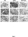

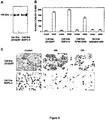
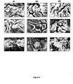
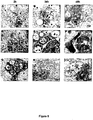
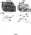


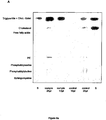
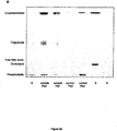
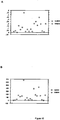
Classifications
-
- C—CHEMISTRY; METALLURGY
- C07—ORGANIC CHEMISTRY
- C07K—PEPTIDES
- C07K16/00—Immunoglobulins [IGs], e.g. monoclonal or polyclonal antibodies
- C07K16/18—Immunoglobulins [IGs], e.g. monoclonal or polyclonal antibodies against material from animals or humans
- C07K16/28—Immunoglobulins [IGs], e.g. monoclonal or polyclonal antibodies against material from animals or humans against receptors, cell surface antigens or cell surface determinants
- C07K16/30—Immunoglobulins [IGs], e.g. monoclonal or polyclonal antibodies against material from animals or humans against receptors, cell surface antigens or cell surface determinants from tumour cells
- C07K16/3076—Immunoglobulins [IGs], e.g. monoclonal or polyclonal antibodies against material from animals or humans against receptors, cell surface antigens or cell surface determinants from tumour cells against structure-related tumour-associated moieties
-
- A—HUMAN NECESSITIES
- A61—MEDICAL OR VETERINARY SCIENCE; HYGIENE
- A61P—SPECIFIC THERAPEUTIC ACTIVITY OF CHEMICAL COMPOUNDS OR MEDICINAL PREPARATIONS
- A61P35/00—Antineoplastic agents
-
- A—HUMAN NECESSITIES
- A61—MEDICAL OR VETERINARY SCIENCE; HYGIENE
- A61K—PREPARATIONS FOR MEDICAL, DENTAL OR TOILETRY PURPOSES
- A61K39/00—Medicinal preparations containing antigens or antibodies
- A61K2039/505—Medicinal preparations containing antigens or antibodies comprising antibodies
-
- C—CHEMISTRY; METALLURGY
- C07—ORGANIC CHEMISTRY
- C07K—PEPTIDES
- C07K2317/00—Immunoglobulins specific features
- C07K2317/20—Immunoglobulins specific features characterized by taxonomic origin
- C07K2317/21—Immunoglobulins specific features characterized by taxonomic origin from primates, e.g. man
-
- C—CHEMISTRY; METALLURGY
- C07—ORGANIC CHEMISTRY
- C07K—PEPTIDES
- C07K2317/00—Immunoglobulins specific features
- C07K2317/70—Immunoglobulins specific features characterized by effect upon binding to a cell or to an antigen
- C07K2317/73—Inducing cell death, e.g. apoptosis, necrosis or inhibition of cell proliferation
-
- C—CHEMISTRY; METALLURGY
- C07—ORGANIC CHEMISTRY
- C07K—PEPTIDES
- C07K2317/00—Immunoglobulins specific features
- C07K2317/70—Immunoglobulins specific features characterized by effect upon binding to a cell or to an antigen
- C07K2317/77—Internalization into the cell
Definitions
- the present invention is related to the field of cancer diagnosis and treatment and, more specifically, to the identification of polypeptides, such as antibodies, useful in the diagnosis, detection, monitoring, and treatment of neoplasms in a mammal, e.g., a human.
- stage III or IV an advanced stage
- stage III or IV the overall 5-year survival rate for these women with advanced stage ovarian cancer has remained constant over the past 30 years, at approximately 15%.
- women diagnosed with cancer confined to the ovary (stage I) have an overall 5-year survival rate approaching 90%.
- neoplasms e.g. adenocarcinoma of the lung, squamous cell lung carcinoma, intestinal type gastric carcinoma, diffuse type gastric carcinoma, adenocarcinoma of the colon, adenocarcinoma of the prostate, squamous cell carcinoma of the esophagus, adenocarcinoma of the esophagus, lobular carcinoma of the of the breast, ductal carcinoma of the breast, adenocarcinoma of the pancreas, adenocarcinoma of the ovary, or adenocarcinoma of the uterus) as this would increase the chance of treating the neoplasm and, thereby, lead to an improved prognosis for long-term survival.
- neoplasms e.g. adenocarcinoma of the lung, squamous cell lung carcinoma, intestinal type gastric carcinoma, diffuse type gastric carcinoma, adenocarcinoma of the colon,
- WO 03/076472 A1 describes the antibody CM-1 that induces apoptosis of a neoplastic cell to which it binds, but does not induce apoptosis of a non-neoplastic cell.
- This antibody specifically binds to at least one of HT-29, (ATCC Accession No. HTB-38); CACO-2 (ATCC Accssion No. HTB-37; COLO-320 DSMZ Accession No. ACC 144, COLO-260F (DSMZ Accession No. ACC 21) or COLO-678 (DSMZ Accession No. 194) cells and not to non-neoplastic cells.
- This antibody is considered an important tool in cancer diagnosis and treatment.
- SAM-6 which reacts with an epitope specific for neoplastic cells.
- This polypeptides is not only an excellent diagnostic tool, but also can inhibit cell proliferation, induce the intracellular accumulation of lipids and apoptosis of the neoplastic cells to which it binds. These characteristic result In a treatment for neoplastic diseases that lacks the side-effects of many existing therapeutics.
- the invention features a purified antibody or functional fragment thereof that binds to neoplastic cells, wherein said antibody or functional fragment thereof has an amino acid sequence at least 75% identical to the sequence of SEQ ID NO 1 and/or SEQ ID NO 3, and wherein said antibody or functional fragment thereof specifically binds to BXPC-3 (ATCC Accession No. CRL-1697), 23132/87 (DSMZ Accession No. ACC 201), COLO-206F (DSMZAccession No. ACC 21), COLO-699 (DSMZ Accession No. ACC 196), and LOU-NH91 (DSMZ Accession No.
- CDR complementarity determining regions
- said antibody comprises complementarity determining regions (CDR) identical to the amino acid sequence Ser-Gly-Asp-Lys-Leu-Gly-Asp-Lys-Tyr-Ala-Cys (CDR1) and Gln-Asp-Ser-Lys-Arg-Pro-Ser (CDR2) and Gln-Ala-Trp-Asp-Ser-Ser-Ile-Val-Val (CDR3) of SEQ ID NO: 1 and identical to the amino acid sequence Ser-Tyr-Ala-Met-His (CDR1) and Val-Ile-Ser-Tyr-Asp-Gly-Ser-Asn-Lys-Tyr-Tyr-Ala-Asp-Ser-Val-Lys-Gly (CDR2) and Asp-Arg-Leu-Ala-Val-Ala-Gly-Lys-Thr-P
- the antibody of the invention or the fragment thereof specifically binds to a adenocarcinoma of the lung, squamous cell lung carcinoma, intestinal type gastric carcinoma, diffuse type gastric carcinoma, adenocarcinoma of the colon, adenocarcinoma of the prostate, squamous cell carcinoma of the esophagus, adenocarcinoma of the esophagus, lobular carcinoma of the breast, ductal carcinoma of the breast, adenocarcinoma of the pancreas, adenocarcinoma of the ovary, and adenocarcinoma of the uterus cell and not to a non-neoplastic cell.
- the polypeptide inhibits cell proliferation when bound to a neoplastic cell, but does not inhibit cell proliferation of a non-neoplastic cell.
- the polypeptide binds to low density lipoproteins (LDL) and/or oxidized low density lipoproteins (oxLDL) and/or binds to very low density lipoproteins (VLDL) and induces the intracellular accumulation of lipids when bound to a neoplastic cell, but does not induce the intracellular accumulation of lipids in a non-neoplastic cell.
- LDL low density lipoproteins
- oxLDL oxidized low density lipoproteins
- VLDL very low density lipoproteins
- the polypeptide induces apoptosis of a neoplastic cell to which it binds, but does not induce apoptosis of a non-neoplastic cell.
- the antibody or a functional fragment thereof is selected from the group consisting of Fv, Fab, Fab', and F(ab') 2 and the functional fragment includes a fragment that is identical to the sequence of SEQ ID NOS: 1 and/or 3.
- the antibody is a monoclonal antibody, e.g., a human monoclonal antibody.
- the invention features a cell that expresses an antibody or fragment thereof that includes a sequence that is substantially identical to the amino acid sequence of SEQ ID NO:1 and 3.
- the invention features a method of generating the cell according of the third aspect. This method involves the steps of: (a) contacting lymphocytes with a heteromyeloma cell line under conditions that result in the fusion of a lymphocyte with a heteromyeloma cell, where the fusion results in a hybridoma, (b) determining whether said hybridoma produces a polypeptide that inhibits proliferation in a neoplastic cell to which it binds, but does not inhibit proliferation in a non-neoplastic cell and, (c) determining whether the hybridoma produces a polypeptide that specifically binds to at least one of BXPC-3 (ATCC Accession No.
- the invention features a use of the purified antibody or fragment thereof of any one of the first two aspects of the invention in a method of diagnosing a neoplasm in a mammal, e.g., a human.
- This method involves the steps of: (a) contacting a cell or tissue sample of the mammal with the purified polypeptide of any one of the first thirteen aspects of the invention, and (b) detecting whether the purified polypeptide binds to the cell or tissue sample, where binding of the purified polypeptide to the cell or tissue sample is indicative of the mammal having a neoplasm.
- the neoplasm is a adenocarcinoma of the lung, squamous cell lung carcinoma, intestinal type gastric carcinoma, diffuse type gastric carcinoma, adenocarcinoma of the colon, adenocarcinoma of the prostate, squamous cell carcinoma of the esophagus, adenocarcinoma of the esophagus, lobular carcinoma of the breast, ductal carcinoma of the breast, adenocarcinoma of the pancreas, adenocarcinoma of the ovary, or adenocarcinoma of the uterus.
- the polypeptide is an antibody or the polypeptide is conjugated to a detectable agent selected from the group consisting of a radionuclide, a fluorescent marker, an enzyme, a cytotoxin, a cytokine, and a growth inhibitor.
- a detectable agent selected from the group consisting of a radionuclide, a fluorescent marker, an enzyme, a cytotoxin, a cytokine, and a growth inhibitor.
- the polypeptide may be conjugated to a protein purification tag, e.g., a cleavable protein purification tag.
- the invention features a use of the purified antibody or fragment of any one of the first two aspects of the invention for treating a proliferative disorder in a mammal, e.g., a human.
- This method involves the step of contacting a cell sample with the purified polypeptide of any one of the first seven aspects, where binding of the purified polypeptide to the cell results in the reduction in proliferation of the cell.
- the proliferative disorder is a adenocarcinoma of the lung, squamous cell lung carcinoma, intestinal type gastric carcinoma, diffuse type gastric carcinoma, adenocarcinoma, of the colon, adenocarcinoma of the prostate, squamous cell carcinoma of the esophagus, adenocarcinoma of the esophagus, lobular carcinoma of the breast, ductal carcinoma of the breast, adenocarcinoma, of the pancreas, adenocarcinoma of the ovary, and adenocarcinoma of the uterus.
- the polypeptide is an antibody or the polypeptide is conjugated to a detectable agent selected from the group consisting of a radionuclide, a fluorescent marker, an enzyme, a cytotoxin, a cytokine, and a growth inhibitor.
- the detectable agent is capable of inducing apoptosis of the cell.
- the polypeptide may be conjugated to a protein purification tag, e.g., a protein purification tag that is cleavable.
- the proliferative disorder is a stomach adenocarcinoma, colorectal adenocarcinoma, squamous cell lung carcinoma, lung adenocarcinoma, squamous cell carcinoma of the esophagus, adenocarcinoma of the pancreas, urothel carcinoma of the urinary bladder, renal cell carcinoma of the kidney, adenocarcinoma of the prostate, ductal carcinoma of the breast, lobular carcinoma of the breast, adenocarcinoma of the ovary, adenocarcinoma of the endometrium, and adenocarcinoma of the uterus.
- the polypeptide is an antibody or the polypeptide is conjugated to a detectable agent selected from the group consisting of a radionuclide, a fluorescent marker, an enzyme, a cytotoxin, a cytokine, and a growth inhibitor.
- the detectable agent is capable of inhibiting cell proliferation of the cell.
- the polypeptide may be conjugated to a protein purification tag, e.g., a protein purification tag that is cleavable.
- the invention features a medicament that contains the purified polypeptide of any one of the first two aspects of the invention in a pharmaceutically acceptable carrier for the production of a medicament that inhibits cell proliferation, that induces intracellular accumulation of lipids, or that induces apotose.
- the invention features a diagnostic agent that contains the purified polypeptide of any one of the first five aspects of the invention.
- the invention features a vector, for instance, a plasmid or viral expression vector, containing the nucleic acid molecule of the twenty-forth aspect.
- the vector may be contained in a cell, such as a mammalian, e.g., a human, cell.
- detecttable agent is meant a compound that is linked to a diagnostic agent to facilitate detection. Such a “detectable agent” may be covalently or non-covalently linked to a diagnostic agent. In addition, the linkage may be direct or indirect. Examples of “detectable agents” include, protein purification tags, cytotoxins, enzymes, paramagnetic labels, enzyme substrates, co-factors, enzymatic inhibitors, dyes, radionuclides, chemiluminescent labels, fluorescent markers, growth inhibitors, cytokines, antibodies, and biotin.
- a “diagnostic agent” is meant a compound that may be used to detect a neoplastic cell by employing any one of the assays described herein as well as any other method that is standard in the art.
- a diagnostic agent may include, for example, an antibody which specifically binds to at least one of the following cells: BXPC-3 (ATCC Accession No. CRL-1687), 23132/87 (DSMZ Accession No. ACC 201), COLO-206F (DSMZ Accession No. ACC 21), COLO-699 (DSMZ Accession No. ACC 196) or LOU-NH91 (DSMZ Accession No. ACC 393) but not to non-neoplastic cells.
- a “diagnostic agent” may inhibit cell proliferation, induce apoptosis, or both only when it is bound to a neoplastic cell, but not a non-neoplastic cell.
- neoplastic cells that may be detected with such a "diagnostic agent” include adenocarcinoma of the lung, squamous cell lung carcinoma, intestinal type gastric carcinoma, diffuse type gastric carcinoma, adenocarcinoma of the colon, adenocarcinoma of the prostate, squamous cell carcinoma of the esophagus, adenocarcinoma of the esophagus, lobular carcinoma of the breast, ductal carcinoma of the breast, adenocarcinoma of the pancreas, adenocarcinoma of the ovary, or adenocarcinoma of the uterus.
- adenocarcinoma of the lung squamous cell lung carcinoma, intestinal type gastric carcinoma, diffuse type gastric carcinoma, adenocarcinoma of the colon, adenocarcinoma of the prostate, squamous cell carcinoma of the esophagus, adenocarcinoma of
- a "diagnostic agent” may include, for example, peptides, polypeptides, synthetic organic molecules, naturally-occurring organic molecules, nucleic acid molecules, and components thereof, as well as one or more detectable agent covalently or non-covalently linked to the diagnostic agent.
- a “functional fragment,” as used herein in reference to polypeptide, is meant a fragment that retains at least one biological activity of the full-length polypeptide. Examples of such a biological activity are the ability to specifically bind an antigen, induce apoptosis, and/or inhibit cell proliferation. These biological activities may be determined, for example, using any one of the assays described herein.
- a "functional fragment” has an amino acid sequence that is substantially identical to a fragment, e.g., 5, 10, 15, 20, 15, 30, 50, 75, or 100 contiguous amino acids, of the amino acid sequence of SEQ ID NO:1 or 3.
- a "functional fragment” is identical to a fragment of the sequence of SEQ ID NO: 1 or 3.
- Such a "functional fragment” may contain 5, 10, 15, 20, 15, 30, 50, 75, or 100 contiguous amino acids of SEQ ID NO: 1 or 3, or may be the entire amino acid sequence of SEQ ID NO: 1 or 3.
- the immunoglobulin's hypervariable segments are meant. This term considers that V L , and V H regions are not uniformly variable; rather most of their amino acid variations are concentrated into three short hypervariable sequences, which are essential for the secificity of the antibody.
- the identification of the CDRs was supported by BLAST-Software ( Altschul, Stephen F., Thomas L. Madden, Alejandro A. Schulffer, Jinghui Zhang, Zheng Zhang, Webb Miller, and David J. Lipman (1997), "Grapped BLAST and PSI-BLAST: a new gerenation of protein database search programs", Nucleic Acids Res. 25:3389-3402 . (NCBI database)).
- a “hybridoma,” as used herein, is any cell that is artificially created by the fusion of a normal cell such as an activated lymphocyte with a neoplastic cell, e.g., a myeloma.
- the hybrid cell which results from the fusion of at least two cells, may produce a monoclonal antibody or T cell product identical to those produced by the immunologically-competent parent.
- these cells like the neoplastic parent, are immortal.
- Inhibiting cell proliferation refers to a reduction in the rate of cell division of a cell in comparison with the normal rate of cell division of that type of cell. Inhibition of cell proliferation may be assayed using a number of methods standard in the art, for example, the MTT cell proliferation assay described herein, BrdU incorporation, and 3 H thymidine uptake. Such assays are described, for example, in Ausubel et al., Current Protocols in Molecular Biology, Wiley Interscience, New York, 2001 ; and Sambrook et al., Molecular Cloning: A Laboratory Manual, Cold Spring Harbor Laboratory, N.Y., 1989 . Desirably, the inhibition of cell proliferation is 20%, 40%, 50%, or 75%. In desirable embodiments, the inhibition of cell proliferation is 80%, 90%, 95%, or even a complete inhibition of cell proliferation.
- Binding of lipids means any interaction between lipids, especially low density lipoproteins (LDL) and/or ox LDL and the polypeptide, which is strong enough to interfere with the cell cycle of a neoplastic cell. The interference finally leads to the intracellular accumulation of lipids. Yet it is unclear wheater the polypeptide interacts with the lipids first to form a complex that is subsequently interacting with the neoplastic cell or wheater the polypeptide interacts directly with a receptor on the surface of neoplastic cells.
- LDL low density lipoproteins
- polypeptide which is an antibody, may be acitve in its monomeric or in its pentameric form.
- “Intracellular accumultation of lipids” as refered to herein means increasing concentration of intracellular lipids, especially of low density lipoproteins (LDL) and/or ox LDL, in comparison to the normal concentration of lipids in that type of cell.
- LDL was shown to be the intracellularly enriched lipid form by a chromatographic analysis documenting that Cholesterolester and Triglycerids where increased in cells incubated with the purified polypeptid. It is only LDL that contains both forms in these amounts. In the consequence the intracellular lipid accumulation leads to apoptosis, e.g. "Lipoptosis", of neoplastic cells.
- Intracellular lipid accumulation may be assayed and visualized using a number of methods standard in the art for example Sudan III staining of neutral lipids described herein or staining with the fluorescence stain Nile Red ( Greenspan, P., Mayer, E.P., and Fowler, D. Nile Red: A Selective Fluorescent Stain for Intracellular Lipid Droplets. J. Cell Biol. 100, 965-973, 1985 ).
- “Inducing apoptosis,” as used herein, refers to the appearance of characteristics in a cell that are well defined in the art (see, e.g., Wyllie et al., Br. J. Cancer 80 Suppl. 1:34-37, 1999 ; Kerr et al., Br. J. Cancer 26:239-257, 1972 ). These characteristics include morphological characteristics, such as membrane blebbing, DNA condensation, as well as changes in F-actin content, mitochondrial mass, and membrane potential.
- the induction of apoptosis may be assayed using a number of methods standard in the art, for example, a cell death ELISA, TUNEL staining, DNA stains, e.g., Hoechst 33258, and staining with various vital dyes such as acridine orange, Mito Tracker Red ® staining (Molecular Probes, Eugene, OR), and Annexin V ® staining (Becton Dickinson, NJ).
- inducing apoptosis refers to an increase in the number of cells undergoing apoptosis when compared with a control cell population. For instance, the increase of apoptosis may be 10%, 20%, 40%, 50%, or 75%. In desirable embodiments, the induction of apoptosis results in an increase of apoptosis that is 2-fold, 3-fold, 10-fold, or even 100-fold over that seen in a control cell population.
- a “neoplastic cell,” as used herein, refers to a cell which is undergoing cell division, not undergoing apoptosis, or both, under inappropriate conditions.
- a “neoplastic cell” may undergo cell division when a corresponding non-neoplastic cell does not undergo cell division, or, alternatively, a “neoplastic cell” may not respond to normal cell-cycle checkpoint controls.
- proliferative disease refers to any disorder that results in the abnormal proliferation of a cell.
- proliferative diseases are various types of neoplasms, such as adenocarcinoma of the lung, squamous cell lung carcinoma, intestinal type gastric carcinoma, diffuse type gastric carcinoma, adenocarcinoma of the colon, adenocarcinoma of the prostate, squamous cell carcinoma of the esophagus, adenocarcinoma of the esophagus, lobular carcinoma of the breast, ductal carcinoma of the breast, adenocarcinoma of the pancreas, adenocarcinoma of the ovary, or adenocarcinoma of the uterus.
- proliferative diseases may also be the result of the cell becoming infected with a transforming virus.
- a “protein purification tag,” as used herein, is a peptide, e.g., an epitope tag, that is covalently or non-covalently added to a protein to aid in the purification of the protein. Desirably such peptides bind with high affinity to an antibody or to another peptide such as biotin or avidin.
- epitope tags include His-tags, HA-tags, FLAG ® -tags, and c-Myc-tags. However, any epitope that is recognized by an antibody also may be used as a protein purification tag.
- Protein purification tags may be cleaved from a protein, for example, by using an enzyme, e.g., thrombin, or a chemical, e.g., cyanogen bromide.
- an antibody e.g., the SAM-6 human monoclonal antibody, that specifically binds to BXPC-3 (ATCC Accession No. CRL-1687), 23132/87 (DSMZ Accession No. ACC 201), COLO-206F (DSMZ Accession No. ACC 21), COLO-699 (DSMZ Accession No. ACC 196) or LOU-NH91 (DSMZ Accession No. ACC 393), or BXPC-3 (ATCC Accession No.
- CRL-1687 cells desirably has an affinity for its antigen that is least 2-fold, 5-fold, 10-fold, 30-fold, or 1 00-fold greater than for an equal amount of any other antigen, including related antigens. Binding of a polypeptides to another polypeptide may be determined as described herein, and by any number of standard methods in the art, e.g., Western analysis, ELISA, or co-immunoprecipitation.
- substantially identical is meant a polypeptide or nucleic acid exhibiting at least 75%, 80%, 85%, or 90% identity to a reference amino acid (e.g., the sequence of SEQ ID NO:1 or 3 or nucleic acid sequence (e.g., the sequence of SEQ ID NO:2 or 4.
- the polypeptide or nucleic acid sequence is at least 95%, 98%, 99%, or 100% identical to a reference amino acid or nucleic acid sequence.
- the length of comparison sequences will generally be at least 5, 10, or 15 amino acids and desirably at least 20 or 25 contiguous amino acids.
- the length of comparison sequences is at least 30, 50, 75, 90, 95, or 100 contiguous amino acids, or even the full-length amino acid sequence.
- the length of comparison sequences will generally be at least 15, 30, or 45 contiguous nucleotides, and desirably at least 60 contiguous nucleotides.
- the length of comparison sequences is at least 75, 150, 225, 270, 285, or 300 contiguous nucleotides, or even the full-length nucleotide sequence.
- Sequence identity may be measured using sequence analysis software on the default setting (e.g., Sequence Analysis Software Package of the Genetics Computer Group, University of Wisconsin Biotechnology Center, 1710 University Avenue, Madison, WI 53705). Such software may match similar sequences by assigning degrees of homology to various substitutions, deletions, and other modifications. Conservative substitutions typically include substitutions within the following groups: glycine, alanine, valine, isoleucine, leucine; aspartic acid, glutamic acid, asparagine, glutamine; serine, threonine; lysine, arginine; and phenylalanine, tyrosine.
- Multiple sequences may also be aligned using the Clustal W(1.4) program (produced by Julie D. Thompson and Toby Gibson of the European Molecular Biology Laboratory, Germany and Desmond Higgins of European Bioinformatics Institute, Cambridge, UK) by setting the pairwise alignment mode to "slow," the pairwise alignment parameters to include an open gap penalty of 10.0 and an extend gap penalty of 0.1, as well as setting the similarity matrix to "blosum.”
- the multiple alignment parameters may include an open gap penalty of 10.0, an extend gap penalty of 0.1, as well as setting the similarity matrix to "blosum,” the delay divergent to 40%, and the gap distance to 8.
- a factor is substantially pure when it is at least 50%, by weight, free from proteins, antibodies, and naturally-occurring organic molecules with which it is naturally associated, or in reference to a nucleic acid molecule, is free from the nucleic acid sequences that naturally flank the sequence of the nucleic acid molecule.
- the factor is at least 75%, more desirably, at least 90%, and most desirably, at least 99%, by weight, pure.
- a substantially pure factor may be obtained by chemical synthesis, separation of the factor from natural sources, or production of the factor in a recombinant host cell that does not naturally produce the factor.
- Proteins, vesicles, and organelles may be purified by one skilled in the art using standard techniques, such as those described by Ausubel et al. (Current Protocols in Molecular Biology, Wiley Interscience, New York, 2001 ).
- the factor is desirably at least 2, 5, or 10 times as pure as the starting material, as measured using polyacrylamide gel electrophoresis, column chromatography, optical density, HPLC analysis, or Western analysis ( Ausubel et al., Current Protocols in Molecular Biology, Wiley Interscience, New York, 2001 ).
- Desirable methods of purification include immunoprecipitation, column chromatography such as immunoaffinity chromatography and nickel affinity columns, magnetic bead immunoaffinity purification, and panning with a plate-bound antibody.
- Sequence listing includes sequence protocols of the amino acid sequence (SEQ ID NO:1) (1) and the nucleic acid sequence (SEQ ID NO:2) (2) of the variable region of the light chain (V L ) of human monoclonal antibody SAM-6.
- Sequence numbers 3 and 4 are the amino acid sequence (SEQ ID NO:3) (3) and the nucleic acid sequence (SEQ ID NO:4) (4) of the variable region of the heavy chain (V H ) of human monoclonal antibody SAM-6.
- the present invention features polypeptides, such as antibodies, and their use in the treatment and diagnosis of neoplasms.
- SAM-6 human monoclonal antibody
- SAM-6 a human monoclonal antibody
- Antibodies play an essential role in maintaining the health of an individual.
- antibodies are present in serum and bind to and help eliminate diverse pathogens such as bacteria, viruses, and toxins.
- Antibodies consist of Y-shaped protein structures built from two heavy chains and two light chains. Each chain has a modular construction: each light chain consists of two domains, and each heavy chain has at least four domains.
- the antigen binding site is fashioned by one domain from the heavy chain (V H domain) and one domain from the light chain (V L domain).
- small antigen binding fragments can be prepared by linking these two domains, either associated non-covalently, or covalently via disulphide bonds or a peptide linker.
- the antigen binding domains are more variable in amino acid sequence than the other domains of the antibody, and are therefore termed variable (V) domains, in contrast to the constant (C) domains.
- V variable
- C constant domains.
- the constant domains of the antibody are responsible for triggering antibody effector mechanisms, such as complement lysis and cell-mediated killing.
- Antibodies are made by B-lymphocytes in a process involving gene rearrangement. During the development of these cells, the genes encoding the variable domains are assembled from genetic elements. In the case of the V H domains there are three elements, the un-rearranged V H gene, D segment, and J H segment. In the case of the V L domains, there are two elements, the un-rearranged V L (V Lambda or V Kappa) gene and the J L (J Lambda or J Kappa) segment. Random combination of these gene segments and random combination of the rearranged V H and V L domains generate a large repertoire of antibodies, capable of binding to a large diversity of equally diverse antigens.
- the presently claimed polypeptide is any agent that binds to BXPC-3, 23132/87, COLO-206F, COLO-699 and LOU-NH91, but does not bind to non-neoplastic cells.
- the polypeptide may be an antibody, such as a human monoclonal antibody (e.g.,SAM-6), or a functional fragment thereof.
- SAM-6 human monoclonal antibody
- the polypeptide of the invention can exclusively bind to both neoplastic tissues and neoplastic cells, but not to non-neoplastic tissue or cells.
- the polypeptide also may induce apoptosis of a neoplastic cell to which it binds, but not in a non-neoplastic cell, or, alternatively, the polypeptide may inhibit proliferation of the neoplastic cell it binds to, but not in a non-neoplastic cell. Desirably, the polypeptide can simultaneously induce apoptosis and inhibit proliferation of neoplastic cells, but not of non-neoplastic cells. Such a polypeptide is, therefore, useful for the detection, monitoring, prevention, and treatment of cancers in mammals.
- Exemplary cancers amenable to the methods of the current invention include colorectal cancer, ovarian carcinoma, squamous cell lung carcinoma, small cell lung carcinoma, lobular and ductal mammary carcinomas, melanoma, breast cancer, lung cancer, such as lung adenocarcinomas, gastric cancer, pancreatic cancer, such as pancreatic adenocarcinomas, glioma, sarcomas, gastrointestinal cancer, brain tumor, esophageal cancer, such as esophagial squamous cell carcinomas, stomach cancer, osteosarcoma , fibrosarcomas, urinary bladder cancer, prostate cancer, such as prostate adenocarcinomas, renal cancer, ovarian cancer, testicular cancer, endometrial cancer, cervical cancer, uterine adenocarcinomas, Hodgkin's disease, lymphomas, and leukemias.
- lung cancer such as lung adenocarcinomas, gastric cancer
- Such polypeptides are particularly useful for the detection and treatment of adenocarcinoma of the lung, squamous cell lung carcinoma, intestinal type gastric carcinoma, diffuse type gastric carcinoma, adenocarcinoma of the colon, adenocarcinoma of the prostate, squamous cell carcinoma of the esophagus, adenocarcinoma of the esophagus, lobular carcinoma of the breast, ductal carcinoma of the breast, adenocarcinoma of the pancreas, adenocarcinoma of the ovary, or adenocarcinoma of the uterus.
- polypeptides according to the claimed invention can be produced by any method known in the art for small scale, large scale, or commercial production of polypeptides.
- a monoclonal antibody such as SAM-6
- hybridoma cell lines are typically generated by the fusion of spleen lymphocytes or lymph node lymphocytes derived from patients having a neoplasm, such as stomach carchinoma, colon carcinoma or a pancreatic carcinoma, with a heteromyeloma cell line.
- heteromyeloma cell lines include, for example, HAB-1 ( Vollmers et al, Cancer 74:1525-1532, 1994 ), CB-F7 ( Delvig et al., Hum.
- Antibodies Hybridomas 6:42-46, 1995 K6H6B5 ( Delvig et al., Hum. Antibodies Hybridomas 6:42-46, 1995 ), H7NS.934 ( Delvig et al., Hum. Antibodies Hybridomas 6:42-46, 1995 ), SHM-D33 ( Bron et al., Proc. Natl. Acad. Sci. USA 81:3214-3217, 1984 ), and B6B11 ( Borisova et al., Vopr. Virusol. 44:172-174, 1999 ).
- the ability to generate human monoclonal antibodies from lymphocytes of cancer patients allows the isolation of antibodies that are generated by an immune response in the cancer patient to the tumor.
- lymph nodes or spleen are surgically removed from a patient having cancer, such as colon carcinoma or a pancreatic carcinoma.
- Lymphocytes may be prepared as cell suspensions by mechanical means and subsequently fused at, for example, a 1:2 or 1:3 ratio with a heteromyeloma cell line under conditions that result in cell fusion.
- the heteromyeloma cell line HAB-1 which is generated by the fusion of a human lymphocyte with the mouse myeloma NS-0, may be used for this purpose.
- hybridomas are generally stable in growth and antibody production in standard and mass cultures (flasks, miniPerm, fermenters, etc.) for several months. Levels of antibody production typically range between 0.01-0.1 mg/mL in flasks and between 0.1-0.5 mg/mL in miniPerm.
- Cell fusion may be achieved by any method known in the art, and includes, for example, the use of 40% polyethyleneglycol.
- Hybridomas may be cultured in media containing HAT (Hypovanthin-aminopterin-thymidin) and after four weeks, supernatants may be screened for antibody production using an ELISA assay. Positive clones may then be tested in be tested in attachment inhibition and binding assays using commercially available tumor cell lines. Positive clones further may be tested using immunoperoxidase staining of tumor and normal tissues. Thus, clones may be selected on the basis of their reactivity with autologous and allogeneic neoplastic cells.
- the antibody may be purified from mass cultures with use of cation-exchange, hydrophobic interaction, size exclusion, or affinity chromatograhpy, as well as a combination of these methods as described, for example, by Vollmers et al. (Oncology Reports 5:35-40, 1998 ).
- additional functional and immunohistochemical tests of the antibodies produced by the trioma may be performed.
- the antibodies produced by the hybridoma can be tested for their ability to induce apoptosis, inhibit cellular proliferation, or both, relative to untreated control cells.
- the antibodies can also be tested for their ability to specifically bind the neoplastic cell lines like BXPC-3, 23132/87, COLO-206F, COLO-699 or LOU-NH91, relative to non-neoplastic cells.
- the polypeptide including an antibody, or a fragment thereof, may be produced by the expression of the polypeptide or antibody in a host cell such as E. coli or yeast, e.g., S. cerevisiae.
- an antibody of the invention may be identified as follows. A nucleic acid sequence encoding an antibody, or a fragment thereof, may be inserted into filamentous bacteriophage to generate libraries of approximately 10 7 or more antibodies. Each phage expresses an antibody on its surface that is encoded by the nucleic acid it contains. Antibodies of the invention may thus be screened and detected by functional and histochemical assays as described herein, and such genes may be subsequently selected and expressed in E.coli. This system is described, for example, in U.S. Patent No. 5,876,691 .
- Antibodies, or functional fragments thereof may also be generated using, for example, direct synthesis using recombinant methods. These methods are standard in the art. For example, a nucleic acid sequence may be amplified using the polymerase chain reaction (PCR). The PCR technique is known in the art and is described, for example in U.S. Patent No. 4,683,195 . Using standard methods, and as described herein, the sequence of a monoclonal antibody expressed by a hybridoma may be obtained and functional fragments of the antibody may be amplified. For example, whole RNA may be isolated from a hybridoma expressing a tumor-specific monoclonal antibody.
- PCR polymerase chain reaction
- cDNA may then be generated from the RNA using reverse transcriptase and the cDNAs which contain the functional fragments of the variable regions of the heavy and light chains may be amplified using PCR.
- the PCR products may then be purified and cloned into expression vectors, e.g., plasmid or viral vectors.
- expression vectors e.g., plasmid or viral vectors.
- Many standard vectors are available and the selection of the appropriate vector will depend on, for example, the size of the DNA inserted into the vector and the host cell to be transformed with the vector.
- Amino acid sequence variants of a polypeptide such as an antibody, e.g., a SAM-6 antibody
- a polypeptide such as an antibody, e.g., a SAM-6 antibody
- Such variants include, for example, deletions from, or insertions or substitutions of, residues within the amino acid sequence of the SAM-6 antibody.
- deletion, insertion, and substitution can be made to arrive at the final construct, provided that the final construct possesses the desired characteristics, e.g., the ability to induce apoptosis of a neoplastic cell, but not a non-neoplastic cell, or the ability to inhibit the proliferation of a neoplastic cell, but not a non-neoplastic cell.
- the amino acid changes also may after post-translational processes of an antibody, such as changing the number or position of glycosylation sites, altering the membrane anchoring characteristics, or modifying its susceptibility to proteolytic cleavage.
- the location of the mutation site and the nature of the mutation will depend on characteristic(s) to be modified.
- the sites for mutation can be modified individually or in series, e.g., by substituting first with conservative amino acid choices and then with more radical selections depending upon the results achieved, or deleting the target residue.
- a useful method for identification of specific residues or regions for mutagenesis in a polypeptide is called "alanine scanning mutagenesis" and is described, for example, by Cunningham and Wells-(Science 244:1081-1085, 1989 ).
- a residue or group of target residues are identified (e.g., charged residues such as arg, asp, his, lys, and glu) and replaced by a neutral or negatively charged amino acid (most desirably alanine or polyalanine) to affect the interaction of the amino acids with the surrounding aqueous environment in or outside the cell.
- the domains demonstrating functional sensitivity to the substitutions then are refined by introducing further or other variants at or for the sites of substitution.
- the site for introducing an amino acid sequence variation is predetermined, the nature of the mutation need not be predetermined.
- alanine scanning or random mutagenesis may be conducted at the target codon or region and the expressed variants are screened for, e.g., the ability to induce apoptosis of a neoplastic cell and not a non-neoplastic cell, or to inhibit the proliferation of a neoplastic cell and not a non-neoplastic cell.
- the sites of greatest interest for substitutional mutagenesis include sites identified as affecting the biological activity of a polypeptide. These sites, especially those falling within a sequence of at least three other identically conserved sites, may be substituted in a relatively conservative manner. For instance, ala may be substituted with val, leu, or ile; arg may be substituted with lys, gln, or asn; asn may be substituted with gln, his, lys, or arg; asp may be substituted with glu; cys may be substituted with ser; gln may be substituted with asn; glu may be substituted with asp; gly may be substituted with pro; his may be substituted with asn, gln, lys, or arg; ile may be substituted with leu, val, met, ala, or phe; leu may be substituted with ile, val, met, ala, or phe; lys may be substituted with arg,
- the claimed polypeptide such as an antibody (e.g., monoclonal antibody, such as SAM-6), or a fragment thereof, may be linked to a detectable agent to facilitate the purification of the polypeptide as well as the diagnosis, monitoring, or treatment of cancer in a mammal in need thereof.
- a detectable agent e.g., monoclonal antibody, such as SAM-6
- suitable detectable agent will depend on the intended use of the polypeptide and will be apparent to those of ordinary skill in the art.
- Detectable agents according to the claimed invention include, for example, protein purification tags, cytotoxins, enzymes, paramagnetic labels, enzyme substrates, co-factors, enzyme inhibitors, dyes, radionuclides, chemiluminescent labels, fluorescent markers, growth inhibitors, and biotin.
- a protein purification tag may be conjugated to the polypeptide of the invention, to facilitate isolation of the polypeptide.
- tags that can be used include His-tags, HA-tags, FLAG ® -tags, and c-Myc tags.
- An enzymatic or chemical cleavage site may be engineered between the polypeptide and the tag moiety so that the tag can be removed following purification.
- Suitable toxins include diphtheria toxin, Pseudomonas exotoxin A, ricin, and cholera toxin.
- suitable enzyme labels include malate hydrogenase, staphylococcal nuclease, delta-5-steroid isomerase, alcohol dehydrogenase, alpha-glycerol phosphate dehydrogenase, triose phosphate isomerase, peroxidase, alkaline phosphatase, asparaginase, glucose oxidase, beta-galactosidase, ribonuclease, urease, catalase, glucose-6-phosphate dehydrogenase, glucoamylase, and acetylcholinesterase.
- suitable radioisotopic labels include 3 H, 125 I, 131 I, 32 P, 35 S, and 14 C.
- the radioisotope will emit in the 10-5,000 kev range, more desirably 100-500 kev.
- Paramagnetic isotopes may also be conjugated to the polypeptide and used in vivo for the diagnosis and treatment of cancer.
- the use of such conjugated antibodies may be for in vivo nuclear magnetic resonance imaging.
- Such a method has previously been described (see, for example, Schaefer et al., JACC 14:472-480, 1989 ; Shreve et al., Magn. Reson. Med. 3:336-340, 1986 ; Wolf, Physiol. Chem. Phys. Med. NMR 16:93-95, 1984 ; Wesbey et al., Physiol. Chem. Phys.
- the radiolabeled antibody may also be used in radioimmunoguided surgery (RIGS), which involves the surgical removal of any tissue the labeled antibody binds to.
- RIGS radioimmunoguided surgery
- the labeled antibody guides the surgeon towards neoplastic tissue by distinguishing it from non-neoplastic tissue.
- Radiolabels useful for tumor imaging are preferably short-lived radioisotopes.
- radioactive metals with half-lives ranging from 1 hour to 11.4 days are available for conjugation to antibodies, such as scandium-47 (3.4 days), gallium-67 (2.8 days), gallium-68 (68 minutes), technetium-99m (6 hours), indium-111 (3.2 days), and radium-223 (11.4 days), of which gallium-67, technetium-99m, and indium-111 are preferable for gamma camera imaging, gallium-68 is preferable for positron emission tomography, and scandium-47 and radium-223 (and other alpha-emitting radionuclides) are preferable for tumor therapy.
- fluorescent markers examples include fluorescein, isothiocyalate, rhodamine, phycoerythrin, phycocyanin, allophycocyanin, ophthaldehyde, and fluorescamine.
- chemiluminescent markers include a luminal label, isoluminal label, aromatic acridinium ester label, imidazole label, acridinium salt label, oxalate ester label, luciferin label, luciferase label, and aequorin label.
- Conjugation of these detectable agents to the claimed polypeptides can be accomplished using standard techniques commonly known in the art.
- Typical antibody conjugation techniques are described by Kennedy et al. (Clin. Chim. Acta 70, 1-31, 1976 ) and Schurs et al. (Clin. Chim. Acta 81, 1-40, 1977 ) and include, for example, the glutaraldehyde method, the periodate method, the dimaleimide method, the m-maleimidobenzyl-N-hydroxy-succinimide ester method.
- Antibodies may be radiolabeled by any of several techniques known to the art, described, for example, in U.S. patent No. 4,444,744 .
- mixtures of different or the same labeled polypeptides specific to different antigens or different epitopes of the same antigen associated with the same or different tumor or tumor cell types may be used.
- Such a combination may enhance detection, localization and/or therapy in certain cases, and can also increase the range of a broad screen for more than one neoplasm or type of neoplasm.
- the polypeptide of the invention may induce apoptosis of neoplastic cells, inhibit cellular proliferation of neoplastic cells, or both, the polypeptide may in addition be conjugated to an agent that kills neoplastic cells or that inhibits their proliferation.
- the targeting ability of the polypeptide such as an antibody or fragment thereof, results in the delivery of the cytotoxic or anti-proliferative agent to the tumor to enhance the destruction of the tumor.
- the polypeptide therefore may be used for the treatment and prevention of cancer in a mammal, such as a human patient.
- the cytotoxic agent linked to the polypeptide may be any agent that destroys or damages a tumor cell or tumor to which the polypeptide has bound. Examples of such agents include chemotherapeutic agents or radioisotopes, enzymes which activates a pro-drug, or a cytokine.
- chemotherapeutic agents include, for example, taxol, mithramycin, deoxyco-formycin, mitomycin-C, L-asparaginase, interferons (especially IFN-alpha), etoposide, teniposide, anthracyclines (e.g., daunomycin and doxorubicin), methotrexate, vindesine, neocarzinostatin, cis-platinum, chlorambucil, cytosine arabinoside, 5-fluorouridine, melphalan, ricin, and calicheamicin.
- the chemotherapeutic agents may be conjugated to the antibody using conventional methods known in the art.
- Suitable radioisotopes for use as cytotoxic agents are also known to those skilled in the art and include, for example, 131 I, or an astatine such as 211 At. These isotopes may be attached to the polypeptide, either covalently or non-covalently, using conventional techniques known in the art.
- the cytotoxic agent may also be an enzyme, which activates a pro-drug.
- ADEPT antibody directed enzyme pro-drug therapy
- the polypeptide-enzyme conjugate may be administered to the patient and allowed to localize in the region of the tumor to be treated.
- the pro-drug is then administered to the patient such that conversion to the cytotoxic drug is localized in the region of the tumor to be treated under the influence of the localized enzyme.
- An exemplary enzyme is bacterial carboxypeptidase G2 (CPG2) the use of which is described in, for example, WO 88/07378 .
- the polypeptide-enzyme conjugate may, if desired, be modified in accordance with the teaching of WO 09/00427 , such as to accelerate its clearance from areas of the body that are not in the vicinity of a neoplasm.
- the polypeptide-enzyme conjugate may also be used in accordance with WO 89/00427 , for example, by providing an additional component, which inactivates the enzyme in areas of the body that are not in the vicinity of the tumor.
- the cytotoxic agent conjugated to the claimed polypeptide may also be a cytokine such as interleukin-2 (IL-2), interleukin-4 (IL-4), or tumor necrosis factor alpha (TNF-alpha).
- IL-2 interleukin-2
- IL-4 interleukin-4
- TNF-alpha tumor necrosis factor alpha
- the polypeptide targets the cytokine to the tumor so that the cytokine mediates damage to or destruction of the tumor without affecting other tissues.
- the cytokine may be fused to the polypeptide at the DNA level using conventional recombinant DNA techniques.
- any inhibitor of cell proliferation e.g., genistein, tamoxifen, or cyclophosphamide, may be conjugated with a polypeptide of the invention.
- the administration of the claimed polypeptide to a patient be limited to a particular mode of administration, dosage, or frequency of dosing; the present invention contemplates all modes of administration, including intramuscular, intravenous, intraperitoneal, intravesicular, intraarticular, intralesional, subcutaneous, or any other route sufficient to provide a dose adequate to decrease the number of neoplastic cells by inducing apoptosis of neoplastic cells, by inhibiting proliferation of tumor cells, or both.
- the compound(s) may be administered to the patient in a single dose or in multiple doses.
- the doses may be separated from one another by, for example, one day, two days, one week, two weeks, or one month.
- the polypeptide e.g., a monoclonal antibody, such as SAM-6) may be administered once a week for, e.g., 2, 3, 4, 5, 6, 7, 8, 10, 15, 20, or more weeks.
- specific dosage regimes should be adjusted over time according to the individual need and the professional judgment of the person administering or supervising the administration of the compositions.
- the precise dose will vary dependent on the polypeptide used, the density, on the tumor surface, of the ligand to which the polypeptide binds, and the rate of clearance of the polypeptide.
- the dosage of the SAM-6 antibody can be increased if the lower dose does not provide sufficient anti-neoplastic activity.
- the dosage of the SAM-6 antibody can be decreased if the neoplasm is cleared from the patient.
- a therapeutically effective amount of the claimed polypeptide such as a monoclonal antibody or a fragment thereof, may be, for example, in the range of about 0.1 mg to 50 mg/kg body weight/day or 0.70 mg to 350 mg/kg body weight/week.
- a therapeutically effective amount is in the range of about 0.50 mg to 20.0 mg/kg, and more desirably in the range of about 0.50 mg to 15.0 mg/kg for example, about 0.2, 0.3, 0.5, 1.0, 1.5, 2.0, 2.5, 3.0, 3.5, 4.0, 4.5, 5.0, 5.5, 6.0, 7.0, 8.0, 8.5, 9.0, 10.0, 11.0, 12.0, 13.0, 14.0, or 15.0 mg/kg body weight administered daily, every other day, or twice a week.
- a suitable dose is an amount of the polypeptide that, when administered as described above, is capable of inducing apoptosis, and is at least 20% above the basal (i.e., untreated) level.
- an appropriate dosage and treatment regimen provides the active compound(s) in an amount sufficient to provide therapeutic and/or prophylactic benefit.
- Such a response can be monitored by establishing an improved clinical outcome (e.g., more frequent remissions, complete or partial, or longer disease-free survival) in treated patients as compared to non-treated patients.
- the administration of the polypeptide can induce neoplastic cell apoptosis by at least 20%, 40%, 50%, or 75% above that of an untreated control as measured by any standard assay known in the art. More desirably, apoptosis is induced by 80%, 90%, 95%, or even 100% above that of an untreated control.
- the administration of the polypeptide can inhibit neoplastic cell proliferation by at least 20%, 40%, 50%, or 75% below that of an untreated control as measured by any standard assay known in the art. More desirably, proliferation is inhibited by 80%, 90%, 95%, or even 100% below that of an untreated control.
- the polypeptide can simultaneously inhibit proliferation and induce apoptosis of neoplastic cells relative to untreated control cells.
- Such responses can be monitored by any standard technique known in the art.
- the amount of antibody present in a dose ranges from about 25 ⁇ g to 5 mg per kg of host. Suitable dose sizes will vary with the size of the patient, but will typically range from about 0.1 mL to about 5 mL.
- the claimed polypeptide may be administered by any suitable means that results in a concentration having anti-neoplastic properties upon reaching the target region.
- the polypeptide may be contained in any appropriate amount in any suitable carrier substance, and is generally present in an amount of 1-95% by weight of the total weight of the composition.
- the composition may be provided in a dosage form that is suitable for parenteral (e.g., subcutaneous, intravenous, intramuscular, or intraperitoneal) administration route.
- the pharmaceutical compositions may be formulated according to conventional pharmaceutical practice (see, e.g., Remington: The Science and Practice of Pharmacy (20th ed.), ed. A.R. Gennaro, Lippincott Williams & Wilkins, 2000 and Encyclopedia of Pharmaceutical Technology, eds. J. Swarbrick and J. C. Boylan, 1988-1999, Marcel Dekker, New York ).
- the pharmaceutical composition may be administered parenterally by injection, infusion or implantation (subcutaneous, intravenous, intramuscular, intraperitoneal, or the like) in dosage forms, formulations, or via suitable delivery devices or implants containing conventional, non-toxic pharmaceutically acceptable carriers and adjuvants.
- the neoplastic cells are in direct contact with the blood (e.g., leukemias), or if the tumor is only accessible by the bloodstream then the intravenous (I.V.) route may be used.
- the polypeptide may be directly administered into the cavity rather than into the blood stream.
- the formulation and preparation of such compositions are well known to those skilled in the art of pharmaceutical formulation. Formulations can be found in Remington: The Science and Practice of Pharmacy, supra.
- the present invention is directed to a method for detecting or diagnosing a neoplasm in a mammal, preferably a human patient.
- a mammal preferably a human patient.
- any neoplasm in which administration of the claimed polypeptide causes an induction in apoptosis or a reduction in proliferation are amenable to the methods of this invention.
- the claimed polypeptides are particularly useful since they are specific to neoplasms or neoplastic cells, but not normal cells or tissue. Accordingly, this polypeptide can bind to neoplastic cells within the tumor, but not the normal surrounding tissue, thus allowing the detection, the treatment, or both, of a neoplasm in a mammal. For instance, one may use a polypeptide of the invention to determine if a biopsy removed the entire tumor by verifying that no cells bound by the polypeptide remain in the patient or, by verifying that tumor removed from the patient is entirely surrounded by cells that are not bound by the polypeptide.
- neoplastic markers may be assayed within a given sample or individual.
- polypeptides such as antibodies or functional fragments specific for different antigens may be combined within a single assay, or in multiple assays.
- multiple primers or probes specific to neoplasms may be used concurrently. The selection of markers may be based on routine experiments to determine combinations that results in optimal sensitivity.
- the diagnosis of a neoplasm in a mammal involves obtaining a biological sample from the mammal (e.g., human patient), contacting such sample with the polypeptide of the invention (e.g., a monoclonal antibody, such as SAM-6), detecting in the sample the level of reactivity or binding of the polypeptide to neoplastic cells relative to a control sample, which corresponds to non-neoplastic cells derived from healthy tissue from the mammal in which the cancer is being diagnosed or from another patient known not to have neoplasm.
- the methods of this invention are particularly useful for the detection of early stage tumors or metastases, which are otherwise undetectable.
- the methods of this invention may also be used to monitor progression of a neoplasm in a mammal.
- the polypeptide described herein therefore may be used as a marker for the progression of a neoplasm.
- the assays described below, which are used for the diagnosis of a neoplasm may be performed over time, and the change in the level of reactive polypeptide(s) evaluated. For example, the assays may be performed every 24-72 hours for a period of 6 months to 1 year, and thereafter performed as needed.
- a neoplasm is progressing in those patients in whom the level of bound polypeptide detected increases over time.
- the neoplasm is not progressing when the level of bound polypeptide either remains constant or decreases with time.
- the polypeptide of the invention may also be used to determine the presence of tumor cells in the mammal following tumor resection by surgical intervention to determine whether the tumor has been completely removed from the mammal.
- the polypeptide is linked to a detectable agent, which facilitates detection, or measurement of polypeptide reactivity.
- the biological sample is any biological material, which may contain neoplastic cells and include, for example, blood, saliva, tissue, serum, mucus, sputum, urine, or tears.
- the biological sample may also be a tissue section, which may be fixed tissue, fresh tissue, or frozen tissues.
- a neoplasm is detected or diagnosed in the mammal from which the sample was obtained if there is an increase in the level of reactivity of the antibody with the biological sample over the control sample. Such increase is at least 10%, 20%, 30%, 40%, 50%, or more than 50% over control levels.
- the level of binding or reactivity can be determined by any method known in the art and is described in further detail below.
- the diagnosis of neoplasms using the claimed polypeptide may be performed by any method known to those of ordinary skill in the art for using a binding agent to detect polypeptide markers in a sample. See, e.g., Harlow and Lane, Antibodies: A Laboratory Manual, Cold Spring Harbor Laboratory, 1988 .
- the polypeptide may be used for enzyme-linked immunosorbent assay (ELISA), Western blotting or in situ detection of tumor cells in a tissue sample.
- ELISA assay typically involves the use of the polypeptide, such as an antibody, immobilized on a solid support to bind to the tumor cells in the biological sample.
- the bound tumor cell may then be detected using a detection reagent that contains a reporter group and that specifically binds to the antibody/tumor cell complex.
- detection reagents include, for example, any binding agent that specifically binds to the antibody, such as an anti-immunoglobulin, protein G, protein A, or a lectin.
- a competitive assay may be utilized, in which the polypeptide is an antibody and in which the antigens, to which the antibody is specific to is labeled with a reporter group and allowed to bind to the immobilized antibody after incubation of the antibody with the biological sample.
- Diagnosis of a neoplasm in a patient may also be determined by a two-antibody sandwich assay. This assay may be performed by first contacting an antibody that has been immobilized on a solid support, commonly the well of a microtiter plate, with the sample, such that polypeptides within the sample are allowed to bind to the immobilized antibody. Unbound sample is then removed from the immobilized polypeptide-antibody complexes and a detection reagent (preferably a second antibody capable of binding to a different site on the polypeptide) containing a reporter group is added.
- a detection reagent preferably a second antibody capable of binding to a different site on the polypeptide
- the amount of detection reagent that remains bound to the solid support is then determined using a method appropriate for the specific reporter group. For example, to determine the presence or absence of a neoplasm, such as colorectal adenocarcinoma, the signal detected from the reporter group that remains bound to the solid support is generally compared to a signal that corresponds to a predetermined cut-off value.
- the cut-off value for the detection of a neoplasm is the average mean signal obtained when the antibody is incubated with samples from patients without a neoplasm.
- the method employed for detecting the reporter group depends upon the nature of the reporter group. For radioactive groups, scintillation counting or autoradiographic methods may be used. Spectroscopic methods may be used to detect dyes, luminescent groups and fluorescent groups. Biotin may be detected using avidin, coupled to a different reporter group (commonly a radioactive or fluorescent group or an enzyme). Enzyme reporter groups may generally be detected by the addition of substrate (generally for a defined period of time), followed by spectroscopic or other analysis of the reaction products.
- the polypeptides of the invention may also be employed histologically for in situ detection or quantitative determination of tumor cells, for example, by immunofluorescence or immunoelectron microscopy.
- In situ detection or determination may be accomplished by removing a tissue specimen from a patient and allowing a labeled antibody to bind to any tumor cell in the specimen. Using such a procedure not only allows the detection of neoplastic cells in a sample, but also allows for the determination of their spatial distribution.
- the biological sample can be a smear of biological material containing neoplastic cells on a slide, and the detection of neoplastic cells in the biological material is achieved by examining the smear with a microscope or by fluocytometry.
- the antibody of the invention may also be used in vivo for detecting and localizing a neoplasm.
- a method may involve injecting a mammal, desirably a human subject, parenterally with a polypeptide of the invention, such as SAM-6, which has been labeled with a detectable agent, and is described, for instance, in U.S. Patent No. 4,444,744 .
- the polypeptide can be radiolabeled with a pharmacologically inert radioisotope and administered to the patient.
- the activity of the radioisotope can be detected in the mammal using a photoscanning device, and an increase in activity relative to a control reflects the detection and localization of a neoplasm.
- the present invention also features methods for treating neoplasms in a mammal, desirably a human patient.
- the method generally involves the administration of a biologically effective amount or the polypeptide of the invention to the patient.
- the polypeptide is typically administered to the mammal by means of injection using any routes of administration such as by intrathecal, subcutaneous, submucosal, or intracavitary injection as well as for intravenous or intraarterial injection.
- the polypeptide may be injected systemically, for example, by the intravenous injection of the polypeptide such as the SAM-6 antibody into the patient's bloodstream or alternatively, the polypeptide can be directly injected at the site of the neoplasm or at a location in proximity to the neoplastic cells.
- binding of the polypeptide of the invention to neoplastic cells results in an induction in apoptosis, a reduction in cellular proliferation, or both relative to the control sample.
- the antibodies may also activate the complement pathway, which ultimately causes holes to be punctured into the cellular membrane, resulting in cell death.
- the polypeptides may also be conjugated to drugs or toxins as described above. Once attached to the cell surface, the conjugate may be engulfed into the cell cytoplasm where cell enzymes cleave, and, thus, activate or free the drugs or toxins from the conjugate. Once released, the drugs or toxins damage the cell and irreversibly induce cell death. With respect to radiolabeled antibodies, binding to neoplastic cells and the resulting emission of radiation, at a short distance from the cell DNA, produces damage to the latter thus inducing cell death in the next replication round.
- a higher dose of labeled antibody is injected.
- Injection may be intravenous, intraarterial, intralymphatic, intrathecal, or intracavitary, and may be repeated more than once.
- Radiolabeled polypeptides or polypeptide mixtures may be advantageous for some therapies to administer multiple, divided doses of radiolabeled polypeptides or polypeptide mixtures, e.g., in the range of 20-120 mCi (70 kg patient), thus providing higher cell-killing doses to the neoplasm without usually effecting a proportional increase in radiation of normal tissues
- Labeled polypeptides are advantageously used as a primary therapeutic treatment, but may also be used in combination with other anti-neoplastic therapies, e.g., radiation and chemotherapy, and as an adjunct to surgery.
- the administration of such conjugated polypeptides is particularly useful in the case where small metastases cannot be surgically removed.
- Chemotherapeutic agents and/or radiation and/or surgical removal of the neoplasm can optionally be combined with any of the methods of the present invention.
- Classes of compounds that can be used as the chemotherapeutic agent include: alkylating agents, antimetabolites, natural products and their derivatives, hormones and steroids (including synthetic analogs), and synthetics.
- alkylating agents e.g., nitrogen mustards, ethylenimine derivatives, alkyl sulfonates, nitrosoureas and triazenes
- alkylating agents include Uracil mustard, Chlormethine, Cyclophosphamide (Cytoxan ® ), Ifosfamide, Melphalan, Chlorambucil, Pipobroman, Triethylene-melamine, Triethylenethiophosphoramine, Busulfan, Carmustine, Lomustine, Streptozocin, dacarbazine, and Temozolomide.
- Antimetabolites may include, for example, Methotrexate, 5-Fluorouracil, Floxuridine, Cytarabine, 6-Mercaptopurine, 6-Thioguanine, Fludarabine phosphate, Pentostatine, and Gemcitabine.
- Natural products and their derivatives may also be used and include, for example, Vinblastine, Vincristine, Vindesine, Bleomycin, Dactinomycin, Daunorubicin, Doxorubicin, Epirubicin, Idarubicin, paclitaxel (paclitaxel is commercially available as Taxol, Mithramycin, Deoxyco-formycin, Mitomycin-C, L-Asparaginase, Interferons (especially IFN-alpha), Etoposide, and Teniposide.
- Hormones and steroids include, for example, 17-alpha-Ethinylestradiol, Diethylstilbestrol, Testosterone, Prednisone, Fluoxymesterone, Dromostanolone propionate, Testolactone, Megestrolacetate, Tamoxifen, Methylprednisolone, Methyltestosterone, Prednisolone, Triamcinolone, Chlorotrianisene, Hydroxyprogesterone, Aminoglutethimide, Estramustine, Medroxyprogesteroneacetate, Leuprolide, Flutamide, Toremifene, or Zoladex.
- Exemplary synthetics include Cisplatin, Carboplatin, Hydroxyurea, Amsacrine, Procarbazine, Mitotane, Mitoxantrone, Levamisole, and Hexamethylmelamine.
- BXPC-3 pancreatic adenocarcinoma
- 23132/87 gasteric adenocarcinoma
- COLO-206F colon carcinoma
- COLO-699 lung adenocarcinoma
- LOU-NH91 lung squamous cell carcinoma
- RPMI-2650 nasal septum squamous cell carcinoma cell
- HNEpC-c normal nasal epithelial cells
- the cell lines were cultured in RPM1-1640 media (PAA, Vienna, Austria) supplemented with 10% fetal calf serum (FCS), 2mM glutamine and penicillin/streptomycin (both 1%) and incubated in a humidified, 5% CO 2 atmosphere at 37°C.
- FCS fetal calf serum
- PBS phosphate-buffered saline
- HAB-1 heteromyeloma We immortalized lymphocytes by fusing them to the HAB-1 heteromyeloma as follows.
- HAB-1 heteromyeloma cells twice with RPMI 1640 (PAA, Vienna, Austria) without additives and centrifuged the cells for 5 minutes at 1500 rpm.
- RPMI 1640 PAA, Vienna, Austria
- Both the HAB-1 and the lymphocyte cell pellets were resuspended in 10 ml RPMI 1640 without additives and were counted in a Neubauer cell counting chamber.
- RPMI 1640 HAT HAT supplement
- FCS 1% glutamine
- triomas generated are viable and approximately 50% secrete immunoglobulins. Positive clones were tested immunohistochemically on autologous tumor tissue sections and clones that showed a positive reaction were subsequently re-cloned.
- RNASE Kit from Qiagen.
- Total RNA may also be prepared using methods standard in the art, e.g., those described in Krenn et al. (Clin. Exp. Immunol. 115:168-175, 1999 ).
- cDNA synthesis from total RNA obtained from hybridoma cell lines SAM-6 was performed with 5 ⁇ g total RNA using Gibco BRL (Eggenstein, Germany) M-MLV Reverse Transcriptase according to the manufacturer's instructions.
- V H and V L genes The amplification of V H and V L genes was carried out in a 25 ⁇ l volume with 1.75 mM MgCl 2 , 0.4 pM primer, 200 ⁇ M of each dNTP, and 1 U Taq polymerase (MBI Fermentas, St. Leon-Rot, Germany).
- the PCR-products were amplified using the following cycle profiles: 95°C for 2 min, followed by 35 cycles of 94°C for 30 sec; 65°C for 30 sec (for VH3 and VH4 primers); 60°C for VH1, VH2, VH5, VH6 and 52°C for VL primers respectively; a final extension at 72°C for 4 min.
- PCR products were purified using gel electrophoresis through 2% agarose (Roth, Düsseldorf, Germany) followed by gel extraction of the PCR product using a Jetsorb gel extraction kit (Genomed, Bad Oeynhausen, Germany). The PCR products were then cloned using the pCR-Script Amp SK + cloning kit (Stratagene, Heidelberg, Germany). Ten positive clones were sequenced using the DyeDeoxy termination cycle sequencing kit (Applied BioSystems Inc., Roth-stadt, Germany) and analysed with an ABIPrism373 automated DNA sequencer (both strands were sequenced using T3 and T7 primers). The sequences were analysed using the DNASIS for Windows sequence comparison software and the GenBank and IMGT/V-QUEST databases. The International Immunogenetics ("IMGT”) database is coordinated by Marie-Paule Lefranc at the liable Jardin, Montpellier, France.
- IMGT International Immunogenetics
- Paraffin-embedded human tissues were sectioned (2 ⁇ m), the paraffin was removed as follows:
- the supernatant was centrifuged for 40 minutes at 100,000 x g in a swing-out rotor to pellet the membranes. After washing the pellet with hypotonic buffer, the pellet was resuspended in membrane lysis buffer (50 mM HEPES pH 7.4, 0.1 mM EDTA, 10% glycerol, and 1% Triton X-100). Complete protease inhibitor (Boehringer, Mannheim, Germany) also was added to all solutions.
- Adherent growing stomach carcinoma cell line 23132/87 was incubated with 10 ⁇ g/ml SAM-6 antibody or unrelated human control IgM for the indicated periods of time. Then the slides were fixed with 2,5% glutaraldehyde (electron microscopy) or 6,25% glutaraldehyde in Soerensen buffer pH 7,4 (for raster electron microscopy) and prepared for microscopical analysis. Morphology of cells was investigated with scanning electron microscopy and transmission electron microscopy.
- stomach carcinoma cells 23132/87 were grown on glass slides. Adherent cells were incubated for 48 h with antibody SAM-6 (30 ⁇ g/ml). After two washing steps with PBS cells were fixed for 5 min with 60% isopropanol. Before use, a 60% solution of the Sudan III stock (0,5% Sudan III in 100% isopropanol) was matured overnight, filtered and added to the fixed cells. After 15 min cells were washed with distilled H2O, differentiated in 60% isopropanol, washed again and then counterstained for 6 min with Mayers Hemalaun. Finally cells were rinsed with water for 10 min, washed with distilled H2O and mounted with glycerol gelatin.
- Nile red A Selective Fluorescent Stain for Intracellular Lipid Droplets. J. Cell Biol. 100, 965-973, 1985 ). Briefly, stomach carcinoma cells 23132/87 were grown on glass plates and adherent cells were incubated with SAM-6 antibody (30g/ml) for 48 h.
- LDL (Sigma, Taufkirchen, Germany) was oxidised by incubation with 20 ⁇ M CuSO 4 for 3 respectively 15h .
- the amount of oxidiesed LDL was determined with the Mercodia Oxidised LDL ELISA(Mercodia, Uppsala, Sweden)
- Mercodia Oxidised LDL ELISA is a solid phase two-site enzyme immunoassay. It is based on the direct sandwich technique in which two monoclonal antibodies are directed against seperate antigenic determinants on the oxidised apolipoprotein B molecule. During incubation oxidised LDL in the sample reacts with anti-oxidiesed LDL antibodies bound to microtitration well. After washing, that removes non-reactive plasma components, a peroxidase conjugated anti-human apolipoprotein B antibody recognizes the oxidiesed LDL bound to the solid phase.
- the bound conjugate is detected by reaction with 3, 3', 5, 5'-Tetramethylbenzidin (TMB).
- TMB 3, 3', 5, 5'-Tetramethylbenzidin
- BXPC-3 cells were incubated with 30 ⁇ g SAM-6 antibody respectively human unrelated control IgM (Chrompure IgM, Dianova, Germany) for 24h. Then the cells were detached using trypsin/EDTA, followed by two washing Stepps with PBS. The cell pellets were stored at - 20°C until usage. Lipids were extracted from the cell pellets. The extracted lipids were disolved in 250 ⁇ l Chloroform/Methanol (2:1) and 10 respectively 25 ⁇ l were spread at the starting point of a thin layer chromatography plate (coated with SiO 2 , silica gel).
- the markers with different known lipids were loaded.
- cholesterolester cholesterol, triglycerids, oleic acid, phospatidylethanolamine, phosphatidylcholine, sphingomyeline
- Control mice were injected with unrelated human IgM (Chrompure IgM, Dianova, Hamburg, Germany) in the same concentration. Visible tumor growth was measured macroscopically during the experiment. The experiments were terminated when tumors had reached maximal tolerable size (day 25), whereupon the mice were sacrificed, tumor volume respectively tumor weight was determined.
- the SAM-6 monoclonal antibody expressing hybridoma by fusing lymphocytes obtained from the spleen or lymph nodes of a cancer patient with the heteromyeloma cell line HAB-1 ( Faller, et al., Br. J. Cancer 62:595-598, 1990 ).
- the lymphoid sources were not pre-selected in terms of the age or sex of the patient.
- the resultant cell is a type of hybridoma known as a trioma, as it is the fusion of three cells. Like normal B-lymphocytes, this trioma has to ability to produce antibodies.
- the specificity of the trioma as it is the fusion of three cells. Like normal B-lymphocytes, this trioma has to ability to produce antibodies.
- the specificity of the antibody is determined by the specificity of the original lymphocyte from the patient that was used to generate the trioma.
- the hybridoma supernatants were screened for antibody production using an ELISA assay. Following ELISA, antibodies were primarily tested immunohistochemically against their autologous tumor for tumor specific reactivity.
- the SAM-6 antibody was generated from the stomach of a patient with adenocarcinoma.
- the amino acid sequence (SEQ ID NO:1) and the nucleic acid sequence (SEQ ID NO:2) of the variable region of the light chain of human monoclonal antibody SAM-6 are shown in Figure 8a and 8b ; the amino acid sequence (SEQ ID NO:3) and the nucleic acid sequence (SEQ ID NO:4) of the variable region of the heavy chain of human monoclonal antibody SAM-6 are shown in Figure 9a and 9b . In 8b and 9b different complementarity- determining regions (CDRs) are indicated.
- CDRs complementarity- determining regions
- the complementartity-determining regions (CDRs) of the polypeptides sequence comprises a amino acid sequences that are substantially identical to the amino acid sequences Ser-Gly-Asp-Lys-Leu-Gly Asp-Lys-Tyr-Ala-Cys (CDR1), Gln-Asp-Ser-Lys-Arg-Pro-Ser (CDR2) and Gln-Ala-TrpAsp-Ser-Ser-Ile-Val-Val (CDR3) of SEQ ID NO 1 of the variable region of the light chain (V L ).
- CDR1 Ser-Tyr-Ala-Met-His
- CDR2 Val-Ile-Ser-Tyr-Asp-Gly-Ser-Asn-Lys-Tyr-Tyr-Ala-Asp-Ser-Val-Lys-Gly
- Antibodies that are specific for tumor cells and not for normal tissue were further characterized. First, we tested these antibodies against the same types of tumors from different patients. We then tested these antibodies against tumors of other organs and, finally, against normal tissues. Using these assays, we identified the human SAM-6 monoclonal antibodies. The tumor reactive antibodys generated and described in this study is of the IgM/ ⁇ isotype (see Table 1). Table 1: Origin of Monoclonal IgM Antibodies and Clinical Data of Cancer Patients . Antibody Organ Tumor-Type Tumor-stage Tumor-grade Age Sex Source Of Lymphocytes Ig Class SAM-6 Stomach Adenocarcinoma T2N2 G3 51 M Spleen IgM/ ⁇
- V H and V L genes were amplified, cloned and sequenced. The sequences were compared with germ-line sequences in the IMGT/V-QUEST database to identify the most homologous germ-line genes and to detect somatic mutations. The results are represented in Table 2.
- Table 2 Characterization of Variable Heavy and Light Chain Regions of Monoclonal IgM Antibodies Heavy chain Light chain Antibody Germ-line Gene Homology (%) R/S Frame R/S CDR Germ-line gene Homology (%) R/S Frame R/S CDR SAM-6 IgHV3-30.3*01 100 0/0 0/0 IgLV3-1*01 99,6 1/0 0/0
- the degree of identity of the nucleotide sequence of the V L segment to the most homologous V L germ-line gene is again high. The data indicate that the SAM-6 antibody belongs to the family of naturally occurring, non-affinity matured antibodies.
- Table 4 Reaction Pattern of the Monoclonal IgM SAM-6 Antibody on Tumor Tissues Tissue Carcinoma Type Pos Neg Esophagus Squamous cell 3 0 Adeno (Barrett) 4 0 Stomach Adeno (diffuse) 4 0 Adeno (intestinal) 3 0 Colon Adeno 3 0 Pancreas Adeno (ductal) 3 0 Lung Adeno 3 0 Squamous cell 3 1 Breast Invasive (ductal) 4 0 Invasive (lobular) 4 0 Ovary Adeno 3 0 Uterus Adeno 4 0 Prostate Adeno 5 2
- the positive reaction of antibody SAM-6 was not restricted to adenocarcinoma of the stomach as clear positive reactions were observed, among others, on invasive lobular carcinoma of the breast ( Fig. 1A ), adenocarcinoma of the colon ( Fig. 1B ), and squamous cell carcinoma of the esophagus ( Fig. 1 C) .
- the positive control antibody used in these experiments was a mouse monoclonal antibody against human cytokeratin 5/6 ("CK 5/6;" Dako A/S, Denmark) or a mouse monoclonal antibody against human cytokeratin ("CAM 5.2;” Becton Dickinson, New Jersey).
- a number of assays standard in the art may be used to determine if an antibody induces apoptosis of a cell.
- the cell death detection ELISA is based on a quantitative sandwich-enzyme-immunoassay principle using mouse monoclonal antibodies directed against DNA and histones, respectively. This assay allows the specific determination of mono- and oligo-nucleosomes which are released into the cytoplasm of cells which die from apoptosis.
- 1 x 10 4 tumor cells (BXPC-3, 23132/87, RPMI-2650 and HNEpC-c) were plated on 96-well plates and incubated in presence of different concentrations of the human IgM-antibodies for 24 hours at 37°C and 7% CO 2 in an CO 2 incubator. Depleted cell culture supernatant with unrelated IgM antibodies served as negative control. After the incubation period, the cells were centrifuged for 10 minutes and the supernatants were removed. The resulting cell pellets were then incubated with lysis-buffer for 30 minutes at room temperature.
- Antibody-induced apoptosis was measured by determining the color intensity of the green precipitate that it formed as a result of this reaction using an ELISA reader at a wavelength of 405 nm in comparison to ABTS TM solution as a blank (reference wavelength of approximately 490 nm). Based on this color intensity, we calculated the level of the antibody-induced apoptosis.
- the Y-axis in this figure is the difference between the absorbance at 415 nm and at the 490 nm reference wavelength (A 415 -A 490 ) and the negative control is RPMI 1640 medium.
- the concentration of the SAM-6 antibody was either 4 ⁇ g/ml in supernatant.
- Cell proliferation may be assayed by a number of methods that are standard in the art, for example, by the reduction of tetrazolium salts.
- the yellow tetrazolium salt 3-(4,5-dimethylthiazol-2-yl) - 2,5-diphenyltetrazolium bromide (“MTT”) (Sigma, St. Louis, MO), is reduced by metabolically active cells, in part by the action of mitochondrial dehydogenase enzymes to generate reducing equivalents such as NADH and NADPH.
- the resulting intracellular purple formazan can be solubilized and quantified by spectrophotometric means.
- the MTT cell proliferation assay measures the rate of cell proliferation and, when metabolic events lead to apoptosis, the reduction in cell viability.
- MTT assay we trypsinized cells (23132/87) and resuspended the cells in 10 ml of RPMI-1460 medium contains 10% Fetal Calf Serum (FCS), 1% glutamine, and 1% penicillin/streptomycin (complete medium). The cells were then counted and diluted to 1 x 10 6 cells/ml. 50 ⁇ l of this suspension were pipetted into wells of a 96-well plate, resulting in approximately 5 x 10 4 cells/well. The first row of wells was left empty. We then added 50 ⁇ l of the antibody diluted in complete medium to each well. The 96-well plate was then incubated for 24 hours in a 37°C incubator.
- FCS Fetal Calf Serum
- glutamine glutamine
- penicillin/streptomycin complete medium
- MTT solution 5 mg/ml in PBS
- 50 ⁇ l MTT solution 5 mg/ml in PBS
- the 96-well plate was incubated for 30 minutes at 37°C and centrifuged for 5 minutes at 800 g.
- the supernatant was aspirated, 150 ⁇ l of dimethylsulphoxide (DMSO) were added to each well, and the cell pellet was resuspended.
- Absorption was determined at a wavelength of 540 nm and at a reference wavelength of 690 nm in an ELISA reader.
- the SAM-6 antibody inhibited cell proliferation of the tumor cell line, while the controls with depleted cell culture supernatant remained unchanged (see the following figure).
- a patient suspected of having a neoplasm such as a colorectal carcinoma, may be given a dose of radioiodinated SAM-6 antibody, or another tumor-specific polypeptide, and radiolabeled unspecific antibody using the methods described herein. Localization of the tumor for imaging may be effected according to the procedure of Goldenberg et al. (N. Engl. J. Med., 298:1384, 1978 ). By I.V. an infusion of equal volumes of solutions of 131 I- SAM-6 antibody and Tc-99m-labeled unspecific antibody may be administered to a patient.
- the patient Prior to administration of the reagents I.V., the patient is typically pre-tested for hypersensitivity to the antibody preparation (unlabeled) or to antibody of the same species as the antibody preparation.
- the antibody preparation unlabeled
- antibody of the same species Prior to administration of the reagents I.V., the patient is typically pre-tested for hypersensitivity to the antibody preparation (unlabeled) or to antibody of the same species as the antibody preparation.
- Lugol's solution is administered orally, beginning one or more days before injection of the radioiodinated antibody, at a dose of 5 drops twice or three-times daily. Images of various body regions and views may be taken at 4, 8, and 24 hours after injection of the labeled preparations.
- the neoplasm e.g., a colorectal carcinoma
- the neoplasm is detected by gamma camera imaging with subtraction of the Tc-99m counts from those of 131 I, as described for 131 I - labeled anti-CEA antibody and Tc- 99m-labeled human serum albumin by DeLand et al. (Cancer Res. 40:3046, 1980 ).
- imaging is usually clear and improves with time up to the 24 hour scans.
- a patient diagnosed with a neoplasm may be treated with the polypeptides of the invention as follows.
- Lugol's solution may be administered, e.g., 7 drops 3 times daily, to the patient.
- a therapeutic dose of 131 I-SAM-6 antibody may be administered to the patient.
- a 131 I dose of 50 mCi may be given weekly for 3 weeks, and then repeated at intervals adjusted on an individual basis, e.g., every three months, until hematological toxicity interrupts the therapy.
- the exact treatment regimen is generally determined by the attending physician or person supervising the treatment.
- the radioiodinated antibodies may be administered as slow I.V. infusions in 50 ml of sterile physiological saline.
- a reduction in the size of the primary tumor and metastases may be noted, particularly after the second therapy cycle, or 10 weeks after onset of therapy.
- a patient diagnosed with a neoplasm may be treated with solutions of 131 I- SAM-6, 10 B- SAM-6 and a Tc-99m labeled unspecific antibody.
- An amount of 131 I-labeled SAM-6 antibody (in 50 ml of sterile physiological saline) sufficient to provide 100 mCi of 131 I activity based on a 70 kg patient weight may be administered to the patient. This dosage is equal to 3.3 mg of an antibody having 40-80 Boron atoms and 8-16 Boron-10 atoms per antibody molecule.
- the neoplasm is first precisely localized using the procedure of Example 6.
- Lugol's solution should be continuously administered to the patient, as in the previous example.
- a well-collimated beam of thermal neutrons may then be focused on the defined tumor locations. Irradiation with an external neutron beam dose of 400-800 rads, delivered in a period of from 8-20 min, is effected for each tumor locus, and is optionally repeated with administration of the tumor-locating antibody, with or without the radiolabel, at intervals adjusted on an individual basis, but usually not exceeding a total dose of 3200 rads unless simultaneous external irradiation therapy is indicated.
- an anti-tumor agent such as a chemotherapeutic agent, may also be administered to the patient.
Landscapes
- Health & Medical Sciences (AREA)
- Chemical & Material Sciences (AREA)
- Organic Chemistry (AREA)
- Life Sciences & Earth Sciences (AREA)
- Immunology (AREA)
- General Health & Medical Sciences (AREA)
- Medicinal Chemistry (AREA)
- Biophysics (AREA)
- Genetics & Genomics (AREA)
- Biochemistry (AREA)
- Molecular Biology (AREA)
- Proteomics, Peptides & Aminoacids (AREA)
- Cell Biology (AREA)
- Pharmacology & Pharmacy (AREA)
- General Chemical & Material Sciences (AREA)
- Nuclear Medicine, Radiotherapy & Molecular Imaging (AREA)
- Chemical Kinetics & Catalysis (AREA)
- Animal Behavior & Ethology (AREA)
- Public Health (AREA)
- Veterinary Medicine (AREA)
- Peptides Or Proteins (AREA)
- Medicines Containing Antibodies Or Antigens For Use As Internal Diagnostic Agents (AREA)
- Preparation Of Compounds By Using Micro-Organisms (AREA)
- Micro-Organisms Or Cultivation Processes Thereof (AREA)
- Medicines That Contain Protein Lipid Enzymes And Other Medicines (AREA)
- Medicinal Preparation (AREA)
Claims (25)
- Gereinigter Antikörper oder dessen funktionales Fragment, der/das an neoplastische Zellen bindet, wobei der Antikörper oder dessen funktionales Fragment eine zu SEQ ID NO 1 und/oder SEQ ID NO 3 wenigstens 75 % identische Aminosäuresequenz aufweist und wobei Antikörper oder dessen funktionales Fragment spezifisch an BXPC-3 (ATCC Zugangsnummer CRL-1687), 23132/87 (DSMZ ACC 201), COLO-206F (DSMZ Zugangsnummer ACC 21), COLO-699 (DSMZ Zugangsnummer ACC 196) und LOU-NH91 (DSMZ Zugangsnummer ACC 393) Zellen bindet und nicht an nicht-neoplastische Zellen, wobei der Antikörper Complementarity Determining Regions (CDR) umfasst, die identisch zu den Aminosäuresequenzen Ser-Gly-Asp-Lys-Leu-Gly-Asp-Lys-Tyr-Ala-Cys (CDR1) und Gln-Asp-Ser-Lys-Arg-Pro-Ser (CDR2) and Gln-Ala-Trp-Asp-Ser-Ser-Ile-Val-Val (CDR3) der SEQ ID NO: 1 sind und identisch zu den Aminosäuresequenzen Ser-Tyr-Ala-Met-His (CDR1) and Val-Ile-Ser-Tyr-Asp-Gly-Ser-Asn-Lys-Tyr-Tyr-Ala-Asp-Ser-Val-Lys-GIy (CDR2) und Asp-Arg-Leu-Ala-Val-Ala-Gly-Lys-Thr-Phe-Asp-Tyr (CDR3) der SEQ ID NO:3 sind.
- Gereinigter Antikörper nach Anspruch 1, wobei die neoplastischen Zellen ein Adenokarzinom der Lunge, ein Plattenepithellungenkarzinom, ein intestinaler Typ des Magenkarzinoms, ein diffuser Typ des Magenkarzinoms, ein Adenokarzinom des Dickdarms, ein Adenokarzinom der Prostata, ein Plattenepithelkarzinom der Speiseröhre, ein Adenokarzinom der Speiseröhre, ein lobuläres Mammakarzinom, ein duktales Mammakarzinom, ein Adenokarzinom des Pankreas, ein Adenokarzinom des Eierstocks oder ein Adenokarzinom der Gebärmutterzelle sind.
- Gereinigter Antikörper nach einem der Ansprüche 1-2, wobei der Antikörper an Lipoproteine niederer Dichte (Low Density Lipoproteins, LDL) oder an oxidierte Lipoproteine niederer Dichte (oxLDL) oder an Lipoproteine sehr niedriger Dichte (Very Low Density Lipoproteins, VLDL) bindet oder die intrazelluläre Akkumulation von Lipiden induziert, wenn an eine neoplastische Zelle gebunden, aber keine Akkumulation von Lipiden in einer nicht-neoplastischen Zelle induziert.
- Gereinigter Antikörper nach einem der Ansprüche 1 bis 3, wobei der Antikörper die Apoptose einer neoplastischen Zelle induziert, an die er bindet.
- Gereinigter Antikörper nach einem der Ansprüche 1 bis 4, wobei der Antikörper ein monoklonaler Antikörper ist.
- Gereinigter Antikörper nach Anspruch 5, wobei der Antikörper ein humaner monoklonaler Antikörper ist.
- Zelle, die einen der Antikörper der Ansprüche 1 bis 3 exprimiert.
- Zell nach Anspruch 8, worin die Zelle eine Hybridomazelle ist.
- Verwendung des gereinigten Antikörpers nach einem der Ansprüche 1 bis 6 in einem Verfahren der In-vitro-Diagnose einer Neoplasie in einem Säuger, das Verfahren umfassend die Schritte: (a) Kontaktieren einer Zelle oder einer Gewebeprobe des Säugers mit einem gereinigten Antikörper nach einem der Ansprüche 1 bis 6 und (b) Detektieren, ob der Antikörper an die Zelle oder Gewebeprobe bindet, wobei Bindung des Antikörpers an die Zelle oder Gewebeprobe darauf hinweist, dass der Säuger eine Neoplasie aufweist.
- Verwendung nach Anspruch 9, worin der Säuger ein Mensch ist.
- Verwendung nach Anspruch 9, worin die Neoplasie ein Adenokarzinom der Lunge, ein Plattenepithellungenkarzinom, ein intestinaler Typ des Magenkarzinoms, ein diffuser Typ des Magenkarzinoms, ein Adenokarzinom des Dickdarms, ein Adenokarzinom der Prostata, ein Plattenepithelkarzinom der Speiseröhre, ein Adenokarzinom der Speiseröhre, ein lobuläres Mammakarzinom, ein duktales Mammakarzinom, ein Adenokarzinom des Pankreas, ein Adenokarzinom des Eierstocks oder ein Adenokarzinom der Gebärmutterzelle ist.
- Verwendung nach Anspruch 9, worin der Antikörper ein monoklonaler Antikörper ist.
- Verwendung nach Anspruch 9, worin der Antikörper an ein nachweisbares Mittel geknüpft ist, ausgewählt aus der Gruppe, bestehend aus einem Radionuklid, einem Fluoreszensmarker, einem Enzym, einem Zytotoxin, einem Cytokin und einem Wachstumsinhibitor.
- Verwendung nach Anspruch 9, worin der Antikörper an ein Proteinreinigungs-Tag geknüpft ist.
- Verwendung nach Anspruch 14, worin das Proteinreinigungs-Tag abspaltbar ist.
- Medikament, umfassend einen gereinigten Antikörper nach einem der Ansprüche 1 bis 6 in a pharmazeutisch verträglichen Träger.
- Diagnosemittel, umfassend den gereinigten Antikörper nach einem der Ansprüche 1 bis 6.
- Isoliertes Nukleinsäuremolekül, umfassend die Sequenz von SEQ ID NO:2 oder 4.
- Vektor, umfassend das Nukleinsäuremolekül nach Anspruch 18.
- Zelle, umfassend den Vektor nach Anspruch 19.
- Medikament nach Anspruch 16 zur Behandlung einer proliferativen Erkrankung.
- Medikament nach Anspruch 21, worin die proliferative Erkrankung ein Adenokarzinom der Lunge, ein Plattenepithellungenkarzinom, ein intestinaler Typ des Magenkarzinoms, ein diffuser Typ des Magenkarzinoms, ein Adenokarzinom des Dickdarms, ein Adenokarzinom der Prostata, ein Plattenepithelkarzinom der Speiseröhre, ein Adenokarzinom der Speiseröhre, ein lobuläres Mammakarzinom, ein duktales Mammakarzinom, ein Adenokarzinom des Pankreas, ein Adenokarzinom des Eierstocks oder ein Adenokarzinom der Gebärmutterzelle ist.
- Medikament nach Anspruch 21, worin die proliferative Erkrankung in einem Säuger ist.
- Medikament nach Anspruch 23, worin der Säuger ein Mensch ist.
- Medikament nach Anspruch 21, worin Antikörper ein monoklonaler Antikörper ist.
Priority Applications (2)
| Application Number | Priority Date | Filing Date | Title |
|---|---|---|---|
| EP10182086A EP2341077A1 (de) | 2003-11-14 | 2004-11-12 | Antikörper SAM-6, der spezifisch von Adenocarzinom ist, und dessen Verwendungen |
| EP04797921A EP1709083B1 (de) | 2003-11-14 | 2004-11-12 | Adenocarcinomspezifischer antikörper sam-6 und anwendungen dafür |
Applications Claiming Priority (3)
| Application Number | Priority Date | Filing Date | Title |
|---|---|---|---|
| EP03026161A EP1531162A1 (de) | 2003-11-14 | 2003-11-14 | Antikörper SAM-6, der spezifisch von Adenocarzinom ist, und dessen Verwendungen |
| PCT/EP2004/012970 WO2005047332A1 (en) | 2003-11-14 | 2004-11-12 | Adenocarcinoma specific antibody sam-6, and uses thereof |
| EP04797921A EP1709083B1 (de) | 2003-11-14 | 2004-11-12 | Adenocarcinomspezifischer antikörper sam-6 und anwendungen dafür |
Related Child Applications (1)
| Application Number | Title | Priority Date | Filing Date |
|---|---|---|---|
| EP10182086.8 Division-Into | 2010-09-29 |
Publications (2)
| Publication Number | Publication Date |
|---|---|
| EP1709083A1 EP1709083A1 (de) | 2006-10-11 |
| EP1709083B1 true EP1709083B1 (de) | 2012-04-18 |
Family
ID=34429369
Family Applications (3)
| Application Number | Title | Priority Date | Filing Date |
|---|---|---|---|
| EP03026161A Withdrawn EP1531162A1 (de) | 2003-11-14 | 2003-11-14 | Antikörper SAM-6, der spezifisch von Adenocarzinom ist, und dessen Verwendungen |
| EP04797921A Expired - Lifetime EP1709083B1 (de) | 2003-11-14 | 2004-11-12 | Adenocarcinomspezifischer antikörper sam-6 und anwendungen dafür |
| EP10182086A Withdrawn EP2341077A1 (de) | 2003-11-14 | 2004-11-12 | Antikörper SAM-6, der spezifisch von Adenocarzinom ist, und dessen Verwendungen |
Family Applications Before (1)
| Application Number | Title | Priority Date | Filing Date |
|---|---|---|---|
| EP03026161A Withdrawn EP1531162A1 (de) | 2003-11-14 | 2003-11-14 | Antikörper SAM-6, der spezifisch von Adenocarzinom ist, und dessen Verwendungen |
Family Applications After (1)
| Application Number | Title | Priority Date | Filing Date |
|---|---|---|---|
| EP10182086A Withdrawn EP2341077A1 (de) | 2003-11-14 | 2004-11-12 | Antikörper SAM-6, der spezifisch von Adenocarzinom ist, und dessen Verwendungen |
Country Status (7)
| Country | Link |
|---|---|
| US (2) | US8163552B2 (de) |
| EP (3) | EP1531162A1 (de) |
| JP (1) | JP2008507951A (de) |
| AT (1) | ATE554109T1 (de) |
| AU (1) | AU2004289821B2 (de) |
| CA (1) | CA2545454C (de) |
| WO (1) | WO2005047332A1 (de) |
Families Citing this family (7)
| Publication number | Priority date | Publication date | Assignee | Title |
|---|---|---|---|---|
| EP1521776A2 (de) | 2002-07-04 | 2005-04-13 | Oncomab Gmbh | Neoplasma-spezifische antikörper und deren verwendungen |
| DE10311248A1 (de) | 2003-03-14 | 2004-09-30 | Müller-Hermelink, Hans Konrad, Prof. Dr. | Humaner monoklonaler Antikörper |
| EP1531162A1 (de) | 2003-11-14 | 2005-05-18 | Heinz Vollmers | Antikörper SAM-6, der spezifisch von Adenocarzinom ist, und dessen Verwendungen |
| DE10353175A1 (de) | 2003-11-14 | 2005-06-16 | Müller-Hermelink, Hans Konrad, Prof. Dr. | Humaner monoklonaler Antikörper mit fettsenkender Wirkung |
| US20080199475A1 (en) * | 2006-11-27 | 2008-08-21 | Patrys Limited | Novel glycosylated peptide target in neoplastic cells |
| WO2009104100A2 (en) * | 2008-02-19 | 2009-08-27 | Patrys Limited | Antibody combinations, and methods of making and using same |
| AU2010210312A1 (en) * | 2009-02-09 | 2011-08-04 | Patrys Limited | SAM-6 variants, target and methods of use |
Family Cites Families (58)
| Publication number | Priority date | Publication date | Assignee | Title |
|---|---|---|---|---|
| US4444744A (en) | 1980-03-03 | 1984-04-24 | Goldenberg Milton David | Tumor localization and therapy with labeled antibodies to cell surface antigens |
| US4683195A (en) | 1986-01-30 | 1987-07-28 | Cetus Corporation | Process for amplifying, detecting, and/or-cloning nucleic acid sequences |
| US5374548A (en) | 1986-05-02 | 1994-12-20 | Genentech, Inc. | Methods and compositions for the attachment of proteins to liposomes using a glycophospholipid anchor |
| GB8705477D0 (en) | 1987-03-09 | 1987-04-15 | Carlton Med Prod | Drug delivery systems |
| DE3722647A1 (de) | 1987-07-09 | 1989-01-19 | Gerhard Ohlenschlaeger | Galenische verwendung eines tripeptids als arzneimittel |
| DE4107154A1 (de) | 1990-10-11 | 1992-04-16 | Boehringer Mannheim Gmbh | Monoklonale antikoerper gegen melanom |
| AU662311B2 (en) | 1991-02-05 | 1995-08-31 | Novartis Ag | Recombinant antibodies specific for a growth factor receptor |
| GB9105292D0 (en) | 1991-03-13 | 1991-04-24 | Sandoz Ltd | Improvements in or relating to organic compounds |
| US5639641A (en) | 1992-09-09 | 1997-06-17 | Immunogen Inc. | Resurfacing of rodent antibodies |
| GB9324807D0 (en) | 1993-12-03 | 1994-01-19 | Cancer Res Campaign Tech | Tumour antibody |
| US5622701A (en) * | 1994-06-14 | 1997-04-22 | Protein Design Labs, Inc. | Cross-reacting monoclonal antibodies specific for E- and P-selectin |
| US5639863A (en) * | 1994-06-21 | 1997-06-17 | Dan; Michael D. | Human monoclonal antibodies specific to cell cycle independent glioma surface antigen |
| GB9424449D0 (en) | 1994-12-02 | 1995-01-18 | Wellcome Found | Antibodies |
| WO1997002479A2 (en) | 1995-06-30 | 1997-01-23 | Yale University | Human monoclonal anti-tumor antibodies |
| GB9601081D0 (en) | 1995-10-06 | 1996-03-20 | Cambridge Antibody Tech | Specific binding members for human transforming growth factor beta;materials and methods |
| CA2269204C (en) | 1996-10-18 | 2012-01-24 | Genentech, Inc. | Anti-erbb2 antibodies |
| ATE516354T1 (de) * | 1997-05-15 | 2011-07-15 | Genentech Inc | Apo-2-rezeptor |
| US6185924B1 (en) | 1997-10-17 | 2001-02-13 | Hitachi, Ltd. | Gas turbine with turbine blade cooling |
| EP0927757A1 (de) | 1997-12-03 | 1999-07-07 | Leadd B.V. | Verfahren und Mitteln um Apoptose zu induzieren durch Interferenz mit Bip-ähnliche Proteine |
| CA2319089A1 (en) * | 1998-04-09 | 1999-10-21 | Genset S.A. | 5' ests and encoded human proteins |
| ATE391136T1 (de) | 1998-06-18 | 2008-04-15 | Imed Ab | Fas peptide und antikörper zur modulierung von apoptosis |
| PT1107996E (pt) * | 1998-08-28 | 2002-10-31 | Genentech Inc | Anticorpos humanos anti-factor ix/ixa |
| GB9822115D0 (en) | 1998-10-09 | 1998-12-02 | King S College London | Treatment of inflammatory disease |
| PT1141019E (pt) | 1998-12-22 | 2004-09-30 | Heinz Peter Vollmers | Fragmento da glicoproteina cd55/daf para a obtencao de medicamentos antitumorais com elevada eficacia assim como processo para a sua utilizacao |
| EP2319870A3 (de) | 1999-03-25 | 2011-10-26 | Abbott GmbH & Co. KG | Humane Antikörper die humanes IL-12 binden und Herstellungsverfahren |
| US7049132B1 (en) | 1999-06-28 | 2006-05-23 | University Of Southern California | Stress-responsive induction of a therapeutic agent and methods of use |
| EP1212422B1 (de) * | 1999-08-24 | 2007-02-21 | Medarex, Inc. | Humane antikörper gegen ctla-4 und deren verwendungen |
| US6677442B1 (en) | 1999-10-29 | 2004-01-13 | University Of Kentucky Research Foundation | Nucleic acid encoding human REV1 protein |
| DE60134459D1 (de) | 2000-02-23 | 2008-07-31 | Amgen Inc | Antagonistische selektive bindungsagenzien des osteoprotegerin-bindungsproteins |
| TWI318983B (en) | 2000-05-02 | 2010-01-01 | Uab Research Foundation | An antibody selective for a tumor necrosis factor-related apoptosis-inducing ligand receptor and uses thereof |
| US7220840B2 (en) * | 2000-06-16 | 2007-05-22 | Human Genome Sciences, Inc. | Antibodies that immunospecifically bind to B lymphocyte stimulator protein |
| KR20060088905A (ko) | 2000-06-16 | 2006-08-07 | 휴먼 게놈 사이언시즈, 인코포레이티드 | 면역특이적으로 BLyS에 결합하는 항체 |
| UA81743C2 (uk) * | 2000-08-07 | 2008-02-11 | Центокор, Инк. | МОНОКЛОНАЛЬНЕ АНТИТІЛО ЛЮДИНИ, ЩО СПЕЦИФІЧНО ЗВ'ЯЗУЄТЬСЯ З ФАКТОРОМ НЕКРОЗУ ПУХЛИН АЛЬФА (ФНПα), ФАРМАЦЕВТИЧНА КОМПОЗИЦІЯ, ЩО ЙОГО МІСТИТЬ, ТА СПОСІБ ЛІКУВАННЯ РЕВМАТОЇДНОГО АРТРИТУ |
| AU2001297702A1 (en) * | 2000-11-08 | 2002-10-15 | Human Genome Sciences, Inc. | Antibodies that immunospecifically bind to trail receptors |
| US20030105003A1 (en) | 2001-04-05 | 2003-06-05 | Jan Nilsson | Peptide-based immunization therapy for treatment of atherosclerosis and development of peptide-based assay for determination of immune responses against oxidized low density lipoprotein |
| EP1390741B1 (de) * | 2001-04-17 | 2012-10-31 | Abmaxis, Inc. | Auf strukturen beruhende konstruktion einer bibliothek menschlicher antikörper |
| WO2003011907A2 (de) | 2001-07-24 | 2003-02-13 | Mueller-Hermelink Hans Konrad | Rezeptor, dessen verwendung sowie mausantikörper |
| US20040048243A1 (en) | 2001-09-07 | 2004-03-11 | Wadih Arap | Methods and compositions for in vitro targeting |
| US7671010B2 (en) | 2002-08-30 | 2010-03-02 | The Board Of Regents Of The University Of Texas System | Compositions and methods of use of targeting peptides for diagnosis and therapy of human cancer |
| EP2298817A1 (de) | 2001-12-03 | 2011-03-23 | Alexion Pharmaceuticals, Inc. | Hybridantikörper |
| DE10210427A1 (de) * | 2002-03-09 | 2003-10-09 | Hans Konrad Mueller-Hermelink | Humaner monoklonaler Antikörper |
| AU2003222069A1 (en) | 2002-03-29 | 2003-10-20 | Centocor, Inc. | Mammalian cdr mimetibodies, compositions, methods and uses |
| DE60305919T2 (de) * | 2002-06-28 | 2007-01-18 | Domantis Limited, Cambridge | Dual-specifische liganden mit erhöhter halbwertszeit |
| DE10230516A1 (de) | 2002-07-06 | 2004-01-15 | Müller-Hermelink, Hans Konrad, Prof. Dr. | Humaner monoklonaler Antikörper |
| EP1521776A2 (de) | 2002-07-04 | 2005-04-13 | Oncomab Gmbh | Neoplasma-spezifische antikörper und deren verwendungen |
| AU2002364501A1 (en) | 2002-08-30 | 2004-03-19 | Board Of Regents, The University Of Texas System | Compositions and methods of use of targeting peptides for diagnosis and therapy of human cancer |
| SE0302312D0 (sv) | 2002-10-04 | 2003-08-27 | Forskarpatent I Syd Ab | Peptide-based passive immunization therapy for treatment of atherosclerosis |
| DE10311248A1 (de) | 2003-03-14 | 2004-09-30 | Müller-Hermelink, Hans Konrad, Prof. Dr. | Humaner monoklonaler Antikörper |
| US20050123571A1 (en) | 2003-06-06 | 2005-06-09 | Rossini Aldo A. | Modulation of apoptosis |
| AU2004288166B2 (en) | 2003-10-27 | 2008-12-18 | University Of Southern California | Methods and compositions for modulating apoptosis |
| EP2343321A1 (de) | 2003-11-12 | 2011-07-13 | Patrys Limited | Humane karzinomspezifische Antikörper |
| EP1531162A1 (de) | 2003-11-14 | 2005-05-18 | Heinz Vollmers | Antikörper SAM-6, der spezifisch von Adenocarzinom ist, und dessen Verwendungen |
| DE10353175A1 (de) | 2003-11-14 | 2005-06-16 | Müller-Hermelink, Hans Konrad, Prof. Dr. | Humaner monoklonaler Antikörper mit fettsenkender Wirkung |
| WO2005065418A2 (en) | 2003-12-31 | 2005-07-21 | Board Of Regents, The University Of Texas System | Compositions and methods of use of targeting peptides for diagnosis and therapy |
| GB0404936D0 (en) | 2004-03-04 | 2004-04-07 | Univ London | Screen |
| DE102004015179A1 (de) | 2004-03-25 | 2005-10-13 | Oncomab Gmbh | Antigen des PM-2 Antikörpers und dessen Verwendung |
| US20080199475A1 (en) | 2006-11-27 | 2008-08-21 | Patrys Limited | Novel glycosylated peptide target in neoplastic cells |
| AU2010210312A1 (en) | 2009-02-09 | 2011-08-04 | Patrys Limited | SAM-6 variants, target and methods of use |
-
2003
- 2003-11-14 EP EP03026161A patent/EP1531162A1/de not_active Withdrawn
-
2004
- 2004-11-12 AU AU2004289821A patent/AU2004289821B2/en not_active Ceased
- 2004-11-12 JP JP2006543403A patent/JP2008507951A/ja active Pending
- 2004-11-12 US US10/579,290 patent/US8163552B2/en not_active Expired - Fee Related
- 2004-11-12 WO PCT/EP2004/012970 patent/WO2005047332A1/en not_active Ceased
- 2004-11-12 EP EP04797921A patent/EP1709083B1/de not_active Expired - Lifetime
- 2004-11-12 EP EP10182086A patent/EP2341077A1/de not_active Withdrawn
- 2004-11-12 CA CA2545454A patent/CA2545454C/en not_active Expired - Fee Related
- 2004-11-12 AT AT04797921T patent/ATE554109T1/de active
-
2012
- 2012-03-23 US US13/428,832 patent/US8741296B2/en not_active Expired - Fee Related
Also Published As
| Publication number | Publication date |
|---|---|
| US8163552B2 (en) | 2012-04-24 |
| US20080108133A1 (en) | 2008-05-08 |
| JP2008507951A (ja) | 2008-03-21 |
| US20130039849A1 (en) | 2013-02-14 |
| ATE554109T1 (de) | 2012-05-15 |
| AU2004289821B2 (en) | 2011-10-06 |
| EP1709083A1 (de) | 2006-10-11 |
| EP2341077A1 (de) | 2011-07-06 |
| CA2545454A1 (en) | 2005-05-26 |
| CA2545454C (en) | 2016-09-13 |
| EP1531162A1 (de) | 2005-05-18 |
| WO2005047332A1 (en) | 2005-05-26 |
| US8741296B2 (en) | 2014-06-03 |
| AU2004289821A1 (en) | 2005-05-26 |
Similar Documents
| Publication | Publication Date | Title |
|---|---|---|
| US8562995B2 (en) | Neoplasm specific antibodies and uses thereof | |
| US8741296B2 (en) | Adenocarcinoma specific antibody SAM-6, and uses thereof | |
| US20110076763A1 (en) | Neoplasm specific antibodies and uses thereof | |
| US7741444B2 (en) | Neoplasm specific antibodies and uses thereof | |
| EP2343321A1 (de) | Humane karzinomspezifische Antikörper | |
| JP2008507951A5 (de) |
Legal Events
| Date | Code | Title | Description |
|---|---|---|---|
| PUAI | Public reference made under article 153(3) epc to a published international application that has entered the european phase |
Free format text: ORIGINAL CODE: 0009012 |
|
| 17P | Request for examination filed |
Effective date: 20060511 |
|
| AK | Designated contracting states |
Kind code of ref document: A1 Designated state(s): AT BE BG CH CY CZ DE DK EE ES FI FR GB GR HU IE IS IT LI LU MC NL PL PT RO SE SI SK TR |
|
| DAX | Request for extension of the european patent (deleted) | ||
| 17Q | First examination report despatched |
Effective date: 20070718 |
|
| RAP1 | Party data changed (applicant data changed or rights of an application transferred) |
Owner name: PATRYS PTY LIMITED |
|
| RAP1 | Party data changed (applicant data changed or rights of an application transferred) |
Owner name: PATRYS LIMITED |
|
| GRAP | Despatch of communication of intention to grant a patent |
Free format text: ORIGINAL CODE: EPIDOSNIGR1 |
|
| GRAS | Grant fee paid |
Free format text: ORIGINAL CODE: EPIDOSNIGR3 |
|
| GRAA | (expected) grant |
Free format text: ORIGINAL CODE: 0009210 |
|
| AK | Designated contracting states |
Kind code of ref document: B1 Designated state(s): AT BE BG CH CY CZ DE DK EE ES FI FR GB GR HU IE IS IT LI LU MC NL PL PT RO SE SI SK TR |
|
| REG | Reference to a national code |
Ref country code: GB Ref legal event code: FG4D |
|
| REG | Reference to a national code |
Ref country code: CH Ref legal event code: EP |
|
| REG | Reference to a national code |
Ref country code: IE Ref legal event code: FG4D |
|
| REG | Reference to a national code |
Ref country code: AT Ref legal event code: REF Ref document number: 554109 Country of ref document: AT Kind code of ref document: T Effective date: 20120515 |
|
| REG | Reference to a national code |
Ref country code: DE Ref legal event code: R096 Ref document number: 602004037453 Country of ref document: DE Effective date: 20120614 |
|
| REG | Reference to a national code |
Ref country code: NL Ref legal event code: VDEP Effective date: 20120418 |
|
| REG | Reference to a national code |
Ref country code: AT Ref legal event code: MK05 Ref document number: 554109 Country of ref document: AT Kind code of ref document: T Effective date: 20120418 |
|
| PG25 | Lapsed in a contracting state [announced via postgrant information from national office to epo] |
Ref country code: FI Free format text: LAPSE BECAUSE OF FAILURE TO SUBMIT A TRANSLATION OF THE DESCRIPTION OR TO PAY THE FEE WITHIN THE PRESCRIBED TIME-LIMIT Effective date: 20120418 Ref country code: PL Free format text: LAPSE BECAUSE OF FAILURE TO SUBMIT A TRANSLATION OF THE DESCRIPTION OR TO PAY THE FEE WITHIN THE PRESCRIBED TIME-LIMIT Effective date: 20120418 Ref country code: IS Free format text: LAPSE BECAUSE OF FAILURE TO SUBMIT A TRANSLATION OF THE DESCRIPTION OR TO PAY THE FEE WITHIN THE PRESCRIBED TIME-LIMIT Effective date: 20120818 Ref country code: CY Free format text: LAPSE BECAUSE OF FAILURE TO SUBMIT A TRANSLATION OF THE DESCRIPTION OR TO PAY THE FEE WITHIN THE PRESCRIBED TIME-LIMIT Effective date: 20120418 Ref country code: SE Free format text: LAPSE BECAUSE OF FAILURE TO SUBMIT A TRANSLATION OF THE DESCRIPTION OR TO PAY THE FEE WITHIN THE PRESCRIBED TIME-LIMIT Effective date: 20120418 |
|
| PG25 | Lapsed in a contracting state [announced via postgrant information from national office to epo] |
Ref country code: GR Free format text: LAPSE BECAUSE OF FAILURE TO SUBMIT A TRANSLATION OF THE DESCRIPTION OR TO PAY THE FEE WITHIN THE PRESCRIBED TIME-LIMIT Effective date: 20120719 Ref country code: PT Free format text: LAPSE BECAUSE OF FAILURE TO SUBMIT A TRANSLATION OF THE DESCRIPTION OR TO PAY THE FEE WITHIN THE PRESCRIBED TIME-LIMIT Effective date: 20120820 Ref country code: SI Free format text: LAPSE BECAUSE OF FAILURE TO SUBMIT A TRANSLATION OF THE DESCRIPTION OR TO PAY THE FEE WITHIN THE PRESCRIBED TIME-LIMIT Effective date: 20120418 |
|
| PG25 | Lapsed in a contracting state [announced via postgrant information from national office to epo] |
Ref country code: BE Free format text: LAPSE BECAUSE OF FAILURE TO SUBMIT A TRANSLATION OF THE DESCRIPTION OR TO PAY THE FEE WITHIN THE PRESCRIBED TIME-LIMIT Effective date: 20120418 |
|
| PG25 | Lapsed in a contracting state [announced via postgrant information from national office to epo] |
Ref country code: NL Free format text: LAPSE BECAUSE OF FAILURE TO SUBMIT A TRANSLATION OF THE DESCRIPTION OR TO PAY THE FEE WITHIN THE PRESCRIBED TIME-LIMIT Effective date: 20120418 Ref country code: DK Free format text: LAPSE BECAUSE OF FAILURE TO SUBMIT A TRANSLATION OF THE DESCRIPTION OR TO PAY THE FEE WITHIN THE PRESCRIBED TIME-LIMIT Effective date: 20120418 Ref country code: SK Free format text: LAPSE BECAUSE OF FAILURE TO SUBMIT A TRANSLATION OF THE DESCRIPTION OR TO PAY THE FEE WITHIN THE PRESCRIBED TIME-LIMIT Effective date: 20120418 Ref country code: RO Free format text: LAPSE BECAUSE OF FAILURE TO SUBMIT A TRANSLATION OF THE DESCRIPTION OR TO PAY THE FEE WITHIN THE PRESCRIBED TIME-LIMIT Effective date: 20120418 Ref country code: EE Free format text: LAPSE BECAUSE OF FAILURE TO SUBMIT A TRANSLATION OF THE DESCRIPTION OR TO PAY THE FEE WITHIN THE PRESCRIBED TIME-LIMIT Effective date: 20120418 Ref country code: CZ Free format text: LAPSE BECAUSE OF FAILURE TO SUBMIT A TRANSLATION OF THE DESCRIPTION OR TO PAY THE FEE WITHIN THE PRESCRIBED TIME-LIMIT Effective date: 20120418 Ref country code: AT Free format text: LAPSE BECAUSE OF FAILURE TO SUBMIT A TRANSLATION OF THE DESCRIPTION OR TO PAY THE FEE WITHIN THE PRESCRIBED TIME-LIMIT Effective date: 20120418 |
|
| PLBE | No opposition filed within time limit |
Free format text: ORIGINAL CODE: 0009261 |
|
| STAA | Information on the status of an ep patent application or granted ep patent |
Free format text: STATUS: NO OPPOSITION FILED WITHIN TIME LIMIT |
|
| 26N | No opposition filed |
Effective date: 20130121 |
|
| PG25 | Lapsed in a contracting state [announced via postgrant information from national office to epo] |
Ref country code: ES Free format text: LAPSE BECAUSE OF FAILURE TO SUBMIT A TRANSLATION OF THE DESCRIPTION OR TO PAY THE FEE WITHIN THE PRESCRIBED TIME-LIMIT Effective date: 20120729 |
|
| REG | Reference to a national code |
Ref country code: DE Ref legal event code: R097 Ref document number: 602004037453 Country of ref document: DE Effective date: 20130121 |
|
| REG | Reference to a national code |
Ref country code: CH Ref legal event code: PL |
|
| PG25 | Lapsed in a contracting state [announced via postgrant information from national office to epo] |
Ref country code: BG Free format text: LAPSE BECAUSE OF FAILURE TO SUBMIT A TRANSLATION OF THE DESCRIPTION OR TO PAY THE FEE WITHIN THE PRESCRIBED TIME-LIMIT Effective date: 20120718 Ref country code: CH Free format text: LAPSE BECAUSE OF NON-PAYMENT OF DUE FEES Effective date: 20121130 Ref country code: LI Free format text: LAPSE BECAUSE OF NON-PAYMENT OF DUE FEES Effective date: 20121130 |
|
| REG | Reference to a national code |
Ref country code: IE Ref legal event code: MM4A |
|
| PG25 | Lapsed in a contracting state [announced via postgrant information from national office to epo] |
Ref country code: IE Free format text: LAPSE BECAUSE OF NON-PAYMENT OF DUE FEES Effective date: 20121112 |
|
| PG25 | Lapsed in a contracting state [announced via postgrant information from national office to epo] |
Ref country code: MC Free format text: LAPSE BECAUSE OF NON-PAYMENT OF DUE FEES Effective date: 20121130 Ref country code: TR Free format text: LAPSE BECAUSE OF FAILURE TO SUBMIT A TRANSLATION OF THE DESCRIPTION OR TO PAY THE FEE WITHIN THE PRESCRIBED TIME-LIMIT Effective date: 20120418 |
|
| PG25 | Lapsed in a contracting state [announced via postgrant information from national office to epo] |
Ref country code: LU Free format text: LAPSE BECAUSE OF NON-PAYMENT OF DUE FEES Effective date: 20121112 |
|
| PG25 | Lapsed in a contracting state [announced via postgrant information from national office to epo] |
Ref country code: HU Free format text: LAPSE BECAUSE OF FAILURE TO SUBMIT A TRANSLATION OF THE DESCRIPTION OR TO PAY THE FEE WITHIN THE PRESCRIBED TIME-LIMIT Effective date: 20041112 |
|
| PGFP | Annual fee paid to national office [announced via postgrant information from national office to epo] |
Ref country code: DE Payment date: 20141105 Year of fee payment: 11 Ref country code: GB Payment date: 20141112 Year of fee payment: 11 Ref country code: FR Payment date: 20141110 Year of fee payment: 11 |
|
| PGFP | Annual fee paid to national office [announced via postgrant information from national office to epo] |
Ref country code: IT Payment date: 20141118 Year of fee payment: 11 |
|
| REG | Reference to a national code |
Ref country code: DE Ref legal event code: R119 Ref document number: 602004037453 Country of ref document: DE |
|
| GBPC | Gb: european patent ceased through non-payment of renewal fee |
Effective date: 20151112 |
|
| PG25 | Lapsed in a contracting state [announced via postgrant information from national office to epo] |
Ref country code: IT Free format text: LAPSE BECAUSE OF NON-PAYMENT OF DUE FEES Effective date: 20151112 |
|
| REG | Reference to a national code |
Ref country code: FR Ref legal event code: ST Effective date: 20160729 |
|
| PG25 | Lapsed in a contracting state [announced via postgrant information from national office to epo] |
Ref country code: DE Free format text: LAPSE BECAUSE OF NON-PAYMENT OF DUE FEES Effective date: 20160601 Ref country code: GB Free format text: LAPSE BECAUSE OF NON-PAYMENT OF DUE FEES Effective date: 20151112 |
|
| PG25 | Lapsed in a contracting state [announced via postgrant information from national office to epo] |
Ref country code: FR Free format text: LAPSE BECAUSE OF NON-PAYMENT OF DUE FEES Effective date: 20151130 |