EP1689882B1 - Procede d'analyse avec multiplexage de radioactivites - Google Patents
Procede d'analyse avec multiplexage de radioactivites Download PDFInfo
- Publication number
- EP1689882B1 EP1689882B1 EP04775740.6A EP04775740A EP1689882B1 EP 1689882 B1 EP1689882 B1 EP 1689882B1 EP 04775740 A EP04775740 A EP 04775740A EP 1689882 B1 EP1689882 B1 EP 1689882B1
- Authority
- EP
- European Patent Office
- Prior art keywords
- radiation
- sample
- molecules
- molecule
- radioactive
- Prior art date
- Legal status (The legal status is an assumption and is not a legal conclusion. Google has not performed a legal analysis and makes no representation as to the accuracy of the status listed.)
- Expired - Lifetime
Links
- 230000002285 radioactive effect Effects 0.000 title claims description 45
- 238000004458 analytical method Methods 0.000 title claims description 38
- 238000000034 method Methods 0.000 claims description 74
- 230000005855 radiation Effects 0.000 claims description 59
- 238000002372 labelling Methods 0.000 claims description 42
- 238000004949 mass spectrometry Methods 0.000 claims description 11
- 230000005291 magnetic effect Effects 0.000 claims description 9
- 239000008240 homogeneous mixture Substances 0.000 claims description 8
- 230000005258 radioactive decay Effects 0.000 claims description 6
- 238000000926 separation method Methods 0.000 claims description 6
- 238000002156 mixing Methods 0.000 claims description 4
- 238000010835 comparative analysis Methods 0.000 claims description 2
- 108090000623 proteins and genes Proteins 0.000 description 53
- 102000004169 proteins and genes Human genes 0.000 description 49
- 235000018102 proteins Nutrition 0.000 description 48
- 239000000523 sample Substances 0.000 description 23
- 230000026731 phosphorylation Effects 0.000 description 21
- 238000006366 phosphorylation reaction Methods 0.000 description 21
- 108020004414 DNA Proteins 0.000 description 20
- 238000001514 detection method Methods 0.000 description 19
- 108091032973 (ribonucleotides)n+m Proteins 0.000 description 16
- 238000004587 chromatography analysis Methods 0.000 description 16
- 238000011002 quantification Methods 0.000 description 16
- YZCKVEUIGOORGS-NJFSPNSNSA-N Tritium Chemical compound [3H] YZCKVEUIGOORGS-NJFSPNSNSA-N 0.000 description 15
- 229910052722 tritium Inorganic materials 0.000 description 15
- 239000000090 biomarker Substances 0.000 description 14
- 239000000499 gel Substances 0.000 description 14
- 239000000463 material Substances 0.000 description 14
- 229910019142 PO4 Inorganic materials 0.000 description 10
- 235000001014 amino acid Nutrition 0.000 description 10
- 150000001413 amino acids Chemical class 0.000 description 10
- 239000000203 mixture Substances 0.000 description 10
- 235000021317 phosphate Nutrition 0.000 description 10
- 229940079593 drug Drugs 0.000 description 9
- 239000003814 drug Substances 0.000 description 9
- 238000002474 experimental method Methods 0.000 description 9
- YBJHBAHKTGYVGT-ZKWXMUAHSA-N (+)-Biotin Chemical compound N1C(=O)N[C@@H]2[C@H](CCCCC(=O)O)SC[C@@H]21 YBJHBAHKTGYVGT-ZKWXMUAHSA-N 0.000 description 8
- 239000000975 dye Substances 0.000 description 8
- FFEARJCKVFRZRR-BYPYZUCNSA-N L-methionine Chemical compound CSCC[C@H](N)C(O)=O FFEARJCKVFRZRR-BYPYZUCNSA-N 0.000 description 7
- 238000003491 array Methods 0.000 description 7
- XUJNEKJLAYXESH-UHFFFAOYSA-N cysteine Natural products SCC(N)C(O)=O XUJNEKJLAYXESH-UHFFFAOYSA-N 0.000 description 7
- 238000003384 imaging method Methods 0.000 description 7
- 229930182817 methionine Natural products 0.000 description 7
- 239000002773 nucleotide Substances 0.000 description 7
- 125000003729 nucleotide group Chemical group 0.000 description 7
- 150000003013 phosphoric acid derivatives Chemical class 0.000 description 7
- 239000002243 precursor Substances 0.000 description 7
- 230000035945 sensitivity Effects 0.000 description 7
- 108091000080 Phosphotransferase Proteins 0.000 description 6
- 239000011324 bead Substances 0.000 description 6
- 230000000052 comparative effect Effects 0.000 description 6
- 238000001502 gel electrophoresis Methods 0.000 description 6
- 238000001114 immunoprecipitation Methods 0.000 description 6
- 102000020233 phosphotransferase Human genes 0.000 description 6
- 239000010409 thin film Substances 0.000 description 6
- 230000008901 benefit Effects 0.000 description 5
- 229960002685 biotin Drugs 0.000 description 5
- 239000011616 biotin Substances 0.000 description 5
- 230000000903 blocking effect Effects 0.000 description 5
- 238000004364 calculation method Methods 0.000 description 5
- 230000013595 glycosylation Effects 0.000 description 5
- 238000006206 glycosylation reaction Methods 0.000 description 5
- 230000004481 post-translational protein modification Effects 0.000 description 5
- 235000020958 biotin Nutrition 0.000 description 4
- 238000006243 chemical reaction Methods 0.000 description 4
- 239000003795 chemical substances by application Substances 0.000 description 4
- 239000002299 complementary DNA Substances 0.000 description 4
- 238000013537 high throughput screening Methods 0.000 description 4
- 230000006872 improvement Effects 0.000 description 4
- 230000011987 methylation Effects 0.000 description 4
- 238000007069 methylation reaction Methods 0.000 description 4
- 238000001556 precipitation Methods 0.000 description 4
- 238000000163 radioactive labelling Methods 0.000 description 4
- 238000003345 scintillation counting Methods 0.000 description 4
- 238000002415 sodium dodecyl sulfate polyacrylamide gel electrophoresis Methods 0.000 description 4
- 238000011282 treatment Methods 0.000 description 4
- QTBSBXVTEAMEQO-UHFFFAOYSA-N Acetic acid Chemical compound CC(O)=O QTBSBXVTEAMEQO-UHFFFAOYSA-N 0.000 description 3
- 102000004190 Enzymes Human genes 0.000 description 3
- 108090000790 Enzymes Proteins 0.000 description 3
- WSFSSNUMVMOOMR-UHFFFAOYSA-N Formaldehyde Chemical compound O=C WSFSSNUMVMOOMR-UHFFFAOYSA-N 0.000 description 3
- 229920002684 Sepharose Polymers 0.000 description 3
- 239000000427 antigen Substances 0.000 description 3
- 102000036639 antigens Human genes 0.000 description 3
- 108091007433 antigens Proteins 0.000 description 3
- 125000004429 atom Chemical group 0.000 description 3
- 230000021615 conjugation Effects 0.000 description 3
- 230000009977 dual effect Effects 0.000 description 3
- 239000007850 fluorescent dye Substances 0.000 description 3
- 229910052739 hydrogen Inorganic materials 0.000 description 3
- 239000001257 hydrogen Substances 0.000 description 3
- 238000010348 incorporation Methods 0.000 description 3
- 230000003993 interaction Effects 0.000 description 3
- 230000002503 metabolic effect Effects 0.000 description 3
- 125000002496 methyl group Chemical group [H]C([H])([H])* 0.000 description 3
- 230000004048 modification Effects 0.000 description 3
- 238000012986 modification Methods 0.000 description 3
- NBIIXXVUZAFLBC-UHFFFAOYSA-K phosphate Chemical compound [O-]P([O-])([O-])=O NBIIXXVUZAFLBC-UHFFFAOYSA-K 0.000 description 3
- 239000010452 phosphate Substances 0.000 description 3
- 230000008569 process Effects 0.000 description 3
- 238000003498 protein array Methods 0.000 description 3
- 230000009822 protein phosphorylation Effects 0.000 description 3
- 238000000575 proteomic method Methods 0.000 description 3
- 238000004445 quantitative analysis Methods 0.000 description 3
- IJGRMHOSHXDMSA-UHFFFAOYSA-N Atomic nitrogen Chemical compound N#N IJGRMHOSHXDMSA-UHFFFAOYSA-N 0.000 description 2
- WQZGKKKJIJFFOK-QTVWNMPRSA-N D-mannopyranose Chemical compound OC[C@H]1OC(O)[C@@H](O)[C@@H](O)[C@@H]1O WQZGKKKJIJFFOK-QTVWNMPRSA-N 0.000 description 2
- 230000007067 DNA methylation Effects 0.000 description 2
- BWGNESOTFCXPMA-UHFFFAOYSA-N Dihydrogen disulfide Chemical compound SS BWGNESOTFCXPMA-UHFFFAOYSA-N 0.000 description 2
- UFHFLCQGNIYNRP-UHFFFAOYSA-N Hydrogen Chemical compound [H][H] UFHFLCQGNIYNRP-UHFFFAOYSA-N 0.000 description 2
- 102100034343 Integrase Human genes 0.000 description 2
- KDXKERNSBIXSRK-UHFFFAOYSA-N Lysine Natural products NCCCCC(N)C(O)=O KDXKERNSBIXSRK-UHFFFAOYSA-N 0.000 description 2
- 239000004472 Lysine Substances 0.000 description 2
- PXHVJJICTQNCMI-UHFFFAOYSA-N Nickel Chemical compound [Ni] PXHVJJICTQNCMI-UHFFFAOYSA-N 0.000 description 2
- 108010092799 RNA-directed DNA polymerase Proteins 0.000 description 2
- 101710120037 Toxin CcdB Proteins 0.000 description 2
- 238000013459 approach Methods 0.000 description 2
- 230000015572 biosynthetic process Effects 0.000 description 2
- 238000004113 cell culture Methods 0.000 description 2
- 239000013592 cell lysate Substances 0.000 description 2
- 238000007385 chemical modification Methods 0.000 description 2
- 239000003086 colorant Substances 0.000 description 2
- 238000010168 coupling process Methods 0.000 description 2
- 201000010099 disease Diseases 0.000 description 2
- 208000037265 diseases, disorders, signs and symptoms Diseases 0.000 description 2
- 238000001962 electrophoresis Methods 0.000 description 2
- 238000005516 engineering process Methods 0.000 description 2
- 150000002148 esters Chemical class 0.000 description 2
- 239000010408 film Substances 0.000 description 2
- 239000011521 glass Substances 0.000 description 2
- 238000004128 high performance liquid chromatography Methods 0.000 description 2
- 238000012203 high throughput assay Methods 0.000 description 2
- -1 lysine Chemical class 0.000 description 2
- 108020004999 messenger RNA Proteins 0.000 description 2
- 238000001466 metabolic labeling Methods 0.000 description 2
- 238000002705 metabolomic analysis Methods 0.000 description 2
- 230000001431 metabolomic effect Effects 0.000 description 2
- 238000000148 multi-dimensional chromatography Methods 0.000 description 2
- 108020004707 nucleic acids Proteins 0.000 description 2
- 102000039446 nucleic acids Human genes 0.000 description 2
- 150000007523 nucleic acids Chemical class 0.000 description 2
- 125000002467 phosphate group Chemical group [H]OP(=O)(O[H])O[*] 0.000 description 2
- 238000011160 research Methods 0.000 description 2
- 238000003786 synthesis reaction Methods 0.000 description 2
- 238000012360 testing method Methods 0.000 description 2
- 238000005406 washing Methods 0.000 description 2
- 229920000936 Agarose Polymers 0.000 description 1
- 108090001008 Avidin Proteins 0.000 description 1
- OKTJSMMVPCPJKN-NJFSPNSNSA-N Carbon-14 Chemical compound [14C] OKTJSMMVPCPJKN-NJFSPNSNSA-N 0.000 description 1
- 108020004635 Complementary DNA Proteins 0.000 description 1
- 230000008836 DNA modification Effects 0.000 description 1
- SXRSQZLOMIGNAQ-UHFFFAOYSA-N Glutaraldehyde Chemical compound O=CCCCC=O SXRSQZLOMIGNAQ-UHFFFAOYSA-N 0.000 description 1
- 238000000636 Northern blotting Methods 0.000 description 1
- 108091028043 Nucleic acid sequence Proteins 0.000 description 1
- 102000004160 Phosphoric Monoester Hydrolases Human genes 0.000 description 1
- 108090000608 Phosphoric Monoester Hydrolases Proteins 0.000 description 1
- OAICVXFJPJFONN-OUBTZVSYSA-N Phosphorus-32 Chemical compound [32P] OAICVXFJPJFONN-OUBTZVSYSA-N 0.000 description 1
- OAICVXFJPJFONN-NJFSPNSNSA-N Phosphorus-33 Chemical compound [33P] OAICVXFJPJFONN-NJFSPNSNSA-N 0.000 description 1
- 239000012083 RIPA buffer Substances 0.000 description 1
- 108010090804 Streptavidin Proteins 0.000 description 1
- NINIDFKCEFEMDL-UHFFFAOYSA-N Sulfur Chemical compound [S] NINIDFKCEFEMDL-UHFFFAOYSA-N 0.000 description 1
- NINIDFKCEFEMDL-AKLPVKDBSA-N Sulfur-35 Chemical compound [35S] NINIDFKCEFEMDL-AKLPVKDBSA-N 0.000 description 1
- 108091023040 Transcription factor Proteins 0.000 description 1
- 102000040945 Transcription factor Human genes 0.000 description 1
- IKHGUXGNUITLKF-XPULMUKRSA-N acetaldehyde Chemical compound [14CH]([14CH3])=O IKHGUXGNUITLKF-XPULMUKRSA-N 0.000 description 1
- 238000003314 affinity selection Methods 0.000 description 1
- 230000004075 alteration Effects 0.000 description 1
- 230000003321 amplification Effects 0.000 description 1
- 239000012491 analyte Substances 0.000 description 1
- QVGXLLKOCUKJST-UHFFFAOYSA-N atomic oxygen Chemical compound [O] QVGXLLKOCUKJST-UHFFFAOYSA-N 0.000 description 1
- 230000027455 binding Effects 0.000 description 1
- 230000006696 biosynthetic metabolic pathway Effects 0.000 description 1
- 238000004061 bleaching Methods 0.000 description 1
- 238000010804 cDNA synthesis Methods 0.000 description 1
- 238000005251 capillar electrophoresis Methods 0.000 description 1
- PFKFTWBEEFSNDU-UHFFFAOYSA-N carbonyldiimidazole Chemical compound C1=CN=CN1C(=O)N1C=CN=C1 PFKFTWBEEFSNDU-UHFFFAOYSA-N 0.000 description 1
- 230000001413 cellular effect Effects 0.000 description 1
- 230000019522 cellular metabolic process Effects 0.000 description 1
- 238000005119 centrifugation Methods 0.000 description 1
- 230000008859 change Effects 0.000 description 1
- 239000003153 chemical reaction reagent Substances 0.000 description 1
- 230000009137 competitive binding Effects 0.000 description 1
- 230000000295 complement effect Effects 0.000 description 1
- 150000001875 compounds Chemical class 0.000 description 1
- 238000010276 construction Methods 0.000 description 1
- 230000008878 coupling Effects 0.000 description 1
- 238000005859 coupling reaction Methods 0.000 description 1
- ATDGTVJJHBUTRL-UHFFFAOYSA-N cyanogen bromide Chemical compound BrC#N ATDGTVJJHBUTRL-UHFFFAOYSA-N 0.000 description 1
- 229960003067 cystine Drugs 0.000 description 1
- LEVWYRKDKASIDU-IMJSIDKUSA-N cystine group Chemical group C([C@@H](C(=O)O)N)SSC[C@@H](C(=O)O)N LEVWYRKDKASIDU-IMJSIDKUSA-N 0.000 description 1
- 230000002950 deficient Effects 0.000 description 1
- 238000011161 development Methods 0.000 description 1
- 238000001085 differential centrifugation Methods 0.000 description 1
- 238000009792 diffusion process Methods 0.000 description 1
- BFMYDTVEBKDAKJ-UHFFFAOYSA-L disodium;(2',7'-dibromo-3',6'-dioxido-3-oxospiro[2-benzofuran-1,9'-xanthene]-4'-yl)mercury;hydrate Chemical compound O.[Na+].[Na+].O1C(=O)C2=CC=CC=C2C21C1=CC(Br)=C([O-])C([Hg])=C1OC1=C2C=C(Br)C([O-])=C1 BFMYDTVEBKDAKJ-UHFFFAOYSA-L 0.000 description 1
- VHJLVAABSRFDPM-QWWZWVQMSA-N dithiothreitol Chemical compound SC[C@@H](O)[C@H](O)CS VHJLVAABSRFDPM-QWWZWVQMSA-N 0.000 description 1
- 230000001516 effect on protein Effects 0.000 description 1
- 238000006911 enzymatic reaction Methods 0.000 description 1
- 238000000605 extraction Methods 0.000 description 1
- 230000006126 farnesylation Effects 0.000 description 1
- 238000001215 fluorescent labelling Methods 0.000 description 1
- BLFFZQXRENNMTF-UHFFFAOYSA-N fluoromethyl pyridin-1-ium-1-sulfonate Chemical compound FCOS(=O)(=O)[N+]1=CC=CC=C1 BLFFZQXRENNMTF-UHFFFAOYSA-N 0.000 description 1
- 239000012634 fragment Substances 0.000 description 1
- 238000011331 genomic analysis Methods 0.000 description 1
- 150000004676 glycans Chemical class 0.000 description 1
- 239000001963 growth medium Substances 0.000 description 1
- 238000010438 heat treatment Methods 0.000 description 1
- 150000002431 hydrogen Chemical class 0.000 description 1
- 125000004435 hydrogen atom Chemical group [H]* 0.000 description 1
- 238000003364 immunohistochemistry Methods 0.000 description 1
- 238000000338 in vitro Methods 0.000 description 1
- 239000003112 inhibitor Substances 0.000 description 1
- 230000026045 iodination Effects 0.000 description 1
- 238000006192 iodination reaction Methods 0.000 description 1
- 239000006166 lysate Substances 0.000 description 1
- 125000003588 lysine group Chemical group [H]N([H])C([H])([H])C([H])([H])C([H])([H])C([H])([H])C([H])(N([H])[H])C(*)=O 0.000 description 1
- 238000001819 mass spectrum Methods 0.000 description 1
- 239000003068 molecular probe Substances 0.000 description 1
- 229910052759 nickel Inorganic materials 0.000 description 1
- 229910052757 nitrogen Inorganic materials 0.000 description 1
- 238000003199 nucleic acid amplification method Methods 0.000 description 1
- QNDVLZJODHBUFM-WFXQOWMNSA-N okadaic acid Chemical compound C([C@H](O1)[C@H](C)/C=C/[C@H]2CC[C@@]3(CC[C@H]4O[C@@H](C([C@@H](O)[C@@H]4O3)=C)[C@@H](O)C[C@H](C)[C@@H]3[C@@H](CC[C@@]4(OCCCC4)O3)C)O2)C(C)=C[C@]21O[C@H](C[C@@](C)(O)C(O)=O)CC[C@H]2O QNDVLZJODHBUFM-WFXQOWMNSA-N 0.000 description 1
- VEFJHAYOIAAXEU-UHFFFAOYSA-N okadaic acid Natural products CC(CC(O)C1OC2CCC3(CCC(O3)C=CC(C)C4CC(=CC5(OC(CC(C)(O)C(=O)O)CCC5O)O4)C)OC2C(O)C1C)C6OC7(CCCCO7)CCC6C VEFJHAYOIAAXEU-UHFFFAOYSA-N 0.000 description 1
- 230000003287 optical effect Effects 0.000 description 1
- 239000003960 organic solvent Substances 0.000 description 1
- 150000002924 oxiranes Chemical class 0.000 description 1
- 229910052760 oxygen Inorganic materials 0.000 description 1
- 239000001301 oxygen Substances 0.000 description 1
- 230000005298 paramagnetic effect Effects 0.000 description 1
- KHIWWQKSHDUIBK-UHFFFAOYSA-N periodic acid Chemical compound OI(=O)(=O)=O KHIWWQKSHDUIBK-UHFFFAOYSA-N 0.000 description 1
- 238000003322 phosphorimaging Methods 0.000 description 1
- 229940097886 phosphorus 32 Drugs 0.000 description 1
- 230000000865 phosphorylative effect Effects 0.000 description 1
- 102000054765 polymorphisms of proteins Human genes 0.000 description 1
- 238000002360 preparation method Methods 0.000 description 1
- 238000012545 processing Methods 0.000 description 1
- 230000006916 protein interaction Effects 0.000 description 1
- 230000009145 protein modification Effects 0.000 description 1
- 238000000746 purification Methods 0.000 description 1
- 230000009467 reduction Effects 0.000 description 1
- 230000008844 regulatory mechanism Effects 0.000 description 1
- 238000009877 rendering Methods 0.000 description 1
- 230000004044 response Effects 0.000 description 1
- 229920006298 saran Polymers 0.000 description 1
- 238000012216 screening Methods 0.000 description 1
- 238000004062 sedimentation Methods 0.000 description 1
- 150000003384 small molecules Chemical class 0.000 description 1
- 239000002904 solvent Substances 0.000 description 1
- 238000000638 solvent extraction Methods 0.000 description 1
- 238000007619 statistical method Methods 0.000 description 1
- 239000011232 storage material Substances 0.000 description 1
- 239000000758 substrate Substances 0.000 description 1
- 229910052717 sulfur Inorganic materials 0.000 description 1
- 239000011593 sulfur Substances 0.000 description 1
- YBBRCQOCSYXUOC-UHFFFAOYSA-N sulfuryl dichloride Chemical compound ClS(Cl)(=O)=O YBBRCQOCSYXUOC-UHFFFAOYSA-N 0.000 description 1
- 239000002887 superconductor Substances 0.000 description 1
- 230000002194 synthesizing effect Effects 0.000 description 1
- 230000001225 therapeutic effect Effects 0.000 description 1
- 235000002374 tyrosine Nutrition 0.000 description 1
- 150000003668 tyrosines Chemical class 0.000 description 1
- 230000034512 ubiquitination Effects 0.000 description 1
- 238000010798 ubiquitination Methods 0.000 description 1
- 238000001262 western blot Methods 0.000 description 1
Images


Classifications
-
- G—PHYSICS
- G01—MEASURING; TESTING
- G01N—INVESTIGATING OR ANALYSING MATERIALS BY DETERMINING THEIR CHEMICAL OR PHYSICAL PROPERTIES
- G01N33/00—Investigating or analysing materials by specific methods not covered by groups G01N1/00 - G01N31/00
- G01N33/48—Biological material, e.g. blood, urine; Haemocytometers
- G01N33/50—Chemical analysis of biological material, e.g. blood, urine; Testing involving biospecific ligand binding methods; Immunological testing
- G01N33/58—Chemical analysis of biological material, e.g. blood, urine; Testing involving biospecific ligand binding methods; Immunological testing involving labelled substances
- G01N33/60—Chemical analysis of biological material, e.g. blood, urine; Testing involving biospecific ligand binding methods; Immunological testing involving labelled substances involving radioactive labelled substances
Definitions
- This invention related to laboratory analytical methods, specifically the methods here allows both samples to be compared to be co-analyzed simultaneously for more reliable results.
- the invention has applications in proteomics, phosho-proteomics, glycomics, metabolomics, transcriptomics and genomics analyses.
- a tissue section can be used for simultaneous multiple immuno-histochemistry experiment by using antibody conjugated to different color dyes.
- the intensity of each dye is used to compare the relative abundance of each type of antigens that the different color-coded antibodies bind to.
- fluorescent dyes of different colors are used to label DNA, RNA, and even proteins to enable two or more of these samples to be mixed together and analyzed simultaneously.
- One application is a DNA array where DNA or RNA from different samples are labeled with dyes that fluoresce at two different wavelength, mixed, applied to the same array for competitive binding and then read to see which sample has more of which genes expressed.
- Another important application is the labeling of two proteins samples by different fluorescent colors dyes such as Cydyes® sold by Amersham Biosciences and Alexa® fluor dyes sold by Molecular Probes. These protein samples are then mixed and co-separated by 2-dimensional gel electrophoresis into thousands of dots based on the proteins' differences in isoelectric points and apparent molecular weights.
- a fluorescent scanner is used to read Cy2®, Cy3® or Cy5® signal separately and computer software compare the signal intensity between them for quantitative comparison of protein abundance between test samples.
- the current labeling techniques for fluorescent labeling require coupling of bulky and disruptive fluorochromes to amino acids or nucleotides within the molecule of interest to facilitate detection.
- DNA and RNA often can accommodate structural modifications caused by covalent linkage with dyes for DNA array analysis, proteins cannot for protein array analysis.
- a fluorochrome such as Cydye® often will modify amino acids, such as lysine, and will effectively change the epitope structure that the lysine is involved in.
- the label interferes with the analyte and often prevents an antibody from binding normally to labeled proteins the same way it would to unlabeled proteins.
- dye may alter the structure of DNA or RNA significantly, such that proteins, e.g., transcription factors, won't be able to recognize and bind as they would do with native sequences.
- Radioactive labeling solves the problem by making the labeling group much smaller, or better yet, by isotope replacement with identical atoms. Additionally, radioactive labeling is at least 100 times more sensitive than many other methods upon detection. Replacement labeling can be done with direct incorporation of labeled precursors such as amino acids yielding labeled proteins with absolutely no chemical modification. Such metabolic labeling techniques are done routinely in research laboratories.
- X-ray photography can be used to detect and record the respective radiation. Since X-ray film has a limited linear range thus usually not used for quantitative analysis, other methods have evolved over time. A commonly used method comprises the use of a phosphorescent storage imaging screen.
- the high-energy radiation excites the storage material's electrons into their phosphoresced state at which they will remain until excited again by the right quanta of energy.
- a phosphorescent screen captures some of the radiation energy, stores it, and gives back when read with a tuned laser.
- Such devices are also commonly used in photography.
- Another method that enables quantitative analysis of radiation is Scintillation Counting. A scintillation material is mixed with the radioactive sample so that when the material is struck with high-energy radiation it will give up light for easy detection and quantification. Direct detection with devices such as Geiger counter is also possible; however, the cost for making such instruments has limited its use.
- WO 02/059365 describes a method and apparatus for simultaneous quantification of the amounts of one or more radio active nuclides within arbitrary regions on a surface where these nuclides have been deposited, adsorbed or fixed.
- US 6,203,993 relates to methods useful for identifying and analysing nucleic acids, especially variants of single nuclotide polymorphisms, that are indicative of disease or the predisposition for disease.
- the present invention discloses a method for comparing samples comprising the steps of: a) providing two samples for comparative analysis; b) labeling molecules in a first sample by covalent linkage with a first radioactive isotope; c) labeling molecules in a second sample by covalent linkage with a second radioactive isotope which has a different half-life compared to the half-life of said first radioactive isotope; d) mixing said first sample and said second sample into a homogeneous mixture; e) subjecting said homogeneous mixture to any means of separation that can separate the molecules into fractions or bands or dots; f) quantifying a total amount of radiation from each fraction or band or dot; g) quantifying a total amount of post-decay radiation from said each fraction or band or dot after a period of radioactive decay; h) examining every fractions or band's or dot's total radiation and total post-decay radiation whereby any fraction or band or dot with a deviated
- a differentially abundant molecule from said fraction or said band or said dot with deviated radioactive isotopes' ratio can be isolated wherein said differentially abundant molecule is responsible for causing said deviated radioactive isotopes' ratio.
- the reading results can be used to make quantitative comparison of similar molecules between the tested samples. Alternatively, if one sample has a known amount of one or more molecules, then the comparison allows quantification of these molecules in the unknown tested sample.
- This invention provides a novel method for coding individual samples, enabling the samples to be homogenized, testing the samples simultaneously and precisely the same way, then distinguishing and quantifying which molecules belong to which samples.
- One innovation is in the method that enables quantification of more than one isotope in a mixture. This is accomplished by exploiting the difference in their half-life. By quantifying total radiation, then storing the sample for radioactive decay (while preserving other attributes) before quantifying total radiation again, the amount of radiation between isotopes with different half-life can be selectively quantified. In addition to the time and cost savings by running two or more samples simultaneously, the increased reliability also provides new possibilities for analysis.
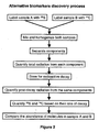
- the main object of this invention is to provide an improved method for biomarker discovery. Differentially abundance molecules between two samples can be isolated for further studies. Generally two samples, whether drug treated compared against vehicle treated, or diseased compared against healthy, are labeled, mixed together, and separated by any number of means commonly known to one skilled in the art, into many fractions, bands, or dots. For example, molecules may be separated by chromatography, electrophoresis, immunoprecipitation, immunomagnetic capturing, array profiling, differential extraction or precipitation such as salt-cut precipitation or organic solvent extraction or precipitation, heat-treatment precipitation, microfluidic device, capillary electrophoresis, differential centrifugation, or gradient separation...etc.
- the ratio of radioactive isotopes in each fraction can be easily monitored. Any fractions whose ratios deviate from a standard ratio can be examined further to identify the exact molecules in those fractions that are responsible. These are the molecules of interest because their levels of abundance vary between the tested samples.
- the molecules can be identified by mass spectrometry and used as biomarkers for drug efficacy or diseased condition. Radioactive labeling with different isotopes also results in same molecules of different mass that can be differentiated and quantitatively compared during mass spectrometry analysis.
- Another object of this invention is to provide a method for studying different degree of modifications to a particular type of molecules such as post-translational modifications of proteins, or methylation of DNA.
- One set of sample is labeled with 32 P phosphates while the other set undergoing a different treatment is labeled with 33 P phosphates to study differential phosphorylation.
- one set of sample is labeled with 3 H sugar such as mannose while the other set is labeled with 14 C mannose for differential glycosylation studies.
- Phosphorylation or glycosylation samples can also be labeled with another isotope such as 35 S to determine the proportion of proteins that are modified.
- the two sets are mixed for analysis after labeling allows an unsurpassed degree of comparison.
- Other forms of post-translational modifications such as methylation, farnesylation, ubiquitination...etc. can also be studied by varying the types of labels use.
- One set of DNA can be methylated with 3 H donor groups while the other set methylated with 14 C donor groups.
- the DNA is then mixed together, digested into smaller fragments, and profiled onto a DNA array. Aberrations in signal ratios between the isotopes signify variations of the degree of methylation for particular captured genes.
- a further object of this invention is to provide an improved method for genomic and proteomic analysis using radioactive isotopes of different half-life to enable simultaneous processing and quantification of multiple samples of DNA, RNA, proteins, and other molecules without damaging or rendering these molecules incomparable or incompatible for analysis purposes, e.g., chemically modifying them. Additionally and importantly, radioactive labeling also provides much higher sensitivity than any other methods of labeling.
- the invention provides a labeling and detection method for comparing profiles of a plurality of molecules derived from comparable sources.
- the method employs the use of radioactive isotopes which offer superior sensitivity for detection of any existing methods.
- radioactive isotopes can be incorporated directly into the molecules of interest eliminating any need for making any chemical modification to these molecules.
- the invention also teaches ways to make supporting devices for increase performance of the method.
- samples from treated and untreated cells labeled with different isotopes such as 3 H or 14 C are mixed together for analysis.
- the mixture is subjected to different types of chromatography one after another hereby refers to as tandem chromatography.
- the chromatography fractionates proteins based on their variations in size, charge, affinity to certain groups etc...
- the fractions are continuously monitored to find any fraction with deviation in 3 H/ 14 C isotope ratio.
- the fractions with significant ratio deviation are further analyzed to single out the exact molecules responsible for the variation. These molecules are then identified by mass spectrometry.
- monoclonal antibodies against these molecules are made as component for an array for high-throughput screening.
- the array is used to capture and compare proteins from the original samples to validate the changes and the array itself. Then the array is ready to be used to screen for similar changes.
- a working drug with known therapeutic efficacy can be used to produce biomarkers in a simple system such as a cell culture model. The discovered biomarkers are then used to make antibody arrays for high-throughput screening of other potential compounds with the same cell culture model.
- mixtures containing at least two isotopes having significant differences in half-life are used for the purpose of quantitative analysis and comparison.
- This method of analysis can also be performed where the selective blocking method fails.
- An exemplary example is 14 C (156keVmax) and 35 S (167keVmax).
- 14 C and 35 S are used together; a suitable method of analysis is used to quantify total radiation, then store for a decay period and then quantify total radiation again.
- the pre-decay and post-decay amounts of radiation read are used to calculate the amount unique to each isotope. This is possible because these two isotopes have different half-life: 5730 years for 14 C and 87.4 days for 35 S.
- the time period between the first quantification and the second quantification is 87.4 days which is exactly equal to one half-life of 35 S. Then the signal difference between the first read and the second read must be equal to half the total 35 S signal from the first read. As a result signal unique to 35 S or 14 C can be calculated.
- At least two samples can be analyzed and compared by labeling with radioactive isotopes of different half-lives.
- the labeling may be done by covalent linkage like the one using NHS ester or by incorporation if the samples comprise live cells and are actively synthesizing the molecules of interest.
- the two samples are mixed for analysis.
- the analysis can include but are not limited to gel electrophoresis, chromatography, and DNA array.
- analyses like protein array, SDS-PAGE, Immunoprecipitation, 2-d gel for proteomic analysis, various forms of chromatography including HPLC are preferred choices.
- the labeling of molecules with different radioactive isotopes is used to quantitatively compare subcomponents of molecules.
- These comprise certain characteristic of molecules such as the degree of phosphorylation of proteins or the degree of methylation of DNA.
- the degree of existing phosphorylation of proteins from two samples can be compared by further phosphorylating these proteins with 32 P or 33 P phosphates.
- the amount of labeled phosphates quantified on these proteins is thus indicative of how many phosphorylation sites are available. Such availability is indicative of preexisting degree of phosphorylation.
- DNA methylation from two samples can be compared.
- the above methods can also be combined to quantify isotopes' signal from a mixture of 3 H, 14 C and 35 S.
- the total radiation is first read and then the partial radiation passing through a screen is then read.
- the screen is designed to block proportionally higher percentage of radiation from tritium, thus tritium signal can be calculated from the first two read (for comparison reasons).
- the mixture, gel or array is stored for a significant amount of time relative to 35 S's half-life.
- the sample is read again for total radiation.
- the reduction in signal is mostly due to 35 S because even for 87 days, tritium with a half-life of ⁇ 12 years and 14 C with a half-life of ⁇ 5730 years hardly decay at all.
- dots of just 3 H, 14 C, and 35 S should be added to the gel or array as internal standards to base the calculation on.
- the use of specialized software can significantly automate the task minimizing operator errors.
- the user simply need to specify the standard dots representing single isotope of 3 H, 14 C, or 35 S from all three exposures and the computer can do the rest. Even if exposure time may be inconsistent between the three times, there is enough data to perform the calculation to obtain the required signal reading for individual isotope.
- the software can ask for exposure durations and decay time to perform the calculations necessary to obtain signal for individual isotope.
- Proteins can incorporate 3 H and 14 C labeled amino acids or even 35 S when Cystein and Methionine are used for double-label or triple-label multiplexing analysis.
- multiple labels can be used for studying protein modifications, and interactions.
- Glycosylation can be differentially labeled with 3 H or 14 C glycan while proteins are labeled with 35 S.
- Phosphorylation can also be compared with 32 P and 33 P while protein abundance is compared with 3 H and 14 C (for comparison reasons).
- the quadruple labeling also serves to determine which proportion of proteins is phosphorylated as well as comparing relative amount of phosphorylation and relative amount of protein expression all in one experiment.
- radioactive Cystein and Methionine are only available commercially with 35 S label while other amino acids are available with both tritium and 14 C labels.
- the main reason is because there is little value for using tritium or 14 C labeled Cystein or Methionine; as a result, these amino acids are not made and sold commercially.
- radioactive multiplexing techniques these amino acids will become much more useful especially because they enable triple labeling when 3 H, 14 C, and 35 S Cystein and Methionine are used for the experiment.
- many enzymes can be severely affected when normal hydrogen is replaced with tritium. Because tritium weight three times more than hydrogen, tritium molecular kinetic is much lower.
- metabolic precursors can also be used to perform the same labeling experiment.
- These metabolic precursors are molecules that can be easily converted and incorporated into protein as Cystein or Methionine while retaining the radioactive label.
- such precursor encompassed molecules that are unique to Cystein or Methionine biosynthesis pathways, and derivatives of post synthesis.
- Cystine the oxidized form of two Cysteins covalently linked by a sulfide bond.
- Other slightly modified versions of Cystein or Methionine or larger molecules containing these amino acids as components can also be used as precursors.
- These precursors are known to those skilled in the art and can vary slightly depending on the biological system used. In addition, when this method becomes more useful, those skilled in the art will be able to apply atomic replacements of nitrogen and oxygen with other radioactive atoms to enable even more multiplexing capability.
- Protein array is a method that will benefit greatly from this novel approach.
- An array is used for profiling and analyzing many molecules simultaneously using just minute amount of sample.
- the current method for detection and quantification of proteins or DNA/RNA on the array use fluorescent dyes, thus the array support only needs to be a sturdy structure that does not fluoresce.
- Described is an array's support that also serves the dual purpose of a screen, which blocks higher proportion of radiation from one particular label but not the others. Depending on the thickness and type of material used, such screen can allow differentially detection and quantification of tritium, carbon-14, sulfur-35, phosphorus-33, phosphorus-32, and other radioactive isotopes from each others.
- the screening technique is particularly useful for radioactive labeled samples whereby at least most of signal from one label is blocked by the screen while higher percentage of the other signal is allowed to pass making simultaneous detection and quantification of both signals possible.
- the array support described is the inclusion of scintillation material to convert radiation into light for easier detection.
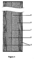
- the array can have a sturdy transparent supporting material such as glass, then a layer of scintillation, then a thin film covering material.
- the thin film will also serve the dual purpose of a blocking screen which will be extremely useful for selectively blocking tritium while allowing a high percentage of 14 C or 35 S signals to pass.
- On top of the thin film is the activated surface where the antibodies are spotted for capturing and profiling antigens. Once captured, the unscreened radiation from these antigens can be read by placing a second scintillation layer on top of the array and then reading the light emitting as a result of radiation striking scintillation materials.
- the partially screened radiation can also be read simultaneously from the bottom of the array by reading the light from the embedded scintillation layer.
- Confocal optical technology allows high resolution reading that can essentially bypass the thickness of the glass layer.
- the thin film acting as blocking screen also block emitting light. Thicker or denser variation of this screen can also be used to differentially block 32 P and 33 P for DNA arrays or phosphorylation studies.
- a thin film of phosphorescent material is used in place of the first and second scintillation material layers described above.
- the phosphorescent material can capture and store the radiation signature to be read back with a properly tuned laser.
- the phosphorescent material is erased by photo-bleaching and then the array is also exposed to another phosphorescent screen "cover slip" on top of the array for equal amount of time before both screens are read.
- DNA arrays will also benefit where higher detection sensitivity is needed. Low abundance DNA or RNA can be detected more easily. Even direct detection is possible eliminating the extra amplification steps such as PCR. Other detections for things that cannot be amplified such as DNA modification are also possible. In addition, pre-purification or other enrichment methods can be used prior to array analysis. These methods can range from eliminating certain high abundant DNA/RNA to specifically trapping certain types of DNA/RNA.
- Chromatography methods have not been able to replaced 2-D gel electrophoresis probably due to the lack of a multiplexing approach. Unlike electrophoresis where sample can be compared side by side or multiplexed on the same gel, chromatography of two samples usually have to be done sequentially for comparison. One type of chromatography can separate samples into fractions which can later be used to separate further by another type of chromatography. It is unlikely that the same sample run back to back on many sequential types of chromatography would yield exactly the same set of fractions. Therefore, the method has not been popular for comparing samples. However, with our labeling and detection method, two samples can be mixed together to be co-separated by sequential chromatography.
- a radiation detector can monitor the proportion of the isotopes in real-time through a flow cell or small aliquots from fractions can be removed and counted by scintillation counting. Samples labeled with 3 H and 14 C, or 32 P and 33 P can readily be distinguished by existing scintillation counter (for comparison reasons). Radiation of aliquots from the same fraction can be determined before and after a decay period to allow differential quantification especially for 14 C and 35 S.
- an immobilized DNA sequence can be used to capture labeled RNA from two samples for comparison. Then optionally the captured molecules can be separated later for further analysis if necessary.
- An obvious advantage of this method is to replace the labor intensive Northern blotting method.
- nucleotides or other precursors that are made with one or more atoms replaced with radioactive isotopes can also be used.
- a well-known method is iodination using 125 I to modify Tyrosines.
- Other coupling methods that add a group of molecules can also be used. These methods use NHS ester, cyanogen bromide, bis-oxirane (epoxides), carbonyldiimidazole, sulfonyl chloride, periodate, fluoromethyl pyridinium sulfonate, glutaraldehyde, acetaldehyde, and formaldehyde...etc to create covalent linkage.
- a gentle way to label protein with at least one phosphorylation site is to use a kinase to add a phosphate group with 32 P or 33 P.
- Protein with known phosphorylation sites can be phosphorylated with specific kinase.
- the reaction can be used to label protein or to determine the amount of existing phosphorylation in these proteins by quantifying the amount of available phosphorylation sites.
- Radiation detection and quantification is made possible by exposing to phosphor-imaging screen which store radiation energy to be read back.
- Another way to quantify radiation is to use scintillation materials to convert radiation into lights and then detect and quantify the amount of generated light.
- Using electronic detectors that can detect radiation directly is also possible, but is more costly.
- Mixture of isotopes such as 14 C and 35 S, or 32 P and 33 P can be quantified by reading radiation from one set of aliquots first, while storing a duplicate set of aliquots for decaying to be read later on.
- the pre-decay and post-decay amounts of radiation are used in combination with the isotopes' half-life to determine the amount of radiation unique to each isotope.
- radioactive isotopes While using radioactive isotopes provides a rapid way to detect and quantified multiplexed samples for rapid quantitative comparison, most samples of interest also require identification by mass spectrometry at least initially. Labeling with different radioactive isotopes produces different mass of the same molecules thus results in distinct peaks in a mass spectrum. Such quality can also be exploited to make quantitative comparison especially to reconfirm previous comparison made with other methods.
- a further improvement method for reading arrays or gels that can increase the resolution of the radiation read is the use of a magnetic field to redirect radiant electrons.
- Beta radiation is essentially electrons or positrons traveling at high velocity. By applying a magnetic field perpendicular to the array, any electrons or positrons not traveling in parallel with the magnetic field will experience a force and be redirected. When electrons mostly travel perpendicular to the array to strike detector or storage screen, better resolution is achieved.
- Permanent magnets preferably with circular or disc shape can be placed below or above or on both sides of the exposure cassette during exposure. Magnets with other shapes can also be used as long as the magnetic line of force will pass perpendicularly through the exposure cassette. Magnets of circular disc shape are likely to have magnetic lines of force perpendicular to the circle thus when thin magnets are used this feature is helpful. In addition to permanent magnets, other electro-magnets including those with superconductor coils can also be used.
- tags that not only can label molecules of interest with various radioactive isotopes, but also provide a means to separate and enrich the labeled molecules away from the unlabeled molecules.
- tags can be linked to an affinity tags such as biotin, immino-biotin, 6His...so they can be quickly selected by capturing agents such as avidin, strepavidin, or nickel.
- labeling tags can also be linked to beads such as agarose, sepharose®, paramagnetic beads so that labeled molecules can be separated by physical means such as sedimentation or use of a magnetic field. These labeling tags can also be immobilized by covalent linkage to a surface of a container or reaction vessel.
- affinity tags can remain with the labeled molecules of interest throughout the analysis, sometimes it is desirable to remove them.
- Biotin is a small affinity tags that by itself can carry 3 H, 14 C, or 35 S. Biotin can be linked to other conjugation reactive groups such as NHS to target specific molecules such as Lysine residues on proteins. A cleavable linkage between the affinity tags and the conjugation reactive groups can also be added so bulky biotin can be removed as in the case of Biotin-SS-NHS.
- the labels need to be in the conjugation reactive group (NHS in this case), additionally, the sulfur next to NHS can also be 35 S labeled,
- a reducing reagent such as dithiothreitol or 2-mecaptoethanol after affinity selection.
- selection means such as beads and immobilization must have a means to release the labeled molecules of interest with a part of the label (with the radioactive isotope of choice) still attached.
- One such means is the disulfide SS linkage described above.
- Another mean is the use of a photo-cleavable linkage.
- An additional means is to use a chemically or enzymatically cleavable linkage. Many such linkages are available and known to those skilled in the art.
- Samples from two sources such as drug-treated and vehicle treated cells are used to look for biomarkers. These samples are mixed together for analysis. A small aliquot is counted on scintillation counter capable of distinguishing between 3 H and 14 C to establish the ratio of samples mixture. The mixture is then separated by tandem chromatography. The fractions resulting from one type of chromatography are further separated by another form of chromatography. Every fraction produced is monitored for 3 H/ 14 C ratio to select for fractions with significant ratio deviation. These fractions of interest can be analyzed further by SDS-PAGE and applied to the two screen system to quantify 3 H/ 14 C ratio. The bands showing significant ratio difference are cut out for identification by mass spectrometry.
- Antibodies are made against the identified proteins or partial protein sequences resulting from mass spectrometry analyses. These antibodies are used to make antibody arrays to rapidly screen for the same biomarker changes in micro format. The array is exposed to the same mixture from original samples to validate their effectiveness in identifying the biomarkers whose changes are originally found by other methods. Once validated, the arrays are ready for high-throughput screening use.
- Post-translational modifications of proteins are very important regulatory mechanism in eukaryotic organisms.
- One such important modification is phosphorylation: a phosphate added to an enzyme or receptor can turn it on or off depending on the type of enzyme or receptor.
- a dual labeling system using chemically similar labeling agents that comprises either 32 P or 33 P is used to determine the degree of phosphorylation in two set of cells or cells undergoing two different treatments.
- Cellular drug response can be studied as followed: (1) Cells are grown equally in two culture dishes. Prior to labeling, the cells are starved of phosphate by washing and replacing growth media with phosphate-deficient media for 1 hour. (2) Labeling media are added wherein one contains 32 P phosphates while the other contains 33 P phosphates. At the same time one set is (or had been) treated with drug while the other is treated with vehicles (any solvent used to carry the drug) or another drug for efficacy comparison. (3) After a predetermined amount of treatment time, radioactive media is washed away and the cells are harvested and lysed in RIPA buffer with phosphatase inhibitors such as NaF and okadaic acid. The cell lysates are mixed together and subjected to different analyses as described below.
- SDS-PAGE and 2-D gel electrophoresis are performed to separate the different proteins for analysis.
- the gels are then fixed in 10% acetic acid and dry on gel drier.
- the other screen is also made of the identical phosphorescent material but placed on much better backing plate.
- the first screen was designed to capture both radiation signals from 32 P and 33 P while allowing much higher proportion of radiation signal from 32 P to pass through to the second screen. Since 33 P signal is relatively weak, the first screen can be thin enough so that the signal picked up on the second screen is not significantly more diffused.
- each dried gel can be exposed to a single phosphorescent imaging screen, then read, then expose to the same screen again then read again after a period of radioactive decay, then repeat if necessary.
- Use the reading before and after a period of radioactive decay to enable quantification of 32 P and 33 P in each band or spot and thus identify bands or spots with deviated ratio of radioactive isotopes. From these calculations, the relative amount of phosphorylation for each protein between samples is calculated. These differences can assess the relative drug effectiveness.
- the mixed cell lysate is subjected to immuno-precipitation to study the phosphorylation and its effect on protein interaction.
- Lysate is precleared with protein G coupled to sepharose beads. Then appropriate antibody is added and incubated for 2 hours followed by the addition of protein G sepharose beads for an additional hour. Beads are collected by centrifugation, washed for at least three times to remove non-specific interaction, and then subject to SDS-PAGE analysis.
- the gel is fixed, dried marked with control markers, and then exposed to the two-layer phosphorescent imaging screen system designed for 32 P and 33 P. The screens are read and the bands of interest are quantified and normalized with control markers enabling quantitative comparison between the two samples (comparative example).
- proteins co-labeled with 3 H and 14 C for interaction studies can also be differentiated and quantified especially after a decay period.
- a variation of the above method is used to compare the degree of phosphorylation for one or a plurality of proteins between two clinical samples.
- the sample are fractionated to some degree of purity suitable for the experiment before one of the sample is subjected to kinase phosphorylation using 32 P ATP while the other sample with 33 P ATP.
- the kinases will add phosphate groups to phosphorylation sites that do not already have phosphates. Because there are many kinases and each of them will specifically phosphorylate different sites, this method can target specific protein or family of proteins. After kinases reactions, the resulting proteins are mixed together for various types of analyses as outlined above.
- the signals are then quantified accordingly.
- the dots or bands of interest can also be cut out for scintillation counting, or mass spectrometry analysis.
- Scintillation counter technology is advanced enough to enable the instrument to distinguish the difference between emissions resulting from 3 H versus that resulting from 14 C. While the main quantification method for radioactivity presented here uses phosphoimaging screen and scintillation counting, other methods, variations and combinations of existing methods can be used and are apparent to those skilled in the art.
- the RNA can be labeled with either 32 P or 33 P at their end terminals and then applied to a complementary DNA array.
- the array is then exposed to the two-screen phosphorescent imaging system so signals specific to either 32 P or 33 P could be quantified and compared after normalized with internal controls' signals (comparative example).
- total signals can be quantified before and after a few days and used to calculate signal unique to 32 P or 33 P.
- the half-life of 32 P is shorter than that of 33 P (14.3 days compared to 25.3 days) thus makes the calculation possible.
- a Reverse Transcriptase reaction can be performed with either 32 P or 33 P labeled poly dT or labeled nucleotides.
- the poly dT is hybridized to the poly A tail of mRNA and reverse transcriptase synthesizes the rest of the complementary sequence to form a single stranded cDNA.
- the RNA is then digested away by RNAse and the cDNA is applied to an array for analysis.
- these procedures can also compare the expression level of different genes in the same tissue because each RNA molecule is labeled with the same amount of radioactivity (except when cDNA is synthesized with labeled nucleotides.
- radioactive nucleotides i.e. [ 32 P/ 33 P]-dATPs are used for the synthesis step so more labels are incorporated into each molecule.
- 35 S nucleotides can also be used in place of 33 P nucleotides.
- 35 S is also superior with half-life of 87.2 days compared to 33 P's half-life of 25.3 days while comparing with 32 P's half-life of 14.3 days.
Landscapes
- Life Sciences & Earth Sciences (AREA)
- Health & Medical Sciences (AREA)
- Engineering & Computer Science (AREA)
- Molecular Biology (AREA)
- Biomedical Technology (AREA)
- Chemical & Material Sciences (AREA)
- Hematology (AREA)
- Immunology (AREA)
- Urology & Nephrology (AREA)
- Cell Biology (AREA)
- Microbiology (AREA)
- Biotechnology (AREA)
- Food Science & Technology (AREA)
- Medicinal Chemistry (AREA)
- Physics & Mathematics (AREA)
- Analytical Chemistry (AREA)
- Biochemistry (AREA)
- General Health & Medical Sciences (AREA)
- General Physics & Mathematics (AREA)
- Pathology (AREA)
- Investigating Or Analysing Biological Materials (AREA)
- Measurement Of Radiation (AREA)
Claims (7)
- Procédé de comparaison d'échantillons, comprenant les étapes suivantes :a) la fourniture de deux échantillons à des fins d'analyse comparative ;b) le marquage des molécules d'un premier échantillon par liaison covalente avec un premier isotope radioactif ;c) le marquage des molécules d'un second échantillon par liaison covalente avec un second isotope radioactif qui a une demi-vie différente de la demi-vie dudit premier isotope radioactif ;d) le mélange dudit premier échantillon et dudit second échantillon pour obtenir un mélange homogène ;e) la soumission dudit mélange homogène à de quelconques moyens de séparation aptes à séparer les molécules en fractions ou en bandes ou en points ;f) la quantification d'une quantité totale de rayonnement émise par chaque fraction ou bande ou point ;g) la quantification d'une quantité totale de rayonnement après désintégration émise par chacune desdites fractions ou bandes ou chacun desdits points après une période de désintégration radioactive ;h) l'examen du rayonnement total et du rayonnement total après désintégration émis par chaque fraction ou bande ou point afin d'identifier une quelconque fraction ou bande ou un quelconque point présentant un écart du rapport entre le rayonnement total et le rayonnement total après désintégration.
- Procédé selon la revendication 1, comprenant en outre une étape d'isolement d'une molécule qui provoque ledit écart du rapport entre le rayonnement total et le rayonnement total après désintégration, de l'un de la fraction ou de la bande ou du point présentant un écart du rapport entre le rayonnement total et le rayonnement total après désintégration.
- Procédé selon la revendication 2, comprenant en outre une étape d'identification de ladite molécule qui provoque ledit écart du rapport entre le rayonnement total et le rayonnement total après désintégration par spectrométrie de masse.
- Procédé selon la revendication 1, comprenant en outre une étape d'application d'un champ magnétique dont les lignes de force magnétique sont perpendiculaires au plan d'un gel ou d'un réseau, pour améliorer la résolution du rayonnement bêta lorsque ledit gel ou réseau est utilisé en tant que lesdits moyens de séparation.
- Procédé d'isolement d'une molécule présente en différentes quantités entre deux échantillons, comprenant les étapes suivantes :a) la fourniture de deux échantillons à des fins d'analyse ;b) le marquage des molécules d'un premier échantillon par liaison covalente avec un premier isotope radioactif ;c) le marquage des molécules d'un second échantillon par liaison covalente avec un second isotope radioactif qui a une demi-vie différente de la demi-vie dudit premier isotope radioactif ;d) le mélange dudit premier échantillon et dudit second échantillon pour obtenir un mélange homogène ;e) la soumission dudit mélange homogène à de quelconques moyens de séparation aptes à séparer les molécules en fractions ou en bandes ou en points ;f) l'utilisation de la différence de demi-vie entre ledit premier isotope radioactif et ledit second isotope radioactif pour identifier une fraction ou une bande ou un point présentant un écart du rapport des isotopes radioactifs ; etg) l'isolement de ladite molécule présente en différentes quantités de ladite fraction ou de ladite bande ou dudit point présentant un écart du rapport des isotopes radioactifs dans lequel ladite molécule présente en différentes quantités est responsable dudit écart du rapport des isotopes radioactifs.
- Procédé selon la revendication 5, comprenant en outre une étape d'identification de ladite molécule présente en différentes quantités par spectrométrie de masse.
- Procédé de quantification d'une molécule dans un échantillon, comprenant les étapes suivantes :a) la fourniture d'un premier échantillon contenant une quantité inconnue de ladite molécule à des fins d'analyse ;b) la fourniture d'un second échantillon contenant une quantité connue de ladite molécule à des fins d'analyse ;c) le marquage des molécules d'un premier échantillon par liaison covalente avec un premier isotope radioactif ;d) le marquage des molécules d'un second échantillon par liaison covalente avec un second isotope radioactif qui a une demi-vie différente de la demi-vie dudit premier isotope radioactif ;e) le mélange dudit premier échantillon et dudit second échantillon pour obtenir un mélange homogène ;f) la soumission dudit mélange homogène à de quelconques moyens de séparation aptes à séparer les molécules en fractions ou en bandes ou en points ;g) l'identification d'une fraction ou d'une bande ou d'un point qui contient ladite molécule ;h) la quantification d'une quantité totale de rayonnement émise par ladite fraction ou ladite bande ou ledit point ;i) la quantification d'une quantité totale de rayonnement après désintégration émise par ladite fraction ou ladite bande ou ledit point après une période de désintégration radioactive ;j) l'utilisation du rapport entre ladite quantité totale de rayonnement et ladite quantité totale de rayonnement après désintégration pour calculer ladite quantité inconnue de ladite molécule dans ledit premier échantillon en fonction de ladite quantité connue de ladite molécule dans ledit second échantillon.
Applications Claiming Priority (2)
| Application Number | Priority Date | Filing Date | Title |
|---|---|---|---|
| US10/680,277 US7029855B1 (en) | 2003-01-28 | 2003-10-07 | Radioactive multiplexing analytical methods for biomarkers discovery |
| PCT/US2004/002442 WO2005045056A2 (fr) | 2003-10-07 | 2004-01-28 | Procede d'analyse avec multiplexage de radioactivites |
Publications (3)
| Publication Number | Publication Date |
|---|---|
| EP1689882A2 EP1689882A2 (fr) | 2006-08-16 |
| EP1689882A4 EP1689882A4 (fr) | 2007-03-21 |
| EP1689882B1 true EP1689882B1 (fr) | 2014-11-26 |
Family
ID=34394320
Family Applications (1)
| Application Number | Title | Priority Date | Filing Date |
|---|---|---|---|
| EP04775740.6A Expired - Lifetime EP1689882B1 (fr) | 2003-10-07 | 2004-01-28 | Procede d'analyse avec multiplexage de radioactivites |
Country Status (5)
| Country | Link |
|---|---|
| US (1) | US7175986B2 (fr) |
| EP (1) | EP1689882B1 (fr) |
| AU (1) | AU2004288113A1 (fr) |
| CA (1) | CA2542018A1 (fr) |
| WO (1) | WO2005045056A2 (fr) |
Families Citing this family (3)
| Publication number | Priority date | Publication date | Assignee | Title |
|---|---|---|---|---|
| EP1689882B1 (fr) | 2003-10-07 | 2014-11-26 | TRAN, Nathaniel Tue | Procede d'analyse avec multiplexage de radioactivites |
| US20060172429A1 (en) * | 2005-01-31 | 2006-08-03 | Nilsson Erik J | Methods of identification of biomarkers with mass spectrometry techniques |
| CN101906452A (zh) * | 2010-07-09 | 2010-12-08 | 复旦大学 | 一种糖苷内切酶催化同位素标记n-糖链的方法 |
Family Cites Families (18)
| Publication number | Priority date | Publication date | Assignee | Title |
|---|---|---|---|---|
| US4016250A (en) * | 1974-03-22 | 1977-04-05 | Cornell Research Foundation, Inc. | Method for testing for pregnancy |
| DE81371T1 (de) * | 1981-12-07 | 1983-10-27 | Vg Isotopes Ltd., Winsford, Cheshire | Mehrfachkollektor massenspektrometer. |
| US4628205A (en) | 1985-04-08 | 1986-12-09 | Packard Instrument Company, Inc. | Regionless multiple label scintillation counting |
| US4886761A (en) * | 1987-03-26 | 1989-12-12 | Yellowstone Diagnostics Corporation | Polysilicon binding assay support and methods |
| SE8705056D0 (sv) | 1987-12-18 | 1987-12-18 | Wallac Oy | Liquid scintillation counter |
| SE8802861D0 (sv) | 1988-08-10 | 1988-08-10 | Wallac Oy | An apparatus and a method for measuring the activity of radioactive samples containing a multiple of radioactive isotopes |
| US5807522A (en) * | 1994-06-17 | 1998-09-15 | The Board Of Trustees Of The Leland Stanford Junior University | Methods for fabricating microarrays of biological samples |
| US5753917A (en) | 1995-06-06 | 1998-05-19 | Engdahl; John C. | Dual crystal scintillation camera |
| US6203993B1 (en) * | 1996-08-14 | 2001-03-20 | Exact Science Corp. | Methods for the detection of nucleic acids |
| US6979728B2 (en) * | 1998-05-04 | 2005-12-27 | Baylor College Of Medicine | Articles of manufacture and methods for array based analysis of biological molecules |
| WO2000063701A2 (fr) * | 1999-04-15 | 2000-10-26 | The Board Of Trustees Of The Leland Stanford Junior University | Jeux ordonnes de microechantillons de polypeptides |
| US6379970B1 (en) * | 1999-04-30 | 2002-04-30 | The Arizona Board Of Regents On Behalf Of The University Of Arizona | Analysis of differential protein expression |
| EP1158057A1 (fr) * | 2000-05-18 | 2001-11-28 | Centre National De La Recherche Scientifique | Compositions et procédés applicables à l'étude du dosage génétique |
| EP1290450A2 (fr) * | 2000-06-09 | 2003-03-12 | MDS Proteomics, Inc. | Etiquetage d'echantillons proteomiques pendant la proteolyse a des fins de quantification et d'analyse multiple d'echantillons |
| NO316478B1 (no) | 2001-01-12 | 2004-01-26 | Biomolex As | Fremgangsmåte og apparat for simultan kvantifisering av ulike radionuklideri et stort antall regioner på overflaten av en biologiskmikromatrise eller lignende objekt |
| EP1506399A2 (fr) * | 2001-05-21 | 2005-02-16 | Aclara BioSciences, Inc. | Procedes et compositions pour l'analyse de proteines |
| US7029855B1 (en) | 2003-01-28 | 2006-04-18 | Proteomyx Inc. | Radioactive multiplexing analytical methods for biomarkers discovery |
| EP1689882B1 (fr) | 2003-10-07 | 2014-11-26 | TRAN, Nathaniel Tue | Procede d'analyse avec multiplexage de radioactivites |
-
2004
- 2004-01-28 EP EP04775740.6A patent/EP1689882B1/fr not_active Expired - Lifetime
- 2004-01-28 CA CA002542018A patent/CA2542018A1/fr not_active Abandoned
- 2004-01-28 AU AU2004288113A patent/AU2004288113A1/en not_active Abandoned
- 2004-01-28 WO PCT/US2004/002442 patent/WO2005045056A2/fr active Application Filing
- 2004-04-28 US US10/835,027 patent/US7175986B2/en not_active Expired - Fee Related
Also Published As
| Publication number | Publication date |
|---|---|
| EP1689882A4 (fr) | 2007-03-21 |
| WO2005045056A2 (fr) | 2005-05-19 |
| US7175986B2 (en) | 2007-02-13 |
| WO2005045056A3 (fr) | 2005-08-18 |
| US20050074794A1 (en) | 2005-04-07 |
| CA2542018A1 (fr) | 2005-05-19 |
| AU2004288113A1 (en) | 2005-05-19 |
| EP1689882A2 (fr) | 2006-08-16 |
Similar Documents
| Publication | Publication Date | Title |
|---|---|---|
| Zhang et al. | Recent advances in analytical methods for the therapeutic drug monitoring of immunosuppressive drugs | |
| Wilson et al. | Recent developments in protein microarray technology | |
| Cifani et al. | Towards comprehensive and quantitative proteomics for diagnosis and therapy of human disease | |
| MacBeath | Protein microarrays and proteomics | |
| Yang et al. | Deep profiling of cellular heterogeneity by emerging single‐cell proteomic technologies | |
| Poetz et al. | Protein microarrays: catching the proteome | |
| Kasper et al. | The application of multiplexed, multi‐dimensional ultra‐high‐performance liquid chromatography/tandem mass spectrometry to the high‐throughput screening of lysosomal storage disorders in newborn dried bloodspots | |
| IL180304A (en) | Mass defect labeling for the determination of oligomer sequences | |
| Fredolini et al. | Immunocapture strategies in translational proteomics | |
| Honoré et al. | Functional genomics studied by proteomics | |
| CN103959064B (zh) | 一种测定样品中抗原含量的方法 | |
| Guo et al. | A proteomic primer for the clinician | |
| US20100112722A1 (en) | Immunoassays and Characterization of Biomolecular Interactions Using Self-Assembled Monolayers | |
| Hudler et al. | Proteomic strategies and challenges in tumor metastasis research | |
| Nakajima et al. | Toward proteome‐wide exploration of proteins in dried blood spots using liquid chromatography‐coupled mass spectrometry | |
| Jiang et al. | Bladder cancer hunting: A microfluidic paper‐based analytical device | |
| EP1689882B1 (fr) | Procede d'analyse avec multiplexage de radioactivites | |
| US7029855B1 (en) | Radioactive multiplexing analytical methods for biomarkers discovery | |
| Careri et al. | Element-tagged immunoassay with inductively coupled plasma mass spectrometry for multianalyte detection | |
| Jain et al. | Technologies for discovery of biomarkers | |
| US20040023274A1 (en) | Method for the quantification of carbohydrates | |
| Morozov et al. | Parallel determination of multiple protein metabolite interactions using cell extract, protein microarrays and mass spectrometric detection | |
| Halvorsen et al. | Is this the end of dried blood spots as we know it? | |
| Nedelkov et al. | High-throughput affinity mass spectrometry | |
| Jozwik et al. | Discovery of a Hidden Proinflammatory Signaling Proteome Using a Large-Scale, Targeted Antibody Microarray Platform |
Legal Events
| Date | Code | Title | Description |
|---|---|---|---|
| PUAI | Public reference made under article 153(3) epc to a published international application that has entered the european phase |
Free format text: ORIGINAL CODE: 0009012 |
|
| 17P | Request for examination filed |
Effective date: 20060504 |
|
| AK | Designated contracting states |
Kind code of ref document: A2 Designated state(s): AT BE BG CH CY CZ DE DK EE ES FI FR GB GR HU IE IT LI LU MC NL PT RO SE SI SK TR |
|
| DAX | Request for extension of the european patent (deleted) | ||
| A4 | Supplementary search report drawn up and despatched |
Effective date: 20070216 |
|
| RIC1 | Information provided on ipc code assigned before grant |
Ipc: C12Q 1/68 20060101ALI20070213BHEP Ipc: G01N 33/60 20060101ALI20070213BHEP Ipc: C07H 21/04 20060101ALI20070213BHEP Ipc: C07K 14/00 20060101ALI20070213BHEP Ipc: G01N 33/53 20060101AFI20070213BHEP Ipc: C07H 21/02 20060101ALI20070213BHEP |
|
| 17Q | First examination report despatched |
Effective date: 20070612 |
|
| GRAP | Despatch of communication of intention to grant a patent |
Free format text: ORIGINAL CODE: EPIDOSNIGR1 |
|
| INTG | Intention to grant announced |
Effective date: 20130405 |
|
| GRAJ | Information related to disapproval of communication of intention to grant by the applicant or resumption of examination proceedings by the epo deleted |
Free format text: ORIGINAL CODE: EPIDOSDIGR1 |
|
| GRAP | Despatch of communication of intention to grant a patent |
Free format text: ORIGINAL CODE: EPIDOSNIGR1 |
|
| INTG | Intention to grant announced |
Effective date: 20131011 |
|
| GRAS | Grant fee paid |
Free format text: ORIGINAL CODE: EPIDOSNIGR3 |
|
| GRAA | (expected) grant |
Free format text: ORIGINAL CODE: 0009210 |
|
| AK | Designated contracting states |
Kind code of ref document: B1 Designated state(s): AT BE BG CH CY CZ DE DK EE ES FI FR GB GR HU IE IT LI LU MC NL PT RO SE SI SK TR |
|
| REG | Reference to a national code |
Ref country code: GB Ref legal event code: FG4D |
|
| REG | Reference to a national code |
Ref country code: CH Ref legal event code: EP |
|
| REG | Reference to a national code |
Ref country code: AT Ref legal event code: REF Ref document number: 698482 Country of ref document: AT Kind code of ref document: T Effective date: 20141215 |
|
| REG | Reference to a national code |
Ref country code: IE Ref legal event code: FG4D |
|
| REG | Reference to a national code |
Ref country code: DE Ref legal event code: R096 Ref document number: 602004046220 Country of ref document: DE Effective date: 20141231 |
|
| REG | Reference to a national code |
Ref country code: FR Ref legal event code: PLFP Year of fee payment: 12 |
|
| REG | Reference to a national code |
Ref country code: NL Ref legal event code: VDEP Effective date: 20141126 |
|
| REG | Reference to a national code |
Ref country code: AT Ref legal event code: MK05 Ref document number: 698482 Country of ref document: AT Kind code of ref document: T Effective date: 20141126 |
|
| PG25 | Lapsed in a contracting state [announced via postgrant information from national office to epo] |
Ref country code: FI Free format text: LAPSE BECAUSE OF FAILURE TO SUBMIT A TRANSLATION OF THE DESCRIPTION OR TO PAY THE FEE WITHIN THE PRESCRIBED TIME-LIMIT Effective date: 20141126 Ref country code: NL Free format text: LAPSE BECAUSE OF FAILURE TO SUBMIT A TRANSLATION OF THE DESCRIPTION OR TO PAY THE FEE WITHIN THE PRESCRIBED TIME-LIMIT Effective date: 20141126 Ref country code: ES Free format text: LAPSE BECAUSE OF FAILURE TO SUBMIT A TRANSLATION OF THE DESCRIPTION OR TO PAY THE FEE WITHIN THE PRESCRIBED TIME-LIMIT Effective date: 20141126 Ref country code: PT Free format text: LAPSE BECAUSE OF FAILURE TO SUBMIT A TRANSLATION OF THE DESCRIPTION OR TO PAY THE FEE WITHIN THE PRESCRIBED TIME-LIMIT Effective date: 20150326 |
|
| PG25 | Lapsed in a contracting state [announced via postgrant information from national office to epo] |
Ref country code: AT Free format text: LAPSE BECAUSE OF FAILURE TO SUBMIT A TRANSLATION OF THE DESCRIPTION OR TO PAY THE FEE WITHIN THE PRESCRIBED TIME-LIMIT Effective date: 20141126 Ref country code: CY Free format text: LAPSE BECAUSE OF FAILURE TO SUBMIT A TRANSLATION OF THE DESCRIPTION OR TO PAY THE FEE WITHIN THE PRESCRIBED TIME-LIMIT Effective date: 20141126 Ref country code: GR Free format text: LAPSE BECAUSE OF FAILURE TO SUBMIT A TRANSLATION OF THE DESCRIPTION OR TO PAY THE FEE WITHIN THE PRESCRIBED TIME-LIMIT Effective date: 20150227 Ref country code: SE Free format text: LAPSE BECAUSE OF FAILURE TO SUBMIT A TRANSLATION OF THE DESCRIPTION OR TO PAY THE FEE WITHIN THE PRESCRIBED TIME-LIMIT Effective date: 20141126 |
|
| PGFP | Annual fee paid to national office [announced via postgrant information from national office to epo] |
Ref country code: GB Payment date: 20150225 Year of fee payment: 12 Ref country code: FR Payment date: 20150202 Year of fee payment: 12 |
|
| REG | Reference to a national code |
Ref country code: CH Ref legal event code: NV Representative=s name: FIAMMENGHI-FIAMMENGHI, CH |
|
| PG25 | Lapsed in a contracting state [announced via postgrant information from national office to epo] |
Ref country code: CZ Free format text: LAPSE BECAUSE OF FAILURE TO SUBMIT A TRANSLATION OF THE DESCRIPTION OR TO PAY THE FEE WITHIN THE PRESCRIBED TIME-LIMIT Effective date: 20141126 Ref country code: RO Free format text: LAPSE BECAUSE OF FAILURE TO SUBMIT A TRANSLATION OF THE DESCRIPTION OR TO PAY THE FEE WITHIN THE PRESCRIBED TIME-LIMIT Effective date: 20141126 Ref country code: DK Free format text: LAPSE BECAUSE OF FAILURE TO SUBMIT A TRANSLATION OF THE DESCRIPTION OR TO PAY THE FEE WITHIN THE PRESCRIBED TIME-LIMIT Effective date: 20141126 Ref country code: SK Free format text: LAPSE BECAUSE OF FAILURE TO SUBMIT A TRANSLATION OF THE DESCRIPTION OR TO PAY THE FEE WITHIN THE PRESCRIBED TIME-LIMIT Effective date: 20141126 Ref country code: EE Free format text: LAPSE BECAUSE OF FAILURE TO SUBMIT A TRANSLATION OF THE DESCRIPTION OR TO PAY THE FEE WITHIN THE PRESCRIBED TIME-LIMIT Effective date: 20141126 |
|
| PGFP | Annual fee paid to national office [announced via postgrant information from national office to epo] |
Ref country code: CH Payment date: 20150429 Year of fee payment: 12 |
|
| REG | Reference to a national code |
Ref country code: DE Ref legal event code: R097 Ref document number: 602004046220 Country of ref document: DE |
|
| PG25 | Lapsed in a contracting state [announced via postgrant information from national office to epo] |
Ref country code: LU Free format text: LAPSE BECAUSE OF FAILURE TO SUBMIT A TRANSLATION OF THE DESCRIPTION OR TO PAY THE FEE WITHIN THE PRESCRIBED TIME-LIMIT Effective date: 20150128 |
|
| PG25 | Lapsed in a contracting state [announced via postgrant information from national office to epo] |
Ref country code: MC Free format text: LAPSE BECAUSE OF FAILURE TO SUBMIT A TRANSLATION OF THE DESCRIPTION OR TO PAY THE FEE WITHIN THE PRESCRIBED TIME-LIMIT Effective date: 20141126 |
|
| PLBE | No opposition filed within time limit |
Free format text: ORIGINAL CODE: 0009261 |
|
| STAA | Information on the status of an ep patent application or granted ep patent |
Free format text: STATUS: NO OPPOSITION FILED WITHIN TIME LIMIT |
|
| PGFP | Annual fee paid to national office [announced via postgrant information from national office to epo] |
Ref country code: DE Payment date: 20150729 Year of fee payment: 12 |
|
| 26N | No opposition filed |
Effective date: 20150827 |
|
| REG | Reference to a national code |
Ref country code: IE Ref legal event code: MM4A |
|
| PG25 | Lapsed in a contracting state [announced via postgrant information from national office to epo] |
Ref country code: IT Free format text: LAPSE BECAUSE OF FAILURE TO SUBMIT A TRANSLATION OF THE DESCRIPTION OR TO PAY THE FEE WITHIN THE PRESCRIBED TIME-LIMIT Effective date: 20141126 |
|
| PG25 | Lapsed in a contracting state [announced via postgrant information from national office to epo] |
Ref country code: IE Free format text: LAPSE BECAUSE OF NON-PAYMENT OF DUE FEES Effective date: 20150128 |
|
| PG25 | Lapsed in a contracting state [announced via postgrant information from national office to epo] |
Ref country code: SI Free format text: LAPSE BECAUSE OF FAILURE TO SUBMIT A TRANSLATION OF THE DESCRIPTION OR TO PAY THE FEE WITHIN THE PRESCRIBED TIME-LIMIT Effective date: 20141126 |
|
| REG | Reference to a national code |
Ref country code: DE Ref legal event code: R119 Ref document number: 602004046220 Country of ref document: DE |
|
| REG | Reference to a national code |
Ref country code: CH Ref legal event code: PL |
|
| GBPC | Gb: european patent ceased through non-payment of renewal fee |
Effective date: 20160128 |
|
| REG | Reference to a national code |
Ref country code: FR Ref legal event code: ST Effective date: 20160930 |
|
| PG25 | Lapsed in a contracting state [announced via postgrant information from national office to epo] |
Ref country code: CH Free format text: LAPSE BECAUSE OF NON-PAYMENT OF DUE FEES Effective date: 20160131 Ref country code: DE Free format text: LAPSE BECAUSE OF NON-PAYMENT OF DUE FEES Effective date: 20160802 Ref country code: GB Free format text: LAPSE BECAUSE OF NON-PAYMENT OF DUE FEES Effective date: 20160128 Ref country code: LI Free format text: LAPSE BECAUSE OF NON-PAYMENT OF DUE FEES Effective date: 20160131 |
|
| PG25 | Lapsed in a contracting state [announced via postgrant information from national office to epo] |
Ref country code: FR Free format text: LAPSE BECAUSE OF NON-PAYMENT OF DUE FEES Effective date: 20160201 |
|
| PG25 | Lapsed in a contracting state [announced via postgrant information from national office to epo] |
Ref country code: BG Free format text: LAPSE BECAUSE OF FAILURE TO SUBMIT A TRANSLATION OF THE DESCRIPTION OR TO PAY THE FEE WITHIN THE PRESCRIBED TIME-LIMIT Effective date: 20141126 Ref country code: HU Free format text: LAPSE BECAUSE OF FAILURE TO SUBMIT A TRANSLATION OF THE DESCRIPTION OR TO PAY THE FEE WITHIN THE PRESCRIBED TIME-LIMIT; INVALID AB INITIO Effective date: 20040128 |
|
| PG25 | Lapsed in a contracting state [announced via postgrant information from national office to epo] |
Ref country code: BE Free format text: LAPSE BECAUSE OF NON-PAYMENT OF DUE FEES Effective date: 20150131 |
|
| PG25 | Lapsed in a contracting state [announced via postgrant information from national office to epo] |
Ref country code: TR Free format text: LAPSE BECAUSE OF FAILURE TO SUBMIT A TRANSLATION OF THE DESCRIPTION OR TO PAY THE FEE WITHIN THE PRESCRIBED TIME-LIMIT Effective date: 20141126 |