EP1677256A2 - Procédé et arrangement pour la reconstruction multirésolutive pour l'imagerie médicale à rayons X - Google Patents
Procédé et arrangement pour la reconstruction multirésolutive pour l'imagerie médicale à rayons X Download PDFInfo
- Publication number
- EP1677256A2 EP1677256A2 EP05112611A EP05112611A EP1677256A2 EP 1677256 A2 EP1677256 A2 EP 1677256A2 EP 05112611 A EP05112611 A EP 05112611A EP 05112611 A EP05112611 A EP 05112611A EP 1677256 A2 EP1677256 A2 EP 1677256A2
- Authority
- EP
- European Patent Office
- Prior art keywords
- coefficients
- multiresolution representation
- multiresolution
- representation
- ray
- Prior art date
- Legal status (The legal status is an assumption and is not a legal conclusion. Google has not performed a legal analysis and makes no representation as to the accuracy of the status listed.)
- Granted
Links
- 238000000034 method Methods 0.000 title claims abstract description 54
- 238000003384 imaging method Methods 0.000 title claims abstract description 25
- 238000005259 measurement Methods 0.000 claims description 17
- 239000011159 matrix material Substances 0.000 claims description 7
- 238000004519 manufacturing process Methods 0.000 claims description 2
- 230000008030 elimination Effects 0.000 description 10
- 238000003379 elimination reaction Methods 0.000 description 10
- 238000009607 mammography Methods 0.000 description 9
- 238000005070 sampling Methods 0.000 description 5
- 238000003325 tomography Methods 0.000 description 4
- 238000005516 engineering process Methods 0.000 description 3
- 230000005855 radiation Effects 0.000 description 2
- 238000002083 X-ray spectrum Methods 0.000 description 1
- 238000004458 analytical method Methods 0.000 description 1
- 238000013459 approach Methods 0.000 description 1
- 230000002238 attenuated effect Effects 0.000 description 1
- 210000000481 breast Anatomy 0.000 description 1
- 238000004891 communication Methods 0.000 description 1
- 238000002591 computed tomography Methods 0.000 description 1
- 238000002059 diagnostic imaging Methods 0.000 description 1
- 239000003814 drug Substances 0.000 description 1
- 238000000338 in vitro Methods 0.000 description 1
- 238000010348 incorporation Methods 0.000 description 1
- 238000003825 pressing Methods 0.000 description 1
- 238000012545 processing Methods 0.000 description 1
- 229910052704 radon Inorganic materials 0.000 description 1
- SYUHGPGVQRZVTB-UHFFFAOYSA-N radon atom Chemical compound [Rn] SYUHGPGVQRZVTB-UHFFFAOYSA-N 0.000 description 1
- 238000004904 shortening Methods 0.000 description 1
Images


Classifications
-
- G—PHYSICS
- G06—COMPUTING; CALCULATING OR COUNTING
- G06T—IMAGE DATA PROCESSING OR GENERATION, IN GENERAL
- G06T11/00—2D [Two Dimensional] image generation
- G06T11/003—Reconstruction from projections, e.g. tomography
- G06T11/006—Inverse problem, transformation from projection-space into object-space, e.g. transform methods, back-projection, algebraic methods
-
- G—PHYSICS
- G06—COMPUTING; CALCULATING OR COUNTING
- G06T—IMAGE DATA PROCESSING OR GENERATION, IN GENERAL
- G06T2211/00—Image generation
- G06T2211/40—Computed tomography
- G06T2211/424—Iterative
-
- G—PHYSICS
- G06—COMPUTING; CALCULATING OR COUNTING
- G06T—IMAGE DATA PROCESSING OR GENERATION, IN GENERAL
- G06T2211/00—Image generation
- G06T2211/40—Computed tomography
- G06T2211/436—Limited angle
Definitions
- Three-dimensional X-ray imaging is based on taking several one-dimensional (1-D) or two-dimensional (2-D) projection images of a three-dimensional (3-D) body from different directions. If 1-D projection images are available from all around a 2-D slice of the body with dense angular sampling, the inner structure of the slice can be determined. This is known as Computerized Tomography (CT) imaging technology, which is widely used in medicine today.
- CT Computerized Tomography
- a crucial part of CT technology is the reconstruction algorithm taking the X-ray images as argument and returning a voxel representation of the 3-D body.
- a collection of X-ray images of a 3-D body is called sparse projection data if (a) the images are taken from a limited angle of view or (b) there is only a small number of images. Sparse projection data does not contain sufficient information to completely describe the 3-D body, However, in many practical imaging situations only sparse projection data is available.
- the aim of the invention is to overcome the problems met in 3-D reconstruction of the body that occur when using traditional reconstruction algorithms with sparse projection data.
- This is achieved by a method for producing three-dimensional information of an object in medical X-ray imaging in which method the object is X-radiated from at least two different directions and the said X-radiation is detected to form projection data of the object.
- the X-ray attenuation coefficient inside the object is represented by a linear combination of multiresolution basis functions, This multiresolution representation is such that not all of the basis functions are needed to represent the unknown coefficient.
- a priori information and some measurement information are combined to give information about what basis functions are not needed in the representation, leading to computationally economic multiresolution representation.
- projection data is a collection of images, where each image is either (i) a traditional projection image taken with X-ray source, object and detector stationary, (ii) scanned projection image taken with moving X-ray source, moving detector and moving detector pixels to form an image, or (iii) a tomosynthetic slice achieved by moving the X-ray source, object, and/or detector during the exposure for emphasizing some sharp layer inside the object and blurring other layers.
- a panoramic dental X-ray imaging device produces a tomosynthetic slice with sharp layer along the dental arc.
- the invention also relates to a medical X-ray device arrangement for producing three-dimensional information of an object in medical X-ray imaging, the medical X-ray device arrangement comprising an X-ray source for X-radiating the object from at least two different directions and a detector for detecting the X-radiation to form projection data of the object.
- the medical X-ray device arrangement comprises means for representing the X-ray attenuation coefficient inside the object by a linear combination of multiresolution basis functions. This multiresolution representation is such that not all of the basis functions are needed to represent the unknown coefficient. A priori information and some measurement information are combined to give information about what basis functions are not needed in the representation, leading to computationally economic multiresolution representation.
- benefits of the invention over conventional CT reconstruction include reduced artefacts.
- benefits of the invention over tomosynthesis include reduced artefacts, improved contrast and improved image quality.
- Figure 1 shows the acquisition of a traditional X-ray projection image.
- Figures 2A - 2B show two kinds of data.
- 2A scanned projection image, where a narrow detector moves during exposure to form a projection image.
- 2B tomosynthetic slice where X-ray source and detector move during exposure to form a sharp layer inside the object.
- Figures 3A - 3D show different types of projection data. Every black dot represents a location of the X-ray source for taking one projection image.
- 3A dense full-angle data.
- 3B dense limited-angle data.
- 3C sparse full-angle data.
- 3D sparse limited-angle data.
- Figure 4 shows "pencil beam” X-ray attenuation model.
- Figure 5 shows basic flow chart of the method according to the invention.
- Figure 6 shows an intraoral X-ray device arrangement presenting one preferred embodiment of the invention.
- Figure 7 shows measurement geometry of dental limited-angle tomography with a digital intraoral sensor.
- Figure 8 shows measurement geometry of mammography limited-angle tomography with a mammography device.
- Figure 9 shows results of 3-D reconstruction of head phantom in the first preferred embodiment.
- Left column tomosynthetic slices through the teeth of the head phantom.
- Right column corresponding slices produced by the invention.
- Figure 10 shows results of 3-D reconstruction of mammography specimen in the second preferred embodiment.
- Left column tomosynthetic slices through the specimen.
- Right column corresponding slices produced by the invention.
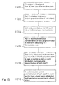
- Figure 11 shows basic flow chart of the thresholding method presenting one preferred embodiment of the invention.
- Figure 12 shows basic flow chart of the thresholding method presenting one preferred embodiment of the invention.
- Figure 13 shows basic flow chart of the thresholding method presenting one preferred embodiment of the invention.
- X-ray images are not always available from all around the body.
- the body might be visible only from certain directions due to imaging geometry
- the X-ray detector is in fixed position behind the tissue and the X-ray source moves with respect to the detector. This situation is called limited-angle tomography.
- the number of radiographs should be minimized in medical applications for reducing the X-ray dose of the patient and shortening the time needed for imaging. Such situations lead to sparse projection data.
- a regularized inversion algorithm is used to create a new type of 3-D medical X-ray imaging using sparse projection data as input. This new imaging is intermediate between a projection radiograph and a full CT scan.
- Figure 1 is shown a simple example of X-ray imaging, where an X-ray source 2 is placed on one side of an object 4 under imaging. Radiation passes through the object and is detected by a detector 6 on the other side.
- the X-ray source is for example an X-ray source of an intraoral X-ray source of a dentist, of a dental panoramic X-ray device, of a surgical C-arm X-ray device, of a mammography device, of a CT scanner or of any other medical X-ray device and the detector 6 is a detector of some of those devices.
- the detector 6 is a digital sensor that can be thought of as a 2-D array of almost point-like detectors.
- the 3-D body under imaging is modelled by non-negative X-ray attenuation coefficient g.
- the X-radiation has initial intensity I 0 when entering the object 4 and a smaller intensity I 1 when exiting the object.
- one pixel value in the measured data represents (i) the integral of g along one line L in case of a projection image.
- Figures 3A and 3B illustrate the types of tomographic data resulting from different imaging situations. For clarity, here are presented two-dimensional examples; similar situations can be considered in three dimensions.
- radiation dose is reduced using coarse sampling of the angular variable.
- Figures 3A and 3B present dense angular sampling with full angle and limited angle measurements, respectively.
- Figures 3C and 3D present coarse angular sampling, also with full and limited angles of measurement.
- A is a linear operator modelling the X-ray attenuation process.
- A can model various ways to collect data. For example, A can model a collection of two or more measurements, where some of the measurements may be traditional projection images, some of the measurements may be scanned projection images, and some of the measurements may be tomosynthetic slices. Traditional projection image is acquired by keeping X-ray source 2, object 4 and detector 6 fixed during the exposure as shown in Figure 1.
- Scanned projection image is acquired by keeping X-ray source 2 and object 4 fixed during the exposure while moving detector 6 and its pixels as shown in Figure 2A.
- Tomosynthetic slice is acquired by keeping object 4 fixed during the exposure while moving detector 6, Its pixels, and X-ray source 2 as shown in Figure 2B to form an image where a layer inside the object is sharply shown and layers away from the sharp layer are blurred and superimposed.
- A can model the discrete pencil beam model shown in Figure 4, or the Radon transform.
- FIG 5 is presented a basic flow chart of the method according to the invention.
- the object is X-radiated from at least two different directions
- the said X-radiation is detected to form projection data of the object.
- said projection data is utilized in regularized reconstruction method based on the use of a multiresolution representation of the X-ray attenuation coefficient
- a regularized reconstruction method as opposed to a general reconstruction method, produces reconstructions from given measurement data that are not sensitive to measurement noise. Regularization is very important in the reconstruction of an object from sparse projection data since said object is not completely specified by the data alone and a non-regularized reconstruction method is likely to produce reconstructions that are extremely sensitive to measurement noise.
- the reconstruction process is based on two ideas.
- the rationale behind the second idea is as follows. The most interesting features of the attenuation coefficient are boundaries between different tissue types. Typically the attenuation coefficient is smooth apart from such boundary curves.
- First elimination criterion compute back-projection image BP using the given radiographs, compute the multiresolution representation of BP, and set to zero all coefficients that are small in absolute value.
- Figure 11 show basic flow chart of the first elimination criterion.
- the object is X-radiated from at least two different directions.
- said X-radiation is detected to form projection data of said object.
- said projection data is back-projected to form a reconstruction of the object.
- said back-projection reconstruction is transformed into a multiresolution representation.
- part of said multiresolution representation is eliminated according to a thresholding rule
- a multiresolution-based reconstruction of said object is made on the basis of said partly eliminated multiresolution representation.
- Second elimination criterion compute multiresolution representation of all projection images, set small coefficients to zero and back-project the remaining coefficients into the reconstruction domain. In the reconstruction, let only the coefficients on the back-projected set differ from zero.
- An additional criterion for selecting coefficients can be that they occur in at least two or more projection images,
- Figure 12 shows basic flow chart of the second elimination criterion.
- the object is X-radiated from at least two different directions.
- said X-radiation is detected to form projection data of said object.
- said projection data is transformed into a multiresolution representation.
- part of said multiresolution representation of said projection data is eliminated according to a thresholding rule.
- method step 1208 said partly eliminated multiresolution representation of said projection data is back-projected to form a partly eliminated multiresolution representation of said object.
- method step 1210 a multiresolution-based reconstruction of said object is made on the basis of said partly eliminated multiresolution representation of said object.
- Third elimination criterion first compute a multiresolution-based reconstruction of the object using only the multiresolution coefficients of the N coarsest levels of scale (for example levels 1-4).
- Step 1 Compute the multiresolution representation of the reconstruction.
- Step 2 Choose the areas of the object where the multiresolution coefficients are large and take along the next level of multiresolution coefficients from these areas only
- Step 3 Compute a multiresolution-based reconstruction with the multiresolution coefficients of levels N+1. Repeat steps 1, 2 and 3 until desired level is reached.
- Figure 13 shows a basic flow chart of the third elimination criterion.
- the object is X-radiated from at least two different directions.
- method step 1302 said X-radiation is detected to form projection data of said object.
- step 1304 starting level j 0 and stopping level j end are chosen and index j is set to j 0 .
- method step 1306 a multiresolution-based reconstruction of said object is made on multiresolution levels j 0 - j based on the eliminated representation of said levels,
- stopping criterion is tested and if it is not fulfilled, the index j is increased by one.
- said reconstruction of said object is transformed to form a multiresolution representation of said object on levels ⁇ j 0 ,...,j ⁇ .
- step 1312 part of said multiresolution representation of said object id eliminated on level j according to a thresholding rule. The flow continues from step 1312 to step 1306 until said stopping criterion is fulfilled.
- X-ray projection images are conventionally used in dental radiology.
- certain diagnostic tasks require more precise knowledge of the 3-D structure of tissue than is available in two-dimensional radiographs.
- FIG 7 an intraoral X-ray device 5 arrangement presenting the first preferred embodiment of the invention. It is important to notice that this is only an example of a medical X-ray device 5 arrangement where the invention is possible to be utilized.
- the medical x-ray device 5 in the preferred embodiments of the invention is for example a dental panoramic X-ray device, a surgical C-arm X-ray device, a CT scanner or a mammography device.
- the example of the detector 6 used in the first preferred embodiment of the invention is based on charge coupled device (CCD) technology and has dynamic range of 4096 grey levels.
- the size of the active imaging area is 34 mm * 26 mm and the resolution is 872 * 664 pixels. After exposure, each pixel contains an integer proportional to the number of X-ray quanta that hit the pixel's area.
- Alternative detectors include any other digital intraoral sensor, digitized X-ray film, or any intraoral sensing device converting detected X-ray photons to a digital image.
- the articulated arm arrangement 3 moves the X-ray source 2 to the right position.
- the X-radiation begins by pressing the exposure button 12.
- the X-ray source 2 X-radiates the object 4, which is for example the teeth of a patient.
- the detector 6 detects the X-radiation.
- the image information achieved by detecting the X-radiation is sent by communication link 16 to the computer 14.
- Two or more X-ray images are taken as described above.
- the computer comprises the software means 15 to process the image information according to the invention.
- the first computer 14 is computer that is used in x-ray imaging.
- the second computer 14 is computer that is used in processing the image information according to the invention. It is possible to have the second computer 14 far away from the actual medical x-ray device 5. For simplicity in figure 6 is shown only one computer 14.
- the dentist's X-ray equipment is used for taking a set of 2-D projection images that are used as input for a regularized reconstruction method based on the use of a multiresolution representation of the X-ray attenuation coefficient of the tissue.
- Such equipment includes an intraoral X-ray unit and a digital intraoral sensor.
- the three-dimensional problem can be reduced to a stack of two-dimensional problems each corresponding to a plane determined by a constant value of z.
- Each row in the detector corresponds to one such 2-D problem.
- m ( i ) A x ( i ) + e ( i ) denote the i th 2-D tomographic problem.
- the vector m(i) contains the readings on i th row from each of the seven radiographs.
- the vector x(i) is the i th slice of the 3-D representation x of the object 4 under imaging,
- x(i) is a 2-D array of pixels.
- the matrix A comes from the two-dimensional pencil beam model for X-ray attenuation. This is presented in figure 4. There the unknown 2-D slice of the object 4 is divided into small pixels 10, and the matrix A contains the length of the path of the X-ray inside each pixel.
- multiresolution representation is chosen to be wavelet expansion.
- the first elimination criterion is used.
- the invention is utilized in mammography imaging ( Figure 8).
- the object 4 is a human breast and the medical x-ray device 5 arrangement is full-field digital mammography equipment.
- the cross section is X-radiated from a limited-angle collection of directions, and the reconstruction is computed analogously to the first preferred embodiment. Results from sparse projection data collected from a mammography specimen are shown in Figure 10.
- the basic method steps are same as mentioned with the flow chart figure 11.
- the utilizing of the invention in the second preferred embodiment is similar to what is described with the first preferred embodiment of the invention and elsewhere in this application except different medical x-ray imaging applications and their differences because of different medical x-ray devices and different objects to be x-ray imaged.
Landscapes
- Physics & Mathematics (AREA)
- Engineering & Computer Science (AREA)
- General Physics & Mathematics (AREA)
- Theoretical Computer Science (AREA)
- Algebra (AREA)
- Mathematical Analysis (AREA)
- Mathematical Optimization (AREA)
- Mathematical Physics (AREA)
- Pure & Applied Mathematics (AREA)
- Apparatus For Radiation Diagnosis (AREA)
- Image Processing (AREA)
Applications Claiming Priority (1)
| Application Number | Priority Date | Filing Date | Title |
|---|---|---|---|
| US11/027,482 US7215730B2 (en) | 2004-12-30 | 2004-12-30 | Method and arrangement for multiresolutive reconstruction for medical X-ray imaging |
Publications (3)
| Publication Number | Publication Date |
|---|---|
| EP1677256A2 true EP1677256A2 (fr) | 2006-07-05 |
| EP1677256A3 EP1677256A3 (fr) | 2012-08-22 |
| EP1677256B1 EP1677256B1 (fr) | 2013-09-18 |
Family
ID=36215746
Family Applications (1)
| Application Number | Title | Priority Date | Filing Date |
|---|---|---|---|
| EP05112611.8A Not-in-force EP1677256B1 (fr) | 2004-12-30 | 2005-12-21 | Procédé et arrangement pour la reconstruction multirésolutive pour l'imagerie médicale à rayons X |
Country Status (3)
| Country | Link |
|---|---|
| US (1) | US7215730B2 (fr) |
| EP (1) | EP1677256B1 (fr) |
| JP (1) | JP4794296B2 (fr) |
Cited By (4)
| Publication number | Priority date | Publication date | Assignee | Title |
|---|---|---|---|---|
| CN108601572A (zh) * | 2016-01-25 | 2018-09-28 | 昂达博思有限公司 | 一种用于产生三维图像的具有x射线探测器的固定阵列和x射线发射器的固定阵列的医学成像系统 |
| US10255696B2 (en) | 2015-12-11 | 2019-04-09 | Shanghai United Imaging Healthcare Co., Ltd. | System and method for image reconstruction |
| US10339634B2 (en) | 2015-12-11 | 2019-07-02 | Shanghai United Imaging Healthcare Co., Ltd. | System and method for image reconstruction |
| CN110268441A (zh) * | 2017-01-09 | 2019-09-20 | 凯尔索富特环球控股有限公司 | 获得物体的多个部件的3d模型数据的方法 |
Families Citing this family (6)
| Publication number | Priority date | Publication date | Assignee | Title |
|---|---|---|---|---|
| US7274766B2 (en) * | 2004-12-30 | 2007-09-25 | Instrumentarium Corporation | Method and arrangement for three-dimensional medical X-ray imaging |
| US7840053B2 (en) * | 2007-04-05 | 2010-11-23 | Liao Hstau Y | System and methods for tomography image reconstruction |
| US8189735B2 (en) * | 2010-07-22 | 2012-05-29 | General Electric Company | System and method for reconstruction of X-ray images |
| US9250794B2 (en) | 2012-01-23 | 2016-02-02 | Victor Manuel SUAREZ ROVERE | Method and apparatus for time-varying tomographic touch imaging and interactive system using same |
| WO2015197419A1 (fr) * | 2014-06-25 | 2015-12-30 | Koninklijke Philips N.V. | Appareil d'imagerie par tomographie par ordinateur à échantillonnage angulaire clairsemé |
| KR102348139B1 (ko) * | 2014-10-31 | 2022-01-10 | 한국전기연구원 | 이중 해상도의 관심 영역 내외 투영 데이터를 이용한 체내 단층 촬영 방법 및 시스템 |
Citations (2)
| Publication number | Priority date | Publication date | Assignee | Title |
|---|---|---|---|---|
| US5953388A (en) * | 1997-08-18 | 1999-09-14 | George Mason University | Method and apparatus for processing data from a tomographic imaging system |
| US20040120564A1 (en) * | 2002-12-19 | 2004-06-24 | Gines David Lee | Systems and methods for tomographic reconstruction of images in compressed format |
Family Cites Families (6)
| Publication number | Priority date | Publication date | Assignee | Title |
|---|---|---|---|---|
| EP0712092A1 (fr) * | 1994-11-10 | 1996-05-15 | Agfa-Gevaert N.V. | Procédé d'amélioration d'images |
| US6614429B1 (en) * | 1999-05-05 | 2003-09-02 | Microsoft Corporation | System and method for determining structure and motion from two-dimensional images for multi-resolution object modeling |
| US6768782B1 (en) * | 2002-12-16 | 2004-07-27 | University Of Notre Dame Du Lac | Iterative method for region-of-interest reconstruction |
| US7424141B2 (en) * | 2003-08-29 | 2008-09-09 | Agilent Technologies, Inc. | System and method for performing auto-focused tomosynthesis |
| KR101044940B1 (ko) * | 2004-06-23 | 2011-06-28 | 삼성전자주식회사 | 에지 플로우 방향성 필터와 커블릿 변환을 이용한 블록현상 제거 방법 및 장치 |
| US7646924B2 (en) * | 2004-08-09 | 2010-01-12 | David Leigh Donoho | Method and apparatus for compressed sensing |
-
2004
- 2004-12-30 US US11/027,482 patent/US7215730B2/en active Active
-
2005
- 2005-12-21 EP EP05112611.8A patent/EP1677256B1/fr not_active Not-in-force
- 2005-12-22 JP JP2005369164A patent/JP4794296B2/ja active Active
Patent Citations (2)
| Publication number | Priority date | Publication date | Assignee | Title |
|---|---|---|---|---|
| US5953388A (en) * | 1997-08-18 | 1999-09-14 | George Mason University | Method and apparatus for processing data from a tomographic imaging system |
| US20040120564A1 (en) * | 2002-12-19 | 2004-06-24 | Gines David Lee | Systems and methods for tomographic reconstruction of images in compressed format |
Non-Patent Citations (2)
| Title |
|---|
| KISILEV P ET AL: "Wavelet representation and total variation regularization in emission tomography", PROCEEDINGS 2001 INTERNATIONAL CONFERENCE ON IMAGE PROCESSING. ICIP 2001 - THESSALONIKI, GREECE, OCT. 7 - 10, 2001; [INTERNATIONAL CONFERENCE ON IMAGE PROCESSING], INSTITUTE OF ELECTRICAL AND ELECTRONICS ENGINEERS, NEW YORK, NY, vol. 1, 7 October 2001 (2001-10-07), pages 702-705, XP010564956, DOI: 10.1109/ICIP.2001.959142 ISBN: 978-0-7803-6725-8 * |
| NOWAK R D ET AL: "Platelets: a multiscale approach for recovering edges and surfaces in photon-limited medical imaging", IEEE TRANSACTIONS ON MEDICAL IMAGING, IEEE SERVICE CENTER, PISCATAWAY, NJ, US, vol. 22, no. 3, 1 March 2003 (2003-03-01), pages 332-350, XP011096520, ISSN: 0278-0062, DOI: 10.1109/TMI.2003.809622 * |
Cited By (7)
| Publication number | Priority date | Publication date | Assignee | Title |
|---|---|---|---|---|
| US10255696B2 (en) | 2015-12-11 | 2019-04-09 | Shanghai United Imaging Healthcare Co., Ltd. | System and method for image reconstruction |
| US10339634B2 (en) | 2015-12-11 | 2019-07-02 | Shanghai United Imaging Healthcare Co., Ltd. | System and method for image reconstruction |
| US11341613B2 (en) | 2015-12-11 | 2022-05-24 | Shanghai United Imaging Healthcare Co., Ltd. | System and method for image reconstruction |
| CN108601572A (zh) * | 2016-01-25 | 2018-09-28 | 昂达博思有限公司 | 一种用于产生三维图像的具有x射线探测器的固定阵列和x射线发射器的固定阵列的医学成像系统 |
| CN108601572B (zh) * | 2016-01-25 | 2021-12-17 | 昂达博思有限公司 | X射线成像系统及构造二维x射线图像的方法 |
| CN110268441A (zh) * | 2017-01-09 | 2019-09-20 | 凯尔索富特环球控股有限公司 | 获得物体的多个部件的3d模型数据的方法 |
| CN110268441B (zh) * | 2017-01-09 | 2024-02-02 | 凯尔索富特环球控股有限公司 | 获得物体的多个部件的3d模型数据的方法 |
Also Published As
| Publication number | Publication date |
|---|---|
| US20060146983A1 (en) | 2006-07-06 |
| EP1677256B1 (fr) | 2013-09-18 |
| EP1677256A3 (fr) | 2012-08-22 |
| US7215730B2 (en) | 2007-05-08 |
| JP4794296B2 (ja) | 2011-10-19 |
| JP2006187608A (ja) | 2006-07-20 |
Similar Documents
| Publication | Publication Date | Title |
|---|---|---|
| EP1677256B1 (fr) | Procédé et arrangement pour la reconstruction multirésolutive pour l'imagerie médicale à rayons X | |
| US7269241B2 (en) | Method and arrangement for medical X-ray imaging and reconstruction from sparse data | |
| JP5508667B2 (ja) | 対象の多重スペクトル画像を形成する方法 | |
| EP1677255B1 (fr) | Procédé et arrangement pour imagerie médicale tridimensionnelle à rayons X | |
| US8280135B2 (en) | System and method for highly attenuating material artifact reduction in x-ray computed tomography | |
| JP4152649B2 (ja) | Ctスカウト画像処理のための方法及び装置 | |
| US7545908B2 (en) | Method and arrangement relating to x-ray imaging | |
| US8965078B2 (en) | Projection-space denoising with bilateral filtering in computed tomography | |
| US8005184B2 (en) | Ultra low radiation dose X-ray CT scanner | |
| WO2011016508A1 (fr) | Appareil d'imagerie par rayonnement et procédé d'imagerie à l'aide de rayonnements | |
| JP2008062035A5 (fr) | ||
| JP3987024B2 (ja) | 横方向のフィルタリング処理を用いたトモシンセシス画像を強調する方法及びシステム | |
| KR20060120511A (ko) | X선 ct 이미지의 재구성 방법과 x선 ct 시스템 | |
| US20080240525A1 (en) | Method and system for reconstructing a medical image of an object | |
| US6973157B2 (en) | Method and apparatus for weighted backprojection reconstruction in 3D X-ray imaging | |
| JP4676641B2 (ja) | マルチ・スライスct走査の螺旋再構成の方法及び装置 | |
| JP4584550B2 (ja) | X線計測装置 | |
| US7068752B2 (en) | Method and arrangement for medical X-ray imaging | |
| KR102387403B1 (ko) | 잘림 아티팩트 저감 프로젝션 데이터 보정방법 | |
| EP4295312B1 (fr) | Décomposition de matériel en domaine de projection pour imagerie spectrale | |
| JP2023039438A (ja) | 画像生成装置、x線ct装置及び画像生成方法 | |
| Fränkel et al. | Total variation regularization in digital breast tomosynthesis |
Legal Events
| Date | Code | Title | Description |
|---|---|---|---|
| PUAI | Public reference made under article 153(3) epc to a published international application that has entered the european phase |
Free format text: ORIGINAL CODE: 0009012 |
|
| AK | Designated contracting states |
Kind code of ref document: A2 Designated state(s): AT BE BG CH CY CZ DE DK EE ES FI FR GB GR HU IE IS IT LI LT LU LV MC NL PL PT RO SE SI SK TR |
|
| AX | Request for extension of the european patent |
Extension state: AL BA HR MK YU |
|
| PUAL | Search report despatched |
Free format text: ORIGINAL CODE: 0009013 |
|
| AK | Designated contracting states |
Kind code of ref document: A3 Designated state(s): AT BE BG CH CY CZ DE DK EE ES FI FR GB GR HU IE IS IT LI LT LU LV MC NL PL PT RO SE SI SK TR |
|
| AX | Request for extension of the european patent |
Extension state: AL BA HR MK YU |
|
| RIC1 | Information provided on ipc code assigned before grant |
Ipc: G06T 11/00 20060101AFI20120718BHEP |
|
| 17P | Request for examination filed |
Effective date: 20130222 |
|
| GRAP | Despatch of communication of intention to grant a patent |
Free format text: ORIGINAL CODE: EPIDOSNIGR1 |
|
| AKX | Designation fees paid |
Designated state(s): AT BE BG CH CY CZ DE DK EE ES FI FR GB GR HU IE IS IT LI LT LU LV MC NL PL PT RO SE SI SK TR |
|
| INTG | Intention to grant announced |
Effective date: 20130422 |
|
| GRAS | Grant fee paid |
Free format text: ORIGINAL CODE: EPIDOSNIGR3 |
|
| GRAA | (expected) grant |
Free format text: ORIGINAL CODE: 0009210 |
|
| AK | Designated contracting states |
Kind code of ref document: B1 Designated state(s): AT BE BG CH CY CZ DE DK EE ES FI FR GB GR HU IE IS IT LI LT LU LV MC NL PL PT RO SE SI SK TR |
|
| REG | Reference to a national code |
Ref country code: GB Ref legal event code: FG4D |
|
| REG | Reference to a national code |
Ref country code: CH Ref legal event code: EP |
|
| REG | Reference to a national code |
Ref country code: IE Ref legal event code: FG4D |
|
| REG | Reference to a national code |
Ref country code: AT Ref legal event code: REF Ref document number: 633124 Country of ref document: AT Kind code of ref document: T Effective date: 20131015 |
|
| REG | Reference to a national code |
Ref country code: DE Ref legal event code: R096 Ref document number: 602005041247 Country of ref document: DE Effective date: 20131114 |
|
| PG25 | Lapsed in a contracting state [announced via postgrant information from national office to epo] |
Ref country code: CY Free format text: LAPSE BECAUSE OF FAILURE TO SUBMIT A TRANSLATION OF THE DESCRIPTION OR TO PAY THE FEE WITHIN THE PRESCRIBED TIME-LIMIT Effective date: 20130710 Ref country code: SE Free format text: LAPSE BECAUSE OF FAILURE TO SUBMIT A TRANSLATION OF THE DESCRIPTION OR TO PAY THE FEE WITHIN THE PRESCRIBED TIME-LIMIT Effective date: 20130918 Ref country code: LT Free format text: LAPSE BECAUSE OF FAILURE TO SUBMIT A TRANSLATION OF THE DESCRIPTION OR TO PAY THE FEE WITHIN THE PRESCRIBED TIME-LIMIT Effective date: 20130918 |
|
| REG | Reference to a national code |
Ref country code: NL Ref legal event code: VDEP Effective date: 20130918 |
|
| REG | Reference to a national code |
Ref country code: AT Ref legal event code: MK05 Ref document number: 633124 Country of ref document: AT Kind code of ref document: T Effective date: 20130918 |
|
| REG | Reference to a national code |
Ref country code: LT Ref legal event code: MG4D |
|
| PG25 | Lapsed in a contracting state [announced via postgrant information from national office to epo] |
Ref country code: SI Free format text: LAPSE BECAUSE OF FAILURE TO SUBMIT A TRANSLATION OF THE DESCRIPTION OR TO PAY THE FEE WITHIN THE PRESCRIBED TIME-LIMIT Effective date: 20130918 Ref country code: GR Free format text: LAPSE BECAUSE OF FAILURE TO SUBMIT A TRANSLATION OF THE DESCRIPTION OR TO PAY THE FEE WITHIN THE PRESCRIBED TIME-LIMIT Effective date: 20131219 Ref country code: LV Free format text: LAPSE BECAUSE OF FAILURE TO SUBMIT A TRANSLATION OF THE DESCRIPTION OR TO PAY THE FEE WITHIN THE PRESCRIBED TIME-LIMIT Effective date: 20130918 Ref country code: FI Free format text: LAPSE BECAUSE OF FAILURE TO SUBMIT A TRANSLATION OF THE DESCRIPTION OR TO PAY THE FEE WITHIN THE PRESCRIBED TIME-LIMIT Effective date: 20130918 |
|
| PG25 | Lapsed in a contracting state [announced via postgrant information from national office to epo] |
Ref country code: BE Free format text: LAPSE BECAUSE OF FAILURE TO SUBMIT A TRANSLATION OF THE DESCRIPTION OR TO PAY THE FEE WITHIN THE PRESCRIBED TIME-LIMIT Effective date: 20130918 Ref country code: CY Free format text: LAPSE BECAUSE OF FAILURE TO SUBMIT A TRANSLATION OF THE DESCRIPTION OR TO PAY THE FEE WITHIN THE PRESCRIBED TIME-LIMIT Effective date: 20130918 |
|
| PG25 | Lapsed in a contracting state [announced via postgrant information from national office to epo] |
Ref country code: CZ Free format text: LAPSE BECAUSE OF FAILURE TO SUBMIT A TRANSLATION OF THE DESCRIPTION OR TO PAY THE FEE WITHIN THE PRESCRIBED TIME-LIMIT Effective date: 20130918 Ref country code: RO Free format text: LAPSE BECAUSE OF FAILURE TO SUBMIT A TRANSLATION OF THE DESCRIPTION OR TO PAY THE FEE WITHIN THE PRESCRIBED TIME-LIMIT Effective date: 20130918 Ref country code: NL Free format text: LAPSE BECAUSE OF FAILURE TO SUBMIT A TRANSLATION OF THE DESCRIPTION OR TO PAY THE FEE WITHIN THE PRESCRIBED TIME-LIMIT Effective date: 20130918 Ref country code: EE Free format text: LAPSE BECAUSE OF FAILURE TO SUBMIT A TRANSLATION OF THE DESCRIPTION OR TO PAY THE FEE WITHIN THE PRESCRIBED TIME-LIMIT Effective date: 20130918 Ref country code: SK Free format text: LAPSE BECAUSE OF FAILURE TO SUBMIT A TRANSLATION OF THE DESCRIPTION OR TO PAY THE FEE WITHIN THE PRESCRIBED TIME-LIMIT Effective date: 20130918 Ref country code: IS Free format text: LAPSE BECAUSE OF FAILURE TO SUBMIT A TRANSLATION OF THE DESCRIPTION OR TO PAY THE FEE WITHIN THE PRESCRIBED TIME-LIMIT Effective date: 20140118 |
|
| PG25 | Lapsed in a contracting state [announced via postgrant information from national office to epo] |
Ref country code: PL Free format text: LAPSE BECAUSE OF FAILURE TO SUBMIT A TRANSLATION OF THE DESCRIPTION OR TO PAY THE FEE WITHIN THE PRESCRIBED TIME-LIMIT Effective date: 20130918 Ref country code: ES Free format text: LAPSE BECAUSE OF FAILURE TO SUBMIT A TRANSLATION OF THE DESCRIPTION OR TO PAY THE FEE WITHIN THE PRESCRIBED TIME-LIMIT Effective date: 20130918 Ref country code: AT Free format text: LAPSE BECAUSE OF FAILURE TO SUBMIT A TRANSLATION OF THE DESCRIPTION OR TO PAY THE FEE WITHIN THE PRESCRIBED TIME-LIMIT Effective date: 20130918 |
|
| REG | Reference to a national code |
Ref country code: DE Ref legal event code: R097 Ref document number: 602005041247 Country of ref document: DE |
|
| PG25 | Lapsed in a contracting state [announced via postgrant information from national office to epo] |
Ref country code: PT Free format text: LAPSE BECAUSE OF FAILURE TO SUBMIT A TRANSLATION OF THE DESCRIPTION OR TO PAY THE FEE WITHIN THE PRESCRIBED TIME-LIMIT Effective date: 20140120 |
|
| PLBE | No opposition filed within time limit |
Free format text: ORIGINAL CODE: 0009261 |
|
| STAA | Information on the status of an ep patent application or granted ep patent |
Free format text: STATUS: NO OPPOSITION FILED WITHIN TIME LIMIT |
|
| REG | Reference to a national code |
Ref country code: CH Ref legal event code: PL |
|
| 26N | No opposition filed |
Effective date: 20140619 |
|
| GBPC | Gb: european patent ceased through non-payment of renewal fee |
Effective date: 20131221 |
|
| PG25 | Lapsed in a contracting state [announced via postgrant information from national office to epo] |
Ref country code: IT Free format text: LAPSE BECAUSE OF FAILURE TO SUBMIT A TRANSLATION OF THE DESCRIPTION OR TO PAY THE FEE WITHIN THE PRESCRIBED TIME-LIMIT Effective date: 20130918 Ref country code: LU Free format text: LAPSE BECAUSE OF FAILURE TO SUBMIT A TRANSLATION OF THE DESCRIPTION OR TO PAY THE FEE WITHIN THE PRESCRIBED TIME-LIMIT Effective date: 20131221 Ref country code: MC Free format text: LAPSE BECAUSE OF FAILURE TO SUBMIT A TRANSLATION OF THE DESCRIPTION OR TO PAY THE FEE WITHIN THE PRESCRIBED TIME-LIMIT Effective date: 20130918 |
|
| REG | Reference to a national code |
Ref country code: IE Ref legal event code: MM4A |
|
| REG | Reference to a national code |
Ref country code: FR Ref legal event code: ST Effective date: 20140829 |
|
| PG25 | Lapsed in a contracting state [announced via postgrant information from national office to epo] |
Ref country code: DK Free format text: LAPSE BECAUSE OF FAILURE TO SUBMIT A TRANSLATION OF THE DESCRIPTION OR TO PAY THE FEE WITHIN THE PRESCRIBED TIME-LIMIT Effective date: 20130918 |
|
| REG | Reference to a national code |
Ref country code: DE Ref legal event code: R097 Ref document number: 602005041247 Country of ref document: DE Effective date: 20140619 |
|
| PG25 | Lapsed in a contracting state [announced via postgrant information from national office to epo] |
Ref country code: CH Free format text: LAPSE BECAUSE OF NON-PAYMENT OF DUE FEES Effective date: 20131231 Ref country code: LI Free format text: LAPSE BECAUSE OF NON-PAYMENT OF DUE FEES Effective date: 20131231 Ref country code: IE Free format text: LAPSE BECAUSE OF NON-PAYMENT OF DUE FEES Effective date: 20131221 |
|
| PG25 | Lapsed in a contracting state [announced via postgrant information from national office to epo] |
Ref country code: FR Free format text: LAPSE BECAUSE OF NON-PAYMENT OF DUE FEES Effective date: 20131231 Ref country code: GB Free format text: LAPSE BECAUSE OF NON-PAYMENT OF DUE FEES Effective date: 20131221 |
|
| PG25 | Lapsed in a contracting state [announced via postgrant information from national office to epo] |
Ref country code: TR Free format text: LAPSE BECAUSE OF FAILURE TO SUBMIT A TRANSLATION OF THE DESCRIPTION OR TO PAY THE FEE WITHIN THE PRESCRIBED TIME-LIMIT Effective date: 20130918 |
|
| PG25 | Lapsed in a contracting state [announced via postgrant information from national office to epo] |
Ref country code: HU Free format text: LAPSE BECAUSE OF FAILURE TO SUBMIT A TRANSLATION OF THE DESCRIPTION OR TO PAY THE FEE WITHIN THE PRESCRIBED TIME-LIMIT; INVALID AB INITIO Effective date: 20051221 Ref country code: BG Free format text: LAPSE BECAUSE OF FAILURE TO SUBMIT A TRANSLATION OF THE DESCRIPTION OR TO PAY THE FEE WITHIN THE PRESCRIBED TIME-LIMIT Effective date: 20130918 |
|
| PGFP | Annual fee paid to national office [announced via postgrant information from national office to epo] |
Ref country code: DE Payment date: 20191119 Year of fee payment: 15 |
|
| REG | Reference to a national code |
Ref country code: DE Ref legal event code: R119 Ref document number: 602005041247 Country of ref document: DE |
|
| PG25 | Lapsed in a contracting state [announced via postgrant information from national office to epo] |
Ref country code: DE Free format text: LAPSE BECAUSE OF NON-PAYMENT OF DUE FEES Effective date: 20210701 |