EP1664765B1 - Markierungsfreies verfahren zur klassifizierung und charakterisierung zellulärer ereignisse - Google Patents
Markierungsfreies verfahren zur klassifizierung und charakterisierung zellulärer ereignisse Download PDFInfo
- Publication number
- EP1664765B1 EP1664765B1 EP03817382A EP03817382A EP1664765B1 EP 1664765 B1 EP1664765 B1 EP 1664765B1 EP 03817382 A EP03817382 A EP 03817382A EP 03817382 A EP03817382 A EP 03817382A EP 1664765 B1 EP1664765 B1 EP 1664765B1
- Authority
- EP
- European Patent Office
- Prior art keywords
- stimulus
- receptor
- changes
- value
- time point
- Prior art date
- Legal status (The legal status is an assumption and is not a legal conclusion. Google has not performed a legal analysis and makes no representation as to the accuracy of the status listed.)
- Expired - Lifetime
Links
- 230000001413 cellular effect Effects 0.000 title claims abstract description 32
- 238000003368 label free method Methods 0.000 title claims description 3
- 238000012512 characterization method Methods 0.000 title description 8
- 210000004027 cell Anatomy 0.000 claims abstract description 98
- 239000003446 ligand Substances 0.000 claims abstract description 54
- 230000006907 apoptotic process Effects 0.000 claims abstract description 12
- 230000019491 signal transduction Effects 0.000 claims abstract description 12
- 210000000130 stem cell Anatomy 0.000 claims abstract description 11
- 210000004881 tumor cell Anatomy 0.000 claims abstract description 11
- 231100000135 cytotoxicity Toxicity 0.000 claims abstract description 7
- 230000003013 cytotoxicity Effects 0.000 claims abstract description 7
- 230000024245 cell differentiation Effects 0.000 claims abstract description 6
- 230000010799 Receptor Interactions Effects 0.000 claims abstract 7
- 102000005962 receptors Human genes 0.000 claims description 111
- 108020003175 receptors Proteins 0.000 claims description 111
- 238000000034 method Methods 0.000 claims description 81
- 230000037361 pathway Effects 0.000 claims description 34
- 230000036755 cellular response Effects 0.000 claims description 18
- 230000004044 response Effects 0.000 claims description 18
- 239000000126 substance Substances 0.000 claims description 13
- 108090000623 proteins and genes Proteins 0.000 claims description 4
- 102000004169 proteins and genes Human genes 0.000 claims description 3
- XLYOFNOQVPJJNP-UHFFFAOYSA-N water Substances O XLYOFNOQVPJJNP-UHFFFAOYSA-N 0.000 claims description 3
- 150000001720 carbohydrates Chemical class 0.000 claims description 2
- 150000002632 lipids Chemical class 0.000 claims description 2
- 102000039446 nucleic acids Human genes 0.000 claims description 2
- 108020004707 nucleic acids Proteins 0.000 claims description 2
- 150000007523 nucleic acids Chemical class 0.000 claims description 2
- 238000001228 spectrum Methods 0.000 claims description 2
- 150000003384 small molecules Chemical class 0.000 claims 1
- 102000003688 G-Protein-Coupled Receptors Human genes 0.000 abstract description 23
- 108090000045 G-Protein-Coupled Receptors Proteins 0.000 abstract description 23
- 238000005259 measurement Methods 0.000 abstract description 11
- 108090000873 Receptor Protein-Tyrosine Kinases Proteins 0.000 abstract description 6
- 108020005497 Nuclear hormone receptor Proteins 0.000 abstract description 4
- 108020004017 nuclear receptors Proteins 0.000 abstract description 4
- 102000004022 Protein-Tyrosine Kinases Human genes 0.000 abstract description 2
- 102000006255 nuclear receptors Human genes 0.000 abstract 1
- 238000004458 analytical method Methods 0.000 description 27
- 230000000638 stimulation Effects 0.000 description 18
- 150000001875 compounds Chemical class 0.000 description 17
- 239000000556 agonist Substances 0.000 description 16
- 239000005557 antagonist Substances 0.000 description 14
- 210000004978 chinese hamster ovary cell Anatomy 0.000 description 11
- 238000005516 engineering process Methods 0.000 description 11
- 230000008569 process Effects 0.000 description 11
- 238000001514 detection method Methods 0.000 description 10
- 239000006185 dispersion Substances 0.000 description 10
- 230000000694 effects Effects 0.000 description 10
- 239000003814 drug Substances 0.000 description 9
- 230000036515 potency Effects 0.000 description 9
- 230000008859 change Effects 0.000 description 8
- 229940079593 drug Drugs 0.000 description 8
- 239000012636 effector Substances 0.000 description 8
- 230000003993 interaction Effects 0.000 description 8
- 238000012549 training Methods 0.000 description 8
- 230000008901 benefit Effects 0.000 description 6
- 108091008880 orphan GPCRs Proteins 0.000 description 6
- 102000007399 Nuclear hormone receptor Human genes 0.000 description 5
- 102000004278 Receptor Protein-Tyrosine Kinases Human genes 0.000 description 5
- 230000004913 activation Effects 0.000 description 5
- AIXAANGOTKPUOY-UHFFFAOYSA-N carbachol Chemical compound [Cl-].C[N+](C)(C)CCOC(N)=O AIXAANGOTKPUOY-UHFFFAOYSA-N 0.000 description 5
- 229960004484 carbachol Drugs 0.000 description 5
- 238000012912 drug discovery process Methods 0.000 description 5
- 108700008625 Reporter Genes Proteins 0.000 description 4
- 230000001464 adherent effect Effects 0.000 description 4
- 102000006240 membrane receptors Human genes 0.000 description 4
- 239000000523 sample Substances 0.000 description 4
- 238000012360 testing method Methods 0.000 description 4
- 108010001857 Cell Surface Receptors Proteins 0.000 description 3
- IVOMOUWHDPKRLL-KQYNXXCUSA-N Cyclic adenosine monophosphate Chemical compound C([C@H]1O2)OP(O)(=O)O[C@H]1[C@@H](O)[C@@H]2N1C(N=CN=C2N)=C2N=C1 IVOMOUWHDPKRLL-KQYNXXCUSA-N 0.000 description 3
- 108091006027 G proteins Proteins 0.000 description 3
- 102000030782 GTP binding Human genes 0.000 description 3
- 108091000058 GTP-Binding Proteins 0.000 description 3
- 102000016978 Orphan receptors Human genes 0.000 description 3
- 108070000031 Orphan receptors Proteins 0.000 description 3
- 238000003556 assay Methods 0.000 description 3
- 239000000872 buffer Substances 0.000 description 3
- 238000004422 calculation algorithm Methods 0.000 description 3
- 231100000433 cytotoxic Toxicity 0.000 description 3
- 230000001472 cytotoxic effect Effects 0.000 description 3
- 238000007876 drug discovery Methods 0.000 description 3
- 238000002474 experimental method Methods 0.000 description 3
- 238000012544 monitoring process Methods 0.000 description 3
- 238000001890 transfection Methods 0.000 description 3
- 230000009466 transformation Effects 0.000 description 3
- 102100021569 Apoptosis regulator Bcl-2 Human genes 0.000 description 2
- 102000003745 Hepatocyte Growth Factor Human genes 0.000 description 2
- 108090000100 Hepatocyte Growth Factor Proteins 0.000 description 2
- 101000971171 Homo sapiens Apoptosis regulator Bcl-2 Proteins 0.000 description 2
- 108090000723 Insulin-Like Growth Factor I Proteins 0.000 description 2
- 102000014429 Insulin-like growth factor Human genes 0.000 description 2
- 230000003321 amplification Effects 0.000 description 2
- 230000001640 apoptogenic effect Effects 0.000 description 2
- 210000000170 cell membrane Anatomy 0.000 description 2
- 238000006243 chemical reaction Methods 0.000 description 2
- 239000003153 chemical reaction reagent Substances 0.000 description 2
- 108091007930 cytoplasmic receptors Proteins 0.000 description 2
- 230000034994 death Effects 0.000 description 2
- 229940000406 drug candidate Drugs 0.000 description 2
- 238000011156 evaluation Methods 0.000 description 2
- 239000007850 fluorescent dye Substances 0.000 description 2
- 230000014509 gene expression Effects 0.000 description 2
- 239000003112 inhibitor Substances 0.000 description 2
- 230000003834 intracellular effect Effects 0.000 description 2
- 230000006662 intracellular pathway Effects 0.000 description 2
- 150000002500 ions Chemical class 0.000 description 2
- 239000011159 matrix material Substances 0.000 description 2
- 230000010534 mechanism of action Effects 0.000 description 2
- 230000003551 muscarinic effect Effects 0.000 description 2
- 238000003199 nucleic acid amplification method Methods 0.000 description 2
- 230000003287 optical effect Effects 0.000 description 2
- 230000000144 pharmacologic effect Effects 0.000 description 2
- 230000035790 physiological processes and functions Effects 0.000 description 2
- 238000012552 review Methods 0.000 description 2
- 230000001960 triggered effect Effects 0.000 description 2
- JKMHFZQWWAIEOD-UHFFFAOYSA-N 2-[4-(2-hydroxyethyl)piperazin-1-yl]ethanesulfonic acid Chemical compound OCC[NH+]1CCN(CCS([O-])(=O)=O)CC1 JKMHFZQWWAIEOD-UHFFFAOYSA-N 0.000 description 1
- 206010001197 Adenocarcinoma of the cervix Diseases 0.000 description 1
- 208000034246 Adenocarcinoma of the cervix uteri Diseases 0.000 description 1
- 241000894006 Bacteria Species 0.000 description 1
- 241000283690 Bos taurus Species 0.000 description 1
- 102400000967 Bradykinin Human genes 0.000 description 1
- 101800004538 Bradykinin Proteins 0.000 description 1
- 102100031650 C-X-C chemokine receptor type 4 Human genes 0.000 description 1
- 101710082513 C-X-C chemokine receptor type 4 Proteins 0.000 description 1
- 102400000113 Calcitonin Human genes 0.000 description 1
- 108060001064 Calcitonin Proteins 0.000 description 1
- OYPRJOBELJOOCE-UHFFFAOYSA-N Calcium Chemical compound [Ca] OYPRJOBELJOOCE-UHFFFAOYSA-N 0.000 description 1
- BHPQYMZQTOCNFJ-UHFFFAOYSA-N Calcium cation Chemical compound [Ca+2] BHPQYMZQTOCNFJ-UHFFFAOYSA-N 0.000 description 1
- 241000699802 Cricetulus griseus Species 0.000 description 1
- 108090000204 Dipeptidase 1 Proteins 0.000 description 1
- 102400000686 Endothelin-1 Human genes 0.000 description 1
- 101800004490 Endothelin-1 Proteins 0.000 description 1
- QXZGBUJJYSLZLT-UHFFFAOYSA-N H-Arg-Pro-Pro-Gly-Phe-Ser-Pro-Phe-Arg-OH Natural products NC(N)=NCCCC(N)C(=O)N1CCCC1C(=O)N1C(C(=O)NCC(=O)NC(CC=2C=CC=CC=2)C(=O)NC(CO)C(=O)N2C(CCC2)C(=O)NC(CC=2C=CC=CC=2)C(=O)NC(CCCN=C(N)N)C(O)=O)CCC1 QXZGBUJJYSLZLT-UHFFFAOYSA-N 0.000 description 1
- 239000007995 HEPES buffer Substances 0.000 description 1
- 239000012981 Hank's balanced salt solution Substances 0.000 description 1
- 102000004310 Ion Channels Human genes 0.000 description 1
- 108090000862 Ion Channels Proteins 0.000 description 1
- 108060001084 Luciferase Proteins 0.000 description 1
- 239000005089 Luciferase Substances 0.000 description 1
- 102000007207 Muscarinic M1 Receptor Human genes 0.000 description 1
- 108010008406 Muscarinic M1 Receptor Proteins 0.000 description 1
- 102000003945 NF-kappa B Human genes 0.000 description 1
- 108010057466 NF-kappa B Proteins 0.000 description 1
- 108090000412 Protein-Tyrosine Kinases Proteins 0.000 description 1
- 108010085012 Steroid Receptors Proteins 0.000 description 1
- 102000007451 Steroid Receptors Human genes 0.000 description 1
- 239000012190 activator Substances 0.000 description 1
- 230000001800 adrenalinergic effect Effects 0.000 description 1
- 108020004101 alpha-2 Adrenergic Receptor Proteins 0.000 description 1
- 102000015006 alpha2-adrenergic receptor activity proteins Human genes 0.000 description 1
- 210000002403 aortic endothelial cell Anatomy 0.000 description 1
- 238000013459 approach Methods 0.000 description 1
- 238000011948 assay development Methods 0.000 description 1
- 238000002820 assay format Methods 0.000 description 1
- 230000002238 attenuated effect Effects 0.000 description 1
- 102000006635 beta-lactamase Human genes 0.000 description 1
- 230000009287 biochemical signal transduction Effects 0.000 description 1
- 239000012620 biological material Substances 0.000 description 1
- 230000000903 blocking effect Effects 0.000 description 1
- QXZGBUJJYSLZLT-FDISYFBBSA-N bradykinin Chemical compound NC(=N)NCCC[C@H](N)C(=O)N1CCC[C@H]1C(=O)N1[C@H](C(=O)NCC(=O)N[C@@H](CC=2C=CC=CC=2)C(=O)N[C@@H](CO)C(=O)N2[C@@H](CCC2)C(=O)N[C@@H](CC=2C=CC=CC=2)C(=O)N[C@@H](CCCNC(N)=N)C(O)=O)CCC1 QXZGBUJJYSLZLT-FDISYFBBSA-N 0.000 description 1
- BBBFJLBPOGFECG-VJVYQDLKSA-N calcitonin Chemical compound N([C@H](C(=O)N[C@@H](CC(C)C)C(=O)NCC(=O)N[C@@H](CCCCN)C(=O)N[C@@H](CC(C)C)C(=O)N[C@@H](CO)C(=O)N[C@@H](CCC(N)=O)C(=O)N[C@@H](CCC(O)=O)C(=O)N[C@@H](CC(C)C)C(=O)N[C@@H](CC=1NC=NC=1)C(=O)N[C@@H](CCCCN)C(=O)N[C@@H](CC(C)C)C(=O)N[C@@H](CCC(N)=O)C(=O)N[C@@H]([C@@H](C)O)C(=O)N[C@@H](CC=1C=CC(O)=CC=1)C(=O)N1[C@@H](CCC1)C(=O)N[C@@H](CCCNC(N)=N)C(=O)N[C@@H]([C@@H](C)O)C(=O)N[C@@H](CC(N)=O)C(=O)N[C@@H]([C@@H](C)O)C(=O)NCC(=O)N[C@@H](CO)C(=O)NCC(=O)N[C@@H]([C@@H](C)O)C(=O)N1[C@@H](CCC1)C(N)=O)C(C)C)C(=O)[C@@H]1CSSC[C@H](N)C(=O)N[C@@H](CO)C(=O)N[C@@H](CC(N)=O)C(=O)N[C@@H](CC(C)C)C(=O)N[C@@H](CO)C(=O)N[C@@H]([C@@H](C)O)C(=O)N1 BBBFJLBPOGFECG-VJVYQDLKSA-N 0.000 description 1
- 229960004015 calcitonin Drugs 0.000 description 1
- 229910052791 calcium Inorganic materials 0.000 description 1
- 239000011575 calcium Substances 0.000 description 1
- 229910001424 calcium ion Inorganic materials 0.000 description 1
- 210000003321 cartilage cell Anatomy 0.000 description 1
- 230000030833 cell death Effects 0.000 description 1
- 230000006037 cell lysis Effects 0.000 description 1
- 201000006662 cervical adenocarcinoma Diseases 0.000 description 1
- 238000007635 classification algorithm Methods 0.000 description 1
- 230000000295 complement effect Effects 0.000 description 1
- 230000008878 coupling Effects 0.000 description 1
- 238000010168 coupling process Methods 0.000 description 1
- 238000005859 coupling reaction Methods 0.000 description 1
- 238000012258 culturing Methods 0.000 description 1
- 102000003675 cytokine receptors Human genes 0.000 description 1
- 108010057085 cytokine receptors Proteins 0.000 description 1
- 210000000805 cytoplasm Anatomy 0.000 description 1
- 230000001086 cytosolic effect Effects 0.000 description 1
- 231100000050 cytotoxic potential Toxicity 0.000 description 1
- 230000007423 decrease Effects 0.000 description 1
- 230000001419 dependent effect Effects 0.000 description 1
- 230000018732 detection of tumor cell Effects 0.000 description 1
- 238000010586 diagram Methods 0.000 description 1
- 230000004069 differentiation Effects 0.000 description 1
- 201000010099 disease Diseases 0.000 description 1
- 208000037265 diseases, disorders, signs and symptoms Diseases 0.000 description 1
- 231100000673 dose–response relationship Toxicity 0.000 description 1
- 210000003527 eukaryotic cell Anatomy 0.000 description 1
- 210000002064 heart cell Anatomy 0.000 description 1
- 238000002847 impedance measurement Methods 0.000 description 1
- 238000001566 impedance spectroscopy Methods 0.000 description 1
- 238000001453 impedance spectrum Methods 0.000 description 1
- 230000005764 inhibitory process Effects 0.000 description 1
- 230000010354 integration Effects 0.000 description 1
- 239000007788 liquid Substances 0.000 description 1
- 210000005229 liver cell Anatomy 0.000 description 1
- 238000004519 manufacturing process Methods 0.000 description 1
- 239000000463 material Substances 0.000 description 1
- 230000007246 mechanism Effects 0.000 description 1
- 239000002609 medium Substances 0.000 description 1
- 108020004084 membrane receptors Proteins 0.000 description 1
- 230000001394 metastastic effect Effects 0.000 description 1
- 206010061289 metastatic neoplasm Diseases 0.000 description 1
- 238000012986 modification Methods 0.000 description 1
- 230000004048 modification Effects 0.000 description 1
- 230000004660 morphological change Effects 0.000 description 1
- 230000017074 necrotic cell death Effects 0.000 description 1
- 238000005457 optimization Methods 0.000 description 1
- 210000001672 ovary Anatomy 0.000 description 1
- 230000002018 overexpression Effects 0.000 description 1
- 230000000737 periodic effect Effects 0.000 description 1
- 230000035479 physiological effects, processes and functions Effects 0.000 description 1
- 230000006461 physiological response Effects 0.000 description 1
- 210000001236 prokaryotic cell Anatomy 0.000 description 1
- 229940127293 prostanoid Drugs 0.000 description 1
- 150000003814 prostanoids Chemical class 0.000 description 1
- 238000004445 quantitative analysis Methods 0.000 description 1
- 239000000700 radioactive tracer Substances 0.000 description 1
- 239000000018 receptor agonist Substances 0.000 description 1
- 229940044601 receptor agonist Drugs 0.000 description 1
- 230000000284 resting effect Effects 0.000 description 1
- 230000035945 sensitivity Effects 0.000 description 1
- 238000012163 sequencing technique Methods 0.000 description 1
- 210000002966 serum Anatomy 0.000 description 1
- 239000007787 solid Substances 0.000 description 1
- 238000004611 spectroscopical analysis Methods 0.000 description 1
- 230000004936 stimulating effect Effects 0.000 description 1
- 239000000758 substrate Substances 0.000 description 1
- 239000000725 suspension Substances 0.000 description 1
- 230000036962 time dependent Effects 0.000 description 1
- 210000001519 tissue Anatomy 0.000 description 1
- 239000003104 tissue culture media Substances 0.000 description 1
- 238000000844 transformation Methods 0.000 description 1
- VBEQCZHXXJYVRD-GACYYNSASA-N uroanthelone Chemical compound C([C@@H](C(=O)N[C@H](C(=O)N[C@@H](CS)C(=O)N[C@@H](CC(N)=O)C(=O)N[C@@H](CS)C(=O)N[C@H](C(=O)N[C@@H]([C@@H](C)CC)C(=O)NCC(=O)N[C@@H](CC=1C=CC(O)=CC=1)C(=O)N[C@@H](CO)C(=O)NCC(=O)N[C@@H](CC(O)=O)C(=O)N[C@@H](CCCNC(N)=N)C(=O)N[C@@H](CS)C(=O)N[C@@H](CCC(N)=O)C(=O)N[C@@H]([C@@H](C)O)C(=O)N[C@@H](CCCNC(N)=N)C(=O)N[C@@H](CC(O)=O)C(=O)N[C@@H](CC(C)C)C(=O)N[C@@H](CCCNC(N)=N)C(=O)N[C@@H](CC=1C2=CC=CC=C2NC=1)C(=O)N[C@@H](CC=1C2=CC=CC=C2NC=1)C(=O)N[C@@H](CCC(O)=O)C(=O)N[C@@H](CC(C)C)C(=O)N[C@@H](CCCNC(N)=N)C(O)=O)C(C)C)[C@@H](C)O)NC(=O)[C@H](CO)NC(=O)[C@H](CC(O)=O)NC(=O)[C@H](CC(C)C)NC(=O)[C@H](CO)NC(=O)[C@H](CCC(O)=O)NC(=O)[C@@H](NC(=O)[C@H](CC=1NC=NC=1)NC(=O)[C@H](CCSC)NC(=O)[C@H](CS)NC(=O)[C@@H](NC(=O)CNC(=O)CNC(=O)[C@H](CC(N)=O)NC(=O)[C@H](CC(C)C)NC(=O)[C@H](CS)NC(=O)[C@H](CC=1C=CC(O)=CC=1)NC(=O)CNC(=O)[C@H](CC(O)=O)NC(=O)[C@H](CC=1C=CC(O)=CC=1)NC(=O)[C@H](CO)NC(=O)[C@H](CO)NC(=O)[C@H]1N(CCC1)C(=O)[C@H](CS)NC(=O)CNC(=O)[C@H]1N(CCC1)C(=O)[C@H](CC=1C=CC(O)=CC=1)NC(=O)[C@H](CO)NC(=O)[C@@H](N)CC(N)=O)C(C)C)[C@@H](C)CC)C1=CC=C(O)C=C1 VBEQCZHXXJYVRD-GACYYNSASA-N 0.000 description 1
- 239000013598 vector Substances 0.000 description 1
- 238000011179 visual inspection Methods 0.000 description 1
Images


Classifications
-
- G—PHYSICS
- G01—MEASURING; TESTING
- G01N—INVESTIGATING OR ANALYSING MATERIALS BY DETERMINING THEIR CHEMICAL OR PHYSICAL PROPERTIES
- G01N33/00—Investigating or analysing materials by specific methods not covered by groups G01N1/00 - G01N31/00
- G01N33/48—Biological material, e.g. blood, urine; Haemocytometers
- G01N33/483—Physical analysis of biological material
- G01N33/4833—Physical analysis of biological material of solid biological material, e.g. tissue samples, cell cultures
- G01N33/4836—Physical analysis of biological material of solid biological material, e.g. tissue samples, cell cultures using multielectrode arrays
-
- G—PHYSICS
- G01—MEASURING; TESTING
- G01N—INVESTIGATING OR ANALYSING MATERIALS BY DETERMINING THEIR CHEMICAL OR PHYSICAL PROPERTIES
- G01N33/00—Investigating or analysing materials by specific methods not covered by groups G01N1/00 - G01N31/00
- G01N33/48—Biological material, e.g. blood, urine; Haemocytometers
- G01N33/50—Chemical analysis of biological material, e.g. blood, urine; Testing involving biospecific ligand binding methods; Immunological testing
- G01N33/5005—Chemical analysis of biological material, e.g. blood, urine; Testing involving biospecific ligand binding methods; Immunological testing involving human or animal cells
Definitions
- the present invention is related to methods for label-free interrogation and characterization of the physiological responses of cells using electromagnetic energy.
- the present invention allows monitoring of specific receptor activation from all classes of receptors following perturbation of the cell in real-time without the use of tracer molecules or the need for system enhancements (such as transfection and/or overexpression) by monitoring cellular physiology (through electrical properties) in a single assay format.
- GPCRs G-protein coupled receptors
- PTKRs Protein Tyrosine Kinase receptors
- the primary signaling pathways activated by stimulation of these three receptor families are, 1) for Gi-coupled receptors, a decline in intracellular 3',5' cyclic adenosine monophosphate (cAMP), 2) for Gs-coupled receptors, an increase in cAMP and, 3) for Gq-coupled receptors, a increase in intracellular calcium ions.
- cAMP 3',5' cyclic adenosine monophosphate
- Gs-coupled receptors an increase in cAMP
- Gq-coupled receptors a increase in intracellular calcium ions.
- ⁇ -dispersion information enables the evaluation of the biological cell resting potential and cell morphology, while information on the permittivity and the conductivity of cellular subcompartments - for example the cell membrane, the cytoplasm- are revealed only in the ⁇ -dispersions range.
- stably transfected reporter gene systems for the characterization of GPCR's as offered by Vertex Pharmaceuticals.
- These systems use a promiscuous G-protein that links the multiple GPCR subtypes (Gi, Gs, or Gq) through one pathway (typically linked through luciferase or beta-lactamase expression) so that any G protein can be characterized through the measurement of light output alone.
- GPCR subtypes Gi, Gs, or Gq
- Gq GPCR subtypes
- beta-lactamase expression typically linked through luciferase or beta-lactamase expression
- WO03016887 discloses a label-free method for the detection of a change in cellular activity as a result of the addition of a test substance to the medium in which the cells are located. According to this method microwaves are applied to the cells by means of an applicator comprising for example a microstrip or a coplanar waveguide. The response of this applicator to microwaves is measured for example by means of a network analyzer at a plurality of frequencies.
- WO03016887 discloses that the method could be used in a typical drug-discovery process (e.g. hit detection, lead discovery, or lead optimization).
- the muscarinic m1 receptor that had been transfected into CHOk1 cells (Chinese Hamster Ovary wild-type cells) to form CHOm1 (transfected) cells was activated in the presence of an agonist.
- CHOm1 and CHOk1 cells were treated with carbachol, a known activator of the m1 receptor. Measurements were made over the range from 50 MHZ to 1 GHz. In a first series of measurements, taken 7 minutes after addition of carbachol and plotted over the indicated frequency range, un-transfected cells (CHOk1) were similar to controls, while transfected (CHOm1) cells treated with 10 uM carbachol showed a significant change in signal at all frequencies over the tested range.
- the signal over the 50 MHZ to 1 GHz range was integrated and the resulting integral for the signal of a test compound/cellular system was compared to the integrated signal for a buffer/cellular system.
- CHOk1 cells treated with various concentrations of carbachol showed essentially no change in signal over time, while CHOm1 transfected cells showed a typical dose/response effect of increased activity with time after addition of various concentrations of carbachol. According to WO03016887 this method can thus be used to detect and distinguish agonist and antagonist activity.
- the present invention relates the details of the changes in electrical properties to the family of the second messenger pathway triggered, and demonstrates the ability to classify pathways based on changes in cellular electrical properties in the absence of any prior knowledge of the receptor or pathway under evaluation.
- the ability to assign an unknown ligand to an interaction with a specific pathway represents a major benefit over existing technology. No one existing technology can classify all of these signal transduction pathways simultaneously.
- pathway classification for the orphan receptor is achieved simultaneously with the discovery of the ligand and without a need to create stably transfected cell lines containing reporter gene systems.
- the present invention does not need to use labels to detect stimulation of pathways, and does not need to artificially amplify receptor number by transfection in order to detect a response.
- the present invention is a method for the classification of different cellular events, such as activation of a signaling pathway.
- it enables the assignment, in real time, of a specific second messenger pathway to an unknown ligand/receptor pair, without the use of molecular labels.
- the classification is based on changes in the electrical properties of the cell. Living cells are incorporated into an electrical circuit.
- the properties of this circuit are influenced by these cells. These properties can be measured by multiple frequency measurements over a range of frequencies. It is worth noting that cellular events may change the cell-circuit interactions as well as the electrical properties of the cell. The changes in cell-circuit interactions could include changes in the details of the cell positioning and attachment with respect to the electrodes or changes in cell morphology. These cellular event induced changes in cell-circuit interactions may contribute to the classification of the cellular events. Frequencies of the electromagnetic spectrum (e.g., microwave, radio-wave, audio, IR, optical, x-ray) are used. To facilitate throughput, these electrical circuits can be incorporated into the wells of a microtitre plate. In one embodiment, a 96-well microtitre plate is used. Alternative embodiments incorporate plates of various numbers of wells. Data may be collected before, during, and after the addition of a specific receptor ligand or other stimuli. If the cells respond to this stimulus, the measured electrical properties will also change.
- our electrical circuit consists of interdigitated coplanar electrodes patterned on the bottom of a 96-well microtitre plate, although additional 2-D and 3-D embodiments may be used, such as co-planar waveguides, coaxial electrodes, parallel plate electrodes, or any other microscopic or macroscopic electrode geometry commonly used to probe electrical properties of a solid or liquid sample.
- additional 2-D and 3-D embodiments may be used, such as co-planar waveguides, coaxial electrodes, parallel plate electrodes, or any other microscopic or macroscopic electrode geometry commonly used to probe electrical properties of a solid or liquid sample.
- alternative electrode types may be used, in one embodiment we have used interdigitated coplanar electrodes because they have a greater region of sensitivity when compared to other electrode types and are less sensitive to changes in well diameter.
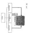
- FIGURE 1 Cells, in a physiological buffer, are plated onto the surface partially covered by the electrodes and each well can be connected to one or more impedance analyzers through a signal multiplexer.
- both the impedance magnitude and phase are recorded at periodic intervals. These frequencies were the full range capable on the Agilent impedance analyzer used, and spans the typical alpha and beta dispersion range for cells in physiological buffer. Additional information (dispersions) can be obtained at higher frequencies, including the gamma dispersions from bound water (GHz ranges), but become more difficult with the existing device architecture due to microwave resonance effects. Alternative embodiments utilize different frequency ranges. Representative frequency ranges from alternative embodiments include 10Hz to 1000GHz, 100Hz to 500GHz, and 100Hz to 1GHz.
- the cell Upon interaction of the ligand with its receptor, the cell undergoes physiological changes over time that alter the measured electrical properties. As stated above, changes in the cellular electrical properties are sufficient for distinguishing which second messenger pathway has triggered the cellular changes. In one embodiment of our method, the needed cellular electrical property information is present in the changing impedance. In alternative embodiments, similar data sets may be used; for example, if one recorded complex reflection coefficients (S parameters), one would have the same information, as S parameter data can be converted to impedance data. Other examples include the measurement of resistance, reactance, admittance, conductance, or susceptance.
- S parameters complex reflection coefficients
- the classification is performed using other information (impedance at fewer frequencies, impedance magnitude only, impedance phase only, or properties such as total circuit resistance or capacitance, or changes in circuit voltage or current). While thorough studies still need to be completed, preliminary evidence suggests that simpler measurements will be sufficient for classification of the pathway.
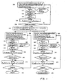
- the impedance data is processed in the manner shown in Figure 2.
- First, for each time point we measure the impedance over a range of frequencies after placing cells with known receptor types in a well of a micro titer plate (element 210 in Figure 2).
- the substance is a drug of interest, but in alternative embodiments, the substance may be a specific ligand, a protein, a lipid, a carbohydrate, a nucleic acid, water, an ion, or any other substance of interest.
- the graphs contain "kinetic" trends that by eye can often be associated with specific second messenger pathways.
- the objective quantitative classification performed by a computer does not rely on these patterns and trends. Rather, the computer algorithm needs only the coefficients at one time point after drug addition (corresponding to impedance data collected at this time and at the selected pre-drug addition time).
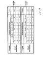
- the set of 7 magnitude coefficients and 7 phase coefficients are then compared to coefficient sets of known pathways (from a training data set) and assigned to a known pathway using standard multidimensional data classification algorithms.
- Alternative embodiments may contain coefficients fit to more or less Legendre polynomials, or time-dependent feature vectors parameterized with alternative methods.
- the ligand-receptor interaction can be classified into one of the four categories, Gi, Gs, Gq, or PTKR.
- the "reference" impedance magnitude and phase over a range of frequencies are measured at a time point corresponding to close but just preceding drug addition 232.
- the impedance magnitude and phase over a range of frequencies is measured at each subsequent time point for each frequency 236, and the "reference" impedance magnitude and phase are subtracted 238.
- the parameterized coefficients of the cellular response to the unknown ligand are calculated and compared to the coefficients for known ligands 240, 242, 244.
- the classification of the cellular response to the unknown ligand using a known receptor/ligand interaction yields valuable information regarding the cellular response, the stimulated receptor subtype, and the second messenger pathway.
- the exposure of intact cells with orphan or unknown receptors to potential ligand compounds allows for discovery of the ligand for the orphan thus de-orphanizing the receptor.
- the signal transduction pathway utilized by the receptor is ascertained by comparison to known patterns derived from training sets.(260-276).
- Gi, Gs, and Gq related receptors were studied.
- PTKR receptors were studied.
- Gi, Gs, and Gq receptors were studied in Chinese hamster ovary cells (CHO), while Gi, Gq, and PTKR receptors were studied in human cervical adenocarcinoma cells (HeLa).
- the data is presented in three categories.
- the first category is raw impedance difference data to show a sample of the actual data.
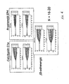
- the second category is Legendre polynomial parameterized data
- the third category is multidimensional classification data.
- Cells are plated into the wells of 96-well microtitre plates containing interdigitated electrodes fused to the bottom of the wells.
- the cells are plated in standard tissue culture media containing serum and allowed to incubate and adhere overnight in a standard 37oC CO2 incubator.
- the next day the plates containing adhered cells are rinsed with HANKS balanced salt solution containing 10 mM HEPES (HH).
- HH is then added to the wells and the plates are allowed to equilibrate to room temperature for 1hr.
- the plates are then moved onto the reading instrument and baseline readings are taken for 15 min. At the end of the 15 min timepoint, ligands for the various receptors are added to the appropriate wells and readings are taken every 20 sec for 30 min.
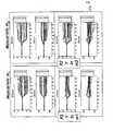
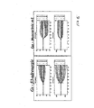
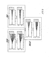
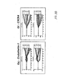
- Figures 4-12 Legendre parameter graphs are presented in Figures 4-12.
- Figures 4, 5, and 6 represent data collected using CHO cells and Figures 7-12 represent data collected using HeLa cells.
- Figures 4-12 represent data collected using HeLa cells.
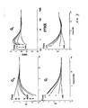
- three different ligands that stimulate three different Gs receptors were used to generate the data.
- the receptors were the calcitonin Cla (endogenous), the prostanoid EP4 (endogenous), and the beta3 adrenergic (transfected) receptor.
- the transformation analysis produces 7 parameters for the impedance magnitude and 7 parameters for the impedance phase. These parameters are labeled C0-C6 on the graphs.
- C0-C6 For each receptor, a set of two graphs is seen. The top graph shows the Legendre parameters for the impedance magnitude and the lower graph shows that for the impedance phase.
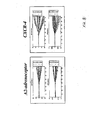
- Figure 10 shows the comparison of one set of patterns for Gq with one Gi set.
- Figure 11 shows the comparison of that same set of Gq patterns with that produced by a representative PTKR.
- Figure 12 the last comparison is shown between the Gi set and the PTKR set.
- the method is used for hit identification for agonists of known receptors.
- the question being asked by a pharmaceutical company is: Are there agonists for known receptors contained within the company's compound library?
- GPCRs G-protein coupled receptors
- a parental cell line is transfected with a nonsense sequence so as to be used as a control for the transfection process during analysis of data.
- Unknown compounds potential ligands
- Our method requires minimal assay development with no need for reporter systems to see the response of the transfected receptor, no need for additional reagents or fluorescent tags, and can be used with many different kinds of receptors including GPCRs, protein tyrosine kinase (PTKR), and nuclear receptors.
- GPCRs protein tyrosine kinase
- PTKR protein tyrosine kinase
- nuclear receptors No other single method or instrument for the analysis of GPCR second messenger pathways can interrogate all three (Gi, Gs, and Gq) pathways on the same platform, as this can. In addition, it can be used with adherent and nonadherent cells.
- the receptors of interest here are orphan GPCRs (oGPCRs). These are receptors that were discovered during sequencing of the human genome and that are potentially extremely important mediators of many disease processes. At present, their ligands are not known and the second messenger pathways they utilize are also unknown. In this case we establish again a database of patterns (a training set) for HEK293 cells (or any other cell line, depending on the need of the customer) and include patterns produced during stimulation with ligands specific for Gi, Gs, or Gq GPCRs. The oGPCR gene is transfected into HEK293 cells and the parental cells are nonsense transfected. Both cell lines are then exposed to the company's compound library.
- oGPCRs orphan GPCRs
- EC 50 values are then calculated from the fitting equations.
- the rank order potency of a series of structurally similar agonists can be achieved by comparing the EC 50 values generated for each compound in separate experiments.
- determining hit identification and IC 50 values for antagonists is easily accomplished using our system.
- the company's compound library would be applied to cells containing the receptor of interest. After stimulation with a known or discovered ligand, hits would be revealed as attenuated responses.
- IC 50 value determinations antagonist is added to the cells at increasing concentrations and the response to a half maximal stimulating concentration of agonist is monitored over time. The kinetic information is recorded to determine when the inhibition has reached a maximal value. The minimal impedance signal, recorded at a set endpoint, will be plotted against the inhibitor concentration added, and the data fitted to sigmoidal curves.
- the IC 50 values are then calculated from the fitting equations.
- the rank order potency of a series of structurally similar antagonists can be achieved by comparing the IC 50 values generated for each compound in separate experiments.
- the same method and instrument that can identify the ligand and the receptor second messenger pathway can be used to do antagonist hit identification, generate IC 50 values, and do rank ordering of inhibitors for potency.
- the yield is highly useful pharmacological information.
- apoptotic patterns for HEK293 cells (or any other cell line, depending on the need of the customer) and include patterns produced during stimulation with known effectors of apoptosis.
- patterns generated would be compared to the apoptosis patterns generated in the training sets. If a match were made to one of these patterns, then a new effector of apoptosis would be discovered.
- This analysis could be done separately or in conjunction with agonist or antagonist fishing. This would be especially useful during antagonist fishing, since the information could be obtained simultaneously during data acquisition and any stimulators of apoptosis could be eliminated immediately as potential drug candidates. Thus tremendous amounts of time and effort could be saved in the drug discovery process.
- Our method provides for the simultaneous analysis of apoptotic potential for any compound tested and therefore manifests a tremendous amount of savings for the drug discovery process as potential candidates that stimulate apoptosis can be eliminated early on in the process.
- cytotoxic patterns (a training set) for HEK293 cells (or any other cell line, depending on the need of the customer) and include patterns produced during stimulation with known effectors of cytotoxicity.
- patterns generated would be compared to the cytotoxicity patterns generated in the training sets. If a match were made to one of these patterns, then a new effector of cytotoxicity would be discovered.
- This analysis could be done separately or in conjunction with agonist or antagonist fishing. This would be especially useful during antagonist fishing, since the information could be obtained simultaneously during data acquisition and any cytotoxic compounds could be eliminated immediately as potential drug candidates. Thus tremendous amounts of time and effort could be saved in the drug discovery process.
- the patterns that are generated with our method are representations of complex integrated cellular events that take place in and around the cell after stimulation. For this case we are trying to determine what will be the effect of one stimulus on another. For example, if we stimulate a known Gi GPCR receptor, and while the cell is responding to this agonist, we apply a ligand for a PTKR, what would the effect be? Since our method generates representations of integrated cellular events, we will be able to study these integrations with as many stimuli as we want. First, we establish sets of database patterns that represent stimulation of discreet pathways within a cell type, such as already discussed for GPCRs.
- Tumor cells start out as relatively benign cells and as they divide and multiply, become more and more aggressive and ultimately become highly metastatic. When this occurs, the tumor cells break free of any restraints that they still have and spread throughout the body. This process is termed progression and ultimately leads to the death of the patient. As this process occurs the cells respond increasingly differently to the same stimuli as compared to earlier progeny. Again since our method produces an integrated representation of the cell, as the tumor cell progresses it should produce different patterns to the same stimuli. In this case, databases of patterns to certain ligands or stimuli will be created. As the tumor cells progress, the same ligands will be reapplied and the changes in the patterns recorded. Thus when test tumor cells are interrogated with our method, their pattern of response can be compared to the database and a determination of their state of progression can be made. Thus we have created an analysis tool to classify the state of progression of tumor cells.
- Stem cells start out as pluri-potential cells with the ability to differentiate into any cell type. As the cell receives different stimuli from its environment, it changes the complement of expressed proteins it has and changes its ultimate function. At some point this process stops and the cell is locked into performing this one function. Thus it has differentiated to become a liver cell or a cartilage cell or a cardiac cell. Again since our method produces an integrated representation of the cell, as the stem cell differentiates, it should produce different patterns to the same stimuli. In this case, databases of patterns to certain ligands or stimuli will be created. As the stem cell differentiates, the same ligands will be reapplied and the changes in the patterns recorded. Thus, when test stem cells are interrogated with our method, their pattern of response can be compared to the database and a determination of their state of differentiation can be made. Thus we have created an analysis tool to classify what a particular stem cell will differentiate into.
- the invention encompasses the process of pathway classification and incorporates the ability of the device and algorithms and software to classify unknown ligands as interacting with a number of cell surface receptors. This has been demonstrated through experimentation involving a Gi, Gs, or Gq GPCR or a PTKR.
- a unique aspect of the invention is the combination of processes that lead to classification of signal transduction pathways without the need for any kind of label or special amplification mechanism.
- the cell itself acts as the amplifying unit. In one embodiment, adherent eukaryotic cells were used with this system. An alternative embodiment incorporates the use of prokaryotes.
- inventions may incorporate either eurkaryotic or prokaryotic cell types as well as non adherent cells.
- the various embodiments of the method have the potential to classify any global cellular event as long as there are unique changes in the dielectric properties of cells after stimulation and therefore is not limited to the aforementioned pathways.
Landscapes
- Health & Medical Sciences (AREA)
- Life Sciences & Earth Sciences (AREA)
- Engineering & Computer Science (AREA)
- Biomedical Technology (AREA)
- Chemical & Material Sciences (AREA)
- Immunology (AREA)
- Physics & Mathematics (AREA)
- Hematology (AREA)
- Molecular Biology (AREA)
- Urology & Nephrology (AREA)
- Pathology (AREA)
- General Physics & Mathematics (AREA)
- General Health & Medical Sciences (AREA)
- Biochemistry (AREA)
- Food Science & Technology (AREA)
- Medicinal Chemistry (AREA)
- Analytical Chemistry (AREA)
- Cell Biology (AREA)
- Microbiology (AREA)
- Biotechnology (AREA)
- Tropical Medicine & Parasitology (AREA)
- Optics & Photonics (AREA)
- Biophysics (AREA)
- Measuring Or Testing Involving Enzymes Or Micro-Organisms (AREA)
- Investigating Or Analysing Biological Materials (AREA)
- Investigating Or Analyzing Materials By The Use Of Electric Means (AREA)
- Mobile Radio Communication Systems (AREA)
- Ultra Sonic Daignosis Equipment (AREA)
- Sorting Of Articles (AREA)
Claims (16)
- Marker-freies Verfahren zum Klassifizieren zellulärer Ereignisse, wobei die Klassifizierung durch Messen von Änderungen der elektrischen Eigenschaften von Zellen, die in eine elektrische Schaltung einbezogen sind, nach dem Anwenden eines Reizes erreicht wird, wobei das Verfahren umfasst:(a) Messen eines Werts einer elektrischen Eigenschaft nach dem Anwenden einer Mehrzahl von Frequenzen des elektromagnetischen Spektrums innerhalb eines Bereichs von Frequenzen für ausgewählte Zeitpunkte während eines ausgewählten Zeitraums für die elektrische Schaltung, die Zellen mit einem Rezeptor umfasst, die einen bekannten Rezeptortyp und einen bekannten Messenger-Weg aufweisen,(b) Auswählen eines Bezugszeitpunkts, der einem Zeitraum unmittelbar vor dem Hinzufügen eines bekannten Rezeptorreizes entspricht,(c) Hinzufügen des bekannten Reizes zu der elektrischen Schaltung derart, dass der Reiz mit dem Rezeptor eine Wechselwirkung eingehen kann,(d) Berechnen von Änderungen des Werts der elektrischen Eigenschaft für jede Frequenz durch Subtrahieren des Werts der gemessenen elektrischen Eigenschaft für den Bezugszeitpunkt von dem Wert der gemessenen elektrischen Eigenschaft für jeden Zeitpunkt nach dem Bezugszeitpunkt,(e) Parametrisieren der Änderungen des Werts der elektrischen Eigenschaft für jeden Zeitpunkt, Rezeptor und Reiz, und(f) Zuordnen der parametrisierten Änderungen des Werts der elektrischen Eigenschaft zu einer bekannten Reiz/Rezeptor-Wechselwirkungsklasse.
- Verfahren nach Anspruch 1, das ferner die Schritteg) Messen des Werts der elektrischen Eigenschaft für die elektrische Schaltung, die Zellen umfasst, für eine Mehrzahl von Frequenzen in einem Bereich von Frequenzen für ausgewählte Zeitpunkte während eines ausgewählten Zeitraums für eine Zelle mit einem bekannten Rezeptortyp,h) Auswählen eines Bezugszeitpunkts, der einem Zeitraum unmittelbar vor dem Hinzufügen eines unbekannten Reizes entspricht,i) Hinzufügen des unbekannten Reizes zu der Schaltung derart, dass der Reiz mit dem Rezeptor eine Wechselwirkung eingehen kann,j) Berechnen von Änderungen des Werts der elektrischen Eigenschaft für jede Frequenz durch Subtrahieren des Werts der elektrischen Eigenschaft für den Bezugszeitpunkt von dem Wert der elektrischen Eigenschaft für jeden Zeitpunkt nach dem Hinzufügen des unbekannten Reizes,k) Parametrisieren der Änderungen des Werts der elektrischen Eigenschaft für jeden Zeitpunkt, Rezeptor und Reiz,l) Vergleichen der parametrisierten Änderungen des Werts der elektrischen Eigenschaft nach dem Hinzufügen des unbekannten Reizes mit den parametrisierten Änderungen des Werts für bekannte Reize zum Korrelieren der Änderungen des Werts der elektrischen Eigenschaft mit einer zellulären Reaktion, undm) Zuordnen der zellulären Reaktion auf den unbekannten Reiz zu einer bekannten Substanz/Rezeptor-Wechselwirkungsklasse und Klassifizieren des Reizes umfasst.
- Verfahren nach Anspruch 1, das ferner die Schritteg) Messen des Werts der elektrischen Eigenschaft für die elektrische Schaltung, die Zellen umfasst, für eine Mehrzahl von Frequenzen innerhalb eines Bereichs von Frequenzen für ausgewählte Zeitpunkte während eines ausgewählten Zeitraums für eine Zelle mit einem unbekannten Rezeptortyp,h) Auswählen eines Bezugszeitpunkts, der einem Zeitraum unmittelbar vor dem Hinzufügen eines Reizes mit einer bekannten Reaktion entspricht,i) Hinzufügen des Reizes zu der Schaltung derart, dass der Reiz mit dem Rezeptor eine Wechselwirkung eingehen kann,j) Berechnen von Änderungen des Werts der elektrischen Eigenschaft für jede Frequenz durch Subtrahieren des Werts der elektrischen Eigenschaft für den Bezugszeitpunkt von dem Wert der elektrischen Eigenschaft für jeden Zeitpunkt nach dem Hinzufügen des Reizes,k) Parametrisieren der Änderungen des Werts der elektrischen Eigenschaft für jeden Zeitpunkt, Rezeptor und Reiz,l) Vergleichen der parametrisierten Änderungen des Werts der elektrischen Eigenschaft nach dem Hinzufügen des Reizes mit den parametrisierten Änderungen des Werts der elektrischen Eigenschaften für bekannte Rezeptoren zum Korrelieren der Änderungen des Werts der elektrischen Eigenschaft mit einer zellulären Reaktion, undm) Zuordnen der zellulären Reaktion des unbekannten Rezeptors zu einer bekannten Substanz/Rezeptor-Wechselwirkungsklasse und Klassifizieren des Rezeptors umfasst.
- Verfahren nach Anspruch 1, bei dem die elektrische Eigenschaft die komplexe Impedanz ist.
- Verfahren nach Anspruch 4, das ferner die Schritteg) Messen der komplexen Impedanz für eine Mehrzahl von Frequenzen innerhalb eines Bereichs von Frequenzen für jeden Zeitpunkt während eines ausgewählten Zeitraums für die elektrische Schaltung, die Zellen mit einem bekannten Rezeptortyp umfasst,h) Auswählen eines Bezugszeitpunkts, der einem Zeitraum unmittelbar vor dem Hinzufügen eines unbekannten Reizes entspricht,i) Hinzufügen des unbekannten Reizes zu der elektrischen Schaltung derart, dass der Reiz mit den Zellrezeptoren eine Wechselwirkung eingehen kann,j) Berechnen von Änderungen der Impedanz für jede Frequenz durch Subtrahieren des Werts der Impedanz für den Bezugszeitpunkt von dem Wert der Impedanz für jeden Zeitpunkt nach dem Hinzufügen des unbekannten Reizes,k) Parametrisieren der Änderungen der komplexen Impedanz für jeden Zeitpunkt, Rezeptor und Reiz,l) Vergleichen der parametrisierten Änderungen der komplexen Impedanz nach dem Hinzufügen des unbekannten Reizes mit den parametrisierten Änderungen der komplexen Impedanz für bekannte Reize zum Korrelieren der parametrisierten Änderungen der komplexen Impedanz mit einer zellulären Reaktion, undm) Zuordnen der zellulären Reaktion zu einer bekannten Substanz/Rezeptor-Wechselwirkungsklasse und Klassifizieren des Reizes umfasst.
- Verfahren nach Anspruch 4, das ferner die Schritteg) Messen der komplexen Impedanz für eine Mehrzahl von Frequenzen innerhalb eines Bereichs von Frequenzen für jeden Zeitpunkt während eines ausgewählten Zeitraums für die elektrische Schaltung, die Zellen mit einem unbekannten Rezeptortyp umfasst,h) Auswählen eines Bezugszeitpunkts, der einem Zeitraum unmittelbar vor dem Hinzufügen eines Reizes mit einer bekannten Reaktion entspricht,i) Hinzufügen des Reizes zu der elektrischen Schaltung derart, dass der Reiz mit den Zellrezeptoren eine Wechselwirkung eingehen kann,j) Berechnen von Änderungen der komplexen Impedanz für jede Frequenz durch Subtrahieren der komplexen Impedanz für den Bezugszeitpunkt von der komplexen Impedanz für jeden Zeitpunkt nach dem Hinzufügen des Reizes,k) Parametrisieren der Änderungen der komplexen Impedanz für jeden Zeitpunkt, Rezeptor und Reiz,l) Vergleichen der parametrisierten Änderungen der komplexen Impedanz nach dem Hinzufügen des Reizes mit den parametrisierten Änderungen der komplexen Impedanz für bekannte Rezeptoren zum Korrelieren der parametrisierten Änderungen der komplexen Impedanz mit einer zellulären Reaktion, undm) Zuordnen der zellulären Reaktion zu einer bekannten Substanz/Rezeptor-Wechselwirkungsklasse und Klassifizieren des Rezeptors umfasst.
- Verfahren nach Anspruch 4, bei dem die Änderungen der komplexen Impedanz als Widerstand oder Reaktanz gemessen werden.
- Verfahren nach Anspruch 4, bei dem die Änderungen der komplexen Impedanz als Scheinleitwert, Wirkleitwert oder Blindleitwert gemessen werden.
- Verfahren nach einem der Ansprüche 1 bis 8, bei dem die zellulären Ereignisse aus der Gruppe, bestehend aus einer Signalübertragung von Ligand/Rezeptor-Wechseiwirkungen, Cytotoxizität, Apoptose, Tumorzellenprogression und Stammzellendifferenzierung, ausgewählt sind.
- Verfahren nach einem der Ansprüche 1 bis 9, bei dem die elektrischen Eigenschaften aus der Gruppe, umfassend lmpedanzphase, lmpedanzgröße, komplexe Rückwirkungskoeffizienten, Gesamtwiderstand der Schaltung und Gesamtkapazität der Schaltung, ausgewählt sind.
- Verfahren nach einem der vorstehenden Ansprüche, bei dem der Bereich der Frequenzen 10 Hz bis 1000 GHz beträgt.
- Verfahren nach einem der vorstehenden Ansprüche, bei dem der Reiz eine Substanz ist.
- Verfahren nach Anspruch 12, bei dem die Substanz ein kleiner Molekülligand ist.
- Verfahren nach Anspruch 12, bei dem die Substanz ein Ligand, ein Protein, ein Antikörper, ein Lipid, ein Kohlenhydrat, eine Nukleinsäure, Wasser oder ein Ion ist.
- Verfahren nach einem der vorstehenden Ansprüche, bei dem die Klassifizierung in Echtzeit erreicht wird,
- Verfahren nach einem der vorstehenden Ansprüche, bei dem die elektrische Schaltung doppelkammförmige, koplanare Elektroden umfasst.
Applications Claiming Priority (1)
| Application Number | Priority Date | Filing Date | Title |
|---|---|---|---|
| PCT/CA2003/001060 WO2005005979A1 (en) | 2003-07-14 | 2003-07-14 | Label-free method for classification and characterization of cellular events |
Publications (2)
| Publication Number | Publication Date |
|---|---|
| EP1664765A1 EP1664765A1 (de) | 2006-06-07 |
| EP1664765B1 true EP1664765B1 (de) | 2007-08-29 |
Family
ID=33569429
Family Applications (1)
| Application Number | Title | Priority Date | Filing Date |
|---|---|---|---|
| EP03817382A Expired - Lifetime EP1664765B1 (de) | 2003-07-14 | 2003-07-14 | Markierungsfreies verfahren zur klassifizierung und charakterisierung zellulärer ereignisse |
Country Status (7)
| Country | Link |
|---|---|
| EP (1) | EP1664765B1 (de) |
| JP (1) | JP4668786B2 (de) |
| AT (1) | ATE371865T1 (de) |
| AU (1) | AU2003250657A1 (de) |
| CA (1) | CA2531342C (de) |
| DE (1) | DE60316044T2 (de) |
| WO (1) | WO2005005979A1 (de) |
Families Citing this family (33)
| Publication number | Priority date | Publication date | Assignee | Title |
|---|---|---|---|---|
| US7560269B2 (en) | 2002-12-20 | 2009-07-14 | Acea Biosciences, Inc. | Real time electronic cell sensing system and applications for cytotoxicity profiling and compound assays |
| US7470533B2 (en) | 2002-12-20 | 2008-12-30 | Acea Biosciences | Impedance based devices and methods for use in assays |
| US7732127B2 (en) | 2002-12-20 | 2010-06-08 | Acea Biosciences, Inc. | Dynamic monitoring of cell adhesion and spreading using the RT-CES system |
| US8206903B2 (en) | 2002-12-20 | 2012-06-26 | Acea Biosciences | Device and method for electroporation-based delivery of molecules into cells and dynamic monitoring of cell responses |
| WO2004010102A2 (en) | 2002-07-20 | 2004-01-29 | Acea Biosciences, Inc. | Impedance based apparatuses and methods for analyzing cells and particles |
| US8263375B2 (en) | 2002-12-20 | 2012-09-11 | Acea Biosciences | Dynamic monitoring of activation of G-protein coupled receptor (GPCR) and receptor tyrosine kinase (RTK) in living cells using real-time microelectronic cell sensing technology |
| US7468255B2 (en) | 2002-12-20 | 2008-12-23 | Acea Biosciences | Method for assaying for natural killer, cytotoxic T-lymphocyte and neutrophil-mediated killing of target cells using real-time microelectronic cell sensing technology |
| US7192752B2 (en) | 2002-12-20 | 2007-03-20 | Acea Biosciences | Real time electronic cell sensing systems and applications for cell-based assays |
| US9612234B2 (en) | 2008-05-05 | 2017-04-04 | Acea Biosciences, Inc. | Data analysis of impedance-based cardiomyocyte-beating signals as detected on real-time cell analysis (RTCA) cardio instruments |
| US11346797B2 (en) | 2002-12-20 | 2022-05-31 | Agilent Technologies, Inc. | System and method for monitoring cardiomyocyte beating, viability, morphology and electrophysiological properties |
| US10215748B2 (en) | 2002-12-20 | 2019-02-26 | Acea Biosciences, Inc. | Using impedance-based cell response profiling to identify putative inhibitors for oncogene addicted targets or pathways |
| US10539523B2 (en) | 2002-12-20 | 2020-01-21 | Acea Biosciences, Inc. | System and method for monitoring cardiomyocyte beating, viability, morphology, and electrophysiological properties |
| US10551371B2 (en) | 2003-11-10 | 2020-02-04 | Acea Biosciences, Inc. | System and method for monitoring cardiomyocyte beating, viability and morphology and for screening for pharmacological agents which may induce cardiotoxicity or modulate cardiomyocyte function |
| BR0317782B1 (pt) | 2002-12-27 | 2016-06-07 | Imerys Pigments Inc | Composição de pigmento de revestimento para papel e produto revestido com revestimento |
| CA2575297C (en) | 2004-08-04 | 2014-09-23 | Acea Biosciences, Inc. | Method for assaying for natural killer, cytotoxic t-lymphocyte and neutrophil-mediated killing of target cells using real-time microelectronic cell sensing technology |
| WO2007022043A2 (en) * | 2005-08-15 | 2007-02-22 | Janssen Pharmaceutica N.V. | Method of measuring the biological activity of an urotensin ii receptor |
| US7459918B2 (en) * | 2005-09-09 | 2008-12-02 | David Jones | Impedance measurement system with incorporated internal measurement drift compensation networks |
| KR100801694B1 (ko) * | 2005-09-15 | 2008-02-11 | 삼성전자주식회사 | 원핵 세포를 이용한 독성물질 유해성의 전기적 분석방법 및분석장치 |
| EP2064644A4 (de) * | 2006-09-20 | 2010-12-29 | Acea Biosciences Inc | Verwendung einer impedanz-basierten zellprofilierung zur klassifikation von zellenreaktionsprofilen bei der zugabe biologisch aktiver wirkstoffe |
| US8041515B2 (en) | 2006-09-20 | 2011-10-18 | Acea Biosciences, Inc. | Use of impedance-based cytological profiling to classify cellular response profiles upon exposure to biologically active agents |
| JP5168478B2 (ja) * | 2007-06-04 | 2013-03-21 | 株式会社リコー | 電子写真感光体、電子写真方法、電子写真装置、並びに電子写真装置用プロセスカートリッジ |
| US8426148B2 (en) | 2007-10-06 | 2013-04-23 | Corning Incorporated | Label-free methods using a resonant waveguide grating biosensor to determine GPCR signaling pathways |
| US8703428B2 (en) | 2007-10-06 | 2014-04-22 | Corning Incorporated | Single-cell label-free assay |
| US8846575B2 (en) | 2008-03-05 | 2014-09-30 | Corning Incorporated | High-throughput high-information content label-free cell biology screening methods |
| US8759013B2 (en) | 2008-03-05 | 2014-06-24 | Corning Incorporated | Dual-target biosensor cell assays |
| US8313898B2 (en) | 2008-03-05 | 2012-11-20 | Corning Incorporated | Dual-target biosensor cell assays |
| JP2009244197A (ja) * | 2008-03-31 | 2009-10-22 | Hioki Ee Corp | 薬剤感受性試験方法及び装置 |
| CA2723223C (en) | 2008-05-05 | 2017-06-06 | Acea Biosciences, Inc. | Label-free monitoring of excitation-contraction coupling and excitable cells using impedance based systems with millisecond time resolution |
| KR101527797B1 (ko) * | 2014-06-03 | 2015-06-15 | 한국과학기술원 | 신호전달경로의 활성화 상태 분석방법 및 이를 이용한 개인 맞춤형 치료제의 선정방법 |
| US12066428B2 (en) | 2015-11-20 | 2024-08-20 | Agilent Technologies, Inc. | Cell-substrate impedance monitoring of cancer cells |
| CN110582569B (zh) | 2017-03-03 | 2024-04-02 | 安捷伦科技有限公司 | 用于iPSC和ESC衍生的心肌细胞的功能成熟的方法和系统 |
| CN112730821B (zh) * | 2019-10-14 | 2024-04-09 | 泰州医药城国科化物生物医药科技有限公司 | 一种受体拮抗剂长效性分析方法 |
| US20210301245A1 (en) | 2020-03-29 | 2021-09-30 | Agilent Technologies, Inc. | Systems and methods for electronically and optically monitoring biological samples |
Family Cites Families (5)
| Publication number | Priority date | Publication date | Assignee | Title |
|---|---|---|---|---|
| US5792668A (en) * | 1993-08-06 | 1998-08-11 | Solid State Farms, Inc. | Radio frequency spectral analysis for in-vitro or in-vivo environments |
| US6048692A (en) * | 1997-10-07 | 2000-04-11 | Motorola, Inc. | Sensors for electrically sensing binding events for supported molecular receptors |
| US6377057B1 (en) * | 1999-02-18 | 2002-04-23 | The Board Of Trustees Of The Leland Stanford Junior University | Classification of biological agents according to the spectral density signature of evoked changes in cellular electric potential |
| WO2003016555A1 (en) * | 2001-08-09 | 2003-02-27 | Matsushita Electric Industrial Co., Ltd. | Cell diagnosing method, and device and apparatus used for it |
| US20030032000A1 (en) * | 2001-08-13 | 2003-02-13 | Signature Bioscience Inc. | Method for analyzing cellular events |
-
2003
- 2003-07-14 DE DE60316044T patent/DE60316044T2/de not_active Expired - Lifetime
- 2003-07-14 AT AT03817382T patent/ATE371865T1/de not_active IP Right Cessation
- 2003-07-14 WO PCT/CA2003/001060 patent/WO2005005979A1/en not_active Ceased
- 2003-07-14 AU AU2003250657A patent/AU2003250657A1/en not_active Abandoned
- 2003-07-14 JP JP2005503785A patent/JP4668786B2/ja not_active Expired - Fee Related
- 2003-07-14 CA CA2531342A patent/CA2531342C/en not_active Expired - Fee Related
- 2003-07-14 EP EP03817382A patent/EP1664765B1/de not_active Expired - Lifetime
Also Published As
| Publication number | Publication date |
|---|---|
| WO2005005979A1 (en) | 2005-01-20 |
| CA2531342A1 (en) | 2005-01-20 |
| DE60316044T2 (de) | 2008-05-15 |
| CA2531342C (en) | 2011-07-05 |
| AU2003250657A1 (en) | 2005-01-28 |
| ATE371865T1 (de) | 2007-09-15 |
| EP1664765A1 (de) | 2006-06-07 |
| DE60316044D1 (de) | 2007-10-11 |
| JP4668786B2 (ja) | 2011-04-13 |
| JP2007504801A (ja) | 2007-03-08 |
Similar Documents
| Publication | Publication Date | Title |
|---|---|---|
| EP1664765B1 (de) | Markierungsfreies verfahren zur klassifizierung und charakterisierung zellulärer ereignisse | |
| US20050014130A1 (en) | Label-free method for classification and characterization of cellular events | |
| Abassi et al. | Label-free, real-time monitoring of IgE-mediated mast cell activation on microelectronic cell sensor arrays | |
| US8921041B2 (en) | Device and method for electroporation-based delivery of molecules into cells and dynamic monitoring of cell responses | |
| Peters et al. | Human stem cell-derived cardiomyocytes in cellular impedance assays: bringing cardiotoxicity screening to the front line | |
| Ciambrone et al. | Cellular dielectric spectroscopy: a powerful new approach to label-free cellular analysis | |
| McGuinness | Impedance-based cellular assay technologies: recent advances, future promise | |
| Atienzar et al. | The use of real-time cell analyzer technology in drug discovery: defining optimal cell culture conditions and assay reproducibility with different adherent cellular models | |
| JP2005514909A (ja) | 真核生物細胞の電場による刺激 | |
| JP2012509667A (ja) | 分子を特徴付けるための方法 | |
| Toh et al. | Application of high-throughput automated patch-clamp electrophysiology to study voltage-gated ion channel function in primary cortical cultures | |
| Greife et al. | Structural assemblies of the di-and oligomeric G-protein coupled receptor TGR5 in live cells: an MFIS-FRET and integrative modelling study | |
| EP1773979B1 (de) | Dynamische überwachung der aktivierung von g-protein (gpcr) in lebenden zellen unter verwendung von mikroelektronischer-echtzeit-zellmesstechnologie | |
| Irelan et al. | Rapid and quantitative assessment of cell quality, identity, and functionality for cell-based assays using real-time cellular analysis | |
| Rocheville et al. | Mining the potential of label-free biosensors for seven-transmembrane receptor drug discovery | |
| US8313898B2 (en) | Dual-target biosensor cell assays | |
| Filippini et al. | Microplate based biosensing with a computer screen aided technique | |
| Lu et al. | Label-free imaging of histamine mediated G protein-coupled receptors activation in live cells | |
| US20090093011A1 (en) | Biosensors for ligand-directed functional selectivity | |
| Grundmann | Label‐free dynamic mass redistribution and bio‐impedance methods for drug discovery | |
| McAnally et al. | A systematic approach to identify biased agonists of the apelin receptor through high-throughput screening | |
| Wolf et al. | Microelectrode chip based real time monitoring of vital MCF-7 mamma carcinoma cells by impedance spectroscopy | |
| McGuinness et al. | Enhanced selectivity screening of GPCR ligands using a label-free cell based assay technology | |
| US20120015846A1 (en) | Label-free Cellular Pharmacology For Drug Antitarget Assessment | |
| EP1815025B1 (de) | Dynamische überwachung der zellantworten zur zuführung von molekülen |
Legal Events
| Date | Code | Title | Description |
|---|---|---|---|
| PUAI | Public reference made under article 153(3) epc to a published international application that has entered the european phase |
Free format text: ORIGINAL CODE: 0009012 |
|
| 17P | Request for examination filed |
Effective date: 20060210 |
|
| AK | Designated contracting states |
Kind code of ref document: A1 Designated state(s): AT BE BG CH CY CZ DE DK EE ES FI FR GB GR HU IE IT LI LU MC NL PT RO SE SI SK TR |
|
| 17Q | First examination report despatched |
Effective date: 20060626 |
|
| DAX | Request for extension of the european patent (deleted) | ||
| GRAP | Despatch of communication of intention to grant a patent |
Free format text: ORIGINAL CODE: EPIDOSNIGR1 |
|
| GRAS | Grant fee paid |
Free format text: ORIGINAL CODE: EPIDOSNIGR3 |
|
| GRAA | (expected) grant |
Free format text: ORIGINAL CODE: 0009210 |
|
| AK | Designated contracting states |
Kind code of ref document: B1 Designated state(s): AT BE BG CH CY CZ DE DK EE ES FI FR GB GR HU IE IT LI LU MC NL PT RO SE SI SK TR |
|
| REG | Reference to a national code |
Ref country code: GB Ref legal event code: FG4D |
|
| REG | Reference to a national code |
Ref country code: CH Ref legal event code: EP |
|
| REG | Reference to a national code |
Ref country code: IE Ref legal event code: FG4D |
|
| REF | Corresponds to: |
Ref document number: 60316044 Country of ref document: DE Date of ref document: 20071011 Kind code of ref document: P |
|
| PG25 | Lapsed in a contracting state [announced via postgrant information from national office to epo] |
Ref country code: FI Free format text: LAPSE BECAUSE OF FAILURE TO SUBMIT A TRANSLATION OF THE DESCRIPTION OR TO PAY THE FEE WITHIN THE PRESCRIBED TIME-LIMIT Effective date: 20070829 Ref country code: ES Free format text: LAPSE BECAUSE OF FAILURE TO SUBMIT A TRANSLATION OF THE DESCRIPTION OR TO PAY THE FEE WITHIN THE PRESCRIBED TIME-LIMIT Effective date: 20071210 Ref country code: NL Free format text: LAPSE BECAUSE OF FAILURE TO SUBMIT A TRANSLATION OF THE DESCRIPTION OR TO PAY THE FEE WITHIN THE PRESCRIBED TIME-LIMIT Effective date: 20070829 |
|
| NLV1 | Nl: lapsed or annulled due to failure to fulfill the requirements of art. 29p and 29m of the patents act | ||
| PG25 | Lapsed in a contracting state [announced via postgrant information from national office to epo] |
Ref country code: CH Free format text: LAPSE BECAUSE OF FAILURE TO SUBMIT A TRANSLATION OF THE DESCRIPTION OR TO PAY THE FEE WITHIN THE PRESCRIBED TIME-LIMIT Effective date: 20070829 Ref country code: LI Free format text: LAPSE BECAUSE OF FAILURE TO SUBMIT A TRANSLATION OF THE DESCRIPTION OR TO PAY THE FEE WITHIN THE PRESCRIBED TIME-LIMIT Effective date: 20070829 Ref country code: AT Free format text: LAPSE BECAUSE OF FAILURE TO SUBMIT A TRANSLATION OF THE DESCRIPTION OR TO PAY THE FEE WITHIN THE PRESCRIBED TIME-LIMIT Effective date: 20070829 |
|
| REG | Reference to a national code |
Ref country code: CH Ref legal event code: PL |
|
| ET | Fr: translation filed | ||
| PG25 | Lapsed in a contracting state [announced via postgrant information from national office to epo] |
Ref country code: BE Free format text: LAPSE BECAUSE OF FAILURE TO SUBMIT A TRANSLATION OF THE DESCRIPTION OR TO PAY THE FEE WITHIN THE PRESCRIBED TIME-LIMIT Effective date: 20070829 |
|
| PG25 | Lapsed in a contracting state [announced via postgrant information from national office to epo] |
Ref country code: GR Free format text: LAPSE BECAUSE OF FAILURE TO SUBMIT A TRANSLATION OF THE DESCRIPTION OR TO PAY THE FEE WITHIN THE PRESCRIBED TIME-LIMIT Effective date: 20071130 Ref country code: DK Free format text: LAPSE BECAUSE OF FAILURE TO SUBMIT A TRANSLATION OF THE DESCRIPTION OR TO PAY THE FEE WITHIN THE PRESCRIBED TIME-LIMIT Effective date: 20070829 |
|
| PG25 | Lapsed in a contracting state [announced via postgrant information from national office to epo] |
Ref country code: CZ Free format text: LAPSE BECAUSE OF FAILURE TO SUBMIT A TRANSLATION OF THE DESCRIPTION OR TO PAY THE FEE WITHIN THE PRESCRIBED TIME-LIMIT Effective date: 20070829 Ref country code: SK Free format text: LAPSE BECAUSE OF FAILURE TO SUBMIT A TRANSLATION OF THE DESCRIPTION OR TO PAY THE FEE WITHIN THE PRESCRIBED TIME-LIMIT Effective date: 20070829 Ref country code: PT Free format text: LAPSE BECAUSE OF FAILURE TO SUBMIT A TRANSLATION OF THE DESCRIPTION OR TO PAY THE FEE WITHIN THE PRESCRIBED TIME-LIMIT Effective date: 20080129 |
|
| PG25 | Lapsed in a contracting state [announced via postgrant information from national office to epo] |
Ref country code: SE Free format text: LAPSE BECAUSE OF FAILURE TO SUBMIT A TRANSLATION OF THE DESCRIPTION OR TO PAY THE FEE WITHIN THE PRESCRIBED TIME-LIMIT Effective date: 20071129 Ref country code: RO Free format text: LAPSE BECAUSE OF FAILURE TO SUBMIT A TRANSLATION OF THE DESCRIPTION OR TO PAY THE FEE WITHIN THE PRESCRIBED TIME-LIMIT Effective date: 20070829 |
|
| PLBE | No opposition filed within time limit |
Free format text: ORIGINAL CODE: 0009261 |
|
| STAA | Information on the status of an ep patent application or granted ep patent |
Free format text: STATUS: NO OPPOSITION FILED WITHIN TIME LIMIT |
|
| 26N | No opposition filed |
Effective date: 20080530 |
|
| PG25 | Lapsed in a contracting state [announced via postgrant information from national office to epo] |
Ref country code: MC Free format text: LAPSE BECAUSE OF NON-PAYMENT OF DUE FEES Effective date: 20080731 |
|
| PG25 | Lapsed in a contracting state [announced via postgrant information from national office to epo] |
Ref country code: EE Free format text: LAPSE BECAUSE OF FAILURE TO SUBMIT A TRANSLATION OF THE DESCRIPTION OR TO PAY THE FEE WITHIN THE PRESCRIBED TIME-LIMIT Effective date: 20070829 |
|
| PG25 | Lapsed in a contracting state [announced via postgrant information from national office to epo] |
Ref country code: SI Free format text: LAPSE BECAUSE OF FAILURE TO SUBMIT A TRANSLATION OF THE DESCRIPTION OR TO PAY THE FEE WITHIN THE PRESCRIBED TIME-LIMIT Effective date: 20070829 |
|
| PG25 | Lapsed in a contracting state [announced via postgrant information from national office to epo] |
Ref country code: IE Free format text: LAPSE BECAUSE OF NON-PAYMENT OF DUE FEES Effective date: 20080714 Ref country code: CY Free format text: LAPSE BECAUSE OF FAILURE TO SUBMIT A TRANSLATION OF THE DESCRIPTION OR TO PAY THE FEE WITHIN THE PRESCRIBED TIME-LIMIT Effective date: 20070829 |
|
| PG25 | Lapsed in a contracting state [announced via postgrant information from national office to epo] |
Ref country code: BG Free format text: LAPSE BECAUSE OF FAILURE TO SUBMIT A TRANSLATION OF THE DESCRIPTION OR TO PAY THE FEE WITHIN THE PRESCRIBED TIME-LIMIT Effective date: 20071129 |
|
| PG25 | Lapsed in a contracting state [announced via postgrant information from national office to epo] |
Ref country code: HU Free format text: LAPSE BECAUSE OF FAILURE TO SUBMIT A TRANSLATION OF THE DESCRIPTION OR TO PAY THE FEE WITHIN THE PRESCRIBED TIME-LIMIT Effective date: 20080301 Ref country code: LU Free format text: LAPSE BECAUSE OF NON-PAYMENT OF DUE FEES Effective date: 20080714 |
|
| PG25 | Lapsed in a contracting state [announced via postgrant information from national office to epo] |
Ref country code: TR Free format text: LAPSE BECAUSE OF FAILURE TO SUBMIT A TRANSLATION OF THE DESCRIPTION OR TO PAY THE FEE WITHIN THE PRESCRIBED TIME-LIMIT Effective date: 20070829 |
|
| PG25 | Lapsed in a contracting state [announced via postgrant information from national office to epo] |
Ref country code: IT Free format text: LAPSE BECAUSE OF NON-PAYMENT OF DUE FEES Effective date: 20080731 |
|
| REG | Reference to a national code |
Ref country code: FR Ref legal event code: PLFP Year of fee payment: 13 |
|
| PGFP | Annual fee paid to national office [announced via postgrant information from national office to epo] |
Ref country code: GB Payment date: 20150727 Year of fee payment: 13 Ref country code: DE Payment date: 20150729 Year of fee payment: 13 |
|
| PGFP | Annual fee paid to national office [announced via postgrant information from national office to epo] |
Ref country code: FR Payment date: 20150717 Year of fee payment: 13 |
|
| REG | Reference to a national code |
Ref country code: DE Ref legal event code: R119 Ref document number: 60316044 Country of ref document: DE |
|
| GBPC | Gb: european patent ceased through non-payment of renewal fee |
Effective date: 20160714 |
|
| PG25 | Lapsed in a contracting state [announced via postgrant information from national office to epo] |
Ref country code: FR Free format text: LAPSE BECAUSE OF NON-PAYMENT OF DUE FEES Effective date: 20160801 Ref country code: DE Free format text: LAPSE BECAUSE OF NON-PAYMENT OF DUE FEES Effective date: 20170201 |
|
| REG | Reference to a national code |
Ref country code: FR Ref legal event code: ST Effective date: 20170331 |
|
| PG25 | Lapsed in a contracting state [announced via postgrant information from national office to epo] |
Ref country code: GB Free format text: LAPSE BECAUSE OF NON-PAYMENT OF DUE FEES Effective date: 20160714 |