EP1641815B1 - Uran-chelatisierende peptide und deren verwendung - Google Patents
Uran-chelatisierende peptide und deren verwendung Download PDFInfo
- Publication number
- EP1641815B1 EP1641815B1 EP04767541.8A EP04767541A EP1641815B1 EP 1641815 B1 EP1641815 B1 EP 1641815B1 EP 04767541 A EP04767541 A EP 04767541A EP 1641815 B1 EP1641815 B1 EP 1641815B1
- Authority
- EP
- European Patent Office
- Prior art keywords
- peptide
- uranium
- cam
- fusion protein
- protein
- Prior art date
- Legal status (The legal status is an assumption and is not a legal conclusion. Google has not performed a legal analysis and makes no representation as to the accuracy of the status listed.)
- Expired - Lifetime
Links
- 108090000765 processed proteins & peptides Proteins 0.000 title claims description 236
- 102000004196 processed proteins & peptides Human genes 0.000 title claims description 55
- JFALSRSLKYAFGM-UHFFFAOYSA-N uranium(0) Chemical compound [U] JFALSRSLKYAFGM-UHFFFAOYSA-N 0.000 claims description 126
- 229910052770 Uranium Inorganic materials 0.000 claims description 118
- 239000011575 calcium Substances 0.000 claims description 56
- OYPRJOBELJOOCE-UHFFFAOYSA-N Calcium Chemical compound [Ca] OYPRJOBELJOOCE-UHFFFAOYSA-N 0.000 claims description 52
- 108010041952 Calmodulin Proteins 0.000 claims description 52
- 102000000584 Calmodulin Human genes 0.000 claims description 52
- 229910052791 calcium Inorganic materials 0.000 claims description 52
- 102000004169 proteins and genes Human genes 0.000 claims description 37
- 108090000623 proteins and genes Proteins 0.000 claims description 37
- 102000037865 fusion proteins Human genes 0.000 claims description 22
- 108020001507 fusion proteins Proteins 0.000 claims description 22
- 229920001184 polypeptide Polymers 0.000 claims description 18
- XLYOFNOQVPJJNP-UHFFFAOYSA-N water Substances O XLYOFNOQVPJJNP-UHFFFAOYSA-N 0.000 claims description 16
- 210000004027 cell Anatomy 0.000 claims description 15
- 108020004707 nucleic acids Proteins 0.000 claims description 13
- 102000039446 nucleic acids Human genes 0.000 claims description 13
- 150000007523 nucleic acids Chemical class 0.000 claims description 13
- 239000002689 soil Substances 0.000 claims description 13
- 239000013598 vector Substances 0.000 claims description 11
- 108010048367 enhanced green fluorescent protein Proteins 0.000 claims description 10
- 108091005941 EBFP Proteins 0.000 claims description 8
- 108091005942 ECFP Proteins 0.000 claims description 8
- 102000034287 fluorescent proteins Human genes 0.000 claims description 8
- 108091006047 fluorescent proteins Proteins 0.000 claims description 8
- -1 EYFP Proteins 0.000 claims description 7
- 230000009261 transgenic effect Effects 0.000 claims description 7
- 241000122205 Chamaeleonidae Species 0.000 claims description 6
- 210000000988 bone and bone Anatomy 0.000 claims description 5
- 210000003527 eukaryotic cell Anatomy 0.000 claims description 5
- 239000003153 chemical reaction reagent Substances 0.000 claims description 4
- 239000003814 drug Substances 0.000 claims description 4
- 230000004927 fusion Effects 0.000 claims description 4
- 210000003734 kidney Anatomy 0.000 claims description 4
- 210000001236 prokaryotic cell Anatomy 0.000 claims description 4
- 238000011109 contamination Methods 0.000 claims description 3
- 230000029142 excretion Effects 0.000 claims description 3
- GOLORTLGFDVFDW-UHFFFAOYSA-N 3-(1h-benzimidazol-2-yl)-7-(diethylamino)chromen-2-one Chemical compound C1=CC=C2NC(C3=CC4=CC=C(C=C4OC3=O)N(CC)CC)=NC2=C1 GOLORTLGFDVFDW-UHFFFAOYSA-N 0.000 claims description 2
- 125000001295 dansyl group Chemical group [H]C1=C([H])C(N(C([H])([H])[H])C([H])([H])[H])=C2C([H])=C([H])C([H])=C(C2=C1[H])S(*)(=O)=O 0.000 claims description 2
- GNBHRKFJIUUOQI-UHFFFAOYSA-N fluorescein Chemical compound O1C(=O)C2=CC=CC=C2C21C1=CC=C(O)C=C1OC1=CC(O)=CC=C21 GNBHRKFJIUUOQI-UHFFFAOYSA-N 0.000 claims description 2
- 108010021843 fluorescent protein 583 Proteins 0.000 claims description 2
- 238000001727 in vivo Methods 0.000 claims description 2
- 238000005067 remediation Methods 0.000 claims 2
- 241001465754 Metazoa Species 0.000 claims 1
- 230000008685 targeting Effects 0.000 claims 1
- 239000000243 solution Substances 0.000 description 47
- 229910052751 metal Inorganic materials 0.000 description 41
- 239000002184 metal Substances 0.000 description 41
- 235000018102 proteins Nutrition 0.000 description 32
- 125000005289 uranyl group Chemical group 0.000 description 25
- 102220472091 Protein ENL_D20T_mutation Human genes 0.000 description 23
- 150000002500 ions Chemical class 0.000 description 21
- 230000035772 mutation Effects 0.000 description 20
- 150000002739 metals Chemical class 0.000 description 18
- 238000001228 spectrum Methods 0.000 description 18
- 238000004448 titration Methods 0.000 description 18
- 238000010494 dissociation reaction Methods 0.000 description 17
- 230000005593 dissociations Effects 0.000 description 17
- 238000001514 detection method Methods 0.000 description 15
- 238000004088 simulation Methods 0.000 description 15
- 238000004458 analytical method Methods 0.000 description 14
- 241000894007 species Species 0.000 description 14
- 239000003446 ligand Substances 0.000 description 13
- HCHKCACWOHOZIP-UHFFFAOYSA-N Zinc Chemical compound [Zn] HCHKCACWOHOZIP-UHFFFAOYSA-N 0.000 description 11
- 229910001424 calcium ion Inorganic materials 0.000 description 11
- 229910052725 zinc Inorganic materials 0.000 description 11
- 239000011701 zinc Substances 0.000 description 11
- BHPQYMZQTOCNFJ-UHFFFAOYSA-N Calcium cation Chemical compound [Ca+2] BHPQYMZQTOCNFJ-UHFFFAOYSA-N 0.000 description 10
- 150000001413 amino acids Chemical class 0.000 description 10
- 230000005284 excitation Effects 0.000 description 10
- 238000002474 experimental method Methods 0.000 description 10
- 239000000203 mixture Substances 0.000 description 10
- 102000001189 Cyclic Peptides Human genes 0.000 description 9
- 108010069514 Cyclic Peptides Proteins 0.000 description 9
- QIVBCDIJIAJPQS-VIFPVBQESA-N L-tryptophane Chemical compound C1=CC=C2C(C[C@H](N)C(O)=O)=CNC2=C1 QIVBCDIJIAJPQS-VIFPVBQESA-N 0.000 description 9
- QIVBCDIJIAJPQS-UHFFFAOYSA-N Tryptophan Natural products C1=CC=C2C(CC(N)C(O)=O)=CNC2=C1 QIVBCDIJIAJPQS-UHFFFAOYSA-N 0.000 description 9
- 238000007792 addition Methods 0.000 description 9
- 235000001014 amino acid Nutrition 0.000 description 9
- 229940024606 amino acid Drugs 0.000 description 9
- 238000005202 decontamination Methods 0.000 description 9
- 230000003588 decontaminative effect Effects 0.000 description 9
- NBIIXXVUZAFLBC-UHFFFAOYSA-K phosphate Chemical compound [O-]P([O-])([O-])=O NBIIXXVUZAFLBC-UHFFFAOYSA-K 0.000 description 9
- 239000008363 phosphate buffer Substances 0.000 description 9
- 229910019142 PO4 Inorganic materials 0.000 description 8
- 229910052771 Terbium Inorganic materials 0.000 description 8
- 230000015572 biosynthetic process Effects 0.000 description 8
- 239000002609 medium Substances 0.000 description 8
- 229910021645 metal ion Inorganic materials 0.000 description 8
- 239000010452 phosphate Substances 0.000 description 8
- 239000000126 substance Substances 0.000 description 8
- 229910052693 Europium Inorganic materials 0.000 description 7
- 229910002651 NO3 Inorganic materials 0.000 description 7
- NHNBFGGVMKEFGY-UHFFFAOYSA-N Nitrate Chemical compound [O-][N+]([O-])=O NHNBFGGVMKEFGY-UHFFFAOYSA-N 0.000 description 7
- 238000002983 circular dichroism Methods 0.000 description 7
- WYICGPHECJFCBA-UHFFFAOYSA-N dioxouranium(2+) Chemical compound O=[U+2]=O WYICGPHECJFCBA-UHFFFAOYSA-N 0.000 description 7
- 238000002330 electrospray ionisation mass spectrometry Methods 0.000 description 7
- 238000002189 fluorescence spectrum Methods 0.000 description 7
- 229910052747 lanthanoid Inorganic materials 0.000 description 7
- 150000002602 lanthanoids Chemical class 0.000 description 7
- 238000004519 manufacturing process Methods 0.000 description 7
- GZCRRIHWUXGPOV-UHFFFAOYSA-N terbium atom Chemical compound [Tb] GZCRRIHWUXGPOV-UHFFFAOYSA-N 0.000 description 7
- 231100000331 toxic Toxicity 0.000 description 7
- 230000002588 toxic effect Effects 0.000 description 7
- 229910002007 uranyl nitrate Inorganic materials 0.000 description 7
- YMWUJEATGCHHMB-UHFFFAOYSA-N Dichloromethane Chemical compound ClCCl YMWUJEATGCHHMB-UHFFFAOYSA-N 0.000 description 6
- 241000196324 Embryophyta Species 0.000 description 6
- 239000007987 MES buffer Substances 0.000 description 6
- DTQVDTLACAAQTR-UHFFFAOYSA-N Trifluoroacetic acid Chemical compound OC(=O)C(F)(F)F DTQVDTLACAAQTR-UHFFFAOYSA-N 0.000 description 6
- 230000035508 accumulation Effects 0.000 description 6
- 238000009825 accumulation Methods 0.000 description 6
- 230000009920 chelation Effects 0.000 description 6
- OGPBJKLSAFTDLK-UHFFFAOYSA-N europium atom Chemical compound [Eu] OGPBJKLSAFTDLK-UHFFFAOYSA-N 0.000 description 6
- 230000001965 increasing effect Effects 0.000 description 6
- 239000011777 magnesium Substances 0.000 description 6
- 238000000034 method Methods 0.000 description 6
- 238000012546 transfer Methods 0.000 description 6
- 238000011282 treatment Methods 0.000 description 6
- 241000282414 Homo sapiens Species 0.000 description 5
- SECXISVLQFMRJM-UHFFFAOYSA-N N-Methylpyrrolidone Chemical compound CN1CCCC1=O SECXISVLQFMRJM-UHFFFAOYSA-N 0.000 description 5
- 230000006978 adaptation Effects 0.000 description 5
- 230000002349 favourable effect Effects 0.000 description 5
- 238000002360 preparation method Methods 0.000 description 5
- 239000011347 resin Substances 0.000 description 5
- 229920005989 resin Polymers 0.000 description 5
- 239000003643 water by type Substances 0.000 description 5
- 241000894006 Bacteria Species 0.000 description 4
- 102000005701 Calcium-Binding Proteins Human genes 0.000 description 4
- 108010045403 Calcium-Binding Proteins Proteins 0.000 description 4
- QOSSAOTZNIDXMA-UHFFFAOYSA-N Dicylcohexylcarbodiimide Chemical compound C1CCCCC1N=C=NC1CCCCC1 QOSSAOTZNIDXMA-UHFFFAOYSA-N 0.000 description 4
- AYFVYJQAPQTCCC-GBXIJSLDSA-N L-threonine Chemical compound C[C@@H](O)[C@H](N)C(O)=O AYFVYJQAPQTCCC-GBXIJSLDSA-N 0.000 description 4
- FYYHWMGAXLPEAU-UHFFFAOYSA-N Magnesium Chemical compound [Mg] FYYHWMGAXLPEAU-UHFFFAOYSA-N 0.000 description 4
- 102100035044 Myosin light chain kinase, smooth muscle Human genes 0.000 description 4
- 108010074596 Myosin-Light-Chain Kinase Proteins 0.000 description 4
- NQRYJNQNLNOLGT-UHFFFAOYSA-N Piperidine Chemical compound C1CCNCC1 NQRYJNQNLNOLGT-UHFFFAOYSA-N 0.000 description 4
- 238000010521 absorption reaction Methods 0.000 description 4
- 239000012736 aqueous medium Substances 0.000 description 4
- CKLJMWTZIZZHCS-REOHCLBHSA-N aspartic acid group Chemical group N[C@@H](CC(=O)O)C(=O)O CKLJMWTZIZZHCS-REOHCLBHSA-N 0.000 description 4
- 102220450464 c.56T>G Human genes 0.000 description 4
- 230000008859 change Effects 0.000 description 4
- 239000002738 chelating agent Substances 0.000 description 4
- 238000002866 fluorescence resonance energy transfer Methods 0.000 description 4
- 239000005090 green fluorescent protein Substances 0.000 description 4
- 230000003993 interaction Effects 0.000 description 4
- 229910052749 magnesium Inorganic materials 0.000 description 4
- 230000007935 neutral effect Effects 0.000 description 4
- 102200082887 rs33950093 Human genes 0.000 description 4
- 102220059963 rs786201995 Human genes 0.000 description 4
- 239000000523 sample Substances 0.000 description 4
- 239000011734 sodium Substances 0.000 description 4
- MTCFGRXMJLQNBG-REOHCLBHSA-N (2S)-2-Amino-3-hydroxypropansäure Chemical compound OC[C@H](N)C(O)=O MTCFGRXMJLQNBG-REOHCLBHSA-N 0.000 description 3
- 125000003088 (fluoren-9-ylmethoxy)carbonyl group Chemical group 0.000 description 3
- QTBSBXVTEAMEQO-UHFFFAOYSA-N Acetic acid Chemical compound CC(O)=O QTBSBXVTEAMEQO-UHFFFAOYSA-N 0.000 description 3
- WEVYAHXRMPXWCK-UHFFFAOYSA-N Acetonitrile Chemical compound CC#N WEVYAHXRMPXWCK-UHFFFAOYSA-N 0.000 description 3
- WHUUTDBJXJRKMK-UHFFFAOYSA-N Glutamic acid Natural products OC(=O)C(N)CCC(O)=O WHUUTDBJXJRKMK-UHFFFAOYSA-N 0.000 description 3
- DGAQECJNVWCQMB-PUAWFVPOSA-M Ilexoside XXIX Chemical compound C[C@@H]1CC[C@@]2(CC[C@@]3(C(=CC[C@H]4[C@]3(CC[C@@H]5[C@@]4(CC[C@@H](C5(C)C)OS(=O)(=O)[O-])C)C)[C@@H]2[C@]1(C)O)C)C(=O)O[C@H]6[C@@H]([C@H]([C@@H]([C@H](O6)CO)O)O)O.[Na+] DGAQECJNVWCQMB-PUAWFVPOSA-M 0.000 description 3
- 108090000362 Lymphotoxin-beta Proteins 0.000 description 3
- 101000932912 Paramecium tetraurelia Calmodulin Proteins 0.000 description 3
- AYFVYJQAPQTCCC-UHFFFAOYSA-N Threonine Natural products CC(O)C(N)C(O)=O AYFVYJQAPQTCCC-UHFFFAOYSA-N 0.000 description 3
- 239000004473 Threonine Substances 0.000 description 3
- 102000013534 Troponin C Human genes 0.000 description 3
- 239000007864 aqueous solution Substances 0.000 description 3
- 230000008033 biological extinction Effects 0.000 description 3
- 150000004649 carbonic acid derivatives Chemical class 0.000 description 3
- 125000000151 cysteine group Chemical group N[C@@H](CS)C(=O)* 0.000 description 3
- 238000010586 diagram Methods 0.000 description 3
- 125000000291 glutamic acid group Chemical group N[C@@H](CCC(O)=O)C(=O)* 0.000 description 3
- 229910001385 heavy metal Inorganic materials 0.000 description 3
- 230000010354 integration Effects 0.000 description 3
- 238000004949 mass spectrometry Methods 0.000 description 3
- 238000001819 mass spectrum Methods 0.000 description 3
- 244000005700 microbiome Species 0.000 description 3
- 238000012986 modification Methods 0.000 description 3
- 230000004048 modification Effects 0.000 description 3
- 239000012071 phase Substances 0.000 description 3
- 239000012429 reaction media Substances 0.000 description 3
- 229910052708 sodium Inorganic materials 0.000 description 3
- 238000003860 storage Methods 0.000 description 3
- 230000002123 temporal effect Effects 0.000 description 3
- 231100000419 toxicity Toxicity 0.000 description 3
- 230000001988 toxicity Effects 0.000 description 3
- FPIRBHDGWMWJEP-UHFFFAOYSA-N 1-hydroxy-7-azabenzotriazole Chemical compound C1=CN=C2N(O)N=NC2=C1 FPIRBHDGWMWJEP-UHFFFAOYSA-N 0.000 description 2
- 238000005160 1H NMR spectroscopy Methods 0.000 description 2
- 241000242764 Aequorea victoria Species 0.000 description 2
- QGZKDVFQNNGYKY-UHFFFAOYSA-N Ammonia Chemical compound N QGZKDVFQNNGYKY-UHFFFAOYSA-N 0.000 description 2
- IJGRMHOSHXDMSA-UHFFFAOYSA-N Atomic nitrogen Chemical compound N#N IJGRMHOSHXDMSA-UHFFFAOYSA-N 0.000 description 2
- RTZKZFJDLAIYFH-UHFFFAOYSA-N Diethyl ether Chemical compound CCOCC RTZKZFJDLAIYFH-UHFFFAOYSA-N 0.000 description 2
- DCXYFEDJOCDNAF-REOHCLBHSA-N L-asparagine Chemical compound OC(=O)[C@@H](N)CC(N)=O DCXYFEDJOCDNAF-REOHCLBHSA-N 0.000 description 2
- QPCDCPDFJACHGM-UHFFFAOYSA-N N,N-bis{2-[bis(carboxymethyl)amino]ethyl}glycine Chemical compound OC(=O)CN(CC(O)=O)CCN(CC(=O)O)CCN(CC(O)=O)CC(O)=O QPCDCPDFJACHGM-UHFFFAOYSA-N 0.000 description 2
- 102000001675 Parvalbumin Human genes 0.000 description 2
- 108060005874 Parvalbumin Proteins 0.000 description 2
- ISWSIDIOOBJBQZ-UHFFFAOYSA-N Phenol Chemical compound OC1=CC=CC=C1 ISWSIDIOOBJBQZ-UHFFFAOYSA-N 0.000 description 2
- 108091000080 Phosphotransferase Proteins 0.000 description 2
- ZLMJMSJWJFRBEC-UHFFFAOYSA-N Potassium Chemical compound [K] ZLMJMSJWJFRBEC-UHFFFAOYSA-N 0.000 description 2
- 108020004511 Recombinant DNA Proteins 0.000 description 2
- 241000242583 Scyphozoa Species 0.000 description 2
- MTCFGRXMJLQNBG-UHFFFAOYSA-N Serine Natural products OCC(N)C(O)=O MTCFGRXMJLQNBG-UHFFFAOYSA-N 0.000 description 2
- 125000000539 amino acid group Chemical group 0.000 description 2
- 238000013459 approach Methods 0.000 description 2
- 235000003704 aspartic acid Nutrition 0.000 description 2
- 229910052788 barium Inorganic materials 0.000 description 2
- OQFSQFPPLPISGP-UHFFFAOYSA-N beta-carboxyaspartic acid Natural products OC(=O)C(N)C(C(O)=O)C(O)=O OQFSQFPPLPISGP-UHFFFAOYSA-N 0.000 description 2
- 239000012472 biological sample Substances 0.000 description 2
- 229920001222 biopolymer Polymers 0.000 description 2
- 210000004556 brain Anatomy 0.000 description 2
- 210000004899 c-terminal region Anatomy 0.000 description 2
- 230000001413 cellular effect Effects 0.000 description 2
- 239000007795 chemical reaction product Substances 0.000 description 2
- 238000001142 circular dichroism spectrum Methods 0.000 description 2
- 238000010668 complexation reaction Methods 0.000 description 2
- 239000000356 contaminant Substances 0.000 description 2
- 238000007796 conventional method Methods 0.000 description 2
- 235000018417 cysteine Nutrition 0.000 description 2
- 230000007423 decrease Effects 0.000 description 2
- 238000010511 deprotection reaction Methods 0.000 description 2
- 150000002148 esters Chemical class 0.000 description 2
- 238000000605 extraction Methods 0.000 description 2
- 238000010304 firing Methods 0.000 description 2
- 239000012530 fluid Substances 0.000 description 2
- 239000003269 fluorescent indicator Substances 0.000 description 2
- 238000009472 formulation Methods 0.000 description 2
- LNTHITQWFMADLM-UHFFFAOYSA-N gallic acid Chemical compound OC(=O)C1=CC(O)=C(O)C(O)=C1 LNTHITQWFMADLM-UHFFFAOYSA-N 0.000 description 2
- 239000007789 gas Substances 0.000 description 2
- 235000013922 glutamic acid Nutrition 0.000 description 2
- 239000004220 glutamic acid Substances 0.000 description 2
- 230000002209 hydrophobic effect Effects 0.000 description 2
- 229910021644 lanthanide ion Inorganic materials 0.000 description 2
- 239000000463 material Substances 0.000 description 2
- KBOPZPXVLCULAV-UHFFFAOYSA-N mesalamine Chemical compound NC1=CC=C(O)C(C(O)=O)=C1 KBOPZPXVLCULAV-UHFFFAOYSA-N 0.000 description 2
- 229960004963 mesalazine Drugs 0.000 description 2
- 125000000896 monocarboxylic acid group Chemical group 0.000 description 2
- 230000003287 optical effect Effects 0.000 description 2
- 239000008188 pellet Substances 0.000 description 2
- 229960003330 pentetic acid Drugs 0.000 description 2
- 238000010647 peptide synthesis reaction Methods 0.000 description 2
- 102000020233 phosphotransferase Human genes 0.000 description 2
- 229910052700 potassium Inorganic materials 0.000 description 2
- 239000011591 potassium Substances 0.000 description 2
- 239000000047 product Substances 0.000 description 2
- 230000001737 promoting effect Effects 0.000 description 2
- 125000006239 protecting group Chemical group 0.000 description 2
- 239000010453 quartz Substances 0.000 description 2
- 230000004044 response Effects 0.000 description 2
- VYPSYNLAJGMNEJ-UHFFFAOYSA-N silicon dioxide Inorganic materials O=[Si]=O VYPSYNLAJGMNEJ-UHFFFAOYSA-N 0.000 description 2
- 239000007790 solid phase Substances 0.000 description 2
- 238000004611 spectroscopical analysis Methods 0.000 description 2
- 239000011550 stock solution Substances 0.000 description 2
- 229910052712 strontium Inorganic materials 0.000 description 2
- 238000003786 synthesis reaction Methods 0.000 description 2
- 125000000999 tert-butyl group Chemical group [H]C([H])([H])C(*)(C([H])([H])[H])C([H])([H])[H] 0.000 description 2
- 230000007704 transition Effects 0.000 description 2
- 108091005957 yellow fluorescent proteins Proteins 0.000 description 2
- DHBXNPKRAUYBTH-UHFFFAOYSA-N 1,1-ethanedithiol Chemical compound CC(S)S DHBXNPKRAUYBTH-UHFFFAOYSA-N 0.000 description 1
- SNUSZUYTMHKCPM-UHFFFAOYSA-N 1-hydroxypyridin-2-one Chemical compound ON1C=CC=CC1=O SNUSZUYTMHKCPM-UHFFFAOYSA-N 0.000 description 1
- KIUMMUBSPKGMOY-UHFFFAOYSA-N 3,3'-Dithiobis(6-nitrobenzoic acid) Chemical compound C1=C([N+]([O-])=O)C(C(=O)O)=CC(SSC=2C=C(C(=CC=2)[N+]([O-])=O)C(O)=O)=C1 KIUMMUBSPKGMOY-UHFFFAOYSA-N 0.000 description 1
- FWMNVWWHGCHHJJ-SKKKGAJSSA-N 4-amino-1-[(2r)-6-amino-2-[[(2r)-2-[[(2r)-2-[[(2r)-2-amino-3-phenylpropanoyl]amino]-3-phenylpropanoyl]amino]-4-methylpentanoyl]amino]hexanoyl]piperidine-4-carboxylic acid Chemical compound C([C@H](C(=O)N[C@H](CC(C)C)C(=O)N[C@H](CCCCN)C(=O)N1CCC(N)(CC1)C(O)=O)NC(=O)[C@H](N)CC=1C=CC=CC=1)C1=CC=CC=C1 FWMNVWWHGCHHJJ-SKKKGAJSSA-N 0.000 description 1
- DCXYFEDJOCDNAF-UHFFFAOYSA-N Asparagine Natural products OC(=O)C(N)CC(N)=O DCXYFEDJOCDNAF-UHFFFAOYSA-N 0.000 description 1
- 241000283690 Bos taurus Species 0.000 description 1
- 101100401100 Caenorhabditis elegans mes-1 gene Proteins 0.000 description 1
- BVKZGUZCCUSVTD-UHFFFAOYSA-L Carbonate Chemical compound [O-]C([O-])=O BVKZGUZCCUSVTD-UHFFFAOYSA-L 0.000 description 1
- 235000014653 Carica parviflora Nutrition 0.000 description 1
- 229910014472 Ca—O Inorganic materials 0.000 description 1
- 241000243321 Cnidaria Species 0.000 description 1
- 108020004414 DNA Proteins 0.000 description 1
- ZAFNJMIOTHYJRJ-UHFFFAOYSA-N Diisopropyl ether Chemical compound CC(C)OC(C)C ZAFNJMIOTHYJRJ-UHFFFAOYSA-N 0.000 description 1
- 101100536354 Drosophila melanogaster tant gene Proteins 0.000 description 1
- 108010043121 Green Fluorescent Proteins Proteins 0.000 description 1
- 102000004144 Green Fluorescent Proteins Human genes 0.000 description 1
- 241000282412 Homo Species 0.000 description 1
- 102220511074 Hydroxycarboxylic acid receptor 3_E31D_mutation Human genes 0.000 description 1
- OUYCCCASQSFEME-QMMMGPOBSA-N L-tyrosine Chemical compound OC(=O)[C@@H](N)CC1=CC=C(O)C=C1 OUYCCCASQSFEME-QMMMGPOBSA-N 0.000 description 1
- 241000219739 Lens Species 0.000 description 1
- 102000006830 Luminescent Proteins Human genes 0.000 description 1
- 108010047357 Luminescent Proteins Proteins 0.000 description 1
- 102000016349 Myosin Light Chains Human genes 0.000 description 1
- 108010067385 Myosin Light Chains Proteins 0.000 description 1
- 206010028980 Neoplasm Diseases 0.000 description 1
- GRYLNZFGIOXLOG-UHFFFAOYSA-N Nitric acid Chemical compound O[N+]([O-])=O GRYLNZFGIOXLOG-UHFFFAOYSA-N 0.000 description 1
- 241000283973 Oryctolagus cuniculus Species 0.000 description 1
- 241000223792 Paramecium tetraurelia Species 0.000 description 1
- 230000005679 Peltier effect Effects 0.000 description 1
- 239000002202 Polyethylene glycol Substances 0.000 description 1
- KEAYESYHFKHZAL-UHFFFAOYSA-N Sodium Chemical compound [Na] KEAYESYHFKHZAL-UHFFFAOYSA-N 0.000 description 1
- 239000007983 Tris buffer Substances 0.000 description 1
- 241000251539 Vertebrata <Metazoa> Species 0.000 description 1
- PTFCDOFLOPIGGS-UHFFFAOYSA-N Zinc dication Chemical compound [Zn+2] PTFCDOFLOPIGGS-UHFFFAOYSA-N 0.000 description 1
- 230000002378 acidificating effect Effects 0.000 description 1
- 230000002411 adverse Effects 0.000 description 1
- 229910052784 alkaline earth metal Inorganic materials 0.000 description 1
- 150000001342 alkaline earth metals Chemical class 0.000 description 1
- 229910021529 ammonia Inorganic materials 0.000 description 1
- 230000003321 amplification Effects 0.000 description 1
- 239000000427 antigen Substances 0.000 description 1
- 102000036639 antigens Human genes 0.000 description 1
- 108091007433 antigens Proteins 0.000 description 1
- 239000003125 aqueous solvent Substances 0.000 description 1
- 235000009582 asparagine Nutrition 0.000 description 1
- 229960001230 asparagine Drugs 0.000 description 1
- 238000003556 assay Methods 0.000 description 1
- 239000012298 atmosphere Substances 0.000 description 1
- QVGXLLKOCUKJST-UHFFFAOYSA-N atomic oxygen Chemical compound [O] QVGXLLKOCUKJST-UHFFFAOYSA-N 0.000 description 1
- 230000003416 augmentation Effects 0.000 description 1
- 238000009412 basement excavation Methods 0.000 description 1
- 235000015278 beef Nutrition 0.000 description 1
- 210000001124 body fluid Anatomy 0.000 description 1
- 239000010839 body fluid Substances 0.000 description 1
- 239000000872 buffer Substances 0.000 description 1
- 229910052793 cadmium Inorganic materials 0.000 description 1
- BDOSMKKIYDKNTQ-UHFFFAOYSA-N cadmium atom Chemical compound [Cd] BDOSMKKIYDKNTQ-UHFFFAOYSA-N 0.000 description 1
- 238000004422 calculation algorithm Methods 0.000 description 1
- 238000004364 calculation method Methods 0.000 description 1
- 125000002915 carbonyl group Chemical group [*:2]C([*:1])=O 0.000 description 1
- 150000001732 carboxylic acid derivatives Chemical group 0.000 description 1
- 150000001768 cations Chemical class 0.000 description 1
- 238000005119 centrifugation Methods 0.000 description 1
- 239000003795 chemical substances by application Substances 0.000 description 1
- 230000002759 chromosomal effect Effects 0.000 description 1
- 229910017052 cobalt Inorganic materials 0.000 description 1
- 239000010941 cobalt Substances 0.000 description 1
- GUTLYIVDDKVIGB-UHFFFAOYSA-N cobalt atom Chemical compound [Co] GUTLYIVDDKVIGB-UHFFFAOYSA-N 0.000 description 1
- 238000010835 comparative analysis Methods 0.000 description 1
- 230000000536 complexating effect Effects 0.000 description 1
- 150000001875 compounds Chemical class 0.000 description 1
- 150000004696 coordination complex Chemical class 0.000 description 1
- 238000005859 coupling reaction Methods 0.000 description 1
- 125000004122 cyclic group Chemical group 0.000 description 1
- 230000003247 decreasing effect Effects 0.000 description 1
- 239000008367 deionised water Substances 0.000 description 1
- 229910021641 deionized water Inorganic materials 0.000 description 1
- 230000008021 deposition Effects 0.000 description 1
- 238000011161 development Methods 0.000 description 1
- AQEFLFZSWDEAIP-UHFFFAOYSA-N di-tert-butyl ether Chemical compound CC(C)(C)OC(C)(C)C AQEFLFZSWDEAIP-UHFFFAOYSA-N 0.000 description 1
- 238000003745 diagnosis Methods 0.000 description 1
- 238000000188 dichroism spectroscopy Methods 0.000 description 1
- 238000006073 displacement reaction Methods 0.000 description 1
- 238000004090 dissolution Methods 0.000 description 1
- 238000001035 drying Methods 0.000 description 1
- KSCFJBIXMNOVSH-UHFFFAOYSA-N dyphylline Chemical compound O=C1N(C)C(=O)N(C)C2=C1N(CC(O)CO)C=N2 KSCFJBIXMNOVSH-UHFFFAOYSA-N 0.000 description 1
- 230000007613 environmental effect Effects 0.000 description 1
- 238000011066 ex-situ storage Methods 0.000 description 1
- 239000013604 expression vector Substances 0.000 description 1
- 125000005519 fluorenylmethyloxycarbonyl group Chemical group 0.000 description 1
- 238000002795 fluorescence method Methods 0.000 description 1
- 239000007850 fluorescent dye Substances 0.000 description 1
- 239000012634 fragment Substances 0.000 description 1
- 229940074391 gallic acid Drugs 0.000 description 1
- 235000004515 gallic acid Nutrition 0.000 description 1
- 238000010353 genetic engineering Methods 0.000 description 1
- 239000003102 growth factor Substances 0.000 description 1
- 238000003306 harvesting Methods 0.000 description 1
- 238000009396 hybridization Methods 0.000 description 1
- 150000004679 hydroxides Chemical class 0.000 description 1
- 230000001939 inductive effect Effects 0.000 description 1
- 239000003112 inhibitor Substances 0.000 description 1
- 230000009878 intermolecular interaction Effects 0.000 description 1
- 230000003834 intracellular effect Effects 0.000 description 1
- 239000007791 liquid phase Substances 0.000 description 1
- 238000012423 maintenance Methods 0.000 description 1
- 238000005259 measurement Methods 0.000 description 1
- 230000007246 mechanism Effects 0.000 description 1
- 230000001404 mediated effect Effects 0.000 description 1
- 125000001570 methylene group Chemical group [H]C([H])([*:1])[*:2] 0.000 description 1
- 238000012544 monitoring process Methods 0.000 description 1
- 239000000178 monomer Substances 0.000 description 1
- 210000003205 muscle Anatomy 0.000 description 1
- 230000004118 muscle contraction Effects 0.000 description 1
- 239000002547 new drug Substances 0.000 description 1
- 150000002823 nitrates Chemical class 0.000 description 1
- 229910017604 nitric acid Inorganic materials 0.000 description 1
- 229910052757 nitrogen Inorganic materials 0.000 description 1
- 238000003199 nucleic acid amplification method Methods 0.000 description 1
- 239000001301 oxygen Substances 0.000 description 1
- 229910052760 oxygen Inorganic materials 0.000 description 1
- 239000002245 particle Substances 0.000 description 1
- LQRJAEQXMSMEDP-XCHBZYMASA-N peptide a Chemical compound N([C@@H](CC=1C2=CC=CC=C2NC=1)C(=O)N[C@@H](C)C(=O)NCCCC[C@@H](NC(=O)[C@H](C)NC(=O)[C@H](CC=1C2=CC=CC=C2NC=1)NC(=O)C(\NC(=O)[C@@H](CCCCN)NC(=O)CNC(C)=O)=C/C=1C=CC=CC=1)C(N)=O)C(=O)C(\NC(=O)[C@@H](CCCCN)NC(=O)CNC(C)=O)=C\C1=CC=CC=C1 LQRJAEQXMSMEDP-XCHBZYMASA-N 0.000 description 1
- 230000007030 peptide scission Effects 0.000 description 1
- 150000003013 phosphoric acid derivatives Chemical class 0.000 description 1
- 238000000918 plasma mass spectrometry Methods 0.000 description 1
- 239000013612 plasmid Substances 0.000 description 1
- 229920001223 polyethylene glycol Polymers 0.000 description 1
- 230000012743 protein tagging Effects 0.000 description 1
- 238000000746 purification Methods 0.000 description 1
- 230000001105 regulatory effect Effects 0.000 description 1
- 210000005084 renal tissue Anatomy 0.000 description 1
- 230000010076 replication Effects 0.000 description 1
- 238000001226 reprecipitation Methods 0.000 description 1
- 238000003757 reverse transcription PCR Methods 0.000 description 1
- 230000002441 reversible effect Effects 0.000 description 1
- 150000003839 salts Chemical class 0.000 description 1
- 239000012488 sample solution Substances 0.000 description 1
- 238000012216 screening Methods 0.000 description 1
- 230000009919 sequestration Effects 0.000 description 1
- 238000005245 sintering Methods 0.000 description 1
- 238000011895 specific detection Methods 0.000 description 1
- 238000003756 stirring Methods 0.000 description 1
- 238000006467 substitution reaction Methods 0.000 description 1
- 239000000758 substrate Substances 0.000 description 1
- 239000006228 supernatant Substances 0.000 description 1
- 230000001360 synchronised effect Effects 0.000 description 1
- 125000004213 tert-butoxy group Chemical group [H]C([H])([H])C(O*)(C([H])([H])[H])C([H])([H])[H] 0.000 description 1
- HNKJADCVZUBCPG-UHFFFAOYSA-N thioanisole Chemical compound CSC1=CC=CC=C1 HNKJADCVZUBCPG-UHFFFAOYSA-N 0.000 description 1
- 231100000167 toxic agent Toxicity 0.000 description 1
- 239000003440 toxic substance Substances 0.000 description 1
- 230000002103 transcriptional effect Effects 0.000 description 1
- 238000013519 translation Methods 0.000 description 1
- ZGYICYBLPGRURT-UHFFFAOYSA-N tri(propan-2-yl)silicon Chemical compound CC(C)[Si](C(C)C)C(C)C ZGYICYBLPGRURT-UHFFFAOYSA-N 0.000 description 1
- LENZDBCJOHFCAS-UHFFFAOYSA-N tris Chemical compound OCC(N)(CO)CO LENZDBCJOHFCAS-UHFFFAOYSA-N 0.000 description 1
- 125000002221 trityl group Chemical group [H]C1=C([H])C([H])=C([H])C([H])=C1C([*])(C1=C(C(=C(C(=C1[H])[H])[H])[H])[H])C1=C([H])C([H])=C([H])C([H])=C1[H] 0.000 description 1
- OUYCCCASQSFEME-UHFFFAOYSA-N tyrosine Natural products OC(=O)C(N)CC1=CC=C(O)C=C1 OUYCCCASQSFEME-UHFFFAOYSA-N 0.000 description 1
- 238000000825 ultraviolet detection Methods 0.000 description 1
- AAORDHMTTHGXCV-UHFFFAOYSA-N uranium(6+) Chemical compound [U+6] AAORDHMTTHGXCV-UHFFFAOYSA-N 0.000 description 1
- 239000013603 viral vector Substances 0.000 description 1
- 230000003612 virological effect Effects 0.000 description 1
- 238000005406 washing Methods 0.000 description 1
Images









Classifications
-
- C—CHEMISTRY; METALLURGY
- C07—ORGANIC CHEMISTRY
- C07K—PEPTIDES
- C07K19/00—Hybrid peptides, i.e. peptides covalently bound to nucleic acids, or non-covalently bound protein-protein complexes
-
- C—CHEMISTRY; METALLURGY
- C07—ORGANIC CHEMISTRY
- C07K—PEPTIDES
- C07K14/00—Peptides having more than 20 amino acids; Gastrins; Somatostatins; Melanotropins; Derivatives thereof
- C07K14/435—Peptides having more than 20 amino acids; Gastrins; Somatostatins; Melanotropins; Derivatives thereof from animals; from humans
- C07K14/46—Peptides having more than 20 amino acids; Gastrins; Somatostatins; Melanotropins; Derivatives thereof from animals; from humans from vertebrates
- C07K14/47—Peptides having more than 20 amino acids; Gastrins; Somatostatins; Melanotropins; Derivatives thereof from animals; from humans from vertebrates from mammals
- C07K14/4701—Peptides having more than 20 amino acids; Gastrins; Somatostatins; Melanotropins; Derivatives thereof from animals; from humans from vertebrates from mammals not used
- C07K14/4728—Calcium binding proteins, e.g. calmodulin
-
- A—HUMAN NECESSITIES
- A61—MEDICAL OR VETERINARY SCIENCE; HYGIENE
- A61K—PREPARATIONS FOR MEDICAL, DENTAL OR TOILETRY PURPOSES
- A61K38/00—Medicinal preparations containing peptides
-
- C—CHEMISTRY; METALLURGY
- C07—ORGANIC CHEMISTRY
- C07K—PEPTIDES
- C07K2319/00—Fusion polypeptide
-
- G—PHYSICS
- G01—MEASURING; TESTING
- G01N—INVESTIGATING OR ANALYSING MATERIALS BY DETERMINING THEIR CHEMICAL OR PHYSICAL PROPERTIES
- G01N2333/00—Assays involving biological materials from specific organisms or of a specific nature
- G01N2333/435—Assays involving biological materials from specific organisms or of a specific nature from animals; from humans
- G01N2333/46—Assays involving biological materials from specific organisms or of a specific nature from animals; from humans from vertebrates
- G01N2333/47—Assays involving proteins of known structure or function as defined in the subgroups
- G01N2333/4701—Details
- G01N2333/4727—Calcium binding proteins, e.g. calmodulin
Definitions
- the present invention relates to uranium chelating peptides and their applications for the decontamination of soil and water, as well as for the detection and treatment of uranium-contaminated persons.
- Uranium is normally present in the environment in very low concentrations, in the form of isotopes 238 (99.27%) and 235 (0.72%) which are decomposed by emission of alpha particles of low radiological toxicity; the uranyl form (UO 2 2+ ) is the most common form of uranium in an oxygenated atmosphere.
- the concentration of this metal may to be much higher and dangerous for man, because of its accumulation in the kidneys and in the bones: toxicity for the renal tissue and development of cancers of the bone tissue.
- the decontamination of contaminated sites and people requires, on the one hand, means to neutralize the toxicity of uranium in the environment and in the body of the infected individuals, and on the other hand effective detection reagents. and specific for this metal.
- detection reagents and specific for this metal.
- plants are able to absorb toxic metals from the roots and accumulate them in leaves, which are then harvested and stored at appropriate sites; the organisms currently available do not allow effective decontamination of contaminated soils and waters due to their low extraction capacity, tolerance and accumulation of high concentrations of toxic metals.
- ICP-MS plasma mass spectrometry
- Uranium-contaminated persons are treated by administering chelating agents that bind the uranium, thereby promoting its excretion and thereby reducing its deposition in the kidneys and bones.
- chelating agents that bind the uranium, thereby promoting its excretion and thereby reducing its deposition in the kidneys and bones.
- main chelating agents of uranium are: diethylene triamine pentaacetic acid (DTPA), 5-aminosalicylic acid (5-AS), gallic acid, sulfocathecole, carboxycathecole and hydroxypyridinone ; these chelating agents have the disadvantage of not being specific for uranium.
- calmodulin With a mass of 16.7 kDa, calmodulin is ubiquitous in eukaryotic cells and plays an important role in signal translation between different cell compartments mediated by changes in calcium concentration. In response to an increase in intracellular calcium concentration, it undergoes a conformational change that allows it to bind and activate various cellular protein targets. This structural change is similar to that observed for troponin C and is essential in muscle contraction.
- the X-ray structure has been solved and shows that the protein has two N- and C-terminal domains, each comprising two helix-loop-helix calcium ion fixation sites, the most common structural motif for Calcium chelation, present in many other calcium binding proteins such as, for example, troponin C and parvalbumin.
- the subject of the present invention is a mutant peptide of the calcium fixation site I of calmodulin defined by one of the following sequences: SEQ ID NO: 4, 5, 6, 7, 9, 10 and 12.
- the peptide according to the invention is modified relative to the corresponding natural peptide, so that the number of carboxylic acid residues present in the loop (D20, D22 and D24, E31) is decreased so that it is less than 4 by substituting at least the residue 24 or two of the other residues with one or two neutral residues.
- said loop is that of the calcium fixation site I of calmodulin, defined by the sequence of 12 amino acids, located at positions 20 to 31, with reference to the human sequence (SWISSPROT P02593).
- Said peptide is derived entirely from calmodulin, it comprises the sequences of the helices adjacent to said loop which are present in the calcium binding site I, namely the sequences corresponding to those located at positions 7 to 19 and 29 to 38, with reference to the human sequence (SWISSPROT P02593).
- Said peptide is completely derived from the I site of calmodulin and has the mutations F19C and V35C; cyclic peptides having such mutations and after attachment of certain metals advantageously have an ordered structure of the helix-loop-helix type when they are in the form of isolated peptides.
- Said peptide also includes the T26Y mutation, a mutation of the T26 residue into a fluorescent amino acid residue, sensitive to changes in the chemical environment induced by uptake of uranyl, said residue being located in the helices at a lower distance at 20 ⁇ from the uranyl binding site.
- said peptide it is conjugated with at least one appropriate fluorophore, such as dansyl, coumarin, fluorescein and Alexa derivatives at a suitable amino acid residue, for example at the positions 15 and 16 responsive to conformational changes and the chemical environment, induced by the binding of uranyl to said fluorophore-labeled peptide.
- at least one appropriate fluorophore such as dansyl, coumarin, fluorescein and Alexa derivatives
- said peptide is coupled to two different fluorophores, respectively a fluorescence donor and a fuorescence acceptor, at positions inducing conformational variations favorable to a transfer of energy from the donor to the acceptor of the fluorescence (approximation of fluorophores), during the fixation of uranyl to said peptide labeled with two fluorophores.
- fluorophores respectively a fluorescence donor and a fuorescence acceptor
- fluorescent proteins in particular GFP and its mutants (EBFP, ECFP, EYFP, EGFP), as well as other fluorescent molecules, derived from marine organisms, for example DsRed, a fluorescent protein.
- red tropical coral Dixosoma Bowen B. et al., Photochem. Photobiol., 2003, 77, 4, 362-369
- CopGFP a green fluorescent protein ( green monomeric GFP-like protein ), distributed in particular by Evrogen ( www.evrogen.com ) or PhiYFP, a yellow fluorescent protein ( yellow fluorescent protein ) distributed in particular by Evrogen (www.evrogen.com). .com).
- said peptide it is combined, by any suitable means, with molecules that make it possible to target the kidney and / or in the bones, for example specific scFv molecules, specific growth factors or specific peptides.
- said peptide is associated, by any appropriate means, with molecules promoting its excretion in vivo , for example to polyethylene glycol molecules.
- the present invention also relates to a polypeptide characterized in that it comprises the concatenation of at least two identical or different peptides, as defined above.
- Such polypeptides increase the affinity for uranium, due to the cooperation between different binding sites.
- the subject of the present invention is also a peptide composition, characterized in that it comprises a polypeptide as defined above, comprising the concatenation of at least two identical or different peptides, as defined above, associated with one another by any appropriate means and at least one suitable vehicle.
- the present invention also relates to a fusion protein, characterized in that it consists of the in-phase fusion of the sequence of at least one peptide as defined above with the sequence of a suitable protein.
- said peptide is fused at a permissive site of said protein, i.e. at a region of that protein which, when fused to the sequence of at least a peptide according to the invention confers on said fusion protein an affinity and a specificity for uranium VI, of the order of that of the isolated peptide.
- proteins having a helix-loop-helix motif capable of binding calcium in particular calmodulin and the proteins derived from the preceding ones, in particular "chameleon" sensors, according to the principle as described in Miyaki et al. and Truong et al., supra.
- the sequence of said peptide is inserted in place of the corresponding sequence in calmodulin, namely in place of the loop or the helix-loop motif. -helix (non-mutated).
- said fusion protein it is conjugated by any appropriate means to at least one fluorophore, as defined above.
- one end of said protein is coupled to a fluorescence donor, and the other end is coupled to a fluorescence acceptor.
- said protein comprises at one of its ends the sequence of EBFP or ECFP and at the other end the sequence of EGFP or EYFP.
- Peptides, polypeptides and fluorescent proteins as defined above which have a high affinity and a specificity for uranium, represent specific fluorescent sensors of uranium VI useful for the detection and determination of uranium in uranium.
- the present invention also relates to the use of a peptide, a polypeptide or a fusion protein, as defined above, for the preparation of a reagent intended for the detection of soils and uranium-contaminated waters, as well as for the diagnosis of uranium-contaminated individuals.
- said peptides and said fusion proteins are conjugated to at least one fluorophore, as defined above.
- the present invention also relates to the use of a peptide, a polypeptide or a fusion protein, as defined above, for the preparation of a medicament intended for the treatment of individuals contaminated by the uranium.
- said proteins capable of binding calcium are in particular troponin C or parvalbumin.
- the peptide used either corresponds to the entire protein or contains mutations of at least one residue of each helix in a cysteine residue, so as to obtain a cyclic peptide having an ordered structure of helix-loop-helix type.
- the peptides according to the invention are prepared by conventional solid or liquid phase synthesis techniques, known in themselves to those skilled in the art.
- the proteins according to the invention are prepared by recombinant DNA techniques, known in themselves to those skilled in the art.
- the present invention also relates to an isolated nucleic acid molecule, characterized in that it comprises a sequence coding for a peptide, a polypeptide or a fusion protein, as defined above.
- nucleic acid molecules according to the invention are obtained by conventional methods, known per se, according to the standard protocols such as those described in Current Protocols in Molecular Biology (Frederick M. AUSUBEL, 2000, Wiley and his Inc, Library of Congress, USA ).
- sequences encoding a peptide, a polypeptide or a protein according to the invention may be obtained by amplification of a nucleic sequence by PCR or RT-PCR or by screening genomic DNA libraries by hybridization with a homologous probe. For example, they are amplified by PCR using an appropriate primer pair as defined above.
- the present invention also relates to a eukaryotic or prokaryotic recombinant vector, characterized in that it comprises an insert consisting of a nucleic acid molecule encoding a peptide, a polypeptide or a fusion protein as defined above.
- a nucleic acid molecule of interest can be inserted in order to introduce and maintain it in a eukaryotic or prokaryotic host cell are known per se; the choice of an appropriate vector depends on the use envisaged for this vector (for example replication of the sequence of interest, expression of this sequence, maintenance of the sequence in extrachromosomal form or integration into the chromosomal material of the host ), as well as the nature of the host cell.
- viral or non-viral vectors such as plasmids may be used.
- said recombinant vector is an expression vector wherein said nucleic acid molecule or a fragment thereof is under the control of appropriate transcriptional and translational regulatory elements.
- said vector may comprise sequences (labels or tag ) fused in phase with the 5 'and / or 3' end of said insert, useful for the immobilization, and / or the detection and / or purification of the protein fusion, peptide or polypeptide expressed from said vector.
- vectors are constructed and introduced into host cells by conventional methods of recombinant DNA and genetic engineering, which are known per se.
- the subject of the present invention is also eukaryotic or prokaryotic cells modified with a recombinant vector as defined above.
- the present invention also relates to a transgenic non-human animal organism, characterized in that it comprises cells modified by a nucleic acid molecule as defined above.
- the present invention also relates to a transgenic plant, characterized in that it comprises cells modified by a nucleic acid molecule as defined above.
- prokaryotic or eukaryotic cells modified by a nucleic acid molecule as defined above and the transgenic plants as defined above, which express proteins enriched in the peptide sequence according to the invention, are endowed with a capacity increased fixation, absorption and accumulation of uranium; they are therefore useful for the decontamination of soils and fluids contaminated by uranium.
- the present invention also relates to the use of prokaryotic or eukaryotic cells modified by a nucleic acid molecule as defined above and transgenic plants as defined above for the decontamination of soil and water. contaminated by uranium.
- transformed cells are also useful in particular for the production of the peptide, the polypeptide or the fusion protein as defined above.
- the present invention also relates to a kit for the detection of a contamination by uranium, characterized in that it comprises at least: a peptide, a polypeptide or a fusion protein as defined above.
- the peptides were synthesized in solid phase, with an Applied Biosystems automatic peptide synthesizer, mod. 433A, and in Fmoc chemistry, which uses the fluorenylmethyloxycarbonyl (Fmoc) group for the temporary protection of the ⁇ -amino function of the amino acids.
- the protecting groups used for the side chains of the amino acids were tert-butyl ether (tBu) for the Ser, Thr and Tyr residues; tert-butyl ester (OtBu) for Asp, Glu; trityl (Trt) for Gln, Asn, Cys; tert-butyloxycarbonyl (Boc) for Lys and 2,2,5,7,8-penta-methylchroman-6-sulphonyl (Pmc) for Arg.
- tBu tert-butyl ether
- OtBu tert-butyl ester
- Trt trityl
- Trt tert-butyloxycarbonyl
- Pmc 2,2,5,7,8-penta-methylchroman-6-sulphonyl
- the coupling reaction was carried out in the presence of an excess of 10 equivalents of amino acid (1 millimole) relative to the resin (0.1 millimole). This was first deprotected from the Fmoc group by a 20% piperidine solution. The excess piperidine was removed by washing with N-methylpyrrolidone (NMP). The monitoring of the deprotection reaction was carried out by UV detection at 305 nm of the dibenzofulvene-piperidine adducts.
- the amino acid was dissolved in a mixture of 1 ml of NMP and 1 ml of a 1M solution of 1-N-hydroxy-7-azabenzotriazole (HOAt) in NMP.
- the resin was washed several times with dichloromethane (DCM). Peptide cleavage and deprotection of amino acid side chain protecting groups were performed under acidic conditions.
- the resin was suspended (100 ml per gram of resin) in a solution of 81.5% trifluoroacetic acid (TFA), 5% phenol, 5% thioanisole, 5% water, 2.5 g. % ethanedithiol and 1% triisopropylsilane for three hours with stirring at room temperature. After sintering, the reaction medium was precipitated with diisopropyl ether and then centrifuged. The pellet was separated from the supernatant and dissolved in TFA. After reprecipitation with ether and centrifugation, the pellet was again dissolved in 20% acetic acid and then lyophilized.
- TFA trifluoroacetic acid
- the crude reaction product obtained was purified a first time on reverse phase Vydac C18 preparative column (1.0 x 25.0 cm) using a 0-60% acetonitrile gradient over 90 minutes.
- the pure linear peptide was then lyophilized and then redissolved in 200 ml of a 100 mM Tris solution, pH 8.0.
- One equivalent of 5,5'-dithiobis (2-nitrobenzoic acid) was added so as to lead to the specific formation of the intramolecular disulfide bridge between the two cysteines.
- the reaction medium was then acidified and then purified using the same protocol as for the crude reaction product.
- the pure product fractions were pooled and lyophilized. The purity of the product was confirmed by electrospray mass spectrometry.
- Stock solutions were prepared by dissolution in water, and the concentrations were determined spectrophotometrically using molar extinction coefficients of 1280 M -1 .cm -1 for tyrosine, 120 M -1 .cm -1 for the disulfide bridge and 5690 M -1 .cm -1 for tryptophan.
- All the metal salts used are nitrates (> 99.9% purity, Aldrich, France).
- the fluorescence spectra are recorded on a Cary Eclipse spectrometer (Varian, France) equipped with a thermostatically controlled cell holder.
- the excitation wavelength used is 280 nm with slit widths of 10 nm in excitation and 2.5 nm in emission.
- the spectra are recorded between 300 nm and 450 nm in a 1 cm cuvette of optical path.
- Nd-YAG laser minilite model, CONTINUUM
- CONTINUUM Nd-YAG laser
- the beam was directed into a 4 ml quartz tank and then into the FLUO 2001 spectrofluorometer measuring cell (Dilor, France) using quartz lenses.
- the light was then concentrated at the input of a polychromator, and the detection of the signal was carried out using a bar of 1024 photodiodes cooled by the Peltier effect (-30 ° C).
- the recording of the spectra was carried out by integrating the signal detected by the photodiodes for a duration of 0.5s.
- An electronic circuit synchronized with the LASER made it possible to perform the detection after a delay of 90 ⁇ s for a duration of 50 ⁇ s.
- the set was controlled by a computer (DELL).
- Electrospray-type mass spectra in positive detection mode were recorded with Q-TOF II (MICROMASS).
- the sample to be analyzed was introduced into the source via a syringe pump (HARVARD APPARATUS). Nitrogen was used as a drying and colliding gas with a source heated to 80 ° C.
- the cone voltage was 30 volts, and a high voltage of 3500 kV was applied to the capillary.
- the sample solution flow rate was set at 5 ⁇ L.min -1 .
- the spectra represent the average of 40 scans recorded between 400 and 3000 m / z at a scanning speed of 6 s / scan.
- the CD spectra were recorded with a CD6 device (JOBIN YVON) equipped with a thermostatically controlled drum holder controlled by a computer using the CDMax software.
- the compounds were solubilized at a concentration of 5 ⁇ M in a 1 mM MES buffer at pH 6.5.
- the spectra were recorded at room temperature between 180 nm and 250 nm using a 0.1 mm optical path vessel. Each spectrum represents the average of 4 successive accumulations obtained with an integration time of 0.5 s and a step of 0.5 nm.
- the spectra were smoothed using the algorithm included in the CDMax software.
- the linear peptide of 33 residues corresponding to the site I of the calmodulin (CaM: EQIAEFKEAFALFDKDGDGTITTKELGTVMRSL, SEQ ID NO: 1) tested by circular dichroism, does not present any ordered structure, even put in the presence of an excess of calcium ions. At high concentration (100 ⁇ M) it aggregates in solution, probably due to the intermolecular interactions between the hydrophobic parts of the unstructured propellers.
- peptides comprising a disulfide bridge connecting the positions 13 and 29 of said peptide corresponding respectively to positions 19 and 35 in the sequence of calmodulin were prepared.
- peptides comprising Phe19Cys and Va135Cys mutations were synthesized.
- the Thr26Tyr mutation was inserted to allow a fluorescent probe to be introduced into the coordinating loop, in order to follow the fixation of the metal.
- glutamic acid at the 25-position of the peptide or in the 31-position of calmodulin was eventually mutated to aspartic acid (Glu31Asp).
- the corresponding CaM-M1c peptide was synthesized and its affinity to various metals tested by mass spectrometry, circular dichroism (DC) and time-resolved fluorescence (SLRT).
- DC spectra recorded in the presence of 8 equivalents of metals show a good affinity for calcium, cadmium, terbium, europium and uranium, as well as a lower affinity with cobalt. No interaction is detected with the other elements of the alkaline earth column (Mg, Sr, Ba).
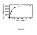
- Circular dichroism (DC) analysis of the CaM1c peptide shows a typical spectrum of a disordered structure with a minimum of 190 nm ( figure 3 ). The absence of secondary structure was confirmed by proton nuclear magnetic resonance spectroscopy (1H NMR). By the addition of calcium in solution, the DC spectrum assumes a shape that is typical of a helical conformation with minima at 206 and 222 nm. A titration of CaM1c by DC was then carried out and the intensity of the dichroic signal at 222 nm was reported as a function of the concentration of calcium ions added ( figure 4 ). The binding isothermal curve which passes through the experimental points demonstrates a stoichiometry of 1/1 Ca / peptide and makes it possible to calculate a dissociation constant Kd of 30 ⁇ M.
- the mass spectrum of the peptide in the absence of the metal shows three main peaks at 736.8, 920.7 and 1227.3 m / z, corresponding to the five, four and three times protonated peptide, respectively.
- the introduction of increasing calcium concentrations to the peptide leads to a modification of this spectrum with new peaks at 744.7, 930.6, 1240.5 m / z, compatible with a 1: 1 peptide: calcium complex and presenting the same state of charge.
- the fluorescence emission of the two lanthanides is observed by a mechanism of energy transfer via Tyr20 of the peptide, with an increase of the fluorescence emitted from the metal to a limit corresponding to a lanthanide: peptide ratio of 1: 1.
- the stronger emission is located at 545 nm.
- the spectrum shows fluorescence emission maxima at 593 and 618 nm.
- a second peptide comprising an additional mutation namely: the substitution of glutamic acid at position 31 of the sequence of the calmodulin to aspartic acid (Glu31Asp), has also been synthesized.
- the side chain of the amino acid is shortened by a methylene group, and the cavity formed by the loop is therefore larger.
- ESI / MS, DC, SLRT show that this peptide loses affinity for all divalent metals, as well as for the uranyl ion. Only the affinities for lanthanides with dissociation constants of 3.5 ⁇ 1 ⁇ M and 3.2 ⁇ 0.8 ⁇ M for Terbium and Europium are maintained.
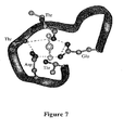
- FIG. 7 A schematic representation of uranyl in the coordinating loop of the mutated calmodulin (CaM-M3c peptide), obtained from the three-dimensional structure of the calmodulin loop (PDB code: 1EXR) is presented in figure 7 .
- the affinity of the CaM-M3c peptide for different metal ions was tested using two spectroscopic methods : circular dichroism (DC) and electrospray mass spectrometry (ESI-MS).
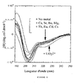
- the figure 8 shows that only the addition of an excess of uranyl leads to a modification of the dichroic spectrum of the peptide.
- CaM M3c In this case, two new minima at 207 nm and 222 nm are observed. They are characteristic of a secondary structure ordered in ⁇ helix. This result is confirmed by the ESI-MS analysis: only the addition of uranyl in solution leads to the appearance of a mass peak compatible with the formation of a 1/1 peptide / UO 2 complex.
- the CaM-M4c, CaM-M5c, CaM-M6c, CaM-M7c, CaM-M8c, CaM-M9c and CaM-M10c peptides gave the same results in circular and mass dichroism spectroscopy (ESI-MS), which indicates that these peptides do not bind calcium, lanthanides and other ions tested but only uranium (VI).
- ESI-MS circular and mass dichroism spectroscopy
- the analysis of the coordination of the uranyl ion by a biological molecule is considered at a pH close to the physiological value, that is to say between 6 and 8. However, at this pH, the uranyl ion is no longer in solution only in a single UO 2 2+ form , but in the form of various complexes derived from this metal core: hydroxos complexes and carbonates by example. This phenomenon is referred to as speciation.
- the amount of each species present in aqueous solution depends on the concentration of uranium, the concentration of dissolved gases (carbonates), as well as the thermodynamic parameters associated with the metal ion.
- the speciation diagram shows that the majority species at pH 6.5 are species (UO 2 ) 3 (OH) 5 + , UO 2 (OH) 2 and UO 2 (OH) + respectively representing 52.1; 16 and 25.3% of the U (VI) in solution.
- the minority species are UO 2 2+ , UO 2 (OH) 3 - as well as uranium / carbonate complexes.
- each of these uranium species participates in the overall fluorescence intensity detected after excitation.
- the calculation of a dissociation constant is therefore impossible since we can not isolate the contribution of each of the species present in solution.
- the titrations were performed using the temporal resolution, i.e., the difference in fluorescence lifetime of each of the species involved, as described in Example 1.
- the only species detected is the monohydroxo UO 2 (OH) + complex.
- Uranium at 2 ⁇ M was titrated with the CaM-M3c peptide in a pure aqueous medium whose pH was adjusted to 6.5 with ammonia.
- the parameters of the temporal resolution 70 ⁇ s of delay, 100 ⁇ s of gate width, 0.5 s of integration) make it possible to visualize only the monohydroxylated species of uranium.
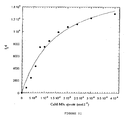
- the graph of the intensity at 520 nm as a function of the added peptide concentration can be simulated by the theoretical expression corresponding to the chemical equilibrium at stake, taking into account that the concentration of UO 2 (OH) + in solution is equal to 17.21% of the U (VI) introduced.
- the uranium / phosphate complexes have the particularity of increasing the fluorescence intensity of the metal ion while the other known ligands lead to an attenuation of the fluorescence.
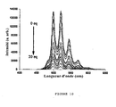
- This medium is artificially contaminated by lower and lower concentrations of uranium (20 ⁇ M, 2 ⁇ M, 0.2 ⁇ M). In each case, the peptide is added until a complete extinction of the fluorescence of the uranium ( figure 12 ).
- the apparent dissociation constants calculated for each of these assays are 10 ⁇ 1 ⁇ M. They are of the same order of magnitude as the dissociation constant calculated in deionized water at pH 6.5 and in phosphate buffer. This highlights the absence of competition between U (VI) and the other metal cations on the one hand, and between the peptide and the other ligands of uranium on the other hand: the CaM-M3c peptide is therefore selective for uranium under these conditions.
- the results obtained show that the CaM-M3c peptide which possesses the D20T and D24T mutations is selective for uranium VI.
- the CaM-M3c peptide coordinates the uranium with a dissociation constant between 3.8 and 18 ⁇ M in the various media tested. No measurable affinity for Mg, Ca, Sr, Ba, Eu, Tb is detected.
- Example 4 Analysis of the interaction of uranium with the native calmodulin of bovine brain
- uranium in the form of uranyl, 2.0 ⁇ M
- the coordination of uranium with the native protein in 1 mM phosphate medium was also studied.
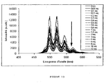
- the complexation of uranium with the protein results in a fall in the fluorescence intensity of uranyl.
- the spectra of the titration are represented at figure 13 .
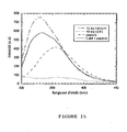
- a solution of 0.4 ⁇ M uranyl nitrate in 1.0 mM phosphate buffer (pH 7.0) was titrated with calmodulin solution to a protein concentration of 8 ⁇ M. After addition of 8 ⁇ M calmodulin, calcium ions were added up to the concentration of 8 mM ( figure 14 ).
- the values of the uranyl titration by calmodulin were interpreted by simulating the experimental data by a system of equations corresponding to one, two, three or four independent sites for uranium ( figure 16 ).
- the Dynafit software Kuzmic, P. 1996, Anal. Biochem. 237, 260-273 ) was used for these simulations. Only the system taking into account two sites with high affinity leads to an acceptable simulation with dissociation constants of 3.3 ⁇ 0.4 ⁇ M and 0.72 ⁇ 0.2 ⁇ M for each of the two sites.
- the competition values with the calcium ions were also interpreted with a system of adapted equations and the simulation of the experimental points with this system is reported in the figure 17 .
- the simulation was performed considering that in the presence of 8 ⁇ M of calmodulin and 0.4 ⁇ M of uranyl, each of the two high affinity sites is equiprobably populated with uranium. It is also assumed that each of these two sites is independent of the other.
- the simulation is carried out by a system of four chemical equilibria: two correspond to the dissociation equilibria of the "site-uranium" complexes, and two correspond to the displacement of uranium by calcium in each of the sites.
- the simulation confirms that only one of the two sites complexed with uranium is displaced by calcium. This shows that the calmodulin protein can bind uranium in calcium complexation sites.
- This energy transfer phenomenon further shows that the uranium is in this complex located at less than 15 ⁇ (maximum distance for a transfer of energy) of tryptophan, which is in agreement with the distances measured on the structure with X-rays (10 ⁇ and 6.5 ⁇ ) between the tryptophan of the peptide and two of the four calcium.
- chameleon proteins derived from calmodulin comprising the sequence of at least one peptide according to the invention can, in the presence of uranium, bind the MLCK substrate peptide and that can therefore be used as specific biosensors of the uranium VI.
Landscapes
- Chemical & Material Sciences (AREA)
- Life Sciences & Earth Sciences (AREA)
- Organic Chemistry (AREA)
- Health & Medical Sciences (AREA)
- Biochemistry (AREA)
- General Health & Medical Sciences (AREA)
- Genetics & Genomics (AREA)
- Medicinal Chemistry (AREA)
- Molecular Biology (AREA)
- Proteomics, Peptides & Aminoacids (AREA)
- Biophysics (AREA)
- Zoology (AREA)
- Toxicology (AREA)
- Gastroenterology & Hepatology (AREA)
- Peptides Or Proteins (AREA)
- Breeding Of Plants And Reproduction By Means Of Culturing (AREA)
- Measuring Or Testing Involving Enzymes Or Micro-Organisms (AREA)
- Micro-Organisms Or Cultivation Processes Thereof (AREA)
- Processing Of Solid Wastes (AREA)
- Purification Treatments By Anaerobic Or Anaerobic And Aerobic Bacteria Or Animals (AREA)
Claims (23)
- Isoliertes Peptid, dadurch gekennzeichnet, dass es ausgewählt ist aus der Gruppe bestehend aus solchen mit den Sequenzen SEQ ID Nrn. 4 bis 7, 9, 10 und 12.
- Das Peptid gemäß Anspruch 1, dadurch gekennzeichnet, dass es mit mindestens einem Fluorophor konjugiert ist.
- Das Peptid gemäß Anspruch 2, dadurch gekennzeichnet, dass das Fluorophor ein fluoreszierendes Protein ist ausgewählt aus: EBFP, ECFP, EYFP, EGFP, DsRed, CopGFP und PhiYFP.
- Das Peptid gemäß Anspruch 2, dadurch gekennzeichnet, dass das Fluorophor ausgewählt ist aus Dansyl, Cumarin, Fluorescein und Derivate der Alexa-Fluoreszenzfarbstoffe.
- Polypeptid, dadurch gekennzeichnet, dass es die Verknüpfung von mindesétens zwei identischen oder unterschiedlichen Peptiden gemäß irgendeinem der Ansprüche 1 bis 4 enthält.
- Fusionsprotein, dadurch gekennzeichnet, dass es durch die Fusion der Sequenz von mindestens einem Peptid gemäß Anspruch 1 mit einer Sequenz eines entsprechenden Proteins gebildet wird.
- Das Fusionsprotein gemäß Anspruch 6, dadurch gekennzeichnet, dass das entsprechende Protein ausgewählt ist aus der Gruppe bestehend aus Calmodulin, Chamäleon-Proteinen, welche von dem vorhergehenden abgeleitet sind und Proteinen, die ein Helix-Loop-Helix-Motiv aufweisen, die in der Lage sind, Calcium zu binden.
- Das Fusionsprotein gemäß Anspruch 6 oder 7, dadurch gekennzeichnet, dass es mit mindestens einem Fluorophor konjugiert ist, wie dieses in Anspruch 3 oder 4 beschrieben ist.
- Das Fusionsprotein gemäß Anspruch 8, dadurch gekennzeichnet, dass ein Terminus des Proteins mit einem Fluoreszenz-Donor gekoppelt ist und der andere mit einem Fluoreszenzakzeptor gekoppelt ist.
- Das Fusionsprotein gemäß Anspruch 9, dadurch gekennzeichnet, dass es an einem seiner Termini die Sequenz von EBFP oder ECFP und an dem anderen Terminus die Sequenz von EGFP oder EYFP aufweist.
- Isoliertes Nukleinsäuremolekül, dadurch gekennzeichnet, dass es eine Sequenz enthält, welches für ein Peptid, ein Polypeptid oder ein Fusionsprotein gemäß irgendeinem der Ansprüche 1 bis 10 kodiert.
- Rekombinanter eukaryotischer oder prokaryotischer Vektor, dadurch gekennzeichnet, dass er ein Insert enthält bestehend aus einem Nukleinsäuremolekül gemäß Anspruch 11.
- Eukaryotische oder prokaryotische Zelle, dadurch gekennzeichnet, dass sie durch einen rekombinanten Vektor gemäß Anspruch 12 modifiziert ist.
- Nicht-menschlicher tierischer transgener Organismus, dadurch gekennzeichnet, dass er Zellen enthält, die mittels eines Nukleinsäuremoleküls gemäß Anspruch 11 modifiziert sind.
- Transgene Pflanze, dadurch gekennzeichnet, dass sie Zellen enthält, die mittels eines Nukleinsäuremoleküls gemäß Anspruch 11 modifiziert sind.
- Verwendung von mindestens einem isolierten Peptid gemäß irgendeinem der Ansprüche 1 bis 4, eines Polypeptids gemäß Anspruch 5 oder eines Fusionsproteins gemäß irgendeinem der Ansprüche 6 bis 10, zur Herstellung eines Reagens, welches zum Nachweis von mit Uran kontaminierten Böden und Wasser bestimmt ist.
- Verwendung von mindestens einem isolierten Peptid gemäß irgendeinem der Ansprüche 1 bis 4, eines Polypeptids gemäß Anspruch 5 oder eines Fusionsproteins gemäß irgendeinem der Ansprüche 6 bis 10, zur Herstellung eines Reagens, welches zum Nachweis von mit Uran kontaminierten Individuen bestimmt ist.
- Verwendung von mindestens einer Zelle gemäß Anspruch 13 zur Sanierung von mit Uran kontaminierten Böden und Wasser.
- Verwendung einer transgenen Pflanze gemäß Anspruch 15 zur Sanierung von mit Uran kontaminierten Böden und Wasser.
- Peptid, Polypeptid oder Fusionsprotein gemäß irgendeinem der Ansprüche 1 bis 10 zur Verwendung als Arzneimittel zur Behandlung von mit Uran kontaminierten Individuen.
- Das Peptid zur Verwendung als Arzneimittel gemäß Anspruch 20, dadurch gekennzeichnet, dass es mit mindestens einem Molekül assoziiert ist, welches die zielgerichtete Anreicherung in den Nieren und/oder in den Knochen erlaubt.
- Das Peptid zur Verwendung als Arzneimittel gemäß Anspruch 20 oder 21, dadurch gekennzeichnet, dass es mit einem Molekül assoziiert ist, welches seine in vivo Ausscheidung begünstigt.
- Kit zum Nachweis einer Kontamination mit Uran, dadurch gekennzeichnet, dass es mindestens ein isoliertes Peptid, ein Polypeptid oder ein Fusionsprotein gemäß irgendeinem der Ansprüche 1 bis 10 enthält.
Applications Claiming Priority (2)
| Application Number | Priority Date | Filing Date | Title |
|---|---|---|---|
| FR0308211A FR2857014B1 (fr) | 2003-07-04 | 2003-07-04 | Peptides chelatant l'uranium et leurs applications |
| PCT/FR2004/001698 WO2005012336A2 (fr) | 2003-07-04 | 2004-07-01 | Peptides chelatant l'uranium et leurs applications |
Publications (2)
| Publication Number | Publication Date |
|---|---|
| EP1641815A2 EP1641815A2 (de) | 2006-04-05 |
| EP1641815B1 true EP1641815B1 (de) | 2015-05-27 |
Family
ID=33522781
Family Applications (1)
| Application Number | Title | Priority Date | Filing Date |
|---|---|---|---|
| EP04767541.8A Expired - Lifetime EP1641815B1 (de) | 2003-07-04 | 2004-07-01 | Uran-chelatisierende peptide und deren verwendung |
Country Status (8)
| Country | Link |
|---|---|
| US (3) | US7888311B2 (de) |
| EP (1) | EP1641815B1 (de) |
| JP (1) | JP5086636B2 (de) |
| CA (1) | CA2531231C (de) |
| ES (1) | ES2543162T3 (de) |
| FR (1) | FR2857014B1 (de) |
| RU (1) | RU2006103257A (de) |
| WO (1) | WO2005012336A2 (de) |
Families Citing this family (5)
| Publication number | Priority date | Publication date | Assignee | Title |
|---|---|---|---|---|
| US8793787B2 (en) * | 2004-04-01 | 2014-07-29 | Fireeye, Inc. | Detecting malicious network content using virtual environment components |
| US9255019B2 (en) | 2012-04-12 | 2016-02-09 | The University Of Chicago | Protein scaffolds for selective enrichment of metal ions |
| EP2784094A1 (de) | 2013-03-28 | 2014-10-01 | Commissariat A L'energie Atomique Et Aux Energies Alternatives | Neue uranchelatisierende, vom EF-Hand-Calciumsbindungsmotiv abgeleitete Peptide zur Uran-Biodetektion und -Biodekontamination |
| US9845343B2 (en) * | 2013-03-28 | 2017-12-19 | Commissariat A L'energie Atomique Et Aux Energies Alternatives | Uranium-chelating peptides derived from EF-hand calcium-binding motif useful for uranium biodetection and biodecontamination |
| CN115991757B (zh) * | 2022-07-15 | 2023-08-11 | 南宁师范大学 | 信号增强型检测汞离子的荧光蛋白探针及其制备与应用 |
-
2003
- 2003-07-04 FR FR0308211A patent/FR2857014B1/fr not_active Expired - Lifetime
-
2004
- 2004-07-01 ES ES04767541.8T patent/ES2543162T3/es not_active Expired - Lifetime
- 2004-07-01 RU RU2006103257/13A patent/RU2006103257A/ru not_active Application Discontinuation
- 2004-07-01 JP JP2006516341A patent/JP5086636B2/ja not_active Expired - Fee Related
- 2004-07-01 WO PCT/FR2004/001698 patent/WO2005012336A2/fr not_active Ceased
- 2004-07-01 US US10/562,323 patent/US7888311B2/en not_active Expired - Fee Related
- 2004-07-01 CA CA2531231A patent/CA2531231C/fr not_active Expired - Fee Related
- 2004-07-01 EP EP04767541.8A patent/EP1641815B1/de not_active Expired - Lifetime
-
2010
- 2010-12-28 US US12/979,787 patent/US9321849B2/en not_active Expired - Fee Related
-
2016
- 2016-03-25 US US15/080,821 patent/US20180134810A1/en not_active Abandoned
Also Published As
| Publication number | Publication date |
|---|---|
| JP5086636B2 (ja) | 2012-11-28 |
| FR2857014B1 (fr) | 2006-11-24 |
| US7888311B2 (en) | 2011-02-15 |
| FR2857014A1 (fr) | 2005-01-07 |
| ES2543162T3 (es) | 2015-08-17 |
| US20100015056A1 (en) | 2010-01-21 |
| JP2007537695A (ja) | 2007-12-27 |
| US9321849B2 (en) | 2016-04-26 |
| WO2005012336A2 (fr) | 2005-02-10 |
| CA2531231A1 (fr) | 2005-02-10 |
| RU2006103257A (ru) | 2006-07-27 |
| WO2005012336A3 (fr) | 2005-05-26 |
| US20180134810A1 (en) | 2018-05-17 |
| EP1641815A2 (de) | 2006-04-05 |
| US20110093964A1 (en) | 2011-04-21 |
| CA2531231C (fr) | 2014-04-01 |
Similar Documents
| Publication | Publication Date | Title |
|---|---|---|
| Bracha‐Drori et al. | Detection of protein–protein interactions in plants using bimolecular fluorescence complementation | |
| Belousov et al. | Genetically encoded fluorescent indicator for intracellular hydrogen peroxide | |
| DK2836503T3 (en) | Genetically coded probe for quantifying lactate, and methods for quantifying metabolic rates and lactate transport. | |
| US20180134810A1 (en) | Uranium-chelating peptides and uses thereof | |
| Fresnais et al. | Recent advances in the characterization of hair of mummies from the Chilean Andean coast | |
| Tewari et al. | Manganese induces oligomerization to promote down-regulation of the intracellular trafficking receptor used by Shiga toxin | |
| Becker et al. | On-resin assembly of a linkerless lanthanide (III)-based luminescence label and its application to the total synthesis of site-specifically labeled mechanosensitive channels | |
| Brazard et al. | Photoantenna in two cryptochrome–photolyase proteins from O. tauri: Presence, nature and ultrafast photoinduced dynamics | |
| Clainche et al. | Modulating the affinity and the selectivity of engineered calmodulin EF‐Hand peptides for lanthanides | |
| Mohsin et al. | Genetically encoded FRET-based nanosensor for in vivo monitoring of zinc concentration in physiological environment of living cell | |
| Sundaramoorthy | Development of a fluorescent biosensor for the personalized healthcare of diseases. | |
| US8852882B2 (en) | Biosensors and their use | |
| Abounorinejad | The Effect of Solvent on Silicon and Germanium Rhodamines and Synthesis of the AQP4ex C-Terminus Extension Peptide | |
| JP2018121557A (ja) | 活性イオウ分子種に応答して遺伝子発現を制御するタンパク質 | |
| Le Clainche et al. | Binding toxic metals to new Calmodulin peptides | |
| EP2981555B1 (de) | Neue uranchelatisierende, vom ef-hand-calciumsbindungsmotiv abgeleitete peptide zur uran-biodetektion und -biodekontamination | |
| WO2017079656A2 (en) | Artificial proteins and compositions and methods thereof | |
| Johnston | Studying the chaperone function of small heat-shock proteins using single-molecule fluorescence microscopy | |
| EP1730297A1 (de) | Verfahren zum screening von eine lkb-alpha-protein-ubiquitinierung modulierenden agentien sowie mittel zur durchführung des verfahrens | |
| Wei | Nucleosome structure and dynamics studied with single-molecule methods | |
| ES2427852A1 (es) | Sensores de calcio y métodos para la detección de calcio libre intracelular | |
| KR20200068359A (ko) | 푸코실락토오스 검출용 바이오센서 및 이를 이용하여 푸코실락토오스의 정량 분석 방법 | |
| ES2445018B1 (es) | VARIANTES DE LA PROTEÍNA DE FUSIÓN SENSIBLE AL CALCIO tdTOMATO-AEQUORINA | |
| Kagawa et al. | Energy transduction in H+-ATPase | |
| CN112194729A (zh) | 一种用于检测泛素-蛋白酶体系统降解活性的生物探针 |
Legal Events
| Date | Code | Title | Description |
|---|---|---|---|
| PUAI | Public reference made under article 153(3) epc to a published international application that has entered the european phase |
Free format text: ORIGINAL CODE: 0009012 |
|
| 17P | Request for examination filed |
Effective date: 20060109 |
|
| AK | Designated contracting states |
Kind code of ref document: A2 Designated state(s): AT BE BG CH CY CZ DE DK EE ES FI FR GB GR HU IE IT LI LU MC NL PL PT RO SE SI SK TR |
|
| RIN1 | Information on inventor provided before grant (corrected) |
Inventor name: MONJARDET, VERONIQUE Inventor name: LE CLAINCHE, LOIC Inventor name: VITA, CLAUDIO |
|
| RIN1 | Information on inventor provided before grant (corrected) |
Inventor name: MONJARDET, VERONIQUE Inventor name: LE CLAINCHE, LOIC Inventor name: VITA, CLAUDIO |
|
| DAX | Request for extension of the european patent (deleted) | ||
| 17Q | First examination report despatched |
Effective date: 20090319 |
|
| RAP1 | Party data changed (applicant data changed or rights of an application transferred) |
Owner name: COMMISSARIAT A L'ENERGIE ATOMIQUE ET AUX ENERGIES |
|
| GRAP | Despatch of communication of intention to grant a patent |
Free format text: ORIGINAL CODE: EPIDOSNIGR1 |
|
| INTG | Intention to grant announced |
Effective date: 20140924 |
|
| RIN1 | Information on inventor provided before grant (corrected) |
Inventor name: VITA, CLAUDIO Inventor name: LE CLAINCHE, LOIC Inventor name: MONJARDET, VERONIQUE |
|
| GRAP | Despatch of communication of intention to grant a patent |
Free format text: ORIGINAL CODE: EPIDOSNIGR1 |
|
| INTG | Intention to grant announced |
Effective date: 20150312 |
|
| GRAS | Grant fee paid |
Free format text: ORIGINAL CODE: EPIDOSNIGR3 |
|
| GRAA | (expected) grant |
Free format text: ORIGINAL CODE: 0009210 |
|
| AK | Designated contracting states |
Kind code of ref document: B1 Designated state(s): AT BE BG CH CY CZ DE DK EE ES FI FR GB GR HU IE IT LI LU MC NL PL PT RO SE SI SK TR |
|
| REG | Reference to a national code |
Ref country code: GB Ref legal event code: FG4D Free format text: NOT ENGLISH |
|
| REG | Reference to a national code |
Ref country code: CH Ref legal event code: EP |
|
| REG | Reference to a national code |
Ref country code: AT Ref legal event code: REF Ref document number: 728804 Country of ref document: AT Kind code of ref document: T Effective date: 20150615 |
|
| REG | Reference to a national code |
Ref country code: IE Ref legal event code: FG4D Free format text: LANGUAGE OF EP DOCUMENT: FRENCH |
|
| REG | Reference to a national code |
Ref country code: CH Ref legal event code: NV Representative=s name: ISLER AND PEDRAZZINI AG, CH |
|
| REG | Reference to a national code |
Ref country code: DE Ref legal event code: R096 Ref document number: 602004047266 Country of ref document: DE Effective date: 20150709 |
|
| REG | Reference to a national code |
Ref country code: FR Ref legal event code: PLFP Year of fee payment: 12 |
|
| REG | Reference to a national code |
Ref country code: ES Ref legal event code: FG2A Ref document number: 2543162 Country of ref document: ES Kind code of ref document: T3 Effective date: 20150817 |
|
| REG | Reference to a national code |
Ref country code: SE Ref legal event code: TRGR |
|
| REG | Reference to a national code |
Ref country code: AT Ref legal event code: MK05 Ref document number: 728804 Country of ref document: AT Kind code of ref document: T Effective date: 20150527 |
|
| PG25 | Lapsed in a contracting state [announced via postgrant information from national office to epo] |
Ref country code: PT Free format text: LAPSE BECAUSE OF FAILURE TO SUBMIT A TRANSLATION OF THE DESCRIPTION OR TO PAY THE FEE WITHIN THE PRESCRIBED TIME-LIMIT Effective date: 20150928 |
|
| REG | Reference to a national code |
Ref country code: NL Ref legal event code: MP Effective date: 20150527 |
|
| PG25 | Lapsed in a contracting state [announced via postgrant information from national office to epo] |
Ref country code: AT Free format text: LAPSE BECAUSE OF FAILURE TO SUBMIT A TRANSLATION OF THE DESCRIPTION OR TO PAY THE FEE WITHIN THE PRESCRIBED TIME-LIMIT Effective date: 20150527 Ref country code: BG Free format text: LAPSE BECAUSE OF FAILURE TO SUBMIT A TRANSLATION OF THE DESCRIPTION OR TO PAY THE FEE WITHIN THE PRESCRIBED TIME-LIMIT Effective date: 20150827 Ref country code: GR Free format text: LAPSE BECAUSE OF FAILURE TO SUBMIT A TRANSLATION OF THE DESCRIPTION OR TO PAY THE FEE WITHIN THE PRESCRIBED TIME-LIMIT Effective date: 20150828 |
|
| PG25 | Lapsed in a contracting state [announced via postgrant information from national office to epo] |
Ref country code: EE Free format text: LAPSE BECAUSE OF FAILURE TO SUBMIT A TRANSLATION OF THE DESCRIPTION OR TO PAY THE FEE WITHIN THE PRESCRIBED TIME-LIMIT Effective date: 20150527 Ref country code: DK Free format text: LAPSE BECAUSE OF FAILURE TO SUBMIT A TRANSLATION OF THE DESCRIPTION OR TO PAY THE FEE WITHIN THE PRESCRIBED TIME-LIMIT Effective date: 20150527 |
|
| PG25 | Lapsed in a contracting state [announced via postgrant information from national office to epo] |
Ref country code: CZ Free format text: LAPSE BECAUSE OF FAILURE TO SUBMIT A TRANSLATION OF THE DESCRIPTION OR TO PAY THE FEE WITHIN THE PRESCRIBED TIME-LIMIT Effective date: 20150527 Ref country code: RO Free format text: LAPSE BECAUSE OF NON-PAYMENT OF DUE FEES Effective date: 20150527 Ref country code: MC Free format text: LAPSE BECAUSE OF FAILURE TO SUBMIT A TRANSLATION OF THE DESCRIPTION OR TO PAY THE FEE WITHIN THE PRESCRIBED TIME-LIMIT Effective date: 20150527 Ref country code: PL Free format text: LAPSE BECAUSE OF FAILURE TO SUBMIT A TRANSLATION OF THE DESCRIPTION OR TO PAY THE FEE WITHIN THE PRESCRIBED TIME-LIMIT Effective date: 20150527 Ref country code: SK Free format text: LAPSE BECAUSE OF FAILURE TO SUBMIT A TRANSLATION OF THE DESCRIPTION OR TO PAY THE FEE WITHIN THE PRESCRIBED TIME-LIMIT Effective date: 20150527 |
|
| REG | Reference to a national code |
Ref country code: DE Ref legal event code: R097 Ref document number: 602004047266 Country of ref document: DE |
|
| PG25 | Lapsed in a contracting state [announced via postgrant information from national office to epo] |
Ref country code: LU Free format text: LAPSE BECAUSE OF FAILURE TO SUBMIT A TRANSLATION OF THE DESCRIPTION OR TO PAY THE FEE WITHIN THE PRESCRIBED TIME-LIMIT Effective date: 20150701 |
|
| PLBE | No opposition filed within time limit |
Free format text: ORIGINAL CODE: 0009261 |
|
| STAA | Information on the status of an ep patent application or granted ep patent |
Free format text: STATUS: NO OPPOSITION FILED WITHIN TIME LIMIT |
|
| REG | Reference to a national code |
Ref country code: IE Ref legal event code: MM4A |
|
| 26N | No opposition filed |
Effective date: 20160301 |
|
| PG25 | Lapsed in a contracting state [announced via postgrant information from national office to epo] |
Ref country code: SI Free format text: LAPSE BECAUSE OF FAILURE TO SUBMIT A TRANSLATION OF THE DESCRIPTION OR TO PAY THE FEE WITHIN THE PRESCRIBED TIME-LIMIT Effective date: 20150527 |
|
| PG25 | Lapsed in a contracting state [announced via postgrant information from national office to epo] |
Ref country code: IE Free format text: LAPSE BECAUSE OF NON-PAYMENT OF DUE FEES Effective date: 20150701 |
|
| REG | Reference to a national code |
Ref country code: FR Ref legal event code: PLFP Year of fee payment: 13 |
|
| PG25 | Lapsed in a contracting state [announced via postgrant information from national office to epo] |
Ref country code: HU Free format text: LAPSE BECAUSE OF FAILURE TO SUBMIT A TRANSLATION OF THE DESCRIPTION OR TO PAY THE FEE WITHIN THE PRESCRIBED TIME-LIMIT; INVALID AB INITIO Effective date: 20040701 |
|
| PG25 | Lapsed in a contracting state [announced via postgrant information from national office to epo] |
Ref country code: CY Free format text: LAPSE BECAUSE OF FAILURE TO SUBMIT A TRANSLATION OF THE DESCRIPTION OR TO PAY THE FEE WITHIN THE PRESCRIBED TIME-LIMIT Effective date: 20150527 Ref country code: NL Free format text: LAPSE BECAUSE OF FAILURE TO SUBMIT A TRANSLATION OF THE DESCRIPTION OR TO PAY THE FEE WITHIN THE PRESCRIBED TIME-LIMIT Effective date: 20150527 |
|
| REG | Reference to a national code |
Ref country code: FR Ref legal event code: PLFP Year of fee payment: 14 |
|
| PG25 | Lapsed in a contracting state [announced via postgrant information from national office to epo] |
Ref country code: TR Free format text: LAPSE BECAUSE OF FAILURE TO SUBMIT A TRANSLATION OF THE DESCRIPTION OR TO PAY THE FEE WITHIN THE PRESCRIBED TIME-LIMIT Effective date: 20150527 |
|
| PGFP | Annual fee paid to national office [announced via postgrant information from national office to epo] |
Ref country code: FI Payment date: 20170626 Year of fee payment: 14 |
|
| PGFP | Annual fee paid to national office [announced via postgrant information from national office to epo] |
Ref country code: GB Payment date: 20170721 Year of fee payment: 14 Ref country code: CH Payment date: 20170724 Year of fee payment: 14 Ref country code: DE Payment date: 20170713 Year of fee payment: 14 Ref country code: FR Payment date: 20170731 Year of fee payment: 14 Ref country code: ES Payment date: 20170809 Year of fee payment: 14 Ref country code: IT Payment date: 20170713 Year of fee payment: 14 |
|
| PGFP | Annual fee paid to national office [announced via postgrant information from national office to epo] |
Ref country code: BE Payment date: 20170728 Year of fee payment: 14 Ref country code: SE Payment date: 20170719 Year of fee payment: 14 |
|
| REG | Reference to a national code |
Ref country code: DE Ref legal event code: R119 Ref document number: 602004047266 Country of ref document: DE |
|
| REG | Reference to a national code |
Ref country code: CH Ref legal event code: PL |
|
| GBPC | Gb: european patent ceased through non-payment of renewal fee |
Effective date: 20180701 |
|
| REG | Reference to a national code |
Ref country code: BE Ref legal event code: MM Effective date: 20180731 |
|
| PG25 | Lapsed in a contracting state [announced via postgrant information from national office to epo] |
Ref country code: FR Free format text: LAPSE BECAUSE OF NON-PAYMENT OF DUE FEES Effective date: 20180731 Ref country code: FI Free format text: LAPSE BECAUSE OF NON-PAYMENT OF DUE FEES Effective date: 20180701 Ref country code: GB Free format text: LAPSE BECAUSE OF NON-PAYMENT OF DUE FEES Effective date: 20180701 Ref country code: LI Free format text: LAPSE BECAUSE OF NON-PAYMENT OF DUE FEES Effective date: 20180731 Ref country code: DE Free format text: LAPSE BECAUSE OF NON-PAYMENT OF DUE FEES Effective date: 20190201 Ref country code: CH Free format text: LAPSE BECAUSE OF NON-PAYMENT OF DUE FEES Effective date: 20180731 |
|
| PG25 | Lapsed in a contracting state [announced via postgrant information from national office to epo] |
Ref country code: SE Free format text: LAPSE BECAUSE OF NON-PAYMENT OF DUE FEES Effective date: 20180702 Ref country code: BE Free format text: LAPSE BECAUSE OF NON-PAYMENT OF DUE FEES Effective date: 20180731 |
|
| PG25 | Lapsed in a contracting state [announced via postgrant information from national office to epo] |
Ref country code: IT Free format text: LAPSE BECAUSE OF NON-PAYMENT OF DUE FEES Effective date: 20180701 |
|
| REG | Reference to a national code |
Ref country code: ES Ref legal event code: FD2A Effective date: 20190916 |
|
| PG25 | Lapsed in a contracting state [announced via postgrant information from national office to epo] |
Ref country code: ES Free format text: LAPSE BECAUSE OF NON-PAYMENT OF DUE FEES Effective date: 20180702 |