EP1631826B1 - Verwendung des proteins masp als marker für kolorektale karzinome - Google Patents
Verwendung des proteins masp als marker für kolorektale karzinome Download PDFInfo
- Publication number
- EP1631826B1 EP1631826B1 EP04734663A EP04734663A EP1631826B1 EP 1631826 B1 EP1631826 B1 EP 1631826B1 EP 04734663 A EP04734663 A EP 04734663A EP 04734663 A EP04734663 A EP 04734663A EP 1631826 B1 EP1631826 B1 EP 1631826B1
- Authority
- EP
- European Patent Office
- Prior art keywords
- masp
- colorectal cancer
- diagnosis
- protein
- sample
- Prior art date
- Legal status (The legal status is an assumption and is not a legal conclusion. Google has not performed a legal analysis and makes no representation as to the accuracy of the status listed.)
- Expired - Lifetime
Links
- 206010009944 Colon cancer Diseases 0.000 title claims abstract description 78
- 208000001333 Colorectal Neoplasms Diseases 0.000 title claims abstract description 49
- 108090000623 proteins and genes Proteins 0.000 title claims description 43
- 102000004169 proteins and genes Human genes 0.000 title claims description 41
- 239000003550 marker Substances 0.000 title claims description 31
- 238000003745 diagnosis Methods 0.000 claims abstract description 36
- 238000000034 method Methods 0.000 claims abstract description 36
- 239000007788 liquid Substances 0.000 claims abstract description 25
- 238000005259 measurement Methods 0.000 claims abstract description 10
- 239000011230 binding agent Substances 0.000 claims description 22
- 230000009870 specific binding Effects 0.000 claims description 18
- 210000002966 serum Anatomy 0.000 claims description 15
- 108010005173 SERPIN-B5 Proteins 0.000 claims description 13
- 102100030333 Serpin B5 Human genes 0.000 claims description 12
- 208000003200 Adenoma Diseases 0.000 claims description 10
- 206010001233 Adenoma benign Diseases 0.000 claims description 8
- 210000004369 blood Anatomy 0.000 claims description 7
- 239000008280 blood Substances 0.000 claims description 7
- 238000013399 early diagnosis Methods 0.000 claims description 7
- 239000003153 chemical reaction reagent Substances 0.000 claims description 5
- 230000015572 biosynthetic process Effects 0.000 claims description 4
- 239000002243 precursor Substances 0.000 claims description 4
- 230000001900 immune effect Effects 0.000 claims description 2
- 238000001514 detection method Methods 0.000 abstract description 17
- 102000004528 Mannose-Binding Protein-Associated Serine Proteases Human genes 0.000 abstract 3
- 108010042484 Mannose-Binding Protein-Associated Serine Proteases Proteins 0.000 abstract 3
- 210000001519 tissue Anatomy 0.000 description 36
- 206010028980 Neoplasm Diseases 0.000 description 35
- 239000000523 sample Substances 0.000 description 33
- FAPWRFPIFSIZLT-UHFFFAOYSA-M Sodium chloride Chemical compound [Na+].[Cl-] FAPWRFPIFSIZLT-UHFFFAOYSA-M 0.000 description 15
- HEMHJVSKTPXQMS-UHFFFAOYSA-M Sodium hydroxide Chemical compound [OH-].[Na+] HEMHJVSKTPXQMS-UHFFFAOYSA-M 0.000 description 15
- 108010022366 Carcinoembryonic Antigen Proteins 0.000 description 14
- 102100025475 Carcinoembryonic antigen-related cell adhesion molecule 5 Human genes 0.000 description 14
- 230000014509 gene expression Effects 0.000 description 14
- 239000000499 gel Substances 0.000 description 13
- 229940027941 immunoglobulin g Drugs 0.000 description 13
- 238000012360 testing method Methods 0.000 description 13
- 201000011510 cancer Diseases 0.000 description 12
- 241000283973 Oryctolagus cuniculus Species 0.000 description 11
- 210000004027 cell Anatomy 0.000 description 11
- 230000035945 sensitivity Effects 0.000 description 11
- 201000010099 disease Diseases 0.000 description 10
- 208000037265 diseases, disorders, signs and symptoms Diseases 0.000 description 10
- 108091003079 Bovine Serum Albumin Proteins 0.000 description 9
- 229920001213 Polysorbate 20 Polymers 0.000 description 8
- 230000000875 corresponding effect Effects 0.000 description 8
- 239000012894 fetal calf serum Substances 0.000 description 8
- 238000003018 immunoassay Methods 0.000 description 8
- 239000000256 polyoxyethylene sorbitan monolaurate Substances 0.000 description 8
- 235000010486 polyoxyethylene sorbitan monolaurate Nutrition 0.000 description 8
- 239000012980 RPMI-1640 medium Substances 0.000 description 7
- XSQUKJJJFZCRTK-UHFFFAOYSA-N Urea Chemical compound NC(N)=O XSQUKJJJFZCRTK-UHFFFAOYSA-N 0.000 description 7
- 238000003556 assay Methods 0.000 description 7
- 230000027455 binding Effects 0.000 description 7
- 239000000872 buffer Substances 0.000 description 7
- 238000002649 immunization Methods 0.000 description 7
- 230000003053 immunization Effects 0.000 description 7
- 238000001155 isoelectric focusing Methods 0.000 description 7
- 210000002381 plasma Anatomy 0.000 description 7
- 238000001262 western blot Methods 0.000 description 7
- IAZDPXIOMUYVGZ-UHFFFAOYSA-N Dimethylsulphoxide Chemical compound CS(C)=O IAZDPXIOMUYVGZ-UHFFFAOYSA-N 0.000 description 6
- 206010027476 Metastases Diseases 0.000 description 6
- 238000004458 analytical method Methods 0.000 description 6
- 210000001072 colon Anatomy 0.000 description 6
- 239000012634 fragment Substances 0.000 description 6
- 210000004379 membrane Anatomy 0.000 description 6
- 239000012528 membrane Substances 0.000 description 6
- 108020004999 messenger RNA Proteins 0.000 description 6
- 239000002953 phosphate buffered saline Substances 0.000 description 6
- 239000006228 supernatant Substances 0.000 description 6
- 238000002965 ELISA Methods 0.000 description 5
- 241000282414 Homo sapiens Species 0.000 description 5
- 229910019142 PO4 Inorganic materials 0.000 description 5
- 239000002299 complementary DNA Substances 0.000 description 5
- 238000002405 diagnostic procedure Methods 0.000 description 5
- 239000006166 lysate Substances 0.000 description 5
- 210000004877 mucosa Anatomy 0.000 description 5
- NBIIXXVUZAFLBC-UHFFFAOYSA-K phosphate Chemical compound [O-]P([O-])([O-])=O NBIIXXVUZAFLBC-UHFFFAOYSA-K 0.000 description 5
- 239000010452 phosphate Substances 0.000 description 5
- 239000011780 sodium chloride Substances 0.000 description 5
- AJPJDKMHJJGVTQ-UHFFFAOYSA-M sodium dihydrogen phosphate Chemical compound [Na+].OP(O)([O-])=O AJPJDKMHJJGVTQ-UHFFFAOYSA-M 0.000 description 5
- 229910000162 sodium phosphate Inorganic materials 0.000 description 5
- 239000000243 solution Substances 0.000 description 5
- 230000004083 survival effect Effects 0.000 description 5
- 241000588724 Escherichia coli Species 0.000 description 4
- DBMJMQXJHONAFJ-UHFFFAOYSA-M Sodium laurylsulphate Chemical compound [Na+].CCCCCCCCCCCCOS([O-])(=O)=O DBMJMQXJHONAFJ-UHFFFAOYSA-M 0.000 description 4
- 238000005119 centrifugation Methods 0.000 description 4
- 239000002609 medium Substances 0.000 description 4
- WWZKQHOCKIZLMA-UHFFFAOYSA-N octanoic acid Chemical compound CCCCCCCC(O)=O WWZKQHOCKIZLMA-UHFFFAOYSA-N 0.000 description 4
- 238000004393 prognosis Methods 0.000 description 4
- 238000000746 purification Methods 0.000 description 4
- 238000012216 screening Methods 0.000 description 4
- 238000002415 sodium dodecyl sulfate polyacrylamide gel electrophoresis Methods 0.000 description 4
- 239000000439 tumor marker Substances 0.000 description 4
- UMCMPZBLKLEWAF-BCTGSCMUSA-N 3-[(3-cholamidopropyl)dimethylammonio]propane-1-sulfonate Chemical compound C([C@H]1C[C@H]2O)[C@H](O)CC[C@]1(C)[C@@H]1[C@@H]2[C@@H]2CC[C@H]([C@@H](CCC(=O)NCCC[N+](C)(C)CCCS([O-])(=O)=O)C)[C@@]2(C)[C@@H](O)C1 UMCMPZBLKLEWAF-BCTGSCMUSA-N 0.000 description 3
- PEDCQBHIVMGVHV-UHFFFAOYSA-N Glycerine Chemical compound OCC(O)CO PEDCQBHIVMGVHV-UHFFFAOYSA-N 0.000 description 3
- 108060003951 Immunoglobulin Proteins 0.000 description 3
- OKKJLVBELUTLKV-UHFFFAOYSA-N Methanol Chemical compound OC OKKJLVBELUTLKV-UHFFFAOYSA-N 0.000 description 3
- 239000007983 Tris buffer Substances 0.000 description 3
- 208000009956 adenocarcinoma Diseases 0.000 description 3
- 239000000427 antigen Substances 0.000 description 3
- 102000036639 antigens Human genes 0.000 description 3
- 108091007433 antigens Proteins 0.000 description 3
- 238000013459 approach Methods 0.000 description 3
- 230000000903 blocking effect Effects 0.000 description 3
- 239000004202 carbamide Substances 0.000 description 3
- 230000002596 correlated effect Effects 0.000 description 3
- 239000012228 culture supernatant Substances 0.000 description 3
- 238000009826 distribution Methods 0.000 description 3
- 230000002550 fecal effect Effects 0.000 description 3
- RAXXELZNTBOGNW-UHFFFAOYSA-N imidazole Natural products C1=CNC=N1 RAXXELZNTBOGNW-UHFFFAOYSA-N 0.000 description 3
- 102000018358 immunoglobulin Human genes 0.000 description 3
- 238000000338 in vitro Methods 0.000 description 3
- 238000011534 incubation Methods 0.000 description 3
- 239000012139 lysis buffer Substances 0.000 description 3
- 238000001840 matrix-assisted laser desorption--ionisation time-of-flight mass spectrometry Methods 0.000 description 3
- 230000009401 metastasis Effects 0.000 description 3
- 239000013610 patient sample Substances 0.000 description 3
- 239000008188 pellet Substances 0.000 description 3
- 230000008569 process Effects 0.000 description 3
- 108090000765 processed proteins & peptides Proteins 0.000 description 3
- 210000000664 rectum Anatomy 0.000 description 3
- LENZDBCJOHFCAS-UHFFFAOYSA-N tris Chemical compound OCC(N)(CO)CO LENZDBCJOHFCAS-UHFFFAOYSA-N 0.000 description 3
- YBJHBAHKTGYVGT-ZKWXMUAHSA-N (+)-Biotin Chemical compound N1C(=O)N[C@@H]2[C@H](CCCCC(=O)O)SC[C@@H]21 YBJHBAHKTGYVGT-ZKWXMUAHSA-N 0.000 description 2
- IJGRMHOSHXDMSA-UHFFFAOYSA-N Atomic nitrogen Chemical compound N#N IJGRMHOSHXDMSA-UHFFFAOYSA-N 0.000 description 2
- 241000894006 Bacteria Species 0.000 description 2
- 239000005635 Caprylic acid (CAS 124-07-2) Substances 0.000 description 2
- 201000009030 Carcinoma Diseases 0.000 description 2
- 102000004889 Interleukin-6 Human genes 0.000 description 2
- 108090001005 Interleukin-6 Proteins 0.000 description 2
- TWRXJAOTZQYOKJ-UHFFFAOYSA-L Magnesium chloride Chemical compound [Mg+2].[Cl-].[Cl-] TWRXJAOTZQYOKJ-UHFFFAOYSA-L 0.000 description 2
- 241000699666 Mus <mouse, genus> Species 0.000 description 2
- 241000699670 Mus sp. Species 0.000 description 2
- 206010035226 Plasma cell myeloma Diseases 0.000 description 2
- 102000007056 Recombinant Fusion Proteins Human genes 0.000 description 2
- 108010008281 Recombinant Fusion Proteins Proteins 0.000 description 2
- 229930006000 Sucrose Natural products 0.000 description 2
- CZMRCDWAGMRECN-UGDNZRGBSA-N Sucrose Chemical compound O[C@H]1[C@H](O)[C@@H](CO)O[C@@]1(CO)O[C@@H]1[C@H](O)[C@@H](O)[C@H](O)[C@@H](CO)O1 CZMRCDWAGMRECN-UGDNZRGBSA-N 0.000 description 2
- 239000012505 Superdex™ Substances 0.000 description 2
- BFNBIHQBYMNNAN-UHFFFAOYSA-N ammonium sulfate Chemical compound N.N.OS(O)(=O)=O BFNBIHQBYMNNAN-UHFFFAOYSA-N 0.000 description 2
- 229910052921 ammonium sulfate Inorganic materials 0.000 description 2
- 235000011130 ammonium sulphate Nutrition 0.000 description 2
- OHDRQQURAXLVGJ-HLVWOLMTSA-N azane;(2e)-3-ethyl-2-[(e)-(3-ethyl-6-sulfo-1,3-benzothiazol-2-ylidene)hydrazinylidene]-1,3-benzothiazole-6-sulfonic acid Chemical compound [NH4+].[NH4+].S/1C2=CC(S([O-])(=O)=O)=CC=C2N(CC)C\1=N/N=C1/SC2=CC(S([O-])(=O)=O)=CC=C2N1CC OHDRQQURAXLVGJ-HLVWOLMTSA-N 0.000 description 2
- 238000001574 biopsy Methods 0.000 description 2
- 238000009534 blood test Methods 0.000 description 2
- 210000001124 body fluid Anatomy 0.000 description 2
- 238000004113 cell culture Methods 0.000 description 2
- 239000013522 chelant Substances 0.000 description 2
- 238000002052 colonoscopy Methods 0.000 description 2
- 239000000839 emulsion Substances 0.000 description 2
- 230000004927 fusion Effects 0.000 description 2
- 102000037865 fusion proteins Human genes 0.000 description 2
- 108020001507 fusion proteins Proteins 0.000 description 2
- 230000002068 genetic effect Effects 0.000 description 2
- FDGQSTZJBFJUBT-UHFFFAOYSA-N hypoxanthine Chemical compound O=C1NC=NC2=C1NC=N2 FDGQSTZJBFJUBT-UHFFFAOYSA-N 0.000 description 2
- 229940100601 interleukin-6 Drugs 0.000 description 2
- 238000002955 isolation Methods 0.000 description 2
- 238000009533 lab test Methods 0.000 description 2
- 230000003902 lesion Effects 0.000 description 2
- 210000001165 lymph node Anatomy 0.000 description 2
- 238000002824 mRNA display Methods 0.000 description 2
- 230000003211 malignant effect Effects 0.000 description 2
- 206010061289 metastatic neoplasm Diseases 0.000 description 2
- 238000012986 modification Methods 0.000 description 2
- 230000004048 modification Effects 0.000 description 2
- 238000012544 monitoring process Methods 0.000 description 2
- 239000004570 mortar (masonry) Substances 0.000 description 2
- 201000000050 myeloid neoplasm Diseases 0.000 description 2
- 229960002446 octanoic acid Drugs 0.000 description 2
- 238000002264 polyacrylamide gel electrophoresis Methods 0.000 description 2
- 238000002360 preparation method Methods 0.000 description 2
- 102000004196 processed proteins & peptides Human genes 0.000 description 2
- 230000005180 public health Effects 0.000 description 2
- 230000001105 regulatory effect Effects 0.000 description 2
- 238000002271 resection Methods 0.000 description 2
- 239000012723 sample buffer Substances 0.000 description 2
- 210000004989 spleen cell Anatomy 0.000 description 2
- 238000003756 stirring Methods 0.000 description 2
- 239000005720 sucrose Substances 0.000 description 2
- 238000002560 therapeutic procedure Methods 0.000 description 2
- UMGDCJDMYOKAJW-UHFFFAOYSA-N thiourea Chemical compound NC(N)=S UMGDCJDMYOKAJW-UHFFFAOYSA-N 0.000 description 2
- 238000013519 translation Methods 0.000 description 2
- 238000011282 treatment Methods 0.000 description 2
- 210000004881 tumor cell Anatomy 0.000 description 2
- YMXHPSHLTSZXKH-RVBZMBCESA-N (2,5-dioxopyrrolidin-1-yl) 5-[(3as,4s,6ar)-2-oxo-1,3,3a,4,6,6a-hexahydrothieno[3,4-d]imidazol-4-yl]pentanoate Chemical compound C([C@H]1[C@H]2NC(=O)N[C@H]2CS1)CCCC(=O)ON1C(=O)CCC1=O YMXHPSHLTSZXKH-RVBZMBCESA-N 0.000 description 1
- KHNDABJZSPPYLE-FUGFVFQCSA-N (2,5-dioxopyrrolidin-1-yl) 6-[[2-[[(3s,5r,8r,9s,10s,12r,13s,14s,17r)-12,14-dihydroxy-10,13-dimethyl-17-(5-oxo-2h-furan-3-yl)-1,2,3,4,5,6,7,8,9,11,12,15,16,17-tetradecahydrocyclopenta[a]phenanthren-3-yl]oxy]acetyl]amino]hexanoate Chemical compound O([C@@H]1C[C@H]2CC[C@H]3[C@@]4(O)CC[C@@H]([C@]4([C@@H](C[C@@H]3[C@@]2(C)CC1)O)C)C=1COC(=O)C=1)CC(=O)NCCCCCC(=O)ON1C(=O)CCC1=O KHNDABJZSPPYLE-FUGFVFQCSA-N 0.000 description 1
- VYQIXWBVBNHBAV-HVDRVSQOSA-N (2S)-2-amino-3-(2-diazoacetyl)oxypropanoic acid 1,7-dihydropurin-6-one Chemical compound O=C1NC=NC2=C1NC=N2.OC(=O)[C@@H](N)COC(=O)C=[N+]=[N-] VYQIXWBVBNHBAV-HVDRVSQOSA-N 0.000 description 1
- AGNGYMCLFWQVGX-AGFFZDDWSA-N (e)-1-[(2s)-2-amino-2-carboxyethoxy]-2-diazonioethenolate Chemical compound OC(=O)[C@@H](N)CO\C([O-])=C\[N+]#N AGNGYMCLFWQVGX-AGFFZDDWSA-N 0.000 description 1
- 108091032973 (ribonucleotides)n+m Proteins 0.000 description 1
- -1 2,4,6-trimethylphenyl Chemical group 0.000 description 1
- NZQLLBQLFWYNQV-WKUSAUFCSA-N 2-[2-[bis(carboxymethyl)amino]ethyl-(carboxymethyl)amino]acetic acid;(2s,3s)-1,4-bis(sulfanyl)butane-2,3-diol Chemical compound SC[C@@H](O)[C@H](O)CS.OC(=O)CN(CC(O)=O)CCN(CC(O)=O)CC(O)=O NZQLLBQLFWYNQV-WKUSAUFCSA-N 0.000 description 1
- AXAVXPMQTGXXJZ-UHFFFAOYSA-N 2-aminoacetic acid;2-amino-2-(hydroxymethyl)propane-1,3-diol Chemical compound NCC(O)=O.OCC(N)(CO)CO AXAVXPMQTGXXJZ-UHFFFAOYSA-N 0.000 description 1
- IITIZHOBOIBGBW-UHFFFAOYSA-N 3-ethyl-2h-1,3-benzothiazole Chemical compound C1=CC=C2N(CC)CSC2=C1 IITIZHOBOIBGBW-UHFFFAOYSA-N 0.000 description 1
- UQRONKZLYKUEMO-UHFFFAOYSA-N 4-methyl-1-(2,4,6-trimethylphenyl)pent-4-en-2-one Chemical group CC(=C)CC(=O)Cc1c(C)cc(C)cc1C UQRONKZLYKUEMO-UHFFFAOYSA-N 0.000 description 1
- 238000013296 A/J mouse Methods 0.000 description 1
- QTBSBXVTEAMEQO-UHFFFAOYSA-M Acetate Chemical compound CC([O-])=O QTBSBXVTEAMEQO-UHFFFAOYSA-M 0.000 description 1
- 241000588832 Bordetella pertussis Species 0.000 description 1
- 208000026310 Breast neoplasm Diseases 0.000 description 1
- 208000009458 Carcinoma in Situ Diseases 0.000 description 1
- 241000700198 Cavia Species 0.000 description 1
- VEXZGXHMUGYJMC-UHFFFAOYSA-M Chloride anion Chemical compound [Cl-] VEXZGXHMUGYJMC-UHFFFAOYSA-M 0.000 description 1
- 108020004635 Complementary DNA Proteins 0.000 description 1
- 108020004414 DNA Proteins 0.000 description 1
- 238000011238 DNA vaccination Methods 0.000 description 1
- SHIBSTMRCDJXLN-UHFFFAOYSA-N Digoxigenin Natural products C1CC(C2C(C3(C)CCC(O)CC3CC2)CC2O)(O)C2(C)C1C1=CC(=O)OC1 SHIBSTMRCDJXLN-UHFFFAOYSA-N 0.000 description 1
- KCXVZYZYPLLWCC-UHFFFAOYSA-N EDTA Chemical compound OC(=O)CN(CC(O)=O)CCN(CC(O)=O)CC(O)=O KCXVZYZYPLLWCC-UHFFFAOYSA-N 0.000 description 1
- 102000004190 Enzymes Human genes 0.000 description 1
- 108090000790 Enzymes Proteins 0.000 description 1
- 241001198387 Escherichia coli BL21(DE3) Species 0.000 description 1
- 102000010834 Extracellular Matrix Proteins Human genes 0.000 description 1
- 108010037362 Extracellular Matrix Proteins Proteins 0.000 description 1
- 102000003886 Glycoproteins Human genes 0.000 description 1
- 108090000288 Glycoproteins Proteins 0.000 description 1
- 241000147041 Guaiacum officinale Species 0.000 description 1
- 108010001336 Horseradish Peroxidase Proteins 0.000 description 1
- VEXZGXHMUGYJMC-UHFFFAOYSA-N Hydrochloric acid Chemical compound Cl VEXZGXHMUGYJMC-UHFFFAOYSA-N 0.000 description 1
- UGQMRVRMYYASKQ-UHFFFAOYSA-N Hypoxanthine nucleoside Natural products OC1C(O)C(CO)OC1N1C(NC=NC2=O)=C2N=C1 UGQMRVRMYYASKQ-UHFFFAOYSA-N 0.000 description 1
- 102000008394 Immunoglobulin Fragments Human genes 0.000 description 1
- 108010021625 Immunoglobulin Fragments Proteins 0.000 description 1
- 102000004856 Lectins Human genes 0.000 description 1
- 108090001090 Lectins Proteins 0.000 description 1
- 206010058467 Lung neoplasm malignant Diseases 0.000 description 1
- 239000000020 Nitrocellulose Substances 0.000 description 1
- 108091028043 Nucleic acid sequence Proteins 0.000 description 1
- 206010061902 Pancreatic neoplasm Diseases 0.000 description 1
- 241001494479 Pecora Species 0.000 description 1
- 108091005804 Peptidases Proteins 0.000 description 1
- 208000006994 Precancerous Conditions Diseases 0.000 description 1
- 239000004365 Protease Substances 0.000 description 1
- 241000700157 Rattus norvegicus Species 0.000 description 1
- 102100037486 Reverse transcriptase/ribonuclease H Human genes 0.000 description 1
- 239000012722 SDS sample buffer Substances 0.000 description 1
- 101000980867 Schizosaccharomyces pombe (strain 972 / ATCC 24843) Curved DNA-binding protein Proteins 0.000 description 1
- 102000008847 Serpin Human genes 0.000 description 1
- 108050000761 Serpin Proteins 0.000 description 1
- 101000982319 Shallot virus X Uncharacterized ORF4 protein Proteins 0.000 description 1
- 208000005718 Stomach Neoplasms Diseases 0.000 description 1
- 108010090804 Streptavidin Proteins 0.000 description 1
- 102000001742 Tumor Suppressor Proteins Human genes 0.000 description 1
- 108010040002 Tumor Suppressor Proteins Proteins 0.000 description 1
- 239000008351 acetate buffer Substances 0.000 description 1
- 239000002253 acid Substances 0.000 description 1
- 239000000654 additive Substances 0.000 description 1
- 230000000996 additive effect Effects 0.000 description 1
- 239000002671 adjuvant Substances 0.000 description 1
- 230000029936 alkylation Effects 0.000 description 1
- 238000005804 alkylation reaction Methods 0.000 description 1
- WNROFYMDJYEPJX-UHFFFAOYSA-K aluminium hydroxide Chemical compound [OH-].[OH-].[OH-].[Al+3] WNROFYMDJYEPJX-UHFFFAOYSA-K 0.000 description 1
- 150000001413 amino acids Chemical class 0.000 description 1
- 239000012491 analyte Substances 0.000 description 1
- 238000002820 assay format Methods 0.000 description 1
- 229950011321 azaserine Drugs 0.000 description 1
- 244000052616 bacterial pathogen Species 0.000 description 1
- 230000008901 benefit Effects 0.000 description 1
- 229960002685 biotin Drugs 0.000 description 1
- 235000020958 biotin Nutrition 0.000 description 1
- 239000011616 biotin Substances 0.000 description 1
- 238000007413 biotinylation Methods 0.000 description 1
- 230000006287 biotinylation Effects 0.000 description 1
- 230000036765 blood level Effects 0.000 description 1
- 229940098773 bovine serum albumin Drugs 0.000 description 1
- 210000000481 breast Anatomy 0.000 description 1
- 201000008275 breast carcinoma Diseases 0.000 description 1
- 239000007853 buffer solution Substances 0.000 description 1
- 238000010804 cDNA synthesis Methods 0.000 description 1
- 230000030833 cell death Effects 0.000 description 1
- 210000000170 cell membrane Anatomy 0.000 description 1
- 239000003795 chemical substances by application Substances 0.000 description 1
- 238000002512 chemotherapy Methods 0.000 description 1
- 238000010367 cloning Methods 0.000 description 1
- 208000029742 colonic neoplasm Diseases 0.000 description 1
- 239000013068 control sample Substances 0.000 description 1
- 238000007796 conventional method Methods 0.000 description 1
- 238000013211 curve analysis Methods 0.000 description 1
- 230000001086 cytosolic effect Effects 0.000 description 1
- 238000011161 development Methods 0.000 description 1
- 208000010643 digestive system disease Diseases 0.000 description 1
- QONQRTHLHBTMGP-UHFFFAOYSA-N digitoxigenin Natural products CC12CCC(C3(CCC(O)CC3CC3)C)C3C11OC1CC2C1=CC(=O)OC1 QONQRTHLHBTMGP-UHFFFAOYSA-N 0.000 description 1
- SHIBSTMRCDJXLN-KCZCNTNESA-N digoxigenin Chemical compound C1([C@@H]2[C@@]3([C@@](CC2)(O)[C@H]2[C@@H]([C@@]4(C)CC[C@H](O)C[C@H]4CC2)C[C@H]3O)C)=CC(=O)OC1 SHIBSTMRCDJXLN-KCZCNTNESA-N 0.000 description 1
- 239000012470 diluted sample Substances 0.000 description 1
- LOKCTEFSRHRXRJ-UHFFFAOYSA-I dipotassium trisodium dihydrogen phosphate hydrogen phosphate dichloride Chemical compound P(=O)(O)(O)[O-].[K+].P(=O)(O)([O-])[O-].[Na+].[Na+].[Cl-].[K+].[Cl-].[Na+] LOKCTEFSRHRXRJ-UHFFFAOYSA-I 0.000 description 1
- 229940042399 direct acting antivirals protease inhibitors Drugs 0.000 description 1
- CETRZFQIITUQQL-UHFFFAOYSA-N dmso dimethylsulfoxide Chemical compound CS(C)=O.CS(C)=O CETRZFQIITUQQL-UHFFFAOYSA-N 0.000 description 1
- 229960001484 edetic acid Drugs 0.000 description 1
- 230000000694 effects Effects 0.000 description 1
- 238000001962 electrophoresis Methods 0.000 description 1
- 210000002919 epithelial cell Anatomy 0.000 description 1
- 210000000981 epithelium Anatomy 0.000 description 1
- 239000006167 equilibration buffer Substances 0.000 description 1
- 238000002474 experimental method Methods 0.000 description 1
- 239000013604 expression vector Substances 0.000 description 1
- 210000002744 extracellular matrix Anatomy 0.000 description 1
- 230000002496 gastric effect Effects 0.000 description 1
- 239000011521 glass Substances 0.000 description 1
- 239000003102 growth factor Substances 0.000 description 1
- 229940091561 guaiac Drugs 0.000 description 1
- 201000003911 head and neck carcinoma Diseases 0.000 description 1
- 230000036541 health Effects 0.000 description 1
- 238000000703 high-speed centrifugation Methods 0.000 description 1
- 238000000265 homogenisation Methods 0.000 description 1
- 238000009396 hybridization Methods 0.000 description 1
- 210000004408 hybridoma Anatomy 0.000 description 1
- 238000010191 image analysis Methods 0.000 description 1
- 229940072221 immunoglobulins Drugs 0.000 description 1
- 238000011532 immunohistochemical staining Methods 0.000 description 1
- 239000003547 immunosorbent Substances 0.000 description 1
- 238000012744 immunostaining Methods 0.000 description 1
- 201000004933 in situ carcinoma Diseases 0.000 description 1
- 238000011065 in-situ storage Methods 0.000 description 1
- 230000008595 infiltration Effects 0.000 description 1
- 238000001764 infiltration Methods 0.000 description 1
- 230000002401 inhibitory effect Effects 0.000 description 1
- 239000002198 insoluble material Substances 0.000 description 1
- 238000007912 intraperitoneal administration Methods 0.000 description 1
- 239000002523 lectin Substances 0.000 description 1
- YFVGRULMIQXYNE-UHFFFAOYSA-M lithium;dodecyl sulfate Chemical compound [Li+].CCCCCCCCCCCCOS([O-])(=O)=O YFVGRULMIQXYNE-UHFFFAOYSA-M 0.000 description 1
- 210000004072 lung Anatomy 0.000 description 1
- 201000005296 lung carcinoma Diseases 0.000 description 1
- 229910001629 magnesium chloride Inorganic materials 0.000 description 1
- 238000004519 manufacturing process Methods 0.000 description 1
- 239000000463 material Substances 0.000 description 1
- 230000001394 metastastic effect Effects 0.000 description 1
- 208000011645 metastatic carcinoma Diseases 0.000 description 1
- 238000013508 migration Methods 0.000 description 1
- 230000005012 migration Effects 0.000 description 1
- 239000000203 mixture Substances 0.000 description 1
- 230000000869 mutational effect Effects 0.000 description 1
- 229920001220 nitrocellulos Polymers 0.000 description 1
- 229910052757 nitrogen Inorganic materials 0.000 description 1
- 238000012758 nuclear staining Methods 0.000 description 1
- 239000002773 nucleotide Substances 0.000 description 1
- 125000003729 nucleotide group Chemical group 0.000 description 1
- 230000003287 optical effect Effects 0.000 description 1
- 210000000056 organ Anatomy 0.000 description 1
- 230000002018 overexpression Effects 0.000 description 1
- 239000000137 peptide hydrolase inhibitor Substances 0.000 description 1
- 239000008363 phosphate buffer Substances 0.000 description 1
- 230000036470 plasma concentration Effects 0.000 description 1
- ONJQDTZCDSESIW-UHFFFAOYSA-N polidocanol Chemical compound CCCCCCCCCCCCOCCOCCOCCOCCOCCOCCOCCOCCOCCO ONJQDTZCDSESIW-UHFFFAOYSA-N 0.000 description 1
- 229920002401 polyacrylamide Polymers 0.000 description 1
- 108091033319 polynucleotide Proteins 0.000 description 1
- 239000002157 polynucleotide Substances 0.000 description 1
- 102000040430 polynucleotide Human genes 0.000 description 1
- 229920001184 polypeptide Polymers 0.000 description 1
- 238000001556 precipitation Methods 0.000 description 1
- 238000012545 processing Methods 0.000 description 1
- 239000000047 product Substances 0.000 description 1
- 208000037821 progressive disease Diseases 0.000 description 1
- 238000002731 protein assay Methods 0.000 description 1
- 238000000159 protein binding assay Methods 0.000 description 1
- 239000012460 protein solution Substances 0.000 description 1
- 230000002797 proteolythic effect Effects 0.000 description 1
- 238000012207 quantitative assay Methods 0.000 description 1
- 238000003259 recombinant expression Methods 0.000 description 1
- 238000010188 recombinant method Methods 0.000 description 1
- 230000009467 reduction Effects 0.000 description 1
- 238000011160 research Methods 0.000 description 1
- 210000003705 ribosome Anatomy 0.000 description 1
- 238000010079 rubber tapping Methods 0.000 description 1
- 239000012146 running buffer Substances 0.000 description 1
- 150000003839 salts Chemical class 0.000 description 1
- 238000003118 sandwich ELISA Methods 0.000 description 1
- 239000013049 sediment Substances 0.000 description 1
- 238000000926 separation method Methods 0.000 description 1
- 238000013207 serial dilution Methods 0.000 description 1
- 241000894007 species Species 0.000 description 1
- 238000011895 specific detection Methods 0.000 description 1
- 238000010186 staining Methods 0.000 description 1
- 238000010561 standard procedure Methods 0.000 description 1
- 239000000758 substrate Substances 0.000 description 1
- 238000001356 surgical procedure Methods 0.000 description 1
- 239000000725 suspension Substances 0.000 description 1
- 230000001225 therapeutic effect Effects 0.000 description 1
- 238000013518 transcription Methods 0.000 description 1
- 230000035897 transcription Effects 0.000 description 1
- 238000012546 transfer Methods 0.000 description 1
- 230000005740 tumor formation Effects 0.000 description 1
- 210000004509 vascular smooth muscle cell Anatomy 0.000 description 1
- 239000013598 vector Substances 0.000 description 1
- 210000000504 visceral peritoneum Anatomy 0.000 description 1
- 238000012800 visualization Methods 0.000 description 1
Images

Classifications
-
- G—PHYSICS
- G01—MEASURING; TESTING
- G01N—INVESTIGATING OR ANALYSING MATERIALS BY DETERMINING THEIR CHEMICAL OR PHYSICAL PROPERTIES
- G01N33/00—Investigating or analysing materials by specific methods not covered by groups G01N1/00 - G01N31/00
- G01N33/48—Biological material, e.g. blood, urine; Haemocytometers
- G01N33/50—Chemical analysis of biological material, e.g. blood, urine; Testing involving biospecific ligand binding methods; Immunological testing
- G01N33/53—Immunoassay; Biospecific binding assay; Materials therefor
- G01N33/574—Immunoassay; Biospecific binding assay; Materials therefor for cancer
- G01N33/57407—Specifically defined cancers
- G01N33/57419—Specifically defined cancers of colon
-
- G—PHYSICS
- G01—MEASURING; TESTING
- G01N—INVESTIGATING OR ANALYSING MATERIALS BY DETERMINING THEIR CHEMICAL OR PHYSICAL PROPERTIES
- G01N2333/00—Assays involving biological materials from specific organisms or of a specific nature
- G01N2333/81—Protease inhibitors
- G01N2333/8107—Endopeptidase (E.C. 3.4.21-99) inhibitors
-
- G—PHYSICS
- G01—MEASURING; TESTING
- G01N—INVESTIGATING OR ANALYSING MATERIALS BY DETERMINING THEIR CHEMICAL OR PHYSICAL PROPERTIES
- G01N2800/00—Detection or diagnosis of diseases
- G01N2800/52—Predicting or monitoring the response to treatment, e.g. for selection of therapy based on assay results in personalised medicine; Prognosis
Definitions
- the present invention relates to the diagnosis of colorectal cancer. It discloses the use of MASP (maspin precursor) protein in the diagnosis of colorectal cancer. Furthermore, it especially relates to a method for diagnosis of colorectal cancer from a liquid sample, derived from an individual by measuring MASP in said sample. Measurement of MASP can, e.g., be used in the early detection or diagnosis of colorectal cancer.
- MASP maspin precursor
- CRC colorectal cancer
- the prognosis in advanced stages of tumor is poor. More than one third of the patients will die from progressive disease within five years after diagnosis, corresponding to a survival rate of about 40% for five years.
- Current treatment is only curing a fraction of the patients and clearly has the best effect on those patients diagnosed in an early stage of disease.
- CRC colorectal cancer
- WO 01/96390 shall be mentioned and discussed.
- This application describes and claims more than two hundred isolated polynucleotides and the corresponding polypeptides as such, as well as their use in the detection of CRC.
- differences on the level of mRNA are not mirrored by the level of the corresponding proteins.
- a protein encoded by a rare mRNA may be found in very high amounts and a protein encoded by an abundant mRNA may nonetheless be hard to detect and find at all.
- This lack of correlation between mRNA-level and protein level is due to reasons like mRNA stability, efficiency of translation, stability of the protein, etc.
- WO 02/078636 reports about nine colorectal cancer-associated spots as found by surface-enhanced laser desorption and ionization (SELDI). These spots are seen more frequently in sera obtained from patients with CRC as compared to sera obtained from healthy controls. However, the identity of the molecule(s) comprised in such spot, e.g., its (their sequence), is not known.
- a new diagnostic marker as a single marker should be at least as good as the best single marker known in the art. Or, a new marker should lead to a progress in diagnostic sensitivity and/or specificity either if used alone or in combination with one or more other markers, respectively.
- the diagnostic sensitivity and/or specificity of a test is best assessed by its receiver-operating characteristics, which will be described in detail below.
- CEA carcinoembryonic antigen
- serum CEA determination possesses neither sensitivity nor the specificity to enable its use as a screening test for colorectal cancer in the asymptomatic population (Reynoso, G., et al., JAMA 220 (1972) 361-365; Sturgeon, C., Clinical Chemistry 48 (2002) 1151-1159).
- the present invention therefore relates to a method for the diagnosis of colorectal cancer comprising the steps of a) providing a liquid sample obtained from an individual, b) contacting said sample with a specific binding agent for MASP under conditions appropriate for formation of a complex between said binding agent and MASP, and c) correlating the amount of complex formed in (b) to the diagnosis of colorectal cancer.
- a preferred method uses a liquid sample obtained from an individual.
- Another preferred embodiment of the invention is a method for the diagnosis of colorectal cancer comprising the steps of a) contacting a liquid sample obtained from an individual with a specific binding agent for MASP under conditions appropriate for formation of a complex between said binding agent and MASP, and b) correlating the amount of complex formed in (a) to the diagnosis of colorectal cancer.
- any such diagnosis is made in vitro.
- the patient sample is discarded afterwards.
- the patient sample is solely used for the in vitro diagnostic method of the invention and the material of the patient sample is not transferred back into the patient's body.
- the sample is a liquid sample.
- the protein MASP (maspin precursor; Swiss-PROT: P36952) is characterized by the sequence given in SEQ ID NO: 1.
- the cloned human maspin cDNA encodes a 42-kDa protein that shares homology with the serpin superfamily of protease inhibitors. Immunostaining studies demonstrate that maspin is found in the extracellular matrix and at the plasma membrane (Zou, Z., et al., Science 263 (1994) 526-529).
- the human MASP gene (SERPINB5 of PI5) was originally isolated from normal mammary epithelium by subtractive hybridization on the basis of its expression at the mRNA level (Zou et al., supra). Maspin was expressed in normal mammary epithelial cells but not in most mammary carcinoma cell lines. Zou et al. (supra) showed that its expression reduces the ability of transformed cells to induce tumor formation and metastasis, suggesting that the maspin gene encodes a tumor suppressor.
- the present invention shall not be construed to be limited to the full-length protein MASP of SEQ ID NO :1.
- Physiological or artificial fragments of MASP, secondary modifications of MASP, as well as allelic variants of MASP are also encompassed by the present invention.
- Artificial fragments preferably encompass a peptide produced synthetically or by recombinant techniques, which at least comprises one epitope of diagnostic interest consisting of at least 6 contiguous amino acids as derived from the sequence disclosed in SEQ ID NO:1. Such fragment may advantageously be used for generation of antibodies or as a standard in an immunoassay. More preferred the artificial fragment comprises at least two epitopes of interest appropriate for setting up a sandwich immunoassay.
- novel marker MASP may be used for monitoring as well as for screening purposes.
- the diagnostic method according to the present invention may help to assess tumor load, efficacy of treatment and tumor recurrence in the follow-up of patients.
- Increased levels of MASP are directly correlated to tumor burden. After chemotherapy a short term (few hours to 14 days) increase in MASP may serve as an indicator of tumor cell death. In the follow-up of patients (from 3 months to 10 years) an increase of MASP can be used as an indicator for tumor recurrence.
- the diagnostic method according to the present invention is used for screening purposes. I.e., it is used to assess subjects without a prior diagnosis of CRC by measuring the level of MASP and correlating the level measured to the presence or absence of CRC.
- the staging of cancer is the classification of the disease in terms of extent, progression, and severity. It groups cancer patients so that generalizations can be made about prognosis and the choice of therapy.
- TNM the most widely used classification of the anatomical extent of cancer. It represents an internationally accepted, uniform staging system. There are three basic variables: T (the extent of the primary tumor), N (the status of regional lymph nodes) and M (the presence or absence of distant metastases).
- TNM criteria are published by the UICC (International Union against Cancer), Sobin, L.H., Wittekind, Ch. (eds): TNM Classification of Malignant Tumours, fifth edition, 1997).
- early diagnosis of CRC refers to a diagnosis at a pre-malignant state (adenoma) or at a tumor stage where no metastases at all (neither proximal nor distal), i.e., adenoma, T is , N0, M0 or T1-4; N0; M0 are present.
- T is denotes carcinoma in situ .
- the detection of MASP is used to diagnose CRC as early as in the adenoma stage.
- the diagnostic method according to the present invention is based on a liquid sample which is derived from an individual. Unlike to methods known from the art MASP is specifically measured from this liquid sample by use of a specific binding agent.
- a specific binding agent is, e.g., a receptor for MASP, a lectin binding to MASP or an antibody to MASP.
- a specific binding agent has at least an affinity of 10 7 l/mol for its corresponding target molecule.
- the specific binding agent preferably has an affinity of 10 8 l/mol or even more preferred of 10 9 l/mol for its target molecule.
- the term specific is used to indicate that other biomolecules present in the sample do not significantly bind to with the binding agent specific for MASP.
- the level of binding to a biomolecule other than the target molecule results in a binding affinity which is only 10%, more preferably only 5% of the affinity of the target molecule or less.
- a most preferred specific binding agent will fulfill both the above minimum criteria for affinity as well as for specificity.
- a specific binding agent preferably is an antibody reactive with MASP.
- the term antibody refers to a polyclonal antibody, a monoclonal antibody, fragments of such antibodies, as well as to genetic constructs comprising the binding domain of an antibody.
- antibody refers to a polyclonal antibody, a monoclonal antibody, fragments of such antibodies, as well as genetic constructs comprising the binding domain of an antibody. Any antibody fragment retaining the above criteria of a specific binding agent can be used.
- Antibodies are generated by state of the art procedures, e.g., as described in Tijssen (Tijssen, P., Practice and theory of enzyme immunoassays 11 (1990) the whole book, especially pages 43-78; Elsevier, Amsterdam). In addition, the skilled artisan is well aware of methods based on immunosorbents that can be used for the specific isolation of antibodies. By these means the quality of polyclonal antibodies and hence their performance in immunoassays can be enhanced. (Tijssen, P., supra , pages 108-115).
- polyclonal antibodies raised in rabbits have been used.
- polyclonal antibodies from different species e.g. rats or guinea pigs
- monoclonal antibodies can also be used. Since monoclonal antibodies can be produced in any amount required with constant properties, they represent ideal tools in development of an assay for clinical routine.
- the generation and use of monoclonal antibodies to MASP in a method according to the present invention is yet another preferred embodiment.
- MASP has been identified as a marker which is useful in the diagnosis of CRC
- alternative ways may be used to reach a result comparable to the achievements of the present invention.
- alternative strategies to generate antibodies may be used.
- Such strategies comprise amongst others the use of synthetic peptides, representing an epitope of MASP for immunization.
- DNA Immunization also known as DNA vaccination may be used.
- the liquid sample obtained from an individual is incubated with the specific binding agent for MASP under conditions appropriate for formation of a binding agent MASP-complex.
- Such conditions need not be specified, since the skilled artisan without any inventive effort can easily identify such appropriate incubation conditions.
- the amount of complex is measured and correlated to the diagnosis of CRC.
- the skilled artisan will appreciate there are numerous methods to measure the amount of the specific binding agent MASP-complex all described in detail in relevant textbooks (cf., e.g., Tijssen P., supra, or Diamandis, et al., eds. (1996) Immunoassay, Academic Press, Boston).
- MASP is detected in a sandwich type assay format.
- a first specific binding agent is used to capture MASP on the one side and a second specific binding agent, which is labeled to be directly or indirectly detectable is used on the other side.
- MASP can be measured from a liquid sample obtained from an individual sample. No tissue and no biopsy sample is required to apply the marker MASP in the diagnosis of CRC.
- the method according to the present invention is practiced with serum as liquid sample material.
- the method according to the present invention is practiced with plasma as liquid sample material.
- the method according to the present invention is practiced with whole blood as liquid sample material.
- stool can be prepared in various ways known to the skilled artisan to result in a liquid sample as well.
- sample liquid derived from stool also represents a preferred embodiment according to the present invention.
- Antibodies to MASP with great advantage can be used in established procedures, e.g., to detect colorectal cancer cells in situ, in biopsies, or in immunohistological procedures.
- an antibody to MASP is used in a qualitative (MASP present or absent) or quantitative (MASP amount is determined) immunoassay.
- the present invention relates to use of protein MASP as a marker molecule in the diagnosis of colorectal cancer from a liquid sample obtained from an individual.
- marker molecule is used to indicate that an increased level of the analyte MASP as measured from a bodily fluid of an individual marks the presence of CRC.
- novel marker MASP in the early diagnosis of colorectal cancer.
- the use of protein MASP itself represents a significant progress to the challenging field of CRC diagnosis. Combining measurements of MASP with other known markers, like CEA, or with other markers of CRC yet to be discovered, leads to further improvements. Therefore in a further preferred embodiment the present invention relates to the use of MASP as a marker molecule for colorectal cancer in combination with one or more marker molecules for colorectal cancer in the diagnosis of colorectal cancer from a liquid sample obtained from an individual.
- the expression "one or more” denotes 1 to 10, preferably 1 to 5, more preferred 3.
- Preferred selected other CRC markers with which the measurement of MASP may be combined are CEA, CA 19-9, CA 72-4, and/or CA 242.
- a very much preferred embodiment of the present invention is the use of protein MASP as a marker molecule for colorectal cancer in combination with one or more marker molecules for colorectal cancer in the diagnosis of colorectal cancer from a liquid sample obtained from an individual, whereby the at least one other marker molecule is selected from the group consisting of CEA, CA 19-9, CA 72-4, and CA 242.
- Diagnostic reagents in the field of specific binding assays like immunoassays, usually are best provided in the form of a kit, which comprises the specific binding agent and the auxiliary reagents required to perform the assay.
- the present invention therefore also relates to an immunological kit comprising at least one specific binding agent for MASP and auxiliary reagents for measurement of MASP.
- the ROC graph is a plot of all of the sensitivity/specificity pairs resulting from continuously varying the decision thresh-hold over the entire range of data observed.
- the clinical performance of a laboratory test depends on its diagnostic accuracy, or the ability to correctly classify subjects into clinically relevant subgroups. Diagnostic accuracy measures the test's ability to correctly distinguish two different conditions of the subjects investigated. Such conditions are for example health and disease or benign versus malignant disease.
- the ROC plot depicts the overlap between the two distributions by plotting the sensitivity versus 1 - specificity for the complete range of decision thresholds.
- sensitivity or the true-positive fraction [defined as (number of true-positive test results) / (number of true-positive + number of false-negative test results)]. This has also been referred to as positivity in the presence of a disease or condition. It is calculated solely from the affected subgroup.
- false-positive fraction or 1 - specificity [defined as (number of false-positive results) / (number of true-negative + number of false-positive results)]. It is an index of specificity and is calculated entirely from the unaffected subgroup.
- the ROC plot is independent of the prevalence of disease in the sample.
- Each point on the ROC plot represents a sensitivity/-specificity pair corresponding to a particular decision threshold.
- a test with perfect discrimination has an ROC plot that passes through the upper left corner, where the true-positive fraction is 1.0, or 100% (perfect sensitivity), and the false-positive fraction is 0 (perfect specificity).
- the theoretical plot for a test with no discrimination is a 45° diagonal line from the lower left corner to the upper right corner. Most plots fall in between these two extremes.
- Clinical utility of the novel marker MASP has been assessed in comparison to and in combination with the established marker CEA using a receiver operator curve analysis (ROC; Zweig, M. H., and Campbell, G., Clin. Chem. 39 (1993) 561-577).
- This analysis has been based on well-defined patient cohorts consisting of 50 samples each from patients in T1-3; N0; M0, more progressed tumor, i.e., T4 and/or various severity of metastasis (N+ and/or M+), and healthy controls, respectively.
- the diagnostic method based on measurement of MASP alone in comparison to the established marker CEA alone has been found to have an at least as good a diagnostic accuracy (sensitivity/specificity profile) as demonstrated by the area under the curve.
- tissue specimen from 10 patients suffering from colorectal cancer are analyzed. From each patient three different tissue types are collected from therapeutic resections: tumor tissue (>80% tumor) (T), adjacent healthy tissue (N) and stripped mucosa from adjacent healthy mucosa (M). The latter two tissue types serves as matched healthy control samples. Tissues are immediately snap frozen after resection and stored at -80°C before processing. Tumors are diagnosed by histopathological criteria.
- 0.8-1.2 g of frozen tissue are put into a mortar and completely frozen by liquid nitrogen.
- the tissue is pulverized in the mortar, dissolved in the 10-fold volume (w/v) of lysis buffer (40 mM Na-citrate, 5 mM MgCl 2 , 1% Genapol X-080, 0.02% Na-azide, Complete® EDTA-free [Roche Diagnostics GmbH, Mannheim, Germany, Cat. No. 1 873 580]) and subsequently homogenized in a Wheaton® glass homogenizer (20 x loose fitting, 20 x tight fitting).
- lysis buffer 40 mM Na-citrate, 5 mM MgCl 2 , 1% Genapol X-080, 0.02% Na-azide, Complete® EDTA-free [Roche Diagnostics GmbH, Mannheim, Germany, Cat. No. 1 873 580]
- IPG strips pH 4-7 (Amersham Biosciences, Freiburg, Germany) overnight.
- the IEF is performed using the following gradient protocol: 1.) 1 minute to 500 V; 2.) 2 h to 3,500 V; 3.) 22 h at constant 3,500 V giving rise to 82 kVh. After IEF, strips are stored at -80°C or directly used for SDS-PAGE.
- the strips Prior to SDS-PAGE the strips are incubated in equilibration buffer (6 M urea, 50 mM Tris/HCl, pH 8.8, 30% glycerol, 2% SDS), for reduction DTT (15 min, + 50 mg DTT/10 ml), and for alkylation IAA (15 min, + 235 mg iodacetamide/10 ml) is added.
- equilibration buffer 6 M urea, 50 mM Tris/HCl, pH 8.8, 30% glycerol, 2% SDS
- DTT 15 min, + 50 mg DTT/10 ml
- alkylation IAA 15 min, + 235 mg iodacetamide/10 ml
- the strips are put on 12.5% polyacrylamide gels and subjected to electrophoresis at 1 W/gel for 1 h and thereafter at 17 W/gel. Subsequently, the gels are fixed (50% methanol, 10% acetate) and stained overnight with Novex TM Colloidal Blue Sta
- Each patient is analyzed separately by image analysis with the ProteomeWeaver® software (Definiens AG, Germany, Ober).
- all spots of the gel are excised by a picking robot and the proteins present in the spots are identified by MALDI-TOF mass spectrometry (Ultraflex TM Tof/Tof, Bruker Daltonik GmbH, Bremen, Germany).
- MALDI-TOF mass spectrometry Ultraflex TM Tof/Tof, Bruker Daltonik GmbH, Bremen, Germany.
- 4 gels from the tumor sample are compared with 4 gels each from adjacent normal and stripped mucosa tissue and analyzed for distinctive spots corresponding to differentially expressed proteins.
- protein MASP is found to be specifically expressed or strongly overexpressed in tumor tissue and not detectable or less strongly expressed in healthy control tissue. It therefore - amongst many other proteins - qualifies as a candidate marker for use in the diagnosis of colorectal cancer.
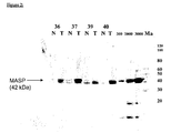
- Polyclonal antibody to the colorectal cancer marker protein MASP is generated for further use of the antibody in the measurement of serum and plasma and blood levels of MASP by immunodetection assays, e.g. Western Blotting and ELISA.
- recombinant expression of the protein is performed for obtaining immunogens.
- the expression is done applying a combination of the RTS 100 expression system and E.coli.
- the DNA sequence is analyzed and recommendations for high yield cDNA silent mutational variants and respective PCR-primer sequences are obtained using the "ProteoExpert RTS E.coli HY” system. This is a commercial web based service (www.proteoexpert.com).
- the "RTS 100 E. coli Linear Template Generation Set, His-tag” (Roche Diagnostics GmbH, Mannheim, Germany, Cat.No.
- His-MASP fusion protein Purification of His-MASP fusion protein is done following standard procedures on a Ni-chelate column. Briefly, 11 of bacteria culture containing the expression vector for the His-MASP fusion protein is pelleted by centrifugation. The cell pellet is resuspended in lysis buffer, containing phosphate, pH 8.0, 7 M guanidium chloride, imidazole and thioglycerole, followed by homogenization using a Ultra-Turrax® . Insoluble material is pelleted by high speed centrifugation and the supernatant is applied to a Ni-chelate chromatographic column. The column is washed with several bed volumes of lysis buffer followed by washes with buffer, containing phosphate, pH 8,0 and Urea. Finally, bound antigen is eluted using a phosphate buffer containing SDS under acid conditions.
- mice 12 week old A/J mice are initially immunized intraperitoneally with 100 ⁇ g MASP.. This is followed after 6 weeks by two further intraperitoneal immunizations at monthly intervals. In this process each mouse is administered 100 ⁇ g MASP adsorbed to aluminum hydroxide and 10 9 germs of Bordetella pertussis. Subsequently the last two immunizations are carried out intravenously on the 3rd and 2nd day before fusion using 100 ⁇ g MASP in PBS buffer for each.
- Spleen cells of the mice immunized according to a) are fused with myeloma cells according to Galfre, G., and Milstein, C., Methods in Enzymology 73 (1981) 3-46.
- ca. 1*10 8 spleen cells of the immunized mouse are mixed with 2x10 7 myeloma cells (P3X63-Ag8-653, ATCC CRL1580) and centrifuged (10 min at 300 g and 4° C.). The cells are then washed once with RPMI 1640 medium without fetal calf serum (FCS) and centrifuged again at 400 g in a 50 ml conical tube.
- FCS fetal calf serum
- the sedimented cells are taken up in RPMI 1640 medium containing 10% FCS and sown in hypoxanthine-azaserine selection medium (100 mmol/l hypoxanthine, 1 ⁇ g/ml azaserine in RPMI 1640+10% FCS).
- Interleukin 6 at 100 U/ml is added to the medium as a growth factor.
- MASP-positive primary cultures are cloned in 96-well cell culture plates by means of a fluorescence activated cell sorter. In this process again interleukin 6 at 100 U/ml is added to the medium as a growth additive.
- the hybridoma cells obtained are sown at a density of 1x10 5 cells per ml in RPMI 1640 medium containing 10% FCS and proliferated for 7 days in a fermenter (Thermodux Co., Wertheim/Main, Model MCS-104XL, Order No. 144-050). On average concentrations of 100 ⁇ g monoclonal antibody per ml are obtained in the culture supernatant. Purification of this antibody from the culture supernatant is carried out by conventional methods in protein chemistry (e.g. according to Bruck, C., et al., Methods in Enzymology 121 (1986) 587-695).
- a fresh emulsion of the protein solution (100 ⁇ g/ml protein MASP) and complete Freund's adjuvant at the ratio of 1:1 is prepared.
- Each rabbit is immunized with 1 ml of the emulsion at days 1, 7, 14 and 30, 60 and 90. Blood is drawn and resulting anti-MASP serum used for further experiments as described in examples 3 and 4.
- IgG immunoglobulin G
- rabbit serum is diluted with 4 volumes of acetate buffer (60 mM, pH 4.0). The pH is adjusted to 4.5 with 2 M Tris-base. Caprylic acid (25 ⁇ l/ml of diluted sample) is added drop-wise under vigorous stirring. After 30 min the sample is centrifuged (13,000 x g, 30 min, 4°C), the pellet discarded and the supernatant collected. The pH of the supernatant is adjusted to 7.5 by the addition of 2 M Tris-base and filtered (0.2 ⁇ m).
- the immunoglobulin in the supernatant is precipitated under vigorous stirring by the drop-wise addition of a 4 M ammonium sulfate solution to a final concentration of 2 M.
- the precipitated immunoglobulins are collected by centrifugation (8,000 x g, 15 min, 4°C).
- the supernatant is discarded.
- the pellet is dissolved in 10 mM NaH 2 PO 4 /NaOH, pH 7.5, 30 mM NaCl and exhaustively dialyzed.
- the dialysate is centrifuged (13,000 x g, 15 min, 4°C) and filtered (0.2 ⁇ m).
- Polyclonal rabbit IgG is brought to 10 mg/ml in 10 mM NaH 2 PO 4 /NaOH, pH 7.5, 30 mM NaCl. Per ml IgG solution 50 ⁇ l Biotin -N-hydroxysuccinimide (3.6 mg/ml in DMSO) are added. After 30 min at room temperature, the sample is chromatographed on Superdex 200 (10 mM NaH 2 PO 4 /NaOH, pH 7.5, 30 mM NaCl). The fraction containing biotinylated IgG are collected. Monoclonal antibodies are biotinylated according to the same procedure.
- Polyclonal rabbit IgG is brought to 10 mg/ml in 10 mM NaH 2 PO 4 /NaOH, 30 mM NaCl, pH 7.5.
- Per ml IgG solution 50 ⁇ l digoxigenin-3-O-methylcarbonyl- ⁇ -aminocaproic acid-N-hydroxysuccinimide ester (Roche Diagnostics, Mannheim, Germany, Cat. No. 1 333 054) (3.8 mg/ml in DMSO) are added. After 30 min at room temperature, the sample is chromatographed on Superdex® 200 (10 mM NaH 2 PO 4 /NaOH, pH 7.5, 30 mM NaCl). The fractions containing digoxigenylated IgG are collected. Monoclonal antibodies are labeled with digoxigenin according to the same procedure.
- Tissue lysates from tumor samples and healthy control samples are prepared as described in Example 1, "Tissue preparation”.
- SDS-PAGE and Western-Blotting are carried out using reagents and equipment of Invitrogen, Düsseldorf, Germany.
- 10 ⁇ g of tissue lysate are diluted in reducing NuPAGE® (Invitrogen) SDS sample buffer and heated for 10 min at 95°C.
- Samples are run on 4-12% NuPAGE® gels (Tris-Glycine) in the MES running buffer system.
- the gel-separated protein mixture is blotted onto nitrocellulose membranes using the Invitrogen XCell II TM Blot Module (Invitrogen) and the NuPAGE® transfer buffer system.
- the membranes are washed 3 times in PBS/0.05% Tween-20 and blocked with Roti® -Block blocking buffer (A151.1; Carl Roth GmbH, Düsseldorf, Germany) for 2 h.
- the primary antibody polyclonal rabbit anti-MASP serum (generation described in Example 2), is diluted 1:10,000 in Roti® -Block blocking buffer and incubated with the membrane for 1 h.
- the membranes are washed 6 times in PBS/0.05% Tween-20.
- the specifically bound primary rabbit antibody is labeled with an POD-conjugated polyclonal sheep anti-rabbit IgG antibody, diluted to 10 mU/ml in 0.5 x Roti® -Block blocking buffer.
- the membranes are washed 6 times in PBS/0.05% Tween-20.
- the membrane is incubated with the Lumi-Light PLUS Western Blotting Substrate (Order-No. 2015196, Roche Diagnostics GmbH, Mannheim, Germany) and exposed to an autoradiographic film.
- a sandwich ELISA For detection of MASP in human serum or plasma, a sandwich ELISA is developed. For capture and detection of the antigen, aliquots of the anti-MASP polyclonal antibody (see Example 2) are conjugated with biotin and digoxygenin, respectively.
- Streptavidin-coated 96-well microwell plates are incubated with 100 ⁇ l biotinylated anti-MASP polyclonal antibody for 60 min at 10 ⁇ g/ml in 10 mM phosphate, pH 7.4, 1% BSA, 0.9% NaCl and 0.1% Tween-20. After incubation, plates are washed three times with 0.9% NaCl , 0.1% Tween-20. Wells are then incubated for 2 h with either a serial dilution of the recombinant protein (see Example 2) as standard antigen or with diluted plasma samples from patients. After binding of MASP, plates are washed three times with 0.9% NaCl , 0.1% Tween-20.
- wells are incubated with 100 ⁇ l of digoxygenylated anti-MASP polyclonal antibody for 60 min at 10 ⁇ g/ml in 10 mM phosphate, pH 7.4, 1% BSA, 0.9% NaCl and 0.1% Tween-20. Thereafter, plates are washed three times to remove unbound antibody.
- wells are incubated with 20 mU/ml anti-digoxigenin-POD conjugates (Roche Diagnostics GmbH, Mannheim, Germany, Catalog No. 1633716) for 60 min in 10 mM phosphate, pH 7.4, 1% BSA, 0.9% NaCl and 0.1% Tween-20. Plates are subsequently washed three times with the same buffer.
- ABTS solution (Roche Diagnostics GmbH, Mannheim, Germany, Catalog No. 11685767) and OD is measured after 30-60 min at 405 nm with an ELISA reader.
- Accuracy is assessed by analyzing individual liquid samples obtained from well-characterized patient cohorts, i.e., 50 patients having undergone colonoscopy and found to be free of adenoma or CRC, 50 patients diagnosed and staged as T is -3, N0, M0 of CRC, and 50 patients diagnosed with progressed CRC, having at least tumor infiltration in at least one proximal lymph node or more severe forms of metastasis, respectively.
- CEA as measured by a commercially available assay (Roche Diagnostics, CEA-assay (Cat. No. 1 173 1629 for Elecsys® Systems immunoassay analyzer) and MASP measured as described above are quantified in a serum obtained from each of these individuals.
- ROC-analysis is performed according to Zweig, M. H., and Campbell, supra. Discriminatory power for differentiating patients in the group T is -3, N0, M0 from healthy individuals as measured by the area under the curve is found to be at least as good for MASP as compared to the established marker CEA.
Landscapes
- Health & Medical Sciences (AREA)
- Life Sciences & Earth Sciences (AREA)
- Engineering & Computer Science (AREA)
- Immunology (AREA)
- Urology & Nephrology (AREA)
- Hematology (AREA)
- Biomedical Technology (AREA)
- Chemical & Material Sciences (AREA)
- Molecular Biology (AREA)
- Medicinal Chemistry (AREA)
- Physics & Mathematics (AREA)
- Cell Biology (AREA)
- Hospice & Palliative Care (AREA)
- Biotechnology (AREA)
- Food Science & Technology (AREA)
- Oncology (AREA)
- Microbiology (AREA)
- Analytical Chemistry (AREA)
- Biochemistry (AREA)
- General Health & Medical Sciences (AREA)
- General Physics & Mathematics (AREA)
- Pathology (AREA)
- Peptides Or Proteins (AREA)
- Investigating Or Analysing Biological Materials (AREA)
- Measuring Or Testing Involving Enzymes Or Micro-Organisms (AREA)
Claims (10)
- Verfahren zur Diagnose kolorektaler Karzinome, bei dem man die folgenden Schritte durchführt:(a) Bereitstellen einer einem Individuum entnommenen Probe,(b) Inkontaktbringen der Probe mit einem spezifischen Bindungsmittel für den Maspin-Precursor (MASP) unter zur Ausbildung eines Komplexes zwischen dem Bindungsmittel und MASP geeigneten Bedingungen und(c) Korrelieren der in (b) gebildeten Menge an Komplex mit der Diagnose kolorektaler Karzinome.
- Verfahren nach Anspruch 1, ferner dadurch gekennzeichnet, daß es sich bei der Probe um Serum handelt.
- Verfahren nach Anspruch 1, ferner dadurch gekennzeichnet, daß es sich bei der Probe um Plasma handelt.
- Verfahren nach Anspruch 1, ferner dadurch gekennzeichnet, daß es sich bei der Probe um Vollblut handelt.
- Verwendung des Proteins MASP als ein Markermolekül bei der Diagnose kolorektaler Karzinome aus einer einem Individuum entnommenen flüssigen Probe.
- Verwendung des Proteins MASP als ein Markermolekül bei der Frühdiagnose kolorektaler Karzinome aus einer einem Individuum entnommenen flüssigen Probe.
- Verwendung nach Anspruch 6, wobei die Frühdiagnose mit aus CRC-Patienten im Adenomstadium stammender Probe durchgeführt wird.
- Verwendung nach Anspruch 6, wobei die Frühdiagnose mit aus CRC-Patienten im Stadium Tis-3; N0; M0 stammender Probe durchgeführt wird.
- Verwendung des Proteins MASP als ein Markermolekül für kolorektale Karzinome in Kombination mit einem weiteren Markermolekül für kolorektale Karzinome bei der Diagnose kolorektaler Karzinome aus einer einem Individuum entnommenen flüssigen Probe.
- Immunologischer Kit, umfassend wenigstens ein spezifisches Bindungsmittel für MASP sowie Hilfreagentien zur Messung von MASP.
Priority Applications (1)
| Application Number | Priority Date | Filing Date | Title |
|---|---|---|---|
| EP04734663A EP1631826B1 (de) | 2003-05-26 | 2004-05-25 | Verwendung des proteins masp als marker für kolorektale karzinome |
Applications Claiming Priority (3)
| Application Number | Priority Date | Filing Date | Title |
|---|---|---|---|
| EP03011158 | 2003-05-26 | ||
| PCT/EP2004/005598 WO2004104593A1 (en) | 2003-05-26 | 2004-05-25 | Use of protein masp as a marker for colorectal cancer |
| EP04734663A EP1631826B1 (de) | 2003-05-26 | 2004-05-25 | Verwendung des proteins masp als marker für kolorektale karzinome |
Publications (2)
| Publication Number | Publication Date |
|---|---|
| EP1631826A1 EP1631826A1 (de) | 2006-03-08 |
| EP1631826B1 true EP1631826B1 (de) | 2006-08-16 |
Family
ID=33462076
Family Applications (1)
| Application Number | Title | Priority Date | Filing Date |
|---|---|---|---|
| EP04734663A Expired - Lifetime EP1631826B1 (de) | 2003-05-26 | 2004-05-25 | Verwendung des proteins masp als marker für kolorektale karzinome |
Country Status (9)
| Country | Link |
|---|---|
| US (1) | US20060121540A1 (de) |
| EP (1) | EP1631826B1 (de) |
| JP (1) | JP4241822B2 (de) |
| CN (1) | CN100504393C (de) |
| AT (1) | ATE353544T1 (de) |
| CA (1) | CA2523690C (de) |
| DE (1) | DE602004001994T2 (de) |
| ES (1) | ES2271892T3 (de) |
| WO (1) | WO2004104593A1 (de) |
Families Citing this family (8)
| Publication number | Priority date | Publication date | Assignee | Title |
|---|---|---|---|---|
| CA2585788C (en) * | 2004-12-23 | 2013-03-05 | F. Hoffmann-La Roche Ag | Use of cyfra 21-1 as a marker for colorectal cancer |
| WO2006066917A2 (en) * | 2004-12-23 | 2006-06-29 | Roche Diagnostics Gmbh | Use of asc as a marker for colorectal cancer |
| EP1941764B1 (de) * | 2005-10-04 | 2016-04-20 | Telefonaktiebolaget LM Ericsson (publ) | Automatischer aufbau von nachbarlisten in einem mobilsystem |
| EP2680003A1 (de) * | 2012-06-28 | 2014-01-01 | Fundació Institut d'Investigació Biomèdica de Bellvitge | Serum-Biomarker zur Diagnose eines kolorektalen Karzinoms |
| ES2619116B1 (es) * | 2015-12-23 | 2018-04-12 | Fundación Para La Investigación Biomédica Del Hospital Universitario 12 De Octubre | Biomarcador para el diagnóstico, pronóstico y seguimiento de cáncer colorrectal de aparición precoz |
| CN107227366B (zh) * | 2017-07-05 | 2020-05-19 | 昆明医科大学第一附属医院 | 多功能转录调控因子ctcf的dna结合位点ctcf_113的应用 |
| CN108562746A (zh) * | 2018-04-08 | 2018-09-21 | 深圳市盛波尔生命科学技术有限责任公司 | Cnpy2异构体2在结直肠癌诊断、预后、复发转移及放化疗疗效预测中的应用 |
| GB201907663D0 (en) * | 2019-05-30 | 2019-07-17 | Ab Mavatar | Method for diagnosing colorectal cancer |
Family Cites Families (3)
| Publication number | Priority date | Publication date | Assignee | Title |
|---|---|---|---|---|
| US5470970A (en) * | 1991-02-28 | 1995-11-28 | Dana-Farber Cancer Institute, Inc. | Maspin, a serpin with tumor suppresing activity |
| US20020182191A1 (en) * | 1998-12-23 | 2002-12-05 | Corixa Corporation | Compounds for immunotherapy and diagnosis of colon cancer and methods for their use |
| AUPP713498A0 (en) * | 1998-11-17 | 1998-12-10 | Chandler, Howard Milne | A method of detecting blood |
-
2004
- 2004-05-25 EP EP04734663A patent/EP1631826B1/de not_active Expired - Lifetime
- 2004-05-25 CN CNB2004800143194A patent/CN100504393C/zh not_active Expired - Fee Related
- 2004-05-25 CA CA2523690A patent/CA2523690C/en not_active Expired - Fee Related
- 2004-05-25 ES ES04734663T patent/ES2271892T3/es not_active Expired - Lifetime
- 2004-05-25 AT AT04734663T patent/ATE353544T1/de active
- 2004-05-25 DE DE602004001994T patent/DE602004001994T2/de not_active Expired - Lifetime
- 2004-05-25 WO PCT/EP2004/005598 patent/WO2004104593A1/en not_active Ceased
- 2004-05-25 JP JP2006500105A patent/JP4241822B2/ja not_active Expired - Fee Related
-
2005
- 2005-11-23 US US11/287,575 patent/US20060121540A1/en not_active Abandoned
Also Published As
| Publication number | Publication date |
|---|---|
| CN1795385A (zh) | 2006-06-28 |
| DE602004001994D1 (de) | 2006-09-28 |
| US20060121540A1 (en) | 2006-06-08 |
| DE602004001994T2 (de) | 2007-09-13 |
| JP2006524794A (ja) | 2006-11-02 |
| CA2523690A1 (en) | 2004-12-02 |
| ATE353544T1 (de) | 2006-09-15 |
| CN100504393C (zh) | 2009-06-24 |
| JP4241822B2 (ja) | 2009-03-18 |
| WO2004104593A1 (en) | 2004-12-02 |
| CA2523690C (en) | 2011-05-03 |
| EP1631826A1 (de) | 2006-03-08 |
| ES2271892T3 (es) | 2007-04-16 |
Similar Documents
| Publication | Publication Date | Title |
|---|---|---|
| EP1579220B1 (de) | Verwendung von nicotinamide n-methyltransferase zur diagnose von kolorektalem krebs | |
| EP1649289B1 (de) | Verwendung der proteine proteinase 3 (prn3) und leukozyten elastatse inhibitor (ileu) als marker für kolorektale karzinome | |
| EP1631826B1 (de) | Verwendung des proteins masp als marker für kolorektale karzinome | |
| US20060188949A1 (en) | Use of protein PLST as a marker for colorectal cancer | |
| WO2005015218A1 (en) | Use of proteins proteinase 3 (prn3) and leukocyte elastase inhibitor (ileu) as a marker for colorectal cancer | |
| WO2004071267A2 (en) | Diagnosis of colorectal cancer by detection of nicotinamide n-methyltransferase in a stool sample | |
| EP1654542B1 (de) | Verwendung des proteins spermidinsynthase (spee) als marker für kolorektalkarzinom | |
| EP1654538B1 (de) | Verwendung des proteins proteasome aktivator untereinheit 3 als marker für kolorektale karzinome | |
| WO2005095978A1 (en) | Pyrroline-5-carboxylate reductase as a marker for colorectal concer | |
| US20060194266A1 (en) | Use of protein RLA-0 as a marker for colorectal cancer | |
| WO2005015221A1 (en) | Use of protein sahh as a marker for colorectal cancer | |
| US20070218510A1 (en) | Use of protein PSA3 as a marker for colorectal cancer | |
| WO2005015223A1 (en) | Use of protein acidic ribosomal protein p0 (rla-0) as a marker for colorectal cancer | |
| WO2005015233A1 (en) | Use of protein spermidine synthase (spee) as a marker for colorectal cancer | |
| WO2005015234A1 (en) | Use of protein sahh as a marker for colorectal cancer | |
| WO2004104592A1 (en) | Use of protein masp as a marker for colorectal cancer | |
| WO2005015232A1 (en) | Use of protein proteasmose activator subunit 3 (pse3) as a marker for colorectal cancer | |
| WO2005015227A1 (en) | Use of protein t- plastin (plst) as a marker for colorectal cancer | |
| WO2005015222A1 (en) | Use of the far upstream element (fuse) binding protein (fubp) as a marker for colorectal cancer | |
| WO2005015225A1 (en) | Use of the far upstream element (fuse) binding protein (fubp) as a marker for colorectal cancer | |
| WO2005015230A1 (en) | Use of protein psa3 as a marker for colorectal cancer | |
| WO2005095979A1 (en) | Use of protein proc as a marker for colorectal cancer |
Legal Events
| Date | Code | Title | Description |
|---|---|---|---|
| PUAI | Public reference made under article 153(3) epc to a published international application that has entered the european phase |
Free format text: ORIGINAL CODE: 0009012 |
|
| 17P | Request for examination filed |
Effective date: 20051227 |
|
| AK | Designated contracting states |
Kind code of ref document: A1 Designated state(s): AT BE BG CH CY CZ DE DK EE ES FI FR GB GR HU IE IT LI LU MC NL PL PT RO SE SI SK TR |
|
| GRAP | Despatch of communication of intention to grant a patent |
Free format text: ORIGINAL CODE: EPIDOSNIGR1 |
|
| GRAS | Grant fee paid |
Free format text: ORIGINAL CODE: EPIDOSNIGR3 |
|
| GRAA | (expected) grant |
Free format text: ORIGINAL CODE: 0009210 |
|
| RAP1 | Party data changed (applicant data changed or rights of an application transferred) |
Owner name: ROCHE DIAGNOSTICS GMBH Owner name: F. HOFFMANN-LA ROCHE AG |
|
| DAX | Request for extension of the european patent (deleted) | ||
| RIN1 | Information on inventor provided before grant (corrected) |
Inventor name: TACKE, MICHAEL Inventor name: ROESSLER, MARKUS Inventor name: PALME, STEFAN Inventor name: ZOLG, WERNER Inventor name: BERNDT, PETER Inventor name: ROLLINGER, WOLFGANG Inventor name: LANGEN, HANNO Inventor name: KARL, JOHANN Inventor name: HAGMANN, MARIE-LUISE |
|
| AK | Designated contracting states |
Kind code of ref document: B1 Designated state(s): AT BE BG CH CY CZ DE DK EE ES FI FR GB GR HU IE IT LI LU MC NL PL PT RO SE SI SK TR |
|
| PG25 | Lapsed in a contracting state [announced via postgrant information from national office to epo] |
Ref country code: IT Free format text: LAPSE BECAUSE OF FAILURE TO SUBMIT A TRANSLATION OF THE DESCRIPTION OR TO PAY THE FEE WITHIN THE PRESCRIBED TIME-LIMIT;WARNING: LAPSES OF ITALIAN PATENTS WITH EFFECTIVE DATE BEFORE 2007 MAY HAVE OCCURRED AT ANY TIME BEFORE 2007. THE CORRECT EFFECTIVE DATE MAY BE DIFFERENT FROM THE ONE RECORDED. Effective date: 20060816 Ref country code: FI Free format text: LAPSE BECAUSE OF FAILURE TO SUBMIT A TRANSLATION OF THE DESCRIPTION OR TO PAY THE FEE WITHIN THE PRESCRIBED TIME-LIMIT Effective date: 20060816 Ref country code: BE Free format text: LAPSE BECAUSE OF FAILURE TO SUBMIT A TRANSLATION OF THE DESCRIPTION OR TO PAY THE FEE WITHIN THE PRESCRIBED TIME-LIMIT Effective date: 20060816 Ref country code: SI Free format text: LAPSE BECAUSE OF FAILURE TO SUBMIT A TRANSLATION OF THE DESCRIPTION OR TO PAY THE FEE WITHIN THE PRESCRIBED TIME-LIMIT Effective date: 20060816 Ref country code: SK Free format text: LAPSE BECAUSE OF FAILURE TO SUBMIT A TRANSLATION OF THE DESCRIPTION OR TO PAY THE FEE WITHIN THE PRESCRIBED TIME-LIMIT Effective date: 20060816 Ref country code: CZ Free format text: LAPSE BECAUSE OF FAILURE TO SUBMIT A TRANSLATION OF THE DESCRIPTION OR TO PAY THE FEE WITHIN THE PRESCRIBED TIME-LIMIT Effective date: 20060816 Ref country code: RO Free format text: LAPSE BECAUSE OF FAILURE TO SUBMIT A TRANSLATION OF THE DESCRIPTION OR TO PAY THE FEE WITHIN THE PRESCRIBED TIME-LIMIT Effective date: 20060816 Ref country code: NL Free format text: LAPSE BECAUSE OF FAILURE TO SUBMIT A TRANSLATION OF THE DESCRIPTION OR TO PAY THE FEE WITHIN THE PRESCRIBED TIME-LIMIT Effective date: 20060816 Ref country code: PL Free format text: LAPSE BECAUSE OF FAILURE TO SUBMIT A TRANSLATION OF THE DESCRIPTION OR TO PAY THE FEE WITHIN THE PRESCRIBED TIME-LIMIT Effective date: 20060816 |
|
| REG | Reference to a national code |
Ref country code: GB Ref legal event code: FG4D |
|
| REG | Reference to a national code |
Ref country code: CH Ref legal event code: EP |
|
| REG | Reference to a national code |
Ref country code: IE Ref legal event code: FG4D |
|
| REF | Corresponds to: |
Ref document number: 602004001994 Country of ref document: DE Date of ref document: 20060928 Kind code of ref document: P |
|
| PG25 | Lapsed in a contracting state [announced via postgrant information from national office to epo] |
Ref country code: BG Free format text: LAPSE BECAUSE OF FAILURE TO SUBMIT A TRANSLATION OF THE DESCRIPTION OR TO PAY THE FEE WITHIN THE PRESCRIBED TIME-LIMIT Effective date: 20061116 Ref country code: DK Free format text: LAPSE BECAUSE OF FAILURE TO SUBMIT A TRANSLATION OF THE DESCRIPTION OR TO PAY THE FEE WITHIN THE PRESCRIBED TIME-LIMIT Effective date: 20061116 Ref country code: SE Free format text: LAPSE BECAUSE OF FAILURE TO SUBMIT A TRANSLATION OF THE DESCRIPTION OR TO PAY THE FEE WITHIN THE PRESCRIBED TIME-LIMIT Effective date: 20061116 |
|
| PG25 | Lapsed in a contracting state [announced via postgrant information from national office to epo] |
Ref country code: PT Free format text: LAPSE BECAUSE OF FAILURE TO SUBMIT A TRANSLATION OF THE DESCRIPTION OR TO PAY THE FEE WITHIN THE PRESCRIBED TIME-LIMIT Effective date: 20070116 |
|
| NLV1 | Nl: lapsed or annulled due to failure to fulfill the requirements of art. 29p and 29m of the patents act | ||
| ET | Fr: translation filed | ||
| REG | Reference to a national code |
Ref country code: ES Ref legal event code: FG2A Ref document number: 2271892 Country of ref document: ES Kind code of ref document: T3 |
|
| PLBE | No opposition filed within time limit |
Free format text: ORIGINAL CODE: 0009261 |
|
| STAA | Information on the status of an ep patent application or granted ep patent |
Free format text: STATUS: NO OPPOSITION FILED WITHIN TIME LIMIT |
|
| 26N | No opposition filed |
Effective date: 20070518 |
|
| PG25 | Lapsed in a contracting state [announced via postgrant information from national office to epo] |
Ref country code: MC Free format text: LAPSE BECAUSE OF NON-PAYMENT OF DUE FEES Effective date: 20070531 |
|
| PG25 | Lapsed in a contracting state [announced via postgrant information from national office to epo] |
Ref country code: GR Free format text: LAPSE BECAUSE OF FAILURE TO SUBMIT A TRANSLATION OF THE DESCRIPTION OR TO PAY THE FEE WITHIN THE PRESCRIBED TIME-LIMIT Effective date: 20061117 |
|
| PG25 | Lapsed in a contracting state [announced via postgrant information from national office to epo] |
Ref country code: IE Free format text: LAPSE BECAUSE OF NON-PAYMENT OF DUE FEES Effective date: 20070525 |
|
| PG25 | Lapsed in a contracting state [announced via postgrant information from national office to epo] |
Ref country code: EE Free format text: LAPSE BECAUSE OF FAILURE TO SUBMIT A TRANSLATION OF THE DESCRIPTION OR TO PAY THE FEE WITHIN THE PRESCRIBED TIME-LIMIT Effective date: 20060816 |
|
| PGRI | Patent reinstated in contracting state [announced from national office to epo] |
Ref country code: IT Effective date: 20080601 |
|
| PG25 | Lapsed in a contracting state [announced via postgrant information from national office to epo] |
Ref country code: LU Free format text: LAPSE BECAUSE OF NON-PAYMENT OF DUE FEES Effective date: 20070525 Ref country code: CY Free format text: LAPSE BECAUSE OF FAILURE TO SUBMIT A TRANSLATION OF THE DESCRIPTION OR TO PAY THE FEE WITHIN THE PRESCRIBED TIME-LIMIT Effective date: 20060816 |
|
| PG25 | Lapsed in a contracting state [announced via postgrant information from national office to epo] |
Ref country code: HU Free format text: LAPSE BECAUSE OF FAILURE TO SUBMIT A TRANSLATION OF THE DESCRIPTION OR TO PAY THE FEE WITHIN THE PRESCRIBED TIME-LIMIT Effective date: 20070217 Ref country code: TR Free format text: LAPSE BECAUSE OF FAILURE TO SUBMIT A TRANSLATION OF THE DESCRIPTION OR TO PAY THE FEE WITHIN THE PRESCRIBED TIME-LIMIT Effective date: 20060816 |
|
| REG | Reference to a national code |
Ref country code: FR Ref legal event code: PLFP Year of fee payment: 13 |
|
| REG | Reference to a national code |
Ref country code: FR Ref legal event code: PLFP Year of fee payment: 14 |
|
| REG | Reference to a national code |
Ref country code: FR Ref legal event code: PLFP Year of fee payment: 15 |
|
| PGFP | Annual fee paid to national office [announced via postgrant information from national office to epo] |
Ref country code: GB Payment date: 20180328 Year of fee payment: 15 |
|
| PGFP | Annual fee paid to national office [announced via postgrant information from national office to epo] |
Ref country code: DE Payment date: 20180507 Year of fee payment: 15 Ref country code: ES Payment date: 20180604 Year of fee payment: 15 Ref country code: CH Payment date: 20180425 Year of fee payment: 15 |
|
| PGFP | Annual fee paid to national office [announced via postgrant information from national office to epo] |
Ref country code: IT Payment date: 20180522 Year of fee payment: 15 Ref country code: FR Payment date: 20180416 Year of fee payment: 15 Ref country code: AT Payment date: 20180425 Year of fee payment: 15 |
|
| REG | Reference to a national code |
Ref country code: DE Ref legal event code: R119 Ref document number: 602004001994 Country of ref document: DE |
|
| REG | Reference to a national code |
Ref country code: CH Ref legal event code: PL |
|
| REG | Reference to a national code |
Ref country code: AT Ref legal event code: MM01 Ref document number: 353544 Country of ref document: AT Kind code of ref document: T Effective date: 20190525 |
|
| GBPC | Gb: european patent ceased through non-payment of renewal fee |
Effective date: 20190525 |
|
| PG25 | Lapsed in a contracting state [announced via postgrant information from national office to epo] |
Ref country code: LI Free format text: LAPSE BECAUSE OF NON-PAYMENT OF DUE FEES Effective date: 20190531 Ref country code: CH Free format text: LAPSE BECAUSE OF NON-PAYMENT OF DUE FEES Effective date: 20190531 Ref country code: AT Free format text: LAPSE BECAUSE OF NON-PAYMENT OF DUE FEES Effective date: 20190525 |
|
| PG25 | Lapsed in a contracting state [announced via postgrant information from national office to epo] |
Ref country code: DE Free format text: LAPSE BECAUSE OF NON-PAYMENT OF DUE FEES Effective date: 20191203 Ref country code: IT Free format text: LAPSE BECAUSE OF FAILURE TO SUBMIT A TRANSLATION OF THE DESCRIPTION OR TO PAY THE FEE WITHIN THE PRESCRIBED TIME-LIMIT Effective date: 20190525 Ref country code: GB Free format text: LAPSE BECAUSE OF NON-PAYMENT OF DUE FEES Effective date: 20190525 |
|
| PG25 | Lapsed in a contracting state [announced via postgrant information from national office to epo] |
Ref country code: FR Free format text: LAPSE BECAUSE OF NON-PAYMENT OF DUE FEES Effective date: 20190531 |
|
| REG | Reference to a national code |
Ref country code: ES Ref legal event code: FD2A Effective date: 20200930 |
|
| PG25 | Lapsed in a contracting state [announced via postgrant information from national office to epo] |
Ref country code: ES Free format text: LAPSE BECAUSE OF NON-PAYMENT OF DUE FEES Effective date: 20190526 |