EP1631314B1 - Verfahren zur änderung der bindungsspezifität von plasmaproteinen durch oxidations-reduktionsreaktionen - Google Patents
Verfahren zur änderung der bindungsspezifität von plasmaproteinen durch oxidations-reduktionsreaktionen Download PDFInfo
- Publication number
- EP1631314B1 EP1631314B1 EP20040754487 EP04754487A EP1631314B1 EP 1631314 B1 EP1631314 B1 EP 1631314B1 EP 20040754487 EP20040754487 EP 20040754487 EP 04754487 A EP04754487 A EP 04754487A EP 1631314 B1 EP1631314 B1 EP 1631314B1
- Authority
- EP
- European Patent Office
- Prior art keywords
- autoantibodies
- serum
- plasma
- biological fluid
- blood
- Prior art date
- Legal status (The legal status is an assumption and is not a legal conclusion. Google has not performed a legal analysis and makes no representation as to the accuracy of the status listed.)
- Expired - Lifetime
Links
- 230000027455 binding Effects 0.000 title claims abstract description 88
- 238000000034 method Methods 0.000 title claims description 89
- 102000004506 Blood Proteins Human genes 0.000 title abstract description 33
- 108010017384 Blood Proteins Proteins 0.000 title abstract description 33
- 238000006479 redox reaction Methods 0.000 title description 6
- 210000004369 blood Anatomy 0.000 claims abstract description 112
- 239000008280 blood Substances 0.000 claims abstract description 109
- 239000007800 oxidant agent Substances 0.000 claims abstract description 33
- 239000007788 liquid Substances 0.000 claims abstract description 6
- 210000002966 serum Anatomy 0.000 claims description 84
- 229940025294 hemin Drugs 0.000 claims description 61
- BTIJJDXEELBZFS-QDUVMHSLSA-K hemin Chemical group CC1=C(CCC(O)=O)C(C=C2C(CCC(O)=O)=C(C)\C(N2[Fe](Cl)N23)=C\4)=N\C1=C/C2=C(C)C(C=C)=C3\C=C/1C(C)=C(C=C)C/4=N\1 BTIJJDXEELBZFS-QDUVMHSLSA-K 0.000 claims description 61
- 210000002381 plasma Anatomy 0.000 claims description 51
- 239000013060 biological fluid Substances 0.000 claims description 43
- 230000000694 effects Effects 0.000 claims description 33
- 239000000203 mixture Substances 0.000 claims description 32
- 239000000284 extract Substances 0.000 claims description 27
- 238000003556 assay Methods 0.000 claims description 26
- 239000000427 antigen Substances 0.000 claims description 25
- 102000036639 antigens Human genes 0.000 claims description 23
- 108091007433 antigens Proteins 0.000 claims description 23
- 210000004700 fetal blood Anatomy 0.000 claims description 15
- 239000003446 ligand Substances 0.000 claims description 12
- 108060003951 Immunoglobulin Proteins 0.000 claims description 9
- 230000004075 alteration Effects 0.000 claims description 9
- 102000018358 immunoglobulin Human genes 0.000 claims description 9
- 238000012216 screening Methods 0.000 claims description 6
- 230000003169 placental effect Effects 0.000 claims description 5
- 238000001990 intravenous administration Methods 0.000 claims description 4
- 238000000926 separation method Methods 0.000 claims description 3
- 108090000623 proteins and genes Proteins 0.000 abstract description 52
- 102000004169 proteins and genes Human genes 0.000 abstract description 51
- 239000010836 blood and blood product Substances 0.000 abstract description 6
- 229940125691 blood product Drugs 0.000 abstract description 6
- 230000008859 change Effects 0.000 abstract description 5
- 230000001590 oxidative effect Effects 0.000 abstract description 3
- 239000003918 blood extract Substances 0.000 abstract description 2
- 239000000523 sample Substances 0.000 description 72
- 235000018102 proteins Nutrition 0.000 description 48
- 238000002474 experimental method Methods 0.000 description 44
- 210000003743 erythrocyte Anatomy 0.000 description 35
- HEMHJVSKTPXQMS-UHFFFAOYSA-M Sodium hydroxide Chemical compound [OH-].[Na+] HEMHJVSKTPXQMS-UHFFFAOYSA-M 0.000 description 27
- 238000011534 incubation Methods 0.000 description 27
- 239000000243 solution Substances 0.000 description 27
- 238000012360 testing method Methods 0.000 description 22
- IAZDPXIOMUYVGZ-UHFFFAOYSA-N Dimethylsulphoxide Chemical compound CS(C)=O IAZDPXIOMUYVGZ-UHFFFAOYSA-N 0.000 description 21
- 238000002360 preparation method Methods 0.000 description 20
- LOKCTEFSRHRXRJ-UHFFFAOYSA-I dipotassium trisodium dihydrogen phosphate hydrogen phosphate dichloride Chemical compound P(=O)(O)(O)[O-].[K+].P(=O)(O)([O-])[O-].[Na+].[Na+].[Cl-].[K+].[Cl-].[Na+] LOKCTEFSRHRXRJ-UHFFFAOYSA-I 0.000 description 19
- 239000002953 phosphate buffered saline Substances 0.000 description 19
- 238000002965 ELISA Methods 0.000 description 17
- CIWBSHSKHKDKBQ-JLAZNSOCSA-N Ascorbic acid Chemical compound OC[C@H](O)[C@H]1OC(=O)C(O)=C1O CIWBSHSKHKDKBQ-JLAZNSOCSA-N 0.000 description 16
- 239000001963 growth medium Substances 0.000 description 16
- XEEYBQQBJWHFJM-UHFFFAOYSA-N Iron Chemical compound [Fe] XEEYBQQBJWHFJM-UHFFFAOYSA-N 0.000 description 15
- 238000007630 basic procedure Methods 0.000 description 15
- 208000023275 Autoimmune disease Diseases 0.000 description 14
- XEYBHCRIKKKOSS-UHFFFAOYSA-N disodium;azanylidyneoxidanium;iron(2+);pentacyanide Chemical compound [Na+].[Na+].[Fe+2].N#[C-].N#[C-].N#[C-].N#[C-].N#[C-].[O+]#N XEYBHCRIKKKOSS-UHFFFAOYSA-N 0.000 description 14
- 210000001616 monocyte Anatomy 0.000 description 14
- 229940083618 sodium nitroprusside Drugs 0.000 description 14
- 210000004027 cell Anatomy 0.000 description 13
- 201000010099 disease Diseases 0.000 description 12
- 208000037265 diseases, disorders, signs and symptoms Diseases 0.000 description 12
- 230000008569 process Effects 0.000 description 12
- 230000001580 bacterial effect Effects 0.000 description 11
- 230000001419 dependent effect Effects 0.000 description 11
- OKTJSMMVPCPJKN-UHFFFAOYSA-N Carbon Chemical compound [C] OKTJSMMVPCPJKN-UHFFFAOYSA-N 0.000 description 10
- 239000003963 antioxidant agent Substances 0.000 description 10
- 235000006708 antioxidants Nutrition 0.000 description 10
- 239000003610 charcoal Substances 0.000 description 10
- 239000010439 graphite Substances 0.000 description 10
- 229910002804 graphite Inorganic materials 0.000 description 10
- MWUXSHHQAYIFBG-UHFFFAOYSA-N nitrogen oxide Inorganic materials O=[N] MWUXSHHQAYIFBG-UHFFFAOYSA-N 0.000 description 10
- 238000007254 oxidation reaction Methods 0.000 description 10
- 150000003904 phospholipids Chemical class 0.000 description 10
- 230000003647 oxidation Effects 0.000 description 9
- 230000009257 reactivity Effects 0.000 description 9
- UJKPHYRXOLRVJJ-MLSVHJFASA-N CC(O)C1=C(C)/C2=C/C3=N/C(=C\C4=C(CCC(O)=O)C(C)=C(N4)/C=C4\N=C(\C=C\1/N\2)C(C)=C4C(C)O)/C(CCC(O)=O)=C3C Chemical compound CC(O)C1=C(C)/C2=C/C3=N/C(=C\C4=C(CCC(O)=O)C(C)=C(N4)/C=C4\N=C(\C=C\1/N\2)C(C)=C4C(C)O)/C(CCC(O)=O)=C3C UJKPHYRXOLRVJJ-MLSVHJFASA-N 0.000 description 8
- 101710148556 Methylcarbamoylase mom Proteins 0.000 description 8
- 229910052742 iron Inorganic materials 0.000 description 8
- ZZZCUOFIHGPKAK-UHFFFAOYSA-N D-erythro-ascorbic acid Natural products OCC1OC(=O)C(O)=C1O ZZZCUOFIHGPKAK-UHFFFAOYSA-N 0.000 description 7
- MJVAVZPDRWSRRC-UHFFFAOYSA-N Menadione Chemical compound C1=CC=C2C(=O)C(C)=CC(=O)C2=C1 MJVAVZPDRWSRRC-UHFFFAOYSA-N 0.000 description 7
- 229930003268 Vitamin C Natural products 0.000 description 7
- 238000010790 dilution Methods 0.000 description 7
- 239000012895 dilution Substances 0.000 description 7
- 206010025135 lupus erythematosus Diseases 0.000 description 7
- 235000019154 vitamin C Nutrition 0.000 description 7
- 239000011718 vitamin C Substances 0.000 description 7
- 108010077861 Kininogens Proteins 0.000 description 6
- 102000010631 Kininogens Human genes 0.000 description 6
- 230000003078 antioxidant effect Effects 0.000 description 6
- 235000010633 broth Nutrition 0.000 description 6
- 239000013641 positive control Substances 0.000 description 6
- 239000012286 potassium permanganate Substances 0.000 description 6
- 239000007787 solid Substances 0.000 description 6
- 238000011282 treatment Methods 0.000 description 6
- 108010054147 Hemoglobins Proteins 0.000 description 5
- 102000001554 Hemoglobins Human genes 0.000 description 5
- MHAJPDPJQMAIIY-UHFFFAOYSA-N Hydrogen peroxide Chemical compound OO MHAJPDPJQMAIIY-UHFFFAOYSA-N 0.000 description 5
- 241001465754 Metazoa Species 0.000 description 5
- 239000007983 Tris buffer Substances 0.000 description 5
- 238000006243 chemical reaction Methods 0.000 description 5
- 150000001875 compounds Chemical class 0.000 description 5
- 239000004615 ingredient Substances 0.000 description 5
- LENZDBCJOHFCAS-UHFFFAOYSA-N tris Chemical compound OCC(N)(CO)CO LENZDBCJOHFCAS-UHFFFAOYSA-N 0.000 description 5
- 241000283690 Bos taurus Species 0.000 description 4
- 229910021578 Iron(III) chloride Inorganic materials 0.000 description 4
- 108010094028 Prothrombin Proteins 0.000 description 4
- 102100027378 Prothrombin Human genes 0.000 description 4
- 239000006146 Roswell Park Memorial Institute medium Substances 0.000 description 4
- 239000011324 bead Substances 0.000 description 4
- 239000006143 cell culture medium Substances 0.000 description 4
- 239000003085 diluting agent Substances 0.000 description 4
- 238000000684 flow cytometry Methods 0.000 description 4
- 239000003112 inhibitor Substances 0.000 description 4
- RBTARNINKXHZNM-UHFFFAOYSA-K iron trichloride Chemical compound Cl[Fe](Cl)Cl RBTARNINKXHZNM-UHFFFAOYSA-K 0.000 description 4
- -1 lipid peroxides Chemical class 0.000 description 4
- 239000006166 lysate Substances 0.000 description 4
- 239000002609 medium Substances 0.000 description 4
- 230000003287 optical effect Effects 0.000 description 4
- 229920003023 plastic Polymers 0.000 description 4
- 239000004033 plastic Substances 0.000 description 4
- 229940039716 prothrombin Drugs 0.000 description 4
- 239000000126 substance Substances 0.000 description 4
- VTLYFUHAOXGGBS-UHFFFAOYSA-N Fe3+ Chemical compound [Fe+3] VTLYFUHAOXGGBS-UHFFFAOYSA-N 0.000 description 3
- 241000711549 Hepacivirus C Species 0.000 description 3
- 239000003146 anticoagulant agent Substances 0.000 description 3
- 229940127219 anticoagulant drug Drugs 0.000 description 3
- 230000015572 biosynthetic process Effects 0.000 description 3
- 239000000872 buffer Substances 0.000 description 3
- 230000015556 catabolic process Effects 0.000 description 3
- 238000001514 detection method Methods 0.000 description 3
- 210000003754 fetus Anatomy 0.000 description 3
- 238000010438 heat treatment Methods 0.000 description 3
- 210000005260 human cell Anatomy 0.000 description 3
- 210000000987 immune system Anatomy 0.000 description 3
- 230000002401 inhibitory effect Effects 0.000 description 3
- 230000003993 interaction Effects 0.000 description 3
- 210000004698 lymphocyte Anatomy 0.000 description 3
- 230000008774 maternal effect Effects 0.000 description 3
- 229910052751 metal Inorganic materials 0.000 description 3
- 239000002184 metal Substances 0.000 description 3
- 239000000693 micelle Substances 0.000 description 3
- 210000000440 neutrophil Anatomy 0.000 description 3
- 230000033116 oxidation-reduction process Effects 0.000 description 3
- 230000009870 specific binding Effects 0.000 description 3
- 235000012711 vitamin K3 Nutrition 0.000 description 3
- 239000011652 vitamin K3 Substances 0.000 description 3
- 108010032595 Antibody Binding Sites Proteins 0.000 description 2
- IJGRMHOSHXDMSA-UHFFFAOYSA-N Atomic nitrogen Chemical compound N#N IJGRMHOSHXDMSA-UHFFFAOYSA-N 0.000 description 2
- 241000894006 Bacteria Species 0.000 description 2
- 102000015081 Blood Coagulation Factors Human genes 0.000 description 2
- 108010039209 Blood Coagulation Factors Proteins 0.000 description 2
- 206010053567 Coagulopathies Diseases 0.000 description 2
- RYGMFSIKBFXOCR-UHFFFAOYSA-N Copper Chemical compound [Cu] RYGMFSIKBFXOCR-UHFFFAOYSA-N 0.000 description 2
- 238000012286 ELISA Assay Methods 0.000 description 2
- 229910001335 Galvanized steel Inorganic materials 0.000 description 2
- 102000003886 Glycoproteins Human genes 0.000 description 2
- 108090000288 Glycoproteins Proteins 0.000 description 2
- 102000015779 HDL Lipoproteins Human genes 0.000 description 2
- 108010010234 HDL Lipoproteins Proteins 0.000 description 2
- 102000013271 Hemopexin Human genes 0.000 description 2
- 108010026027 Hemopexin Proteins 0.000 description 2
- 108010000487 High-Molecular-Weight Kininogen Proteins 0.000 description 2
- 102000007330 LDL Lipoproteins Human genes 0.000 description 2
- 108010007622 LDL Lipoproteins Proteins 0.000 description 2
- 238000012773 Laboratory assay Methods 0.000 description 2
- 108010061951 Methemoglobin Proteins 0.000 description 2
- 206010039710 Scleroderma Diseases 0.000 description 2
- 229930003779 Vitamin B12 Natural products 0.000 description 2
- 230000009471 action Effects 0.000 description 2
- 230000002529 anti-mitochondrial effect Effects 0.000 description 2
- 230000001363 autoimmune Effects 0.000 description 2
- 210000003719 b-lymphocyte Anatomy 0.000 description 2
- 239000003114 blood coagulation factor Substances 0.000 description 2
- 239000005018 casein Substances 0.000 description 2
- 239000013592 cell lysate Substances 0.000 description 2
- 238000005119 centrifugation Methods 0.000 description 2
- 230000035602 clotting Effects 0.000 description 2
- AGVAZMGAQJOSFJ-WZHZPDAFSA-M cobalt(2+);[(2r,3s,4r,5s)-5-(5,6-dimethylbenzimidazol-1-yl)-4-hydroxy-2-(hydroxymethyl)oxolan-3-yl] [(2r)-1-[3-[(1r,2r,3r,4z,7s,9z,12s,13s,14z,17s,18s,19r)-2,13,18-tris(2-amino-2-oxoethyl)-7,12,17-tris(3-amino-3-oxopropyl)-3,5,8,8,13,15,18,19-octamethyl-2 Chemical compound [Co+2].N#[C-].[N-]([C@@H]1[C@H](CC(N)=O)[C@@]2(C)CCC(=O)NC[C@@H](C)OP(O)(=O)O[C@H]3[C@H]([C@H](O[C@@H]3CO)N3C4=CC(C)=C(C)C=C4N=C3)O)\C2=C(C)/C([C@H](C\2(C)C)CCC(N)=O)=N/C/2=C\C([C@H]([C@@]/2(CC(N)=O)C)CCC(N)=O)=N\C\2=C(C)/C2=N[C@]1(C)[C@@](C)(CC(N)=O)[C@@H]2CCC(N)=O AGVAZMGAQJOSFJ-WZHZPDAFSA-M 0.000 description 2
- 230000000052 comparative effect Effects 0.000 description 2
- 239000002131 composite material Substances 0.000 description 2
- 229910052802 copper Inorganic materials 0.000 description 2
- 239000010949 copper Substances 0.000 description 2
- 230000001086 cytosolic effect Effects 0.000 description 2
- 238000000799 fluorescence microscopy Methods 0.000 description 2
- 239000008397 galvanized steel Substances 0.000 description 2
- 150000003278 haem Chemical class 0.000 description 2
- 238000013383 initial experiment Methods 0.000 description 2
- 210000000265 leukocyte Anatomy 0.000 description 2
- 150000002632 lipids Chemical class 0.000 description 2
- 229920002521 macromolecule Polymers 0.000 description 2
- 239000001755 magnesium gluconate Substances 0.000 description 2
- 229960003035 magnesium gluconate Drugs 0.000 description 2
- 235000015778 magnesium gluconate Nutrition 0.000 description 2
- IAKLPCRFBAZVRW-XRDLMGPZSA-L magnesium;(2r,3s,4r,5r)-2,3,4,5,6-pentahydroxyhexanoate;hydrate Chemical compound O.[Mg+2].OC[C@@H](O)[C@@H](O)[C@H](O)[C@@H](O)C([O-])=O.OC[C@@H](O)[C@@H](O)[C@H](O)[C@@H](O)C([O-])=O IAKLPCRFBAZVRW-XRDLMGPZSA-L 0.000 description 2
- 230000014759 maintenance of location Effects 0.000 description 2
- 238000004519 manufacturing process Methods 0.000 description 2
- 239000011859 microparticle Substances 0.000 description 2
- 230000014508 negative regulation of coagulation Effects 0.000 description 2
- 210000004492 nuclear pore Anatomy 0.000 description 2
- 229920001463 polyanetholesulfonic acid sodium salt Polymers 0.000 description 2
- 239000000047 product Substances 0.000 description 2
- 238000000159 protein binding assay Methods 0.000 description 2
- LXNHXLLTXMVWPM-UHFFFAOYSA-N pyridoxine Chemical compound CC1=NC=C(CO)C(CO)=C1O LXNHXLLTXMVWPM-UHFFFAOYSA-N 0.000 description 2
- 150000003254 radicals Chemical class 0.000 description 2
- 230000000306 recurrent effect Effects 0.000 description 2
- 230000002829 reductive effect Effects 0.000 description 2
- 238000007423 screening assay Methods 0.000 description 2
- 239000010935 stainless steel Substances 0.000 description 2
- 229910001220 stainless steel Inorganic materials 0.000 description 2
- 238000012289 standard assay Methods 0.000 description 2
- 208000024891 symptom Diseases 0.000 description 2
- 201000000596 systemic lupus erythematosus Diseases 0.000 description 2
- 230000032258 transport Effects 0.000 description 2
- 230000001960 triggered effect Effects 0.000 description 2
- 210000002993 trophoblast Anatomy 0.000 description 2
- 239000001974 tryptic soy broth Substances 0.000 description 2
- 108010050327 trypticase-soy broth Proteins 0.000 description 2
- 210000004881 tumor cell Anatomy 0.000 description 2
- 235000019163 vitamin B12 Nutrition 0.000 description 2
- 239000011715 vitamin B12 Substances 0.000 description 2
- 229940041603 vitamin k 3 Drugs 0.000 description 2
- XLYOFNOQVPJJNP-UHFFFAOYSA-N water Chemical compound O XLYOFNOQVPJJNP-UHFFFAOYSA-N 0.000 description 2
- 238000001262 western blot Methods 0.000 description 2
- OUGSRCWSHMWPQE-WMZOPIPTSA-N (13s,14s)-3-hydroxy-13-methyl-7,11,12,14,15,16-hexahydro-6h-cyclopenta[a]phenanthren-17-one Chemical compound OC1=CC=C2C(CC[C@]3([C@H]4CCC3=O)C)=C4CCC2=C1 OUGSRCWSHMWPQE-WMZOPIPTSA-N 0.000 description 1
- ZIIUUSVHCHPIQD-UHFFFAOYSA-N 2,4,6-trimethyl-N-[3-(trifluoromethyl)phenyl]benzenesulfonamide Chemical compound CC1=CC(C)=CC(C)=C1S(=O)(=O)NC1=CC=CC(C(F)(F)F)=C1 ZIIUUSVHCHPIQD-UHFFFAOYSA-N 0.000 description 1
- 206010000234 Abortion spontaneous Diseases 0.000 description 1
- 102000002260 Alkaline Phosphatase Human genes 0.000 description 1
- 108020004774 Alkaline Phosphatase Proteins 0.000 description 1
- 108090000672 Annexin A5 Proteins 0.000 description 1
- 102000004121 Annexin A5 Human genes 0.000 description 1
- 206010003178 Arterial thrombosis Diseases 0.000 description 1
- 108091003079 Bovine Serum Albumin Proteins 0.000 description 1
- 208000003174 Brain Neoplasms Diseases 0.000 description 1
- KSFOVUSSGSKXFI-GAQDCDSVSA-N CC1=C/2NC(\C=C3/N=C(/C=C4\N\C(=C/C5=N/C(=C\2)/C(C=C)=C5C)C(C=C)=C4C)C(C)=C3CCC(O)=O)=C1CCC(O)=O Chemical compound CC1=C/2NC(\C=C3/N=C(/C=C4\N\C(=C/C5=N/C(=C\2)/C(C=C)=C5C)C(C=C)=C4C)C(C)=C3CCC(O)=O)=C1CCC(O)=O KSFOVUSSGSKXFI-GAQDCDSVSA-N 0.000 description 1
- 240000006432 Carica papaya Species 0.000 description 1
- 235000009467 Carica papaya Nutrition 0.000 description 1
- 102000014914 Carrier Proteins Human genes 0.000 description 1
- 108010076119 Caseins Proteins 0.000 description 1
- 206010008748 Chorea Diseases 0.000 description 1
- 208000035473 Communicable disease Diseases 0.000 description 1
- 102000016550 Complement Factor H Human genes 0.000 description 1
- 108010053085 Complement Factor H Proteins 0.000 description 1
- 208000011231 Crohn disease Diseases 0.000 description 1
- 102000004127 Cytokines Human genes 0.000 description 1
- 108090000695 Cytokines Proteins 0.000 description 1
- 238000009007 Diagnostic Kit Methods 0.000 description 1
- 208000006926 Discoid Lupus Erythematosus Diseases 0.000 description 1
- 102000016955 Erythrocyte Anion Exchange Protein 1 Human genes 0.000 description 1
- 108010014384 Erythrocyte Anion Exchange Protein 1 Proteins 0.000 description 1
- LFQSCWFLJHTTHZ-UHFFFAOYSA-N Ethanol Chemical compound CCO LFQSCWFLJHTTHZ-UHFFFAOYSA-N 0.000 description 1
- 108010074864 Factor XI Proteins 0.000 description 1
- 208000004248 Familial Primary Pulmonary Hypertension Diseases 0.000 description 1
- WQZGKKKJIJFFOK-GASJEMHNSA-N Glucose Natural products OC[C@H]1OC(O)[C@H](O)[C@@H](O)[C@@H]1O WQZGKKKJIJFFOK-GASJEMHNSA-N 0.000 description 1
- 101800000194 Growth hormone-binding protein Proteins 0.000 description 1
- 102400001066 Growth hormone-binding protein Human genes 0.000 description 1
- 208000030836 Hashimoto thyroiditis Diseases 0.000 description 1
- 108010001336 Horseradish Peroxidase Proteins 0.000 description 1
- 102000009490 IgG Receptors Human genes 0.000 description 1
- 108010073807 IgG Receptors Proteins 0.000 description 1
- 102100035792 Kininogen-1 Human genes 0.000 description 1
- 102000004895 Lipoproteins Human genes 0.000 description 1
- 108090001030 Lipoproteins Proteins 0.000 description 1
- 108010058188 Low-Molecular-Weight Kininogen Proteins 0.000 description 1
- 206010025323 Lymphomas Diseases 0.000 description 1
- 241000124008 Mammalia Species 0.000 description 1
- 108010064719 Oxyhemoglobins Proteins 0.000 description 1
- 239000001888 Peptone Substances 0.000 description 1
- 108010080698 Peptones Proteins 0.000 description 1
- 102000015439 Phospholipases Human genes 0.000 description 1
- 108010064785 Phospholipases Proteins 0.000 description 1
- 108090000113 Plasma Kallikrein Proteins 0.000 description 1
- 101800004937 Protein C Proteins 0.000 description 1
- 102000017975 Protein C Human genes 0.000 description 1
- 102000029301 Protein S Human genes 0.000 description 1
- 108010066124 Protein S Proteins 0.000 description 1
- 229940096437 Protein S Drugs 0.000 description 1
- 201000004681 Psoriasis Diseases 0.000 description 1
- 208000010378 Pulmonary Embolism Diseases 0.000 description 1
- 101800001700 Saposin-D Proteins 0.000 description 1
- 206010040070 Septic Shock Diseases 0.000 description 1
- 229930006000 Sucrose Natural products 0.000 description 1
- CZMRCDWAGMRECN-UGDNZRGBSA-N Sucrose Chemical compound O[C@H]1[C@H](O)[C@@H](CO)O[C@@]1(CO)O[C@@H]1[C@H](O)[C@@H](O)[C@H](O)[C@@H](CO)O1 CZMRCDWAGMRECN-UGDNZRGBSA-N 0.000 description 1
- 244000223014 Syzygium aromaticum Species 0.000 description 1
- 210000001744 T-lymphocyte Anatomy 0.000 description 1
- 230000002159 abnormal effect Effects 0.000 description 1
- 238000005299 abrasion Methods 0.000 description 1
- 239000002253 acid Substances 0.000 description 1
- 238000010306 acid treatment Methods 0.000 description 1
- 239000000654 additive Substances 0.000 description 1
- 208000026935 allergic disease Diseases 0.000 description 1
- 238000004458 analytical method Methods 0.000 description 1
- 230000003429 anti-cardiolipin effect Effects 0.000 description 1
- 235000010323 ascorbic acid Nutrition 0.000 description 1
- 239000011668 ascorbic acid Substances 0.000 description 1
- 229960005070 ascorbic acid Drugs 0.000 description 1
- QVGXLLKOCUKJST-UHFFFAOYSA-N atomic oxygen Chemical compound [O] QVGXLLKOCUKJST-UHFFFAOYSA-N 0.000 description 1
- 230000008901 benefit Effects 0.000 description 1
- 108091008324 binding proteins Proteins 0.000 description 1
- 230000002051 biphasic effect Effects 0.000 description 1
- 210000003969 blast cell Anatomy 0.000 description 1
- 210000001124 body fluid Anatomy 0.000 description 1
- 229940098773 bovine serum albumin Drugs 0.000 description 1
- 230000005587 bubbling Effects 0.000 description 1
- 229940041514 candida albicans extract Drugs 0.000 description 1
- 239000000969 carrier Substances 0.000 description 1
- BECPQYXYKAMYBN-UHFFFAOYSA-N casein, tech. Chemical compound NCCCCC(C(O)=O)N=C(O)C(CC(O)=O)N=C(O)C(CCC(O)=N)N=C(O)C(CC(C)C)N=C(O)C(CCC(O)=O)N=C(O)C(CC(O)=O)N=C(O)C(CCC(O)=O)N=C(O)C(C(C)O)N=C(O)C(CCC(O)=N)N=C(O)C(CCC(O)=N)N=C(O)C(CCC(O)=N)N=C(O)C(CCC(O)=O)N=C(O)C(CCC(O)=O)N=C(O)C(COP(O)(O)=O)N=C(O)C(CCC(O)=N)N=C(O)C(N)CC1=CC=CC=C1 BECPQYXYKAMYBN-UHFFFAOYSA-N 0.000 description 1
- 235000021240 caseins Nutrition 0.000 description 1
- 230000001413 cellular effect Effects 0.000 description 1
- 239000003795 chemical substances by application Substances 0.000 description 1
- 229930002875 chlorophyll Natural products 0.000 description 1
- 235000019804 chlorophyll Nutrition 0.000 description 1
- ATNHDLDRLWWWCB-AENOIHSZSA-M chlorophyll a Chemical compound C1([C@@H](C(=O)OC)C(=O)C2=C3C)=C2N2C3=CC(C(CC)=C3C)=[N+]4C3=CC3=C(C=C)C(C)=C5N3[Mg-2]42[N+]2=C1[C@@H](CCC(=O)OC\C=C(/C)CCC[C@H](C)CCC[C@H](C)CCCC(C)C)[C@H](C)C2=C5 ATNHDLDRLWWWCB-AENOIHSZSA-M 0.000 description 1
- 208000012601 choreatic disease Diseases 0.000 description 1
- 238000004891 communication Methods 0.000 description 1
- 230000000295 complement effect Effects 0.000 description 1
- 208000004921 cutaneous lupus erythematosus Diseases 0.000 description 1
- 230000007812 deficiency Effects 0.000 description 1
- 230000002950 deficient Effects 0.000 description 1
- 239000008121 dextrose Substances 0.000 description 1
- 206010012601 diabetes mellitus Diseases 0.000 description 1
- 238000007865 diluting Methods 0.000 description 1
- AIUDWMLXCFRVDR-UHFFFAOYSA-N dimethyl 2-(3-ethyl-3-methylpentyl)propanedioate Chemical class CCC(C)(CC)CCC(C(=O)OC)C(=O)OC AIUDWMLXCFRVDR-UHFFFAOYSA-N 0.000 description 1
- 230000008034 disappearance Effects 0.000 description 1
- 239000012153 distilled water Substances 0.000 description 1
- 239000003814 drug Substances 0.000 description 1
- 238000002848 electrochemical method Methods 0.000 description 1
- 206010015037 epilepsy Diseases 0.000 description 1
- 238000011156 evaluation Methods 0.000 description 1
- 230000012953 feeding on blood of other organism Effects 0.000 description 1
- 108010045631 ferrylhemoglobin Proteins 0.000 description 1
- 239000012530 fluid Substances 0.000 description 1
- 239000011521 glass Substances 0.000 description 1
- 208000005017 glioblastoma Diseases 0.000 description 1
- 208000035474 group of disease Diseases 0.000 description 1
- 230000009931 harmful effect Effects 0.000 description 1
- 238000007654 immersion Methods 0.000 description 1
- 238000010820 immunofluorescence microscopy Methods 0.000 description 1
- 229940072221 immunoglobulins Drugs 0.000 description 1
- 230000005764 inhibitory process Effects 0.000 description 1
- 230000002452 interceptive effect Effects 0.000 description 1
- JEIPFZHSYJVQDO-UHFFFAOYSA-N iron(III) oxide Inorganic materials O=[Fe]O[Fe]=O JEIPFZHSYJVQDO-UHFFFAOYSA-N 0.000 description 1
- 238000003771 laboratory diagnosis Methods 0.000 description 1
- 230000006651 lactation Effects 0.000 description 1
- 239000000463 material Substances 0.000 description 1
- 230000005226 mechanical processes and functions Effects 0.000 description 1
- 230000007246 mechanism Effects 0.000 description 1
- 230000001404 mediated effect Effects 0.000 description 1
- 239000012528 membrane Substances 0.000 description 1
- 150000002739 metals Chemical class 0.000 description 1
- 230000003278 mimic effect Effects 0.000 description 1
- 238000002156 mixing Methods 0.000 description 1
- 230000004048 modification Effects 0.000 description 1
- 238000012986 modification Methods 0.000 description 1
- 201000006417 multiple sclerosis Diseases 0.000 description 1
- 229910052757 nitrogen Inorganic materials 0.000 description 1
- 210000000056 organ Anatomy 0.000 description 1
- 230000002018 overexpression Effects 0.000 description 1
- 238000012261 overproduction Methods 0.000 description 1
- 230000036542 oxidative stress Effects 0.000 description 1
- 239000001301 oxygen Substances 0.000 description 1
- 229910052760 oxygen Inorganic materials 0.000 description 1
- 239000002245 particle Substances 0.000 description 1
- 235000019319 peptone Nutrition 0.000 description 1
- 229940066779 peptones Drugs 0.000 description 1
- 150000004965 peroxy acids Chemical class 0.000 description 1
- 102000036213 phospholipid binding proteins Human genes 0.000 description 1
- 108091011000 phospholipid binding proteins Proteins 0.000 description 1
- 210000002826 placenta Anatomy 0.000 description 1
- 238000011176 pooling Methods 0.000 description 1
- 150000004032 porphyrins Chemical class 0.000 description 1
- 230000003389 potentiating effect Effects 0.000 description 1
- 239000002244 precipitate Substances 0.000 description 1
- 230000002265 prevention Effects 0.000 description 1
- 238000012545 processing Methods 0.000 description 1
- 229960000856 protein c Drugs 0.000 description 1
- 229950003776 protoporphyrin Drugs 0.000 description 1
- RADKZDMFGJYCBB-UHFFFAOYSA-N pyridoxal hydrochloride Natural products CC1=NC=C(CO)C(C=O)=C1O RADKZDMFGJYCBB-UHFFFAOYSA-N 0.000 description 1
- FCHXJFJNDJXENQ-UHFFFAOYSA-N pyridoxal hydrochloride Chemical compound Cl.CC1=NC=C(CO)C(C=O)=C1O FCHXJFJNDJXENQ-UHFFFAOYSA-N 0.000 description 1
- 230000002468 redox effect Effects 0.000 description 1
- 230000001603 reducing effect Effects 0.000 description 1
- 230000009467 reduction Effects 0.000 description 1
- 238000011946 reduction process Methods 0.000 description 1
- 238000006722 reduction reaction Methods 0.000 description 1
- 238000011160 research Methods 0.000 description 1
- 239000011347 resin Substances 0.000 description 1
- 229920005989 resin Polymers 0.000 description 1
- 230000004044 response Effects 0.000 description 1
- 230000002441 reversible effect Effects 0.000 description 1
- 206010039073 rheumatoid arthritis Diseases 0.000 description 1
- 210000003296 saliva Anatomy 0.000 description 1
- 150000003839 salts Chemical class 0.000 description 1
- 230000028327 secretion Effects 0.000 description 1
- 230000036303 septic shock Effects 0.000 description 1
- 230000009919 sequestration Effects 0.000 description 1
- 239000007790 solid phase Substances 0.000 description 1
- 208000000995 spontaneous abortion Diseases 0.000 description 1
- 239000011550 stock solution Substances 0.000 description 1
- 238000006467 substitution reaction Methods 0.000 description 1
- 239000005720 sucrose Substances 0.000 description 1
- 239000013589 supplement Substances 0.000 description 1
- 230000003319 supportive effect Effects 0.000 description 1
- 238000012956 testing procedure Methods 0.000 description 1
- 206010043554 thrombocytopenia Diseases 0.000 description 1
- 210000001519 tissue Anatomy 0.000 description 1
- 230000007704 transition Effects 0.000 description 1
- 210000002700 urine Anatomy 0.000 description 1
- 210000003462 vein Anatomy 0.000 description 1
- 229930003231 vitamin Natural products 0.000 description 1
- 235000013343 vitamin Nutrition 0.000 description 1
- 239000011782 vitamin Substances 0.000 description 1
- 229940088594 vitamin Drugs 0.000 description 1
- 235000019158 vitamin B6 Nutrition 0.000 description 1
- 239000011726 vitamin B6 Substances 0.000 description 1
- 229940011671 vitamin b6 Drugs 0.000 description 1
- 150000003722 vitamin derivatives Chemical class 0.000 description 1
- 239000012138 yeast extract Substances 0.000 description 1
Images


Classifications
-
- C—CHEMISTRY; METALLURGY
- C07—ORGANIC CHEMISTRY
- C07K—PEPTIDES
- C07K16/00—Immunoglobulins [IGs], e.g. monoclonal or polyclonal antibodies
-
- A—HUMAN NECESSITIES
- A61—MEDICAL OR VETERINARY SCIENCE; HYGIENE
- A61P—SPECIFIC THERAPEUTIC ACTIVITY OF CHEMICAL COMPOUNDS OR MEDICINAL PREPARATIONS
- A61P1/00—Drugs for disorders of the alimentary tract or the digestive system
- A61P1/04—Drugs for disorders of the alimentary tract or the digestive system for ulcers, gastritis or reflux esophagitis, e.g. antacids, inhibitors of acid secretion, mucosal protectants
-
- A—HUMAN NECESSITIES
- A61—MEDICAL OR VETERINARY SCIENCE; HYGIENE
- A61P—SPECIFIC THERAPEUTIC ACTIVITY OF CHEMICAL COMPOUNDS OR MEDICINAL PREPARATIONS
- A61P11/00—Drugs for disorders of the respiratory system
-
- A—HUMAN NECESSITIES
- A61—MEDICAL OR VETERINARY SCIENCE; HYGIENE
- A61P—SPECIFIC THERAPEUTIC ACTIVITY OF CHEMICAL COMPOUNDS OR MEDICINAL PREPARATIONS
- A61P15/00—Drugs for genital or sexual disorders; Contraceptives
- A61P15/06—Antiabortive agents; Labour repressants
-
- A—HUMAN NECESSITIES
- A61—MEDICAL OR VETERINARY SCIENCE; HYGIENE
- A61P—SPECIFIC THERAPEUTIC ACTIVITY OF CHEMICAL COMPOUNDS OR MEDICINAL PREPARATIONS
- A61P17/00—Drugs for dermatological disorders
-
- A—HUMAN NECESSITIES
- A61—MEDICAL OR VETERINARY SCIENCE; HYGIENE
- A61P—SPECIFIC THERAPEUTIC ACTIVITY OF CHEMICAL COMPOUNDS OR MEDICINAL PREPARATIONS
- A61P17/00—Drugs for dermatological disorders
- A61P17/06—Antipsoriatics
-
- A—HUMAN NECESSITIES
- A61—MEDICAL OR VETERINARY SCIENCE; HYGIENE
- A61P—SPECIFIC THERAPEUTIC ACTIVITY OF CHEMICAL COMPOUNDS OR MEDICINAL PREPARATIONS
- A61P19/00—Drugs for skeletal disorders
- A61P19/02—Drugs for skeletal disorders for joint disorders, e.g. arthritis, arthrosis
-
- A—HUMAN NECESSITIES
- A61—MEDICAL OR VETERINARY SCIENCE; HYGIENE
- A61P—SPECIFIC THERAPEUTIC ACTIVITY OF CHEMICAL COMPOUNDS OR MEDICINAL PREPARATIONS
- A61P19/00—Drugs for skeletal disorders
- A61P19/04—Drugs for skeletal disorders for non-specific disorders of the connective tissue
-
- A—HUMAN NECESSITIES
- A61—MEDICAL OR VETERINARY SCIENCE; HYGIENE
- A61P—SPECIFIC THERAPEUTIC ACTIVITY OF CHEMICAL COMPOUNDS OR MEDICINAL PREPARATIONS
- A61P25/00—Drugs for disorders of the nervous system
-
- A—HUMAN NECESSITIES
- A61—MEDICAL OR VETERINARY SCIENCE; HYGIENE
- A61P—SPECIFIC THERAPEUTIC ACTIVITY OF CHEMICAL COMPOUNDS OR MEDICINAL PREPARATIONS
- A61P25/00—Drugs for disorders of the nervous system
- A61P25/08—Antiepileptics; Anticonvulsants
- A61P25/10—Antiepileptics; Anticonvulsants for petit-mal
-
- A—HUMAN NECESSITIES
- A61—MEDICAL OR VETERINARY SCIENCE; HYGIENE
- A61P—SPECIFIC THERAPEUTIC ACTIVITY OF CHEMICAL COMPOUNDS OR MEDICINAL PREPARATIONS
- A61P25/00—Drugs for disorders of the nervous system
- A61P25/14—Drugs for disorders of the nervous system for treating abnormal movements, e.g. chorea, dyskinesia
-
- A—HUMAN NECESSITIES
- A61—MEDICAL OR VETERINARY SCIENCE; HYGIENE
- A61P—SPECIFIC THERAPEUTIC ACTIVITY OF CHEMICAL COMPOUNDS OR MEDICINAL PREPARATIONS
- A61P29/00—Non-central analgesic, antipyretic or antiinflammatory agents, e.g. antirheumatic agents; Non-steroidal antiinflammatory drugs [NSAID]
-
- A—HUMAN NECESSITIES
- A61—MEDICAL OR VETERINARY SCIENCE; HYGIENE
- A61P—SPECIFIC THERAPEUTIC ACTIVITY OF CHEMICAL COMPOUNDS OR MEDICINAL PREPARATIONS
- A61P37/00—Drugs for immunological or allergic disorders
- A61P37/02—Immunomodulators
-
- A—HUMAN NECESSITIES
- A61—MEDICAL OR VETERINARY SCIENCE; HYGIENE
- A61P—SPECIFIC THERAPEUTIC ACTIVITY OF CHEMICAL COMPOUNDS OR MEDICINAL PREPARATIONS
- A61P37/00—Drugs for immunological or allergic disorders
- A61P37/02—Immunomodulators
- A61P37/06—Immunosuppressants, e.g. drugs for graft rejection
-
- A—HUMAN NECESSITIES
- A61—MEDICAL OR VETERINARY SCIENCE; HYGIENE
- A61P—SPECIFIC THERAPEUTIC ACTIVITY OF CHEMICAL COMPOUNDS OR MEDICINAL PREPARATIONS
- A61P5/00—Drugs for disorders of the endocrine system
- A61P5/14—Drugs for disorders of the endocrine system of the thyroid hormones, e.g. T3, T4
-
- A—HUMAN NECESSITIES
- A61—MEDICAL OR VETERINARY SCIENCE; HYGIENE
- A61P—SPECIFIC THERAPEUTIC ACTIVITY OF CHEMICAL COMPOUNDS OR MEDICINAL PREPARATIONS
- A61P7/00—Drugs for disorders of the blood or the extracellular fluid
- A61P7/02—Antithrombotic agents; Anticoagulants; Platelet aggregation inhibitors
-
- A—HUMAN NECESSITIES
- A61—MEDICAL OR VETERINARY SCIENCE; HYGIENE
- A61P—SPECIFIC THERAPEUTIC ACTIVITY OF CHEMICAL COMPOUNDS OR MEDICINAL PREPARATIONS
- A61P7/00—Drugs for disorders of the blood or the extracellular fluid
- A61P7/04—Antihaemorrhagics; Procoagulants; Haemostatic agents; Antifibrinolytic agents
-
- A—HUMAN NECESSITIES
- A61—MEDICAL OR VETERINARY SCIENCE; HYGIENE
- A61P—SPECIFIC THERAPEUTIC ACTIVITY OF CHEMICAL COMPOUNDS OR MEDICINAL PREPARATIONS
- A61P9/00—Drugs for disorders of the cardiovascular system
- A61P9/12—Antihypertensives
-
- C—CHEMISTRY; METALLURGY
- C07—ORGANIC CHEMISTRY
- C07K—PEPTIDES
- C07K16/00—Immunoglobulins [IGs], e.g. monoclonal or polyclonal antibodies
- C07K16/18—Immunoglobulins [IGs], e.g. monoclonal or polyclonal antibodies against material from animals or humans
-
- G—PHYSICS
- G01—MEASURING; TESTING
- G01N—INVESTIGATING OR ANALYSING MATERIALS BY DETERMINING THEIR CHEMICAL OR PHYSICAL PROPERTIES
- G01N33/00—Investigating or analysing materials by specific methods not covered by groups G01N1/00 - G01N31/00
- G01N33/48—Biological material, e.g. blood, urine; Haemocytometers
- G01N33/50—Chemical analysis of biological material, e.g. blood, urine; Testing involving biospecific ligand binding methods; Immunological testing
- G01N33/53—Immunoassay; Biospecific binding assay; Materials therefor
- G01N33/5306—Improving reaction conditions, e.g. reduction of non-specific binding, promotion of specific binding
-
- G—PHYSICS
- G01—MEASURING; TESTING
- G01N—INVESTIGATING OR ANALYSING MATERIALS BY DETERMINING THEIR CHEMICAL OR PHYSICAL PROPERTIES
- G01N33/00—Investigating or analysing materials by specific methods not covered by groups G01N1/00 - G01N31/00
- G01N33/48—Biological material, e.g. blood, urine; Haemocytometers
- G01N33/50—Chemical analysis of biological material, e.g. blood, urine; Testing involving biospecific ligand binding methods; Immunological testing
- G01N33/53—Immunoassay; Biospecific binding assay; Materials therefor
- G01N33/536—Immunoassay; Biospecific binding assay; Materials therefor with immune complex formed in liquid phase
- G01N33/537—Immunoassay; Biospecific binding assay; Materials therefor with immune complex formed in liquid phase with separation of immune complex from unbound antigen or antibody
- G01N33/5375—Immunoassay; Biospecific binding assay; Materials therefor with immune complex formed in liquid phase with separation of immune complex from unbound antigen or antibody by changing the physical or chemical properties of the medium or immunochemicals, e.g. temperature, density, pH, partitioning
-
- G—PHYSICS
- G01—MEASURING; TESTING
- G01N—INVESTIGATING OR ANALYSING MATERIALS BY DETERMINING THEIR CHEMICAL OR PHYSICAL PROPERTIES
- G01N33/00—Investigating or analysing materials by specific methods not covered by groups G01N1/00 - G01N31/00
- G01N33/48—Biological material, e.g. blood, urine; Haemocytometers
- G01N33/50—Chemical analysis of biological material, e.g. blood, urine; Testing involving biospecific ligand binding methods; Immunological testing
- G01N33/53—Immunoassay; Biospecific binding assay; Materials therefor
- G01N33/564—Immunoassay; Biospecific binding assay; Materials therefor for pre-existing immune complex or autoimmune disease, i.e. systemic lupus erythematosus, rheumatoid arthritis, multiple sclerosis, rheumatoid factors or complement components C1-C9
-
- G—PHYSICS
- G01—MEASURING; TESTING
- G01N—INVESTIGATING OR ANALYSING MATERIALS BY DETERMINING THEIR CHEMICAL OR PHYSICAL PROPERTIES
- G01N33/00—Investigating or analysing materials by specific methods not covered by groups G01N1/00 - G01N31/00
- G01N33/48—Biological material, e.g. blood, urine; Haemocytometers
- G01N33/50—Chemical analysis of biological material, e.g. blood, urine; Testing involving biospecific ligand binding methods; Immunological testing
- G01N33/68—Chemical analysis of biological material, e.g. blood, urine; Testing involving biospecific ligand binding methods; Immunological testing involving proteins, peptides or amino acids
- G01N33/6854—Immunoglobulins
-
- C—CHEMISTRY; METALLURGY
- C07—ORGANIC CHEMISTRY
- C07K—PEPTIDES
- C07K2317/00—Immunoglobulins specific features
- C07K2317/20—Immunoglobulins specific features characterized by taxonomic origin
- C07K2317/21—Immunoglobulins specific features characterized by taxonomic origin from primates, e.g. man
-
- Y—GENERAL TAGGING OF NEW TECHNOLOGICAL DEVELOPMENTS; GENERAL TAGGING OF CROSS-SECTIONAL TECHNOLOGIES SPANNING OVER SEVERAL SECTIONS OF THE IPC; TECHNICAL SUBJECTS COVERED BY FORMER USPC CROSS-REFERENCE ART COLLECTIONS [XRACs] AND DIGESTS
- Y10—TECHNICAL SUBJECTS COVERED BY FORMER USPC
- Y10S—TECHNICAL SUBJECTS COVERED BY FORMER USPC CROSS-REFERENCE ART COLLECTIONS [XRACs] AND DIGESTS
- Y10S424/00—Drug, bio-affecting and body treating compositions
- Y10S424/81—Drug, bio-affecting and body treating compositions involving autoimmunity, allergy, immediate hypersensitivity, delayed hypersensitivity, immunosuppression, immunotolerance, or anergy
-
- Y—GENERAL TAGGING OF NEW TECHNOLOGICAL DEVELOPMENTS; GENERAL TAGGING OF CROSS-SECTIONAL TECHNOLOGIES SPANNING OVER SEVERAL SECTIONS OF THE IPC; TECHNICAL SUBJECTS COVERED BY FORMER USPC CROSS-REFERENCE ART COLLECTIONS [XRACs] AND DIGESTS
- Y10—TECHNICAL SUBJECTS COVERED BY FORMER USPC
- Y10S—TECHNICAL SUBJECTS COVERED BY FORMER USPC CROSS-REFERENCE ART COLLECTIONS [XRACs] AND DIGESTS
- Y10S436/00—Chemistry: analytical and immunological testing
- Y10S436/825—Pretreatment for removal of interfering factors from sample
-
- Y—GENERAL TAGGING OF NEW TECHNOLOGICAL DEVELOPMENTS; GENERAL TAGGING OF CROSS-SECTIONAL TECHNOLOGIES SPANNING OVER SEVERAL SECTIONS OF THE IPC; TECHNICAL SUBJECTS COVERED BY FORMER USPC CROSS-REFERENCE ART COLLECTIONS [XRACs] AND DIGESTS
- Y10—TECHNICAL SUBJECTS COVERED BY FORMER USPC
- Y10S—TECHNICAL SUBJECTS COVERED BY FORMER USPC CROSS-REFERENCE ART COLLECTIONS [XRACs] AND DIGESTS
- Y10S530/00—Chemistry: natural resins or derivatives; peptides or proteins; lignins or reaction products thereof
- Y10S530/868—Chemistry: natural resins or derivatives; peptides or proteins; lignins or reaction products thereof involving autoimmunity, allergy, immediate hypersensitivity, delayed hypersensitivity, immunosuppression, or immunotolerance
Definitions
- the present invention relates to a method of altering a binding specificity of a plasma protein that has a binding specificity that can be altered by oxidation-reduction reactions.
- the present invention further relates to a method of obtaining autoantibodies by unmasking autoantibodies naturally present in the blood, plasma or serum of normal subjects.
- autoimmune disease refers to a group of diseases wherein the immune system mistakenly attacks cells, tissues and organs of a person's own body. Typically, autoimmune diseases involve antibody binding of the body's own components, such as common proteins and lipids. Antibodies that bind to self-compounds (or, more typically, to compounds that are so common that they are found in every organism) are referred to as autoantibodies.
- autoantibody binding of phospholipids and/or phospholipid-binding plasma proteins is associated with diseases such as systemic lupus erythematosus (SLE), deep vein and recurrent arterial thrombosis, pulmonary embolisms, recurrent spontaneous abortion, thrombocytopenia, chorea, epilepsy, livedo, idiopathic pulmonary hypertension, rheumatological conditions and a host of collagenous diseases.
- Other diseases associated with autoantibodies include multiple sclerosis, Crohn's disease, discoid lupus erythematosus, Hashimoto's thyroiditis, psoriasis, diabetes and rheumatoid arthritis. There are about 80 different autoimmune diseases, and as a group, these diseases affect millions of people.
- a conventional theory regarding the etiology of autoimmune diseases has been that these diseases are caused by an overproduction of autoantibodies in the diseased individual, possibly due to an overexpression of a gene encoding such autoantibodies.
- the blood of an affected individual contains an elevated level of the particular autoantibody causing the disease, while the blood of a normal individual contains none of the autoantibody or only a trivial amount.
- This theory is seemingly supported by conventional assays, in which abundant autoantibodies can be detected in blood, or blood products such as plasma or serum, from subjects having an autoimmune disease, whereas only a zero or minimal amount of autoantibodies can be detected in blood or blood products from subjects that do not have an autoimmune disease.
- the present invention is based on the remarkable discovery, reported herein, that blood from normal individuals in fact contains a significant number of autoantibodies, in a wide variety of types and specificities. It is possible to detect and isolate these autoantibodies from blood or a blood product of a normal individual if the blood or blood product is treated by oxidation, by, for example with an oxidizing agent or electric current, according to a method described herein.
- This discovery of autoantibodies in significant quantities in normal blood is previously unreported and, to the best of the inventor's knowledge, the existence of such autoantibodies in significant quantities in normal blood was completely unknown prior to the present invention.
- autoantibodies may be obtained by manipulating normal blood taken from persons who do not have any symptoms of autoimmune disease, then it must be that the immune system of normal persons routinely creates and circulates these autoantibodies, but in some form wherein they are masked or blocked, or otherwise prevented from having any harmful effects.
- the sequestration could be in the form of macromolecules such as a low or high-density lipoproteins (LDL, HDL) or some other type of microparticles, vesicles or micelles that could have the ability to keep autoantibodies cordoned off and separated from other components of the bloodstream.
- LDL low or high-density lipoproteins
- HDL high-density lipoproteins
- autoimmune disease could be triggered, not by the production of autoantibodies per se, but by the breakdown, disruption or lack of formation of the macromolecules, microparticles, vesicles or micelles sequestering the autoantibodies.
- This theory seemed supported by the initial experiments wherein autoantibodies were obtained from blood or serum samples after fairly drastic manipulation of the samples including shaking and heating.
- An immediate practical use of the discovery that forms the basis of the present invention is that it allows for an almost unlimited supply of autoantibodies to be obtained, which autoantibodies can be used as standards in diagnostic kits for the laboratory diagnosis of autoimmune and other aPL-related diseases.
- collection of large amounts of autoantibodies for commercial use has been difficult because it was thought that the autoantibodies had to be obtained from individuals having an autoimmune disease or testing positive for autoantibodies in standard assays.
- the amount of such blood that can be obtained from phlebotomy of individual patients or by pooling blood from a group of patients known to test positive for autoantibodies is limited.
- Other methods of obtaining autoantibodies such as screening phage libraries as described in U.S. Patent No. 5,885,793 , may be difficult and time-consuming.
- US 4,950,612 discloses a method for releasing vitamin B12 bound to endogenous serum binding proteins. This method uses an oxidising agent (peroxy acid) and enables serum vitamin B12 concentrations to be determined.
- an oxidising agent peroxy acid
- US 5,939,394 discloses the administration of magnesium gluconate for the treatment or prevention of allergic diseases, autoimmune diseases, septic shock or infectious diseases. It is thought that the magnesium gluconate acts by preventing or inhibiting abnormal production of lipid peroxides or cytokines.
- EP 0 778 025 discloses a method of treating or inhibiting free radical induced disease states by administering an antioxidant amount of 8,9-dehydroestrone or its pharmaceutically acceptable salt of the 3-sulfate ester, to a mammal in need thereof.
- Testing blood samples for the presence or absence of masked antibodies may have important diagnostic value as it might presage or predict what antibodies could appear subsequent to oxidative stress in particular individuals
- the invention provides a method for obtaining or isolating an autoantibody from an antibody-containing biological fluid comprising diluted whole blood, serum or plasma from an antibody-containing extract of a biological fluid comprising diluted whole blood , said biological fluid or extract containing masked autoantibodies that, before the method is carried out, are not capable of binding to a self antigen and therefore are not detectable by an assay based on receptor-ligand binding, the method comprising the steps of exposing the biological fluid or extract to an oxidising agent or to a 6-24 Volt DC electric current for a few seconds to a few minutes sufficient to alter a binding specificity of the masked autoantibody so that the said autoantibody becomes capable of binding to an antigen, thereby becoming detectable and recoverable from the biological fluid or extract by receptor-ligand binding separation method, and recovering the autoantibody from the biological fluid.
- the antibody-containing extract of a biological fluid may be intravenous immunoglobulin (Ivlg); or the oxidizing agent may be hemin.
- Another method of the invention comprises screening a normal individual's biological fluid or extract to determine an amount of autoantibodies that are masked, comprising the steps of assaying a diluted whole blood, serum or plasma sample from a subject to determine an amount and/or type of masked autoantibodies detectable in the sample, treating the sample from the subject by exposing the sample to an oxidising agent or a 6-24 Volt DC electric current for a few seconds to a few minutes, assaying the sample from the subject to determine an amount and/or type of autoantibodies detectable in the treated sample, and comparing the amount and/or type of masked autoantibodies detectable in the untreated sample with the amount and/or type of autoantibodies detectable in the treated sample.
- Another method of the invention comprises effecting an alteration of the binding specificity of a masked autoantibody comprising exposing a composition comprising at least one masked antibody suspended or dissolved in a liquid medium comprising diluted whole blood, serum or plasma or diluted placental cord blood to an oxidising agent or a 6-24 Volt DC current for a few seconds to a few minutes sufficient to effect the alteration of the binding specificity of said masked autoantibody, wherein the binding specificity is altered from the masked autoantibody not having binding capability with respect to a specific antigen or ligand to an autoantibody having binding capability with respect to the specific antigen or ligand.
- the method comprises isolating and recovering the autoantibody having an altered binding specificity.
- composition, biological fluid, antibody-containing extract of a biological fluid, diluted whole blood, serum or plasma or diluted placental cord blood may be exposed to hemin for a period of time sufficient to alter the binding specificity of the antibody.
- composition, biological fluid, antibody-containing extract of a biological fluid, whole blood, serum or plasma may be exposed to a 6-24 volt DC electric current for a few seconds to a few minutes.
- the biological fluid may comprise diluted serum or plasma.
- the liquid medium may comprise diluted serum or plasma.
- a method of altering a binding specificity of at least one circulating protein in a biological fluid or in a protein-containing extract of a biological fluid, the circulating protein having a binding site with a binding specificity that can be altered by a change in a redox state of the protein by exposing the protein in the biological fluid or extract to an oxidizing agent or to a direct electric current (DC) to effect the alteration of the binding specificity of the circulating protein.
- DC direct electric current
- a method comprising the steps of providing a composition comprising at least one plasma protein suspended or dissolved in a liquid medium, the plasma protein having a binding specificity that can be altered by a change in its redox state, and exposing the composition to an oxidizing agent or a DC electric potential sufficient to effect the alteration of the binding specificity of the plasma protein.
- the invention in another embodiment, relates to a method of obtaining autoantibodies or other masked circulating proteins from a biological fluid or from an extract of a biological fluid by exposing the autoantibody or other masked circulating protein in the biological fluid or extract to an oxidizing agent or to a DC electric current sufficient to alter the binding specificity of the autoantibody or other masked circulating protein so that the autoantibody or other masked circulating protein becomes capable of binding to an antigen or ligand, thereby becoming detectable and recoverable from the biological fluid or extract, and recovering the autoantibody or other masked circulating protein from the biological fluid.
- blood, plasma or serum, or a blood extract such as an immunoglobulin mixture may be exposed to an oxidizing agent or to a DC electric current to effect the alteration of the binding specificity of at least one autoantibody contained in the blood, plasma, serum or extract, so that the autoantibody becomes detectable in and recoverable from the blood, plasma, serum or extract.
- the present invention relates to a method of altering the binding specificity of at least one plasma protein or circulating protein in a biological fluid or extract of a biological fluid.
- circulating protein and "plasma protein” are used to refer to a protein naturally found in the circulation system of animals.
- circulating proteins include antibodies and other plasma proteins.
- the method of the invention is not meant to apply universally to all plasma proteins or circulating proteins, but rather applies to any plasma protein or circulating protein that has the property of having a binding specificity that can be altered by a change in the redox state of the protein.
- the discovery by the inventor that there are circulating proteins, such as autoantibodies, that have this property forms a basis of the invention.
- non-antibody proteins that have been found to have a binding specificity that can be altered by a change in the redox state include kininogen and prothrombin and/or beta2 glycoprotein.
- masked circulating protein is newly coined for the present invention to designate and describe a circulating protein that, in normal individuals, is present in the blood, but is not detectable by conventional binding assays based on receptor-ligand binding because its binding site is, in the normal individual or in a sample taken from the normal individual, masked or blocked or otherwise prevented from binding an antigen, and that, when a sample containing the masked circulating protein is treated by changing its redox state, such as by exposure to an oxidizing agent or electric current according to a method of the present invention, becomes capable of binding an antigen and thereby becomes detectable in a sample.
- An example of a masked circulating protein is an autoantibody.
- autoantibodies circulate in significant quantities in normal blood, but they are not detectable in conventional assays based on antibody-antigen binding. As discussed herein, an autoantibody becomes detectable and recoverable when the autoantibody is subjected to oxidation-reduction conditions sufficient to alter its binding specificity. Autoantibodies that have been unmasked by oxidation include anti-phospholipid, anti-nucleolar (scleroderma associated), anti-lamins (very bright at nuclear pores), anti-mitochondrial (cytoplasmic), and anti-centriole antibodies.
- altering the binding specificity of a protein refers to a process whereby a protein is changed or altered, such as by oxidation or reduction, so that it becomes capable of specific binding of an antigen or ligand that it had not previously been capable of specifically binding or becomes incapable of specific binding of an antigen or ligand that it had previously been capable of specifically binding.
- unmasking refers to a process wherein the binding specificity of a masked circulating protein is altered so that the protein becomes detectable by a binding assay based on the altered binding specificity.
- autoantibody refers to any naturally occurring antibody produced by the immune system of an animal and that binds to a self-antigen, that is, to a compound or antigen produced by the animal itself.
- biological fluid includes any bodily fluid that contains circulating proteins, including plasma, serum and whole blood, saliva, urine, lactation fluids and other secretions.
- protein-containing extract of a biological fluid refers to any preparation that is collected or separated from a biological fluid, such as immunoglobulin fractions.
- Blood, serum or plasma that may be used in the present invention may be freshly obtained from an individual, or it may be obtained from such sources as pooled blood or plasma preparations obtained from blood banks or other blood collection facilities.
- the blood, serum or plasma may also be from collections that are out-of-date or otherwise found to be substandard by blood banks or blood collection facilities.
- blood or serum used in the method of the invention is diluted to reduce the effect of any antioxidants that may be contained in the blood, plasma or serum.
- the binding specificity of at least one circulating protein or plasma protein in a biological fluid is altered by exposing the protein to an oxidant or to an electric current.
- the binding specificity of a masked circulating protein can by altered so that the protein is unmasked, that is, so that it is able to bind an antigen that it was not able to bind before the method was carried out.
- a protein that has had its binding specificity altered may then be isolated and recovered by any separation method based on specific binding.
- the oxidizing agent can be any compound that is capable of altering the redox state of a biological molecule. More specifically, the oxidizing agent is a molecule that has the ability to be reduced by acting as an electron acceptor for other molecules that act as electron donors. Examples of oxidizing agents include, but are not limited to hemin, chlorophyll, or other ring compounds containing a strong oxidizing metal, and KMnO 4 . Typically, when an oxidizing agent is used, a mixture of the biological fluid or extract and the oxidizing agent must be incubated for a period of time, typically for about a day or overnight.
- the oxidizing agent should be used at a concentration sufficient enough to alter the binding specificity of a protein having an alterable binding specificity, but not at a concentration that might destroy the protein.
- autoantibodies it has been found that different types of autoantibodies can interact differently with different antioxidants. For example, for the unmasking of aPC autoantibodies, the results are poor with hemin and very good with KMnO 4 .
- a DC electric current may be used to carry out the method of the invention, the method may be carried out by any means of delivering an electric current, such as by immersing positive and negative electrodes into a conductive solution containing the sample to be treated.
- a solution containing a biological fluid may be exposed to an electric potential of a sufficient magnitude and of a sufficient duration to alter the binding specificity of a protein having an alterable binding specificity. It has been found that positive results may be obtained by exposing a solution to an electric potential of 6 - 24 volts for a few seconds to a few minutes. As discussed in the examples, an extended exposure to an electric current may result in reversibility of the alteration of the binding specificity.
- the autoantibody be exposed to the oxidizing agent or electric current in an amount or for a time sufficient to oxidize an antigen binding site in a Fab portion of the autoantibody.
- Whether a particular protein of interest is one that has a binding specificity that can be altered by changing its redox state and the effectiveness of any set of conditions for altering the binding specificity of the particular protein of interest may be readily determined by ELISA or other ligand-receptor assays. Such assays can be carried out before and after a protein is subjected to redox conditions to see whether the process has altered the binding specificity of the protein. For example, the best oxidizing agent to recover a specific autoantibody can be readily determined by simple experimentation.
- a further aspect of the present invention is the possibility of treating a subject having an autoimmune disease, either by administering to the subject an amount of an antioxidant sufficient to inactivate autoantibodies in the subject or by taking a blood sample from the subject, exposing the blood sample to an antioxidant or electric current sufficient to inactivate autoantibodies in said blood sample, and returning the blood sample to the subject.
- a further aspect of the present invention is a method of screening a normal individual's biological fluid or extract to determine which autoantibodies are masked and thus construct a potential antibody profile of autoantibodies that could cause autoimmune disease in that individual if exposed or unmasked by oxidation or an electromotive force.
- a blood, plasma or serum sample from a subject can be assayed to determine an amount and/or type of autoantibodies detectable in the sample.
- a blood, plasma or serum sample from the subject can be treated by exposing the sample to an oxidizing agent or a DC electric current, and the treated blood, plasma or serum sample from the subject can be assayed to determine an amount and/or type of autoantibodies detectable in the treated sample.
- the amount and/or type of autoantibodies detectable in the sample before the treating step can be compared with the amount and/or type of autoantibodies detectable in the sample after the treating step.
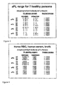
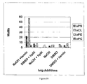
- Figure 1 shows the 24 separate aPL specificities that were tested for by using the comprehensive in-house ELISA aPL format.
- aPS antiphosphatidylserine
- aCL anticardiolipin
- aPE antiphosphatidylethanolamine
- aPC antiphosphatidylcholine.
- IgG immunoglobulin isotypes were sought, IgG, IgA and IgM.
- each specificity and each isotype were assessed in the presence (dependent) and absence (independent) of a buffer diluent supplement, 10% adult bovine plasma (ABP), which contains the phospholipid-binding plasma proteins) or 1 % bovine serum albumin, (BSA, which is devoid of phospholipid-binding plasma proteins), respectively.
- ABSP adult bovine plasma
- BSA bovine serum albumin
- a positive result in the column indicated as PL binding protein "dependent" means that the antiphospholipid antibody (aPL) is actually binding to a plasma protein that initially has bound to the particular phospholipid indicated.
- Plasma proteins that typically can be bound by PS and CL include the following: beta 2 -glycoprotein I, prothrombin, protein C, protein S, annexin V, and complement components Factor H and C4 (see, for example, McIntyre, J.A., Wagenknecht, D.R. and Faulk, W.P. Antiphospholipid antibodies: Discovery, definition, detection and disease. Prog. Lipid Res. 42(3): 176-237, at page 182 ).
- Plasma proteins that typically can be bound by phospholipid PE include the following: high and low molecular weight kininogens, and factor XI and prekallikrein. The latter two proteins can be detected by virtue of their fidelity in binding to high molecular weight kininogen.
- the plasma proteins that bind to PC have not yet been defined. In certain experiments, plasma-protein independent aPL are observed (see Figure 3 ). A possible explanation for this activity is that it represents the presence of residual phospholipid-binding plasma proteins that are present in the original blood sample.
- FIG. 2 A sample of blood from a normal subject was incubated and tested according to the procedure described above. The results of the aPL ELISA are shown in Figure 2 . As shown in Figure 2 , the incubated blood sample shows a dramatic presence of autoantibody activity, in comparison to the normal, untreated blood shown in the Normal ranges column. In particular, strong autoantibody activity is shown in the protein-dependent category for aPS (IgG), aCL (all isotypes), and aPE (IgG). The low or absent IgG aPC autoantibody activity was a characteristic finding in the early examples and in procedures in which hemin was used as the oxidizing agent.
- IgG protein-dependent category for aPS
- aCL all isotypes
- PE IgG
- FIG. 3 is a composite table showing the range of aPL seroconversion for these seven samples.
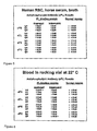
- a serum sample from a normal subject was incubated and tested according to the basic procedure described above.
- horse red blood cells (RBC) were used instead of human RBC.
- the results of the aPL ELISA are shown in Figure 4 .
- significant aPL activity was obtained, particularly with respect to aPS (IgG and IgM) and aCL (IgA and IgM).
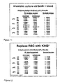
- a blood sample from a normal subject was incubated and tested according to the basic procedure described above, except that the incubation was carried out at room temperature (22°C), instead of at an elevated temperature.
- Figure 6 shows that the sample did not undergo seroconversion when incubated at room temperature.
- the particulate component acts as an abrasive upon the RBC membrane, probably causing release of the NO ion from the RBC, either by interacting with the RBC AE1/Band 3 protein or with the SNO-hemoglobin transition molecules or both.
- the possibility of mechanical abrasion is supported by the observation in Example 6, wherein negative assay results are shown for an incubation mixture that is not rocked or shaken.
- the particulate solids may also serve a mechanical function of assisting autoantibody release.
- a blood sample from a normal subject was incubated and tested according to the basic procedure described above except that the incubation mixture was kept stationary, instead of being shaken or rocked.
- Figure 8 shows that the sample did not undergo seroconversion when it was kept stationary.
- FIG. 10 shows that the sample exhibited seroconversion in the Becton Dickinson medium, indicating that the method of the present invention is not dependent upon a bacterial culture growth medium from a particular source.
- FIG. 11 shows that the sample exhibited seroconversion even under anaerobic conditions and that the method of the present invention is not dependent upon an aerobic environment.
- a blood sample from a normal subject was incubated and tested according to the basic procedure described above, with the feature that K562 cells (a human hematopoetic tumor cell line) were used instead of red blood cells. Further, only 11.3 million K562 cells were present in the culture media, compared to 3-4 mls of packed RBC typically used in the method of the invention.
- Figure 12 shows that the sample exhibited seroconversion.
- RPMI cell culture medium used for growing human cells
- Figure 13 shows that seroconversion did not occur. This experiment shows the importance of some ingredient in the bacterial culture media for the purpose of this invention. While RPMI is a culture media designed for human cells, it does not support aPL release when substituted for vial broth. Listings and comparisons of the ingredients in the two different microbiology vial broths with RPMI show that RPMI lacks hemin and menadione (a man-made provitamin K) called vitamin K3. It is known that hemin is a porphyrin chelater of iron (Fe+++) derived from RBC, and menadione is a fat-soluble vitamin. This indicates that redox reactions may play a role in autoantibody release
- the placental cord blood was drawn after the birth of the baby, but before the placenta was detached from the uterine wall.
- Neither the mother's blood nor the baby's cord blood showed the presence of aPL in conventional laboratory assays.
- strong aPL antibody was demonstrated present in the cord blood samples, as shown in Figure 14 .
- the antibodies were IgG only, an observation that is compatible with antibodies of maternal origin.
- a plasma sample from a normal subject was incubated and tested according to the basic procedure described above; with the feature that sodium nitroprusside (SNP, 200 micromolar) was used in place of RBC in the incubation mixture.
- Figure 15 shows that the sample exhibited seroconversion.
- a blood sample from a normal subject was incubated according to the basic procedure described above and was tested for lupus anticoagulant activity.
- Lupus anticoagulant or inhibitor is another type of aPL and is typically detectable only by functional laboratory assays.
- the results in Figure 16 show a strong lupus anticoagulant (LA) in the seroconverted blood taken from a lupus inhibitor negative individual and processed by the method of this invention. While initially corrected by adding normal plasma to the seroconverted broth in the dRVVT assay, incubation for 1-2 hours resulted in the reappearance of the inhibitor. This time frame is proposed as the time it takes for the LA or unmasked antibodies to bind the relevant phospholipid-binding plasma proteins introduced by the mixing study.
- dPT dilute prothrombin time
- Figure 17 lists additional autoantibody specificities identified by using the Hep-2 cell line. Identified were anti-nucleolar (scleroderma associated), anti-lamins (very bright at nuclear pores), anti-mitochondrial (cytoplasmic), and anti-centriole.
- the results show that autoantibodies released by the method of the present invention can also be detected by a different methodology of detection, fluorescence microscopy, as opposed to ELISA-based testing. The results confirm that many types of autoantibodies besides aPL are masked in the blood of individuals whose serum and plasma test negative for these antibodies in routine laboratory analyses.
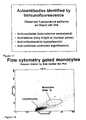
- FIG. 18 depicts the forward scatter (size) and side scatter (granularity) profile of the normal subject's monocyte population of cells as defined by flow cytometry. This monocyte population of cells was confirmed by showing reactivity with CD 14 monoclonal antibodies.
- Figure 19A shows anti-monocyte reactivity with NHS.
- the median reactivity shown is 743.50 on a linear scale.
- Figure 19B shows the auto-anti-monocyte activity of the normal subject's serum; this subject does not have antibody activity to autologous monocytes. The median reactivity shown is 737.00.
- Figure 19C shows the auto-anti-monocyte activity of a blood sample from the subject shown in Figure 19B after it is treated according to the method of the invention. The median value is shown is 864.00, indicating strong auto-anti-monocyte activity.
- the plasma processed according to the teachings of the invention were used at a dilution of 1/8, it showed more reactivity with monocytes than did the undiluted positive control sera.
- this example shows that blood or serum samples processed according to the method of this invention release autoantibodies that specifically target monocytes. The same results were documented for four additional samples from other individuals when processed according to the teachings of the invention.
- red blood cells were replaced with sodium nitroprusside (SNP) and ferric chloride. This substitution was made because sodium nitroprusside is a powerful nitric oxide producer, and it is known that the RBC are carriers of NO - .
- Ferric chloride FeCl 3 stock solution, 25 uM
- NO - may be involved in antibody unmasking, and suggest that the mechanical action of a solid phase material in the culture bottle disrupts the red blood cells and releases NO - .
- the release or modification of NO may enable the hemoglobin molecule to participate in redox reactions.
- IvIg intravenous immunoglobulin
- IvIg is an alcohol precipitate fraction of pooled plasma from multiple donors, typically from 1,000 - 10,000 donors.
- IvIg contains primarily IgG, and is mostly devoid of IgA, IgM and other plasma proteins.
- IvIg is also free of lipoprotein micelles, vesicles or other macromolecular structures. Therefore, if IvIg were to test positive for the presence of autoantibodies after an incubation treatment, it would have to be that the autoantibodies were obtained by an alteration of IgG antibodies already present in the IvIg preparation and not by a breakdown of structures or vesicles concealing the autoantibodies.
- IvIg Immune Globulin Intravenous (Human) Gammar- PI.V., Aventis Behring, Kankakee, Illinois).
- a 5 gram commercial preparation of lyophilized IvIg was reconstituted in sterile phosphate buffered saline (PBS, 100mg/ml). 1.7 ml of the reconsitituted IvIg solution was added to a culture bottle containing the bacterial culture growth medium (without red blood cells or charcoal) and was incubated at 37 °C for 20 hours. The incubated mixture showed seroconversion and the presence of aPL IgG (data not shown). (As expected, only IgG was detected, not IgA or IgM.)
- Example 19 it is shown that autoantibodies can be obtained by incubating a commercial IvIg preparation in a bacterial growth medium. The next step was to try to determine which ingredients in the bacterial culture growth medium play a role in producing detectable autoantibodies.
- IvIg in 2% tryptic soy broth (TSB), (which contains peptones in a 17 to 3 ratio of pancreatic digest of casein to papaya digest of soy, respectively) (the remainder being water) was incubated at 37°C for 20 hours with shaking. The incubated mixture was tested for the presence of aPL, and the result was negative.
- TLB tryptic soy broth
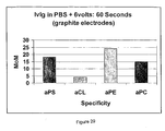
- IvIg was incubated in a test tube in soy broth, sodium nitroprusside (SNP) and hemin (an iron (ferric) containing protoporphyrin) at 37°C for 20 hours with shaking.
- SNP sodium nitroprusside
- hemin an iron (ferric) containing protoporphyrin
- the amounts used were 60 microliters of IvIg, 5 microliters of SNP and 5 microliters of hemin in a total of 1 ml of soy broth.
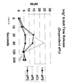
- the incubated mixture tested positive for the presence of aPL, particularly aPS (15 MoM) and aPE (41 MoM). (data not shown)
- Reconstituted lyophilized IvIg (at a concentration of 100 mg/ml) was added to and incubated in a phosphate buffered saline (PBS) solution with hemin for 20 hours at 37 °C.
- PBS phosphate buffered saline
- the amounts used were 300 ⁇ l of IvIg solution and 5 ⁇ l of a hemin solution (75 ⁇ g) in a total volume 1 ml.
- the incubated mixture showed significant amounts of aPS and aPE IgG, and, to a lesser extent, aCL IgG.
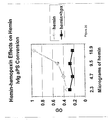
- IvIg was incubated in a Tris buffer with hemin, for 20 hours at 37 °C, similar to the process of Example 21, with the added feature that an increasing amount of human serum (the inventor's) was added to the batches before incubation.
- Each separate batch was tested for the presence of aPS, aCL, aPE and aPC autoantibodies, and the results are shown in Figure 22 .
- the results shown in Figure 22 demonstrate that increasing amounts of serum did have an inhibitory effect on the release of antiphospholipid antibodies. Similar results were shown with substituting plasma for serum (data not shown).
- hemin which contains an iron molecule in the ferric state and which is known as an active oxidizing agent, may act to oxidize a binding site of certain immunoglobulin molecules so that the altered binding site is able to bind self antigens. This process may be inhibited by substances, perhaps antioxidants, in the blood.
- Human serum (the inventor's) was diluted 1/10 in Tris buffer. In a series of experiments, this diluted serum, in 1 ml batches, was incubated with an increasing amounts of hemin, specifically, 0 ⁇ l, 10 ⁇ l, 25 ⁇ l and 50 ⁇ l. (Previously, it had been found that hemin by itself was not sufficient to cause the release of autoantibodies from blood or serum, although it was sufficient to cause such release from IvIg. Therefore, the purpose of diluting the serum was to dilute the effect of any interfering substances found in the blood, such as antioxidants.) The batches were tested for the presence of aPS, aCL, aPE and aPC autoantibodies, and the results are shown in Figure 23 .
- Hpx hemopexin
- Lyophilized Hpx purchased from SciPac (Kent, England) was reconstituted in PBS at 10mg/ml. Shown in Fig. 26 are the aPS redox data resulting from adding increasing concentrations of hemin to the IvIgG countered with adding equamolar concentrations of Hpx. Because there is an 1:1 binding interaction between hemin and Hpx, the Hpx was able to negate the redox capability of the ferric iron present in hemin.
- HRP horseradish peroxidase
- the blots are shown in Figure 27 .
- the "B" lysate is a B lymphocyte cell line called Raja from a patient with a lymphoma.
- the "T” lysate is a T-lymphocyte-derived cell line called Jurkat again from a leukemic patient.
- the U87MG lysate is a glioblastoma blast cell line (brain cancer).
- the reduced lysates were run into the gel at 50 mg/ml concentration.
- 75 ⁇ g of hemin was combined with 1 ml of PBS containing 6 mg of IvIgG. Incubation was for 20 hours at 37 degrees.
- the blot wherein hemin-treated IvIg was used as the primary antibodies is labeled "Test IgG; the blot wherein untreated IvIg was used as the primary antibodies is labeled "Control”, and the blot to which anti-human HRP-tagged conjugate was applied without primary antibodies is labeled "Secondary”.
- the hemin-treated and untreated IgG preparations were diluted 1/1000 respectively.
- the anti-human HRP-tagged conjugate was used at a dilution of 1/5000.
- oxidizing agents other than hemin and in particular, oxidizing agents that do not contain iron, would be effective to unmask autoantibodies.
- a mixture of 25 ⁇ g of potassium permanganate (KMnO 4 ) at a concentration of 100 ⁇ M, and 2 mg of IvIg in a total volume of 1 ml of phosphate buffered saline was incubated overnight at 37 °C.
- aPC and aPS could be detected.
- aCL was usually detected, but not aPE (data not shown). It was later determined that a reason why aPE is not detected is because KMnO 4 alters the PE phospholipid antigen used in the ELISA testing.
- IvIg was dissolved in a phosphate buffered saline solution, and, in separate experiments, galvanized steel, copper, or stainless steel electrodes were connected to the positive and negative terminals of a 9-volt battery and were submersed into the solution for 1 - 2 minutes. During this period, bubbling was noticed in the solution and the PBS solution changed color (blue when copper wires were used, brown when stainless steel wires were used and green when galvanized steel wires were used).
- the treated solution tested positive for the presence of aPS, aCL, aPE and aPC autoantibodies, in aPL dependent testing, and positive for the presence of aPS, aPE and aPC autoantibodies in aPL independent testing.
- graphite electrodes were used in place of the metal electrodes.
- Graphite is inert, but is able to pass electrons into conducting solutions without participating in reactions.
- IvIg was dissolved in a phosphate buffered saline solution, and graphite electrodes connected to the positive and negative terminals of a 6-volt battery were submersed into the solution for 60 seconds. As shown in Figure 29 , the treated solution tested positive for the presence of aPS, aPE and aPC autoantibodies.
- Example 32 Based on the results in Example 32, the next question that was asked was whether autoantibodies of a patent having an autoimmune disease could be remasked if the patient serum was exposed to an electric current. Serum from a patient having elevated levels of aPS and aCL was diluted 1/400 with phosphate-buffered saline (the dilution in PBS was in an amount that would achieve an OD value of 1.000 in 10-15 minutes) and graphite electrodes connected to the positive and negative terminals of a 6-volt battery were submersed into the solution for a variable amount of time. As shown in Figure 32 , the amount of aCL and aPS detectable in the samples of the autoimmune patient's serum declined significantly after 30 seconds and was no longer detectable after 2 minutes. These experiments were repeated for other patients' antibodies and the same result was obtained (data not shown).
- kininogen is the plasma protein responsible for providing a positive ELISA signal for aPE dependent reactions (the kininogen binds to PE, then the antibody binds to the kininogen, the aPE however will not bind to either PE or kininogen independently), this shows that the kininogen in the ABP sample is being altered by the redox exposure.
- aCL is also negative at 240 seconds exposure and since this patient serum requires either prothrombin and/or beta2 glycoprotein (or both could be involved) for producing a positive signal in the aPL ELISA, these two proteins must also be altered by the redox reactions. The same two plasma proteins are involved in the aPS example.
Landscapes
- Health & Medical Sciences (AREA)
- Life Sciences & Earth Sciences (AREA)
- Chemical & Material Sciences (AREA)
- Immunology (AREA)
- Engineering & Computer Science (AREA)
- General Health & Medical Sciences (AREA)
- Medicinal Chemistry (AREA)
- Organic Chemistry (AREA)
- Molecular Biology (AREA)
- Hematology (AREA)
- Biomedical Technology (AREA)
- Chemical Kinetics & Catalysis (AREA)
- Pharmacology & Pharmacy (AREA)
- Animal Behavior & Ethology (AREA)
- Public Health (AREA)
- Veterinary Medicine (AREA)
- Nuclear Medicine, Radiotherapy & Molecular Imaging (AREA)
- General Chemical & Material Sciences (AREA)
- Bioinformatics & Cheminformatics (AREA)
- Urology & Nephrology (AREA)
- Biochemistry (AREA)
- Physics & Mathematics (AREA)
- Pathology (AREA)
- General Physics & Mathematics (AREA)
- Analytical Chemistry (AREA)
- Food Science & Technology (AREA)
- Microbiology (AREA)
- Cell Biology (AREA)
- Biotechnology (AREA)
- Proteomics, Peptides & Aminoacids (AREA)
- Genetics & Genomics (AREA)
- Biophysics (AREA)
- Rheumatology (AREA)
- Neurosurgery (AREA)
- Diabetes (AREA)
- Neurology (AREA)
- Rehabilitation Therapy (AREA)
- Endocrinology (AREA)
- Physical Education & Sports Medicine (AREA)
- Dermatology (AREA)
Claims (9)
- Verfahren zum Erlangen und Isolieren eines Autoantikörpers aus einer Antikörper enthaltenden biologischen Flüssigkeit, die verdünntes Vollblut, Serum oder Plasma umfasst, oder aus einem Antikörper enthaltenden Extrakt einer biologischen Flüssigkeit, der verdünntes Vollblut umfasst, wobei die biologische Flüssigkeit oder der Extrakt maskierte Autoantikörper enthält, die vor dem Ausführen des Verfahrens nicht imstande sind, an ein Eigenantigen zu binden, und daher durch eine Prüfung, die auf Rezeptor-Ligand-Bindung basiert, nicht detektierbar sind, das Verfahren die folgenden Schritte umfassend:Aussetzen der biologischen Flüssigkeit oder des Extrakts einem Oxidationsmittel oder einem elektrischen Strom von 6 - 24 Volt Gleichstrom für einige Sekunden bis einige Minuten, die ausreichend sind, um eine Bindungsspezifität des maskierten Autoantikörpers zu ändern, so dass der Autoantikörper fähig wird, an ein Antigen zu binden, dadurch detektierbar und aus der biologischen Flüssigkeit oder dem Extrakt durch ein Rezeptor-Ligand-Bindungs-Trennverfahren wiedergewinnbar wird, undWiedergewinnen des Autoantikörpers aus der biologischen Flüssigkeit.
- Verfahren nach Anspruch 1, wobei:a) der Antikörper enthaltende Extrakt einer biologischen Flüssigkeit intravenöses Immunglobulin (IvIg) ist; oderb) das Oxidationsmittel Hämin ist.
- Verfahren zum Untersuchen der biologischen Flüssigkeit oder des Extrakts eines normalen Individuums zum Bestimmen einer Menge von Autoantikörpern, die maskiert sind, die folgenden Schritte umfassend:Prüfen einer Vollblut-, Serum- oder Plasmaprobe von einer Person, um eine Menge und/oder einen Typ von maskierten Autoantikörpern, die in der Probe detektierbar sind, zu bestimmen,Behandeln der Probe von der Person, indem die Probe einem Oxidationsmittel oder einem elektrischen Strom von 6 - 24 Volt Gleichstrom für einige Sekunden bis einige Minuten ausgesetzt wird,Prüfen der Probe von der Person, um eine Menge und/oder einen Typ von Autoantikörpern, die in der behandelten Probe detektierbar sind, zu bestimmen, undVergleichen der Menge und/oder des Typs von maskierten Autoantikörpern, die in der nicht behandelten Probe detektierbar sind, mit der Menge und/oder dem Typ von Autoantikörpern, die in der behandelten Probe detektierbar sind.
- Verfahren zum Bewirken einer Änderung der Bindungsspezifität eines maskierten Autoantikörpers, umfassend, eine Zusammensetzung, die mindestens einen maskierten Antikörper umfasst, der in einem flüssigen Medium, das Vollblut, Serum oder Plasma oder verdünntes plazentares Nabelschnurblut umfasst, suspendiert oder gelöst ist, einem Oxidationsmittel oder einem Strom von 6 - 24 Volt Gleichstrom für einige Sekunden bis einige Minuten auszusetzen, die ausreichend sind, um die Änderung der Bindungsspezifität des maskierten Autoantikörpers zu bewirken, wobei die Bindungsspezifität von dem maskierten Autoantikörper, der in Bezug auf ein spezifisches Antigen oder einen spezifischen Ligand keine Bindungsfähigkeit aufweist, zu einem Autoantikörper, der in Bezug auf das spezifische Antigen oder den spezifischen Ligand Bindungsfähigkeit aufweist, geändert wird.
- Verfahren nach Anspruch 4, umfassend, den Autoantikörper mit einer geänderten Bindungsspezifität zu isolieren und wiederzugewinnen.
- Verfahren nach Anspruch 1, 3 oder 4, wobei die Zusammensetzung, die biologische Flüssigkeit, der Antikörper enthaltende Extrakt einer biologischen Flüssigkeit, das Vollblut, das Serum oder das Plasma für eine Zeitperiode, die ausreichend ist, um die Bindungsspezifität des Antikörpers zu ändern, Hämin ausgesetzt wird.
- Verfahren nach Anspruch 1, 3 oder 4, wobei die Zusammensetzung, die biologische Flüssigkeit, der Antikörper enthaltende Extrakt einer biologischen Flüssigkeit, das Vollblut, das Serum oder das Plasma für einige Sekunden bis einige Minuten einem elektrischen Strom von 6 - 24 Volt Gleichstrom ausgesetzt wird.
- Verfahren nach entweder Anspruch 1 oder Anspruch 3, wobei die biologische Flüssigkeit verdünntes Serum oder Plasma umfasst.
- Verfahren nach Anspruch 4, wobei das flüssige Medium verdünntes Serum oder Plasma umfasst.
Applications Claiming Priority (2)
| Application Number | Priority Date | Filing Date | Title |
|---|---|---|---|
| US47660703P | 2003-06-09 | 2003-06-09 | |
| PCT/US2004/017889 WO2004111608A2 (en) | 2003-06-09 | 2004-06-09 | Method of altering the binding specificity of plasma proteins by oxidation-reduction reactions |
Publications (3)
| Publication Number | Publication Date |
|---|---|
| EP1631314A2 EP1631314A2 (de) | 2006-03-08 |
| EP1631314A4 EP1631314A4 (de) | 2006-08-16 |
| EP1631314B1 true EP1631314B1 (de) | 2011-08-03 |
Family
ID=33551622
Family Applications (1)
| Application Number | Title | Priority Date | Filing Date |
|---|---|---|---|
| EP20040754487 Expired - Lifetime EP1631314B1 (de) | 2003-06-09 | 2004-06-09 | Verfahren zur änderung der bindungsspezifität von plasmaproteinen durch oxidations-reduktionsreaktionen |
Country Status (9)
| Country | Link |
|---|---|
| US (4) | US7368542B2 (de) |
| EP (1) | EP1631314B1 (de) |
| JP (1) | JP4662557B2 (de) |
| CN (1) | CN1832756A (de) |
| AT (1) | ATE518886T1 (de) |
| AU (1) | AU2004248155B2 (de) |
| BR (1) | BRPI0411103B1 (de) |
| CA (1) | CA2524325C (de) |
| WO (1) | WO2004111608A2 (de) |
Families Citing this family (27)
| Publication number | Priority date | Publication date | Assignee | Title |
|---|---|---|---|---|
| US7368542B2 (en) | 2003-06-09 | 2008-05-06 | Redox-Reactive Reagents Llc | Method of altering the binding specificity of plasma proteins by oxidation-reduction reactions |
| US9557325B2 (en) * | 2003-06-09 | 2017-01-31 | Redox-Reactive Reagents Llc | Method of altering the binding specificity of proteins by oxidation-reduction reactions |
| US7892751B2 (en) | 2003-06-09 | 2011-02-22 | Redox-Reactive Reagents Llc | Method of detecting or diagnosing of a neurodegenerative disease or condition |
| CA2584211C (en) * | 2004-10-22 | 2014-07-08 | Amgen Inc. | Methods for refolding of recombinant antibodies |
| KR101322247B1 (ko) * | 2007-09-06 | 2013-10-25 | 삼성전자주식회사 | 진공청소기용 흡입브러시 및 그 높이조절방법 |
| JP5963341B2 (ja) * | 2007-09-14 | 2016-08-10 | アムジエン・インコーポレーテツド | 均質な抗体集団 |
| US7989596B2 (en) * | 2007-09-19 | 2011-08-02 | Redox-Reactive Reagents Llc | Method of altering the binding specificity of monoclonal antibodies by oxidation-reduction reactions |
| JP5565607B2 (ja) | 2009-07-15 | 2014-08-06 | 国立大学法人 東京大学 | 敗血症又は多臓器不全の予後診断方法及び予後診断用キット |
| WO2011094535A2 (en) * | 2010-01-28 | 2011-08-04 | The Board Of Trustees Of The Leland Stanford Junior University | Biomarkers of aging for detection and treatment of disorders |
| US10626399B2 (en) | 2010-01-28 | 2020-04-21 | The Board Of Trustees Of The Leland Stanford Junior University | Methods of treating cognitive symptoms of an aging-associated impairment by modulating C-C chemokine receptor type 3 (CCR3) |
| US20160208011A1 (en) | 2010-01-28 | 2016-07-21 | The Board Of Trustees Of The Leland Stanford Junior University | Ccr3 modulation in the treatment of aging-associated impairments, and compositions for practicing the same |
| US9161968B2 (en) | 2011-04-08 | 2015-10-20 | The Board Of Trustees Of The Leland Stanford Junior University | Methods of neuroprotection involving macrophage colony stimulating factor receptor agonists |
| ES2684976T3 (es) | 2012-04-13 | 2018-10-05 | Diabetomics, Inc. | Biomarcadores maternos para diabetes gestacional |
| EP2996772B1 (de) | 2013-05-13 | 2018-12-19 | Momenta Pharmaceuticals, Inc. | Verfahren zur behandlung von neurodegeneration |
| US10633453B2 (en) | 2013-05-28 | 2020-04-28 | Kaohsiung Medical University | Antibody locker for the inactivation of protein drug |
| NZ720949A (en) | 2013-12-09 | 2019-03-29 | Univ Leland Stanford Junior | Methods and compositions for treating aging-associated conditions |
| US10905779B2 (en) | 2013-12-09 | 2021-02-02 | The Board Of Trustees Of The Leland Stanford Junior University | Methods for screening human blood products comprising plasma using immunocompromised rodent models |
| SI3307296T1 (sl) | 2015-06-15 | 2022-04-29 | The Board of Trustees of the Leland Stanford Junior University Office of the General Counsel | TIMP2 za uporabo pri zdravljenju s starostjo povezanih stanj |
| KR20180032489A (ko) * | 2016-09-22 | 2018-03-30 | 아주대학교산학협력단 | 액상 플라즈마를 이용한 건선의 예방 또는 치료 방법 |
| KR20180032490A (ko) * | 2016-09-22 | 2018-03-30 | 아주대학교산학협력단 | 액상 플라즈마를 이용한 아토피성 피부염의 예방 또는 치료 방법 |
| US11583689B2 (en) * | 2016-09-22 | 2023-02-21 | Ajou University Industry-Academic Cooperation Foundation | Composition for atopy or psoriasis treatment comprising liquid type plasma |
| KR101933920B1 (ko) | 2016-09-22 | 2018-12-31 | 아주대학교 산학협력단 | 액상 플라즈마를 이용한 비만의 예방 또는 치료 방법 |
| EA201991353A1 (ru) | 2016-12-09 | 2019-11-29 | Двухвалентные антитела, маскированные спирализованными спиралями | |
| US11510853B2 (en) | 2017-09-18 | 2022-11-29 | Ajou University Industry-Academic Cooperation Foundation | Composition for skin soothing containing liquid-phase plasma |
| PE20220383A1 (es) * | 2019-04-18 | 2022-03-18 | Janssen Biotech Inc | Glicoproteinas sialiladas |
| EP3756648A1 (de) * | 2019-06-27 | 2020-12-30 | Imnate Sarl | Verbesserte impfstoffformulierungen |
| TR202016418A2 (tr) * | 2020-10-14 | 2022-04-21 | Serkan Tunc | Kan plazmasinin üzeri̇nden elektri̇k akiminin geçi̇ri̇lmesi̇yle i̇çeri̇si̇nde ölü bakteri̇, parazi̇t vi̇rüs vb. kalintilarin oluştuğu yeni̇ plazmanin canli vücuduna serum olarak veri̇lmesi̇yle bağişiklik geli̇şti̇rme yöntemi̇ |
Family Cites Families (15)
| Publication number | Priority date | Publication date | Assignee | Title |
|---|---|---|---|---|
| US4632980A (en) * | 1985-04-03 | 1986-12-30 | Immunologics | Ozone decontamination of blood and blood products |
| US4703001A (en) | 1985-10-23 | 1987-10-27 | Synbiotics, Corporation | Immunoassay for the detection of serum analytes using pH dependent chastropic acids |
| US4950612A (en) * | 1987-12-16 | 1990-08-21 | Microgenics Corporation | Peroxy acid pretreatment in vitamin B12 assay |
| ATE88453T1 (de) * | 1988-02-22 | 1993-05-15 | Fina Research | Katalytisches einstufenverfahren zur direkten umwandlung von polysacchariden in mehrwertige alkohole. |
| US5061790A (en) * | 1989-07-10 | 1991-10-29 | Molecular Diagnostics, Inc. | Oxidative denaturation of protein analytes |
| US5980954A (en) * | 1992-02-07 | 1999-11-09 | Vasogen Ireland Limited | Treatment of autoimmune diseases |
| US6660267B1 (en) * | 1992-12-21 | 2003-12-09 | Promega Corporation | Prevention and treatment of sepsis |
| US5880954A (en) * | 1995-12-04 | 1999-03-09 | Thomson; Robert | Continous real time safety-related control system |
| AU703814B2 (en) * | 1995-12-04 | 1999-04-01 | Wyeth | Antioxidant |
| US5939394A (en) * | 1996-01-18 | 1999-08-17 | Fleming & Company | Methods and compositions for the prevention and treatment of immunological disorders, inflammatory diseases and infections |
| FR2794461B1 (fr) * | 1999-06-07 | 2004-01-23 | Lab Francais Du Fractionnement | Procede de preparation de nouvelles fractions d'ig humaines possedant une activite immunomodulatrice |
| FR2822238B1 (fr) * | 2001-03-16 | 2003-08-29 | Gemac | Methode et trousse pour le suivi des maladies neurodegeneratives |
| US20040101909A1 (en) | 2002-08-20 | 2004-05-27 | Hema-Quebec, 2535 Boul. Laurier, Ste-Foy, Quebec, Canada G1V 4M3 | Purification of polyreactive autoantibodies and uses thereof |
| US9557325B2 (en) * | 2003-06-09 | 2017-01-31 | Redox-Reactive Reagents Llc | Method of altering the binding specificity of proteins by oxidation-reduction reactions |
| US7368542B2 (en) | 2003-06-09 | 2008-05-06 | Redox-Reactive Reagents Llc | Method of altering the binding specificity of plasma proteins by oxidation-reduction reactions |
-
2004
- 2004-06-09 US US10/863,365 patent/US7368542B2/en not_active Expired - Lifetime
- 2004-06-09 AT AT04754487T patent/ATE518886T1/de not_active IP Right Cessation
- 2004-06-09 WO PCT/US2004/017889 patent/WO2004111608A2/en not_active Ceased
- 2004-06-09 AU AU2004248155A patent/AU2004248155B2/en not_active Ceased
- 2004-06-09 CA CA2524325A patent/CA2524325C/en not_active Expired - Fee Related
- 2004-06-09 CN CNA2004800159879A patent/CN1832756A/zh active Pending
- 2004-06-09 EP EP20040754487 patent/EP1631314B1/de not_active Expired - Lifetime
- 2004-06-09 BR BRPI0411103A patent/BRPI0411103B1/pt not_active IP Right Cessation
- 2004-06-09 JP JP2006515229A patent/JP4662557B2/ja not_active Expired - Fee Related
-
2008
- 2008-04-23 US US12/107,819 patent/US8334146B2/en not_active Expired - Fee Related
-
2012
- 2012-12-12 US US13/712,151 patent/US9169317B2/en not_active Expired - Fee Related
-
2015
- 2015-10-27 US US14/923,945 patent/US10072068B2/en not_active Expired - Fee Related
Also Published As
| Publication number | Publication date |
|---|---|
| US20050101016A1 (en) | 2005-05-12 |
| US20080280281A1 (en) | 2008-11-13 |
| EP1631314A4 (de) | 2006-08-16 |
| AU2004248155A1 (en) | 2004-12-23 |
| WO2004111608A2 (en) | 2004-12-23 |
| EP1631314A2 (de) | 2006-03-08 |
| US9169317B2 (en) | 2015-10-27 |
| AU2004248155B2 (en) | 2008-10-23 |
| ATE518886T1 (de) | 2011-08-15 |
| JP4662557B2 (ja) | 2011-03-30 |
| BRPI0411103A (pt) | 2006-07-18 |
| BRPI0411103B1 (pt) | 2019-01-15 |
| CN1832756A (zh) | 2006-09-13 |
| US20130109838A1 (en) | 2013-05-02 |
| US20160046695A1 (en) | 2016-02-18 |
| CA2524325C (en) | 2017-08-01 |
| WO2004111608A3 (en) | 2005-04-07 |
| US10072068B2 (en) | 2018-09-11 |
| US8334146B2 (en) | 2012-12-18 |
| US7368542B2 (en) | 2008-05-06 |
| WO2004111608A8 (en) | 2005-12-29 |
| JP2006526027A (ja) | 2006-11-16 |
| CA2524325A1 (en) | 2004-12-23 |
Similar Documents
| Publication | Publication Date | Title |
|---|---|---|
| US10072068B2 (en) | Method for altering the binding specificity of plasma proteins by oxidation | |
| US9557325B2 (en) | Method of altering the binding specificity of proteins by oxidation-reduction reactions | |
| Gloor et al. | Persistence of low levels of alloantibody after desensitization in crossmatch-positive living-donor kidney transplantation | |
| Sumitran‐Holgersson | HLA‐specific alloantibodies and renal graft outcome | |
| McIntyre et al. | Autoantibodies unmasked by redox reactions | |
| McIntyre | The appearance and disappearance of antiphospholipid autoantibodies subsequent to oxidation–reduction reactions | |
| Wahrmann et al. | Anti-A/B antibody depletion by semiselective versus ABO blood group-specific immunoadsorption | |
| Liaskos et al. | Prevalence and clinical significance of anticardiolipin antibodies in patients with type 1 autoimmune hepatitis | |
| US7989596B2 (en) | Method of altering the binding specificity of monoclonal antibodies by oxidation-reduction reactions | |
| Taylor et al. | Complement-opsonized IgG antibody/dsDNA immune complexes bind to CR1 clusters on isolated human erythrocytes | |
| MXPA05013402A (en) | Method of altering the binding specificity of plasma proteins by oxidation-reduction reactions | |
| Lee et al. | Distribution of anti-ABO immunoglobulin G subclass and C1q antibody in ABO-incompatible kidney transplantation | |
| McIntyre et al. | Autoantibody potential of cancer therapeutic monoclonal antibodies | |
| Bloemen et al. | Inverted erythrocyte membranes demonstrate β2GPI-antiphospholipid antibody interactions and membrane crosslinking | |
| León et al. | Anti-human erythrocyte antibodies in horse-derived antivenoms used in the treatment of snakebite envenomations | |
| JPH08503770A (ja) | 抗hpa抗体の検出 | |
| Minty | The interaction of phagocytic cells with immune complexes in rheumatoid arthritis |
Legal Events
| Date | Code | Title | Description |
|---|---|---|---|
| PUAI | Public reference made under article 153(3) epc to a published international application that has entered the european phase |
Free format text: ORIGINAL CODE: 0009012 |
|
| 17P | Request for examination filed |
Effective date: 20051024 |
|
| AK | Designated contracting states |
Kind code of ref document: A2 Designated state(s): AT BE BG CH CY CZ DE DK EE ES FI FR GB GR HU IE IT LI LU MC NL PL PT RO SE SI SK TR |
|
| AX | Request for extension of the european patent |
Extension state: AL HR LT LV MK |
|
| RIC1 | Information provided on ipc code assigned before grant |
Ipc: A61K 39/395 20060101AFI20060113BHEP Ipc: C07K 1/113 20060101ALI20060630BHEP Ipc: G01N 33/564 20060101ALI20060630BHEP Ipc: C07K 1/26 20060101ALI20060630BHEP Ipc: C07K 16/18 20060101ALI20060630BHEP Ipc: C07K 1/24 20060101ALI20060630BHEP Ipc: G01N 33/561 20060101ALI20060630BHEP Ipc: C07K 16/00 20060101ALI20060630BHEP |
|
| A4 | Supplementary search report drawn up and despatched |
Effective date: 20060714 |
|
| RAP1 | Party data changed (applicant data changed or rights of an application transferred) |
Owner name: REDOX-REACTIVE REAGENTS L.L.C. |
|
| RIN1 | Information on inventor provided before grant (corrected) |
Inventor name: MCINTYRE, JOHN A. |
|
| 17Q | First examination report despatched |
Effective date: 20070724 |
|
| GRAP | Despatch of communication of intention to grant a patent |
Free format text: ORIGINAL CODE: EPIDOSNIGR1 |
|
| RIC1 | Information provided on ipc code assigned before grant |
Ipc: G01N 33/564 20060101ALI20110103BHEP Ipc: G01N 33/561 20060101ALI20110103BHEP Ipc: C07K 16/18 20060101ALI20110103BHEP Ipc: C07K 16/00 20060101AFI20110103BHEP |
|
| GRAS | Grant fee paid |
Free format text: ORIGINAL CODE: EPIDOSNIGR3 |
|
| GRAA | (expected) grant |
Free format text: ORIGINAL CODE: 0009210 |
|
| RIN1 | Information on inventor provided before grant (corrected) |
Inventor name: MCINTYRE, JOHN A. |
|
| AK | Designated contracting states |
Kind code of ref document: B1 Designated state(s): AT BE BG CH CY CZ DE DK EE ES FI FR GB GR HU IE IT LI LU MC NL PL PT RO SE SI SK TR |
|
| AX | Request for extension of the european patent |
Extension state: AL HR LT LV MK |
|
| REG | Reference to a national code |
Ref country code: GB Ref legal event code: FG4D |
|
| REG | Reference to a national code |
Ref country code: CH Ref legal event code: EP |
|
| REG | Reference to a national code |
Ref country code: IE Ref legal event code: FG4D |
|
| REG | Reference to a national code |
Ref country code: DE Ref legal event code: R096 Ref document number: 602004033777 Country of ref document: DE Effective date: 20111006 |
|
| REG | Reference to a national code |
Ref country code: NL Ref legal event code: VDEP Effective date: 20110803 |
|
| LTIE | Lt: invalidation of european patent or patent extension |
Effective date: 20110803 |
|
| PG25 | Lapsed in a contracting state [announced via postgrant information from national office to epo] |
Ref country code: NL Free format text: LAPSE BECAUSE OF FAILURE TO SUBMIT A TRANSLATION OF THE DESCRIPTION OR TO PAY THE FEE WITHIN THE PRESCRIBED TIME-LIMIT Effective date: 20110803 Ref country code: PT Free format text: LAPSE BECAUSE OF FAILURE TO SUBMIT A TRANSLATION OF THE DESCRIPTION OR TO PAY THE FEE WITHIN THE PRESCRIBED TIME-LIMIT Effective date: 20111205 Ref country code: SE Free format text: LAPSE BECAUSE OF FAILURE TO SUBMIT A TRANSLATION OF THE DESCRIPTION OR TO PAY THE FEE WITHIN THE PRESCRIBED TIME-LIMIT Effective date: 20110803 Ref country code: FI Free format text: LAPSE BECAUSE OF FAILURE TO SUBMIT A TRANSLATION OF THE DESCRIPTION OR TO PAY THE FEE WITHIN THE PRESCRIBED TIME-LIMIT Effective date: 20110803 |
|
| REG | Reference to a national code |
Ref country code: AT Ref legal event code: MK05 Ref document number: 518886 Country of ref document: AT Kind code of ref document: T Effective date: 20110803 |
|
| PG25 | Lapsed in a contracting state [announced via postgrant information from national office to epo] |
Ref country code: PL Free format text: LAPSE BECAUSE OF FAILURE TO SUBMIT A TRANSLATION OF THE DESCRIPTION OR TO PAY THE FEE WITHIN THE PRESCRIBED TIME-LIMIT Effective date: 20110803 Ref country code: CY Free format text: LAPSE BECAUSE OF FAILURE TO SUBMIT A TRANSLATION OF THE DESCRIPTION OR TO PAY THE FEE WITHIN THE PRESCRIBED TIME-LIMIT Effective date: 20110803 Ref country code: SI Free format text: LAPSE BECAUSE OF FAILURE TO SUBMIT A TRANSLATION OF THE DESCRIPTION OR TO PAY THE FEE WITHIN THE PRESCRIBED TIME-LIMIT Effective date: 20110803 Ref country code: GR Free format text: LAPSE BECAUSE OF FAILURE TO SUBMIT A TRANSLATION OF THE DESCRIPTION OR TO PAY THE FEE WITHIN THE PRESCRIBED TIME-LIMIT Effective date: 20111104 Ref country code: AT Free format text: LAPSE BECAUSE OF FAILURE TO SUBMIT A TRANSLATION OF THE DESCRIPTION OR TO PAY THE FEE WITHIN THE PRESCRIBED TIME-LIMIT Effective date: 20110803 |
|
| PG25 | Lapsed in a contracting state [announced via postgrant information from national office to epo] |
Ref country code: BE Free format text: LAPSE BECAUSE OF FAILURE TO SUBMIT A TRANSLATION OF THE DESCRIPTION OR TO PAY THE FEE WITHIN THE PRESCRIBED TIME-LIMIT Effective date: 20110803 |
|
| PG25 | Lapsed in a contracting state [announced via postgrant information from national office to epo] |
Ref country code: CZ Free format text: LAPSE BECAUSE OF FAILURE TO SUBMIT A TRANSLATION OF THE DESCRIPTION OR TO PAY THE FEE WITHIN THE PRESCRIBED TIME-LIMIT Effective date: 20110803 Ref country code: SK Free format text: LAPSE BECAUSE OF FAILURE TO SUBMIT A TRANSLATION OF THE DESCRIPTION OR TO PAY THE FEE WITHIN THE PRESCRIBED TIME-LIMIT Effective date: 20110803 |
|
| PG25 | Lapsed in a contracting state [announced via postgrant information from national office to epo] |
Ref country code: EE Free format text: LAPSE BECAUSE OF FAILURE TO SUBMIT A TRANSLATION OF THE DESCRIPTION OR TO PAY THE FEE WITHIN THE PRESCRIBED TIME-LIMIT Effective date: 20110803 Ref country code: IT Free format text: LAPSE BECAUSE OF FAILURE TO SUBMIT A TRANSLATION OF THE DESCRIPTION OR TO PAY THE FEE WITHIN THE PRESCRIBED TIME-LIMIT Effective date: 20110803 Ref country code: RO Free format text: LAPSE BECAUSE OF FAILURE TO SUBMIT A TRANSLATION OF THE DESCRIPTION OR TO PAY THE FEE WITHIN THE PRESCRIBED TIME-LIMIT Effective date: 20110803 |
|
| PLBE | No opposition filed within time limit |
Free format text: ORIGINAL CODE: 0009261 |
|
| STAA | Information on the status of an ep patent application or granted ep patent |
Free format text: STATUS: NO OPPOSITION FILED WITHIN TIME LIMIT |
|
| PG25 | Lapsed in a contracting state [announced via postgrant information from national office to epo] |
Ref country code: DK Free format text: LAPSE BECAUSE OF FAILURE TO SUBMIT A TRANSLATION OF THE DESCRIPTION OR TO PAY THE FEE WITHIN THE PRESCRIBED TIME-LIMIT Effective date: 20110803 |
|
| 26N | No opposition filed |
Effective date: 20120504 |
|
| REG | Reference to a national code |
Ref country code: DE Ref legal event code: R097 Ref document number: 602004033777 Country of ref document: DE Effective date: 20120504 |
|
| PG25 | Lapsed in a contracting state [announced via postgrant information from national office to epo] |
Ref country code: MC Free format text: LAPSE BECAUSE OF NON-PAYMENT OF DUE FEES Effective date: 20120630 |
|
| REG | Reference to a national code |
Ref country code: CH Ref legal event code: PL |
|
| REG | Reference to a national code |
Ref country code: CH Ref legal event code: PL |
|
| REG | Reference to a national code |
Ref country code: IE Ref legal event code: MM4A |
|
| PG25 | Lapsed in a contracting state [announced via postgrant information from national office to epo] |
Ref country code: CH Free format text: LAPSE BECAUSE OF NON-PAYMENT OF DUE FEES Effective date: 20120630 Ref country code: IE Free format text: LAPSE BECAUSE OF NON-PAYMENT OF DUE FEES Effective date: 20120609 Ref country code: ES Free format text: LAPSE BECAUSE OF FAILURE TO SUBMIT A TRANSLATION OF THE DESCRIPTION OR TO PAY THE FEE WITHIN THE PRESCRIBED TIME-LIMIT Effective date: 20111114 Ref country code: LI Free format text: LAPSE BECAUSE OF NON-PAYMENT OF DUE FEES Effective date: 20120630 |
|
| PG25 | Lapsed in a contracting state [announced via postgrant information from national office to epo] |
Ref country code: BG Free format text: LAPSE BECAUSE OF FAILURE TO SUBMIT A TRANSLATION OF THE DESCRIPTION OR TO PAY THE FEE WITHIN THE PRESCRIBED TIME-LIMIT Effective date: 20111103 |
|
| PG25 | Lapsed in a contracting state [announced via postgrant information from national office to epo] |
Ref country code: TR Free format text: LAPSE BECAUSE OF FAILURE TO SUBMIT A TRANSLATION OF THE DESCRIPTION OR TO PAY THE FEE WITHIN THE PRESCRIBED TIME-LIMIT Effective date: 20110803 |
|
| PG25 | Lapsed in a contracting state [announced via postgrant information from national office to epo] |
Ref country code: LU Free format text: LAPSE BECAUSE OF NON-PAYMENT OF DUE FEES Effective date: 20120609 |
|
| PG25 | Lapsed in a contracting state [announced via postgrant information from national office to epo] |
Ref country code: HU Free format text: LAPSE BECAUSE OF FAILURE TO SUBMIT A TRANSLATION OF THE DESCRIPTION OR TO PAY THE FEE WITHIN THE PRESCRIBED TIME-LIMIT Effective date: 20040609 |
|
| REG | Reference to a national code |
Ref country code: FR Ref legal event code: PLFP Year of fee payment: 13 |
|
| REG | Reference to a national code |
Ref country code: FR Ref legal event code: PLFP Year of fee payment: 14 |
|
| REG | Reference to a national code |
Ref country code: FR Ref legal event code: PLFP Year of fee payment: 15 |
|
| PGFP | Annual fee paid to national office [announced via postgrant information from national office to epo] |
Ref country code: FR Payment date: 20200625 Year of fee payment: 17 Ref country code: DE Payment date: 20200629 Year of fee payment: 17 |
|
| PGFP | Annual fee paid to national office [announced via postgrant information from national office to epo] |
Ref country code: GB Payment date: 20200629 Year of fee payment: 17 |
|
| REG | Reference to a national code |
Ref country code: DE Ref legal event code: R119 Ref document number: 602004033777 Country of ref document: DE |
|
| GBPC | Gb: european patent ceased through non-payment of renewal fee |
Effective date: 20210609 |
|
| PG25 | Lapsed in a contracting state [announced via postgrant information from national office to epo] |
Ref country code: GB Free format text: LAPSE BECAUSE OF NON-PAYMENT OF DUE FEES Effective date: 20210609 Ref country code: DE Free format text: LAPSE BECAUSE OF NON-PAYMENT OF DUE FEES Effective date: 20220101 |
|
| PG25 | Lapsed in a contracting state [announced via postgrant information from national office to epo] |
Ref country code: FR Free format text: LAPSE BECAUSE OF NON-PAYMENT OF DUE FEES Effective date: 20210630 |