EP1590362B1 - Improved method for bisulfite treatment - Google Patents
Improved method for bisulfite treatment Download PDFInfo
- Publication number
- EP1590362B1 EP1590362B1 EP04705800A EP04705800A EP1590362B1 EP 1590362 B1 EP1590362 B1 EP 1590362B1 EP 04705800 A EP04705800 A EP 04705800A EP 04705800 A EP04705800 A EP 04705800A EP 1590362 B1 EP1590362 B1 EP 1590362B1
- Authority
- EP
- European Patent Office
- Prior art keywords
- bisulfite
- solution
- nucleic acid
- reaction
- value
- Prior art date
- Legal status (The legal status is an assumption and is not a legal conclusion. Google has not performed a legal analysis and makes no representation as to the accuracy of the status listed.)
- Expired - Lifetime
Links
- 238000000034 method Methods 0.000 title claims abstract description 80
- LSNNMFCWUKXFEE-UHFFFAOYSA-M Bisulfite Chemical compound OS([O-])=O LSNNMFCWUKXFEE-UHFFFAOYSA-M 0.000 title claims abstract description 78
- 238000011282 treatment Methods 0.000 title description 8
- 150000007523 nucleic acids Chemical class 0.000 claims abstract description 63
- 102000039446 nucleic acids Human genes 0.000 claims abstract description 60
- 108020004707 nucleic acids Proteins 0.000 claims abstract description 60
- 238000006243 chemical reaction Methods 0.000 claims abstract description 54
- OPTASPLRGRRNAP-UHFFFAOYSA-N cytosine Chemical compound NC=1C=CNC(=O)N=1 OPTASPLRGRRNAP-UHFFFAOYSA-N 0.000 claims abstract description 45
- 229940104302 cytosine Drugs 0.000 claims abstract description 21
- ISAKRJDGNUQOIC-UHFFFAOYSA-N Uracil Chemical compound O=C1C=CNC(=O)N1 ISAKRJDGNUQOIC-UHFFFAOYSA-N 0.000 claims description 35
- QIGBRXMKCJKVMJ-UHFFFAOYSA-N Hydroquinone Chemical compound OC1=CC=C(O)C=C1 QIGBRXMKCJKVMJ-UHFFFAOYSA-N 0.000 claims description 26
- 229940035893 uracil Drugs 0.000 claims description 17
- -1 bisulfite ions Chemical class 0.000 claims description 7
- 230000011987 methylation Effects 0.000 abstract description 8
- 238000007069 methylation reaction Methods 0.000 abstract description 8
- 239000000243 solution Substances 0.000 description 39
- 239000002585 base Substances 0.000 description 38
- 108020004414 DNA Proteins 0.000 description 34
- 102000053602 DNA Human genes 0.000 description 34
- HEMHJVSKTPXQMS-UHFFFAOYSA-M Sodium hydroxide Chemical compound [OH-].[Na+] HEMHJVSKTPXQMS-UHFFFAOYSA-M 0.000 description 30
- 108091034117 Oligonucleotide Proteins 0.000 description 20
- LRSASMSXMSNRBT-UHFFFAOYSA-N 5-methylcytosine Chemical class CC1=CNC(=O)N=C1N LRSASMSXMSNRBT-UHFFFAOYSA-N 0.000 description 17
- LFQSCWFLJHTTHZ-UHFFFAOYSA-N Ethanol Chemical compound CCO LFQSCWFLJHTTHZ-UHFFFAOYSA-N 0.000 description 17
- 238000003199 nucleic acid amplification method Methods 0.000 description 14
- 230000003321 amplification Effects 0.000 description 13
- 238000004128 high performance liquid chromatography Methods 0.000 description 13
- FAPWRFPIFSIZLT-UHFFFAOYSA-M Sodium chloride Chemical compound [Na+].[Cl-] FAPWRFPIFSIZLT-UHFFFAOYSA-M 0.000 description 11
- 239000011521 glass Substances 0.000 description 11
- 239000000203 mixture Substances 0.000 description 11
- 239000000523 sample Substances 0.000 description 11
- 239000003153 chemical reaction reagent Substances 0.000 description 10
- 239000000047 product Substances 0.000 description 10
- 238000001514 detection method Methods 0.000 description 9
- 239000007790 solid phase Substances 0.000 description 9
- 238000005259 measurement Methods 0.000 description 8
- 238000011534 incubation Methods 0.000 description 7
- 239000002245 particle Substances 0.000 description 7
- 108091081021 Sense strand Proteins 0.000 description 6
- 230000000692 anti-sense effect Effects 0.000 description 6
- 239000012472 biological sample Substances 0.000 description 6
- 239000000872 buffer Substances 0.000 description 6
- 230000000694 effects Effects 0.000 description 6
- 239000013580 millipore water Substances 0.000 description 6
- 239000011780 sodium chloride Substances 0.000 description 6
- 102100030943 Glutathione S-transferase P Human genes 0.000 description 5
- 101001010139 Homo sapiens Glutathione S-transferase P Proteins 0.000 description 5
- 230000003196 chaotropic effect Effects 0.000 description 5
- 238000003556 assay Methods 0.000 description 4
- 239000003795 chemical substances by application Substances 0.000 description 4
- 230000009615 deamination Effects 0.000 description 4
- 238000006481 deamination reaction Methods 0.000 description 4
- 230000006326 desulfonation Effects 0.000 description 4
- 238000005869 desulfonation reaction Methods 0.000 description 4
- 238000007855 methylation-specific PCR Methods 0.000 description 4
- 230000008569 process Effects 0.000 description 4
- 230000035484 reaction time Effects 0.000 description 4
- HRZFUMHJMZEROT-UHFFFAOYSA-L sodium disulfite Chemical compound [Na+].[Na+].[O-]S(=O)S([O-])(=O)=O HRZFUMHJMZEROT-UHFFFAOYSA-L 0.000 description 4
- JTBBWRKSUYCPFY-UHFFFAOYSA-N 2,3-dihydro-1h-pyrimidin-4-one Chemical compound O=C1NCNC=C1 JTBBWRKSUYCPFY-UHFFFAOYSA-N 0.000 description 3
- 241000124008 Mammalia Species 0.000 description 3
- 241001465754 Metazoa Species 0.000 description 3
- VYPSYNLAJGMNEJ-UHFFFAOYSA-N Silicium dioxide Chemical compound O=[Si]=O VYPSYNLAJGMNEJ-UHFFFAOYSA-N 0.000 description 3
- 230000002378 acidificating effect Effects 0.000 description 3
- 238000004458 analytical method Methods 0.000 description 3
- 230000000295 complement effect Effects 0.000 description 3
- 230000009089 cytolysis Effects 0.000 description 3
- 238000002474 experimental method Methods 0.000 description 3
- 239000012530 fluid Substances 0.000 description 3
- 238000003752 polymerase chain reaction Methods 0.000 description 3
- 108090000623 proteins and genes Proteins 0.000 description 3
- 230000035945 sensitivity Effects 0.000 description 3
- 230000000405 serological effect Effects 0.000 description 3
- 210000001519 tissue Anatomy 0.000 description 3
- 238000005406 washing Methods 0.000 description 3
- 108010037497 3'-nucleotidase Proteins 0.000 description 2
- 229930024421 Adenine Natural products 0.000 description 2
- GFFGJBXGBJISGV-UHFFFAOYSA-N Adenine Chemical compound NC1=NC=NC2=C1N=CN2 GFFGJBXGBJISGV-UHFFFAOYSA-N 0.000 description 2
- 238000007399 DNA isolation Methods 0.000 description 2
- 230000007067 DNA methylation Effects 0.000 description 2
- KCXVZYZYPLLWCC-UHFFFAOYSA-N EDTA Chemical compound OC(=O)CN(CC(O)=O)CCN(CC(O)=O)CC(O)=O KCXVZYZYPLLWCC-UHFFFAOYSA-N 0.000 description 2
- TWRXJAOTZQYOKJ-UHFFFAOYSA-L Magnesium chloride Chemical compound [Mg+2].[Cl-].[Cl-] TWRXJAOTZQYOKJ-UHFFFAOYSA-L 0.000 description 2
- 229910004879 Na2S2O5 Inorganic materials 0.000 description 2
- 108091028043 Nucleic acid sequence Proteins 0.000 description 2
- 238000012408 PCR amplification Methods 0.000 description 2
- 239000007983 Tris buffer Substances 0.000 description 2
- 229960000643 adenine Drugs 0.000 description 2
- 230000003698 anagen phase Effects 0.000 description 2
- 230000008901 benefit Effects 0.000 description 2
- 210000004027 cell Anatomy 0.000 description 2
- 150000001875 compounds Chemical class 0.000 description 2
- 238000004925 denaturation Methods 0.000 description 2
- 230000036425 denaturation Effects 0.000 description 2
- 230000001419 dependent effect Effects 0.000 description 2
- 238000011156 evaluation Methods 0.000 description 2
- 230000030279 gene silencing Effects 0.000 description 2
- 238000009396 hybridization Methods 0.000 description 2
- 238000007834 ligase chain reaction Methods 0.000 description 2
- 238000013507 mapping Methods 0.000 description 2
- 239000000463 material Substances 0.000 description 2
- 230000007246 mechanism Effects 0.000 description 2
- 238000002156 mixing Methods 0.000 description 2
- 238000010369 molecular cloning Methods 0.000 description 2
- 210000002381 plasma Anatomy 0.000 description 2
- 238000000746 purification Methods 0.000 description 2
- 239000011541 reaction mixture Substances 0.000 description 2
- 210000002966 serum Anatomy 0.000 description 2
- 235000010262 sodium metabisulphite Nutrition 0.000 description 2
- 230000006641 stabilisation Effects 0.000 description 2
- 239000000126 substance Substances 0.000 description 2
- RWQNBRDOKXIBIV-UHFFFAOYSA-N thymine Chemical compound CC1=CNC(=O)NC1=O RWQNBRDOKXIBIV-UHFFFAOYSA-N 0.000 description 2
- LENZDBCJOHFCAS-UHFFFAOYSA-N tris Chemical compound OCC(N)(CO)CO LENZDBCJOHFCAS-UHFFFAOYSA-N 0.000 description 2
- 210000002700 urine Anatomy 0.000 description 2
- HNSDLXPSAYFUHK-UHFFFAOYSA-N 1,4-bis(2-ethylhexyl) sulfosuccinate Chemical compound CCCCC(CC)COC(=O)CC(S(O)(=O)=O)C(=O)OCC(CC)CCCC HNSDLXPSAYFUHK-UHFFFAOYSA-N 0.000 description 1
- FWMNVWWHGCHHJJ-SKKKGAJSSA-N 4-amino-1-[(2r)-6-amino-2-[[(2r)-2-[[(2r)-2-[[(2r)-2-amino-3-phenylpropanoyl]amino]-3-phenylpropanoyl]amino]-4-methylpentanoyl]amino]hexanoyl]piperidine-4-carboxylic acid Chemical compound C([C@H](C(=O)N[C@H](CC(C)C)C(=O)N[C@H](CCCCN)C(=O)N1CCC(N)(CC1)C(O)=O)NC(=O)[C@H](N)CC=1C=CC=CC=1)C1=CC=CC=C1 FWMNVWWHGCHHJJ-SKKKGAJSSA-N 0.000 description 1
- 230000030933 DNA methylation on cytosine Effects 0.000 description 1
- 230000008836 DNA modification Effects 0.000 description 1
- 108060002716 Exonuclease Proteins 0.000 description 1
- 108700039691 Genetic Promoter Regions Proteins 0.000 description 1
- ZRALSGWEFCBTJO-UHFFFAOYSA-N Guanidine Chemical class NC(N)=N ZRALSGWEFCBTJO-UHFFFAOYSA-N 0.000 description 1
- NYHBQMYGNKIUIF-UUOKFMHZSA-N Guanosine Chemical group C1=NC=2C(=O)NC(N)=NC=2N1[C@@H]1O[C@H](CO)[C@@H](O)[C@H]1O NYHBQMYGNKIUIF-UUOKFMHZSA-N 0.000 description 1
- 241000282412 Homo Species 0.000 description 1
- 108091092195 Intron Proteins 0.000 description 1
- 239000004367 Lipase Substances 0.000 description 1
- 108090001060 Lipase Proteins 0.000 description 1
- 102000004882 Lipase Human genes 0.000 description 1
- 206010028980 Neoplasm Diseases 0.000 description 1
- 108091092724 Noncoding DNA Proteins 0.000 description 1
- 238000010222 PCR analysis Methods 0.000 description 1
- 102000035195 Peptidases Human genes 0.000 description 1
- 108091005804 Peptidases Proteins 0.000 description 1
- 206010036790 Productive cough Diseases 0.000 description 1
- 239000004365 Protease Substances 0.000 description 1
- 229920005654 Sephadex Polymers 0.000 description 1
- 239000012507 Sephadex™ Substances 0.000 description 1
- XUIMIQQOPSSXEZ-UHFFFAOYSA-N Silicon Chemical compound [Si] XUIMIQQOPSSXEZ-UHFFFAOYSA-N 0.000 description 1
- DWAQJAXMDSEUJJ-UHFFFAOYSA-M Sodium bisulfite Chemical compound [Na+].OS([O-])=O DWAQJAXMDSEUJJ-UHFFFAOYSA-M 0.000 description 1
- 241000251539 Vertebrata <Metazoa> Species 0.000 description 1
- JLCPHMBAVCMARE-UHFFFAOYSA-N [3-[[3-[[3-[[3-[[3-[[3-[[3-[[3-[[3-[[3-[[3-[[5-(2-amino-6-oxo-1H-purin-9-yl)-3-[[3-[[3-[[3-[[3-[[3-[[5-(2-amino-6-oxo-1H-purin-9-yl)-3-[[5-(2-amino-6-oxo-1H-purin-9-yl)-3-hydroxyoxolan-2-yl]methoxy-hydroxyphosphoryl]oxyoxolan-2-yl]methoxy-hydroxyphosphoryl]oxy-5-(5-methyl-2,4-dioxopyrimidin-1-yl)oxolan-2-yl]methoxy-hydroxyphosphoryl]oxy-5-(6-aminopurin-9-yl)oxolan-2-yl]methoxy-hydroxyphosphoryl]oxy-5-(6-aminopurin-9-yl)oxolan-2-yl]methoxy-hydroxyphosphoryl]oxy-5-(6-aminopurin-9-yl)oxolan-2-yl]methoxy-hydroxyphosphoryl]oxy-5-(6-aminopurin-9-yl)oxolan-2-yl]methoxy-hydroxyphosphoryl]oxyoxolan-2-yl]methoxy-hydroxyphosphoryl]oxy-5-(5-methyl-2,4-dioxopyrimidin-1-yl)oxolan-2-yl]methoxy-hydroxyphosphoryl]oxy-5-(4-amino-2-oxopyrimidin-1-yl)oxolan-2-yl]methoxy-hydroxyphosphoryl]oxy-5-(5-methyl-2,4-dioxopyrimidin-1-yl)oxolan-2-yl]methoxy-hydroxyphosphoryl]oxy-5-(5-methyl-2,4-dioxopyrimidin-1-yl)oxolan-2-yl]methoxy-hydroxyphosphoryl]oxy-5-(6-aminopurin-9-yl)oxolan-2-yl]methoxy-hydroxyphosphoryl]oxy-5-(6-aminopurin-9-yl)oxolan-2-yl]methoxy-hydroxyphosphoryl]oxy-5-(4-amino-2-oxopyrimidin-1-yl)oxolan-2-yl]methoxy-hydroxyphosphoryl]oxy-5-(4-amino-2-oxopyrimidin-1-yl)oxolan-2-yl]methoxy-hydroxyphosphoryl]oxy-5-(4-amino-2-oxopyrimidin-1-yl)oxolan-2-yl]methoxy-hydroxyphosphoryl]oxy-5-(6-aminopurin-9-yl)oxolan-2-yl]methoxy-hydroxyphosphoryl]oxy-5-(4-amino-2-oxopyrimidin-1-yl)oxolan-2-yl]methyl [5-(6-aminopurin-9-yl)-2-(hydroxymethyl)oxolan-3-yl] hydrogen phosphate Polymers Cc1cn(C2CC(OP(O)(=O)OCC3OC(CC3OP(O)(=O)OCC3OC(CC3O)n3cnc4c3nc(N)[nH]c4=O)n3cnc4c3nc(N)[nH]c4=O)C(COP(O)(=O)OC3CC(OC3COP(O)(=O)OC3CC(OC3COP(O)(=O)OC3CC(OC3COP(O)(=O)OC3CC(OC3COP(O)(=O)OC3CC(OC3COP(O)(=O)OC3CC(OC3COP(O)(=O)OC3CC(OC3COP(O)(=O)OC3CC(OC3COP(O)(=O)OC3CC(OC3COP(O)(=O)OC3CC(OC3COP(O)(=O)OC3CC(OC3COP(O)(=O)OC3CC(OC3COP(O)(=O)OC3CC(OC3COP(O)(=O)OC3CC(OC3COP(O)(=O)OC3CC(OC3COP(O)(=O)OC3CC(OC3COP(O)(=O)OC3CC(OC3CO)n3cnc4c(N)ncnc34)n3ccc(N)nc3=O)n3cnc4c(N)ncnc34)n3ccc(N)nc3=O)n3ccc(N)nc3=O)n3ccc(N)nc3=O)n3cnc4c(N)ncnc34)n3cnc4c(N)ncnc34)n3cc(C)c(=O)[nH]c3=O)n3cc(C)c(=O)[nH]c3=O)n3ccc(N)nc3=O)n3cc(C)c(=O)[nH]c3=O)n3cnc4c3nc(N)[nH]c4=O)n3cnc4c(N)ncnc34)n3cnc4c(N)ncnc34)n3cnc4c(N)ncnc34)n3cnc4c(N)ncnc34)O2)c(=O)[nH]c1=O JLCPHMBAVCMARE-UHFFFAOYSA-N 0.000 description 1
- 238000009825 accumulation Methods 0.000 description 1
- 239000000654 additive Substances 0.000 description 1
- 230000032683 aging Effects 0.000 description 1
- 239000003513 alkali Substances 0.000 description 1
- 239000012670 alkaline solution Substances 0.000 description 1
- 238000007844 allele-specific PCR Methods 0.000 description 1
- 210000004102 animal cell Anatomy 0.000 description 1
- 239000000427 antigen Substances 0.000 description 1
- 108091007433 antigens Proteins 0.000 description 1
- 102000036639 antigens Human genes 0.000 description 1
- 239000007864 aqueous solution Substances 0.000 description 1
- 239000012148 binding buffer Substances 0.000 description 1
- 230000033228 biological regulation Effects 0.000 description 1
- 238000001574 biopsy Methods 0.000 description 1
- 210000004369 blood Anatomy 0.000 description 1
- 239000008280 blood Substances 0.000 description 1
- 210000001185 bone marrow Anatomy 0.000 description 1
- 201000011510 cancer Diseases 0.000 description 1
- 230000002490 cerebral effect Effects 0.000 description 1
- 239000007806 chemical reaction intermediate Substances 0.000 description 1
- 239000007795 chemical reaction product Substances 0.000 description 1
- 238000011157 data evaluation Methods 0.000 description 1
- 238000006731 degradation reaction Methods 0.000 description 1
- 238000011033 desalting Methods 0.000 description 1
- 239000003599 detergent Substances 0.000 description 1
- 238000011161 development Methods 0.000 description 1
- 230000018109 developmental process Effects 0.000 description 1
- 238000010790 dilution Methods 0.000 description 1
- 239000012895 dilution Substances 0.000 description 1
- 238000006073 displacement reaction Methods 0.000 description 1
- 239000000975 dye Substances 0.000 description 1
- 238000001962 electrophoresis Methods 0.000 description 1
- 239000003480 eluent Substances 0.000 description 1
- 230000002255 enzymatic effect Effects 0.000 description 1
- 230000004049 epigenetic modification Effects 0.000 description 1
- ZMMJGEGLRURXTF-UHFFFAOYSA-N ethidium bromide Chemical compound [Br-].C12=CC(N)=CC=C2C2=CC=C(N)C=C2[N+](CC)=C1C1=CC=CC=C1 ZMMJGEGLRURXTF-UHFFFAOYSA-N 0.000 description 1
- 229960005542 ethidium bromide Drugs 0.000 description 1
- 102000013165 exonuclease Human genes 0.000 description 1
- 239000000835 fiber Substances 0.000 description 1
- 238000001917 fluorescence detection Methods 0.000 description 1
- 239000012634 fragment Substances 0.000 description 1
- 238000002523 gelfiltration Methods 0.000 description 1
- 230000014509 gene expression Effects 0.000 description 1
- 210000005260 human cell Anatomy 0.000 description 1
- 229940079826 hydrogen sulfite Drugs 0.000 description 1
- 230000007062 hydrolysis Effects 0.000 description 1
- 238000006460 hydrolysis reaction Methods 0.000 description 1
- XLYOFNOQVPJJNP-UHFFFAOYSA-M hydroxide Chemical compound [OH-] XLYOFNOQVPJJNP-UHFFFAOYSA-M 0.000 description 1
- 230000003993 interaction Effects 0.000 description 1
- 238000009830 intercalation Methods 0.000 description 1
- 235000019421 lipase Nutrition 0.000 description 1
- 239000007788 liquid Substances 0.000 description 1
- 230000007774 longterm Effects 0.000 description 1
- 230000002934 lysing effect Effects 0.000 description 1
- 229910001629 magnesium chloride Inorganic materials 0.000 description 1
- 239000006148 magnetic separator Substances 0.000 description 1
- 230000001404 mediated effect Effects 0.000 description 1
- 239000012528 membrane Substances 0.000 description 1
- 238000012544 monitoring process Methods 0.000 description 1
- 239000003960 organic solvent Substances 0.000 description 1
- 239000012071 phase Substances 0.000 description 1
- 150000008300 phosphoramidites Chemical class 0.000 description 1
- 101150082998 pi gene Proteins 0.000 description 1
- 239000013612 plasmid Substances 0.000 description 1
- 239000004033 plastic Substances 0.000 description 1
- 229920003023 plastic Polymers 0.000 description 1
- 238000001556 precipitation Methods 0.000 description 1
- 238000002360 preparation method Methods 0.000 description 1
- 238000012545 processing Methods 0.000 description 1
- 239000002516 radical scavenger Substances 0.000 description 1
- 230000014493 regulation of gene expression Effects 0.000 description 1
- 230000008439 repair process Effects 0.000 description 1
- 230000003252 repetitive effect Effects 0.000 description 1
- 238000012552 review Methods 0.000 description 1
- 238000012216 screening Methods 0.000 description 1
- 238000000926 separation method Methods 0.000 description 1
- 238000012163 sequencing technique Methods 0.000 description 1
- 238000004904 shortening Methods 0.000 description 1
- 229910052710 silicon Inorganic materials 0.000 description 1
- 239000010703 silicon Substances 0.000 description 1
- 239000000377 silicon dioxide Substances 0.000 description 1
- 229910052814 silicon oxide Inorganic materials 0.000 description 1
- 229940079827 sodium hydrogen sulfite Drugs 0.000 description 1
- 235000010267 sodium hydrogen sulphite Nutrition 0.000 description 1
- 238000010532 solid phase synthesis reaction Methods 0.000 description 1
- 210000003802 sputum Anatomy 0.000 description 1
- 208000024794 sputum Diseases 0.000 description 1
- 238000011105 stabilization Methods 0.000 description 1
- 239000003381 stabilizer Substances 0.000 description 1
- 238000010561 standard procedure Methods 0.000 description 1
- 239000011550 stock solution Substances 0.000 description 1
- 230000004083 survival effect Effects 0.000 description 1
- 229940113082 thymine Drugs 0.000 description 1
- 238000012546 transfer Methods 0.000 description 1
- 238000000108 ultra-filtration Methods 0.000 description 1
- 238000002604 ultrasonography Methods 0.000 description 1
- XLYOFNOQVPJJNP-UHFFFAOYSA-N water Substances O XLYOFNOQVPJJNP-UHFFFAOYSA-N 0.000 description 1
Images


Classifications
-
- C—CHEMISTRY; METALLURGY
- C07—ORGANIC CHEMISTRY
- C07H—SUGARS; DERIVATIVES THEREOF; NUCLEOSIDES; NUCLEOTIDES; NUCLEIC ACIDS
- C07H21/00—Compounds containing two or more mononucleotide units having separate phosphate or polyphosphate groups linked by saccharide radicals of nucleoside groups, e.g. nucleic acids
- C07H21/04—Compounds containing two or more mononucleotide units having separate phosphate or polyphosphate groups linked by saccharide radicals of nucleoside groups, e.g. nucleic acids with deoxyribosyl as saccharide radical
-
- C—CHEMISTRY; METALLURGY
- C01—INORGANIC CHEMISTRY
- C01D—COMPOUNDS OF ALKALI METALS, i.e. LITHIUM, SODIUM, POTASSIUM, RUBIDIUM, CAESIUM, OR FRANCIUM
- C01D5/00—Sulfates or sulfites of sodium, potassium or alkali metals in general
- C01D5/14—Preparation of sulfites
- C01D5/145—Pyrosulfites or metabisulfites
-
- C—CHEMISTRY; METALLURGY
- C12—BIOCHEMISTRY; BEER; SPIRITS; WINE; VINEGAR; MICROBIOLOGY; ENZYMOLOGY; MUTATION OR GENETIC ENGINEERING
- C12Q—MEASURING OR TESTING PROCESSES INVOLVING ENZYMES, NUCLEIC ACIDS OR MICROORGANISMS; COMPOSITIONS OR TEST PAPERS THEREFOR; PROCESSES OF PREPARING SUCH COMPOSITIONS; CONDITION-RESPONSIVE CONTROL IN MICROBIOLOGICAL OR ENZYMOLOGICAL PROCESSES
- C12Q1/00—Measuring or testing processes involving enzymes, nucleic acids or microorganisms; Compositions therefor; Processes of preparing such compositions
- C12Q1/68—Measuring or testing processes involving enzymes, nucleic acids or microorganisms; Compositions therefor; Processes of preparing such compositions involving nucleic acids
- C12Q1/6813—Hybridisation assays
- C12Q1/6827—Hybridisation assays for detection of mutation or polymorphism
Definitions
- the present application is directed to a method for performing a bisulfite reaction to determine methylation positions in a nucleic acid, i.e. methylated and non-methylated cytosines, whereby the nucleic acid is incubated in a solution comprising the nucleic acid for a time period of 1.5 to 3.5 hours at a temperature between 70 and 90 °C, whereby the concentration of bisulfite in the solution is between 3 M and 6.25 M and whereby the pH value of the solution is between 5.0 and 6.0 whereby the nucleic acid, i.e. the cytosine bases in the nucleic acid, is deaminated. Then the solution comprising the deaminated nucleic acid is desulfonated and preferably desalted.
- the application is further related to kit with a solution comprising bisulfite with a certain pH and uses thereof as well as a kit comprising the solution.
- Noncoding DNA containing introns, repetitive elements, and potentially active transposable elements requires effective mechanisms for its long term silencing. Mammals appear to have taken advantage of the possibilities afforded by cytosine methylation to provide a heritable mechanism for altering DNA-protein interactions to assist in such silencing. DNA methylation is essential for the development of mammals and plays a potential role during aging and cancer. The involvement of methylation in the regulation of gene expression and as an epigenetic modification marking imprinted genes is well established.
- methylation occurs only at cytosines residues and more specifically only on cytosine residues adjacent to a guanosine residue, i.e. at the sequence CG.
- the detection and mapping of DNA methylation sites are essential steps towards understanding the molecular signals which indicate whether a given sequence is methylated.
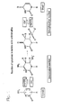
- the bisulfite method of mapping 5-methylcytosine uses the effect that sodium hydrogen sulfite reacts with cytosine but not or only poorly with 5-methyl-cytosine. Cytosine reacts with bisulfite to form a sulfonated cytosine reaction intermediate being prone to deamination resulting in a sulfonated uracil which can be desulfonated to uracil under alkaline conditions (see Fig. 1).
- uracil has the base pairing behavior of thymine different to the educt cytosine whereas 5-methylcytosine has the base pairing behavior of cytosine.
- Basic studies on the reaction of uracil and cytosine derivatives with bisulfite have been performed by Shapiro et al., JACS 92 (1970) 422-424 .
- Hayatsu, H., et al., J. Am. Chem. Soc. 92 (1970) 724-726 describe the reaction of cytosine with 3 M bisulfite at a pH value around 6 at a temperature of 80 °C for 30 min.
- Slae and Shapiro, J. Org. Chem. 43 (1978) 4197-4200 describe the deamination of cytidine with 1 M bisulfite around neutral pH at various temperatures whereby the reaction time is not described. There were no investigations of the deamination of cytosine or methyl-cytosine in nucleic acids in these documents.
- Raizis, A.M., et al., Anal. Biochem. 226 (1995) 161-166 disclose a bisulfite method for 5-methylcytosine mapping that minimizes template degradation. They investigate a method minimizing template degradation using 5 M bisulfite solutions in the presence of 100 mM hydroquinone at a pH value of 5 at 50°C. A maximum yield of PCR product was observed after 4 hours. Other conditions as increased pH and lower temperatures were also investigated.
- Clark, S.J., et al., Nucleic Acids Res 22 (1994) 2990-2997 disclose the use of a 3 to 4 M bisulfite solution at a pH value of 4.8 to 5.8 at a temperature of 37 to 72 °C for 8 to 16 hours in the bisulfite treatment of DNA.
- Tasheva, E.S., and Roufa, D.J., Mol. Cell. Biol. 14 (1994) 5636-5644 disclose the use of a 1 M bisulfite solution at a pH value of 5 at a temperature of 50 °C for 48 hours in the bisulfite treatment of fragments of genomic DNA.
- Grigg, G.W., DNA Seq 6 (1996) 189-198 discloses the use of a 3.1 M bisulfite solution at a pH value of 5 at a temperature of 50 °C for 16 hours in the bisulfite treatment of DNA.
- Olek, A., et al., Nucleic Acids Res. 24 (1996) 5064-5066 disclose a method for bisulfite base sequencing whereby bisulfite treatment and subsequent PCR steps are performed on material embedded in agarose beads.
- a 5 M bisulfite solution at a pH value of 5 at a temperature of 50 °C is used for 4 hours in the bisulfite treatment of DNA.
- Kits for performing bisulfite treatments are commercially available from Intergen, now distributed by Serologicals Corporation, Norcross, GA, USA, e.g. CpGenome TM DNA modification kit (http://www.serologicals.com/products/int_prod/index. html).
- the present invention provides a method for the conversion of a cytosine base, preferably cytosine bases, in a nucleic acid to an uracil base, preferably cytosine bases, whereby preferably a 5-methyl-cytosine base, preferably 5-methyl-cytosine bases, is not significantly converted,comprising the steps of
- the invention provides a solution with a pH value between 5.4 and 5.6 and comprising bisulfite in a concentration between 3 M and 6.25 M, uses thereof and kits comprising this solution.
- a "bisulfite reaction”, “bisulfite treatment” or “bisulfite method” shall mean a reaction for the conversion of a cytosine base, preferably cytosine bases, in a nucleic acid to an uracil base, preferably uracil bases, in the presence of bisulfite ions whereby preferably a 5-methyl-cytosine base, preferably 5-methyl-cytosine bases, is not significantly converted.
- This reaction for the detection of methylated cytosines is described in detail by Frommer et al., supra and Grigg and Clark, supra.
- the bisulfite reaction contains a deamination step and a desulfonation step which can be conducted separately or simultaneously (see Figure 1; Grigg and Clark, supra).
- the statement that 5-methyl-cytosine bases are not significantly converted shall only take the fact into account that it cannot be excluded that a small percentage of 5-methyl-cytosine bases is converted to uracil although it is intended to convert only and exclusively the (non-methylated) cytosine bases (Frommer et al., supra).
- the expert skilled in the art knows how to perform the bisulfite reaction, e.g. by referring to Frommer et al., supra or Grigg and Clark, supra who disclose the principal parameters of the bisulfite reaction.
- a buffer containing bisulfite ions, optionally chaotropic agents and optionally further reagents as an alcohol or stabilizers as hydroquinone are employed and the pH is in the acidic range.
- the concentration of bisulfite is between 0.1 and 6 M bisulfite, preferably between 1 M and 5.5 M, the concentration of the chaotropic agent is between 1 and 8 M, whereby preferably guanidinium salts are employed, the pH is in the acidic range, preferably between 4.5 and 6.5, the temperature is between 0 oC and 90 oC, preferably between room temperature (25 oC) and 90 oC, and the reaction time is between 30 min and 24 hours or 48 hours or even longer, but preferably between 1 hour and 24 hours.
- the desulfonation step is performed by adding an alkaline solution or buffer as e.g.
- a solution only containing a hydroxide e.g sodium hydroxide, or a solution containing ethanol, sodium chloride and sodium hydroxide (e.g. 38% EtOH, 100 mM NaCl, 200 mM NaOH) and incubating at room temperature or elevated temperatures for several min, preferably between 5 min and 60 min.
- a hydroxide e.g sodium hydroxide
- a solution containing ethanol, sodium chloride and sodium hydroxide e.g. 38% EtOH, 100 mM NaCl, 200 mM NaOH
- the method according to the invention allows a relatively short reaction time of the bisulfite reaction giving the possibility to perform a DNA assay within one working day.
- One parameter to speed the reaction is the temperature.
- a low pH value is of advantage.
- a reaction time of e.g. between 120 and 180 min is possible.
- the reaction under conditions according to the invention is more specific for cytosine compared to 5-methylcytosine as with standard conditions after 16 h. Additives for stabilization of the bisulfite reagent like hydroquinone are possible.
- the invention is related to a method for the conversion of a cytosine base, preferably cytosine bases in a nucleic acid to an uracil base, preferably uracil bases, whereby preferably a 5-methyl-cytosine base, preferably 5-methyl-cytosine bases, is not significantly converted, comprising the steps of
- the method may further comprise the step of desalting the solution comprising the deaminated and desulfonated nucleic acid. This can be achieved e.g. by ultrafiltration, gel filtration, precipiation as known to the expert skilled in the art or by binding to magnetic glass particles as described in WO 96/41811 .
- the temperature in the method according to the invention is between 75 any 85 °C.
- the concentration of bisulfite is between 3.2 M and 6 M, preferably between 4.75 M and 5.5 M.
- the pH value of the solution is between 5.25 and 5.75.
- the time period is between 1.75 and 3 hours.
- the time period is between 2 and 3 hours, preferably between 2 and 2.5 hours.
- the reaction is also possible in a time period between 0.75 and 3.5 hours.
- the temperature is 80 °C
- the concentration of bisulfite is 5 M
- the pH value of the solution is 5.5
- the time period is preferably between 2 and 2.5 or 3 hours, most preferred 2 hours.
- the method is preferably performed in solution, however, it is also feasible that the method according to the invention is performed while the nucleic acid is in a solid phase bound form, i.e. it is bound to a solid phase under suitable conditions.
- the solid phase may be a silicon oxide, preferably in the form of glass fleeces or fibers or magnetic glass particles as described in WO96/41811 , WO 00/32762 and WO 01/37291 .
- the principal method of performing a bisulfite treatment while the nucleic acid is bound to a solid phase is e.g. described e.g. in the European patent application with the number EP 02 019 097.1 and EP 02 028 114.3 .
- the nucleic acid is deoxyribonucleic acid (DNA), in particular genomic DNA or nucleic acid, i.e. the DNA or nucleic acid which is found in the organism's genome and is passed on to offspring as information necessary for survival.
- DNA deoxyribonucleic acid
- the phrase is used to distinguish between other types of DNA, such as found within plasmids.
- the source of the nucleic acid may be eukaryotic or prokarytic, preferably from vertebrates, particularly from mammalians, most preferred from animals or humans.
- the nucleic acid is obtained from a biological sample using the solid phases as described above and methods known to the expert in the field.
- the biological sample comprises cells from multicellular organisms as e.g. human and animal cells such as leucocytes, and immunologically active low and high molecular chemical compounds such as haptens, antigens, antibodies and nucleic acids, blood plasma, cerebral fluid, sputum, stool, biopsy specimens, bone marrow, oral rinses, blood serum, tissues, urine or mixtures thereof.
- the biological sample is a fluid from the human or animal body.
- the biological sample can be blood, blood plasma, blood serum, tissue or urine.
- the biological sample comprising the nucleic acids is lysed to create a mixture of biological compounds comprising nucleic acids and other components.
- Procedures for lysing biological samples are known by the expert and can be chemical, enzymatic or physical in nature. A combination of these procedures is applicable as well. For instance, lysis can be performed using ultrasound, high pressure, shear forces, alkali, detergents or chaotropic saline solutions, or proteases or lipases.
- lysis procedure to obtain nucleic acids special reference is made to Sambrook, J., et al., in "Molecular Cloning: A Laboratory Manual” (1989), eds. J. Sambrook, E. F. Fritsch and T.
- nucleic acids are isolated from the lysis mixture using the methods known to the expert skilled in the art, e.g. using solid phases as magnetic glass particles ( WO96/41811 ), and can then be subjected to the methods according to the invention, i.e. the bisulfite treatment according to the invention.
- Chaotropic agents are also used to lyse cells to prepare a mixture between nucleic acids and other biological substances (see e.g.
- the nucleic acid is amplified after the steps of the method according to the invention with the polymerase chain reaction (PCR: EP 0 201 184 ; EP-A-0 200 362 ; US 4,683,202 ).
- the amplification method may also be the Ligase Chain Reaction (LCR: Wu, D. Y., and Wallace, R. B., Genomics 4 (1989) 560-569 ; and Barany, F., Proc. Natl. Acad. Sci. USA 88 (1991) 189-193 ), Polymerase Ligase Chain Reaction ( Barany, F., PCR Methods Appl. 1 (1991) 5-16 ), Gap-LCR ( PCT Patent Publication No.
- amplification methods are the methylation specific PCR method (MSP) disclosed in US 5,786,146 which combines bisulfite treatment and allele-specific PCR (see e.g. US 5,137,806 , US 5,595,890 , US 5,639,611 ).
- MSP methylation specific PCR method
- the method may further comprise the step of detecting the amplified nucleic acid.
- the amplified nucleic acid may be determined or detected by standard analytical methods known to the person skilled in the art and described e.g. by Sambrook, J., et al., in "Molecular Cloning: A Laboratory Manual” (1989), eds. J. Sambrook, E. F. Fritsch and T. Maniatis, Cold Spring Harbor Laboratory Press, Cold Spring Harbor, NY ; Lottspeich and Zorbas, in "Bioanalytik” (1998), eds. L. a.
- the detection methods may include but are not limited to the binding or intercalating of specific dyes as ethidium bromide which intercalates into the double-stranded DNA and changes its fluorescence thereafter.
- the purified nucleic acids may also be separated by electrophoretic methods optionally after a restriction digest and visualized thereafter.
- probe-based assays which exploit the oligonucleotide hybridisation to specific sequences and subsequent detection of the hybrid. It is also possible to sequence the target nucleic acid after further steps known to the expert in the field. Other methods apply a diversity of nucleic acid sequences to a silicon chip to which specific probes are bound and yield a signal when a complementary sequence binds.
- the nucleic acid is detected by measuring the intensity of fluorescence light during amplification.
- This method entails the monitoring of real time fluorescence.
- a particularly preferred method exploiting simultaneous amplification and detection by measuring the intensity of fluorescent light is the TaqMan® method disclosed in WO 92/02638 and the corresponding US patents US 5,210,015 , US 5,804,375 , US 5,487,972 . This method exploits the exonuclease activity of a polymerase to generate a signal.
- the nucleic acid is detected by a process comprising contacting the sample with an oligonucleotide containing a sequence complementary to a region of the target nucleic acid and a labeled oligonucleotide containing a sequence complementary to a second region of the same target nucleic acid strand, but not including the nucleic acid sequence defined by the first oligonucleotide, to create a mixture of duplexes during hybridization conditions, wherein the duplexes comprise the target nucleic acid annealed to the first oligonucleotide and to the labeled oligonucleotide such that the 3'-end of the first oligonucleotide is adjacent to the 5'-end of the labeled oligonucleotide.
- this mixture is treated with a template-dependent nucleic acid polymerase having a 5' to 3' nuclease activity under conditions sufficient to permit the 5' to 3' nuclease activity of the polymerase to cleave the annealed, labeled oligonucleotide and release labeled fragments.
- the signal generated by the hydrolysis of the labeled oligonucleotide is detected and/ or measured.
- TaqMan® technology eliminates the need for a solid phase bound reaction complex to be formed and made detectable.
- the amplification and/ or detection reaction of the method according to the invention is a homogeneous solution-phase assay.
- the method is automated, i.e. the method carries out an automatable process as e.g. described in WO 99/16781 .
- Automatable process means that the steps of the process are suitable to be carried out with an apparatus or machine capable of operating with little or no external control or influence by a human being.
- Automated method means that the steps of the automatable method are carried out with an apparatus or machine capable of operating with little or no external control or influence by a human being. Only the preparation steps for the method may have to be done by hand, e.g. the storage containers have to filled up and put into place, the choice of the samples has to be done by a human being and further steps known to the expert in the field, e.g. the operation of the controlling computer.
- the apparatus or machine may e.g. add automatically liquids, mix the samples or carry out incubation steps at specific temperatures.
- a machine or apparatus is a robot controlled by a computer which carries out a program in which the single steps and commands are specified.
- the method is in a high-throughput format, i.e. the automated methods is carried out in a high-throughput format which means that the methods and the used machine or apparatus are optimized for a high-throughput of samples in a short time.
- the method according to the invention is used in diagnostics, for diagnostic analysis or for bioanalytics, or for the screening of tissue or fluids from the human or even animal body for the presence of a certain methylation pattern. Further, the method according to the invention is used to enhance the speed, accuracy or sensitivity of the detection of methylation sites in nucleic acids.
- a solution with a pH value between 5.25 and 5.75 and comprising bisulfite in a concentration between 3 M and 6.25 M is used in a reaction at a reaction temperature between 70 and 90 °C wherein a cytosine base, preferably cytosine bases, in a nucleic acid are converted to an uracil base, preferably uracil bases, in the presence of bisulfite ions whereby preferably a 5-methyl-cytosine base, preferably 5-methyl-cytosine bases, is not significantly converted.
- the concentration of bisulfite is between 3.2 M and 6 M, preferably between 4.75 M and 5.5 M.
- the pH value of the solution is 5.5 and the concentration of bisulfite is 5 M.
- the solution may also contain hydroquinone for stabilisation.
- the solution according to the invention is preferably an aqueous solution.
- the reaction temperature is between 75 and 85 °C.
- kits comprising a solution according to the invention.
- the solution has a pH value between 5.25 and 5.75, more preferably between 5.4 and 5.6, and comprises bisulfite in a concentration between 3 M and 6.25 M.
- the concentration of bisulfite is between 3.5 M and 6 M, preferably between 4.75 M and 5.5 M.
- the solution may optionally contain hydroquinone.
- the pH value of the solution is 5.5 and the concentration of bisulfite is 5 M.
- kits known in the art further comprise plastics ware which may be used during the bisulfite procedure as e.g. microtiter-plates in the 96 or 384 well format or reaction tubes manufactured e.g.
- the kit may further comprise a washing solution which is suitable for the washing step of the solid phase, in particular, the glass fleece or membrane or the magnetic glass particles. Often the washing solution is provided as a stock solution which has to be diluted before the use.
- the kit may further comprise an eluent, i.e. a solution or a buffer (e.g. TE, 10 mM Tris, 1 mM EDTA, pH 8.0) or pure water to elute the DNA or RNA bound to the solid phase. Further, additional reagents may be present which contain buffers suitable for use in the present invention.
- the kit according to the invention is used for a reaction wherein a cytosine base, preferably cytosine bases, in a nucleic acid are converted to an uracil base, preferably uracil bases, in the presence of bisulfite ions whereby preferably a 5-methyl-cytosine base, preferably 5-methyl-cytosine bases, are not significantly converted.
- a solution is provided with a pH value between 5.4 and 5.6 and comprising bisulfite in a concentration between 3.5 M and 6.25 M.
- the solution optionally contains hydroquinone or other radical scavengers.
- the concentration of bisulfite is between 3.75 M and 6 M, preferably between 4.75 M and 5.5 M.
- the pH value of the solution is 5.5 and the concentration of bisulfite is 5 M.
- Bisulfite reagent pH 5,0/50°C: ("Standard") 1,9g Na 2 S 2 O 5 2ml Millipore water 0,7ml 2M NaOH 0,5ml 1M hydroquinone (optional) addition of Millipore water to a volume of 4 ml
- Bisulfite reagent pH 5,5/80°C: 1,9g Na 2 S 2 O 5 1ml Millipore water 2ml 2M NaOH 0,5ml 1M hydroquinone (optional) addition of Millipore water to a volume of 4 ml
- Hydroquinone can be added optionally; it is not necessary if the reagent is prepared freshly for the experiment.
- oligonucleotides are synthesized using standard automated solid-phase synthesis procedure applying phosphoramidite chemistry.
- Conditions according to the invention lead to a similar product yield after 2 h reaction time as standard conditions after 8-16 h. Specificity of bisulfite reaction is significantly better for conditions according to the invention after 2 h compared to "standard conditions" after 16 h.
- Fluorescence measurements are normalized by dividing by an initial fluorescence measurement, i.e., the background fluorescence, obtained during a cycle early in the reaction while the fluorescence measurements between cycles appear to be relatively constant.
- the cycle number chosen for the initial fluorescence measurement is the same for all reactions compared, so that all measurements represent increases relative to the same reaction cycle.
- the number of cycles required to reach a particular threshold value (C T value or crossing point) is inversely proportional to the logarithm of (1 + E).
- the C T value represents a measure of the reaction efficiency that allows comparisons between reactions.
- a decrease in the C T value which means that the reaction reached the threshold value in fewer cycles, indicates an increase in reaction efficiency.
- the C T is defined herein as the number of amplification cycles carried out until the fluorescence exceeded an arbitrary fluorescence level (AFL).
- AFL was chosen close to the baseline fluorescence level, but above the range of random fluctuations in the measured fluorescence, so that the reaction kinetics were measured during the geometric growth phase of the amplification. Accumulation of amplified product in later cycles inhibits the reaction and eventually leads to a reaction plateau. An AFL of 1.5 was chosen for all reactions.
- a PCR amplification consists of discrete cycles and the fluorescence measurements are carried out once per cycle, the measured fluorescence typically increases from below the AFL to above the AFL in a single cycle.
- an "exact" number of cycles to reach the AFL threshold referred to herein as the C T value or crossing point, was calculated by interpolating fluorescence measurements between cycles.
- 112 ⁇ l of the denatured DNA are mixed with 200 ⁇ l bisulfite reagent (2.5M sodium disulfite, 125 mM hydroquinone, pH 5.1) and incubated for 20 h at 50 °C ("Standard method") or 112 ⁇ l of the denatured DNA are mixed with 200 ⁇ l bisulfite reagent (2.5M sodium disulfite, 125 mM hydroquinone, pH 5.5) and incubated for 2h at 80°C ("BIS-METHOD").
- bisulfite reagent 2.5M sodium disulfite, 125 mM hydroquinone, pH 5.1
- 312 ⁇ l of the deaminated DNA (from both methods respectively) are mixed with 600 ⁇ l binding buffer (MagNAPure DNA Isolation Kit I, Roche Cat. Nr. 3 003 990) and 75 ⁇ l magnetic glass particle solution (MagNAPure DNA Isolation Kit I) and incubated for 15min/ room temperature with continuous mixing in order to bind the nucleic acid to the MGFS according to the method described in the European patent applications with the numbers EP02019097.1 or EP02028114.3 . Thereafter, the magnetic glass particles (MGPs) are washed three times with 1 ml 70% ethanol. Bound free separation is done in a magnetic separator (Roche Cat.1641794).
- LightCycler® FastStart DNA Master HybridizationProbe 1x (Roche 2239272), 2mM MgCl 2 , forward Primer 0.5 ⁇ M, reversed Primer 0.5 ⁇ M, donor probe 250 nM, acceptor probe 250 nM, template 10 ⁇ l, total PCR volume 20 ⁇ l.
Abstract
Description
- The present application is directed to a method for performing a bisulfite reaction to determine methylation positions in a nucleic acid, i.e. methylated and non-methylated cytosines, whereby the nucleic acid is incubated in a solution comprising the nucleic acid for a time period of 1.5 to 3.5 hours at a temperature between 70 and 90 °C, whereby the concentration of bisulfite in the solution is between 3 M and 6.25 M and whereby the pH value of the solution is between 5.0 and 6.0 whereby the nucleic acid, i.e. the cytosine bases in the nucleic acid, is deaminated. Then the solution comprising the deaminated nucleic acid is desulfonated and preferably desalted. The application is further related to kit with a solution comprising bisulfite with a certain pH and uses thereof as well as a kit comprising the solution.
- Genes constitute only a small proportion of the total mammalian genome, and the precise control of their expression in the presence of an overwhelming background of noncoding deoxyribonucleic acid (DNA) presents a substantial problem for their regulation. Noncoding DNA, containing introns, repetitive elements, and potentially active transposable elements requires effective mechanisms for its long term silencing. Mammals appear to have taken advantage of the possibilities afforded by cytosine methylation to provide a heritable mechanism for altering DNA-protein interactions to assist in such silencing. DNA methylation is essential for the development of mammals and plays a potential role during aging and cancer. The involvement of methylation in the regulation of gene expression and as an epigenetic modification marking imprinted genes is well established. In mammals, methylation occurs only at cytosines residues and more specifically only on cytosine residues adjacent to a guanosine residue, i.e. at the sequence CG. The detection and mapping of DNA methylation sites are essential steps towards understanding the molecular signals which indicate whether a given sequence is methylated.
- This is currently accomplished by the so-called bisulfite method described by Frommer, M., et al., Proc. Natl. Acad. Sci. USA 89 (1992) 1827-1831, for the detection of 5-methyl-cytosines. The bisulfite method of mapping 5-methylcytosine uses the effect that sodium hydrogen sulfite reacts with cytosine but not or only poorly with 5-methyl-cytosine. Cytosine reacts with bisulfite to form a sulfonated cytosine reaction intermediate being prone to deamination resulting in a sulfonated uracil which can be desulfonated to uracil under alkaline conditions (see Fig. 1). It is common knowledge that uracil has the base pairing behavior of thymine different to the educt cytosine whereas 5-methylcytosine has the base pairing behavior of cytosine. This makes the discrimination of methylated or non-methylated cytosines possible by e.g. bisulfite genomic sequencing (Grigg, G., and Clark, S., Bioessays 16 (1994) 431-436; Grigg, G.W., DNA Seq. 6 (1996) 189-198) or methylation specific PCR (MSP) disclosed in
US 5,786,146 . Basic studies on the reaction of uracil and cytosine derivatives with bisulfite have been performed by Shapiro et al., JACS 92 (1970) 422-424. - There are various documents addressing specific aspects of the bisulfite reaction.
- Hayatsu, H., et al., Biochemistry 9 (1970) 2858-2865 reacted uracil, cytosine or their derivatives with 1 M bisulfite, at a pH value around 6, at 37°C for 24 hours. Hayatsu, H., et al., J. Am. Chem. Soc. 92 (1970) 724-726 describe the reaction of cytosine with 3 M bisulfite at a pH value around 6 at a temperature of 80 °C for 30 min. Slae and Shapiro, J. Org. Chem. 43 (1978) 4197-4200 describe the deamination of cytidine with 1 M bisulfite around neutral pH at various temperatures whereby the reaction time is not described. There were no investigations of the deamination of cytosine or methyl-cytosine in nucleic acids in these documents.
- Paulin, R., et al., Nucl. Acids Res. 26 (1998) 5009-5010 investigate the effects of urea on the efficiency of bisulfite-mediated sequencing of 5-methylcytosine in DNA. The DNA is reacted with 3.44 M bisulfite in the presence of 5.36 M urea and 0.5 mM hydroquinone, at a pH value of 5.0 at a temperature of 55 °C for 15 hours.
- Raizis, A.M., et al., Anal. Biochem. 226 (1995) 161-166 disclose a bisulfite method for 5-methylcytosine mapping that minimizes template degradation. They investigate a method minimizing template degradation using 5 M bisulfite solutions in the presence of 100 mM hydroquinone at a pH value of 5 at 50°C. A maximum yield of PCR product was observed after 4 hours. Other conditions as increased pH and lower temperatures were also investigated.
- Grunau, C., et al., Nucleic Acids Res 29 (2001) e65-5, , perform a systematic investigation of critical experimental parameters of the bisulfite reaction. They investigate bisulfite solutions of 3.87 to 4.26 and 5.2 to 5.69 M at a pH value of 5. Temperatures that were tested are 15, 35, 55, 80, 85 and 95 °C for 1, 4 and 18 hours. DNA degradation is a problem in these investigations.
- Wang, R.Y., et al., Nucleic Acids Res. 8 (1980) 4777-4790 disclose the use of a 3 M bisulfite solution at a pH value of 5.5 at a temperature of 37°C for various time periods in the bisulfite treatment of DNA. Feil, R., et al., Nucleic Acids Res 22 (1994) 695-696 disclose the use of a 3.5 M bisulfite solution at a pH value of 5 at a temperature of 0°C for 24 hours in the bisulfite treatment of DNA. Clark, S.J., et al., Nucleic Acids Res 22 (1994) 2990-2997, disclose the use of a 3 to 4 M bisulfite solution at a pH value of 4.8 to 5.8 at a temperature of 37 to 72 °C for 8 to 16 hours in the bisulfite treatment of DNA. Tasheva, E.S., and Roufa, D.J., Mol. Cell. Biol. 14 (1994) 5636-5644 disclose the use of a 1 M bisulfite solution at a pH value of 5 at a temperature of 50 °C for 48 hours in the bisulfite treatment of fragments of genomic DNA. Grigg, G.W., DNA Seq 6 (1996) 189-198 discloses the use of a 3.1 M bisulfite solution at a pH value of 5 at a temperature of 50 °C for 16 hours in the bisulfite treatment of DNA. Komiyama, M., and Oshima, S., Tetrahedron Letters 35 (1994) 8185-8188 disclose the use of a 1 M bisulfite solution at a pH value of 5 at a temperature of 37 °C for 4 hours in the bisulfite treatment of DNA whereby diethylenetriamine is present.
- Olek, A., et al., Nucleic Acids Res. 24 (1996) 5064-5066 disclose a method for bisulfite base sequencing whereby bisulfite treatment and subsequent PCR steps are performed on material embedded in agarose beads. A 5 M bisulfite solution at a pH value of 5 at a temperature of 50 °C is used for 4 hours in the bisulfite treatment of DNA.
- A review of DNA methylation analysis can be found in Oakeley, E. J., Pharmacol. Ther. 84 (1999) 389-400.
- Different additional components in the bisulfite mixture are disclosed by
WO 01/98528 WO02/31186 - Kits for performing bisulfite treatments are commercially available from Intergen, now distributed by Serologicals Corporation, Norcross, GA, USA, e.g. CpGenome™ DNA modification kit (http://www.serologicals.com/products/int_prod/index. html).
- All prior art methods for the bisulfite treatment have disadvantages. Therefore, the problem to be solved by the present invention was to provide a method which overcomes the disadvantages of the prior art methods.
- The present invention provides a method for the conversion of a cytosine base, preferably cytosine bases, in a nucleic acid to an uracil base, preferably cytosine bases, whereby preferably a 5-methyl-cytosine base, preferably 5-methyl-cytosine bases, is not significantly converted,comprising the steps of
- a) incubating a solution comprising the nucleic acid for a time period of 1.5 to 3.5 hours at a temperature between 70 and 90 °C, whereby the concentration of bisulfite in the solution is between 3 M and 6.25 M and whereby the pH value of the solution is between 5.0 and 6.0 whereby the nucleic acid is deaminated, and
- b) incubating the solution comprising the deaminated nucleic acid under alkaline conditions whereby the deaminated nucleic acid is desulfonated.
- Further, the invention provides a solution with a pH value between 5.4 and 5.6 and comprising bisulfite in a concentration between 3 M and 6.25 M, uses thereof and kits comprising this solution.
- As known to the expert skilled in the art and according to the invention, the term "bisulfite" is used interchangeably for "hydrogensulfite".
- According to the invention the term a "bisulfite reaction", "bisulfite treatment" or "bisulfite method" shall mean a reaction for the conversion of a cytosine base, preferably cytosine bases, in a nucleic acid to an uracil base, preferably uracil bases, in the presence of bisulfite ions whereby preferably a 5-methyl-cytosine base, preferably 5-methyl-cytosine bases, is not significantly converted. This reaction for the detection of methylated cytosines is described in detail by Frommer et al., supra and Grigg and Clark, supra. The bisulfite reaction contains a deamination step and a desulfonation step which can be conducted separately or simultaneously (see Figure 1; Grigg and Clark, supra). The statement that 5-methyl-cytosine bases are not significantly converted shall only take the fact into account that it cannot be excluded that a small percentage of 5-methyl-cytosine bases is converted to uracil although it is intended to convert only and exclusively the (non-methylated) cytosine bases (Frommer et al., supra). The expert skilled in the art knows how to perform the bisulfite reaction, e.g. by referring to Frommer et al., supra or Grigg and Clark, supra who disclose the principal parameters of the bisulfite reaction. From Grunau et al., supra, it is known to the expert in the field what variations of the bisulfite method are possible. In summary, in the deamination step a buffer containing bisulfite ions, optionally chaotropic agents and optionally further reagents as an alcohol or stabilizers as hydroquinone are employed and the pH is in the acidic range. The concentration of bisulfite is between 0.1 and 6 M bisulfite, preferably between 1 M and 5.5 M, the concentration of the chaotropic agent is between 1 and 8 M, whereby preferably guanidinium salts are employed, the pH is in the acidic range, preferably between 4.5 and 6.5, the temperature is between 0 oC and 90 oC, preferably between room temperature (25 oC) and 90 oC, and the reaction time is between 30 min and 24 hours or 48 hours or even longer, but preferably between 1 hour and 24 hours. The desulfonation step is performed by adding an alkaline solution or buffer as e.g. a solution only containing a hydroxide, e.g sodium hydroxide, or a solution containing ethanol, sodium chloride and sodium hydroxide (e.g. 38% EtOH, 100 mM NaCl, 200 mM NaOH) and incubating at room temperature or elevated temperatures for several min, preferably between 5 min and 60 min.
- The method according to the invention allows a relatively short reaction time of the bisulfite reaction giving the possibility to perform a DNA assay within one working day. One parameter to speed the reaction is the temperature. To decrease the DNA degradation process, a low pH value is of advantage. By the use of a 5 M bisulfite solution of a pH value of 5.5 at a temperature of approximately 80°C, a reaction time of e.g. between 120 and 180 min is possible. Further, the reaction under conditions according to the invention is more specific for cytosine compared to 5-methylcytosine as with standard conditions after 16 h. Additives for stabilization of the bisulfite reagent like hydroquinone are possible.
- The invention is related to a method for the conversion of a cytosine base, preferably cytosine bases in a nucleic acid to an uracil base, preferably uracil bases, whereby preferably a 5-methyl-cytosine base, preferably 5-methyl-cytosine bases, is not significantly converted, comprising the steps of
- a) incubating a solution comprising the nucleic acid for a time period of 1.5 to 3.5 hours at a temperature between 70 and 90 °C, whereby the concentration of bisulfite in the solution is between 3 M and 6.25 M and whereby the pH value of the solution is between 5.0 and 6.0 whereby the nucleic acid is deaminated, and
- b) incubating the solution comprising the deaminated nucleic acid under alkaline conditions whereby the deaminated nucleic acid is desulfonated.
- In a preferred embodiment of the invention, the method may further comprise the step of desalting the solution comprising the deaminated and desulfonated nucleic acid. This can be achieved e.g. by ultrafiltration, gel filtration, precipiation as known to the expert skilled in the art or by binding to magnetic glass particles as described in
WO 96/41811 - In a preferred embodiment of the invention, the temperature in the method according to the invention is between 75 any 85 °C. In another preferred embodiment of the invention, the concentration of bisulfite is between 3.2 M and 6 M, preferably between 4.75 M and 5.5 M. In another preferred embodiment of the invention, the pH value of the solution is between 5.25 and 5.75. In another preferred embodiment of the invention, the time period is between 1.75 and 3 hours. In another preferred embodiment of the invention, the time period is between 2 and 3 hours, preferably between 2 and 2.5 hours. The reaction is also possible in a time period between 0.75 and 3.5 hours. In the most preferred embodiment of the invention, in step a) the temperature is 80 °C, the concentration of bisulfite is 5 M, the pH value of the solution is 5.5 and the time period is preferably between 2 and 2.5 or 3 hours, most preferred 2 hours.
- The method is preferably performed in solution, however, it is also feasible that the method according to the invention is performed while the nucleic acid is in a solid phase bound form, i.e. it is bound to a solid phase under suitable conditions. The solid phase may be a silicon oxide, preferably in the form of glass fleeces or fibers or magnetic glass particles as described in
WO96/41811 WO 00/32762 WO 01/37291 EP 02 019 097.1 EP 02 028 114.3 - The expert skilled in the art knows how to perform the bisulfite reaction, e.g. by referring to Frommer et al., supra, Grigg and Clark, supra or Grunau et al., supra who disclose the principal parameters of the bisulfite reaction.
- In an embodiment of the invention, the nucleic acid is deoxyribonucleic acid (DNA), in particular genomic DNA or nucleic acid, i.e. the DNA or nucleic acid which is found in the organism's genome and is passed on to offspring as information necessary for survival. The phrase is used to distinguish between other types of DNA, such as found within plasmids. The source of the nucleic acid may be eukaryotic or prokarytic, preferably from vertebrates, particularly from mammalians, most preferred from animals or humans.
- In an embodiment of the invention the nucleic acid is obtained from a biological sample using the solid phases as described above and methods known to the expert in the field. The biological sample comprises cells from multicellular organisms as e.g. human and animal cells such as leucocytes, and immunologically active low and high molecular chemical compounds such as haptens, antigens, antibodies and nucleic acids, blood plasma, cerebral fluid, sputum, stool, biopsy specimens, bone marrow, oral rinses, blood serum, tissues, urine or mixtures thereof. In a preferred embodiment of the invention the biological sample is a fluid from the human or animal body. The biological sample can be blood, blood plasma, blood serum, tissue or urine. The biological sample comprising the nucleic acids is lysed to create a mixture of biological compounds comprising nucleic acids and other components. Procedures for lysing biological samples are known by the expert and can be chemical, enzymatic or physical in nature. A combination of these procedures is applicable as well. For instance, lysis can be performed using ultrasound, high pressure, shear forces, alkali, detergents or chaotropic saline solutions, or proteases or lipases. For the lysis procedure to obtain nucleic acids, special reference is made to Sambrook, J., et al., in "Molecular Cloning: A Laboratory Manual" (1989), eds. J. Sambrook, E. F. Fritsch and T. Maniatis, Cold Spring Harbour Laboratory Press, Cold Spring Harbour, NY; and Ausubel, F., et al., in "Current protocols in molecular biology" (1994), eds. F. Ausubel, R. Brent and K. R.E., Wiley & Sons, New York. Then the nucleic acids are isolated from the lysis mixture using the methods known to the expert skilled in the art, e.g. using solid phases as magnetic glass particles (
WO96/41811 EP 0 389 063 - In a preferred embodiment of the invention, the nucleic acid is amplified after the steps of the method according to the invention with the polymerase chain reaction (PCR:
EP 0 201 184EP- ;A-0 200 362US 4,683,202 ). The amplification method may also be the Ligase Chain Reaction (LCR: Wu, D. Y., and Wallace, R. B., Genomics 4 (1989) 560-569; and Barany, F., Proc. Natl. Acad. Sci. USA 88 (1991) 189-193), Polymerase Ligase Chain Reaction (Barany, F., PCR Methods Appl. 1 (1991) 5-16), Gap-LCR (PCT Patent Publication No. WO 90/01069 EP- ), 3SR (Kwoh, D. Y., et al., Proc. Natl. Acad. Sci. USA 86 (1989) 1173-1177; Guatelli, J. C., et al., Proc. Natl. Acad. Sci. USA 87 (1990) 1874-1878; PCT Patent Publication No.A 0 439 182WO 92/0880A U.S. Pat. No. US 5,130,238 ). Further, there are strand displacement amplification (SDA), transciption mediated amplification (TMA), and Q□-amplification (for a review see e.g. Whelen, A.C., and Persing, D.H., Annu. Rev. Microbiol. 50 (1996) 349-373; Abramson, R. D., and Myers, T.W., Curr. Opin. Biotechnol. 4 (1993) 41-47). Particularly preferred amplification methods according to the invention are the methylation specific PCR method (MSP) disclosed inUS 5,786,146 which combines bisulfite treatment and allele-specific PCR (see e.g.US 5,137,806 ,US 5,595,890 ,US 5,639,611 ). - In a preferred embodiment, the method may further comprise the step of detecting the amplified nucleic acid. The amplified nucleic acid may be determined or detected by standard analytical methods known to the person skilled in the art and described e.g. by Sambrook, J., et al., in "Molecular Cloning: A Laboratory Manual" (1989), eds. J. Sambrook, E. F. Fritsch and T. Maniatis, Cold Spring Harbor Laboratory Press, Cold Spring Harbor, NY; Lottspeich and Zorbas, in "Bioanalytik" (1998), eds. L. a. Zorbas, Spektrum Akademischer Verlag, Heidelberg, Berlin, Germany; or by Ausubel, F., et al., in "Current protocols in molecular biology" (1994), eds. F. Ausubel, R. Brent and K. R.E., Wiley & Sons Verlag, New York. There may be also further purification steps before the target nucleic acid is detected e.g. a precipitation step. The detection methods may include but are not limited to the binding or intercalating of specific dyes as ethidium bromide which intercalates into the double-stranded DNA and changes its fluorescence thereafter. The purified nucleic acids may also be separated by electrophoretic methods optionally after a restriction digest and visualized thereafter. There are also probe-based assays which exploit the oligonucleotide hybridisation to specific sequences and subsequent detection of the hybrid. It is also possible to sequence the target nucleic acid after further steps known to the expert in the field. Other methods apply a diversity of nucleic acid sequences to a silicon chip to which specific probes are bound and yield a signal when a complementary sequence binds.
- In a particularly preferred embodiment of the invention, the nucleic acid is detected by measuring the intensity of fluorescence light during amplification. This method entails the monitoring of real time fluorescence. A particularly preferred method exploiting simultaneous amplification and detection by measuring the intensity of fluorescent light is the TaqMan® method disclosed in
WO 92/02638 US patents US 5,210,015 ,US 5,804,375 ,US 5,487,972 . This method exploits the exonuclease activity of a polymerase to generate a signal. In detail, the nucleic acid is detected by a process comprising contacting the sample with an oligonucleotide containing a sequence complementary to a region of the target nucleic acid and a labeled oligonucleotide containing a sequence complementary to a second region of the same target nucleic acid strand, but not including the nucleic acid sequence defined by the first oligonucleotide, to create a mixture of duplexes during hybridization conditions, wherein the duplexes comprise the target nucleic acid annealed to the first oligonucleotide and to the labeled oligonucleotide such that the 3'-end of the first oligonucleotide is adjacent to the 5'-end of the labeled oligonucleotide. Then this mixture is treated with a template-dependent nucleic acid polymerase having a 5' to 3' nuclease activity under conditions sufficient to permit the 5' to 3' nuclease activity of the polymerase to cleave the annealed, labeled oligonucleotide and release labeled fragments. The signal generated by the hydrolysis of the labeled oligonucleotide is detected and/ or measured. TaqMan® technology eliminates the need for a solid phase bound reaction complex to be formed and made detectable. In more general terms, the amplification and/ or detection reaction of the method according to the invention is a homogeneous solution-phase assay. Further preferred methods are the formats used in the LightCycler® instrument (see e.g.US 6,174,670 ). Particularly preferred is the use of bisulfite treatment, amplification with or without methylation specific primers in the presence of a methylation-specific probe and real-time fluorescence detection as described inUS 6,331,393 . - In a preferred embodiment of the present invention, the method is automated, i.e. the method carries out an automatable process as e.g. described in
WO 99/16781 - Preferably, the method according to the invention is used in diagnostics, for diagnostic analysis or for bioanalytics, or for the screening of tissue or fluids from the human or even animal body for the presence of a certain methylation pattern. Further, the method according to the invention is used to enhance the speed, accuracy or sensitivity of the detection of methylation sites in nucleic acids.
- In other embodiment of the invention, a solution with a pH value between 5.25 and 5.75 and comprising bisulfite in a concentration between 3 M and 6.25 M is used in a reaction at a reaction temperature between 70 and 90 °C wherein a cytosine base, preferably cytosine bases, in a nucleic acid are converted to an uracil base, preferably uracil bases, in the presence of bisulfite ions whereby preferably a 5-methyl-cytosine base, preferably 5-methyl-cytosine bases, is not significantly converted. Preferably, the concentration of bisulfite is between 3.2 M and 6 M, preferably between 4.75 M and 5.5 M. In the most preferred embodiment, the pH value of the solution is 5.5 and the concentration of bisulfite is 5 M. The solution may also contain hydroquinone for stabilisation. The solution according to the invention is preferably an aqueous solution. Preferably, the reaction temperature is between 75 and 85 °C.
- In another embodiment of the invention a kit comprising a solution according to the invention. Preferably, The solution has a pH value between 5.25 and 5.75, more preferably between 5.4 and 5.6, and comprises bisulfite in a concentration between 3 M and 6.25 M. Preferably, the concentration of bisulfite is between 3.5 M and 6 M, preferably between 4.75 M and 5.5 M. The solution may optionally contain hydroquinone. In the most preferred embodiment, the pH value of the solution is 5.5 and the concentration of bisulfite is 5 M. Such kits known in the art further comprise plastics ware which may be used during the bisulfite procedure as e.g. microtiter-plates in the 96 or 384 well format or reaction tubes manufactured e.g. by Eppendorf, Hamburg, Germany. The kit may further comprise a washing solution which is suitable for the washing step of the solid phase, in particular, the glass fleece or membrane or the magnetic glass particles. Often the washing solution is provided as a stock solution which has to be diluted before the use. The kit may further comprise an eluent, i.e. a solution or a buffer (e.g. TE, 10 mM Tris, 1 mM EDTA, pH 8.0) or pure water to elute the DNA or RNA bound to the solid phase. Further, additional reagents may be present which contain buffers suitable for use in the present invention. Preferably, the kit according to the invention is used for a reaction wherein a cytosine base, preferably cytosine bases, in a nucleic acid are converted to an uracil base, preferably uracil bases, in the presence of bisulfite ions whereby preferably a 5-methyl-cytosine base, preferably 5-methyl-cytosine bases, are not significantly converted.
- In another embodiment of the invention, a solution is provided with a pH value between 5.4 and 5.6 and comprising bisulfite in a concentration between 3.5 M and 6.25 M. The solution optionally contains hydroquinone or other radical scavengers. Preferably, the concentration of bisulfite is between 3.75 M and 6 M, preferably between 4.75 M and 5.5 M. In the most preferred embodiment, the pH value of the solution is 5.5 and the concentration of bisulfite is 5 M.
- The following examples, references, sequence listing and figures are provided to aid the understanding of the present invention, the true scope of which is set forth in the appended claims.
-
- Fig. 1:
- The steps of the bisulfite method
- Fig. 2 to 14:
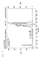
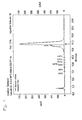
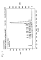
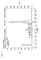
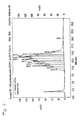
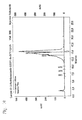
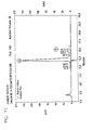
- HPLC profiles of the reaction mixtures after certain time periods as indicated in the examples
-
Bisulfite reagent pH = 5,0/50°C:
("Standard")1,9g Na2S2O5
2ml Millipore water
0,7ml 2M NaOH
0,5ml 1M hydroquinone (optional)
addition of Millipore water to a volume of 4 mlBisulfite reagent pH = 5,5/80°C: 1,9g Na2S2O5
1ml Millipore water
2ml 2M NaOH
0,5ml 1M hydroquinone (optional)
addition of Millipore water to a volume of 4 ml - Hydroquinone can be added optionally; it is not necessary if the reagent is prepared freshly for the experiment.
- The oligonucleotides are synthesized using standard automated solid-phase synthesis procedure applying phosphoramidite chemistry.
-
- SEQ ID NO: 1: 5'-d(GAGGGGCGCCCTGGAGTCCC)-3' (sense strand)
- SEQ ID NO: 2:5'-d(GGGACTCCAGGGCGCCCCTC)-3' (antisense strand)
- SEQ ID NO: 3: 5'-d(GAGGGGUGUUUTGGAGTUUU)-3' (sense strand C converted to U (product))
- SEQ ID NO: 4: 5'-d(GGGAUTUUAGGGUGUUUUTU)-3'(antisense strand C converted to U (product))
- SEQ ID NO: 5: 5'-d(T5CT5)-3'
- SEQ ID NO: 6: 5'-d(T5CMeT5)-3'
- SEQ ID NO: 7: 5'-d(T5UT5)-3'
- SEQ ID NO: 8: 5'-d(T11)-3'
- Ca. 5 nmole of a single stranded oligonucleotide or 5 nmole of each strand of a double stranded oligonucleotide are dissolved in 20µl of Millipore water, then 200µl of the bisulfite reagent are added. Thereafter the reaction tube is placed into a thermomixer (50°C or 80°C; 600rpm). After t = x hours the reaction is stopped by addition of 500µl of 2,5M NaOH (desulfonation). After 30min at r.t. the reaction mixture is desalted over a Sephadex G25 column. The oligonucleotide containing fraction is evaporated and dissolved in 200µl of Millipore water to be analyzed by HPLC.
-
Analytical HPLC: column: Dionex DNA Pac PA-100 SEL buffer A: 0,01M NaOH, 0,2 M NaCl buffer B: 0,01M NaOH, 1M NaCl gradient: 50-100% B in 25min - Data evaluation: HPLC chromatograms are compared by HPLC-area% of the product peak at t = x and are shown in Figs. 2 to 12
- A double determination was performed according to the standard protocol described above with the GSTP1 sequences SEQ ID NO:1 which is the sense strand and SEQ ID NO:2 which is the antisense strand, the mean average values are calculated.
t[min] HPLC area% product Figure 10 0 2 30 27,0 3 60 77,5 4 90 87,5 5 120 89,9 6 150 90,4 7 180 88,6 8 - A double determination was performed according to the standard protocol described above with the GSTP1 sequences SEQ ID NO:1 which is the sense strand and SEQ ID NO:2 which is the antisense strand, the mean average values are calculated.
t [h] HPLC area% product Figure 1 15,6 9 2 41,2 10 4 72,5 8 89,4 11 16 91,8 12 20 85,0 - In order to evaluate the specificity of bisulfite reaction, the oligonucleotide 5'-T5CMeT5-3' (SEQ ID NO: 6) was evaluated under the indicated conditions. The results were as follows:
- 5
M bisulfite pH 5,0 / T = 50°C / t = 16 h (standard conditions) (see Fig. 13 for an exemplary chromatogram):sample HPLC area% T5CMe T5 HPLC area% T11 ratio HPLC area% T11 / T5CMeT5 1 82,4 5,20 0,0630 2 83,3 5,11 0,0614 3 80,2 5,52 0,0688 4 80,5 5,92 0,0735 Mean value 81,6 5,44 0,0667 - 5 M bisulfite pH 5,5 / T = 80°C / t = 2 h (optimized conditions) (see Fig. 14 for an exemplary chromatogram)
sample HPLC area% T5CMeT5 HPLC area% T11 ratio HPLC area% T11 / T5CMeT5 1 89,9 2,65 0,0295 2 89,5 2,46 0,0275 3 90,2 2,24 0,0248 4 89,7 2,88 0,0322 Mean value 89,8 2,56 0,0285 - Conditions according to the invention lead to a similar product yield after 2 h reaction time as standard conditions after 8-16 h. Specificity of bisulfite reaction is significantly better for conditions according to the invention after 2 h compared to "standard conditions" after 16 h.
- The fact that the bisulfite reaction has worked and converted non-methylated cytosines to uracil can also be demonstrated by a polymerase chain reaction whereby primers are used which are specific to a region of the nucleic acid sequence wherein non-methylated cytosines have been converted to uracils, i.e. the base adenine in the primer is opposite to the uracil being the bisulfite reaction product from non-methylated cytosines. In case of incomplete conversion, the primer could not hybridize to this region as there would be cytosines not matching the adenine bases in the primer. This would have the effect that no PCR product would be obtained.
- An improved method to perform rapid polymerase chain reactions is disclosed e.g. in
US 6,174,670 and is used in the LightCycler® instrument (Roche, Mannheim, Germany). In this method, two labeled probes can come into close proximity in an amplificate dependent manner so that the two labels can perform a fluorescence energy transfer (FRET). The amount of the amplificate thereby correlates with the intensity of the emitted light of a certain wavelength. This specific PCR method can therefore be used to analyze whether a complete conversion of non-methylated cytosines was obtained, by e.g. analyzing the promoter region of the glutathion-S-transferase π gene (see e.g.US 5,552,277 , Genbank accession code M24485 and Morrow et al. (1989) ) using suitable probes and primers. However, the expert skilled in the art knows that other methods can be used for this evaluation as well. Fluorescence measurements are normalized by dividing by an initial fluorescence measurement, i.e., the background fluorescence, obtained during a cycle early in the reaction while the fluorescence measurements between cycles appear to be relatively constant. The cycle number chosen for the initial fluorescence measurement is the same for all reactions compared, so that all measurements represent increases relative to the same reaction cycle. In the early cycles of a polymerase chain reaction amplification, the number of target molecules can be described by the geometric equation Ni = No x (1 + E)i, where No = the number of target molecules at the start of the reaction, Ni = the number of target molecules at the completion of the i-th cycle, E = the efficiency of the amplification (0 ≤ E ≤ 1). During this geometric growth phase of the amplification, the number of cycles required to reach a particular threshold value (CT value or crossing point) is inversely proportional to the logarithm of (1 + E). Thus, the CT value represents a measure of the reaction efficiency that allows comparisons between reactions. A decrease in the CT value, which means that the reaction reached the threshold value in fewer cycles, indicates an increase in reaction efficiency. As the increase in amplification product is monitored by measuring the increase in reaction fluorescence, the CT is defined herein as the number of amplification cycles carried out until the fluorescence exceeded an arbitrary fluorescence level (AFL). The AFL was chosen close to the baseline fluorescence level, but above the range of random fluctuations in the measured fluorescence, so that the reaction kinetics were measured during the geometric growth phase of the amplification. Accumulation of amplified product in later cycles inhibits the reaction and eventually leads to a reaction plateau. An AFL of 1.5 was chosen for all reactions. Because a PCR amplification consists of discrete cycles and the fluorescence measurements are carried out once per cycle, the measured fluorescence typically increases from below the AFL to above the AFL in a single cycle. To improve the precision of the measurements, an "exact" number of cycles to reach the AFL threshold, referred to herein as the CT value or crossing point, was calculated by interpolating fluorescence measurements between cycles. - 100 µl of methylated DNA (Intergen, distributed by Serologicals Corporation, Norcross, GA, USA; Cat S 7821) dilution (100ng/ assay spiked in 1000ng human DNA background, Roche Cat.1691112; 4 replicates per method), and 12 µl 2 M NaOH are mixed and incubated for 15 min at 37 ° C.
- 112 µl of the denatured DNA are mixed with 200 µl bisulfite reagent (2.5M sodium disulfite, 125 mM hydroquinone, pH 5.1) and incubated for 20 h at 50 °C ("Standard method")
or
112 µl of the denatured DNA are mixed with 200 µl bisulfite reagent (2.5M sodium disulfite, 125 mM hydroquinone, pH 5.5) and incubated for 2h at 80°C ("BIS-METHOD"). - 312 µl of the deaminated DNA (from both methods respectively) are mixed with 600 µl binding buffer (MagNAPure DNA Isolation Kit I, Roche Cat. Nr. 3 003 990) and 75µl magnetic glass particle solution (MagNAPure DNA Isolation Kit I) and incubated for 15min/ room temperature with continuous mixing in order to bind the nucleic acid to the MGFS according to the method described in the European patent applications with the numbers
EP02019097.1 EP02028114.3 - LightCycler® FastStart DNA Master HybridizationProbe 1x (Roche 2239272), 2mM MgCl2, forward Primer 0.5 µM, reversed Primer 0.5 µM,
donor probe 250 nM,acceptor probe 250 nM, template 10 µl,total PCR volume 20 µl. -
55 cycles 95°C/10s 65°C/10s - signal acquisition 72°C/ 10s Ramp time 20°C/s - Samples were run in parallel in the same run on the LightCycler® instrument.
-
Sample-Nr.* BIS-Method Ct-value Median Ct- value 1 "standard" 30.08 2 "standard" 30.07 3 "standard" 30.13 4 "standard" 30.13 30.10 5 "BIS Method" 29.11 6 "BIS Method" 30.14 7 "BIS Method" 30.14 8 "BIS Method" 29.58 29.74 - The crossing points show that the "BIS Method" according to the invention is slightly more sensitive as the "standard" method.
- The following experiments were performed using the experimental setup of the example under 1.2.1 whereby the temperature and the incubation time were varied and the indicated ct-values measured.
Sample Incubation time [min] Ct-value Temperature Median Ct- value 1 180 28.56 80 °C 28.35 2 180 28.15 80 °C 3 180 28.03 80 ° C 4 180 28.64 80 °C 5 150 28.86 80 °C 28.57 6 150 28.64 80 °C 7 150 28.08 80 °C 8 150 28.71 80 °C 9 120 28.94 80 °C 28.94 10 120 29.02 80 °C 11 120 28.77 80 °C 12 120 29.04 80 °C 13 90 29.76 80°C 29.67 14 90 29.76 80 °C 15 90 29.60 80 °C 16 90 29.57 80 °C 17 60 30.02 95 °C 30.86 18 60 29.86 95 °C 19 60 33.54 95 ° C 20 60 30.01 95 °C - This experiment shows that the extension of incubation time to 3 hours is not critical, whereas shortening of the incubation time to 90min. results in a small loss of sensitivity. A higher loss of sensitivity resulted when the incubation time was shortened to 60 min but incubation temperature was elevated to 95°C.
-
- Abramson, R. D., and Myers, T.W., Curr. Opin. Biotechnol. 4 (1993) 41-47
- Ausubel, F., et al., in "Current protocols in molecular biology" (1994), eds. F. Ausubel, R. Brent and K. R.E., Wiley & Sons, New York
- Barany, F., PCR Meth. Appl. 1 (1991) 5-16
- Barany, F., Proc. Natl. Acad. Sci. USA 88 (1991) 189-193
- Clark, S.J., et al., Nucl. Acids Res. 22 (1994) 2990-2997
-
EP 0 201 184 -
EP 0 389 063 -
EP 02 019 097 -
EP 02 028 114 -
EP- A 0 439 182 -
EP- A-0 200 362 - Feil, R., et al., Nucl. Acids Res. 22 (1994) 695-696
- Frommer, M., et al., Proc. Natl. Acad. Sci. USA 89 (1992) 1827-1831
- Grigg, G., and Clark, S., Bioessays 16 (1994) 431-436
- Grigg, G.W., DNA Seq. 6 (1996) 189-198
- Grunau, C., et al., Nucl. Acids Res. 29 (2001) e65-5
- Guatelli, J. C., et al., Proc. Natl. Acad. Sci. USA 87 (1990) 1874-1878
- Hayatsu, H., et al., Biochem. 9 (1970) 2858-2865
- Hayatsu, H., et al., J. Am. Chem. Soc. 92 (1970) 724-726
- Komiyama, M., and Oshima, S., Tetrahedron Lett. 35 (1994) 8185-8188
- Kwoh, D. Y., et al., Proc. Natl. Acad. Sci. USA 86 (1989) 1173-1177
- Lottspeich and Zorbas, in "Bioanalytik" (1998), eds. L. a. Zorbas, Spektrum Akademischer Verlag, Heidelberg, Berlin, Germany
- Oakeley, E. J., Pharmacol. Ther. 84 (1999) 389-400
- Olek, A., et al., Nucl. Acids Res. 24 (1996) 5064-5066
- Paulin, R., et al., Nucl. Acids Res. 26 (1998) 5009-5010
- Raizis, A.M., et al., Anal. Biochem. 226 (1995) 161-166
- Sambrook, J., et al., in "Molecular Cloning: A Laboratory Manual" (1989), eds. J. Sambrook, E. F. Fritsch and T. Maniatis, Cold Spring Harbor Laboratory Press, Cold Spring Harbor, NY
- Shapiro et al., JACS 92 (1970) 422-424
- Slae and Shapiro, J. Org. Chem. 43 (1978) 4197-4200
- Tasheva, E.S., and Roufa, D.J., Mol. Cell. Biol. 14 (1994) 5636-5644
-
US 4,683,202 -
US 5,130,238 -
US 5,137,806 -
US 5,210,015 -
US 5,487,972 -
US 5,595,890 -
US 5,639,611 -
US 5,786,146 -
US 5,804,375 -
US 6,174,670 -
US 6,331,393 - Wang, R.Y., et al., Nucl. Acids Res. 8 (1980) 4777-4790
- Whelen, A.C., and Persing, D.H., Annu. Rev. Microbiol. 50 (1996) 349-373
-
WO 00/32762 -
WO 01/37291 -
WO 01/98528 -
WO 02/31186 -
WO 90/01069 -
WO 92/02638 -
WO 92/0880A -
WO 96/41811 -
WO 99/16781 - Wu, D. Y., and Wallace, R. B., Genomics 4 (1989) 560-569
-
- <110> Roche Diagnostics GmbH
F. Hoffmann-La Roche AG - <120> Improved method for bisulfite treatment
- <130> 21581 WO
- <140>
<141> - <150>
EP03001854.3
<151> 2003-01-29 - <150>
EP03010020.0
<151> 2003-05-02 - <160> 8
- <170> PatentIn Ver. 2.1
- <210> 1
<211> 20
<212> DNA
<213> Artificial Sequence - <220>
<223> Description of Artificial Sequence:sense strand - <400> 1
-
gaggggcgcc ctggagtccc 20
-
- <210> 2
<211> 20
<212> DNA
<213> Artificial Sequence - <220>
<223> Description of Artificial Sequence:antisense strand - <400> 2
-
gggactccag ggcgcccctc 20
-
- <210> 3
<211> 20
<212> DNA
<213> Artificial Sequence - <220>
<223> Description of Artificial Sequence:sense strand, c converted to n, n denotes deoxyuracil - <400> 3
-
gaggggngnn ntggagtnnn 20
-
- <210> 4
<211> 20
<212> DNA
<213> Artificial Sequence - <220>
<223> Description of Artificial Sequence:antisense strand, c converted to n, n denotes deoxyuracil - <400> 4
-
gggantnnag ggngnnnntn 20
-
- <210> 5
<211> 11
<212> DNA
<213> Artificial Sequence - <220>
<223> Description of Artificial Sequence:synthesized oligonucleotide - <400> 5
- tttttctttt t 11
- <210> 6
<211> 11
<212> DNA
<213> Artificial Sequence - <220>
<223> Description of Artificial Sequence:synthesized oligonucleotide, c inposition 6 is methylated - <400> 6
- tttttctttt t 11
- <210> 7
<211> 11
<212> DNA
<213> Artificial Sequence - <220>
<223> Description of Artificial Sequence:synthesized oligonucleotide, c converted to n, n denotes deoxyuracil - <400> 7
- tttttntttt t 11
<211> 11
<212> DNA
<213> Artificial Sequence - <220>
<223> Description of Artificial Sequence:synthesized oligonucleotide - <400> 8
- tttttttttt t 11
Claims (14)
- Method for the conversion of a cytosine base in a nucleic acid to an uracil base comprising the steps ofa) incubating a solution comprising the nucleic acid for a time period of 1.5 to 3.5 hours at a temperature between 70 and 90 °C, whereby the concentration of bisulfite in the solution is between 3 M and 6.25 M and whereby the pH value of the solution is between 5.0 and 6.0 whereby the nucleic acid is deaminated, andb) incubating the solution comprising the deaminated nucleic acid under alkaline conditions whereby the deaminated nucleic acid is desulfonated.
- Method according to claim 1,
characterized in that
in step a) the temperature is between 75 and 85 °C. - Method according to any of the claims 1 to 2,
characterized in that
the concentration of bisulfite is between 3.2 M and 6 M. - Method according to any of the claims 1 to 3,
characterized in that
the pH value of the solution is between 5.25 and 5.75. - Method according to any of the claims 1 to 4,
characterized in that
the time period is between 1.75 and 3 hours. - Method according to any of the claims 1 to 5,
characterized in that
the time period is between 2 and 3 hours. - Method according to any of the claims 1 to 6,
characterized in that
in step a) the temperature is 80 °C, the concentration of bisulfite is 5 M, the pH value of the solution is 5.5 and the time period is between 2 and 3 hours. - Use of a solution with a pH value between 5.25 and 5.75 comprising bisulfite in a concentration between 3 M and 6.25 M at a reaction temperature between 70 and 90 °C and optionally comprising hydroquinone in a reaction wherein a cytosine base in a nucleic acid is converted to an uracil base in the presence of bisulfite ions.
- Use according to claim 8 wherein the concentration of bisulfite is between 3.2 M and 6 M.
- Use according to any of the claims 8 to 9 wherein the pH value of the solution is 5.5 and wherein the concentration of bisulfite is 5 M.
- Kit comprising a solution with a pH value between 5.25 and 5.75 comprising bisulfite in a concentration between 3 M and 6.25 M and optionally comprising hydroquinone.
- Solution with a pH value between 5.4 and 5.6 and comprising bisulfite in a concentration between 3.5 M and 6.25 M and optionally comprising hydroquinone.
- Solution according to claim 12 wherein the concentration of bisulfite is between 3.75 M and 6 M.
- Solution according to any of the claims 12 to 13 wherein the pH value of the solution is 5.5 and wherein the concentration of bisulfite is 5 M.
Priority Applications (2)
| Application Number | Priority Date | Filing Date | Title |
|---|---|---|---|
| DE602004009038.7T DE602004009038T3 (en) | 2003-01-29 | 2004-01-28 | Improved bisulfite treatment method |
| EP04705800.3A EP1590362B8 (en) | 2003-01-29 | 2004-01-28 | Improved method for bisulfite treatment |
Applications Claiming Priority (6)
| Application Number | Priority Date | Filing Date | Title |
|---|---|---|---|
| EP03001854 | 2003-01-29 | ||
| EP03001854A EP1443052A1 (en) | 2003-01-29 | 2003-01-29 | Improved method for bisulfite treatment of nucleic acid |
| EP03010020 | 2003-05-02 | ||
| EP03010020 | 2003-05-02 | ||
| PCT/EP2004/000729 WO2004067545A1 (en) | 2003-01-29 | 2004-01-28 | Improved method for bisulfite treatment |
| EP04705800.3A EP1590362B8 (en) | 2003-01-29 | 2004-01-28 | Improved method for bisulfite treatment |
Publications (4)
| Publication Number | Publication Date |
|---|---|
| EP1590362A1 EP1590362A1 (en) | 2005-11-02 |
| EP1590362B1 true EP1590362B1 (en) | 2007-09-19 |
| EP1590362B2 EP1590362B2 (en) | 2015-01-14 |
| EP1590362B8 EP1590362B8 (en) | 2015-06-03 |
Family
ID=32826770
Family Applications (1)
| Application Number | Title | Priority Date | Filing Date |
|---|---|---|---|
| EP04705800.3A Expired - Lifetime EP1590362B8 (en) | 2003-01-29 | 2004-01-28 | Improved method for bisulfite treatment |
Country Status (7)
| Country | Link |
|---|---|
| US (1) | US7413855B2 (en) |
| EP (1) | EP1590362B8 (en) |
| JP (2) | JP2006516391A (en) |
| AT (1) | ATE373673T1 (en) |
| DE (1) | DE602004009038T3 (en) |
| ES (1) | ES2294462T5 (en) |
| WO (1) | WO2004067545A1 (en) |
Cited By (1)
| Publication number | Priority date | Publication date | Assignee | Title |
|---|---|---|---|---|
| WO2010076546A1 (en) | 2009-01-05 | 2010-07-08 | Biomerieux | Method for amplifying and/or detecting nucleic acids, kits and uses of said method |
Families Citing this family (29)
| Publication number | Priority date | Publication date | Assignee | Title |
|---|---|---|---|---|
| DE602004009038T3 (en) † | 2003-01-29 | 2015-06-25 | Roche Diagnostics Gmbh | Improved bisulfite treatment method |
| US7262013B2 (en) | 2003-08-29 | 2007-08-28 | Applera Corporation | Bisulfite method |
| US7534873B2 (en) | 2003-08-29 | 2009-05-19 | Applied Biosystems, Llc | Method and materials for quaternary amine catalyzed bisulfite conversion of cytosine to uracil |
| US7371526B2 (en) | 2003-08-29 | 2008-05-13 | Applera Corporation | Method and materials for bisulfite conversion of cytosine to uracil |
| US7368239B2 (en) | 2003-08-29 | 2008-05-06 | Applera Corporation | Method and materials for polyamine catalyzed bisulfite conversion of cytosine to uracil |
| ATE419394T1 (en) | 2003-09-04 | 2009-01-15 | Human Genetic Signatures Pty | NUCLEIC ACID DETECTION TEST |
| US20080070240A2 (en) * | 2004-04-08 | 2008-03-20 | Toyo Boseki Kabushiki Kaisha | Composition for deaminating dna and method of detecting methylated dna |
| EP1632578A1 (en) | 2004-09-03 | 2006-03-08 | Roche Diagnostics GmbH | DNA decontamination method |
| US7658288B2 (en) | 2004-11-08 | 2010-02-09 | Applied Biosystems, Llc | Bisulfite conversion reagent |
| CA2589668C (en) | 2004-12-03 | 2014-09-02 | Human Genetic Signatures Pty Ltd | Methods for simplifying microbial nucleic acids by chemical modification of cytosines |
| US8129107B2 (en) | 2005-04-01 | 2012-03-06 | Epigenomics Ag | Bisulfite conversion of DNA |
| CN101203618B (en) * | 2005-05-26 | 2013-03-13 | 人类遗传标记控股有限公司 | Isothermal strand displacement amplification using primers containing a non-regular base |
| US20060286577A1 (en) * | 2005-06-17 | 2006-12-21 | Xiyu Jia | Methods for detection of methylated DNA |
| US8343738B2 (en) * | 2005-09-14 | 2013-01-01 | Human Genetic Signatures Pty. Ltd. | Assay for screening for potential cervical cancer |
| CN101331148B (en) | 2005-12-14 | 2012-06-27 | 霍夫曼-拉罗奇有限公司 | New method for bisulfite treatment |
| WO2007123553A1 (en) * | 2006-04-24 | 2007-11-01 | Xiyu Jia | Methods for detection of methylated dna |
| US20080050738A1 (en) * | 2006-05-31 | 2008-02-28 | Human Genetic Signatures Pty Ltd. | Detection of target nucleic acid |
| WO2008109945A1 (en) * | 2007-03-12 | 2008-09-18 | Human Genetic Signatures Pty Ltd | Analysis of ribonucleic acid |
| AU2008229628A1 (en) * | 2007-03-16 | 2008-09-25 | Human Genetic Signatures Pty Ltd | Assay for gene expression |
| JP5431351B2 (en) * | 2007-11-27 | 2014-03-05 | ヒューマン ジェネティック シグネチャーズ ピーティーワイ リミテッド | Enzymes for amplifying and copying bisulfite modified nucleic acids |
| JP2011505153A (en) * | 2007-12-05 | 2011-02-24 | ヒューマン ジェネティック シグネチャーズ ピーティーワイ リミテッド | Bisulfite treatment of RNA |
| EP2222850A4 (en) * | 2007-12-20 | 2011-12-07 | Human Genetic Signatures Pty | Elimination of contaminants associated with nucleic acid amplification |
| WO2010065576A2 (en) * | 2008-12-04 | 2010-06-10 | The Johns Hopkins University | The antisense transcriptomes of cells |
| NZ595640A (en) * | 2009-04-17 | 2014-01-31 | Nat Defense Medical Ct | A method for screening cancer |
| MX350658B (en) | 2011-09-07 | 2017-09-13 | Human Genetic Signatures Pty Ltd | Molecular detection assay. |
| EP2809811B1 (en) | 2012-01-30 | 2018-04-11 | Exact Sciences Development Company, LLC | Modification of dna on magnetic beads |
| WO2014099121A1 (en) * | 2012-12-20 | 2014-06-26 | General Electric Company | Formulations for nucleic acid stabilization on solid substrates |
| AU2019351130A1 (en) | 2018-09-27 | 2021-04-08 | Grail, Llc | Methylation markers and targeted methylation probe panel |
| CN115246823B (en) * | 2022-09-01 | 2024-02-20 | 渤海大学 | Dual-functional near infrared emission fluorescent probe for detecting bisulphite and indicating fish freshness and synthesis method and application thereof |
Family Cites Families (28)
| Publication number | Priority date | Publication date | Assignee | Title |
|---|---|---|---|---|
| US4335148A (en) † | 1980-06-04 | 1982-06-15 | Pennwalt Corporation | Methods of preventing grain sprouting after harvest through the application of sulfur dioxide, nitrogen and ammonia gases |
| DK171161B1 (en) | 1985-03-28 | 1996-07-08 | Hoffmann La Roche | A method for detecting the presence or absence of at least one specific nucleic acid sequence in a sample or for distinguishing two different nucleic acid sequences in this sample |
| US4683202A (en) * | 1985-03-28 | 1987-07-28 | Cetus Corporation | Process for amplifying nucleic acid sequences |
| US4844880A (en) * | 1985-12-27 | 1989-07-04 | Hmc Patents Holding Co., Inc. | Process for the manufacture of sodium metabisulfite |
| IE61148B1 (en) * | 1988-03-10 | 1994-10-05 | Ici Plc | Method of detecting nucleotide sequences |
| US5130238A (en) * | 1988-06-24 | 1992-07-14 | Cangene Corporation | Enhanced nucleic acid amplification process |
| DE68926504T2 (en) | 1988-07-20 | 1996-09-12 | David Segev | METHOD FOR AMPLIFICATING AND DETECTING NUCLEIC ACID SEQUENCES |
| US5639611A (en) * | 1988-12-12 | 1997-06-17 | City Of Hope | Allele specific polymerase chain reaction |
| NL8900725A (en) | 1989-03-23 | 1990-10-16 | Az Univ Amsterdam | METHOD AND COMBINATION OF AGENTS FOR INSULATING NUCLEIC ACID. |
| US5234809A (en) * | 1989-03-23 | 1993-08-10 | Akzo N.V. | Process for isolating nucleic acid |
| US5137806A (en) * | 1989-12-11 | 1992-08-11 | Board Of Regents, The University Of Texas System | Methods and compositions for the detection of sequences in selected DNA molecules |
| KR950013953B1 (en) | 1990-01-26 | 1995-11-18 | 애보트 래보라토리즈 | Method of amplifying target nucleic acids applicable to ligase chain reactions |
| US5210015A (en) | 1990-08-06 | 1993-05-11 | Hoffman-La Roche Inc. | Homogeneous assay system using the nuclease activity of a nucleic acid polymerase |
| ZA918965B (en) | 1990-11-13 | 1992-08-26 | Siska Diagnostics Inc | Nucleic acid amplification by two-enzyme,self-sustained sequence replication |
| US5552277A (en) * | 1994-07-19 | 1996-09-03 | The Johns Hopkins University School Of Medicine | Genetic diagnosis of prostate cancer |
| DE19520398B4 (en) | 1995-06-08 | 2009-04-16 | Roche Diagnostics Gmbh | Magnetic pigment |
| US5786146A (en) * | 1996-06-03 | 1998-07-28 | The Johns Hopkins University School Of Medicine | Method of detection of methylated nucleic acid using agents which modify unmethylated cytosine and distinguishing modified methylated and non-methylated nucleic acids |
| CA2591550C (en) * | 1996-06-04 | 2009-04-28 | University Of Utah Research Foundation | Monitoring hybridization during pcr |
| DE19743518A1 (en) | 1997-10-01 | 1999-04-15 | Roche Diagnostics Gmbh | Automated, universally applicable sample preparation method |
| EP1071691B1 (en) | 1998-02-04 | 2005-09-28 | MERCK PATENT GmbH | Method for isolating and purifying nucleic acids |
| ATE248911T1 (en) | 1998-11-30 | 2003-09-15 | Roche Diagnostics Gmbh | MAGNETIC PARTICLES FOR PURIFYING NUCLEIC ACIDS |
| SE9804469L (en) | 1998-12-21 | 2000-06-22 | Volvo Ab | Device for a motor vehicle |
| US6331393B1 (en) * | 1999-05-14 | 2001-12-18 | University Of Southern California | Process for high-throughput DNA methylation analysis |
| SK287932B6 (en) | 1999-11-17 | 2012-05-03 | Roche Diagnostics Gmbh | Magnetic glass particles, method for their preparation and uses thereof |
| DE10029915B4 (en) | 2000-06-19 | 2005-06-16 | Epigenomics Ag | Method for the detection of cytosine methylations |
| DE10050942B4 (en) | 2000-10-10 | 2005-11-17 | Epigenomics Ag | Method for the detection of cytosine methylations |
| EP1394172A1 (en) | 2002-08-29 | 2004-03-03 | Boehringer Mannheim Gmbh | Improved method for bisulfite treatment |
| DE602004009038T3 (en) † | 2003-01-29 | 2015-06-25 | Roche Diagnostics Gmbh | Improved bisulfite treatment method |
-
2004
- 2004-01-28 DE DE602004009038.7T patent/DE602004009038T3/en not_active Expired - Lifetime
- 2004-01-28 US US10/540,406 patent/US7413855B2/en not_active Expired - Fee Related
- 2004-01-28 WO PCT/EP2004/000729 patent/WO2004067545A1/en active IP Right Grant
- 2004-01-28 EP EP04705800.3A patent/EP1590362B8/en not_active Expired - Lifetime
- 2004-01-28 AT AT04705800T patent/ATE373673T1/en active
- 2004-01-28 ES ES04705800.3T patent/ES2294462T5/en not_active Expired - Lifetime
- 2004-01-28 JP JP2005518651A patent/JP2006516391A/en active Pending
-
2009
- 2009-06-25 JP JP2009150412A patent/JP2009261405A/en not_active Withdrawn
Cited By (1)
| Publication number | Priority date | Publication date | Assignee | Title |
|---|---|---|---|---|
| WO2010076546A1 (en) | 2009-01-05 | 2010-07-08 | Biomerieux | Method for amplifying and/or detecting nucleic acids, kits and uses of said method |
Also Published As
| Publication number | Publication date |
|---|---|
| DE602004009038T3 (en) | 2015-06-25 |
| EP1590362B2 (en) | 2015-01-14 |
| JP2006516391A (en) | 2006-07-06 |
| US7413855B2 (en) | 2008-08-19 |
| ATE373673T1 (en) | 2007-10-15 |
| ES2294462T3 (en) | 2008-04-01 |
| WO2004067545A1 (en) | 2004-08-12 |
| US20060058518A1 (en) | 2006-03-16 |
| EP1590362A1 (en) | 2005-11-02 |
| ES2294462T5 (en) | 2015-05-06 |
| DE602004009038D1 (en) | 2007-10-31 |
| DE602004009038T2 (en) | 2008-06-19 |
| JP2009261405A (en) | 2009-11-12 |
| EP1590362B8 (en) | 2015-06-03 |
Similar Documents
| Publication | Publication Date | Title |
|---|---|---|
| EP1590362B1 (en) | Improved method for bisulfite treatment | |
| US7501240B2 (en) | Method for bisulfite treatment | |
| EP1963352B1 (en) | New method for bisulfite treatment | |
| CA2438327C (en) | Improved method for bisulfite treatment | |
| EP1789586B1 (en) | Dna decontamination method | |
| EP1394172A1 (en) | Improved method for bisulfite treatment | |
| EP1780292A1 (en) | Gene methylation assay controls | |
| EP1443052A1 (en) | Improved method for bisulfite treatment of nucleic acid | |
| US8673571B2 (en) | Method for accurate assessment of DNA quality after bisulfite treatment |
Legal Events
| Date | Code | Title | Description |
|---|---|---|---|
| PUAI | Public reference made under article 153(3) epc to a published international application that has entered the european phase |
Free format text: ORIGINAL CODE: 0009012 |
|
| 17P | Request for examination filed |
Effective date: 20050829 |
|
| AK | Designated contracting states |
Kind code of ref document: A1 Designated state(s): AT BE BG CH CY CZ DE DK EE ES FI FR GB GR HU IE IT LI LU MC NL PT RO SE SI SK TR |
|
| AX | Request for extension of the european patent |
Extension state: AL LT LV MK |
|
| DAX | Request for extension of the european patent (deleted) | ||
| RAP1 | Party data changed (applicant data changed or rights of an application transferred) |
Owner name: F.HOFFMANN-LA ROCHE AG Owner name: ROCHE DIAGNOSTICS GMBH |
|
| 17Q | First examination report despatched |
Effective date: 20060713 |
|
| GRAP | Despatch of communication of intention to grant a patent |
Free format text: ORIGINAL CODE: EPIDOSNIGR1 |
|
| RTI1 | Title (correction) |
Free format text: IMPROVED METHOD FOR BISULFITE TREATMENT |
|
| RTI1 | Title (correction) |
Free format text: IMPROVED METHOD FOR BISULFITE TREATMENT |
|
| GRAS | Grant fee paid |
Free format text: ORIGINAL CODE: EPIDOSNIGR3 |
|
| GRAA | (expected) grant |
Free format text: ORIGINAL CODE: 0009210 |
|
| AK | Designated contracting states |
Kind code of ref document: B1 Designated state(s): AT BE BG CH CY CZ DE DK EE ES FI FR GB GR HU IE IT LI LU MC NL PT RO SE SI SK TR |
|
| REG | Reference to a national code |
Ref country code: GB Ref legal event code: FG4D |
|
| REG | Reference to a national code |
Ref country code: CH Ref legal event code: EP |
|
| REF | Corresponds to: |
Ref document number: 602004009038 Country of ref document: DE Date of ref document: 20071031 Kind code of ref document: P |
|
| REG | Reference to a national code |
Ref country code: IE Ref legal event code: FG4D |
|
| ET | Fr: translation filed | ||
| PG25 | Lapsed in a contracting state [announced via postgrant information from national office to epo] |
Ref country code: FI Free format text: LAPSE BECAUSE OF FAILURE TO SUBMIT A TRANSLATION OF THE DESCRIPTION OR TO PAY THE FEE WITHIN THE PRESCRIBED TIME-LIMIT Effective date: 20070919 |
|
| NLV1 | Nl: lapsed or annulled due to failure to fulfill the requirements of art. 29p and 29m of the patents act | ||
| PG25 | Lapsed in a contracting state [announced via postgrant information from national office to epo] |
Ref country code: BE Free format text: LAPSE BECAUSE OF FAILURE TO SUBMIT A TRANSLATION OF THE DESCRIPTION OR TO PAY THE FEE WITHIN THE PRESCRIBED TIME-LIMIT Effective date: 20070919 |
|
| REG | Reference to a national code |
Ref country code: ES Ref legal event code: FG2A Ref document number: 2294462 Country of ref document: ES Kind code of ref document: T3 |
|
| PG25 | Lapsed in a contracting state [announced via postgrant information from national office to epo] |
Ref country code: GR Free format text: LAPSE BECAUSE OF FAILURE TO SUBMIT A TRANSLATION OF THE DESCRIPTION OR TO PAY THE FEE WITHIN THE PRESCRIBED TIME-LIMIT Effective date: 20071220 Ref country code: NL Free format text: LAPSE BECAUSE OF FAILURE TO SUBMIT A TRANSLATION OF THE DESCRIPTION OR TO PAY THE FEE WITHIN THE PRESCRIBED TIME-LIMIT Effective date: 20070919 |
|
| PG25 | Lapsed in a contracting state [announced via postgrant information from national office to epo] |
Ref country code: SK Free format text: LAPSE BECAUSE OF FAILURE TO SUBMIT A TRANSLATION OF THE DESCRIPTION OR TO PAY THE FEE WITHIN THE PRESCRIBED TIME-LIMIT Effective date: 20070919 Ref country code: CZ Free format text: LAPSE BECAUSE OF FAILURE TO SUBMIT A TRANSLATION OF THE DESCRIPTION OR TO PAY THE FEE WITHIN THE PRESCRIBED TIME-LIMIT Effective date: 20070919 |
|
| PLBI | Opposition filed |
Free format text: ORIGINAL CODE: 0009260 |
|
| PG25 | Lapsed in a contracting state [announced via postgrant information from national office to epo] |
Ref country code: SE Free format text: LAPSE BECAUSE OF FAILURE TO SUBMIT A TRANSLATION OF THE DESCRIPTION OR TO PAY THE FEE WITHIN THE PRESCRIBED TIME-LIMIT Effective date: 20071219 Ref country code: RO Free format text: LAPSE BECAUSE OF FAILURE TO SUBMIT A TRANSLATION OF THE DESCRIPTION OR TO PAY THE FEE WITHIN THE PRESCRIBED TIME-LIMIT Effective date: 20070919 |
|
| PLAX | Notice of opposition and request to file observation + time limit sent |
Free format text: ORIGINAL CODE: EPIDOSNOBS2 |
|
| 26 | Opposition filed |
Opponent name: EPIGENOMICS AG Effective date: 20080619 |
|
| PG25 | Lapsed in a contracting state [announced via postgrant information from national office to epo] |
Ref country code: DK Free format text: LAPSE BECAUSE OF FAILURE TO SUBMIT A TRANSLATION OF THE DESCRIPTION OR TO PAY THE FEE WITHIN THE PRESCRIBED TIME-LIMIT Effective date: 20070919 |
|
| PG25 | Lapsed in a contracting state [announced via postgrant information from national office to epo] |
Ref country code: MC Free format text: LAPSE BECAUSE OF NON-PAYMENT OF DUE FEES Effective date: 20080131 |
|
| PLAF | Information modified related to communication of a notice of opposition and request to file observations + time limit |
Free format text: ORIGINAL CODE: EPIDOSCOBS2 |
|
| PG25 | Lapsed in a contracting state [announced via postgrant information from national office to epo] |
Ref country code: EE Free format text: LAPSE BECAUSE OF FAILURE TO SUBMIT A TRANSLATION OF THE DESCRIPTION OR TO PAY THE FEE WITHIN THE PRESCRIBED TIME-LIMIT Effective date: 20070919 Ref country code: IE Free format text: LAPSE BECAUSE OF NON-PAYMENT OF DUE FEES Effective date: 20080128 |
|
| PLBB | Reply of patent proprietor to notice(s) of opposition received |
Free format text: ORIGINAL CODE: EPIDOSNOBS3 |
|
| PG25 | Lapsed in a contracting state [announced via postgrant information from national office to epo] |
Ref country code: SI Free format text: LAPSE BECAUSE OF FAILURE TO SUBMIT A TRANSLATION OF THE DESCRIPTION OR TO PAY THE FEE WITHIN THE PRESCRIBED TIME-LIMIT Effective date: 20070919 |
|
| PG25 | Lapsed in a contracting state [announced via postgrant information from national office to epo] |
Ref country code: CY Free format text: LAPSE BECAUSE OF FAILURE TO SUBMIT A TRANSLATION OF THE DESCRIPTION OR TO PAY THE FEE WITHIN THE PRESCRIBED TIME-LIMIT Effective date: 20070919 |
|
| PG25 | Lapsed in a contracting state [announced via postgrant information from national office to epo] |
Ref country code: BG Free format text: LAPSE BECAUSE OF FAILURE TO SUBMIT A TRANSLATION OF THE DESCRIPTION OR TO PAY THE FEE WITHIN THE PRESCRIBED TIME-LIMIT Effective date: 20071219 |
|
| RTI2 | Title (correction) |
Free format text: IMPROVED METHOD FOR BISULFITE TREATMENT |
|
| APBM | Appeal reference recorded |
Free format text: ORIGINAL CODE: EPIDOSNREFNO |
|
| APBP | Date of receipt of notice of appeal recorded |
Free format text: ORIGINAL CODE: EPIDOSNNOA2O |
|
| APAH | Appeal reference modified |
Free format text: ORIGINAL CODE: EPIDOSCREFNO |
|
| APBM | Appeal reference recorded |
Free format text: ORIGINAL CODE: EPIDOSNREFNO |
|
| APBP | Date of receipt of notice of appeal recorded |
Free format text: ORIGINAL CODE: EPIDOSNNOA2O |
|
| APBQ | Date of receipt of statement of grounds of appeal recorded |
Free format text: ORIGINAL CODE: EPIDOSNNOA3O |
|
| PG25 | Lapsed in a contracting state [announced via postgrant information from national office to epo] |
Ref country code: LU Free format text: LAPSE BECAUSE OF NON-PAYMENT OF DUE FEES Effective date: 20080128 Ref country code: HU Free format text: LAPSE BECAUSE OF FAILURE TO SUBMIT A TRANSLATION OF THE DESCRIPTION OR TO PAY THE FEE WITHIN THE PRESCRIBED TIME-LIMIT Effective date: 20080320 |
|
| PG25 | Lapsed in a contracting state [announced via postgrant information from national office to epo] |
Ref country code: TR Free format text: LAPSE BECAUSE OF FAILURE TO SUBMIT A TRANSLATION OF THE DESCRIPTION OR TO PAY THE FEE WITHIN THE PRESCRIBED TIME-LIMIT Effective date: 20070919 |
|
| PG25 | Lapsed in a contracting state [announced via postgrant information from national office to epo] |
Ref country code: PT Free format text: LAPSE BECAUSE OF FAILURE TO SUBMIT A TRANSLATION OF THE DESCRIPTION OR TO PAY THE FEE WITHIN THE PRESCRIBED TIME-LIMIT Effective date: 20070919 |
|
| PLBP | Opposition withdrawn |
Free format text: ORIGINAL CODE: 0009264 |
|
| RAP2 | Party data changed (patent owner data changed or rights of a patent transferred) |
Owner name: EPIGENOMICS AG |
|
| APAH | Appeal reference modified |
Free format text: ORIGINAL CODE: EPIDOSCREFNO |
|
| APBU | Appeal procedure closed |
Free format text: ORIGINAL CODE: EPIDOSNNOA9O |
|
| PUAH | Patent maintained in amended form |
Free format text: ORIGINAL CODE: 0009272 |
|
| STAA | Information on the status of an ep patent application or granted ep patent |
Free format text: STATUS: PATENT MAINTAINED AS AMENDED |
|
| 27A | Patent maintained in amended form |
Effective date: 20150114 |
|
| AK | Designated contracting states |
Kind code of ref document: B2 Designated state(s): AT BE BG CH CY CZ DE DK EE ES FI FR GB GR HU IE IT LI LU MC NL PT RO SE SI SK TR |
|
| REG | Reference to a national code |
Ref country code: DE Ref legal event code: R102 Ref document number: 602004009038 Country of ref document: DE |
|
| REG | Reference to a national code |
Ref country code: CH Ref legal event code: AELC |
|
| REG | Reference to a national code |
Ref country code: DE Ref legal event code: R102 Ref document number: 602004009038 Country of ref document: DE Effective date: 20150114 |
|
| REG | Reference to a national code |
Ref country code: ES Ref legal event code: DC2A Ref document number: 2294462 Country of ref document: ES Kind code of ref document: T5 Effective date: 20150506 |
|
| REG | Reference to a national code |
Ref country code: GB Ref legal event code: 732E Free format text: REGISTERED BETWEEN 20150423 AND 20150430 |
|
| PGFP | Annual fee paid to national office [announced via postgrant information from national office to epo] |
Ref country code: AT Payment date: 20150129 Year of fee payment: 12 |
|
| REG | Reference to a national code |
Ref country code: AT Ref legal event code: MK05 Ref document number: 373673 Country of ref document: AT Kind code of ref document: T Effective date: 20070919 |
|
| REG | Reference to a national code |
Ref country code: FR Ref legal event code: PLFP Year of fee payment: 13 |
|
| PG25 | Lapsed in a contracting state [announced via postgrant information from national office to epo] |
Ref country code: AT Free format text: THE PATENT HAS BEEN ANNULLED BY A DECISION OF A NATIONAL AUTHORITY Effective date: 20070919 |
|
| REG | Reference to a national code |
Ref country code: FR Ref legal event code: PLFP Year of fee payment: 14 |
|
| PGFP | Annual fee paid to national office [announced via postgrant information from national office to epo] |
Ref country code: DE Payment date: 20170127 Year of fee payment: 14 Ref country code: CH Payment date: 20170125 Year of fee payment: 14 Ref country code: FR Payment date: 20170126 Year of fee payment: 14 |
|
| PGFP | Annual fee paid to national office [announced via postgrant information from national office to epo] |
Ref country code: GB Payment date: 20170131 Year of fee payment: 14 |
|
| PGFP | Annual fee paid to national office [announced via postgrant information from national office to epo] |
Ref country code: IT Payment date: 20170123 Year of fee payment: 14 Ref country code: ES Payment date: 20170221 Year of fee payment: 14 |
|
| REG | Reference to a national code |
Ref country code: DE Ref legal event code: R119 Ref document number: 602004009038 Country of ref document: DE |
|
| REG | Reference to a national code |
Ref country code: CH Ref legal event code: PL |
|
| GBPC | Gb: european patent ceased through non-payment of renewal fee |
Effective date: 20180128 |
|
| PG25 | Lapsed in a contracting state [announced via postgrant information from national office to epo] |
Ref country code: FR Free format text: LAPSE BECAUSE OF NON-PAYMENT OF DUE FEES Effective date: 20180131 Ref country code: DE Free format text: LAPSE BECAUSE OF NON-PAYMENT OF DUE FEES Effective date: 20180801 |
|
| REG | Reference to a national code |
Ref country code: FR Ref legal event code: ST Effective date: 20180928 |
|
| PG25 | Lapsed in a contracting state [announced via postgrant information from national office to epo] |
Ref country code: LI Free format text: LAPSE BECAUSE OF NON-PAYMENT OF DUE FEES Effective date: 20180131 Ref country code: GB Free format text: LAPSE BECAUSE OF NON-PAYMENT OF DUE FEES Effective date: 20180128 Ref country code: CH Free format text: LAPSE BECAUSE OF NON-PAYMENT OF DUE FEES Effective date: 20180131 |
|
| PG25 | Lapsed in a contracting state [announced via postgrant information from national office to epo] |
Ref country code: IT Free format text: LAPSE BECAUSE OF NON-PAYMENT OF DUE FEES Effective date: 20180128 |
|
| REG | Reference to a national code |
Ref country code: ES Ref legal event code: FD2A Effective date: 20190731 |
|
| PG25 | Lapsed in a contracting state [announced via postgrant information from national office to epo] |
Ref country code: ES Free format text: LAPSE BECAUSE OF NON-PAYMENT OF DUE FEES Effective date: 20180129 |