EP1577383B1 - Adaptor protein binding to mammalian toll-like receptor 3 and gene thereof - Google Patents
Adaptor protein binding to mammalian toll-like receptor 3 and gene thereof Download PDFInfo
- Publication number
- EP1577383B1 EP1577383B1 EP03774108.9A EP03774108A EP1577383B1 EP 1577383 B1 EP1577383 B1 EP 1577383B1 EP 03774108 A EP03774108 A EP 03774108A EP 1577383 B1 EP1577383 B1 EP 1577383B1
- Authority
- EP
- European Patent Office
- Prior art keywords
- human
- ticam
- protein
- amino acid
- interferon
- Prior art date
- Legal status (The legal status is an assumption and is not a legal conclusion. Google has not performed a legal analysis and makes no representation as to the accuracy of the status listed.)
- Expired - Fee Related
Links
Images


Classifications
-
- C—CHEMISTRY; METALLURGY
- C07—ORGANIC CHEMISTRY
- C07K—PEPTIDES
- C07K14/00—Peptides having more than 20 amino acids; Gastrins; Somatostatins; Melanotropins; Derivatives thereof
- C07K14/435—Peptides having more than 20 amino acids; Gastrins; Somatostatins; Melanotropins; Derivatives thereof from animals; from humans
- C07K14/46—Peptides having more than 20 amino acids; Gastrins; Somatostatins; Melanotropins; Derivatives thereof from animals; from humans from vertebrates
- C07K14/47—Peptides having more than 20 amino acids; Gastrins; Somatostatins; Melanotropins; Derivatives thereof from animals; from humans from vertebrates from mammals
- C07K14/4701—Peptides having more than 20 amino acids; Gastrins; Somatostatins; Melanotropins; Derivatives thereof from animals; from humans from vertebrates from mammals not used
- C07K14/4702—Regulators; Modulating activity
- C07K14/4705—Regulators; Modulating activity stimulating, promoting or activating activity
-
- A—HUMAN NECESSITIES
- A61—MEDICAL OR VETERINARY SCIENCE; HYGIENE
- A61P—SPECIFIC THERAPEUTIC ACTIVITY OF CHEMICAL COMPOUNDS OR MEDICINAL PREPARATIONS
- A61P17/00—Drugs for dermatological disorders
-
- A—HUMAN NECESSITIES
- A61—MEDICAL OR VETERINARY SCIENCE; HYGIENE
- A61P—SPECIFIC THERAPEUTIC ACTIVITY OF CHEMICAL COMPOUNDS OR MEDICINAL PREPARATIONS
- A61P31/00—Antiinfectives, i.e. antibiotics, antiseptics, chemotherapeutics
- A61P31/12—Antivirals
-
- A—HUMAN NECESSITIES
- A61—MEDICAL OR VETERINARY SCIENCE; HYGIENE
- A61P—SPECIFIC THERAPEUTIC ACTIVITY OF CHEMICAL COMPOUNDS OR MEDICINAL PREPARATIONS
- A61P35/00—Antineoplastic agents
-
- A—HUMAN NECESSITIES
- A61—MEDICAL OR VETERINARY SCIENCE; HYGIENE
- A61P—SPECIFIC THERAPEUTIC ACTIVITY OF CHEMICAL COMPOUNDS OR MEDICINAL PREPARATIONS
- A61P37/00—Drugs for immunological or allergic disorders
-
- A—HUMAN NECESSITIES
- A61—MEDICAL OR VETERINARY SCIENCE; HYGIENE
- A61P—SPECIFIC THERAPEUTIC ACTIVITY OF CHEMICAL COMPOUNDS OR MEDICINAL PREPARATIONS
- A61P37/00—Drugs for immunological or allergic disorders
- A61P37/02—Immunomodulators
-
- A—HUMAN NECESSITIES
- A61—MEDICAL OR VETERINARY SCIENCE; HYGIENE
- A61P—SPECIFIC THERAPEUTIC ACTIVITY OF CHEMICAL COMPOUNDS OR MEDICINAL PREPARATIONS
- A61P37/00—Drugs for immunological or allergic disorders
- A61P37/08—Antiallergic agents
-
- A—HUMAN NECESSITIES
- A61—MEDICAL OR VETERINARY SCIENCE; HYGIENE
- A61K—PREPARATIONS FOR MEDICAL, DENTAL OR TOILETRY PURPOSES
- A61K38/00—Medicinal preparations containing peptides
Definitions
- the present invention relates to a novel adaptor protein that can induce production of type I interferon by binding to mammalian Toll-like receptor 3, mutants of the same, and a gene for encoding the same, and use of the same.
- Interferon is a protein, which plays an important role in adaptive immune response against viruses.
- TLR Toll-like receptor
- TLR families which are principal tissues appearing to be in large part conserved across Drosophila and human, recognize a variety of microbial components and mediate (i) activation of nuclear factor ⁇ B (hereinafter just referred to as "NF- ⁇ B” for abbreviation) and (ii) other signaling pathways.
- NF- ⁇ B nuclear factor ⁇ B
- TLR family proteins is made of (i) an extracellular domain containing a plurality of leucine-rich repeats (LRRs) and a carboxylic terminal (C-terminal) flanking region, and (ii) a cytoplasmic (intercellular) signaling domain.
- LRRs leucine-rich repeats
- C-terminal carboxylic terminal
- cytoplasmic intercellular signaling domain.
- the cytoplasmic signaling domain is called Toll/interleucine-1 receptor homology domain (TIR).
- TIR Toll/interleucine-1 receptor homology domain
- Each TLR recognizes one or more distinct ligand(s) with its extracellular domain, and induces immune response(s), presumably via the intercellular TIR.
- Each TLR induces different, sometimes overlapping immune responses.
- TLR2 TLR4, TLR5, TLR7, and TLR9 transmit signals via the adaptor molecule MyD88 upon agonist stimulation, thereby to activate NF- ⁇ B.
- TIRAP an adapter molecule in the Toll signaling pathway. Nat. Immunol. 2: 835-841 (2001 ), and Document 3: Fitzgerald, K. A., et al. Mal (MyD88-adapter-like) is required for Toll-like receptor-4 signal transduction. Nature 413: 78-83 (2001 )).
- TLR 4 is involved in activation of NF- ⁇ B, MAPK, and interferon ⁇ promoter.
- This unique ability of TLR 4 to induce the activation of interferon ⁇ promoter is ascribed to signaling pathway mediated by the adaptor molecule Mal/TIRAP that binds to TLR 4.
- This signaling pathway is called "a MyD88-independent pathway”. That is, in the signaling via TLR 4, activation of NF- ⁇ B and type-I interferon promoter is controlled by cooperation between TLR 4 and the adaptor molecule Mal/TRRAP that is a second adaptor molecule different from the adaptor molecule MyD88.
- human TLR 3 relates to recognition of double-stranded RNA on a cell surface of human fibroblast, and triggers downstream signaling that induces the interferon ⁇ production.
- Document 4 Matsumoto, M., Kikkawa, S., Kohase, M., Miyake, K., & Seya, T. Establishment of a monoclonal antibody against human Toll-like receptor 3 that blocks double-stranded RNA-mediated signaling. Biochem. Biophys. Res. Commun. 293: 1364-1369 (electrically published on May 31, 2002 ).
- the inventors of the present invention showed that the interferon ⁇ promoter activation and interferon ⁇ production is rapidly and strongly induced by human TLR 3 in response to double-stranded RNA.
- Reporter gene assay showed that human TLR3 mediates the interferon ⁇ promoter activation, and to a lesser extent the NF- ⁇ B activation (c.f. Document 4 for example).
- This result with respect to TLR3 is quite different from those of TLR, TLR2, TLR5, TLR7, and TL9, which activate NF- ⁇ B and p38 MAPK (MAP kinase) through the adaptor molecule MyD88 after recognizing their specific ligands.
- MAPK MAP kinase
- type I interferon (interferon ⁇ and interferon ⁇ ) that TLR 3 induces is known to have anti-virus effect and anti-cancer effect. Specifically, type I interferon has the following effects.
- interferon ⁇ formulation and interferon ⁇ formulation have been used for treating hepatitis B, hepatitis C, hepatitis C-induced liver and kidney cancers, and the like.
- “Sumiferon (Registered Trademark)" made by Sumitomo Pharmaceuticals, which is a wild type interferon ⁇ , are used successfully in clinical applications.
- the interferon formulations used for viral infectious diseases and tumors is for systemic administration: at therapeutic concentrations the interferon formulations have strong side effects such as worsening pschoneurosis (depression and the like), autoimmune disease (thyroid insufficiency, autoimmune hepatitis, hemolytic anemia, ulcerative colitis, rheumatoid arthritis, and the like), and the like side effect.
- interferon formulations occur presumably because interferon is introduced even in normal host cells that do not produce interferon, thereby inducing presentation of autoantigen to T cells in all portions where the interferon is induced, and thus making it easy to cause autoimmune phenomenon to occur, even though the presentation of antoantigen to T cells is normally caused when immune responds to exogenous antigen.
- the interferon formulations have difficulties to maintain sufficient anti-cancer effect if systemically administered. Moreover, even if administered locally, it is considered that the side effects are difficult to avoid completely.
- novel protein that inhibits the type I interferon production in vivo. Further it will be possible to provide a therapeutic agent that inhibits the type I interferon production in vivo by this protein thereby treating autoimmune disease, atopic disease, and the like.
- WO 02/053737 A discloses a protein having an NF- ⁇ B effect to be used in, for example, diagnosing, treating or preventing diseases in which the excessive activation or inhibition of NF- ⁇ B participate.
- Kurt-Jones E. et al. (Journal of Endocrine Research, Vol. 10, No. 6, 2004, 419-424 ) relates to the use of murine embryonic fibroblasts to define Toll-like receptor activation and specifity.
- an object of the present invention is to provide the subject matter as defined in the independent claims.
- TLR3 transmits a signal to indicate presence of double-stranded RNA (ribonucleic acid) independent of the currently known adaptor molecules MyD88 and Mal/TIRAP, thereby to induce interferon (IFN) ⁇ production, the adaptor molecule MyD88 considered to be essential to all signalings of the TLR family known at present, and the adaptor Mal/TIRAP being able to associate with TLR4.
- IFN interferon
- novel adaptor protein having a property of specifically binding to mammalian Toll-like receptor 3 the inventors of the present invention identified a novel adaptor protein having the property of specifically binding to mammalian Toll-like receptor 3, and determined a full-length gene sequence of the novel adaptor protein. Further, the inventors of the present invention found out that the novel protein has a function of activating a nuclear factor and inducing interferon ⁇ production.
- a protein as described herein is a protein having an amino acid sequence set forth in SEQ ID NO: 2 or No: 4, or mutants thereof.
- TICAM-1 This protein is named "TICAM-1" (hereinafter, this protein is referred to as "adaptor protein TICAM-1"where appropriate).
- SEQ ID NO: 2 illustrates an amino acid sequences derived from human
- SEQ ID NO: 4 illustrates an amino acid sequence derived from mouse.
- the adaptor protein TICAM-1 was, as described above, confirmed to bind to TLR3 specifically but not other TLRs, TLR3 mutants, and the like, and to having a function of activating mammalian nuclear factor ⁇ B (NF- ⁇ B) and interferon ⁇ promoter, thereby inducing interferon ⁇ production.
- NF- ⁇ B mammalian nuclear factor ⁇ B
- the protein can enhance mammalian type I interferon production.
- the anti-virus effect and anti-tumor effect of the type I interferon it is possible to prevent/treat (prevent or treat) viral infectious diseases such as hepatitis B, hepatitis C, and the like, to perform autoimmune therapy or the like for tumor (cancer).
- RNA double-stranded RNA plays a role in most of viral infectious diseases. Recognition of this double-stranded RNA by TLR3 induces the type I interferon production via TICAM-1 mediated signaling pathway. Therefore, administration of the adaptor protein TICAM-1 to a viral infectious disease reinforces immune response of innate immune system against the viral infectious disease. Therefore, it is possible to prevent or treat the virus infectious disease by the anti-virus effect of type I interferon, while avoiding side effects such as autoimmune diseases caused by introduction of type I interferon in body part that is not infected with virus.
- the adaptor protein TICAM-1 solely induces the type I interferon production, without associating with TLR3.
- the adaptor protein TICAM-1 can induce the type I interferon production even without viral infection, and is effective for various diseases that can be ameliorated by the interferon I production.
- the administration of the adaptor protein TICAM-1 to tumor can induce the type I interferon production. Therefore, the use of type I interferon can treat the tumor.
- the protein proves that there is a new TLR3-mediated signaling pathway that is independent of the adaptor molecule MyD88 and Mal/TIRAP.
- This new signaling pathway explains a missing link of the signaling from TLR family to the activation of interferon ⁇ promoter activation via a TLR family.
- the protein is not only useful as a research material to study and analyze the TLR3-mediated signaling and control mechanism, but also useful to perform, via such studies, pathological analysis on various diseases with which the signaling system and its control mechanism relate to.
- the "protein” may be isolated and purified from cells, tissues, or the like, or may be expressed in the cells by introducing, into host cells, a gene encoding the protein.
- the protein may contain a polypeptide additionally.
- An example of such polypeptide addition is epitope tagging of the protein according to the present invention with HA, Flag, or the like.
- mutants of the protein having the amino acid sequence set forth in SEQ ID NO: 2 or No: 4 have amino acid sequence set forth in SEQ ID NO: 2 or No: 4 in which one or more amino acids are replaced, deleted, inserted, and/or added, and either of the following features: (a) having an amino acid sequence ranging from 394-position to 532-position (TIR domain) in the amino acid sequence as set forth in SEQ ID NO: 2; (b) having an amino acid sequence ranging from 396-position to 534-position (TIR domain) in the amino acid sequence set forth in SEQ ID NO: 4; (c) a property of specifically binding to mammalian Toll-like receptor 3, and a property of inducing mammalian type I interferon; (d) the property of inducing mammalian type I interferon, and abnormality (reduction or deletion) in the property of specifically binding to mammalian Toll-like receptor 3, and (e) the property of specifically binding to mammalian Toll-like receptor 3, and abnormality in
- a specific example of the protein that has the feature (e) is a human protein that has the amino acid sequence set forth in SEQ ID NO: 2 in which at least amino acid 434 is replaced or deleted, and at least part of an amino acid sequence ranging from 394-position to 532-position (TIR domain) is contained.
- the mutants of the proteins are not only useful as research material to study and analyze TLR3-mediated signaling system and its control mechanism, but also useful to perform, via such studies, pathological analysis on various diseases with which the signaling system and its control system involve.
- mutant proteins respectively having the features (a) to (c) can enhance the mammalian type I interferon production, like the protein having the amino acid sequence set forth in SEQ ID NO: 2 or NO: 4. Therefore, it is possible to use the anti-virus effect or the anti-tumor effect of the type I interferon, thereby to perform prevention/treatment (prevention or treatment) of the viral infectious diseases such as hepatitis B, hepatitis C, and the like, autoimmune therapy for tumor (cancer), or the like.
- the mutant protein having the feature (d) can enhance the mammalian type I interferon production. Therefore, it can be used as a therapeutic agent for preventing or treating diseases (cancer or viral infectious diseases, and the like) that can be ameliorated by enhancement of type I interferon production.
- the mutant protein having the feature (e) binds to TLR3 thereby to inhibit the TLR 3-mediated signaling that induces the type I interferon production.
- this mutant protein inhibits the type I interferon production. Therefore, it is considered that this mutant protein can be used to prevent or treat autoimmune diseases, atopic disease, and the like that are caused by the type I interferon production.
- mutant proteins having the features (a) to (e) are useful to analyze a function of a protein, e.g. by finding which region of a protein is essential for an activity, by comparing the mutant and a wild-type protein.
- a protein having an amino acid sequence set forth in SEQ ID NO: 2 or NO: 4 in which one or more amino acids are replaced, deleted, inserted, and/or added, is a mutant (mutant protein) mutated from a protein having an amino acid sequence set forth in SEQ ID NO: 2 or NO: 4.
- mutant mutant protein
- artificial mutation by a known mutation protein preparation method is mainly meant by the word "mutation”.
- the mutant may be prepared by isolation and purification of such a mutant protein naturally exists.
- a gene according to the present invention is a gene encoding any one of the proteins according to the present invention.
- the gene is useful as a research material to study and analyze the TLR3-mediated signaling system and its control mechanism. In addition, there is a possibility that, via such studies, the gene can be useful to analyze various diseases to which the signaling system and its control mechanism relate.
- the “gene” encompasses at least genome DNAs, cDNAs, and mRNAs.
- Examples of the gene described herein includes: (1) cDNA derived from human, the cDNA having, as an open reading frame, 63 to 2198 bases of base sequence shown in SEQ ID NO: 1, (2) cDNA derived from mouse, the cDNA having, as an open reading frame, 66 to 2261 bases of base sequence shown in SEQ ID NO: 3, and (3) mRNAs having base sequences corresponding to these cDNAs.
- the "gene” encompasses not only double-stranded DNAs, but also each single-stranded DNA such as sense strand and antisense strand, and RNAs. Further, the “gene” may contain an other sequence than the open reading frame.
- the other sequence may be a sequence of an untranslated region (UTR), a vector sequence (encompassing an expression vector sequence), and other sequences.
- the gene may be multiplied arbitrarily by multiplying, by using an appropriate host, a gene of the present invention constituted by linking a sequence encoding the protein, to a vector sequence.
- part of the sequence of the gene may be used as a probe. Examples of cases where the gene of the present invention is used as a probe include a case where part of the sequence of the gene of the present invention is fixed on a chip thereby constituting a DNA chip and this DNA chip is used for various tests and diagnosis.
- a recombinant expression vector according to the present invention contains the gene according to the present invention.
- This recombinant expression vector allows introduction of the gene in target cells by transformation of the target cells by a known genetic engineering technique (gene manipulation).
- the transformant prepared by the transformation is useful as a research material to study and analyze the TLR3-mediated signaling system and its control mechanism. In addition, there is a possibility that, via such studies, the transformant can be useful to analyze various diseases to which the signaling system and its control mechanism relate.
- the recombinant expression vector allows gene therapy for diseases (viral infectious diseases, tumors, and the other diseases) that can be ameliorated by enhancing type I interferon production, or diseases that are caused by the type I interferon production.
- An antibody as described herein is an antibody that specifically binds to any of the proteins mentioned above.
- the antibody is expected to be useful for detection of expression of the adaptor protein TICAM-1 in host cells and production, purification, and the like, of the adaptor protein
- the antibody that specifically binds to a protein having a property of specifically binding to mammalian Toll-like receptor 3 can inhibit the type I interferon production that is caused by immune response mediated by the mammalian Toll-like receptor 3.
- the antibody is useful as a therapeutic agent for preventing/treating diseases (viral infectious diseases, tumors, and the other diseases) that can be ameliorated by enhancing type I interferon production.
- the inventors of the present invention found via experiments on transfection of HEK 293 cells described in [Examples] that TLR3 signaling induced by double-stranded RNA and mediated by human TLR3 is introduced through its cytoplasmic tail. TIR, and largely independent of adaptor molecule MyD88 and Mal/TRRAP.
- the inventors expected that there is another adaptor molecule, which preferentially mediates signaling that induces the type I interferon, but which is distinct from the already known adaptor molecule MyD88 and Mal/TIRAP. Identification of the unknown adaptor molecule was attempted.
- this alternative adaptor molecule binds to Toll/interleukin-1 receptor homology domain (hereinafter "TIR domain”) of human TLR3. Functionally, the alternative adaptor molecule induces the activation of the type I interferon promoter in response to polyinosinic acid-cytidylic acid (hereinafter referred to as "poly (I:C)").
- poly (I:C) polyinosinic acid-cytidylic acid
- This adaptor molecule is named as "TIR domain-containing adaptor molecule” (hereinafter referred to as "TICAM-1” for abbreviation) by the inventors because it has the TIR domain.
- SEQ ID NO: 2 and NO: 4 illustrate amino acid sequences of the adaptor protein TICAM-1.
- SEQ ID NO: 2 illustrates an amino acid sequence of an adaptor protein TICAM-1 derived from human (hereinafter referred to as “human TICAM-1 "for abbreviation).
- SEQ ID NO: 4 illustrates adaptor protein TICAM-1 derived from mouse(hereinafter referred to as “mouse TICAM-1”for abbreviation).
- SEQ ID NO: 1 and NO: 3 illustrate base sequences of full-length cDNA sequence encoding the adaptor proteins TICAM-1.
- SEQ ID NO: 1 illustrates base sequence of cDNA encoding the human TICAM-1, while SEQ ID NO: 3 illustrate base sequence encoding the mouse TICAM-1.
- positions 63 to 2198 in the base sequence corresponds to open reading frame (ORF) encoding the human TICAM-1.
- positions 66 to 2261 in the base sequence correspond to open reading frame (ORF) encoding the mouse TICAM-1.
- Each of the cDNA sequences of SEQ ID NO: 1 and NO: 3 contains untranslated regions (UTR) on 5' and 3' ends.
- the human TICAM-1 is made of an N-terminal proline-rich domain (from 1-position to 393-position in the amino acid sequence), TIR domain (from 394-position to 532-position in the amino acid sequence), and, a C-terminal proline-rich domain (from 532-position to 712-position in the amino acid sequence) ( Figure 5 ).
- the TIR domain of the cDNA sequence of human TICAM-1 has low similarity to TIR domains of the known adaptor molecules, human Mal/TIRAP (c.f. Non-Patent Documents 3 and 4) (i.e. "Mal (TIR).hu” and “MyhD88 (TIR).hu of Figure 6 ) and human MyD88. Further, the TIR domain of the cDNA sequence of human TICAM-1 is unique in that (F/Y)D in Box1, RD in Box 2, and FW in Box 3 (c.f. Xu, Y, et al.
- BB loop c.f. Xu, Y, et al. Structural basis for signal transduction by the Toll/interleukin-1 receptor domains, Nature 408: 111-115 (2000 )
- the prolines in BB loop are essential for the signaling via TIR-MyD88 and conserved in the known adaptor molecule MyD88 and Mal/TIRAP.
- the proteins and genes are likely applicable for use in diagnosis. It is expected that, by detecting whether or not the adaptor protein TICAM-1 or a mutant similar thereto in function is expressed in cells sampled from an individual to be diagnosed, it is possible to diagnose the individual has an immunodeficiency syndrome that is caused by depletion or mutation in the adaptor protein TICAM-1.
- methods to detect the expression of the adaptor protein TICAM-1 or the mutant similar thereto in function the following methods are considered as applicable: to detect the gene of the adaptor protein TICAM-1 by using DNA-tip ; to test single nucleotide polymorphisms (SNPs) of a gene of the adaptor protein of the individual; and the other methods.
- the gene encoding the adaptor protein TICAM-1 may be cloned from a DNA fragment containing the gene sequence, based on the disclosed sequence information mentioned above.
- a probe may be prepared which specifically hybridizes with part of the sequence of cDNA encoding the adaptor protein TICAM-1, so that screening may be performed through human or mouse genomic DNA library or cDNA library with the probe.
- the probe may have any sequence and length, as long as it specifically hybridizes with at least part of (i) the base sequence of the cDNA encoding the adaptor protein TICAM-1 or (ii) its complementary sequence. Each step of the screening may be carried out in a condition usually used.
- a clone thus obtained through the screening can be analyzed in more detail by constructing a restriction map and determination of its base sequence (sequencing). These analysis methods makes it possible to easily check whether a DNA fragment including the gene sequence is obtained or not.
- the gene (polynucleotide) according to the present invention can be obtained by amplification means such as PCR or the like, apart from the screening methods.
- amplification means such as PCR or the like
- the gene thus obtained ((i) the cDNA encoding the adaptor protein TICAM-1, (ii) the cDNA encoding the homologous molecule thereof or the like, (iii) or the like) may be introduced in a microorganism (such as Escherichia coli, yeast, or the like), animal cells, or the like, so as to perform expression and purification of the protein encoded by the cDNA.
- a microorganism such as Escherichia coli, yeast, or the like
- animal cells or the like
- the adaptor protein TICAM-1 can accelerate the type I interferon production by the following function mechanisms.
- the adaptor protein TICAM-1 is useful as follows.
- cells targeted by the therapeutic agent be cells that express, on theirs surfaces, TLR3 that recognizes the double-stranded RNA of the virus.
- the present invention is also applicable to cells that do not express TLR3 on their surfaces.
- the adaptor protein be introduced in the cells that express, on theirs surfaces, TLR3 that recognizes the double-stranded RNA of the virus. This makes it possible to cause the type I interferon production locally in the virus-infected portion.
- it is possible to effectively treat the viral infectious disease by increasing the amount of the type I interferon produced, while keeping, as low as possible, the side effect caused by increasing the amount of the type I interferon.
- human TLR3 is expressed in various dendritic cell (DC) subsets. Further, it has been reported that human TLR3 is expressed in human intestinal epithelial cells and human fibroblasts. (c.f. M.Muzio, D.Bosisio, N.Polentarutti, G.D'amico, A.Stoppacciro, R.Mancinelli C.van't Veer, G.Penton-Rol, L.P.Ruco, P.Allavena, A.Mantovani, J.Immunol. 164 (2000) 5998-6004 , and E.Cario, D.K.Podolsky, Infect.Immun.
- the therapeutic agent for treating the virus infectious disease is effective for the cells that produce the type I interferon by expressing human TLR3, especially, for the cells that produce the type I interferon when they expressing human TLR3 on their surface and recognize an RNA virus.
- human fibroblasts such as human lung fibroblasts, human preputial fibroblasts, and the like, (ii) the human dendritic cells, (iii) human intestinal epithelial cells, (iv) and the like.
- the fibroblasts are known to produce the type I interferon via different signaling pathways when infected with RNA viruses or treated with the double-stranded RNA.
- the present invention is expected to quite effective for the fibroblasts.
- examples of the cells that produce the type I interferon by expressing mouse TLR3 include mouse fibroblasts and the like.
- the preventive/therapeutic agent containing the adaptor protein TICAM-1 phleboclysis, hypodermic injection, and the like injection are preferable.
- the therapeutic agent may be administered by non-oral manners such as sublingual tablet, rectal administration, and the like. Further, it is possible to prepare this therapeutic agent according to a general method of producing protein formulations. Furthermore, it is possible to use this therapeutic agent as an emulsifier for liposome formulations and the like.
- the adaptor protein TICAM-1 is expected to have the following usefulness beside the one mentioned above.
- the type I interferon's immune control effect is enhanced, thereby to enable a kind of autoimmune-disease treatment.
- the adaptor protein TICAM-1 is expected to be used for screening and the like of compounds that enhance the binding of TLR3 and the adaptor protein TICAM-1 or the downstream signaling from the adaptor protein TICAM-I to the type I interferon production, and compounds that inhibit (is inhibitors of) the downstream signaling from the adapter protein TCAM-1 to the type I interferon production.
- the adaptor protein TICAM-I is a molecule that binds to TLR3 and induces the type I interferon production.
- the adaptor protein TICAM-1 is considered to be a molecule that plays a quite important role in the signaling system through TLR3.
- the gene and protein is useful as a research material for analysis of the TLR3-mediated signaling system and its control mechanism. Through the research, the gene and protein according to the present invention may be possibly useful for pharmacologically analyzing various illnesses to which the signaling system and its control mechanism relate.
- the mutant may be prepared by using commercially-available mutation inducing kit (e.g. a position specific mutation inducing kit "Quick Change” by Stratagene).
- a position specific mutation inducing kit "Quick Change” by Stratagene e.g. a position specific mutation inducing kit "Quick Change” by Stratagene.
- mutant protein had the property of specifically binding to TLR3 and the property of inducing the type I interferon production, the mutant protein made of almost only a region (TIR domain) that is homologous with the Toll/interleukin 1 receptor among the amino acid sequence constituting human TICAM-1, that is made of an amino acid sequence ranging from 387-position to 556-position.
- the mutant has the TIR domain (an amino acid sequence ranging 394-position to 532-potion in the amino acid sequence constituting human TICAM-1, or an amino acid sequence ranging from 396-position to 534-position in the amino acid sequence constituting the mouse TICAM-1), the mutant has the property of specifically binding to TLR3 and the property of inducing the type I interferon production.
- the mutant of the adaptor protein TICAM-1 having the property of specifically binding to TLR3 and the property of inducing the type I interferon production, is similarly useful as the adaptor protein TICAM-1. That is, the mutant has usefulness as the virus infectious diseases, usefulness as a therapeutic agent for treating tumors, and other usefulness.
- a mutant protein was prepared by introducing a point mutation to the mutant made of almost only the TIR domain (from 387-position to 556-position in the amino acid sequence) in the amino acid sequence constituting human TICAM-1, the point mutation substituting with hystidine an amino acid (proline) at position 434 (in the full-length human TICAM-1).
- the mutant protein kept the property of specifically binding to TLR3, but lost the property of inducing the type I interferon production.
- mutant protein having the property of specifically binding to human TLR3 but abnormality in the property of inducing the type I interferon can be prepared by introducing mutation at least in that part, which includes amino acid 434 (proline) of the TIR domain of the adaptor protein TICAM-1 or of the mutant at least having the TIR domain.
- the mutant protein having the property of specifically binding to human TLR3 but abnormality in the property of inducing the type I interferon can inhibit the signaling that leads to the type I interferon production downstream from TRL3.
- this makes it possible to treat the autoimmune diseases, atopic diseases, and the like illness caused by the signaling pathway mediated by the control of the adaptor protein TICAM-1 and leading to the type I interferon production downstream from TLR3.
- the mutant protein is useful as follows:
- a mutant protein keeping the property of specifically binding to human TLR but having abnormality in the property of inducing the type I interferon production can be prepared by introducing mutation in that part, which does not include the amino acid 434 (proline) of the TIR domain of human TICAM-1 or its mutant of at least having the TIR domain so that the property of specifically binding to human TLR3 is lost.
- the mutant protein keeping the property of specifically binding to human TLR but having abnormality in the property of inducing the type I interferon production is expected to be useful as a therapeutic agent for preventing/treating the virus infectious diseases, and for treating the tumors, like the adaptor protein TICAM-1.
- the usefulness of the mutant protein as the therapeutic agent for preventing/ treating the virus infectious diseases is, however, obtained as a result of the enhancement of the type I interferon production by the function mechanisms (b) (sole expression of the adaptor protein TICAM-1), but not the enhancement of the type I interferon production by the function mechanisms (a) (virus immune response). Therefore, the mutant is inferior to the adaptor protein TICAM-1 in terms of the inhibition of the side effect such as autoimmune diseases, and virus inhibition.
- a recombinant expression vector contains the gene encoding the adaptor protein TICAM-1 or its mutant.
- the recombinant expression vector is useful as a research material to analyze the TLR3-mediated signaling and its control mechanism by introducing the recombinant expression vector into various cells and expressing the adapter protein TCAM-1 in the cells.
- the recombinant expression vector is not only useful as a research material for analysis of the TLR3-mediated signaling and its control mechanism.
- the gene and protein according to the present invention may be possibly useful for pharmacologically analyzing various illnesses to which the signaling and its control mechanism relate.
- the recombinant expression vector is useful like the protein that is coded by the gene that the recombinant expression vector contains. That is, the recombinant expression vector is (i) useful as a therapeutic agent for preventing/treating the virus infection, (ii) useful as a therapeutic agent for treating tumor, (iii) useful as a therapeutic agent for treating the autoimmune diseases, and (iv) useful as a therapeutic agent for treating the atopic deceases.
- the gene may be introduced by: (1) a method including sampling the target cell from an disease portion of a mammal, introducing the recombinant expression vector into the target cell thus sampled, and returning the target cell into a body of the mammal; (2) a method including introducing, locally into an organ or disease portion of the mammal, the recombinant vector together with cationic liposome; (3) a method including introducing the gene into the target cell by using, as the recombinant expression vector, a virus vector such as retrovirus vector, adenovirus vector, and the like, by means of an infection ability of the virus vector; (4) an electroporation method including applying a constant electric field via electric electrodes after introducing the recombinant expression vector locally into an organ or disease portion of the mammal; and the other methods.
- the type I interferon production is enhanced not only in the tumor section but also in other parts of the body.
- the adaptor protein TICAM-1 by locally increasing concentration of the adaptor protein TICAM-1, e.g., by the gene transfer methods mentioned above, it is possible to realize a more appropriate method for treating the tumor.
- the recombinant expression vector is very useful as a therapeutic agent for treating the tumor.
- An antibody is an antibody that specifically binds to the adaptor protein TICAM-1 or its mutant.
- the antibody is considered to be useful for detecting whether the adaptor protein TICAM-1 is expressed in the host cell, for producing/ purifying the adaptor protein TICAM-1, and for other purposes.
- the type I interferon production can be inhibited by blocking the signaling to the type I interferon production downstream from TLR3 by the antibody that specifically binds to the adaptor protein TICAM-1, or that specifically binds to the mutant of the adaptor protein TICAM-1, the mutant having the property of specifically binding to TLR3 and the property of inducing the type I interferon.
- the antibody is useful as a therapeutic agent for treating the autoimmune diseases and a therapeutic agent for treating the atopic diseases, like the mutant protein having the property of specifically binding to TLR3 but having abnormality in the property of inducing the type I interferon.
- the antibody is possibly useful as a therapeutic agent for diagnosis. That is, by detecting, by using any one of the antibodies, whether the adaptor protein TICAM-1 (or a mutant having the similar function) is expressed in a cell sampled from an individual to be diagnosed, it is expected that, based on a result of the detection, the individual can be diagnosed as to whether the individual has an immunodeficiency caused by deletion or mutation of the adaptor protein TICAM-1.
- the antibody may be a monoclonal antibody or polyclonal antibody, even though it is preferable that the antibody be monoclonal, because homogenous property of the monoclonal antibody allows easy supply, it can be changed to a human antibody in the future, it can be preserved as hybridoma, and because of the other reasons.
- the monoclonal antibody can be prepared by the following methods. That is, firstly, a mouse is immunized with the adaptor protein TICAM-1, its fragment, or another derivative thereof, or its analog, or a cell that expresses these, as an immunogen. Then, a hybridoma is prepared by the fusing of the immunized mouse spleen lymphocyte and a mouse myeloma cell.
- the immunity control can be performed according to various known methods, such as a hybridoma method ( Kohler,G. and Milstein,C., Nature 256,495-497 (1975 )), trioma method, human B-cell hybridoma method ( Kozbor, Immunology Today 4, 72 (1983 )), EBV- hybridoma method ( Monoclonal Antibodies and Cancer Therapy, Alan R Liss, Inc.,77-96 (1985 ), and other methods.
- Human lung fibroblast cell line MRC-5 was obtained from deposit at "Riken Cell Bank” of Institute of Physical and Chemical Research (3-1-1, Koyadai, Tsukuba City, Ibaraki Prefecture). Moreover, human epithelial cell line HeLa was kindly provided from Dr. Takashi FUJITA (Tokyo Metropolitan Institute of Medical Science). Further, HEK (Human Embryo Kidney) 293 cell line used in Examples did not express Human TLR3. For mRNA of human TICAM-1, HEK 293 cell line expressed an extremely low-level of mRNA of human TCAM-1 (not shown).
- the cell line MRC-5 and cell line HeLa were cultured in MEM (Modified Eagle Medium).
- MEM Modified Eagle Medium
- an antibiotic and 5wt% of heat-inactivated FCS made by JRH Biosciences
- MRC-5 the antibiotic and 10wt% of heat-inactivated FCS had been added.
- the HEK 293 cells were cultured in DMEM (Duldecco's Modified Eagle Medium) to which the antibiotic and 10wt% of heat-inactivated FCS had been added.
- Poly (I:C) was purchased from Amersham Pharmacia Biotech.
- polymixin B, LPS (Lipopolysaccharide; a cell wall constituent of gram-negative bacteria recognizable for Human TLR4, hereinafter "LPS") from Esherichia coli serotype 0111:B4, and mouse IgG1 were purchased from Sigma.
- Mycoplasma lipopeptide MALP-2 was prepared as described in M.Nishiguchi, M.Matsumoto, T.Takao, M.Hoshino, Y.Shimonishi, S.Tsuji, N.A.Begum, O.Takuchi, S.Akira, K.Toyoshima, T.Seya, J.Immunol. 166 (2001) 2610-2616 .
- LPS Lipopolysaccharide; a cell wall constituent of gram-negative bacteria recognizable for Human TLR4, hereinafter "LPS”
- LPS Lipopolysacc
- TLR 3.7 Monoclonal antibody against human TLR3 and monoclonal antibody (TLR 2.45) against human TLR2 were produced by as described in Non-Patent Document 1 (see also Xu, Y, et al., Structural basis for signal transduction by the Toll / interleukin-1 receptor domains, Nature 408: 111-115 (2000 )).
- Monoclonal antibody against human TLR4 was kindly provided from Dr.
- Kensuke MIYAKE Institute of medical Science, the university of Tokyo (as to preparation thereof, see R.Shimazu, S.Akashi, H.Ogata, Y.Nagai, K.Fukudome, K.Miyake, M.Kimoto, J.Exp.Med. 189 (1999) 1777-1782 ).
- cDNAs of Human TLR2, human TLR3, MyD88, and Mal/TIRAP were generated by PCR using cDNAs obtained from human monocytes cultured for 9 days together with recombinant human GM-CSF (Made by Peprotech). Then the cDNAs were cloned into a mammalian expression vector pEFBOS, thereby producing human TLR2 expression vector, human TLR3 expression vector, MyD88 expression vector, and Mal/TIRAP expression vector.
- the expression vector pEFBOS was a plasmid vector kindly provided from professor Shigekazu NAGATA at Osaka University.
- the human TLR4 expression vectors and MD-2 expression vectors were kindly provided from Dr. Kensuke MIYAKE (Institute of Medical Science, the University of Tokyo).
- Human TLR4 expression vector which was flag-tagged (flag-labeled) at its N-terminal
- human TLR2 expression vector which was flag-tagged at its N-terminal
- cDNAs encoding human TLR4 or human TLR2 in (ii) a plasmid vector pCMV-Flag (made by Sigma) having a flag-tag at its N-terminal.
- Human CD14 expression vector (pME18S/CD14) was kindly provided from Dr. Hitoshi NISHIMURA (University of Tsukuba University).
- cDNA encoding a human TLR3 mutant in which TIR domain is deleted (this mutant is a protein having an amino acid sequence ranging from 1-position to 729-position in the amino acid sequence constituting TLR3; hereinafter "human TLR3deICYT") was generated from cDNA of human TLR3 by PCR (Polymerase Chain Reaction). This cDNA was cloned into the expression vector pEFBOS so as to obtain human TLR3deICYT expression vector.
- cDNA encoding a dominant-negative mutant of human My D88 (this mutant is a protein having an amino acid sequence ranging from 152-position to 296-position in the amino acid sequence constituting the adaptor protein MyD88 derived from human) was generated from cDNA of myeloid cell line P39 by PCR, and cloned into the expression vector pEFBOS, so as to obtain TIRMyD88 expression vector.
- An expression vector encoding a human TLR3 mutant (Ala795His; hereinafter “human TLR3 A795H") in which amino acid 795 (alanine) was replaced with histidine, and cDNA encoding a dominant-negative mutant of adaptor molecule Mal/TIRAP (Mal Pro125His; hereinafter, this dominant negative mutant is referred to as "MalP125H”; MalP125H is an adaptor molecule Mal/TIRAP mutant in which amino acid 125 (proline) was replaced with histidine) were generated from human TLR 3 or Mal/TIRAP by using "Quick-change" site-directed mutagenesis kit (made by Stratagene).
- reporter gene assay was performed to find level of NF- ⁇ B activation.
- HEK (human embryo kidney) 293 cells (2 ⁇ 10 5 cells per well) were transiently transfected in 24-well plates using gene transfecting cationic lipid "Lipofectamine 2000" reagent (made by Gibco), with a luciferase-linked NF- ⁇ B reporter gene (made by Stratagene, 0,1 ⁇ g), together with the followings: (1) empty vector, (2) TIRMyD88 expression Vector (0.2 ⁇ g), (3) TIRMyD88 expression vector (0, 0.05 ⁇ g, 0.2 ⁇ g, or 0.6 ⁇ g) and human TLR2 expression vector(0.1 ⁇ g), (4) empty vector, (5) TIRMyD88 expression vector (0.2 ⁇ g), (6) TIRMyD88 expression vector (0, 0.05 ⁇ g, 0.2 ⁇ g, or 0.6 ⁇ g) and human TLR3 expression vector(0.1 ⁇ g), (7) empty vector, (8) MalP125H expression vector (0.21 ⁇ g), (9) MalP125H expression vector (0, 0.2 ⁇ g, or 0.6 ⁇ g) and human TLR4/
- the "empty vector” was plasmid vector pEFBOS in which no cDNA was placed.
- the "human TLR4/CD 14/MD-2 expression vector” was a combination of human TLR4 expression vector, human CD14 expression vector, and MD-2 expression vector.
- transfected cells were stimulated for 6 hours in the following manner: transfected cells (1) to (3) with medium alone or with MALP-2 (100nM) treated with polymyxin B; transfected cells (4) to (6), and (10) to (12) with medium alone or with poly (I:C) (10 ⁇ g/ml) treated with polymyxin B; transfected cells (7) to (9) with medium alone, or with LPS (100ng/ml).
- the cells were lysed with an lysis buffer (made by Promega). Then luciferase activities of thus prepared lysates were measured following the manufacturer's instruction (the luciferase activity indicate level of NK- ⁇ B activation by the stimulation after transfection). Measurement values were representative of three independent experiments.
- reporter gene assay was performed with p-1251uc reporter plasmid in order to investigate the level of interferon ⁇ promoter activation.
- This "p-1251uc reporter plasmid” was kindly provided from Professor Tadatsugu TANIGUCHI at graduate School of Medical science, the university of Tokyo ( T.Taniguchi, K.Ogasawara, A.Takaoka, N.Tanaka, Annu.Rev.Immunol. 19 (2001) 623-655 ).
- the p-1251uc reporter plasmid was prepared by inserting a region (-125 to + 19) encoding a human interferon ⁇ promoter into a reporter gene expression vector.
- the reporter gene expression vector was "Picagene", luciferase reporter plasmid (made by Toyo Ink MFG. Co., Ltd.).
- the reporter gene assay using the p-125luc reporter plasmid was performed in the similar manner to the reporter gene assay using the luciferase-linked NF- ⁇ B reporter gene, except that: in lieu of the luciferase-linked NF- ⁇ B reporter gene, p-1251uc reporter plasmid (0.1 ⁇ g) was used; in lieu of the vectors (1) to (3), (1') empty vector, (2') MalP125H expression vector (0.2 ⁇ g), and (3') MalP125H expression vector (0, 0.2 ⁇ g, or 0.6 ⁇ g) and human TLR4/CD14/MD-2 expression vector (0.3 ⁇ g) was used; and the cells transfected with the vectors (1) to (3) were stimulated with medium alone or with LPS (100ng/ml).
- the expression of the dominant-negative mutant TIR MyD88 of the adaptor molecule MyD88 had very little effect on the NF- ⁇ B activation and interferon ⁇ promoter activation, which are mediated by human TLR3 and caused by poly (I:C) (c.f. Figures 1(b) and 2(b) ).
- HEK (human embryo kidney) 293 cells (2 ⁇ 10 5 cells per well) were transiently transfected in 24-well plates using gene transfecting cationic lipid "Lipofectamine 2000" reagent (made by Gibco), with a luciferase-linked NF- ⁇ B reporter gene (made by Stratagene, 0,1 ⁇ g), together with the followings: empty vector, human TLR3 expression vector (0.1 ⁇ g), human TLR3delCYT expression vector (0.1 ⁇ g), or human TLR3A795H expression vector (0.1 ⁇ g).
- the transfected cells (1) to (3) were stimulated for 6 hours with medium alone or with poly (I:C) treated with polymyxin B. Then, the cells were lysed with the lysis buffer (made by Promega). Luciferase activities in thus prepared lysates were measured. Measurement values were representative of three independent experiments.
- the measurement values of the luciferase activities of the cells stimulated after the transfection are illustrated in black in Figures 3(a) and 3(b) , while the measurement values of the luciferase activities of the cells not stimulated (stimulated with only medium after the transfection) are illustrated in while in Figures 3(a) and 3(b) .
- the measurement values of the luciferase activities are expressed as relative values (fold stimulation) where the measurement values of the luciferase activities not stimulated after the transfection are put as 1.
- the present invention gave the following results.
- the cells transfected with cDNA encoding human TLR3delCYT which was a protein prepared by deleting the TIR domain from human TLR3, did not respond to poly (I:C) similarly (c.f. Figures 3(a) and 3(b) ).
- poly (I:C) similarly to TLR3delCYT.
- point mutation at amino acid 795 (alanine) of human TLR3 is crucial for the downstream signaling down to NF- ⁇ B and for activation of the interferon promoter ⁇ .
- the cells expressing human TLR3 containing the TIR domain activated the interferon ⁇ promoter upon stimulation with poly (I:C) (see Figure 3 (b) ).
- a yeast two-hybrid system showed that a tail of TLR3 was specifically associated with a certain protein (c.f. Figure 4 ).
- the screening of the novel adaptor molecule that specifically binds to TIR domain of human TLR3 was carried out by using a yeast two-hybrid system "Matchmaker two-hybrid system 3" (made by Clontech) so as to search, through a cDNA library, a protein that interacts with the TIR domain of human TLR3.
- plasmid vector pGBKT7 (made by Clontech), which was a fusion vector of DNA binding domain of a transcription factor GAL4, cDNA encoding the TIR domain of human TLR3 and cDNA encoding the TIR domain of human TLR 4 were respectively inserted, thereby to prepare plasmid vector pGBKT7-TLR3 and plasmid vector pGBKT7-TLR4, as bait plasmid vectors.
- the TIR domain of human TLR3 was an amino acid sequence ranging from 745-position to 904-position in the amino acid sequence constituting human TLR 3
- the TIR domain of human TLR 4 was an amino acid sequence ranging from 625-position to 799-position in the amino acid sequence constituting human TLR3.
- prey plasmid vector group was prepared by placing "Match maker” cDNA library (made by Clontech) derived from human lung into the plasmid vector pGADT7(made by Clontech), which was a fusion vector of activation domain of a transcription factor GAL4.
- yeast strain AH 109 (made by Clontech) was transformed on a yeast medium with the bait plasmid vector pGBKT7-TLR3 and the prey plasmid vector group prepared from the "Match maker” cDNA library.
- the yeast medium used here is described in Sherman, F., Fink, G. R., & Hicks, J. B. Methods in Yeast Genetics. Cold Spring Harhor, NY: Cold Spring Harhor press. (1986 ).
- the yeast cells can grow on the plate only when the prey plasmid vector and the bait plasmid vector interact.
- SD Synthetic Dropout
- Plasmids were recovered from the clones. Sequences of the inserted in the plasmids were analyzed.
- clone 2.3A1-1 contained human hypothetical mRNA sequence CL24751 deduced from EST (Expressed Sequence Tag) sequence of NCBI. Twelve base pairs in 5' region of the inserts overlapped with 3' end of the hypothetical mRNA "LOC148022".
- LOC 148022 and CL24751 are respectively 5' fragment and 3' fragment of the cDNA for encoding the novel adaptor protein.
- TICAM-1 TIR Containing Adaptor Molecule-1
- TICAM protein TICAM-1 (mouse TICAM-1) derived from mouse.
- Mouse TICAM-1 is homologous with human TICAM-1.
- TICAM-1. hu is the amino acid sequence of human TICAM-1
- TICAM-1, mu is the amino acid sequence of mouse LOC224899.
- asterisk (*) indicates identical residues
- double dots (:) indicates conserved substitutions
- single dot ( ⁇ ) indicates semi-conversed substitutions.
- mouse LOC224899 is very similar to the amino acid sequence of human TICAM-1. This suggested that mouse LOC224899 was highly likely the mouse homologue of human TICAM-1. From this, mouse LOC224899 was identified as the mouse homologue of human TICAM-1, "mouse TICAM-1".
- human TICAM-1 and mouse TICAM-1 had TIR-like motif (TIR domain) analogous to TIR indicated by line in Figure 5 .
- the region was an amino acid sequence ranging from 394-position to 532-position in human TICAM-1, and an amino acid sequence ranging from 396-position to 534-position in mouse TICAM-1.
- Mouse TICAM-1 was such that the proline-rich region located in N- and C-terminal domains of the TIR domain of human TICAM-1 was partially deleted (c.f. Figure 5 ). Mouse TICAM-1 was 54% identical to human TICAM-1.
- a plasmid vector pGADT7-TICAM-1 was prepared as so-called prey vector by placing, into multi cloning sites of the plasmid vector pGADT7 (made by Clontech), a cDNA presumed to include partial fragment of TICAM-1. Moreover, as prey vectors, a plasmid vector pGADT7 was used as a control, beside this plasmid vector pGADT7-TICAM-1.
- the plasmid vector pGBKT7-TLR3 and pGBKTG7-TLR4 were used as bait vectors.
- the plasmid vector pGBKT7 was control.
- yeast strain AH 109 (made by Clontech) was transformed with one of the two types of prey plasmid vectors (plasmid vector pGADT7 or pGADT7-TICAM-1), and one of the three types of bait plasmid vector (plasmid vector pGBKT7, pGBKT7-TLR3, or pGBKT7-TLR4), and then streak-cultured on an SD-His-Trp-Leu-Ade plate. Results are given in Figure 4 .
- pGADT7-TICAM-1 which was identified by screening by the yeast two-hybrid system, contained partial fragment of gene encoding the adaptor protein TICAM-1 (human TICAM-1) that specifically binds to the TIR domain of human TLR3.
- TIR domain from 394-position to 532-position
- TIR domain (396-position to 534-position) of mouse TICAM-1
- TIR domain 160-position to 296-position
- TIR domain 85-position to 216-position
- Mal/TIRAP accession No. AF406652
- TICAM-1. hu indicates human TICAM-1
- TICAM-1. mu indicates mouse TICAM-1.
- Asterisk (*) indicates identical residues
- double dots (:) indicates conserved substitutions
- single dot ( ⁇ ) indicates semi-conversed substitutions.
- mRNA of TICAM-1 in various human tissues was examined by Northern blotting by using a Northern blot kit (made by Clontech). That is, mRNA extracted from various human tissues were electrophoresed under denaturing condition, using humane 12-lane "Human MTN Blot” membranes and "Blot It” membranes (made by Clontech).
- Patterns of the electrophoresis were transferred to filters and hybridized under stringent conditions using C-terminal TICAM-1 probe ( 32 P-labeled 1406 to 2193 bases of cDNA encoding human TICAM-1).
- the filters were exposed for 1 day using an image analyzer "BAS2000" (made by Fujifilm), thereby to develop the patterns. Results are given in the upper panel of Figure 7 .
- mRNA of 2.6 kilo bases (kb) of human TICAM-1 was detected in most of the tissues encompassing peripheral blood leukocyte(PBL), brain, spleen, thymus, liver, lung, kidney, skeletal muscle, heart, and placenta, and others.
- PBL peripheral blood leukocyte
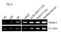
- Total RNA was isolated from plural kinds of the human cells and human cell lines indicated in Figure 8 . Then RT-PCR was performed with the total RNA and primers for human TICAM-1. 35 PCR cycles were carried out.
- Mf denotes microphages
- iDC denotes immature dendritic cells
- - denotes the control without template.
- 20-cycle PCR was performed using ⁇ -actin primer as control.
- iDC immature dendritic cells
- M ⁇ microphages
- NK natural killer cells
- mRNA of human TICAM-1 was positive in cell lines of lymphoid lineage (Tcell CCRF-CEM, T cell Molt-4, B cell Namalwa) and fibroblasts (MRC5).
- TIR domain of human TLR3 with TICAM-1 was confirmed by immunoprecipitation in HEK293 cells expressing (flag-tagged) human TLR3 and (HA-tagged)human TICAM-1.
- HEK293 cells were transiently transfected in 6-well plates using Lipofectamine 2000 reagent with 3 ⁇ g of human TLR3-Flag expression vector (flag-tagged TICAM-1 expression vector) and 0.5 ⁇ g of TICAM-1-HA expression vector (HA-tagged TICAM-1 expression vector). A total amount of DNA was kept constant (4 ⁇ g) by adding empty vector.
- the cells were stimulated for 15 min with medium alone (lanes 1 and 3 in Figure 9 ), or with 10 ⁇ g /ml of poly (I:C) (lanes 2 and 4 in Figure 9 ), and then lysed with a lysis buffer (pH 7.5; containing 25mM of Tris, 150mM of NaCl, 1wt% of NP-40, 2mM of PMSE, 25mM indoacetoamindo, 10mM of EDTA).
- a lysis buffer pH 7.5; containing 25mM of Tris, 150mM of NaCl, 1wt% of NP-40, 2mM of PMSE, 25mM indoacetoamindo, 10mM of EDTA).
- IP immunoprecipitation probe
- mouse IgG1 Lanes 1 and 2 in Figure 9
- anti-Flag M2 antibody Lanes 3 and 4; made by Sigma
- the precipitants were boiled with DPBS containing 1wt% SDS, 0.2wt% of NP-40, and 5wt% of 2-mercaptoethanol.
- eluates were immunoblotted using anti-Flag antibody or anti-HA antibody, and then subjected to SDS-PAGE.
- the lysates were also immunoblotted in order to investigate expression of the transfected TICAM-1-HA.
- Results are illustrated in Figure 9 .
- “+” denotes that the transfection or stimulation was performed, whereas “-” denotes that the transfection or stimulation was not performed.
- the middle panel illustrates part of the Western blot (immune blot) of the cell lysates.
- the lower panel is the Western blot of the whole cell lysates.
- HEK293 cells was transiently transfected with human TICAM-1-HA (0.05 ⁇ g) and (1) empty vector (4 ⁇ g), (2) human TLR3-Flag expression vector (3 ⁇ g), (3) TLR3A795H-Flag expression vector (3 ⁇ g), (4) Flag-TLR2 expression vector (2 ⁇ g), (5) Flag-TLR4 expression vector (2 ⁇ g), or (6) MD-2 expression vector (1 ⁇ g).
- a total amount of DNA was kept constant (4 ⁇ g) by adding empty vector. Twenty four hours after the transfection, the cells were lysed with lysis buffer.
- Lysates were immunoprecipitated with mouse IgG1 (not illustrated) or anti-FlagM2 antibody (lanes 1 to 5), and then boiled with DPBS containing 1wt% of SDS, 0.2wt% of NP-40, and 5wt% of 2-mercaptoethanol, thereby to elute immunoprecipitated protein. Next, elutes were immunoblotted with the anti-Flag antibody or anti-HA antibody and then subjected to SDS-PAGE. The lysates were also immunoblotted in order to investigate expression of the transfected TICAM-1-HA. Immunoblotting was performed with the anti-flag antibody or anti-HA antibody.
- Figure 10 illustrates results thus obtained.
- “+” denotes that the transfection was performed, while “-” denotes that the transfection was not performed.
- the immunoprecipitated and Flag-tagged TLR protein is illustrated.
- TICAM-1 interacting with wild-type TLR3 (parenthesis or arrow; only lane 2 is illustrated).
- the transfected TICAM-1-HA (arrow) is illustrated.
- expression vector for human TICAM-1 and three types of expression vectors respectively encoding its three types of mutants were prepared.
- the three types of mutants are: a mutant in which an amino acid sequence ranging from 1-position to 288-position of human TICAM-1 is deleted (hereinafter, this mutant is called “human TICAM-1 ( ⁇ N288)”); a mutant in which an amino acid sequence ranging from 1-position to 386-position of human TICAM-1 is deleted (hereinafter, this mutant is called “human TICAM-1 ( ⁇ N386)”); and a mutant which is made of almost only TIR domain of human TICAM-1 (hereinafter, this mutant is called "human TICAM-1 ⁇ TIR”).
- the mutant which is made of almost only TIR domain of human TICAM-1 was more specifically a mutant in which an amino acid sequence ranging from 1-position to 386-position and an amino acid sequence ranging from 557-position to 712-position of human TICAM-1 were deleted, that is, a mutant protein made of an amino acid sequence ranging from 387-position to 556-position of the amino acid sequence constituting human TICAM-1.
- Human TICAM-1 Expression vector was prepared by placing the full-length cDNA encoding TICAM-1 into XhoI-NotI site of the plasmid vector pEFBOS.
- pEFBOS-TICAM-1 ⁇ N288)
- pEFBOS-TICAM-1 ⁇ N386
- pEFBOS-TICAM-1 TIR
- pEFBOS-TICAM-1 TIR
- an amino acid sequence ranging from 289-position to 712-position (ii) an amino acid sequence ranging from 387-position to 712-position, and (iii) an amino acid sequence ranging from 387-position to 556-position
- (i), (ii) and (iii) are regions of the human TICAM-1 following the Kozack sequence and the first ATG.
- the plasmid vectors were prepared by using "Plasmid Maxi" kit (made by Qiagen) containing no endotoxi
- TICAM-1 reporter gene assay to measure the level of NF- ⁇ B activation and interferon ⁇ activation was performed for human TICAM-1 and its mutant. That is, functional binding of TICAM-1 to the interferon ⁇ promoter and the other was tested in HEK293 cells.
- HEK 293 cells in 24-well plates (2 ⁇ 10 5 cells per well) were transfected with (i) 0.1 ⁇ g of luciferase-linked NF- ⁇ B reporter gene (made by Stratagene) (c.f. 12(b)) or 0.1 ⁇ g of p125Luc reporter plasmid (c.f. 12(a)), and (ii) empty vector, human TICAM-1 TIR, human TICAM-1 ( ⁇ N386), human TICAM-1 ( ⁇ N288), or the vector expressing the full-length human TICAM-1 (10ng or 100ng respectively). A total amount (0.8 ⁇ g to 1.0 ⁇ g) of transfected DNA was adjusted by adding the empty vector. Moreover, plasmid vector pCMV ⁇ (made by Clontech; 5ng) was used as internal control.
- the result of a similar transfection experiment to test the ability of human TICAM-1 to activate NF- ⁇ B showed that the full-length TICAM-1 activates NF- ⁇ B promoter to lesser extent than the interferon ⁇ promoter.
- the mutant lacking the N-terminal domain showed a greater ability to activate NF- ⁇ B than the cells having TIR domain or the full-length TICAM-1 (see Figures 12(a) and 12(b) ). Therefore, it is considered that the N-terminal sequence direct a strong preference for the interferon ⁇ production on the transfectant, and relatively suppress the NF- ⁇ B activation at the same time.
- the C-terminal domain slightly enhanced both of interferon ⁇ activation and NF- ⁇ B activation (see Figures 12(a) and 12(b) ).
- the interferon ⁇ promoter activation was induced without transfection of TLR3 in some cases (see Figure 12(a) and 12(b) ).
- TIR domain minimal essential constituent of TICAM-1
- I:C poly (I:C) on the interferon ⁇ promoter activation is additive.
- a dominant-negative mutant of human TICAM-1 ⁇ TIR was prepared by inserting dot mutation into the human TICAM-1 ⁇ TIR expression vector (pEFBOS-TICAM-1 (TIR)), the dot mutation replacing amino acid 434 (proline) with hystidine.
- HEK293 cells in 24-well plates were transfected with (i) 0.1 ⁇ g of luciferase-linked NF- ⁇ B reporter gene (made by Stratagene) (c.f. Figure 13(b) ) or 0.1 ⁇ g of p125 ⁇ luc reporter plasmid (c.f.
- TICAM-1 ⁇ TIR (0.1ng and 10ng) or a plasmid (0, 0.2 ⁇ g, 0.6 ⁇ g), the plasmid encoding TICAM-1 ⁇ TIR mutant TIR ⁇ P434H of TICAM-1 TIR, and (iii) empty vector, TLR2 (0.1 ⁇ g), or TLR3 (0.6 ⁇ g).
- a total amount (0.8 ⁇ g to 1.0 ⁇ g) of transfected DNA was adjusted by adding the empty vectors.
- plasmid vector pCMV ⁇ made by Clontech; 5ng was used as an internal control.
- the cells were stimulated for 6 hours with 100nM of MALP-2 (TLR2 stimulation) or 10 ⁇ g/ml of poly (I:C) (TLR3 stimulation), and then lyzed with the lysis buffer (made by Promega).
- Luciferase activity that indicate the interferon ⁇ activation or NF- ⁇ B activation caused by stimulation after the transfection in thus prepared lysates were measured according to the manufacturer's instructions. This experiment was repeated three times. Representing values thereof were considered as measurement values.
- Level of the interferon ⁇ activation is illustrated in Figure 13(a)
- level of the NF- ⁇ B activation is illustrated in Figure 13(b) .
- TICAM-1 is specific for human TLR3.
- TLR2 coexpressed with TIR of TICAM-1 in HEK293 did not activate the expression of interferon ⁇ promoter even in the cells stimulated with mycoplasma lipopeptide MALP-2, which is a human TLR2 ligand.
- a reporter gene assay was carried out to compare human TICMA-1, the known adaptor molecule MyD88, and Mal/TIRAP in terms of levels of interferon ⁇ and NF- ⁇ B activations.
- HEK293 cells on 24-well plates were transfected with (i) 0.1 ⁇ g of luciferase-linked NF- ⁇ B reporter gene (made by Stratagene) (c.f. Figure 14(b) ) or 0.1 ⁇ g of p125 luc reporter plasmid (c.f. Figure 14(a) ), (ii) empty vector, MyD88, Mal/TIRAP, or the full-length TICAM-1 (respectively 10, 100, or 200ng).
- a total amount (0.8 ⁇ g to 1.0 ⁇ g) of transfected DNA was adjusted by adding the empty vectors.
- plasmid vector pCMV ⁇ made by Clontech; 5ng was used as an internal control.
- the cells were lysed with the lysis buffer (made by Promega).
- the interferon ⁇ activity or Luciferase activity in the lysates were measured according to the manufacturer's instructions, the luciferase activity indicating interferon ⁇ or NF- ⁇ B activation. This experiment was repeated three times, and representing values were considered as measurement values.
- Level of the interferon ⁇ activation is illustrated in Figure 14(a) and level of the interferon activation of NF- ⁇ B is illustrated in Figure 14(b) .
- HEK293 cells were transiently transfected with plasmids (100ng) respectively encoding the adaptor molecule Mal-HA, MyD88-HA, and TICAM-1-HA. 24hous after the transfection, the cells were lysed. Expressed proteins were analyzed by Western blotting with anti-HA antibody.
- human TICAM-1 knockdown cells were produced by transfection using single-stranded RNA.
- the single-stranded RNA of human TICAM-1 is illustrated in Figure 16 : sense is r[GACCAGACGCCACUCCAAC]d[TT], while antisense is r[GUUGGAGUGGCGUCUGGUC]d[TT] (TICAM-1).
- r and d respectively denote ribonucleotide and deoxyribonucleotide.
- the single-stranded RNA region in the message of TICAM-1 is given under the single-stranded RNA sequence.
- the location of the single stranded RNA in the human TICAM-1 message effective for gene silencing is positioned at 986 to 1008 bases (bp), as shown in Figure 6 .
- the primer for human TICAM-1 had the following sequence: 5'CCAGATGCAACCTCCACTGG3' (5' primer) and 5'TGGAGGAAGGAACAGGACACC 3' (3' primer).
- RNAi RNA interference
- TICAM-1 is the adaptor molecule that links (i) TLR3 activation mediated by double-stranded RNA and (ii) the interferon ⁇ production.
- HeLa cells or MRC-5 cells were transfected with (i) buffer only, (ii) single-stranded RNA of Lamin A/C (Control; final concentration 200nM), or (iii) single-stranded RNA of human TICAM-1. 48 hours from the transfection, HeLa cells and MRC-5 cells were stimulated respectively with 50 ⁇ g/ml and 10 ⁇ g/ml of poly (I:C).
- Figure 18(a) illustrates measurement results of HeLa cells
- Figure 18(b) illustrates measurement results of MRC-5 cells.
- RNAi Basic method of RNAi was described in Elbashir, S. M., Harborth, J., Lendeckel, W., Yalcin, A., Weber, K., & Tuschl, T. Duplexes of 21-nucleotide RNAs mediate RNA interference in cultured mammalian cells. Nature 411: 494-498 (2001 ). Moreover, its detailed procedure and conditions are described in Oshiumi, H., Brgum, N. A., Matsumoto, M., & Seya, T. RNA interference for mammalian cells, Folia Pharme, Jpn. 120: 91-95 (2002 ).
- the single-stranded RNA of human TICAM-1 had the sequence whose sense is r[GACCAGACGCCACUCCAAC]d[TT] and antisense is r[GUUGGAGUGGCGUCUGGUC]d[TT] (TICAM-1).
- single-stranded RNA of lamin A/C has the sequence whose sense is r[CUGGACUUCCAGAAGAACA]d[TT] and antisense is r[UGUUCUUCUGGAAGUCCAG]d[TT].
- "r” and “d” respectively denote ribonucleotide and deoxyribonucleotide.
- the single-stranded RNA region in the message of TICAM-1 is given under the single-stranded RNA sequence. These single-stranded RNAs were purchased from Xeragon Inc.(USA).
- HeLa cells or MRC-5 cells stimulated with poly (I:C) (10 ⁇ g/ml or 50 ⁇ g/ml) were cultured for 24 hours (24h), and supernatants were collected. Human interferon ⁇ concentrations in the supernatants from the culture supernatants were measured by ELISA (Enzyme Linked Immunosorbent Assay; TEB) Results are illustrated in Figures 18(a) and 18(b) .
- TICAM-1 is an adaptor molecule that links the double-stranded RNA-mediated TLR3 activation and the interferon ⁇ production.
- the present invention relates to a novel protein that induces type I interferon production by specifically binding to mammalian Toll-like receptor 3, and its mutants, a gene of the protein, a recombinant expression vector containing the gene, an antibody against the protein.
- the prevent invention is useful for (i) studying and analyzing the TLR3-mediated signaling system and its control mechanism, (ii) pathological analysis on various diseases to which the signaling system and its control mechanism involve, (iii) prevention and therapy of viral infectious diseases such as hepatitis B, hepatitis C, and the like, (iv) therapy of tumors, (v) therapy of autoimmune diseases, (vi) therapy of atopic diseases, (vii) and other usages. Therefore, the present invention is expected to contribute to medical care development such as various pharmaceutical industries.
Description
- The present invention relates to a novel adaptor protein that can induce production of type I interferon by binding to mammalian Toll-
like receptor 3, mutants of the same, and a gene for encoding the same, and use of the same. - Interferon is a protein, which plays an important role in adaptive immune response against viruses.
- Because of its important role in the adaptive immune system, there have been many studies on the role of the interferon in the immune system. However, it has been only limited knowledge on a signaling pathway for in-vivo production of interferon in the immune system responds to pathogens.
- After recent discovery of Toll-like receptor (hereinafter just referred to as "TLR" for abbreviation where appropriate) as a receptor that recognize pathogens in mammal innate immune system, researches on TLR are leading to the understanding of the signaling pathway relating to the pathogen recognition in the innate immune system.
- The mammalian TLR families, which are principal tissues appearing to be in large part conserved across Drosophila and human, recognize a variety of microbial components and mediate (i) activation of nuclear factor κB (hereinafter just referred to as "NF-κB" for abbreviation) and (ii) other signaling pathways.
- For human, 10 receptors (
human TLR 1 to 10) belonging to the TLR family have been identified by now and mouse homologue thereof (mouse TLR 1 to 10) have been identified. TLR family proteins is made of (i) an extracellular domain containing a plurality of leucine-rich repeats (LRRs) and a carboxylic terminal (C-terminal) flanking region, and (ii) a cytoplasmic (intercellular) signaling domain. The cytoplasmic signaling domain is called Toll/interleucine-1 receptor homology domain (TIR). Each TLR recognizes one or more distinct ligand(s) with its extracellular domain, and induces immune response(s), presumably via the intercellular TIR. Each TLR induces different, sometimes overlapping immune responses. All TLR family proteins contain a TIR domain in their cytoplasmic region, and most of the TIR domain is considered to be responsible for signaling and interaction with the adaptor molecule MyD88 or Mal/TIRAP. That is, TLR2, TLR4, TLR5, TLR7, and TLR9 transmit signals via the adaptor molecule MyD88 upon agonist stimulation, thereby to activate NF-κB. - Meanwhile, it was reported recently that the adaptor molecule Mal/TIRAP (an adaptor molecule called MyD88-adapter-like or TIRAP), which associates with
TLR 4, relates to the signaling pathway viaTLR 4, (e.g. refer to Document 1: Kawai, T., et a. Lipopolysaccharide stimulates the MyD88-independent pathway and results in activation of IFN-), Document 2: Horng, T., Barton, G. M., & Medzhitov, R. TIRAP: an adapter molecule in the Toll signaling pathway. Nat. Immunol. 2: 835-841 (2001), and Document 3: Fitzgerald, K. A., et al. Mal (MyD88-adapter-like) is required for Toll-like receptor-4 signal transduction. Nature 413: 78-83 (2001)). - According to the report,
TLR 4 is involved in activation of NF-κB, MAPK, and interferon β promoter. This unique ability ofTLR 4 to induce the activation of interferon β promoter is ascribed to signaling pathway mediated by the adaptor molecule Mal/TIRAP that binds toTLR 4. This signaling pathway is called "a MyD88-independent pathway". That is, in the signaling viaTLR 4, activation of NF-κB and type-I interferon promoter is controlled by cooperation betweenTLR 4 and the adaptor molecule Mal/TRRAP that is a second adaptor molecule different from the adaptor molecule MyD88. - In Microphages (Mf), STAT1 α/β phosphorylation is induced by the interferon β activation as a result of
TLR 4 stimulation, notTLR 2 stimulation. Expression of the gene encoding interferon β subsequently induces production of MCP (Monocyte Chemoattractant Protein)-5, IP (interferon Inductive Protein)-10m, and iNOS (inductive NO synthetic enzyme). Again, this occurs via the MyD88-independent pathway, even in MyD88-/- cell (a cell from which the adaptor molecule MyD88 is deleted). - A current concept considered most likely is that the adaptor molecule Mal/TIRAP covers the MyD88-independent pathway.
- In contrast, the inventor of the present invention studied immune responses induced by double-stranded RNA and mediated via
human TLR 3, and found thathuman TLR 3 relates to recognition of double-stranded RNA on a cell surface of human fibroblast, and triggers downstream signaling that induces the interferon β production. (e.g. Document 4: Matsumoto, M., Kikkawa, S., Kohase, M., Miyake, K., & Seya, T. Establishment of a monoclonal antibody against human Toll-). That is, the inventors of the present invention showed that the interferon β promoter activation and interferon β production is rapidly and strongly induced byhuman TLR 3 in response to double-stranded RNA. Reporter gene assay showed that human TLR3 mediates the interferon β promoter activation, and to a lesser extent the NF-κB activation (c.f.Document 4 for example). This result with respect to TLR3 is quite different from those of TLR, TLR2, TLR5, TLR7, and TL9, which activate NF-κB and p38 MAPK (MAP kinase) through the adaptor molecule MyD88 after recognizing their specific ligands. - The type I interferon (interferon α and interferon β) that
TLR 3 induces is known to have anti-virus effect and anti-cancer effect. Specifically, type I interferon has the following effects. - Type I interferon is known to exhibit the anti-virus effect by the following function mechanisms:
- 1) The type I interferon destabilizes mRNA of a virus and activates intercellular gene that inhibits protein translation of hosts, thereby to inhibit replication and multiplication of the virus;
- 2) The type I interferon induces expression of MHC class I molecule so as to induce resistance against natural killer (NK) cells. Further, type I interferon enhances sensitivity with respect to CD+8 cytotoxic T cell. In addition, type I interferon participates to inhibition of T cell activation and to T cell suppressor activation; and
- 3) The type I interferon activates natural killer (NK) cells that selectively damage virus-infected cells, and causes NK-cell induced apoptosis in the virus. Moreover, the type I interferon is known to exhibit anti-tumor effect by the following function mechanisms:
- 1) The type I interferon destabilizes mRNA in tumor cells and activates intercellular gene that inhibits protein translation of hosts. This inhibits protein synthesis in the tumor cells, thereby inhibiting multiplication of the tumor cells;
- 2) The type I interferon activates anti-tumor effectors such as microphages, NK cells, natural killer T (NKT) cells, and the like. Via damaging of the tumor cells by these anti-tumor effectors, apoptosis is brought about in the tumor cells;
- 3) The type I interferon activates NK cells that selectively damage virus-infected cells, and induces NK cell-induced apoptosis in the tumor cells.
- Further, as described above, the type I interferonparticipates inhibition of T cell activation, and enhancement of T cell suppressor activity. Therefore, it is considered that some kinds of autoimmune diseases can be ameliorated by the type I interferon.
- Because of the anti-virus effect and anti-tumor effect as mentioned above of the type I interferon, interferon α formulation and interferon β formulation have been used for treating hepatitis B, hepatitis C, hepatitis C-induced liver and kidney cancers, and the like. For example, "Sumiferon (Registered Trademark) " made by Sumitomo Pharmaceuticals, which is a wild type interferon α, are used successfully in clinical applications.
- However, the researches on TLR that have been carried out so far indicate that the signaling pathway leading to the type I interferon production induced by TLR3 witch recognized double-stranded RNA is different from the signaling pathways mediated via other TLRs. However, it has not been understood which protein participates in the signaling pathway.
- It is believed that discovery of existence of a protein that induces the signaling by specifically binding to TLR3, the signaling causing the type I interferon production in downstream will lead to understanding of a signaling pathway important for innate immune response against viruses and a control mechanism of the signaling pathway. The understanding of the signaling pathway and its control mechanism is expected to be used in pathological analysis of various illnesses relating to the innate immune system and development of therapeutic agents that control innate immune response.
- Moreover, the interferon formulations used for viral infectious diseases and tumors is for systemic administration: at therapeutic concentrations the interferon formulations have strong side effects such as worsening pschoneurosis (depression and the like), autoimmune disease (thyroid insufficiency, autoimmune hepatitis, hemolytic anemia, ulcerative colitis, rheumatoid arthritis, and the like), and the like side effect. The side effects caused by the interferon formulations occur presumably because interferon is introduced even in normal host cells that do not produce interferon, thereby inducing presentation of autoantigen to T cells in all portions where the interferon is induced, and thus making it easy to cause autoimmune phenomenon to occur, even though the presentation of antoantigen to T cells is normally caused when immune responds to exogenous antigen.
- Because they have strong side effects at their therapeutic concentration, the interferon formulations have difficulties to maintain sufficient anti-cancer effect if systemically administered. Moreover, even if administered locally, it is considered that the side effects are difficult to avoid completely.
- It is considered that if a novel protein that specifically binds to TLR3 so as to induce the downstream signaling that leads to the type I interferon production is found out, it will be possible to develop a new therapeutic agent for treating a viral infectious disease, tumor, or the like by locally enhancing the type I interferon production in vivo.
- Moreover, functional analysis of the novel protein is expected to provide a protein that inhibits the type I interferon production in vivo. Further it will be possible to provide a therapeutic agent that inhibits the type I interferon production in vivo by this protein thereby treating autoimmune disease, atopic disease, and the like.
-
WO 02/053737 A - DATABASE UniProt [Online] 1 October 2002, EBI Accession No. UniProt: Q8JCV0, Database Accession No. Q8JCV0 describes a
TICAM 1 protein fragment. - Kurt-Jones E. et al. (Journal of Endocrine Research, Vol. 10, No. 6, 2004, 419-424) relates to the use of murine embryonic fibroblasts to define Toll-like receptor activation and specifity.
- In view of the foregoing problems, an object of the present invention is to provide the subject matter as defined in the independent claims.
- In order to attain the object, the inventors of the present invention accomplished the present invention, which is defined by the subject matter of the independent claims.
- The inventors of the present invention recently found out that human TLR3 transmits a signal to indicate presence of double-stranded RNA (ribonucleic acid) independent of the currently known adaptor molecules MyD88 and Mal/TIRAP, thereby to induce interferon (IFN) β production, the adaptor molecule MyD88 considered to be essential to all signalings of the TLR family known at present, and the adaptor Mal/TIRAP being able to associate with TLR4. As a result of diligent works on novel adaptor protein having a property of specifically binding to mammalian Toll-
like receptor 3, the inventors of the present invention identified a novel adaptor protein having the property of specifically binding to mammalian Toll-like receptor 3, and determined a full-length gene sequence of the novel adaptor protein. Further, the inventors of the present invention found out that the novel protein has a function of activating a nuclear factor and inducing interferon β production. - A protein as described herein is a protein having an amino acid sequence set forth in SEQ ID NO: 2 or No: 4, or mutants thereof.
- As descried later, the inventors of the present invention firstly identified this protein and determined its full-length amino acid sequences. This protein is named "TICAM-1" (hereinafter, this protein is referred to as "adaptor protein TICAM-1"where appropriate). SEQ ID NO: 2 illustrates an amino acid sequences derived from human, while SEQ ID NO: 4 illustrates an amino acid sequence derived from mouse. The adaptor protein TICAM-1 was, as described above, confirmed to bind to TLR3 specifically but not other TLRs, TLR3 mutants, and the like, and to having a function of activating mammalian nuclear factor κB (NF-κB) and interferon β promoter, thereby inducing interferon β production.
- Therefore, the protein can enhance mammalian type I interferon production. Thus, by using the anti-virus effect and anti-tumor effect of the type I interferon it is possible to prevent/treat (prevent or treat) viral infectious diseases such as hepatitis B, hepatitis C, and the like, to perform autoimmune therapy or the like for tumor (cancer).
- It has been proved that viral double-stranded RNA plays a role in most of viral infectious diseases. Recognition of this double-stranded RNA by TLR3 induces the type I interferon production via TICAM-1 mediated signaling pathway. Therefore, administration of the adaptor protein TICAM-1 to a viral infectious disease reinforces immune response of innate immune system against the viral infectious disease. Therefore, it is possible to prevent or treat the virus infectious disease by the anti-virus effect of type I interferon, while avoiding side effects such as autoimmune diseases caused by introduction of type I interferon in body part that is not infected with virus.
- Moreover, as described later, it was confirmed that the adaptor protein TICAM-1 solely induces the type I interferon production, without associating with TLR3. Thus, the adaptor protein TICAM-1 can induce the type I interferon production even without viral infection, and is effective for various diseases that can be ameliorated by the interferon I production. For example, the administration of the adaptor protein TICAM-1 to tumor can induce the type I interferon production. Therefore, the use of type I interferon can treat the tumor.
- Moreover, the protein proves that there is a new TLR3-mediated signaling pathway that is independent of the adaptor molecule MyD88 and Mal/TIRAP. This new signaling pathway explains a missing link of the signaling from TLR family to the activation of interferon β promoter activation via a TLR family. Thus, the protein is not only useful as a research material to study and analyze the TLR3-mediated signaling and control mechanism, but also useful to perform, via such studies, pathological analysis on various diseases with which the signaling system and its control mechanism relate to.
- It has not studied whether the protein has a function of inducing the interferon α production. However, a previous research confirmed that the interferon α production and interferon β production shares the same signaling pathway. Thus, it is presumed that the protein also has a function of inducing the interferon α production.
- Here, the "protein" may be isolated and purified from cells, tissues, or the like, or may be expressed in the cells by introducing, into host cells, a gene encoding the protein. Moreover, the protein may contain a polypeptide additionally. An example of such polypeptide addition is epitope tagging of the protein according to the present invention with HA, Flag, or the like.
- The mutants of the protein having the amino acid sequence set forth in SEQ ID NO: 2 or No: 4 have amino acid sequence set forth in SEQ ID NO: 2 or No: 4 in which one or more amino acids are replaced, deleted, inserted, and/or added, and either of the following features: (a) having an amino acid sequence ranging from 394-position to 532-position (TIR domain) in the amino acid sequence as set forth in SEQ ID NO: 2; (b) having an amino acid sequence ranging from 396-position to 534-position (TIR domain) in the amino acid sequence set forth in SEQ ID NO: 4; (c) a property of specifically binding to mammalian Toll-
like receptor 3, and a property of inducing mammalian type I interferon; (d) the property of inducing mammalian type I interferon, and abnormality (reduction or deletion) in the property of specifically binding to mammalian Toll-like receptor 3, and (e) the property of specifically binding to mammalian Toll-like receptor 3, and abnormality in the property of inducing mammalian type I interferon. A specific example of the protein that has the feature (e) is a human protein that has the amino acid sequence set forth in SEQ ID NO: 2 in which at least amino acid 434 is replaced or deleted, and at least part of an amino acid sequence ranging from 394-position to 532-position (TIR domain) is contained. - The mutants of the proteins are not only useful as research material to study and analyze TLR3-mediated signaling system and its control mechanism, but also useful to perform, via such studies, pathological analysis on various diseases with which the signaling system and its control system involve.
- Further, the mutant proteins respectively having the features (a) to (c) can enhance the mammalian type I interferon production, like the protein having the amino acid sequence set forth in SEQ ID NO: 2 or NO: 4. Therefore, it is possible to use the anti-virus effect or the anti-tumor effect of the type I interferon, thereby to perform prevention/treatment (prevention or treatment) of the viral infectious diseases such as hepatitis B, hepatitis C, and the like, autoimmune therapy for tumor (cancer), or the like.
- The mutant protein having the feature (d) can enhance the mammalian type I interferon production. Therefore, it can be used as a therapeutic agent for preventing or treating diseases (cancer or viral infectious diseases, and the like) that can be ameliorated by enhancement of type I interferon production.
- The mutant protein having the feature (e) binds to TLR3 thereby to inhibit the TLR 3-mediated signaling that induces the type I interferon production. Thus, this mutant protein inhibits the type I interferon production. Therefore, it is considered that this mutant protein can be used to prevent or treat autoimmune diseases, atopic disease, and the like that are caused by the type I interferon production.
- Moreover, the mutant proteins having the features (a) to (e) are useful to analyze a function of a protein, e.g. by finding which region of a protein is essential for an activity, by comparing the mutant and a wild-type protein.
- The wording "one or more amino acids are replaced, deleted, inserted, and/or added" is to mean that one or more amino acids are replaced, deleted, inserted, and/or added in a number that can be replaced, deleted, inserted, and/or added by a known mutation preparation method, such as site-directed mutagenesis or the like. In other words, a protein having an amino acid sequence set forth in SEQ ID NO: 2 or NO: 4, in which one or more amino acids are replaced, deleted, inserted, and/or added, is a mutant (mutant protein) mutated from a protein having an amino acid sequence set forth in SEQ ID NO: 2 or NO: 4. Here, artificial mutation by a known mutation protein preparation method is mainly meant by the word "mutation". However, the mutant may be prepared by isolation and purification of such a mutant protein naturally exists.
- A gene according to the present invention is a gene encoding any one of the proteins according to the present invention.
- The gene is useful as a research material to study and analyze the TLR3-mediated signaling system and its control mechanism. In addition, there is a possibility that, via such studies, the gene can be useful to analyze various diseases to which the signaling system and its control mechanism relate.
- The "gene" encompasses at least genome DNAs, cDNAs, and mRNAs. Examples of the gene described herein includes: (1) cDNA derived from human, the cDNA having, as an open reading frame, 63 to 2198 bases of base sequence shown in SEQ ID NO: 1, (2) cDNA derived from mouse, the cDNA having, as an open reading frame, 66 to 2261 bases of base sequence shown in SEQ ID NO: 3, and (3) mRNAs having base sequences corresponding to these cDNAs.
- Moreover, the "gene" encompasses not only double-stranded DNAs, but also each single-stranded DNA such as sense strand and antisense strand, and RNAs. Further, the "gene" may contain an other sequence than the open reading frame. The other sequence may be a sequence of an untranslated region (UTR), a vector sequence (encompassing an expression vector sequence), and other sequences. For example, the gene may be multiplied arbitrarily by multiplying, by using an appropriate host, a gene of the present invention constituted by linking a sequence encoding the protein, to a vector sequence. Moreover, part of the sequence of the gene may be used as a probe. Examples of cases where the gene of the present invention is used as a probe include a case where part of the sequence of the gene of the present invention is fixed on a chip thereby constituting a DNA chip and this DNA chip is used for various tests and diagnosis.
- A recombinant expression vector according to the present invention contains the gene according to the present invention.
- Use of this recombinant expression vector allows introduction of the gene in target cells by transformation of the target cells by a known genetic engineering technique (gene manipulation).
- The transformant prepared by the transformation is useful as a research material to study and analyze the TLR3-mediated signaling system and its control mechanism. In addition, there is a possibility that, via such studies, the transformant can be useful to analyze various diseases to which the signaling system and its control mechanism relate.
- By introducing the gene contained in the recombinant expression vector into mammalian target cells (host cells) by the transformation and expressing the gene in the target cells, it is possible to control mammalian type I interferon production. Thus, the recombinant expression vector allows gene therapy for diseases (viral infectious diseases, tumors, and the other diseases) that can be ameliorated by enhancing type I interferon production, or diseases that are caused by the type I interferon production.
- An antibody as described herein is an antibody that specifically binds to any of the proteins mentioned above.
- The antibody is expected to be useful for detection of expression of the adaptor protein TICAM-1 in host cells and production, purification, and the like, of the adaptor protein
- TICAM-1.
- Moreover, the antibody that specifically binds to a protein having a property of specifically binding to mammalian Toll-
like receptor 3 can inhibit the type I interferon production that is caused by immune response mediated by the mammalian Toll-like receptor 3. The antibody is useful as a therapeutic agent for preventing/treating diseases (viral infectious diseases, tumors, and the other diseases) that can be ameliorated by enhancing type I interferon production. - For a fuller understanding of the nature and advantages of the invention, reference should be made to the ensuing detailed description taken in conjunction with the accompanying drawings.
-
-
Figures 1(a) to 1(d) are views illustrating results of reporter gene expression assays on NF-κB activation caused by human TLR. -
Figures 2(a) to 2(d) are views illustrating results of reporter gene expression assays on interferon β promoter activation caused by human TLR. -
Figure 3(a) is a view illustrating results of reporter gene expression assays on NF-κB activation caused by human TLR3 and its mutant.Figure 3(b) is a view illustrating results of reporter gene expression assays on interferon β promoter activation caused by human TLR3 and its mutant. -
Figure 4 is a view illustrating results of investigations on interactions between proteins by yeast two-hybrid system. -
Figure 5 is a view illustrating homology search for human TICAM-1 and mouse TICAM-1. -
Figure 6 is a view illustrating TIR domains of human TICAM-1, mouse TICAM-1, and known adaptor molecules Mal, and MyD88. -
Figure 7 is a view illustrating expression of mRNA of TICAM-1 in various human tissues. -
Figure 8 is a view illustrating results of detection of mRNA of TICAM-1 in various types of human cells and human cell lines by RT-RCR. -
Figure 9 is a view illustrating results of immunoprecipitation for analyzing interaction between human TICAM-1 and human TLR3. -
Figure 10 is a view illustrating results of immunoprecipitation for analyzing interaction between human TI CAM-1 and a human TLR other than human TLR3, or a human TLR3 mutant. -
Figure 11 is a view illustrating a mutant of human TICAM-1. -
Figure 12(a) is a view illustrating measurement results of interferon β promoter activation caused by human TICAM-1 and its mutant.Figure 12(b) is a view illustrating NF-κB activation caused by human TICAM-1 and its mutant. -
Figure 13(a) is a view illustrating measurement results of interferon β activation by the reporter gene expression for analyzing the signaling mediated by TLR3.Figure 13(b) is a view illustrating measurement results of NF-κB activation by the reporter gene expression for analyzing the signaling mediated by TLR3. -
Figure 14(a) is a view illustrating results of interferon β activation caused by human TI CAM-1 and a known adaptor molecule.Figure 14(b) is a view illustrating measurements result of NF-κB activation caused by human TICAM-1 and the known adaptor molecule. -
Figure 15 is a view illustrating results of western blot that indicated expression of human TICAM-1 and the known adaptor molecule in HEK293 cells.
Figure 16 is a view illustrating a sequence of single-stranded RNA of human TICAM-1.
Figures 17(a) and 17(b) are views illustrating results of RT-PCR analysis.
Figures 18(a) and 18(b) are views illustrating results of analysis by RNAi. - An exemplary embodiment of the present invention is described below. The embodiment is not to limit the present invention.
- The inventors of the present invention found via experiments on transfection of HEK 293 cells described in [Examples] that TLR3 signaling induced by double-stranded RNA and mediated by human TLR3 is introduced through its cytoplasmic tail. TIR, and largely independent of adaptor molecule MyD88 and Mal/TRRAP.
- Based on this the inventors expected that there is another adaptor molecule that predominantly participates at least in the type I interferon promoter activation that is mediated by TLR3.
- The inventors expected that there is another adaptor molecule, which preferentially mediates signaling that induces the type I interferon, but which is distinct from the already known adaptor molecule MyD88 and Mal/TIRAP. Identification of the unknown adaptor molecule was attempted.
- As a result, the inventors successfully identified an alternative adaptor molecule. Physically, this alternative adaptor molecule binds to Toll/interleukin-1 receptor homology domain (hereinafter "TIR domain") of human TLR3. Functionally, the alternative adaptor molecule induces the activation of the type I interferon promoter in response to polyinosinic acid-cytidylic acid (hereinafter referred to as "poly (I:C)"). This adaptor molecule is named as "TIR domain-containing adaptor molecule" (hereinafter referred to as "TICAM-1" for abbreviation) by the inventors because it has the TIR domain.
- SEQ ID NO: 2 and NO: 4 illustrate amino acid sequences of the adaptor protein TICAM-1. SEQ ID NO: 2 illustrates an amino acid sequence of an adaptor protein TICAM-1 derived from human (hereinafter referred to as "human TICAM-1 "for abbreviation). SEQ ID NO: 4 illustrates adaptor protein TICAM-1 derived from mouse(hereinafter referred to as "mouse TICAM-1"for abbreviation).
- Moreover, SEQ ID NO: 1 and NO: 3 illustrate base sequences of full-length cDNA sequence encoding the adaptor proteins TICAM-1. SEQ ID NO: 1 illustrates base sequence of cDNA encoding the human TICAM-1, while SEQ ID NO: 3 illustrate base sequence encoding the mouse TICAM-1.
- In the base sequence of SEQ ID NO: 1, positions 63 to 2198 in the base sequence corresponds to open reading frame (ORF) encoding the human TICAM-1. In the base sequence of SEQ ID NO: 3, positions 66 to 2261 in the base sequence correspond to open reading frame (ORF) encoding the mouse TICAM-1. Each of the cDNA sequences of SEQ ID NO: 1 and NO: 3 contains untranslated regions (UTR) on 5' and 3' ends.
- It was found that the human TICAM-1 is made of an N-terminal proline-rich domain (from 1-position to 393-position in the amino acid sequence), TIR domain (from 394-position to 532-position in the amino acid sequence), and, a C-terminal proline-rich domain (from 532-position to 712-position in the amino acid sequence) (
Figure 5 ). - The TIR domain of the cDNA sequence of human TICAM-1 has low similarity to TIR domains of the known adaptor molecules, human Mal/TIRAP (c.f.
Non-Patent Documents 3 and 4) (i.e. "Mal (TIR).hu" and "MyhD88 (TIR).hu ofFigure 6 ) and human MyD88. Further, the TIR domain of the cDNA sequence of human TICAM-1 is unique in that (F/Y)D in Box1, RD inBox 2, and FW in Box 3 (c.f. Xu, Y, et al. Structural basis for signal transduction by the Toll/interleukin-1 receptor domains, Nature 408: 111-115 (2000)), which are conserved sequences in the TIR domains of other TIR domain containing proteins, are missing in the TIR domain of the cDNA sequence of human TICAM. - In contrast, in the human TICAM-1, prolines in so-called BB loop (c.f. Xu, Y, et al. Structural basis for signal transduction by the Toll/interleukin-1 receptor domains, Nature 408: 111-115 (2000)) was well conserved. The prolines in BB loop are essential for the signaling via TIR-MyD88 and conserved in the known adaptor molecule MyD88 and Mal/TIRAP.
- Another important difference between (i) the human TICAM-1 and (ii) the known adaptor molecule MyD88 and Mal/TIRAP is in that a Death domain (cell death inducing region) or an shorter Mal-like N-terminal domain is deleted in the human TICAM-1, and the human TICAM-1 contains a proline-rich long N- and C-terminal stretches:
- Moreover, a database search through human genome draft sequence using the cDNA sequence of the human TICAM-1. The database search showed that the cDNA sequence encoding the human TICAM-1 is located in chromosome 19p13. 3 (gene situ).
- The proteins and genes are likely applicable for use in diagnosis. It is expected that, by detecting whether or not the adaptor protein TICAM-1 or a mutant similar thereto in function is expressed in cells sampled from an individual to be diagnosed, it is possible to diagnose the individual has an immunodeficiency syndrome that is caused by depletion or mutation in the adaptor protein TICAM-1. As methods to detect the expression of the adaptor protein TICAM-1 or the mutant similar thereto in function the following methods are considered as applicable: to detect the gene of the adaptor protein TICAM-1 by using DNA-tip ; to test single nucleotide polymorphisms (SNPs) of a gene of the adaptor protein of the individual; and the other methods.
- Here, how to obtain the proteins and genes is explained.
- There is no particular limitation as to how to obtain the gene encoding the adaptor protein TICAM-1. By using various methods, the gene encoding the adaptor protein TICAM-1 may be cloned from a DNA fragment containing the gene sequence, based on the disclosed sequence information mentioned above. For example, a probe may be prepared which specifically hybridizes with part of the sequence of cDNA encoding the adaptor protein TICAM-1, so that screening may be performed through human or mouse genomic DNA library or cDNA library with the probe.
- The probe may have any sequence and length, as long as it specifically hybridizes with at least part of (i) the base sequence of the cDNA encoding the adaptor protein TICAM-1 or (ii) its complementary sequence. Each step of the screening may be carried out in a condition usually used.
- A clone thus obtained through the screening can be analyzed in more detail by constructing a restriction map and determination of its base sequence (sequencing). These analysis methods makes it possible to easily check whether a DNA fragment including the gene sequence is obtained or not.
- Moreover, by selecting a sequence of the probe from among TIR domains considered to be important in the functions of the adaptor protein TICAM-1, and screening through human, mouse, or other mammal's cDNA library, it is possible to isolate a gene encoding a homologous molecule or analogue molecule having a function similar to that of the adaptor protein TICAM-1, and perform cloning with the gene.
- The gene (polynucleotide) according to the present invention can be obtained by amplification means such as PCR or the like, apart from the screening methods. For example, it is possible to mass-produce a DNA fragment including the polynucleotide according to the present invention by preparing primers respectively from the sequence (or complementary sequence thereof) of the untranslated regions at 5' and 3' ends among the cDNA sequences of the adaptor protein TICAM-1, and then performing PCR or the like using the primer and a human/mouse genome DNA (or cDNA) or the like as a template.
- There is no particular limitation in how to obtain the protein. For example, the gene thus obtained ((i) the cDNA encoding the adaptor protein TICAM-1, (ii) the cDNA encoding the homologous molecule thereof or the like, (iii) or the like) may be introduced in a microorganism (such as Escherichia coli, yeast, or the like), animal cells, or the like, so as to perform expression and purification of the protein encoded by the cDNA. This allows easy preparation of the protein such as the protein adaptor protein TICAM-1, and the like.
- In case of introducing the extraneous gene in the host, there are various recombinant expression vector and hosts available. Into their extraneous gene recombination region of the recombinant expression vector a promoter that can function in the host is inserted. It is possible to select the recombinant expression vector and the host according to purposes. Depending on the host used and property of the protein thus produced, a different method is applied to collect the protein after the production. It is possible to purify the targeted protein relatively easily by using a tag or the like.
- The adaptor protein TICAM-1 can accelerate the type I interferon production by the following function mechanisms.
- (a) In a host cell which is infected with a virus and in which TLR3 is expressed, the adaptor protein TICAM-1 binds to TLR3 that recognized the double-stranded RNA from the virus. The binding induces signaling that leads to induction of downstream type I interferon production. The signaling strongly enhance the type I interferon production. Thereby, the type I interferon production can be strongly enhanced cell-specifically (locally) in the host cell infected with the virus. That is, it is possible to enhance the immune response against viruses.
- (b) In a host cell which is not infected with the virus, the adaptor protein TICAM-1 is solely expressed, thereby to induce the signaling that induces the downstream type I interferon production. This enhances the type I interferon production. The sole expression of the adaptor protein TICAM-1 also has a high type I interferon induction activity. However, an amount of the type I interferon is smaller in this function mechanisms (b) than in the function mechanisms (a).
- As a result, the adaptor protein TICAM-1 is useful as follows.
- 1) The adaptor protein TICAM-1 or a mixture containing the adaptor protein TICAM-1 as an active ingredient is quite useful as a therapeutic agent for preventing/treating a viral infectious disease. That is, mainly as a result of the enhancement of the type I interferon production by the function mechanisms (a), it is possible to enhance the type I interferon-caused anti-virus effect specifically (locally) in the virus-infected host cell. That is, it is possible to allow type I interferon to have a greater bio-defense function against the virus infection. Therefore, it is possible to prevent or treat the virus infectious disease while keeping, as low as possible, the side effect (such as autoimmune disease and the like) due to excess production of type I interferon. Examples of the virus infectious disease is applicable include any illness that has been confirmed that the type I interferon is effective, that is, viral hepatitis B and hepatitis C (especially of genotype IIa) and the like. Moreover, if is expected to be applicable to Acquired Immune Deficiency Syndrome (AIDS; immunologic unresponsiveness-causing infection disease) caused by human immunodeficiency virus (HIV). Moreover, the adaptor protein TICAM-1 can prevent virus infection by expressing the adaptor protein TICAM-1 at a portion at which latent infection is suspected. Moreover, the adaptor protein TICAM-1 exhibits a high type I interferon induction activity even if solely expressed (i.e. in the function mechanisms (b)). Thus, the adaptor protein TICAM-1 exhibits a sufficient anti-virus effect even without TLR3 expression (i.e. in case where the function mechanisms (a) is not effective).
- 2) The adaptor protein TICAM-I or the mixture containing the adaptor protein TICAM-1 as the active ingredient is useful as the therapeutic agent for tumor treatment. That is, the enhancement of the type I interferon by the function mechanisms (b) results in enhancement of the anti-tumor effect by the type I interferon. This makes it possible to treat tumor (cancer) by innate immunity therapy. Examples of the tumor include malignant tumors (hepatoma, kidney cancer, juvenile pharynx/villous tumor, malignant lymphoma, cerebral tumor, glioblastoma, medulloblastoma, astrocytoma, dermal malignant melanoma, and the like) that has been confirmed that the type I interferon is effective.
- It is preferable that cells targeted by the therapeutic agent be cells that express, on theirs surfaces, TLR3 that recognizes the double-stranded RNA of the virus. However, because the protein can solely induce the type I interferon production, the present invention is also applicable to cells that do not express TLR3 on their surfaces. However, in case where the protein is applied to the viral infectious diseases, it is preferable that the adaptor protein be introduced in the cells that express, on theirs surfaces, TLR3 that recognizes the double-stranded RNA of the virus. This makes it possible to cause the type I interferon production locally in the virus-infected portion. Thus, it is possible to effectively treat the viral infectious disease by increasing the amount of the type I interferon produced, while keeping, as low as possible, the side effect caused by increasing the amount of the type I interferon.
- According to the studies by the inventors, human TLR3 is expressed in various dendritic cell (DC) subsets. Further, it has been reported that human TLR3 is expressed in human intestinal epithelial cells and human fibroblasts. (c.f. M.Muzio, D.Bosisio, N.Polentarutti, G.D'amico, A.Stoppacciro, R.Mancinelli C.van't Veer, G.Penton-Rol, L.P.Ruco, P.Allavena, A.Mantovani, J.Immunol. 164 (2000) 5998-6004, and E.Cario, D.K.Podolsky, Infect.Immun. 68 (2000) 7010-7017) These suggest that its function closely relates to responses of the innate immune system to a microbial nuclear constituent. Therefore, the therapeutic agent for treating the virus infectious disease is effective for the cells that produce the type I interferon by expressing human TLR3, especially, for the cells that produce the type I interferon when they expressing human TLR3 on their surface and recognize an RNA virus. Examples of such cells include (i) human fibroblasts such as human lung fibroblasts, human preputial fibroblasts, and the like, (ii) the human dendritic cells, (iii) human intestinal epithelial cells, (iv) and the like. Especially, the fibroblasts are known to produce the type I interferon via different signaling pathways when infected with RNA viruses or treated with the double-stranded RNA. The present invention is expected to quite effective for the fibroblasts. Moreover, examples of the cells that produce the type I interferon by expressing mouse TLR3 include mouse fibroblasts and the like.
- Moreover, to administer the preventive/therapeutic agent containing the adaptor protein TICAM-1, phleboclysis, hypodermic injection, and the like injection are preferable. However, the therapeutic agent may be administered by non-oral manners such as sublingual tablet, rectal administration, and the like. Further, it is possible to prepare this therapeutic agent according to a general method of producing protein formulations. Furthermore, it is possible to use this therapeutic agent as an emulsifier for liposome formulations and the like.
- The adaptor protein TICAM-1 is expected to have the following usefulness beside the one mentioned above.
- As a result of the enhancement of the type I interferon production by the function mechanisms (b), the type I interferon's immune control effect is enhanced, thereby to enable a kind of autoimmune-disease treatment.
- Moreover, the adaptor protein TICAM-1 is expected to be used for screening and the like of compounds that enhance the binding of TLR3 and the adaptor protein TICAM-1 or the downstream signaling from the adaptor protein TICAM-I to the type I interferon production, and compounds that inhibit (is inhibitors of) the downstream signaling from the adapter protein TCAM-1 to the type I interferon production.
- Moreover, as described later in Examples, the adaptor protein TICAM-I is a molecule that binds to TLR3 and induces the type I interferon production. Thus, the adaptor protein TICAM-1 is considered to be a molecule that plays a quite important role in the signaling system through TLR3. Thus, the gene and protein is useful as a research material for analysis of the TLR3-mediated signaling system and its control mechanism. Through the research, the gene and protein according to the present invention may be possibly useful for pharmacologically analyzing various illnesses to which the signaling system and its control mechanism relate.
- There known a large number of protein mutants having similar activity and functions to their wild types. Therefore, there is no particular limitation as to how to produce a mutant of the adaptor protein TICAM-1, the mutant having both of the property of specifically binding to TLR3 and the property of inducing the type I interferon production, like the adaptor protein TICAM-1. For example, it is possible to produce the mutant by using known mutation protein preparation methods, such as a site-directed mutagenesis inducing method (Hashimoto-Gotoh, Gene 152,271-275(1995) and others), a method of preparing the mutant by introducing point mutation in a base sequence by using PCR or the like, a mutated strain preparation method by insertion of tansposon. Moreover, the mutant may be prepared by using commercially-available mutation inducing kit (e.g. a position specific mutation inducing kit "Quick Change" by Stratagene). In this case, by inducing mutation in a position other than the TIR domain that is considered as a region that have the property of specifically binding to TLR3 and the property of inducing the type I interferon production, it is possible to surely prepare the mutant of the adaptor protein TICAM-1, the mutant having the property of specifically binding to TLR3 and the property of inducing the type I interferon production.
- That is, as described later in Examples, a mutant protein had the property of specifically binding to TLR3 and the property of inducing the type I interferon production, the mutant protein made of almost only a region (TIR domain) that is homologous with the Toll/
interleukin 1 receptor among the amino acid sequence constituting human TICAM-1, that is made of an amino acid sequence ranging from 387-position to 556-position. Because of this, it is expected that as long as a mutant has the TIR domain (an amino acid sequence ranging 394-position to 532-potion in the amino acid sequence constituting human TICAM-1, or an amino acid sequence ranging from 396-position to 534-position in the amino acid sequence constituting the mouse TICAM-1), the mutant has the property of specifically binding to TLR3 and the property of inducing the type I interferon production. - The mutant of the adaptor protein TICAM-1 having the property of specifically binding to TLR3 and the property of inducing the type I interferon production, is similarly useful as the adaptor protein TICAM-1. That is, the mutant has usefulness as the virus infectious diseases, usefulness as a therapeutic agent for treating tumors, and other usefulness.
- On the other hand, it is expected that it is possible to prepare a mutant protein having abnormality either in the property of specifically binding to TLR3 and the property of inducing the type I interferon production, by causing mutation in the TIR domain of the adaptor protein TICAM-1, the TIR domain considered as the important region for the property of specifically binding to TLR3 and the property of inducing the type I interferon production.
- As described later in Examples, a mutant protein was prepared by introducing a point mutation to the mutant made of almost only the TIR domain (from 387-position to 556-position in the amino acid sequence) in the amino acid sequence constituting human TICAM-1, the point mutation substituting with hystidine an amino acid (proline) at position 434 (in the full-length human TICAM-1). The mutant protein kept the property of specifically binding to TLR3, but lost the property of inducing the type I interferon production. This indicates that a mutant protein having the property of specifically binding to human TLR3 but abnormality in the property of inducing the type I interferon can be prepared by introducing mutation at least in that part, which includes amino acid 434 (proline) of the TIR domain of the adaptor protein TICAM-1 or of the mutant at least having the TIR domain.
- The mutant protein having the property of specifically binding to human TLR3 but abnormality in the property of inducing the type I interferon can inhibit the signaling that leads to the type I interferon production downstream from TRL3. As a result, it is expected that this makes it possible to treat the autoimmune diseases, atopic diseases, and the like illness caused by the signaling pathway mediated by the control of the adaptor protein TICAM-1 and leading to the type I interferon production downstream from TLR3. Specifically speaking the mutant protein is useful as follows:
- a) The mutant protein or a mixture containing it as an active ingredient is useful as a therapeutic agent for treating the autoimmune diseases.
- b) The mutant protein or a mixture containing it as an active ingredient is useful as a therapeutic agent for treating the atopic diseases.
- On the other hand, it is expected that a mutant protein keeping the property of specifically binding to human TLR but having abnormality in the property of inducing the type I interferon production can be prepared by introducing mutation in that part, which does not include the amino acid 434 (proline) of the TIR domain of human TICAM-1 or its mutant of at least having the TIR domain so that the property of specifically binding to human TLR3 is lost.
- The mutant protein keeping the property of specifically binding to human TLR but having abnormality in the property of inducing the type I interferon production is expected to be useful as a therapeutic agent for preventing/treating the virus infectious diseases, and for treating the tumors, like the adaptor protein TICAM-1.The usefulness of the mutant protein as the therapeutic agent for preventing/ treating the virus infectious diseases is, however, obtained as a result of the enhancement of the type I interferon production by the function mechanisms (b) (sole expression of the adaptor protein TICAM-1), but not the enhancement of the type I interferon production by the function mechanisms (a) (virus immune response). Therefore, the mutant is inferior to the adaptor protein TICAM-1 in terms of the inhibition of the side effect such as autoimmune diseases, and virus inhibition.
- A recombinant expression vector contains the gene encoding the adaptor protein TICAM-1 or its mutant.
- The recombinant expression vector is useful as a research material to analyze the TLR3-mediated signaling and its control mechanism by introducing the recombinant expression vector into various cells and expressing the adapter protein TCAM-1 in the cells. The recombinant expression vector is not only useful as a research material for analysis of the TLR3-mediated signaling and its control mechanism. Through the research, the gene and protein according to the present invention may be possibly useful for pharmacologically analyzing various illnesses to which the signaling and its control mechanism relate.
- Moreover, it is possible to attain in vivo production of the adaptor protein TICAM-1 or its mutant by introducing the gene contained in the recombinant expression vector into a known mammalian target cell (host cell) using a gene transfer method and express the gene in the cell. Therefore, the recombinant expression vector is useful like the protein that is coded by the gene that the recombinant expression vector contains. That is, the recombinant expression vector is (i) useful as a therapeutic agent for preventing/treating the virus infection, (ii) useful as a therapeutic agent for treating tumor, (iii) useful as a therapeutic agent for treating the autoimmune diseases, and (iv) useful as a therapeutic agent for treating the atopic deceases. There is no particular limitation as to how to introduce the gene into the mammalian target cells, by using the recombinant expression vector. For example, the gene may be introduced by: (1) a method including sampling the target cell from an disease portion of a mammal, introducing the recombinant expression vector into the target cell thus sampled, and returning the target cell into a body of the mammal; (2) a method including introducing, locally into an organ or disease portion of the mammal, the recombinant vector together with cationic liposome;
(3) a method including introducing the gene into the target cell by using, as the recombinant expression vector, a virus vector such as retrovirus vector, adenovirus vector, and the like, by means of an infection ability of the virus vector; (4) an electroporation method including applying a constant electric field via electric electrodes after introducing the recombinant expression vector locally into an organ or disease portion of the mammal; and the other methods. - In case where the adaptor protein TICAM-1 is systemically administered to a tumor, the type I interferon production is enhanced not only in the tumor section but also in other parts of the body. Thus it is expected that it is difficult to maintain a sufficient anti-tumor effect while avoiding the side effects caused by the type I interferon. On the other hand, by locally increasing concentration of the adaptor protein TICAM-1, e.g., by the gene transfer methods mentioned above, it is possible to realize a more appropriate method for treating the tumor. Thus, the recombinant expression vector is very useful as a therapeutic agent for treating the tumor.
- An antibody is an antibody that specifically binds to the adaptor protein TICAM-1 or its mutant.
- The antibody is considered to be useful for detecting whether the adaptor protein TICAM-1 is expressed in the host cell, for producing/ purifying the adaptor protein TICAM-1, and for other purposes.
- Further, it is expected that the type I interferon production can be inhibited by blocking the signaling to the type I interferon production downstream from TLR3 by the antibody that specifically binds to the adaptor protein TICAM-1, or that specifically binds to the mutant of the adaptor protein TICAM-1, the mutant having the property of specifically binding to TLR3 and the property of inducing the type I interferon. Thus, it is expected that the antibody is useful as a therapeutic agent for treating the autoimmune diseases and a therapeutic agent for treating the atopic diseases, like the mutant protein having the property of specifically binding to TLR3 but having abnormality in the property of inducing the type I interferon. Moreover, it is expected that efficiency of introducing a gene of interest into the host cell by the virus vector can be improved by infecting the host cell with the virus vector in which the gene of interest is inserted while the type I interferon production is inhibited by using the antibody.
- Moreover, the antibody is possibly useful as a therapeutic agent for diagnosis. That is, by detecting, by using any one of the antibodies, whether the adaptor protein TICAM-1 (or a mutant having the similar function) is expressed in a cell sampled from an individual to be diagnosed, it is expected that, based on a result of the detection, the individual can be diagnosed as to whether the individual has an immunodeficiency caused by deletion or mutation of the adaptor protein TICAM-1.
- The antibody may be a monoclonal antibody or polyclonal antibody, even though it is preferable that the antibody be monoclonal, because homogenous property of the monoclonal antibody allows easy supply, it can be changed to a human antibody in the future, it can be preserved as hybridoma, and because of the other reasons.
- The monoclonal antibody can be prepared by the following methods. That is, firstly, a mouse is immunized with the adaptor protein TICAM-1, its fragment, or another derivative thereof, or its analog, or a cell that expresses these, as an immunogen. Then, a hybridoma is prepared by the fusing of the immunized mouse spleen lymphocyte and a mouse myeloma cell.
- Next, the monoclonal antibody is produced from the hybridoma. The immunity control can be performed according to various known methods, such as a hybridoma method (Kohler,G. and Milstein,C., Nature 256,495-497 (1975)), trioma method, human B-cell hybridoma method (Kozbor, )), EBV- hybridoma method (Monoclonal Antibodies and Cancer Therapy, Alan R Liss, Inc.,77-96 (1985), and other methods.
- The present invention is described below in more details referring to Examples. Note that the present invention is not limited by the description in Examples and can be modified in many ways within the scope of the present invention.
- Human lung fibroblast cell line MRC-5 was obtained from deposit at "Riken Cell Bank" of Institute of Physical and Chemical Research (3-1-1, Koyadai, Tsukuba City, Ibaraki Prefecture). Moreover, human epithelial cell line HeLa was kindly provided from Dr. Takashi FUJITA (Tokyo Metropolitan Institute of Medical Science). Further, HEK (Human Embryo Kidney) 293 cell line used in Examples did not express Human TLR3. For mRNA of human TICAM-1, HEK 293 cell line expressed an extremely low-level of mRNA of human TCAM-1 (not shown).
- The cell line MRC-5 and cell line HeLa were cultured in MEM (Modified Eagle Medium). In MEM, for HeLa, an antibiotic and 5wt% of heat-inactivated FCS (made by JRH Biosciences) had been added, while for MRC-5, the antibiotic and 10wt% of heat-inactivated FCS had been added. The HEK 293 cells were cultured in DMEM (Duldecco's Modified Eagle Medium) to which the antibiotic and 10wt% of heat-inactivated FCS had been added.
- Poly (I:C) was purchased from Amersham Pharmacia Biotech. polymixin B, LPS (Lipopolysaccharide; a cell wall constituent of gram-negative bacteria recognizable for Human TLR4, hereinafter "LPS") from Esherichia coli serotype 0111:B4, and mouse IgG1 were purchased from Sigma. Mycoplasma lipopeptide MALP-2 was prepared as described in M.Nishiguchi, M.Matsumoto, T.Takao, M.Hoshino, Y.Shimonishi, S.Tsuji, N.A.Begum, O.Takuchi, S.Akira, K.Toyoshima, T.Seya, J.Immunol. 166 (2001) 2610-2616. These reagents, except LPS, were treated with polymyxin B (10µg/ml) at 37°C for one hour before cell stimulation.
- Monoclonal antibody (TLR 3.7) against human TLR3 and monoclonal antibody (TLR 2.45) against human TLR2 were produced by as described in Non-Patent Document 1 (see also Xu, Y, et al., Structural basis for signal transduction by the Toll / interleukin-1 receptor domains, Nature 408: 111-115 (2000)). Monoclonal antibody against human TLR4 was kindly provided from Dr. Kensuke MIYAKE (Institute of medical Science, the university of Tokyo) (as to preparation thereof, see R.Shimazu, S.Akashi, H.Ogata, Y.Nagai, K.Fukudome, K.Miyake, M.Kimoto, J.Exp.Med. 189 (1999) 1777-1782).
- cDNAs of Human TLR2, human TLR3, MyD88, and Mal/TIRAP were generated by PCR using cDNAs obtained from human monocytes cultured for 9 days together with recombinant human GM-CSF (Made by Peprotech). Then the cDNAs were cloned into a mammalian expression vector pEFBOS, thereby producing human TLR2 expression vector, human TLR3 expression vector, MyD88 expression vector, and Mal/TIRAP expression vector. The expression vector pEFBOS was a plasmid vector kindly provided from professor Shigekazu NAGATA at Osaka University.
- The human TLR4 expression vectors and MD-2 expression vectors were kindly provided from Dr. Kensuke MIYAKE (Institute of Medical Science, the University of Tokyo).
- Human TLR4 expression vector, which was flag-tagged (flag-labeled) at its N-terminal), and human TLR2 expression vector, which was flag-tagged at its N-terminal, were prepared by placing (i) cDNAs encoding human TLR4 or human TLR2 in (ii) a plasmid vector pCMV-Flag (made by Sigma) having a flag-tag at its N-terminal.
- Human CD14 expression vector (pME18S/CD14) was kindly provided from Dr. Hitoshi NISHIMURA (University of Tsukuba University).
- cDNA encoding a human TLR3 mutant in which TIR domain is deleted (this mutant is a protein having an amino acid sequence ranging from 1-position to 729-position in the amino acid sequence constituting TLR3; hereinafter "human TLR3deICYT") was generated from cDNA of human TLR3 by PCR (Polymerase Chain Reaction). This cDNA was cloned into the expression vector pEFBOS so as to obtain human TLR3deICYT expression vector.
- cDNA encoding a dominant-negative mutant of human My D88 (this mutant is a protein having an amino acid sequence ranging from 152-position to 296-position in the amino acid sequence constituting the adaptor protein MyD88 derived from human) was generated from cDNA of myeloid cell line P39 by PCR, and cloned into the expression vector pEFBOS, so as to obtain TIRMyD88 expression vector.
- An expression vector encoding a human TLR3 mutant (Ala795His; hereinafter "human TLR3 A795H") in which amino acid 795 (alanine) was replaced with histidine, and cDNA encoding a dominant-negative mutant of adaptor molecule Mal/TIRAP (Mal Pro125His; hereinafter, this dominant negative mutant is referred to as "MalP125H"; MalP125H is an adaptor molecule Mal/TIRAP mutant in which amino acid 125 (proline) was replaced with histidine) were generated from
human TLR 3 or Mal/TIRAP by using "Quick-change" site-directed mutagenesis kit (made by Stratagene). Then, the cDNAs encoding human TLR3 A795H and cDNA encoding MalP125H were respectively cloned into plasmid vector pEFBOS, so as to obtain human TLR3 A795H expression vector and MalP125 expression vector. - Investigated here was whether, as a result of HEK293 cell transfection, human TLR3 perform signaling mediated by the adaptor molecule MyD88 or Mal/TIRAP in response to poly (I:C) analogous to double-stranded RNA.
- Firstly, reporter gene assay was performed to find level of NF-κB activation.
- HEK (human embryo kidney) 293 cells (2 ×10⁵ cells per well) were transiently transfected in 24-well plates using gene transfecting cationic lipid "
Lipofectamine 2000" reagent (made by Gibco), with a luciferase-linked NF-κB reporter gene (made by Stratagene, 0,1µg), together with the followings: (1) empty vector, (2) TIRMyD88 expression Vector (0.2µg), (3) TIRMyD88 expression vector (0, 0.05µg, 0.2µg, or 0.6µg) and human TLR2 expression vector(0.1µg), (4) empty vector, (5) TIRMyD88 expression vector (0.2µg), (6) TIRMyD88 expression vector (0, 0.05µg, 0.2µg, or 0.6µg) and human TLR3 expression vector(0.1µg), (7) empty vector, (8) MalP125H expression vector (0.21µg), (9) MalP125H expression vector (0, 0.2µg, or 0.6µg) and human TLR4/CD14/MD-2 expression vector (0.3µg), (10) empty vector, (11) MalP125H expression vector (0.2µg), (12) MalP125 expression vector (0, 0.2µg, or 0.6µg) and human TLR3 expression vector (0.1µg). A total amount (0.8µg to 1.0µg) of transfected DNA was adjusted by adding the empty vectors. Moreover, plasmid vector pCMVβ (made by Clontech; 5ng) was used as an internal control. - The "empty vector" was plasmid vector pEFBOS in which no cDNA was placed. Moreover, the "human TLR4/CD 14/MD-2 expression vector" was a combination of human TLR4 expression vector, human CD14 expression vector, and MD-2 expression vector.
- Twenty four hours after the transfection, transfected cells were stimulated for 6 hours in the following manner: transfected cells (1) to (3) with medium alone or with MALP-2 (100nM) treated with polymyxin B; transfected cells (4) to (6), and (10) to (12) with medium alone or with poly (I:C) (10 µg/ml) treated with polymyxin B; transfected cells (7) to (9) with medium alone, or with LPS (100ng/ml). After that, the cells were lysed with an lysis buffer (made by Promega). Then luciferase activities of thus prepared lysates were measured following the manufacturer's instruction (the luciferase activity indicate level of NK- κB activation by the stimulation after transfection). Measurement values were representative of three independent experiments.
- In
Figures 1(a) to 1(d) , the measurements values of the luciferase activities of the cells stimulated after the transfection were illustrated in gray or black, while the measurement values of the luciferase activities of the cells not stimulated after the transfection (i.e. stimulated with medium alone) are illustrated in white. The measurement values of the luciferase activities are illustrated in relative values (fold stimulation) where the measurement values of the luciferase activities not stimulated after the transfection are put as 1. - Next, reporter gene assay was performed with p-1251uc reporter plasmid in order to investigate the level of interferon β promoter activation.
- This "p-1251uc reporter plasmid" was kindly provided from Professor Tadatsugu TANIGUCHI at Graduate School of Medical science, the university of Tokyo (T.Taniguchi, K.Ogasawara, A.Takaoka, N.Tanaka, Annu.Rev.Immunol. 19 (2001) 623-655). The p-1251uc reporter plasmid was prepared by inserting a region (-125 to + 19) encoding a human interferon β promoter into a reporter gene expression vector. Here, the reporter gene expression vector was "Picagene", luciferase reporter plasmid (made by Toyo Ink MFG. Co., Ltd.).
- The reporter gene assay using the p-125luc reporter plasmid was performed in the similar manner to the reporter gene assay using the luciferase-linked NF-κB reporter gene, except that: in lieu of the luciferase-linked NF- κB reporter gene, p-1251uc reporter plasmid (0.1µg) was used; in lieu of the vectors (1) to (3), (1') empty vector, (2') MalP125H expression vector (0.2µg), and (3') MalP125H expression vector (0, 0.2µg, or 0.6µg) and human TLR4/CD14/MD-2 expression vector (0.3µg) was used; and the cells transfected with the vectors (1) to (3) were stimulated with medium alone or with LPS (100ng/ml).
- Then, luciferase activities were measured, which indicate level of interferon β promoter activation by the stimulation after the transfection. Measurement values obtained were given in
Figures 2(a) to 2(d) in the same manner asFigures 1(a) to 1(d) . - In the present Example, the following results were obtained.
- Firstly, the expression of the dominant-negative mutant TIR MyD88 of the adaptor molecule MyD88 inhibited the human TLR2-mediated NF-κB activation caused by the human TLR2 ligand MALP-2 in a dose-dependent manner (
Figure 1(a) ). - On the other hand, the expression of the dominant-negative mutant TIR MyD88 of the adaptor molecule MyD88 had very little effect on the NF-κB activation and interferon β promoter activation, which are mediated by human TLR3 and caused by poly (I:C) (c.f.
Figures 1(b) and2(b) ). - The similar experiment using the dominant-negative mutant MalP125H of the adaptor molecule Mal/TIRAP showed that the adaptor molecule Mal/TIRAP does not contribute to the human TLR3-mediated signaling (c.f.
Figure 1(d) and2(d) ). - In contrast, the TLR4-medicated activation of NF-κB and interferon β promoter by LPS was inhibited by the dominant-negative mutant MalP125H of the adaptor molecule Mal/TIRAP, in a dose-dependent manner (c.f.
Figure 1 (c) andFigure 2(c) ). - From the above results, it was understood that the human TLR3-mediated signaling that induces the interferon β production is independent of the known adaptor molecules MyD88 and Mal/TIRAP.
- HEK (human embryo kidney) 293 cells (2 × 10⁵ cells per well) were transiently transfected in 24-well plates using gene transfecting cationic lipid "
Lipofectamine 2000" reagent (made by Gibco), with a luciferase-linked NF-κB reporter gene (made by Stratagene, 0,1µg), together with the followings: empty vector, human TLR3 expression vector (0.1µg), human TLR3delCYT expression vector (0.1µg), or human TLR3A795H expression vector (0.1µg). - Twenty four hours after the transfection, the transfected cells (1) to (3) were stimulated for 6 hours with medium alone or with poly (I:C) treated with polymyxin B. Then, the cells were lysed with the lysis buffer (made by Promega). Luciferase activities in thus prepared lysates were measured. Measurement values were representative of three independent experiments.
- The measurement values of the luciferase activities of the cells stimulated after the transfection are illustrated in black in
Figures 3(a) and 3(b) , while the measurement values of the luciferase activities of the cells not stimulated (stimulated with only medium after the transfection) are illustrated in while inFigures 3(a) and 3(b) . The measurement values of the luciferase activities are expressed as relative values (fold stimulation) where the measurement values of the luciferase activities not stimulated after the transfection are put as 1. - The present invention gave the following results.
- The cells transfected with cDNA encoding human TLR3delCYT, which was a protein prepared by deleting the TIR domain from human TLR3, did not respond to poly (I:C) similarly (c.f.
Figures 3(a) and 3(b) ). - The cell transfected with the cDNA encoding the mutant human TLR3 A795H of human TLR3 abolished responsiveness to poly (I:C) similarly to TLR3delCYT. This result indicates that point mutation at amino acid 795 (alanine) of human TLR3 is crucial for the downstream signaling down to NF-κB and for activation of the interferon promoter β. On the other hand, the cells expressing human TLR3 containing the TIR domain activated the interferon β promoter upon stimulation with poly (I:C) (see
Figure 3 (b) ). - A yeast two-hybrid system showed that a tail of TLR3 was specifically associated with a certain protein (c.f.
Figure 4 ). - Next, to identify an adaptor molecule for
human TLR 3 signaling, screening was performed by using the yeast two-hybrid system. - The screening of the novel adaptor molecule that specifically binds to TIR domain of human TLR3 was carried out by using a yeast two-hybrid system "Matchmaker two-
hybrid system 3" (made by Clontech) so as to search, through a cDNA library, a protein that interacts with the TIR domain of human TLR3. - Firstly, into multicloning sites of a plasmid vector pGBKT7 (made by Clontech), which was a fusion vector of DNA binding domain of a transcription factor GAL4, cDNA encoding the TIR domain of human TLR3 and cDNA encoding the TIR domain of
human TLR 4 were respectively inserted, thereby to prepare plasmid vector pGBKT7-TLR3 and plasmid vector pGBKT7-TLR4, as bait plasmid vectors. Here, the TIR domain of human TLR3 was an amino acid sequence ranging from 745-position to 904-position in the amino acid sequence constitutinghuman TLR 3, while the TIR domain ofhuman TLR 4 was an amino acid sequence ranging from 625-position to 799-position in the amino acid sequence constituting human TLR3. - Moreover, so-called "prey plasmid vector group" was prepared by placing "Match maker" cDNA library (made by Clontech) derived from human lung into the plasmid vector pGADT7(made by Clontech), which was a fusion vector of activation domain of a transcription factor GAL4.
- Then, a yeast strain AH 109 (made by Clontech) was transformed on a yeast medium with the bait plasmid vector pGBKT7-TLR3 and the prey plasmid vector group prepared from the "Match maker" cDNA library. The yeast medium used here is described in Sherman, F., Fink, G. R., & Hicks, J. B. Methods in Yeast Genetics. Cold Spring Harhor, NY: Cold Spring Harhor press. (1986).
- In the yeast two-hybrid system, the yeast cells can grow on the plate only when the prey plasmid vector and the bait plasmid vector interact. Here, from 1,000,000 transformants to be screened, 5 clones grew on SD-Trp-Leu-His-Ade plates (SD (Synthetic Dropout) medium not containing tryptophan, leucine, histidine, and adenine).
- Plasmids were recovered from the clones. Sequences of the inserted in the plasmids were analyzed.
- Blast search analysis showed that clone 2.3A1-1 contained human hypothetical mRNA sequence CL24751 deduced from EST (Expressed Sequence Tag) sequence of NCBI. Twelve base pairs in 5' region of the inserts overlapped with 3' end of the hypothetical mRNA "LOC148022".
- The fact that CL24751 was only 75bp distanced from LOC148022 in human genome, and that the 3' end of LOC148022 had no terminal poly(adenyl acid) suggested that these two annotated mRNAs were derived from one transcript.
- To confirm this, RT-PCR was performed using a 5' primer located in LOC148022 and a 3' primer located in CL24751. From mRNAs of various cells and tissues, the expected DNA fragment was successfully amplified. From this, the inventors of the present invention concluded that LOC 148022 and CL24751 are respectively 5' fragment and 3' fragment of the cDNA for encoding the novel adaptor protein. By connecting the two cDNA fragments obtained by PCR, a full-length cDNA was obtained.
- The inventors named this protein "TICAM-1 (TIR Containing Adaptor Molecule-1).
- Moreover, the inventors found protein TICAM=1 (mouse TICAM-1) derived from mouse. Mouse TICAM-1 is homologous with human TICAM-1.
- Firstly, a sequence homologous with human TICAM-1 was searched from the base sequence of mouse cDNA deposited in EST. As a result, mouse LOC224899 homologous with human TICAM-1 was found. This base sequence was deduced as a base sequence of cDNA encoding mouse homologue of human TICAM-1.
- Screening analysis was performed by BLAST program to investigate homology between the amino acid sequence of mouse LOC224899 presumed to encode the mouse homologue of human TICAM-1 and the amino acid sequence of human TICAM-1. The result of the analysis is illustrated in
Figure 5 . The amino acid sequences of the human TICAM-1 protein and the mouse TICAM-1 protein were deduced from the base sequence of cDNAs. The amino acid sequences of the proteins were aligned by using Clustal W. - In
Figure 5 , "TICAM-1. hu" is the amino acid sequence of human TICAM-1, while "TICAM-1, mu" is the amino acid sequence of mouse LOC224899. InFigure 5 , asterisk (*) indicates identical residues, while double dots (:) indicates conserved substitutions, and single dot (·) indicates semi-conversed substitutions. - From the results illustrated in
Figure 5 , the EST-deduced amino acid sequence of mouse LOC224899 is very similar to the amino acid sequence of human TICAM-1. This suggested that mouse LOC224899 was highly likely the mouse homologue of human TICAM-1. From this, mouse LOC224899 was identified as the mouse homologue of human TICAM-1, "mouse TICAM-1". - Moreover, human TICAM-1 and mouse TICAM-1 had TIR-like motif (TIR domain) analogous to TIR indicated by line in
Figure 5 . The region was an amino acid sequence ranging from 394-position to 532-position in human TICAM-1, and an amino acid sequence ranging from 396-position to 534-position in mouse TICAM-1. - Mouse TICAM-1 was such that the proline-rich region located in N- and C-terminal domains of the TIR domain of human TICAM-1 was partially deleted (c.f.
Figure 5 ). Mouse TICAM-1 was 54% identical to human TICAM-1. - The cDNA sequences and amino acid sequences of human TICAM-1 and mouse TICAM-1 was deposited in DDBJ database with Genebank accession numbers AB086380 and AB091053. (They have not yet published at the filing of this application.)
- Interaction between human TLR3 and human TICAM-1 was analyzed by the yeast hybrid-system.
- Firstly, a plasmid vector pGADT7-TICAM-1 was prepared as so-called prey vector by placing, into multi cloning sites of the plasmid vector pGADT7 (made by Clontech), a cDNA presumed to include partial fragment of TICAM-1. Moreover, as prey vectors, a plasmid vector pGADT7 was used as a control, beside this plasmid vector pGADT7-TICAM-1.
- Meanwhile, as bait vectors, the plasmid vector pGBKT7-TLR3 and pGBKTG7-TLR4, and the plasmid vector pGBKT7 (made by Clontech) were used. The plasmid vector pGBKT7 was control.
- Using the same yeast medium employed in Example 4, a yeast strain AH 109 (made by Clontech) was transformed with one of the two types of prey plasmid vectors (plasmid vector pGADT7 or pGADT7-TICAM-1), and one of the three types of bait plasmid vector (plasmid vector pGBKT7, pGBKT7-TLR3, or pGBKT7-TLR4), and then streak-cultured on an SD-His-Trp-Leu-Ade plate. Results are given in
Figure 4 . - In this case, among all the combinations, only the combination of the prey plasmid vector pGADT7-TICAM-1 and the bait plasmid vector pGBKT7-TLR3 allowed the cell grow. Moreover, in yeast two-hybrid system using human TLR2 in lieu of human TLR3, TICAM-1 did not cloned in the yeast (not illustrated).
- From this, it was confirmed that pGADT7-TICAM-1, which was identified by screening by the yeast two-hybrid system, contained partial fragment of gene encoding the adaptor protein TICAM-1 (human TICAM-1) that specifically binds to the TIR domain of human TLR3.
- Alignment of the TIR domains of human and mouse TICAM-1, Mal (TIRAP), and MyD88 was performed, so as to compare the aligned TIR domains.
- The TIR domain (from 394-position to 532-position) of human TICAM-1, TIR domain (396-position to 534-position) of mouse TICAM-1, TIR domain (160-position to 296-position) of human MyD88 (accession No. U70451), and TIR domain (85-position to 216-position) of human Mal/TIRAP (accession No. AF406652) were aligned using Clustal W. The regions of the TIR domains were presumed using SMART program. Results of alignments are illustrated in
Figure 6 . - Again in
Figure 6 , "TICAM-1. hu" indicates human TICAM-1, while "TICAM-1. mu" indicates mouse TICAM-1. Asterisk (*) indicates identical residues, while double dots (:) indicates conserved substitutions, and single dot (·) indicates semi-conversed substitutions. For details ofBoxes 1 to 3, please refer to the Documents mentioned above. - Expression of mRNA of TICAM-1 in various human tissues was examined by Northern blotting by using a Northern blot kit (made by Clontech). That is, mRNA extracted from various human tissues were electrophoresed under denaturing condition, using humane 12-lane "Human MTN Blot" membranes and "Blot It" membranes (made by Clontech).
- Patterns of the electrophoresis were transferred to filters and hybridized under stringent conditions using C-terminal TICAM-1 probe (32P-labeled 1406 to 2193 bases of cDNA encoding human TICAM-1). The filters were exposed for 1 day using an image analyzer "BAS2000" (made by Fujifilm), thereby to develop the patterns. Results are given in the upper panel of
Figure 7 . - Similar operation was performed using labeled β-actin probe as control. Patters on filters were developed by 4-hour exposure. Results are given in the lower panel in
Figure 7 . - As illustrated in
Figure 7 , mRNA of 2.6 kilo bases (kb) of human TICAM-1 was detected in most of the tissues encompassing peripheral blood leukocyte(PBL), brain, spleen, thymus, liver, lung, kidney, skeletal muscle, heart, and placenta, and others. - Total RNA was isolated from plural kinds of the human cells and human cell lines indicated in
Figure 8 . Then RT-PCR was performed with the total RNA and primers for human TICAM-1. 35 PCR cycles were carried out. - In
Figure 8 , "Mf" denotes microphages, while iDC denotes immature dendritic cells. "-" denotes the control without template. Moreover, 20-cycle PCR was performed using β-actin primer as control. - All of the immature dendritic cells (iDC), microphages (MΦ), and NK (natural killer) cells, each of which were prepared from human peripheral blood, expressed mRNA of human TICAM-1 (
Figure 8 ). Moreover, mRNA of human TICAM-1 was positive in cell lines of lymphoid lineage (Tcell CCRF-CEM, T cell Molt-4, B cell Namalwa) and fibroblasts (MRC5). - Summarizing these results, it was deduced that the expression of human TICAM-1 is ubiquitous and weak compared with the expression of β-actin.
- Association of TIR domain of human TLR3 with TICAM-1 was confirmed by immunoprecipitation in HEK293 cells expressing (flag-tagged) human TLR3 and (HA-tagged)human TICAM-1.
- Firstly, HEK293 cells were transiently transfected in 6-well
plates using Lipofectamine 2000 reagent with 3µg of human TLR3-Flag expression vector (flag-tagged TICAM-1 expression vector) and 0.5µg of TICAM-1-HA expression vector (HA-tagged TICAM-1 expression vector). A total amount of DNA was kept constant (4µg) by adding empty vector. - Twenty four hours after the transfection, the cells were stimulated for 15 min with medium alone (
lanes Figure 9 ), or with 10µg /ml of poly (I:C) (lanes Figure 9 ), and then lysed with a lysis buffer (pH 7.5; containing 25mM of Tris, 150mM of NaCl, 1wt% of NP-40, 2mM of PMSE, 25mM indoacetoamindo, 10mM of EDTA). - After centrifugation, cell lysates were incubated at 4°C for 2 hours together with an immunoprecipitation probe (IP), that is, mouse IgG1 (
lanes Figure 9 ) or anti-Flag M2 antibody (lanes - Then, to elute protein thus immunoprecipitated, the precipitants were boiled with DPBS containing 1wt% SDS, 0.2wt% of NP-40, and 5wt% of 2-mercaptoethanol. Next, eluates were immunoblotted using anti-Flag antibody or anti-HA antibody, and then subjected to SDS-PAGE. The lysates were also immunoblotted in order to investigate expression of the transfected TICAM-1-HA.
- Results (western blot; WB) are illustrated in
Figure 9 . InFigure 9 , "+" denotes that the transfection or stimulation was performed, whereas "-" denotes that the transfection or stimulation was not performed. The middle panel illustrates part of the Western blot (immune blot) of the cell lysates. The lower panel is the Western blot of the whole cell lysates. - As illustrated in
Figure 9 , the molecule complex containing both ofhuman TLR 3 and human TICAM-1 was detected by the immunoprecipitation using anti-Flag antibody (lanes Figure 9 ; the position indicated by the arrow in the upper panel). Therefore, it was confirmed that human TICAM-1 associates with human TLR3. - Moreover, under the presence of a specific monoclonal antibody TLR3.7 against human TLR3 (c.f. Non-Patent Document), similar association of human TLR3 with human TICAM-1 was confirmed (not illustrated). Moreover, the stimulation of the human TLR3-expressing cells by poly (I:C) had almost no effect on the molecular association (
Figure 9 ). - The Western blot (WB) of the whole cell lysates showed the expression of TICAM-1-HA (at the position indicated by the arrow in the middle panel). By the Western blot of the whole cell lysates detected a high-molecular-mass form above the major band of human TICAM-1 was also detected (upper panel in
Figure 9 ). This high-molecular-mass form presumably represents a phosphorylated form - Next, it was investigated whether TICAM-1 binds to other TLR and TLR3 mutant under the same conditions (
Figure 10 ). - Firstly, HEK293 cells was transiently transfected with human TICAM-1-HA (0.05µg) and (1) empty vector (4µg), (2) human TLR3-Flag expression vector (3µg), (3) TLR3A795H-Flag expression vector (3µg), (4) Flag-TLR2 expression vector (2µg), (5) Flag-TLR4 expression vector (2µg), or (6) MD-2 expression vector (1µg). A total amount of DNA was kept constant (4µg) by adding empty vector. Twenty four hours after the transfection, the cells were lysed with lysis buffer.
- Lysates were immunoprecipitated with mouse IgG1 (not illustrated) or anti-FlagM2 antibody (
lanes 1 to 5), and then boiled with DPBS containing 1wt% of SDS, 0.2wt% of NP-40, and 5wt% of 2-mercaptoethanol, thereby to elute immunoprecipitated protein. Next, elutes were immunoblotted with the anti-Flag antibody or anti-HA antibody and then subjected to SDS-PAGE. The lysates were also immunoblotted in order to investigate expression of the transfected TICAM-1-HA. Immunoblotting was performed with the anti-flag antibody or anti-HA antibody. -
Figure 10 illustrates results thus obtained. In the table, "+" denotes that the transfection was performed, while "-" denotes that the transfection was not performed. In the upper panel ofFigure 10 , the immunoprecipitated and Flag-tagged TLR protein is illustrated. In the middle panel, TICAM-1 interacting with wild-type TLR3 (parenthesis or arrow; onlylane 2 is illustrated). In the lower panel ofFigure 10 , the transfected TICAM-1-HA (arrow) is illustrated. - As illustrated in
Figure 10 , human TLR3 was coprecipitated with human TICAM-1, but not with TLR3 mutant A795H, TLR2, and TLR4. That is, A795H TLR3 mutant lost the ability of binding to TICAM-1 (c.f.Figure 10 ). TLR2 and TLR4 could not be coprecipitated with TICAM-1 even when they are coexpressed with MD-2 (c.f.Figure 10 ). These results were confirmed in the case where the monoclonal antibody againstTLR 2 or the monoclonal antibody against TLR4 was used (not illustrated). - These results confirmed that human TICAM-1 specifically binds to human TLR3.
- Moreover, together with the results of the yeast two-hybrid analysis and specificity confirmation in the experiments using the monoclonal antibodies for TLR2, TLR3, and TLR4, it was concluded that in human cells, the TIR domain of human TLR3 can associate with the adaptor protein TICAM-1, but TLR2 and TLR4 cannot associate with the adaptor protein TICAM-1.
- As illustrated in
Figure 11 , expression vector for human TICAM-1 and three types of expression vectors respectively encoding its three types of mutants were prepared. The three types of mutants are: a mutant in which an amino acid sequence ranging from 1-position to 288-position of human TICAM-1 is deleted (hereinafter, this mutant is called "human TICAM-1 (ΔN288)"); a mutant in which an amino acid sequence ranging from 1-position to 386-position of human TICAM-1 is deleted (hereinafter, this mutant is called "human TICAM-1 (ΔN386)"); and a mutant which is made of almost only TIR domain of human TICAM-1 (hereinafter, this mutant is called "human TICAM-1·TIR").
The mutant which is made of almost only TIR domain of human TICAM-1 (human TICAM-1-TIR) was more specifically a mutant in which an amino acid sequence ranging from 1-position to 386-position and an amino acid sequence ranging from 557-position to 712-position of human TICAM-1 were deleted, that is, a mutant protein made of an amino acid sequence ranging from 387-position to 556-position of the amino acid sequence constituting human TICAM-1. - Human TICAM-1 Expression vector was prepared by placing the full-length cDNA encoding TICAM-1 into XhoI-NotI site of the plasmid vector pEFBOS. pEFBOS-TICAM-1 (ΔN288), pEFBOS-TICAM-1 (ΔN386), and pEFBOS-TICAM-1 (TIR) were respectively prepared by inserting, into the XhoI-NotI site of the plasmid vector pEFBOS, (i) an amino acid sequence ranging from 289-position to 712-position, (ii) an amino acid sequence ranging from 387-position to 712-position, and (iii) an amino acid sequence ranging from 387-position to 556-position; (i), (ii) and (iii) are regions of the human TICAM-1 following the Kozack sequence and the first ATG. The plasmid vectors were prepared by using "Plasmid Maxi" kit (made by Qiagen) containing no endotoxin.
- To investigate function of human TICAM-1, reporter gene assay to measure the level of NF-κB activation and interferon β activation was performed for human TICAM-1 and its mutant. That is, functional binding of TICAM-1 to the interferon β promoter and the other was tested in HEK293 cells.
- By using gene transfecting cationic lipid "
Lipofectamine 2000" reagent (made by Gibco), the HEK 293 cells in 24-well plates (2 × 10⁵ cells per well) were transfected with (i) 0.1µg of luciferase-linked NF-κB reporter gene (made by Stratagene) (c.f. 12(b)) or 0.1µg of p125Luc reporter plasmid (c.f. 12(a)), and (ii) empty vector, human TICAM-1 TIR, human TICAM-1 (ΔN386), human TICAM-1 (ΔN288), or the vector expressing the full-length human TICAM-1 (10ng or 100ng respectively). A total amount (0.8µg to 1.0µg) of transfected DNA was adjusted by adding the empty vector. Moreover, plasmid vector pCMV β (made by Clontech; 5ng) was used as internal control. - Twenty four hours after the transfection, the cells were lysed with lysis buffer (made by Promega). Luciferase activity in thus prepared lysates were measured according to the manufacturer's instructions, the luciferase activity indicating level of NF-κB activation caused by stimulation after the transfection. Moreover, the experiment was performed three times. Representing values were considered as measurement values. Level of the interferon β activation is illustrated in
Figure 12(a) , and that of the NF-κB activation is illustrated inFigure 12(b) . - Significant enhancement of the interferon β promoter activation was observed in the HEK293 cells in which the small amount (0.1µg) of the full-length human TICAM-1 was expressed (see
Figure 12(a) ). Chromosome deletion analysis ofFigures 12(a) and 12(b) suggested that the TIR domain derived from human TICAM-1 had the function described above, unlike TIR of MyD88, which served as dominant-negative. Ligation of N-terminal region and C-terminal region to the TIR domain enhanced the interferon β promoter activation (seeFigure 12(a) ). This suggested that the TIR domain of human TICAM-1 is a minimal essential element sufficient to activate the interferon β promoter by causing the tail of human TLR3 to bind to downstream molecule. - Moreover, the result of a similar transfection experiment to test the ability of human TICAM-1 to activate NF-κB showed that the full-length TICAM-1 activates NF-κB promoter to lesser extent than the interferon β promoter. The mutant lacking the N-terminal domain showed a greater ability to activate NF-κB than the cells having TIR domain or the full-length TICAM-1 (see
Figures 12(a) and 12(b) ). Therefore, it is considered that the N-terminal sequence direct a strong preference for the interferon β production on the transfectant, and relatively suppress the NF-κB activation at the same time. Moreover, the C-terminal domain slightly enhanced both of interferon β activation and NF-κB activation (seeFigures 12(a) and 12(b) ). - Further, in the cell expressing the full-length human TICAM-1, the interferon β promoter activation was induced without transfection of TLR3 in some cases (see
Figure 12(a) and 12(b) ). - This suggested that human TICAM-1 spontaneously activates interferon β promoter, thereby to induce the interferon β production.
- The spontaneous induction of the interferon β production is ascribed to homo-dimerization or complex formation of human TICAM-1 with an unknown molecule. This finding result is consistent with those about Mal that tends to auto-dimerize, and with structural analysis result of TIR domain of the adaptor molecule MyD88, which also tends to auto-dimerize.
- Next, a reporter gene assay was carried out to analyze the human TLR3-mediated signaling. Specifically speaking, the TIR domain (minimal essential constituent of TICAM-1) was used in HEK293 cells to test whether the effect of TLR3 and/or poly (I:C) on the interferon β promoter activation is additive.
- Firstly, a dominant-negative mutant of human TICAM-1·TIR was prepared by inserting dot mutation into the human TICAM-1·TIR expression vector (pEFBOS-TICAM-1 (TIR)), the dot mutation replacing amino acid 434 (proline) with hystidine.
- By using the gene transfecting cationic lipid "
Lipfectamine 2000" reagent (made by Gibco), HEK293 cells in 24-well plates were transfected with (i) 0.1µg of luciferase-linked NF-κB reporter gene (made by Stratagene) (c.f.Figure 13(b) ) or 0.1µg of p125·luc reporter plasmid (c.f.Figure 13(a) ), (ii) TICAM-1·TIR (0.1ng and 10ng) or a plasmid (0, 0.2µg, 0.6µg), the plasmid encoding TICAM-1·TIR mutant TIR·P434H of TICAM-1 TIR, and (iii) empty vector, TLR2 (0.1µg), or TLR3 (0.6µg). - A total amount (0.8µg to 1.0µg) of transfected DNA was adjusted by adding the empty vectors. Moreover, plasmid vector pCMVβ (made by Clontech; 5ng) was used as an internal control.
- Twenty four hours after the transfection, the cells were stimulated for 6 hours with 100nM of MALP-2 (TLR2 stimulation) or 10µg/ml of poly (I:C) (TLR3 stimulation), and then lyzed with the lysis buffer (made by Promega). Luciferase activity that indicate the interferon β activation or NF-κB activation caused by stimulation after the transfection in thus prepared lysates were measured according to the manufacturer's instructions. This experiment was repeated three times. Representing values thereof were considered as measurement values. Level of the interferon β activation is illustrated in
Figure 13(a) , and level of the NF-κB activation is illustrated inFigure 13(b) . - The transfection of TLR3 and TIR of TICAM-1 resulted in strong interferon β promoter activation (see,
Figures 13(a) and 13(b) . Moreover, as clearly shown in the results of low-dose TICAM-1 TIR transfectants (seeFigures 13(a) and 13(b) ), the poly (I:C) stimulation of the transfectant enhanced the TICAM-1 mediated interferon β promoter activation. It was observed that NF-κB activation was enhanced by human TLR3 co-transfection and poly (I:C) stimulation of the cell expressing human TICAM-1 (seeFigures 13(a) and 13(b) ). On the other hand, the activation of NF-κB did not occur in response to the human TLR2 stimulation, even if the cells expressing human TLR2 were additionally transfected with TICAM-1 (seeFigures 13(a) and 13(b) ). This indicates that human TICAM-1 is specific for human TLR3. Moreover, TLR2 coexpressed with TIR of TICAM-1 in HEK293 did not activate the expression of interferon β promoter even in the cells stimulated with mycoplasma lipopeptide MALP-2, which is a human TLR2 ligand. - This indicates that the functional association of human TLR3 with human TICAM-1 relates to the poly (I:3)-mediated activation of interferon β promoter.
- Moreover, it was noted with interest that the poly (I:C)-TLR3-mediated activations of the interferon β promoter and NF-κB were completely cancelled by transfecting with the dominant-negative mutant P434H in lieu of the activating TICAM-1 TIR, the dominant-negative mutant P434H being a mutant of TICAM-1 TIR. This canceling is presumably because the dominant-negative mutant P434H had the ability of binding to human TLR3 but lost the ability of inducing the interferon β production. Thus, this mutant rather blocks the human TLR3-mediated signaling thereby to inhibit the interferon β production.
- Next, a reporter gene assay was carried out to compare human TICMA-1, the known adaptor molecule MyD88, and Mal/TIRAP in terms of levels of interferon β and NF-κB activations.
- By using the gene transfecting cationic lipid "
Lipfectamine 2000" reagent (made by Gibco), HEK293 cells on 24-well plates were transfected with (i) 0.1µg of luciferase-linked NF-κB reporter gene (made by Stratagene) (c.f.Figure 14(b) ) or 0.1µg of p125 luc reporter plasmid (c.f.Figure 14(a) ), (ii) empty vector, MyD88, Mal/TIRAP, or the full-length TICAM-1 (respectively 10, 100, or 200ng). - A total amount (0.8µg to 1.0µg) of transfected DNA was adjusted by adding the empty vectors. Moreover, plasmid vector pCMVβ (made by Clontech; 5ng) was used as an internal control.
- Twenty four hours after the transfection, the cells were lysed with the lysis buffer (made by Promega). The interferon β activity or Luciferase activity in the lysates were measured according to the manufacturer's instructions, the luciferase activity indicating interferon β or NF-κB activation. This experiment was repeated three times, and representing values were considered as measurement values. Level of the interferon β activation is illustrated in
Figure 14(a) and level of the interferon activation of NF-κB is illustrated inFigure 14(b) . The comparison between activation profiles of interferon β promoter and NF-κB (Figures 14(a) and 14(b) ) showed that these three adaptor molecules induced similar levels of NF-κB activation (Figures 14(a) and 14(b) ), but induction of interferon β promoter activation by the full-length human TICAM-1 was more than 100-fold stronger than that by the adaptor molecule Mal/TIRAP or MyD88, without any additional stimulation such as TLR3 and/or poly(I:C)(Figures 14(a) and (14(b) ). This suggested that human TICAM-1 can significantly enhanced the interferon β production by directly acting on the interferon β promoter. - HEK293 cells were transiently transfected with plasmids (100ng) respectively encoding the adaptor molecule Mal-HA, MyD88-HA, and TICAM-1-HA. 24hous after the transfection, the cells were lysed. Expressed proteins were analyzed by Western blotting with anti-HA antibody.
- Thus obtained western blot is illustrated in
Figure 15 . The black arrow on the left indicates the expression of Ma1-HA, the empty arrow in middle indicates the expression of MyD88-HA, and the arrow on the right indicates the expression of TICAM-1-HA. The results illustrated inFigure 15 confirmed that these adaptor molecules had appropriate levels of protein expression. - Next, to confirm the signaling to cause TICAM-1-medicated induction of interferon β production, human TICAM-1 knockdown cells were produced by transfection using single-stranded RNA.
- The single-stranded RNA of human TICAM-1 is illustrated in
Figure 16 : sense is r[GACCAGACGCCACUCCAAC]d[TT], while antisense is r[GUUGGAGUGGCGUCUGGUC]d[TT] (TICAM-1). "r" and "d" respectively denote ribonucleotide and deoxyribonucleotide. The single-stranded RNA region in the message of TICAM-1 is given under the single-stranded RNA sequence. - The location of the single stranded RNA in the human TICAM-1 message effective for gene silencing is positioned at 986 to 1008 bases (bp), as shown in
Figure 6 . - HeLa cells and MRC-5 cells expressed TLR3 on cell surface and produced interferon β in response to exogenously added poly (I:C). Both of them contained mRNA of TICAM-1 and TLR3 (data not shown). In the cells in which the single-stranded RNA is transfected in the position, the production of interferon β is partly reduced.
- RT-PCR analysis was carried out with the primer for human TICAM-1 or the primer for β-actin. Results are given in
Figures 17(a) and 17(b). Figure 17(a) is for HeLa cells, andFigure 17(b) is for MRC-5 cells. - The primer for human TICAM-1 had the following sequence: 5'CCAGATGCAACCTCCACTGG3' (5' primer) and 5'TGGAGGAAGGAACAGGACACC 3' (3' primer).
- In the HeLa cells and MRC-5 cells, mRNA level of human TICAM-1 was suppressed by approximately 80% by RNAi (RNA interference) method (
Figures 17(a) and 17(b) ). - These tests directly proved that TICAM-1 is the adaptor molecule that links (i) TLR3 activation mediated by double-stranded RNA and (ii) the interferon β production.
- It was investigated whether or not supplementation of double-stranded RNA induces the interferon β protein production in TCAM-1-deleted HeLa cells and MRC-5 cells.
- Using "Oligofectamine (made by Invitrogen; final concentration 200nM), HeLa cells or MRC-5 cells were transfected with (i) buffer only, (ii) single-stranded RNA of Lamin A/C (Control; final concentration 200nM), or (iii) single-stranded RNA of human TICAM-1. 48 hours from the transfection, HeLa cells and MRC-5 cells were stimulated respectively with 50µg/ml and 10µg/ml of poly (I:C).
- Interferon β concentration of supernatants before 24-hour stimulation (i.e. after 0-hour stimulation) and after 24-hour stimulation were measured by ELISA.
Figure 18(a) illustrates measurement results of HeLa cells, andFigure 18(b) illustrates measurement results of MRC-5 cells. - Basic method of RNAi was described in Elbashir, S. M., Harborth, J., Lendeckel, W., Yalcin, A., Weber, K., & Tuschl, T. Duplexes of 21-nucleotide RNAs mediate RNA interference in cultured mammalian cells. Nature 411: 494-498 (2001). Moreover, its detailed procedure and conditions are described in Oshiumi, H., Brgum, N. A., Matsumoto, M., & Seya, T. RNA interference for mammalian cells, Folia Pharme, Jpn. 120: 91-95 (2002).
- The single-stranded RNA of human TICAM-1 had the sequence whose sense is r[GACCAGACGCCACUCCAAC]d[TT] and antisense is r[GUUGGAGUGGCGUCUGGUC]d[TT] (TICAM-1). Moreover, single-stranded RNA of lamin A/C has the sequence whose sense is r[CUGGACUUCCAGAAGAACA]d[TT] and antisense is r[UGUUCUUCUGGAAGUCCAG]d[TT]. Here, "r" and "d" respectively denote ribonucleotide and deoxyribonucleotide. The single-stranded RNA region in the message of TICAM-1 is given under the single-stranded RNA sequence. These single-stranded RNAs were purchased from Xeragon Inc.(USA).
- Cells (HeLa cells or MRC-5 cells) stimulated with poly (I:C) (10µg/ml or 50µg/ml) were cultured for 24 hours (24h), and supernatants were collected. Human interferon β concentrations in the supernatants from the culture supernatants were measured by ELISA (Enzyme Linked Immunosorbent Assay; TEB) Results are illustrated in
Figures 18(a) and 18(b) . - In these cells, the level of interferon β production was specifically inhibited by single-stranded RNA to approximately 50% (
Figures 18(a) and 18(b) ). - Therefore, these experiments proved that TICAM-1 is an adaptor molecule that links the double-stranded RNA-mediated TLR3 activation and the interferon β production.
- As described above, the present invention relates to a novel protein that induces type I interferon production by specifically binding to mammalian Toll-
like receptor 3, and its mutants, a gene of the protein, a recombinant expression vector containing the gene, an antibody against the protein. As described above, the prevent invention is useful for (i) studying and analyzing the TLR3-mediated signaling system and its control mechanism, (ii) pathological analysis on various diseases to which the signaling system and its control mechanism involve, (iii) prevention and therapy of viral infectious diseases such as hepatitis B, hepatitis C, and the like, (iv) therapy of tumors, (v) therapy of autoimmune diseases, (vi) therapy of atopic diseases, (vii) and other usages. Therefore, the present invention is expected to contribute to medical care development such as various pharmaceutical industries. -
- <110> Japan Science and Technology Agency
- <120> Novel Apaptor Protein capable of binding Mammal Toll-
Like Rceptor 3 - <130> A211-02/PCT
- <150>
JP 2002-349015
<151> 2002-11-29 - <160> 4
- <170> PatentIn Ver. 2.1
- <210> 1
<211> 2460
<212> DNA
<213> Homo sapiens - <220>
<221> CDS
<222> (63)..(2198) - <400> 1


- <210> 2
<211> 712
<212> PRT
<213> Homo sapiens - <400> 2


- <210> 3
<211> 2299
<212> DNA
<213> Mus musculus - <220>
<221> CDS
<222> (66).. (2261) - <400> 3


- <210> 4
<211> 732
<212> PRT
<213> Mus musculus - <400> 4


Claims (17)
- Use of a protein made of an amino acid sequence set forth in SEQ ID NO: 2, or a protein made of an amino acid sequence in SEQ ID NO: 4 for a screening of compounds that enhance the binding of TLR3 and said proteins or the downstream signaling from said proteins to the type I interferon production, and compounds that inhibit the downstream signaling from said proteins to the type I interferon production.
- A therapeutic agent for use in treating a disease selected from cancer or viral infectious disease, the therapeutic agent containing a protein made of an amino acid sequence ranging from 394-position to 532-position in the amino acid sequence as set forth in SEQ ID NO: 2, or a protein made of an amino acid sequence ranging from 396-position to 534-position of the amino acid sequence set forth in SEQ ID NO: 4.
- The therapeutic agent as set forth in claim 3, wherein the cancer is hepatoma, kidney cancer, juvenile pharynx, papilloma tumor, malignant lymphoma, cerebral tumor, glioblastoma, medulloblastoma, astrocytoma, or dermal malignant melanoma.
- The therapeutic agent as set forth in claim 3, wherein the viral infectious disease is hepatitis B or hepatitis C.
- A therapeutic agent for use in treating a disease selected from cancer or viral infectious disease, the therapeutic agent containing a vector containing a gene encoding a protein made of an amino acid sequence ranging from 394-position to 532-position in the amino acid sequence set forth in SEQ ID NO: 2, or a protein made of an amino sequence ranging from 396-position to 534-position of the amino acid sequence set forth in SEQ ID NO: 4.
- A protein made of an amino acid sequence ranging from 394-position to 532-position in an amino acid sequence set forth in SEQ ID NO: 2, the protein having a property of specifically binding to mammalian Toll-like receptor 3 and a property of inducing type I interferon production, or a protein made of an amino acid sequence ranging from 396-position to 534-position in an amino acid sequence set forth in SEQ ID NO: 4, the protein having a property of specifically binding to mammalian Toll-like receptor 3 and a property of inducing type I interferon production.
- A gene encoding a protein as set of forth in claim 6.
- The gene as set forth in claim 7, made of a base sequence ranging from 1242-position to 1658-position in a base sequence set forth in SEQ ID NO: 1, or made of a base sequence ranging from 1251-position to 1667-position in a base sequence set forth in SEQ ID NO: 3.
- A recombinant expression vector having a gene as set forth in claim 7 or 8.
- A cell transformed with a recombinant expression vector as set forth in claim 9.
- The cell of claim 10, wherein a Toll-like receptor 3 is expressed in the cell.
- A protein made of an amino acid sequence ranging from 394-position to 532-position in an amino acid sequence set forth in SEQ ID NO: 2 in which proline at 434-position is replaced with histidine, the protein having a property of specifically binding to mammalian Toll-like receptor 3 but abnormality in a property of inducing type I interferon production.
- A gene encoding a protein as set forth in claim 12.
- A recombinant expression vector having a gene as set forth in claim 13.
- A cell transformed with a recombinant expression vector as set forth in claim 14.
- A protein as defined in claim 6 for use as a therapeutic agent.
- A cell comprising a vector containing a gene encoding a protein made of an amino acid sequence set forth in SEQ ID NO: 2 or a protein made of an amino acid sequence in SEQ ID NO: 4, wherein a Toll-like receptor 3 is expressed in the cell; and wherein the cell is a human fibroblast, a human dendritic cell, a human intestinal epithelial cell, or mouse fibroblast.
Applications Claiming Priority (3)
| Application Number | Priority Date | Filing Date | Title |
|---|---|---|---|
| JP2002349015 | 2002-11-29 | ||
| JP2002349015A JP3810731B2 (en) | 2002-11-29 | 2002-11-29 | Novel adapter protein that binds to mammalian Toll-like receptor 3 and gene thereof |
| PCT/JP2003/014854 WO2004050868A1 (en) | 2002-11-29 | 2003-11-20 | Novel adaptor protein binding to mammalian toll-like receptor 3 and gene thereof |
Publications (3)
| Publication Number | Publication Date |
|---|---|
| EP1577383A1 EP1577383A1 (en) | 2005-09-21 |
| EP1577383A4 EP1577383A4 (en) | 2006-06-14 |
| EP1577383B1 true EP1577383B1 (en) | 2013-11-06 |
Family
ID=32462946
Family Applications (1)
| Application Number | Title | Priority Date | Filing Date |
|---|---|---|---|
| EP03774108.9A Expired - Fee Related EP1577383B1 (en) | 2002-11-29 | 2003-11-20 | Adaptor protein binding to mammalian toll-like receptor 3 and gene thereof |
Country Status (5)
| Country | Link |
|---|---|
| US (3) | US7582437B2 (en) |
| EP (1) | EP1577383B1 (en) |
| JP (1) | JP3810731B2 (en) |
| CA (1) | CA2507716C (en) |
| WO (1) | WO2004050868A1 (en) |
Families Citing this family (7)
| Publication number | Priority date | Publication date | Assignee | Title |
|---|---|---|---|---|
| EP1768699A1 (en) * | 2004-07-20 | 2007-04-04 | Shering Corporation | Induction of apoptosis in toll-like receptor expressing tumor cells |
| CA2483732A1 (en) * | 2004-09-29 | 2006-03-29 | Ronan Le Goffic | Toll-like receptor 3, its signalling associated molecule trif and their use thereof |
| CA2587676A1 (en) * | 2004-11-19 | 2006-05-26 | Institut Gustave Roussy | Improved treatment of cancer by double-stranded rna |
| US20100069297A1 (en) * | 2005-04-15 | 2010-03-18 | Mathew J Fenton | Selective Inhibition of TLR4 Signaling |
| US8367316B2 (en) | 2009-05-01 | 2013-02-05 | Alfagene Bioscience, Inc. | Human gastrointestinal stem cell-derived primary intestinal epithelial cell system and methods of use thereof |
| EP2483407A2 (en) | 2009-09-30 | 2012-08-08 | President and Fellows of Harvard College | Methods for modulation of autophagy through the modulation of autophagy-enhancing gene products |
| US11524052B2 (en) * | 2018-01-02 | 2022-12-13 | Rush University Medical Center | Compositions and methods for treating neurological and other disorders |
Citations (1)
| Publication number | Priority date | Publication date | Assignee | Title |
|---|---|---|---|---|
| WO2002053737A1 (en) * | 2000-12-28 | 2002-07-11 | Asahi Kasei Kabushiki Kaisha | Nf-kb activating gene |
Family Cites Families (5)
| Publication number | Priority date | Publication date | Assignee | Title |
|---|---|---|---|---|
| AU5623000A (en) | 1999-06-18 | 2001-01-09 | Incyte Genomics, Inc. | Human transcriptional regulator proteins |
| US20040214167A9 (en) | 2000-12-28 | 2004-10-28 | Akio Matsuda | NF-kappa B activating gene |
| US20030170719A1 (en) | 2000-12-28 | 2003-09-11 | Akio Matsuda | NF-kappa B activating gene |
| US7227007B2 (en) | 2000-12-28 | 2007-06-05 | Asahi Kasei Pharma Corporation | NF-κB activating gene |
| JP3886942B2 (en) | 2002-11-14 | 2007-02-28 | 独立行政法人科学技術振興機構 | Signal transduction protein involved in immunostimulation by virus-derived double-stranded RNA and endotoxin and its gene |
-
2002
- 2002-11-29 JP JP2002349015A patent/JP3810731B2/en not_active Expired - Fee Related
-
2003
- 2003-11-20 WO PCT/JP2003/014854 patent/WO2004050868A1/en active Application Filing
- 2003-11-20 US US10/536,802 patent/US7582437B2/en not_active Expired - Fee Related
- 2003-11-20 EP EP03774108.9A patent/EP1577383B1/en not_active Expired - Fee Related
- 2003-11-20 CA CA2507716A patent/CA2507716C/en not_active Expired - Fee Related
-
2009
- 2009-03-31 US US12/385,128 patent/US8021879B2/en not_active Expired - Fee Related
- 2009-03-31 US US12/385,127 patent/US8029778B2/en not_active Expired - Fee Related
Patent Citations (2)
| Publication number | Priority date | Publication date | Assignee | Title |
|---|---|---|---|---|
| WO2002053737A1 (en) * | 2000-12-28 | 2002-07-11 | Asahi Kasei Kabushiki Kaisha | Nf-kb activating gene |
| EP1354950A1 (en) * | 2000-12-28 | 2003-10-22 | Asahi Kasei Kabushiki Kaisha | Nf-kb activating gene |
Non-Patent Citations (1)
| Title |
|---|
| KURT-JONES E A ET AL: "Use of murine embryonic fibroblasts to define toll-like receptor activation and specificity", JOURNAL OF ENDOTOXIN RESEARCH, CHURCHILL LIVINGSTONE, EDINBURGH, GB, vol. 10, no. 6, 1 December 2004 (2004-12-01), pages 419 - 424, XP009157321, ISSN: 0968-0519, DOI: 10.1179/096805104225006516 * |
Also Published As
| Publication number | Publication date |
|---|---|
| US8029778B2 (en) | 2011-10-04 |
| US20060134128A1 (en) | 2006-06-22 |
| CA2507716C (en) | 2011-08-16 |
| JP2004180540A (en) | 2004-07-02 |
| EP1577383A4 (en) | 2006-06-14 |
| US20100087510A1 (en) | 2010-04-08 |
| EP1577383A1 (en) | 2005-09-21 |
| CA2507716A1 (en) | 2004-06-17 |
| US7582437B2 (en) | 2009-09-01 |
| WO2004050868A1 (en) | 2004-06-17 |
| US8021879B2 (en) | 2011-09-20 |
| US20090275644A1 (en) | 2009-11-05 |
| JP3810731B2 (en) | 2006-08-16 |
Similar Documents
| Publication | Publication Date | Title |
|---|---|---|
| US8021879B2 (en) | Adaptor protein that binds to mammalian toll-like receptor 3 and gene thereof | |
| Landolfo et al. | The Ifi 200 genes: an emerging family of IFN-inducible genes | |
| US9541555B2 (en) | Replication protein | |
| US20080220455A1 (en) | p53-DEPENDENT APOPTOSIS-INDUCING PROTEIN AND METHOD OF SCREENING FOR APOPTOSIS REGULATOR | |
| JP2002525067A (en) | Leptin-inducible gene | |
| KR20100015944A (en) | Mek ligands and polynucleotides encoding mek ligands | |
| JP2005514921A (en) | PGC-1β, novel PGC-1 homologue and use thereof | |
| WO1995011312A1 (en) | Compositions for inhibition of intracellular transcription factors and methods therefor | |
| US6559128B1 (en) | Inhibitors of G protein-mediated signaling, methods of making them, and uses thereof | |
| JP4499414B2 (en) | Use of CRMP family proteins to treat diseases related to the immune system | |
| US20200216506A1 (en) | Nucleic acids that manipulate immune pathways | |
| Benzel et al. | Strain-specific complementation between NRIF1 and NRIF2, two zinc finger proteins sharing structural and biochemical properties | |
| Zhu et al. | Cloning and characterization of a new silver‐stainable protein SSP29, a member of the LRR family | |
| Cvekl et al. | cDNA encoding a chicken protein (CRP1) with homology to hnRNP type A/B | |
| Soudant et al. | A residue of the ETS domain mutated in the v-ets oncogene is essential for the DNA-binding and transactivating properties of the ETS-1 and ETS-2 proteins | |
| US20080193488A1 (en) | Variant Polypeptide and Screening Assay | |
| US20040248139A1 (en) | Eit-6, a polypeptide encoded by an estrogen regulated gene | |
| JP4230294B2 (en) | Novel adapter molecule that binds to mammalian Toll-like receptor 4 and mediates type I interferon induction and uses thereof | |
| US20030049623A1 (en) | PR/SET-domain containing nucleic acids, polypeptides, antibodies and methods of use | |
| EP1346045A1 (en) | Human protooncogene and protein encoded therein | |
| JP2002223765A (en) | Human malignant melanoma antigen | |
| CA2433180A1 (en) | Apoptosis promoting and suppressing substances interacting with cgi-94, and methods for screening the same | |
| WO2007096854A1 (en) | Compositions and methods for modulating an immune response | |
| EP1489180A1 (en) | P18abeta rp gene and p18abeta rp protein, novel gene/protein (p60trp) interacting therewith to inhibit cell death and cell death promoter | |
| JP2003116558A (en) | Gene relating to adult t-cell leukemia and protein expressed therefrom |
Legal Events
| Date | Code | Title | Description |
|---|---|---|---|
| PUAI | Public reference made under article 153(3) epc to a published international application that has entered the european phase |
Free format text: ORIGINAL CODE: 0009012 |
|
| 17P | Request for examination filed |
Effective date: 20050628 |
|
| AK | Designated contracting states |
Kind code of ref document: A1 Designated state(s): DE FR GB IT |
|
| A4 | Supplementary search report drawn up and despatched |
Effective date: 20060517 |
|
| RIN1 | Information on inventor provided before grant (corrected) |
Inventor name: OSHIUMI, HIROYUKI Inventor name: MATSUMOTO, MISAKO Inventor name: SEYA, TSUKASA |
|
| 17Q | First examination report despatched |
Effective date: 20080325 |
|
| RTI1 | Title (correction) |
Free format text: ADAPTOR PROTEIN BINDING TO MAMMALIAN TOLL-LIKE RECEPTOR 3 AND GENE THEREOF |
|
| GRAP | Despatch of communication of intention to grant a patent |
Free format text: ORIGINAL CODE: EPIDOSNIGR1 |
|
| INTG | Intention to grant announced |
Effective date: 20130531 |
|
| GRAS | Grant fee paid |
Free format text: ORIGINAL CODE: EPIDOSNIGR3 |
|
| GRAA | (expected) grant |
Free format text: ORIGINAL CODE: 0009210 |
|
| AK | Designated contracting states |
Kind code of ref document: B1 Designated state(s): DE FR GB IT |
|
| REG | Reference to a national code |
Ref country code: GB Ref legal event code: FG4D |
|
| REG | Reference to a national code |
Ref country code: DE Ref legal event code: R096 Ref document number: 60345251 Country of ref document: DE Effective date: 20131224 |
|
| REG | Reference to a national code |
Ref country code: DE Ref legal event code: R097 Ref document number: 60345251 Country of ref document: DE |
|
| PLBE | No opposition filed within time limit |
Free format text: ORIGINAL CODE: 0009261 |
|
| STAA | Information on the status of an ep patent application or granted ep patent |
Free format text: STATUS: NO OPPOSITION FILED WITHIN TIME LIMIT |
|
| 26N | No opposition filed |
Effective date: 20140807 |
|
| REG | Reference to a national code |
Ref country code: DE Ref legal event code: R097 Ref document number: 60345251 Country of ref document: DE Effective date: 20140807 |
|
| REG | Reference to a national code |
Ref country code: FR Ref legal event code: PLFP Year of fee payment: 13 |
|
| PGFP | Annual fee paid to national office [announced via postgrant information from national office to epo] |
Ref country code: IT Payment date: 20151118 Year of fee payment: 13 Ref country code: GB Payment date: 20151023 Year of fee payment: 13 Ref country code: DE Payment date: 20151019 Year of fee payment: 13 |
|
| PGFP | Annual fee paid to national office [announced via postgrant information from national office to epo] |
Ref country code: FR Payment date: 20151026 Year of fee payment: 13 |
|
| REG | Reference to a national code |
Ref country code: DE Ref legal event code: R119 Ref document number: 60345251 Country of ref document: DE |
|
| GBPC | Gb: european patent ceased through non-payment of renewal fee |
Effective date: 20161120 |
|
| REG | Reference to a national code |
Ref country code: FR Ref legal event code: ST Effective date: 20170731 |
|
| PG25 | Lapsed in a contracting state [announced via postgrant information from national office to epo] |
Ref country code: FR Free format text: LAPSE BECAUSE OF NON-PAYMENT OF DUE FEES Effective date: 20161130 Ref country code: IT Free format text: LAPSE BECAUSE OF NON-PAYMENT OF DUE FEES Effective date: 20161120 |
|
| PG25 | Lapsed in a contracting state [announced via postgrant information from national office to epo] |
Ref country code: GB Free format text: LAPSE BECAUSE OF NON-PAYMENT OF DUE FEES Effective date: 20161120 Ref country code: DE Free format text: LAPSE BECAUSE OF NON-PAYMENT OF DUE FEES Effective date: 20170601 |