TECHNICAL FIELD
-
The present invention relates to a novel gene, p18AβrP,
whose expression is increased in oligodendrocytes by amyloid-β
protein (hereinafter abbreviated to Aβ). The present gene and
its product, a p18AβrP protein, have novel functions of
suppressing the promotion of neurite elongation and the
sustaining of survival by neurotrophic factors to promote cell
death, by interacting with the heat shock protein Hsp 70 and the
tumor suppressor protein Tid-1. The present invention also
relates to screening systems to which these events are applied,
a cell-death suppressing gene and protein (p60TRP) identified
using the screening system, and cell-death promoting substances
and their genes. In addition, the present invention relates to
diagnosis, treatment, and prophylaxis of diseases associated with
cell death, employing these cell-death suppressing or promoting
substances.
BACKGROUND ART
-
At present, the cause of Alzheimer's disease is unknown.
However, there are reported, as its pathological characteristics,
senile plaques, neurofibrillary tangles, remarkable
encephalatrophies of cerebral cortex and hippocampus by cell
death, and the like. Hyman et al. (Science, 225, 1168 (1984))
and Braak et al . (Acta Neuropathol., 82, 239 (1991)), respectively,
found specific degenerations in the area of and around entorhinal
cortex as early pathological changes in Alzheimer's disease, and
Braak et al. reported that in entorhinal cortex, their causes were
suspected to be due to the result of the degeneration of
oligodendrocytes supplying nutriments to nerve cells
(Alzheimer's Res., 3, 235 (1997)). However, any investigation
to seek their ascertainment has not been made yet.
DISCLOSURE OF THE INVENTION
-
In view of these circumstances, the present inventors have
intensively studied. As a result, they observed cell death when
used rat oligodendrocytes and added Aβ, the main component of
senile plaques present in the brain affected with Alzheimer's
disease. Screening of genes involved in this cell death resulted
in finding a novel gene p18AβrP. In addition, examining of
functions of this gene gave such a result that in cells in which
this gene was expressed, the induction of cell differentiation
triggered by neurotrophic factors was inhibited to cause cell
death. Additionally, it turned out that the present protein, i.e.
the transcription product of this gene, has novel functions of
suppressing the elongation and branching of neurites and the
sustaining of survival to promote cell death by interacting with
the heat shock protein Hsp 70 and/or the tumor suppressor protein
Tid-1. Furthermore, screening of genes/proteins suppressing
this cell death resulted in finding a novel gene/protein p60TRP.
-
The present inventors added to rat oligodendrocyte CG-4
cells Aβ1-42 (containing amino acids 1 to 42 of β-amyloid protein;
treatment at 10 µg/ml and at 37°C for 60 hours), as an example
of β-amyloid protein (Aβ) considered to be a neurotoxin of
Alzheimer's disease, with the result that cell death was observed
in about 50% of the cells (Brain Aging, 2, 30 (2002)). Cell death
like this was also identified with other Aβ peptides such as
Aβ25-35 (containing amino acids 25 to 35 of β-amyloid protein),
Aβ1-40 (containing amino acids 1 to 40 of β-amyloid protein), or
Aβ1-43 (containing amino acids 1 to 43 of β-amyloid protein) (see,
for example, Cell. Mol. Life. Sci., 57, 705 (2000); J. Neurosci.,
21, 9235 (2001)), and therefore it is believed that properties
causing cell death are universal to Aβ peptides. Thus, the
present inventors conducted the screening of genes involved in
cell death induced by Aβ, with the result that increased
expression of the present gene was observed (see Examples and Fig.
2). The present cDNA displays homologies to already found genes
of human (J. Biol. Chem., 277, 7540 (2002)) and mouse (Gene Data
Bank AK013396, AK018385, AK020147, AK011454; Hayashizaki, Y. et
al., 2000), but is a novel gene and has differences in the coded
amino acid sequence from human and mouse sequences. In addition,
it turned out that the p18AβrP protein has novel functions of
suppressing a neurite-elongating effect of neurotrophic factors,
to induce cell death, as a result of interacting with the heat
shock protein Hsp 70 and the tumor suppressor protein Tid-1 (see
Examples and Fig. 3). Furthermore, screening of factors
suppressing this cell death observed in cells expressing the
present gene resulted in finding a novel gene/protein (p60TRP).
-
Therefore, the present invention provides DNAs, proteins,
substances, a vector, transformants, pharmaceutical
compositions, and a kit according to (1) to (31) below:
- (1) a p18AβrP cDNA comprising the base sequence of
nucleotides 147 to 647 of SEQ ID NO:3;
- (2) an mRNA which is complementary to the cDNA according
to (1);
- (3) a p18AβrP protein having the amino acid sequence of SEQ
ID NO:4;
- (4) a DNA coding for a p18AβrP protein which has one or more
amino acids inserted, deleted, or substituted in the protein
according to (3) and possesses properties of:
- a) increasing the expression by Aβ,
- b) interacting with Hsp70 and/or Tid-1, and
- c) inhibiting the cell differentiation;
- (5) a DNA which is hybridizable under stringent conditions
with the DNA according to (1) and codes for a p18AβrP protein
possessing properties of:
- a) increasing the expression by Aβ,
- b) interacting with Hsp70 and/or Tid-1, and
- c) inhibiting the cell differentiation;
- (6) a p18AβrP protein coded by the DNA according to (4) or
(5);
- (7) a vector containing the DNA according to any one of (1) ,
(4), and (5) ;
- (8) a transformant which has undergone gene transfer by
means of the vector according to (7);
- (9) a method of screening cell-death promoting or
suppressing substances, which comprises contacting a cell
expressing p18AβrP with a test substance in the presence of a
differentiation inducing factor, and determining the suppression
or promotion of cell death;
- (10) a substance interacting with p18AβrP to suppress cell
death;
- (11) a substance interacting with p18AβrP to suppress cell
death, wherein the substance is found by the cell assay system
according to (9);
- (12) a substance according to (10) or (11), which suppresses
cell death by affecting the interaction of p18AβrP with Hsp70
and/or Tid-1;
- (13) a cDNA comprising the base sequence of nucleotides 82
to 1701 of SEQ ID NO:5;
- (14) an mRNA which is complementary to the cDNA according
to (13) ;
- (15) a protein having the amino acid sequence of SEQ ID NO:6;
- (16) a DNA coding for a protein which has one or more amino
acids inserted, deleted, or substituted in the protein according
to (15) and possesses a cell-death suppressing effect similar to
that of the protein according to (15);
- (17) a DNA which is hybridizable under stringent conditions
with the DNA according to (13) and codes for a protein possessing
a cell-death suppressing effect similar to that of the protein
according to (15);
- (18) a protein coded by the DNA according to (16) or (17)
and possessing a cell-death suppressing effect similar to that
of the protein according to (15);
- (19) a protein according to (15), which is p60TRP, or its
variant protein possessing a cell-death suppressing effect
similar to that of p60TRP;
- (20) a vector containing the DNA according to any one of
(13), (16) and (17);
- (21) a transformant which has undergone gene transfer by
means of the vector according to (20);
- (22) a pharmaceutical composition for the treatment of
diseases associated with cell death, which contains a protein,
DNA, vector, or transformant according to any one of (10) to (21);
- (23) a substance interacting with p18AβrP to promote cell
death;
- (24) a substance interacting with p18AβrP to promote cell
death, which the substance is found by the cell assay system
according to (9);
- (25) a substance according to (23) or (24), which promotes
cell death by affecting the interaction of p18AβrP with Hsp70
and/or Tid-1;
- (26) a pharmaceutical composition for the treatment and/or
prophylaxis of diseases caused by cell hyperproliferation, which
contains a substance according to any one of (23) to (25);
- (27) a method for the diagnosis of diseases associated with
cell death, characterized by determining the level of expression
of the p18AβrP gene or p18AβrP protein in cells or tissues obtained
from a subject;
- (28) a kit for the diagnosis of diseases associated with
cell death, characterized by determining the level of expression
of the p18AβrP gene or p18AβrP protein in cells or tissues obtained
from a subject;
- (29) a substance which enhances a cell-death suppressing
effect of p60TRP by interacting with p60TRP to inhibit the
cell-death signal via p18AβrP;
- (30) a substance which attenuates a cell-death suppressing
effect of p60TRP by interacting with p60TRP to inhibit the
suppression of the cell-death signal via p18AβrP; and
- (31) a pharmaceutical composition according to (22), which
further contains a substance according to (29) enhancing a
cell-death suppressing effect of p60TRP.
-
BRIEF DESRIPTION OF THE DRAWINGS
-
Fig. 1 shows the base sequence (Fig. 1a) and the amino acid
sequence (Fig. 1b) of a p18AβrP cDNA. Fig. 1c shows a comparison
of the amino acid sequences of the rat p18AβrP protein of the
present invention (R), and human (H) and mouse (M) homologous
proteins, and a human short homologous protein (sH). The amino
acid sequences underlined in Fig. 1c indicate putative nNOS PDZ
domains.
-
Fig. 2 (left panel) shows a Southern blot of rat CG4
oligodendrocyte lysates after the addition of Aβ1-42 (10 µg/ml)
at 37°C for 60 hours: lane 1, control; lane 2, Aβ treatment (Aβ).
Fig. 2 (right panel) shows quantifications of the p18AβrP mRNA
transcript. Values are expressed as densitometric ratio of PCR
products of p18AβrP and S12 ± standard error in six individual
experiments (** P < 0.01, significance level versus the control
group).
-
Fig. 3 shows the neurite elongation in cells expressing or
not expressing p18AβrP after the addition of NGF. At 120 hours
after NGF was added, p18AβrP-positive cells showed no neurite
elongation (indicated by arrow), whereas cells without expression
clearly showed neurite elongation. The scale bar in Panel i
corresponds to 25 µm. The scale bar of this length corresponds
to 75 µm in Panel a, and 50 µm in Panels b to h. The
p18AβrP-positive cells did not survive more than 2 weeks after
its expression, and were killed.
-
Fig. 4 shows the result of investigating the expression
pattern of p18AβrP. It was shown that its mRNA was expressed in
all of the 22 tissues examined. The expression was demonstrated
in the following tissues: 1, brain; 2, heart; 3, kidney; 4, spleen;
5, liver; 6, colon; 7, liver; 8, small intestine; 9, muscle; 10,
stomach; 11, testis; 12, salivary gland; 13, throid; 14, adrenal;
15, pancreas; 16, ovary; 17, uterus; 18, prostate; 19, skin; 20,
leukocyte; 21, bone marrow; and 22, fetus brain.
-
Fig. 5a shows the nucleotide sequence of a rat p60TRP cDNA
and Fig. 5b shows the amino acid sequence of a rat p60TRP protein.
-
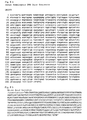
Fig. 6a shows the nucleotide sequence of a human homologous
p60TRP cDNA and Fig. 6b shows the amino acid sequence of a human
p60TRP protein.
-
Fig. 7 shows the presence of p60TRP and the result of RT-PCR
in surviving cells (PC12 cells). Panel A shows surviving colonies,
Panel B shows surviving colonies cultured for 3 days, and Panel
C shows electrophoresis on agarose gel of RT-PCR products.
-
Fig. 8 shows a semi-quantification of mRNA expression by
RT-PCR, representing the tissue-specific expression of p60TRP:
lane M, markers; lane 1, heart; lane 2, brain; lane 3, kidney;
lane 4, liver; lane 5, spleen; lane 6, pancreas; lane 7, lung;
and lane 8, skeletal muscle.
-
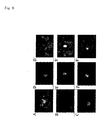
Fig. 9 shows fluorescence images of p60TRP-GFP and
p60TRP-DsRedI in CHO cells. Each Panel has the same magnification.
Panel A represents p60TRP-CT-GFP, and the others (B to F')
represent p60TRP-CT-DsRed1. Panel B shows the presence of p60TRP
in the cytoplasm, and Panel C shows the presence of p60TRP in the
nucleus. Panels D and D' , E and E', and F and F' , respectively,
show its presence in the nucleus, and images of the cell periphery
also can be seen by the Panels marked with prime. The length of
the scale bar in Panel F' corresponds to 50 µm.
-
Fig. 10 shows effects on PC12 cells by expressing p60TRP.
Fluorescence microscope images are shown which were taken after
p60TRP was co-expressed with GFP by means of p60TRP-IRES-GFP and
NGF (50 ng/ml) was added 24 hours after the expression, followed
by culturing for additional 120 hours. Cells expressing p60TRP
can be distinguished by fluorescence emitted from GFP. In PC 12
cells, p60TRP did not affect NGF-induced neurite elongation. The
scale bar corresponds to 50 µm. Each Panel has the same
magnification.
-
Fig. 11 shows the results of Western blot analysis of PP2A.
Lane 1 indicates molecular weight markers, and lane 2 indicates
a protein precipitated with a polyclonal goat anti-PP2A antibody
when employing PC12 cells into which p60TRP-IRES-GFP has been
introduced. Lane 3 indicates a protein precipitated with a
polyclonal rabbit anti-p60TRP antibody, with a single band
detected at about 60 kDa.
-
Fig. 12 shows percentages of cell survival in PC12 cells
by p60TRP gene knock-out. Each percent survival is plotted as
means ± standard error in 8 independent experiments. Relative
to a percent survival of 100% in control-1, no change could be
seen in control-2 expressing only GFP, having 102%, whereas the
percent survival was significantly reduced to 76% with p60TRP
knock-outed p60TRP-siRNA. Significance level: * p < 0.05 (versus
control).
DETAILED DESCRIPTION OF THE INVENTION
-
The present invention will be further described in detail
below.
-
In one embodiment, the present invention relates to a
p18AβrP cDNA comprising the base sequence of nucleotides 147 to
647 of SEQ ID NO: 3. The present invention also relates to an mRNA
which is complementary to the cDNA described above. The
above-described cDNA codes for a p18AβrP protein as shown in SEQ
ID NO:4, and therefore the p18AβrP protein of the present
invention has the amino acid sequence of SEQ ID NO:4. The
above-described cDNA can be typically obtained by methods
employing, for example, RT-PCR procedures, cDNA differential
procedures, and others, as described in the section Methods in
Examples, which methods are known to those skilled in the art.
-
In another embodiment, therefore, the present invention
relates to a p18AβrP protein having the amino acid sequence of
SEQ ID NO:4. Analysis of amino acid sequences of proteins can
be performed, for example, by methods described in the section
Methods in Examples. This protein is coded by the base sequence
of nucleotides 147 to 647 of SEQ ID NO:3. This protein possesses
properties of:
- a) increasing the expression by Aβ,
- b) interacting with Hsp70 and/or Tid-1, and
- c) inhibiting the cell differentiation.
-
-
With respect to the property a), as mentioned above, the
present inventors identified cell death when conducted
experiments by conducted employing rat oligodendrocytes, and
adding Aβ protein, the main component of senile plaques, cleaved
from its precursor protein APP to the cells, and thus screening
genes involved in this cell death resulting in finding a novel
gene p18AβrP was found. That is to say, the expression of the
p18AβrP protein is increased by Aβ. The increased expression can
be observed at both mRNA and protein levels: the increase in mRNA
amount can be examined, for example, by RT-PCR, complimentary
hybridization, RNase protection analysis, and the like, and the
increase in protein expression can be examined, for example, by
Western blotting, radioimmunoassay, fluorescent antibody
methods, immunological antibody methods, and the like. With
respect to the property b), the present inventors revealed novel
functions, such that p18AβrP protein suppresses the elongation
and branching of neurites and the sustaining of survival by
differentiation inducing factors to promote cell death through
the interaction with the heat shock protein Hsp70 and/or the tumor
suppressor protein Tid-1 (see Examples), as mentioned above.
Furthermore, with respect to the property c), the present
inventors found that cells expressing this gene inhibited cell
differentiation induced by neurotrophic factors and gave rise to
cell death (see Examples). Thus, the activity of this protein
can be determined by examining the degree of such differentiation
inhibition and cell death (for example, the percentage of cells
subjected to cell death, the time to cell death) . An example of
methods for measuring activities is a method comprising
introducing a gene coding for the p18AβrP protein into an
appropriate vector, using it to transform a cell, preferably nerve
cell, culturing the transformed cell under the action of a
differentiation inducing factor (for example, NGF) , and comparing
the cell death to that of an untransformed cell cultured under
the same condition (control). In order to evaluate whether cell
death has been caused, a microscopic method is commonly used.
When cell death takes place, there are observed, for example, cell
condensation, vacuolation, surface smoothing, fragmentation of
the cell and the nucleus, and the like.
-
The p18AβrP protein of the present invention also includes
variants of the protein of SEQ ID NO: 4. That is, any protein
having the above-described properties a), b) , and c), even though
one or more amino acids are inserted, deleted, or substituted in
the protein of SEQ ID NO:4 is within the p18AβrP protein of the
present invention. Any DNA coding for such a protein is also
included in the present invention. Insertion, deletion, or
substitution of one or more amino acids can be made by known methods,
such as, for example, site-directed mutagenesis and PCR. Thus,
even though a protein has one or more amino acids inserted, deleted,
or substituted, such a protein retains the above-described
properties a), b), and c), and also falls within the p18AβrP
protein of the present invention.
-
The present invention further relates to a DNA which is
hybridizable under stringent conditions with the cDNA comprising
the base sequence of nucleotides 147 to 647 of SEQ ID NO:3. This
DNA codes for a p18AβrP protein possessing the above-described
properties a), b), and c). Stringent conditions are well known
to those skilled in the art and described in many textbooks and
the literature, for example. Stringent conditions are
exemplified by conditions allowing hybridization at 42°C in a
solution containing 6X SSC, 5X Denhardt's reagent, 0.5% SDS, and
100 µg/ml salmon sperm DNA.
-
The present invention further relates to a DNA having a
nucleotide sequence homology of at least 60% or more, preferably
70% or more, more preferably 80% or more, and most preferably 90%
or more and less than 100%, based on a cDNA comprising the base
sequence of nucleotides 147 to 647 of SEQ ID NO:3.
-
A protein coded by any one of the DNAs mentioned above is
also included in the p18AβrP protein of the present invention.
-
In a further embodiment, the present invention relates to
a vector containing any one of the DNAs described above coding
for the p18AβrP protein. Vectors which may be used for
introducing any one of the above-described DNAs coding for the
p18AβrP protein to transform a nerve cell utilize a variety of
vectors described in the literature or commercial available
vectors, for example, pVgRXR, pIND, pIND/V5-His, pIND/GFP,
pcDNA3.1 or pcDNA3.1/myc or pcDNA3.1/His or pcDNA3.1/V5-His or
pcDNA3.1-CT-GFP-TOPO®, or virus vectors such as LNCX2. Methods
for transformation are known, such as, calcium phosphate methods,
Super Fector reagent methods, and lipid-mediated methods
employing LipofectAMINE reagent or the like. Cells to be used
for screening may utilize any kind of cells, preferably nerve
cells. Examples of preferred nerve cells include PC12 cell, NB2a,
Neuro-2A, B104, SHSY-5Y, primary culture nerve cell, and the like.
The above-described vector, cell transformation, and conditions
for culturing cells are determined depending upon individual
cells, and are within the knowledge of those skilled in the art
or can be readily determined. Such transformants are included
in the present invention. It is desirable to attach a detectable
tag to a p18AβrP protein to be expressed, so that the expression
of the p18AβrP protein can be detected with ease. The tag can
be attached at the DNA level: for example, green fluorescent
protein DNA, red fluorescent protein DNA, myc gene, or the like,
fused to the DNA sequence coding for the p18AβrP protein, can be
expressed. In particular, fusions with green fluorescent
protein are simple and sensitive, and thus may be recommended.
After selection of cells displaying the expression, it is
necessary to examine the above-described properties a) , b), and
c) .
-
The p18AβrP protein can be isolated by extraction of cells
expressing the p18AβrP protein by appropriates procedures known
to those skilled in the art (using immunoprecipitation with a
p18AβrP-specific antibody, for example), followed by
purification (using a Sepharose column utilizing the above-mentioned
antibody as a carrier, for example).
-
The present invention further relates to a method of
screening cell-death promoting or suppressing substances, which
comprises contacting a cell expressing p18AβrP with a test
substance in the presence of a differentiation inducing factor,
and determining the suppression or enhancement of cell death.
-
Examples of the method of screening substances which
interact with p18AβrP to suppress its function or which suppress
cell death by affecting the interaction of p18AβrP with Hsp70
and/or Tid-1 include a method comprising introducing a gene coding
the p1BAβrP protein into an appropriate vector, using it to
transform a cell, preferably a nerve cell, culturing the
transformed cell under the action of a differentiation inducing
factor (for example, NGF) and in the presence of a test substance,
and in the case of a nerve cell, assaying neurite elongation and
cell death , and comparing with the result obtained without the
test substance and/or without the differentiation inducing factor
(i.e., a control system). Appropriate vectors and cells are known
to those skilled in the art and include the above-mentioned
examples. In a cell expressing p18AβrP, also in the presence of
a differentiation inducing factor at a given concentration,
neuritis elongation is suppressed (in the case of a nerve cell) ,
so that cell death can be observed. On the other hand, the
presence of a substance which interacts with p18AβrP to suppress
its function or which suppresses cell death by affecting the
interaction of p18AβrP with Hsp70 and/or Tid-1 allows growth of
neurites (in the case of a nerve cell), avoiding cell death. In
such a case, it is likely that the test substance is a substance
which interacts with p18AβrP to suppress its function to suppress
cell death or which suppresses cell death by affecting the
interaction of p18AβrP with Hsp70 and/or Tid-1.
-
Examples of the method of screening substances which
interact with p18AβrP to promote its function or which promote
cell death by affecting the interaction of p18AβrP with Hsp70
and/or Tid-1 include a method comprising introducing a gene coding
for the p18AβrP protein into an appropriate vector, using it to
transform a cell, preferably a nerve cell, culturing the
transformed cell is cultured under the action of a differentiation
inducing factor and in the presence of a test substance, assaying
neurite elongation and cell death, and comparing with the result
obtained in the absence of the test substance. Appropriate
vectors and cells are known to those skilled in the art and include
the above-described examples. When in the presence of a
differentiation inducing factor at higher concentrations than
employed in screening substances which interact with p18AβrP to
suppress its functions, a test substance is added to a nerve cell
elongating neurites or a cell causing no cell death, whereby the
growth of neurites is suppressed (in the case of nerve cell) or
cell death is observed, it is likely that the test substance is
a substance which interacts with p18AβrP to enhance its function
to promote cell death or which promotes cell death by affecting
the interaction of p18AβrP with Hsp70 and/or Tid-1.
-
Examples of the method of evaluating whether cell death has
been caused include a method by a microscopic method as described
above.
-
The present invention further relates to substances which
interact with p18AβrP to suppress and promote cell death. Said
substances are ones interacting with p18AβrP, and the present
invention also includes substances which suppress or promote cell
death by affecting the interaction of p18AβrP with Hsp70 and/or
Tid-1. Preferably, cell-death suppressing or promoting
substances are substances obtained by the above-described
screening methods. Such substances may be any kind of molecules,
including, for example, proteins, peptides, and small molecules
such as other low molecular-weight organic compounds.
-
The cell-death suppressing substances of the present
invention are useful for the prophylaxis and/or treatment of, for
example, neurodegenerative diseases in brain associated with cell
death of nerve cells, such as Alzheimer' s disease, Down' s syndrome,
and other dementias, as well as Huntington's chorea disease,
amyotrophic lateral sclerosis, spinocerebellar degenerative
disease, Parkinson's disease, and the like. In one embodiment,
therefore, the present invention relates to a pharmaceutical
composition for the treatment and/or prophylaxis of diseases
associated with cell death, containing a cell-death suppressing
substance as described above. Usually, pharmaceutical
compositions contain pharmaceutically acceptable carriers or
excipients. Methods of manufacturing pharmaceutical
compositions, dosage forms, and pharmaceutically acceptable
carriers or excipients are selected depending upon conditions of
subjects, sites to be treated, routes of administration, and
others, and can be readily selected by those skilled in the art.
-
During the above-described screening of the present
invention, there was found a 60TRP novel gene coding for the p60TRP
protein suppressing cell death and the p60TRP protein coded by
the gene.
-
In a further embodiment, therefore, the present invention
relates to a cDNA comprising the base sequence of nucleotides 82
to 1701 of SEQ ID NO:5. The present invention also relates to
an mRNA which is complementary to the above-described cDNA. The
above-described cDNA is one coding for the protein of SEQ ID NO:6.
The amino acid sequence of SEQ ID NO: 6 is of the rat p60TRP protein,
and thus the nucleotide sequence (nucleotides 82 to 1701) of SEQ
ID NO:5 codes for the rat p60TRP protein. Hereinafter, the rat
p60TRP is simply referred to p60TRP. Thus, the p60TRP protein
of the present invention has the amino acid sequence of SEQ ID
NO:6. The above-described cDNA can be typically obtained by
methods employing, for example, RT-PCR procedures, cDNA
differential procedures, and others, as described in the section
Methods in Examples, which methods are known to those skilled in
the art.
-
In another embodiment, the present invention relates to a
DNA coding for a variant p60TRP protein which has one or more amino
acids inserted, deleted, or substituted in the amino acid sequence
of the p60TRP protein (SEQ ID NO:6) and displays a cell-death
suppressing effect similar to that of the protein having the amino
acid sequence of SEQ ID NO:6, and to a DNA coding for a variant
p60TRP protein which is hybridizable with the above-described DNA
under stringent conditions and displays a cell-death suppressing
effect similar to that of the protein having the amino acid
sequence of SEQ ID NO:6. Stringent conditions are described in
many textbooks and the literature and well known to those skilled
in the art. Stringent conditions are exemplified by conditions
allowing hybridization at 42°C in a solution containing 6X SSC,
5X Denhardt' s reagent, 0.5% SDS, and 100 µg/ml salmon sperm DNA.
-
The present invention further relates to a DNA having a
nucleotide sequence homology of at least more than 60%, preferably
70% or more, more preferably 80% or more, and most preferably 90%
or more and less than 100%, based on a cDNA comprising the base
sequence of nucleotides 147 to 647 of SEQ ID NO:3.
-
A protein coded by any one of the DNAs described above is
also included in the p60TRP protein of the present invention.
-
In this specification, a variant p60TRP protein as
described above may be designated as a p60TRP protein, as long
as it has a cell-death suppressing effect similar to that of the
protein having the amino acid sequence of SEQ ID NO:6, that is
to say, a p60TRP protein. Additionally, a p60TRP homologous
protein from species other than rat, for example, a human p60TRP
protein, may be designated as a variant p60TRP or simply p60TRP
protein, as long as it has a cell-death suppressing effect similar
to that of the protein having the amino acid sequence of SEQ ID
NO:6, that is, a p60TRP protein. Such a homologous p60TRP protein
has a homology of at least 60% or more, preferably 70% or more,
more preferably 80% or more, and most preferably 90% or more and
less than 100%, based on the rat p60TRP protein.
-
In a further embodiment, the present invention relates to
a vector containing the DNA coding for the above-described p60TRP
protein, including a variant, and also to a transformant such as
a cell transformed with the vector.
-
Vectors which may be used for introducing any one of the
above-described DNAs coding for the p60TRP protein to transform
a nerve cell utilize a variety of vectors described in the
literature and a variety of commercial available vectors, for
example, pVgRXR, pIND, pIND/V5-His, pIND/GFP, pcDNA3.1 or
pcDNA3.1/myc or pcDNA3.1/His or pcDNA3.1/V5-His or pcDNA3.1-CT-GFP-TOPO®,
or virus vectors such as LNCX2. Methods for
transformation are known, such as calcium phosphate methods,
Super Fector reagent methods, and lipid-mediated methods
employing LipofectAMINE reagent or the like. Cells to be used
for screening may utilize any kind of cells, preferably nerve
cells. Examples of preferred nerve cells include PC12 cell, NB2a,
Neuro-2A, B104, SHSY-5Y, primary culture nerve cell, and the like.
Construction of the above-described vector, cell transformation,
and conditions for culturing cells are determined depending upon
individual cells, and are within the knowledge of those skilled
in the art or can be readily determined. Such a transformant is
also included in the present invention. It is desirable to attach
a detectable tag to a p60TRP protein to be expressed, so that the
expression of the p60TRP protein can be detected with ease. A
tag can be attached at the DNA level: for example, green
fluorescent protein DNA, red fluorescent protein DNA, myc gene,
or the like, fused to the DNA sequence coding for p60TRP protein,
can be expressed. In particular, fusions with green fluorescent
protein are simple and sensitive, and thus may be recommended.
After selection of cells displaying the expression, it is
necessary to examine the cell-death suppressing effect in the
screening assay described herein.
-
The p60TRP protein can be isolated by extraction of cells
expressing the p60TRP protein by appropriates procedures known
to those skilled in the art (using immunoprecipitation with a
p60TRP-specific antibody, for example), followed by purification
(using a Sepharose column utilizing the above-mentioned antibody
as a carrier, for example).
-
In a further embodiment, the present invention relates to
a pharmaceutical composition for suppressing cell death,
containing the above-described p60TRP protein, including a
variant, a DNA coding for the protein, a vector containing the
DNA coding for the above-described p60TRP protein, including a
variant, or a transformant transformed with the vector. In
addition, the present invention also relates to a method for
suppressing cell death in a subject, characterized by
administering one of the materials or the transformant to the
subject, and to their use for manufacturing a medicament for
suppressing cell death. Methods for manufacturing
pharmaceutical compositions, dosage forms, and pharmaceutically
acceptable carriers or excipients are selected depending upon
conditions of subjects, sites to be treated, routes of
administration, and others, and can be readily selected by those
skilled in the art. These compositions, methods, and uses for
suppressing cell death allow treatment and/or prophylaxis of
various neurodegenerative diseases in brain associated with cell
death, such as Alzheimer's disease, Down's syndrome, and other
dementias, as well as Huntington's chorea disease, amyotrophic
lateral sclerosis, spinocerebellar degenerative diseases,
Parkinson's disease, and the like. In the above-described
treatment, when employing a p60TRP gene or a vector containing
the gene, gene therapy is provided, in which it is possible to
introduce the gene or vector into cells obtained from a subject,
which can be cultured and then return back to the subject, or
alternatively the p60TRP gene or vector containing the gene may
be introduced directly into a subject.
-
In addition, the present invention provides substances
regulating (promoting or suppressing) the cell-death effect of
p60TRP. Specifically, such substances are substances promoting
the cell-death suppressing effect of p60TRP by interacting with
p60TRP to inhibit cell death signal via p18AβrP (p60TRP agonists) ,
and substances attenuating the cell-death suppressing effect of
p60TRP by interacting with p60TRP to suppress the inhibition of
cell death signal via p18AβrP (p60TRP antagonists). These
substances regulating the cell-death suppressing effect of p60TRP
include, but are limited to, natural or synthetic proteins,
peptides, nucleic acids, and the like, and may be natural or
synthetic low molecular-weight compounds. Examples of p60TRP
agonists are, for example, PP2A, Ran BP5, and other proteins. For
screening such agonists and antagonists of p60TRP, two-hybrid
systems can be generally employed, as described below.
-
In addition, for example, a p60TRP agonist can be added to
a pharmaceutical composition containing p60TRP to further enhance
the cell-death suppressing effect, thereby allowing more
effective treatment or prophylaxis of diseases associated with
cell death such as Alzheimer's disease.
-
The cell-death promoting substances of the present
invention are useful, for example, for establishing cell death
systems and for various researches relating to cell death, e.g.
elucidating mechanisms of diseases associated with cell death of
nerve cells, such as Alzheimer's disease. Furthermore, the
cell-death promoting substances of the present invention are
useful for the treatment and/or prophylaxis of diseases resulting
from cell hyperproliferation, such as cancers and autoimmune
diseases. Therefore, in another embodiment, the present
invention relates to a pharmaceutical composition for the
treatment and/or prophylaxis of diseases resulting from cell
hyperproliferation, containing a cell-death promoting substance
as described above. Methods of manufacturing pharmaceutical
compositions, dosage forms, and pharmaceutically acceptable
carriers or excipients are selected depending upon conditions of
subjects, sites to be treated, routes of administration, and
others, and can be readily selected by those skilled in the art.
-
The present invention also relates to a method for the
treatment and/or prophylaxis of diseases resulting from cell
hyperproliferation in a subject, characterized by administering
to the subject a cell-death promoting substance as described above.
The present invention also relates to the use of a cell-death
promoting substance as described above in manufacturing a
medicament for the treatment and/or prophylaxis of diseases
resulting from cell hyperproliferation. Gene therapy as
described above may be also applied in the treatment and/or
prophylaxis of diseases resulting from cell hyperproliferation.
-
The present invention further relates to a method of
examining cell death in cells, particularly nerve cells, and to
a method for the diagnosis of diseases associated with such cell
death, characterized by examining the level of expression of the
p18AβrP gene or p18AβrP protein in cells and tissues obtained from
a subject. One may detects the p18AβrP gene in cell and tissue
samples obtained from a subject, for example, at the mRNA level,
or the p18AβrP protein may be detected in cell samples. Such
detection can be achieved by procedures well known to those
skilled in the art, such as hybridization employing a probe
(preferably, labeled with a radioisotope, fluorophore, enzyme,
or the like, for example) complimentary to the p18AβrP gene, or
by utilizing the binding to a monoclonal antibody (preferably,
labeled with a radioisotope, fluorophore, enzyme, or the like,
for example) directed to the p18AβrP protein. In this case, it
is possible to determine the intensity of expression of the
p18AβrP gene or the amount of the p18AβrP protein by examining
the signal intensity of the label.
-
The present invention also relates to a kit for examining
cell death in cells, particularly nerve cells, and to a kit for
the diagnosis of diseases associated with such cell death,
characterized by examining the level of expression of the p18AβrP
gene or p18AβrP protein in cells and tissues obtained from a
subject. As components of the kit are included, for example, a
probe (preferably, labeled with a radioisotope, fluorophore,
enzyme, or the like, for example) complimentary to the p18AβrP
gene and/or a monoclonal antibody (preferably, labeled with a
radioisotope, fluorophore, enzyme, or the like, for example)
directed to the p18AβrP protein. The kit usually has its
instructions for operation appended thereto.
EXAMPLES
-
The following Examples describe the present invention, but
are strictly for the purpose of illustration, and are not intended
to be limiting to the present invention.
I. Experimental Methods
-
Preparation of rat cells: CG-4 (an oligodendrocyte
precursor cell, gifted by professor Kazuhiro IKENAGA, Department
of Neuronal Information, National Institute for Physiological
Sciences, Okazaki National Research Institutes) was cultured in
a medium of DMEM/F-12 (1:1 v/v), supplemented N1 (5 mg/l of insulin,
16.1 mg/l of putrescine, 50 mg/l of transferrin, 4.6 mg/l of
D-galactose, 8 mg/l of Na selenite, 2.4 g/l of HCO3), and 30% (v/v)
of B104 cell serum-free medium. For inducing the differentiation
to oligodendrocytes, CG-4 cells were cultured for 24 hours and
B104 cells without a mitogenic factor, after that 2% FCS (Gibco)
was added to enhance survival.
-
RT-PCR methods: Analysis of mRNA and isolation of p60TRP
were conducted using RT-PCR (Heese et al., Eur. J. Neurosci., 15,
79 (2002)). Briefly, total RNA of cells was prepared according
to the TRIzol® Reagent protocol (Gibco BRL, NT, USA). After
extraction of mRNA with chloroform, the RNA was precipitated by
adding an equal volume of isopropyl alcohol to an aqueous layer,
rinsed with 75% ethanol, and dissolved in RNase-free water to
measure the absorbance (at 260 nm) on a spectrophotometer. Total
RNA of each sample (0.2 µg/ml) was first subjected to reverse
transcription to cDNA (oligo(dT)-primed-SMART™ cDNA-synthesis
(Clontech, Tokyo, Japan) ; Superscript II™ (Gibco BRL, NY, USA) ) ,
and 0.5 µl aliquot was used for PCR reactions (reaction volume:
25 µl) employing 18AβrP-specific primers (sense: 5'-atgagtgaatggacgaagaaaagccccttagaatgggaggat-3'
(SEQ ID NO:1);
antisense: 5'-tctgggaagctgaaagatggccttgaataagatcctgaattcggg-3'
(SEQ ID NO:2)). The number of cycling reactions employed for
amplification of each cDNA was set in the linearity range
according to the Elongase™ enzyme mix protocol. Denaturing in
the amplification step was carried out at 94°C for one minute,
and annealing was carried out, with the specific primers, at 65°C
for 50 seconds and then at 68°C for additional one minute longer
(65°C, 24 cycles). Rat p60TRP cDNA was prepared by 5' -RACE-RT-PCR
using a primer derived from a rat brain cDNA library (pAP3neo,
Takara) . Isolation of p60TRP was carried out with the following
procedure (Heese et al., Eur. J. Neurosci., 15, 79 (2002)).
Briefly, total cellular RNA was isolated according to the TRIzol®
Reagent protocol (Gibco BRL). After extraction with chloroform,
the RNA was precipitated by adding isopropyl alcohol to an aqueous
layer, rinsed with 75% ethanol, and redissolved in RNase-free
water and quantified on a spectrophotometer (at 260 nm). Total
RNA of each sample (0.2 µg/ml) was first subjected to
transcription to cDNA (oligo(dT)-primed-SMART™ cDNA-synthesis;
Clontech; Superscript II™ (Gibco)), and 0.5 µl aliquot was used
for PCR amplification reactions (reaction volume: 25 µl)
employing rat (r) and human (h) p60TRP-specific primers (for
isolation: sense: 5'-gcgtaatacgactcactatagggaattcgacgt-3' (SEQ
ID NO:9), antisense: 5'-cgcgacgtacgatttaaattaaccctcactaaa-3'
(SEQ ID NO:10); r-sense: 5'-atgactggctcaaagaataaggctcgggctcaggctaaactg-3'
(SEQ ID NO:11),
r-antisense: 5'-ttacattctttcaataatccctttaacttcacggaatatggcagt-3'
(SEQ ID
NO:12); h-sense: 5'-atggctgggactaagaataagacaagagcccaggccaaaac-3'
(SEQ ID NO:13),
h-antisense: 5'-cattgtttcaataatctctttaacttccctgaaaatggccatgag-3
(SEQ ID
NO:14)). The number of cycles was set in the linearity range
according to the Elongase™ enzyme mix protocol (Gibco) . PCR was
carried out as follows: denaturing was at 94°C for 0.5 minutes,
annealing at 65°C for 50 seconds (annealing temperature), and
extending at 68°C for two minutes (annealing temperature: 65°C,
16 cycles).
-
PCR amplification reactions (60°C, 16 cycles) of a
constitutively expressing ribosomal protein S12 were used for
measurement of introduced RNA. Controls without reverse
transcription employing RNA samples or without RNA were used to
ascertain the absence of contamination with DNA. PCR reactions
were analyzed by electrophoresing on 1.5% agarose gel,
transferring DNA fragments onto a nylon membrane, and allowing
to hybridize with a fluorescently labeled DNAprobe. The membrane
was subjected to analysis using a FluoroImager 595 (Image Quant
ver. 5.0 (Molecular Dynamics, Tokyo, Japan)). In addition to
non-parametric statistical analysis (Kruskal-Wallis test),
statistical analysis of the results was carried out using analysis
of variance (ANOVA) , and errors were expressed by standard errors.
-
CDNA differential: PCR-Select™ cDNA differential
(Clontech) was carried out, in order to determine the difference
in expressed mRNA between groups of cells in which Aβ1-42 was added
to induce cell death and control cells. Briefly, CG-4 cells were
incubated under conditions of fetal calf serum (FCS) ± Aβ1-42 (10
µg/ml) for 60 hours and subjected to cDNA differential (Heese et
al., Neurosci. Lett., 288, 37 (2000); Biochem. Biophys. Res.
Commun., 289, 924 (2001)). The first-strand synthesis was
carried out by converting mRNA of each groups to cDNA by the
SMART™-PCR-cDNA synthesis (Clontech) and using a modified
oligo-dT primer (a CDS primer) . SMART™-oligonucleotide-anchor
and polyA+ sequences were used as universal priming sites for cDNA
amplification from end to end (LD-PCR). Hybridization of cDNAs
derived from the Aβ1-42 treatment cell group and cDNAs derived
from the control cell group was performed to remove these cDNAs,
and unhybridized cDNAs were referred to as differential cDNAs
activated with Aβ1-42. The differential cDNAs were cloned into
the TOPO®-T/A cloning vector (Invitrogen) and subjected to
identification by southern blotting. The base sequence was
analyzed on a sequencer (ABI PRIM™ BigDye™ Terminator Cycle
Sequencing Ready Reaction Kit (Perkin-Elemer; sequencer: ABI
PRISM Model 310)).
-
CDNA cloning: After cDNA subtraction, a full-length cDNA
from EST sequences of rat p18AβrP was obtained by employing
oligonucleotides designed from partial cDNA/EST sequences from
a database (http://www.ncbi.nlm.nih.gov) and carrying out the
screening using a rat brain cDNA library (ClonCapture Ready™ Super
DNA; Clontech, Tokyo, Japan) in 5'-RACE (rapid amplification of
cDNA ends) and RT-PCR experiments. A p18AβrP construct for
analysis was made by inserting the rat p18AβrP cDNA into the
pCR®II-TOPO® T/A cloning vector (Invitrogen, Tokyo, Japan). A
p18AβrP Construct for expression (p18AβrP-CT-GFP) was made by
inserting pcDNA3.1CT-GFP-TOPO® (Invitrogen, Tokyo, Japan) for
the expression of green fluorescent protein (GFP) into the rat
p18AβrP cDNA at the C-terminal of p18AβrP.
Analysis of the p18AβrP cDNA sequences and amino acid:
-
Analysis of the p18AβrP cDNA and amino acid sequences were carried
out using PDB, SwissProt, PIF, and PRF, in addition to the NCBI
(National Center for Biotechnology Information) Blastp 2.0
program (Nucleic Acids Res., 25, 3389 (1997)) to un-overlapped
GenBank CDS translations and the UniGene database (NCBI) (Nucleic
Acids Res., 25, 2289 (1997)). Homology search was carried out
using Blast and FASTA (Wisconsin Package ver. 10.0, Genetics
Computer Group (GCG), Madison, WI) algorithms, and BestFit
(Eisconsin Package Version 10.0, GCG) . Motifs of the amino acid
sequence were searched using PROSITE-Profile, and BLOCKS-,
ProDom-, PRINTS-, Pfam-and PSORTII-programs (Nucleic Acids Res.,
27, 260 (1999), Intellig. Syst. Mol. Biol., 4, 109 (1996);
Intellig. Syst. Mol. Biol., 5, 147 (1997)). Phosphorylation
sites were searched using NetPhos 2.0 (J. Mol. Biol., 294, 1351
(1999)), and other analyses were carried out using not only the
ExPASy www server (http://www.expasy.ch), but also
http://www/softberry.com/index.html and the amino acid
composition search (AACompIdent)
http://kr.expasy.org/tools/aacomp/. The determined p18AβrP
cDNA and amino acid sequences are shown in SEQ ID NO:3 (Fig. 1a)
and SEQ ID NO:4 (Fig. 1b), respectively. In addition, Fig. 1c
shows a comparison of the amino acid sequences of the rat p18AβrP
protein of the present invention and human and mouse homologous
proteins.
-
Analysis of p18AβrP expressing tissues: Twenty-two tissues
from rats were taken according to usual procedures, to prepare
samples: 1 = brain; 2 = heart; 3 = kidney; 4 = spleen; 5 = liver;
6 = colon; 7 = liver; 8 = small intestine; 9 = muscle; 10 = stomach;
11 = testis; 12 = salivary gland; 13 = throid; 14 = adrenal; 15
= pancreas; 16 = ovary; 17 = uterus; 18 = prostate; 19 = skin;
20 = leukocyte; 21 = bone marrow; and 22 = fetus brain (the number
corresponds to the lane number in Fig. 4). Tissue samples were
prepared by preparing cDNAs form each of the tissue samples in
the previously described method (see Section RT-PCR) . In order
to examine the tissue-specific expression of p18AβrP, Rapid-Scan™-Gene-Expression
panels (Origene Technologies, MD, USA) was
used. PCR products were analyzed using a standard 2% DNA
electrophoresis agarose E-gel™ (Invitrogen).
-
Methods of screening cell death suppressing and/or
promoting substances - Obtaining of a cell-death suppressing
substance p60TRP: In PC12 cells expressing p18AβrP, there were
not observed cell differentiation, survival, and neurite
elongation, in spite of the addition of NGF, whereas when the
expression of a rat cDNA library in these cells resulted in the
finding of surviving cells, in spite of the expression of p18AβrP.
Thus, by the death trap screening method (Semin. Immunol., 9, 17
(1997)) from these surviving cells, a gene involved in this
survival was found with RT-PCR and named a p60TRP gene. Also,
the cell-death suppressing protein coded by this gene was named
a p60TRP protein.
-
Analysis of the p60TRP cDNA base sequence and the protein
primary structure: Sequence analysis of the p60TRP cDNA and
protein was carried out using as a search tool the NCBI (National
Center for Biotechnology Information) Blast 2.0 program employing
the UniGene database (NCBI) (Nucleic Acids Res., 25, 3389 (1997))
and the GenBank CDS translation + PDB + Swiss Prot + PIR + PRF
databases. For homology search, Blast and FASTA (Wisconsin
Package Version 10.0, GCG (Genetics Computer Group) algorithms
were used, and BestFit (Wisconsin Package Version 10.0 GCG) was
used for alignment. The protein sequence was determined
employing the ExPASy-www-server (http://www.expasy.ch);
softberry: http://www.softberry.com/index.html and the amino
acid composition search (AACompIdent):
http://kr.expasy.org/tools/aacomp/. Motifs of the amino acid
sequence were searched using PROSITE Profile, BLOCKS-ProDom-PRINTS-Pfam-and
PSORT II-programs (Nucleic Acids Res., 27, 260
(1999); Mol. Biol., 4, 109 (1996); Mol. Biol., 5, 147 (1997)).
Phosphorylation sites were searched using the NetPhos 2. 0 protein
phosphorylation search server (J. Mol. Biol., 294, 1351 (1991)).
-
Analysis of p60TRP expression in tissues: Analysis of the
tissue-specific gene expression of p60TRP utilized Rapid-Scan™-Gene-Expression
panels (Origene Technologies, Rockville,
MD, USA), and tissue cDNAs were subjected to semi-quantitative
RT-PCR analysis. PCR products were analyzed using a standard 1.5%
DNA electrophoresis agarose E gel™ (Invitrogen, Brain Aging, 2,
30 (2002)).
-
Cell culture: B104, rat neuroblastoma cell (available from
Professor Kazuhiro Ikenaka, National Institute for Physiological
Sciences, Okazaki National Research Institutes, for example) , and
PC12 cell (available fromATCC (American Type Culture Collection) ,
ATCC No. CRL-1721, for example) were cultured in Dulbecco's
Modified Eagle Medium (D-MEM)/F-12 (1:1) containing N2
supplemented with 10% fetal calf serum (FCS; Gibco BRL, Grand
Island, NY, USA), and CHO (Chinese hamster ovary) cell line was
cultured in DMEM supplemented with 10% FCS, under the condition
of 5% CO2/95% air at 37°C. CG-4 (an oligodendrocyte precursor
cell) was cultured in a medium of DMEM/F-12 (1:1 v/v),
supplemented N1 (5 mg/l of insulin, 16.1 mg/l of putrescine, 50
mg/l of transferrin, 4.6 mg/l of D-galactose, 8 mg/l of Na selenite,
2.4 g/l of HCO3), and 30% (v/v) of B104 cell serum-free medium.
For inducing the differentiation to oligodendrocytes, CG-4 cells
were cultured for 24 hours and B104 cells without a mitogenic
factor, after that 2% FCS (Gibco) was added to enhance survival.
For inducing cell death, cells were cultured for 60 to 72 hours
with FCS ± Aβ (Peptide Institute, Inc., Osaka, Japan; dissolved
in a serum-free solution, in 1 mg/ml phosphate buffered saline
(PBS), pH 7.4, to 10 µg/ml, followed by incubation at 37°C for
24 hours), and then surviving cells were measured using a Promega
kit (CellTiter96® AQueous One Solution cell proliferation assay) .
For inducing neurite elongation, PC 12 cells were subjected to
gene transfer with p18AβrP-CT-GFP or a control vector, and then
induced with NGF (50 ng/ml) for 24 hours.
-
Gene transfer into cells: Constructed were p18AβrP-CT-GFP
having GFP (green fluorescent protein) introduced at the C-terminal
of p18AβrP; p60TRP-CT-GFP and p60TRP-CT-DsRed1 having,
respectively, GFP (pcDNA3.1CT-GFP-TOPO® (Invitrogen) or DsRed1
(NheI and HindIII restriction enzyme cloning sites, Clontech,
Tokyo, Japan) introduced at the C-terminal of the rat p60TRP cDNA.
Additionally, p60TRP was subcloned into pIRES2-EGFP (Clontech),
in order to allow co-expression with GFP from the same mRNA
(p60TRP-IRES-GFP). P18AβrP-CT-GFP or p60TRP-DsRed1, p60TRP-GFP,
p60TRP-IRES-GFP, and GFP (Clontech, Tokyo, Japan) expression
vector, or an empty vector (control) was transiently gene
transferred into CHO cell (available from ATCC, ATCC No. CCL-61,
for example) and PC12 cell (available from ATCC, ATCC No.
CCL-1271, for example) (SuperFector, B-Bridge, San Jose, CA, USA),
which were cultured using a D-MEM/F12 (1: 1) /N2 containing culture
medium supplemented with 10% fetal calf serum (FCS, Gibco BRL,
Tokyo) at 37°C under the condition of 5% CO2/95% air. The
efficiency of gene transfer was verified by co-expression of GFP
and p53-/PKC-DsRed1 (Clontech) , so as to be always 50 to 60% (Eur.
J. Neurosci., 15, 79 (2002)). Cell death/survival was evaluated
under a fluorescent microscope (Olympus IX70, Olympus, Tokyo,
Japan) at 48 hours after gene transfer. Further, 24 hours later,
NGF (murine NGF 2.5S, 50 ng/ml; Invitrogen) was added to the PC12
cells, and after 120 hours, surviving cells were measured in the
CellTiter96® AQueous Assay (Promega, Madison, WI, USA) and observed
under a fluorescent microscope.
-
Two-Hybrid system: A yeast strain MaV203 (available from
Invitrogen, for example) was used. Rat p18AβrP or rat p60TRP was
subcloned into the pDEST™32 vector (Invitrogen) having a GAL4
DNA binding domain from pENTR/D-TOPO®. Also, pEXP-AD502 was used
as an expression vector for an activation domain having a
ProQuest™ two-hybrid rat brain cDNA library (Invitrogen). For
selection for activity, three reporter genes, HIS3, URA3, and lacZ,
were used. Each of these genes was stably integrated into a yeast
gene at a different site, and the promoter region of the HIS3,
lacZ, and URA3 is different except for the GAL4 binding domain.
It is reported that the ProQuest™ Two-Hybrid System enables three
independent transcriptions to take place from respective separate
chromosomes, thereby giving reduced false-positive reactions, as
compared to standard two-hybrid systems. The induction of the
HIS3 and URA3 reporter genes is caused depending upon the
two-hybrid, and respectively enables cells to grow also on a plate
lacking histidine or uracil, so that cells can be discriminated.
On the other hand, the induction of the lacZ gene can be done with
X-gal (5-brome-4-chloro-3-indolyl-β-D-galactopyranoside),
resulting in blue color. In addition, due to the toxicity
resulting from the conversion of 5-fluoroorotate (5-FOA) to
5-fluorouracil, the induction of URA3 enables cells to grow in
a culture medium lacking uracil and inhibits the growth in a
culture medium containing 5-FOA. Therefore, this system enables
the screening of four phenotypes, that is, proteins displaying
the true interaction by means of His (3AT®), β-gal, Ura+ and 5-FOAs
and thus the elimination of false-positive reactions. The use
of the ARS/CEN vector also can reduce the expression level and
the toxicity. Positive clones can be identified by retransformation.
When an interacting protein is contained, a
yeast cell binds to db-rat p60TRP or db-rat p18AβrP and ad-Y
(wherein Y is, for example, PP2A or TID-1). The plasmid DNA from
a yeast strain containing the above can be introduced into an E.
coli cell by electroporation, and the transformant can be selected
with ampicillin or the db-rat p18AβrP can be selected with
gentamycin. The plasmid DNA of these E. coli cells, ad-Y (wherein
Y is, for example, PP2A or TID-1), can be introduce into MaV203
together with pDBleu or db-rat p18Aβr or db-rat p60TRP, and the
induction of the reporter genes by db-rat p18Aβr or pdb-rat p60TRP
will give true positive reactions.
-
Production of anti-p60TRP antibody: A rabbit anti-p60TRP
antibody was prepared and used which has a high affinity for the
N-terminal domain of p60TRP (amino acid (aa)-35 to aa-45:
RGAGKNRDKGK-cys).
-
Protein immunoprecipitation and western blot analysis: 24
hours after transient gene transfer of p60TRP-IRES-GFP into about
5 x 10⁶ PC12 cells, culturing was continued for additional 48 hours.
After that, the cells were washed twice with Tris-saline buffer
(TBS, pH 7.2) and lysed at 4°C with 0.5 ml of an ice-cooled buffer
(150 mM NaCl, 50mM Tris-HCl pH 8.0, 1% NP40, 2% glycerol, 1mM PMSF,
10 µg/ml aprotinin, 1 µg/ml leupeptin, 0.5mM Na vanadate), and
the nucleus was removed by centrifugation at 4°C. After
incubating with the antibody (anti-p60TRP) at 4°C for 2 hours,
500 µl of 50% Protein-A Sepharose® CL-4B was added and incubation
was carried out for additional 2 hours. Immunoprecipitate was
washed three times, and 50 µl of the Laemmli-protein buffer
(Bio-Rad, Tokyo, Japan) was added, followed by western blot
analysis. Briefly, proteins were electrophoresed on 10%
polyacrylamide gel to separate. The proteins were transferred
onto a polyvinylidene fluoride membrane (PVDF) (Bio-Rad, Tokyo,
Japan), and then immunoreacted with an anti-PP2A (regulatory
subunit) antibody (SantaCruz, CA, USA), and a secondary
fluorescein-conjugated anti-goat antibody and a tertiary
alkaline phosphatase-conjugated anti-fluorescein antibody were
added and incubated with the substrate of alkaline phosphatase
(the ECF™ western blotting kit, Amersham/Pharmacia, Tokyo,
Japan).
-
Evaluation of cell death: PC12 cells were cultured to a
confluency of 50 to 80% and treated with trypsin 24 hours prior
to gene transfer, diluted 5 times in a fresh medium lacking
antibiotics (1 to 3 x 10⁵ cells/ml), and transferred into a 24-well
plate (500 µl/well) and cultured. In experiments for evaluating
cell death, investigation was made on cells expressing no GFP
(control-1), cells expressing only GFP (control-2), and cells
having the p60TRP gene knock-outed by SiRNA. The gene knock-out
by SiRNA, was carried out employing Oligofectamine and
introducing 0.5 µg of siRNA per well (Brain Aging, 2, 44 (2002)).
The efficiency of gene transfer was determined under a fluorescent
microscope after the co-expression of 1 µg of a GFP expression
vector and 0.2 µg of siRNA (Mol. Brain Res., 104, 127 (2002) ; Nature,
411, 494 (2001)) . The percentage of cell death/survival by SiRNA
was determined 48 hours after expression, by the Cell-Titer 96®
AQueous One solution assay (Promega, Madison, WI: Neurosci. Lett.,
288, 37 (2000)). The siRNA sequence for p60TRP utilized the
sequence of nt 310 to nt 330 relative to the start codon.
P60TRP-specific 21-nucleic acid duplex siRNA was obtained from
Dharmacon Research (Lafayetta, CO, USA; B-Bridge International,
Tokyo, Japan).
-
The experimental procedures described above are typical as
known in the art, and those skilled in the art will recognize or
should easily conceive their variations and alternative
procedures.
II. Experimental Results
-
The experimental results according to the present invention
will be described as follows:
- (1) The base sequence of the rat p18AβrP cDNA of the present
invention is homologous to that of human and mouse sequences, and
their amino acid sequences are compared and shown in Fig. 1c.
Accordingly, it has turned out that the base sequence of the rat
p18AβrP cDNA of the present invention is a novel sequence that
is different from the human base sequence described in the
above-mentioned literature and sequences found in the Gene Data
Bank, and that as can be seen from Fig. 1c, the amino acid sequence
of the p18AβrP protein is a novel sequence that is different from
that of human and mouse sequences.
- (2) It was shown that the expression of p18AβrP in rat CG4
oligodendrocyte lysates was increased by Aβ1-42, when examined
by Southern blotting after addition of Aβ1-42 (10 µg/ml) at 37°C
for 60 hours (see, Fig. 2 left panel): lane 1, control; lane 2,
Aβ treatment (Aβ). The amount of transcription of the p18AβrP
mRNA, densitometric quantification resulting in finding to
increase by the addition of Aβ1-42 (see, Fig. 2 right panel).
- (3) In PC12 cells subjected to differentiation induced by
NGF, p18AβrP gave rise to suppressed elongation of neurites and
cell death. For the purpose of investigating functions of p18AβrP
in nerve cells, a gene of a protein having GFP fused to the
C-terminal of p18AβrP was introduced into PC 12 cells, and its
expression was identified after 48 hours. Subsequently, 24 hours
later, NGF (50 ng/ml) was added and fluorescent microscopic
observations were made after 120 hours, with the result that as
shown in Fig. 3, elongation of neurites was suppressed and cell
death was observed in spite of the presence of NGF, and 2 weeks
later, all the cells were killed. On the contrary, in cells having
no expression of the gene (having no fluorescence), NGF clearly
promoted neurite elongation and allowed survival of the cells.
Some of these patterns are shown in Fig. 3, Panels a to i. For
example, it was observed in Panels c and f that p18AβrP-positive
cells emitting GFP fluorescence as indicated by the arrow were
shrinked, caused cell death, and floating. It was also observed
that the cell indicated by the arrow in Panel d retained the
cellular morphology, while the neurite elongation was suppressed.
- (4) The Two-Hybrid system as explained above was employed
to screen a protein or proteins interacting with p18AβrP of the
present invention, with the result that such a protein was found
to interact with the heat shock protein hsp70 and the tumor
suppressor protein Tid-1. It is likely that effects of inducing
cell death and suppressing cell differentiation as described
above resulted from such interaction.
- (5) Non-quantitative analysis of mRNA expression was
carried out by RT-PCR in terms of the expression of p18AβrP mRNA
in rat various tissues. As shown in Fig. 4, its expression was
identified in twenty-two organs and tissues, including brain.
- (6) Isolation, Characterization of p60TRP, and Expression
in Tissues and Cells
After gene transfer of p18AβrP and a rat brain cDNA library,
p60TRP was found in surviving cells by RT-PCR (Fig. 7) . The base
sequence of a rat cDNA of this novel gene is shown in SEQ ID NO:5
and Fig. 5A, and the amino acid sequence in SEQ ID NO:6 and Fig.
5B, while for comparison, its homologous human gene cDNA is shown
in SEQ ID NO:7 and Fig. 6A, and the amino acid sequence in SEQ
ID NO:8 and Fig. 6B. The protein coded by the rat cDNA
(nucleotides 82 to 1701 of SEQ ID NO:5) is a protein consisting
of 539 amino acids of SEQ ID NO:6 and having a molecular weight
of about 59.72 kDa. The base sequence of this rat p60TRP cDNA
and its amino acid sequence are clearly different from that of
human sequences, and thus are novel sequences. The amino acid
homology between the rat p60TRP and human homologous p60TRP
proteins suggests that the human homologous p60TRP protein also
possesses a cell-death suppressing effect similar to that of the
rat p60TRP protein. From their sequences, p60TRP proteins likely
constitutes a novel protein family and have a bHLH domain (amino
acids 491 to 507 of the sequence of SEQ ID NO:6). Members of the
p60TRP protein family having such a domain include O43168, Q96D09,
QBVZ3, Q9CVV3, Q9H969, Q920R4, Q9BE11, Q9C0G2, Q9CXQ7, Q9UJC4,
Q8R095, O60267, Q9NPE4/Q9UH62, Q9NTS2, Q9BTM6, Q9H2Q0, Q9P291,
Q9NWJ13, Q9CX19, Q9DC32, Q9CZ87, Q9CUN3, Q9D0L7, Q9CS81, and
Q9CX83.Further, RT-PCR was employed to search the expression
pattern in tissues, with the result that the mRNA was identified
in brain, kidney, and spleen at high expression levels, and also
in heart and skeletal muscle, whereas no expression was detected
in lung and liver (Fig. 8) . Fig. 9 shows fluorescence images of
p60TRP-GFP and p60TRP-DsRed1 in CHO cells. In the CHO cells, the
p60TRP-GFP or p60TRP-DsRed1 fusion protein was localized
particularly in the cytoplasm (Fig. 9 Panels A and B) and also
existed in the nucleus (Fig. 9 Panels C to F).In addition, p60TRP did not affect the neurite elongation
induced by NGF in PC12 cells. As shown in Fig. 10, when NGF (50
ng/ml) was added 24 hours after p60TRP was expressed in PC12 cells,
which were cultured for additional 120 hours, the fact that the
neurite-elongating effect of NGF was not suppressed was able to
be observed in cells co-expressing p60TRP and GFP.
- (7) Proteins Interacting with P60TRP
The Two-Hybrid system revealed the interaction of two proteins
with p60TRP, i.e., PP2A (protein-phosphatase 2A) responsible for
a crucial dephosphorylation reaction in the intracellular signal
transduction and RanBP5 (Ran-binding protein 5) involved in the
transport of the bHLH transcription factor from the cytoplasm into
the nucleus. As shown in Fig. 11, immunoprecipitation and its
western blot analysis showed that p60TRP binds to PP2A. It is
likely that p60TRP interacts with these proteins and inhibits the
cell-death signal via p18AβrP, thereby leading to inhibiting cell
death.
- (8) Immunoprecipitation of P60TRP and PP2A
Fig. 11 represents the result of western blot analysis of
immunoprecipitates of p60TRP and PP2A. It was ascertained by
immunoprecipitation experiments that p60TRP expressed in PC12
cells was co-precipitated at about 60 kDa by interacting and
complexing with PP2A.
- (9) Decrease in Percent Cell Survival by P60TRP Gene
Knock-out
-
-
Gene knock-out by p60TRP-specific siRNA significantly
reduced the percentage of surviving PC12 cells, as compared to
control groups. That is, as shown in Fig. 12, the control-1
displayed a percent survival of 100% and the control-2 GFP did
not change the percent survival with the percentage being 102%,
whereas p60TRP-siRNA with knock-outed p60TRP significantly
reduced the percentage of surviving cells to 76% (p < 0.05) . This
further ascertained the cell-death suppressing effect of the
p60TRP protein.
-
The present invention thus has been described in particular
and in detail with reference to the Examples. However, it is
possible for those skilled in the art to make modifications and
variations other than described above, such as, for example, easy
selection of appropriate cell lines, to carry out the invention.
INDUSTRIAL APPLICABILITY
-
According to the present invention, a novel gene p18AβrP
was found whose expression was increased in oligodendrocytes by
amyloid-β protein (hereinafter refereed to Aβ) and its functions
were demonstrated. That is, it has turned out that the present
gene and its product, p18AβrP protein, possess novel functions
of suppressing the promotion of neurite elongation by
neurotrophic factors and the sustaining of cell death to promote
cell death by interacting with the heat shock protein Hsp70 and
the tumor suppressor protein Tid-1. Therefore, the present
invention provides screening systems in which these are applied,
substances involved in promoting and suppressing cell death which
are obtainable using such screening systems, diagnosis and
prophylaxis of diseases employing them. By the above-described
screening system, the present invention further has found, for
the first time, a rat cell-death suppressing protein p60TRP and
its coding gene, and also identified operations and effects of
the p60TRP protein, i.e. an effect of suppressing cell death, for
the first time. Therefore, the present invention provides
diagnosis and prophylaxis of diseases associated with cell death
employing them.
SEOUENCE LISTING FREE TEXT
SEQ ID NO: 1
-
A sense primer to amplify p18AβrP cDNA.
SEQ ID NO: 2
-
An antisense primer to amplify p18AβrP cDNA.
SEQ ID NO: 3
-
A nucleotide sequence of cDNA encoding p18AβrP protein.
SEQ ID NO: 4
-
An amino acid sequence of p18AβrP protein.
SEQ ID NO: 5
-
A nucleotide sequence of cDNA encoding rat p60TRP protein.
SEQ ID NO: 6
-
An amino acid sequence of rat p60TRP protein.
SEQ ID NO: 7
-
A nucleotide sequence of cDNA encoding human p60TRP protein.
SEQ ID NO: 8
-
An amino acid sequence of human p60TRP protein.
SEQ ID NO: 9
-
A sense primer (specific to rat/human p60TRP) to amplify p60TRP
cDNA.
SEQ ID NO: 10
-
An antisense primer (specific to rat/human p60TRP) to amplify
p60TRP cDNA.
SEQ ID NO: 11
-
A sense primer (specific to rat p60TRP) to amplify p60TRP cDNA.
SEQ ID NO: 12
-
An antisense primer (specific to rat p60TRP) to amplify p60TRP
cDNA.
SEQ ID NO: 13
-
A sense primer (specific to human p60TRP) to amplify p60TRP cDNA.
SEQ ID NO: 14
-
An antisense primer (specific to human p60TRP) to amplify p60TRP
cDNA.