EP1569550B1 - Method for determining endothelial dependent vasoactivity - Google Patents
Method for determining endothelial dependent vasoactivity Download PDFInfo
- Publication number
- EP1569550B1 EP1569550B1 EP03812666A EP03812666A EP1569550B1 EP 1569550 B1 EP1569550 B1 EP 1569550B1 EP 03812666 A EP03812666 A EP 03812666A EP 03812666 A EP03812666 A EP 03812666A EP 1569550 B1 EP1569550 B1 EP 1569550B1
- Authority
- EP
- European Patent Office
- Prior art keywords
- pressure
- blood vessel
- related signals
- subject
- elapsed time
- Prior art date
- Legal status (The legal status is an assumption and is not a legal conclusion. Google has not performed a legal analysis and makes no representation as to the accuracy of the status listed.)
- Expired - Lifetime
Links
- 238000000034 method Methods 0.000 title claims abstract description 77
- 230000001419 dependent effect Effects 0.000 title claims abstract description 55
- 230000003511 endothelial effect Effects 0.000 title claims abstract description 49
- 210000004204 blood vessel Anatomy 0.000 claims abstract description 78
- 230000008859 change Effects 0.000 claims abstract description 23
- 230000000694 effects Effects 0.000 claims description 38
- 230000007246 mechanism Effects 0.000 claims description 32
- 210000001367 artery Anatomy 0.000 claims description 29
- 210000003403 autonomic nervous system Anatomy 0.000 claims description 29
- 238000004458 analytical method Methods 0.000 claims description 22
- 210000002302 brachial artery Anatomy 0.000 claims description 21
- 238000000354 decomposition reaction Methods 0.000 claims description 18
- 230000000638 stimulation Effects 0.000 claims description 18
- 230000004936 stimulating effect Effects 0.000 claims description 15
- 239000000126 substance Substances 0.000 claims description 14
- 239000012530 fluid Substances 0.000 claims description 12
- 208000018672 Dilatation Diseases 0.000 claims description 11
- 239000012528 membrane Substances 0.000 claims description 10
- 238000012545 processing Methods 0.000 claims description 10
- 210000001715 carotid artery Anatomy 0.000 claims description 9
- 210000002321 radial artery Anatomy 0.000 claims description 9
- 238000004364 calculation method Methods 0.000 claims description 6
- 239000000919 ceramic Substances 0.000 claims description 6
- 230000003340 mental effect Effects 0.000 claims description 6
- 230000008569 process Effects 0.000 claims description 4
- 230000003595 spectral effect Effects 0.000 claims description 3
- 230000004938 stress stimulation Effects 0.000 claims description 2
- 230000002159 abnormal effect Effects 0.000 description 63
- MWUXSHHQAYIFBG-UHFFFAOYSA-N Nitric oxide Chemical compound O=[N] MWUXSHHQAYIFBG-UHFFFAOYSA-N 0.000 description 48
- 238000005259 measurement Methods 0.000 description 39
- 230000008753 endothelial function Effects 0.000 description 38
- 230000004044 response Effects 0.000 description 28
- 230000008694 endothelial dysfunction Effects 0.000 description 27
- 206010048554 Endothelial dysfunction Diseases 0.000 description 26
- 210000003038 endothelium Anatomy 0.000 description 25
- 238000002604 ultrasonography Methods 0.000 description 20
- 238000011084 recovery Methods 0.000 description 19
- XLYOFNOQVPJJNP-UHFFFAOYSA-N water Substances O XLYOFNOQVPJJNP-UHFFFAOYSA-N 0.000 description 17
- 230000007423 decrease Effects 0.000 description 15
- 210000004369 blood Anatomy 0.000 description 13
- 239000008280 blood Substances 0.000 description 13
- 230000036544 posture Effects 0.000 description 13
- 230000002889 sympathetic effect Effects 0.000 description 13
- 238000012360 testing method Methods 0.000 description 13
- 208000029078 coronary artery disease Diseases 0.000 description 12
- 230000004087 circulation Effects 0.000 description 11
- 230000006870 function Effects 0.000 description 10
- 201000001320 Atherosclerosis Diseases 0.000 description 9
- SNIOPGDIGTZGOP-UHFFFAOYSA-N Nitroglycerin Chemical compound [O-][N+](=O)OCC(O[N+]([O-])=O)CO[N+]([O-])=O SNIOPGDIGTZGOP-UHFFFAOYSA-N 0.000 description 9
- 206010047139 Vasoconstriction Diseases 0.000 description 9
- 230000036772 blood pressure Effects 0.000 description 9
- 210000004351 coronary vessel Anatomy 0.000 description 9
- 229960003711 glyceryl trinitrate Drugs 0.000 description 9
- 230000001734 parasympathetic effect Effects 0.000 description 9
- 230000002093 peripheral effect Effects 0.000 description 9
- 239000000006 Nitroglycerin Substances 0.000 description 8
- 230000001404 mediated effect Effects 0.000 description 8
- 230000002829 reductive effect Effects 0.000 description 8
- 230000025033 vasoconstriction Effects 0.000 description 8
- 230000004872 arterial blood pressure Effects 0.000 description 7
- 230000000747 cardiac effect Effects 0.000 description 7
- 230000008602 contraction Effects 0.000 description 7
- 206010020565 Hyperaemia Diseases 0.000 description 6
- 230000017531 blood circulation Effects 0.000 description 6
- 230000003247 decreasing effect Effects 0.000 description 6
- 238000003745 diagnosis Methods 0.000 description 6
- 238000010586 diagram Methods 0.000 description 6
- 201000010099 disease Diseases 0.000 description 6
- 208000037265 diseases, disorders, signs and symptoms Diseases 0.000 description 6
- HVYWMOMLDIMFJA-DPAQBDIFSA-N cholesterol Chemical compound C1C=C2C[C@@H](O)CC[C@]2(C)[C@@H]2[C@@H]1[C@@H]1CC[C@H]([C@H](C)CCCC(C)C)[C@@]1(C)CC2 HVYWMOMLDIMFJA-DPAQBDIFSA-N 0.000 description 5
- 238000001727 in vivo Methods 0.000 description 5
- 238000000718 qrs complex Methods 0.000 description 5
- 238000010183 spectrum analysis Methods 0.000 description 5
- 210000005166 vasculature Anatomy 0.000 description 5
- 206010020772 Hypertension Diseases 0.000 description 4
- 210000001765 aortic valve Anatomy 0.000 description 4
- 235000019504 cigarettes Nutrition 0.000 description 4
- 230000010339 dilation Effects 0.000 description 4
- 230000004064 dysfunction Effects 0.000 description 4
- 230000001771 impaired effect Effects 0.000 description 4
- 230000000670 limiting effect Effects 0.000 description 4
- 230000033001 locomotion Effects 0.000 description 4
- 208000022064 reactive hyperemia Diseases 0.000 description 4
- 238000005070 sampling Methods 0.000 description 4
- 230000035945 sensitivity Effects 0.000 description 4
- QZAYGJVTTNCVMB-UHFFFAOYSA-N serotonin Chemical compound C1=C(O)C=C2C(CCN)=CNC2=C1 QZAYGJVTTNCVMB-UHFFFAOYSA-N 0.000 description 4
- 230000000391 smoking effect Effects 0.000 description 4
- 208000010110 spontaneous platelet aggregation Diseases 0.000 description 4
- 230000001515 vagal effect Effects 0.000 description 4
- 108090000190 Thrombin Proteins 0.000 description 3
- 206010047141 Vasodilatation Diseases 0.000 description 3
- 210000000709 aorta Anatomy 0.000 description 3
- 210000000748 cardiovascular system Anatomy 0.000 description 3
- 150000003943 catecholamines Chemical class 0.000 description 3
- 238000005314 correlation function Methods 0.000 description 3
- 230000006735 deficit Effects 0.000 description 3
- 238000011161 development Methods 0.000 description 3
- 230000018109 developmental process Effects 0.000 description 3
- 206010012601 diabetes mellitus Diseases 0.000 description 3
- 230000002526 effect on cardiovascular system Effects 0.000 description 3
- 238000005516 engineering process Methods 0.000 description 3
- 239000000835 fiber Substances 0.000 description 3
- 230000004217 heart function Effects 0.000 description 3
- 230000001976 improved effect Effects 0.000 description 3
- 239000000463 material Substances 0.000 description 3
- 230000002107 myocardial effect Effects 0.000 description 3
- 108020003175 receptors Proteins 0.000 description 3
- 102000005962 receptors Human genes 0.000 description 3
- 230000009467 reduction Effects 0.000 description 3
- 239000000523 sample Substances 0.000 description 3
- 210000003491 skin Anatomy 0.000 description 3
- 230000000087 stabilizing effect Effects 0.000 description 3
- 229960004072 thrombin Drugs 0.000 description 3
- 238000011282 treatment Methods 0.000 description 3
- 230000024883 vasodilation Effects 0.000 description 3
- 210000000707 wrist Anatomy 0.000 description 3
- 208000024172 Cardiovascular disease Diseases 0.000 description 2
- 102000008186 Collagen Human genes 0.000 description 2
- 108010035532 Collagen Proteins 0.000 description 2
- 108010023302 HDL Cholesterol Proteins 0.000 description 2
- 206010019280 Heart failures Diseases 0.000 description 2
- 241000282412 Homo Species 0.000 description 2
- 208000035150 Hypercholesterolemia Diseases 0.000 description 2
- 241000699670 Mus sp. Species 0.000 description 2
- 208000030831 Peripheral arterial occlusive disease Diseases 0.000 description 2
- 241000700159 Rattus Species 0.000 description 2
- 241000287181 Sturnus vulgaris Species 0.000 description 2
- 208000007536 Thrombosis Diseases 0.000 description 2
- OIPILFWXSMYKGL-UHFFFAOYSA-N acetylcholine Chemical compound CC(=O)OCC[N+](C)(C)C OIPILFWXSMYKGL-UHFFFAOYSA-N 0.000 description 2
- 229960004373 acetylcholine Drugs 0.000 description 2
- 108091008698 baroreceptors Proteins 0.000 description 2
- 230000006399 behavior Effects 0.000 description 2
- 230000008901 benefit Effects 0.000 description 2
- 239000000872 buffer Substances 0.000 description 2
- 238000012512 characterization method Methods 0.000 description 2
- 229920001436 collagen Polymers 0.000 description 2
- 230000000052 comparative effect Effects 0.000 description 2
- 230000002517 constrictor effect Effects 0.000 description 2
- 238000007405 data analysis Methods 0.000 description 2
- 239000003814 drug Substances 0.000 description 2
- 230000009977 dual effect Effects 0.000 description 2
- 230000002964 excitative effect Effects 0.000 description 2
- 230000000004 hemodynamic effect Effects 0.000 description 2
- 230000006872 improvement Effects 0.000 description 2
- 238000012623 in vivo measurement Methods 0.000 description 2
- 230000002401 inhibitory effect Effects 0.000 description 2
- 208000028867 ischemia Diseases 0.000 description 2
- 210000005240 left ventricle Anatomy 0.000 description 2
- 238000004519 manufacturing process Methods 0.000 description 2
- 230000004048 modification Effects 0.000 description 2
- 238000012986 modification Methods 0.000 description 2
- 208000031225 myocardial ischemia Diseases 0.000 description 2
- 210000000056 organ Anatomy 0.000 description 2
- 230000008506 pathogenesis Effects 0.000 description 2
- 230000008288 physiological mechanism Effects 0.000 description 2
- 230000036316 preload Effects 0.000 description 2
- 210000001774 pressoreceptor Anatomy 0.000 description 2
- 230000001681 protective effect Effects 0.000 description 2
- 230000000541 pulsatile effect Effects 0.000 description 2
- 230000011514 reflex Effects 0.000 description 2
- 238000011160 research Methods 0.000 description 2
- 229940076279 serotonin Drugs 0.000 description 2
- 238000004904 shortening Methods 0.000 description 2
- 210000001013 sinoatrial node Anatomy 0.000 description 2
- 230000002966 stenotic effect Effects 0.000 description 2
- 230000007704 transition Effects 0.000 description 2
- 238000012285 ultrasound imaging Methods 0.000 description 2
- 238000010200 validation analysis Methods 0.000 description 2
- 239000002550 vasoactive agent Substances 0.000 description 2
- 230000000283 vasomotion Effects 0.000 description 2
- 230000002861 ventricular Effects 0.000 description 2
- 206010001497 Agitation Diseases 0.000 description 1
- 206010002383 Angina Pectoris Diseases 0.000 description 1
- 108010071619 Apolipoproteins Proteins 0.000 description 1
- 102000007592 Apolipoproteins Human genes 0.000 description 1
- 208000031104 Arterial Occlusive disease Diseases 0.000 description 1
- 206010003210 Arteriosclerosis Diseases 0.000 description 1
- 206010003211 Arteriosclerosis coronary artery Diseases 0.000 description 1
- 241000282472 Canis lupus familiaris Species 0.000 description 1
- 102000003727 Caveolin 1 Human genes 0.000 description 1
- 108090000026 Caveolin 1 Proteins 0.000 description 1
- 208000001778 Coronary Occlusion Diseases 0.000 description 1
- 206010011086 Coronary artery occlusion Diseases 0.000 description 1
- 108010037462 Cyclooxygenase 2 Proteins 0.000 description 1
- 208000005156 Dehydration Diseases 0.000 description 1
- 206010016803 Fluid overload Diseases 0.000 description 1
- WQZGKKKJIJFFOK-GASJEMHNSA-N Glucose Natural products OC[C@H]1OC(O)[C@H](O)[C@@H](O)[C@@H]1O WQZGKKKJIJFFOK-GASJEMHNSA-N 0.000 description 1
- 208000010496 Heart Arrest Diseases 0.000 description 1
- 208000031226 Hyperlipidaemia Diseases 0.000 description 1
- FFFHZYDWPBMWHY-VKHMYHEASA-N L-homocysteine Chemical compound OC(=O)[C@@H](N)CCS FFFHZYDWPBMWHY-VKHMYHEASA-N 0.000 description 1
- 108010028554 LDL Cholesterol Proteins 0.000 description 1
- 208000008589 Obesity Diseases 0.000 description 1
- 206010033307 Overweight Diseases 0.000 description 1
- 208000005764 Peripheral Arterial Disease Diseases 0.000 description 1
- 208000004880 Polyuria Diseases 0.000 description 1
- TUZYXOIXSAXUGO-UHFFFAOYSA-N Pravastatin Natural products C1=CC(C)C(CCC(O)CC(O)CC(O)=O)C2C(OC(=O)C(C)CC)CC(O)C=C21 TUZYXOIXSAXUGO-UHFFFAOYSA-N 0.000 description 1
- 102100038280 Prostaglandin G/H synthase 2 Human genes 0.000 description 1
- 102000003923 Protein Kinase C Human genes 0.000 description 1
- 108090000315 Protein Kinase C Proteins 0.000 description 1
- 208000001647 Renal Insufficiency Diseases 0.000 description 1
- 206010053648 Vascular occlusion Diseases 0.000 description 1
- 230000007488 abnormal function Effects 0.000 description 1
- 230000005856 abnormality Effects 0.000 description 1
- 230000001154 acute effect Effects 0.000 description 1
- 230000003044 adaptive effect Effects 0.000 description 1
- 230000001800 adrenalinergic effect Effects 0.000 description 1
- 210000004079 adrenergic fiber Anatomy 0.000 description 1
- 230000004075 alteration Effects 0.000 description 1
- 206010003119 arrhythmia Diseases 0.000 description 1
- 230000006793 arrhythmia Effects 0.000 description 1
- 208000021328 arterial occlusion Diseases 0.000 description 1
- 208000011775 arteriosclerosis disease Diseases 0.000 description 1
- 230000002238 attenuated effect Effects 0.000 description 1
- 230000002567 autonomic effect Effects 0.000 description 1
- 230000035581 baroreflex Effects 0.000 description 1
- 230000003542 behavioural effect Effects 0.000 description 1
- 102000012740 beta Adrenergic Receptors Human genes 0.000 description 1
- 108010079452 beta Adrenergic Receptors Proteins 0.000 description 1
- 230000033228 biological regulation Effects 0.000 description 1
- 230000003139 buffering effect Effects 0.000 description 1
- 238000004422 calculation algorithm Methods 0.000 description 1
- 230000000647 cardiovagal effect Effects 0.000 description 1
- 230000009084 cardiovascular function Effects 0.000 description 1
- 230000037198 cardiovascular physiology Effects 0.000 description 1
- 230000001413 cellular effect Effects 0.000 description 1
- 108091008690 chemoreceptors Proteins 0.000 description 1
- 235000012000 cholesterol Nutrition 0.000 description 1
- 230000015271 coagulation Effects 0.000 description 1
- 238000005345 coagulation Methods 0.000 description 1
- 238000004891 communication Methods 0.000 description 1
- 238000010276 construction Methods 0.000 description 1
- 208000026758 coronary atherosclerosis Diseases 0.000 description 1
- 230000002596 correlated effect Effects 0.000 description 1
- 230000000875 corresponding effect Effects 0.000 description 1
- 230000008878 coupling Effects 0.000 description 1
- 238000010168 coupling process Methods 0.000 description 1
- 238000005859 coupling reaction Methods 0.000 description 1
- 230000018044 dehydration Effects 0.000 description 1
- 238000006297 dehydration reaction Methods 0.000 description 1
- 238000001514 detection method Methods 0.000 description 1
- 230000037213 diet Effects 0.000 description 1
- 235000005911 diet Nutrition 0.000 description 1
- 230000004069 differentiation Effects 0.000 description 1
- 230000035619 diuresis Effects 0.000 description 1
- 231100000673 dose–response relationship Toxicity 0.000 description 1
- 229940079593 drug Drugs 0.000 description 1
- 230000008497 endothelial barrier function Effects 0.000 description 1
- 210000002889 endothelial cell Anatomy 0.000 description 1
- 239000000066 endothelium dependent relaxing factor Substances 0.000 description 1
- 210000003989 endothelium vascular Anatomy 0.000 description 1
- 229940011871 estrogen Drugs 0.000 description 1
- 239000000262 estrogen Substances 0.000 description 1
- 238000000605 extraction Methods 0.000 description 1
- 210000001105 femoral artery Anatomy 0.000 description 1
- 235000013305 food Nutrition 0.000 description 1
- 210000000609 ganglia Anatomy 0.000 description 1
- 239000008103 glucose Substances 0.000 description 1
- 208000019622 heart disease Diseases 0.000 description 1
- 230000001631 hypertensive effect Effects 0.000 description 1
- 238000001802 infusion Methods 0.000 description 1
- 230000005764 inhibitory process Effects 0.000 description 1
- 230000030214 innervation Effects 0.000 description 1
- 230000000297 inotrophic effect Effects 0.000 description 1
- 230000003993 interaction Effects 0.000 description 1
- 238000002608 intravascular ultrasound Methods 0.000 description 1
- 230000009191 jumping Effects 0.000 description 1
- 201000006370 kidney failure Diseases 0.000 description 1
- 108010022197 lipoprotein cholesterol Proteins 0.000 description 1
- 239000007788 liquid Substances 0.000 description 1
- 210000004072 lung Anatomy 0.000 description 1
- 238000012544 monitoring process Methods 0.000 description 1
- 210000001616 monocyte Anatomy 0.000 description 1
- 210000000663 muscle cell Anatomy 0.000 description 1
- 210000002464 muscle smooth vascular Anatomy 0.000 description 1
- 210000004165 myocardium Anatomy 0.000 description 1
- 210000000107 myocyte Anatomy 0.000 description 1
- 210000002569 neuron Anatomy 0.000 description 1
- 235000020824 obesity Nutrition 0.000 description 1
- 230000003534 oscillatory effect Effects 0.000 description 1
- 235000020825 overweight Nutrition 0.000 description 1
- 210000005037 parasympathetic nerve Anatomy 0.000 description 1
- 230000036961 partial effect Effects 0.000 description 1
- 230000010412 perfusion Effects 0.000 description 1
- 230000003836 peripheral circulation Effects 0.000 description 1
- 239000008177 pharmaceutical agent Substances 0.000 description 1
- 230000000144 pharmacologic effect Effects 0.000 description 1
- 230000037081 physical activity Effects 0.000 description 1
- 230000000704 physical effect Effects 0.000 description 1
- 230000003389 potentiating effect Effects 0.000 description 1
- 229960002965 pravastatin Drugs 0.000 description 1
- TUZYXOIXSAXUGO-PZAWKZKUSA-N pravastatin Chemical compound C1=C[C@H](C)[C@H](CC[C@@H](O)C[C@@H](O)CC(O)=O)[C@H]2[C@@H](OC(=O)[C@@H](C)CC)C[C@H](O)C=C21 TUZYXOIXSAXUGO-PZAWKZKUSA-N 0.000 description 1
- 230000001902 propagating effect Effects 0.000 description 1
- 229940127293 prostanoid Drugs 0.000 description 1
- 150000003814 prostanoids Chemical class 0.000 description 1
- 230000002685 pulmonary effect Effects 0.000 description 1
- 239000003642 reactive oxygen metabolite Substances 0.000 description 1
- 230000009257 reactivity Effects 0.000 description 1
- 230000007115 recruitment Effects 0.000 description 1
- 230000029865 regulation of blood pressure Effects 0.000 description 1
- 230000001105 regulatory effect Effects 0.000 description 1
- 230000000241 respiratory effect Effects 0.000 description 1
- 230000024977 response to activity Effects 0.000 description 1
- 230000002441 reversible effect Effects 0.000 description 1
- 238000012552 review Methods 0.000 description 1
- 229960000672 rosuvastatin Drugs 0.000 description 1
- BPRHUIZQVSMCRT-VEUZHWNKSA-N rosuvastatin Chemical compound CC(C)C1=NC(N(C)S(C)(=O)=O)=NC(C=2C=CC(F)=CC=2)=C1\C=C\[C@@H](O)C[C@@H](O)CC(O)=O BPRHUIZQVSMCRT-VEUZHWNKSA-N 0.000 description 1
- 210000002235 sarcomere Anatomy 0.000 description 1
- 230000002784 sclerotic effect Effects 0.000 description 1
- 238000012216 screening Methods 0.000 description 1
- 230000005586 smoking cessation Effects 0.000 description 1
- 210000002460 smooth muscle Anatomy 0.000 description 1
- 238000001228 spectrum Methods 0.000 description 1
- 210000000278 spinal cord Anatomy 0.000 description 1
- 210000000952 spleen Anatomy 0.000 description 1
- 238000010561 standard procedure Methods 0.000 description 1
- 238000007619 statistical method Methods 0.000 description 1
- 230000008093 supporting effect Effects 0.000 description 1
- 230000002791 sympathovagal effect Effects 0.000 description 1
- 210000000225 synapse Anatomy 0.000 description 1
- 230000009885 systemic effect Effects 0.000 description 1
- 230000028016 temperature homeostasis Effects 0.000 description 1
- 230000003827 upregulation Effects 0.000 description 1
- 230000002792 vascular Effects 0.000 description 1
- 208000021331 vascular occlusion disease Diseases 0.000 description 1
- 210000004509 vascular smooth muscle cell Anatomy 0.000 description 1
- 239000005526 vasoconstrictor agent Substances 0.000 description 1
- 229940124549 vasodilator Drugs 0.000 description 1
- 239000003071 vasodilator agent Substances 0.000 description 1
- 230000001457 vasomotor Effects 0.000 description 1
- 230000001196 vasorelaxation Effects 0.000 description 1
- 208000003663 ventricular fibrillation Diseases 0.000 description 1
Images


Classifications
-
- A—HUMAN NECESSITIES
- A61—MEDICAL OR VETERINARY SCIENCE; HYGIENE
- A61B—DIAGNOSIS; SURGERY; IDENTIFICATION
- A61B5/00—Measuring for diagnostic purposes; Identification of persons
- A61B5/02—Detecting, measuring or recording for evaluating the cardiovascular system, e.g. pulse, heart rate, blood pressure or blood flow
- A61B5/02007—Evaluating blood vessel condition, e.g. elasticity, compliance
-
- A—HUMAN NECESSITIES
- A61—MEDICAL OR VETERINARY SCIENCE; HYGIENE
- A61B—DIAGNOSIS; SURGERY; IDENTIFICATION
- A61B5/00—Measuring for diagnostic purposes; Identification of persons
- A61B5/02—Detecting, measuring or recording for evaluating the cardiovascular system, e.g. pulse, heart rate, blood pressure or blood flow
- A61B5/021—Measuring pressure in heart or blood vessels
- A61B5/02133—Measuring pressure in heart or blood vessels by using induced vibration of the blood vessel
-
- A—HUMAN NECESSITIES
- A61—MEDICAL OR VETERINARY SCIENCE; HYGIENE
- A61B—DIAGNOSIS; SURGERY; IDENTIFICATION
- A61B5/00—Measuring for diagnostic purposes; Identification of persons
- A61B5/02—Detecting, measuring or recording for evaluating the cardiovascular system, e.g. pulse, heart rate, blood pressure or blood flow
- A61B5/024—Measuring pulse rate or heart rate
- A61B5/02405—Determining heart rate variability
-
- A—HUMAN NECESSITIES
- A61—MEDICAL OR VETERINARY SCIENCE; HYGIENE
- A61B—DIAGNOSIS; SURGERY; IDENTIFICATION
- A61B5/00—Measuring for diagnostic purposes; Identification of persons
- A61B5/02—Detecting, measuring or recording for evaluating the cardiovascular system, e.g. pulse, heart rate, blood pressure or blood flow
- A61B5/026—Measuring blood flow
- A61B5/0285—Measuring or recording phase velocity of blood waves
-
- A—HUMAN NECESSITIES
- A61—MEDICAL OR VETERINARY SCIENCE; HYGIENE
- A61B—DIAGNOSIS; SURGERY; IDENTIFICATION
- A61B5/00—Measuring for diagnostic purposes; Identification of persons
- A61B5/40—Detecting, measuring or recording for evaluating the nervous system
- A61B5/4029—Detecting, measuring or recording for evaluating the nervous system for evaluating the peripheral nervous systems
- A61B5/4035—Evaluating the autonomic nervous system
-
- A—HUMAN NECESSITIES
- A61—MEDICAL OR VETERINARY SCIENCE; HYGIENE
- A61B—DIAGNOSIS; SURGERY; IDENTIFICATION
- A61B5/00—Measuring for diagnostic purposes; Identification of persons
- A61B5/41—Detecting, measuring or recording for evaluating the immune or lymphatic systems
- A61B5/414—Evaluating particular organs or parts of the immune or lymphatic systems
- A61B5/416—Evaluating particular organs or parts of the immune or lymphatic systems the spleen
-
- A—HUMAN NECESSITIES
- A61—MEDICAL OR VETERINARY SCIENCE; HYGIENE
- A61B—DIAGNOSIS; SURGERY; IDENTIFICATION
- A61B5/00—Measuring for diagnostic purposes; Identification of persons
- A61B5/72—Signal processing specially adapted for physiological signals or for diagnostic purposes
- A61B5/7235—Details of waveform analysis
- A61B5/7253—Details of waveform analysis characterised by using transforms
- A61B5/726—Details of waveform analysis characterised by using transforms using Wavelet transforms
Definitions
- the present invention relates to measuring endothelial dependent vasoactivity and, more particularly, to a non-invasive method and system for determining endothelial dependent vasoactivity.
- Hemodynamics is a subchapter of cardiovascular physiology, which deals with the forces the heart has to develop in order to circulate blood throughout the cardiovascular system. To a physician, these forces are manifested as blood pressure and blood flow paired values measured simultaneously at different points of the cardiovascular system.
- the flow of blood through the vasculature has a pulsatile nature.
- the heart contracts, part of the blood contained within the left ventricle is squeezed into the aorta from which the blood flows into the entire cardiovascular system. Since blood is an incompressible fluid, when it is squeezed into the vasculature, which exhibits a resistance to blood flow, blood pressure is generated.
- the arterial blood pressure increases to its highest, the systolic level.
- the left ventricle is refilled with oxygenated blood from the lungs during the relaxation phase of the cardiac cycle (the diastole), and the ventricle is disconnected from the vasculature by the aortic valve, the pressure in the vasculature decreases to its lowest level.
- SI Stroke Index
- MAP Mean Arterial Pressure
- Intravascular volume is the amount of fluid circulating in the vasculature. This modulator can be affected, for example, by dehydration, diuresis, venoconstriction of the spleen, volume overload due to heart or kidney failure and the like.
- Inotropy is the ability of the cardiac muscle to contract, Myocytes are the only muscle cells which are able to vary the strength of contraction. Inotropy can be affected by exercise, stress and pharmaceutical agents, which increase the strength of myocardial contractions, or by cardiac diseases such as heart failure, which is expressed by decrease of the strength of contractions.
- the myocardial contractility is controlled by positive and negative inotropes which instantaneously affect the level of inotropic state. Changes in inotropy alter the rate of force and pressure development by the ventricle.
- the heart has the intrinsic capability of increasing its force of contraction when preload is increased.
- the preload is related to the sarcomere length via the well known Starling law.
- Vasoactivity referrers to the ability of blood vessels to expand and contract. Through vasoactivity the body controls the flow of blood through individual organs, accommodate the variation in blood flow and regulate arterial pressure.
- the endothelium-dependent relaxation of blood vessels is due to the release of potent non-proslanoid vasodilator substances by the endothelium (the inner most cellular layer of the blood vessel) surrounding the blood vessel.
- the endothelium-derived relaxing factor is believed to be nitric oxide (NO), which is released by different stimuli substances produced during platelet aggregation.
- NO nitric oxide
- the endothelial action of thrombin and platelet products is crucial for the protective role played by the normal endothelium against unwanted coagulation. Therefore, local platelet aggregation, with the associated release of serotonin and ADP, together with the production of thrombin, leads to a major local release of NO.
- the release of NO to the blood vessel also inhibits platelet adhesion at the endothelium blood interface, exerts a major feedback on platelet aggregation, thereby eliminates the imminent danger of vascular occlusion.
- the endothelial barrier prevents the platelet derived vasoconstrictor substances from reaching the smooth muscle. NO can also be released by other stimuli like flow mediated vasoactivity and increased sympathetic activity (alpha receptor stimulation).
- endothelial dysfunction dysfunction of endothelial dependent vasoactivity
- endothelial dysfunction and coronary artery disease are also linked to over-weight, obesity, hypertension, hypercholesterolemia, hyperlipidemia, diabetes mellitus, cigarette smoking and homocysteine.
- the vascular endothelium plays a fundamental role in several processes related to thrombosis. Impaired endothelium function may also promote the development of atherosclerosis through its effects on vaso-regulation, platelet and monocyte adhesion.
- Endothelial dysfunction increases in men over the age of about 40 and in women after the age of about 55, whether or not other coronary risk factors are present.
- the specific cause of the decrease in endothelial function with age is yet unknown. Estrogen appears to be a major factor associated with gender differences in age-related endothelial function.
- endothelial function Other factors which affect endothelial function include hypertension [ Perticone F, et al., "Prognostic significance of endothelial dysfunction in hypertensive patients,” Circulation 2001, 104:191-196 ], diabetes [ Cosentino F et al., "Endothelial dysfunction in diabetes mellitus,” J Cardiovasc Pharmacol, 1998, 32:54-61 ; Cosentino F et al., "High glucose causes upregulation of Cyclooxygenase-2 and alters prostanoid profile in human endothelial cells.
- coronary artery disease is related to atherosclerothic disease in the aorta and the carotid artery [ Khoury Z et al., "Relation of coronary artery disease to atherosclerothic disease in the aorta, carotid, and femoral arteries evaluated by ultrasound,” Am J Cardiol 1997, 80:1429-1433 ].
- Assessment of endothelium dependent vasoreactivity (EDV) in coronary arteries may be performed by measurements of changes in peripheral arterial diameter due to pharmacological or mechanical stimuli.
- One method for measuring the inner diameter of a blood vessel is by an intravascular ultrasound device having an intravascular catheter and an ultrasound transducer array mounted thereon.
- the intravascular catheter is inserted directly into the artery of interest to thereby determine its inner diameter.
- Such a device is highly invasive, expensive and requires costly additional technical expertise to operate.
- Another known device for measuring the intravascular diameter of a blood vessel has an elongated flexible sheath and a catheter which is longer than the sheath.
- the sheath has an outer diameter which is less than the intravascular diameter.
- the catheter proximal end extends outwardly from the proximal end of the sheath and includes a measuring scale directly proportional to a position of a sensor extending from the catheter.
- non-invasive methods for the measurement of arterial diameter by high resolution non-invasive ultra-sound systems.
- the physician operates an ultrasound transducer to obtain appropriate ultrasound images of the brachial artery for measuring artery diameter thereof.
- This method is time consuming, and requires a highly trained physician or technician to hold the transducer stably during the measurement.
- an automatic measurement system having a robot arm manipulating ultrasound imaging probe is used.
- the system automatically navigates the ultrasound imaging probe to an appropriate position and measure changes in diameter of brachial artery with improved reproducibility compared with manual measurement.
- the autonomic nervous system plays a cardinal role in the control of cardiovascular function.
- Heart rate, heart excitability and contractility are under the constant influence of the parasympathetic-sympathetic balance.
- Parasympathetic nerves and sympathetic fibers innervate the sino-atrial node; the parasympathetic influence is inhibitory while the sympathetic influence is excitatory.
- the parasympathetic fibers to the SA node are driven by inhibitory and excitatory inputs from peripheral receptors (baroreceptors, chemoreceptors, cardiac, pulmonary and airway receptors).
- Behavioral adaptive influence of the heart rate at the sinus node is mediated by supramedullary input to the cardiovagal neurons.
- the origin of the sympathetic innervation of the heart is located at the T2-T5 segment of the spinal cord and the preganglionic fibers synapse in the cervical ganglia.
- Normal cardiac function is regulated by the complex balance of the sympathetic and parasympathetic outflows to the heart. This balance is also responsible for the susceptibility to arrhythmias: while vagal activity has a protective role, sympathetic activity lowers the threshold to ventricular fibrillation.
- Normal heart function, heart rate included, is modulated by the fluctuations in the sympathetic and parasympathetic flow to the heart. These fluctuations induce beat-to-beat variability in heart rate and arterial pressure. Hence, the analysis of the instantaneous fluctuations in cardiovascular variables supplies valuable information on the autonomic control in an intact organism.
- WO 02134105 A discloses a method and a system for measuring endothelial function of a patient according to the preambles of claims 1 and 22 by monitoring the peripheral arterial tone of a digit of the patient during and after application of an occluding pressure on the arm.
- EP 1 360 929 A published between the priority date and the filing date of the present application discloses a similar method and system where the difference in pulse propagation time between the two arms of a patient is measured.
- the method further comprising determining an autonomic nervous system activity of the subject.
- the determining of the autonomic nervous system activity is by heart rate variability analysis of the pressure-related signals.
- the determining of the autonomic nervous system activity comprises recording electrocardiogram signals of a chest of the subject and performing heart rate variability analysis of the electrocardiogram signals, thereby determining the autonomic nervous system activity.
- the method further comprises determining a pre-ejection period and valve-artery period.
- valve of the valve-artery period is an aortic valve and the artery of the valve-artery period is a carotid artery.
- the determination of the pre-ejection period and the valve-artery period comprises determining an elapsed time between peaks of the electrocardiogram signals and peaks of the pressure-related signals.
- the peaks of the electrocardiogram signals comprise QRS peaks.
- the method further comprising stimulating the at least one blood vessel.
- the stimulating of the at least one blood vessel is effected by a procedure selected from the group consisting of a mechanical stimulation, a thermal stimulation a chemical stimulation, an electrical stimulation a mental stress stimulation and a physical exercise stimulation.
- the stimulating of the at least one blood vessel is by applying external pressure on the at least one blood vessel.
- the stimulating of the at least one blood vessel is by reducing a temperature of the at least one blood vessel.
- the method further comprising correlating the endothelial functioning and the autonomic nervous system activity, so as to obtain a correlation function, and using the correlation function to at least preliminarily determine the endothelial dependent vasoactivity of the subject.
- the recording of the pressure-related signals is by piezoelectric ceramic elements.
- the recording of the pressure-related signals is by a membrane-based sensor.
- an electrate microphone the membrane-based sensor is an electrate microphone.
- the extracting of the at least one parameter comprises: (a) scanning pressure-related signals recorded of a first location and detecting a first peak; (b) scanning pressure-related signals recorded of a second location and detecting a second peak corresponding to the first peak; (c) measuring an elapsed time between the first peak and the second peak; and (d) repeating the steps (a)-(c) at least once.
- system further comprising electronic-calculation functionality for determining an autonomic nervous system activity of the subject.
- the processing unit is operable to calculate heart rate variability from the pressure-related signals thereby to determine the autonomic nervous system activity.
- system further comprising at least one electrocardiogram lead designed connectable to a chest of the subject.
- the processing unit is operable to calculate heart rate variability from electrocardiogram signals sensed by the at least one electrocardiogram lead, thereby to determine the autonomic nervous system activity.
- system further comprising a spectral analyzer for analyzing the at least one parameter and obtaining a frequency decomposition of the at least one parameter, the frequency decomposition being representative of the endothelial dependent vasoactivity of the subject.
- system further comprising a mechanism for stimulating the at least one blood vessel.
- the mechanism for stimulating the at least one blood vessel is selected from the group consisting of a mechanical mechanism, a thermal mechanism, an electrical mechanism and a mechanism for generating mental stress.
- the mechanism is operable to apply external pressure on the at least one blood vessel.
- the mechanism comprises a sphingomanometer.
- the mechanism is operable to reduce a temperature of the at least one blood vessel.
- the mechanism is a bath or a cuff of fluid, the fluid being at a predetermined temperature.
- the sensors are piezoelectric ceramic elements.
- the sensors membrane-based are sensors.
- the sensors are electrate microphones.
- the method further comprising obtaining a frequency decomposition of the at least one parameter, and using the frequency decomposition for determining the endothelial dependent vasoactivity of the subject.
- the at least one parameter is selected from the group consisting of an amplitude of the pressure-related signals, a width of the pressure-related signals and an elapsed time between two peaks of the pressure-related signals.
- the method further comprising obtaining a frequency decomposition of the amplitudes, and using the frequency decomposition for determining the endothelial dependent vasoactivity of the subject.
- the method further comprising obtaining a frequency decomposition of the width, and using the frequency decomposition for determining the endothelial dependent vasoactivity of the subject.
- the method further comprising obtaining a frequency decomposition of the elapsed time, and using the frequency decomposition for determining the endothelial dependent vasoactivity of the subject.
- the at least one characteristic of the at least one blood vessel is selected from the group consisting of a radius of the at least one blood vessel and an elastic modulus of the at least one blood vessel.
- the present invention successfully addresses the shortcomings of the presently known configurations by providing a method and system for assessing endothelial dependent vasoactivity enjoying properties far exceeding prior art technologies.
- selected steps of the invention could be implemented as a chip or a circuit.
- selected steps of the invention could be implemented as a plurality of software instructions being executed by a computer using any suitable operating system.
- selected steps of the method and system of the invention could be described as being performed by a data processor, such as a computing platform for executing a plurality of instructions.
- the present invention is of a non-invasive method and system for determining endothelial dependent vasoactivity which can be used in early stage diagnosis of endothelial dysfunction related diseases.
- the present invention can be used to screen and diagnose large population and to differentiate between subjects being in different stages and combinations of endothelial and coronary artery dysfunction.
- the present invention can be used to diagnose pathogenesis of cardiovascular disease, atherosclerosis and the like.
- a pulsatile flow of fluid through an elastic conduit is accompanied by elevated friction and normal forces between the conduit and the fluid.
- Such flow is characterized by dominant peripheral energy propagation, i . e ., along the wall of the elastic conduit.
- the speed of a pressure pulse propagating through the conduit is determined by the elastic and geometric properties of the conduit's wall as well as by the physical properties of the fluid.
- the velocity of the pressure pulse generated by ventricular ejection can be calculated using Moens-Korteweg model, according to which the square of the pulse wave velocity, c, equals the area of the artery divided by the density of the blood and the mechanical compliance of the artery.
- the mechanical compliance defined as the derivative of the cross-sectional area with respect to the pressure, is, to a good approximation 2R /( E h ), where, R is the radius of the artery, h is the thickness of the artery's wall and E is its Young modulus.
- the present invention exploits the relation between the pulse wave velocity and the geometrical and elastic properties of the arterial wall for the purpose of determining vasoactivity.
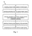
- Figure 1 is a flowchart diagram of a non-invasive method 10 of determining endothelial dependent vasoactivity of a subject, according to one aspect of the present invention.
- pressure-related signals are recorded of several locations adjacent to one or more blood vessels.
- the pressure-related signals are typically electrical signals, which are recorded, e.g ., using piezoelectric ceramic elements or membrane-based sensors, such as, but not limited to, electrate microphones.
- these pressure-related signals are related to the pulse wave velocity of the blood, hence can be used to characterize the geometrical and elastic properties of the arterial wall.
- At least one parameter is extracted from the pressure-related signals.
- extracted parameters include, without limitation, amplitude of the signals, width thereof and/or elapsed time between peaks of two pressure-related signals.
- the amplitude parameter is preferably defined as the height of the signal above a predetermined zero-level.
- the width parameter is preferably defined as the distance between two points of equal height or two inflection points on the same signal.
- the elapsed time parameter is preferably defined as the elapsed time between two peaks of signals recorded of two different locations, either two locations near the same blood vessels or near different blood vessels.
- the elapsed time parameter is directly related to the pulse wave velocity. More specifically, knowing the transit time, t , of the pulse wave between two locations and its traveling distance, L , one can calculate the pulse wave velocity, by division ( L / t ) or differentiation ( dL / dt ).
- any of the above parameters may be extracted from the signals by any appropriate method known in the art, such as, but not limited to, correlation method, peak detection, mathematical fitting (e.g ., polynomial fitting), frequency decomposition (e.g ., Fourier transform), data folding and the like.
- the extraction is performed a plurality of times, so as to obtain, for each type of parameter, a plurality of values which may then be averaged.
- a third step of method 10, designated by Block 16 the parameter(s) are used to determine a change of one or more blood vessel characteristics, e.g ., geometrical or elastic properties thereof. Such a change characterizes endothelial function of the blood vessel.
- the elapsed time parameter is sensitive to arterial radius changes at the initial stage of arterial dilatation, and the amplitude parameter is sensitive to arterial radius changes at relatively large arterial dilatation.
- a judicious use of the elapsed time parameter and the amplitude parameter allows an accurate and reliable measurement of changes in the arterial radius at a wide range of values.
- radius changes are the favored blood vessel characteristics in the according to the presently preferred embodiment of the invention, other blood vessel characteristics, e.g ., elastic modulus are not excluded.
- the elastic modulus of the blood vessel is, to a good approximation, a constant quantity.
- the elastic module becomes radius-dependent [ Armentano R.L et al., "Arterial wall mechanics in conscious dogs - assessment of viscous, internal, and elastic moduli to characterize aortic wall behavior,” Circulation Research 1995, 76:468-78 ], and can be determined using the elapsed time parameter.
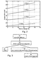
- Equation 2 A consequence of Equation 2 is that as the artery's radius increase the pulse wave velocity decreases. In terms of elapsed time, a decrease in the pulse wave velocity is manifested as an increment of the elapsed time between two peaks of the signals.
- Figure 2 shows theoretical estimations of the relative changes in the elapsed time as a function of changes in arterial radius, assuming an approximately constant Young modulus.
- the different lines in Figure 2 correspond to different effective blood flow distances, L .
- the calculations were performed using typical initial radius and elasticity modulus, taken from the literature.
- NO is known to have a buffering influence on arterial pressure variability.
- An acute change of arterial pressure alters shear stress, thus modifying NO generation and release.
- Subsequent vasodilatation or vasoconstriction occurs in response to the varying NO levels, which in turn readjust vascular resistance to reduce arterial pressure variability.
- NO acts rapidly: it diffuses out of the endothelium to the subjacent vascular smooth muscle cells, where it clauses vaso-relaxation within seconds.
- NO can affect the regulation of blood pressure more rapidly than the arterial baroreflex.
- the above parameters can be further analyzed for the purpose of obtaining other observables sensitive to the above physiological mechanism.
- Many analysis procedures are contemplated by the present invention, including, without limitation, spectral analysis, modulation analysis and the like.
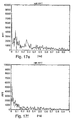
- the method further comprises an optional step, designated in Figure 1 by Block 17 , in which a frequency decomposition is obtained from one or more of the parameters, e.g ., by performing spectral analysis.
- the obtained frequency decomposition can be used for determining the endothelial dependent vasoactivity of the subject.
- endothelial dysfunction can be determined when the frequency decomposition includes higher power in the high frequency range, e.g ., above about 0.15 Hz.
- endothelial dysfunction can be diagnosed when a decrease in power in lower frequency ranges (e.g ., below about 0.12 Hz, below about 0.08 Hz, or below 0.06 Hz).
- such increment of power in the high frequency range can be followed by increased variability of the elapsed time and the amplitude parameters.
- Block 18 an autonomic nervous system activity of the subject is characterized.
- Heart rate changes commonly referred to as heart rate variability, are known to be a direct consequence of alterations in the activity of autonomic nervous system.
- the autonomic nervous system characterization is done by heart rate variability analysis.
- Heart rate variability may be determined in more than one way.
- the heart rate variability is determined from the pressure-related signals.
- the pressure-related signals can be divided into segments, preferably equally distributed, where in each segment the mean heart rate is calculated by subtraction of subsequent peak times.
- the heart rate variability is then defined as the standard deviation of the heart rate in each segment.
- Typical duration for each segment is between 5 seconds and 15 seconds, inclusive.
- heart rate variability is obtained by a different measurement, which may be, for example, electrocardiogram measurement or any other procedure for recording electrical signals of the chest of the subject.
- a known device for determining heart rate variability is a Holter monitor, which is a recorder for a continuous, typically twenty-four hour, electrocardiographic recording of the heart rate.
- heart rate variability may be determined by extracting a series of cardiac R-R intervals from the electrocardiogram signals.
- Electrocardiogram signals include, inter alia , the so-called P waves, T waves and QRS complexes, which QRS complexes include Q waves, R-waves and S waves.
- An R-R interval is the elapsed time between two successive R-waves of the electrocardiogram signals.
- any of the above definitions may be used when extracting the cardiac R-R intervals.
- the procedure of extracting cardiac R-R intervals from the electrocardiogram signals is well known in the art and can be executed, either manually or automatically, e.g ., by a data processor which, in one embodiment, can be associated with the medical apparatus which provides the signals.
- the cardiac R-R intervals are analyzed for the purpose of determining the heart rate variability. This can be done, for example, by obtaining a frequency decomposition of the cardiac R-R intervals (e . g ., Fourier-Transform, wavelet transform, autoregressive methods , maximal entropy and the like), or by any other algorithm for analyzing a sequential database.
- a frequency decomposition of the cardiac R-R intervals e . g ., Fourier-Transform, wavelet transform, autoregressive methods , maximal entropy and the like
- heart rate variability analysis includes the calculations of several characterizing parameters, such as, but not limited to, standard deviation of normal-to-normal beats (SDNN), low-frequency power (LF), high-frequency power (HF), a very-low-frequency power and any combination (e.g ., ratio) of these parameters.
- SDNN standard deviation of normal-to-normal beats
- LF low-frequency power
- HF high-frequency power
- a very-low-frequency power e.g ., ratio
- SDNN equals the square root of the total power of the spectral analysis and indicates parasympathetic activity. In coronary artery disease patients, for example, where there is a reduced parasympathetic activity, the SDNN is small.
- the very-low, low- and high-frequency ranges of the frequency decomposition are associated with different physiological mechanisms.
- the very-low frequency range which typically peaks at about 0.04 Hz, is mainly associated with thermoregulation
- the low frequency range which typically peaks at about 0.12 Hz, relates to the baro-receptors reflex
- the high frequency range which typically peaks at about 0.3 Hz relates to the respiratory cycle.
- the LF parameter indicates both parasympathetic and sympathetic activity
- the HF parameter reflects parasympathetic activity
- the LF/HF ratio is typically used as an index of sympatho-vagal balance.
- the heart rate variability analysis is performed over short time intervals, typically from about 3 minutes to 5 about minutes, during the baseline of endothelial function, so as to obtain a sufficient indication of possible impairment in autonomic nervous system activity and the blood vessel function.
- the endothelial function of blood vessels is affected, as stated, by NO release, which is attributed to local platelet aggregation, production of thrombin and release of serotonin and ADP.
- NO release which is attributed to local platelet aggregation, production of thrombin and release of serotonin and ADP.
- the response of the blood vessel when exposed to specific conditions and stimuli can serve as an indicator for rate of NO release.
- Stimuli for myocardial ischemia such as exercise and exposure to cold are associated with adrenergic stimulation and increased circulating catecholamines.
- Such stimuli have been associated with absolute decrease in myocardial perfusion and epicardial constriction in patients with early and advanced coronary atherosclerosis.
- method 10 further comprises an optional step in which the blood vessel is stimulated, prior to the above measurements.
- a proper stimulus to the blood vessel can significantly enhance the accuracy of the measurement. For example, by determining the blood vessel characteristics after a stimulus which, in normal blood vessel, increases NO release, the physician or the nurse may gain information about the level of response of the blood vessel to that specific stimulus.
- stimuli are contemplated, provided that these stimuli generate a detectable response of the blood vessel in terms of vasoactivity.
- Representative examples include, without limitation, mechanical stimuli, thermal stimuli, chemical stimuli, electrical stimuli and the like.
- Mechanical stimulus may be, for example, an external pressure applied on the blood vessel, e.g ., using a sphingomanometer, so as to temporarily occlude the blood flow therein.
- the response of a normal blood vessel to such occlusion is increased release of NO and endothelial function, which can be detected by measurement of the blood vessel characteristics and/or heart rate variability as further detailed hereinabove.
- Thermal stimulus may be, for example, a dramatic temperature decrease, typically to about 5-10 degrees centigrade which induces vasoconstriction.
- a chemical stimulus is preferably non invasive and may be a vasoactive agent, capable of altering the physiologic state of the blood vessel.
- a representative example of a chemical stimulus is nitroglycerin.
- the type of stimulus or stimuli which is used preferably depends on (i) the blood vessel under determination, (ii) the overall medical condition of the subject and (iii) the probability that the subject is suffering from endothelial dysfunction. Other selection rules for the type of stimuli are also contemplated.
- the determination protocol preferably include two phases, in which in a first phase, designated by Blocks 22-23, the blood vessel characteristics and the heart rate variability are determined under a first stimulus, thereby obtaining a preliminary diagnosis.
- the preliminary diagnosis can be characterized, e.g ., using a correlation function which correlates between the different measurements.
- the first phase of the determination protocol allows to preliminary determine of both the level of endothelial dependent vasoactivity, and the level of autonomic nervous system activity.
- a preliminary characterization of the probability that the subject is suffering from endothelial dysfunction can be obtained, using a two-valued index ( V , A ), where " V " stands for the level of endothelial dependent vasoactivity and " A " stands for the level autonomic nervous system activity.
- V stands for the level of endothelial dependent vasoactivity
- A stands for the level autonomic nervous system activity.
- the physician or the nurse can decide whether to finish the protocol (Block 24) or to perform an additional determination phase (Block 26), under other types of stimuli and/or at different locations on the subject's body.
- the additional determination phase is then preferably used for obtaining a final diagnosis (Block 28).
- a thermal phase e . g ., a cold pressure test
- the thermal phase of the determination protocol can also comprise a continuous measurement of heart rate variability (e.g ., using one ore more electrocardiogram leads) simultaneously with the pressure-related signals recording.
- this phase is performed in a temperature-controlled room, where the subject is exposed to a sequence of different temperatures during the examination.
- the subject may be exposed to an alternating sequence of predetermined periods in which an exposure to a low temperature period is followed by a recovery period in which the subject the temperature is increased to a normal value.
- Typical temperature ranges are 0-15 °C for low temperature periods and 22-27 °C for recovery periods.
- the exposure to different temperatures may be done by any thermal mechanism capable of maintaining a substantially constant temperature for a predetermined period of time. Representative examples include, without limitations, a bath of liquid being in the desired temperature, and a thermal device being in the desired temperature and capable of surrounding an external organ of the subject. Such a thermal device may be in a form of a cuff containing fluid.
- one elapsed time parameter referred to herein as T 1
- T 2 an additional elapsed time parameter referred to herein as T 2 , is preferably extracted by measuring the transit time between peaks of the QRS complexes detected by the electrocardiogram lead and peaks of the pressure-related signals recorded of the carotid.
- T 2 is the sum of two physiological periods: (i) the pre-ejection period, which is the time needed for the electrical activity of the heart to cause the iso-volumic contraction that leads to the opening of the aortic valve; and (ii) the valve-carotid period, which is the time needed for a pulse wave to move from the aortic valve to the measured location on the carotid.
- the pre-ejection period which is the time needed for the electrical activity of the heart to cause the iso-volumic contraction that leads to the opening of the aortic valve
- the valve-carotid period which is the time needed for a pulse wave to move from the aortic valve to the measured location on the carotid.
- T 2 restores its typical baseline value the subject is diagnosed as having normal endothelial activity, because the increment of the valve-carotid period compensates the shortening of the pre-ejection period, which, as stated continues during recovery.
- the pulse wave amplitude is obtained from the same three arteries sites, i.e ., brachial, radial and carotid arteries.
- the thermal stimulus is less comfortable to the subject, it is only performed on those subjects who are more likely to suffer from endothelial dysfunction.
- the second phase includes also measurements on the carotid thereby significantly increases the accuracy of the results.
- the carotid cannot be mechanically occluded, even for a short duration, without risking the subject.
- the second phase preferably includes thermal stimulus so as to allow the measurement to be performed on the carotid.
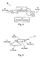
- a system 30 for determining endothelial dependent vasoactivity of the subject may be used for executing selected steps of method 10.
- FIG. 4 is a schematic illustration of system 30 which, in its basic configuration comprises an arrangement of sensors 32 for recording the pressure-related signals.
- sensors 32 can be piezoelectric ceramic elements or membrane-based sensors, such as, but not limited to, electrate microphones.
- sensors 32 are preferably positioned on several locations adjacent to one or more blood vessel.
- System 30 further comprises a processing unit 34 which receives, records and processes the pressure-related signals, sensed by sensors 32.
- unit 34 is programmed to extract parameter(s) from the pressure-related signals, and to use the parameter(s) for the purpose of determining a change of blood vessel characteristics and/or autonomic nervous system activity, as further detailed hereinabove.
- system 30 may further comprise a mechanism 36 for stimulating the blood vessel.
- Mechanism 36 is preferably capable of stimulating the blood vessel in any of the above types stimuli, hence can be a mechanical, thermal, electrical mechanism or chemical mechanism.
- mechanism 36 can be a mechanism for generating mental stress or a device for allowing the subject to perform physical exercise.
- mechanism 36 can comprise a sphingomanometer
- thermal stimulus mechanism 36 can be realized as a low temperature room or a bath of cold fluid
- electrical stimulus mechanism 36 can comprise electrodes
- chemical stimulus mechanism 36 may be a vasoactive agent, and the like.
- the present invention also contemplates the determination of an autonomic nervous system activity, e.g ., by heart rate variability analysis.
- system 30 further comprising one or more electrocardiogram leads 38 being for sensing electrical signals of the chest of the subject.
- processing unit 34 calculates heart rate variability from the electrocardiogram signals sensed leads 28, as further detailed hereinabove.
- the present invention provides cost effective system and method.
- a typical examination period for an individual subject is relatively short (from about 5 minutes to about 30 minutes) and can be executed by paramedical staff, without the supervision of specialist medical staff.
- the examination results are automatically analyzed, hence quickly providing general practitioners, cardiologists or internal medicine specialists with accurate and reliable information.
- the present invention can be routinely used for screening and diagnosis of large population and to differentiate between subjects in different stages and combinations of endothelial and coronary artery dysfunction.
- a first prototype system has been designed and constructed.
- the system included (i) transducers and an amplifier, designed and assembled for the research; (ii) a processing unit (desktop computer, Pentium IV); (iii) an A/D sampling card, purchased from National Instruments DAQ NI-488.2; (iv) data acquisition software, purchased from National InstrumentsTM Labview 5.1.1TM, custom designed; and (v) data analysis software, purchased from MatlabTM, custom designed.
- FIG. 5 is a schematic illustration of a transducer 50.
- Transducer 50 included an electrate microphone 56 and a stethoscope 51.
- Transducer 50 was capable of detecting small movements of the subject's skin generated by the blood pulse wave passing thereunder.
- Microphone 56 was connected to stethoscope 51 by a short conduit 58, allowing a communication between a membrane 52 of stethoscope 51 and a membrane 54 of microphone 56.
- a blood pulse wave passing under the skin generates vibrations in membrane 52, which are transmitted by conduit 58 to membrane 54, thus creating an electrical signal in microphone 56.
- Figure 6 shows the response of the transducer 50 to an input signal of approximately 1 Hz, obtained by physically oscillating the microphones. Note that although in its origin a typical stethoscope is designed to detect frequencies above 20 Hz, in practice the sensitivity range of transducer 50 is larger. Specifically, transducer 50 is capable of sensing low frequencies oscillatory motion.
- sampling rate of the data acquisition software was chosen to be 1000 Hz. This sampling rate provided the necessary precision for calculating the elapsed time between two successive pulses. Other sampling frequencies were tested and found less effective (higher frequencies demanded more memory and improvement in accuracy was negligible).
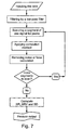
- Figure 7 is a detailed flowchart diagram of the data analysis procedure.
- the raw data, as recorded using the transducers from two locations of the subject's body was loaded from the data acquisition software and filtered by a low pass filter (15 Hz).
- the data was in a form of a plurality of 10 seconds segments. Each segment was scanned for its peaks.
- Peaks were defined by a zero derivative and were accepted for calculation if the following conditions were met: (i) the value of the peak was above a predetermined threshold, selected to be 70 % of the average maximum value; and (ii) the time interval between the peak and its former peak (of the same location) was more than 0.25 seconds.
- the elapsed time between two appropriate peaks from two different locations of the body was measured. Mean value and the standard deviation of the elapsed time was calculated, so as to eliminate unreasonable results (originated from noise, movement of the subject, erroneous calculation etc .). The standard deviation acceptance range was about 10 %. This process was repeated for all the peaks of all the segments.
- the accepted peaks were used for calculating heart rate, heart rate variability and standard deviation (respectively designated in Figure 7 by HR, HRV and SD).
- the heart rate was defined as the time between successive peaks of the same location.
- the heart rate variability was obtained by calculating the standard deviation of the heart rate in each segment.
- the heart rate variability was averaged over all the accepted segments and a standard deviation of the heart rate variability was obtained. A graphical output of the results was produced in the final step.
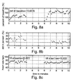
- Figures 8a-c are representative graphical output of the procedure.
- Figure 8a shows the relative change in elapsed time between the two transducers in % designated PWT (pulse-wave time parameter), as a function of time in minutes. Each point represents an average of approximately 10 seconds. The average value of PWT during the first three minutes of baseline is presented numerically; the dotted line represents the relative average value of baseline.
- PWT pulse-wave time parameter
- Figure 8b shows the percentage of the standard deviation of PWT calculated for the points represented in Figure 8a .
- High standard deviation during baseline represents movements of the subject or a noisy recording.
- Figure 8c shows the heart rate as a function of time. Each point represents an average of approximately 10 seconds. Numerical values of heart rate are presented (designated HR in Figure 8c ).
- Example 2 In vivo tests were performed on 21 volunteers, using the first prototype system of Example 1. Two transducers were connected to the subject under examination. A first transducer was connected to the radial artery at the wrist, and a second transducer was connected to the brachial artery about 5-10 cm above the elbow on the proximal side of the arm. The transducers were fastened with a cuff inflated to a pressure of 20 mmHg, so as to improve that signal to noise ratio, and to prevent partial occlusion of the vessel. An additional cuff, purchased from Hokanson, US, was positioned above the first cuff, for the purpose of implementing ischemia (mechanical stimulus).
- ischemia mechanical stimulus
- Subjects were tested at different hours of the day without fasting. Each subject was in a sitting posture in a temperature-controlled room (18 °C - 24 °C). The examination of each subject included: (i) three minutes of baseline recording (without stimulus); (ii) three minutes of induced ischemia in the brachial artery (using the additional cuff); and (iii) five minutes of recording during recovery subsequently to cuff release.
- Vasoconstriction was induced by submerging the right hand in cold water (8 °C), during continuous recording from the left hand in a sitting position. The subjects also underwent relaxation periods in which the right hand was in water at room temperature (21 °C).
- each subject included: (i) a few minutes in water in room temperature; (ii) three minutes of baseline recording (room temperature); (iii) one minute of vasoconstriction (cold water); (iv) three minutes of room temperature; (v) two minutes of vasoconstriction; (vi) at least three minutes of recovery in room temperature.
- vasomotor activity Several factors, such as temperature, food, drugs, physical exercise before examination and sympathetic stimuli, can affect vasomotor activity. In the above tests, it has been observed, that in some cases, examined individuals who were supposed to be with normal endothelium dependent vasoreactivity had different responses to reactive hyperemia at subsequent examinations. It was also found that these changes were in correlation with the elapsed time parameter measured in baseline. When the elapsed time parameter during baseline was relatively high (above 40-42 ms) the subject's response to reactive hyperemia was weak or completely absent. When the elapsed time parameter during baseline was lower (20-40 ms) the subjects' response to reactive hyperemia was normal. This implied that there is a "physiological window" in which the system produces the most reliable results.
- the results obtained by the first prototype system of Example 1 were compared to results obtained using a high-resolution ultrasonography device (HP sonos 5500). Each subject underwent a first examination using the ultrasonography device and a second examination using the prototype system, with 30 minutes rest between the tests. All subjects were examined after fasting for 8 hours without smoking or coffee.
- the ultrasonography examination of each subject included: (i) three minutes baseline recordings; (ii) three minutes of brachial artery occlusion; and (iii) ten minutes of recovery with continuous recording.
- D 1 -D 4 4 artery diameters, designated D 1 -D 4 , were calculated off line from the ultrasound images: D 1 , calculated during baseline phase, 1 minute from start; D 2 , calculated during baseline phase, 2 minutes from start; D 3 , calculated during cuff phase, 1 minute after deflation, and D 4 . calculated during cuff phase 1.5 minutes after deflation.
- the baseline diameter is defined as the average between D 1 and D 2 and the absolute diameter change is defined as the subtraction of D 4 from the baseline diameter.
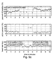
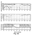
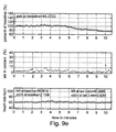
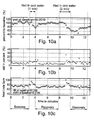
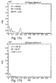
- FIG. 9a-e The prototype system examination of each subject included the combined stimuli protocol as further detailed above.
- the output of this examination is shown in Figures 9a-e .
- Figures 9a-b show examples of the output obtained for individuals with abnormal endothelial activity
- Figures 9c-d show examples of the output obtained for individuals with normal endothelial activity
- Figure 9e is an example of an output in which the elapsed time decreases while the arterial diameter is increasing as a result of nitroglycerin intake in a lying posture.
- Each Figure shows PWT, the percentage of the standard deviation of PWT and the heart rate as a function of time, as further explained hereinabove (see description of Figures 8a-c ).
- Table 1 Type of response Maximum change in PWT after cuff release [%] Response duration, T , after cuff release EDV function None ⁇ ( PWT ) ⁇ 0 -------- Abnormal Weak ⁇ ( PWT ) ⁇ 10 T ⁇ 2 min Abnormal Normal 10 ⁇ ⁇ ( PWT ) ⁇ 20 2 ⁇ T ⁇ 4 Normal Strong ⁇ ( PWT ) > 20 T > 4 Normal Negative below baseline ------------------------------------------------------------------------------
- Tables 2-3 show the results obtained in the mechanical stimulus examination, where Table 2 shows the results obtained for low risk subjects and Table 3 shows the results obtained for high risk subjects.
- Table 4 Population age n Normal EDV % Abnormal EDV % High risk 55.3 11 2/11 (1) 18% 9 82% Low risk 37 10 8/10 80% 2 20% (1) p ⁇ 0.002 high Vs low risk group
- the average age of the groups with normal endothelium dependent vasoreactivity was 36.7 ⁇ 12.5 years and with abnormal endothelium dependent vasoreactivity was the 54.5 ⁇ 11.5 (p ⁇ 0.01).
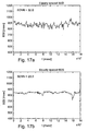
- Figures 10a-c show relative changes in the elapsed time, standard deviation and heart rate variability, of one subject examined in the thermal stimulus test.
- Table 5 shows the results of relative change in elapsed time for 6 subjects exposed to cold water.
- Table 5 No Baseline 100% (1) 1-minute submergence: PWT relative to baseline recovery time (minutes) Baseline 100% (2) 2-minute submergence: PWT relative to baseline (3) recovery time (minutes) 1 100% 70% 1 100% 40% 2 2 100% 70% 4 100% 40% above 6 3 100% 85% 2 100% 20% 3 4 100% 80% 1 100% 30% above 6 5 100% 80% 1 100% 25% 2.5 6 100% 75% 1.5 100% 25% 1.5 Av. 100% 76.6 ⁇ 6.0 % 1.75 ⁇ 1.2 100% 30 ⁇ 8.3 % 3.5 ⁇ 2.0 (1) p ⁇ 0.001 between control and 1 minute submergence. (2) p ⁇ 2.5E-6 between control and 2 minutes submergence. (3) p ⁇ 0.05 between recovery time of 1 Vs 2 minutes submergence.
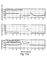
- Figures 11a-b show the effect of posture on the elapsed time and the measurement of endothelium dependent vasoreactivity.
- Two individuals were examined with nitroglycerin while they were postured in a lying position for comparison with the ultrasonography examination.
- the prototype system indicated abnormal response while the ultrasonography examination indicated normal response of increased hyperemia to nitroglycerine intake.
- Table 7 No Age & Gen. Examination a Examination b EDV functioning PWT at baseline Type of response PWT at baseline Type of response 1 34
- F 42 Abnormal 39 Normal Normal 2 55
- M 26 Normal 27 Normal Normal 5 39
- Abnormal 37 Abnormal Abnormal 7
- Abnormal 51 Abnormal Abnormal Abnormal Abnormal
- Table 8 shows the effect of a moderate walking on PWT .
- the obtained results indicate that short and moderate effort of walking shorten elapsed time (p ⁇ 0.05).
- Table 8 No. PWT Reduction in PWT [%] before walking [ms] (1) after walking [ms] 1 37.5 34.4 8.26 2 45.4 32.7 27.9 3 46.3 35.1 24.1 4 52.2 41.3 20.88 5 46.5 38.22 17.8 Aver age 45.8 ⁇ 5.3 36.3 ⁇ 3.4 19.78 (1) p ⁇ 0.05 between values obtained before and after walking
- the ultrasonography examination and the prototype system examination had similar results (p ⁇ 0.02).
- the ultrasonography examination indicated a FMD of 7.8 %, which is 1.8 % above the borderline of 6 %.
- the combined stimuli examination showed that a moderate walk for about 2 minutes before the examination, which causes a moderate elevation and alpha sympathetic activity, led to vasoconstriction and PWT reduction, hence allowed the examination to be conduct within the "physiological window". Intensifying the physical activity (running or jumping) before examination causes an opposite effect in which probably beta receptors are also active causing vasodilatation. In such a case it reduces the probability that the examination will be conducted within the "physiological window.”
- a second prototype system has been designed and constructed.
- the system included: (i) a custom designed data logger; (ii) a brachial, radial and carotid transducers, all being operative at low frequencies and based on piezoelectric ceramic elements; (iii) an electrocardiogram chest electrode; (iv) a standard personal computer; and (v) data analysis software (see Example 1).
- the custom designed data logger included an amplifier, a four-channel A/D card connected to the computer via a USB cable and a small LCD monitor.
- the brachial transducer was a coin shaped transducer, about 2 cm in diameter, attached to a dual compartment sphyngmanometric cuff, so as to allow both arterial occlusion (mechanical stimulus) and attachment of the transducer with a constant and controlled force.
- the dual compartment sphyngmanometric cuff included two separate air compartments: a low pressure compartment ( ⁇ 20 mmHg) for applying force on the brachial transducer thus coupling the transducer to the skin with a controlled force; and a high pressure compartment (up to 300 mmHg) for applying the mechanical stimulus on the artery.
- the high-pressure compartment facilitates quick release of pressure.
- the radial and carotid transducers were pencil shaped transducers with attached to stabilizing devices facilitating constant applied force for applanation tonometry, where the radial transducer was attached to a wrist stabilizing device and the carotid transducer was attached to a neck stabilizing device.
- the brachial and the radial transducers were connected to the subject under examination, as further detailed in Example 2.
- the elapsed time parameter, PWT the elapsed time parameter
- PWA the amplitude parameter
- the examination of each subject also included examination using the ultrasonography device (see further details in Example 2, above).
- Figures 12a-c show the elapsed time ( Figure 12a ), standard deviation ( Figure 12b ) and amplitude ( Figure 12c ) during supine position of a subject who has been diagnosed by US measurements as having normal endothelial function.
- the brachial artery was occluded for three minutes. After reopening of the occlusion (6 min) elapsed time showed only minor change while the amplitude showed dramatic changes.
- the value of PWT was relatively long, about 45 ms.
- Figures 13a-c show the elapsed time ( Figure 13a ), standard deviation ( Figure 13b ) and amplitude ( Figure 13c ) during sitting position of a subject who has been diagnosed by US measurements as having normal endothelial function.
- the brachial artery was occluded for three minutes. As shown, in this case the elapsed time was increased and almost no change in the amplitude.
- the value of PWT was relatively small, about 15 ms, indicating that the initial diameter of the artery was relatively small.
- Figures 14a-c show the elapsed time ( Figure 15a ), standard deviation ( Figure 15b ) and amplitude ( Figure 15c ) during sitting position of another subject who has been diagnosed by US measurements as having normal endothelial function.
- the value of PWT was small, about 33 ms, indicating that the measurement was initiated within the "physiological window.” With the increment of the artery's radius, a non-linear region, characterized by a non-linear amplitude increment, was observed.
- Figures 15a-c show the elapsed time ( Figure 14a ), standard deviation ( Figure 14b ) and amplitude ( Figure 14c ) during sitting position of a subject who has been diagnosed by US measurements as having abnormal endothelial function. As shown, in this case no change was observed in the elapsed time or in the amplitude, indicating abnormal endothelial function. The value of PWT was short, about 24 ms, indicating that the measurement was carried out within the above mentioned "physiological window.”
- Figures 16a-c show the elapsed time ( Figure 16a ), standard deviation ( Figure 16b ) and amplitude ( Figure 16c ) after the nitroglycerin treatment and during supine position of a subject who has been diagnosed by US measurements as having normal endothelial function. As shown, in this case the elapsed time was reduced and the amplitude was increased. The value of PWT was relatively large, about 45 ms, indicating that the initial diameter of the artery was relatively wide.
- the amplitude parameter was included in analysis procedure.
- the measurement of endothelial function was based on the change of elapsed time parameter thereby requiring the measurement to be carried out within the "physiological window," where the initial radius of the artery diameter is sufficiently small.
- the elasticity module of the artery's wall is, to a good approximation, constant causing a linear dependence of the pulse wave velocity on the inverse of the radius (the pulse wave velocity decreased as the arterial radius is increased).
- Heart rate variability analysis was carried out in 12 of the subjects who had undergone examination for the endothelial dysfunction measurement of Example 4. In 10 subjects heart rate variability analysis indicated normal autonomic nervous system activity and therefore possible normal coronary function. The analysis is based on 3 min recording using electrocardiogram lead II of the chest of the subjects, during baseline (before application of the mechanical stimulus).
- Figures 17a , 17c , 17e , and 17g show heart rate variability analysis of a subject with normal heart rate variability activity and endothelial dysfunction.
- Figures 17b , 17d , 17f , and 17h show heart rate variability analysis of a subject with abnormal activity but with normal brachial endothelial function. Endothelial function or dysfunction are not shown in Figures 17a-h .
- Figures 17a-b show beat-to-beat analysis, referred to hereinafter as B2B analysis.
- B2B analysis beat-to-beat analysis
- For the subjects with the differences between subjects with normal and abnormal heart rate variability activity are manifested by the value of SDNN, which is in the normal range in Figure 17a and significantly lower in Figure 17b .
- a reduced value of SDNN reflects a reduced parasympathetic activity.
- high fluctuations between beats are shown in Figure 17a , compared to Figure 17b .
- Figures 17c-d show the power spectrum densities.
- the normal HF peak around 0.3 Hz shown for the normal subject ( Figure 17c ) is absent from Figure 17d , hence indicates abnormal heart rate variability.
- HF value is relatively high in Figure 17c compared to Figure 17d
- LF/HF is low in Figure 17c compared to Figure 17d , hence also supporting the abnormal heart rate variability diagnosis of the subject of Figure 17d .
- Figures 17e-f show Fast Fourier Transform analysis of the RRI series. Again, a clear peak shown around 0.3 Hz in Figure 17e , is almost completely absent in Figure 17f .
- Figures 17g-h show relative incidence, as a function of B2B (measured in ms).
- a relatively wide histogram of time intervals between beats is presented in Figure 17g , compared to the narrow histogram shown in Figure 17h , indicating normal heart rate variability for the subject of Figure 17g and abnormal heart rate variability for the subject of Figure 17h .
- electrocardiogram lead II connected to the chest
- transducers connected to the brachial, radial and carotid arteries.
- Sample rate for all the channels was 1000 Hz.
- the examination was performed in a temperature-controlled room (22 °C). Blood pressure was measured with an automated sphyngo-manometer before the recording.
- the recording protocol included the following periods of thermal stimuli: 3 minutes at room temperature, 4 minutes at 25 °C, 2 minutes at 5 °C and 10 minutes at 25 °C. Each stimulus was applied by a bath containing water in the respective temperature, in which the subjects placed the right wrist.
- T 1 brachial-radial transit time
- T 2 QRS-carotid transit time
- three amplitude parameters one for each artery and heart rate.
- FIG 18a shows changes of the two elapsed time parameters T 1 (blue line) and T 2 (red line), measured in ms. As shown, both T 1 and T 2 restore and continue to increase above their original (baseline) values during recovery, indicating normal endothelial function, as further detailed hereinabove.
- Figure 18b shows changes of all three amplitudes, measured as percentage of the baseline value.
- a decrement in all amplitudes is observed during the low temperature period (7 ⁇ t ⁇ 9 min).
- the decrement in the amplitude of the pulse measured of the carotid artery is less pronounced than the decrement in the amplitudes measured of the brachial and radial arteries.
- all amplitudes exhibit an increment, where the increment in the amplitude of the pulse measured of the carotid artery is more pronounced, and reaches a value of about 50 % above baseline.
- the increments in the amplitudes of the pulse measured of the brachial and radial arteries are less pronounced and reach about 10-20 % below baseline.
- Figure 18c shows the heart rate, measured in bits/min. A clear peak was observed during the low temperature period (7 ⁇ t ⁇ 9 min), indicating increased sympathetic activity. A minor heart rate increment was also observed during the first transition (from room temperature to 25 °C). As shown in Figure 18c the heart rate resorted its baseline value during the second recovery period (9 ⁇ t ⁇ 19 min).
Landscapes
- Health & Medical Sciences (AREA)
- Life Sciences & Earth Sciences (AREA)
- Cardiology (AREA)
- Molecular Biology (AREA)
- Animal Behavior & Ethology (AREA)
- Veterinary Medicine (AREA)
- Biophysics (AREA)
- Pathology (AREA)
- Engineering & Computer Science (AREA)
- Biomedical Technology (AREA)
- Heart & Thoracic Surgery (AREA)
- Medical Informatics (AREA)
- Public Health (AREA)
- Surgery (AREA)
- Physics & Mathematics (AREA)
- General Health & Medical Sciences (AREA)
- Physiology (AREA)
- Vascular Medicine (AREA)
- Neurology (AREA)
- Hematology (AREA)
- Neurosurgery (AREA)
- Immunology (AREA)
- Measuring Pulse, Heart Rate, Blood Pressure Or Blood Flow (AREA)
- Investigating Or Analysing Biological Materials (AREA)
Applications Claiming Priority (3)
| Application Number | Priority Date | Filing Date | Title |
|---|---|---|---|
| US43173902P | 2002-12-09 | 2002-12-09 | |
| US431739P | 2002-12-09 | ||
| PCT/IL2003/001025 WO2004052196A1 (en) | 2002-12-09 | 2003-12-03 | Method for determining endothelial dependent vasoactivity |
Publications (2)
| Publication Number | Publication Date |
|---|---|
| EP1569550A1 EP1569550A1 (en) | 2005-09-07 |
| EP1569550B1 true EP1569550B1 (en) | 2010-06-23 |
Family
ID=32507789
Family Applications (1)
| Application Number | Title | Priority Date | Filing Date |
|---|---|---|---|
| EP03812666A Expired - Lifetime EP1569550B1 (en) | 2002-12-09 | 2003-12-03 | Method for determining endothelial dependent vasoactivity |
Country Status (8)
| Country | Link |
|---|---|
| US (2) | US7374541B2 (enExample) |
| EP (1) | EP1569550B1 (enExample) |
| JP (1) | JP4615315B2 (enExample) |
| AT (1) | ATE471692T1 (enExample) |
| AU (1) | AU2003302882B2 (enExample) |
| CA (1) | CA2508590A1 (enExample) |
| DE (1) | DE60333120D1 (enExample) |
| WO (1) | WO2004052196A1 (enExample) |
Cited By (2)
| Publication number | Priority date | Publication date | Assignee | Title |
|---|---|---|---|---|
| US11039753B2 (en) | 2016-12-15 | 2021-06-22 | Baxter International Inc. | System and method for monitoring and determining patient parameters from sensed venous waveform |
| US11039754B2 (en) | 2018-05-14 | 2021-06-22 | Baxter International Inc. | System and method for monitoring and determining patient parameters from sensed venous waveform |
Families Citing this family (34)
| Publication number | Priority date | Publication date | Assignee | Title |
|---|---|---|---|---|
| AU2003302882B2 (en) | 2002-12-09 | 2008-06-19 | Ramot At Tel Aviv University Ltd. | System for determining endothelial dependent vasoactivity |
| US7771364B2 (en) | 2004-01-27 | 2010-08-10 | Spirocor Ltd. | Method and system for cardiovascular system diagnosis |
| US7131949B2 (en) * | 2004-04-13 | 2006-11-07 | Omron Healthcare Co., Ltd. | Method and apparatus for examining vascular endothelial functions |
| US7664551B2 (en) * | 2004-07-07 | 2010-02-16 | Medtronic Transneuronix, Inc. | Treatment of the autonomic nervous system |
| EP1785088A1 (en) * | 2005-11-14 | 2007-05-16 | Congener Wellness Corp. | A system and method for the management and control of cardiovascular related diseases, such as hypertension |
| RU2309668C1 (ru) | 2006-02-20 | 2007-11-10 | Александр Сергеевич Парфенов | Способ неинвазивного определения функции эндотелия и устройство для его осуществления |
| JP4802324B2 (ja) * | 2006-02-22 | 2011-10-26 | 国立大学法人 名古屋工業大学 | 脈診解析のための脈波測定装置 |
| US7894900B2 (en) * | 2006-08-30 | 2011-02-22 | Smartimplant OÜ | Device and method for monitoring cardiac pacing rate |
| JP5441707B2 (ja) | 2006-12-09 | 2014-03-12 | マシモ コーポレイション | プレチスモグラフ変動プロセッサ |
| US8100834B2 (en) * | 2007-02-27 | 2012-01-24 | J&M Shuler, Inc. | Method and system for monitoring oxygenation levels of a compartment for detecting conditions of a compartment syndrome |
| CN102579082B (zh) * | 2007-06-04 | 2014-05-28 | 柯尼卡美能达株式会社 | 超声波诊断装置及用于超声波诊断装置的超声波探头 |
| JP2009178456A (ja) * | 2008-01-31 | 2009-08-13 | Nippon Koden Corp | 自律神経活動計測装置及び計測方法 |
| JP5192859B2 (ja) * | 2008-03-17 | 2013-05-08 | 株式会社ユネクス | 生体血管状態測定装置 |
| WO2010056538A1 (en) * | 2008-10-29 | 2010-05-20 | Tim Maguire | An automated vessel puncture device using three-dimensional(3d) near infrared (nir) imaging and a robotically driven needle |
| JP5167156B2 (ja) * | 2009-01-19 | 2013-03-21 | 株式会社デンソー | 生体状態評価装置、生体状態評価システム、プログラム、及び記録媒体 |
| US8057400B2 (en) * | 2009-05-12 | 2011-11-15 | Angiologix, Inc. | System and method of measuring changes in arterial volume of a limb segment |
| ITPI20090099A1 (it) * | 2009-07-31 | 2011-02-01 | Cnr Consiglio Naz Delle Ric Erche | Apparecchiatura per la misura della velocità di propagazione di un'onda pressoria nel sistema arterioso |
| US8668649B2 (en) * | 2010-02-04 | 2014-03-11 | Siemens Medical Solutions Usa, Inc. | System for cardiac status determination |
| GB2478291A (en) * | 2010-03-02 | 2011-09-07 | Univ Lancaster | Endothelium assessment probe |
| US9307928B1 (en) | 2010-03-30 | 2016-04-12 | Masimo Corporation | Plethysmographic respiration processor |
| JP5518554B2 (ja) * | 2010-04-16 | 2014-06-11 | 酒井医療株式会社 | 血管硬さ評価システム及び血管硬さ指標の算出方法 |
| WO2012021765A2 (en) * | 2010-08-12 | 2012-02-16 | Board Of Trustees Of Michigan State University | Methods and apparatus for determining arterial pulse wave velocity |
| US9320445B2 (en) | 2011-05-17 | 2016-04-26 | Siemens Medical Solutions Usa, Inc. | System for cardiac condition detection responsive to blood pressure analysis |
| US10398323B2 (en) * | 2012-08-28 | 2019-09-03 | Board Of Trustees Of Michigan State University | Methods and apparatus for determining pulse transit time as a function of blood pressure |
| JP5571234B2 (ja) * | 2013-06-26 | 2014-08-13 | 日本光電工業株式会社 | 自律神経活動計測装置 |
| JP6349075B2 (ja) * | 2013-11-22 | 2018-06-27 | 三星電子株式会社Samsung Electronics Co.,Ltd. | 心拍数測定装置及び心拍数測定方法 |
| US10945612B2 (en) | 2014-04-03 | 2021-03-16 | The Regents Of The University Of California | Assessing endothelial function using a blood pressure cuff |
| US10105089B2 (en) | 2014-06-18 | 2018-10-23 | Hong Kong Applied Science And Technology Research Institute Co., Ltd. | Systems and methods for blood pressure measurement with psychological status validation |
| US10588577B2 (en) | 2015-01-29 | 2020-03-17 | Siemens Healthcare Gmbh | Patient signal analysis based on affine template matching |
| CN108135506B (zh) * | 2015-08-14 | 2021-11-02 | 加利福尼亚大学董事会 | 用于评估哺乳动物中的内皮功能的方法和装置 |
| US20180192916A1 (en) * | 2017-01-10 | 2018-07-12 | General Electric Company | Imaging system for diagnosing patient condition |
| EP3782542B1 (en) * | 2019-08-20 | 2024-04-10 | Stichting IMEC Nederland | A method and system for microcirculation function assessment |
| JP7560822B2 (ja) * | 2020-02-25 | 2024-10-03 | 日本光電工業株式会社 | 生体情報処理装置、生体情報処理方法、プログラム及び記憶媒体 |
| RU2751817C1 (ru) * | 2020-08-31 | 2021-07-19 | Закрытое Акционерное Общество "Ес-Лизинг" | Компьютеризированный способ неинвазивного выявления нарушений углеводного обмена по вариабельности сердечного ритма и носимое автономное устройство для его реализации |
Family Cites Families (20)
| Publication number | Priority date | Publication date | Assignee | Title |
|---|---|---|---|---|
| US3623476A (en) * | 1970-02-16 | 1971-11-30 | Philips Corp | Blood pressure measurement apparatus |
| US5111826A (en) * | 1984-12-07 | 1992-05-12 | Nasiff Roger E | Indirect continuous blood pressure method |
| JP2979933B2 (ja) * | 1993-08-03 | 1999-11-22 | セイコーエプソン株式会社 | 脈波解析装置 |
| JPH08266489A (ja) * | 1995-03-29 | 1996-10-15 | Nippon Ham Kk | 動脈硬化部位の検出方法 |
| JPH11128185A (ja) * | 1997-10-31 | 1999-05-18 | Matsushita Electric Ind Co Ltd | 心拍変動解析方法および装置 |
| IL124787A0 (en) * | 1998-06-07 | 1999-01-26 | Itamar Medical C M 1997 Ltd | Pressure applicator devices particularly useful for non-invasive detection of medical conditions |
| IL128482A (en) | 1999-02-11 | 2003-06-24 | Ultrasis Internat 1993 Ltd | Method and device for continuous analysis of cardiovascular activity of a subject |
| JP3027750B1 (ja) * | 1999-05-17 | 2000-04-04 | 日本コーリン株式会社 | 下肢上肢血圧指数測定装置および下肢血圧測定装置 |
| US6616613B1 (en) * | 2000-04-27 | 2003-09-09 | Vitalsines International, Inc. | Physiological signal monitoring system |
| NZ524765A (en) * | 2000-07-19 | 2006-10-27 | Nigel E Sharrock | Non-invasive measurement of suprasystolic signals |
| CA2424389C (en) * | 2000-10-23 | 2011-07-12 | Itamar Medical Ltd. | Method and apparatus for non-invasively evaluating endothelial activity in a patient |
| JP3590583B2 (ja) * | 2000-12-28 | 2004-11-17 | フクダ電子株式会社 | 血管内皮機能測定装置 |
| US20020149152A1 (en) | 2001-02-23 | 2002-10-17 | Alan Taylor | Multiple-stage game of chance and method of and system for playing the game |
| JP2002253519A (ja) * | 2001-03-01 | 2002-09-10 | Nippon Koden Corp | 血液量測定方法及び生体信号モニタ装置 |
| JP2002291709A (ja) * | 2001-03-30 | 2002-10-08 | Nippon Colin Co Ltd | 圧脈波検出装置 |
| JP3533406B2 (ja) * | 2001-07-02 | 2004-05-31 | コーリンメディカルテクノロジー株式会社 | 動脈硬化評価装置 |
| GB0205653D0 (en) * | 2002-03-11 | 2002-04-24 | Micro Medical Ltd | A method of measuring endothelial function in a person |
| AU2003302882B2 (en) | 2002-12-09 | 2008-06-19 | Ramot At Tel Aviv University Ltd. | System for determining endothelial dependent vasoactivity |
| US20060195035A1 (en) * | 2005-02-28 | 2006-08-31 | Dehchuan Sun | Non-invasive radial artery blood pressure waveform measuring apparatus system and uses thereof |
| JP4752673B2 (ja) * | 2005-10-06 | 2011-08-17 | コニカミノルタセンシング株式会社 | 脈波データ解析方法、システム、プログラム |
-
2003
- 2003-12-03 AU AU2003302882A patent/AU2003302882B2/en not_active Expired - Fee Related
- 2003-12-03 DE DE60333120T patent/DE60333120D1/de not_active Expired - Lifetime
- 2003-12-03 AT AT03812666T patent/ATE471692T1/de not_active IP Right Cessation
- 2003-12-03 WO PCT/IL2003/001025 patent/WO2004052196A1/en not_active Ceased
- 2003-12-03 CA CA002508590A patent/CA2508590A1/en not_active Abandoned
- 2003-12-03 JP JP2004558325A patent/JP4615315B2/ja not_active Expired - Fee Related
- 2003-12-03 US US10/537,913 patent/US7374541B2/en not_active Expired - Fee Related
- 2003-12-03 EP EP03812666A patent/EP1569550B1/en not_active Expired - Lifetime
-
2008
- 2008-04-25 US US12/149,100 patent/US8187196B2/en not_active Expired - Fee Related
Cited By (3)
| Publication number | Priority date | Publication date | Assignee | Title |
|---|---|---|---|---|
| US11039753B2 (en) | 2016-12-15 | 2021-06-22 | Baxter International Inc. | System and method for monitoring and determining patient parameters from sensed venous waveform |
| US11950890B2 (en) | 2016-12-15 | 2024-04-09 | Baxter International Inc. | System and method for monitoring and determining patient parameters from sensed venous waveform |
| US11039754B2 (en) | 2018-05-14 | 2021-06-22 | Baxter International Inc. | System and method for monitoring and determining patient parameters from sensed venous waveform |
Also Published As
| Publication number | Publication date |
|---|---|
| US8187196B2 (en) | 2012-05-29 |
| DE60333120D1 (de) | 2010-08-05 |
| JP4615315B2 (ja) | 2011-01-19 |
| US7374541B2 (en) | 2008-05-20 |
| JP2006516000A (ja) | 2006-06-15 |
| AU2003302882A1 (en) | 2004-06-30 |
| CA2508590A1 (en) | 2004-06-24 |
| US20060149152A1 (en) | 2006-07-06 |
| AU2003302882B2 (en) | 2008-06-19 |
| US20080200820A1 (en) | 2008-08-21 |
| ATE471692T1 (de) | 2010-07-15 |
| EP1569550A1 (en) | 2005-09-07 |
| WO2004052196A1 (en) | 2004-06-24 |
Similar Documents
| Publication | Publication Date | Title |
|---|---|---|
| EP1569550B1 (en) | Method for determining endothelial dependent vasoactivity | |
| US7771364B2 (en) | Method and system for cardiovascular system diagnosis | |
| US8821403B2 (en) | Non-invasive measurement of suprasystolic signals | |
| US20070021673A1 (en) | Method and system for cardiovascular system diagnosis | |
| US6994675B2 (en) | Non-invasive measurement of suprasystolic signals | |
| US20080045844A1 (en) | Method and system for cardiovascular system diagnosis | |
| CN102469947B (zh) | 使用信号渡越时间来监控心血管状况 | |
| US20080287811A1 (en) | Method for Assessing The Functional Condition Of Cardiovascular System | |
| US20060224070A1 (en) | System and method for non-invasive cardiovascular assessment from supra-systolic signals obtained with a wideband external pulse transducer in a blood pressure cuff | |
| JP4668421B2 (ja) | 被験者の心臓血管活動の連続的解析方法および装置 | |
| JP3122105B2 (ja) | 高血圧症判定装置 | |
| US11510585B2 (en) | Method to quantify the hemodynamic and vascular properties in vivo from arterial waveform measurements | |
| JPH08583A (ja) | 脈波伝達時間監視装置 | |
| Nagy et al. | Sensor fusion for the accurate non-invasive measurement of blood pressure | |
| GB2456947A (en) | Non invasive determination of stroke volume based on incident wave suprasystolic blood pressure amplitude |
Legal Events
| Date | Code | Title | Description |
|---|---|---|---|
| PUAI | Public reference made under article 153(3) epc to a published international application that has entered the european phase |
Free format text: ORIGINAL CODE: 0009012 |
|
| 17P | Request for examination filed |
Effective date: 20050608 |
|
| AK | Designated contracting states |
Kind code of ref document: A1 Designated state(s): AT BE BG CH CY CZ DE DK EE ES FI FR GB GR HU IE IT LI LU MC NL PT RO SE SI SK |
|
| AX | Request for extension of the european patent |
Extension state: AL LT LV MK |
|
| DAX | Request for extension of the european patent (deleted) | ||
| RIN1 | Information on inventor provided before grant (corrected) |
Inventor name: AMITZUR, GIORA Inventor name: ZIMERMAN, ELYA Inventor name: EINAV, SHMUEL Inventor name: PELEG, ERAN |
|
| 17Q | First examination report despatched |
Effective date: 20060317 |
|
| GRAP | Despatch of communication of intention to grant a patent |
Free format text: ORIGINAL CODE: EPIDOSNIGR1 |
|
| RBV | Designated contracting states (corrected) |
Designated state(s): AT BE BG CH CY CZ DE DK EE ES FI FR GB GR HU IE IT LI LU MC NL PT RO SE SI SK |
|
| RBV | Designated contracting states (corrected) |
Designated state(s): AT BE BG CH CY CZ DE DK EE ES FI FR GB GR HU IE IT LI LU MC NL PT RO SE SI SK TR |
|
| GRAS | Grant fee paid |
Free format text: ORIGINAL CODE: EPIDOSNIGR3 |
|
| GRAA | (expected) grant |
Free format text: ORIGINAL CODE: 0009210 |
|
| AK | Designated contracting states |
Kind code of ref document: B1 Designated state(s): AT BE BG CH CY CZ DE DK EE ES FI FR GB GR HU IE IT LI LU MC NL PT RO SE SI SK TR |
|
| REG | Reference to a national code |
Ref country code: CH Ref legal event code: EP |
|
| REG | Reference to a national code |
Ref country code: IE Ref legal event code: FG4D |
|
| REF | Corresponds to: |
Ref document number: 60333120 Country of ref document: DE Date of ref document: 20100805 Kind code of ref document: P |
|
| REG | Reference to a national code |
Ref country code: NL Ref legal event code: VDEP Effective date: 20100623 |
|
| PG25 | Lapsed in a contracting state [announced via postgrant information from national office to epo] |
Ref country code: SE Free format text: LAPSE BECAUSE OF FAILURE TO SUBMIT A TRANSLATION OF THE DESCRIPTION OR TO PAY THE FEE WITHIN THE PRESCRIBED TIME-LIMIT Effective date: 20100623 |
|
| PG25 | Lapsed in a contracting state [announced via postgrant information from national office to epo] |
Ref country code: FI Free format text: LAPSE BECAUSE OF FAILURE TO SUBMIT A TRANSLATION OF THE DESCRIPTION OR TO PAY THE FEE WITHIN THE PRESCRIBED TIME-LIMIT Effective date: 20100623 Ref country code: AT Free format text: LAPSE BECAUSE OF FAILURE TO SUBMIT A TRANSLATION OF THE DESCRIPTION OR TO PAY THE FEE WITHIN THE PRESCRIBED TIME-LIMIT Effective date: 20100623 Ref country code: SI Free format text: LAPSE BECAUSE OF FAILURE TO SUBMIT A TRANSLATION OF THE DESCRIPTION OR TO PAY THE FEE WITHIN THE PRESCRIBED TIME-LIMIT Effective date: 20100623 |
|
| PG25 | Lapsed in a contracting state [announced via postgrant information from national office to epo] |
Ref country code: GR Free format text: LAPSE BECAUSE OF FAILURE TO SUBMIT A TRANSLATION OF THE DESCRIPTION OR TO PAY THE FEE WITHIN THE PRESCRIBED TIME-LIMIT Effective date: 20100924 |
|
| PG25 | Lapsed in a contracting state [announced via postgrant information from national office to epo] |
Ref country code: NL Free format text: LAPSE BECAUSE OF FAILURE TO SUBMIT A TRANSLATION OF THE DESCRIPTION OR TO PAY THE FEE WITHIN THE PRESCRIBED TIME-LIMIT Effective date: 20100623 Ref country code: EE Free format text: LAPSE BECAUSE OF FAILURE TO SUBMIT A TRANSLATION OF THE DESCRIPTION OR TO PAY THE FEE WITHIN THE PRESCRIBED TIME-LIMIT Effective date: 20100623 |
|
| PG25 | Lapsed in a contracting state [announced via postgrant information from national office to epo] |
Ref country code: RO Free format text: LAPSE BECAUSE OF FAILURE TO SUBMIT A TRANSLATION OF THE DESCRIPTION OR TO PAY THE FEE WITHIN THE PRESCRIBED TIME-LIMIT Effective date: 20100623 Ref country code: CY Free format text: LAPSE BECAUSE OF FAILURE TO SUBMIT A TRANSLATION OF THE DESCRIPTION OR TO PAY THE FEE WITHIN THE PRESCRIBED TIME-LIMIT Effective date: 20100623 Ref country code: CZ Free format text: LAPSE BECAUSE OF FAILURE TO SUBMIT A TRANSLATION OF THE DESCRIPTION OR TO PAY THE FEE WITHIN THE PRESCRIBED TIME-LIMIT Effective date: 20100623 Ref country code: PT Free format text: LAPSE BECAUSE OF FAILURE TO SUBMIT A TRANSLATION OF THE DESCRIPTION OR TO PAY THE FEE WITHIN THE PRESCRIBED TIME-LIMIT Effective date: 20101025 Ref country code: SK Free format text: LAPSE BECAUSE OF FAILURE TO SUBMIT A TRANSLATION OF THE DESCRIPTION OR TO PAY THE FEE WITHIN THE PRESCRIBED TIME-LIMIT Effective date: 20100623 Ref country code: BE Free format text: LAPSE BECAUSE OF FAILURE TO SUBMIT A TRANSLATION OF THE DESCRIPTION OR TO PAY THE FEE WITHIN THE PRESCRIBED TIME-LIMIT Effective date: 20100623 |
|
| PG25 | Lapsed in a contracting state [announced via postgrant information from national office to epo] |
Ref country code: IT Free format text: LAPSE BECAUSE OF FAILURE TO SUBMIT A TRANSLATION OF THE DESCRIPTION OR TO PAY THE FEE WITHIN THE PRESCRIBED TIME-LIMIT Effective date: 20100623 |
|
| PG25 | Lapsed in a contracting state [announced via postgrant information from national office to epo] |
Ref country code: DK Free format text: LAPSE BECAUSE OF FAILURE TO SUBMIT A TRANSLATION OF THE DESCRIPTION OR TO PAY THE FEE WITHIN THE PRESCRIBED TIME-LIMIT Effective date: 20100623 |
|
| PLBE | No opposition filed within time limit |
Free format text: ORIGINAL CODE: 0009261 |
|
| STAA | Information on the status of an ep patent application or granted ep patent |
Free format text: STATUS: NO OPPOSITION FILED WITHIN TIME LIMIT |
|
| 26N | No opposition filed |
Effective date: 20110324 |
|
| REG | Reference to a national code |
Ref country code: DE Ref legal event code: R097 Ref document number: 60333120 Country of ref document: DE Effective date: 20110323 |
|
| PG25 | Lapsed in a contracting state [announced via postgrant information from national office to epo] |
Ref country code: MC Free format text: LAPSE BECAUSE OF NON-PAYMENT OF DUE FEES Effective date: 20101231 |
|
| REG | Reference to a national code |
Ref country code: CH Ref legal event code: PL |
|
| PG25 | Lapsed in a contracting state [announced via postgrant information from national office to epo] |
Ref country code: IE Free format text: LAPSE BECAUSE OF NON-PAYMENT OF DUE FEES Effective date: 20101203 Ref country code: LI Free format text: LAPSE BECAUSE OF NON-PAYMENT OF DUE FEES Effective date: 20101231 Ref country code: CH Free format text: LAPSE BECAUSE OF NON-PAYMENT OF DUE FEES Effective date: 20101231 |
|
| PG25 | Lapsed in a contracting state [announced via postgrant information from national office to epo] |
Ref country code: HU Free format text: LAPSE BECAUSE OF FAILURE TO SUBMIT A TRANSLATION OF THE DESCRIPTION OR TO PAY THE FEE WITHIN THE PRESCRIBED TIME-LIMIT Effective date: 20101224 Ref country code: LU Free format text: LAPSE BECAUSE OF NON-PAYMENT OF DUE FEES Effective date: 20101203 Ref country code: BG Free format text: LAPSE BECAUSE OF FAILURE TO SUBMIT A TRANSLATION OF THE DESCRIPTION OR TO PAY THE FEE WITHIN THE PRESCRIBED TIME-LIMIT Effective date: 20100623 |
|
| PG25 | Lapsed in a contracting state [announced via postgrant information from national office to epo] |
Ref country code: TR Free format text: LAPSE BECAUSE OF FAILURE TO SUBMIT A TRANSLATION OF THE DESCRIPTION OR TO PAY THE FEE WITHIN THE PRESCRIBED TIME-LIMIT Effective date: 20100623 |
|
| PG25 | Lapsed in a contracting state [announced via postgrant information from national office to epo] |
Ref country code: BG Free format text: LAPSE BECAUSE OF FAILURE TO SUBMIT A TRANSLATION OF THE DESCRIPTION OR TO PAY THE FEE WITHIN THE PRESCRIBED TIME-LIMIT Effective date: 20100923 |
|
| PG25 | Lapsed in a contracting state [announced via postgrant information from national office to epo] |
Ref country code: ES Free format text: LAPSE BECAUSE OF FAILURE TO SUBMIT A TRANSLATION OF THE DESCRIPTION OR TO PAY THE FEE WITHIN THE PRESCRIBED TIME-LIMIT Effective date: 20101004 |
|
| PGFP | Annual fee paid to national office [announced via postgrant information from national office to epo] |
Ref country code: DE Payment date: 20141211 Year of fee payment: 12 Ref country code: GB Payment date: 20141219 Year of fee payment: 12 |
|
| PGFP | Annual fee paid to national office [announced via postgrant information from national office to epo] |
Ref country code: FR Payment date: 20141219 Year of fee payment: 12 |
|
| REG | Reference to a national code |
Ref country code: DE Ref legal event code: R119 Ref document number: 60333120 Country of ref document: DE |
|
| GBPC | Gb: european patent ceased through non-payment of renewal fee |
Effective date: 20151203 |
|
| REG | Reference to a national code |
Ref country code: FR Ref legal event code: ST Effective date: 20160831 |
|
| PG25 | Lapsed in a contracting state [announced via postgrant information from national office to epo] |
Ref country code: DE Free format text: LAPSE BECAUSE OF NON-PAYMENT OF DUE FEES Effective date: 20160701 Ref country code: GB Free format text: LAPSE BECAUSE OF NON-PAYMENT OF DUE FEES Effective date: 20151203 |
|
| PG25 | Lapsed in a contracting state [announced via postgrant information from national office to epo] |
Ref country code: FR Free format text: LAPSE BECAUSE OF NON-PAYMENT OF DUE FEES Effective date: 20151231 |