EP1558648B1 - Human monoclonal antibodies against cd20 - Google Patents
Human monoclonal antibodies against cd20 Download PDFInfo
- Publication number
- EP1558648B1 EP1558648B1 EP03809145A EP03809145A EP1558648B1 EP 1558648 B1 EP1558648 B1 EP 1558648B1 EP 03809145 A EP03809145 A EP 03809145A EP 03809145 A EP03809145 A EP 03809145A EP 1558648 B1 EP1558648 B1 EP 1558648B1
- Authority
- EP
- European Patent Office
- Prior art keywords
- antibody
- cells
- human
- cell
- antibodies
- Prior art date
- Legal status (The legal status is an assumption and is not a legal conclusion. Google has not performed a legal analysis and makes no representation as to the accuracy of the status listed.)
- Revoked
Links
- 241000282414 Homo sapiens Species 0.000 title claims abstract description 413
- 239000000203 mixture Substances 0.000 claims abstract description 100
- 230000009261 transgenic effect Effects 0.000 claims abstract description 63
- 101100495232 Homo sapiens MS4A1 gene Proteins 0.000 claims abstract description 26
- 238000002560 therapeutic procedure Methods 0.000 claims abstract description 19
- 210000004027 cell Anatomy 0.000 claims description 651
- 102100022005 B-lymphocyte antigen CD20 Human genes 0.000 claims description 291
- 101000897405 Homo sapiens B-lymphocyte antigen CD20 Proteins 0.000 claims description 291
- 230000027455 binding Effects 0.000 claims description 150
- 238000000034 method Methods 0.000 claims description 75
- 239000000427 antigen Substances 0.000 claims description 70
- 102000036639 antigens Human genes 0.000 claims description 70
- 108091007433 antigens Proteins 0.000 claims description 70
- 230000004540 complement-dependent cytotoxicity Effects 0.000 claims description 68
- 210000003719 b-lymphocyte Anatomy 0.000 claims description 66
- 239000012636 effector Substances 0.000 claims description 62
- 208000037265 diseases, disorders, signs and symptoms Diseases 0.000 claims description 56
- 125000003275 alpha amino acid group Chemical group 0.000 claims description 51
- 108060003951 Immunoglobulin Proteins 0.000 claims description 49
- 102000018358 immunoglobulin Human genes 0.000 claims description 49
- 230000000295 complement effect Effects 0.000 claims description 40
- 239000003814 drug Substances 0.000 claims description 38
- 230000001404 mediated effect Effects 0.000 claims description 37
- 201000010099 disease Diseases 0.000 claims description 36
- 150000007523 nucleic acids Chemical class 0.000 claims description 29
- 102000039446 nucleic acids Human genes 0.000 claims description 27
- 108020004707 nucleic acids Proteins 0.000 claims description 27
- 239000003795 chemical substances by application Substances 0.000 claims description 25
- 108090000765 processed proteins & peptides Proteins 0.000 claims description 24
- 229940127121 immunoconjugate Drugs 0.000 claims description 23
- 238000004519 manufacturing process Methods 0.000 claims description 23
- 102000009109 Fc receptors Human genes 0.000 claims description 22
- 108010087819 Fc receptors Proteins 0.000 claims description 22
- 239000013604 expression vector Substances 0.000 claims description 22
- 239000002773 nucleotide Substances 0.000 claims description 22
- 125000003729 nucleotide group Chemical group 0.000 claims description 22
- 229940124597 therapeutic agent Drugs 0.000 claims description 22
- 208000010839 B-cell chronic lymphocytic leukemia Diseases 0.000 claims description 21
- 108091028043 Nucleic acid sequence Proteins 0.000 claims description 21
- 201000003444 follicular lymphoma Diseases 0.000 claims description 21
- 238000000338 in vitro Methods 0.000 claims description 17
- 102000004196 processed proteins & peptides Human genes 0.000 claims description 17
- 206010025323 Lymphomas Diseases 0.000 claims description 16
- 230000002147 killing effect Effects 0.000 claims description 16
- 229920001184 polypeptide Polymers 0.000 claims description 16
- 206010039073 rheumatoid arthritis Diseases 0.000 claims description 16
- 230000001939 inductive effect Effects 0.000 claims description 15
- AOJJSUZBOXZQNB-TZSSRYMLSA-N Doxorubicin Chemical compound O([C@H]1C[C@@](O)(CC=2C(O)=C3C(=O)C=4C=CC=C(C=4C(=O)C3=C(O)C=21)OC)C(=O)CO)[C@H]1C[C@H](N)[C@H](O)[C@H](C)O1 AOJJSUZBOXZQNB-TZSSRYMLSA-N 0.000 claims description 14
- DQLATGHUWYMOKM-UHFFFAOYSA-L cisplatin Chemical compound N[Pt](N)(Cl)Cl DQLATGHUWYMOKM-UHFFFAOYSA-L 0.000 claims description 14
- 230000012010 growth Effects 0.000 claims description 14
- 208000031422 Lymphocytic Chronic B-Cell Leukemia Diseases 0.000 claims description 13
- 231100000599 cytotoxic agent Toxicity 0.000 claims description 13
- 229940079593 drug Drugs 0.000 claims description 13
- 208000023275 Autoimmune disease Diseases 0.000 claims description 12
- CMSMOCZEIVJLDB-UHFFFAOYSA-N Cyclophosphamide Chemical compound ClCCN(CCCl)P1(=O)NCCCO1 CMSMOCZEIVJLDB-UHFFFAOYSA-N 0.000 claims description 12
- 208000025205 Mantle-Cell Lymphoma Diseases 0.000 claims description 12
- 230000001684 chronic effect Effects 0.000 claims description 12
- 239000003018 immunosuppressive agent Substances 0.000 claims description 12
- 208000003950 B-cell lymphoma Diseases 0.000 claims description 11
- 201000009277 hairy cell leukemia Diseases 0.000 claims description 11
- 206010021245 Idiopathic thrombocytopenic purpura Diseases 0.000 claims description 10
- 206010035226 Plasma cell myeloma Diseases 0.000 claims description 10
- 208000031981 Thrombocytopenic Idiopathic Purpura Diseases 0.000 claims description 10
- 230000001154 acute effect Effects 0.000 claims description 10
- 201000003710 autoimmune thrombocytopenic purpura Diseases 0.000 claims description 10
- 108010021625 Immunoglobulin Fragments Proteins 0.000 claims description 9
- 102000008394 Immunoglobulin Fragments Human genes 0.000 claims description 9
- 201000009594 Systemic Scleroderma Diseases 0.000 claims description 9
- 206010042953 Systemic sclerosis Diseases 0.000 claims description 9
- 229960004397 cyclophosphamide Drugs 0.000 claims description 9
- 229940127089 cytotoxic agent Drugs 0.000 claims description 9
- 208000026278 immune system disease Diseases 0.000 claims description 9
- 206010073478 Anaplastic large-cell lymphoma Diseases 0.000 claims description 8
- 208000011691 Burkitt lymphomas Diseases 0.000 claims description 8
- 102000004127 Cytokines Human genes 0.000 claims description 8
- 108090000695 Cytokines Proteins 0.000 claims description 8
- 201000004624 Dermatitis Diseases 0.000 claims description 8
- 208000022559 Inflammatory bowel disease Diseases 0.000 claims description 8
- 208000032004 Large-Cell Anaplastic Lymphoma Diseases 0.000 claims description 8
- 208000008691 Precursor B-Cell Lymphoblastic Leukemia-Lymphoma Diseases 0.000 claims description 8
- 230000015572 biosynthetic process Effects 0.000 claims description 8
- JCKYGMPEJWAADB-UHFFFAOYSA-N chlorambucil Chemical compound OC(=O)CCCC1=CC=C(N(CCCl)CCCl)C=C1 JCKYGMPEJWAADB-UHFFFAOYSA-N 0.000 claims description 8
- 239000003937 drug carrier Substances 0.000 claims description 8
- 108020001507 fusion proteins Proteins 0.000 claims description 8
- 102000037865 fusion proteins Human genes 0.000 claims description 8
- DLGOEMSEDOSKAD-UHFFFAOYSA-N Carmustine Chemical compound ClCCNC(=O)N(N=O)CCCl DLGOEMSEDOSKAD-UHFFFAOYSA-N 0.000 claims description 7
- 102000006496 Immunoglobulin Heavy Chains Human genes 0.000 claims description 7
- 108010019476 Immunoglobulin Heavy Chains Proteins 0.000 claims description 7
- 229960004630 chlorambucil Drugs 0.000 claims description 7
- 239000002254 cytotoxic agent Substances 0.000 claims description 7
- 206010012818 diffuse large B-cell lymphoma Diseases 0.000 claims description 7
- 229960004679 doxorubicin Drugs 0.000 claims description 7
- 208000007475 hemolytic anemia Diseases 0.000 claims description 7
- 229940125721 immunosuppressive agent Drugs 0.000 claims description 7
- 230000003393 splenic effect Effects 0.000 claims description 7
- 206010001052 Acute respiratory distress syndrome Diseases 0.000 claims description 6
- 208000028564 B-cell non-Hodgkin lymphoma Diseases 0.000 claims description 6
- 108010006654 Bleomycin Proteins 0.000 claims description 6
- PMATZTZNYRCHOR-CGLBZJNRSA-N Cyclosporin A Chemical compound CC[C@@H]1NC(=O)[C@H]([C@H](O)[C@H](C)C\C=C\C)N(C)C(=O)[C@H](C(C)C)N(C)C(=O)[C@H](CC(C)C)N(C)C(=O)[C@H](CC(C)C)N(C)C(=O)[C@@H](C)NC(=O)[C@H](C)NC(=O)[C@H](CC(C)C)N(C)C(=O)[C@H](C(C)C)NC(=O)[C@H](CC(C)C)N(C)C(=O)CN(C)C1=O PMATZTZNYRCHOR-CGLBZJNRSA-N 0.000 claims description 6
- 108010036949 Cyclosporine Proteins 0.000 claims description 6
- FBOZXECLQNJBKD-ZDUSSCGKSA-N L-methotrexate Chemical compound C=1N=C2N=C(N)N=C(N)C2=NC=1CN(C)C1=CC=C(C(=O)N[C@@H](CCC(O)=O)C(O)=O)C=C1 FBOZXECLQNJBKD-ZDUSSCGKSA-N 0.000 claims description 6
- 208000013616 Respiratory Distress Syndrome Diseases 0.000 claims description 6
- 208000006673 asthma Diseases 0.000 claims description 6
- 229960005243 carmustine Drugs 0.000 claims description 6
- 229960004316 cisplatin Drugs 0.000 claims description 6
- 206010028417 myasthenia gravis Diseases 0.000 claims description 6
- 201000008383 nephritis Diseases 0.000 claims description 6
- 231100000336 radiotoxic Toxicity 0.000 claims description 6
- 230000001690 radiotoxic effect Effects 0.000 claims description 6
- 201000000596 systemic lupus erythematosus Diseases 0.000 claims description 6
- 206010009900 Colitis ulcerative Diseases 0.000 claims description 5
- 208000011231 Crohn disease Diseases 0.000 claims description 5
- 206010018364 Glomerulonephritis Diseases 0.000 claims description 5
- 206010072579 Granulomatosis with polyangiitis Diseases 0.000 claims description 5
- 208000030836 Hashimoto thyroiditis Diseases 0.000 claims description 5
- 208000031671 Large B-Cell Diffuse Lymphoma Diseases 0.000 claims description 5
- 208000021386 Sjogren Syndrome Diseases 0.000 claims description 5
- 108091008874 T cell receptors Proteins 0.000 claims description 5
- 102000016266 T-Cell Antigen Receptors Human genes 0.000 claims description 5
- 206010067584 Type 1 diabetes mellitus Diseases 0.000 claims description 5
- 201000006704 Ulcerative Colitis Diseases 0.000 claims description 5
- 208000033559 Waldenström macroglobulinemia Diseases 0.000 claims description 5
- 208000010668 atopic eczema Diseases 0.000 claims description 5
- 229960001561 bleomycin Drugs 0.000 claims description 5
- OYVAGSVQBOHSSS-UAPAGMARSA-O bleomycin A2 Chemical compound N([C@H](C(=O)N[C@H](C)[C@@H](O)[C@H](C)C(=O)N[C@@H]([C@H](O)C)C(=O)NCCC=1SC=C(N=1)C=1SC=C(N=1)C(=O)NCCC[S+](C)C)[C@@H](O[C@H]1[C@H]([C@@H](O)[C@H](O)[C@H](CO)O1)O[C@@H]1[C@H]([C@@H](OC(N)=O)[C@H](O)[C@@H](CO)O1)O)C=1N=CNC=1)C(=O)C1=NC([C@H](CC(N)=O)NC[C@H](N)C(N)=O)=NC(N)=C1C OYVAGSVQBOHSSS-UAPAGMARSA-O 0.000 claims description 5
- 208000025750 heavy chain disease Diseases 0.000 claims description 5
- 229960003444 immunosuppressant agent Drugs 0.000 claims description 5
- 230000002401 inhibitory effect Effects 0.000 claims description 5
- 208000032839 leukemia Diseases 0.000 claims description 5
- 229960000485 methotrexate Drugs 0.000 claims description 5
- 201000006417 multiple sclerosis Diseases 0.000 claims description 5
- 208000002574 reactive arthritis Diseases 0.000 claims description 5
- 206010043554 thrombocytopenia Diseases 0.000 claims description 5
- FWMNVWWHGCHHJJ-SKKKGAJSSA-N 4-amino-1-[(2r)-6-amino-2-[[(2r)-2-[[(2r)-2-[[(2r)-2-amino-3-phenylpropanoyl]amino]-3-phenylpropanoyl]amino]-4-methylpentanoyl]amino]hexanoyl]piperidine-4-carboxylic acid Chemical compound C([C@H](C(=O)N[C@H](CC(C)C)C(=O)N[C@H](CCCCN)C(=O)N1CCC(N)(CC1)C(O)=O)NC(=O)[C@H](N)CC=1C=CC=CC=1)C1=CC=CC=C1 FWMNVWWHGCHHJJ-SKKKGAJSSA-N 0.000 claims description 4
- 201000001320 Atherosclerosis Diseases 0.000 claims description 4
- 208000036170 B-Cell Marginal Zone Lymphoma Diseases 0.000 claims description 4
- 208000032568 B-cell prolymphocytic leukaemia Diseases 0.000 claims description 4
- 208000023328 Basedow disease Diseases 0.000 claims description 4
- 208000027496 Behcet disease Diseases 0.000 claims description 4
- 206010012438 Dermatitis atopic Diseases 0.000 claims description 4
- 208000015023 Graves' disease Diseases 0.000 claims description 4
- 241000725303 Human immunodeficiency virus Species 0.000 claims description 4
- 208000010159 IgA glomerulonephritis Diseases 0.000 claims description 4
- 206010021263 IgA nephropathy Diseases 0.000 claims description 4
- 201000001779 Leukocyte adhesion deficiency Diseases 0.000 claims description 4
- 208000005777 Lupus Nephritis Diseases 0.000 claims description 4
- 201000009906 Meningitis Diseases 0.000 claims description 4
- 208000034578 Multiple myelomas Diseases 0.000 claims description 4
- GXCLVBGFBYZDAG-UHFFFAOYSA-N N-[2-(1H-indol-3-yl)ethyl]-N-methylprop-2-en-1-amine Chemical compound CN(CCC1=CNC2=C1C=CC=C2)CC=C GXCLVBGFBYZDAG-UHFFFAOYSA-N 0.000 claims description 4
- 241000721454 Pemphigus Species 0.000 claims description 4
- 208000007452 Plasmacytoma Diseases 0.000 claims description 4
- 206010036105 Polyneuropathy Diseases 0.000 claims description 4
- 208000024588 Primary cutaneous follicle center lymphoma Diseases 0.000 claims description 4
- 208000035416 Prolymphocytic B-Cell Leukemia Diseases 0.000 claims description 4
- 201000004681 Psoriasis Diseases 0.000 claims description 4
- 201000001263 Psoriatic Arthritis Diseases 0.000 claims description 4
- 208000036824 Psoriatic arthropathy Diseases 0.000 claims description 4
- 208000003782 Raynaud disease Diseases 0.000 claims description 4
- 208000012322 Raynaud phenomenon Diseases 0.000 claims description 4
- 208000033464 Reiter syndrome Diseases 0.000 claims description 4
- 206010046851 Uveitis Diseases 0.000 claims description 4
- 201000008937 atopic dermatitis Diseases 0.000 claims description 4
- 208000020832 chronic kidney disease Diseases 0.000 claims description 4
- 208000022831 chronic renal failure syndrome Diseases 0.000 claims description 4
- 229960001265 ciclosporin Drugs 0.000 claims description 4
- 229930182912 cyclosporin Natural products 0.000 claims description 4
- 206010014599 encephalitis Diseases 0.000 claims description 4
- 210000002865 immune cell Anatomy 0.000 claims description 4
- 230000001861 immunosuppressant effect Effects 0.000 claims description 4
- 201000006747 infectious mononucleosis Diseases 0.000 claims description 4
- 201000007919 lymphoplasmacytic lymphoma Diseases 0.000 claims description 4
- 201000007924 marginal zone B-cell lymphoma Diseases 0.000 claims description 4
- 208000021937 marginal zone lymphoma Diseases 0.000 claims description 4
- 201000000638 mature B-cell neoplasm Diseases 0.000 claims description 4
- 208000010915 neoplasm of mature B-cells Diseases 0.000 claims description 4
- 230000007824 polyneuropathy Effects 0.000 claims description 4
- 208000017805 post-transplant lymphoproliferative disease Diseases 0.000 claims description 4
- 208000002491 severe combined immunodeficiency Diseases 0.000 claims description 4
- 241001529453 unidentified herpesvirus Species 0.000 claims description 4
- 206010002412 Angiocentric lymphomas Diseases 0.000 claims description 3
- 108010041012 Integrin alpha4 Proteins 0.000 claims description 3
- 102000012355 Integrin beta1 Human genes 0.000 claims description 3
- 108010022222 Integrin beta1 Proteins 0.000 claims description 3
- 206010065857 Primary Effusion Lymphoma Diseases 0.000 claims description 3
- 206010036711 Primary mediastinal large B-cell lymphomas Diseases 0.000 claims description 3
- 239000002738 chelating agent Substances 0.000 claims description 3
- 208000026876 intravascular large B-cell lymphoma Diseases 0.000 claims description 3
- 208000006116 lymphomatoid granulomatosis Diseases 0.000 claims description 3
- 210000003527 eukaryotic cell Anatomy 0.000 claims description 2
- 102000019034 Chemokines Human genes 0.000 claims 1
- 108010012236 Chemokines Proteins 0.000 claims 1
- 208000032852 chronic lymphocytic leukemia Diseases 0.000 claims 1
- 230000001900 immune effect Effects 0.000 claims 1
- 241001465754 Metazoa Species 0.000 abstract description 48
- 210000004408 hybridoma Anatomy 0.000 abstract description 30
- 230000001225 therapeutic effect Effects 0.000 abstract description 28
- 238000011830 transgenic mouse model Methods 0.000 abstract description 16
- 239000008194 pharmaceutical composition Substances 0.000 abstract description 15
- 230000006798 recombination Effects 0.000 abstract description 15
- 238000005215 recombination Methods 0.000 abstract description 15
- 238000002405 diagnostic procedure Methods 0.000 abstract 1
- 229960004641 rituximab Drugs 0.000 description 139
- 230000009089 cytolysis Effects 0.000 description 105
- 108090000623 proteins and genes Proteins 0.000 description 93
- 108700019146 Transgenes Proteins 0.000 description 71
- 241000699670 Mus sp. Species 0.000 description 51
- 238000000684 flow cytometry Methods 0.000 description 49
- 241000699666 Mus <mouse, genus> Species 0.000 description 44
- 150000001875 compounds Chemical class 0.000 description 36
- 238000007792 addition Methods 0.000 description 34
- 230000010056 antibody-dependent cellular cytotoxicity Effects 0.000 description 34
- 230000006907 apoptotic process Effects 0.000 description 33
- 210000004369 blood Anatomy 0.000 description 33
- 239000008280 blood Substances 0.000 description 33
- 238000002474 experimental method Methods 0.000 description 33
- 210000004602 germ cell Anatomy 0.000 description 32
- 239000012634 fragment Substances 0.000 description 31
- 230000014509 gene expression Effects 0.000 description 31
- 210000002966 serum Anatomy 0.000 description 31
- 230000000694 effects Effects 0.000 description 30
- 238000011282 treatment Methods 0.000 description 30
- 239000013598 vector Substances 0.000 description 30
- 108010047041 Complementarity Determining Regions Proteins 0.000 description 29
- XJMOSONTPMZWPB-UHFFFAOYSA-M propidium iodide Chemical compound [I-].[I-].C12=CC(N)=CC=C2C2=CC=C(N)C=C2[N+](CCC[N+](C)(CC)CC)=C1C1=CC=CC=C1 XJMOSONTPMZWPB-UHFFFAOYSA-M 0.000 description 29
- 235000018102 proteins Nutrition 0.000 description 29
- 102000004169 proteins and genes Human genes 0.000 description 29
- 108020004414 DNA Proteins 0.000 description 28
- 210000005087 mononuclear cell Anatomy 0.000 description 27
- 206010028980 Neoplasm Diseases 0.000 description 26
- 230000006037 cell lysis Effects 0.000 description 25
- 238000011534 incubation Methods 0.000 description 25
- 108091003079 Bovine Serum Albumin Proteins 0.000 description 24
- 229940027941 immunoglobulin g Drugs 0.000 description 24
- 239000013612 plasmid Substances 0.000 description 22
- 238000003752 polymerase chain reaction Methods 0.000 description 22
- 210000004881 tumor cell Anatomy 0.000 description 22
- 208000035475 disorder Diseases 0.000 description 20
- 238000002360 preparation method Methods 0.000 description 20
- 238000011579 SCID mouse model Methods 0.000 description 19
- -1 e.g. Proteins 0.000 description 19
- 230000006870 function Effects 0.000 description 19
- 239000000523 sample Substances 0.000 description 19
- 239000000243 solution Substances 0.000 description 19
- 239000002609 medium Substances 0.000 description 18
- 238000001727 in vivo Methods 0.000 description 17
- 102100039487 Deoxyhypusine hydroxylase Human genes 0.000 description 16
- 208000015914 Non-Hodgkin lymphomas Diseases 0.000 description 16
- 238000004458 analytical method Methods 0.000 description 16
- 238000003556 assay Methods 0.000 description 16
- 108010028753 deoxyhypusine hydroxylase Proteins 0.000 description 16
- 239000013615 primer Substances 0.000 description 16
- 102100025680 Complement decay-accelerating factor Human genes 0.000 description 15
- 238000002965 ELISA Methods 0.000 description 15
- 101000856022 Homo sapiens Complement decay-accelerating factor Proteins 0.000 description 15
- 229940098773 bovine serum albumin Drugs 0.000 description 15
- 238000010494 dissociation reaction Methods 0.000 description 15
- 230000005593 dissociations Effects 0.000 description 15
- 238000002866 fluorescence resonance energy transfer Methods 0.000 description 15
- 238000001802 infusion Methods 0.000 description 15
- 108010073807 IgG Receptors Proteins 0.000 description 14
- 230000004083 survival effect Effects 0.000 description 14
- XLYOFNOQVPJJNP-UHFFFAOYSA-N water Substances O XLYOFNOQVPJJNP-UHFFFAOYSA-N 0.000 description 14
- 125000000539 amino acid group Chemical group 0.000 description 13
- 238000000540 analysis of variance Methods 0.000 description 13
- 230000001419 dependent effect Effects 0.000 description 13
- 241000894007 species Species 0.000 description 13
- 102000009490 IgG Receptors Human genes 0.000 description 12
- 238000012449 Kunming mouse Methods 0.000 description 12
- 239000013504 Triton X-100 Substances 0.000 description 12
- 229920004890 Triton X-100 Polymers 0.000 description 12
- 230000003053 immunization Effects 0.000 description 12
- 241001529936 Murinae Species 0.000 description 11
- 206010057249 Phagocytosis Diseases 0.000 description 11
- 230000002776 aggregation Effects 0.000 description 11
- 238000004220 aggregation Methods 0.000 description 11
- 235000001014 amino acid Nutrition 0.000 description 11
- 238000002347 injection Methods 0.000 description 11
- 239000007924 injection Substances 0.000 description 11
- 150000002632 lipids Chemical class 0.000 description 11
- 239000012528 membrane Substances 0.000 description 11
- 102000005962 receptors Human genes 0.000 description 11
- 108020003175 receptors Proteins 0.000 description 11
- VYZAMTAEIAYCRO-BJUDXGSMSA-N Chromium-51 Chemical compound [51Cr] VYZAMTAEIAYCRO-BJUDXGSMSA-N 0.000 description 10
- 108020004635 Complementary DNA Proteins 0.000 description 10
- 108700005091 Immunoglobulin Genes Proteins 0.000 description 10
- FAPWRFPIFSIZLT-UHFFFAOYSA-M Sodium chloride Chemical compound [Na+].[Cl-] FAPWRFPIFSIZLT-UHFFFAOYSA-M 0.000 description 10
- 238000010804 cDNA synthesis Methods 0.000 description 10
- 239000002299 complementary DNA Substances 0.000 description 10
- 230000004927 fusion Effects 0.000 description 10
- 238000002649 immunization Methods 0.000 description 10
- 230000006698 induction Effects 0.000 description 10
- 238000001990 intravenous administration Methods 0.000 description 10
- 239000002502 liposome Substances 0.000 description 10
- 210000004698 lymphocyte Anatomy 0.000 description 10
- 230000035772 mutation Effects 0.000 description 10
- 210000000440 neutrophil Anatomy 0.000 description 10
- 230000008782 phagocytosis Effects 0.000 description 10
- 229940124292 CD20 monoclonal antibody Drugs 0.000 description 9
- LFQSCWFLJHTTHZ-UHFFFAOYSA-N Ethanol Chemical compound CCO LFQSCWFLJHTTHZ-UHFFFAOYSA-N 0.000 description 9
- 238000012413 Fluorescence activated cell sorting analysis Methods 0.000 description 9
- PEDCQBHIVMGVHV-UHFFFAOYSA-N Glycerine Chemical compound OCC(O)CO PEDCQBHIVMGVHV-UHFFFAOYSA-N 0.000 description 9
- DNIAPMSPPWPWGF-UHFFFAOYSA-N Propylene glycol Chemical compound CC(O)CO DNIAPMSPPWPWGF-UHFFFAOYSA-N 0.000 description 9
- 239000006146 Roswell Park Memorial Institute medium Substances 0.000 description 9
- 229940024606 amino acid Drugs 0.000 description 9
- 150000001413 amino acids Chemical class 0.000 description 9
- 238000010367 cloning Methods 0.000 description 9
- 230000001472 cytotoxic effect Effects 0.000 description 9
- MHMNJMPURVTYEJ-UHFFFAOYSA-N fluorescein-5-isothiocyanate Chemical compound O1C(=O)C2=CC(N=C=S)=CC=C2C21C1=CC=C(O)C=C1OC1=CC(O)=CC=C21 MHMNJMPURVTYEJ-UHFFFAOYSA-N 0.000 description 9
- 238000001943 fluorescence-activated cell sorting Methods 0.000 description 9
- 229940072221 immunoglobulins Drugs 0.000 description 9
- 210000001616 monocyte Anatomy 0.000 description 9
- 239000000047 product Substances 0.000 description 9
- 230000001105 regulatory effect Effects 0.000 description 9
- 150000003839 salts Chemical class 0.000 description 9
- 239000006228 supernatant Substances 0.000 description 9
- 231100000588 tumorigenic Toxicity 0.000 description 9
- 230000000381 tumorigenic effect Effects 0.000 description 9
- 108091032973 (ribonucleotides)n+m Proteins 0.000 description 8
- 206010069754 Acquired gene mutation Diseases 0.000 description 8
- 102100022002 CD59 glycoprotein Human genes 0.000 description 8
- 108010017213 Granulocyte-Macrophage Colony-Stimulating Factor Proteins 0.000 description 8
- 102100039620 Granulocyte-macrophage colony-stimulating factor Human genes 0.000 description 8
- 101000897400 Homo sapiens CD59 glycoprotein Proteins 0.000 description 8
- 101000878602 Homo sapiens Immunoglobulin alpha Fc receptor Proteins 0.000 description 8
- 102100038005 Immunoglobulin alpha Fc receptor Human genes 0.000 description 8
- 108091034117 Oligonucleotide Proteins 0.000 description 8
- 239000012894 fetal calf serum Substances 0.000 description 8
- 238000009472 formulation Methods 0.000 description 8
- 229940099472 immunoglobulin a Drugs 0.000 description 8
- 230000006872 improvement Effects 0.000 description 8
- 230000006882 induction of apoptosis Effects 0.000 description 8
- 210000001165 lymph node Anatomy 0.000 description 8
- 210000002540 macrophage Anatomy 0.000 description 8
- 230000007246 mechanism Effects 0.000 description 8
- DAEPDZWVDSPTHF-UHFFFAOYSA-M sodium pyruvate Chemical compound [Na+].CC(=O)C([O-])=O DAEPDZWVDSPTHF-UHFFFAOYSA-M 0.000 description 8
- 230000037439 somatic mutation Effects 0.000 description 8
- 241000588724 Escherichia coli Species 0.000 description 7
- 102000004269 Granulocyte Colony-Stimulating Factor Human genes 0.000 description 7
- 108010017080 Granulocyte Colony-Stimulating Factor Proteins 0.000 description 7
- 108010074328 Interferon-gamma Proteins 0.000 description 7
- 241000282567 Macaca fascicularis Species 0.000 description 7
- 238000012408 PCR amplification Methods 0.000 description 7
- 101150117115 V gene Proteins 0.000 description 7
- 239000004480 active ingredient Substances 0.000 description 7
- 230000010261 cell growth Effects 0.000 description 7
- 230000004069 differentiation Effects 0.000 description 7
- 238000012377 drug delivery Methods 0.000 description 7
- 238000003384 imaging method Methods 0.000 description 7
- 230000028993 immune response Effects 0.000 description 7
- 238000002703 mutagenesis Methods 0.000 description 7
- 231100000350 mutagenesis Toxicity 0.000 description 7
- 230000036961 partial effect Effects 0.000 description 7
- 210000005259 peripheral blood Anatomy 0.000 description 7
- 239000011886 peripheral blood Substances 0.000 description 7
- 229920001223 polyethylene glycol Polymers 0.000 description 7
- 230000004044 response Effects 0.000 description 7
- 238000012163 sequencing technique Methods 0.000 description 7
- 238000005406 washing Methods 0.000 description 7
- KCXVZYZYPLLWCC-UHFFFAOYSA-N EDTA Chemical compound OC(=O)CN(CC(O)=O)CCN(CC(O)=O)CC(O)=O KCXVZYZYPLLWCC-UHFFFAOYSA-N 0.000 description 6
- 238000012450 HuMAb Mouse Methods 0.000 description 6
- PXIPVTKHYLBLMZ-UHFFFAOYSA-N Sodium azide Chemical compound [Na+].[N-]=[N+]=[N-] PXIPVTKHYLBLMZ-UHFFFAOYSA-N 0.000 description 6
- 108060008682 Tumor Necrosis Factor Proteins 0.000 description 6
- 102000000852 Tumor Necrosis Factor-alpha Human genes 0.000 description 6
- 238000010521 absorption reaction Methods 0.000 description 6
- 239000002253 acid Substances 0.000 description 6
- 238000000246 agarose gel electrophoresis Methods 0.000 description 6
- 239000002246 antineoplastic agent Substances 0.000 description 6
- 210000004978 chinese hamster ovary cell Anatomy 0.000 description 6
- 239000002619 cytotoxin Substances 0.000 description 6
- 239000006185 dispersion Substances 0.000 description 6
- 239000002552 dosage form Substances 0.000 description 6
- 239000000499 gel Substances 0.000 description 6
- 230000013595 glycosylation Effects 0.000 description 6
- 238000006206 glycosylation reaction Methods 0.000 description 6
- 230000001976 improved effect Effects 0.000 description 6
- 238000007912 intraperitoneal administration Methods 0.000 description 6
- 238000005304 joining Methods 0.000 description 6
- 230000004048 modification Effects 0.000 description 6
- 238000012986 modification Methods 0.000 description 6
- 201000000050 myeloid neoplasm Diseases 0.000 description 6
- 210000003819 peripheral blood mononuclear cell Anatomy 0.000 description 6
- 238000002741 site-directed mutagenesis Methods 0.000 description 6
- 238000012360 testing method Methods 0.000 description 6
- 230000005945 translocation Effects 0.000 description 6
- 108090000672 Annexin A5 Proteins 0.000 description 5
- 102000004121 Annexin A5 Human genes 0.000 description 5
- 208000025321 B-lymphoblastic leukemia/lymphoma Diseases 0.000 description 5
- 102000017420 CD3 protein, epsilon/gamma/delta subunit Human genes 0.000 description 5
- 108050005493 CD3 protein, epsilon/gamma/delta subunit Proteins 0.000 description 5
- 241000196324 Embryophyta Species 0.000 description 5
- 102000004190 Enzymes Human genes 0.000 description 5
- 108090000790 Enzymes Proteins 0.000 description 5
- 241000233866 Fungi Species 0.000 description 5
- 108010042653 IgA receptor Proteins 0.000 description 5
- ZDXPYRJPNDTMRX-VKHMYHEASA-N L-glutamine Chemical compound OC(=O)[C@@H](N)CCC(N)=O ZDXPYRJPNDTMRX-VKHMYHEASA-N 0.000 description 5
- 239000012980 RPMI-1640 medium Substances 0.000 description 5
- 240000004808 Saccharomyces cerevisiae Species 0.000 description 5
- AYFVYJQAPQTCCC-UHFFFAOYSA-N Threonine Natural products CC(O)C(N)C(O)=O AYFVYJQAPQTCCC-UHFFFAOYSA-N 0.000 description 5
- 239000004473 Threonine Substances 0.000 description 5
- JLCPHMBAVCMARE-UHFFFAOYSA-N [3-[[3-[[3-[[3-[[3-[[3-[[3-[[3-[[3-[[3-[[3-[[5-(2-amino-6-oxo-1H-purin-9-yl)-3-[[3-[[3-[[3-[[3-[[3-[[5-(2-amino-6-oxo-1H-purin-9-yl)-3-[[5-(2-amino-6-oxo-1H-purin-9-yl)-3-hydroxyoxolan-2-yl]methoxy-hydroxyphosphoryl]oxyoxolan-2-yl]methoxy-hydroxyphosphoryl]oxy-5-(5-methyl-2,4-dioxopyrimidin-1-yl)oxolan-2-yl]methoxy-hydroxyphosphoryl]oxy-5-(6-aminopurin-9-yl)oxolan-2-yl]methoxy-hydroxyphosphoryl]oxy-5-(6-aminopurin-9-yl)oxolan-2-yl]methoxy-hydroxyphosphoryl]oxy-5-(6-aminopurin-9-yl)oxolan-2-yl]methoxy-hydroxyphosphoryl]oxy-5-(6-aminopurin-9-yl)oxolan-2-yl]methoxy-hydroxyphosphoryl]oxyoxolan-2-yl]methoxy-hydroxyphosphoryl]oxy-5-(5-methyl-2,4-dioxopyrimidin-1-yl)oxolan-2-yl]methoxy-hydroxyphosphoryl]oxy-5-(4-amino-2-oxopyrimidin-1-yl)oxolan-2-yl]methoxy-hydroxyphosphoryl]oxy-5-(5-methyl-2,4-dioxopyrimidin-1-yl)oxolan-2-yl]methoxy-hydroxyphosphoryl]oxy-5-(5-methyl-2,4-dioxopyrimidin-1-yl)oxolan-2-yl]methoxy-hydroxyphosphoryl]oxy-5-(6-aminopurin-9-yl)oxolan-2-yl]methoxy-hydroxyphosphoryl]oxy-5-(6-aminopurin-9-yl)oxolan-2-yl]methoxy-hydroxyphosphoryl]oxy-5-(4-amino-2-oxopyrimidin-1-yl)oxolan-2-yl]methoxy-hydroxyphosphoryl]oxy-5-(4-amino-2-oxopyrimidin-1-yl)oxolan-2-yl]methoxy-hydroxyphosphoryl]oxy-5-(4-amino-2-oxopyrimidin-1-yl)oxolan-2-yl]methoxy-hydroxyphosphoryl]oxy-5-(6-aminopurin-9-yl)oxolan-2-yl]methoxy-hydroxyphosphoryl]oxy-5-(4-amino-2-oxopyrimidin-1-yl)oxolan-2-yl]methyl [5-(6-aminopurin-9-yl)-2-(hydroxymethyl)oxolan-3-yl] hydrogen phosphate Polymers Cc1cn(C2CC(OP(O)(=O)OCC3OC(CC3OP(O)(=O)OCC3OC(CC3O)n3cnc4c3nc(N)[nH]c4=O)n3cnc4c3nc(N)[nH]c4=O)C(COP(O)(=O)OC3CC(OC3COP(O)(=O)OC3CC(OC3COP(O)(=O)OC3CC(OC3COP(O)(=O)OC3CC(OC3COP(O)(=O)OC3CC(OC3COP(O)(=O)OC3CC(OC3COP(O)(=O)OC3CC(OC3COP(O)(=O)OC3CC(OC3COP(O)(=O)OC3CC(OC3COP(O)(=O)OC3CC(OC3COP(O)(=O)OC3CC(OC3COP(O)(=O)OC3CC(OC3COP(O)(=O)OC3CC(OC3COP(O)(=O)OC3CC(OC3COP(O)(=O)OC3CC(OC3COP(O)(=O)OC3CC(OC3COP(O)(=O)OC3CC(OC3CO)n3cnc4c(N)ncnc34)n3ccc(N)nc3=O)n3cnc4c(N)ncnc34)n3ccc(N)nc3=O)n3ccc(N)nc3=O)n3ccc(N)nc3=O)n3cnc4c(N)ncnc34)n3cnc4c(N)ncnc34)n3cc(C)c(=O)[nH]c3=O)n3cc(C)c(=O)[nH]c3=O)n3ccc(N)nc3=O)n3cc(C)c(=O)[nH]c3=O)n3cnc4c3nc(N)[nH]c4=O)n3cnc4c(N)ncnc34)n3cnc4c(N)ncnc34)n3cnc4c(N)ncnc34)n3cnc4c(N)ncnc34)O2)c(=O)[nH]c1=O JLCPHMBAVCMARE-UHFFFAOYSA-N 0.000 description 5
- 239000002671 adjuvant Substances 0.000 description 5
- 230000009830 antibody antigen interaction Effects 0.000 description 5
- 238000012512 characterization method Methods 0.000 description 5
- 238000006243 chemical reaction Methods 0.000 description 5
- 239000013068 control sample Substances 0.000 description 5
- 231100000433 cytotoxic Toxicity 0.000 description 5
- 238000012217 deletion Methods 0.000 description 5
- 230000037430 deletion Effects 0.000 description 5
- 238000009826 distribution Methods 0.000 description 5
- 238000004128 high performance liquid chromatography Methods 0.000 description 5
- 230000003211 malignant effect Effects 0.000 description 5
- 239000000463 material Substances 0.000 description 5
- 244000005700 microbiome Species 0.000 description 5
- 210000000822 natural killer cell Anatomy 0.000 description 5
- 239000008188 pellet Substances 0.000 description 5
- 208000017426 precursor B-cell acute lymphoblastic leukemia Diseases 0.000 description 5
- 238000003127 radioimmunoassay Methods 0.000 description 5
- 230000010076 replication Effects 0.000 description 5
- 239000011780 sodium chloride Substances 0.000 description 5
- 238000010561 standard procedure Methods 0.000 description 5
- 238000004114 suspension culture Methods 0.000 description 5
- 238000001890 transfection Methods 0.000 description 5
- IIZPXYDJLKNOIY-JXPKJXOSSA-N 1-palmitoyl-2-arachidonoyl-sn-glycero-3-phosphocholine Chemical compound CCCCCCCCCCCCCCCC(=O)OC[C@H](COP([O-])(=O)OCC[N+](C)(C)C)OC(=O)CCC\C=C/C\C=C/C\C=C/C\C=C/CCCCC IIZPXYDJLKNOIY-JXPKJXOSSA-N 0.000 description 4
- 102000002260 Alkaline Phosphatase Human genes 0.000 description 4
- 108020004774 Alkaline Phosphatase Proteins 0.000 description 4
- IJGRMHOSHXDMSA-UHFFFAOYSA-N Atomic nitrogen Chemical compound N#N IJGRMHOSHXDMSA-UHFFFAOYSA-N 0.000 description 4
- 108010040476 FITC-annexin A5 Proteins 0.000 description 4
- 229920001917 Ficoll Polymers 0.000 description 4
- 101000913074 Homo sapiens High affinity immunoglobulin gamma Fc receptor I Proteins 0.000 description 4
- 241000701044 Human gammaherpesvirus 4 Species 0.000 description 4
- 102000008070 Interferon-gamma Human genes 0.000 description 4
- 101150008942 J gene Proteins 0.000 description 4
- 108060001084 Luciferase Proteins 0.000 description 4
- 239000005089 Luciferase Substances 0.000 description 4
- 206010033799 Paralysis Diseases 0.000 description 4
- 108010029485 Protein Isoforms Proteins 0.000 description 4
- 102000001708 Protein Isoforms Human genes 0.000 description 4
- 241000700605 Viruses Species 0.000 description 4
- 150000007513 acids Chemical class 0.000 description 4
- RJURFGZVJUQBHK-UHFFFAOYSA-N actinomycin D Natural products CC1OC(=O)C(C(C)C)N(C)C(=O)CN(C)C(=O)C2CCCN2C(=O)C(C(C)C)NC(=O)C1NC(=O)C1=C(N)C(=O)C(C)=C2OC(C(C)=CC=C3C(=O)NC4C(=O)NC(C(N5CCCC5C(=O)N(C)CC(=O)N(C)C(C(C)C)C(=O)OC4C)=O)C(C)C)=C3N=C21 RJURFGZVJUQBHK-UHFFFAOYSA-N 0.000 description 4
- 230000004913 activation Effects 0.000 description 4
- 239000011230 binding agent Substances 0.000 description 4
- 230000004071 biological effect Effects 0.000 description 4
- 201000011510 cancer Diseases 0.000 description 4
- 239000000969 carrier Substances 0.000 description 4
- 230000001413 cellular effect Effects 0.000 description 4
- 238000005119 centrifugation Methods 0.000 description 4
- 239000003153 chemical reaction reagent Substances 0.000 description 4
- 238000000576 coating method Methods 0.000 description 4
- 239000004074 complement inhibitor Substances 0.000 description 4
- 238000003306 harvesting Methods 0.000 description 4
- 238000009396 hybridization Methods 0.000 description 4
- 208000015181 infectious disease Diseases 0.000 description 4
- 229960003130 interferon gamma Drugs 0.000 description 4
- 239000007951 isotonicity adjuster Substances 0.000 description 4
- 239000000787 lecithin Substances 0.000 description 4
- 229940067606 lecithin Drugs 0.000 description 4
- 235000010445 lecithin Nutrition 0.000 description 4
- 230000000670 limiting effect Effects 0.000 description 4
- 239000007788 liquid Substances 0.000 description 4
- 238000000386 microscopy Methods 0.000 description 4
- 238000011275 oncology therapy Methods 0.000 description 4
- 238000001543 one-way ANOVA Methods 0.000 description 4
- 208000012111 paraneoplastic syndrome Diseases 0.000 description 4
- 239000000843 powder Substances 0.000 description 4
- 230000002035 prolonged effect Effects 0.000 description 4
- RXWNCPJZOCPEPQ-NVWDDTSBSA-N puromycin Chemical compound C1=CC(OC)=CC=C1C[C@H](N)C(=O)N[C@H]1[C@@H](O)[C@H](N2C3=NC=NC(=C3N=C2)N(C)C)O[C@@H]1CO RXWNCPJZOCPEPQ-NVWDDTSBSA-N 0.000 description 4
- 230000008707 rearrangement Effects 0.000 description 4
- 238000000926 separation method Methods 0.000 description 4
- 229940054269 sodium pyruvate Drugs 0.000 description 4
- 239000002904 solvent Substances 0.000 description 4
- 230000003068 static effect Effects 0.000 description 4
- UCSJYZPVAKXKNQ-HZYVHMACSA-N streptomycin Chemical compound CN[C@H]1[C@H](O)[C@@H](O)[C@H](CO)O[C@H]1O[C@@H]1[C@](C=O)(O)[C@H](C)O[C@H]1O[C@@H]1[C@@H](NC(N)=N)[C@H](O)[C@@H](NC(N)=N)[C@H](O)[C@H]1O UCSJYZPVAKXKNQ-HZYVHMACSA-N 0.000 description 4
- 238000007920 subcutaneous administration Methods 0.000 description 4
- 238000006467 substitution reaction Methods 0.000 description 4
- 230000008685 targeting Effects 0.000 description 4
- 239000003104 tissue culture media Substances 0.000 description 4
- 238000004448 titration Methods 0.000 description 4
- OGWKCGZFUXNPDA-UHFFFAOYSA-N vincristine Natural products C1C(CC)(O)CC(CC2(C(=O)OC)C=3C(=CC4=C(C56C(C(C(OC(C)=O)C7(CC)C=CCN(C67)CC5)(O)C(=O)OC)N4C=O)C=3)OC)CN1CCC1=C2NC2=CC=CC=C12 OGWKCGZFUXNPDA-UHFFFAOYSA-N 0.000 description 4
- OGWKCGZFUXNPDA-XQKSVPLYSA-N vincristine Chemical compound C([N@]1C[C@@H](C[C@]2(C(=O)OC)C=3C(=CC4=C([C@]56[C@H]([C@@]([C@H](OC(C)=O)[C@]7(CC)C=CCN([C@H]67)CC5)(O)C(=O)OC)N4C=O)C=3)OC)C[C@@](C1)(O)CC)CC1=C2NC2=CC=CC=C12 OGWKCGZFUXNPDA-XQKSVPLYSA-N 0.000 description 4
- 229960004528 vincristine Drugs 0.000 description 4
- 238000001262 western blot Methods 0.000 description 4
- FPKVOQKZMBDBKP-UHFFFAOYSA-N 1-[4-[(2,5-dioxopyrrol-1-yl)methyl]cyclohexanecarbonyl]oxy-2,5-dioxopyrrolidine-3-sulfonic acid Chemical compound O=C1C(S(=O)(=O)O)CC(=O)N1OC(=O)C1CCC(CN2C(C=CC2=O)=O)CC1 FPKVOQKZMBDBKP-UHFFFAOYSA-N 0.000 description 3
- STQGQHZAVUOBTE-UHFFFAOYSA-N 7-Cyan-hept-2t-en-4,6-diinsaeure Natural products C1=2C(O)=C3C(=O)C=4C(OC)=CC=CC=4C(=O)C3=C(O)C=2CC(O)(C(C)=O)CC1OC1CC(N)C(O)C(C)O1 STQGQHZAVUOBTE-UHFFFAOYSA-N 0.000 description 3
- QTBSBXVTEAMEQO-UHFFFAOYSA-N Acetic acid Chemical compound CC(O)=O QTBSBXVTEAMEQO-UHFFFAOYSA-N 0.000 description 3
- DCXYFEDJOCDNAF-UHFFFAOYSA-N Asparagine Natural products OC(=O)C(N)CC(N)=O DCXYFEDJOCDNAF-UHFFFAOYSA-N 0.000 description 3
- 108091026890 Coding region Proteins 0.000 description 3
- 102100032768 Complement receptor type 2 Human genes 0.000 description 3
- 101710112752 Cytotoxin Proteins 0.000 description 3
- FBPFZTCFMRRESA-FSIIMWSLSA-N D-Glucitol Natural products OC[C@H](O)[C@H](O)[C@@H](O)[C@H](O)CO FBPFZTCFMRRESA-FSIIMWSLSA-N 0.000 description 3
- FBPFZTCFMRRESA-JGWLITMVSA-N D-glucitol Chemical compound OC[C@H](O)[C@@H](O)[C@H](O)[C@H](O)CO FBPFZTCFMRRESA-JGWLITMVSA-N 0.000 description 3
- 108010092160 Dactinomycin Proteins 0.000 description 3
- 108010010803 Gelatin Proteins 0.000 description 3
- 102100026122 High affinity immunoglobulin gamma Fc receptor I Human genes 0.000 description 3
- 101000941929 Homo sapiens Complement receptor type 2 Proteins 0.000 description 3
- HEFNNWSXXWATRW-UHFFFAOYSA-N Ibuprofen Chemical compound CC(C)CC1=CC=C(C(C)C(O)=O)C=C1 HEFNNWSXXWATRW-UHFFFAOYSA-N 0.000 description 3
- 108010067060 Immunoglobulin Variable Region Proteins 0.000 description 3
- 102000017727 Immunoglobulin Variable Region Human genes 0.000 description 3
- 102100037850 Interferon gamma Human genes 0.000 description 3
- CKLJMWTZIZZHCS-REOHCLBHSA-N L-aspartic acid Chemical compound OC(=O)[C@@H](N)CC(O)=O CKLJMWTZIZZHCS-REOHCLBHSA-N 0.000 description 3
- 229930182816 L-glutamine Natural products 0.000 description 3
- COLNVLDHVKWLRT-QMMMGPOBSA-N L-phenylalanine Chemical compound OC(=O)[C@@H](N)CC1=CC=CC=C1 COLNVLDHVKWLRT-QMMMGPOBSA-N 0.000 description 3
- 241000124008 Mammalia Species 0.000 description 3
- NWIBSHFKIJFRCO-WUDYKRTCSA-N Mytomycin Chemical compound C1N2C(C(C(C)=C(N)C3=O)=O)=C3[C@@H](COC(N)=O)[C@@]2(OC)[C@@H]2[C@H]1N2 NWIBSHFKIJFRCO-WUDYKRTCSA-N 0.000 description 3
- 101710160107 Outer membrane protein A Proteins 0.000 description 3
- 206010034277 Pemphigoid Diseases 0.000 description 3
- 239000002202 Polyethylene glycol Substances 0.000 description 3
- ONIBWKKTOPOVIA-UHFFFAOYSA-N Proline Natural products OC(=O)C1CCCN1 ONIBWKKTOPOVIA-UHFFFAOYSA-N 0.000 description 3
- 108020004511 Recombinant DNA Proteins 0.000 description 3
- 229920002684 Sepharose Polymers 0.000 description 3
- MTCFGRXMJLQNBG-UHFFFAOYSA-N Serine Natural products OCC(N)C(O)=O MTCFGRXMJLQNBG-UHFFFAOYSA-N 0.000 description 3
- 206010047115 Vasculitis Diseases 0.000 description 3
- 230000007720 allelic exclusion Effects 0.000 description 3
- 230000004075 alteration Effects 0.000 description 3
- 239000003242 anti bacterial agent Substances 0.000 description 3
- 239000002260 anti-inflammatory agent Substances 0.000 description 3
- 229940121363 anti-inflammatory agent Drugs 0.000 description 3
- 239000003963 antioxidant agent Substances 0.000 description 3
- 235000006708 antioxidants Nutrition 0.000 description 3
- 206010003246 arthritis Diseases 0.000 description 3
- 235000009582 asparagine Nutrition 0.000 description 3
- 229960001230 asparagine Drugs 0.000 description 3
- 235000003704 aspartic acid Nutrition 0.000 description 3
- 230000001580 bacterial effect Effects 0.000 description 3
- 230000008901 benefit Effects 0.000 description 3
- OQFSQFPPLPISGP-UHFFFAOYSA-N beta-carboxyaspartic acid Natural products OC(=O)C(N)C(C(O)=O)C(O)=O OQFSQFPPLPISGP-UHFFFAOYSA-N 0.000 description 3
- 230000029918 bioluminescence Effects 0.000 description 3
- 238000005415 bioluminescence Methods 0.000 description 3
- 230000008499 blood brain barrier function Effects 0.000 description 3
- 210000001218 blood-brain barrier Anatomy 0.000 description 3
- 210000000170 cell membrane Anatomy 0.000 description 3
- 239000006285 cell suspension Substances 0.000 description 3
- 230000008859 change Effects 0.000 description 3
- 210000000349 chromosome Anatomy 0.000 description 3
- KRKNYBCHXYNGOX-UHFFFAOYSA-N citric acid Chemical compound OC(=O)CC(O)(C(O)=O)CC(O)=O KRKNYBCHXYNGOX-UHFFFAOYSA-N 0.000 description 3
- 239000013599 cloning vector Substances 0.000 description 3
- 239000011248 coating agent Substances 0.000 description 3
- 230000001268 conjugating effect Effects 0.000 description 3
- 238000010276 construction Methods 0.000 description 3
- 238000007796 conventional method Methods 0.000 description 3
- 239000012228 culture supernatant Substances 0.000 description 3
- 210000001151 cytotoxic T lymphocyte Anatomy 0.000 description 3
- STQGQHZAVUOBTE-VGBVRHCVSA-N daunorubicin Chemical compound O([C@H]1C[C@@](O)(CC=2C(O)=C3C(=O)C=4C=CC=C(C=4C(=O)C3=C(O)C=21)OC)C(C)=O)[C@H]1C[C@H](N)[C@H](O)[C@H](C)O1 STQGQHZAVUOBTE-VGBVRHCVSA-N 0.000 description 3
- 238000003745 diagnosis Methods 0.000 description 3
- 239000003085 diluting agent Substances 0.000 description 3
- 239000002612 dispersion medium Substances 0.000 description 3
- 231100000673 dose–response relationship Toxicity 0.000 description 3
- 239000000975 dye Substances 0.000 description 3
- 238000004520 electroporation Methods 0.000 description 3
- 239000003623 enhancer Substances 0.000 description 3
- 210000003979 eosinophil Anatomy 0.000 description 3
- 238000011156 evaluation Methods 0.000 description 3
- 230000007717 exclusion Effects 0.000 description 3
- 239000008273 gelatin Substances 0.000 description 3
- 229920000159 gelatin Polymers 0.000 description 3
- 235000019322 gelatine Nutrition 0.000 description 3
- 235000011852 gelatine desserts Nutrition 0.000 description 3
- 230000002068 genetic effect Effects 0.000 description 3
- 230000036541 health Effects 0.000 description 3
- 210000000987 immune system Anatomy 0.000 description 3
- 239000002596 immunotoxin Substances 0.000 description 3
- 239000007943 implant Substances 0.000 description 3
- CGIGDMFJXJATDK-UHFFFAOYSA-N indomethacin Chemical compound CC1=C(CC(O)=O)C2=CC(OC)=CC=C2N1C(=O)C1=CC=C(Cl)C=C1 CGIGDMFJXJATDK-UHFFFAOYSA-N 0.000 description 3
- 239000000411 inducer Substances 0.000 description 3
- 208000027866 inflammatory disease Diseases 0.000 description 3
- 230000002757 inflammatory effect Effects 0.000 description 3
- 239000004615 ingredient Substances 0.000 description 3
- 230000005764 inhibitory process Effects 0.000 description 3
- 238000007918 intramuscular administration Methods 0.000 description 3
- 239000003446 ligand Substances 0.000 description 3
- 238000012423 maintenance Methods 0.000 description 3
- 239000011159 matrix material Substances 0.000 description 3
- 239000013642 negative control Substances 0.000 description 3
- 239000000041 non-steroidal anti-inflammatory agent Substances 0.000 description 3
- 229940021182 non-steroidal anti-inflammatory drug Drugs 0.000 description 3
- 231100000252 nontoxic Toxicity 0.000 description 3
- 230000003000 nontoxic effect Effects 0.000 description 3
- 239000003921 oil Substances 0.000 description 3
- 235000019198 oils Nutrition 0.000 description 3
- 239000002245 particle Substances 0.000 description 3
- COLNVLDHVKWLRT-UHFFFAOYSA-N phenylalanine Natural products OC(=O)C(N)CC1=CC=CC=C1 COLNVLDHVKWLRT-UHFFFAOYSA-N 0.000 description 3
- XNGIFLGASWRNHJ-UHFFFAOYSA-L phthalate(2-) Chemical compound [O-]C(=O)C1=CC=CC=C1C([O-])=O XNGIFLGASWRNHJ-UHFFFAOYSA-L 0.000 description 3
- 229920005862 polyol Polymers 0.000 description 3
- 150000003077 polyols Chemical class 0.000 description 3
- 238000010149 post-hoc-test Methods 0.000 description 3
- 230000035755 proliferation Effects 0.000 description 3
- 238000003259 recombinant expression Methods 0.000 description 3
- 238000010188 recombinant method Methods 0.000 description 3
- 108091008146 restriction endonucleases Proteins 0.000 description 3
- 230000000717 retained effect Effects 0.000 description 3
- 230000003248 secreting effect Effects 0.000 description 3
- 239000006152 selective media Substances 0.000 description 3
- 238000002415 sodium dodecyl sulfate polyacrylamide gel electrophoresis Methods 0.000 description 3
- 239000000600 sorbitol Substances 0.000 description 3
- 230000009870 specific binding Effects 0.000 description 3
- 210000004988 splenocyte Anatomy 0.000 description 3
- 238000003860 storage Methods 0.000 description 3
- 239000000758 substrate Substances 0.000 description 3
- 235000000346 sugar Nutrition 0.000 description 3
- 150000008163 sugars Chemical class 0.000 description 3
- 239000004094 surface-active agent Substances 0.000 description 3
- 208000024891 symptom Diseases 0.000 description 3
- 208000011580 syndromic disease Diseases 0.000 description 3
- 125000000341 threoninyl group Chemical group [H]OC([H])(C([H])([H])[H])C([H])(N([H])[H])C(*)=O 0.000 description 3
- 230000014621 translational initiation Effects 0.000 description 3
- 230000004614 tumor growth Effects 0.000 description 3
- 210000003462 vein Anatomy 0.000 description 3
- YBJHBAHKTGYVGT-ZKWXMUAHSA-N (+)-Biotin Chemical compound N1C(=O)N[C@@H]2[C@H](CCCCC(=O)O)SC[C@@H]21 YBJHBAHKTGYVGT-ZKWXMUAHSA-N 0.000 description 2
- JWDFQMWEFLOOED-UHFFFAOYSA-N (2,5-dioxopyrrolidin-1-yl) 3-(pyridin-2-yldisulfanyl)propanoate Chemical compound O=C1CCC(=O)N1OC(=O)CCSSC1=CC=CC=N1 JWDFQMWEFLOOED-UHFFFAOYSA-N 0.000 description 2
- MTCFGRXMJLQNBG-REOHCLBHSA-N (2S)-2-Amino-3-hydroxypropansäure Chemical compound OC[C@H](N)C(O)=O MTCFGRXMJLQNBG-REOHCLBHSA-N 0.000 description 2
- JKMHFZQWWAIEOD-UHFFFAOYSA-N 2-[4-(2-hydroxyethyl)piperazin-1-yl]ethanesulfonic acid Chemical compound OCC[NH+]1CCN(CCS([O-])(=O)=O)CC1 JKMHFZQWWAIEOD-UHFFFAOYSA-N 0.000 description 2
- KIUMMUBSPKGMOY-UHFFFAOYSA-N 3,3'-Dithiobis(6-nitrobenzoic acid) Chemical compound C1=C([N+]([O-])=O)C(C(=O)O)=CC(SSC=2C=C(C(=CC=2)[N+]([O-])=O)C(O)=O)=C1 KIUMMUBSPKGMOY-UHFFFAOYSA-N 0.000 description 2
- VXPSQDAMFATNNG-UHFFFAOYSA-N 3-[2-(2,5-dioxopyrrol-3-yl)phenyl]pyrrole-2,5-dione Chemical compound O=C1NC(=O)C(C=2C(=CC=CC=2)C=2C(NC(=O)C=2)=O)=C1 VXPSQDAMFATNNG-UHFFFAOYSA-N 0.000 description 2
- NLXLAEXVIDQMFP-UHFFFAOYSA-N Ammonia chloride Chemical compound [NH4+].[Cl-] NLXLAEXVIDQMFP-UHFFFAOYSA-N 0.000 description 2
- CIWBSHSKHKDKBQ-JLAZNSOCSA-N Ascorbic acid Chemical compound OC[C@H](O)[C@H]1OC(=O)C(O)=C1O CIWBSHSKHKDKBQ-JLAZNSOCSA-N 0.000 description 2
- 206010055128 Autoimmune neutropenia Diseases 0.000 description 2
- 239000004322 Butylated hydroxytoluene Substances 0.000 description 2
- NLZUEZXRPGMBCV-UHFFFAOYSA-N Butylhydroxytoluene Chemical compound CC1=CC(C(C)(C)C)=C(O)C(C(C)(C)C)=C1 NLZUEZXRPGMBCV-UHFFFAOYSA-N 0.000 description 2
- 101100172879 Caenorhabditis elegans sec-5 gene Proteins 0.000 description 2
- 206010057248 Cell death Diseases 0.000 description 2
- 101150097493 D gene Proteins 0.000 description 2
- IGXWBGJHJZYPQS-SSDOTTSWSA-N D-Luciferin Chemical compound OC(=O)[C@H]1CSC(C=2SC3=CC=C(O)C=C3N=2)=N1 IGXWBGJHJZYPQS-SSDOTTSWSA-N 0.000 description 2
- FBPFZTCFMRRESA-KVTDHHQDSA-N D-Mannitol Chemical compound OC[C@@H](O)[C@@H](O)[C@H](O)[C@H](O)CO FBPFZTCFMRRESA-KVTDHHQDSA-N 0.000 description 2
- 102000053602 DNA Human genes 0.000 description 2
- 108010014303 DNA-directed DNA polymerase Proteins 0.000 description 2
- 102000016928 DNA-directed DNA polymerase Human genes 0.000 description 2
- 206010015226 Erythema nodosum Diseases 0.000 description 2
- 108010008165 Etanercept Proteins 0.000 description 2
- 241000287828 Gallus gallus Species 0.000 description 2
- DHMQDGOQFOQNFH-UHFFFAOYSA-N Glycine Chemical compound NCC(O)=O DHMQDGOQFOQNFH-UHFFFAOYSA-N 0.000 description 2
- 208000009329 Graft vs Host Disease Diseases 0.000 description 2
- 239000007995 HEPES buffer Substances 0.000 description 2
- 241000238631 Hexapoda Species 0.000 description 2
- 241000282412 Homo Species 0.000 description 2
- 101000878605 Homo sapiens Low affinity immunoglobulin epsilon Fc receptor Proteins 0.000 description 2
- 101000917826 Homo sapiens Low affinity immunoglobulin gamma Fc region receptor II-a Proteins 0.000 description 2
- 101000917824 Homo sapiens Low affinity immunoglobulin gamma Fc region receptor II-b Proteins 0.000 description 2
- 101000716102 Homo sapiens T-cell surface glycoprotein CD4 Proteins 0.000 description 2
- 101000946843 Homo sapiens T-cell surface glycoprotein CD8 alpha chain Proteins 0.000 description 2
- 108010054477 Immunoglobulin Fab Fragments Proteins 0.000 description 2
- 102000001706 Immunoglobulin Fab Fragments Human genes 0.000 description 2
- 102000000589 Interleukin-1 Human genes 0.000 description 2
- 108010002352 Interleukin-1 Proteins 0.000 description 2
- 102000051628 Interleukin-1 receptor antagonist Human genes 0.000 description 2
- 108700021006 Interleukin-1 receptor antagonist Proteins 0.000 description 2
- 102000000588 Interleukin-2 Human genes 0.000 description 2
- 108010002350 Interleukin-2 Proteins 0.000 description 2
- 102000004889 Interleukin-6 Human genes 0.000 description 2
- 108090001005 Interleukin-6 Proteins 0.000 description 2
- 206010059176 Juvenile idiopathic arthritis Diseases 0.000 description 2
- HNDVDQJCIGZPNO-YFKPBYRVSA-N L-histidine Chemical compound OC(=O)[C@@H](N)CC1=CN=CN1 HNDVDQJCIGZPNO-YFKPBYRVSA-N 0.000 description 2
- AGPKZVBTJJNPAG-WHFBIAKZSA-N L-isoleucine Chemical compound CC[C@H](C)[C@H](N)C(O)=O AGPKZVBTJJNPAG-WHFBIAKZSA-N 0.000 description 2
- FFEARJCKVFRZRR-BYPYZUCNSA-N L-methionine Chemical compound CSCC[C@H](N)C(O)=O FFEARJCKVFRZRR-BYPYZUCNSA-N 0.000 description 2
- AYFVYJQAPQTCCC-GBXIJSLDSA-N L-threonine Chemical compound C[C@@H](O)[C@H](N)C(O)=O AYFVYJQAPQTCCC-GBXIJSLDSA-N 0.000 description 2
- QIVBCDIJIAJPQS-VIFPVBQESA-N L-tryptophane Chemical compound C1=CC=C2C(C[C@H](N)C(O)=O)=CNC2=C1 QIVBCDIJIAJPQS-VIFPVBQESA-N 0.000 description 2
- OUYCCCASQSFEME-QMMMGPOBSA-N L-tyrosine Chemical compound OC(=O)[C@@H](N)CC1=CC=C(O)C=C1 OUYCCCASQSFEME-QMMMGPOBSA-N 0.000 description 2
- KZSNJWFQEVHDMF-BYPYZUCNSA-N L-valine Chemical compound CC(C)[C@H](N)C(O)=O KZSNJWFQEVHDMF-BYPYZUCNSA-N 0.000 description 2
- ROHFNLRQFUQHCH-UHFFFAOYSA-N Leucine Natural products CC(C)CC(N)C(O)=O ROHFNLRQFUQHCH-UHFFFAOYSA-N 0.000 description 2
- 208000034624 Leukocytoclastic Cutaneous Vasculitis Diseases 0.000 description 2
- 208000032514 Leukocytoclastic vasculitis Diseases 0.000 description 2
- GQYIWUVLTXOXAJ-UHFFFAOYSA-N Lomustine Chemical compound ClCCN(N=O)C(=O)NC1CCCCC1 GQYIWUVLTXOXAJ-UHFFFAOYSA-N 0.000 description 2
- 102100038007 Low affinity immunoglobulin epsilon Fc receptor Human genes 0.000 description 2
- 102100029204 Low affinity immunoglobulin gamma Fc region receptor II-a Human genes 0.000 description 2
- 208000028018 Lymphocytic leukaemia Diseases 0.000 description 2
- 229930195725 Mannitol Natural products 0.000 description 2
- 241000699660 Mus musculus Species 0.000 description 2
- BLXXJMDCKKHMKV-UHFFFAOYSA-N Nabumetone Chemical compound C1=C(CCC(C)=O)C=CC2=CC(OC)=CC=C21 BLXXJMDCKKHMKV-UHFFFAOYSA-N 0.000 description 2
- 239000000020 Nitrocellulose Substances 0.000 description 2
- 108091005461 Nucleic proteins Chemical group 0.000 description 2
- 241000283973 Oryctolagus cuniculus Species 0.000 description 2
- 241001494479 Pecora Species 0.000 description 2
- 201000011152 Pemphigus Diseases 0.000 description 2
- 229930182555 Penicillin Natural products 0.000 description 2
- JGSARLDLIJGVTE-MBNYWOFBSA-N Penicillin G Chemical compound N([C@H]1[C@H]2SC([C@@H](N2C1=O)C(O)=O)(C)C)C(=O)CC1=CC=CC=C1 JGSARLDLIJGVTE-MBNYWOFBSA-N 0.000 description 2
- ISWSIDIOOBJBQZ-UHFFFAOYSA-N Phenol Chemical compound OC1=CC=CC=C1 ISWSIDIOOBJBQZ-UHFFFAOYSA-N 0.000 description 2
- NBIIXXVUZAFLBC-UHFFFAOYSA-N Phosphoric acid Chemical compound OP(O)(O)=O NBIIXXVUZAFLBC-UHFFFAOYSA-N 0.000 description 2
- ZTHYODDOHIVTJV-UHFFFAOYSA-N Propyl gallate Chemical compound CCCOC(=O)C1=CC(O)=C(O)C(O)=C1 ZTHYODDOHIVTJV-UHFFFAOYSA-N 0.000 description 2
- 240000004350 Prunus spinosa Species 0.000 description 2
- 235000010829 Prunus spinosa Nutrition 0.000 description 2
- 206010037549 Purpura Diseases 0.000 description 2
- 241001672981 Purpura Species 0.000 description 2
- 108010039491 Ricin Proteins 0.000 description 2
- UIIMBOGNXHQVGW-UHFFFAOYSA-M Sodium bicarbonate Chemical compound [Na+].OC([O-])=O UIIMBOGNXHQVGW-UHFFFAOYSA-M 0.000 description 2
- 229930006000 Sucrose Natural products 0.000 description 2
- CZMRCDWAGMRECN-UGDNZRGBSA-N Sucrose Chemical compound O[C@H]1[C@H](O)[C@@H](CO)O[C@@]1(CO)O[C@@H]1[C@H](O)[C@@H](O)[C@H](O)[C@@H](CO)O1 CZMRCDWAGMRECN-UGDNZRGBSA-N 0.000 description 2
- 102100036011 T-cell surface glycoprotein CD4 Human genes 0.000 description 2
- 102100034922 T-cell surface glycoprotein CD8 alpha chain Human genes 0.000 description 2
- 206010043540 Thromboangiitis obliterans Diseases 0.000 description 2
- GLNADSQYFUSGOU-GPTZEZBUSA-J Trypan blue Chemical compound [Na+].[Na+].[Na+].[Na+].C1=C(S([O-])(=O)=O)C=C2C=C(S([O-])(=O)=O)C(/N=N/C3=CC=C(C=C3C)C=3C=C(C(=CC=3)\N=N\C=3C(=CC4=CC(=CC(N)=C4C=3O)S([O-])(=O)=O)S([O-])(=O)=O)C)=C(O)C2=C1N GLNADSQYFUSGOU-GPTZEZBUSA-J 0.000 description 2
- QIVBCDIJIAJPQS-UHFFFAOYSA-N Tryptophan Natural products C1=CC=C2C(CC(N)C(O)=O)=CNC2=C1 QIVBCDIJIAJPQS-UHFFFAOYSA-N 0.000 description 2
- VGQOVCHZGQWAOI-UHFFFAOYSA-N UNPD55612 Natural products N1C(O)C2CC(C=CC(N)=O)=CN2C(=O)C2=CC=C(C)C(O)=C12 VGQOVCHZGQWAOI-UHFFFAOYSA-N 0.000 description 2
- KZSNJWFQEVHDMF-UHFFFAOYSA-N Valine Natural products CC(C)C(N)C(O)=O KZSNJWFQEVHDMF-UHFFFAOYSA-N 0.000 description 2
- 206010047112 Vasculitides Diseases 0.000 description 2
- JXLYSJRDGCGARV-WWYNWVTFSA-N Vinblastine Natural products O=C(O[C@H]1[C@](O)(C(=O)OC)[C@@H]2N(C)c3c(cc(c(OC)c3)[C@]3(C(=O)OC)c4[nH]c5c(c4CCN4C[C@](O)(CC)C[C@H](C3)C4)cccc5)[C@@]32[C@H]2[C@@]1(CC)C=CCN2CC3)C JXLYSJRDGCGARV-WWYNWVTFSA-N 0.000 description 2
- 230000002378 acidificating effect Effects 0.000 description 2
- RJURFGZVJUQBHK-IIXSONLDSA-N actinomycin D Chemical compound C[C@H]1OC(=O)[C@H](C(C)C)N(C)C(=O)CN(C)C(=O)[C@@H]2CCCN2C(=O)[C@@H](C(C)C)NC(=O)[C@H]1NC(=O)C1=C(N)C(=O)C(C)=C2OC(C(C)=CC=C3C(=O)N[C@@H]4C(=O)N[C@@H](C(N5CCC[C@H]5C(=O)N(C)CC(=O)N(C)[C@@H](C(C)C)C(=O)O[C@@H]4C)=O)C(C)C)=C3N=C21 RJURFGZVJUQBHK-IIXSONLDSA-N 0.000 description 2
- 230000009471 action Effects 0.000 description 2
- 201000000028 adult respiratory distress syndrome Diseases 0.000 description 2
- 239000011543 agarose gel Substances 0.000 description 2
- 235000004279 alanine Nutrition 0.000 description 2
- 230000001668 ameliorated effect Effects 0.000 description 2
- 229940045799 anthracyclines and related substance Drugs 0.000 description 2
- VGQOVCHZGQWAOI-HYUHUPJXSA-N anthramycin Chemical compound N1[C@@H](O)[C@@H]2CC(\C=C\C(N)=O)=CN2C(=O)C2=CC=C(C)C(O)=C12 VGQOVCHZGQWAOI-HYUHUPJXSA-N 0.000 description 2
- 230000000844 anti-bacterial effect Effects 0.000 description 2
- 239000003429 antifungal agent Substances 0.000 description 2
- 229940121375 antifungal agent Drugs 0.000 description 2
- 229940034982 antineoplastic agent Drugs 0.000 description 2
- 125000000613 asparagine group Chemical group N[C@@H](CC(N)=O)C(=O)* 0.000 description 2
- 230000001363 autoimmune Effects 0.000 description 2
- WZSDNEJJUSYNSG-UHFFFAOYSA-N azocan-1-yl-(3,4,5-trimethoxyphenyl)methanone Chemical compound COC1=C(OC)C(OC)=CC(C(=O)N2CCCCCCC2)=C1 WZSDNEJJUSYNSG-UHFFFAOYSA-N 0.000 description 2
- 239000012148 binding buffer Substances 0.000 description 2
- 230000000903 blocking effect Effects 0.000 description 2
- 210000000601 blood cell Anatomy 0.000 description 2
- 239000000872 buffer Substances 0.000 description 2
- 239000007975 buffered saline Substances 0.000 description 2
- 208000000594 bullous pemphigoid Diseases 0.000 description 2
- 235000010354 butylated hydroxytoluene Nutrition 0.000 description 2
- 229940095259 butylated hydroxytoluene Drugs 0.000 description 2
- AIYUHDOJVYHVIT-UHFFFAOYSA-M caesium chloride Chemical compound [Cl-].[Cs+] AIYUHDOJVYHVIT-UHFFFAOYSA-M 0.000 description 2
- 239000012876 carrier material Substances 0.000 description 2
- RZEKVGVHFLEQIL-UHFFFAOYSA-N celecoxib Chemical compound C1=CC(C)=CC=C1C1=CC(C(F)(F)F)=NN1C1=CC=C(S(N)(=O)=O)C=C1 RZEKVGVHFLEQIL-UHFFFAOYSA-N 0.000 description 2
- 230000011712 cell development Effects 0.000 description 2
- 230000022534 cell killing Effects 0.000 description 2
- 238000012412 chemical coupling Methods 0.000 description 2
- 235000013330 chicken meat Nutrition 0.000 description 2
- OSASVXMJTNOKOY-UHFFFAOYSA-N chlorobutanol Chemical compound CC(C)(O)C(Cl)(Cl)Cl OSASVXMJTNOKOY-UHFFFAOYSA-N 0.000 description 2
- HVYWMOMLDIMFJA-DPAQBDIFSA-N cholesterol Chemical compound C1C=C2C[C@@H](O)CC[C@]2(C)[C@@H]2[C@@H]1[C@@H]1CC[C@H]([C@H](C)CCCC(C)C)[C@@]1(C)CC2 HVYWMOMLDIMFJA-DPAQBDIFSA-N 0.000 description 2
- 239000013611 chromosomal DNA Substances 0.000 description 2
- 238000011260 co-administration Methods 0.000 description 2
- 238000002648 combination therapy Methods 0.000 description 2
- 230000024203 complement activation Effects 0.000 description 2
- 230000004154 complement system Effects 0.000 description 2
- 230000009918 complex formation Effects 0.000 description 2
- 230000021615 conjugation Effects 0.000 description 2
- 238000013270 controlled release Methods 0.000 description 2
- 230000001276 controlling effect Effects 0.000 description 2
- 230000008878 coupling Effects 0.000 description 2
- 238000010168 coupling process Methods 0.000 description 2
- 238000005859 coupling reaction Methods 0.000 description 2
- 239000006071 cream Substances 0.000 description 2
- 238000004132 cross linking Methods 0.000 description 2
- 239000003431 cross linking reagent Substances 0.000 description 2
- 230000009260 cross reactivity Effects 0.000 description 2
- 208000018261 cutaneous leukocytoclastic angiitis Diseases 0.000 description 2
- 229960000640 dactinomycin Drugs 0.000 description 2
- 229960000975 daunorubicin Drugs 0.000 description 2
- 230000001934 delay Effects 0.000 description 2
- CFCUWKMKBJTWLW-UHFFFAOYSA-N deoliosyl-3C-alpha-L-digitoxosyl-MTM Natural products CC=1C(O)=C2C(O)=C3C(=O)C(OC4OC(C)C(O)C(OC5OC(C)C(O)C(OC6OC(C)C(O)C(C)(O)C6)C5)C4)C(C(OC)C(=O)C(O)C(C)O)CC3=CC2=CC=1OC(OC(C)C1O)CC1OC1CC(O)C(O)C(C)O1 CFCUWKMKBJTWLW-UHFFFAOYSA-N 0.000 description 2
- 238000013461 design Methods 0.000 description 2
- 238000011161 development Methods 0.000 description 2
- 230000018109 developmental process Effects 0.000 description 2
- DCOPUUMXTXDBNB-UHFFFAOYSA-N diclofenac Chemical compound OC(=O)CC1=CC=CC=C1NC1=C(Cl)C=CC=C1Cl DCOPUUMXTXDBNB-UHFFFAOYSA-N 0.000 description 2
- HUPFGZXOMWLGNK-UHFFFAOYSA-N diflunisal Chemical compound C1=C(O)C(C(=O)O)=CC(C=2C(=CC(F)=CC=2)F)=C1 HUPFGZXOMWLGNK-UHFFFAOYSA-N 0.000 description 2
- 230000029087 digestion Effects 0.000 description 2
- UGMCXQCYOVCMTB-UHFFFAOYSA-K dihydroxy(stearato)aluminium Chemical compound CCCCCCCCCCCCCCCCCC(=O)O[Al](O)O UGMCXQCYOVCMTB-UHFFFAOYSA-K 0.000 description 2
- 238000010790 dilution Methods 0.000 description 2
- 239000012895 dilution Substances 0.000 description 2
- 208000037765 diseases and disorders Diseases 0.000 description 2
- 229960000284 efalizumab Drugs 0.000 description 2
- 230000008030 elimination Effects 0.000 description 2
- 238000003379 elimination reaction Methods 0.000 description 2
- 210000003743 erythrocyte Anatomy 0.000 description 2
- 239000012530 fluid Substances 0.000 description 2
- GNBHRKFJIUUOQI-UHFFFAOYSA-N fluorescein Chemical compound O1C(=O)C2=CC=CC=C2C21C1=CC=C(O)C=C1OC1=CC(O)=CC=C21 GNBHRKFJIUUOQI-UHFFFAOYSA-N 0.000 description 2
- 238000000799 fluorescence microscopy Methods 0.000 description 2
- 239000007850 fluorescent dye Substances 0.000 description 2
- 230000003325 follicular Effects 0.000 description 2
- 238000001502 gel electrophoresis Methods 0.000 description 2
- 238000012215 gene cloning Methods 0.000 description 2
- ZDXPYRJPNDTMRX-UHFFFAOYSA-N glutamine Natural products OC(=O)C(N)CCC(N)=O ZDXPYRJPNDTMRX-UHFFFAOYSA-N 0.000 description 2
- 208000024908 graft versus host disease Diseases 0.000 description 2
- 210000003714 granulocyte Anatomy 0.000 description 2
- 230000009036 growth inhibition Effects 0.000 description 2
- 239000001963 growth medium Substances 0.000 description 2
- HNDVDQJCIGZPNO-UHFFFAOYSA-N histidine Natural products OC(=O)C(N)CC1=CN=CN1 HNDVDQJCIGZPNO-UHFFFAOYSA-N 0.000 description 2
- 238000002744 homologous recombination Methods 0.000 description 2
- 230000006801 homologous recombination Effects 0.000 description 2
- XXSMGPRMXLTPCZ-UHFFFAOYSA-N hydroxychloroquine Chemical compound ClC1=CC=C2C(NC(C)CCCN(CCO)CC)=CC=NC2=C1 XXSMGPRMXLTPCZ-UHFFFAOYSA-N 0.000 description 2
- 210000000962 igg plasma cell Anatomy 0.000 description 2
- 229940121354 immunomodulator Drugs 0.000 description 2
- 238000009169 immunotherapy Methods 0.000 description 2
- 230000002637 immunotoxin Effects 0.000 description 2
- 229940051026 immunotoxin Drugs 0.000 description 2
- 231100000608 immunotoxin Toxicity 0.000 description 2
- 239000003112 inhibitor Substances 0.000 description 2
- 239000007972 injectable composition Substances 0.000 description 2
- 238000011081 inoculation Methods 0.000 description 2
- 238000003780 insertion Methods 0.000 description 2
- 230000037431 insertion Effects 0.000 description 2
- NOESYZHRGYRDHS-UHFFFAOYSA-N insulin Chemical compound N1C(=O)C(NC(=O)C(CCC(N)=O)NC(=O)C(CCC(O)=O)NC(=O)C(C(C)C)NC(=O)C(NC(=O)CN)C(C)CC)CSSCC(C(NC(CO)C(=O)NC(CC(C)C)C(=O)NC(CC=2C=CC(O)=CC=2)C(=O)NC(CCC(N)=O)C(=O)NC(CC(C)C)C(=O)NC(CCC(O)=O)C(=O)NC(CC(N)=O)C(=O)NC(CC=2C=CC(O)=CC=2)C(=O)NC(CSSCC(NC(=O)C(C(C)C)NC(=O)C(CC(C)C)NC(=O)C(CC=2C=CC(O)=CC=2)NC(=O)C(CC(C)C)NC(=O)C(C)NC(=O)C(CCC(O)=O)NC(=O)C(C(C)C)NC(=O)C(CC(C)C)NC(=O)C(CC=2NC=NC=2)NC(=O)C(CO)NC(=O)CNC2=O)C(=O)NCC(=O)NC(CCC(O)=O)C(=O)NC(CCCNC(N)=N)C(=O)NCC(=O)NC(CC=3C=CC=CC=3)C(=O)NC(CC=3C=CC=CC=3)C(=O)NC(CC=3C=CC(O)=CC=3)C(=O)NC(C(C)O)C(=O)N3C(CCC3)C(=O)NC(CCCCN)C(=O)NC(C)C(O)=O)C(=O)NC(CC(N)=O)C(O)=O)=O)NC(=O)C(C(C)CC)NC(=O)C(CO)NC(=O)C(C(C)O)NC(=O)C1CSSCC2NC(=O)C(CC(C)C)NC(=O)C(NC(=O)C(CCC(N)=O)NC(=O)C(CC(N)=O)NC(=O)C(NC(=O)C(N)CC=1C=CC=CC=1)C(C)C)CC1=CN=CN1 NOESYZHRGYRDHS-UHFFFAOYSA-N 0.000 description 2
- 238000002955 isolation Methods 0.000 description 2
- 229960000310 isoleucine Drugs 0.000 description 2
- AGPKZVBTJJNPAG-UHFFFAOYSA-N isoleucine Natural products CCC(C)C(N)C(O)=O AGPKZVBTJJNPAG-UHFFFAOYSA-N 0.000 description 2
- 210000001503 joint Anatomy 0.000 description 2
- 201000002215 juvenile rheumatoid arthritis Diseases 0.000 description 2
- DKYWVDODHFEZIM-UHFFFAOYSA-N ketoprofen Chemical compound OC(=O)C(C)C1=CC=CC(C(=O)C=2C=CC=CC=2)=C1 DKYWVDODHFEZIM-UHFFFAOYSA-N 0.000 description 2
- VHOGYURTWQBHIL-UHFFFAOYSA-N leflunomide Chemical compound O1N=CC(C(=O)NC=2C=CC(=CC=2)C(F)(F)F)=C1C VHOGYURTWQBHIL-UHFFFAOYSA-N 0.000 description 2
- 230000000527 lymphocytic effect Effects 0.000 description 2
- 208000003747 lymphoid leukemia Diseases 0.000 description 2
- 239000012139 lysis buffer Substances 0.000 description 2
- 239000000594 mannitol Substances 0.000 description 2
- 235000010355 mannitol Nutrition 0.000 description 2
- 238000013507 mapping Methods 0.000 description 2
- 239000003550 marker Substances 0.000 description 2
- 238000005259 measurement Methods 0.000 description 2
- GLVAUDGFNGKCSF-UHFFFAOYSA-N mercaptopurine Chemical compound S=C1NC=NC2=C1NC=N2 GLVAUDGFNGKCSF-UHFFFAOYSA-N 0.000 description 2
- MYWUZJCMWCOHBA-VIFPVBQESA-N methamphetamine Chemical compound CN[C@@H](C)CC1=CC=CC=C1 MYWUZJCMWCOHBA-VIFPVBQESA-N 0.000 description 2
- 229960004452 methionine Drugs 0.000 description 2
- 235000013336 milk Nutrition 0.000 description 2
- 239000008267 milk Substances 0.000 description 2
- 210000004080 milk Anatomy 0.000 description 2
- CFCUWKMKBJTWLW-BKHRDMLASA-N mithramycin Chemical compound O([C@@H]1C[C@@H](O[C@H](C)[C@H]1O)OC=1C=C2C=C3C[C@H]([C@@H](C(=O)C3=C(O)C2=C(O)C=1C)O[C@@H]1O[C@H](C)[C@@H](O)[C@H](O[C@@H]2O[C@H](C)[C@H](O)[C@H](O[C@@H]3O[C@H](C)[C@@H](O)[C@@](C)(O)C3)C2)C1)[C@H](OC)C(=O)[C@@H](O)[C@@H](C)O)[C@H]1C[C@@H](O)[C@H](O)[C@@H](C)O1 CFCUWKMKBJTWLW-BKHRDMLASA-N 0.000 description 2
- 229960004857 mitomycin Drugs 0.000 description 2
- 239000003068 molecular probe Substances 0.000 description 2
- CMWTZPSULFXXJA-VIFPVBQESA-N naproxen Chemical compound C1=C([C@H](C)C(O)=O)C=CC2=CC(OC)=CC=C21 CMWTZPSULFXXJA-VIFPVBQESA-N 0.000 description 2
- 229960005027 natalizumab Drugs 0.000 description 2
- 229920001220 nitrocellulos Polymers 0.000 description 2
- 229910052757 nitrogen Inorganic materials 0.000 description 2
- 229920002113 octoxynol Polymers 0.000 description 2
- 230000003287 optical effect Effects 0.000 description 2
- 210000000056 organ Anatomy 0.000 description 2
- 230000003204 osmotic effect Effects 0.000 description 2
- OFPXSFXSNFPTHF-UHFFFAOYSA-N oxaprozin Chemical compound O1C(CCC(=O)O)=NC(C=2C=CC=CC=2)=C1C1=CC=CC=C1 OFPXSFXSNFPTHF-UHFFFAOYSA-N 0.000 description 2
- 238000007911 parenteral administration Methods 0.000 description 2
- 239000006072 paste Substances 0.000 description 2
- 201000001976 pemphigus vulgaris Diseases 0.000 description 2
- 229940049954 penicillin Drugs 0.000 description 2
- VLTRZXGMWDSKGL-UHFFFAOYSA-N perchloric acid Chemical compound OCl(=O)(=O)=O VLTRZXGMWDSKGL-UHFFFAOYSA-N 0.000 description 2
- 230000035699 permeability Effects 0.000 description 2
- 230000000144 pharmacologic effect Effects 0.000 description 2
- YBYRMVIVWMBXKQ-UHFFFAOYSA-N phenylmethanesulfonyl fluoride Chemical compound FS(=O)(=O)CC1=CC=CC=C1 YBYRMVIVWMBXKQ-UHFFFAOYSA-N 0.000 description 2
- 208000031223 plasma cell leukemia Diseases 0.000 description 2
- 238000013492 plasmid preparation Methods 0.000 description 2
- 210000004180 plasmocyte Anatomy 0.000 description 2
- 229960003171 plicamycin Drugs 0.000 description 2
- 238000002600 positron emission tomography Methods 0.000 description 2
- 230000003389 potentiating effect Effects 0.000 description 2
- OIGNJSKKLXVSLS-VWUMJDOOSA-N prednisolone Chemical compound O=C1C=C[C@]2(C)[C@H]3[C@@H](O)C[C@](C)([C@@](CC4)(O)C(=O)CO)[C@@H]4[C@@H]3CCC2=C1 OIGNJSKKLXVSLS-VWUMJDOOSA-N 0.000 description 2
- 229960005205 prednisolone Drugs 0.000 description 2
- XOFYZVNMUHMLCC-ZPOLXVRWSA-N prednisone Chemical compound O=C1C=C[C@]2(C)[C@H]3C(=O)C[C@](C)([C@@](CC4)(O)C(=O)CO)[C@@H]4[C@@H]3CCC2=C1 XOFYZVNMUHMLCC-ZPOLXVRWSA-N 0.000 description 2
- 229960004618 prednisone Drugs 0.000 description 2
- 239000003755 preservative agent Substances 0.000 description 2
- 210000001948 pro-b lymphocyte Anatomy 0.000 description 2
- MFDFERRIHVXMIY-UHFFFAOYSA-N procaine Chemical compound CCN(CC)CCOC(=O)C1=CC=C(N)C=C1 MFDFERRIHVXMIY-UHFFFAOYSA-N 0.000 description 2
- 229960004919 procaine Drugs 0.000 description 2
- 125000001500 prolyl group Chemical group [H]N1C([H])(C(=O)[*])C([H])([H])C([H])([H])C1([H])[H] 0.000 description 2
- AQHHHDLHHXJYJD-UHFFFAOYSA-N propranolol Chemical compound C1=CC=C2C(OCC(O)CNC(C)C)=CC=CC2=C1 AQHHHDLHHXJYJD-UHFFFAOYSA-N 0.000 description 2
- 229950010131 puromycin Drugs 0.000 description 2
- 230000005855 radiation Effects 0.000 description 2
- 231100001258 radiotoxin Toxicity 0.000 description 2
- 230000009257 reactivity Effects 0.000 description 2
- 230000000306 recurrent effect Effects 0.000 description 2
- 230000002829 reductive effect Effects 0.000 description 2
- 238000012552 review Methods 0.000 description 2
- 238000009738 saturating Methods 0.000 description 2
- 238000012216 screening Methods 0.000 description 2
- 230000035945 sensitivity Effects 0.000 description 2
- 230000001235 sensitizing effect Effects 0.000 description 2
- 239000012679 serum free medium Substances 0.000 description 2
- 238000002603 single-photon emission computed tomography Methods 0.000 description 2
- GEHJYWRUCIMESM-UHFFFAOYSA-L sodium sulfite Chemical compound [Na+].[Na+].[O-]S([O-])=O GEHJYWRUCIMESM-UHFFFAOYSA-L 0.000 description 2
- 210000001082 somatic cell Anatomy 0.000 description 2
- 230000000392 somatic effect Effects 0.000 description 2
- 210000000952 spleen Anatomy 0.000 description 2
- 239000007921 spray Substances 0.000 description 2
- 238000010186 staining Methods 0.000 description 2
- 229960005322 streptomycin Drugs 0.000 description 2
- 239000000126 substance Substances 0.000 description 2
- 239000005720 sucrose Substances 0.000 description 2
- 150000005846 sugar alcohols Polymers 0.000 description 2
- NCEXYHBECQHGNR-UHFFFAOYSA-N sulfasalazine Natural products C1=C(O)C(C(=O)O)=CC(N=NC=2C=CC(=CC=2)S(=O)(=O)NC=2N=CC=CC=2)=C1 NCEXYHBECQHGNR-UHFFFAOYSA-N 0.000 description 2
- 230000004797 therapeutic response Effects 0.000 description 2
- WYWHKKSPHMUBEB-UHFFFAOYSA-N tioguanine Chemical compound N1C(N)=NC(=S)C2=C1N=CN2 WYWHKKSPHMUBEB-UHFFFAOYSA-N 0.000 description 2
- 210000001519 tissue Anatomy 0.000 description 2
- 230000000699 topical effect Effects 0.000 description 2
- 231100000331 toxic Toxicity 0.000 description 2
- 230000002588 toxic effect Effects 0.000 description 2
- 238000013518 transcription Methods 0.000 description 2
- 230000035897 transcription Effects 0.000 description 2
- 230000009466 transformation Effects 0.000 description 2
- OUYCCCASQSFEME-UHFFFAOYSA-N tyrosine Natural products OC(=O)C(N)CC1=CC=C(O)C=C1 OUYCCCASQSFEME-UHFFFAOYSA-N 0.000 description 2
- 238000011144 upstream manufacturing Methods 0.000 description 2
- 239000004474 valine Substances 0.000 description 2
- 229960003048 vinblastine Drugs 0.000 description 2
- JXLYSJRDGCGARV-XQKSVPLYSA-N vincaleukoblastine Chemical compound C([C@@H](C[C@]1(C(=O)OC)C=2C(=CC3=C([C@]45[C@H]([C@@]([C@H](OC(C)=O)[C@]6(CC)C=CCN([C@H]56)CC4)(O)C(=O)OC)N3C)C=2)OC)C[C@@](C2)(O)CC)N2CCC2=C1NC1=CC=CC=C21 JXLYSJRDGCGARV-XQKSVPLYSA-N 0.000 description 2
- 239000013603 viral vector Substances 0.000 description 2
- 230000003442 weekly effect Effects 0.000 description 2
- 210000005253 yeast cell Anatomy 0.000 description 2
- DGVVWUTYPXICAM-UHFFFAOYSA-N β‐Mercaptoethanol Chemical compound OCCS DGVVWUTYPXICAM-UHFFFAOYSA-N 0.000 description 2
- QIJRTFXNRTXDIP-UHFFFAOYSA-N (1-carboxy-2-sulfanylethyl)azanium;chloride;hydrate Chemical compound O.Cl.SCC(N)C(O)=O QIJRTFXNRTXDIP-UHFFFAOYSA-N 0.000 description 1
- FLCQLSRLQIPNLM-UHFFFAOYSA-N (2,5-dioxopyrrolidin-1-yl) 2-acetylsulfanylacetate Chemical compound CC(=O)SCC(=O)ON1C(=O)CCC1=O FLCQLSRLQIPNLM-UHFFFAOYSA-N 0.000 description 1
- RDJGLLICXDHJDY-NSHDSACASA-N (2s)-2-(3-phenoxyphenyl)propanoic acid Chemical compound OC(=O)[C@@H](C)C1=CC=CC(OC=2C=CC=CC=2)=C1 RDJGLLICXDHJDY-NSHDSACASA-N 0.000 description 1
- VRYALKFFQXWPIH-PBXRRBTRSA-N (3r,4s,5r)-3,4,5,6-tetrahydroxyhexanal Chemical compound OC[C@@H](O)[C@@H](O)[C@H](O)CC=O VRYALKFFQXWPIH-PBXRRBTRSA-N 0.000 description 1
- MZOFCQQQCNRIBI-VMXHOPILSA-N (3s)-4-[[(2s)-1-[[(2s)-1-[[(1s)-1-carboxy-2-hydroxyethyl]amino]-4-methyl-1-oxopentan-2-yl]amino]-5-(diaminomethylideneamino)-1-oxopentan-2-yl]amino]-3-[[2-[[(2s)-2,6-diaminohexanoyl]amino]acetyl]amino]-4-oxobutanoic acid Chemical compound OC[C@@H](C(O)=O)NC(=O)[C@H](CC(C)C)NC(=O)[C@H](CCCN=C(N)N)NC(=O)[C@H](CC(O)=O)NC(=O)CNC(=O)[C@@H](N)CCCCN MZOFCQQQCNRIBI-VMXHOPILSA-N 0.000 description 1
- HMLGSIZOMSVISS-ONJSNURVSA-N (7r)-7-[[(2z)-2-(2-amino-1,3-thiazol-4-yl)-2-(2,2-dimethylpropanoyloxymethoxyimino)acetyl]amino]-3-ethenyl-8-oxo-5-thia-1-azabicyclo[4.2.0]oct-2-ene-2-carboxylic acid Chemical compound N([C@@H]1C(N2C(=C(C=C)CSC21)C(O)=O)=O)C(=O)\C(=N/OCOC(=O)C(C)(C)C)C1=CSC(N)=N1 HMLGSIZOMSVISS-ONJSNURVSA-N 0.000 description 1
- MWWSFMDVAYGXBV-MYPASOLCSA-N (7r,9s)-7-[(2r,4s,5s,6s)-4-amino-5-hydroxy-6-methyloxan-2-yl]oxy-6,9,11-trihydroxy-9-(2-hydroxyacetyl)-4-methoxy-8,10-dihydro-7h-tetracene-5,12-dione;hydrochloride Chemical compound Cl.O([C@@H]1C[C@@](O)(CC=2C(O)=C3C(=O)C=4C=CC=C(C=4C(=O)C3=C(O)C=21)OC)C(=O)CO)[C@H]1C[C@H](N)[C@H](O)[C@H](C)O1 MWWSFMDVAYGXBV-MYPASOLCSA-N 0.000 description 1
- FDKXTQMXEQVLRF-ZHACJKMWSA-N (E)-dacarbazine Chemical compound CN(C)\N=N\c1[nH]cnc1C(N)=O FDKXTQMXEQVLRF-ZHACJKMWSA-N 0.000 description 1
- GVJHHUAWPYXKBD-IEOSBIPESA-N (R)-alpha-Tocopherol Natural products OC1=C(C)C(C)=C2O[C@@](CCC[C@H](C)CCC[C@H](C)CCCC(C)C)(C)CCC2=C1C GVJHHUAWPYXKBD-IEOSBIPESA-N 0.000 description 1
- NNYBQONXHNTVIJ-QGZVFWFLSA-N (R)-etodolac Chemical compound C1CO[C@](CC)(CC(O)=O)C2=C1C(C=CC=C1CC)=C1N2 NNYBQONXHNTVIJ-QGZVFWFLSA-N 0.000 description 1
- IAKHMKGGTNLKSZ-INIZCTEOSA-N (S)-colchicine Chemical compound C1([C@@H](NC(C)=O)CC2)=CC(=O)C(OC)=CC=C1C1=C2C=C(OC)C(OC)=C1OC IAKHMKGGTNLKSZ-INIZCTEOSA-N 0.000 description 1
- PNDPGZBMCMUPRI-HVTJNCQCSA-N 10043-66-0 Chemical compound [131I][131I] PNDPGZBMCMUPRI-HVTJNCQCSA-N 0.000 description 1
- QKNYBSVHEMOAJP-UHFFFAOYSA-N 2-amino-2-(hydroxymethyl)propane-1,3-diol;hydron;chloride Chemical compound Cl.OCC(N)(CO)CO QKNYBSVHEMOAJP-UHFFFAOYSA-N 0.000 description 1
- 108020005345 3' Untranslated Regions Proteins 0.000 description 1
- WUIABRMSWOKTOF-OYALTWQYSA-N 3-[[2-[2-[2-[[(2s,3r)-2-[[(2s,3s,4r)-4-[[(2s,3r)-2-[[6-amino-2-[(1s)-3-amino-1-[[(2s)-2,3-diamino-3-oxopropyl]amino]-3-oxopropyl]-5-methylpyrimidine-4-carbonyl]amino]-3-[(2r,3s,4s,5s,6s)-3-[(2r,3s,4s,5r,6r)-4-carbamoyloxy-3,5-dihydroxy-6-(hydroxymethyl)ox Chemical compound OS([O-])(=O)=O.N([C@H](C(=O)N[C@H](C)[C@@H](O)[C@H](C)C(=O)N[C@@H]([C@H](O)C)C(=O)NCCC=1SC=C(N=1)C=1SC=C(N=1)C(=O)NCCC[S+](C)C)[C@@H](O[C@H]1[C@H]([C@@H](O)[C@H](O)[C@H](CO)O1)O[C@@H]1[C@H]([C@@H](OC(N)=O)[C@H](O)[C@@H](CO)O1)O)C=1NC=NC=1)C(=O)C1=NC([C@H](CC(N)=O)NC[C@H](N)C(N)=O)=NC(N)=C1C WUIABRMSWOKTOF-OYALTWQYSA-N 0.000 description 1
- WUIABRMSWOKTOF-OCBSMOPSSA-N 3-[[2-[2-[2-[[2-[[4-[[2-[[6-amino-2-[3-amino-1-[(2,3-diamino-3-oxopropyl)amino]-3-oxopropyl]-5-methylpyrimidine-4-carbonyl]amino]-3-[(2r,3s,4s,5s,6s)-3-[(2r,3s,4s,5r,6r)-4-carbamoyloxy-3,5-dihydroxy-6-(hydroxymethyl)oxan-2-yl]oxy-4,5-dihydroxy-6-(hydroxym Chemical compound OS([O-])(=O)=O.N=1C(C=2SC=C(N=2)C(=O)NCCC[S+](C)C)=CSC=1CCNC(=O)C(C(O)C)NC(=O)C(C)C(O)C(C)NC(=O)C(C(O[C@H]1[C@H]([C@@H](O)[C@H](O)[C@H](CO)O1)O[C@@H]1[C@H]([C@@H](OC(N)=O)[C@H](O)[C@@H](CO)O1)O)C=1NC=NC=1)NC(=O)C1=NC(C(CC(N)=O)NCC(N)C(N)=O)=NC(N)=C1C WUIABRMSWOKTOF-OCBSMOPSSA-N 0.000 description 1
- XZKIHKMTEMTJQX-UHFFFAOYSA-N 4-Nitrophenyl Phosphate Chemical compound OP(O)(=O)OC1=CC=C([N+]([O-])=O)C=C1 XZKIHKMTEMTJQX-UHFFFAOYSA-N 0.000 description 1
- FJKROLUGYXJWQN-UHFFFAOYSA-N 4-hydroxybenzoic acid Chemical compound OC(=O)C1=CC=C(O)C=C1 FJKROLUGYXJWQN-UHFFFAOYSA-N 0.000 description 1
- QRXMUCSWCMTJGU-UHFFFAOYSA-N 5-bromo-4-chloro-3-indolyl phosphate Chemical compound C1=C(Br)C(Cl)=C2C(OP(O)(=O)O)=CNC2=C1 QRXMUCSWCMTJGU-UHFFFAOYSA-N 0.000 description 1
- 208000023769 AA amyloidosis Diseases 0.000 description 1
- 206010000234 Abortion spontaneous Diseases 0.000 description 1
- 108010066676 Abrin Proteins 0.000 description 1
- 208000007815 Acquired Hyperostosis Syndrome Diseases 0.000 description 1
- 206010056508 Acquired epidermolysis bullosa Diseases 0.000 description 1
- 102000011767 Acute-Phase Proteins Human genes 0.000 description 1
- 108010062271 Acute-Phase Proteins Proteins 0.000 description 1
- 208000026872 Addison Disease Diseases 0.000 description 1
- 201000010000 Agranulocytosis Diseases 0.000 description 1
- 208000032671 Allergic granulomatous angiitis Diseases 0.000 description 1
- 206010001766 Alopecia totalis Diseases 0.000 description 1
- 206010001767 Alopecia universalis Diseases 0.000 description 1
- 208000028185 Angioedema Diseases 0.000 description 1
- 206010002556 Ankylosing Spondylitis Diseases 0.000 description 1
- 208000002267 Anti-neutrophil cytoplasmic antibody-associated vasculitis Diseases 0.000 description 1
- 208000032467 Aplastic anaemia Diseases 0.000 description 1
- 108010039627 Aprotinin Proteins 0.000 description 1
- 239000004475 Arginine Substances 0.000 description 1
- 206010003267 Arthritis reactive Diseases 0.000 description 1
- BSYNRYMUTXBXSQ-UHFFFAOYSA-N Aspirin Chemical compound CC(=O)OC1=CC=CC=C1C(O)=O BSYNRYMUTXBXSQ-UHFFFAOYSA-N 0.000 description 1
- 229930003347 Atropine Natural products 0.000 description 1
- 208000004736 B-Cell Leukemia Diseases 0.000 description 1
- 108050001413 B-lymphocyte antigen CD20 Proteins 0.000 description 1
- 241000894006 Bacteria Species 0.000 description 1
- 208000023514 Barrett esophagus Diseases 0.000 description 1
- 208000009299 Benign Mucous Membrane Pemphigoid Diseases 0.000 description 1
- 208000008439 Biliary Liver Cirrhosis Diseases 0.000 description 1
- 208000033222 Biliary cirrhosis primary Diseases 0.000 description 1
- 241000283724 Bison bonasus Species 0.000 description 1
- 208000019838 Blood disease Diseases 0.000 description 1
- 241000283690 Bos taurus Species 0.000 description 1
- COVZYZSDYWQREU-UHFFFAOYSA-N Busulfan Chemical compound CS(=O)(=O)OCCCCOS(C)(=O)=O COVZYZSDYWQREU-UHFFFAOYSA-N 0.000 description 1
- 101150111062 C gene Proteins 0.000 description 1
- 101150013553 CD40 gene Proteins 0.000 description 1
- 108700020472 CDC20 Proteins 0.000 description 1
- 201000002829 CREST Syndrome Diseases 0.000 description 1
- OYPRJOBELJOOCE-UHFFFAOYSA-N Calcium Chemical compound [Ca] OYPRJOBELJOOCE-UHFFFAOYSA-N 0.000 description 1
- 108090000312 Calcium Channels Proteins 0.000 description 1
- 102000003922 Calcium Channels Human genes 0.000 description 1
- 208000005623 Carcinogenesis Diseases 0.000 description 1
- 208000020446 Cardiac disease Diseases 0.000 description 1
- 101150023302 Cdc20 gene Proteins 0.000 description 1
- 102100038099 Cell division cycle protein 20 homolog Human genes 0.000 description 1
- 241000282693 Cercopithecidae Species 0.000 description 1
- 206010008748 Chorea Diseases 0.000 description 1
- 206010008909 Chronic Hepatitis Diseases 0.000 description 1
- 208000030939 Chronic inflammatory demyelinating polyneuropathy Diseases 0.000 description 1
- 208000006344 Churg-Strauss Syndrome Diseases 0.000 description 1
- 108020004705 Codon Proteins 0.000 description 1
- 208000015943 Coeliac disease Diseases 0.000 description 1
- 208000011038 Cold agglutinin disease Diseases 0.000 description 1
- 108010035532 Collagen Proteins 0.000 description 1
- 102000008186 Collagen Human genes 0.000 description 1
- 208000035473 Communicable disease Diseases 0.000 description 1
- 102000000989 Complement System Proteins Human genes 0.000 description 1
- 108010069112 Complement System Proteins Proteins 0.000 description 1
- 229940124073 Complement inhibitor Drugs 0.000 description 1
- 241000557626 Corvus corax Species 0.000 description 1
- 241000938605 Crocodylia Species 0.000 description 1
- 201000003883 Cystic fibrosis Diseases 0.000 description 1
- UHDGCWIWMRVCDJ-CCXZUQQUSA-N Cytarabine Chemical compound O=C1N=C(N)C=CN1[C@H]1[C@@H](O)[C@H](O)[C@@H](CO)O1 UHDGCWIWMRVCDJ-CCXZUQQUSA-N 0.000 description 1
- 238000007399 DNA isolation Methods 0.000 description 1
- 239000003155 DNA primer Substances 0.000 description 1
- WEAHRLBPCANXCN-UHFFFAOYSA-N Daunomycin Natural products CCC1(O)CC(OC2CC(N)C(O)C(C)O2)c3cc4C(=O)c5c(OC)cccc5C(=O)c4c(O)c3C1 WEAHRLBPCANXCN-UHFFFAOYSA-N 0.000 description 1
- CYCGRDQQIOGCKX-UHFFFAOYSA-N Dehydro-luciferin Natural products OC(=O)C1=CSC(C=2SC3=CC(O)=CC=C3N=2)=N1 CYCGRDQQIOGCKX-UHFFFAOYSA-N 0.000 description 1
- 241000702421 Dependoparvovirus Species 0.000 description 1
- 206010012442 Dermatitis contact Diseases 0.000 description 1
- 206010012468 Dermatitis herpetiformis Diseases 0.000 description 1
- FEWJPZIEWOKRBE-JCYAYHJZSA-N Dextrotartaric acid Chemical compound OC(=O)[C@H](O)[C@@H](O)C(O)=O FEWJPZIEWOKRBE-JCYAYHJZSA-N 0.000 description 1
- 102000016607 Diphtheria Toxin Human genes 0.000 description 1
- 108010053187 Diphtheria Toxin Proteins 0.000 description 1
- 208000006926 Discoid Lupus Erythematosus Diseases 0.000 description 1
- 239000006144 Dulbecco’s modified Eagle's medium Substances 0.000 description 1
- 241001269524 Dura Species 0.000 description 1
- LVGKNOAMLMIIKO-UHFFFAOYSA-N Elaidinsaeure-aethylester Natural products CCCCCCCCC=CCCCCCCCC(=O)OCC LVGKNOAMLMIIKO-UHFFFAOYSA-N 0.000 description 1
- MBYXEBXZARTUSS-QLWBXOBMSA-N Emetamine Natural products O(C)c1c(OC)cc2c(c(C[C@@H]3[C@H](CC)CN4[C@H](c5c(cc(OC)c(OC)c5)CC4)C3)ncc2)c1 MBYXEBXZARTUSS-QLWBXOBMSA-N 0.000 description 1
- 208000018428 Eosinophilic granulomatosis with polyangiitis Diseases 0.000 description 1
- 206010015218 Erythema multiforme Diseases 0.000 description 1
- PIICEJLVQHRZGT-UHFFFAOYSA-N Ethylenediamine Chemical compound NCCN PIICEJLVQHRZGT-UHFFFAOYSA-N 0.000 description 1
- 229940124135 Factor VIII inhibitor Drugs 0.000 description 1
- 206010016228 Fasciitis Diseases 0.000 description 1
- 241000282326 Felis catus Species 0.000 description 1
- 208000001362 Fetal Growth Retardation Diseases 0.000 description 1
- 208000001640 Fibromyalgia Diseases 0.000 description 1
- 108090000331 Firefly luciferases Proteins 0.000 description 1
- BJGNCJDXODQBOB-UHFFFAOYSA-N Fivefly Luciferin Natural products OC(=O)C1CSC(C=2SC3=CC(O)=CC=C3N=2)=N1 BJGNCJDXODQBOB-UHFFFAOYSA-N 0.000 description 1
- GHASVSINZRGABV-UHFFFAOYSA-N Fluorouracil Chemical compound FC1=CNC(=O)NC1=O GHASVSINZRGABV-UHFFFAOYSA-N 0.000 description 1
- 206010070531 Foetal growth restriction Diseases 0.000 description 1
- 101150074355 GS gene Proteins 0.000 description 1
- 208000012841 Gamma-heavy chain disease Diseases 0.000 description 1
- 208000018522 Gastrointestinal disease Diseases 0.000 description 1
- 108700028146 Genetic Enhancer Elements Proteins 0.000 description 1
- 229930182566 Gentamicin Natural products 0.000 description 1
- CEAZRRDELHUEMR-URQXQFDESA-N Gentamicin Chemical compound O1[C@H](C(C)NC)CC[C@@H](N)[C@H]1O[C@H]1[C@H](O)[C@@H](O[C@@H]2[C@@H]([C@@H](NC)[C@@](C)(O)CO2)O)[C@H](N)C[C@@H]1N CEAZRRDELHUEMR-URQXQFDESA-N 0.000 description 1
- 206010018374 Glomerulonephritis minimal lesion Diseases 0.000 description 1
- WQZGKKKJIJFFOK-GASJEMHNSA-N Glucose Natural products OC[C@H]1OC(O)[C@H](O)[C@@H](O)[C@@H]1O WQZGKKKJIJFFOK-GASJEMHNSA-N 0.000 description 1
- WHUUTDBJXJRKMK-UHFFFAOYSA-N Glutamic acid Natural products OC(=O)C(N)CCC(O)=O WHUUTDBJXJRKMK-UHFFFAOYSA-N 0.000 description 1
- 239000004471 Glycine Substances 0.000 description 1
- 102000003886 Glycoproteins Human genes 0.000 description 1
- 108090000288 Glycoproteins Proteins 0.000 description 1
- 208000024869 Goodpasture syndrome Diseases 0.000 description 1
- 201000005569 Gout Diseases 0.000 description 1
- 108010026389 Gramicidin Proteins 0.000 description 1
- 102100030385 Granzyme B Human genes 0.000 description 1
- 208000035895 Guillain-Barré syndrome Diseases 0.000 description 1
- 208000008899 Habitual abortion Diseases 0.000 description 1
- 208000035186 Hemolytic Autoimmune Anemia Diseases 0.000 description 1
- 208000032759 Hemolytic-Uremic Syndrome Diseases 0.000 description 1
- 208000031220 Hemophilia Diseases 0.000 description 1
- 208000009292 Hemophilia A Diseases 0.000 description 1
- HTTJABKRGRZYRN-UHFFFAOYSA-N Heparin Chemical compound OC1C(NC(=O)C)C(O)OC(COS(O)(=O)=O)C1OC1C(OS(O)(=O)=O)C(O)C(OC2C(C(OS(O)(=O)=O)C(OC3C(C(O)C(O)C(O3)C(O)=O)OS(O)(=O)=O)C(CO)O2)NS(O)(=O)=O)C(C(O)=O)O1 HTTJABKRGRZYRN-UHFFFAOYSA-N 0.000 description 1
- 208000005176 Hepatitis C Diseases 0.000 description 1
- 206010019755 Hepatitis chronic active Diseases 0.000 description 1
- 108010027412 Histocompatibility Antigens Class II Proteins 0.000 description 1
- 102000018713 Histocompatibility Antigens Class II Human genes 0.000 description 1
- 208000017604 Hodgkin disease Diseases 0.000 description 1
- 208000021519 Hodgkin lymphoma Diseases 0.000 description 1
- 208000010747 Hodgkins lymphoma Diseases 0.000 description 1
- 101001009603 Homo sapiens Granzyme B Proteins 0.000 description 1
- 101000917858 Homo sapiens Low affinity immunoglobulin gamma Fc region receptor III-A Proteins 0.000 description 1
- 101000917839 Homo sapiens Low affinity immunoglobulin gamma Fc region receptor III-B Proteins 0.000 description 1
- 101000574992 Homo sapiens Mediator of RNA polymerase II transcription subunit 26 Proteins 0.000 description 1
- 101000934346 Homo sapiens T-cell surface antigen CD2 Proteins 0.000 description 1
- 101000914514 Homo sapiens T-cell-specific surface glycoprotein CD28 Proteins 0.000 description 1
- 108010001336 Horseradish Peroxidase Proteins 0.000 description 1
- RKUNBYITZUJHSG-UHFFFAOYSA-N Hyosciamin-hydrochlorid Natural products CN1C(C2)CCC1CC2OC(=O)C(CO)C1=CC=CC=C1 RKUNBYITZUJHSG-UHFFFAOYSA-N 0.000 description 1
- 206010020751 Hypersensitivity Diseases 0.000 description 1
- 208000013016 Hypoglycemia Diseases 0.000 description 1
- 201000009794 Idiopathic Pulmonary Fibrosis Diseases 0.000 description 1
- DGAQECJNVWCQMB-PUAWFVPOSA-M Ilexoside XXIX Chemical compound C[C@@H]1CC[C@@]2(CC[C@@]3(C(=CC[C@H]4[C@]3(CC[C@@H]5[C@@]4(CC[C@@H](C5(C)C)OS(=O)(=O)[O-])C)C)[C@@H]2[C@]1(C)O)C)C(=O)O[C@H]6[C@@H]([C@H]([C@@H]([C@H](O6)CO)O)O)O.[Na+] DGAQECJNVWCQMB-PUAWFVPOSA-M 0.000 description 1
- 102000013463 Immunoglobulin Light Chains Human genes 0.000 description 1
- 108010065825 Immunoglobulin Light Chains Proteins 0.000 description 1
- 108700015690 Immunoglobulin Switch Region Proteins 0.000 description 1
- 206010061218 Inflammation Diseases 0.000 description 1
- 102000004877 Insulin Human genes 0.000 description 1
- 108090001061 Insulin Proteins 0.000 description 1
- 102000003746 Insulin Receptor Human genes 0.000 description 1
- 108010001127 Insulin Receptor Proteins 0.000 description 1
- 206010022489 Insulin Resistance Diseases 0.000 description 1
- 102100034343 Integrase Human genes 0.000 description 1
- 108010064593 Intercellular Adhesion Molecule-1 Proteins 0.000 description 1
- 102100037877 Intercellular adhesion molecule 1 Human genes 0.000 description 1
- 102000019223 Interleukin-1 receptor Human genes 0.000 description 1
- 108050006617 Interleukin-1 receptor Proteins 0.000 description 1
- 108091092195 Intron Proteins 0.000 description 1
- UETNIIAIRMUTSM-UHFFFAOYSA-N Jacareubin Natural products CC1(C)OC2=CC3Oc4c(O)c(O)ccc4C(=O)C3C(=C2C=C1)O UETNIIAIRMUTSM-UHFFFAOYSA-N 0.000 description 1
- 208000009388 Job Syndrome Diseases 0.000 description 1
- 206010023232 Joint swelling Diseases 0.000 description 1
- 208000003456 Juvenile Arthritis Diseases 0.000 description 1
- 101150042441 K gene Proteins 0.000 description 1
- 208000011200 Kawasaki disease Diseases 0.000 description 1
- YQEZLKZALYSWHR-UHFFFAOYSA-N Ketamine Chemical compound C=1C=CC=C(Cl)C=1C1(NC)CCCCC1=O YQEZLKZALYSWHR-UHFFFAOYSA-N 0.000 description 1
- XUJNEKJLAYXESH-REOHCLBHSA-N L-Cysteine Chemical compound SC[C@H](N)C(O)=O XUJNEKJLAYXESH-REOHCLBHSA-N 0.000 description 1
- FFEARJCKVFRZRR-UHFFFAOYSA-N L-Methionine Natural products CSCCC(N)C(O)=O FFEARJCKVFRZRR-UHFFFAOYSA-N 0.000 description 1
- ONIBWKKTOPOVIA-BYPYZUCNSA-N L-Proline Chemical compound OC(=O)[C@@H]1CCCN1 ONIBWKKTOPOVIA-BYPYZUCNSA-N 0.000 description 1
- QNAYBMKLOCPYGJ-REOHCLBHSA-N L-alanine Chemical compound C[C@H](N)C(O)=O QNAYBMKLOCPYGJ-REOHCLBHSA-N 0.000 description 1
- ODKSFYDXXFIFQN-BYPYZUCNSA-P L-argininium(2+) Chemical compound NC(=[NH2+])NCCC[C@H]([NH3+])C(O)=O ODKSFYDXXFIFQN-BYPYZUCNSA-P 0.000 description 1
- 239000011786 L-ascorbyl-6-palmitate Substances 0.000 description 1
- QAQJMLQRFWZOBN-LAUBAEHRSA-N L-ascorbyl-6-palmitate Chemical compound CCCCCCCCCCCCCCCC(=O)OC[C@H](O)[C@H]1OC(=O)C(O)=C1O QAQJMLQRFWZOBN-LAUBAEHRSA-N 0.000 description 1
- DCXYFEDJOCDNAF-REOHCLBHSA-N L-asparagine Chemical compound OC(=O)[C@@H](N)CC(N)=O DCXYFEDJOCDNAF-REOHCLBHSA-N 0.000 description 1
- SHZGCJCMOBCMKK-DHVFOXMCSA-N L-fucopyranose Chemical group C[C@@H]1OC(O)[C@@H](O)[C@H](O)[C@@H]1O SHZGCJCMOBCMKK-DHVFOXMCSA-N 0.000 description 1
- WHUUTDBJXJRKMK-VKHMYHEASA-N L-glutamic acid Chemical compound OC(=O)[C@@H](N)CCC(O)=O WHUUTDBJXJRKMK-VKHMYHEASA-N 0.000 description 1
- ROHFNLRQFUQHCH-YFKPBYRVSA-N L-leucine Chemical compound CC(C)C[C@H](N)C(O)=O ROHFNLRQFUQHCH-YFKPBYRVSA-N 0.000 description 1
- KDXKERNSBIXSRK-YFKPBYRVSA-N L-lysine Chemical compound NCCCC[C@H](N)C(O)=O KDXKERNSBIXSRK-YFKPBYRVSA-N 0.000 description 1
- 229930195722 L-methionine Natural products 0.000 description 1
- GDBQQVLCIARPGH-UHFFFAOYSA-N Leupeptin Natural products CC(C)CC(NC(C)=O)C(=O)NC(CC(C)C)C(=O)NC(C=O)CCCN=C(N)N GDBQQVLCIARPGH-UHFFFAOYSA-N 0.000 description 1
- NNJVILVZKWQKPM-UHFFFAOYSA-N Lidocaine Chemical compound CCN(CC)CC(=O)NC1=C(C)C=CC=C1C NNJVILVZKWQKPM-UHFFFAOYSA-N 0.000 description 1
- 208000001244 Linear IgA Bullous Dermatosis Diseases 0.000 description 1
- 208000004883 Lipoid Nephrosis Diseases 0.000 description 1
- 102100029185 Low affinity immunoglobulin gamma Fc region receptor III-B Human genes 0.000 description 1
- DDWFXDSYGUXRAY-UHFFFAOYSA-N Luciferin Natural products CCc1c(C)c(CC2NC(=O)C(=C2C=C)C)[nH]c1Cc3[nH]c4C(=C5/NC(CC(=O)O)C(C)C5CC(=O)O)CC(=O)c4c3C DDWFXDSYGUXRAY-UHFFFAOYSA-N 0.000 description 1
- 208000019693 Lung disease Diseases 0.000 description 1
- 206010058467 Lung neoplasm malignant Diseases 0.000 description 1
- 108010074338 Lymphokines Proteins 0.000 description 1
- 102000008072 Lymphokines Human genes 0.000 description 1
- KDXKERNSBIXSRK-UHFFFAOYSA-N Lysine Natural products NCCCCC(N)C(O)=O KDXKERNSBIXSRK-UHFFFAOYSA-N 0.000 description 1
- 239000004472 Lysine Substances 0.000 description 1
- FYYHWMGAXLPEAU-UHFFFAOYSA-N Magnesium Chemical compound [Mg] FYYHWMGAXLPEAU-UHFFFAOYSA-N 0.000 description 1
- 102100025546 Mediator of RNA polymerase II transcription subunit 26 Human genes 0.000 description 1
- 102000018697 Membrane Proteins Human genes 0.000 description 1
- 108010052285 Membrane Proteins Proteins 0.000 description 1
- 206010049567 Miller Fisher syndrome Diseases 0.000 description 1
- VFKZTMPDYBFSTM-KVTDHHQDSA-N Mitobronitol Chemical compound BrC[C@@H](O)[C@@H](O)[C@H](O)[C@H](O)CBr VFKZTMPDYBFSTM-KVTDHHQDSA-N 0.000 description 1
- 229930192392 Mitomycin Natural products 0.000 description 1
- 208000003250 Mixed connective tissue disease Diseases 0.000 description 1
- 206010027918 Mononeuropathy multiplex Diseases 0.000 description 1
- 208000012799 Mu-heavy chain disease Diseases 0.000 description 1
- 208000012192 Mucous membrane pemphigoid Diseases 0.000 description 1
- 206010028424 Myasthenic syndrome Diseases 0.000 description 1
- 206010028570 Myelopathy Diseases 0.000 description 1
- 208000009525 Myocarditis Diseases 0.000 description 1
- MBBZMMPHUWSWHV-BDVNFPICSA-N N-methylglucamine Chemical compound CNC[C@H](O)[C@@H](O)[C@H](O)[C@H](O)CO MBBZMMPHUWSWHV-BDVNFPICSA-N 0.000 description 1
- CMWTZPSULFXXJA-UHFFFAOYSA-N Naproxen Natural products C1=C(C(C)C(O)=O)C=CC2=CC(OC)=CC=C21 CMWTZPSULFXXJA-UHFFFAOYSA-N 0.000 description 1
- 238000011887 Necropsy Methods 0.000 description 1
- 208000012902 Nervous system disease Diseases 0.000 description 1
- 208000025966 Neurological disease Diseases 0.000 description 1
- 208000007944 Nodular Nonsuppurative Panniculitis Diseases 0.000 description 1
- 208000025047 Non-histaminic angioedema Diseases 0.000 description 1
- 206010029888 Obliterative bronchiolitis Diseases 0.000 description 1
- 108700026244 Open Reading Frames Proteins 0.000 description 1
- 238000009004 PCR Kit Methods 0.000 description 1
- 206010053869 POEMS syndrome Diseases 0.000 description 1
- 229930012538 Paclitaxel Natural products 0.000 description 1
- 208000002193 Pain Diseases 0.000 description 1
- 206010033661 Pancytopenia Diseases 0.000 description 1
- 208000031845 Pernicious anaemia Diseases 0.000 description 1
- 241000009328 Perro Species 0.000 description 1
- 241000276498 Pollachius virens Species 0.000 description 1
- 229920002732 Polyanhydride Polymers 0.000 description 1
- 206010065159 Polychondritis Diseases 0.000 description 1
- 208000025237 Polyendocrinopathy Diseases 0.000 description 1
- 229920000954 Polyglycolide Polymers 0.000 description 1
- 229920001710 Polyorthoester Polymers 0.000 description 1
- ZLMJMSJWJFRBEC-UHFFFAOYSA-N Potassium Chemical compound [K] ZLMJMSJWJFRBEC-UHFFFAOYSA-N 0.000 description 1
- 208000012654 Primary biliary cholangitis Diseases 0.000 description 1
- 241000125945 Protoparvovirus Species 0.000 description 1
- 108091008109 Pseudogenes Proteins 0.000 description 1
- 102000057361 Pseudogenes Human genes 0.000 description 1
- 101000762949 Pseudomonas aeruginosa (strain ATCC 15692 / DSM 22644 / CIP 104116 / JCM 14847 / LMG 12228 / 1C / PRS 101 / PAO1) Exotoxin A Proteins 0.000 description 1
- 208000003670 Pure Red-Cell Aplasia Diseases 0.000 description 1
- 108020005067 RNA Splice Sites Proteins 0.000 description 1
- 108010092799 RNA-directed DNA polymerase Proteins 0.000 description 1
- 102000018120 Recombinases Human genes 0.000 description 1
- 108010091086 Recombinases Proteins 0.000 description 1
- 241000283984 Rodentia Species 0.000 description 1
- 201000004854 SAPHO syndrome Diseases 0.000 description 1
- AUVVAXYIELKVAI-UHFFFAOYSA-N SJ000285215 Natural products N1CCC2=CC(OC)=C(OC)C=C2C1CC1CC2C3=CC(OC)=C(OC)C=C3CCN2CC1CC AUVVAXYIELKVAI-UHFFFAOYSA-N 0.000 description 1
- 101100010298 Schizosaccharomyces pombe (strain 972 / ATCC 24843) pol2 gene Proteins 0.000 description 1
- 206010039811 Secondary amyloidosis Diseases 0.000 description 1
- 229920005654 Sephadex Polymers 0.000 description 1
- 239000012507 Sephadex™ Substances 0.000 description 1
- DBMJMQXJHONAFJ-UHFFFAOYSA-M Sodium laurylsulphate Chemical compound [Na+].CCCCCCCCCCCCOS([O-])(=O)=O DBMJMQXJHONAFJ-UHFFFAOYSA-M 0.000 description 1
- 206010072148 Stiff-Person syndrome Diseases 0.000 description 1
- ZSJLQEPLLKMAKR-UHFFFAOYSA-N Streptozotocin Natural products O=NN(C)C(=O)NC1C(O)OC(CO)C(O)C1O ZSJLQEPLLKMAKR-UHFFFAOYSA-N 0.000 description 1
- 238000003639 Student–Newman–Keuls (SNK) method Methods 0.000 description 1
- OUUQCZGPVNCOIJ-UHFFFAOYSA-M Superoxide Chemical compound [O-][O] OUUQCZGPVNCOIJ-UHFFFAOYSA-M 0.000 description 1
- 208000027522 Sydenham chorea Diseases 0.000 description 1
- 102100025237 T-cell surface antigen CD2 Human genes 0.000 description 1
- 102100027213 T-cell-specific surface glycoprotein CD28 Human genes 0.000 description 1
- 210000001744 T-lymphocyte Anatomy 0.000 description 1
- 208000001106 Takayasu Arteritis Diseases 0.000 description 1
- FEWJPZIEWOKRBE-UHFFFAOYSA-N Tartaric acid Natural products [H+].[H+].[O-]C(=O)C(O)C(O)C([O-])=O FEWJPZIEWOKRBE-UHFFFAOYSA-N 0.000 description 1
- 208000024313 Testicular Neoplasms Diseases 0.000 description 1
- 201000007023 Thrombotic Thrombocytopenic Purpura Diseases 0.000 description 1
- 239000007983 Tris buffer Substances 0.000 description 1
- 206010044696 Tropical spastic paresis Diseases 0.000 description 1
- 102100040245 Tumor necrosis factor receptor superfamily member 5 Human genes 0.000 description 1
- GBOGMAARMMDZGR-UHFFFAOYSA-N UNPD149280 Natural products N1C(=O)C23OC(=O)C=CC(O)CCCC(C)CC=CC3C(O)C(=C)C(C)C2C1CC1=CC=CC=C1 GBOGMAARMMDZGR-UHFFFAOYSA-N 0.000 description 1
- 241000251539 Vertebrata <Metazoa> Species 0.000 description 1
- 206010047642 Vitiligo Diseases 0.000 description 1
- 208000026736 Weber-Christian disease Diseases 0.000 description 1
- 208000027207 Whipple disease Diseases 0.000 description 1
- VWQVUPCCIRVNHF-OUBTZVSYSA-N Yttrium-90 Chemical compound [90Y] VWQVUPCCIRVNHF-OUBTZVSYSA-N 0.000 description 1
- 239000003070 absorption delaying agent Substances 0.000 description 1
- 229960001138 acetylsalicylic acid Drugs 0.000 description 1
- 229930183665 actinomycin Natural products 0.000 description 1
- 239000012190 activator Substances 0.000 description 1
- 239000013543 active substance Substances 0.000 description 1
- 229960002964 adalimumab Drugs 0.000 description 1
- 230000002411 adverse Effects 0.000 description 1
- 229940013181 advil Drugs 0.000 description 1
- 238000001042 affinity chromatography Methods 0.000 description 1
- 125000003295 alanine group Chemical group N[C@@H](C)C(=O)* 0.000 description 1
- 125000001931 aliphatic group Chemical group 0.000 description 1
- 229910052784 alkaline earth metal Inorganic materials 0.000 description 1
- 150000001342 alkaline earth metals Chemical class 0.000 description 1
- 229940100198 alkylating agent Drugs 0.000 description 1
- 239000002168 alkylating agent Substances 0.000 description 1
- 208000006778 allergic bronchopulmonary aspergillosis Diseases 0.000 description 1
- 208000026935 allergic disease Diseases 0.000 description 1
- 230000000172 allergic effect Effects 0.000 description 1
- 230000007815 allergy Effects 0.000 description 1
- 208000004631 alopecia areata Diseases 0.000 description 1
- 208000032775 alopecia universalis congenita Diseases 0.000 description 1
- 208000025751 alpha chain disease Diseases 0.000 description 1
- 101150087698 alpha gene Proteins 0.000 description 1
- 229940087168 alpha tocopherol Drugs 0.000 description 1
- PMMURAAUARKVCB-UHFFFAOYSA-N alpha-D-ara-dHexp Natural products OCC1OC(O)CC(O)C1O PMMURAAUARKVCB-UHFFFAOYSA-N 0.000 description 1
- 229910000147 aluminium phosphate Inorganic materials 0.000 description 1
- 208000007957 amaurosis fugax Diseases 0.000 description 1
- 150000001408 amides Chemical class 0.000 description 1
- 150000001412 amines Chemical class 0.000 description 1
- 230000003321 amplification Effects 0.000 description 1
- 238000012197 amplification kit Methods 0.000 description 1
- 229960004238 anakinra Drugs 0.000 description 1
- 238000010171 animal model Methods 0.000 description 1
- 238000000137 annealing Methods 0.000 description 1
- MWPLVEDNUUSJAV-UHFFFAOYSA-N anthracene Chemical compound C1=CC=CC2=CC3=CC=CC=C3C=C21 MWPLVEDNUUSJAV-UHFFFAOYSA-N 0.000 description 1
- 230000000340 anti-metabolite Effects 0.000 description 1
- 230000000259 anti-tumor effect Effects 0.000 description 1
- 229940088710 antibiotic agent Drugs 0.000 description 1
- 238000011091 antibody purification Methods 0.000 description 1
- 229940124691 antibody therapeutics Drugs 0.000 description 1
- 238000011230 antibody-based therapy Methods 0.000 description 1
- 230000005888 antibody-dependent cellular phagocytosis Effects 0.000 description 1
- 210000000628 antibody-producing cell Anatomy 0.000 description 1
- 238000011319 anticancer therapy Methods 0.000 description 1
- 230000030741 antigen processing and presentation Effects 0.000 description 1
- 230000000890 antigenic effect Effects 0.000 description 1
- 229940100197 antimetabolite Drugs 0.000 description 1
- 239000002256 antimetabolite Substances 0.000 description 1
- 239000003080 antimitotic agent Substances 0.000 description 1
- 229940041181 antineoplastic drug Drugs 0.000 description 1
- 230000001640 apoptogenic effect Effects 0.000 description 1
- 238000013459 approach Methods 0.000 description 1
- 229960004405 aprotinin Drugs 0.000 description 1
- 239000012062 aqueous buffer Substances 0.000 description 1
- 239000007864 aqueous solution Substances 0.000 description 1
- 229940059756 arava Drugs 0.000 description 1
- ODKSFYDXXFIFQN-UHFFFAOYSA-N arginine Natural products OC(=O)C(N)CCCNC(N)=N ODKSFYDXXFIFQN-UHFFFAOYSA-N 0.000 description 1
- 238000000149 argon plasma sintering Methods 0.000 description 1
- 159000000032 aromatic acids Chemical class 0.000 description 1
- 210000001106 artificial yeast chromosome Anatomy 0.000 description 1
- 125000003118 aryl group Chemical group 0.000 description 1
- 235000010323 ascorbic acid Nutrition 0.000 description 1
- 229960005070 ascorbic acid Drugs 0.000 description 1
- 239000011668 ascorbic acid Substances 0.000 description 1
- 235000010385 ascorbyl palmitate Nutrition 0.000 description 1
- RKUNBYITZUJHSG-SPUOUPEWSA-N atropine Chemical compound O([C@H]1C[C@H]2CC[C@@H](C1)N2C)C(=O)C(CO)C1=CC=CC=C1 RKUNBYITZUJHSG-SPUOUPEWSA-N 0.000 description 1
- 229960000396 atropine Drugs 0.000 description 1
- 201000005000 autoimmune gastritis Diseases 0.000 description 1
- 201000000448 autoimmune hemolytic anemia Diseases 0.000 description 1
- 201000004339 autoimmune neuropathy Diseases 0.000 description 1
- 238000000376 autoradiography Methods 0.000 description 1
- 229960002170 azathioprine Drugs 0.000 description 1
- LMEKQMALGUDUQG-UHFFFAOYSA-N azathioprine Chemical compound CN1C=NC([N+]([O-])=O)=C1SC1=NC=NC2=C1NC=N2 LMEKQMALGUDUQG-UHFFFAOYSA-N 0.000 description 1
- 150000001540 azides Chemical class 0.000 description 1
- 244000052616 bacterial pathogen Species 0.000 description 1
- 210000003651 basophil Anatomy 0.000 description 1
- 239000011324 bead Substances 0.000 description 1
- JUHORIMYRDESRB-UHFFFAOYSA-N benzathine Chemical compound C=1C=CC=CC=1CNCCNCC1=CC=CC=C1 JUHORIMYRDESRB-UHFFFAOYSA-N 0.000 description 1
- WQZGKKKJIJFFOK-VFUOTHLCSA-N beta-D-glucose Chemical compound OC[C@H]1O[C@@H](O)[C@H](O)[C@@H](O)[C@@H]1O WQZGKKKJIJFFOK-VFUOTHLCSA-N 0.000 description 1
- 230000001588 bifunctional effect Effects 0.000 description 1
- 238000004166 bioassay Methods 0.000 description 1
- 229920000249 biocompatible polymer Polymers 0.000 description 1
- 230000008033 biological extinction Effects 0.000 description 1
- 230000008512 biological response Effects 0.000 description 1
- 239000012472 biological sample Substances 0.000 description 1
- 238000001574 biopsy Methods 0.000 description 1
- 229960002685 biotin Drugs 0.000 description 1
- 235000020958 biotin Nutrition 0.000 description 1
- 239000011616 biotin Substances 0.000 description 1
- 239000002981 blocking agent Substances 0.000 description 1
- RSIHSRDYCUFFLA-DYKIIFRCSA-N boldenone Chemical compound O=C1C=C[C@]2(C)[C@H]3CC[C@](C)([C@H](CC4)O)[C@@H]4[C@@H]3CCC2=C1 RSIHSRDYCUFFLA-DYKIIFRCSA-N 0.000 description 1
- 210000001185 bone marrow Anatomy 0.000 description 1
- 238000010322 bone marrow transplantation Methods 0.000 description 1
- 238000009395 breeding Methods 0.000 description 1
- 230000001488 breeding effect Effects 0.000 description 1
- 201000003848 bronchiolitis obliterans Diseases 0.000 description 1
- 208000023367 bronchiolitis obliterans with obstructive pulmonary disease Diseases 0.000 description 1
- 239000007853 buffer solution Substances 0.000 description 1
- 229960002092 busulfan Drugs 0.000 description 1
- DQXBYHZEEUGOBF-UHFFFAOYSA-N but-3-enoic acid;ethene Chemical compound C=C.OC(=O)CC=C DQXBYHZEEUGOBF-UHFFFAOYSA-N 0.000 description 1
- 235000019282 butylated hydroxyanisole Nutrition 0.000 description 1
- 210000004899 c-terminal region Anatomy 0.000 description 1
- 229910052791 calcium Inorganic materials 0.000 description 1
- 239000011575 calcium Substances 0.000 description 1
- 238000004364 calculation method Methods 0.000 description 1
- 230000036952 cancer formation Effects 0.000 description 1
- 150000001718 carbodiimides Chemical class 0.000 description 1
- 150000001720 carbohydrates Chemical group 0.000 description 1
- 239000011545 carbonate/bicarbonate buffer Substances 0.000 description 1
- 231100000504 carcinogenesis Toxicity 0.000 description 1
- 230000000747 cardiac effect Effects 0.000 description 1
- 239000005018 casein Substances 0.000 description 1
- BECPQYXYKAMYBN-UHFFFAOYSA-N casein, tech. Chemical compound NCCCCC(C(O)=O)N=C(O)C(CC(O)=O)N=C(O)C(CCC(O)=N)N=C(O)C(CC(C)C)N=C(O)C(CCC(O)=O)N=C(O)C(CC(O)=O)N=C(O)C(CCC(O)=O)N=C(O)C(C(C)O)N=C(O)C(CCC(O)=N)N=C(O)C(CCC(O)=N)N=C(O)C(CCC(O)=N)N=C(O)C(CCC(O)=O)N=C(O)C(CCC(O)=O)N=C(O)C(COP(O)(O)=O)N=C(O)C(CCC(O)=N)N=C(O)C(N)CC1=CC=CC=C1 BECPQYXYKAMYBN-UHFFFAOYSA-N 0.000 description 1
- 235000021240 caseins Nutrition 0.000 description 1
- 229940047495 celebrex Drugs 0.000 description 1
- 229960000590 celecoxib Drugs 0.000 description 1
- 238000004113 cell culture Methods 0.000 description 1
- 230000024245 cell differentiation Effects 0.000 description 1
- 239000013592 cell lysate Substances 0.000 description 1
- 230000011748 cell maturation Effects 0.000 description 1
- 230000005889 cellular cytotoxicity Effects 0.000 description 1
- 208000011235 central nervous system lupus Diseases 0.000 description 1
- 208000010353 central nervous system vasculitis Diseases 0.000 description 1
- NDAYQJDHGXTBJL-MWWSRJDJSA-N chembl557217 Chemical compound C1=CC=C2C(C[C@H](NC(=O)[C@@H](CC(C)C)NC(=O)[C@H](CC=3C4=CC=CC=C4NC=3)NC(=O)[C@@H](CC(C)C)NC(=O)[C@H](CC=3C4=CC=CC=C4NC=3)NC(=O)[C@@H](CC(C)C)NC(=O)[C@H](CC=3C4=CC=CC=C4NC=3)NC(=O)[C@@H](C(C)C)NC(=O)[C@H](C(C)C)NC(=O)[C@@H](C(C)C)NC(=O)[C@H](C)NC(=O)[C@H](NC(=O)CNC(=O)[C@@H](NC=O)C(C)C)CC(C)C)C(=O)NCCO)=CNC2=C1 NDAYQJDHGXTBJL-MWWSRJDJSA-N 0.000 description 1
- 238000001311 chemical methods and process Methods 0.000 description 1
- 238000002512 chemotherapy Methods 0.000 description 1
- 229960004926 chlorobutanol Drugs 0.000 description 1
- VDANGULDQQJODZ-UHFFFAOYSA-N chloroprocaine Chemical compound CCN(CC)CCOC(=O)C1=CC=C(N)C=C1Cl VDANGULDQQJODZ-UHFFFAOYSA-N 0.000 description 1
- 229960002023 chloroprocaine Drugs 0.000 description 1
- 235000012000 cholesterol Nutrition 0.000 description 1
- OEYIOHPDSNJKLS-UHFFFAOYSA-N choline Chemical compound C[N+](C)(C)CCO OEYIOHPDSNJKLS-UHFFFAOYSA-N 0.000 description 1
- 229960001231 choline Drugs 0.000 description 1
- 201000004709 chorioretinitis Diseases 0.000 description 1
- 201000005795 chronic inflammatory demyelinating polyneuritis Diseases 0.000 description 1
- 208000025302 chronic primary adrenal insufficiency Diseases 0.000 description 1
- 208000024376 chronic urticaria Diseases 0.000 description 1
- 201000010002 cicatricial pemphigoid Diseases 0.000 description 1
- 238000005345 coagulation Methods 0.000 description 1
- 230000015271 coagulation Effects 0.000 description 1
- 230000001149 cognitive effect Effects 0.000 description 1
- 229920001436 collagen Polymers 0.000 description 1
- 230000005757 colony formation Effects 0.000 description 1
- 238000004440 column chromatography Methods 0.000 description 1
- 238000011220 combination immunotherapy Methods 0.000 description 1
- 238000013329 compounding Methods 0.000 description 1
- 239000003636 conditioned culture medium Substances 0.000 description 1
- 208000018631 connective tissue disease Diseases 0.000 description 1
- 239000000470 constituent Substances 0.000 description 1
- 208000010247 contact dermatitis Diseases 0.000 description 1
- 239000000356 contaminant Substances 0.000 description 1
- 239000000599 controlled substance Substances 0.000 description 1
- 238000012937 correction Methods 0.000 description 1
- 239000007822 coupling agent Substances 0.000 description 1
- 229940111134 coxibs Drugs 0.000 description 1
- 201000003278 cryoglobulinemia Diseases 0.000 description 1
- 238000012258 culturing Methods 0.000 description 1
- 230000001186 cumulative effect Effects 0.000 description 1
- 208000004921 cutaneous lupus erythematosus Diseases 0.000 description 1
- 238000005520 cutting process Methods 0.000 description 1
- 230000001351 cycling effect Effects 0.000 description 1
- 239000003255 cyclooxygenase 2 inhibitor Substances 0.000 description 1
- XUJNEKJLAYXESH-UHFFFAOYSA-N cysteine Natural products SCC(N)C(O)=O XUJNEKJLAYXESH-UHFFFAOYSA-N 0.000 description 1
- 229960002433 cysteine Drugs 0.000 description 1
- 235000018417 cysteine Nutrition 0.000 description 1
- 229960001305 cysteine hydrochloride Drugs 0.000 description 1
- 229960000684 cytarabine Drugs 0.000 description 1
- GBOGMAARMMDZGR-TYHYBEHESA-N cytochalasin B Chemical compound C([C@H]1[C@@H]2[C@@H](C([C@@H](O)[C@@H]3/C=C/C[C@H](C)CCC[C@@H](O)/C=C/C(=O)O[C@@]23C(=O)N1)=C)C)C1=CC=CC=C1 GBOGMAARMMDZGR-TYHYBEHESA-N 0.000 description 1
- GBOGMAARMMDZGR-JREHFAHYSA-N cytochalasin B Natural products C[C@H]1CCC[C@@H](O)C=CC(=O)O[C@@]23[C@H](C=CC1)[C@H](O)C(=C)[C@@H](C)[C@@H]2[C@H](Cc4ccccc4)NC3=O GBOGMAARMMDZGR-JREHFAHYSA-N 0.000 description 1
- 238000007822 cytometric assay Methods 0.000 description 1
- 238000004163 cytometry Methods 0.000 description 1
- 208000024389 cytopenia Diseases 0.000 description 1
- 210000000805 cytoplasm Anatomy 0.000 description 1
- 238000013480 data collection Methods 0.000 description 1
- 229940070230 daypro Drugs 0.000 description 1
- 230000034994 death Effects 0.000 description 1
- 230000003247 decreasing effect Effects 0.000 description 1
- 230000007123 defense Effects 0.000 description 1
- 230000002950 deficient Effects 0.000 description 1
- RSIHSRDYCUFFLA-UHFFFAOYSA-N dehydrotestosterone Natural products O=C1C=CC2(C)C3CCC(C)(C(CC4)O)C4C3CCC2=C1 RSIHSRDYCUFFLA-UHFFFAOYSA-N 0.000 description 1
- 206010061811 demyelinating polyneuropathy Diseases 0.000 description 1
- 210000004443 dendritic cell Anatomy 0.000 description 1
- 230000008021 deposition Effects 0.000 description 1
- 201000001981 dermatomyositis Diseases 0.000 description 1
- 238000001514 detection method Methods 0.000 description 1
- 230000001627 detrimental effect Effects 0.000 description 1
- 150000001991 dicarboxylic acids Chemical class 0.000 description 1
- 229960001259 diclofenac Drugs 0.000 description 1
- ZBCBWPMODOFKDW-UHFFFAOYSA-N diethanolamine Chemical compound OCCNCCO ZBCBWPMODOFKDW-UHFFFAOYSA-N 0.000 description 1
- 229940043237 diethanolamine Drugs 0.000 description 1
- 229960000616 diflunisal Drugs 0.000 description 1
- 239000000539 dimer Substances 0.000 description 1
- 230000003292 diminished effect Effects 0.000 description 1
- 239000002270 dispersing agent Substances 0.000 description 1
- 229940072701 dolobid Drugs 0.000 description 1
- 235000013601 eggs Nutrition 0.000 description 1
- 238000001962 electrophoresis Methods 0.000 description 1
- AUVVAXYIELKVAI-CKBKHPSWSA-N emetine Chemical compound N1CCC2=CC(OC)=C(OC)C=C2[C@H]1C[C@H]1C[C@H]2C3=CC(OC)=C(OC)C=C3CCN2C[C@@H]1CC AUVVAXYIELKVAI-CKBKHPSWSA-N 0.000 description 1
- 229960002694 emetine Drugs 0.000 description 1
- AUVVAXYIELKVAI-UWBTVBNJSA-N emetine Natural products N1CCC2=CC(OC)=C(OC)C=C2[C@H]1C[C@H]1C[C@H]2C3=CC(OC)=C(OC)C=C3CCN2C[C@H]1CC AUVVAXYIELKVAI-UWBTVBNJSA-N 0.000 description 1
- 239000003995 emulsifying agent Substances 0.000 description 1
- 239000000839 emulsion Substances 0.000 description 1
- 229940073621 enbrel Drugs 0.000 description 1
- 230000002124 endocrine Effects 0.000 description 1
- 208000030172 endocrine system disease Diseases 0.000 description 1
- 230000012202 endocytosis Effects 0.000 description 1
- 238000005516 engineering process Methods 0.000 description 1
- 230000007613 environmental effect Effects 0.000 description 1
- 230000007515 enzymatic degradation Effects 0.000 description 1
- 238000001976 enzyme digestion Methods 0.000 description 1
- 230000002327 eosinophilic effect Effects 0.000 description 1
- 201000011114 epidermolysis bullosa acquisita Diseases 0.000 description 1
- 150000002148 esters Chemical class 0.000 description 1
- 229960000403 etanercept Drugs 0.000 description 1
- BEFDCLMNVWHSGT-UHFFFAOYSA-N ethenylcyclopentane Chemical compound C=CC1CCCC1 BEFDCLMNVWHSGT-UHFFFAOYSA-N 0.000 description 1
- ZMMJGEGLRURXTF-UHFFFAOYSA-N ethidium bromide Chemical compound [Br-].C12=CC(N)=CC=C2C2=CC=C(N)C=C2[N+](CC)=C1C1=CC=CC=C1 ZMMJGEGLRURXTF-UHFFFAOYSA-N 0.000 description 1
- 229960005542 ethidium bromide Drugs 0.000 description 1
- LVGKNOAMLMIIKO-QXMHVHEDSA-N ethyl oleate Chemical compound CCCCCCCC\C=C/CCCCCCCC(=O)OCC LVGKNOAMLMIIKO-QXMHVHEDSA-N 0.000 description 1
- 229940093471 ethyl oleate Drugs 0.000 description 1
- 239000005038 ethylene vinyl acetate Substances 0.000 description 1
- 229940012017 ethylenediamine Drugs 0.000 description 1
- VJJPUSNTGOMMGY-MRVIYFEKSA-N etoposide Chemical compound COC1=C(O)C(OC)=CC([C@@H]2C3=CC=4OCOC=4C=C3[C@@H](O[C@H]3[C@@H]([C@@H](O)[C@@H]4O[C@H](C)OC[C@H]4O3)O)[C@@H]3[C@@H]2C(OC3)=O)=C1 VJJPUSNTGOMMGY-MRVIYFEKSA-N 0.000 description 1
- 229960005420 etoposide Drugs 0.000 description 1
- 230000029142 excretion Effects 0.000 description 1
- 239000013613 expression plasmid Substances 0.000 description 1
- 239000000284 extract Substances 0.000 description 1
- 238000000605 extraction Methods 0.000 description 1
- 201000001155 extrinsic allergic alveolitis Diseases 0.000 description 1
- 238000009313 farming Methods 0.000 description 1
- RDJGLLICXDHJDY-UHFFFAOYSA-N fenoprofen Chemical compound OC(=O)C(C)C1=CC=CC(OC=2C=CC=CC=2)=C1 RDJGLLICXDHJDY-UHFFFAOYSA-N 0.000 description 1
- 229960001419 fenoprofen Drugs 0.000 description 1
- 239000012091 fetal bovine serum Substances 0.000 description 1
- 230000001605 fetal effect Effects 0.000 description 1
- 208000030941 fetal growth restriction Diseases 0.000 description 1
- 231100000562 fetal loss Toxicity 0.000 description 1
- 229960000390 fludarabine Drugs 0.000 description 1
- GIUYCYHIANZCFB-FJFJXFQQSA-N fludarabine phosphate Chemical compound C1=NC=2C(N)=NC(F)=NC=2N1[C@@H]1O[C@H](COP(O)(O)=O)[C@@H](O)[C@@H]1O GIUYCYHIANZCFB-FJFJXFQQSA-N 0.000 description 1
- 229960002949 fluorouracil Drugs 0.000 description 1
- 239000006260 foam Substances 0.000 description 1
- 229940014144 folate Drugs 0.000 description 1
- OVBPIULPVIDEAO-LBPRGKRZSA-N folic acid Chemical compound C=1N=C2NC(N)=NC(=O)C2=NC=1CNC1=CC=C(C(=O)N[C@@H](CCC(O)=O)C(O)=O)C=C1 OVBPIULPVIDEAO-LBPRGKRZSA-N 0.000 description 1
- 235000019152 folic acid Nutrition 0.000 description 1
- 239000011724 folic acid Substances 0.000 description 1
- 238000005194 fractionation Methods 0.000 description 1
- 238000004108 freeze drying Methods 0.000 description 1
- 230000005714 functional activity Effects 0.000 description 1
- 238000005227 gel permeation chromatography Methods 0.000 description 1
- 238000001415 gene therapy Methods 0.000 description 1
- 238000012637 gene transfection Methods 0.000 description 1
- 238000010353 genetic engineering Methods 0.000 description 1
- 229940084910 gliadel Drugs 0.000 description 1
- 239000003862 glucocorticoid Substances 0.000 description 1
- 239000008103 glucose Substances 0.000 description 1
- 235000013922 glutamic acid Nutrition 0.000 description 1
- 239000004220 glutamic acid Substances 0.000 description 1
- 150000002337 glycosamines Chemical group 0.000 description 1
- 239000003102 growth factor Substances 0.000 description 1
- 229940093915 gynecological organic acid Drugs 0.000 description 1
- 208000019622 heart disease Diseases 0.000 description 1
- 208000014951 hematologic disease Diseases 0.000 description 1
- 210000003958 hematopoietic stem cell Anatomy 0.000 description 1
- 229960002897 heparin Drugs 0.000 description 1
- 229920000669 heparin Polymers 0.000 description 1
- 208000002672 hepatitis B Diseases 0.000 description 1
- 210000003630 histaminocyte Anatomy 0.000 description 1
- 229940096329 human immunoglobulin a Drugs 0.000 description 1
- 229940098197 human immunoglobulin g Drugs 0.000 description 1
- BHEPBYXIRTUNPN-UHFFFAOYSA-N hydridophosphorus(.) (triplet) Chemical compound [PH] BHEPBYXIRTUNPN-UHFFFAOYSA-N 0.000 description 1
- 150000002433 hydrophilic molecules Chemical class 0.000 description 1
- 230000002209 hydrophobic effect Effects 0.000 description 1
- 125000002887 hydroxy group Chemical group [H]O* 0.000 description 1
- 229960004171 hydroxychloroquine Drugs 0.000 description 1
- 206010051040 hyper-IgE syndrome Diseases 0.000 description 1
- 208000022098 hypersensitivity pneumonitis Diseases 0.000 description 1
- 230000002218 hypoglycaemic effect Effects 0.000 description 1
- 229960001680 ibuprofen Drugs 0.000 description 1
- 208000013397 idiopathic acute eosinophilic pneumonia Diseases 0.000 description 1
- 238000005286 illumination Methods 0.000 description 1
- 238000010191 image analysis Methods 0.000 description 1
- 239000012642 immune effector Substances 0.000 description 1
- 230000036737 immune function Effects 0.000 description 1
- 238000003018 immunoassay Methods 0.000 description 1
- 230000002163 immunogen Effects 0.000 description 1
- 208000015446 immunoglobulin a vasculitis Diseases 0.000 description 1
- 230000016784 immunoglobulin production Effects 0.000 description 1
- 239000000367 immunologic factor Substances 0.000 description 1
- 239000002955 immunomodulating agent Substances 0.000 description 1
- 230000001506 immunosuppresive effect Effects 0.000 description 1
- 238000000099 in vitro assay Methods 0.000 description 1
- 230000000415 inactivating effect Effects 0.000 description 1
- 230000002779 inactivation Effects 0.000 description 1
- 201000008319 inclusion body myositis Diseases 0.000 description 1
- 238000010348 incorporation Methods 0.000 description 1
- 229940055742 indium-111 Drugs 0.000 description 1
- APFVFJFRJDLVQX-AHCXROLUSA-N indium-111 Chemical compound [111In] APFVFJFRJDLVQX-AHCXROLUSA-N 0.000 description 1
- 229940089536 indocin Drugs 0.000 description 1
- 229960000905 indomethacin Drugs 0.000 description 1
- 239000003701 inert diluent Substances 0.000 description 1
- 230000004054 inflammatory process Effects 0.000 description 1
- 229960000598 infliximab Drugs 0.000 description 1
- 208000030603 inherited susceptibility to asthma Diseases 0.000 description 1
- ZPNFWUPYTFPOJU-LPYSRVMUSA-N iniprol Chemical compound C([C@H]1C(=O)NCC(=O)NCC(=O)N[C@H]2CSSC[C@H]3C(=O)N[C@@H](CCCCN)C(=O)N[C@@H](C)C(=O)N[C@@H](CCCNC(N)=N)C(=O)N[C@H](C(N[C@H](C(=O)N[C@@H](CCCNC(N)=N)C(=O)N[C@@H](CC=4C=CC(O)=CC=4)C(=O)N[C@@H](CC=4C=CC=CC=4)C(=O)N[C@@H](CC=4C=CC(O)=CC=4)C(=O)N[C@@H](CC(N)=O)C(=O)N[C@@H](C)C(=O)N[C@@H](CCCCN)C(=O)N[C@@H](C)C(=O)NCC(=O)N[C@@H](CC(C)C)C(=O)N[C@@H](CSSC[C@H](NC(=O)[C@H](CC(O)=O)NC(=O)[C@H](CCC(O)=O)NC(=O)[C@H](C)NC(=O)[C@H](CO)NC(=O)[C@H](CCCCN)NC(=O)[C@H](CC=4C=CC=CC=4)NC(=O)[C@H](CC(N)=O)NC(=O)[C@H](CC(N)=O)NC(=O)[C@H](CCCNC(N)=N)NC(=O)[C@H](CCCCN)NC(=O)[C@H](C)NC(=O)[C@H](CCCNC(N)=N)NC2=O)C(=O)N[C@@H](CCSC)C(=O)N[C@@H](CCCNC(N)=N)C(=O)N[C@@H]([C@@H](C)O)C(=O)N[C@@H](CSSC[C@H](NC(=O)[C@H](CC=2C=CC=CC=2)NC(=O)[C@H](CC(O)=O)NC(=O)[C@H]2N(CCC2)C(=O)[C@@H](N)CCCNC(N)=N)C(=O)N[C@@H](CC(C)C)C(=O)N[C@@H](CCC(O)=O)C(=O)N2[C@@H](CCC2)C(=O)N2[C@@H](CCC2)C(=O)N[C@@H](CC=2C=CC(O)=CC=2)C(=O)N[C@@H]([C@@H](C)O)C(=O)NCC(=O)N2[C@@H](CCC2)C(=O)N3)C(=O)NCC(=O)NCC(=O)N[C@@H](C)C(O)=O)C(=O)N[C@@H](CCC(N)=O)C(=O)N[C@H](C(=O)N[C@@H](CC=2C=CC=CC=2)C(=O)N[C@H](C(=O)N1)C(C)C)[C@@H](C)O)[C@@H](C)CC)=O)[C@@H](C)CC)C1=CC=C(O)C=C1 ZPNFWUPYTFPOJU-LPYSRVMUSA-N 0.000 description 1
- 229940125396 insulin Drugs 0.000 description 1
- 230000003426 interchromosomal effect Effects 0.000 description 1
- 229940100601 interleukin-6 Drugs 0.000 description 1
- 230000016507 interphase Effects 0.000 description 1
- 238000001361 intraarterial administration Methods 0.000 description 1
- 238000007913 intrathecal administration Methods 0.000 description 1
- 238000011835 investigation Methods 0.000 description 1
- 229960003299 ketamine Drugs 0.000 description 1
- 229960000991 ketoprofen Drugs 0.000 description 1
- 208000017169 kidney disease Diseases 0.000 description 1
- 229940054136 kineret Drugs 0.000 description 1
- 238000002372 labelling Methods 0.000 description 1
- 238000012332 laboratory investigation Methods 0.000 description 1
- 229960000681 leflunomide Drugs 0.000 description 1
- 125000001909 leucine group Chemical group [H]N(*)C(C(*)=O)C([H])([H])C(C([H])([H])[H])C([H])([H])[H] 0.000 description 1
- 229940063725 leukeran Drugs 0.000 description 1
- 210000000265 leukocyte Anatomy 0.000 description 1
- GDBQQVLCIARPGH-ULQDDVLXSA-N leupeptin Chemical compound CC(C)C[C@H](NC(C)=O)C(=O)N[C@@H](CC(C)C)C(=O)N[C@H](C=O)CCCN=C(N)N GDBQQVLCIARPGH-ULQDDVLXSA-N 0.000 description 1
- 108010052968 leupeptin Proteins 0.000 description 1
- 229960004194 lidocaine Drugs 0.000 description 1
- 108020001756 ligand binding domains Proteins 0.000 description 1
- 208000029631 linear IgA Dermatosis Diseases 0.000 description 1
- 238000012417 linear regression Methods 0.000 description 1
- 238000011068 loading method Methods 0.000 description 1
- 230000004807 localization Effects 0.000 description 1
- 229960002247 lomustine Drugs 0.000 description 1
- 230000007774 longterm Effects 0.000 description 1
- 239000006210 lotion Substances 0.000 description 1
- 201000005202 lung cancer Diseases 0.000 description 1
- 208000020816 lung neoplasm Diseases 0.000 description 1
- 210000004324 lymphatic system Anatomy 0.000 description 1
- 210000005210 lymphoid organ Anatomy 0.000 description 1
- 239000006166 lysate Substances 0.000 description 1
- 229910052749 magnesium Inorganic materials 0.000 description 1
- 239000011777 magnesium Substances 0.000 description 1
- 230000014759 maintenance of location Effects 0.000 description 1
- 238000009115 maintenance therapy Methods 0.000 description 1
- 150000008146 mannosides Chemical class 0.000 description 1
- 230000035800 maturation Effects 0.000 description 1
- 210000003519 mature b lymphocyte Anatomy 0.000 description 1
- 229960004961 mechlorethamine Drugs 0.000 description 1
- HAWPXGHAZFHHAD-UHFFFAOYSA-N mechlorethamine Chemical compound ClCCN(C)CCCl HAWPXGHAZFHHAD-UHFFFAOYSA-N 0.000 description 1
- MIKKOBKEXMRYFQ-WZTVWXICSA-N meglumine amidotrizoate Chemical compound C[NH2+]C[C@H](O)[C@@H](O)[C@H](O)[C@H](O)CO.CC(=O)NC1=C(I)C(NC(C)=O)=C(I)C(C([O-])=O)=C1I MIKKOBKEXMRYFQ-WZTVWXICSA-N 0.000 description 1
- SGDBTWWWUNNDEQ-LBPRGKRZSA-N melphalan Chemical compound OC(=O)[C@@H](N)CC1=CC=C(N(CCCl)CCCl)C=C1 SGDBTWWWUNNDEQ-LBPRGKRZSA-N 0.000 description 1
- 229960001924 melphalan Drugs 0.000 description 1
- 210000002284 membrane microdomain Anatomy 0.000 description 1
- 201000008350 membranous glomerulonephritis Diseases 0.000 description 1
- 229960001428 mercaptopurine Drugs 0.000 description 1
- 229910052751 metal Inorganic materials 0.000 description 1
- 239000002184 metal Substances 0.000 description 1
- 229930182817 methionine Natural products 0.000 description 1
- 239000004530 micro-emulsion Substances 0.000 description 1
- 238000001471 micro-filtration Methods 0.000 description 1
- 206010063344 microscopic polyangiitis Diseases 0.000 description 1
- 230000003278 mimic effect Effects 0.000 description 1
- 150000007522 mineralic acids Chemical class 0.000 description 1
- 229960005485 mitobronitol Drugs 0.000 description 1
- 229960001156 mitoxantrone Drugs 0.000 description 1
- KKZJGLLVHKMTCM-UHFFFAOYSA-N mitoxantrone Chemical compound O=C1C2=C(O)C=CC(O)=C2C(=O)C2=C1C(NCCNCCO)=CC=C2NCCNCCO KKZJGLLVHKMTCM-UHFFFAOYSA-N 0.000 description 1
- 238000010369 molecular cloning Methods 0.000 description 1
- 238000012544 monitoring process Methods 0.000 description 1
- 201000002003 mononeuritis multiplex Diseases 0.000 description 1
- 201000005518 mononeuropathy Diseases 0.000 description 1
- 229940072709 motrin Drugs 0.000 description 1
- 238000010172 mouse model Methods 0.000 description 1
- 208000026114 mu chain disease Diseases 0.000 description 1
- 208000001725 mucocutaneous lymph node syndrome Diseases 0.000 description 1
- 229960004270 nabumetone Drugs 0.000 description 1
- 229940089466 nalfon Drugs 0.000 description 1
- 229940090008 naprosyn Drugs 0.000 description 1
- 229960002009 naproxen Drugs 0.000 description 1
- 230000017066 negative regulation of growth Effects 0.000 description 1
- 210000005170 neoplastic cell Anatomy 0.000 description 1
- 230000009826 neoplastic cell growth Effects 0.000 description 1
- 230000001613 neoplastic effect Effects 0.000 description 1
- 229940063121 neoral Drugs 0.000 description 1
- 208000002040 neurosyphilis Diseases 0.000 description 1
- 230000003448 neutrophilic effect Effects 0.000 description 1
- 238000003199 nucleic acid amplification method Methods 0.000 description 1
- 230000005257 nucleotidylation Effects 0.000 description 1
- 239000002674 ointment Substances 0.000 description 1
- 238000002515 oligonucleotide synthesis Methods 0.000 description 1
- 235000008390 olive oil Nutrition 0.000 description 1
- 239000004006 olive oil Substances 0.000 description 1
- 230000006548 oncogenic transformation Effects 0.000 description 1
- 230000014207 opsonization Effects 0.000 description 1
- 238000012634 optical imaging Methods 0.000 description 1
- 238000005457 optimization Methods 0.000 description 1
- 150000007524 organic acids Chemical class 0.000 description 1
- 235000005985 organic acids Nutrition 0.000 description 1
- 150000002895 organic esters Chemical class 0.000 description 1
- 230000001151 other effect Effects 0.000 description 1
- 229960002739 oxaprozin Drugs 0.000 description 1
- 229960001592 paclitaxel Drugs 0.000 description 1
- 230000000242 pagocytic effect Effects 0.000 description 1
- 206010033675 panniculitis Diseases 0.000 description 1
- 244000052769 pathogen Species 0.000 description 1
- 230000001717 pathogenic effect Effects 0.000 description 1
- 230000007170 pathology Effects 0.000 description 1
- 230000037361 pathway Effects 0.000 description 1
- 230000006320 pegylation Effects 0.000 description 1
- 208000008494 pericarditis Diseases 0.000 description 1
- 210000004976 peripheral blood cell Anatomy 0.000 description 1
- 230000002093 peripheral effect Effects 0.000 description 1
- 238000002823 phage display Methods 0.000 description 1
- 239000000546 pharmaceutical excipient Substances 0.000 description 1
- 150000003904 phospholipids Chemical class 0.000 description 1
- QYSPLQLAKJAUJT-UHFFFAOYSA-N piroxicam Chemical compound OC=1C2=CC=CC=C2S(=O)(=O)N(C)C=1C(=O)NC1=CC=CC=N1 QYSPLQLAKJAUJT-UHFFFAOYSA-N 0.000 description 1
- 210000002826 placenta Anatomy 0.000 description 1
- 229940072689 plaquenil Drugs 0.000 description 1
- 229940063179 platinol Drugs 0.000 description 1
- 229920001200 poly(ethylene-vinyl acetate) Polymers 0.000 description 1
- 229920000747 poly(lactic acid) Polymers 0.000 description 1
- 230000008488 polyadenylation Effects 0.000 description 1
- 201000006292 polyarteritis nodosa Diseases 0.000 description 1
- 239000004633 polyglycolic acid Substances 0.000 description 1
- 239000004626 polylactic acid Substances 0.000 description 1
- 208000005987 polymyositis Diseases 0.000 description 1
- 102000040430 polynucleotide Human genes 0.000 description 1
- 108091033319 polynucleotide Proteins 0.000 description 1
- 239000002157 polynucleotide Substances 0.000 description 1
- 239000013641 positive control Substances 0.000 description 1
- 230000031829 positive regulation of complement activation Effects 0.000 description 1
- 239000011591 potassium Substances 0.000 description 1
- 229910052700 potassium Inorganic materials 0.000 description 1
- 238000011533 pre-incubation Methods 0.000 description 1
- 230000002265 prevention Effects 0.000 description 1
- 125000002924 primary amino group Chemical group [H]N([H])* 0.000 description 1
- 230000008569 process Effects 0.000 description 1
- 238000012545 processing Methods 0.000 description 1
- 230000000750 progressive effect Effects 0.000 description 1
- 210000001236 prokaryotic cell Anatomy 0.000 description 1
- 230000002062 proliferating effect Effects 0.000 description 1
- 230000001737 promoting effect Effects 0.000 description 1
- 230000000644 propagated effect Effects 0.000 description 1
- 230000001902 propagating effect Effects 0.000 description 1
- 239000003380 propellant Substances 0.000 description 1
- 229960003712 propranolol Drugs 0.000 description 1
- 239000000473 propyl gallate Substances 0.000 description 1
- 235000010388 propyl gallate Nutrition 0.000 description 1
- 229940075579 propyl gallate Drugs 0.000 description 1
- 238000000159 protein binding assay Methods 0.000 description 1
- 230000004853 protein function Effects 0.000 description 1
- 230000005892 protein maturation Effects 0.000 description 1
- 208000005069 pulmonary fibrosis Diseases 0.000 description 1
- 238000003906 pulsed field gel electrophoresis Methods 0.000 description 1
- 208000009954 pyoderma gangrenosum Diseases 0.000 description 1
- 230000006824 pyrimidine synthesis Effects 0.000 description 1
- 230000002285 radioactive effect Effects 0.000 description 1
- 239000002287 radioligand Substances 0.000 description 1
- 239000012217 radiopharmaceutical Substances 0.000 description 1
- 229940121896 radiopharmaceutical Drugs 0.000 description 1
- 230000002799 radiopharmaceutical effect Effects 0.000 description 1
- 238000001959 radiotherapy Methods 0.000 description 1
- 238000002708 random mutagenesis Methods 0.000 description 1
- 239000011535 reaction buffer Substances 0.000 description 1
- 239000003087 receptor blocking agent Substances 0.000 description 1
- 230000009467 reduction Effects 0.000 description 1
- 210000001350 reed-sternberg cell Anatomy 0.000 description 1
- 239000012088 reference solution Substances 0.000 description 1
- 229940087462 relafen Drugs 0.000 description 1
- 208000009169 relapsing polychondritis Diseases 0.000 description 1
- 229940116176 remicade Drugs 0.000 description 1
- 230000001850 reproductive effect Effects 0.000 description 1
- 238000011160 research Methods 0.000 description 1
- 230000002441 reversible effect Effects 0.000 description 1
- 201000003068 rheumatic fever Diseases 0.000 description 1
- 229940061969 rheumatrex Drugs 0.000 description 1
- RZJQGNCSTQAWON-UHFFFAOYSA-N rofecoxib Chemical compound C1=CC(S(=O)(=O)C)=CC=C1C1=C(C=2C=CC=CC=2)C(=O)OC1 RZJQGNCSTQAWON-UHFFFAOYSA-N 0.000 description 1
- 150000003873 salicylate salts Chemical class 0.000 description 1
- 229940063122 sandimmune Drugs 0.000 description 1
- 210000003752 saphenous vein Anatomy 0.000 description 1
- 201000000306 sarcoidosis Diseases 0.000 description 1
- 229920006395 saturated elastomer Polymers 0.000 description 1
- 238000013391 scatchard analysis Methods 0.000 description 1
- 208000010157 sclerosing cholangitis Diseases 0.000 description 1
- 239000013049 sediment Substances 0.000 description 1
- 125000003607 serino group Chemical group [H]N([H])[C@]([H])(C(=O)[*])C(O[H])([H])[H] 0.000 description 1
- 230000019491 signal transduction Effects 0.000 description 1
- 238000011125 single therapy Methods 0.000 description 1
- 210000003491 skin Anatomy 0.000 description 1
- 208000017520 skin disease Diseases 0.000 description 1
- 150000003384 small molecules Chemical class 0.000 description 1
- 239000011734 sodium Substances 0.000 description 1
- 229910052708 sodium Inorganic materials 0.000 description 1
- 229910000030 sodium bicarbonate Inorganic materials 0.000 description 1
- WBHQBSYUUJJSRZ-UHFFFAOYSA-M sodium bisulfate Chemical compound [Na+].OS([O-])(=O)=O WBHQBSYUUJJSRZ-UHFFFAOYSA-M 0.000 description 1
- 229910000342 sodium bisulfate Inorganic materials 0.000 description 1
- 229940100996 sodium bisulfate Drugs 0.000 description 1
- HRZFUMHJMZEROT-UHFFFAOYSA-L sodium disulfite Chemical compound [Na+].[Na+].[O-]S(=O)S([O-])(=O)=O HRZFUMHJMZEROT-UHFFFAOYSA-L 0.000 description 1
- 229940001584 sodium metabisulfite Drugs 0.000 description 1
- 235000010262 sodium metabisulphite Nutrition 0.000 description 1
- 159000000000 sodium salts Chemical class 0.000 description 1
- 229940001482 sodium sulfite Drugs 0.000 description 1
- 235000010265 sodium sulphite Nutrition 0.000 description 1
- 235000010199 sorbic acid Nutrition 0.000 description 1
- 239000004334 sorbic acid Substances 0.000 description 1
- 229940075582 sorbic acid Drugs 0.000 description 1
- 238000009987 spinning Methods 0.000 description 1
- 208000000995 spontaneous abortion Diseases 0.000 description 1
- 238000003153 stable transfection Methods 0.000 description 1
- 238000007447 staining method Methods 0.000 description 1
- 210000000130 stem cell Anatomy 0.000 description 1
- 238000011476 stem cell transplantation Methods 0.000 description 1
- 230000001954 sterilising effect Effects 0.000 description 1
- 238000004659 sterilization and disinfection Methods 0.000 description 1
- 238000012414 sterilization procedure Methods 0.000 description 1
- 230000003637 steroidlike Effects 0.000 description 1
- 230000004936 stimulating effect Effects 0.000 description 1
- 230000000638 stimulation Effects 0.000 description 1
- 229960001052 streptozocin Drugs 0.000 description 1
- ZSJLQEPLLKMAKR-GKHCUFPYSA-N streptozocin Chemical compound O=NN(C)C(=O)N[C@H]1[C@@H](O)O[C@H](CO)[C@@H](O)[C@@H]1O ZSJLQEPLLKMAKR-GKHCUFPYSA-N 0.000 description 1
- 201000007497 subacute thyroiditis Diseases 0.000 description 1
- 238000010254 subcutaneous injection Methods 0.000 description 1
- 239000007929 subcutaneous injection Substances 0.000 description 1
- NCEXYHBECQHGNR-QZQOTICOSA-N sulfasalazine Chemical compound C1=C(O)C(C(=O)O)=CC(\N=N\C=2C=CC(=CC=2)S(=O)(=O)NC=2N=CC=CC=2)=C1 NCEXYHBECQHGNR-QZQOTICOSA-N 0.000 description 1
- 229960001940 sulfasalazine Drugs 0.000 description 1
- MLKXDPUZXIRXEP-MFOYZWKCSA-N sulindac Chemical compound CC1=C(CC(O)=O)C2=CC(F)=CC=C2\C1=C/C1=CC=C(S(C)=O)C=C1 MLKXDPUZXIRXEP-MFOYZWKCSA-N 0.000 description 1
- 230000008093 supporting effect Effects 0.000 description 1
- 108010033090 surfactant protein A receptor Proteins 0.000 description 1
- 239000000725 suspension Substances 0.000 description 1
- 230000002459 sustained effect Effects 0.000 description 1
- 238000013268 sustained release Methods 0.000 description 1
- 229940037128 systemic glucocorticoids Drugs 0.000 description 1
- 208000002025 tabes dorsalis Diseases 0.000 description 1
- 239000003826 tablet Substances 0.000 description 1
- 239000011975 tartaric acid Substances 0.000 description 1
- 235000002906 tartaric acid Nutrition 0.000 description 1
- RCINICONZNJXQF-MZXODVADSA-N taxol Chemical compound O([C@@H]1[C@@]2(C[C@@H](C(C)=C(C2(C)C)[C@H](C([C@]2(C)[C@@H](O)C[C@H]3OC[C@]3([C@H]21)OC(C)=O)=O)OC(=O)C)OC(=O)[C@H](O)[C@@H](NC(=O)C=1C=CC=CC=1)C=1C=CC=CC=1)O)C(=O)C1=CC=CC=C1 RCINICONZNJXQF-MZXODVADSA-N 0.000 description 1
- NRUKOCRGYNPUPR-QBPJDGROSA-N teniposide Chemical compound COC1=C(O)C(OC)=CC([C@@H]2C3=CC=4OCOC=4C=C3[C@@H](O[C@H]3[C@@H]([C@@H](O)[C@@H]4O[C@@H](OC[C@H]4O3)C=3SC=CC=3)O)[C@@H]3[C@@H]2C(OC3)=O)=C1 NRUKOCRGYNPUPR-QBPJDGROSA-N 0.000 description 1
- 229960001278 teniposide Drugs 0.000 description 1
- 229960002372 tetracaine Drugs 0.000 description 1
- GKCBAIGFKIBETG-UHFFFAOYSA-N tetracaine Chemical compound CCCCNC1=CC=C(C(=O)OCCN(C)C)C=C1 GKCBAIGFKIBETG-UHFFFAOYSA-N 0.000 description 1
- 125000003396 thiol group Chemical group [H]S* 0.000 description 1
- 208000008732 thymoma Diseases 0.000 description 1
- 229960003087 tioguanine Drugs 0.000 description 1
- AOBORMOPSGHCAX-DGHZZKTQSA-N tocofersolan Chemical compound OCCOC(=O)CCC(=O)OC1=C(C)C(C)=C2O[C@](CCC[C@H](C)CCC[C@H](C)CCCC(C)C)(C)CCC2=C1C AOBORMOPSGHCAX-DGHZZKTQSA-N 0.000 description 1
- 229960000984 tocofersolan Drugs 0.000 description 1
- 238000011200 topical administration Methods 0.000 description 1
- 230000002110 toxicologic effect Effects 0.000 description 1
- 231100000759 toxicological effect Toxicity 0.000 description 1
- 239000003053 toxin Substances 0.000 description 1
- 231100000765 toxin Toxicity 0.000 description 1
- 238000012549 training Methods 0.000 description 1
- 238000012448 transchromosomic mouse model Methods 0.000 description 1
- 230000005030 transcription termination Effects 0.000 description 1
- 238000012546 transfer Methods 0.000 description 1
- 230000014723 transformation of host cell by virus Effects 0.000 description 1
- 238000003146 transient transfection Methods 0.000 description 1
- 108091005703 transmembrane proteins Proteins 0.000 description 1
- 102000035160 transmembrane proteins Human genes 0.000 description 1
- 238000002054 transplantation Methods 0.000 description 1
- 230000032258 transport Effects 0.000 description 1
- LENZDBCJOHFCAS-UHFFFAOYSA-N tris Chemical compound OCC(N)(CO)CO LENZDBCJOHFCAS-UHFFFAOYSA-N 0.000 description 1
- 208000006961 tropical spastic paraparesis Diseases 0.000 description 1
- 230000005740 tumor formation Effects 0.000 description 1
- 238000007492 two-way ANOVA Methods 0.000 description 1
- 208000001072 type 2 diabetes mellitus Diseases 0.000 description 1
- 241000701161 unidentified adenovirus Species 0.000 description 1
- 241001430294 unidentified retrovirus Species 0.000 description 1
- 238000005353 urine analysis Methods 0.000 description 1
- 238000001291 vacuum drying Methods 0.000 description 1
- 238000009777 vacuum freeze-drying Methods 0.000 description 1
- 235000015112 vegetable and seed oil Nutrition 0.000 description 1
- 239000008158 vegetable oil Substances 0.000 description 1
- 239000003981 vehicle Substances 0.000 description 1
- 230000035899 viability Effects 0.000 description 1
- 230000003612 virological effect Effects 0.000 description 1
- 238000012800 visualization Methods 0.000 description 1
- 229940063674 voltaren Drugs 0.000 description 1
- 239000000080 wetting agent Substances 0.000 description 1
- BPICBUSOMSTKRF-UHFFFAOYSA-N xylazine Chemical compound CC1=CC=CC(C)=C1NC1=NCCCS1 BPICBUSOMSTKRF-UHFFFAOYSA-N 0.000 description 1
- 229960001600 xylazine Drugs 0.000 description 1
- 239000002076 α-tocopherol Substances 0.000 description 1
- 235000004835 α-tocopherol Nutrition 0.000 description 1
Images
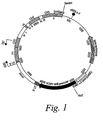
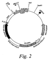
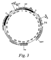


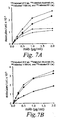

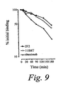
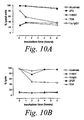
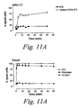

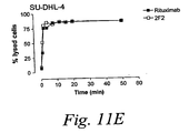


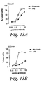
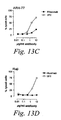

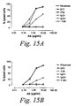


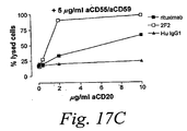
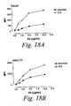


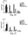
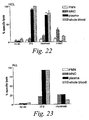


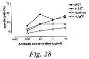


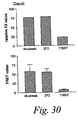
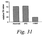


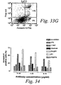
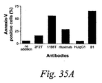
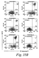
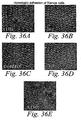

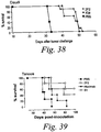

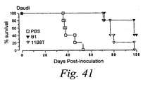

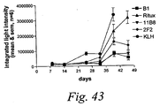
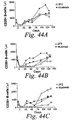


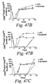
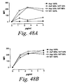

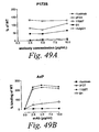

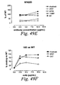


Classifications
-
- C—CHEMISTRY; METALLURGY
- C07—ORGANIC CHEMISTRY
- C07K—PEPTIDES
- C07K16/00—Immunoglobulins [IGs], e.g. monoclonal or polyclonal antibodies
- C07K16/18—Immunoglobulins [IGs], e.g. monoclonal or polyclonal antibodies against material from animals or humans
- C07K16/28—Immunoglobulins [IGs], e.g. monoclonal or polyclonal antibodies against material from animals or humans against receptors, cell surface antigens or cell surface determinants
- C07K16/2887—Immunoglobulins [IGs], e.g. monoclonal or polyclonal antibodies against material from animals or humans against receptors, cell surface antigens or cell surface determinants against CD20
-
- C—CHEMISTRY; METALLURGY
- C07—ORGANIC CHEMISTRY
- C07K—PEPTIDES
- C07K16/00—Immunoglobulins [IGs], e.g. monoclonal or polyclonal antibodies
- C07K16/18—Immunoglobulins [IGs], e.g. monoclonal or polyclonal antibodies against material from animals or humans
- C07K16/28—Immunoglobulins [IGs], e.g. monoclonal or polyclonal antibodies against material from animals or humans against receptors, cell surface antigens or cell surface determinants
-
- A—HUMAN NECESSITIES
- A61—MEDICAL OR VETERINARY SCIENCE; HYGIENE
- A61K—PREPARATIONS FOR MEDICAL, DENTAL OR TOILETRY PURPOSES
- A61K39/00—Medicinal preparations containing antigens or antibodies
- A61K39/395—Antibodies; Immunoglobulins; Immune serum, e.g. antilymphocytic serum
- A61K39/39533—Antibodies; Immunoglobulins; Immune serum, e.g. antilymphocytic serum against materials from animals
- A61K39/3955—Antibodies; Immunoglobulins; Immune serum, e.g. antilymphocytic serum against materials from animals against proteinaceous materials, e.g. enzymes, hormones, lymphokines
-
- A—HUMAN NECESSITIES
- A61—MEDICAL OR VETERINARY SCIENCE; HYGIENE
- A61K—PREPARATIONS FOR MEDICAL, DENTAL OR TOILETRY PURPOSES
- A61K45/00—Medicinal preparations containing active ingredients not provided for in groups A61K31/00 - A61K41/00
- A61K45/06—Mixtures of active ingredients without chemical characterisation, e.g. antiphlogistics and cardiaca
-
- A—HUMAN NECESSITIES
- A61—MEDICAL OR VETERINARY SCIENCE; HYGIENE
- A61P—SPECIFIC THERAPEUTIC ACTIVITY OF CHEMICAL COMPOUNDS OR MEDICINAL PREPARATIONS
- A61P1/00—Drugs for disorders of the alimentary tract or the digestive system
-
- A—HUMAN NECESSITIES
- A61—MEDICAL OR VETERINARY SCIENCE; HYGIENE
- A61P—SPECIFIC THERAPEUTIC ACTIVITY OF CHEMICAL COMPOUNDS OR MEDICINAL PREPARATIONS
- A61P1/00—Drugs for disorders of the alimentary tract or the digestive system
- A61P1/04—Drugs for disorders of the alimentary tract or the digestive system for ulcers, gastritis or reflux esophagitis, e.g. antacids, inhibitors of acid secretion, mucosal protectants
-
- A—HUMAN NECESSITIES
- A61—MEDICAL OR VETERINARY SCIENCE; HYGIENE
- A61P—SPECIFIC THERAPEUTIC ACTIVITY OF CHEMICAL COMPOUNDS OR MEDICINAL PREPARATIONS
- A61P11/00—Drugs for disorders of the respiratory system
-
- A—HUMAN NECESSITIES
- A61—MEDICAL OR VETERINARY SCIENCE; HYGIENE
- A61P—SPECIFIC THERAPEUTIC ACTIVITY OF CHEMICAL COMPOUNDS OR MEDICINAL PREPARATIONS
- A61P11/00—Drugs for disorders of the respiratory system
- A61P11/06—Antiasthmatics
-
- A—HUMAN NECESSITIES
- A61—MEDICAL OR VETERINARY SCIENCE; HYGIENE
- A61P—SPECIFIC THERAPEUTIC ACTIVITY OF CHEMICAL COMPOUNDS OR MEDICINAL PREPARATIONS
- A61P13/00—Drugs for disorders of the urinary system
- A61P13/12—Drugs for disorders of the urinary system of the kidneys
-
- A—HUMAN NECESSITIES
- A61—MEDICAL OR VETERINARY SCIENCE; HYGIENE
- A61P—SPECIFIC THERAPEUTIC ACTIVITY OF CHEMICAL COMPOUNDS OR MEDICINAL PREPARATIONS
- A61P17/00—Drugs for dermatological disorders
-
- A—HUMAN NECESSITIES
- A61—MEDICAL OR VETERINARY SCIENCE; HYGIENE
- A61P—SPECIFIC THERAPEUTIC ACTIVITY OF CHEMICAL COMPOUNDS OR MEDICINAL PREPARATIONS
- A61P17/00—Drugs for dermatological disorders
- A61P17/06—Antipsoriatics
-
- A—HUMAN NECESSITIES
- A61—MEDICAL OR VETERINARY SCIENCE; HYGIENE
- A61P—SPECIFIC THERAPEUTIC ACTIVITY OF CHEMICAL COMPOUNDS OR MEDICINAL PREPARATIONS
- A61P17/00—Drugs for dermatological disorders
- A61P17/08—Antiseborrheics
-
- A—HUMAN NECESSITIES
- A61—MEDICAL OR VETERINARY SCIENCE; HYGIENE
- A61P—SPECIFIC THERAPEUTIC ACTIVITY OF CHEMICAL COMPOUNDS OR MEDICINAL PREPARATIONS
- A61P19/00—Drugs for skeletal disorders
- A61P19/02—Drugs for skeletal disorders for joint disorders, e.g. arthritis, arthrosis
-
- A—HUMAN NECESSITIES
- A61—MEDICAL OR VETERINARY SCIENCE; HYGIENE
- A61P—SPECIFIC THERAPEUTIC ACTIVITY OF CHEMICAL COMPOUNDS OR MEDICINAL PREPARATIONS
- A61P21/00—Drugs for disorders of the muscular or neuromuscular system
- A61P21/04—Drugs for disorders of the muscular or neuromuscular system for myasthenia gravis
-
- A—HUMAN NECESSITIES
- A61—MEDICAL OR VETERINARY SCIENCE; HYGIENE
- A61P—SPECIFIC THERAPEUTIC ACTIVITY OF CHEMICAL COMPOUNDS OR MEDICINAL PREPARATIONS
- A61P25/00—Drugs for disorders of the nervous system
-
- A—HUMAN NECESSITIES
- A61—MEDICAL OR VETERINARY SCIENCE; HYGIENE
- A61P—SPECIFIC THERAPEUTIC ACTIVITY OF CHEMICAL COMPOUNDS OR MEDICINAL PREPARATIONS
- A61P25/00—Drugs for disorders of the nervous system
- A61P25/28—Drugs for disorders of the nervous system for treating neurodegenerative disorders of the central nervous system, e.g. nootropic agents, cognition enhancers, drugs for treating Alzheimer's disease or other forms of dementia
-
- A—HUMAN NECESSITIES
- A61—MEDICAL OR VETERINARY SCIENCE; HYGIENE
- A61P—SPECIFIC THERAPEUTIC ACTIVITY OF CHEMICAL COMPOUNDS OR MEDICINAL PREPARATIONS
- A61P27/00—Drugs for disorders of the senses
- A61P27/02—Ophthalmic agents
-
- A—HUMAN NECESSITIES
- A61—MEDICAL OR VETERINARY SCIENCE; HYGIENE
- A61P—SPECIFIC THERAPEUTIC ACTIVITY OF CHEMICAL COMPOUNDS OR MEDICINAL PREPARATIONS
- A61P29/00—Non-central analgesic, antipyretic or antiinflammatory agents, e.g. antirheumatic agents; Non-steroidal antiinflammatory drugs [NSAID]
-
- A—HUMAN NECESSITIES
- A61—MEDICAL OR VETERINARY SCIENCE; HYGIENE
- A61P—SPECIFIC THERAPEUTIC ACTIVITY OF CHEMICAL COMPOUNDS OR MEDICINAL PREPARATIONS
- A61P3/00—Drugs for disorders of the metabolism
- A61P3/08—Drugs for disorders of the metabolism for glucose homeostasis
- A61P3/10—Drugs for disorders of the metabolism for glucose homeostasis for hyperglycaemia, e.g. antidiabetics
-
- A—HUMAN NECESSITIES
- A61—MEDICAL OR VETERINARY SCIENCE; HYGIENE
- A61P—SPECIFIC THERAPEUTIC ACTIVITY OF CHEMICAL COMPOUNDS OR MEDICINAL PREPARATIONS
- A61P31/00—Antiinfectives, i.e. antibiotics, antiseptics, chemotherapeutics
- A61P31/12—Antivirals
- A61P31/14—Antivirals for RNA viruses
- A61P31/18—Antivirals for RNA viruses for HIV
-
- A—HUMAN NECESSITIES
- A61—MEDICAL OR VETERINARY SCIENCE; HYGIENE
- A61P—SPECIFIC THERAPEUTIC ACTIVITY OF CHEMICAL COMPOUNDS OR MEDICINAL PREPARATIONS
- A61P31/00—Antiinfectives, i.e. antibiotics, antiseptics, chemotherapeutics
- A61P31/12—Antivirals
- A61P31/20—Antivirals for DNA viruses
-
- A—HUMAN NECESSITIES
- A61—MEDICAL OR VETERINARY SCIENCE; HYGIENE
- A61P—SPECIFIC THERAPEUTIC ACTIVITY OF CHEMICAL COMPOUNDS OR MEDICINAL PREPARATIONS
- A61P31/00—Antiinfectives, i.e. antibiotics, antiseptics, chemotherapeutics
- A61P31/12—Antivirals
- A61P31/20—Antivirals for DNA viruses
- A61P31/22—Antivirals for DNA viruses for herpes viruses
-
- A—HUMAN NECESSITIES
- A61—MEDICAL OR VETERINARY SCIENCE; HYGIENE
- A61P—SPECIFIC THERAPEUTIC ACTIVITY OF CHEMICAL COMPOUNDS OR MEDICINAL PREPARATIONS
- A61P35/00—Antineoplastic agents
-
- A—HUMAN NECESSITIES
- A61—MEDICAL OR VETERINARY SCIENCE; HYGIENE
- A61P—SPECIFIC THERAPEUTIC ACTIVITY OF CHEMICAL COMPOUNDS OR MEDICINAL PREPARATIONS
- A61P35/00—Antineoplastic agents
- A61P35/02—Antineoplastic agents specific for leukemia
-
- A—HUMAN NECESSITIES
- A61—MEDICAL OR VETERINARY SCIENCE; HYGIENE
- A61P—SPECIFIC THERAPEUTIC ACTIVITY OF CHEMICAL COMPOUNDS OR MEDICINAL PREPARATIONS
- A61P37/00—Drugs for immunological or allergic disorders
-
- A—HUMAN NECESSITIES
- A61—MEDICAL OR VETERINARY SCIENCE; HYGIENE
- A61P—SPECIFIC THERAPEUTIC ACTIVITY OF CHEMICAL COMPOUNDS OR MEDICINAL PREPARATIONS
- A61P37/00—Drugs for immunological or allergic disorders
- A61P37/02—Immunomodulators
-
- A—HUMAN NECESSITIES
- A61—MEDICAL OR VETERINARY SCIENCE; HYGIENE
- A61P—SPECIFIC THERAPEUTIC ACTIVITY OF CHEMICAL COMPOUNDS OR MEDICINAL PREPARATIONS
- A61P37/00—Drugs for immunological or allergic disorders
- A61P37/02—Immunomodulators
- A61P37/06—Immunosuppressants, e.g. drugs for graft rejection
-
- A—HUMAN NECESSITIES
- A61—MEDICAL OR VETERINARY SCIENCE; HYGIENE
- A61P—SPECIFIC THERAPEUTIC ACTIVITY OF CHEMICAL COMPOUNDS OR MEDICINAL PREPARATIONS
- A61P37/00—Drugs for immunological or allergic disorders
- A61P37/08—Antiallergic agents
-
- A—HUMAN NECESSITIES
- A61—MEDICAL OR VETERINARY SCIENCE; HYGIENE
- A61P—SPECIFIC THERAPEUTIC ACTIVITY OF CHEMICAL COMPOUNDS OR MEDICINAL PREPARATIONS
- A61P43/00—Drugs for specific purposes, not provided for in groups A61P1/00-A61P41/00
-
- A—HUMAN NECESSITIES
- A61—MEDICAL OR VETERINARY SCIENCE; HYGIENE
- A61P—SPECIFIC THERAPEUTIC ACTIVITY OF CHEMICAL COMPOUNDS OR MEDICINAL PREPARATIONS
- A61P5/00—Drugs for disorders of the endocrine system
- A61P5/14—Drugs for disorders of the endocrine system of the thyroid hormones, e.g. T3, T4
-
- A—HUMAN NECESSITIES
- A61—MEDICAL OR VETERINARY SCIENCE; HYGIENE
- A61P—SPECIFIC THERAPEUTIC ACTIVITY OF CHEMICAL COMPOUNDS OR MEDICINAL PREPARATIONS
- A61P7/00—Drugs for disorders of the blood or the extracellular fluid
-
- A—HUMAN NECESSITIES
- A61—MEDICAL OR VETERINARY SCIENCE; HYGIENE
- A61P—SPECIFIC THERAPEUTIC ACTIVITY OF CHEMICAL COMPOUNDS OR MEDICINAL PREPARATIONS
- A61P7/00—Drugs for disorders of the blood or the extracellular fluid
- A61P7/04—Antihaemorrhagics; Procoagulants; Haemostatic agents; Antifibrinolytic agents
-
- A—HUMAN NECESSITIES
- A61—MEDICAL OR VETERINARY SCIENCE; HYGIENE
- A61P—SPECIFIC THERAPEUTIC ACTIVITY OF CHEMICAL COMPOUNDS OR MEDICINAL PREPARATIONS
- A61P7/00—Drugs for disorders of the blood or the extracellular fluid
- A61P7/06—Antianaemics
-
- A—HUMAN NECESSITIES
- A61—MEDICAL OR VETERINARY SCIENCE; HYGIENE
- A61P—SPECIFIC THERAPEUTIC ACTIVITY OF CHEMICAL COMPOUNDS OR MEDICINAL PREPARATIONS
- A61P9/00—Drugs for disorders of the cardiovascular system
- A61P9/08—Vasodilators for multiple indications
-
- A—HUMAN NECESSITIES
- A61—MEDICAL OR VETERINARY SCIENCE; HYGIENE
- A61P—SPECIFIC THERAPEUTIC ACTIVITY OF CHEMICAL COMPOUNDS OR MEDICINAL PREPARATIONS
- A61P9/00—Drugs for disorders of the cardiovascular system
- A61P9/10—Drugs for disorders of the cardiovascular system for treating ischaemic or atherosclerotic diseases, e.g. antianginal drugs, coronary vasodilators, drugs for myocardial infarction, retinopathy, cerebrovascula insufficiency, renal arteriosclerosis
-
- A—HUMAN NECESSITIES
- A61—MEDICAL OR VETERINARY SCIENCE; HYGIENE
- A61P—SPECIFIC THERAPEUTIC ACTIVITY OF CHEMICAL COMPOUNDS OR MEDICINAL PREPARATIONS
- A61P9/00—Drugs for disorders of the cardiovascular system
- A61P9/14—Vasoprotectives; Antihaemorrhoidals; Drugs for varicose therapy; Capillary stabilisers
-
- C—CHEMISTRY; METALLURGY
- C07—ORGANIC CHEMISTRY
- C07K—PEPTIDES
- C07K16/00—Immunoglobulins [IGs], e.g. monoclonal or polyclonal antibodies
- C07K16/18—Immunoglobulins [IGs], e.g. monoclonal or polyclonal antibodies against material from animals or humans
- C07K16/28—Immunoglobulins [IGs], e.g. monoclonal or polyclonal antibodies against material from animals or humans against receptors, cell surface antigens or cell surface determinants
- C07K16/30—Immunoglobulins [IGs], e.g. monoclonal or polyclonal antibodies against material from animals or humans against receptors, cell surface antigens or cell surface determinants from tumour cells
-
- C—CHEMISTRY; METALLURGY
- C07—ORGANIC CHEMISTRY
- C07K—PEPTIDES
- C07K16/00—Immunoglobulins [IGs], e.g. monoclonal or polyclonal antibodies
- C07K16/42—Immunoglobulins [IGs], e.g. monoclonal or polyclonal antibodies against immunoglobulins
- C07K16/4208—Immunoglobulins [IGs], e.g. monoclonal or polyclonal antibodies against immunoglobulins against an idiotypic determinant on Ig
- C07K16/4241—Immunoglobulins [IGs], e.g. monoclonal or polyclonal antibodies against immunoglobulins against an idiotypic determinant on Ig against anti-human or anti-animal Ig
- C07K16/4258—Immunoglobulins [IGs], e.g. monoclonal or polyclonal antibodies against immunoglobulins against an idiotypic determinant on Ig against anti-human or anti-animal Ig against anti-receptor Ig
-
- C—CHEMISTRY; METALLURGY
- C12—BIOCHEMISTRY; BEER; SPIRITS; WINE; VINEGAR; MICROBIOLOGY; ENZYMOLOGY; MUTATION OR GENETIC ENGINEERING
- C12N—MICROORGANISMS OR ENZYMES; COMPOSITIONS THEREOF; PROPAGATING, PRESERVING, OR MAINTAINING MICROORGANISMS; MUTATION OR GENETIC ENGINEERING; CULTURE MEDIA
- C12N5/00—Undifferentiated human, animal or plant cells, e.g. cell lines; Tissues; Cultivation or maintenance thereof; Culture media therefor
- C12N5/10—Cells modified by introduction of foreign genetic material
- C12N5/12—Fused cells, e.g. hybridomas
- C12N5/16—Animal cells
-
- G—PHYSICS
- G01—MEASURING; TESTING
- G01N—INVESTIGATING OR ANALYSING MATERIALS BY DETERMINING THEIR CHEMICAL OR PHYSICAL PROPERTIES
- G01N33/00—Investigating or analysing materials by specific methods not covered by groups G01N1/00 - G01N31/00
- G01N33/48—Biological material, e.g. blood, urine; Haemocytometers
- G01N33/50—Chemical analysis of biological material, e.g. blood, urine; Testing involving biospecific ligand binding methods; Immunological testing
- G01N33/68—Chemical analysis of biological material, e.g. blood, urine; Testing involving biospecific ligand binding methods; Immunological testing involving proteins, peptides or amino acids
- G01N33/6854—Immunoglobulins
- G01N33/686—Anti-idiotype
-
- A—HUMAN NECESSITIES
- A61—MEDICAL OR VETERINARY SCIENCE; HYGIENE
- A61K—PREPARATIONS FOR MEDICAL, DENTAL OR TOILETRY PURPOSES
- A61K39/00—Medicinal preparations containing antigens or antibodies
- A61K2039/505—Medicinal preparations containing antigens or antibodies comprising antibodies
-
- C—CHEMISTRY; METALLURGY
- C07—ORGANIC CHEMISTRY
- C07K—PEPTIDES
- C07K2317/00—Immunoglobulins specific features
- C07K2317/20—Immunoglobulins specific features characterized by taxonomic origin
- C07K2317/21—Immunoglobulins specific features characterized by taxonomic origin from primates, e.g. man
-
- C—CHEMISTRY; METALLURGY
- C07—ORGANIC CHEMISTRY
- C07K—PEPTIDES
- C07K2317/00—Immunoglobulins specific features
- C07K2317/50—Immunoglobulins specific features characterized by immunoglobulin fragments
- C07K2317/54—F(ab')2
-
- C—CHEMISTRY; METALLURGY
- C07—ORGANIC CHEMISTRY
- C07K—PEPTIDES
- C07K2317/00—Immunoglobulins specific features
- C07K2317/50—Immunoglobulins specific features characterized by immunoglobulin fragments
- C07K2317/56—Immunoglobulins specific features characterized by immunoglobulin fragments variable (Fv) region, i.e. VH and/or VL
-
- C—CHEMISTRY; METALLURGY
- C07—ORGANIC CHEMISTRY
- C07K—PEPTIDES
- C07K2317/00—Immunoglobulins specific features
- C07K2317/70—Immunoglobulins specific features characterized by effect upon binding to a cell or to an antigen
- C07K2317/72—Increased effector function due to an Fc-modification
-
- C—CHEMISTRY; METALLURGY
- C07—ORGANIC CHEMISTRY
- C07K—PEPTIDES
- C07K2317/00—Immunoglobulins specific features
- C07K2317/70—Immunoglobulins specific features characterized by effect upon binding to a cell or to an antigen
- C07K2317/73—Inducing cell death, e.g. apoptosis, necrosis or inhibition of cell proliferation
- C07K2317/732—Antibody-dependent cellular cytotoxicity [ADCC]
-
- C—CHEMISTRY; METALLURGY
- C07—ORGANIC CHEMISTRY
- C07K—PEPTIDES
- C07K2317/00—Immunoglobulins specific features
- C07K2317/70—Immunoglobulins specific features characterized by effect upon binding to a cell or to an antigen
- C07K2317/73—Inducing cell death, e.g. apoptosis, necrosis or inhibition of cell proliferation
- C07K2317/734—Complement-dependent cytotoxicity [CDC]
-
- C—CHEMISTRY; METALLURGY
- C07—ORGANIC CHEMISTRY
- C07K—PEPTIDES
- C07K2317/00—Immunoglobulins specific features
- C07K2317/90—Immunoglobulins specific features characterized by (pharmaco)kinetic aspects or by stability of the immunoglobulin
- C07K2317/92—Affinity (KD), association rate (Ka), dissociation rate (Kd) or EC50 value
Definitions
- the CD20 molecule (also called human B-lymphocyte-restricted differentiation antigen or Bp35) is a hydrophobic transmembrane protein with a molecular weight of approximately 35 kD located on pre-B and mature B lymphocytes ( Valentine et al. (1989) J. Biol. Chem. 264(19):11282-11287 ; and Einfield et al. (1988) EMBO J. 7(3):711-717 ). CD20 is found on the surface of greater than 90% ofB cells from peripheral blood or lymphoid organs and is expressed during early pre-B cell development and remains until plasma cell differentiation. CD20 is present on both normal B cells as well as malignant B cells.
- CD20 is expressed on greater than 90% of B cell non-Hodgkin's lymphomas (NHL) ( Anderson et al. (1984) Blood 63(6):1424-1433 ), but is not found on hematopoietic stem cells, pro-B cells, normal plasma cells, or other normal tissues ( Tedder et al. (1985) J. Immunol. 135(2):973-979 ).
- NHL B cell non-Hodgkin's lymphomas
- the 85 amino acid carboxyl-terminal region of the CD20 protein is located within the cytoplasm.
- the length of this region contrasts with that of other B cell-specific surface structures such as IgM, IgD, and IgG heavy chains or histocompatibility antigens class II ⁇ or ⁇ chains, which have relatively short intracytoplasmic regions of 3, 3, 28, 15, and 16 amino acids, respectively ( Komaromy et al. (1983) NAR 11:6775-6785 ).
- 21 are acidic residues, whereas only 2 are basic, indicating that this region has a strong net negative charge.
- GenBank Accession No. is NP_690605.
- CD20 might be involved in regulating an early step(s) in the activation and differentiation process of B cells ( Tedder et al. (1986) Eur.J. Immunol. 16:881-887 ) and could function as a calcium ion channel ( Tedder et al. (1990) J. Cell. Biochem. 14D:195 ).
- CD20 Despite uncertainty about the actual function of CD20 in promoting proliferation and/or differentiation of B cells, it provides an important target for antibody mediated therapy to control or kill B cells involved in cancers and autoimmune disorders.
- the expression of CD20 on tumor cells, e.g., NHL makes it an important target for antibody mediated therapy to specifically target therapeutic agents against CD20-positive neoplastic cells.
- tumor cells e.g., NHL
- results obtained to date clearly establish CD20 as a useful target for immunotherapy, they also show that currently available murine and chimeric antibodies do not constitute ideal therapeutic agents.
- the present invention provides an isolated human monoclonal antibody which binds to human CD20, which is capable of inducing complement dependent cytotoxicity (CDC) of cells expressing CD20 in the presence of complement and which comprises:
- the present invention thus provides improved antibody therapeutics for treating and/or preventing diseases associated with cells expressing CD20, including tumor-related diseases, and immune diseases, including autoimmune diseases.
- diseases associated with cells expressing CD20 including tumor-related diseases, and immune diseases, including autoimmune diseases.
- the antibodies encompassed by the invention are improved in that they are fully human and, thus, are potentially less immunogenic in patients.
- human antibodies of the invention mediate killing of B cells expressing CD20 by a variety of mechanisms.
- human antibodies of the invention induce complement dependent cytotoxicity (CDC), e. g. , at least about 20% CDC mediated lysis, preferably about 30% CDC mediated lysis, and more preferably 40-50% mediated lysis in cells, such as chronic B-lymphocytic leukaemia (B-CLL) cells.
- CDC complement dependent cytotoxicity
- human antibodies of the invention induce apoptosis of cells expressing CD20.
- human antibodies of the invention induce homotypic adhesion of cells expressing CD20.
- human antibodies of the invention may induce antibody dependent cellular cytotoxicity (ADCC) of cells expressing CD20 in the presence of human effector cells (e. g. , monocytes, mononuclear cells, NK cells and PMNs). Furthermore, human antibodies of the invention may induce phagocytosis of cells expressing CD20 in the presence of macrophages.
- the human monoclonal antibodies of the invention may work by one or more of these mechanisms. Examples of cells which can be killed by human antibodies of the present invention include, but are not limited to, B cells expressing CD20, such as tumorigenic B cells and B cells involved in immune diseases.
- the human antibodies are used to mediate killing of B lymphocytes in the treatment of lymphoma, e. g. , B cell non-Hodgkin's lymphoma.
- Human antibodies of the invention include IgG1 (e. g. , IgG1, ⁇ ), IgG3 (e. g. , IgG3, ⁇ ) and IgG4 ( e. g. , IgG4, ⁇ ) antibodies.
- IgG1 e. g. , IgG1, ⁇
- IgG3 e. g. , IgG3, ⁇
- IgG4 e. g. , IgG4, ⁇
- the antibodies can be whole antibodies or antigen-binding fragments thereof including, for example, Fab, F(ab') 2 , Fv, single chain Fv fragments or bispecific antibodies.
- the antigen-binding fragments include binding-domain immunoglobulin fusion proteins comprising (i) a binding domain polypeptide (such as a heavy chain variable region or a light chain variable region) that is fused to an immunoglobulin hinge region polypeptide, (ii) an immunoglobulin heavy chain CH2 constant region fused to the hinge region, and (iii) an immunoglobulin heavy chain CH3 constant region fused to the CH2 constant region.
- binding-domain immunoglobulin fusion proteins are further disclosed in US 2003/0118592 and US 2003/0133939 .
- human antibodies of the present invention include those referred to as 2F2 and 7D8, encoded by human heavy chain and human kappa light chain nucleic acids comprising nucleotide sequences in their variable regions as set forth in SEQ ID NOs: 1 or 5 and SEQ ID NOs: 3 or 7, respectively.
- the human antibodies are characterized by having human heavy chain and human kappa light chain variable regions comprising the amino acid sequences as set forth in SEQ ID NOs: 2 or 6 and SEQ ID NOs: 4 or 8, respectively.
- the human antibodies are characterized by having human heavy chain and human kappa light chain variable regions which are at least 90% homologous, preferably at least 95% homologous, and more preferably at least 98%, or at least 99% homologous to the amino acid sequences as set forth in SEQ ID NO: 2 and SEQ ID NO: 4, respectively; or SEQ ID NO: 6 and SEQ ID NO: 8, respectively.
- antibodies which dissociate from CD20 with a dissociation equilibrium constant (K D ) of approximately 1- 10 nM are also included within the present invention.
- K D dissociation equilibrium constant
- Such antibodies also include those which do not cross-react with related cell-surface antigens and thus do not inhibit their function.
- human anti-CD20 antibodies of the present invention can be characterized by one or more of the following properties:
- the human anti-CD20 antibodies of the present invention can be derivatized, linked to or co-expressed to other binding specificities.
- the invention provides a bispecific or multispecific molecule comprising at least one first binding specificity for CD20 (e.g. , a human anti-CD20 antibody or mimetic thereof), and a second binding specificity for a human effector cell, such as a binding specificity for an Fc receptor (e.g. , a human Fc ⁇ receptor, such as FcyRI, or a human Fc ⁇ receptor) or a T cell receptor, e.g. , CD3.
- a bispecific or multispecific molecule comprising at least one first binding specificity for CD20 (e.g. , a human anti-CD20 antibody or mimetic thereof), and a second binding specificity for a human effector cell, such as a binding specificity for an Fc receptor (e.g. , a human Fc ⁇ receptor, such as FcyRI, or a human Fc ⁇ receptor)
- the present invention includes bispecific molecules that bind to both human CD20 and to an Fc receptor or a T cell receptor, e.g. , CD3.
- Fc receptors are, e.g. , a human IgG receptor, e.g. , an Fc-gamma receptor (Fc ⁇ R), such as Fc ⁇ RI (CD64), Fc ⁇ RII (CD32), and Fc ⁇ RIII (CD16).
- Fc receptors such as human IgA receptors (e.g. , Fc ⁇ RI), also can be targeted.
- the Fc receptor is preferably located on the surface of an effector cell, e.g.
- the bispecific and multispecific molecules bind to an Fc receptor at a site which is distinct from the immunoglobulin Fc (e.g. , IgG or IgA) binding site of the receptor. Therefore, the binding of the bispecific and multispecific molecules is not blocked by physiological levels of immunoglobulins.
- human anti-CD20 antibodies of the invention are derivatized, linked to or co-expressed with another functional molecule, e.g. , another peptide or protein (e.g. , a Fab' fragment).
- another functional molecule e.g. , another peptide or protein (e.g. , a Fab' fragment).
- an antibody of the invention can be functionally linked ( e.g. , by chemical coupling, genetic fusion, noncovalent association or otherwise) to one or more other molecular entities, such as another antibody (e.g. , to produce a bispecific or a multispecific antibody), a cytotoxin, cellular ligand or antigen (e.g. , to produce an immunoconjugate, such as an immunotoxin).
- An antibody of the present invention can be linked to other therapeutic moieties, e.g. , a radioisotope, a small molecule anti-cancer drug, an anti-inflammatory agent, or an immunosuppressive agent. Accordingly, the disclosure encompasses a large variety of antibody conjugates, bispecific and multispecific molecules, and fusion proteins, all of which bind to CD20 expressing cells and which can be used to target other molecules to such cells.
- the invention provides compositions, e.g. , pharmaceutical and diagnostic compositions/kits, comprising a pharmaceutically acceptable carrier formulated along with one or a combination of human monoclonal antibodies of the invention
- the composition includes a combination of antibodies which bind to distinct epitopes or which possess distinct functional characteristics, such as inducing CDC and inducing apoptosis.
- the human antibodies, immunoconjugates, bispecific and multispecific molecules and compositions can be used in a variety of methods for inhibiting growth of cells expressing CD20 and/or killing cells expressing CD20 by contacting the cells with an effective amount of the antibody, immunconjugate, bispecific/multispecific molecule or composition, such that the growth of the cell is inhibited and/or the cell is killed.
- the method includes killing of the cell expressing CD20 in the presence of effector cells, for example, by CDC, apoptosis, ADCC, phagocytosis, or by a combination of two or more of these mechanisms.
- the cells are preferably killed or inhibited without killing or inhibiting the activity of cells which do not express CD20 but which may, for example, express a structurally related cell-surface antigen (i.e. , without cross-reactivity to related but functionally distinct cell surface antigens).
- Cells expressing CD20 which can be inhibited or killed using the human antibodies of the invention include, for example, tumorigenic B cells.
- human antibodies of the present invention can be used to treat and/or prevent a variety of diseases involving cells expressing CD20 by administering the antibodies to patients suffering from such diseases.
- diseases that can be treated (e.g. , ameliorated) or prevented include, but are not limited to, tumorigenic diseases and immune diseases, e.g. , autoimmune diseases.
- tumorigenic diseases which can be treated and/or prevented include B cell lymphoma, e.g.
- NHL including precursor B cell lymphoblastic leukemia/lymphoma and mature B cell neoplasms, such as B cell chronic lymhocytic leukemia (CLL)/small lymphocytic lymphoma (SLL), B cell prolymphocytic leukemia, lymphoplasmacytic lymphoma, mantle cell lymphoma (MCL), follicular lymphoma (FL), including low-grade, intermediate-grade and high-grade FL, cutaneous follicle center lymphoma, marginal zone B cell lymphoma (MALT type, nodal and splenic type), hairy cell leukemia, diffuse large B cell lymphoma, Burkitt's lymphoma, plasmacytoma, plasma cell myeloma, post-transplant lymphoproliferative disorder, Waldenström's macroglobulinemia, and anaplastic large-cell lymphoma (ALCL).
- CLL chronic lymhocytic leukemia
- SLL small lympho
- immune disorders in which CD20 expressing B cells are involved which can be treated and/or prevented include psoriasis, psoriatic arthritis, dermatitis, systemic scleroderma and sclerosis, inflammatory bowel disease (IBD), Crohn's disease, ulcerative colitis, respiratory distress syndrome, meningitis, encephalitis, uveitis, glomerulonephritis, eczema, asthma, atherosclerosis, leukocyte adhesion deficiency, multiple sclerosis, Raynaud's syndrome, Sjögren's syndrome, juvenile onset diabetes, Reiter's disease, Behçet's disease, immune complex nephritis, IgA nephropathy, IgM polyneuropathies, immune-mediated thrombocytopenias, such as acute idiopathic thrombocytopenic purpura and chronic idiopathic thrombocytopenic purpura, hemolytic anemia, myasthenia
- the subject being administered the antibody is additionally treated with a chemotherapeutic agent, radiation, or an agent that modulates, e.g. , enhances or inhibits, the expression or activity of an Fc receptor, e.g. , an Fc ⁇ receptor or an Fc ⁇ receptor, such as a cytokine.
- a chemotherapeutic agent e.g. , radiation, or an agent that modulates, e.g. , enhances or inhibits, the expression or activity of an Fc receptor, e.g. , an Fc ⁇ receptor or an Fc ⁇ receptor, such as a cytokine.
- Typical cytokines for administration during treatment include granulocyte colony-stimulating factor (G-CSF), granulocyte- macrophage colony-stimulating factor (GM-CSF), interferon- ⁇ (IFN- ⁇ ), and tumor necrosis factor (TNF).
- Typical therapeutic agents include, among others, anti-neoplastic agents such as doxorubicin
- the present invention provides a method for detecting in vitro or in vivo the presence of CD20 in a sample or individual, e.g. , for diagnosing a CD20-related disease, preferably at an early stage. This can also be useful for monitoring the disease and effect of treatment and for determining and adjusting the dose of the antibody to be administered.
- the in vivo method can be performed using imaging technique such as PET (positron emission tomography) or SPECT (single photon emission computed tomography). In one embodiment, this is achieved by contacting a sample to be tested, optionally along with a control sample, with a human monoclonal antibody of the invention under conditions that allow for formation of a complex between the antibody and CD20.
- Complex formation is then detected (e.g. , using an FACS analysis or Western blotting).
- complex is detected in both samples and any statistically significant difference in the formation of complexes between the samples is indicative of the presence of CD20 in the test sample.
- the invention provides a transgenic non-human animal, such as a transgenic mouse, which express human monoclonal antibodies that bind to CD20.
- the transgenic non-human animal is a transgenic mouse having a genome comprising a human heavy chain transgene and a human light chain transgene encoding all or a portion of an antibody of the invention.
- the transgenic non-human animal can be immunized with a purified or enriched preparation of CD20 antigen and/or cells expressing CD20.
- the transgenic non-human animal e.g. , the transgenic mouse, is capable of producing multiple isotypes of human monoclonal antibodies to CD20 ( e.g. , IgG, IgA and/or IgM) by undergoing V-D-J recombination and isotype switching. Isotype switching may occur by, e.g. , classical or non-classical isotype switching.
- the invention provides isolated B cells from a transgenic non-human animal as described above, e.g. , a transgenic mouse, which expresses human anti-CD20 antibodies.
- the isolated B cells can then be immortalized by fusion to an immortalized cell to provide a source (e.g. , a hybridoma) of human anti-CD20 antibodies.
- a source e.g. , a hybridoma
- hybridomas i.e. , which produce human anti-CD20 antibodies
- human antibodies of the invention can be obtained directly from hybridomas which express the antibody, or can be cloned and recombinantly expressed in a host cell (e.g. , a CHO cell, a NS/0 cell or a lymphocytic cell).
- a host cell e.g. , a CHO cell, a NS/0 cell or a lymphocytic cell.
- host cells are microorganisms, such as E. coli , and fungi, such as yeast.
- they can be produced recombinantly in a transgenic non-human animal or plant.
- the present invention provides methods for producing human monoclonal antibodies which bind to human CD20.
- the method includes immunizing a transgenic non-human animal, e.g.
- a transgenic mouse as previously described (e.g. , having a genome comprising a human heavy chain transgene and a human light chain transgene encoding all or a portion of an anti-CD20 antibody), with a purified or enriched preparation of human CD20 antigen and/or cells expressing human CD20.
- B cells e.g. , splenic B cells
- myeloma cells to form immortal, hybridoma cells that secrete human monoclonal antibodies against CD20.
- nucleic acid molecules encoding human anti-CD20 antibodies (e.g. , variable regions thereof), as well as recombinant expression vectors which include such nucleic acids, host cells transfected with such vectors and methods of producing the antibodies by culturing these host cells.
- Particular nucleic acids comprise the nucleotide sequences shown in SEQ ID NOs:1 or 5 and SEQ ID NOs:3 or 7, encoding the heavy and light chains, respectively, of human anti-CD20 antibodies 2F2 and 7D8.
- the present invention provides improved antibody-based therapies for treating and diagnosing a variety of disorders involving cells expressing CD20.
- Therapies of the invention employ isolated human monoclonal antibodies which specifically bind to an epitope present on CD20.
- Isolated human monoclonal antibodies encompassed by the present invention include IgA, IgG1-4, IgE, IgM, and IgD antibodies.
- the antibody is an IgG1 antibody, more particularly an IgG1, ⁇ or IgG1, ⁇ isotype.
- the antibody is an IgG3 antibody, more particularly an IgG3, ⁇ or IgG3, ⁇ isotype.
- the antibody is an IgG4 antibody, more particularly an IgG4, ⁇ or IgG4, ⁇ isotype.
- the antibody is an IgA1 or IgA2 antibody.
- the antibody is an IgM antibody.
- the human antibodies are produced in a non-human transgenic animal, e.g. , a transgenic mouse, capable of producing multiple isotypes of human monoclonal antibodies to CD20 by undergoing V-D-J recombination and isotype switching.
- a non-human transgenic animal e.g. , a transgenic mouse
- aspects of the invention include not only antibodies, antibody fragments, and pharmaceutical compositions thereof, but also non-human transgenic animals and host cell transfectomas which produce monoclonal antibodies.
- Methods of using the antibodies of the invention to detect a cell expressing CD20 are encompassed by the invention.
- Methods of using the antibodies of the invention to block or inhibit CD20 induced activities, e.g. , proliferative and/or differentiation activities, are also provided and are useful in the treatment of disorders associated with CD20, such as tumorigenic diseases (e.g. , B cell lymphoma) and autoimmune diseases (e.g. , R.A, Chrohn's disease and Wegener's granulomatosis).
- CD20 and “CD20 antigen” are used interchangeably herein, and include any variants, isoforms and species homologs of human CD20 which are naturally expressed by cells or are expressed on cells transfected with the CD20 gene. Binding of an antibody of the invention to the CD20 antigen mediate the killing of cells expressing CD20 (e.g. , a tumor cell) by inactivating CD20. The killing of the cells expressing CD20 may occur by one or more of the following mechanisms:
- CD20 Synonyms of CD20, as recognized in the art, include B-lymphocyte antigen CD20, B-lymphocyte surface antigen B1, Leu-16, Bp35, BM5, and LF5.
- the term "inhibits growth" is intended to include any measurable decrease in the cell growth when contacted with an anti-CD20 antibody as compared to the growth of the same cells not in contact with an anti-CD20 antibody, e.g. , the inhibition of growth of a cell culture by at least about 10%, 20%, 30%, 40%, 50%, 60%, 70%, 80%, 90%, 99%, or 100%.
- Such a decrease in cell growth can occur by a variety of mechanisms, e.g. , effector cell phagocytosis, ADCC, CDC, and/or apoptosis.
- raft refers to the sphingolipid- and cholesterol-rich membrane microdomains located in the outer leaflet area of the plasma membrane of a cell.
- the ability of certain proteins to associate within such domains can effect the protein's function.
- the translocation of CD20 molecules into lipid rafts after being bound by human antibodies of the present invention, creates a high density of CD20 antigen-antibody complexes in the plasma membranes.
- Such a high density of CD20 antigen-antibody complexes can enable efficient activation of the complement system during CDC.
- antibody as referred to herein includes whole antibodies and any antigen binding fragment (i.e. , "antigen-binding portion") or single chain thereof.
- An “antibody” refers to a glycoprotein comprising at least two heavy (H) chains and two light (L) chains inter-connected by disulfide bonds, or an antigen binding portion thereof.
- Each heavy chain is comprised of a heavy chain variable region (abbreviated herein as V H ) and a heavy chain constant region.
- Each light chain is comprised of a light chain variable region (abbreviated herein as V L ) and a light chain constant region.
- V H and V L regions can be further subdivided into regions of hypervariability, termed complementarity determining regions (CDR), interspersed with regions that are more conserved, termed framework regions (FR).
- CDR complementarity determining regions
- FR framework regions
- Each V H and V L is composed of three CDRs and four FRs, arranged from amino-terminus to carboxy-terminus in the following order: FR1, CDR1, FR2, CDR2, FR3, CDR3, FR4.
- the variable regions of the heavy and light chains contain a binding domain that interacts with an antigen.
- the constant regions of the antibodies may mediate the binding of the immunoglobulin to host tissues or factors, including various cells of the immune system (e.g. , effector cells) and the first component (Clq) of the classical complement system.
- antibody portion refers to one or more fragments of an antibody that retain the ability to specifically bind to an antigen (e.g. , CD20). It has been shown that the antigen-binding function of an antibody can be performed by fragments of a full-length antibody.
- binding fragments encompassed within the term "antigen-binding portion" of an antibody include (i) a Fab fragment, a monovalent fragment consisting of the V L , V H , C L and C H1 domains; (ii) a F(ab') 2 fragment, a bivalent fragment comprising two Fab fragments linked by a disulfide bridge at the hinge region; (iii) a Fd fragment consisting of the V H and C H1 domains; (iv) a Fv fragment consisting of the V L and V H domains of a single arm of an antibody, (v) a dAb fragment ( Ward et al., (1989) Nature 341:544-546 ), which consists of a V H domain; (vi) an isolated complementarity determining region (CDR), and (vii) a combination of two or more isolated CDRs which may optionally be joined by a synthetic linker.
- a Fab fragment a monovalent fragment consisting of the V L , V H
- the two domains of the Fv fragment, V L and V H are coded for by separate genes, they can be joined, using recombinant methods, by a synthetic linker that enables them to be made as a single protein chain in which the V L and V H regions pair to form monovalent molecules (known as single chain Fv (scFv); see e.g. , Bird et al. (1988) Science 242:423-426 ; and Huston et al. (1988) Proc. Natl. Acad. Sci. USA 85:5879-5883 ).
- single chain Fv single chain Fv
- Such single chain antibodies are also intended to be encompassed within the term "antigen-binding portion" of an antibody.
- a further example is binding-domain immunoglobulin fusion proteins comprising (i) a binding domain polypeptide that is fused to an immunoglobulin hinge region polypeptide, (ii) an immunoglobulin heavy chain CH2 constant region fused to the hinge region, and (iii) an immunoglobulin heavy chain CH3 constant region fused to the CH2 constant region.
- the binding domain polypeptide can be a heavy chain variable region or a light chain variable region.
- the binding-domain immunoglobulin fusion proteins are further disclosed in US 2003/0118592 and US 2003/0133939 . These antibody fragments are obtained using conventional techniques known to those with skill in the art, and the fragments are screened for utility in the same manner as are intact antibodies.
- epitope means a protein determinant capable of specific binding to an antibody.
- Epitopes usually consist of chemically active surface groupings of molecules such as amino acids or sugar side chains and usually have specific three dimensional structural characteristics, as well as specific charge characteristics. Conformational and nonconformational epitopes are distinguished in that the binding to the former but not the latter is lost in the presence of denaturing solvents.
- discontinuous epitope means a conformational epitope on a protein antigen which is formed from at least two separate regions in the primary sequence of the protein.
- bispecific molecule is intended to include any agent, e.g. , a protein, peptide, or protein or peptide complex, which has two different binding specificities.
- the molecule may bind to, or interact with, (a) a cell surface antigen and (b) an Fc receptor on the surface of an effector cell.
- multispecific molecule or “heterospecific molecule” is intended to include any agent, e.g. , a protein, peptide, or protein or peptide complex, which has more than two different binding specificities.
- the molecule may bind to, or interact with, (a) a cell surface antigen, (b) an Fc receptor on the surface of an effector cell, and (c) at least one other component.
- the invention includes, but is not limited to, bispecific, trispecific, tetraspecific, and other multispecific molecules which are directed to cell surface antigens, such as CD20, and to other targets, such as Fc receptors on effector cells.
- bispecific antibodies also includes diabodies.
- Diabodies are bivalent, bispecific antibodies in which the V H and V L domains are expressed on a single polypeptide chain, but using a linker that is too short to allow for pairing between the two domains on the same chain, thereby forcing the domains to pair with complementary domains of another chain and creating two antigen binding sites (see e.g. , Holliger, P., et al. (1993) Proc. Natl. Acad. Sci. USA 90:6444-6448 ; Poljak, R.J., et al. (1994) Structure 2:1121-1123 ).
- human antibody derivatives refers to any modified form of the antibody, e.g. , a conjugate of the antibody and another agent or antibody.
- a human antibody is "derived from" a particular germline sequence if the antibody is obtained from a system using human immunoglobulin sequences, e.g. , by immunizing a transgenic mouse carrying human immunoglobulin genes or by screening a human immunoglobulin gene library, and wherein the selected human antibody is at least 90%, more preferably at least 95%, even more preferably at least 96%, 97%, 98%, or 99% identical in amino acid sequence to the amino acid sequence encoded by the germline immunoglobulin gene.
- a human antibody derived from a particular human germline sequence will display no more than 10 amino acid differences, more preferably, no more than 5, or even more preferably, no more than 4, 3, 2, or 1 amino acid difference from the amino acid sequence encoded by the germline immunoglobulin gene.
- heteroantibodies refers to two or more antibodies, derivatives therefrom, or antigen binding regions linked together, at least two of which have different specificities. These different specificities include a binding specificity for an Fc receptor on an effector cell, and a binding specificity for an antigen or epitope on a target cell, e.g. , a tumor cell.
- human antibody is intended to include antibodies having variable and constant regions derived from human germline immunoglobulin sequences.
- the human antibodies of the invention may include amino acid residues not encoded by human germline immunoglobulin sequences ( e.g. , mutations introduced by random or site-specific mutagenesis in vitro or by somatic mutation in vivo ).
- the term “human antibody”, as used herein is not intended to include antibodies in which CDR sequences derived from the germline of another mammalian species, such as a mouse, have been grafted onto human framework sequences.
- the terms “monoclonal antibody” or “monoclonal antibody composition” as used herein refer to a preparation of antibody molecules of single molecular composition.
- a monoclonal antibody composition displays a single binding specificity and affinity for a particular epitope.
- the term “human monoclonal antibody” refers to antibodies displaying a single binding specificity which have variable and constant regions derived from human germline immunoglobulin sequences.
- the human monoclonal antibodies are produced by a hybridoma which includes a B cell obtained from a transgenic or transchromosomal non-human animal, e.g. , a transgenic mouse, having a genome comprising a human heavy chain transgene and a light chain transgene fused to an immortalized cell.
- recombinant human antibody includes all human antibodies that are prepared, expressed, created or isolated by recombinant means, such as (a) antibodies isolated from an animal (e.g. , a mouse) that is transgenic or transchromosomal for human immunoglobulin genes or a hybridoma prepared therefrom (described further in Section I, below), (b) antibodies isolated from a host cell transformed to express the antibody, e.g. , from a transfectoma, (c) antibodies isolated from a recombinant, combinatorial human antibody library, and (d) antibodies prepared, expressed, created or isolated by any other means that involve splicing of human immunoglobulin gene sequences to other DNA sequences.
- Such recombinant human antibodies have variable and constant regions derived from human germline immunoglobulin sequences.
- such recombinant human antibodies can be subjected to in vitro mutagenesis (or, when an animal transgenic for human Ig sequences is used, in vivo somatic mutagenesis) and thus the amino acid sequences of the V H and V L regions of the recombinant antibodies are sequences that, while derived from and related to human germline V H and V L sequences, may not naturally exist within the human antibody germline repertoire in vivo .
- transfectoma includes recombinant eukaryotic host cell expressing the antibody, such as CHO cells, NS/0 cells, HEK293 cells, plant cells, or fungi, including yeast cells.
- a heterologous antibody is defined in relation to the transgenic non-human organism producing such an antibody. This term refers to an antibody having an amino acid sequence or an encoding nucleic acid sequence corresponding to that found in an organism not consisting of the transgenic non-human animal, and generally from a species other than that of the transgenic non-human animal.
- heterohybrid antibody refers to an antibody having a light and heavy chains of different organismal origins.
- an antibody having a human heavy chain associated with a murine light chain is a heterohybrid antibody.
- heterohybrid antibodies include chimeric and humanized antibodies, discussed supra.
- an "isolated antibody,” as used herein, is intended to refer to an antibody which is substantially free of other antibodies having different antigenic specificities (e.g. , an isolated antibody that specifically binds to CD20 is substantially free of antibodies that specifically bind antigens other than CD20).
- An isolated antibody that specifically binds to an epitope, isoform or variant of human CD20 may, however, have cross-reactivity to other related antigens, e.g. , from other species (e.g. , CD20 species homologs).
- an isolated antibody may be substantially free of other cellular material and/or chemicals.
- a combination of "isolated" monoclonal antibodies having different specificities are combined in a well defined composition.
- specific binding refers to antibody binding to a predetermined antigen.
- the antibody binds with an affinity corresponding to a K D of about 1 x 10 -7 M or less, and binds to the predetermined antigen with an affinity corresponding to a K D that is at least two orders of magnitude lower than its affinity for binding to a non-specific antigen (e.g. , BSA, casein) other than the predetermined antigen or a closely-related antigen.
- a non-specific antigen e.g. , BSA, casein
- k d (sec -1 ), as used herein, is intended to refer to the dissociation rate constant of a particular antibody-antigen interaction. Said value is also referred to as the k off value.
- k a (M -1 x sec -1 ), as used herein, is intended to refer to the association rate constant of a particular antibody-antigen interaction.
- K D (M), as used herein, is intended to refer to the dissociation equilibrium constant of a particular antibody-antigen interaction.
- K A (M -1 ), as used herein, is intended to refer to the association equilibrium constant of a particular antibody-antigen interaction and is obtained by dividing the k a by the k d .
- isotype refers to the antibody class (e.g. , IgM or IgG1) that is encoded by heavy chain constant region genes.
- isotype switching refers to the phenomenon by which the class, or isotype, of an antibody changes from one Ig class to one of the other Ig classes.
- nonswitched isotype refers to the isotypic class of heavy chain that is produced when no isotype switching has taken place; the CH gene encoding the nonswitched isotype is typically the first CH gene immediately downstream from the functionally rearranged VDJ gene.
- Isotype switching has been classified as classical or non-classical isotype switching.
- Classical isotype switching occurs by recombination events which involve at least one switch sequence region in the transgene.
- Non-classical isotype switching may occur by, for example, homologous recombination between human ⁇ ⁇ and human ⁇ ⁇ ( ⁇ -associated deletion).
- Alternative non-classical switching mechanisms such as intertransgene and/or interchromosomal recombination, among others, may occur and effectuate isotype switching.
- switch sequence refers to those DNA sequences responsible for switch recombination.
- a "switch donor” sequence typically a ⁇ switch region, will be 5' ( i.e. , upstream) of the construct region to be deleted during the switch recombination.
- the "switch acceptor” region will be between the construct region to be deleted and the replacement constant region ( e.g. , ⁇ , ⁇ , etc.). As there is no specific site where recombination always occurs, the final gene sequence will typically not be predictable from the construct.
- glycosylation pattern is defined as the pattern of carbohydrate units that are covalently attached to a protein, more specifically to an immunoglobulin (antibody) protein.
- a glycosylation pattern of a heterologous antibody can be characterized as being substantially similar to glycosylation patterns which occur naturally on antibodies produced by the species of the non-human transgenic animal, when one of ordinary skill in the art would recognize the glycosylation pattern of the heterologous antibody as being more similar to said pattern of glycosylation in the species of the non-human transgenic animal than to the species from which the CH genes of the transgene were derived.
- naturally-occurring refers to the fact that an object can be found in nature.
- a polypeptide or polynucleotide sequence that is present in an organism (including viruses) that can be isolated from a source in nature and which has not been intentionally modified by man in the laboratory is naturally-occurring.
- rearranged refers to a configuration of a heavy chain or light chain immunoglobulin locus wherein a V segment is positioned immediately adjacent to a D-J or J segment in a conformation encoding essentially a complete V H or V L domain, respectively.
- a rearranged immunoglobulin (antibody) gene locus can be identified by comparison to germline DNA; a rearranged locus will have at least one recombined heptamer/nonamer homology element.
- V segment configuration refers to the configuration wherein the V segment is not recombined so as to be immediately adjacent to a D or J segment.
- nucleic acid molecule is intended to include DNA molecules and RNA molecules.
- a nucleic acid molecule may be single-stranded or double-stranded, but preferably is double-stranded DNA.
- isolated nucleic acid molecule as used herein in reference to nucleic acids encoding whole antibodies or antibody portions (e.g. , V H , V L , CDR3) that bind to CD20, is intended to refer to a nucleic acid molecule in which the nucleotide sequences encoding the intact antibody or antibody portion are free of other nucleotide sequences encoding whole antibodies or antibody portions that bind antigens other than CD20, which other sequences may naturally flank the nucleic acid in human genomic DNA.
- the human anti-CD20 antibody includes the nucleotide or amino acid sequence of 2F2 or 7D8, as well as heavy chain (V H ) and light chain (V L ) variable regions having the sequences shown in SEQ ID NOs: 1 or 5 and SEQ ID NOs: 3 or 7 respectively.
- sequences set forth in SEQ ID NOs: 1-30 include "conservative sequence modifications," i.e ., nucleotide and amino acid sequence modifications which do not significantly affect or alter the binding characteristics of the antibody encoded by the nucleotide sequence or containing the amino acid sequence.
- conservative sequence modifications include nucleotide and amino acid substitutions, additions and deletions.
- Modifications can be introduced into SEQ ID NOs: 1-30 by standard techniques known in the art, such as site-directed mutagenesis and PCR-mediated mutagenesis.
- Conservative amino acid substitutions include ones in which the amino acid residue is replaced with an amino acid residue having a similar side chain. Families of amino acid residues having similar side chains have been defined in the art.
- amino acids with basic side chains e.g. , lysine, arginine, histidine
- acidic side chains e.g. , aspartic acid, glutamic acid
- uncharged polar side chains e.g. , glycine, asparagine, glutamine, serine, threonine, tyrosine, cysteine, tryptophan
- nonpolar side chains e.g. , alanine, valine, leucine, isoleucine, proline, phenylalanine, methionine
- beta-branched side chains e.g.
- a predicted nonessential amino acid residue in a human anti-CD20 antibody is preferably replaced with another amino acid residue from the same side chain family.
- the present invention comprises antibodies in which alterations have been made in the Fc region in order to change the functional or pharmacokinetic properties of the antibodies. Such alterations may result in a decrease or increase of C1q binding and CDC or of Fc ⁇ R binding and ADCC.
- Substitutions can, for example, be made in one or more of the amino acid residues at positions 234, 235, 236, 237, 297, 318, 320 and 322 of the heavy chain constant region, thereby causing an alteration in an effector function while retaining the ability to bind to the antigen as compared with the unmodified antibody, cf. US 5,624,821 and US 5,648,260 .
- the in vivo half-life of the antibodies can also be improved by modifying the salvage receptor epitope of the Ig constant domain or an Ig-like constant domain such that the molecule does not comprise an intact CH2 domain or an intact Ig Fc region, cf. US 6,121,022 and US 6,194,551 .
- the in vivo half-life can furthermore be increased by making mutations in the Fc region, e.g., by substituting threonine for leucine at position 252, by substituting threonine for serine at position 254, or by substituting threonine for phenylalanine at position 256, cf. US 6,277,375 .
- the glycosylation pattern of the antibodies can be modified in order to change the effector function of the antibodies.
- the antibodies can be expressed in a transfectoma which does not add the fucose unit normally attached to Asn at position 297 of the Fc region in order to enhance the affinity of the Fc region for Fc ⁇ RIII which, in turn, will result in an increased ADCC of the antibodies in the presence of NK cells, cf. Shield et al. (2002) JBC , 277:26733.
- modification of galactosylation can be made in order to modify CDC.
- mutations can be introduced randomly along all or part of a anti-CD20 antibody coding sequence, such as by saturation mutagenesis, and the resulting modified anti-CD20 antibodies can be screened for binding activity.
- antibodies encoded by the (heavy and light chain variable region) nucleotide sequences disclosed herein and/or containing the (heavy and light chain variable region) amino acid sequences disclosed herein include substantially similar antibodies encoded by or containing similar sequences which have been conservatively modified. Further discussion as to how such substantially similar antibodies can be generated based on the partial ( i.e. , heavy and light chain variable regions) sequences disclosed herein as SEQ ID Nos:1-30 is provided below.
- nucleic acids For nucleic acids, the term "substantial homology" indicates that two nucleic acids, or designated sequences thereof, when optimally aligned and compared, are identical, with appropriate nucleotide insertions or deletions, in at least about 80% of the nucleotides, usually at least about 90% to 95%, and more preferably at least about 98% to 99.5% of the nucleotides. Alternatively, substantial homology exists when the segments will hybridize under selective hybridization conditions, to the complement of the strand.
- the term "homology” indicates the degree of identity between two nucleic acid or amino acid sequences when optimally aligned and compared with appropriate insertions or deletions. Alternatively, substantial homology exists when the DNA segments will hybridize under selective hybridization conditions, to the complement of the strand.
- the comparison of sequences and determination of percent identity between two sequences can be accomplished using a mathematical algorithm, as described in the non-limiting examples below.
- the percent identity between two nucleotide sequences can be determined using the GAP program in the GCG software package (available at http://www.gcg.com), using a NNWSgapdna.CMP matrix and a gap weight of 40, 50, 60, 70, or 80 and a length weight of 1, 2, 3, 4, 5, or 6.
- the percent identity between two nucleotide or amino acid sequences can also be determined using the algorithm of E. Meyers and W. Miller (Comput. Appl. Biosci., 4:11-17 (1988 )) which has been incorporated into the ALIGN program (version 2.0), using a PAM120 weight residue table, a gap length penalty of 12 and a gap penalty of 4.
- the percent identity between two amino acid sequences can be determined using the Needleman and Wunsch (J. Mol. Biol. 48:444-453 (1970 )) algorithm which has been incorporated into the GAP program in the GCG software package (available at http://www.gcg.com), using either a Blossum 62 matrix or a PAM250 matrix, and a gap weight of 16, 14, 12, 10, 8, 6, or 4 and a length weight of 1, 2, 3, 4, 5, or 6.
- the nucleic acid and protein sequences can further be used as a "query sequence" to perform a search against public databases to, for example, identify related sequences.
- Such searches can be performed using the NBLAST and XBLAST programs (version 2.0) of Altschul, et al. (1990) J. Mol. Biol. 215:403-10 .
- Gapped BLAST can be utilized as described in Altschul et al., (1997) Nucleic Acids Res. 25(17):3389-3402 .
- the default parameters of the respective programs e.g. , XBLAST and NBLAST
- XBLAST and NBLAST See http://www.ncbi.nhn.nih.gov.
- the nucleic acids may be present in whole cells, in a cell lysate, or in a partially purified or substantially pure form.
- a nucleic acid is "isolated” or “rendered substantially pure” when purified away from other cellular components or other contaminants, e.g. , other cellular nucleic acids or proteins, by standard techniques, including alkaline/SDS treatment, CsCl banding, column chromatography, agarose gel electrophoresis and others well known in the art. See , F. Ausubel, et al., ed. Current Protocols in Molecular Biology, Greene Publishing and Wiley Interscience, New York (1987 ).
- nucleic acid compositions while often in a native sequence (except for modified restriction sites and the like), from either cDNA, genomic or mixtures thereof, may be mutated in accordance with standard techniques to provide gene sequences. For coding sequences, these mutations, may affect amino acid sequence as desired.
- DNA sequences substantially homologous to or derived from native V, D, J, constant, switches and other such sequences described herein are contemplated (where "derived" indicates that a sequence is identical or modified from another sequence).
- a nucleic acid is "operably linked" when it is placed into a functional relationship with another nucleic acid sequence.
- a promoter or enhancer is operably linked to a coding sequence if it affects the transcription of the sequence.
- operably linked means that the DNA sequences being linked are contiguous and, where necessary to join two protein coding regions, contiguous and in reading frame.
- operably linked indicates that the sequences are capable of effecting switch recombination.
- vector is intended to refer to a nucleic acid molecule capable of transporting another nucleic acid to which it has been linked.
- plasmid refers to a circular double stranded DNA loop into which additional DNA segments may be ligated.
- viral vector Another type of vector is a viral vector, wherein additional DNA segments may be ligated into the viral genome.
- Certain vectors are capable of autonomous replication in a host cell into which they are introduced (e.g. , bacterial vectors having a bacterial origin of replication and episomal mammalian vectors). Other vectors (e.g.
- non-episomal mammalian vectors can be integrated into the genome of a host cell upon introduction into the host cell, and thereby are replicated along with the host genome.
- certain vectors are capable of directing the expression of genes to which they are operatively linked.
- Such vectors are referred to herein as "recombinant expression vectors" (or simply, “expression vectors”).
- expression vectors of utility in recombinant DNA techniques are often in the form of plasmids.
- plasmid and vector may be used interchangeably as the plasmid is the most commonly used form of vector.
- the invention is intended to include such other forms of expression vectors, such as viral vectors (e.g. , replication defective retroviruses, adenoviruses and adeno-associated viruses), which serve equivalent functions.
- recombinant host cell (or simply “host cell”), as used herein, is intended to refer to a cell into which a recombinant expression vector has been introduced. It should be understood that such terms are intended to refer not only to the particular subject cell but to the progeny of such a cell. Because certain modifications may occur in succeeding generations due to either mutation or environmental influences, such progeny may not, in fact, be identical to the parent cell, but are still included within the scope of the term “host cell” as used herein.
- Recombinant host cells include, for example, transfectomas, such as CHO cells, NS/0 cells, and lymphocytic cells.
- the term “subject” includes any human or non-human animal.
- non-human animal includes all vertebrates, e.g. , mammals and non-mammals, such as non-human primates, sheep, dog, cow, chickens, amphibians, reptiles, etc.
- transgenic, non-human animal refers to a non-human animal having a genome comprising one or more human heavy and/or light chain transgenes or transchromosomes (either integrated or non-integrated into the animal's natural genomic DNA) and which is capable of expressing fully human antibodies.
- a transgenic mouse can have a human light chain transgene and either a human heavy chain transgene or human heavy chain transchromosome, such that the mouse produces human anti-CD20 antibodies when immunized with CD20 antigen and/or cells expressing CD20.
- the human heavy chain transgene can be integrated into the chromosomal DNA of the mouse, as is the case for transgenic, e.g.
- HuMAb mice such as HCo7 or HCo12 mice, or the human heavy chain transgene can be maintained extrachromosomally, as is the case for transchromosomal (e.g. , KM) mice as described in WO 02/43478 .
- transgenic and transchromosomal mice are capable of producing multiple isotypes of human monoclonal antibodies to CD20 (e.g. , IgG, IgA and/or IgE) by undergoing V-D-J recombination and isotype switching.
- Human monoclonal antibodies of the invention can be produced by a variety of techniques, including conventional monoclonal antibody methodology, e.g. , the standard somatic cell hybridization technique of Kohler and Milstein, Nature 256: 495 (1975 ). Although somatic cell hybridization procedures are preferred, in principle, other techniques for producing monoclonal antibody can be employed, e.g. , viral or oncogenic transformation ofB-lymphocytes or phage display techniques using libraries of human antibody genes.
- the preferred animal system for preparing hybridomas that secrete human monoclonal antibodies is the murine system.
- Hybridoma production in the mouse is a very well established procedure. Immunization protocols and techniques for isolation of immunized splenocytes for fusion are known in the art. Fusion partners (e.g. , murine myeloma cells) and fusion procedures are also known.
- human monoclonal antibodies directed against CD20 can be generated using transgenic or transchromosomal mice carrying parts of the human immune system rather than the mouse system.
- transgenic and transchromosomic mice include mice referred to herein as HuMAb mice and KM mice, respectively, and are collectively referred to herein as "transgenic mice.”
- the HuMAb mouse contains a human immunoglobulin gene miniloci that encodes unrearranged human heavy ( ⁇ and ⁇ ) and ⁇ light chain immunoglobulin sequences, together with targeted mutations that inactivate the endogenous ⁇ and ⁇ chain loci ( Lonberg, N. et al. (1994) Nature 368 (6474): 856-859 ). Accordingly, the mice exhibit reduced expression of mouse IgM or ⁇ and in response to immunization, the introduced human heavy and light chain transgenes, undergo class switching and somatic mutation to generate high affinity human IgG ⁇ monoclonal antibodies (Lonberg, N. et al. (1994), supra; reviewed in Lonberg, N.
- the KM mouse contains a human heavy chain transchromosome and a human kappa light chain transgene.
- the endogenous mouse heavy and light chain genes also have been disrupted in the KM mice such that immunization of the mice leads to production of human immunoglobulins rather than mouse immunoglobulins. Construction of KM mice and their use to raise human immunoglobulins is described in detail in WO 02/43478 .
- transgenic or transchromosomal mice containing human immunoglobulin genes can be immunized with an enriched preparation of CD20 antigen and/or cells expressing CD20, as described, for example, by Lonberg et al. (1994), supra; Fishwild et al. (1996), supra , and WO 98/24884 .
- mice can be immunized with DNA encoding human CD20.
- the mice will be 6-16 weeks of age upon the first infusion.
- an enriched preparation (5-50 ⁇ g) of the CD20 antigen can be used to immunize the HuMAb mice intraperitoneally.
- mice can also be immunized with cells expressing CD20, e.g. , a cell line, to promote immune responses.
- the HuMAb transgenic mice respond best when initially immunized intraperitoneally (IP) or subcutaneously (SC) with CD20 expressing cells in complete Freund's adjuvant, followed by every other week IP immunizations (up to a total of 10) with CD20 expressing cells in PBS.
- IP intraperitoneally
- SC subcutaneously
- the immune response can be monitored over the course of the immunization protocol with plasma samples being obtained by retroorbital bleeds.
- the plasma can be screened by FACS analysis (as described below), and mice with sufficient titers of anti-CD20 human immunoglobulin can be used for fusions.
- Mice can be boosted intravenously with CD20 expressing cells 3 days before sacrifice and removal of the spleen.
- splenocytes and lymph node cells from immunized mice can be isolated and fused to an appropriate immortalized cell line, such as a mouse myeloma cell line.
- an appropriate immortalized cell line such as a mouse myeloma cell line.
- the resulting hybridomas can then be screened for the production of antigen-specific antibodies.
- single cell suspensions of splenic lymphocytes from immunized mice can be fused to SP2/0-Ag8.653 nonsecreting mouse myeloma cells (ATCC, CRL 1580) with 50% PEG (w/v).
- Cells can be plated at approximately 1 x 10 5 per well in flat bottom microtiter plate, followed by a two week incubation in selective medium containing besides usual reagents 10% fetal Clone Serum, 5-10% origen hybridoma cloning factor (IGEN) and 1X HAT (Sigma). After approximately two weeks, cells can be cultured in medium in which the HAT is replaced with HT. Individual wells can then be screened by ELISA for human kappa-light chain containing antibodies and by FACS analysis using CD20 expressing cells for CD20 specificity. Once extensive hybridoma growth occurs, medium can be observed usually after 10-14 days.
- the antibody secreting hybridomas can be replated, screened again, and if still positive for human IgG, anti-CD20 monoclonal antibodies can be subcloned at least twice by limiting dilution.
- the stable subclones can then be cultured in vitro to generate antibody in tissue culture medium for characterization.
- Human antibodies of the invention also can be produced in a host cell transfectoma using, for example, a combination of recombinant DNA techniques and gene transfection methods as is well known in the art ( Morrison, S. (1985) Science 229:1202 ).
- the gene(s) of interest e.g. , human antibody genes
- an expression vector such as a eukaryotic expression plasmid such as used by the GS gene expression system disclosed in WO 87/04462 , WO 89/01036 and EP 338 841 or other expression systems well known in the art.
- the purified plasmid with the cloned antibody genes can be introduced in eukaryotic host cells such as CHO cells, NS/0 cells or HEK293 cells or alternatively other eukaryotic cells like a plant derived cells, fungi or yeast cells.
- the method used to introduce these genes could be methods described in the art such as electroporation, lipofectine, lipofectamine or other.
- cells expressing the antibody can be identified and selected. These cells represent the transfectomas which can then be amplified for their expression level and upscaled to produce antibodies.
- Recombinant antibodies can be isolated and purified from these culture supernatants and/or cells.
- the cloned antibody genes can be expressed in other expression systems, including prokaryotic cells, such as microorganisms, such as E. coli , for the production of single chain Fv antibodies, algi, as well as insect cells.
- the antibodies can be produced in transgenic non-human animals, such as in milk from sheep and rabbits or in eggs from hens, or in transgenic plants. See e.g. Verma, R., et al. (1998). Antibody engineering: Comparison of bacterial, yeast, insect and mammalian expression systems. J.Immunol.Meth. 216:165-181 ; Pollock, et al. (1999). Transgenic milk as a method for the production of recombinant antibodies. J.Immunol.Meth. 231:147-157 ; and Fischer, R., et al. (1999). Molecular farming of recombinant antibodies in plants. Biol.Chem. 380:825-839 .
- Antibodies interact with target antigens predominantly through amino acid residues that are located in the six heavy and light chain complementarity determining regions (CDRs). For this reason, the amino acid sequences within CDRs are more diverse between individual antibodies than sequences outside of CDRs. Because CDR sequences are responsible for most antibody-antigen interactions, it is possible to express recombinant antibodies that mimic the properties of specific naturally occurring antibodies by constructing expression vectors that include CDR sequences from the specific naturally occurring antibody grafted onto framework sequences from a different antibody with different properties (see, e.g. , Riechmann, L. et al. (1998) Nature 332:323-327 ; Jones, P. et al. (1986) Nature 321:522-525 ; and Queen, C.
- Such framework sequences can be obtained from public DNA databases that include germline antibody gene sequences. These germline sequences will differ from mature antibody gene sequences because they will not include completely assembled variable genes, which are formed by V(D)J joining during B cell maturation. Germline gene sequences will also differ from the sequences of a high affinity secondary repertoire antibody at individual evenly across the variable region. For example, somatic mutations are relatively infrequent in the amino terminal portion of framework region 1 and in the carboxy-terminal portion of framework region 4. Furthermore, many somatic mutations do not significantly alter the binding properties of the antibody.
- Partial heavy and light chain sequence spanning the CDR regions is typically sufficient for this purpose.
- the partial sequence is used to determine which germline variable and joining gene segments contributed to the recombined antibody variable genes.
- the germline sequence is then used to fill in missing portions of the variable regions.
- Heavy and light chain leader sequences are cleaved during protein maturation and do not contribute to the properties of the final antibody.
- cloned cDNA sequences can be combined with synthetic oligonucleotides by ligation or PCR amplification.
- variable region can be synthesized as a set of short, overlapping, oligonucleotides and combined by PCR amplification to create an entirely synthetic variable region clone.
- This process has certain advantages such as elimination or inclusion or particular restriction sites, or optimization of particular codons.
- the nucleotide sequences of heavy and light chain transcripts from hybridomas are used to design an overlapping set of synthetic oligonucleotides to create synthetic V sequences with identical amino acid coding capacities as the natural sequences.
- the synthetic heavy and kappa chain sequences can differ from the natural sequences in three ways: strings of repeated nucleotide bases are interrupted to facilitate oligonucleotide synthesis and PCR amplification; optimal translation initiation sites are incorporated according to Kozak's rules ( Kozak, 1991, J. Biol. Chem. 266:19867-19870 ); and HindIII sites are engineered upstream of the translation initiation sites.
- the optimized coding and corresponding non-coding, strand sequences are broken down into 30 - 50 nucleotides approximately at the midpoint of the corresponding non-coding oligonucleotide.
- the oligonucleotides can be assembled into overlapping double stranded sets that span segments of 150 - 400 nucleotides.
- the pools are then used as templates to produce PCR amplification products of 150 - 400 nucleotides.
- a single variable region oligonucleotide set will be broken down into two pools which are separately amplified to generate two overlapping PCR products. These overlapping products are then combined by PCR amplification to form the complete variable region.
- the reconstructed heavy and light chain variable regions are then combined with cloned promoter, leader sequence, translation initiation, constant region, 3' untranslated, polyadenylation, and transcription termination, sequences to form expression vector constructs.
- the heavy and light chain expression constructs can be combined into a single vector, co-transfected, serially transfected, or separately transfected into host cells which are then fused to form a host cell expressing both chains.
- Plasmids for use in construction of expression vectors for human IgG ⁇ are described below.
- the plasmids were constructed so that PCR amplified V heavy and V kappa light chain cDNA sequences could be used to reconstruct complete heavy and light chain minigenes.
- These plasmids can be used to express completely human, or chimeric IgG1, ⁇ or IgG4, ⁇ antibodies.
- Similar plasmids can be constructed for expression of other heavy chain isotypes, or for expression of antibodies comprising lambda light chains.
- the structural features of the human anti-CD20 antibodies of the invention are used to create structurally related human anti-CD20 antibodies that retain at least one functional property of the antibodies of the invention, such as binding to CD20. More specifically, one or more CDR regions of 2F2 or 7D8 can be combined recombinantly with known human framework regions and CDRs to create additional, recombinantly-engineered, human anti-CD20 antibodies.
- an anti-CD20 antibody can be prepared by a method comprising preparing an antibody comprising (1) human heavy chain framework regions and human heavy chain CDRs, wherein at least one of the human heavy chain CDRs comprises an amino acid sequence selected from the amino acid sequences of CDRs shown in Figures 53 or 55 (or corresponding amino acid residues in SEQ ID NOs: 13-15 or 19-21); and (2) human light chain framework regions and human light chain CDRs, wherein at least one of the human light chain CDRs comprises an amino acid sequence selected from the amino acid sequences of CDRs shown in Figures 53 or 55 (or corresponding amino acid residues in SEQ ID NOs: 16-18 or 22-24); wherein the antibody retains the ability to bind to CD20.
- the ability of the antibody to bind CD20 can be determined using standard binding assays, such as those set forth in the Examples (e. g. , a FACS analysis).
- the recombinant antibodies prepared as set forth above preferably comprise the heavy and light chain CDR3s of 2F2 or 7D8.
- the antibodies further can comprise the CDR2s of 2F2 or 7D8.
- the antibodies further can comprise the CDR1s of 2F2 or 7D8.
- anti-CD20 antibodies comprise: (1) human heavy chain framework regions, a human heavy chain CDR1 region, a human heavy chain CDR2 region, and a human heavy chain CDR3 region, wherein the human heavy chain CDR3 region is the CDR3 of 2F2 or 7D8 as shown in Figures 53 or 55 (or corresponding amino acid residues as shown in SEQ ID NOs: 15 or 21); and (2) human light chain framework regions, a human light chain CDR1 region, a human light chain CDR2 region, and a human light chain CDR3 region, wherein the human light chain CDR3 region is the CDR3 of 2F2 or 7D8 as shown in Figures 53 or 55 (or corresponding amino acid residues as shown in SEQ ID NOs: 18 or 24), wherein the antibody binds CD20.
- the antibody may further comprise the heavy chain CDR2 and/or the light chain CDR2 of 2F2 or 7D8.
- the antibody may further comprise the heavy chain CDR1 and/or the light chain CDR1 of 2F
- the CDR1, 2, and/or 3 of the engineered antibodies described above comprise the exact amino acid sequence(s) as those of 2F2 or 7D8 disclosed herein.
- the engineered antibody maybe composed of one or more CDRs that are, for example, 90%, 95%, 98% or 99.5% identical to one or more CDRs of 2F2 or 7D8.
- engineered antibodies such as those described above may be selected for their retention of other functional properties of antibodies of the invention, such as:
- selected hybridomas can be grown in two-liter spinner-flasks for monoclonal antibody purification.
- Supernatants can be filtered and concentrated before affinity chromatography with protein A-sepharose (for IgG1 isotype antibodies) (Pharmacia, Piscataway, NJ) or anti-human IgG coated sepharose or protein G-sepharose in case of IgG3 isotype antibodies.
- Eluted IgG can be checked by gel electrophoresis and high performance liquid chromatography to ensure purity.
- the buffer solution can be exchanged into PBS, and the concentration can be determined by OD 280 using 1.43 extinction coefficient.
- the monoclonal antibodies can be aliquoted and stored at -80 °C.
- site-directed or multi-site directed mutagenesis can be used.
- isotype ELISAs can be performed. Wells of microtiter plates can be coated with 10 ⁇ g/ml of anti-human Ig overnight at 4 °C. After blocking with 5% BSA, the plates are reacted with 10 ⁇ g/ml of monoclonal antibodies or purified isotype controls, at ambient temperature for two hours. The wells can then be reacted with either human IgG1, IgG2, IgG3 or IgG4 or human IgM-specific alkaline phosphatase-conjugated probes. After washing, the plates are developed with pNPP substrate (1 mg/ml) and analyzed at OD of 405-650.
- flow cytometry can be used. Briefly, cell lines expressing CD20 (grown under standard growth conditions) are mixed with various concentrations of monoclonal antibodies in PBS containing 0.1% BSA and 0.02% sodium-azide, and incubated at 4 °C for 30 min. After washing, the cells are reacted with fluorescein-labeled anti-human IgG antibody under the same conditions as the primary antibody staining. The samples can be analyzed by flow cytometry with a FACS instrument using light and side scatter properties to gate on single, living cells.
- An alternative assay using fluorescence microscopy may be used (in addition to or instead of) the flow cytometry assay.
- Cells can be stained exactly as described above and examined by fluorescence microscopy. This method allows visualization of individual cells, but may have diminished sensitivity depending on the density of the antigen.
- Anti-CD20 human IgGs can be further tested for reactivity with CD20 antigen by Western blotting. Briefly, cell extracts from cells expressing CD20 can be prepared and subjected to sodium dodecyl sulfate (SDS) polyacrylamide gel electrophoresis. After electrophoresis, the separated antigens will be transferred to nitrocellulose membranes, blocked with 20% mouse serum, and probed with the monoclonal antibodies to be tested. Human IgG binding can be detected using anti-human IgG alkaline phosphatase and developed with BCIP/NBT substrate tablets (Sigma Chem. Co., St. Louis, MO).
- SDS sodium dodecyl sulfate
- human monoclonal anti-CD20 antibodies can be tested for their ability to mediate phagocytosis and killing of cells expressing CD20.
- the testing of monoclonal antibody activity in vitro will provide an initial screening prior to testing in vivo models. Briefly, polymorphonuclear cells (PMNs), NK cells, monocytes or other effector cells, from healthy donors can be purified by Ficoll Hypaque density centrifugation, followed by lysis of contaminating erythrocytes.
- PMNs polymorphonuclear cells
- NK cells NK cells
- monocytes or other effector cells from healthy donors can be purified by Ficoll Hypaque density centrifugation, followed by lysis of contaminating erythrocytes.
- Washed PMNs can be suspended in RPMI supplemented with 10% heat-inactivated fetal calf serum and mixed with 51 Cr labeled cells expressing CD20, at various ratios of effector cells to tumor cells (-effector cells:tumor cells).
- Purified human anti-CD20 IgGs can then be added at various concentrations. Irrelevant human IgG can be used as negative control.
- Assays can be carried out for 4 to 20 hours at 37 °C depending on the effector cell type used. Samples can be assayed for cytolysis by measuring 51 Cr release into the culture supernatant.
- Anti-CD20 monoclonal antibodies can also be tested in combinations with each other to determine whether cytolysis is enhanced with multiple monoclonal antibodies.
- Human monoclonal antibodies which bind to CD20 also can be tested in an in vivo model (e.g. , in mice) to determine their efficacy in controlling growth of CD20-expressing tumor cells. These antibodies can be selected, for example, based on the following criteria, which are not intended to be exclusive:
- Preferred human monoclonal antibodies of the invention meet one or more of these criteria.
- Human monoclonal anti-CD20 antibodies can be tested for their ability to mediate CDC using a variety of known techniques.
- serum for complement can be obtained from the blood of healthy subjects which can be centrifuged and harvested.
- 51 Cr release can for example be measured or elevated membrane permeability can be assessed using a propidium iodide (PI) exclusion assay.
- PI propidium iodide
- target cells can be washed and resuspended in RPMI-1% BSA at 1 x 10 6 /ml.
- concentrations of mAb can be added to the cells and allowed to bind for 10-15 min at room temperature.
- Serum can then be added to a final concentration of 20% (v/v) and the cells incubated at 37 °C for 45 min. All cells from each sample can be added to the PI solution in a FACS tube. The mixture can then be assessed immediately by flow cytometry using a FACScalibur flow cytometer and analysed using CellQuest pro software (BD Biosciences, Mountain view, CA).
- human monoclonal anti-CD20 antibodies can, for example, be incubated with CD20 positive tumor cells, e.g. , Daudi at 37 °C for about 20 hours.
- the cells can be harvested, washed in Annexin-V-FITC binding buffer (BD biosciences), and labeled with Annexin V-FITC (BD biosciences) for 15 min in the dark at 4 °C. All cells from each sample can be added to PI solution (10 ⁇ g/ml in PBS) in a FACS tube and assessed immediately by flow cytometry (as above).
- the human monoclonal antibodies are used in combination, e.g. , as a pharmaceutical composition comprising two or more anti-CD20 monoclonal antibodies.
- human anti-CD20 monoclonal antibodies having different but complementary activities can be combined in a single therapy to achieve a desired therapeutic or diagnostic effect
- the composition includes an anti-CD20 human monoclonal antibody that mediates CDC combined with another human anti-CD20 monoclonal antibody that induces apoptosis.
- the composition includes an anti-CD20 human monoclonal antibody that mediates highly effective killing of target cells in the presence of effector cells, combined with another human anti-CD20 monoclonal antibody that inhibits the growth of cells expressing CD20.
- transgenic and transchromosomal non-human animals such as transgenic or transchromosomal mice, which are capable of expressing human antibodies that specifically bind to CD20.
- the transgenic or transchromosomal mouse have a genome comprising a human heavy chain transgene, such that the mouse produces human anti- CD20 antibodies when immunized with cells expressing CD20.
- the human heavy chain transgene can be integrated into the chromosomal DNA of the mouse, as is the case for transgenic, e.g. , HuMAb mice, as described in detail herein and exemplified.
- the human heavy chain transgene can be maintained extrachromosomally, as is the case for transchromosomal ( e.g.
- transgenic and transchromosomal animals are capable of producing multiple isotypes of human monoclonal antibodies to CD20 (e.g. , IgG, IgA and/or IgE) by undergoing V-D-J/V-J recombination and isotype switching.
- the design of a transgenic or transchromosomal non-human animal that responds to foreign antigen stimulation with a heterologous antibody repertoire requires that the heterologous immunoglobulin transgenes contained within the transgenic animal function correctly throughout the pathway of B cell development. This includes, for example, isotype switching of the heterologous heavy chain transgene.
- transgenes are constructed so as that isotype switching can be induced and one or more of the following characteristics of antibody genes: (1) high level and cell-type specific expression, (2) functional gene rearrangement, (3) activation of and response to allelic exclusion, (4) expression of a sufficient primary repertoire, (5) signal transduction, (6) somatic hypermutation, and (7) domination of the transgene antibody locus during the immune response.
- the transgene need not activate allelic exclusion.
- the transgene comprises a functionally rearranged heavy and/or light chain immunoglobulin gene
- the second criteria of functional gene rearrangement is unnecessary, at least for that transgene which is already rearranged.
- the transgenic or transchromosomal non-human animals used to generate the human monoclonal antibodies of the invention contain rearranged, unrearranged or a combination of rearranged and unrearranged heterologous immunoglobulin heavy and light chain transgenes in the germline of the transgenic animal.
- Each of the heavy chain transgenes comprises at least one C H gene.
- the heavy chain transgene may contain functional isotype switch sequences, which are capable of supporting isotype switching of a heterologous transgene encoding multiple C H genes in the B cells of the transgenic animal.
- Such switch sequences may be those which occur naturally in the germline immunoglobulin locus from the species that serves as the source of the transgene C H genes, or such switch sequences may be derived from those which occur in the species that is to receive the transgene construct (the transgenic animal).
- a human transgene construct that is used to produce a transgenic mouse may produce a higher frequency of isotype switching events if it incorporates switch sequences similar to those that occur naturally in the mouse heavy chain locus, as presumably the mouse switch sequences are optimized to function with the mouse switch recombinase enzyme system, whereas the human switch sequences are not.
- Switch sequences may be isolated and cloned by conventional cloning methods, or may be synthesized de novo from overlapping synthetic oligonucleotides designed on the basis of published sequence information relating to immunoglobulin switch region sequences ( Mills et al., Nucl. Acids Res. 15:7305-7316 (1991 ); Sideras et al., Intl. Immunol. 1:631-642 (1989 )).
- functionally rearranged heterologous heavy and light chain immunoglobulin transgenes are found in a significant fraction of the B cells of the transgenic animal (at least 10 percent).
- the transgenes used to generate the transgenic non-human animals of the invention include a heavy chain transgene comprising DNA encoding at least one variable gene segment, one diversity gene segment, one joining gene segment and at least one constant region gene segment.
- the immunoglobulin light chain transgene comprises DNA encoding at least one variable gene segment, one joining gene segment and at least one constant region gene segment.
- the gene segments encoding the light and heavy chain gene segments are heterologous to the transgenic animal in that they are derived from, or correspond to, DNA encoding immunoglobulin heavy and light chain gene segments from a species not consisting of the transgenic non-human animal.
- the transgene is constructed such that the individual gene segments are unrearranged, i.e.
- transgenes support recombination of the V, D, and J gene segments (functional rearrangement) and preferably support incorporation of all or a portion of a D region gene segment in the resultant rearranged immunoglobulin heavy chain within the transgenic animal when exposed to CD20 antigen.
- the transgenes comprise an unrearranged "mini-locus".
- Such transgenes typically comprise a substantial portion of the C, D, and J segments as well as a subset of the V gene segments.
- the various regulatory sequences e.g. promoters, enhancers, class switch regions, splice-donor and splice-acceptor sequences for RNA processing, recombination signals and the like, comprise corresponding sequences derived from the heterologous DNA.
- Such regulatory sequences may be incorporated into the transgene from the same or a related species of the non-human animal used in the invention.
- human immunoglobulin gene segments may be combined in a transgene with a rodent immunoglobulin enhancer sequence for use in a transgenic mouse.
- synthetic regulatory sequences may be incorporated into the transgene, wherein such synthetic regulatory sequences are not homologous to a functional DNA sequence that is known to occur naturally in the genomes of mammals.
- Synthetic regulatory sequences are designed according to consensus rules, such as, for example, those specifying the permissible sequences of a splice-acceptor site or a promoter/enhancer motif.
- a minilocus comprises a portion of the genomic immunoglobulin locus having at least one internal ( i.e. , not at a terminus of the portion) deletion of a non-essential DNA portion (e.g. , intervening sequence; intron or portion thereof) as compared to the naturally-occurring germline Ig locus.
- transgenic and transchromosomal non-human animals e.g. , mice
- the repertoire will ideally approximate that shown in a human when adjusted for volume, usually with a diversity at least about 10% as great, preferably 25 to 50% or more.
- immunoglobulins ideally IgG
- 10 4 to 10 6 or more will be produced, depending on the number of different V, J and D regions introduced into the mouse genome and driven by the additional diversity generated by V(-D-)J gene segment rearrangements random nucleotide additions at the joining regions.
- the immunoglobulins will exhibit an affinity (K D ) for preselected antigens of below 10 -7 M, such as of below 10 -8 M, 10 -9 M or 10 -10 M or even lower.
- Transgenic and transchromosomal non-human animals e.g. , mice, as described above can be immunized with, for example, cells expressing CD20.
- the transgenic animals can be immunized with DNA encoding human CD20.
- the animals will then produce B cells which undergo class-switching via switch recombination (cis-switching) and express immunoglobulins reactive with CD20.
- the immunoglobulins can be human antibodies (also referred to as "human sequence antibodies”), wherein the heavy and light chain polypeptides are encoded by human transgene sequences, which may include sequences derived by somatic mutation and V region recombinatorial joints, as well as germline-encoded sequences; these human antibodies can be referred to as being substantially identical to a polypeptide sequence encoded by a human V L and J L or V H , D H and J H gene segments, even though other non-germline sequences may be present as a result of somatic mutation and differential V-J and V-D-J recombination joints.
- variable regions of each antibody chain are typically at least 80 percent similar to human germline V, J, and, in the case of heavy chains, D, gene segments; frequently at least 85 percent similar to human germline sequences present on the transgene; often 90 or 95 percent or more similar to human germline sequences present on the transgene.
- non-germline sequences are introduced by somatic mutation and VJ and VDJ joining, the human sequence antibodies will frequently have some variable region sequences which are not encoded by human V, D, or J gene segments as found in the human transgene(s) in the germline of the mice.
- non-germline sequences or individual nucleotide positions
- B cells can be derived from transgenic or transchromosomal non-human animals as described herein.
- the B cells can be used to generate hybridomas expressing human monoclonal antibodies which bind with high affinity (e.g. , a dissociation equilibrium constant (K D ) of lower than 10 -7 M) to human CD20.
- a hybridoma can be provided which produces a human antibody having an affinity (K D ) of below 10 -7 M when determined by scatchard analysis of CD20 expressing cells using a radio-actively labeled monoclonal antibody or by determination of the half-maximal binding concentration using FACS analysis.
- the monoclonal antibody comprises a human sequence light chain composed of (1) a light chain variable region having a polypeptide sequence which is substantially identical to a polypeptide sequence encoded by a human V L gene segment and a human J L segment, and (2) a light chain constant region encoded by a human C L gene segment; and a human sequence heavy chain composed of a (1) a heavy chain variable region having a polypeptide sequence which is substantially identical to a polypeptide sequence encoded by a human V H gene segment, a D region, and a human J H segment, and (2) a constant region encoded by a human C H gene segment.
- V region transgene is a yeast artificial chromosome comprising a portion of a human V H or V L (V K ) gene segment array, as may naturally occur in a human genome or as may be spliced together separately by recombinant methods, which may include out-of-order or omitted V gene segments.
- V gene segments are contained on the YAC.
- the V repertoire expansion method wherein the animal expresses an immunoglobulin chain comprising a variable region sequence encoded by a V region gene segment present on the V region transgene and a C region encoded on the human Ig transgene.
- V repertoire expansion method transgenic animals having at least 5 distinct V genes can be generated; as can animals containing at least about 24 V genes or more.
- Some V gene segments may be non-functional (e.g. , pseudogenes and the like); these segments may be retained or may be selectively deleted by recombinant methods available to the skilled artisan, if desired.
- the trait can be propagated and bred into other genetic backgrounds, including backgrounds where the functional YAC having an expanded V segment repertoire is bred into a non-human animal germline having a different human Ig transgene.
- Multiple functional YACs having an expanded V segment repertoire may be bred into a germline to work with a human Ig transgene (or multiple human Ig transgenes).
- YAC transgenes when integrated into the genome may substantially lack yeast sequences, such as sequences required for autonomous replication in yeast; such sequences may optionally be removed by genetic engineering (e.g. , restriction digestion and pulsed-field gel electrophoresis or other suitable method) after replication in yeast is no longer necessary ( i.e. , prior to introduction into a mouse ES cell or mouse prozygote).
- Methods of propagating the trait of human sequence immunoglobulin expression include breeding a transgenic animal having the human Ig transgene(s), and optionally also having a functional YAC having an expanded V segment repertoire. Both V H and V L gene segments may be present on the YAC.
- the transgenic animal may be bred into any background desired by the practitioner, including backgrounds harboring other human transgenes, including human Ig transgenes and/or transgenes encoding other human lymphocyte proteins.
- a high affinity human sequence immunoglobulin can be produced by a transgenic mouse having an expanded V region repertoire YAC transgene.
- transgenic animal Of these categories of transgenic animal, the preferred order of preference is as follows II > I > III where the endogenous light chain genes (or at least the K gene) have been knocked out by homologous recombination (or other method) and I > II > III where the endogenous light chain genes have not been knocked out and must be dominated by allelic exclusion.
- Human monoclonal antibodies to CD20 can be derivatized or linked to another functional molecule, e.g. , another peptide or protein (e.g. , an Fab' fragment) to generate a bispecific or multispecific molecule which binds to multiple binding sites or target epitopes.
- another functional molecule e.g. , another peptide or protein (e.g. , an Fab' fragment) to generate a bispecific or multispecific molecule which binds to multiple binding sites or target epitopes.
- an antibody of the invention can be functionally linked (e.g. , by chemical coupling, genetic fusion, noncovalent association or otherwise) to one or more other binding molecules, such as another antibody, peptide or binding mimetic.
- the present invention includes bispecific molecules comprising a human monoclonal antibody or antibody fragment of the invention and a second binding specificity for a human effector cell, human Fc receptor or a T cell receptor (second target epitope).
- the second target epitope can thus be an Fc receptor, e.g. , human Fc ⁇ RI (CD64) or a human Fc ⁇ receptor (CD89), or a T cell receptor, e.g. , CD3. Therefore, the bispecific and multispecific molecules can be capable of binding both to Fc ⁇ R, Fc ⁇ R or Fc ⁇ R expressing effector cells (e.g.
- CD20 monocytes, macrophages or polymorphonuclear cells (PMNs)), and to target cells expressing CD20.
- PMNs polymorphonuclear cells
- These bispecific and multispecific molecules target CD20 expressing cells to effector cell and, like the human monoclonal antibodies of the invention, trigger Fc receptor-mediated effector cell activities, such as phagocytosis of a CD20 expressing cells, antibody dependent cellular cytotoxicity (ADCC), cytokine release, or generation of superoxide anion.
- ADCC antibody dependent cellular cytotoxicity
- cytokine release or generation of superoxide anion.
- Bispecific and multispecific molecules can further include a third binding specificity, in addition to an anti-Fc binding specificity and an anti-CD20 binding specificity.
- the third binding specificity is an anti-enhancement factor (EF) portion, e.g. , a molecule which binds to a surface protein involved in cytotoxic activity and thereby increases the immune response against the target cell.
- EF anti-enhancement factor
- the "anti-enhancement factor portion” can be an antibody, functional antibody fragment or a ligand that binds to a given molecule, e.g. , an antigen or a receptor, and thereby results in an enhancement of the effect of the binding determinants for the F c receptor or target cell antigen.
- the "anti-enhancement factor portion” can bind an F c receptor or a target cell antigen.
- the anti-enhancement factor portion can bind to an entity that is different from the entity to which the first and second binding specificities bind.
- the anti-enhancement factor portion can bind a cytotoxic T cell (e.g. , via CD2, CD3, CD8, CD28, CD4, CD40, ICAM-1 or other immune cell that results in an increased immune response against the target cell).
- the bispecific and multispecific molecules comprise as a binding specificity at least one antibody, including, e.g. , an Fab, Fab', F(ab') 2 , Fv, or a single chain Fv.
- the antibody may also be a light chain or heavy chain dimer, or any minimal fragment thereof such as a Fv or a single chain construct as described in Ladner et al. US 4,946,778 .
- the antibody may also be a binding-domain immunoglobulin fusion protein as disclosed in US 2003/0118592 and US 2003/0133939 .
- bispecific and multispecific molecules comprise a binding specificity for an Fc ⁇ R or an Fc ⁇ R present on the surface of an effector cell, and a second binding specificity for a target cell antigen, e.g. , CD20.
- the binding specificity for an Fc receptor is provided by a human monoclonal antibody, the binding of which is not blocked by human immunoglobulin G (IgG).
- IgG receptor refers to any of the eight ⁇ -chain genes located on chromosome 1. These genes encode a total of twelve transmembrane or soluble receptor isoforms which are grouped into three Fc ⁇ receptor classes: Fc ⁇ RI (CD64), Fc ⁇ RII(CD32), and Fc ⁇ RIII (CD 16).
- the Fc ⁇ receptor is a human high affinity Fc ⁇ RI.
- these preferred monoclonal antibodies are described by Fanger et al. in WO 88/00052 and in US 4,954,617 . These antibodies bind to an epitope of Fc ⁇ RI, Fc ⁇ RII or Fc ⁇ RIII at a site which is distinct from the Fc ⁇ binding site of the receptor and, thus, their binding is not blocked substantially by physiological levels of IgG.
- Specific anti-Fc ⁇ RI antibodies useful in this invention are mAb 22, mAb 32, mAb 44, mAb 62 and mAb 197.
- the anti-Fc ⁇ receptor antibody is a humanized form of monoclonal antibody 22 (H22). The production and characterization of the H22 antibody is described in Graziano, R.F.
- H22 antibody producing cell line was deposited at the American Type Culture Collection on November 4, 1992 under the designation HA022CL1 and has the accession No. CRL 11177.
- the binding specificity for an Fc receptor is provided by an antibody that binds to a human IgA receptor, e.g. , an Fc-alpha receptor (Fc ⁇ RI (CD89)), the binding of which is preferably not blocked by human immunoglobulin A (IgA).
- IgA receptor is intended to include the gene product of one ⁇ -gene (Fc ⁇ RI) located on chromosome 19. This gene is known to encode several alternatively spliced transmembrane isoforms of 55 to 110 kDa.
- Fc ⁇ RI (CD89) is constitutively expressed on monocytes/macrophages, eosinophilic and neutrophilic granulocytes, but not on non-effector cell populations. Fc ⁇ RI has medium affinity for both IgA1 and IgA2, which is increased upon exposure to cytokines such as G-CSF or GM-CSF ( Morton, H.C. et al. (1996) Critical Reviews in Immunology 16:423-440 ).
- cytokines such as G-CSF or GM-CSF
- Fc ⁇ RI and Fc ⁇ RI are preferred trigger receptors for use in the invention because they are (1) expressed primarily on immune effector cells, e.g. , monocytes, PMNs, macrophages and dendritic cells; (2) expressed at high levels ( e.g. , 5,000-100,000 per cell); (3) mediators of cytotoxic activities (e.g. , ADCC, phagocytosis); (4) mediate enhanced antigen presentation of antigens, including self-antigens, targeted to them.
- immune effector cells e.g. , monocytes, PMNs, macrophages and dendritic cells
- mediators of cytotoxic activities e.g. , ADCC, phagocytosis
- the bispecific molecule is comprised by two human monoclonal antibodies according to the invention which have complementary functional activities, such as one antibody predominately working by inducing CDC and the other antibody predominately working by inducing apoptosis, e.g. , 2F2 in combination with 11B8.
- bispecific and multispecific molecules are also disclosed which further comprise a binding specificity which recognizes, e.g. , binds to, a target cell antigen, e.g. , CD20.
- the binding specificity is provided by a human monoclonal antibody of the present invention.
- effector cell specific antibody refers to an antibody or functional antibody fragment that binds the Fc receptor of effector cells.
- Preferred antibodies for use in the subject invention bind the Fc receptor of effector cells at a site which is not bound by endogenous immunoglobulin.
- effector cell refers to an immune cell which is involved in the effector phase of an immune response, as opposed to the cognitive and activation phases of an immune response.
- exemplary immune cells include a cell of a myeloid or lymphoid origin, e.g. , lymphocytes (e.g. , B cells and T cells including cytolytic T cells (CTLs)), killer cells, natural killer cells, macrophages, monocytes, eosinophils, neutrophils, polymorphonuclear cells, granulocytes, mast cells, and basophils.
- lymphocytes e.g. , B cells and T cells including cytolytic T cells (CTLs)
- killer cells e.g., natural killer cells, macrophages, monocytes, eosinophils, neutrophils, polymorphonuclear cells, granulocytes, mast cells, and basophils.
- CTLs cytolytic T cells
- Some effector cells express specific Fc receptors and carry out specific
- an effector cell is capable of inducing antibody-dependent cellular cytotoxicity (ADCC), e.g. , a neutrophil capable of inducing ADCC.
- ADCC antibody-dependent cellular cytotoxicity
- monocytes, macrophages, which express FcR are involved in specific killing of target cells and presenting antigens to other components of the immune system, or binding to cells that present antigens.
- an effector cell can phagocytose a target antigen, target cell, or microorganism.
- the expression of a particular FcR on an effector cell can be regulated by humoral factors such as cytokines.
- Fc ⁇ RI has been found to be up-regulated by interferon gamma (IFN- ⁇ ). This enhanced expression increases the cytotoxic activity of Fc ⁇ RI-bearing cells against targets.
- An effector cell can phagocytose or lyse a target antigen or a target cell.
- Target cell shall mean any undesirable cell in a subject (e.g. , a human or animal) that can be targeted by a composition (e.g. , a human monoclonal antibody, a bispecific or a multispecific molecule) of the invention.
- the target cell is a cell expressing or overexpressing CD20.
- Cells expressing CD20 typically include B cells and B cell tumors.
- human monoclonal antibodies are preferred, other antibodies which can be employed in the bispecific or multispecific molecules are murine, chimeric and humanized monoclonal antibodies. Such murine, chimeric and humanized monoclonal antibodies can be prepared by methods known in the art.
- Bispecific and multispecific molecules can be made using chemical techniques (see e.g. , D. M. Kranz et al. (1981) Proc. Natl. Acad. Sci. USA 78:5807 ), "polydoma” techniques (See US.4,474,893 , to Reading), or recombinant DNA techniques.
- bispecific and multispecific molecules can be prepared by conjugating the constituent binding specificities, e.g. , the anti-FcR and anti-CD20 binding specificities, using methods known in the art and described in the examples provided herein.
- each binding specificity of the bispecific and multispecific molecule can be generated separately and then conjugated to one another.
- the binding specificities are proteins or peptides
- a variety of coupling or cross-linking agents can be used for covalent conjugation.
- cross-linking agents examples include protein A, carbodiimide, N-succinimidyl-S-acetylthioacetate (SATA), 5,5'-dithiobis(2-nitrobenzoic acid) (DTNB), o-phenylenedimaleimide (oPDM), N-succinimidyl-3-(2-pyridyldithio)propionate (SPDP), and sulfosuccinimidyl 4-(N-maleimidomethyl)cyclohexane-1-carboxylate (sulfo-SMCC) (see e.g. , Karpovsky et al. (1984) J. Exp. Med.
- the binding specificities are antibodies, they can be conjugated via sulfhydryl bonding of the C-terminus hinge regions of the two heavy chains.
- the hinge region is modified to contain an odd number of sulfhydryl residues, preferably one, prior to conjugation.
- both binding specificities can be encoded in the same vector and expressed and assembled in the same host cell.
- This method is particularly useful where the bispecific and multispecific molecule is a mAb x mAb, mAb x Fab, Fab x F(ab') 2 or ligand x Fab fusion protein.
- a Bispecific and multispecific molecule of the invention e.g. , a bispecific molecule can be a single chain molecule, such as a single chain bispecific antibody, a single chain bispecific molecule comprising one single chain antibody and a binding determinant, or a single chain bispecific molecule comprising two binding determinants.
- Bispecific and multispecific molecules can also be single chain molecules or may comprise at least two single chain molecules.
- Binding of the bispecific and multispecific molecules to their specific targets can be confirmed by enzyme-linked immunosorbent assay (ELISA), a radioimmunoassay (RIA), FACS analysis, a bioassay (e.g. , growth inhibition), or a Western Blot Assay.
- ELISA enzyme-linked immunosorbent assay
- RIA radioimmunoassay
- FACS analysis e.g. , FACS analysis
- bioassay e.g. , growth inhibition
- Western Blot Assay e.g. a Western Blot Assay.
- Each of these assays generally detects the presence of protein-antibody complexes of particular interest by employing a labeled reagent (e.g. , an antibody) specific for the complex of interest.
- the FcR-antibody complexes can be detected using e.g. , an enzyme-linked antibody or antibody fragment which recognizes and specifically binds to the antibody-FcR complexes.
- the complexes can be detected using any of a variety of other immunoassays.
- the antibody can be radioactively labeled and used in a radioimmunoassay (RIA) (see, for example, Weintraub, B., Principles of Radioimmunoassays, Seventh Training Course on Radioligand Assay Techniques, The Endocrine Society, March, 1986).
- RIA radioimmunoassay
- the radioactive isotope can be detected by such means as the use of a ⁇ counter or a scintillation counter or by autoradiography.
- the present invention features a human anti-CD20 monoclonal antibody conjugated to a therapeutic moiety, such as a cytotoxin, a drug (e.g. , an immunosuppressant) or a radioisotope.
- a therapeutic moiety such as a cytotoxin, a drug (e.g. , an immunosuppressant) or a radioisotope.
- cytotoxin e.g. , an immunosuppressant
- a radioisotope e.g., an immunosuppressant
- Such conjugates are referred to herein as “immunoconjugates”.
- Immunoconjugates which include one or more cytotoxins are referred to as "immunotoxins.”
- a cytotoxin or cytotoxic agent includes any agent that is detrimental to ( e.g. , kills) cells.
- Examples include taxol, cytochalasin B, gramicidin D, ethidium bromide, emetine, mitomycin, etoposide, tenoposide, vincristine, vinblastine, colchicin, doxorubicin, daunorubicin, dihydroxy anthracin dione, mitoxantrone, mithramycin, actinomycin D, 1-dehydrotestosterone, glucocorticoids, procaine, tetracaine, lidocaine, propranolol, and puromycin and analogs or homologs thereof.
- Suitable therapeutic agents for forming immunoconjugates of the invention include, but are not limited to, antimetabolites (e.g. , methotrexate, 6-mercaptopurine, 6-thioguanine, cytarabine, fludarabin, 5-fluorouracil decarbazine), alkylating agents (e.g.
- cyclophosphamide busulfan, dibromomannitol, streptozotocin, mitomycin C, and cis-dichlorodiamine platinum (II) (DDP) cisplatin), anthracyclines (e.g. , daunorubicin (formerly daunomycin) and doxorubicin), antibiotics ( e.g. , dactinomycin (formerly actinomycin), bleomycin, mithramycin, and anthramycin (AMC)), and anti-mitotic agents (e.g.
- the therapeutic agent is a cytotoxic agent or a radiotoxic agent.
- the therapeutic agent is an immunosuppressant.
- the therapeutic agent is GM-CSF.
- the therapeutic agent is doxorubicin, cisplatin, bleomycin, sulfate, carmustine, chlorambucil, cyclophosphamide or ricin A.
- Antibodies of the present invention also can be conjugated to a radioisotope, e.g. , iodine-131, yttrium-90 or indium-111, to generate cytotoxic radiopharmaceuticals for treating a CD20-related disorder, such as a cancer.
- a radioisotope e.g. , iodine-131, yttrium-90 or indium-111
- the antibody conjugates of the invention can be used to modify a given biological response, and the drug moiety is not to be construed as limited to classical chemical therapeutic agents.
- the drug moiety may be a protein or polypeptide possessing a desired biological activity.
- Such proteins may include, for example, an enzymatically active toxin, or active fragment thereof, such as abrin, ricin A, pseudomonas exotoxin, or diphtheria toxin; a protein such as tumor necrosis factor or interferon- ⁇ ; or, biological response modifiers such as, for example, lymphokines, interleukin-1 ("IL-1”), interleukin-2 (“IL-2”), interleukin-6 (“IL-6”), granulocyte macrophage colony stimulating factor (“GM-CSF”), granulocyte colony stimulating factor (“G-CSF”), or other growth factors.
- IL-1 interleukin-1
- IL-2 interleukin-2
- IL-6 interleukin-6
- GM-CSF granulocyte macrophage colony stimulating factor
- G-CSF granulocyte colony stimulating factor
- the human monoclonal antibodies according to the invention are attached to a linker-chelator, e.g. , tiuxetan, which allows for the antibody to be conjugated to a radioisotope.
- a linker-chelator e.g. , tiuxetan
- the present invention provides a composition, e.g. , a pharmaceutical composition, containing one or a combination of human monoclonal antibodies of the present invention.
- the pharmaceutical compositions may be formulated with pharmaceutically acceptable carriers or diluents as well as any other known adjuvants and excipients in accordance with conventional techniques such as those disclosed in Remington: The Science and Practice of Pharmacy, 19th Edition, Gennaro, Ed., Mack Publishing Co., Easton, PA, 1995 .
- the compositions include a combination of multiple ( e.g. , two or more) isolated human antibodies of the invention which act by different mechanisms, e.g. , one antibody which predominately acts by inducing CDC in combination with another antibody which predominately acts by inducing apoptosis.
- compositions of the invention also can be administered in combination therapy, i.e. , combined with other agents.
- the combination therapy can include a composition of the present invention with at least one anti-inflammatory agent or at least one immunosuppressive agent.
- such therapeutic agents include one or more anti-inflammatory agents, such as a steroidal drug or a NSAID (nonsteroidal anti-inflammatory drug).
- Preferred agents include, for example, aspirin and other salicylates, Cox-2 inhibitors, such as rofecoxib (Vioxx) and celecoxib (Celebrex), NSAIDs such as ibuprofen (Motrin, Advil), fenoprofen (Nalfon), naproxen (Naprosyn), sulindac (Clinoril), diclofenac (Voltaren), piroxicam (Feldene), ketoprofen (Orudis), diflunisal (Dolobid), nabumetone (Relafen), etodolac (Lodine), oxaprozin (Daypro), and indomethacin (Indocin).
- Cox-2 inhibitors such as rofecoxib (Vioxx) and celecoxib (Celebrex)
- NSAIDs such as ibuprofen (Motrin, Advil), fenoprofen
- such therapeutic agents include one or more DMARDs, such as methotrexate (Rheumatrex), hydroxychloroquine (Plaquenil), sulfasalazine (Asulfidine), pyrimidine synthesis inhibitors, e.g. , leflunomide (Arava), IL-1 receptor blocking agents, e.g. , anakinra (Kineret), and TNF- ⁇ blocking agents, e.g. , etanercept (Enbrel), infliximab (Remicade) and adalimumab.
- DMARDs such as methotrexate (Rheumatrex), hydroxychloroquine (Plaquenil), sulfasalazine (Asulfidine), pyrimidine synthesis inhibitors, e.g. , leflunomide (Arava), IL-1 receptor blocking agents, e.g. , anakinra (K
- such therapeutic agents include one or more immunosuppressive agents, such as cyclosporine (Sandimmune, Neoral) and azathioprine (Imural).
- immunosuppressive agents such as cyclosporine (Sandimmune, Neoral) and azathioprine (Imural).
- such therapeutic agents include one or more chemotherapeutics, such as doxorubicin (Adriamycin), cisplatin (Platinol), bleomycin (Blenoxane), carmustine (Gliadel), cyclophosphamide (Cytoxan, Procytox, Neosar), and chlorambucil (Leukeran).
- chemotherapeutics such as doxorubicin (Adriamycin), cisplatin (Platinol), bleomycin (Blenoxane), carmustine (Gliadel), cyclophosphamide (Cytoxan, Procytox, Neosar), and chlorambucil (Leukeran).
- human antibodies of the present invention may be administered in combination with chlorambucil and prednisolone; cyclophosphamide and prednisolone; cyclophosphamide, vincristine, and prednisone; cyclophosphamide, vincristine, doxorubicin, and prednisone; fludarabine and anthracycline; or in combination with other common multi-drugs regimens for NHL, such as disclosed, e.g. , in Non-Hodgkin's Lymphomas: Making sense of Diagnosis, Treatment, and Options, Lorraine Johnston, 1999, O'Reilly and Associates, Inc .
- the human antibodies may be administered in conjunction with radiotherapy and/or autologous peripheral stem cell or bone marrow transplantation.
- the human antibodies may be administered in combination with one or more antibodies selected from anti-CD25 antibodies, anti-CD19 antibodies, anti-CD21 antibodies, anti-CD22 antibodies, anti-CD37 antibodies, anti-CD38 antibodies, anti-IL6R antibodies, anti-IL8 antibodies, anti-IL15 antibodies, anti-IL15R antibodies, anti-CD4 antibodies, anti-CD11a antibodies (e.g. , efalizumab), anti-alpha-4/beta-1 integrin (VLA4) antibodies (e.g. , natalizumab), and CTLA4-Ig.
- anti-CD25 antibodies e.g. , anti-CD19 antibodies, anti-CD21 antibodies, anti-CD22 antibodies, anti-CD37 antibodies, anti-CD38 antibodies, anti-IL6R antibodies, anti-IL8 antibodies, anti-IL15 antibodies, anti-IL15R antibodies, anti-CD4 antibodies, anti-CD11a antibodies (e.g. , efalizumab), anti-alpha-4/beta-1 integrin
- the human monoclonal antibodies are administered in combination with an anti-CD25 antibody for the treatment of bullous pemphigoid, e.g. , in patients with graft-versus-host disease.
- the human monoclonal antibodies are administered in combination with one or more antibodies selected from anti-CD19 antibodies, anti-CD21 antibodies, anti-CD22 antibodies, anti-CD37 antibodies, and anti-CD38 antibodies for the treatment of malignant diseases.
- the human antibodies are administered in combination with one or more antibodies selected from anti-IL6R antibodies, anti-IL8 antibodies, anti-IL15 antibodies, anti-IL15R antibodies, anti-CD4 antibodies, anti-CD11a antibodies (e.g. , efalizumab), anti-alpha-4/beta-1 integrin (VLA4) antibodies (e.g natalizumab), and CTLA4-Ig for the treatment of inflammatory diseases.
- the human antibodies may be administered in combination with an anti-C3b(i) antibody in order to enhance complement activation.
- pharmaceutically acceptable carrier includes any and all solvents, dispersion media, coatings, antibacterial and antifungal agents, isotonic and absorption delaying agents, and the like that are physiologically compatible.
- the carrier is suitable for intravenous, intramuscular, subcutaneous, parenteral, spinal or epidermal administration ( e.g. , by injection or infusion).
- the active compound i.e. , antibody, bispecific and multispecific molecule, may be coated in a material to protect the compound from the action of acids and other natural conditions that may inactivate the compound.
- a “pharmaceutically acceptable salt” refers to a salt that retains the desired biological activity of the parent compound and does not impart any undesired toxicological effects (see e.g., Berge, S.M., et al. (1977) J. Pharm. Sci. 66:1-19 ). Examples of such salts include acid addition salts and base addition salts.
- Acid addition salts include those derived from nontoxic inorganic acids, such as hydrochloric, nitric, phosphoric, sulfuric, hydrobromic, hydroiodic, phosphorous and the like, as well as from nontoxic organic acids such as aliphatic mono- and dicarboxylic acids, phenyl-substituted alkanoic acids, hydroxy alkanoic acids, aromatic acids, aliphatic and aromatic sulfonic acids and the like.
- nontoxic inorganic acids such as hydrochloric, nitric, phosphoric, sulfuric, hydrobromic, hydroiodic, phosphorous and the like
- nontoxic organic acids such as aliphatic mono- and dicarboxylic acids, phenyl-substituted alkanoic acids, hydroxy alkanoic acids, aromatic acids, aliphatic and aromatic sulfonic acids and the like.
- Base addition salts include those derived from alkaline earth metals, such as sodium, potassium, magnesium, calcium and the like, as well as from nontoxic organic amines, such as N,N'-dibenzylethylenediamine, N-methylglucamine, chloroprocaine, choline, diethanolamine, ethylenediamine, procaine and the like.
- a composition of the present invention can be administered by a variety of methods known in the art. As will be appreciated by the skilled artisan, the route and/or mode of administration will vary depending upon the desired results.
- the active compounds can be prepared with carriers that will protect the compound against rapid release, such as a controlled release formulation, including implants, transdermal patches, and microencapsulated delivery systems.
- Biodegradable, biocompatible polymers can be used, such as ethylene vinyl acetate, polyanhydrides, polyglycolic acid, collagen, polyorthoesters, and polylactic acid. Methods for the preparation of such formulations are generally known to those skilled in the art. See, e.g., Sustained and Controlled Release Drug Delivery Systems, J.R. Robinson, ed., Marcel Dekker, Inc., New York, 1978 .
- the compound may be administered to a subject in an appropriate carrier, for example, liposomes, or a diluent.
- suitable diluents include saline and aqueous buffer solutions.
- Liposomes include water-in-oil-in-water CGF emulsions as well as conventional liposomes ( Strejan et al. (1984) J. Neuroimmunol. 7:27 ).
- Pharmaceutically acceptable carriers include sterile aqueous solutions or dispersions and sterile powders for the extemporaneous preparation of sterile injectable solutions or dispersion.
- sterile aqueous solutions or dispersions and sterile powders for the extemporaneous preparation of sterile injectable solutions or dispersion.
- the use of such media and agents for pharmaceutically active substances is known in the art. Except insofar as any conventional media or agent is incompatible with the active compound, use thereof in the pharmaceutical compositions of the invention is contemplated. Supplementary active compounds can also be incorporated into the compositions.
- compositions typically must be sterile and stable under the conditions of manufacture and storage.
- the composition can be formulated as a solution, microemulsion, liposome, or other ordered structure suitable to high drug concentration.
- the carrier can be a solvent or dispersion medium containing, for example, water, ethanol, polyol (for example, glycerol, propylene glycol, and liquid polyethylene glycol, and the like), and suitable mixtures thereof.
- the proper fluidity can be maintained, for example, by the use of a coating such as lecithin, by the maintenance of the required particle size in the case of dispersion and by the use of surfactants.
- isotonic agents for example, sugars, polyalcohols such as mannitol, sorbitol, or sodium chloride in the composition.
- Prolonged absorption of the injectable compositions can be brought about by including in the composition an agent that delays absorption, for example, monostearate salts and gelatin.
- Sterile injectable solutions can be prepared by incorporating the active compound in the required amount in an appropriate solvent with one or a combination of ingredients enumerated above, as required, followed by sterilization microfiltration.
- dispersions are prepared by incorporating the active compound into a sterile vehicle that contains a basic dispersion medium and the required other ingredients from those enumerated above.
- the preferred methods of preparation are vacuum drying and freeze-drying (lyophilization) that yield a powder of the active ingredient plus any additional desired ingredient from a previously sterile-filtered solution thereof.
- Dosage regimens are adjusted to provide the optimum desired response (e.g., a therapeutic response). For example, a single bolus may be administered, several divided doses may be administered over time or the dose may be proportionally reduced or increased as indicated by the exigencies of the therapeutic situation. It is especially advantageous to formulate parenteral compositions in dosage unit form for ease of administration and uniformity of dosage.
- Dosage unit form as used herein refers to physically discrete units suited as unitary dosages for the subjects to be treated; each unit contains a predetermined quantity of active compound calculated to produce the desired therapeutic effect in association with the required pharmaceutical carrier.
- the specification for the dosage unit forms of the invention are dictated by and directly dependent on (a) the unique characteristics of the active compound and the particular therapeutic effect to be achieved, and (b) the limitations inherent in the art of compounding such an active compound for the treatment of sensitivity in individuals.
- antioxidants examples include: (1) water soluble antioxidants, such as ascorbic acid, cysteine hydrochloride, sodium bisulfate, sodium metabisulfite, sodium sulfite and the like; (2) oil-soluble antioxidants, such as ascorbyl palmitate, butylated hydroxyanisole (BHA), butylated hydroxytoluene (BHT), lecithin, propyl gallate, alpha-tocopherol, and the like; and (3) metal chelating agents, such as citric acid, ethylenediamine tetraacetic acid (EDTA), sorbitol, tartaric acid, phosphoric acid, and the like.
- water soluble antioxidants such as ascorbic acid, cysteine hydrochloride, sodium bisulfate, sodium metabisulfite, sodium sulfite and the like
- oil-soluble antioxidants such as ascorbyl palmitate, butylated hydroxyanisole (BHA), butylated hydroxytoluene (BHT), le
- formulations of the present invention include those suitable for oral, nasal, topical (including buccal and sublingual), rectal, vaginal and/or parenteral administration.
- the formulations may conveniently be presented in unit dosage form and may be prepared by any methods known in the art of pharmacy.
- the amount of active ingredient which can be combined with a carrier material to produce a single dosage form will vary depending upon the subject being treated, and the particular mode of administration.
- the amount of active ingredient which can be combined with a carrier material to produce a single dosage form will generally be that amount of the composition which produces a therapeutic effect. Generally, out of one hundred per cent, this amount will range from about 0.01 per cent to about ninety-nine percent of active ingredient, preferably from about 0.1 per cent to about 70 per cent, most preferably from about 1 per cent to about 30 per cent.
- Formulations of the present invention which are suitable for vaginal administration also include pessaries, tampons, creams, gels, pastes, foams or spray formulations containing such carriers as are known in the art to be appropriate.
- Dosage forms for the topical or transdermal administration of compositions of this invention include powders, sprays, ointments, pastes, creams, lotions, gels, solutions, patches and inhalants.
- the active compound may be mixed under sterile conditions with a pharmaceutically acceptable carrier, and with any preservatives, buffers, or propellants which may be required.
- parenteral administration and “administered parenterally” as used herein means modes of administration other than enteral and topical administration, usually by injection, and includes, without limitation, intravenous, intramuscular, intraarterial, intrathecal, intracapsular, intraorbital, intracardiac, intradermal, intraperitoneal, transtracheal, subcutaneous, subcuticular, intraarticular, subcapsular, subarachnoid, intraspinal, epidural and intrasternal injection and infusion.
- aqueous and nonaqueous carriers examples include water, ethanol, polyols (such as glycerol, propylene glycol, polyethylene glycol, and the like), and suitable mixtures thereof, vegetable oils, such as olive oil, and injectable organic esters, such as ethyl oleate.
- polyols such as glycerol, propylene glycol, polyethylene glycol, and the like
- vegetable oils such as olive oil
- injectable organic esters such as ethyl oleate.
- Proper fluidity can be maintained, for example, by the use of coating materials, such as lecithin, by the maintenance of the required particle size in the case of dispersions, and by the use of surfactants.
- compositions may also contain adjuvants such as preservatives, wetting agents, emulsifying agents and dispersing agents. Prevention of presence of microorganisms may be ensured both by sterilization procedures, supra, and by the inclusion of various antibacterial and antifungal agents, for example, paraben, chlorobutanol, phenol sorbic acid, and the like. It may also be desirable to include isotonic agents, such as sugars, sodium chloride, and the like into the compositions. In addition, prolonged absorption of the injectable pharmaceutical form may be brought about by the inclusion of agents which delay absorption such as aluminum monostearate and gelatin.
- the human monoclonal antibodies of the invention are administered in crystalline form by subcutaneous injection, cf. Yang et al. (2003) PNAS, 100(12):6934-6939
- the compounds of the present invention are administered as pharmaceuticals, to humans and animals, they can be given alone or as a pharmaceutical composition containing, for example, 0.01 to 99.5% (more preferably, 0.1 to 90%) of active ingredient in combination with a pharmaceutically acceptable carrier.
- the compounds of the present invention which may be used in a suitable hydrated form, and/or the pharmaceutical compositions of the present invention, are formulated into pharmaceutically acceptable dosage forms by conventional methods known to those of skill in the art.
- Actual dosage levels of the active ingredients in the pharmaceutical compositions of the present invention may be varied so as to obtain an amount of the active ingredient which is effective to achieve the desired therapeutic response for a particular patient, composition, and mode of administration, without being toxic to the patient.
- the selected dosage level will depend upon a variety of pharmacokinetic factors including the activity of the particular compositions of the present invention employed, or the ester, salt or amide thereof, the route of administration, the time of administration, the rate of excretion of the particular compound being employed, the duration of the treatment, other drugs, compounds and/or materials used in combination with the particular compositions employed, the age, sex, weight, condition, general health and prior medical history of the patient being treated, and like factors well known in the medical arts.
- a physician or veterinarian having ordinary skill in the art can readily determine and prescribe the effective amount of the pharmaceutical composition required.
- the physician or veterinarian could start doses of the compounds of the invention employed in the pharmaceutical composition at levels lower than that required in order to achieve the desired therapeutic effect and gradually increase the dosage until the desired effect is achieved.
- a suitable daily dose of a composition of the invention will be that amount of the compound which is the lowest dose effective to produce a therapeutic effect.
- Such an effective dose will generally depend upon the factors described above. It is preferred that administration be intravenous, intramuscular, intraperitoneal, or subcutaneous, preferably administered proximal to the site of the target.
- the effective daily dose of a therapeutic composition may be administered as two, three, four, five, six or more sub-doses administered separately at appropriate intervals throughout the day, optionally, in unit dosage forms. While it is possible for a compound of the present invention to be administered alone, it is preferable to administer the compound as a pharmaceutical formulation (composition).
- the human monoclonal antibodies according to the invention may be administered by infusion in a weekly dosage of 10 to 500 mg/m 2 , such as 200 to 400 mg/m 2 . Such administration may be repeated, e.g., 1 to 8 times, such as 3 to 5 times. The administration may be performed by continuous infusion over a period of from 2 to 24 hours, such as of from 2 to 12 hours.
- the human monoclonal antibodies are administered by slow continuous infusion over a long period, such as more than 24 hours, in order to reduce toxic side effects.
- the human monoclonal antibodies are administered in a weekly dosage of from 250 mg to 2000 mg, such as for example 300 mg, 500 mg, 700 mg, 1000 mg, 1500 mg or 2000 mg, for up to 8 times, such as from 4 to 6 times.
- the administration may be performed by continuous infusion over a period of from 2 to 24 hours, such as of from 2 to 12 hours. Such regimen may be repeated one or more times as necessary, for example, after 6 months or 12 months.
- the dosage can be determined or adjusted by measuring the amount of circulating monoclonal anti-CD20 antibodies upon administration in a biological sample by using anti-idiotypic antibodies which target the anti-CD20 antibodies.
- the human monoclonal antibodies are administered by maintenance therapy, such as, e.g. , once a week for a period of 6 months or more.
- the human monoclonal antibodies according to the invention may be administered by a regimen including one infusion of a human monoclonal antibody against CD20 followed by an infusion of a human monoclonal antibody against CD20 conjugated to a radioisotope.
- the regimen may be repeated, e.g. , 7 to 9 days later.
- compositions can be administered with medical devices known in the art.
- a therapeutic composition of the invention can be administered with a needleless hypodermic injection device, such as the devices disclosed in US 5,399,163 ; US 5,383,851 ; US 5,312,335 ; US 5,064,413 ; US 4,941,880 ; US 4,790,824 ; or US 4,596,556 .
- Examples of well-known implants and modules useful in the present invention include: US 4,487,603 , which discloses an implantable micro-infusion pump for dispensing medication at a controlled rate; US 4,486,194 , which discloses a therapeutic device for administering medicants through the skin; US 4,447,233 , which discloses a medication infusion pump for delivering medication at a precise infusion rate; US 4,447,224 , which discloses a variable flow implantable infusion apparatus for continuous drug delivery; US 4,439,196 , which discloses an osmotic drug delivery system having multi-chamber compartments; and US 4,475,196 , which discloses an osmotic drug delivery system. Many other such implants, delivery systems, and modules are known to those skilled in the art.
- the human monoclonal antibodies of the invention can be formulated to ensure proper distribution in vivo.
- the blood-brain barrier excludes many highly hydrophilic compounds.
- the therapeutic compounds of the invention cross the BBB (if desired)
- they can be formulated, for example, in liposomes.
- liposomes For methods of manufacturing liposomes, see, e.g. , US 4,522,811 ; US 5,374,548 ; and US 5,399,331 .
- the liposomes may comprise one or more moieties which are selectively transported into specific cells or organs, thus enhance targeted drug delivery ( see, e.g., V.V. Ranade (1989) J. Clin. Pharmacol. 29:685 ).
- Exemplary targeting moieties include folate or biotin (see, e.g. , US 5,416,016 to Low et al. ); mannosides ( Umezawa et al., (1988) Biochem. Biophys. Res. Commun. 153:1038 ); antibodies ( P.G. Bloeman et al. (1995) FEBSLett. 357:140 ; M. Owais et al. (1995) Antimicrob. Agents Chemother. 39:180 ); surfactant protein A receptor ( Briscoe et al. (1995) Am. J. Physiol.
- the therapeutic compounds of the invention are formulated in liposomes; in a more preferred embodiment, the liposomes include a targeting moiety.
- the therapeutic compounds in the liposomes are delivered by bolus injection to a site proximal to the desired area, e.g. , the site of inflammation or infection, or the site of a tumor.
- the composition must be fluid to the extent that easy syringability exists. It must be stable under the conditions of manufacture and storage and must be preserved against the contaminating action of microorganisms such as bacteria and fungi.
- human monoclonal antibodies of the invention can be formulated to prevent or reduce their transport across the placenta. This can be done by methods known in the art, e.g. , by PEGylation of the antibodies or by use of F(ab)2' fragments. Further references can be made to " Cunningham-Rundles C, Zhuo Z, Griffith B, Keenan J. (1992) Biological activities of polyethylene-glycol immunoglobulin conjugates. Resistance to enzymatic degradation. J Immunol Methods. 152:177-190 ; and to " Landor M. (1995) Maternal-fetal transfer of immunoglobulins, Ann Allergy Asthma Immunol 74:279-283 . This is particularly relevant when the antibodies are used for treating or preventing recurrent spontaneous abortion.
- a "therapeutically effective dosage" for tumor therapy can be measured by objective tumor responses which can either be complete or partial.
- a complete response (CR) is defined as no clinical, radiological or other evidence of disease.
- a partial response results from a reduction in aggregate tumor size of greater than 50%.
- Median time to progression is a measure that characterizes the durability of the objective tumor response.
- a "therapeutically effective dosage" for tumor therapy can also be measured by its ability to stabilize the progression of disease.
- the ability of a compound to inhibit cancer can be evaluated in an animal model system predictive of efficacy in human tumors.
- this property of a composition can be evaluated by examining the ability of the compound to inhibit cell growth or apoptosis by in vitro assays known to the skilled practitioner.
- a therapeutically effective amount of a therapeutic compound can decrease tumor size, or otherwise ameliorate symptoms in a subject.
- One of ordinary skill in the art would be able to determine such amounts based on such factors as the subject's size, the severity of the subject's symptoms, and the particular composition or route of administration selected.
- a "therapeutically effective dosage" for rheumatoid arthritis preferably will result in an ACR20 Preliminary Definition of Improvement in the patients, more preferred in an ACR50 Preliminary Definition of Improvement and even more preferred in an ARC70 Preliminary Definition of Improvement.
- ACR20 Preliminary Definition of Improvement is defined as:
- ACR50 and ACR70 are defined in the same way with ⁇ 50% and ⁇ 70% improvements, respectively.
- Felson et al. in American College of Rheumatology Preliminary Definition of Improvement in Rheumatoid Arthritis; Arthritis Rheumatism (1995) 38: 727-735 .
- the composition must be sterile and fluid to the extent that the composition is deliverable by syringe.
- the carrier can be an isotonic buffered saline solution, ethanol, polyol (for example, glycerol, propylene glycol, and liquid polyetheylene glycol, and the like), and suitable mixtures thereof. Proper fluidity can be maintained, for example, by use of coating such as lecithin, by maintenance of required particle size in the case of dispersion and by use of surfactants.
- isotonic agents for example, sugars, polyalcohols such as mannitol or sorbitol, and sodium chloride in the composition. Long-term absorption of the injectable compositions can be brought about by including in the composition an agent which delays absorption, for example, aluminum monostearate or gelatin.
- the compound When the active compound is suitably protected, as described above, the compound may be orally administered, for example, with an inert diluent or an assimilable edible carrier.
- the human antibodies (including immunoconjugates, bispecifics/multispecifics, compositions and other derivatives described herein) of the present invention have numerous in vitro and in vivo diagnostic and therapeutic utilities involving the diagnosis and treatment of disorders involving cells expressing CD20.
- the antibodies can be administered to cells in culture, e.g., in vitro or ex vivo, or to human subjects, e.g., in vivo, to treat, prevent and to diagnose a variety of disorders.
- the term "subject" is intended to include human and non-human animals which respond to the human antibodies against CD20.
- Preferred subjects include human patients having disorders that can be corrected or ameliorated by inhibiting or controlling B cells (normal or malignant).
- human antibodies of the present invention can be used to treat a subject with a tumorigenic disorder, e.g. , a disorder characterized by the presence of tumor cells expressing CD20 including, for example, B cell lymphoma, e.g. , NHL.
- a tumorigenic disorder e.g. , a disorder characterized by the presence of tumor cells expressing CD20 including, for example, B cell lymphoma, e.g. , NHL.
- B cell lymphoma e.g., NHL
- NHL including precursor B cell lymphoblastic leukemia/lymphoma and mature B cell neoplasms, such as B cell chronic lymhocytic leukemia(CLL)/small lymphocytic lymphoma (SLL), B cell prolymphocytic leukemia, lymphoplasmacytic lymphoma, mantle cell lymphoma (MCL), follicular lymphoma (FL), including low-grade, intermediate-grade and high-grade FL, cutaneous follicle center lymphoma, marginal zone B cell lymphoma (MALT type, nodal and splenic type), hairy cell leukemia, diffuse large B cell lymphoma, Burkitt's lymphoma, plasmacytoma, plasma cell myeloma, post-transplant lymphoproliferative disorder, Waldenström's macroglobulinemia, and anaplastic large-cell lymph
- B cell non-Hodgkin's lymphomas are lymphomatoid granulomatosis, primary effusion lymphoma, intravascular large B cell lymphoma, mediastinal large B cell lymphoma, heavy chain diseases (including ⁇ , ⁇ , and ⁇ disease), lymphomas induced by therapy with immunosuppressive agents, such as cyclosporine-induced lymphoma, and methotrexate-induced lymphoma.
- human antibodies of the present invention can be used to treat Hodgkin's lymphoma.
- immune disorders in which CD20 expressing B cells are involved which can be treated and/or prevented include autoimmune disorders, such as psoriasis, psoriatic arthritis, dermatitis, systemic scleroderma and sclerosis, inflammatory bowel disease (IBD), Crohn's disease, ulcerative colitis, respiratory distress syndrome, meningitis, encephalitis, uveitis, glomerulonephritis, eczema, asthma, atherosclerosis, leukocyte adhesion deficiency, multiple sclerosis, Raynaud's syndrome, Sjögren's syndrome, juvenile onset diabetes, Reiter's disease, Behçet's disease, immune complex nephritis, IgA nephropathy, IgM polyneuropathies, immune-mediated thrombocytopenias, such as acute idiopathic thrombocytopenic purpura and chronic idiopathic thrombocytopenic purpura, hemolytic anemia
- inflammatory, immune and/or autoimmune disorders in which autoantibodies and/or excessive B lymphocyte activity are prominent and which can be treated and/or prevented, include the following:
- the disease is an inflammatory, immune and/or autoimmune disorder selected from ulcerative colitis, Crohn's disease, juvenile onset diabetes, multiple sclerosis, immune-mediated thrombocytopenias, such as acute idiopathic thrombocytopenic purpura and chronic idiopathic thrombocytopenic purpura, hemolytic anemia (including autoimmune hemolytic anemia), myasthenia gravis, systemic sclerosis, and pemphigus vulgaris.
- human antibodies of the invention can be used to detect levels of CD20, or levels of cells which contain CD20 on their membrane surface, which levels can then be linked to certain disease symptoms.
- the antibodies can be used to deplete or interact with the function of CD20 expressing cells, thereby implicating these cells as important mediators of the disease. This can be achieved by contacting a sample and a control sample with the anti-CD20 antibody under conditions that allow for the formation of a complex between the antibody and CD20. Any complexes formed between the antibody and CD20 are detected and compared in the sample and the control.
- Human antibodies of the invention can be initially tested for binding activity associated with therapeutic or diagnostic use in vitro.
- the antibodies can be tested using flow cytometric assays described in the Examples below.
- activity of the antibodies in triggering at least one effector-mediated effector cell activity including inhibiting the growth of and/or killing of cells expressing CD20, can be assayed.
- the ability of the antibodies to trigger CDC and/or apoptosis can be assayed. Protocols for assaying for CDC, homotypic adhesion, molecular clustering or apoptosis are described in the Examples below.
- Human antibodies of the invention also have additional utility in therapy and diagnosis of a variety of CD20-related diseases.
- the human antibodies can be used to elicit in vivo or in vitro one or more of the following biological activities: to inhibit the growth of and/or differentiation of a cell expressing CD20; to kill a cell expressing CD20; to mediate phagocytosis or ADCC of a cell expressing CD20 in the presence of human effector cells; to mediate CDC of a cell expressing CD20 in the presence of complement; to mediate apoptosis of a cell expressing CD20; to induce homotypic adhesion; and/or to induce translocation into lipid rafts upon binding CD20.
- the human antibodies are used in vivo to treat, prevent or diagnose a variety of CD20-related diseases.
- CD20-related diseases include, among others, B cell lymphoma, e.g. , NHL, and immune diseases, e.g., autoimmune diseases, such as those listed above.
- the antibodies of the invention are used to treat or to prevent NHL, as the antibodies deplete the CD20 bearing tumor cells).
- Non-Hodgkin's lymphoma is a type of B cell lymphoma.
- Lymphomas e.g., B cell lymphomas
- the normal function of lymphocytes is to defend the body against invaders: germs, viruses, fungi, even cancer.
- malignant lymphocytes can move to many parts of the body.
- lymphoma cells form tumors in the lymphatic system: bone marrow, lymph nodes, spleen, and blood. However, these cells can migrate to other organs. Certain types of lymphoma will tend to grow in locations in which the normal version of the cell resides. For example, it's common for follicular NHL tumors to develop in the lymph nodes.
- CD20 is usually expressed at elevated levels on neoplastic (i.e., tumorigenic) B cells associated with NHL. Accordingly, CD20 binding antibodies of the invention can be used to deplete CD20 bearing tumor cells which lead to NHL and, thus, can be used to prevent or treat this disease.
- Human antibodies e.g ., human monoclonal antibodies, multispecific and bispecific molecules
- CD20 is expressed on B lymphocytes and is involved in the proliferation and/or differentiation of these cells. Since B lymphocytes function as immunomodulators, CD20 is an important target for antibody mediated therapy to target B lymphocytes, e.g ., to inactivate or kill B lymphocytes, involved in autoimmune disorders.
- autoimmune disorders include, for example, the above listed diseases
- Suitable routes of administering the antibody compositions e.g., human monoclonal antibodies, multispecific and bispecific molecules and immunoconjugates
- the antibody compositions can be administered by injection (e.g., intravenous or subcutaneous).
- Suitable dosages of the molecules used will depend on the age and weight of the subject and the concentration and/or formulation of the antibody composition.
- tumor load can be determined and used to calculate suitable dosages.
- human anti-CD20 antibodies of the invention can be co-administered with one or other more therapeutic agents, e.g. , a cytotoxic agent, a radiotoxic agent or an immunosuppressive agent.
- the antibody can be linked to the agent (as an immunocomplex) or can be administered separate from the agent. In the latter case (separate administration), the antibody can be administered before, after or concurrently with the agent or can be co-administered with other known therapies, e.g. , an anti-cancer therapy, e.g. , radiation.
- Such therapeutic agents include, among others, anti-neoplastic agents such as doxorubicin, cisplatin, bleomycin, carmustine, chlorambucil, and cyclophosphamide.
- co-administration of the human anti-CD20 antibodies of the present invention with chemotherapeutic agents provides two anti-cancer agents which operate via different mechanisms which yield a cytotoxic effect to human tumor cells. Such co-administration can solve problems due to development of resistance to drugs or a change in the antigenicity of the tumor cells which would render them unreactive with the antibody.
- Target-specific effector cells e.g., effector cells linked to compositions ( e.g. , human antibodies, multispecific and bispecific molecules) of the invention can also be used as therapeutic agents.
- Effector cells for targeting can be human leukocytes such as macrophages, neutrophils or monocytes. Other cells include eosinophils, natural killer cells and other IgG- or IgA-receptor bearing cells. If desired, effector cells can be obtained from the subject to be treated.
- the target-specific effector cells can be administered as a suspension of cells in a physiologically acceptable solution.
- the number of cells administered can be in the order of 10 8 to 10 9 but will vary depending on the therapeutic purpose. In general, the amount will be sufficient to obtain localization at the target cell, e.g. , a tumor cell expressing CD20, and to effect cell killing by, e.g., phagocytosis. Routes of administration can also vary.
- Target-specific effector cells can be performed in conjunction with other techniques for removal of targeted cells.
- antitumor therapy using the compositions (e.g. , human antibodies, multispecific and bispecific molecules) of the invention and/or effector cells armed with these compositions can be used in conjunction with chemotherapy.
- combination immunotherapy may be used to direct two distinct cytotoxic effector populations toward tumor cell rejection.
- anti-CD20 antibodies linked to anti-Fc- ⁇ RI or anti-CD3 may be used in conjunction with IgG- or IgA-receptor specific binding agents.
- Bispecific and multispecific molecules of the invention can also be used to modulate Fc ⁇ R or Fc ⁇ R levels on effector cells, such as by capping and elimination of receptors on the cell surface. Mixtures of anti-Fc receptors can also be used for this purpose.
- compositions e.g. , human antibodies, multispecific and bispecific molecules and immunoconjugates
- complement binding sites such as portions from IgG1, -2, or -3 or IgM which bind complement
- ex vivo treatment of a population of cells comprising target cells with a binding agent of the invention and appropriate effector cells can be supplemented by the addition of complement or serum containing complement.
- Phagocytosis of target cells coated with a binding agent of the invention can be improved by binding of complement proteins.
- target cells coated with the compositions (e.g., human antibodies, multispecific and bispecific molecules) of the invention can also be lysed by complement.
- the compositions of the invention do not activate complement.
- compositions e.g. , human antibodies, multispecific and bispecific molecules and immunoconjugates
- compositions comprising human antibodies, multispecific or bispecific molecules and serum or complement. These compositions are advantageous in that the complement is located in close proximity to the human antibodies, multispecific or bispecific molecules.
- the human antibodies, multispecific or bispecific molecules of the invention and the complement or serum can be administered separately. Binding of the compositions of the present invention to target cells causes translocation of the CD20 antigen-antibody complex into lipid rafts of the cell membrane. Such translocation creates a high density of antigen-antibody complexes which may efficiently activate and/or enhance CDC.
- kits comprising the antibody compositions of the invention (e.g. , human antibodies and immunoconjugates) and instructions for use.
- the kit can further contain one or more additional reagents, such as an immunosuppressive reagent, a cytotoxic agent or a radiotoxic agent, or one or more additional human antibodies of the invention (e.g. , a human antibody having a complementary activity).
- patients treated with antibody compositions of the invention can be additionally administered (prior to, simultaneously with, or following administration of a human antibody of the invention) with another therapeutic agent, such as a cytotoxic or radiotoxic agent, which enhances or augments the therapeutic effect of the human antibodies.
- another therapeutic agent such as a cytotoxic or radiotoxic agent, which enhances or augments the therapeutic effect of the human antibodies.
- the subject can be additionally treated with an agent that modulates, e.g. , enhances or inhibits, the expression or activity of Fc ⁇ or Fc ⁇ receptors by, for example, treating the subject with a cytokine.
- an agent that modulates e.g. , enhances or inhibits, the expression or activity of Fc ⁇ or Fc ⁇ receptors by, for example, treating the subject with a cytokine.
- Preferred cytokines for administration during treatment with the multispecific molecule include of granulocyte colony-stimulating factor (G-CSF), granulocyte- macrophage colony-stimulating factor (GM-CSF), interferon- ⁇ (IFN- ⁇ ), and tumor necrosis factor (TNF).
- G-CSF granulocyte colony-stimulating factor
- GM-CSF granulocyte- macrophage colony-stimulating factor
- IFN- ⁇ interferon- ⁇
- TNF tumor necrosis factor
- compositions e.g., human antibodies, multispecific and bispecific molecules
- the binding agent can be linked to a molecule that can be detected.
- the invention provides methods for localizing ex vivo or in vitro cells expressing Fc receptors, such as Fc ⁇ R, or CD20.
- the detectable label can be, e.g. , a radioisotope, a fluorescent compound, an enzyme, or an enzyme co-factor.
- the invention provides methods for detecting the presence of CD20 antigen in a sample, or measuring the amount of CD20 antigen, comprising contacting the sample, and a control sample, with a human monoclonal antibody which specifically binds to CD20, under conditions that allow for formation of a complex between the antibody or portion thereof and CD20. The formation of a complex is then detected, wherein a difference complex formation between the sample compared to the control sample is indicative the presence of CD20 antigen in the sample.
- the invention provides methods for treating a disorder involving cells expressing CD20 in a subject, e.g. , non-Hodgkin's lymphoma or rheumatoid arthritis, by administering to the subj ect the human antibodies described above.
- a disorder involving cells expressing CD20 in a subject e.g. , non-Hodgkin's lymphoma or rheumatoid arthritis
- Such antibodies and derivatives thereof are used to inhibit CD20 induced activities associated with certain disorders, e.g. , proliferation and/or differentiation.
- the present invention provides a method for treating or preventing a tumorigenic disorder involving CD20 expressing cells, e.g., NHL.
- the method involves administering to a subject an antibody composition of the present invention in an amount effective to treat or prevent the disorder.
- the antibody composition can be administered alone or along with another therapeutic agent, such as a cytotoxic or a radiotoxic agent which acts in conjunction with or synergistically with the antibody composition to treat or prevent the diseases involving CD20 expressing cells.
- the present invention provides a method for treating non-Hodgkin's lymphoma.
- the present invention provides a method for treating or preventing an autoimmune disorder involving human CD20 expressing cells, e.g., those diseases as listed above.
- the method involves administering to a subject an antibody composition of the present invention in an amount effective to treat or prevent the disorder.
- the antibody composition can be administered alone or along with another therapeutic agent, such as an immunosuppressant which acts in conjunction with or synergistically with the antibody composition to treat or prevent the disease involving cells expressing CD20.
- the invention provides a method for detecting the presence or quantifying the amount of CD20-expressing cells in vivo or in vitro.
- the method comprises (i) administering to a subject a composition (e.g. , a multi- or bispecific molecule) of the invention conjugated to a detectable marker; (ii) exposing the subject to a means for detecting said detectable marker to identify areas containing CD20-expressing cells.
- immunoconjugates of the invention can be used to target compounds (e.g. , therapeutic agents, labels, cytotoxins, radiotoxins immunosuppressants, etc .) to cells which have CD20 expressed on their surface by linking such compounds to the antibody.
- the invention also provides methods for localizing ex vivo or in vitro cells expressing CD20, such as Reed-Sternberg cells (e.g. , with a detectable label, such as a radioisotope, a fluorescent compound, an enzyme, or an enzyme co-factor).
- a detectable label such as a radioisotope, a fluorescent compound, an enzyme, or an enzyme co-factor.
- the immunoconjugates can be used to kill cells which have CD20 expressed on their surface by targeting cytotoxins or radiotoxins to CD20.
- Daudi Negroid Burkitt's Lymphoma ECACC (85011437) ARH-77 IgG plasma cell leukemia DSMZ (ACC 512) DOHH Refractory immunoblastic B cell lymphoma DSMZ (ACC 47) Raji Negroid Burkitt's Lymphoma ECACC (85011429) SU-DHL-4 B-NHL, diffuse histiocytic lymphoma DSMZ (ACC 495) Ramos-EHRB Burkitt's Lymphoma ECACC (85030804) Tanoue Human B-cell leukemia DSMZ (ACC 399)
- Daudi, ARH-77, DOHH, Raji, Ramos-EHRB, and Tanoue B-cell lines were cultured in RPMI 1640 culture medium supplemented with 10% fetal calf serum (FCS) (Optimum C241, Wisent Inc., st Bruno, Canada), 2 mM L-glutamine, 100 IU/ml penicillin, 100 ⁇ g/ml streptomycin, and 1 mM sodium pyruvate (all Gibco BRL, Life Technologies, Paisley, Scotland).
- FCS fetal calf serum
- SU-DHL-4 B-cell line was cultured in the same medium but without sodium pyruvate.
- Cultures were maintained at 37 °C in a humidified 5% CO 2 incubator, split and harvested at 80-90% confluence. Medium was refreshed twice a week. At this time cells were split and seeded out to 1-1.5 x 10 6 cells/ml to ensure viability and optimal growth.
- HCo7 and KM Mice Fully human monoclonal antibodies to CD20 were prepared using HCo7 and KM mice which express human antibody genes.
- the endogenous mouse kappa light chain gene has been homozygously disrupted as described in Chen et al. (1993) EMBO J. 12:811-820 and the endogenous mouse heavy chain gene has been homozygously disrupted as described in Example 1 of PCT Publication WO 01/09187 .
- This mouse strain carries a human kappa light chain transgene, KCo5, as described in Fishwild et al. (1996) Nature Biotechnology 14:845-851 .
- This mouse strain also carries a human heavy chain transchromosome composed of chromosome 14 fragment hCF (SC20) as described in WO 02/43478 .
- the HCo7 mice have a JKD disruption in their endogenous light chain (kappa) genes (as described in Chen et al. (1993) EMBO J. 12: 821-830 ), a CMD disruption in their endogenous heavy chain genes (as described in Example 1 of WO 01/14424 ), a KCo5 human kappa light chain transgene (as described in Fishwild et al. (1996) Nature Biotechnology 14:845-851 ), and a HCo7 human heavy chain transgene (as described in US 5,770,429 ).
- mice were immunized with human CD20 transfected NS/0 cells.
- 1x10 7 cells in 150 ⁇ l PBS were mixed 1:1 with Complete Freunds Adjuvant and injected intraperitoneally (i.p.).
- i.p. Complete Freunds Adjuvant
- i.p. immunizations were done using a similar amount of cells without adjuvant.
- Three and two days prior to fusion the mice were intravenously boosted with 0.5 x 10 7 cells suspended in PBS.
- mice The presence of antibodies directed against human CD20 in the serum of the mice was monitored by flow cytometry using FACS analysis, using human CD20 transfected NS/0 cells as well as CD20 negative parental NS/0 cells.
- mice The mouse splenocytes were isolated from the HCo7 and KM mice and fused with PEG to a mouse myeloma cell line based upon standard protocols. The resulting hybridomas were then screened for human IgG, ⁇ production by ELISA and for CD20 specificity using human CD20 transfected NS/0 and SKBR3 cells by FACS analysis. Single cell suspensions of splenic lymphocytes from immunized mice were fused to one-fourth the number of SP2/0 nonsecreting mouse myeloma cells (ATCC, CRL 1581) with 50% PEG (Sigma).
- Cells were plated at approximately 1 x 10 5 /well in flat bottom microtiter plate, followed by about two week incubation in selective medium containing 10% fetal bovine serum, 10% P388D1 (ATCC, CRL TIB-63) conditioned medium, 3-5% origen (IGEN) in DMEM (Mediatech, CRL 10013, with high glucose, L-glutamine and sodium pyruvate) plus 5 mM HEPES, 0.055 mM 2-mercaptoethanol, 50 mg/ml gentamycin and 1x HAT (Sigma, CRL P-7185). After 1-2 weeks, cells were cultured in medium in which the HAT was replaced with HT.
- selective medium containing 10% fetal bovine serum, 10% P388D1 (ATCC, CRL TIB-63) conditioned medium, 3-5% origen (IGEN) in DMEM (Mediatech, CRL 10013, with high glucose, L-glutamine and sodium pyruvate) plus 5 mM HEPES,
- 2F2 is used herein to designate both the antibody derived from hybridoma clone 2F2 and the identical antibody derived from hybridoma clone 4C9.
- the antibodies of the invention can be switched to other isotypes as determined by the transgenic or transchromosomal non-human animal from which they are derived.
- the 11B8 human monoclonal IgG3, ⁇ antibody can be switched to a human monoclonal IgG1, ⁇ isotype having exactly the same V H and V L sequences.
- the 2F2 IgG1, ⁇ antibody or 7D8 IgG1, ⁇ antibody can be switched to a human monoclonal IgG2, IgG4, IgA1, IgA2 or IgE isotype having exactly the same V H and V L sequences.
- RNA preparation Total RNA was prepared from 5x10 6 cells of all HuMAb CD20 hybridoma cell lines (2F2, 7D8 and 11B8) with RNeasy kit (Qiagen, Westburg, Leusden, Netherlands) according to the manufacturer's protocol.
- cDNA preparation of 2F2 and 7D8 5'-RACE-Ready Complementary DNA (cDNA) of RNA was prepared from 1 ⁇ g total RNA, using the SMART RACE cDNA Amplification kit (Clonetech), following the manufacturer's protocol. V H and V L regions were amplified using an advantage HF 2 PCR Kit (Clonetech, BD) and using the following primers: V K RACE2 5' GCA GGC ACA CAA CAG AGG CAG TTC CAG ATT TC anneals in C-kappa V H RACE2 5' GCT GTG CCC CCA GAG GTG CTC TTG GAG G anneals in C HI
- cDNA preparation of 11B8 Complementary DNA (cDNA) of RNA from 11B8 cells was prepared from 3 ⁇ g total RNA with AMV Reverse Transcriptase with buffer (Roche Diagnostics GmbH, Mannheim, Germany), oligo d(T) 15 (Promega, Madison, WI, USA), dNTP (Roche Diagnostics GmbH, Mannheim, Germany) and RNAsin (Promega) according to the manufacturer's protocol (2000, version 3).
- PCR conditions used to amplify V H and V L regions for cloning 2F2 and 7D8 Polymerase chain reactions (PCR) were performed with HF polymerase mix (Clonetech.) on a T1 cycler (Biometra, Westburg). PCR conditions: 94 °C 30 sec 5 cycles 72 °C 1 min 94 °C 30 sec 70 °C 30 sec 5 cycles 72 °C 1 min 94 °C 30 sec 68 °C 30 sec 27-30 cycles 72 °C 1 min
- PCR conditions used to amplify V H and V L regions for cloning 11B8 Polymerase chain reactions (PCR) were performed with AmpliTaq polymerase (Perkin Elmer) on a T1 Cycler (Biometra, Westburg, Leusden, Netherlands). PCR cycling protocol: 94 °C 2 min 11 cycles 94 °C 30 sec 65 °C 30 sec, minus 1 °C per cycle 72 °C 30 sec 30 cycles 94 °C 30 sec 55 °C 30 sec 72 °C 30 sec 72 °C 10 min cool down to 4 °C
- V H and V L in pGEMT-Vector System II (2F2, 7D8, and 11B8): After analysing the PCR products on an agarose gel, the products were purified with the QIAEX II Gel Extraction Kit (Qiagen, Westburg, Leusden, Netherlands). Two independently amplified PCR products of each V H and V L region were cloned in pGEMT-Vector System II (Promega) according to manufacturer's protocol (1999, version 6).
- 2F2T The heavy chain and light chain variable regions of the 2F2 antibody were amplified, using PCR, from a standard cloning vector, pGem-5Zf (Promega), using primers which included an optimal Kozak sequence and suitable restriction sites to clone the fragments in the GS constant region vectors pCON ⁇ 1f and PCON ⁇ (Lonza).
- the fragments were purified and digested with the restriction enzymes for cloning and ligated in the two vectors.
- the heavy chain variable fragment was digested with Hind III and Bsi WI and ligated into the pCON ⁇ 1f vector which had been digested with Hind III and Bsi WI, and dephosphorylated with alkaline phosphatase.
- the light chain variable fragment was digested with Hind III and Apa I and ligated into the PCON ⁇ vector which had been digested with Hind III and Apa I, and dephosphorylated with alkaline phosphatase.
- the pCON ⁇ 1f/variable-heavy and PCON ⁇ /variable-light vectors are shown in Figures 1 and 2 , respectively.
- the two HC and one LC constructs were combined to give two combinations of LC-HC and transiently co-transfected in CHO-K1 cells to check the constructs for proper production of 2F2 antibody. Normal production levels were reached for all combinations in this expression experiment and 1 clone of each of the HC and LC constructs were chosen for construction of a double-gene vector.
- Standard cloning procedures were used to combine the HC and LC constructs in a double-gene cloning vector, designated pCON ⁇ 1f/ ⁇ 2F2, by ligating the complete expression cassette from the heavy chain vector, pCON ⁇ 1f/variable-heavy, into the light chain vector, pCON ⁇ /variable-light.
- the pCON ⁇ 1f/ ⁇ 2F2 vector is shown in Figure 3 .
- This construct was again functionally tested in a transient transfection in CHO-K1 cells and showed normal expression levels.
- variable regions of the pCON ⁇ 1f/ ⁇ 2F2 plasmid were sequenced to reconfirm the correct sequences.
- Linear plasmid was prepared for stable transfections by digesting pCON ⁇ 1f/ ⁇ 2F2 with a unique restriction enzyme, Pvu I, cutting outside regions vital for expression. Complete linearization was confirmed by agarose gel electrophoresis and the DNA was purified and stored at -20 °C until use.
- the resulting colonies were examined microscopically to verify that the colonies were of a suitable size for assay (covering greater than 60% of the bottom of the well), and that only one colony was present in each well.
- Cell supernatants from 436 transfectants were screened for assembled antibody by IgG, ⁇ -ELISA. Using this data, 111 transfectants were selected for progression and further assessment in static culture. Cultures of the selected cell lines were expanded and adapted to low-serum containing medium (containing bovine serum albumin (BSA) and added 1% dFCS) and a further assessment of productivity in static culture was undertaken (ELISA and measurement of percentage confluence). The 65 highest ranking cell lines were selected for progression.
- BSA bovine serum albumin
- a preliminary assessment of the productivity of the selected cell lines was made in batch shake flask suspension culture in low serum-containing medium (containing BSA and added 1% dFCS). Based upon harvest antibody concentration (by ELISA) and acceptable growth characteristics, 30 cell lines were selected for further evaluation in serum-free medium using a batch shake flask suspension culture. The 10 cell lines that produced the highest antibody concentrations were further evaluated in duplicate fed-batch shake flask suspension cultures in serum-free medium. Product concentrations at harvest were determined by protein A high performance liquid chromatography (HPLC), according to well-known standard methods. All cell lines produced 2F2 antibody (denoted 2F2T) in good yields in the range of from 671-1333 mg/L as determined by protein A HPLC.
- HPLC high performance liquid chromatography
- 11B8T In a similar way a GS-NS/0 cell line was established for recombinant production of 11B8 (denoted 11B8T) modifying the transfection procedure slightly as follows.
- 2F2 and 2F2T bind to CD20-transfected NS/0 cells and Raji cells with similar affinity as measured by flow cytometry using FACScalibur TM (Becton Dickinson, San Diego, CA, USA). No binding to non-transfected NS/0 cells was observed demonstrating the specificity of 2F2 and 2F2T.
- 2F2 and 2F2T also induce CDC in a concentration-dependent manner to the same extent in ARH-77 cells (IgG plasma cell leukemia), Daudi cells, DOHH cells (refractory immunoblastic B cell lymphoma progressed from follicular centroblastic/centrolytic lymphoma, DSMZ, Braunschweig, Germany) and Raji cells, measuring cell lysis (number of PI-positive cells) by flow cytometry (FACScalibur).
- the concentrations of 2F2 and 2F2T were kept constant while serum was added in different concentrations. No significant differences between 2F2 and 2F2T were observed.
- 2F2 and 2F2T bound to cell-associated CD20 binds complement factor C1q strongly and to the same extent.
- the experiment was performed in Daudi cells, DOHH cells, and Raji cells using fluorescein-conjugated anti-C1q polyclonal antibodies for detecting binding of C1q.
- Binding to different cell lines NS/0, NS/0 transfected with human CD20, Daudi and Raji cells were incubated for 30 min at 4 °C with culture supernatant containing human antibodies 2F2, 7D8, and 11B8 followed by incubation with FITC-conjugated anti-human IgG Ab. Binding was assessed by flow cytometry using a FACScalibur flow cytometer. Fluorescence intensities were compared with negative control isotype matched samples. As shown in Figure 4 , all three antibodies bound to NS/0 cells transfected with human CD20, whereas no binding was observed to parental, non-transfected NS/0 cells.
- EC 50 value of 2F2 as determined by flow cytometry In order to determine the apparent affinity of 2F2 for CD20 expressed on human B cells, a binding curve was made of 2F2 using isolated PBMCs from three human donors and gating of CD3-negative cells. The isolated PBMCs were incubated for 1 hour with a concentration range of FITC labeled 2F2 and analysed on FACS, and the mean fluorescence intensity (MFI) determined. The MFI values are shown in Figures 5A and 5B as a function of the antibody concentration. The EC 50 values were calculated by use of Graph Pad Prism 3.02 by non-linear regression. The EC 50 value of 2F2 in humans was similar for all three donors with a mean ( ⁇ s.e.m.) of 287 ⁇ 12.7 ng/ml (1.9 ⁇ 0.1 nM).
- mAbs were iodinated using Iodobeads (Pierce Chemical Co., Rockford, IL).
- 125 I-labelled mAbs were serially diluted and incubated with Ramos-EHRB (cells for 2 hours at 37 °C in the presence of sodium azide and 2-deoxyglucose to prevent endocytosis.
- the cell bound and free 125 I labeled mAbs were then separated by centrifugation at 14,000 x g for 2 min through a mixture of phthalate oils, allowing rapid separation without disturbing the binding equilibrium.
- the pelleted cells together with bound antibody were then counted using a gamma counter (Wallac UK Ltd, Milton Keynes, UK).
- 2F2 and 11B8 exhibit similar K D (or similar saturation points) indicating that both antibodies bind with similar affinity.
- 11B8 saturates at a lower level than 2F2 indicating that it recognized a different form of CD20.
- This is also in agreement with a further experiment showing that a similar number of 2F2 and rituximab antibody molecules binds to CD20 on Ramos-EHRB cells and Daudi cells, as shown by the similar levels of binding saturation (approximately 2-3 x 10 5 antibody molecules per cell).
- 11B8 and B1 in contrast, saturate at half this level and only about 1-2 x 10 5 antibody molecules bind to the Ramos-EHRB cells ( Figure 7A ) and Daudi cells ( Figure 7B ).
- binding curves were confirmed by use of anti-CD20 F(ab') 2 fragments. Again, similar numbers of 2F2 and rituximab-F(ab')2 fragments bound to both Ramos-EHRB and Daudi cells. Also in these experiments, the number of 2F2 or rituximab antibody molecules bound to Ramos-EHRB cells and Daudi cells saturates at approximately twice the number of 11B8 and B1 molecules bound to the cells.
- Dissociation rate To determine the dissociation rate of the mAbs, Ramos-EHRB cells (final volume of 1 ml in the presence of azide/2DOG) were incubated for 2 hours at 37 °C with 2 ⁇ g/ml 125 I mAbs to achieve maximum binding. Following centrifugation in a microfuge (2000 rpm for 2 min), the supernatant was removed, the pellet quickly resuspended in 1 ml medium, and immediately transferred to 9 ml medium at 37 °C in a 15 ml conical tube. At various times over the next 2 hours, 0.4 ml samples were removed and separated on phthalate oils to determine the level of radiolabeled mAbs remaining on the cell surface. As shown in Figure 8 , both 2F2 and 11B8 dissociated significantly more slowly from CD20 than rituximab or B1.
- Ramos-EHBR cells were saturated with 2 ⁇ g/ml of 125 I-labeled F(ab) 2 fragments of 2F2, 11B8, and rituximab, respectively.
- the Ramos-EHBR cells were washed and incubated in the presence of a high concentration of the unlabeled antibody.
- the maximal (initial) binding to Ramos-EHRB cells was set at 100%.
- 0.4 ml samples were removed and separated on phthalate oil to determine the levels of radiolabeled mAb remaining on the cell surface.
- Anti-CD20 mAb functional off rates The impact of the slow 2F2 dissociation rate compared to rituximab was assessed in a functional CDC assay. To this end, Daudi or SU-DHL4 cells were pre-incubated with 10 ⁇ g/ml anti-CD20 mAb or an isotype control antibody, washed and incubated in medium for different time points. At these time points after start of the assay, samples were incubated with complement (normal human serum 20 vol/vol%) and then incubated for another 45 min at 37 °C. Thereafter, cell lysis was determined on FACS by using PI (propidium iodide) staining method.
- complement normal human serum 20 vol/vol%
- the % lysed cells are shown in Figure 10A (Daudi cells) or Figure 10B (SU-DHL4 cells) as a function of incubation time.
- 2F2 induced high CDC in both cell lines, and still lysed up to 90% of the cells after 6 hours, indicating that the CD20 saturation of the cells remained sufficiently high to induce complement-mediated lysis of most of the cells.
- Rituximab in contrast and in agreement with the above dissociation rate studies, dissociated rapidly from the cells and failed to induce specific lysis following the 6 hour incubation period. 11B8 was used as a control and did not induce CDC.
- Serum preparation Serum for complement lysis was prepared by drawing blood from healthy volunteers into autosep gel and clot activator vacutainer tubes (BD biosciences, Rutherford, NJ) which were held at room temperature for 30-60 min and then centrifuged at 3000 rpm for 5 min. Serum was harvested and stored at -80 °C.
- Flow cytometry For flow cytometry a FACScalibur flow cytometer was used with CellQuest pro software (BD Biosciences, Mountain view, CA). At least 5000 events were collected for analysis with cell debris excluded by adjustment of the forward sideward scatter (FCS) threshold.
- FCS forward sideward scatter
- Figure 12A shows cell lysis of Daudi cells
- Figure 12B cell lysis of ARH-77 cells
- Figure 12C cell lysis of DOHH cells
- Figure 12D cell lysis of Raji cells.
- Increased lysis of cells was observed with increased NHS concentration.
- Addition of 2F2 caused maximal lysis of Daudi cells at the highest NHS and antibody concentration.
- Rituximab induced about 50% cell lysis of Daudi cells at the highest NHS concentration.
- 2F2 was able to induce NHS-concentration dependent cell lysis of DOHH cells at both the high and the low concentration, whereas rituximab was not able to induce lysis under these conditions.
- Both 2F2 and rituximab induced a concentration-dependent increase in cell lysis.
- 2F2 induced more than 80% lysis of Daudi cells upon addition of 2 ⁇ g/ml, whereas with rituximab this level was not reached even after addition of 10 ⁇ g/ml.
- 2F2 induced more than 80% lysis of DOHH cells at 0.4 ⁇ g/ml, whereas minimal lysis was observed with rituximab at this concentration.
- the maximal lysis of DOHH cells with rituximab was reached at 10 ⁇ g/ml.
- CDC activity of anti-CD20 in Daudi cells To determine the CDC activity of each antibody, elevated membrane permeability was assessed using FACS analysis of propidium iodide (PI)-stained cells. Briefly, the Daudi cells were washed and resuspended in RPMI/1% BSA at 1 x 10 6 cells/ml. Various concentrations of human monoclonal antibodies were added to the Daudi cells and allowed to bind to CD20 on the cells for 10-15 min at room temperature. Thereafter, serum as a source of complement was added to a final concentration of 20% (v/v) and the mixtures were incubated for 45 min at 37 °C. The cells were then kept at 4 °C until analysis.
- PI propidium iodide
- CDC activity of anti-CD20 in Raji cells was assessed using Raji cells which have relatively high surface expression of CD55 and CD59 and, therefore, are more resistant to complement attack. Human antibodies were added to Raji cells and allowed to bind for 15 min. Human serum (20%) was added and the mixtures incubated for 45 min at 37 °C. As shown in Figure 16A , rituximab was ineffective in mediating CDC of Raji cells whereas significant levels of cell lysis occurred in Raji cells opsonized with 2F2 or 7D8. Accordingly, 2F2 and 7D8 have a unique capacity to lyse CD55/59 positive target cells.
- Figures 17A-C show the percentage of lysed (PI-positive) cells as a function of antibody concentration, and show one experiment which is exemplary of three experiments.
- Figure 17A shows incubation of Raji cells with anti-CD55 antibody
- Figure 17B incubation of Raji cells with anti-CD59 antibody
- Figure 17C incubation of Raji cells with anti-CD55 and anti-CD59 antibodies.
- CDC in heat-inactivated serum Cells (Daudi cells, ARH-77 cells or Raji cells) and antibodies (rituximab, 2F2, 2F2T, 11B8, and isotype control antibody HuMab-KLH IgG1) were pre-incubated in a concentration range of anti-CD20 antibodies for 10 min, before NHS (active or heat-inactivated in a water bath at 57 °C at 30 min) was added. At 45 min after induction of CDC, cells were resuspended in PI solution. Cell lysis (number of PI-positive cells) was measured by flow cytometry. No lysis of the cells was observed in the presence of heat-inactivated serum, irrespective of the cell-line and CD20-antibody used, no CDC was observed in the presence of heat-inactivated serum.
- Enrichment of human neutrophils Polymorphonuclear cells (neutrophils, PMNs) were enriched from heparinized whole blood. Blood was diluted twice in RPMI 1640 and was layered on Ficoll (Lymphocyte Separation Medium 1077 g/ml, 710 g, RT, 20 min; BioWhittaker, cat. 17-829E, lot no. 0148 32) and centrifuged at 2000 rpm for 20 min.
- Ficoll Lymphocyte Separation Medium 1077 g/ml, 710 g, RT, 20 min; BioWhittaker, cat. 17-829E, lot no. 0148 32
- the mononuclear cell layer was removed, and erythrocytes within the pellet containing neutrophils were hypotonically lysed using ice-cold NH 4 Cl solution (155 mM NH 4 Cl, 10 mM NaHCO 3 , 0.1 mM EDTA, pH 7.4). The remaining neutrophils were washed twice and resuspended in RPMI 1640 supplemented with 10% FCS (RPMI-10).
- MNCs Peripheral blood mononuclear cells
- Target B-cells freshly isolated B-cells or from B-cell lines
- 20 ⁇ Ci 51 Cr Amersham Biosciences, Uppsala, Sweden
- RPMI-10 RPMI-10
- an effector to target (E:T) ratio of 40:1 was used for isolated effector cells.
- an amount of 33 vol/vol% was used corresponding to an estimated effector to target ratio of 40:1.
- assays were stopped by centrifugation, and 51 Cr release from triplicates was measured in counts per minute (cpm) in a scintillation counter.
- B-CLL chronic B-lymphocytic leukaemia
- 2F2 mediated specific lysis of B-CLL cells after incubation in whole blood. No specific lysis of B-CLL cells by whole blood was observed with 11B8T (p ⁇ 0.01, ANOVA) or rituximab (p ⁇ 0.001, ANOVA). No specific lysis was observed in the presence of neutrophils.
- rituximab was able to mediate effective ADCC but not CDC of the tumor cells tested, it is likely that whole blood-induced B-cell lysis by 2F2 is mediated via complement.
- Lysis of hairy cell leukaemia (HCL) cells In a third set of experiments lysis of HCL cells by 2F2, 11B8T, and rituximab by ADCC or in the presence of plasma or whole blood was determined. Data are shown in Figure 22 . Whereas neutrophils could not mediate ADCC irrespective of the mAb used, 11B8T was able to induce MNC-mediated lysis of HCL cells more efficiently than 2F2 (p ⁇ 0.001, ANOVA) or rituximab (p ⁇ 0.05, ANOVA). 2F2 and rituximab were not able to induce MNC-mediated lysis of HCL cells.
- Plasma-mediated lysis of the cells was strongly enhanced with 2F2, as compared to rituximab (p ⁇ 0.05, ANOVA), 11B8T (p ⁇ 0.01, ANOVA) or without antibody (p ⁇ 0.001, ANOVA).
- 2F2 When lysis induced by anti-CD20 in the presence of whole blood was studied, 2F2 induced complete lysis of cells, and was superior to rituximab (p ⁇ 0.01, ANOVA), 11B8T or no antibody added (p ⁇ 0.001, ANOVA).
- Lysis of B-ALL cells Using cells from two patients the ability of 2F2 and rituximab to induce lysis B-ALL cells by ADCC or complement was investigated ( Figure 23 ). As was observed in the previous experiments, 2F2 and rituximab induced MNC-mediated ADCC of B-ALL cells to a similar extent. But again 2F2 was able to induce plasma- and whole blood-mediated lysis of B-ALL cells, whereas rituximab was not.
- Preparation 51 Cr-labeled target cells ARH-77 cells and Raji cells were collected (3 x 10 6 cells) in RPMI++, spun down (1500 rpm; 5 min), resuspended in 140 ⁇ l 51 Cr (Chromium-51; CJS11-1mCi, batch 12; 140 ⁇ l is about 100 ⁇ Ci) and incubated (37 °C water bath; 1 hour). After washing cells (1500 rpm, 5 min, in PBS, 3x), cells were resuspended in RPMI++ and counted by trypan blue exclusion. Cells were brought at concentration of 2 x 10 4 cells/ml.
- MNC peripheral blood mononuclear cells
- ADCC set up 50 ⁇ l RPMI++ was pipetted into 96 wells plates, and 50 ⁇ l of 51 Cr-labeled targets cells were added. Thereafter, 50 ⁇ l of antibody was added, diluted in RPMI++ (final concentrations 10, 1, 0.1, 0.01 ⁇ g/ml). Cells were incubated (RT, 10 min), and 50 ⁇ l effector cells were added, resulting in an effector to target ratio of 50:1 (for determination of maximal lysis; 50 ⁇ l 5% Triton-X-100 was added instead of effector cells). Cells were spun down (500 rpm, 5 min), and incubated (37 °C, 5% CO 2 , 4 hours).
- % specific lysis cpm sample - cpm target cells only / cpm maximal lysis - cpm target cells only x 100
- Cy3- and Cy5-conjugated mAb for fluorescence resonance energy transfer Monoclonal antibodies were directly conjugated to bifunctional NHS-ester derivatives of Cy3 and Cy5 (Amserham Biosciences UK Ltd) as described in the manufacturer's instructions. Briefly, mAb were dialyzed against 0.1 M carbonate/bicarbonate buffer (pH 9). Thereafter, dye was dissolved in H 2 O, immediately added to 1 mg of the mAb, and incubated at room temperature in the dark for 45 min. The labeled mAbs were separated from the unconjugated dye by gel chromatography using a PD10-Sephadex G25 column equilibrated in PBS.
- FRET fluorescence resonance energy transfer
- FRET analysis Daudi cells were resuspended at 5 x 10 6 cells/ml in PBS/0.1% BSA, and equimolar donor (Cy3)-conjugated and acceptor (Cy5)-conjugated mAb were combined and added to the cell suspension (final concentration 10 ⁇ g/ml). Cells were incubated for 30 min in the dark, at 4 °C or 37 °C. Each experiment included cells labeled with donor- and acceptor-conjugated mAb after pre-incubation with a 20-fold molar excess of unconjugated mAb, and cells labeled with donor-or acceptor-conjugated mAb in the presence of equimolar unlabeled mAb.
- FRET FL ⁇ 3 D A - FL ⁇ 2 D A / a - FL ⁇ 4 D A / b
- a FL2(D)/FL3(D)
- b FL4(A)/FL3(A)
- Correction parameters were obtained using data collected from single-labeled cells, and side angle light scattering was used to gate out debris and dead cells.
- FRET between donor and acceptor mAb derivatives on dually labeled cells is expressed in terms of acceptor sensitized emission at 488 nm. Larger FRET values indicate closer physical association of the donor- and acceptor labeled antibodies or a higher density of acceptor-labeled mAb in the vicinity of donor-labeled mAb.
- TX Triton X-100
- fluorescence resonance energy transfer (FRET) analysis indicates clustering of CD20 upon incubation with 2F2 or 7D8. No such clustering was observed upon incubation with 11B8.
- FRET fluorescence resonance energy transfer
- the cells were pelleted, washed twice with PBS and lysed in ice-cold lysis buffer (1.0% TX in MES-buffered saline (25 mM MES, pH 6.5, 150 mM NaCl, 1 mM phenylmethylsulfonyl fluoride, 5 ⁇ g/ml aprotinin, 5 ⁇ g/ml leupeptin, 10 mM EDTA)).
- MES-buffered saline 25 mM MES, pH 6.5, 150 mM NaCl, 1 mM phenylmethylsulfonyl fluoride, 5 ⁇ g/ml aprotinin, 5 ⁇ g/ml leupeptin, 10 mM EDTA
- the cell pellet was resuspended thoroughly and incubated for 20 min on ice. Thereafter, the lysate was mixed with 400 ⁇ l cold 60% Optiprep (Sigma).
- the sample was overlaid with a 600 ⁇ l step of each 35%, 30%, 25%, 20%, 0% Optiprep in lysis buffer.
- the gradients were spun at 40.000 rpm at 4 °C for 18 hours.
- Six fractions from the top were collected, resolved on a 4-15% SDS-PAGE gel, transferred onto nitrocellulose membranes and incubated with primary antibody (mouse anti-CD20 polysera; Serotec, UK), followed by HRP-conjugated secondary antibody (rabbit anti mouse-HRP; Jackson, Bar Harbor, Maine, USA). Blots were visualised using Supersignal West Dura extended duration substrate (Pierce, Woburn, MA, USA).
- Apoptosis Daudi cells, 0.5 x 10 6 in 1 ml tissue culture medium, were placed into 24-well flat-bottom plates with 1 or 10 ⁇ g/ml mAb or control antibodies, and incubated at 37°C. After 20 hours, cells were harvested, washed in Annexin-V-FITC binding buffer (BD biosciences) and labeled with Annexin V-FITC (BD biosciences) for 15 min in the dark at 4°C. The cells were kept at 4 °C until analysis. Each sample (150 ⁇ l) was added to 10 ⁇ l of PI solution (10 ⁇ g/ml in PBS) in a FACS tube. The mixture was assessed immediately by flow cytometry using a FACScalibur flow cytometer with CellQuest pro software (BD Biosciences, Mountain view, CA). At least 10,000 events were collected for analysis.
- Daudi cells were incubated for 20 hours in the presence of human antibodies against CD20 (1 ⁇ g/ml) (without the addition of a secondary cross-linking antibody). Induction of apoptosis was assessed by AnnexinV/PI staining using flow cytometry.
- 11B8 shows clear evidence of inducing apoptosis (similar to that induced by an anti-IgM antibody). 2F2 and 7D8 did not induce apoptosis of Daudi cells. An apoptosis-inducing mouse anti-CD20 antibody, AT80, was used as a control.
- FIG. 34 shows the percentage of annexin-V-positive cells.
- the positive control mouse anti-human CD20-mAb, B1 induced a concentration-dependent increase in apoptosis of Raji cells with a maximum of approximately 70% at 10 ⁇ g/ml mAb.
- 11B8 was a strong inducer of apoptosis, resulting in apoptosis of Raji cells with a maximum of 53.4% at 10 ⁇ g/ml mAb.
- 2F2 and rituximab were very poor in inducing apoptosis of Raji cells, with slightly elevated levels of apoptosis compared to negative control levels.
- Ramos-EHRB cells 0.5 x 10 6 in 1 ml tissue culture medium
- Ramos-EHRB cells were incubated at 37°C for 4 hours in the presence of anti-CD20 antibodies 11B8, 2F2, or 7D8 (without cross-linking) and induction of homotypic adhesion was assessed by light microscopy (as described above).
- Daudi cells were placed into 24-well flat-bottom plates with 1 or 10 ⁇ g/ml anti-CD20 mAbs or control antibody, and incubated at 37°C for 4 hours. The extent of homotypic aggregation was determined by light microscopy. As can be seen from Figure 37 , 2F2 hardly induced homotypic aggregation of Daudi cells, with 1.0 ⁇ g/ml (and 10 ⁇ g/ml, data not shown). Rituximab gave little homotypic aggregation of Daudi cells. In contrast, the B1 antibody was a strong inducer of homotypic aggregation.
- Tanoue cells (2.5 x 10 6 in 200 ⁇ l PBS) were injected i.v. into the tail vein of cohorts of 12-16 week old SCID mice (Harlan UK Ltd., Blackthorn, Oxon, UK) followed 7 days later by the injection of 100 ⁇ g (in 200 ⁇ l PBS) of anti-CD20 mAb via the same route.
- 2F2 was compared to rituximab and B1. Animals were sacrified upon presentation of rear-limb paralysis. The results are shown in Figure 39 . At day 39, the first two control mice died, and death within this group was complete at day 54.
- mice Only one mouse died within this time interval following 2F2 treatment and survival was considerably increased for the other mice in this group. One mouse died 81 days following injection of the tumor cells and the remaining mice (60% of the total number) survived beyond the termination of the experiment at 100 days post tumor challenge. Rituximab in contrast only increased survival for 2 out of 5 mice (dying at 66 and 83 days post challenge) and none of the mice survived until the end of the experiment. In the B1-group, the survival of SCID mice was similar to that in the 2F2 group, with two mice dying on day 48, and one mouse on day 76. In this group, forty percent was alive at the time the experiment was terminated.
- Dose response of 2F2 and rituximab treatment of SCID mice challenged with Daudi cells To assess the efficacy of 2F2 in comparison to rituximab in protection against tumorigenesis, a dose titration was performed in therapy of SCID mice challenged with Daudi tumor cells. Daudi cells express more CD20 than Tanoue cells and are more sensitive to killing in vitro. 10 groups of SCID mice (4 per group) and 1 control group (5 SCID mice) were injected with 2.5 x 10 6 Daudi cells (in 200 ⁇ l PBS) i.v. on day 0, and then treated with 20, 5,2, 0.5 or 0.1 ⁇ g (in 200 ⁇ l PBS) rituximab, 2F2 or PBS (control) i.v. on day 7. Animals were sacrificed upon presentation of rear-limb paralysis. The results are shown in Figure 40 .
- Daudi cells (2.5 x 10 6 ) in 200 ⁇ l PBS were injected i.v. into the tail vein of cohorts of 12-16 week old SCID mice, followed 7 days later by the injection of 100 ⁇ g 11B8 or B1 in 200 ⁇ l PBS via the same route. Animals were sacrificed upon presentation of rear-limb paralysis. In control mice treated with PBS, all mice died within a time interval of 35-53 days ( Figure 41 ). 11B8T treatment strongly protected the mice, with mice dying between 72 and 98 days post tumor challenge. In the B1-treatment group, most mice survived until day 98 and 40% of the mice survived beyond the end of the experiment, i.e ., day 100.
- the therapeutic efficacy of anti-CD20 antibodies was evaluated in a mouse model in which disseminated outgrowth of human B-cell tumor cells is followed using external optical imaging.
- tumor cells are transfected with firefly luciferase.
- luciferin Molecular Probes, Leiden, The Netherlands
- the labeled cells can be detected in vivo by bioluminescent imaging using a highly sensitive CCD camera, cf. Wetterwald et al. (2002) American Journal of Pathology, 160(3):1143-1153 .
- Daudi cells were transfected with gWIZ luciferase from Gene Therapy Systems (San Diego, CA) and cultured in RPMI with 10% FCS, Pen/Strep, Sodium Pyruvate and 1 ug/ml puromycin (Sigma). Cells were analysed for luciferase expression (expressed in RLU/1 x 10 5 cells) in a luminometer and for CD20 expression by FACS. 2.5 x 10 6 luciferase-transfected Daudi cells/mouse were injected i.v. into SCID mice.
- mice received a single dose (10 ⁇ g) treatment of 2F2T, 11B8T, rituximab, B1 or isotype control antibody (huIgG1) (6 mice per treatment group).
- mice were anesthetized by i.p. injection of a mixture of ketamine /xylazine /atropine.
- Synthetic D-Luciferin sodium salt, Molecular Probes
- Mice were then placed in a light tight box and after 3 min, imaging was started using a VersArray 1300B liquid nitrogen cooled CCD detector (Roper Scientific).
- Imaging from the back side was performed at one-week intervals. On day 8, the day of treatment, light emission was only detected at the inoculation sites in the tail. Tumor formation at distant sites was detected on day 14 in all mice from the isotype control group (huIgG1) and in one mouse from the rituximab group. In the following weeks light emission steadily increased.
- Figure 42 gives the images of all mice made on day 39 (31 days after treatment), in which bioluminescence is represented in red color (the dark areas in the mice) (light intensity > 50 photons per 5 min) as overlay on the black and white body image of the mice. The tumor mass in each mouse was quantified on day 25, 32, 39, and 46 by integrating the light signals over the body surface, cf.
- Figure 43 The tumor mass in each mouse was quantified on day 25, 32, 39, and 46 by integrating the light signals over the body surface, cf.
- the objective was to determine the pharmacokinetic pattern and pharmacological effects of 2F2 in cynomolgus monkeys (approximately 2 years old; weight range of 2.1-2.6 kg) following once daily intravenous infusion administrations (via the saphenous vein) for 4 consecutive days.
- the study also compared the pharmacological effects of rituximab in order to determine its equivalent potential.
- 6 male and 6 female cynomolgus monkeys were assigned to 6 dose groups that received 2F2 or rituximab at dose levels of 1.25, 6.25 and 12.5 mg/kg/day at a constant dose volume of 10 ml/kg for 4 consecutive days, in total 5, 25 and 50 mg/kg, respectively.
- Figures 44 and 45 shows the flow cytometry analysis of CD20 and CD21 expressing cells in peripheral blood of treated animals, respectively.
- Figure 46 shows the flow cytometry analysis of CD20 expressing cells in lymph nodes.
- data shows that repopulation of CD20 expressing cells in the lymph nodes and peripheral blood of 2F2 treated animals restarted approximately at day 75 post dosing of 25 mg/kg and 50 mg/kg, i.e ., markedly later than in rituximab treated animals.
- Figures 47A-C show the flow cytometric analysis of CD20 low CD23+CD40 high expressing cell subpopulations in the peripheral blood ( Y. Vugmeyster et al. (2003) Cytometry 52A:101-109 ).
- Peripheral blood cells obtained from either 2F2 or rituximab treated monkeys at dose levels of 1.25 mg/kg ( Figure 47A ), 6.25 mg/kg ( Figure 47B ), and 12 .5 mg/kg ( Figure 47C ) once daily by intravenous infusion administrations for 4 consecutive days were incubated with anti-human CD20 FITC murine monoclonal antibody (Coulter) at room temperature for 10 min. Afterwards, count beads were added together with PBS and the cells were washed twice (300 g for 10 min), followed by immediate analysis of CD20 low CD23 + CD40 high vs. CD20 high CD23 + CD40 low expressing cell subpopulation in a flow cytometer (Beckman Coulter).
- results of CD20 low CD23+CD40 high cells shown are expressed as cells per ⁇ l. As can be seen from the Figure 47 2F2 was capable of inducing a complete and longer depletion of CD20 low CD23 + CD40 high expressing cells compared to rituximab.
- AxP mutant A170S, P172S
- WT wild-type
- CD20 expression vector was constructed by amplifying the CD20 coding sequence using suitable primers introducing restriction sites and an ideal Kozak sequence for optimal expression. The amplified fragment was digested and ligated in the expression vector pEE13.4. After transformation in E . coli, colonies were screened for inserts and two clones were selected for sequencing to confirm the correct sequence. The construct was named pEE13.4CD20HS.
- Mutagenesis was performed to introduce the AxP mutation and to introduce 20 mouse mutations in the extracellular loop regions of human CD20. Mutagenesis was checked by restriction enzyme digestion and sequencing. The constructs were transiently transfected in CHO cells (for AxP mutations) or HEK293F cells and analyzed 24 or 48 hours post-transfection using flow cytometry.
- Oligonucleotide PCR Primers Oligonucleotide primers were synthesized and quantified by Isogen BV (Maarssen, The Netherlands). Primers were reconstituted in water in a concentration of 100 pmol/ ⁇ l and stored at -20 °C until required. A summary of PCR and sequencing primers is shown in Table 3.
- Plasmid DNA isolation from E. coli culture Plasmid DNA was isolated from E . coli cultures using kits from Qiagen according to the manufacturer's instructions (Westburg BV, Leusden, The Netherlands). For'bulk' plasmid preparation either a Hi-Speed plasmid Maxi kit or a Hi-Speed plasmid Midi kit were used (Qiagen). For a small scale plasmid preparation ( i.e ., 2 ml of E coli culture) a Qiaprep Spin Miniprep Kit (Qiagen) was used and the DNA eluted in 50 ⁇ l TE (Tris-HCl 10 mM pH 8.0, EDTA 1 mM).
- PCR amplification PCR reactions were performed according to the manufacturer's instructions for the Pfu- Turbo ⁇ Hotstart DNA polymerase (Stratagene, Amsterdam, The Netherlands). Each 20 ⁇ l reaction contained 1 x PCR reaction buffer, 200 ⁇ M mixed dNTPs, 6.7 pmol of each forward and reverse primer, approximately 1 ng template DNA and 1 unit of Pfu-Turbo ⁇ Hotstart DNA polymerase.
- PCR reactions were performed on a T-gradient Thermocycler 96 (Biometra GmbH, Goettingen, Germany) using a 30 cycle program of: +95 °C for 2 min, followed by 30 cycles of: +95 °C for 30 sec, anneal: a gradient of 45-65 °C for 30 sec and extension: +72 °C for 2 min, followed by a final extension step of 10 min at 72°C and subsequent storage at 4°C.
- the completed reactions were analysed by agarose gel electrophoresis.
- Agarose gel electrophoresis was performed according to Sambrook (Molecular Cloning Laboratory Manual, 3rd edition) using gels of 50 ml, in 1 x Tris/acetic acid/EDTA (TAE) buffer. DNA was visualized by the inclusion of ethidium bromide in the gel and observation under UV light. Gel images were recorded by a CCD camera and an image analysis system (GeneGnome; Syngene, Cambridge, UK).
- Restriction enzymes were supplied by New England Biolabs (Beverly, MA) and used according to the supplier's recommendations. In general, 100 ng was digested with 5 units of enzyme(s) in appropriate buffer in a final volume of 10 ⁇ l. Reaction volumes were scaled up as appropriate. Digestions were incubated for a minimum of 60 min at the manufacturer's recommended temperature.
- digestions were performed sequentially so as to offer favourable conditions for each enzyme in turn.
- Alkaline phosphatase treatment Shrimp alkaline phosphatase (USB, Cleveland, OH) was used according to the supplier's recommendations. Alkaline phosphatase removes 5'-phosphate groups from the ends of DNA fragments thereby preventing self-ligation. This is of particular relevance when self re-ligation of a DNA fragment could result in a replication-competent vector.
- the enzyme is active in most restriction enzyme buffers and was added as appropriate. After the digestion, the enzyme was inactivated by raising the temperature to 70°C for 15 min.
- Purification of PCR and restriction enzyme reaction products Purification was carried out using the mini-elute PCR Purification kit (supplied by Qiagen), according to the manufacturer's instructions. Briefly, DNA samples were diluted in 5 volumes of binding buffer I (Qiagen) and loaded onto a mini-elute column within an Eppendorf centrifuge tube. The assembly was centrifuged in a bench-top microcentrifuge. The column was washed twice with buffer II (Qiagen): Following buffer application, the assembly was centrifuged and the flow-through was discarded. The column was dried by centrifugation in the absence of added buffer. DNA was eluted by adding elution buffer to the column and the eluate collected by centrifugation. Isolated DNA was quantified by UV spectroscopy and quality assessed by agarose gel electrophoresis.
- Ligation of DNA fragments Ligations were performed with the Quick Ligation Kit (New England Biolabs) according to the manufacturer's instructions. For each ligation, the vector DNA was mixed with approximately 3-fold molar excess of insert DNA such that the total amount of DNA was lower than 200 ng in 10 ⁇ l, with volume adjusted with water as appropriate. To this was added 10 ⁇ l 2 x Quick Ligation Buffer and 1 ⁇ l Quick T4 DNA ligase and the ligation mix was incubated for 5-30 min at room temperature.
- Transformation of DNA into bacteria Samples of DNA were used to transform One Shot DH5 ⁇ -T1R competent E. coli cells (Invitrogen, Breda, The Netherlands) using the heat-shock method according to the manufacturer's instructions. Briefly, 1-5 ⁇ l of DNA solution (typically 2 ⁇ l of DNA ligation mix) was added to an aliquot of transformation-competent bacterial cells and the mixture incubated on ice for 30 min. The cells were then heat-shocked by transferring to a waterbath at 42 °C for 30 sec followed by a further incubation on ice for 5 min.
- DNA solution typically 2 ⁇ l of DNA ligation mix
- Cells were left to recover by incubation in a non-selective culture medium (SOC) for 1 hour with agitation at 37°C and were subsequently spread onto agar plates containing appropriate selective agent (ampicillin at 50 ⁇ g/ml). Plates were incubated for 16-18 hours at +37 °C or until colonies of bacteria became evident.
- SOC non-selective culture medium
- Bacterial colonies were screened for the presence of vectors containing the desired sequences using the PCR colony screening technique.
- 20 ⁇ l of PCR reaction mix containing 0.5 volumes of HotStarTaq Master Mix (Qiagen), 4 pmol of the forward and reverse primers and completed with water was added to a PCR tube.
- a colony was lightly touched with a 20 ⁇ l pipet tip, once touched in 2 ml LB in a culture tube (for growing bacteria containing the corresponding plasmid) and resuspended in the 20 ⁇ l PCR mix.
- PCR was performed on a T-gradient Thermocycler 96 (Biometra) using a 35 cycle program of +95 °C for 15 min, followed by 35 cycles of: +94 °C for 30 sec, anneal: 55°C for 30 sec and extension: +72 °C for 2 min, followed by a final extension step of 10 min at 72°C and subsequent storage at 4°C.
- the completed reactions were analyzed by agarose gel electrophoresis. See Table 3 for details of primer pairs used for colony PCR
- Mutagenesis The mutagenesis was performed, using either the QuikChange® XL Site-Directed Mutagenesis kit (Cat 200517-5, Lot 1120630, Stratagene Europe) according to the manufacturer's instructions.
- Mutagenesis reactions were concentrated using ethanol precipitation and transformed into either oneshot DH5 ⁇ -T1R competent E. coli cells or electroporated into ElectroTen-Blue® Electroporation-Competent Cells. Colonies were checked by colony PCR and restriction digestion prior to transfection.
- HEK293F cell transfection HEK293F cells were obtained from Invitrogen and transfected according to the manufacturer's instructions, using 293fectin. The HEK293F cells were used for all the single mutant sequences.
- CHO cell transfection CHO cells grown to approximately 95% confluence were transiently transfected with CD20 wild-type, mutant cDNA or a combination of both constructs using lipofectamine 2000 (M668-019, Invitrogen, Breda, Netherlands). To this end, 24 ⁇ g precipitated DNA was diluted (1 ⁇ g/ ⁇ l) in 500 ⁇ l optimem, in ratios ofAxP 100% : WT 0%; AxP 33.3% : WT 66.6%; AxP 66.6% : WT 33.3%;AxP 0% :WT 100%. For each transfection 24 ⁇ l lipofectamine was diluted in 500 ⁇ l optimem.
- diluted lipofectamine was incubated (RT, 5 min), and the diluted DNA combined with the diluted lipofectamine. After gently mixing and incubating the solution (RT, 20 min), 1000 ⁇ l DNA/lipofectamine was added to the CHO cells, thoroughly mixed and incubated for 48 hours at 37°C, 5% CO 2 . Two days after transfection of CHO cells, cells were washed twice with FACS buffer (PBS supplemented with 0.1% BSA and 0.002% NaN 3 ). CHO cells were treated with trypsin/EDTA (Gibco BRL, Life Technologies, Paisley, Scotland) and lifted off the culture plates.
- HEK293F cells and CHO cells were taken up in PBS in a concentration of 2 x 10 6 /ml, and added to round bottom plates (1 x 10 5 /well). Then, 50 ⁇ l CD20 mAb was added, in serial dilutions of 10, 5, 2.5, or 0 ⁇ g per well (4 °C, 30 min). After washing in FACS buffer (PBS supplemented with 0.1% BSA and 0.002% NaN 3 ), the cells were analyzed on a flow cytometer (Becton Dickinson, San Diego, CA, USA), and 5,000 events per sample were acquired at high flow rate.
- FACS buffer PBS supplemented with 0.1% BSA and 0.002% NaN 3
- Figure 49A shows percentage binding of 2F2, 11B8T, B1 or rituximab to mutant P172S vs. WT CD20
- Figure 49B shows percentage binding of 2F2T, 11B8T, B1, CAT (CAT 13.6E12, a mouse monoclonal IgG2A anti-CD20 antibody, Diatec.Com), a control isotype antibody (KLH), or rituximab to mutant CD20 (AxP) vs. WT CD20.
- the 9- and 15-mers were synthesized as loops and screened using credit-card format mini-PEPSCAN cards (455 peptide format/card). In all looped peptides amino acids at varied positions were replaced by a cysteine (e.g ., acetyl-XCXXXXXXXXXXCX-minicard). The peptides were synthesized using standard Fmoc-chemistry and deprotected using TFA with scavengers.
- the deprotected peptides were reacted on the microarray with an 0.5 mM solution of 1,3-bis(bromomethyl)benzene in ammonium bicarbonate (20 mM, pH 7.9), supplemented with acetonitrile (1:1 (v/v)).
- the microarrays were gently shaken in the solution for 30-60 min, while completely covered in the solution.
- microarrays were washed extensively with excess of Millipore H 2 O and sonicated in disrupt-buffer containing 1 % sodium dodecylsulfate, 0.1% ⁇ -mercaptoethanol, in PBS (pH 7.2) at 70°C for 30 min, followed by sonication in millipore H 2 O for another 45 min. Subsequently, the microwells were ready for screening in an ELISA-assay.
- Pepscan ELISA-assay The 455-well credit card-format polyethylene cards, containing the covalently linked peptides, were incubated with serum (diluted 1:1000 in blocking solution which contains 5% horse serum (v/v) and 5% ovalbumin (w/v)) (4 °C, over night). After washing, the peptides were incubated with anti-human antibody peroxidase (dilution 1:1000, 1 hour, 25 °C), and after washing the peroxidase substrate, 2,2'-azino-di-3-ethylbenzthiazoline sulfonate and 2 ⁇ l/ml 3% H 2 O 2 were added. After one hour, the color development was measured.
- the color development of the ELISA was quantified with a CCD-camera and an image processing system.
- the set up consists of a CCD-camera and a 55 mm lens (Sony CCD Video Camera XC-77RR, Nikon micro-nikk or 55 mm f/2.8 lens), a camera adaptor (Sony Camera adaptor DC-77RR) and the Image Processing Software package Optimas, version 6.5 (Media Cybernetics, Silver Spring, MD 20910, U.S.A.). Optimas runs on a pentium II computer system.
- 11B8 showed binding to AGIYAP of the small first extracellular loop of human CD20 at both 10 ⁇ g/ml and 100 ⁇ g/ml, whereas the other antibodies tested did not show significant binding to AGIYAP.
- 11B8 showed binding to MESLNFIRAHTPYI of the second extracellular loop of human CD20 at both 10 ⁇ g/ml and 100 ⁇ g/ml, whereas the other antibodies tested did not show significant binding to MESLNFIRAHTPYI.
- rituximab showed binding to EPANPSEK of the second extracellular loop of human CD20 at both 1 ⁇ g/ml and 10 ⁇ g/ml, whereas the other antibodies tested did not show significant binding to EPANPSEK.
- Anti-idiotypic antibodies were made by immunizing Balb/C mice with 2F2 or 11B8T, and generating hybridomas from spleens of these mice by fusion with NS1 myeloma cells using standard techniques. The following anti-idiotypic antibodies were generated: anti-2F2 sab 1.1, anti-2F2 sab 1.2, anti-2F2 sab 1.3, anti-11B8T sab 2.2, anti-11B8T sab 2.3, anti-l1B8T sab 2.4, anti-11B8T sab 2.5, and anti-11B8T sab 2.6.
- ELISA plates were coated with purified 2F2T, 7D8 or 11B8T (diluted in PBS to a final concentration of 1-2 ⁇ g/ml, 37 °C, 2 hours). Plates were blocked with PBS containing 0.05% Tween-20 and 2% chicken serum (RT, 1 hour). Subsequently, the plates were incubated with supernatants from cultures of the anti-idiotypic antibodies (final concentration adjusted to 1-10 ⁇ g/ml, RT, 2 hours). Bound mouse anti-idiotypic antibodies were detected with rabbit-anti-mouse IgG-HRP conjugated antibody (Jackson ImmunoResearch).
- anti-2F2 sab 1.1, anti-2F2 sab 1.2, and anti-2F2 sab 1.3 bind to 2F2T and 7D8, but not to 11B8T or an unrelated, isotype control human antibody. Since 2F2T and 7D8 are very homologous in V L and V H sequence, reaction of anti-2F2 idiotypic antibodies with 7D8 was expected.
- Figure 51 shows that anti-11B8T sab 2.2, anti-11B8T sab 2.3, anti-11B8T sab 2.4, anti-11B8T sab 2.5, and anti-11B8T sab 2.6 all bind to 11B8T to a similar extent.
- Anti-idiotypic antibodies as an immunodiagnostic tool The 2F2/7D8 and 11B8T specific anti-idiotypic antibodies can be used as an immunodiagnostic tool to detect and quantify levels of human monoclonal antibodies against CD20 in laboratory or patient samples. This may be useful for examining pharmakokinetics of the anti-CD20 antibody or for determining and adjusting the dosage of the anti-CD20 antibody and for monitoring the disease and the effect of treatment in a patient. As an example of such an assay, ELISA plates were coated with 4 ⁇ g/ml anti-2F2 sab 1.1, anti-2F2 sab 1.2 or anti-2F2 sab 1.3.
Landscapes
- Health & Medical Sciences (AREA)
- Chemical & Material Sciences (AREA)
- Life Sciences & Earth Sciences (AREA)
- Organic Chemistry (AREA)
- Medicinal Chemistry (AREA)
- General Health & Medical Sciences (AREA)
- Engineering & Computer Science (AREA)
- Veterinary Medicine (AREA)
- Pharmacology & Pharmacy (AREA)
- Animal Behavior & Ethology (AREA)
- Public Health (AREA)
- Bioinformatics & Cheminformatics (AREA)
- Chemical Kinetics & Catalysis (AREA)
- General Chemical & Material Sciences (AREA)
- Nuclear Medicine, Radiotherapy & Molecular Imaging (AREA)
- Immunology (AREA)
- Molecular Biology (AREA)
- Genetics & Genomics (AREA)
- Biochemistry (AREA)
- Proteomics, Peptides & Aminoacids (AREA)
- Hematology (AREA)
- Biomedical Technology (AREA)
- Diabetes (AREA)
- Virology (AREA)
- Biophysics (AREA)
- Biotechnology (AREA)
- Urology & Nephrology (AREA)
- Neurology (AREA)
- Oncology (AREA)
- Pulmonology (AREA)
- Microbiology (AREA)
- Endocrinology (AREA)
- Cell Biology (AREA)
- Communicable Diseases (AREA)
- Cardiology (AREA)
- Dermatology (AREA)
- Neurosurgery (AREA)
- Heart & Thoracic Surgery (AREA)
- Epidemiology (AREA)
- Zoology (AREA)
Abstract
Description
- The CD20 molecule (also called human B-lymphocyte-restricted differentiation antigen or Bp35) is a hydrophobic transmembrane protein with a molecular weight of approximately 35 kD located on pre-B and mature B lymphocytes (Valentine et al. (1989) J. Biol. Chem. 264(19):11282-11287; and Einfield et al. (1988) EMBO J. 7(3):711-717). CD20 is found on the surface of greater than 90% ofB cells from peripheral blood or lymphoid organs and is expressed during early pre-B cell development and remains until plasma cell differentiation. CD20 is present on both normal B cells as well as malignant B cells. In particular, CD20 is expressed on greater than 90% of B cell non-Hodgkin's lymphomas (NHL) (Anderson et al. (1984) Blood 63(6):1424-1433), but is not found on hematopoietic stem cells, pro-B cells, normal plasma cells, or other normal tissues (Tedder et al. (1985) J. Immunol. 135(2):973-979).
- The 85 amino acid carboxyl-terminal region of the CD20 protein is located within the cytoplasm. The length of this region contrasts with that of other B cell-specific surface structures such as IgM, IgD, and IgG heavy chains or histocompatibility antigens class II α or β chains, which have relatively short intracytoplasmic regions of 3, 3, 28, 15, and 16 amino acids, respectively (Komaromy et al. (1983) NAR 11:6775-6785). Of the last 61 carboxyl-terminal amino acids, 21 are acidic residues, whereas only 2 are basic, indicating that this region has a strong net negative charge. The GenBank Accession No. is NP_690605.
- It is thought that CD20 might be involved in regulating an early step(s) in the activation and differentiation process of B cells (Tedder et al. (1986) Eur.J. Immunol. 16:881-887) and could function as a calcium ion channel (Tedder et al. (1990) J. Cell. Biochem. 14D:195).
- Despite uncertainty about the actual function of CD20 in promoting proliferation and/or differentiation of B cells, it provides an important target for antibody mediated therapy to control or kill B cells involved in cancers and autoimmune disorders. In particular, the expression of CD20 on tumor cells, e.g., NHL, makes it an important target for antibody mediated therapy to specifically target therapeutic agents against CD20-positive neoplastic cells. However, while the results obtained to date clearly establish CD20 as a useful target for immunotherapy, they also show that currently available murine and chimeric antibodies do not constitute ideal therapeutic agents.
- Accordingly, the need exists for improved therapeutic antibodies against CD20 which are effective in preventing and/or treating a range of diseases involving cells expressing CD20.
- The present invention provides an isolated human monoclonal antibody which binds to human CD20, which is capable of inducing complement dependent cytotoxicity (CDC) of cells expressing CD20 in the presence of complement and which comprises:
- (i) a light chain CDR1 region having the amino acid sequence set forth in SEQ ID NO: 16, a light chain CDR2 region having the amino acid sequence set forth in SEQ ID NO: 17 and a light chain CDR3 region having the amino acid sequence set forth in SEQ ID NO: 18;
- (ii)
- (a) a heavy chain CDR1 region having the amino acid sequence set forth in SEQ ID NO: 13 and a heavy chain CDR2 region having the amino acid sequence set forth in SEQ ID NO: 14; or
- (b) a heavy chain CDR1 region having the amino acid sequence set forth in SEQ ID NO: 19 and a heavy chain CDR2 region having the amino acid sequence set forth in SEQ ID NO: 20; and
- (iii) a heavy chain CDR3 region having the amino acid sequence set forth in SEQ ID NO: 15.
- The present invention thus provides improved antibody therapeutics for treating and/or preventing diseases associated with cells expressing CD20, including tumor-related diseases, and immune diseases, including autoimmune diseases. The antibodies encompassed by the invention are improved in that they are fully human and, thus, are potentially less immunogenic in patients.
- As exemplified herein, the human antibodies of the invention mediate killing of B cells expressing CD20 by a variety of mechanisms. In one embodiment, human antibodies of the invention induce complement dependent cytotoxicity (CDC), e. g., at least about 20% CDC mediated lysis, preferably about 30% CDC mediated lysis, and more preferably 40-50% mediated lysis in cells, such as chronic B-lymphocytic leukaemia (B-CLL) cells. In another embodiment, human antibodies of the invention induce apoptosis of cells expressing CD20. In another embodiment, human antibodies of the invention induce homotypic adhesion of cells expressing CD20. Furthermore, the human antibodies of the invention may induce antibody dependent cellular cytotoxicity (ADCC) of cells expressing CD20 in the presence of human effector cells (e. g., monocytes, mononuclear cells, NK cells and PMNs). Furthermore, human antibodies of the invention may induce phagocytosis of cells expressing CD20 in the presence of macrophages. The human monoclonal antibodies of the invention may work by one or more of these mechanisms. Examples of cells which can be killed by human antibodies of the present invention include, but are not limited to, B cells expressing CD20, such as tumorigenic B cells and B cells involved in immune diseases. In a particular embodiment, the human antibodies are used to mediate killing of B lymphocytes in the treatment of lymphoma, e. g., B cell non-Hodgkin's lymphoma.
- Human antibodies of the invention include IgG1 (e. g., IgG1,κ), IgG3 (e. g., IgG3,κ) and IgG4 (e. g., IgG4,κ) antibodies. However, other antibody isotypes are also encompassed by the invention, including IgG2, IgM, IgAl, IgA2, secretory IgA, IgD, and IgE. The antibodies can be whole antibodies or antigen-binding fragments thereof including, for example, Fab, F(ab')2, Fv, single chain Fv fragments or bispecific antibodies. Furthermore, the antigen-binding fragments include binding-domain immunoglobulin fusion proteins comprising (i) a binding domain polypeptide (such as a heavy chain variable region or a light chain variable region) that is fused to an immunoglobulin hinge region polypeptide, (ii) an immunoglobulin heavy chain CH2 constant region fused to the hinge region, and (iii) an immunoglobulin heavy chain CH3 constant region fused to the CH2 constant region. Such binding-domain immunoglobulin fusion proteins are further disclosed in
US 2003/0118592 andUS 2003/0133939 . - Particular human antibodies of the present invention include those referred to as 2F2 and 7D8, encoded by human heavy chain and human kappa light chain nucleic acids comprising nucleotide sequences in their variable regions as set forth in SEQ ID NOs: 1 or 5 and SEQ ID NOs: 3 or 7, respectively. In another embodiment, the human antibodies are characterized by having human heavy chain and human kappa light chain variable regions comprising the amino acid sequences as set forth in SEQ ID NOs: 2 or 6 and SEQ ID NOs: 4 or 8, respectively.
- In yet another embodiment, the human antibodies are characterized by having human heavy chain and human kappa light chain variable regions which are at least 90% homologous, preferably at least 95% homologous, and more preferably at least 98%, or at least 99% homologous to the amino acid sequences as set forth in SEQ ID NO: 2 and SEQ ID NO: 4, respectively; or SEQ ID NO: 6 and SEQ ID NO: 8, respectively.
- Also included within the present invention are antibodies which dissociate from CD20 with a dissociation equilibrium constant (KD) of approximately 1- 10 nM. Such antibodies also include those which do not cross-react with related cell-surface antigens and thus do not inhibit their function.
- In another embodiment, human anti-CD20 antibodies of the present invention can be characterized by one or more of the following properties:
- a) specificity for human CD20;
- b) a binding affinity to CD20 (KD) of about 1-10 nM, preferably, about 1-5 nM and, more preferably, about 1-3 nM as determined by the binding experiment disclosed in Example 5 (
Figure 9 ) herein; - c) a dissociation rate constant (kd) from CD20 of about 10-4 sec-1 or less, preferably, about 10-5 sec-1 or less and, more preferably, about 10-6 sec-1 or less, as determined by the dissociation rate experiment disclosed in Example 5 (
Figure 9 ) herein; - d) the ability to mediate a high level of CDC on either CD55/59 negative or CD55/59 positive cells;
- e) the ability to translocate into lipid rafts upon binding to CD20;
- f) the ability to inhibit the growth of cells which express CD20;
- g) the ability to induce apoptosis of cells which express CD20;
- h) the ability to induce homotypic adhesion of cells which express CD20;
- i) the ability to induce ADCC of cells which express CD20 in the presence of effector cells;
- j) the ability to prolong survival of a subject having tumor cells which express CD20;
- k) the ability to deplete cells which express CD20; and/or
- l) the ability to deplete cells which express low levels of CD20 (CD20low cells).
- The human anti-CD20 antibodies of the present invention can be derivatized, linked to or co-expressed to other binding specificities. In a particular embodiment, the invention provides a bispecific or multispecific molecule comprising at least one first binding specificity for CD20 (e.g., a human anti-CD20 antibody or mimetic thereof), and a second binding specificity for a human effector cell, such as a binding specificity for an Fc receptor (e.g., a human Fcγ receptor, such as FcyRI, or a human Fcα receptor) or a T cell receptor, e.g., CD3.
- Accordingly, the present invention includes bispecific molecules that bind to both human CD20 and to an Fc receptor or a T cell receptor, e.g., CD3. Corresponding multispecific molecules are also disclosed. Examples of Fc receptors are, e.g., a human IgG receptor, e.g., an Fc-gamma receptor (FcγR), such as FcγRI (CD64), FcγRII (CD32), and FcγRIII (CD16). Other Fc receptors, such as human IgA receptors (e.g., FcαRI), also can be targeted. The Fc receptor is preferably located on the surface of an effector cell, e.g., a monocyte, macrophage or an activated mononuclear cell. The bispecific and multispecific molecules bind to an Fc receptor at a site which is distinct from the immunoglobulin Fc (e.g., IgG or IgA) binding site of the receptor. Therefore, the binding of the bispecific and multispecific molecules is not blocked by physiological levels of immunoglobulins.
- In yet another aspect, human anti-CD20 antibodies of the invention are derivatized, linked to or co-expressed with another functional molecule, e.g., another peptide or protein (e.g., a Fab' fragment). For example, an antibody of the invention can be functionally linked (e.g., by chemical coupling, genetic fusion, noncovalent association or otherwise) to one or more other molecular entities, such as another antibody (e.g., to produce a bispecific or a multispecific antibody), a cytotoxin, cellular ligand or antigen (e.g., to produce an immunoconjugate, such as an immunotoxin). An antibody of the present invention can be linked to other therapeutic moieties, e.g., a radioisotope, a small molecule anti-cancer drug, an anti-inflammatory agent, or an immunosuppressive agent. Accordingly, the disclosure encompasses a large variety of antibody conjugates, bispecific and multispecific molecules, and fusion proteins, all of which bind to CD20 expressing cells and which can be used to target other molecules to such cells.
- In still another aspect, the invention provides compositions, e.g., pharmaceutical and diagnostic compositions/kits, comprising a pharmaceutically acceptable carrier formulated along with one or a combination of human monoclonal antibodies of the invention In a particular embodiment, the composition includes a combination of antibodies which bind to distinct epitopes or which possess distinct functional characteristics, such as inducing CDC and inducing apoptosis.
- The human antibodies, immunoconjugates, bispecific and multispecific molecules and compositions can be used in a variety of methods for inhibiting growth of cells expressing CD20 and/or killing cells expressing CD20 by contacting the cells with an effective amount of the antibody, immunconjugate, bispecific/multispecific molecule or composition, such that the growth of the cell is inhibited and/or the cell is killed. In one embodiment, the method includes killing of the cell expressing CD20 in the presence of effector cells, for example, by CDC, apoptosis, ADCC, phagocytosis, or by a combination of two or more of these mechanisms. The cells are preferably killed or inhibited without killing or inhibiting the activity of cells which do not express CD20 but which may, for example, express a structurally related cell-surface antigen (i.e., without cross-reactivity to related but functionally distinct cell surface antigens). Cells expressing CD20 which can be inhibited or killed using the human antibodies of the invention include, for example, tumorigenic B cells.
- Accordingly, human antibodies of the present invention can be used to treat and/or prevent a variety of diseases involving cells expressing CD20 by administering the antibodies to patients suffering from such diseases. Exemplary diseases that can be treated (e.g., ameliorated) or prevented include, but are not limited to, tumorigenic diseases and immune diseases, e.g., autoimmune diseases. Examples of tumorigenic diseases which can be treated and/or prevented include B cell lymphoma, e.g., NHL, including precursor B cell lymphoblastic leukemia/lymphoma and mature B cell neoplasms, such as B cell chronic lymhocytic leukemia (CLL)/small lymphocytic lymphoma (SLL), B cell prolymphocytic leukemia, lymphoplasmacytic lymphoma, mantle cell lymphoma (MCL), follicular lymphoma (FL), including low-grade, intermediate-grade and high-grade FL, cutaneous follicle center lymphoma, marginal zone B cell lymphoma (MALT type, nodal and splenic type), hairy cell leukemia, diffuse large B cell lymphoma, Burkitt's lymphoma, plasmacytoma, plasma cell myeloma, post-transplant lymphoproliferative disorder, Waldenström's macroglobulinemia, and anaplastic large-cell lymphoma (ALCL). Examples of immune disorders in which CD20 expressing B cells are involved which can be treated and/or prevented include psoriasis, psoriatic arthritis, dermatitis, systemic scleroderma and sclerosis, inflammatory bowel disease (IBD), Crohn's disease, ulcerative colitis, respiratory distress syndrome, meningitis, encephalitis, uveitis, glomerulonephritis, eczema, asthma, atherosclerosis, leukocyte adhesion deficiency, multiple sclerosis, Raynaud's syndrome, Sjögren's syndrome, juvenile onset diabetes, Reiter's disease, Behçet's disease, immune complex nephritis, IgA nephropathy, IgM polyneuropathies, immune-mediated thrombocytopenias, such as acute idiopathic thrombocytopenic purpura and chronic idiopathic thrombocytopenic purpura, hemolytic anemia, myasthenia gravis, lupus nephritis, systemic lupus erythematosus, rheumatoid arthritis (RA), atopic dermatitis, pemphigus, Graves' disease, Hashimoto's thyroiditis, Wegener's granulomatosis, Omenn's syndrome, chronic renal failure, acute infectious mononucleosis, HIV, and herpes virus associated diseases. Further examples are severe acute respiratory distress syndrome and choreoretinitis. Yet further examples are diseases and disorders caused by infection of B-cells with virus, such as Epstein-Barr virus (EBV).
- In a particular embodiment of the invention, the subject being administered the antibody is additionally treated with a chemotherapeutic agent, radiation, or an agent that modulates, e.g., enhances or inhibits, the expression or activity of an Fc receptor, e.g., an Fcα receptor or an Fcγ receptor, such as a cytokine. Typical cytokines for administration during treatment include granulocyte colony-stimulating factor (G-CSF), granulocyte- macrophage colony-stimulating factor (GM-CSF), interferon-γ (IFN-γ), and tumor necrosis factor (TNF). Typical therapeutic agents include, among others, anti-neoplastic agents such as doxorubicin, cisplatin, bleomycin, carmustine, chlorambucil, and cyclophosphamide.
- In yet another aspect, the present invention provides a method for detecting in vitro or in vivo the presence of CD20 in a sample or individual, e.g., for diagnosing a CD20-related disease, preferably at an early stage. This can also be useful for monitoring the disease and effect of treatment and for determining and adjusting the dose of the antibody to be administered. The in vivo method can be performed using imaging technique such as PET (positron emission tomography) or SPECT (single photon emission computed tomography). In one embodiment, this is achieved by contacting a sample to be tested, optionally along with a control sample, with a human monoclonal antibody of the invention under conditions that allow for formation of a complex between the antibody and CD20. Complex formation is then detected (e.g., using an FACS analysis or Western blotting). When using a control sample along with the test sample, complex is detected in both samples and any statistically significant difference in the formation of complexes between the samples is indicative of the presence of CD20 in the test sample.
- In yet another aspect, the invention provides a transgenic non-human animal, such as a transgenic mouse, which express human monoclonal antibodies that bind to CD20. In a particular embodiment, the transgenic non-human animal is a transgenic mouse having a genome comprising a human heavy chain transgene and a human light chain transgene encoding all or a portion of an antibody of the invention. The transgenic non-human animal can be immunized with a purified or enriched preparation of CD20 antigen and/or cells expressing CD20. Preferably, the transgenic non-human animal, e.g., the transgenic mouse, is capable of producing multiple isotypes of human monoclonal antibodies to CD20 (e.g., IgG, IgA and/or IgM) by undergoing V-D-J recombination and isotype switching. Isotype switching may occur by, e.g., classical or non-classical isotype switching.
- Accordingly, in yet another aspect, the invention provides isolated B cells from a transgenic non-human animal as described above, e.g., a transgenic mouse, which expresses human anti-CD20 antibodies. The isolated B cells can then be immortalized by fusion to an immortalized cell to provide a source (e.g., a hybridoma) of human anti-CD20 antibodies. Such hybridomas (i.e., which produce human anti-CD20 antibodies) are also included within the scope of the invention.
- As exemplified herein, human antibodies of the invention can be obtained directly from hybridomas which express the antibody, or can be cloned and recombinantly expressed in a host cell (e.g., a CHO cell, a NS/0 cell or a lymphocytic cell). Further examples of host cells are microorganisms, such as E. coli, and fungi, such as yeast. Alternatively, they can be produced recombinantly in a transgenic non-human animal or plant. Accordingly, in another aspect, the present invention provides methods for producing human monoclonal antibodies which bind to human CD20. In one embodiment, the method includes immunizing a transgenic non-human animal, e.g., a transgenic mouse, as previously described (e.g., having a genome comprising a human heavy chain transgene and a human light chain transgene encoding all or a portion of an anti-CD20 antibody), with a purified or enriched preparation of human CD20 antigen and/or cells expressing human CD20. B cells (e.g., splenic B cells) of the animal are then obtained and fused with myeloma cells to form immortal, hybridoma cells that secrete human monoclonal antibodies against CD20.
- Also disclosed are nucleic acid molecules encoding human anti-CD20 antibodies (e.g., variable regions thereof), as well as recombinant expression vectors which include such nucleic acids, host cells transfected with such vectors and methods of producing the antibodies by culturing these host cells. Particular nucleic acids comprise the nucleotide sequences shown in SEQ ID NOs:1 or 5 and SEQ ID NOs:3 or 7, encoding the heavy and light chains, respectively, of human anti-CD20 antibodies 2F2 and 7D8.
- Other features and advantages of the instant invention will be apparent from the following detailed description and claims.
-
-
Figure 1 shows the pCONγ1f/variable-heavy vector used for recombinant production of the human monoclonal antibodies 2F2 and 11B8. -
Figure 2 shows the pCONκ/variable-light vector used for recombinant production of 2F2 and 11B8. -
Figure 3 shows the double-gene cloning vector (pCONγ1f/κ2F2) used for recombinant production of 2F2 and 11B8. -
Figure 4 is a graph comparing the binding of human monoclonal antibodies 2F2, 7D8, and 11B8 to Raji, Daudi, and CD20 transfected NS/0 cells and parental NS/0 cells using flow cytometry. -
Figures 5A and 5B show the binding of 2F2 to PBMCs from three human donors using flow cytometry. -
Figure 6 is a graph comparing the binding affinity of 125I-labeled 2F2 and 125I-labeled 11B8 to Ramos-EHRB cells. -
Figures 7A and 7B show the binding of 125I-labeled 2F2 and 125I-labeled 11B8 compared to 125I-labeled rituximab (chimeric anti-CD20 antibody, IDEC) and 125I-labelled B1 (the term B1 corresponds to the unlabeled form of Bexxar™, which is a 131I-labeled murine anti-human CD20 antibody, Coulter) to Ramos-EHRB cells (A) and Daudi cells (B). -
Figure 8 is a graph comparing the dissociation rates of 125I-labeled 11B8T, 125I-labeled 2F2, 125I-labeled rituximab (RIT), and 125I-labeled B1. -
Figure 9 shows the dissociation rates of the F(ab')2 fragments of 2F2, 11B8T, and rituximab in Ramos-EHRB cells. -
Figures 10A and 10B show the CDC by 2F2T, 11B8T, 7D8, rituximab, and an isotype control antibody (HuMab-KLH) of Daudi cells (A) and SU-DHL-4 cells (B) at different time points (functional off-rate) using flow cytometry. -
Figures 11A-E show the kinetics of CDC induced by 2F2 and rituximab in different cell lines using flow cytometry. -
Figures 12A-D show CDC induced by 2F2 and rituximab in different cell lines as a function of the concentration of complement (normal human serum (NHS)) at two different antibody concentrations using flow cytometry. -
Figures 13A-D show concentration-dependent induction of CDC by 2F2 and rituximab in different cell lines using flow cytometry. -
Figures 14A and 14B show concentration-dependent induction of CDC by 2F2, 2F2T, 11B8T, B1, and rituximab in Daudi cells (A) and Raji cells (B). -
Figures 15A and 15B are graphs comparing CDC of Daudi cells (cells expressing low levels of CD55/59) by human monoclonal antibodies 2F2, 7D8, and 11B8 and rituximab; (A) shows percent lysis of unwashed cells and (B) shows percent lysis of cells which were washed before the addition of serum. -
Figures 16A and 16B are graphs comparing CDC of Raji cells (cells expressing high levels of CD55/59) by human monoclonal antibodies 2F2, 7D8, and 11B8 and rituximab; (A) shows percent lysis of cells not blocked with anti-CD55 and anti-CD59 antibodies, and (B) shows percent lysis of cells blocked with anti-CD55 and anti-CD59 antibodies. -
Figures 17A-C show the role of CD55 and CD59 in CDC induced by 2F2 and rituximab in Raji cells. (A) shows the percentage of lysed cells upon addition of anti-CD55 antibody, (B) shows the percentage of lysed cells upon addition of anti-CD59 antibody, and (C) shows the percentage of lysed cells upon addition of both anti-CD55 antibody and anti-CD59 antibody. -
Figures 18A-D show the binding of complement factor C1q by 2F2 and rituximab in different cell lines as determined by flow cytometry. -
Figures 19A-D show the deposition of complement factor fragment C4c by 2F2 and rituximab in different cell lines as determined by flow cytometry. -
Figure 20 shows lysis of ARH-77 cells by 2F2, rituximab, and 11B8T in the presence of PMNs, MNCs, plasma or whole blood. -
Figure 21 shows lysis of B-CLL cells by 2F2, rituximab, and 11B8T in the presence of PMNs, MNCs, plasma or whole blood. -
Figure 22 shows lysis of HCL (hairy cell leukemia) cells by 2F2, rituximab, and 11B8T in the presence of PMNs, MNCs, plasma or whole blood. -
Figure 23 shows lysis of B-ALL cells by 2F2, and rituximab, in the presence of PMNs, MNCs, plasma or whole blood. -
Figure 24 shows lysis of follicular lymphoma (FL) cells by 2F2, rituximab, and 11B8T in the presence of PMNs, MNCs, plasma or whole blood. -
Figure 25 shows lysis of mantle cell lymphoma cells by 2F2, rituximab, and 11B8T in the presence of PMNs, MNCs, plasma or whole blood. -
Figure 26 shows concentration dependent lysis of ARH-77 cells by 2F2 and rituximab in the presence of whole blood. -
Figure 27 shows MNC-mediated lysis of ARH-77 cells by 2F2T, 11B8T, and rituximab. -
Figure 28 shows MNC-mediated lysis of Raji cells by 2F2T, 11B8T, and rituximab. -
Figures 29A , B, and C are graphs showing clustering of CD20 in the lipid rafts upon incubation with 2F2, 7D8, or 11B8 using FRET analysis and Triton-X insolubility assay. -
Figure 30 shows clustering of CD20 in the lipid rafts upon incubation with 2F2, rituximab, or 11B8T using FRET analysis. -
Figure 31 shows the proportion of CD20 remaining in the insoluble raft fraction after treatment with Triton X-100 (TX) and incubation with 2F2, rituximab, or 11B8T. -
Figure 32 shows the distribution of CD20 between the raft and non-raft membrane fractions upon stimulating Daudi cells with 2F2, rituximab, or 11B8T. -
Figures 33A-G show apoptosis of Daudi cells by 2F2, 7D8, and 11B8 using flow cytometry. -
Figure 34 shows induction of apoptosis of Raji cells by 2F2, 11B8T, rituximab, or B1 using flow cytometry. -
Figure 35A shows induction of apoptosis of Daudi cells by 2F2T, 11B8T, rituximab, or B1 using flow cytometry. -
Figure 35B shows early stage and late stage apoptosis of Daudi cells by human monoclonal antibodies 2F2T, 11B8T, rituximab, and B1 using flow cytometry. -
Figures 36A-E show homotypic adhesion ofRamos-EHRB cells by 2F2, 7D8, and 11B8 using light microscopy. -
Figure 37 show homotypic adhesion of Daudi cells by 2F2, rituximab, and B1 using light microscopy. -
Figure 38 is a graph showing the percent survival of SCID mice injected with Daudi cells and treated with 2F2 or 7D8. -
Figure 39 shows the percent survival of SCID mice injected with Tanoue cells and treated with 2F2, rituximab, or B1. -
Figure 40 shows the percent survival of SCID mice injected with Daudi cells and treated with different concentrations of 2F2 or rituximab. -
Figure 41 shows the percent survival of SCID mice injected with Daudi cells and treated with 11B8T or B1. -
Figure 42 shows bioluminescence imaging of tumor cells in SCID mice on day 39 (31 days after treatment with 10 µg of B1, rituximab, 11B8T, 2F2T, or huIgG1). The bioluminescence is represented in red color (the dark areas in the mice) (light intensity > 50 photons per 5 min) as overlay on the black and white body image of the mice. -
Figure 43 shows the tumor mass in each mouse quantified onday day 8, of 10 µg of B1, rituximab, 11B8T, 2F2T, or huIgG1 by integrating the light signals over the body surface. -
Figures 44A-C show flow cytometric analysis of CD20+ cells in peripheral blood of cynomolgus monkeys following intravenous administration of 2F2 or rituximab at different dosages, 4 x 1.25 mk/kg (A), 4 x 6.25 mg/kg (B), or 4 x 12.50 mg/kg (C). -
Figures 45A-C show flow cytometric analysis of CD21+ cells in peripheral blood of cynomolgus monkeys following intravenous administration of 2F2 or rituximab at different dosages, 4 x 1.25 mk/kg (A), 4 x 6.25 mg/kg (B), or 4 x 12.50 mg/kg (C). -
Figures 46A-C show flow cytometric analysis of CD20+ cells in lymph node of cynomolgus monkeys following intravenous administration of 2F2 or rituximab at different dosages, 4 x 1.25 mk/kg (A), 4 x 6.25 mg/kg (B), or 4 x 12.50 mg/kg (C). -
Figures 47A-C show flow cytometric analysis of CD20lowCD23+CD40high expressing cells in peripheral blood of cynomolgus monkeys following intravenous administration of 2F2 or rituximab at different dosages, 4 x 1.25 mk/kg (A), 4 x 6.25 mg/kg (B), or 4 x 12.50 mg/kg (C). -
Figures 48A-E show binding of rituximab (A), 2F2 (B), 11B8 (C), B1 (D), or an isotype control antibody (E) to CHO cells expressing wild type (WT) CD20, mutant CD20 (AxP), or both WT CD20 and mutant CD20 (AxP) as determined by flow cytometry. -
Figures 49A-F show percentage binding of 2F2, 11B8T, B1 or rituximab to mutant P172S vs. WT CD20 (A), percentage binding of 2F2T, 11B8T, B1, CAT (CAT 13.6E12, a mouse monoclonal IgG2A anti-CD20 antibody, Diatec.Com), a control isotype antibody (KLH) or rituximab to mutant CD20 (AxP) vs. WT CD20 (B), percentage binding of 2F2, 11B8T, By or rituximab to mutant N166D vs. WT CD20 (C), percentage binding of 2F2T, CAT or rituximab to mutant N166D vs. WT CD20 (D), percentage binding of 2F2T, 2F2, 11B8T, B1 or rituximab to mutant N163D vs. WT CD20 (E), and percentage binding of 2F2T, CAT or rituximab to mutant N163D vs. WT CD20 (F). -
Figure 50 shows binding of 2F2T, 7D8, and isotype control antibody, as determined by ELISA, to three anti-idiotypic antibodies, anti-2F2 sab 1.1, anti-2F2 sab 1.2, and anti-2F2 sab 1.3, raised against 2F2. -
Figure 51 shows binding of 11B8T, as determined by ELISA, to anti-idiotypic antibodies, anti-11B8T sab 2.2, anti-11B8T sab 2.3, anti-11B8T sab 2.4, anti-11B8T sab 2.5, and anti-11B8T sab 2.6, raised against 11B8T, but no binding to the anti-idiotypic anti-2F2 antibodies. - Figures 52A-C show dose-dependent binding of 2F2T, as determined by ELISA, to three anti-idiotypic antibodies, anti-2F2 sab 1.1 (A), anti-2F2 sab 1.2 (B), and anti-2F2 sab 1.3 (C), raised against 2F2.
-
Figure 53 shows the amino acid sequence (SEQ ID NO:2) of the heavy chain V region and the amino acid sequence (SEQ ID NO:4) of the light (kappa) chain V region of human monoclonal antibody 2F2 with CDR regions designated. -
Figure 54 shows the nucleotide sequence (SEQ ID NO:1) of the heavy chain V region and the nucleotide sequence (SEQ ID NO:3) of the light (kappa) chain V region of human monoclonal antibody 2F2. -
Figure 55 shows the amino acid sequence (SEQ ID NO:6) of the heavy chain V region and the amino acid sequence (SEQ ID NO:8) of the light (kappa) chain V region of human monoclonal antibody 7D8 with CDR regions designated. -
Figure 56 shows the nucleotide sequence (SEQ ID NO:5) of the heavy chain V region and the nucleotide sequence (SEQ ID NO:7) of the light (kappa) chain V region of human monoclonal antibody 7D8. -
Figure 57 shows the amino acid sequence (SEQ ID NO:10) of the heavy chain V region and the amino acid sequence (SEQ ID NO: 12) of the light (kappa) chain V region of human monoclonal antibody 11B8 with CDR regions designated. -
Figure 58 shows the nucleotide sequence (SEQ ID NO:9) of the heavy chain V region and the nucleotide sequence (SEQ ID NO: 11) of the light (kappa) chain V region of human monoclonal antibody 11B8. - The present invention provides improved antibody-based therapies for treating and diagnosing a variety of disorders involving cells expressing CD20. Therapies of the invention employ isolated human monoclonal antibodies which specifically bind to an epitope present on CD20. Isolated human monoclonal antibodies encompassed by the present invention include IgA, IgG1-4, IgE, IgM, and IgD antibodies.
- In one embodiment the antibody is an IgG1 antibody, more particularly an IgG1,κ or IgG1,λ isotype. In another embodiment the antibody is an IgG3 antibody, more particularly an IgG3,κ or IgG3,λ isotype. In yet another embodiment the antibody is an IgG4 antibody, more particularly an IgG4,κ or IgG4,λ isotype. In still another embodiment the antibody is an IgA1 or IgA2 antibody.
- In still another embodiment the antibody is an IgM antibody.
- In one embodiment, the human antibodies are produced in a non-human transgenic animal, e.g., a transgenic mouse, capable of producing multiple isotypes of human monoclonal antibodies to CD20 by undergoing V-D-J recombination and isotype switching. Accordingly, aspects of the invention include not only antibodies, antibody fragments, and pharmaceutical compositions thereof, but also non-human transgenic animals and host cell transfectomas which produce monoclonal antibodies.
- Methods of using the antibodies of the invention to detect a cell expressing CD20 are encompassed by the invention. Methods of using the antibodies of the invention to block or inhibit CD20 induced activities, e.g., proliferative and/or differentiation activities, are also provided and are useful in the treatment of disorders associated with CD20, such as tumorigenic diseases (e.g., B cell lymphoma) and autoimmune diseases (e.g., R.A, Chrohn's disease and Wegener's granulomatosis).
- In order that the present invention may be more readily understood, certain terms are first defined. Additional definitions are set forth throughout the detailed description.
- The terms "CD20" and "CD20 antigen" are used interchangeably herein, and include any variants, isoforms and species homologs of human CD20 which are naturally expressed by cells or are expressed on cells transfected with the CD20 gene. Binding of an antibody of the invention to the CD20 antigen mediate the killing of cells expressing CD20 (e.g., a tumor cell) by inactivating CD20. The killing of the cells expressing CD20 may occur by one or more of the following mechanisms:
- complement dependent cytotoxity (CDC) of cells expressing CD20;
- apoptosis of cells expressing CD20;
- effector cell phagocytosis of cells expressing CD20; or
- effector cell antibody dependent cellular cytotoxicity (ADCC) of cells expressing CD20.
- Synonyms of CD20, as recognized in the art, include B-lymphocyte antigen CD20, B-lymphocyte surface antigen B1, Leu-16, Bp35, BM5, and LF5.
- As used herein, the term "inhibits growth" (e.g., referring to cells) is intended to include any measurable decrease in the cell growth when contacted with an anti-CD20 antibody as compared to the growth of the same cells not in contact with an anti-CD20 antibody, e.g., the inhibition of growth of a cell culture by at least about 10%, 20%, 30%, 40%, 50%, 60%, 70%, 80%, 90%, 99%, or 100%. Such a decrease in cell growth can occur by a variety of mechanisms, e.g., effector cell phagocytosis, ADCC, CDC, and/or apoptosis.
- The term "raft" refers to the sphingolipid- and cholesterol-rich membrane microdomains located in the outer leaflet area of the plasma membrane of a cell. The ability of certain proteins to associate within such domains can effect the protein's function. For example, the translocation of CD20 molecules into lipid rafts, after being bound by human antibodies of the present invention, creates a high density of CD20 antigen-antibody complexes in the plasma membranes. Such a high density of CD20 antigen-antibody complexes can enable efficient activation of the complement system during CDC.
- The term "antibody" as referred to herein includes whole antibodies and any antigen binding fragment (i.e., "antigen-binding portion") or single chain thereof. An "antibody" refers to a glycoprotein comprising at least two heavy (H) chains and two light (L) chains inter-connected by disulfide bonds, or an antigen binding portion thereof. Each heavy chain is comprised of a heavy chain variable region (abbreviated herein as VH) and a heavy chain constant region. Each light chain is comprised of a light chain variable region (abbreviated herein as VL) and a light chain constant region. The VH and VL regions can be further subdivided into regions of hypervariability, termed complementarity determining regions (CDR), interspersed with regions that are more conserved, termed framework regions (FR). Each VH and VL is composed of three CDRs and four FRs, arranged from amino-terminus to carboxy-terminus in the following order: FR1, CDR1, FR2, CDR2, FR3, CDR3, FR4. The variable regions of the heavy and light chains contain a binding domain that interacts with an antigen. The constant regions of the antibodies may mediate the binding of the immunoglobulin to host tissues or factors, including various cells of the immune system (e.g., effector cells) and the first component (Clq) of the classical complement system.
- The term "antigen-binding portion" of an antibody (or simply "antibody portion"), as used herein, refers to one or more fragments of an antibody that retain the ability to specifically bind to an antigen (e.g., CD20). It has been shown that the antigen-binding function of an antibody can be performed by fragments of a full-length antibody. Examples ofbinding fragments encompassed within the term "antigen-binding portion" of an antibody include (i) a Fab fragment, a monovalent fragment consisting of the VL, VH, CL and CH1 domains; (ii) a F(ab')2 fragment, a bivalent fragment comprising two Fab fragments linked by a disulfide bridge at the hinge region; (iii) a Fd fragment consisting of the VH and CH1 domains; (iv) a Fv fragment consisting of the VL and VH domains of a single arm of an antibody, (v) a dAb fragment (Ward et al., (1989) Nature 341:544-546), which consists of a VH domain; (vi) an isolated complementarity determining region (CDR), and (vii) a combination of two or more isolated CDRs which may optionally be joined by a synthetic linker. Furthermore, although the two domains of the Fv fragment, VL and VH, are coded for by separate genes, they can be joined, using recombinant methods, by a synthetic linker that enables them to be made as a single protein chain in which the VL and VH regions pair to form monovalent molecules (known as single chain Fv (scFv); see e.g., Bird et al. (1988) Science 242:423-426 ; and Huston et al. (1988) Proc. Natl. Acad. Sci. USA 85:5879-5883). Such single chain antibodies are also intended to be encompassed within the term "antigen-binding portion" of an antibody. A further example is binding-domain immunoglobulin fusion proteins comprising (i) a binding domain polypeptide that is fused to an immunoglobulin hinge region polypeptide, (ii) an immunoglobulin heavy chain CH2 constant region fused to the hinge region, and (iii) an immunoglobulin heavy chain CH3 constant region fused to the CH2 constant region. The binding domain polypeptide can be a heavy chain variable region or a light chain variable region. The binding-domain immunoglobulin fusion proteins are further disclosed in
US 2003/0118592 andUS 2003/0133939 . These antibody fragments are obtained using conventional techniques known to those with skill in the art, and the fragments are screened for utility in the same manner as are intact antibodies. - The term "epitope" means a protein determinant capable of specific binding to an antibody. Epitopes usually consist of chemically active surface groupings of molecules such as amino acids or sugar side chains and usually have specific three dimensional structural characteristics, as well as specific charge characteristics. Conformational and nonconformational epitopes are distinguished in that the binding to the former but not the latter is lost in the presence of denaturing solvents.
- The term "discontinuous epitope," as used herein, means a conformational epitope on a protein antigen which is formed from at least two separate regions in the primary sequence of the protein.
- The term "bispecific molecule" is intended to include any agent, e.g., a protein, peptide, or protein or peptide complex, which has two different binding specificities. For example, the molecule may bind to, or interact with, (a) a cell surface antigen and (b) an Fc receptor on the surface of an effector cell. The term "multispecific molecule" or "heterospecific molecule" is intended to include any agent, e.g., a protein, peptide, or protein or peptide complex, which has more than two different binding specificities. For example, the molecule may bind to, or interact with, (a) a cell surface antigen, (b) an Fc receptor on the surface of an effector cell, and (c) at least one other component. Accordingly, the invention includes, but is not limited to, bispecific, trispecific, tetraspecific, and other multispecific molecules which are directed to cell surface antigens, such as CD20, and to other targets, such as Fc receptors on effector cells.
- The term "bispecific antibodies" also includes diabodies. Diabodies are bivalent, bispecific antibodies in which the VH and VL domains are expressed on a single polypeptide chain, but using a linker that is too short to allow for pairing between the two domains on the same chain, thereby forcing the domains to pair with complementary domains of another chain and creating two antigen binding sites (see e.g., Holliger, P., et al. (1993) Proc. Natl. Acad. Sci. USA 90:6444-6448; Poljak, R.J., et al. (1994) Structure 2:1121-1123).
- The term "human antibody derivatives" refers to any modified form of the antibody, e.g., a conjugate of the antibody and another agent or antibody.
- As used herein, a human antibody is "derived from" a particular germline sequence if the antibody is obtained from a system using human immunoglobulin sequences, e.g., by immunizing a transgenic mouse carrying human immunoglobulin genes or by screening a human immunoglobulin gene library, and wherein the selected human antibody is at least 90%, more preferably at least 95%, even more preferably at least 96%, 97%, 98%, or 99% identical in amino acid sequence to the amino acid sequence encoded by the germline immunoglobulin gene. Typically, a human antibody derived from a particular human germline sequence will display no more than 10 amino acid differences, more preferably, no more than 5, or even more preferably, no more than 4, 3, 2, or 1 amino acid difference from the amino acid sequence encoded by the germline immunoglobulin gene.
- As used herein, the term "heteroantibodies" refers to two or more antibodies, derivatives therefrom, or antigen binding regions linked together, at least two of which have different specificities. These different specificities include a binding specificity for an Fc receptor on an effector cell, and a binding specificity for an antigen or epitope on a target cell, e.g., a tumor cell.
- The term "human antibody", as used herein, is intended to include antibodies having variable and constant regions derived from human germline immunoglobulin sequences. The human antibodies of the invention may include amino acid residues not encoded by human germline immunoglobulin sequences (e.g., mutations introduced by random or site-specific mutagenesis in vitro or by somatic mutation in vivo). However, the term "human antibody", as used herein, is not intended to include antibodies in which CDR sequences derived from the germline of another mammalian species, such as a mouse, have been grafted onto human framework sequences.
- The terms "monoclonal antibody" or "monoclonal antibody composition" as used herein refer to a preparation of antibody molecules of single molecular composition. A monoclonal antibody composition displays a single binding specificity and affinity for a particular epitope. Accordingly, the term "human monoclonal antibody" refers to antibodies displaying a single binding specificity which have variable and constant regions derived from human germline immunoglobulin sequences. In one embodiment, the human monoclonal antibodies are produced by a hybridoma which includes a B cell obtained from a transgenic or transchromosomal non-human animal, e.g., a transgenic mouse, having a genome comprising a human heavy chain transgene and a light chain transgene fused to an immortalized cell.
- The term "recombinant human antibody", as used herein, includes all human antibodies that are prepared, expressed, created or isolated by recombinant means, such as (a) antibodies isolated from an animal (e.g., a mouse) that is transgenic or transchromosomal for human immunoglobulin genes or a hybridoma prepared therefrom (described further in Section I, below), (b) antibodies isolated from a host cell transformed to express the antibody, e.g., from a transfectoma, (c) antibodies isolated from a recombinant, combinatorial human antibody library, and (d) antibodies prepared, expressed, created or isolated by any other means that involve splicing of human immunoglobulin gene sequences to other DNA sequences. Such recombinant human antibodies have variable and constant regions derived from human germline immunoglobulin sequences. In certain embodiments, however, such recombinant human antibodies can be subjected to in vitro mutagenesis (or, when an animal transgenic for human Ig sequences is used, in vivo somatic mutagenesis) and thus the amino acid sequences of the VH and VL regions of the recombinant antibodies are sequences that, while derived from and related to human germline VH and VL sequences, may not naturally exist within the human antibody germline repertoire in vivo.
- The term "transfectoma", as used herein, includes recombinant eukaryotic host cell expressing the antibody, such as CHO cells, NS/0 cells, HEK293 cells, plant cells, or fungi, including yeast cells.
- As used herein, a "heterologous antibody" is defined in relation to the transgenic non-human organism producing such an antibody. This term refers to an antibody having an amino acid sequence or an encoding nucleic acid sequence corresponding to that found in an organism not consisting of the transgenic non-human animal, and generally from a species other than that of the transgenic non-human animal.
- As used herein, a "heterohybrid antibody" refers to an antibody having a light and heavy chains of different organismal origins. For example, an antibody having a human heavy chain associated with a murine light chain is a heterohybrid antibody. Examples of heterohybrid antibodies include chimeric and humanized antibodies, discussed supra.
- An "isolated antibody," as used herein, is intended to refer to an antibody which is substantially free of other antibodies having different antigenic specificities (e.g., an isolated antibody that specifically binds to CD20 is substantially free of antibodies that specifically bind antigens other than CD20). An isolated antibody that specifically binds to an epitope, isoform or variant of human CD20 may, however, have cross-reactivity to other related antigens, e.g., from other species (e.g., CD20 species homologs). Moreover, an isolated antibody may be substantially free of other cellular material and/or chemicals. In one embodiment of the invention, a combination of "isolated" monoclonal antibodies having different specificities are combined in a well defined composition.
- As used herein, "specific binding" refers to antibody binding to a predetermined antigen. Typically, the antibody binds with an affinity corresponding to a KD of about 1 x 10⁻⁷ M or less, and binds to the predetermined antigen with an affinity corresponding to a KD that is at least two orders of magnitude lower than its affinity for binding to a non-specific antigen (e.g., BSA, casein) other than the predetermined antigen or a closely-related antigen. The phrases "an antibody recognizing an antigen" and " an antibody specific for an antigen" are used interchangeably herein with the term "an antibody which binds specifically to an antigen".
- As used herein, the term "kd" (sec-1), as used herein, is intended to refer to the dissociation rate constant of a particular antibody-antigen interaction. Said value is also referred to as the koff value.
- The term "ka" (M-1 x sec-1), as used herein, is intended to refer to the association rate constant of a particular antibody-antigen interaction.
- The term "KD" (M), as used herein, is intended to refer to the dissociation equilibrium constant of a particular antibody-antigen interaction.
- The term "KA" (M-1), as used herein, is intended to refer to the association equilibrium constant of a particular antibody-antigen interaction and is obtained by dividing the ka by the kd.
- As used herein, "isotype" refers to the antibody class (e.g., IgM or IgG1) that is encoded by heavy chain constant region genes.
- As used herein, "isotype switching" refers to the phenomenon by which the class, or isotype, of an antibody changes from one Ig class to one of the other Ig classes.
- As used herein, "nonswitched isotype" refers to the isotypic class of heavy chain that is produced when no isotype switching has taken place; the CH gene encoding the nonswitched isotype is typically the first CH gene immediately downstream from the functionally rearranged VDJ gene. Isotype switching has been classified as classical or non-classical isotype switching. Classical isotype switching occurs by recombination events which involve at least one switch sequence region in the transgene. Non-classical isotype switching may occur by, for example, homologous recombination between human σµ and human Σµ (δ-associated deletion). Alternative non-classical switching mechanisms, such as intertransgene and/or interchromosomal recombination, among others, may occur and effectuate isotype switching.
- As used herein, the term "switch sequence" refers to those DNA sequences responsible for switch recombination. A "switch donor" sequence, typically a µ switch region, will be 5' (i.e., upstream) of the construct region to be deleted during the switch recombination. The "switch acceptor" region will be between the construct region to be deleted and the replacement constant region (e.g., γ, ε, etc.). As there is no specific site where recombination always occurs, the final gene sequence will typically not be predictable from the construct.
- As used herein, "glycosylation pattern" is defined as the pattern of carbohydrate units that are covalently attached to a protein, more specifically to an immunoglobulin (antibody) protein. A glycosylation pattern of a heterologous antibody can be characterized as being substantially similar to glycosylation patterns which occur naturally on antibodies produced by the species of the non-human transgenic animal, when one of ordinary skill in the art would recognize the glycosylation pattern of the heterologous antibody as being more similar to said pattern of glycosylation in the species of the non-human transgenic animal than to the species from which the CH genes of the transgene were derived.
- The term "naturally-occurring" as used herein as applied to an object refers to the fact that an object can be found in nature. For example, a polypeptide or polynucleotide sequence that is present in an organism (including viruses) that can be isolated from a source in nature and which has not been intentionally modified by man in the laboratory is naturally-occurring.
- The term "rearranged" as used herein refers to a configuration of a heavy chain or light chain immunoglobulin locus wherein a V segment is positioned immediately adjacent to a D-J or J segment in a conformation encoding essentially a complete VH or VL domain, respectively. A rearranged immunoglobulin (antibody) gene locus can be identified by comparison to germline DNA; a rearranged locus will have at least one recombined heptamer/nonamer homology element.
- The term "unrearranged" or "germline configuration" as used herein in reference to a V segment refers to the configuration wherein the V segment is not recombined so as to be immediately adjacent to a D or J segment.
- The term "nucleic acid molecule", as used herein, is intended to include DNA molecules and RNA molecules. A nucleic acid molecule may be single-stranded or double-stranded, but preferably is double-stranded DNA.
- The term "isolated nucleic acid molecule," as used herein in reference to nucleic acids encoding whole antibodies or antibody portions (e.g., VH, VL, CDR3) that bind to CD20, is intended to refer to a nucleic acid molecule in which the nucleotide sequences encoding the intact antibody or antibody portion are free of other nucleotide sequences encoding whole antibodies or antibody portions that bind antigens other than CD20, which other sequences may naturally flank the nucleic acid in human genomic DNA. In one embodiment, the human anti-CD20 antibody includes the nucleotide or amino acid sequence of 2F2 or 7D8, as well as heavy chain (VH) and light chain (VL) variable regions having the sequences shown in SEQ ID NOs: 1 or 5 and SEQ ID NOs: 3 or 7 respectively.
- As disclosed herein, the sequences set forth in SEQ ID NOs: 1-30 include "conservative sequence modifications," i.e., nucleotide and amino acid sequence modifications which do not significantly affect or alter the binding characteristics of the antibody encoded by the nucleotide sequence or containing the amino acid sequence. Such conservative sequence modifications include nucleotide and amino acid substitutions, additions and deletions. Modifications can be introduced into SEQ ID NOs: 1-30 by standard techniques known in the art, such as site-directed mutagenesis and PCR-mediated mutagenesis. Conservative amino acid substitutions include ones in which the amino acid residue is replaced with an amino acid residue having a similar side chain. Families of amino acid residues having similar side chains have been defined in the art. These families include amino acids with basic side chains (e.g., lysine, arginine, histidine), acidic side chains (e.g., aspartic acid, glutamic acid), uncharged polar side chains (e.g., glycine, asparagine, glutamine, serine, threonine, tyrosine, cysteine, tryptophan), nonpolar side chains (e.g., alanine, valine, leucine, isoleucine, proline, phenylalanine, methionine), beta-branched side chains (e.g., threonine, valine, isoleucine) and aromatic side chains (e.g., tyrosine, phenylalanine, tryptophan, histidine). Thus, a predicted nonessential amino acid residue in a human anti-CD20 antibody is preferably replaced with another amino acid residue from the same side chain family.
- Furthermore, the present invention comprises antibodies in which alterations have been made in the Fc region in order to change the functional or pharmacokinetic properties of the antibodies. Such alterations may result in a decrease or increase of C1q binding and CDC or of FcγR binding and ADCC. Substitutions can, for example, be made in one or more of the amino acid residues at positions 234, 235, 236, 237, 297, 318, 320 and 322 of the heavy chain constant region, thereby causing an alteration in an effector function while retaining the ability to bind to the antigen as compared with the unmodified antibody, cf.
US 5,624,821 andUS 5,648,260 . - The in vivo half-life of the antibodies can also be improved by modifying the salvage receptor epitope of the Ig constant domain or an Ig-like constant domain such that the molecule does not comprise an intact CH2 domain or an intact Ig Fc region, cf.
US 6,121,022 andUS 6,194,551 . The in vivo half-life can furthermore be increased by making mutations in the Fc region, e.g., by substituting threonine for leucine at position 252, by substituting threonine for serine at position 254, or by substituting threonine for phenylalanine at position 256, cf.US 6,277,375 . - Furthermore, the glycosylation pattern of the antibodies can be modified in order to change the effector function of the antibodies. For example, the antibodies can be expressed in a transfectoma which does not add the fucose unit normally attached to Asn at position 297 of the Fc region in order to enhance the affinity of the Fc region for FcγRIII which, in turn, will result in an increased ADCC of the antibodies in the presence of NK cells, cf. Shield et al. (2002) JBC, 277:26733. Furthermore, modification of galactosylation can be made in order to modify CDC.
- Alternatively, in another embodiment, mutations can be introduced randomly along all or part of a anti-CD20 antibody coding sequence, such as by saturation mutagenesis, and the resulting modified anti-CD20 antibodies can be screened for binding activity.
- Accordingly, antibodies encoded by the (heavy and light chain variable region) nucleotide sequences disclosed herein and/or containing the (heavy and light chain variable region) amino acid sequences disclosed herein (i.e., SEQ ID NOs: 1-30) include substantially similar antibodies encoded by or containing similar sequences which have been conservatively modified. Further discussion as to how such substantially similar antibodies can be generated based on the partial (i.e., heavy and light chain variable regions) sequences disclosed herein as SEQ ID Nos:1-30 is provided below.
- For nucleic acids, the term "substantial homology" indicates that two nucleic acids, or designated sequences thereof, when optimally aligned and compared, are identical, with appropriate nucleotide insertions or deletions, in at least about 80% of the nucleotides, usually at least about 90% to 95%, and more preferably at least about 98% to 99.5% of the nucleotides. Alternatively, substantial homology exists when the segments will hybridize under selective hybridization conditions, to the complement of the strand.
- For nucleotide and amino acid sequences, the term "homology" indicates the degree of identity between two nucleic acid or amino acid sequences when optimally aligned and compared with appropriate insertions or deletions. Alternatively, substantial homology exists when the DNA segments will hybridize under selective hybridization conditions, to the complement of the strand.
- The percent identity between two sequences is a function of the number of identical positions shared by the sequences (i.e., % homology = # of identical positions/total # of positions x 100), taking into account the number of gaps, and the length of each gap, which need to be introduced for optimal alignment of the two sequences. The comparison of sequences and determination of percent identity between two sequences can be accomplished using a mathematical algorithm, as described in the non-limiting examples below.
- The percent identity between two nucleotide sequences can be determined using the GAP program in the GCG software package (available at http://www.gcg.com), using a NNWSgapdna.CMP matrix and a gap weight of 40, 50, 60, 70, or 80 and a length weight of 1, 2, 3, 4, 5, or 6. The percent identity between two nucleotide or amino acid sequences can also be determined using the algorithm of E. Meyers and W. Miller (Comput. Appl. Biosci., 4:11-17 (1988)) which has been incorporated into the ALIGN program (version 2.0), using a PAM120 weight residue table, a gap length penalty of 12 and a gap penalty of 4. In addition, the percent identity between two amino acid sequences can be determined using the Needleman and Wunsch (J. Mol. Biol. 48:444-453 (1970)) algorithm which has been incorporated into the GAP program in the GCG software package (available at http://www.gcg.com), using either a Blossum 62 matrix or a PAM250 matrix, and a gap weight of 16, 14, 12, 10, 8, 6, or 4 and a length weight of 1, 2, 3, 4, 5, or 6.
- The nucleic acid and protein sequences can further be used as a "query sequence" to perform a search against public databases to, for example, identify related sequences. Such searches can be performed using the NBLAST and XBLAST programs (version 2.0) of Altschul, et al. (1990) J. Mol. Biol. 215:403-10. BLAST nucleotide searches can be performed with the NBLAST program, score =100, wordlength =12 to obtain nucleotide sequences homologous to the nucleic acid molecules of the invention. BLAST protein searches can be performed with the XBLAST program, score = 50, wordlength = 3 to obtain amino acid sequences homologous to the protein molecules of the invention. To obtain gaped alignments for comparison purposes, Gapped BLAST can be utilized as described in Altschul et al., (1997) Nucleic Acids Res. 25(17):3389-3402. When utilizing BLAST and Gapped BLAST programs, the default parameters of the respective programs (e.g., XBLAST and NBLAST) can be used. See http://www.ncbi.nhn.nih.gov.
- The nucleic acids may be present in whole cells, in a cell lysate, or in a partially purified or substantially pure form. A nucleic acid is "isolated" or "rendered substantially pure" when purified away from other cellular components or other contaminants, e.g., other cellular nucleic acids or proteins, by standard techniques, including alkaline/SDS treatment, CsCl banding, column chromatography, agarose gel electrophoresis and others well known in the art. See, F. Ausubel, et al., ed. Current Protocols in Molecular Biology, Greene Publishing and Wiley Interscience, New York (1987).
- The nucleic acid compositions while often in a native sequence (except for modified restriction sites and the like), from either cDNA, genomic or mixtures thereof, may be mutated in accordance with standard techniques to provide gene sequences. For coding sequences, these mutations, may affect amino acid sequence as desired. In particular, DNA sequences substantially homologous to or derived from native V, D, J, constant, switches and other such sequences described herein are contemplated (where "derived" indicates that a sequence is identical or modified from another sequence).
- A nucleic acid is "operably linked" when it is placed into a functional relationship with another nucleic acid sequence. For instance, a promoter or enhancer is operably linked to a coding sequence if it affects the transcription of the sequence. With respect to transcription of regulatory sequences, operably linked means that the DNA sequences being linked are contiguous and, where necessary to join two protein coding regions, contiguous and in reading frame. For switch sequences, operably linked indicates that the sequences are capable of effecting switch recombination.
- The term "vector," as used herein, is intended to refer to a nucleic acid molecule capable of transporting another nucleic acid to which it has been linked. One type of vector is a "plasmid", which refers to a circular double stranded DNA loop into which additional DNA segments may be ligated. Another type of vector is a viral vector, wherein additional DNA segments may be ligated into the viral genome. Certain vectors are capable of autonomous replication in a host cell into which they are introduced (e.g., bacterial vectors having a bacterial origin of replication and episomal mammalian vectors). Other vectors (e.g., non-episomal mammalian vectors) can be integrated into the genome of a host cell upon introduction into the host cell, and thereby are replicated along with the host genome. Moreover, certain vectors are capable of directing the expression of genes to which they are operatively linked. Such vectors are referred to herein as "recombinant expression vectors" (or simply, "expression vectors"). In general, expression vectors of utility in recombinant DNA techniques are often in the form of plasmids. In the present specification, "plasmid" and "vector" may be used interchangeably as the plasmid is the most commonly used form of vector. However, the invention is intended to include such other forms of expression vectors, such as viral vectors (e.g., replication defective retroviruses, adenoviruses and adeno-associated viruses), which serve equivalent functions.
- The term "recombinant host cell" (or simply "host cell"), as used herein, is intended to refer to a cell into which a recombinant expression vector has been introduced. It should be understood that such terms are intended to refer not only to the particular subject cell but to the progeny of such a cell. Because certain modifications may occur in succeeding generations due to either mutation or environmental influences, such progeny may not, in fact, be identical to the parent cell, but are still included within the scope of the term "host cell" as used herein. Recombinant host cells include, for example, transfectomas, such as CHO cells, NS/0 cells, and lymphocytic cells.
- As used herein, the term "subject" includes any human or non-human animal. The term "non-human animal" includes all vertebrates, e.g., mammals and non-mammals, such as non-human primates, sheep, dog, cow, chickens, amphibians, reptiles, etc.
- The terms "transgenic, non-human animal" refers to a non-human animal having a genome comprising one or more human heavy and/or light chain transgenes or transchromosomes (either integrated or non-integrated into the animal's natural genomic DNA) and which is capable of expressing fully human antibodies. For example, a transgenic mouse can have a human light chain transgene and either a human heavy chain transgene or human heavy chain transchromosome, such that the mouse produces human anti-CD20 antibodies when immunized with CD20 antigen and/or cells expressing CD20. The human heavy chain transgene can be integrated into the chromosomal DNA of the mouse, as is the case for transgenic, e.g., HuMAb mice, such as HCo7 or HCo12 mice, or the human heavy chain transgene can be maintained extrachromosomally, as is the case for transchromosomal (e.g., KM) mice as described in
WO 02/43478 - Various aspects of the invention are described in further detail in the following subsections.
- Human monoclonal antibodies of the invention can be produced by a variety of techniques, including conventional monoclonal antibody methodology, e.g., the standard somatic cell hybridization technique of Kohler and Milstein, Nature 256: 495 (1975). Although somatic cell hybridization procedures are preferred, in principle, other techniques for producing monoclonal antibody can be employed, e.g., viral or oncogenic transformation ofB-lymphocytes or phage display techniques using libraries of human antibody genes.
- The preferred animal system for preparing hybridomas that secrete human monoclonal antibodies is the murine system. Hybridoma production in the mouse is a very well established procedure. Immunization protocols and techniques for isolation of immunized splenocytes for fusion are known in the art. Fusion partners (e.g., murine myeloma cells) and fusion procedures are also known.
- In a preferred embodiment, human monoclonal antibodies directed against CD20 can be generated using transgenic or transchromosomal mice carrying parts of the human immune system rather than the mouse system. These transgenic and transchromosomic mice include mice referred to herein as HuMAb mice and KM mice, respectively, and are collectively referred to herein as "transgenic mice."
- The HuMAb mouse contains a human immunoglobulin gene miniloci that encodes unrearranged human heavy (µ and γ) and κ light chain immunoglobulin sequences, together with targeted mutations that inactivate the endogenous µ and κ chain loci (Lonberg, N. et al. (1994) Nature 368 (6474): 856-859). Accordingly, the mice exhibit reduced expression of mouse IgM or κ and in response to immunization, the introduced human heavy and light chain transgenes, undergo class switching and somatic mutation to generate high affinity human IgG κ monoclonal antibodies (Lonberg, N. et al. (1994), supra; reviewed in Lonberg, N. (1994) Handbook of Experimental Pharmacology 113:49-101; Lonberg, N. and Huszar, D. (1995) Intern. Rev. Immunol. Vol. 13: 65-93, and Harding, F. and Lonberg, N. (1995) Ann. N.Y. Acad. Sci 764: 536-546). The preparation of HuMAb mice is described in detail in Taylor, L. et al. (1992) Nucleic Acids Research 20:6287-6295; Chen, J. et al (1993) International Immunology 5: 647-656; Tuaillon et al. (1994) J. Immunol. 152:2912-2920; Lonberg et al., (1994) Nature 368(6474): 856-859; Lonberg, N. (1994) Handbook of Experimental Pharmacology 113:49-101; Taylor, L. et al. (1994) International Immunology 6: 579-591; Lonberg, N. and Huszar, D. (1995) Intern. Rev. Immunol. Vol. 13:65-93; Harding, F. and Lonberg, N. (1995) Ann. N.Y. Acad. Sci 764:536-546; Fishwild, D. et al. (1996) Nature Biotechnology 14:845-851. See further,
US Nos. 5,545,806 ;5,569,825 ;5,625,126 ;5,633,425 ;5,789,650 ;5,877,397 ;5,661,016 ;5,814,318 ;5,874,299 ; and5,770,429; all to Lonberg and Kay , as well asUS 5,545,807 to Surani et al. ;WO 98/24884 WO 94/25585 WO 93/1227 WO 92/22645 WO 92/03918 WO 01/09187 - The KM mouse contains a human heavy chain transchromosome and a human kappa light chain transgene. The endogenous mouse heavy and light chain genes also have been disrupted in the KM mice such that immunization of the mice leads to production of human immunoglobulins rather than mouse immunoglobulins. Construction of KM mice and their use to raise human immunoglobulins is described in detail in
WO 02/43478 - To generate fully human monoclonal antibodies to CD20, transgenic or transchromosomal mice containing human immunoglobulin genes (e.g., HCo12, HCo7 or KM mice) can be immunized with an enriched preparation of CD20 antigen and/or cells expressing CD20, as described, for example, by Lonberg et al. (1994), supra; Fishwild et al. (1996), supra, and
WO 98/24884 - Cumulative experience with various antigens has shown that the HuMAb transgenic mice respond best when initially immunized intraperitoneally (IP) or subcutaneously (SC) with CD20 expressing cells in complete Freund's adjuvant, followed by every other week IP immunizations (up to a total of 10) with CD20 expressing cells in PBS. The immune response can be monitored over the course of the immunization protocol with plasma samples being obtained by retroorbital bleeds. The plasma can be screened by FACS analysis (as described below), and mice with sufficient titers of anti-CD20 human immunoglobulin can be used for fusions. Mice can be boosted intravenously with
CD20 expressing cells 3 days before sacrifice and removal of the spleen. - To generate hybridomas producing human monoclonal antibodies to human CD20, splenocytes and lymph node cells from immunized mice can be isolated and fused to an appropriate immortalized cell line, such as a mouse myeloma cell line. The resulting hybridomas can then be screened for the production of antigen-specific antibodies. For example, single cell suspensions of splenic lymphocytes from immunized mice can be fused to SP2/0-Ag8.653 nonsecreting mouse myeloma cells (ATCC, CRL 1580) with 50% PEG (w/v). Cells can be plated at approximately 1 x 10⁵ per well in flat bottom microtiter plate, followed by a two week incubation in selective medium containing besides
usual reagents 10% fetal Clone Serum, 5-10% origen hybridoma cloning factor (IGEN) and 1X HAT (Sigma). After approximately two weeks, cells can be cultured in medium in which the HAT is replaced with HT. Individual wells can then be screened by ELISA for human kappa-light chain containing antibodies and by FACS analysis using CD20 expressing cells for CD20 specificity. Once extensive hybridoma growth occurs, medium can be observed usually after 10-14 days. The antibody secreting hybridomas can be replated, screened again, and if still positive for human IgG, anti-CD20 monoclonal antibodies can be subcloned at least twice by limiting dilution. The stable subclones can then be cultured in vitro to generate antibody in tissue culture medium for characterization. - Human antibodies of the invention also can be produced in a host cell transfectoma using, for example, a combination of recombinant DNA techniques and gene transfection methods as is well known in the art (Morrison, S. (1985) Science 229:1202).
- For example, in one embodiment, the gene(s) of interest, e.g., human antibody genes, can be ligated into an expression vector such as a eukaryotic expression plasmid such as used by the GS gene expression system disclosed in
WO 87/04462 WO 89/01036 EP 338 841 - Alternatively, the cloned antibody genes can be expressed in other expression systems, including prokaryotic cells, such as microorganisms, such as E. coli, for the production of single chain Fv antibodies, algi, as well as insect cells. Furthermore, the antibodies can be produced in transgenic non-human animals, such as in milk from sheep and rabbits or in eggs from hens, or in transgenic plants. See e.g. Verma, R., et al. (1998). Antibody engineering: Comparison of bacterial, yeast, insect and mammalian expression systems. J.Immunol.Meth. 216:165-181; Pollock, et al. (1999). Transgenic milk as a method for the production of recombinant antibodies. J.Immunol.Meth. 231:147-157; and Fischer, R., et al. (1999). Molecular farming of recombinant antibodies in plants. Biol.Chem. 380:825-839.
- Antibodies interact with target antigens predominantly through amino acid residues that are located in the six heavy and light chain complementarity determining regions (CDRs). For this reason, the amino acid sequences within CDRs are more diverse between individual antibodies than sequences outside of CDRs. Because CDR sequences are responsible for most antibody-antigen interactions, it is possible to express recombinant antibodies that mimic the properties of specific naturally occurring antibodies by constructing expression vectors that include CDR sequences from the specific naturally occurring antibody grafted onto framework sequences from a different antibody with different properties (see, e.g., Riechmann, L. et al. (1998) Nature 332:323-327; Jones, P. et al. (1986) Nature 321:522-525; and Queen, C. et al. (1989) Proc. Natl. Acad. Sci., U.S.A. 86:10029-10033). Such framework sequences can be obtained from public DNA databases that include germline antibody gene sequences. These germline sequences will differ from mature antibody gene sequences because they will not include completely assembled variable genes, which are formed by V(D)J joining during B cell maturation. Germline gene sequences will also differ from the sequences of a high affinity secondary repertoire antibody at individual evenly across the variable region. For example, somatic mutations are relatively infrequent in the amino terminal portion of
framework region 1 and in the carboxy-terminal portion offramework region 4. Furthermore, many somatic mutations do not significantly alter the binding properties of the antibody. For this reason, it is not necessary to obtain the entire DNA sequence of a particular antibody in order to recreate an intact recombinant antibody having binding properties similar to those of the original antibody (seeWO 99/45962 - The nucleotide sequences of heavy and light chain transcripts from hybridomas are used to design an overlapping set of synthetic oligonucleotides to create synthetic V sequences with identical amino acid coding capacities as the natural sequences. The synthetic heavy and kappa chain sequences can differ from the natural sequences in three ways: strings of repeated nucleotide bases are interrupted to facilitate oligonucleotide synthesis and PCR amplification; optimal translation initiation sites are incorporated according to Kozak's rules (Kozak, 1991, J. Biol. Chem. 266:19867-19870); and HindIII sites are engineered upstream of the translation initiation sites.
- For both the heavy and light chain variable regions, the optimized coding and corresponding non-coding, strand sequences are broken down into 30 - 50 nucleotides approximately at the midpoint of the corresponding non-coding oligonucleotide. Thus, for each chain, the oligonucleotides can be assembled into overlapping double stranded sets that span segments of 150 - 400 nucleotides. The pools are then used as templates to produce PCR amplification products of 150 - 400 nucleotides. Typically, a single variable region oligonucleotide set will be broken down into two pools which are separately amplified to generate two overlapping PCR products. These overlapping products are then combined by PCR amplification to form the complete variable region. It may also be desirable to include an overlapping fragment of the heavy or light chain constant region (including the BbsI site of the kappa light chain, or the AgeI site if the gamma heavy chain) in the PCR amplification to generate fragments that can easily be cloned into the expression vector constructs.
- The reconstructed heavy and light chain variable regions are then combined with cloned promoter, leader sequence, translation initiation, constant region, 3' untranslated, polyadenylation, and transcription termination, sequences to form expression vector constructs. The heavy and light chain expression constructs can be combined into a single vector, co-transfected, serially transfected, or separately transfected into host cells which are then fused to form a host cell expressing both chains.
- Plasmids for use in construction of expression vectors for human IgGκ are described below. The plasmids were constructed so that PCR amplified V heavy and V kappa light chain cDNA sequences could be used to reconstruct complete heavy and light chain minigenes. These plasmids can be used to express completely human, or chimeric IgG1,κ or IgG4,κ antibodies. Similar plasmids can be constructed for expression of other heavy chain isotypes, or for expression of antibodies comprising lambda light chains.
- Thus, the structural features of the human anti-CD20 antibodies of the invention, e. g., 2F2 or 7D8, are used to create structurally related human anti-CD20 antibodies that retain at least one functional property of the antibodies of the invention, such as binding to CD20. More specifically, one or more CDR regions of 2F2 or 7D8 can be combined recombinantly with known human framework regions and CDRs to create additional, recombinantly-engineered, human anti-CD20 antibodies.
- Accordingly, an anti-CD20 antibody can be prepared by a method comprising preparing an antibody comprising (1) human heavy chain framework regions and human heavy chain CDRs, wherein at least one of the human heavy chain CDRs comprises an amino acid sequence selected from the amino acid sequences of CDRs shown in
Figures 53 or55 (or corresponding amino acid residues in SEQ ID NOs: 13-15 or 19-21); and (2) human light chain framework regions and human light chain CDRs, wherein at least one of the human light chain CDRs comprises an amino acid sequence selected from the amino acid sequences of CDRs shown inFigures 53 or55 (or corresponding amino acid residues in SEQ ID NOs: 16-18 or 22-24); wherein the antibody retains the ability to bind to CD20. - The ability of the antibody to bind CD20 can be determined using standard binding assays, such as those set forth in the Examples (e. g., a FACS analysis).
- Since it is well known in the art that antibody heavy and light chain CDR3 domains play a particularly important role in the binding specificity/affinity of an antibody for an antigen, the recombinant antibodies prepared as set forth above preferably comprise the heavy and light chain CDR3s of 2F2 or 7D8. The antibodies further can comprise the CDR2s of 2F2 or 7D8. The antibodies further can comprise the CDR1s of 2F2 or 7D8. Accordingly, such anti-CD20 antibodies comprise: (1) human heavy chain framework regions, a human heavy chain CDR1 region, a human heavy chain CDR2 region, and a human heavy chain CDR3 region, wherein the human heavy chain CDR3 region is the CDR3 of 2F2 or 7D8 as shown in
Figures 53 or55 (or corresponding amino acid residues as shown in SEQ ID NOs: 15 or 21); and (2) human light chain framework regions, a human light chain CDR1 region, a human light chain CDR2 region, and a human light chain CDR3 region, wherein the human light chain CDR3 region is the CDR3 of 2F2 or 7D8 as shown inFigures 53 or55 (or corresponding amino acid residues as shown in SEQ ID NOs: 18 or 24), wherein the antibody binds CD20. The antibody may further comprise the heavy chain CDR2 and/or the light chain CDR2 of 2F2 or 7D8. The antibody may further comprise the heavy chain CDR1 and/or the light chain CDR1 of 2F2 or 7D8. - Preferably, the CDR1, 2, and/or 3 of the engineered antibodies described above comprise the exact amino acid sequence(s) as those of 2F2 or 7D8 disclosed herein. However, the ordinarily skilled artisan will appreciate that some deviation from the exact CDR sequences of 2F2 or 7D8 may be possible while still retaining the ability of the antibody to bind CD20 effectively (e.g., conservative substitutions). Accordingly, in another embodiment, the engineered antibody maybe composed of one or more CDRs that are, for example, 90%, 95%, 98% or 99.5% identical to one or more CDRs of 2F2 or 7D8.
- In addition to simply binding CD20, engineered antibodies such as those described above may be selected for their retention of other functional properties of antibodies of the invention, such as:
- (1) low dissociation rate from CD20;
- (2) high affinity binding to CD20;
- (3) binding to a unique epitope on CD20, and/or binding in a specific orientation to CD20, and/or binding to a specific form of CD20;
- (4) mediation of a high level of CDC on either CD55/59 negative or CD55/59 positive cells;
- (5) translocation into lipid rafts upon binding to CD20;
- (6) inhibition of the growth of cells which express CD20;
- (7) inducement of apoptosis of cells which express CD20;
- (8) inducement of homotypic adhesion of cells which express CD20;
- (9) prolonged survival of a subject having tumor cells which express CD20;
- (10) mediation of ADCC of CD20 targets when mixed with appropriate effector cells;
- (11) ability to deplete cells which express CD20; and/or
- (12) ability to deplete cells which express low levels of CD20 (CD20low cells).
- To purify human anti-CD20 antibodies, selected hybridomas can be grown in two-liter spinner-flasks for monoclonal antibody purification. Supernatants can be filtered and concentrated before affinity chromatography with protein A-sepharose (for IgG1 isotype antibodies) (Pharmacia, Piscataway, NJ) or anti-human IgG coated sepharose or protein G-sepharose in case of IgG3 isotype antibodies. Eluted IgG can be checked by gel electrophoresis and high performance liquid chromatography to ensure purity. The buffer solution can be exchanged into PBS, and the concentration can be determined by OD280 using 1.43 extinction coefficient. The monoclonal antibodies can be aliquoted and stored at -80 °C.
- To determine if the selected human anti-CD20 monoclonal antibodies bind to unique epitopes, site-directed or multi-site directed mutagenesis can be used.
- To determine the isotype of purified antibodies, isotype ELISAs can be performed. Wells of microtiter plates can be coated with 10 µg/ml of anti-human Ig overnight at 4 °C. After blocking with 5% BSA, the plates are reacted with 10 µg/ml of monoclonal antibodies or purified isotype controls, at ambient temperature for two hours. The wells can then be reacted with either human IgG1, IgG2, IgG3 or IgG4 or human IgM-specific alkaline phosphatase-conjugated probes. After washing, the plates are developed with pNPP substrate (1 mg/ml) and analyzed at OD of 405-650.
- In order to demonstrate presence of anti-CD20 antibodies in sera of immunized mice or binding of monoclonal antibodies to live cells expressing the CD20, flow cytometry can be used. Briefly, cell lines expressing CD20 (grown under standard growth conditions) are mixed with various concentrations of monoclonal antibodies in PBS containing 0.1% BSA and 0.02% sodium-azide, and incubated at 4 °C for 30 min. After washing, the cells are reacted with fluorescein-labeled anti-human IgG antibody under the same conditions as the primary antibody staining. The samples can be analyzed by flow cytometry with a FACS instrument using light and side scatter properties to gate on single, living cells. An alternative assay using fluorescence microscopy may be used (in addition to or instead of) the flow cytometry assay. Cells can be stained exactly as described above and examined by fluorescence microscopy. This method allows visualization of individual cells, but may have diminished sensitivity depending on the density of the antigen.
- Anti-CD20 human IgGs can be further tested for reactivity with CD20 antigen by Western blotting. Briefly, cell extracts from cells expressing CD20 can be prepared and subjected to sodium dodecyl sulfate (SDS) polyacrylamide gel electrophoresis. After electrophoresis, the separated antigens will be transferred to nitrocellulose membranes, blocked with 20% mouse serum, and probed with the monoclonal antibodies to be tested. Human IgG binding can be detected using anti-human IgG alkaline phosphatase and developed with BCIP/NBT substrate tablets (Sigma Chem. Co., St. Louis, MO).
- In addition to binding specifically to CD20, human monoclonal anti-CD20 antibodies can be tested for their ability to mediate phagocytosis and killing of cells expressing CD20. The testing of monoclonal antibody activity in vitro will provide an initial screening prior to testing in vivo models. Briefly, polymorphonuclear cells (PMNs), NK cells, monocytes or other effector cells, from healthy donors can be purified by Ficoll Hypaque density centrifugation, followed by lysis of contaminating erythrocytes. Washed PMNs, can be suspended in RPMI supplemented with 10% heat-inactivated fetal calf serum and mixed with 51Cr labeled cells expressing CD20, at various ratios of effector cells to tumor cells (-effector cells:tumor cells). Purified human anti-CD20 IgGs can then be added at various concentrations. Irrelevant human IgG can be used as negative control. Assays can be carried out for 4 to 20 hours at 37 °C depending on the effector cell type used. Samples can be assayed for cytolysis by measuring 51Cr release into the culture supernatant. Anti-CD20 monoclonal antibodies can also be tested in combinations with each other to determine whether cytolysis is enhanced with multiple monoclonal antibodies.
- Human monoclonal antibodies which bind to CD20 also can be tested in an in vivo model (e.g., in mice) to determine their efficacy in controlling growth of CD20-expressing tumor cells. These antibodies can be selected, for example, based on the following criteria, which are not intended to be exclusive:
- 1. binding to live cells expressing CD20;
- 2. low dissociation rate from CD20;
- 3. high affinity of binding to CD20;
- 4. binding to a unique epitope on CD20; and/or binding in a specific orientation to CD20, and/or binding to a specific form of CD20;
- 5. opsonization of cells expressing CD20;
- 6. mediation of growth inhibition, phagocytosis and/or killing of cells expressing CD20 in the presence of human effector cells;
- 7. ability to induce CDC on either CD55/CD59 negative or positive cells;
- 8. ability to induce homotypic adhesion;
- 9. ability to induce translocation into lipid rafts upon binding to CD20;
- 10. ability to induce apoptosis;
- 11. ability to induce ADCC on cells expressing CD20;
- 12. ability to deplete cells which express CD20; and/or
- 13. ability to deplete cells which express low levels of CD20 (CD20low cells).
- Preferred human monoclonal antibodies of the invention meet one or more of these criteria.
- Human monoclonal anti-CD20 antibodies can be tested for their ability to mediate CDC using a variety of known techniques. For example, serum for complement can be obtained from the blood of healthy subjects which can be centrifuged and harvested. To determine the CDC activity of various mAbs, different methods can be used. 51Cr release can for example be measured or elevated membrane permeability can be assessed using a propidium iodide (PI) exclusion assay. Briefly, target cells can be washed and resuspended in RPMI-1% BSA at 1 x 10⁶/ml. Various concentrations of mAb can be added to the cells and allowed to bind for 10-15 min at room temperature. Serum can then be added to a final concentration of 20% (v/v) and the cells incubated at 37 °C for 45 min. All cells from each sample can be added to the PI solution in a FACS tube. The mixture can then be assessed immediately by flow cytometry using a FACScalibur flow cytometer and analysed using CellQuest pro software (BD Biosciences, Mountain view, CA).
- To test for the ability to initiate apoptosis, human monoclonal anti-CD20 antibodies can, for example, be incubated with CD20 positive tumor cells, e.g., Daudi at 37 °C for about 20 hours. The cells can be harvested, washed in Annexin-V-FITC binding buffer (BD biosciences), and labeled with Annexin V-FITC (BD biosciences) for 15 min in the dark at 4 °C. All cells from each sample can be added to PI solution (10 µg/ml in PBS) in a FACS tube and assessed immediately by flow cytometry (as above).
- In a particular embodiment of the invention, the human monoclonal antibodies are used in combination, e.g., as a pharmaceutical composition comprising two or more anti-CD20 monoclonal antibodies. For example, human anti-CD20 monoclonal antibodies having different but complementary activities can be combined in a single therapy to achieve a desired therapeutic or diagnostic effect In a preferred embodiment, the composition includes an anti-CD20 human monoclonal antibody that mediates CDC combined with another human anti-CD20 monoclonal antibody that induces apoptosis. In another embodiment, the composition includes an anti-CD20 human monoclonal antibody that mediates highly effective killing of target cells in the presence of effector cells, combined with another human anti-CD20 monoclonal antibody that inhibits the growth of cells expressing CD20.
- Also disclosed are transgenic and transchromosomal non-human animals, such as transgenic or transchromosomal mice, which are capable of expressing human antibodies that specifically bind to CD20. In a particular embodiment, the transgenic or transchromosomal mouse have a genome comprising a human heavy chain transgene, such that the mouse produces human anti- CD20 antibodies when immunized with cells expressing CD20. The human heavy chain transgene can be integrated into the chromosomal DNA of the mouse, as is the case for transgenic, e.g., HuMAb mice, as described in detail herein and exemplified. Alternatively, the human heavy chain transgene can be maintained extrachromosomally, as is the case for transchromosomal (e.g., KM) mice as described in
WO 02/43478 - Not all of the foregoing criteria need be met. For example, in those embodiments wherein the endogenous immunoglobulin loci of the transgenic animal are functionally disrupted, the transgene need not activate allelic exclusion. Further, in those embodiments wherein the transgene comprises a functionally rearranged heavy and/or light chain immunoglobulin gene, the second criteria of functional gene rearrangement is unnecessary, at least for that transgene which is already rearranged. For background on molecular immunology, see, Fundamental Immunology, 2nd edition (1989), Paul William E., ed. Raven Press, N.Y.
- In certain embodiments, the transgenic or transchromosomal non-human animals used to generate the human monoclonal antibodies of the invention contain rearranged, unrearranged or a combination of rearranged and unrearranged heterologous immunoglobulin heavy and light chain transgenes in the germline of the transgenic animal. Each of the heavy chain transgenes comprises at least one CH gene. In addition, the heavy chain transgene may contain functional isotype switch sequences, which are capable of supporting isotype switching of a heterologous transgene encoding multiple CH genes in the B cells of the transgenic animal. Such switch sequences may be those which occur naturally in the germline immunoglobulin locus from the species that serves as the source of the transgene CH genes, or such switch sequences may be derived from those which occur in the species that is to receive the transgene construct (the transgenic animal). For example, a human transgene construct that is used to produce a transgenic mouse may produce a higher frequency of isotype switching events if it incorporates switch sequences similar to those that occur naturally in the mouse heavy chain locus, as presumably the mouse switch sequences are optimized to function with the mouse switch recombinase enzyme system, whereas the human switch sequences are not. Switch sequences may be isolated and cloned by conventional cloning methods, or may be synthesized de novo from overlapping synthetic oligonucleotides designed on the basis of published sequence information relating to immunoglobulin switch region sequences (Mills et al., Nucl. Acids Res. 15:7305-7316 (1991); Sideras et al., Intl. Immunol. 1:631-642 (1989)). For each of the foregoing transgenic animals, functionally rearranged heterologous heavy and light chain immunoglobulin transgenes are found in a significant fraction of the B cells of the transgenic animal (at least 10 percent).
- The transgenes used to generate the transgenic non-human animals of the invention include a heavy chain transgene comprising DNA encoding at least one variable gene segment, one diversity gene segment, one joining gene segment and at least one constant region gene segment. The immunoglobulin light chain transgene comprises DNA encoding at least one variable gene segment, one joining gene segment and at least one constant region gene segment. The gene segments encoding the light and heavy chain gene segments are heterologous to the transgenic animal in that they are derived from, or correspond to, DNA encoding immunoglobulin heavy and light chain gene segments from a species not consisting of the transgenic non-human animal. In one aspect of the invention, the transgene is constructed such that the individual gene segments are unrearranged, i.e., not rearranged so as to encode a functional immunoglobulin light or heavy chain. Such unrearranged transgenes support recombination of the V, D, and J gene segments (functional rearrangement) and preferably support incorporation of all or a portion of a D region gene segment in the resultant rearranged immunoglobulin heavy chain within the transgenic animal when exposed to CD20 antigen.
- In an alternate embodiment, the transgenes comprise an unrearranged "mini-locus". Such transgenes typically comprise a substantial portion of the C, D, and J segments as well as a subset of the V gene segments. In such transgene constructs, the various regulatory sequences, e.g. promoters, enhancers, class switch regions, splice-donor and splice-acceptor sequences for RNA processing, recombination signals and the like, comprise corresponding sequences derived from the heterologous DNA. Such regulatory sequences may be incorporated into the transgene from the same or a related species of the non-human animal used in the invention. For example, human immunoglobulin gene segments may be combined in a transgene with a rodent immunoglobulin enhancer sequence for use in a transgenic mouse. Alternatively, synthetic regulatory sequences may be incorporated into the transgene, wherein such synthetic regulatory sequences are not homologous to a functional DNA sequence that is known to occur naturally in the genomes of mammals. Synthetic regulatory sequences are designed according to consensus rules, such as, for example, those specifying the permissible sequences of a splice-acceptor site or a promoter/enhancer motif. For example, a minilocus comprises a portion of the genomic immunoglobulin locus having at least one internal (i.e., not at a terminus of the portion) deletion of a non-essential DNA portion (e.g., intervening sequence; intron or portion thereof) as compared to the naturally-occurring germline Ig locus.
- Preferred transgenic and transchromosomal non-human animals, e.g., mice, will exhibit immunoglobulin production with a significant repertoire, ideally substantially similar to that of a human after adjusting for volume.
- The repertoire will ideally approximate that shown in a human when adjusted for volume, usually with a diversity at least about 10% as great, preferably 25 to 50% or more. Generally, at least about a thousand different immunoglobulins (ideally IgG), preferably 104 to 106 or more, will be produced, depending on the number of different V, J and D regions introduced into the mouse genome and driven by the additional diversity generated by V(-D-)J gene segment rearrangements random nucleotide additions at the joining regions. Typically, the immunoglobulins will exhibit an affinity (KD) for preselected antigens of below 10-7 M, such as of below 10-8 M, 10-9 M or 10-10 M or even lower.
- Transgenic and transchromosomal non-human animals, e.g., mice, as described above can be immunized with, for example, cells expressing CD20. Alternatively, the transgenic animals can be immunized with DNA encoding human CD20. The animals will then produce B cells which undergo class-switching via switch recombination (cis-switching) and express immunoglobulins reactive with CD20. The immunoglobulins can be human antibodies (also referred to as "human sequence antibodies"), wherein the heavy and light chain polypeptides are encoded by human transgene sequences, which may include sequences derived by somatic mutation and V region recombinatorial joints, as well as germline-encoded sequences; these human antibodies can be referred to as being substantially identical to a polypeptide sequence encoded by a human VL and JL or VH, DH and JH gene segments, even though other non-germline sequences may be present as a result of somatic mutation and differential V-J and V-D-J recombination joints. The variable regions of each antibody chain are typically at least 80 percent similar to human germline V, J, and, in the case of heavy chains, D, gene segments; frequently at least 85 percent similar to human germline sequences present on the transgene; often 90 or 95 percent or more similar to human germline sequences present on the transgene. However, since non-germline sequences are introduced by somatic mutation and VJ and VDJ joining, the human sequence antibodies will frequently have some variable region sequences which are not encoded by human V, D, or J gene segments as found in the human transgene(s) in the germline of the mice. Typically, such non-germline sequences (or individual nucleotide positions) will cluster in or near CDRs, or in regions where somatic mutations are known to cluster.
- B cells can be derived from transgenic or transchromosomal non-human animals as described herein. The B cells can be used to generate hybridomas expressing human monoclonal antibodies which bind with high affinity (e.g., a dissociation equilibrium constant (KD) of lower than 10-7 M) to human CD20. A hybridoma can be provided which produces a human antibody having an affinity (KD) of below 10-7 M when determined by scatchard analysis of CD20 expressing cells using a radio-actively labeled monoclonal antibody or by determination of the half-maximal binding concentration using FACS analysis.
- Herein the monoclonal antibody comprises a human sequence light chain composed of (1) a light chain variable region having a polypeptide sequence which is substantially identical to a polypeptide sequence encoded by a human VL gene segment and a human JL segment, and (2) a light chain constant region encoded by a human CL gene segment; and
a human sequence heavy chain composed of a (1) a heavy chain variable region having a polypeptide sequence which is substantially identical to a polypeptide sequence encoded by a human VH gene segment, a D region, and a human JH segment, and (2) a constant region encoded by a human CH gene segment. - The development of high affinity human monoclonal antibodies against CD20 can be facilitated by a method for expanding the repertoire of human variable region gene segments in a transgenic non-human animal having a genome comprising an integrated human immunoglobulin transgene, said method comprising introducing into the genome a V gene transgene comprising V region gene segments which are not present in said integrated human immunoglobulin transgene. Often, the V region transgene is a yeast artificial chromosome comprising a portion of a human VH or VL (VK) gene segment array, as may naturally occur in a human genome or as may be spliced together separately by recombinant methods, which may include out-of-order or omitted V gene segments. Often at least five or more functional V gene segments are contained on the YAC. In this variation, it is possible to make a transgenic animal produced by the V repertoire expansion method, wherein the animal expresses an immunoglobulin chain comprising a variable region sequence encoded by a V region gene segment present on the V region transgene and a C region encoded on the human Ig transgene. By means of the V repertoire expansion method, transgenic animals having at least 5 distinct V genes can be generated; as can animals containing at least about 24 V genes or more. Some V gene segments may be non-functional (e.g., pseudogenes and the like); these segments may be retained or may be selectively deleted by recombinant methods available to the skilled artisan, if desired.
- Once the mouse germline has been engineered to contain a functional YAC having an expanded V segment repertoire, substantially not present in the human Ig transgene containing the J and C gene segments, the trait can be propagated and bred into other genetic backgrounds, including backgrounds where the functional YAC having an expanded V segment repertoire is bred into a non-human animal germline having a different human Ig transgene. Multiple functional YACs having an expanded V segment repertoire may be bred into a germline to work with a human Ig transgene (or multiple human Ig transgenes). Although referred to herein as YAC transgenes, such transgenes when integrated into the genome may substantially lack yeast sequences, such as sequences required for autonomous replication in yeast; such sequences may optionally be removed by genetic engineering (e.g., restriction digestion and pulsed-field gel electrophoresis or other suitable method) after replication in yeast is no longer necessary (i.e., prior to introduction into a mouse ES cell or mouse prozygote). Methods of propagating the trait of human sequence immunoglobulin expression, include breeding a transgenic animal having the human Ig transgene(s), and optionally also having a functional YAC having an expanded V segment repertoire. Both VH and VL gene segments may be present on the YAC. The transgenic animal may be bred into any background desired by the practitioner, including backgrounds harboring other human transgenes, including human Ig transgenes and/or transgenes encoding other human lymphocyte proteins. A high affinity human sequence immunoglobulin can be produced by a transgenic mouse having an expanded V region repertoire YAC transgene. Although the foregoing describes a preferred embodiment of the transgenic animal of the invention, other embodiments are contemplated which have been classified in three categories:
- I. Transgenic animals containing an unrearranged heavy and rearranged light chain immunoglobulin transgene;
- II. Transgenic animals containing an unrearranged heavy and unrearranged light chain immunoglobulin transgene; and
- III. Transgenic animal containing rearranged heavy and an unrearranged light chain immunoglobulin transgene.
- Of these categories of transgenic animal, the preferred order of preference is as follows II > I > III where the endogenous light chain genes (or at least the K gene) have been knocked out by homologous recombination (or other method) and I > II > III where the endogenous light chain genes have not been knocked out and must be dominated by allelic exclusion.
- Human monoclonal antibodies to CD20 can be derivatized or linked to another functional molecule, e.g., another peptide or protein (e.g., an Fab' fragment) to generate a bispecific or multispecific molecule which binds to multiple binding sites or target epitopes. For example, an antibody of the invention can be functionally linked (e.g., by chemical coupling, genetic fusion, noncovalent association or otherwise) to one or more other binding molecules, such as another antibody, peptide or binding mimetic.
- Accordingly, the present invention includes bispecific molecules comprising a human monoclonal antibody or antibody fragment of the invention and a second binding specificity for a human effector cell, human Fc receptor or a T cell receptor (second target epitope). Multispecific molecules comprising these binding specificities are also disclosed. The second target epitope can thus be an Fc receptor, e.g., human FcγRI (CD64) or a human Fcα receptor (CD89), or a T cell receptor, e.g., CD3. Therefore, the bispecific and multispecific molecules can be capable of binding both to FcγR, FcαR or FcεR expressing effector cells (e.g., monocytes, macrophages or polymorphonuclear cells (PMNs)), and to target cells expressing CD20. These bispecific and multispecific molecules target CD20 expressing cells to effector cell and, like the human monoclonal antibodies of the invention, trigger Fc receptor-mediated effector cell activities, such as phagocytosis of a CD20 expressing cells, antibody dependent cellular cytotoxicity (ADCC), cytokine release, or generation of superoxide anion.
- Bispecific and multispecific molecules can further include a third binding specificity, in addition to an anti-Fc binding specificity and an anti-CD20 binding specificity. In one embodiment, the third binding specificity is an anti-enhancement factor (EF) portion, e.g., a molecule which binds to a surface protein involved in cytotoxic activity and thereby increases the immune response against the target cell. The "anti-enhancement factor portion" can be an antibody, functional antibody fragment or a ligand that binds to a given molecule, e.g., an antigen or a receptor, and thereby results in an enhancement of the effect of the binding determinants for the Fc receptor or target cell antigen. The "anti-enhancement factor portion" can bind an Fc receptor or a target cell antigen. Alternatively, the anti-enhancement factor portion can bind to an entity that is different from the entity to which the first and second binding specificities bind. For example, the anti-enhancement factor portion can bind a cytotoxic T cell (e.g., via CD2, CD3, CD8, CD28, CD4, CD40, ICAM-1 or other immune cell that results in an increased immune response against the target cell).
- In one embodiment, the bispecific and multispecific molecules comprise as a binding specificity at least one antibody, including, e.g., an Fab, Fab', F(ab')2, Fv, or a single chain Fv. The antibody may also be a light chain or heavy chain dimer, or any minimal fragment thereof such as a Fv or a single chain construct as described in
Ladner et al. US 4,946,778 . The antibody may also be a binding-domain immunoglobulin fusion protein as disclosed inUS 2003/0118592 andUS 2003/0133939 . - In one embodiment bispecific and multispecific molecules comprise a binding specificity for an FcγR or an FcαR present on the surface of an effector cell, and a second binding specificity for a target cell antigen, e.g., CD20.
- In one embodiment, the binding specificity for an Fc receptor is provided by a human monoclonal antibody, the binding of which is not blocked by human immunoglobulin G (IgG). As used herein, the term "IgG receptor" refers to any of the eight γ-chain genes located on
chromosome 1. These genes encode a total of twelve transmembrane or soluble receptor isoforms which are grouped into three Fcγ receptor classes: FcγRI (CD64), FcγRII(CD32), and FcγRIII (CD 16). In one preferred embodiment, the Fcγ receptor is a human high affinity FcγRI. - The production and characterization of these preferred monoclonal antibodies are described by
Fanger et al. in WO 88/00052 US 4,954,617 . These antibodies bind to an epitope of FcγRI, FcγRII or FcγRIII at a site which is distinct from the Fcγ binding site of the receptor and, thus, their binding is not blocked substantially by physiological levels of IgG. Specific anti-FcγRI antibodies useful in this invention are mAb 22,mAb 32, mAb 44, mAb 62 and mAb 197. In other embodiments, the anti-Fcγ receptor antibody is a humanized form of monoclonal antibody 22 (H22). The production and characterization of the H22 antibody is described in Graziano, R.F. et al. (1995) J. Immunol-155 (10): 4996-5002 andWO 94/10332 - In still other preferred embodiments, the binding specificity for an Fc receptor is provided by an antibody that binds to a human IgA receptor, e.g., an Fc-alpha receptor (FcαRI (CD89)), the binding of which is preferably not blocked by human immunoglobulin A (IgA). The term "IgA receptor" is intended to include the gene product of one α-gene (FcαRI) located on chromosome 19. This gene is known to encode several alternatively spliced transmembrane isoforms of 55 to 110 kDa. FcαRI (CD89) is constitutively expressed on monocytes/macrophages, eosinophilic and neutrophilic granulocytes, but not on non-effector cell populations. FcαRI has medium affinity for both IgA1 and IgA2, which is increased upon exposure to cytokines such as G-CSF or GM-CSF (Morton, H.C. et al. (1996) Critical Reviews in Immunology 16:423-440). Four FcαRI-specific monoclonal antibodies, identified as A3, A59, A62 and A77, which bind FcαRI outside the IgA ligand binding domain, have been described (Monteiro, R.C. et al. (1992) J. Immunol. 148:1764).
- FcαRI and FcγRI are preferred trigger receptors for use in the invention because they are (1) expressed primarily on immune effector cells, e.g., monocytes, PMNs, macrophages and dendritic cells; (2) expressed at high levels (e.g., 5,000-100,000 per cell); (3) mediators of cytotoxic activities (e.g., ADCC, phagocytosis); (4) mediate enhanced antigen presentation of antigens, including self-antigens, targeted to them.
- In another embodiment the bispecific molecule is comprised by two human monoclonal antibodies according to the invention which have complementary functional activities, such as one antibody predominately working by inducing CDC and the other antibody predominately working by inducing apoptosis, e.g., 2F2 in combination with 11B8.
- In other embodiments, bispecific and multispecific molecules are also disclosed which further comprise a binding specificity which recognizes, e.g., binds to, a target cell antigen, e.g., CD20. In a preferred embodiment, the binding specificity is provided by a human monoclonal antibody of the present invention.
- An "effector cell specific antibody" as used herein refers to an antibody or functional antibody fragment that binds the Fc receptor of effector cells. Preferred antibodies for use in the subject invention bind the Fc receptor of effector cells at a site which is not bound by endogenous immunoglobulin.
- As used herein, the term "effector cell" refers to an immune cell which is involved in the effector phase of an immune response, as opposed to the cognitive and activation phases of an immune response. Exemplary immune cells include a cell of a myeloid or lymphoid origin, e.g., lymphocytes (e.g., B cells and T cells including cytolytic T cells (CTLs)), killer cells, natural killer cells, macrophages, monocytes, eosinophils, neutrophils, polymorphonuclear cells, granulocytes, mast cells, and basophils. Some effector cells express specific Fc receptors and carry out specific immune functions. In preferred embodiments, an effector cell is capable of inducing antibody-dependent cellular cytotoxicity (ADCC), e.g., a neutrophil capable of inducing ADCC. For example, monocytes, macrophages, which express FcR are involved in specific killing of target cells and presenting antigens to other components of the immune system, or binding to cells that present antigens. In other embodiments, an effector cell can phagocytose a target antigen, target cell, or microorganism. The expression of a particular FcR on an effector cell can be regulated by humoral factors such as cytokines. For example, expression of FcγRI has been found to be up-regulated by interferon gamma (IFN-γ). This enhanced expression increases the cytotoxic activity of FcγRI-bearing cells against targets. An effector cell can phagocytose or lyse a target antigen or a target cell.
- "Target cell" shall mean any undesirable cell in a subject (e.g., a human or animal) that can be targeted by a composition (e.g., a human monoclonal antibody, a bispecific or a multispecific molecule) of the invention. In preferred embodiments, the target cell is a cell expressing or overexpressing CD20. Cells expressing CD20 typically include B cells and B cell tumors.
- While human monoclonal antibodies are preferred, other antibodies which can be employed in the bispecific or multispecific molecules are murine, chimeric and humanized monoclonal antibodies. Such murine, chimeric and humanized monoclonal antibodies can be prepared by methods known in the art.
- Bispecific and multispecific molecules can be made using chemical techniques (see e.g., D. M. Kranz et al. (1981) Proc. Natl. Acad. Sci. USA 78:5807), "polydoma" techniques (See
US.4,474,893 , to Reading), or recombinant DNA techniques. - In particular, bispecific and multispecific molecules can be prepared by conjugating the constituent binding specificities, e.g., the anti-FcR and anti-CD20 binding specificities, using methods known in the art and described in the examples provided herein. For example, each binding specificity of the bispecific and multispecific molecule can be generated separately and then conjugated to one another. When the binding specificities are proteins or peptides, a variety of coupling or cross-linking agents can be used for covalent conjugation. Examples of cross-linking agents include protein A, carbodiimide, N-succinimidyl-S-acetylthioacetate (SATA), 5,5'-dithiobis(2-nitrobenzoic acid) (DTNB), o-phenylenedimaleimide (oPDM), N-succinimidyl-3-(2-pyridyldithio)propionate (SPDP), and sulfosuccinimidyl 4-(N-maleimidomethyl)cyclohexane-1-carboxylate (sulfo-SMCC) (see e.g., Karpovsky et al. (1984) J. Exp. Med. 160:1686; Liu, MA et al. (1985) Proc. Natl. Acad. Sci. USA 82:8648). Other methods include those described by Paulus (Behring Ins. Mitt. (1985) No. 78,118-132); Brennan et al. (Science (1985) 229:81-83), and Glennie et al. (J. Immunol. (1987) 139: 2367-2375). Preferred conjugating agents are SATA and sulfo-SMCC, both available from Pierce Chemical Co. (Rockford, IL).
- When the binding specificities are antibodies, they can be conjugated via sulfhydryl bonding of the C-terminus hinge regions of the two heavy chains. In a particularly preferred embodiment, the hinge region is modified to contain an odd number of sulfhydryl residues, preferably one, prior to conjugation.
- Alternatively, both binding specificities can be encoded in the same vector and expressed and assembled in the same host cell. This method is particularly useful where the bispecific and multispecific molecule is a mAb x mAb, mAb x Fab, Fab x F(ab')2 or ligand x Fab fusion protein. A Bispecific and multispecific molecule of the invention, e.g., a bispecific molecule can be a single chain molecule, such as a single chain bispecific antibody, a single chain bispecific molecule comprising one single chain antibody and a binding determinant, or a single chain bispecific molecule comprising two binding determinants. Bispecific and multispecific molecules can also be single chain molecules or may comprise at least two single chain molecules. Methods for preparing bi- and multispecific molecules are described for example in
US 5,260,203 ;US 5,455,030 ;US 4,881,175 ;US 5,132,405 ;US 5,091,513 ;US 5,476,786 ;US 5,013,653 ;US 5,258,498 ; andUS 5,482,858 . - Binding of the bispecific and multispecific molecules to their specific targets can be confirmed by enzyme-linked immunosorbent assay (ELISA), a radioimmunoassay (RIA), FACS analysis, a bioassay (e.g., growth inhibition), or a Western Blot Assay. Each of these assays generally detects the presence of protein-antibody complexes of particular interest by employing a labeled reagent (e.g., an antibody) specific for the complex of interest. For example, the FcR-antibody complexes can be detected using e.g., an enzyme-linked antibody or antibody fragment which recognizes and specifically binds to the antibody-FcR complexes. Alternatively, the complexes can be detected using any of a variety of other immunoassays. For example, the antibody can be radioactively labeled and used in a radioimmunoassay (RIA) (see, for example, Weintraub, B., Principles of Radioimmunoassays, Seventh Training Course on Radioligand Assay Techniques, The Endocrine Society, March, 1986). The radioactive isotope can be detected by such means as the use of a γ counter or a scintillation counter or by autoradiography.
- In another aspect, the present invention features a human anti-CD20 monoclonal antibody conjugated to a therapeutic moiety, such as a cytotoxin, a drug (e.g., an immunosuppressant) or a radioisotope. Such conjugates are referred to herein as "immunoconjugates". Immunoconjugates which include one or more cytotoxins are referred to as "immunotoxins." A cytotoxin or cytotoxic agent includes any agent that is detrimental to (e.g., kills) cells. Examples include taxol, cytochalasin B, gramicidin D, ethidium bromide, emetine, mitomycin, etoposide, tenoposide, vincristine, vinblastine, colchicin, doxorubicin, daunorubicin, dihydroxy anthracin dione, mitoxantrone, mithramycin, actinomycin D, 1-dehydrotestosterone, glucocorticoids, procaine, tetracaine, lidocaine, propranolol, and puromycin and analogs or homologs thereof.
- Suitable therapeutic agents for forming immunoconjugates of the invention include, but are not limited to, antimetabolites (e.g., methotrexate, 6-mercaptopurine, 6-thioguanine, cytarabine, fludarabin, 5-fluorouracil decarbazine), alkylating agents (e.g., mechlorethamine, thioepa chlorambucil, melphalan, carmustine (BSNU) and lomustine (CCNU), cyclophosphamide, busulfan, dibromomannitol, streptozotocin, mitomycin C, and cis-dichlorodiamine platinum (II) (DDP) cisplatin), anthracyclines (e.g., daunorubicin (formerly daunomycin) and doxorubicin), antibiotics (e.g., dactinomycin (formerly actinomycin), bleomycin, mithramycin, and anthramycin (AMC)), and anti-mitotic agents (e.g., vincristine and vinblastine). In a preferred embodiment, the therapeutic agent is a cytotoxic agent or a radiotoxic agent. In another embodiment, the therapeutic agent is an immunosuppressant. In yet another embodiment, the therapeutic agent is GM-CSF. In a preferred embodiment, the therapeutic agent is doxorubicin, cisplatin, bleomycin, sulfate, carmustine, chlorambucil, cyclophosphamide or ricin A.
- Antibodies of the present invention also can be conjugated to a radioisotope, e.g., iodine-131, yttrium-90 or indium-111, to generate cytotoxic radiopharmaceuticals for treating a CD20-related disorder, such as a cancer. The antibody conjugates of the invention can be used to modify a given biological response, and the drug moiety is not to be construed as limited to classical chemical therapeutic agents. For example, the drug moiety may be a protein or polypeptide possessing a desired biological activity. Such proteins may include, for example, an enzymatically active toxin, or active fragment thereof, such as abrin, ricin A, pseudomonas exotoxin, or diphtheria toxin; a protein such as tumor necrosis factor or interferon-γ; or, biological response modifiers such as, for example, lymphokines, interleukin-1 ("IL-1"), interleukin-2 ("IL-2"), interleukin-6 ("IL-6"), granulocyte macrophage colony stimulating factor ("GM-CSF"), granulocyte colony stimulating factor ("G-CSF"), or other growth factors.
- Techniques for conjugating such therapeutic moiety to antibodies are well known, see, e.g., Arnon et al., "Monoclonal Antibodies For Immunotargeting Of Drugs In Cancer Therapy", in Monoclonal Antibodies And Cancer Therapy, Reisfeld et al. (eds.), pp. 243-56 (Alan R. Liss, Inc. 1985); Hellstrom et al., "Antibodies For Drug Delivery", in Controlled Drug Delivery (2nd Ed.), Robinson et al. (eds.), pp. 623-53 (Marcel Dekker, Inc. 1987); Thorpe, "Antibody Carriers Of Cytotoxic Agents In Cancer Therapy: A Review", in Monoclonal Antibodies '84: Biological And Clinical Applications, Pinchera et al. (eds.), pp. 475-506 (1985); "Analysis, Results, And Future Prospective Of The Therapeutic Use Of Radiolabeled Antibody In Cancer Therapy", in Monoclonal Antibodies For Cancer Detection And Therapy, Baldwin et al. (eds.), pp. 303-16 (Academic Press 1985), and Thorpe et al., "The Preparation And Cytotoxic Properties Of Antibody-Toxin Conjugates", Immunol. Rev., 62:119-58 (1982).
- In a further embodiment, the human monoclonal antibodies according to the invention are attached to a linker-chelator, e.g., tiuxetan, which allows for the antibody to be conjugated to a radioisotope.
- In another aspect, the present invention provides a composition, e.g., a pharmaceutical composition, containing one or a combination of human monoclonal antibodies of the present invention. The pharmaceutical compositions may be formulated with pharmaceutically acceptable carriers or diluents as well as any other known adjuvants and excipients in accordance with conventional techniques such as those disclosed in Remington: The Science and Practice of Pharmacy, 19th Edition, Gennaro, Ed., Mack Publishing Co., Easton, PA, 1995. In one embodiment, the compositions include a combination of multiple (e.g., two or more) isolated human antibodies of the invention which act by different mechanisms, e.g., one antibody which predominately acts by inducing CDC in combination with another antibody which predominately acts by inducing apoptosis.
- Pharmaceutical compositions of the invention also can be administered in combination therapy, i.e., combined with other agents. For example, the combination therapy can include a composition of the present invention with at least one anti-inflammatory agent or at least one immunosuppressive agent. In one embodiment such therapeutic agents include one or more anti-inflammatory agents, such as a steroidal drug or a NSAID (nonsteroidal anti-inflammatory drug). Preferred agents include, for example, aspirin and other salicylates, Cox-2 inhibitors, such as rofecoxib (Vioxx) and celecoxib (Celebrex), NSAIDs such as ibuprofen (Motrin, Advil), fenoprofen (Nalfon), naproxen (Naprosyn), sulindac (Clinoril), diclofenac (Voltaren), piroxicam (Feldene), ketoprofen (Orudis), diflunisal (Dolobid), nabumetone (Relafen), etodolac (Lodine), oxaprozin (Daypro), and indomethacin (Indocin).
- In another embodiment, such therapeutic agents include one or more DMARDs, such as methotrexate (Rheumatrex), hydroxychloroquine (Plaquenil), sulfasalazine (Asulfidine), pyrimidine synthesis inhibitors, e.g., leflunomide (Arava), IL-1 receptor blocking agents, e.g., anakinra (Kineret), and TNF-α blocking agents, e.g., etanercept (Enbrel), infliximab (Remicade) and adalimumab.
- In another embodiment, such therapeutic agents include one or more immunosuppressive agents, such as cyclosporine (Sandimmune, Neoral) and azathioprine (Imural).
- In yet another embodiment, such therapeutic agents include one or more chemotherapeutics, such as doxorubicin (Adriamycin), cisplatin (Platinol), bleomycin (Blenoxane), carmustine (Gliadel), cyclophosphamide (Cytoxan, Procytox, Neosar), and chlorambucil (Leukeran).
- In another embodiment, human antibodies of the present invention may be administered in combination with chlorambucil and prednisolone; cyclophosphamide and prednisolone; cyclophosphamide, vincristine, and prednisone; cyclophosphamide, vincristine, doxorubicin, and prednisone; fludarabine and anthracycline; or in combination with other common multi-drugs regimens for NHL, such as disclosed, e.g., in Non-Hodgkin's Lymphomas: Making sense of Diagnosis, Treatment, and Options, Lorraine Johnston, 1999, O'Reilly and Associates, Inc.
- In yet another embodiment, the human antibodies may be administered in conjunction with radiotherapy and/or autologous peripheral stem cell or bone marrow transplantation.
- In still another embodiment, the human antibodies may be administered in combination with one or more antibodies selected from anti-CD25 antibodies, anti-CD19 antibodies, anti-CD21 antibodies, anti-CD22 antibodies, anti-CD37 antibodies, anti-CD38 antibodies, anti-IL6R antibodies, anti-IL8 antibodies, anti-IL15 antibodies, anti-IL15R antibodies, anti-CD4 antibodies, anti-CD11a antibodies (e.g., efalizumab), anti-alpha-4/beta-1 integrin (VLA4) antibodies (e.g., natalizumab), and CTLA4-Ig.
- In a particular embodiment, the human monoclonal antibodies are administered in combination with an anti-CD25 antibody for the treatment of bullous pemphigoid, e.g., in patients with graft-versus-host disease.
- In another particular embodiment, the human monoclonal antibodies are administered in combination with one or more antibodies selected from anti-CD19 antibodies, anti-CD21 antibodies, anti-CD22 antibodies, anti-CD37 antibodies, and anti-CD38 antibodies for the treatment of malignant diseases.
- In still another particular embodiment, the human antibodies are administered in combination with one or more antibodies selected from anti-IL6R antibodies, anti-IL8 antibodies, anti-IL15 antibodies, anti-IL15R antibodies, anti-CD4 antibodies, anti-CD11a antibodies (e.g., efalizumab), anti-alpha-4/beta-1 integrin (VLA4) antibodies (e.g natalizumab), and CTLA4-Ig for the treatment of inflammatory diseases.
- In yet a further embodiment, the human antibodies may be administered in combination with an anti-C3b(i) antibody in order to enhance complement activation.
- As used herein, "pharmaceutically acceptable carrier" includes any and all solvents, dispersion media, coatings, antibacterial and antifungal agents, isotonic and absorption delaying agents, and the like that are physiologically compatible. Preferably, the carrier is suitable for intravenous, intramuscular, subcutaneous, parenteral, spinal or epidermal administration (e.g., by injection or infusion). Depending on the route of administration, the active compound, i.e., antibody, bispecific and multispecific molecule, may be coated in a material to protect the compound from the action of acids and other natural conditions that may inactivate the compound.
- A "pharmaceutically acceptable salt" refers to a salt that retains the desired biological activity of the parent compound and does not impart any undesired toxicological effects (see e.g., Berge, S.M., et al. (1977) J. Pharm. Sci. 66:1-19). Examples of such salts include acid addition salts and base addition salts. Acid addition salts include those derived from nontoxic inorganic acids, such as hydrochloric, nitric, phosphoric, sulfuric, hydrobromic, hydroiodic, phosphorous and the like, as well as from nontoxic organic acids such as aliphatic mono- and dicarboxylic acids, phenyl-substituted alkanoic acids, hydroxy alkanoic acids, aromatic acids, aliphatic and aromatic sulfonic acids and the like. Base addition salts include those derived from alkaline earth metals, such as sodium, potassium, magnesium, calcium and the like, as well as from nontoxic organic amines, such as N,N'-dibenzylethylenediamine, N-methylglucamine, chloroprocaine, choline, diethanolamine, ethylenediamine, procaine and the like.
- A composition of the present invention can be administered by a variety of methods known in the art. As will be appreciated by the skilled artisan, the route and/or mode of administration will vary depending upon the desired results. The active compounds can be prepared with carriers that will protect the compound against rapid release, such as a controlled release formulation, including implants, transdermal patches, and microencapsulated delivery systems. Biodegradable, biocompatible polymers can be used, such as ethylene vinyl acetate, polyanhydrides, polyglycolic acid, collagen, polyorthoesters, and polylactic acid. Methods for the preparation of such formulations are generally known to those skilled in the art. See, e.g., Sustained and Controlled Release Drug Delivery Systems, J.R. Robinson, ed., Marcel Dekker, Inc., New York, 1978.
- To administer a compound of the invention by certain routes of administration, it may be necessary to coat the compound with, or co-administer the compound with, a material to prevent its inactivation. For example, the compound may be administered to a subject in an appropriate carrier, for example, liposomes, or a diluent. Pharmaceutically acceptable diluents include saline and aqueous buffer solutions. Liposomes include water-in-oil-in-water CGF emulsions as well as conventional liposomes (Strejan et al. (1984) J. Neuroimmunol. 7:27).
- Pharmaceutically acceptable carriers include sterile aqueous solutions or dispersions and sterile powders for the extemporaneous preparation of sterile injectable solutions or dispersion. The use of such media and agents for pharmaceutically active substances is known in the art. Except insofar as any conventional media or agent is incompatible with the active compound, use thereof in the pharmaceutical compositions of the invention is contemplated. Supplementary active compounds can also be incorporated into the compositions.
- Therapeutic compositions typically must be sterile and stable under the conditions of manufacture and storage. The composition can be formulated as a solution, microemulsion, liposome, or other ordered structure suitable to high drug concentration. The carrier can be a solvent or dispersion medium containing, for example, water, ethanol, polyol (for example, glycerol, propylene glycol, and liquid polyethylene glycol, and the like), and suitable mixtures thereof. The proper fluidity can be maintained, for example, by the use of a coating such as lecithin, by the maintenance of the required particle size in the case of dispersion and by the use of surfactants. In many cases, it will be preferable to include isotonic agents, for example, sugars, polyalcohols such as mannitol, sorbitol, or sodium chloride in the composition. Prolonged absorption of the injectable compositions can be brought about by including in the composition an agent that delays absorption, for example, monostearate salts and gelatin.
- Sterile injectable solutions can be prepared by incorporating the active compound in the required amount in an appropriate solvent with one or a combination of ingredients enumerated above, as required, followed by sterilization microfiltration. Generally, dispersions are prepared by incorporating the active compound into a sterile vehicle that contains a basic dispersion medium and the required other ingredients from those enumerated above. In the case of sterile powders for the preparation of sterile injectable solutions, the preferred methods of preparation are vacuum drying and freeze-drying (lyophilization) that yield a powder of the active ingredient plus any additional desired ingredient from a previously sterile-filtered solution thereof.
- Dosage regimens are adjusted to provide the optimum desired response (e.g., a therapeutic response). For example, a single bolus may be administered, several divided doses may be administered over time or the dose may be proportionally reduced or increased as indicated by the exigencies of the therapeutic situation. It is especially advantageous to formulate parenteral compositions in dosage unit form for ease of administration and uniformity of dosage. Dosage unit form as used herein refers to physically discrete units suited as unitary dosages for the subjects to be treated; each unit contains a predetermined quantity of active compound calculated to produce the desired therapeutic effect in association with the required pharmaceutical carrier. The specification for the dosage unit forms of the invention are dictated by and directly dependent on (a) the unique characteristics of the active compound and the particular therapeutic effect to be achieved, and (b) the limitations inherent in the art of compounding such an active compound for the treatment of sensitivity in individuals.
- Examples of pharmaceutically-acceptable antioxidants include: (1) water soluble antioxidants, such as ascorbic acid, cysteine hydrochloride, sodium bisulfate, sodium metabisulfite, sodium sulfite and the like; (2) oil-soluble antioxidants, such as ascorbyl palmitate, butylated hydroxyanisole (BHA), butylated hydroxytoluene (BHT), lecithin, propyl gallate, alpha-tocopherol, and the like; and (3) metal chelating agents, such as citric acid, ethylenediamine tetraacetic acid (EDTA), sorbitol, tartaric acid, phosphoric acid, and the like.
- For the therapeutic compositions, formulations of the present invention include those suitable for oral, nasal, topical (including buccal and sublingual), rectal, vaginal and/or parenteral administration. The formulations may conveniently be presented in unit dosage form and may be prepared by any methods known in the art of pharmacy. The amount of active ingredient which can be combined with a carrier material to produce a single dosage form will vary depending upon the subject being treated, and the particular mode of administration. The amount of active ingredient which can be combined with a carrier material to produce a single dosage form will generally be that amount of the composition which produces a therapeutic effect. Generally, out of one hundred per cent, this amount will range from about 0.01 per cent to about ninety-nine percent of active ingredient, preferably from about 0.1 per cent to about 70 per cent, most preferably from about 1 per cent to about 30 per cent.
- Formulations of the present invention which are suitable for vaginal administration also include pessaries, tampons, creams, gels, pastes, foams or spray formulations containing such carriers as are known in the art to be appropriate. Dosage forms for the topical or transdermal administration of compositions of this invention include powders, sprays, ointments, pastes, creams, lotions, gels, solutions, patches and inhalants. The active compound may be mixed under sterile conditions with a pharmaceutically acceptable carrier, and with any preservatives, buffers, or propellants which may be required.
- The phrases "parenteral administration" and "administered parenterally" as used herein means modes of administration other than enteral and topical administration, usually by injection, and includes, without limitation, intravenous, intramuscular, intraarterial, intrathecal, intracapsular, intraorbital, intracardiac, intradermal, intraperitoneal, transtracheal, subcutaneous, subcuticular, intraarticular, subcapsular, subarachnoid, intraspinal, epidural and intrasternal injection and infusion.
- Examples of suitable aqueous and nonaqueous carriers which may be employed in the pharmaceutical compositions of the invention include water, ethanol, polyols (such as glycerol, propylene glycol, polyethylene glycol, and the like), and suitable mixtures thereof, vegetable oils, such as olive oil, and injectable organic esters, such as ethyl oleate. Proper fluidity can be maintained, for example, by the use of coating materials, such as lecithin, by the maintenance of the required particle size in the case of dispersions, and by the use of surfactants.
- These compositions may also contain adjuvants such as preservatives, wetting agents, emulsifying agents and dispersing agents. Prevention of presence of microorganisms may be ensured both by sterilization procedures, supra, and by the inclusion of various antibacterial and antifungal agents, for example, paraben, chlorobutanol, phenol sorbic acid, and the like. It may also be desirable to include isotonic agents, such as sugars, sodium chloride, and the like into the compositions. In addition, prolonged absorption of the injectable pharmaceutical form may be brought about by the inclusion of agents which delay absorption such as aluminum monostearate and gelatin.
- In one embodiment the human monoclonal antibodies of the invention are administered in crystalline form by subcutaneous injection, cf. Yang et al. (2003) PNAS, 100(12):6934-6939
- When the compounds of the present invention are administered as pharmaceuticals, to humans and animals, they can be given alone or as a pharmaceutical composition containing, for example, 0.01 to 99.5% (more preferably, 0.1 to 90%) of active ingredient in combination with a pharmaceutically acceptable carrier.
- Regardless of the route of administration selected, the compounds of the present invention, which may be used in a suitable hydrated form, and/or the pharmaceutical compositions of the present invention, are formulated into pharmaceutically acceptable dosage forms by conventional methods known to those of skill in the art.
- Actual dosage levels of the active ingredients in the pharmaceutical compositions of the present invention may be varied so as to obtain an amount of the active ingredient which is effective to achieve the desired therapeutic response for a particular patient, composition, and mode of administration, without being toxic to the patient. The selected dosage level will depend upon a variety of pharmacokinetic factors including the activity of the particular compositions of the present invention employed, or the ester, salt or amide thereof, the route of administration, the time of administration, the rate of excretion of the particular compound being employed, the duration of the treatment, other drugs, compounds and/or materials used in combination with the particular compositions employed, the age, sex, weight, condition, general health and prior medical history of the patient being treated, and like factors well known in the medical arts.
- A physician or veterinarian having ordinary skill in the art can readily determine and prescribe the effective amount of the pharmaceutical composition required. For example, the physician or veterinarian could start doses of the compounds of the invention employed in the pharmaceutical composition at levels lower than that required in order to achieve the desired therapeutic effect and gradually increase the dosage until the desired effect is achieved. In general, a suitable daily dose of a composition of the invention will be that amount of the compound which is the lowest dose effective to produce a therapeutic effect. Such an effective dose will generally depend upon the factors described above. It is preferred that administration be intravenous, intramuscular, intraperitoneal, or subcutaneous, preferably administered proximal to the site of the target. If desired, the effective daily dose of a therapeutic composition may be administered as two, three, four, five, six or more sub-doses administered separately at appropriate intervals throughout the day, optionally, in unit dosage forms. While it is possible for a compound of the present invention to be administered alone, it is preferable to administer the compound as a pharmaceutical formulation (composition).
- In one embodiment, the human monoclonal antibodies according to the invention may be administered by infusion in a weekly dosage of 10 to 500 mg/m2, such as 200 to 400 mg/m2. Such administration may be repeated, e.g., 1 to 8 times, such as 3 to 5 times. The administration may be performed by continuous infusion over a period of from 2 to 24 hours, such as of from 2 to 12 hours.
- In another embodiment, the human monoclonal antibodies are administered by slow continuous infusion over a long period, such as more than 24 hours, in order to reduce toxic side effects.
- In still another embodiment the human monoclonal antibodies are administered in a weekly dosage of from 250 mg to 2000 mg, such as for example 300 mg, 500 mg, 700 mg, 1000 mg, 1500 mg or 2000 mg, for up to 8 times, such as from 4 to 6 times. The administration may be performed by continuous infusion over a period of from 2 to 24 hours, such as of from 2 to 12 hours. Such regimen may be repeated one or more times as necessary, for example, after 6 months or 12 months. The dosage can be determined or adjusted by measuring the amount of circulating monoclonal anti-CD20 antibodies upon administration in a biological sample by using anti-idiotypic antibodies which target the anti-CD20 antibodies.
- In yet another embodiment, the human monoclonal antibodies are administered by maintenance therapy, such as, e.g., once a week for a period of 6 months or more.
- In still another embodiment, the human monoclonal antibodies according to the invention may be administered by a regimen including one infusion of a human monoclonal antibody against CD20 followed by an infusion of a human monoclonal antibody against CD20 conjugated to a radioisotope. The regimen may be repeated, e.g., 7 to 9 days later.
- Therapeutic compositions can be administered with medical devices known in the art. For example, in a preferred embodiment, a therapeutic composition of the invention can be administered with a needleless hypodermic injection device, such as the devices disclosed in
US 5,399,163 ;US 5,383,851 ;US 5,312,335 ;US 5,064,413 ;US 4,941,880 ;US 4,790,824 ; orUS 4,596,556 . Examples of well-known implants and modules useful in the present invention include:US 4,487,603 , which discloses an implantable micro-infusion pump for dispensing medication at a controlled rate;US 4,486,194 , which discloses a therapeutic device for administering medicants through the skin;US 4,447,233 , which discloses a medication infusion pump for delivering medication at a precise infusion rate;US 4,447,224 , which discloses a variable flow implantable infusion apparatus for continuous drug delivery;US 4,439,196 , which discloses an osmotic drug delivery system having multi-chamber compartments; andUS 4,475,196 , which discloses an osmotic drug delivery system. Many other such implants, delivery systems, and modules are known to those skilled in the art. - In certain embodiments, the human monoclonal antibodies of the invention can be formulated to ensure proper distribution in vivo. For example, the blood-brain barrier (BBB) excludes many highly hydrophilic compounds. To ensure that the therapeutic compounds of the invention cross the BBB (if desired), they can be formulated, for example, in liposomes. For methods of manufacturing liposomes, see, e.g.,
US 4,522,811 ;US 5,374,548 ; andUS 5,399,331 . The liposomes may comprise one or more moieties which are selectively transported into specific cells or organs, thus enhance targeted drug delivery (see, e.g., V.V. Ranade (1989) J. Clin. Pharmacol. 29:685). Exemplary targeting moieties include folate or biotin (see, e.g.,US 5,416,016 to Low et al. ); mannosides (Umezawa et al., (1988) Biochem. Biophys. Res. Commun. 153:1038); antibodies (P.G. Bloeman et al. (1995) FEBSLett. 357:140; M. Owais et al. (1995) Antimicrob. Agents Chemother. 39:180); surfactant protein A receptor (Briscoe et al. (1995) Am. J. Physiol. 1233:134), different species of which may comprise the formulations of the inventions, as well as components of the invented molecules; p120 (Schreier et al. (1994) J. Biol. Chem. 269:9090); see also K. Keinanen; M.L. Laukkanen (1994) FEBS Lett. 346:123; J.J. Killion; I.J. Fidler (1994) Immunomethods 4:273. In one embodiment of the invention, the therapeutic compounds of the invention are formulated in liposomes; in a more preferred embodiment, the liposomes include a targeting moiety. In a most preferred embodiment, the therapeutic compounds in the liposomes are delivered by bolus injection to a site proximal to the desired area, e.g., the site of inflammation or infection, or the site of a tumor. The composition must be fluid to the extent that easy syringability exists. It must be stable under the conditions of manufacture and storage and must be preserved against the contaminating action of microorganisms such as bacteria and fungi. - In a further embodiment, human monoclonal antibodies of the invention can be formulated to prevent or reduce their transport across the placenta. This can be done by methods known in the art, e.g., by PEGylation of the antibodies or by use of F(ab)2' fragments. Further references can be made to "Cunningham-Rundles C, Zhuo Z, Griffith B, Keenan J. (1992) Biological activities of polyethylene-glycol immunoglobulin conjugates. Resistance to enzymatic degradation. J Immunol Methods. 152:177-190; and to "Landor M. (1995) Maternal-fetal transfer of immunoglobulins, Ann Allergy Asthma Immunol 74:279-283. This is particularly relevant when the antibodies are used for treating or preventing recurrent spontaneous abortion.
- A "therapeutically effective dosage" for tumor therapy can be measured by objective tumor responses which can either be complete or partial. A complete response (CR) is defined as no clinical, radiological or other evidence of disease. A partial response (PR) results from a reduction in aggregate tumor size of greater than 50%. Median time to progression is a measure that characterizes the durability of the objective tumor response.
- A "therapeutically effective dosage" for tumor therapy can also be measured by its ability to stabilize the progression of disease. The ability of a compound to inhibit cancer can be evaluated in an animal model system predictive of efficacy in human tumors. Alternatively, this property of a composition can be evaluated by examining the ability of the compound to inhibit cell growth or apoptosis by in vitro assays known to the skilled practitioner. A therapeutically effective amount of a therapeutic compound can decrease tumor size, or otherwise ameliorate symptoms in a subject. One of ordinary skill in the art would be able to determine such amounts based on such factors as the subject's size, the severity of the subject's symptoms, and the particular composition or route of administration selected.
- A "therapeutically effective dosage" for rheumatoid arthritis preferably will result in an ACR20 Preliminary Definition of Improvement in the patients, more preferred in an ACR50 Preliminary Definition of Improvement and even more preferred in an ARC70 Preliminary Definition of Improvement.
- ACR20 Preliminary Definition of Improvement is defined as:
- ≥20% improvement in: Tender Joint Count (TCJ) and Swollen Joint Count (SWJ) and ≥ 20% improvement in 3 of following 5 assessments: Patient Pain Assessment (VAS), Patient Global assessment (VAS), Physician Global Assessment (VAS), Patent Self-Assessed Disability (HAQ), Acute Phase Reactant (CRP or ESR).
- ACR50 and ACR70 are defined in the same way with ≥50% and ≥70% improvements, respectively. For further details see Felson et al. in American College of Rheumatology Preliminary Definition of Improvement in Rheumatoid Arthritis; Arthritis Rheumatism (1995) 38: 727-735.
- The composition must be sterile and fluid to the extent that the composition is deliverable by syringe. In addition to water, the carrier can be an isotonic buffered saline solution, ethanol, polyol (for example, glycerol, propylene glycol, and liquid polyetheylene glycol, and the like), and suitable mixtures thereof. Proper fluidity can be maintained, for example, by use of coating such as lecithin, by maintenance of required particle size in the case of dispersion and by use of surfactants. In many cases, it is preferable to include isotonic agents, for example, sugars, polyalcohols such as mannitol or sorbitol, and sodium chloride in the composition. Long-term absorption of the injectable compositions can be brought about by including in the composition an agent which delays absorption, for example, aluminum monostearate or gelatin.
- When the active compound is suitably protected, as described above, the compound may be orally administered, for example, with an inert diluent or an assimilable edible carrier.
- The human antibodies (including immunoconjugates, bispecifics/multispecifics, compositions and other derivatives described herein) of the present invention have numerous in vitro and in vivo diagnostic and therapeutic utilities involving the diagnosis and treatment of disorders involving cells expressing CD20. For example, the antibodies can be administered to cells in culture, e.g., in vitro or ex vivo, or to human subjects, e.g., in vivo, to treat, prevent and to diagnose a variety of disorders. As used herein, the term "subject" is intended to include human and non-human animals which respond to the human antibodies against CD20. Preferred subjects include human patients having disorders that can be corrected or ameliorated by inhibiting or controlling B cells (normal or malignant).
- For example, in one embodiment, human antibodies of the present invention can be used to treat a subject with a tumorigenic disorder, e.g., a disorder characterized by the presence of tumor cells expressing CD20 including, for example, B cell lymphoma, e.g., NHL. Examples of tumorigenic diseases which can be treated and/or prevented include B cell lymphoma, e.g., NHL, including precursor B cell lymphoblastic leukemia/lymphoma and mature B cell neoplasms, such as B cell chronic lymhocytic leukemia(CLL)/small lymphocytic lymphoma (SLL), B cell prolymphocytic leukemia, lymphoplasmacytic lymphoma, mantle cell lymphoma (MCL), follicular lymphoma (FL), including low-grade, intermediate-grade and high-grade FL, cutaneous follicle center lymphoma, marginal zone B cell lymphoma (MALT type, nodal and splenic type), hairy cell leukemia, diffuse large B cell lymphoma, Burkitt's lymphoma, plasmacytoma, plasma cell myeloma, post-transplant lymphoproliferative disorder, Waldenström's macroglobulinemia, and anaplastic large-cell lymphoma (ALCL).
- Further examples of B cell non-Hodgkin's lymphomas are lymphomatoid granulomatosis, primary effusion lymphoma, intravascular large B cell lymphoma, mediastinal large B cell lymphoma, heavy chain diseases (including γ, µ, and α disease), lymphomas induced by therapy with immunosuppressive agents, such as cyclosporine-induced lymphoma, and methotrexate-induced lymphoma.
- In a further embodiment, the human antibodies of the present invention can be used to treat Hodgkin's lymphoma.
- Examples of immune disorders in which CD20 expressing B cells are involved which can be treated and/or prevented include autoimmune disorders, such as psoriasis, psoriatic arthritis, dermatitis, systemic scleroderma and sclerosis, inflammatory bowel disease (IBD), Crohn's disease, ulcerative colitis, respiratory distress syndrome, meningitis, encephalitis, uveitis, glomerulonephritis, eczema, asthma, atherosclerosis, leukocyte adhesion deficiency, multiple sclerosis, Raynaud's syndrome, Sjögren's syndrome, juvenile onset diabetes, Reiter's disease, Behçet's disease, immune complex nephritis, IgA nephropathy, IgM polyneuropathies, immune-mediated thrombocytopenias, such as acute idiopathic thrombocytopenic purpura and chronic idiopathic thrombocytopenic purpura, hemolytic anemia, myasthenia gravis, lupus nephritis, systemic lupus erythematosus, rheumatoid arthritis (RA), atopic dermatitis, pemphigus, Graves' disease, Hashimoto's thyroiditis, Wegener's granulomatosis, Omenn's syndrome, chronic renal failure, acute infectious mononucleosis, HIV, and herpes virus associated diseases. Further examples are severe acute respiratory distress syndrome and choreoretinitis. Furthermore, other diseases and disorders include those caused by or mediated by infection of B-cells with virus, such as Epstein-Barr virus (EBV).
- Further examples of inflammatory, immune and/or autoimmune disorders in which autoantibodies and/or excessive B lymphocyte activity are prominent and which can be treated and/or prevented, include the following:
- vasculitides and other vessel disorders, such as microscopic polyangiitis, Churg-Strauss syndrome, and other ANCA-associated vasculitides, polyarteritis nodosa, essential cryoglobulinaemic vasculitis, cutaneous leukocytoclastic angiitis, Kawasaki disease, Takayasu arteritis, giant cell arthritis, Henoch-Schönlein purpura, primary or isolated cerebral angiitis, erythema nodosum, thrombangiitis obliterans, thrombotic thrombocytopenic purpura (including hemolytic uremic syndrome), and secondary vasculitides; including cutaneous leukocytoclastic vasculitis (e.g., secondary to hepatitis B, hepatitis C, Waldenström's macroglobulinemia, B-cell neoplasias, rheumatoid arthritis, Sjögren's syndrome, or systemic lupus erythematosus); further examples are erythema nodosum, allergic vasculitis, panniculitis, Weber-Christian disease, purpura hyperglobulinaemica, and Buerger's disease;
- skin disorders, such as contact dermatitis, linear IgA dermatosis, vitiligo, pyoderma gangrenosum, epidermolysis bullosa acquisita, pemphigus vulgaris (including cicatricial pemphigoid and bullous pemphigoid), alopecia areata (including alopecia universalis and alopecia totalis), dermatitis herpetiformis, erythema multiforme, and chronic autoimmune urticaria (including angioneurotic edema and urticarial vasculitis);
- immune-mediated cytopenias, such as autoimmune neutropenia, and pure red cell aplasia;
- connective tissue disorders, such as CNS lupus, discoid lupus erythematosus, CREST syndrome, mixed connective tissue disease, polymyositis/dermatomyositis, inclusion body myositis, secondary amyloidosis, cryoglobulinemia type I and type II, fibromyalgia, phospholipid antibody syndrome, secondary hemophilia, relapsing polychondritis, sarcoidosis, stiff man syndrome, and rheumatic fever; a further example is eosinophil fasciitis;
- arthritides, such as ankylosing spondylitis, juvenile chronic arthritis, adult Still's disease, and SAPHO syndrome; further examples are sacroileitis, reactive arthritis, Still's disease, and gout;
- hematologic disorders, such as aplastic anemia, primary hemolytic anemia (including cold agglutinin syndrome), hemolytic anemia secondary to CLL or systemic lupus erythematosus; POEMS syndrome, pernicious anemia, and Waldemström's purpura hyperglobulinaemica; further examples are agranulocytosis, autoimmune neutropenia, Franklin's disease, Seligmann's disease, µ-chain disease, paraneoplastic syndrome secondary to thymoma and lymphomas, and factor VIII inhibitor formation;
- endocrinopathies, such as polyendocrinopathy, and Addison's disease; further examples are autoimmune hypoglycemia, autoimmune hypothyroidism, autoimmune insulin syndrome, de Quervain's thyroiditis, and insulin receptor antibody-mediated insulin resistance;
- hepato-gastrointestinal disorders, such as celiac disease, Whipple's disease, primary biliary cirrhosis, chronic active hepatitis, and primary sclerosing cholangiitis; a further example is autoimmune gastritis;
- nephropathies, such as rapid progressive glomerulonephritis, post-streptococcal nephritis, Goodpasture's syndrome, membranous glomerulonephritis, and cryoglobulinemic nephritis; a further example is minimal change disease;
- neurological disorders, such as autoimmune neuropathies, mononeuritis multiplex, Lambert-Eaton's myasthenic syndrome, Sydenham's chorea, tabes dorsalis, and Guillain-Barré's syndrome; further examples are myelopathy/tropical spastic paraparesis, myasthenia gravis, acute inflammatory demyelinating polyneuropathy, and chronic inflammatory demyelinating polyneuropathy;
- cardiac and pulmonary disorders, such as fibrosing alveolitis, bronchiolitis obliterans, allergic aspergillosis, cystic fibrosis, Löffler's syndrome, myocarditis, and pericarditis; further examples are hypersensitivity pneumonitis, and paraneoplastic syndrome secondary to lung cancer;
- allergic disorders, such as bronchial asthma and hyper-IgE syndrome; a further example is amaurosis fugax;
- ophthalmologic disorders, such as idiopathic chorioretinitis;
- infectious diseases, such as parvovirus B infection (including hands-and-socks syndrome); and
- gynecological-obstretical disorders, such as recurrent abortion, recurrent fetal loss, and intrauterine growth retardation; a further example is paraneoplastic syndrome secondary to gynaecological neoplasms;
- male reproductive disorders, such as paraneoplastic syndrome secondary to testicular neoplasms; and
- transplantation-derived disorders, such as allograft and xenograft rejection, and graft-versus-host disease.
- In one embodiment, the disease is an inflammatory, immune and/or autoimmune disorder selected from ulcerative colitis, Crohn's disease, juvenile onset diabetes, multiple sclerosis, immune-mediated thrombocytopenias, such as acute idiopathic thrombocytopenic purpura and chronic idiopathic thrombocytopenic purpura, hemolytic anemia (including autoimmune hemolytic anemia), myasthenia gravis, systemic sclerosis, and pemphigus vulgaris.
- In another embodiment, human antibodies of the invention can be used to detect levels of CD20, or levels of cells which contain CD20 on their membrane surface, which levels can then be linked to certain disease symptoms. Alternatively, the antibodies can be used to deplete or interact with the function of CD20 expressing cells, thereby implicating these cells as important mediators of the disease. This can be achieved by contacting a sample and a control sample with the anti-CD20 antibody under conditions that allow for the formation of a complex between the antibody and CD20. Any complexes formed between the antibody and CD20 are detected and compared in the sample and the control.
- Human antibodies of the invention can be initially tested for binding activity associated with therapeutic or diagnostic use in vitro. For example, the antibodies can be tested using flow cytometric assays described in the Examples below. Moreover, activity of the antibodies in triggering at least one effector-mediated effector cell activity, including inhibiting the growth of and/or killing of cells expressing CD20, can be assayed. For example, the ability of the antibodies to trigger CDC and/or apoptosis can be assayed. Protocols for assaying for CDC, homotypic adhesion, molecular clustering or apoptosis are described in the Examples below.
- Human antibodies of the invention also have additional utility in therapy and diagnosis of a variety of CD20-related diseases. For example, the human antibodies can be used to elicit in vivo or in vitro one or more of the following biological activities: to inhibit the growth of and/or differentiation of a cell expressing CD20; to kill a cell expressing CD20; to mediate phagocytosis or ADCC of a cell expressing CD20 in the presence of human effector cells; to mediate CDC of a cell expressing CD20 in the presence of complement; to mediate apoptosis of a cell expressing CD20; to induce homotypic adhesion; and/or to induce translocation into lipid rafts upon binding CD20.
- In a particular embodiment, the human antibodies are used in vivo to treat, prevent or diagnose a variety of CD20-related diseases. Examples of CD20-related diseases include, among others, B cell lymphoma, e.g., NHL, and immune diseases, e.g., autoimmune diseases, such as those listed above.
- In a particular embodiment, the antibodies of the invention are used to treat or to prevent NHL, as the antibodies deplete the CD20 bearing tumor cells).
- Non-Hodgkin's lymphoma is a type of B cell lymphoma. Lymphomas, e.g., B cell lymphomas, are a group of related cancers that arise when a lymphocyte (a blood cell) becomes malignant. The normal function of lymphocytes is to defend the body against invaders: germs, viruses, fungi, even cancer. There are many subtypes and maturation stages of lymphocytes and, therefore, there are many kinds of lymphomas. Like normal cells, malignant lymphocytes can move to many parts of the body. Typically, lymphoma cells form tumors in the lymphatic system: bone marrow, lymph nodes, spleen, and blood. However, these cells can migrate to other organs. Certain types of lymphoma will tend to grow in locations in which the normal version of the cell resides. For example, it's common for follicular NHL tumors to develop in the lymph nodes.
- CD20 is usually expressed at elevated levels on neoplastic (i.e., tumorigenic) B cells associated with NHL. Accordingly, CD20 binding antibodies of the invention can be used to deplete CD20 bearing tumor cells which lead to NHL and, thus, can be used to prevent or treat this disease.
- Human antibodies (e.g., human monoclonal antibodies, multispecific and bispecific molecules) of the present invention also can be used to block or inhibit other effects of CD20. For example, it is known that CD20 is expressed on B lymphocytes and is involved in the proliferation and/or differentiation of these cells. Since B lymphocytes function as immunomodulators, CD20 is an important target for antibody mediated therapy to target B lymphocytes, e.g., to inactivate or kill B lymphocytes, involved in autoimmune disorders. Such autoimmune disorders include, for example, the above listed diseases
- Suitable routes of administering the antibody compositions (e.g., human monoclonal antibodies, multispecific and bispecific molecules and immunoconjugates) of the invention in vivo and in vitro are well known in the art and can be selected by those of ordinary skill. For example, the antibody compositions can be administered by injection (e.g., intravenous or subcutaneous). Suitable dosages of the molecules used will depend on the age and weight of the subject and the concentration and/or formulation of the antibody composition. Furthermore, tumor load can be determined and used to calculate suitable dosages.
- As previously described, human anti-CD20 antibodies of the invention can be co-administered with one or other more therapeutic agents, e.g., a cytotoxic agent, a radiotoxic agent or an immunosuppressive agent. The antibody can be linked to the agent (as an immunocomplex) or can be administered separate from the agent. In the latter case (separate administration), the antibody can be administered before, after or concurrently with the agent or can be co-administered with other known therapies, e.g., an anti-cancer therapy, e.g., radiation. Such therapeutic agents include, among others, anti-neoplastic agents such as doxorubicin, cisplatin, bleomycin, carmustine, chlorambucil, and cyclophosphamide. Co-administration of the human anti-CD20 antibodies of the present invention with chemotherapeutic agents provides two anti-cancer agents which operate via different mechanisms which yield a cytotoxic effect to human tumor cells. Such co-administration can solve problems due to development of resistance to drugs or a change in the antigenicity of the tumor cells which would render them unreactive with the antibody.
- Target-specific effector cells, e.g., effector cells linked to compositions (e.g., human antibodies, multispecific and bispecific molecules) of the invention can also be used as therapeutic agents. Effector cells for targeting can be human leukocytes such as macrophages, neutrophils or monocytes. Other cells include eosinophils, natural killer cells and other IgG- or IgA-receptor bearing cells. If desired, effector cells can be obtained from the subject to be treated. The target-specific effector cells, can be administered as a suspension of cells in a physiologically acceptable solution. The number of cells administered can be in the order of 108 to 109 but will vary depending on the therapeutic purpose. In general, the amount will be sufficient to obtain localization at the target cell, e.g., a tumor cell expressing CD20, and to effect cell killing by, e.g., phagocytosis. Routes of administration can also vary.
- Therapy with target-specific effector cells can be performed in conjunction with other techniques for removal of targeted cells. For example, antitumor therapy using the compositions (e.g., human antibodies, multispecific and bispecific molecules) of the invention and/or effector cells armed with these compositions can be used in conjunction with chemotherapy. Additionally, combination immunotherapy may be used to direct two distinct cytotoxic effector populations toward tumor cell rejection. For example, anti-CD20 antibodies linked to anti-Fc-γRI or anti-CD3 may be used in conjunction with IgG- or IgA-receptor specific binding agents.
- Bispecific and multispecific molecules of the invention can also be used to modulate FcγR or FcαR levels on effector cells, such as by capping and elimination of receptors on the cell surface. Mixtures of anti-Fc receptors can also be used for this purpose.
- The compositions (e.g., human antibodies, multispecific and bispecific molecules and immunoconjugates) of the invention which have complement binding sites, such as portions from IgG1, -2, or -3 or IgM which bind complement, can also be used in the presence of complement. In one embodiment, ex vivo treatment of a population of cells comprising target cells with a binding agent of the invention and appropriate effector cells can be supplemented by the addition of complement or serum containing complement. Phagocytosis of target cells coated with a binding agent of the invention can be improved by binding of complement proteins. In another embodiment target cells coated with the compositions (e.g., human antibodies, multispecific and bispecific molecules) of the invention can also be lysed by complement. In yet another embodiment, the compositions of the invention do not activate complement.
- The compositions (e.g., human antibodies, multispecific and bispecific molecules and immunoconjugates) of the invention can also be administered together with complement. Accordingly, within the scope of the invention are compositions comprising human antibodies, multispecific or bispecific molecules and serum or complement. These compositions are advantageous in that the complement is located in close proximity to the human antibodies, multispecific or bispecific molecules. Alternatively, the human antibodies, multispecific or bispecific molecules of the invention and the complement or serum can be administered separately. Binding of the compositions of the present invention to target cells causes translocation of the CD20 antigen-antibody complex into lipid rafts of the cell membrane. Such translocation creates a high density of antigen-antibody complexes which may efficiently activate and/or enhance CDC.
- Also within the scope of the present invention are kits comprising the antibody compositions of the invention (e.g., human antibodies and immunoconjugates) and instructions for use. The kit can further contain one or more additional reagents, such as an immunosuppressive reagent, a cytotoxic agent or a radiotoxic agent, or one or more additional human antibodies of the invention (e.g., a human antibody having a complementary activity).
- Accordingly, patients treated with antibody compositions of the invention can be additionally administered (prior to, simultaneously with, or following administration of a human antibody of the invention) with another therapeutic agent, such as a cytotoxic or radiotoxic agent, which enhances or augments the therapeutic effect of the human antibodies.
- In other embodiments, the subject can be additionally treated with an agent that modulates, e.g., enhances or inhibits, the expression or activity of Fcγ or Fcα receptors by, for example, treating the subject with a cytokine. Preferred cytokines for administration during treatment with the multispecific molecule include of granulocyte colony-stimulating factor (G-CSF), granulocyte- macrophage colony-stimulating factor (GM-CSF), interferon-γ (IFN-γ), and tumor necrosis factor (TNF).
- The compositions (e.g., human antibodies, multispecific and bispecific molecules) of the invention can also be used to target cells expressing FcγR or CDC20, for example for labeling such cells. For such use, the binding agent can be linked to a molecule that can be detected. Thus, the invention provides methods for localizing ex vivo or in vitro cells expressing Fc receptors, such as FcγR, or CD20. The detectable label can be, e.g., a radioisotope, a fluorescent compound, an enzyme, or an enzyme co-factor.
- In a particular embodiment, the invention provides methods for detecting the presence of CD20 antigen in a sample, or measuring the amount of CD20 antigen, comprising contacting the sample, and a control sample, with a human monoclonal antibody which specifically binds to CD20, under conditions that allow for formation of a complex between the antibody or portion thereof and CD20. The formation of a complex is then detected, wherein a difference complex formation between the sample compared to the control sample is indicative the presence of CD20 antigen in the sample.
- In other embodiments, the invention provides methods for treating a disorder involving cells expressing CD20 in a subject, e.g., non-Hodgkin's lymphoma or rheumatoid arthritis, by administering to the subj ect the human antibodies described above. Such antibodies and derivatives thereof are used to inhibit CD20 induced activities associated with certain disorders, e.g., proliferation and/or differentiation. By contacting the antibody with CD20 (e.g., by administering the antibody to a subject), the ability of CD20 to induce such activities is inhibited and, thus, the associated disorder is treated.
- Accordingly, in another embodiment, the present invention provides a method for treating or preventing a tumorigenic disorder involving CD20 expressing cells, e.g., NHL. The method involves administering to a subject an antibody composition of the present invention in an amount effective to treat or prevent the disorder. The antibody composition can be administered alone or along with another therapeutic agent, such as a cytotoxic or a radiotoxic agent which acts in conjunction with or synergistically with the antibody composition to treat or prevent the diseases involving CD20 expressing cells. In a particularly preferred embodiment, the present invention provides a method for treating non-Hodgkin's lymphoma.
- In another embodiment, the present invention provides a method for treating or preventing an autoimmune disorder involving human CD20 expressing cells, e.g., those diseases as listed above. The method involves administering to a subject an antibody composition of the present invention in an amount effective to treat or prevent the disorder. The antibody composition can be administered alone or along with another therapeutic agent, such as an immunosuppressant which acts in conjunction with or synergistically with the antibody composition to treat or prevent the disease involving cells expressing CD20.
- In still another embodiment, the invention provides a method for detecting the presence or quantifying the amount of CD20-expressing cells in vivo or in vitro. The method comprises (i) administering to a subject a composition (e.g., a multi- or bispecific molecule) of the invention conjugated to a detectable marker; (ii) exposing the subject to a means for detecting said detectable marker to identify areas containing CD20-expressing cells.
In yet another embodiment; immunoconjugates of the invention can be used to target compounds (e.g., therapeutic agents, labels, cytotoxins, radiotoxins immunosuppressants, etc.) to cells which have CD20 expressed on their surface by linking such compounds to the antibody. Thus, the invention also provides methods for localizing ex vivo or in vitro cells expressing CD20, such as Reed-Sternberg cells (e.g., with a detectable label, such as a radioisotope, a fluorescent compound, an enzyme, or an enzyme co-factor). Alternatively, the immunoconjugates can be used to kill cells which have CD20 expressed on their surface by targeting cytotoxins or radiotoxins to CD20. - The present invention is further illustrated by the following examples which should not be construed as further limiting.
-
Cell line Origin Obtained from Daudi Negroid Burkitt's Lymphoma ECACC (85011437) ARH-77 IgG plasma cell leukemia DSMZ (ACC 512) DOHH Refractory immunoblastic B cell lymphoma DSMZ (ACC 47) Raji Negroid Burkitt's Lymphoma ECACC (85011429) SU-DHL-4 B-NHL, diffuse histiocytic lymphoma DSMZ (ACC 495) Ramos-EHRB Burkitt's Lymphoma ECACC (85030804) Tanoue Human B-cell leukemia DSMZ (ACC 399) - Daudi, ARH-77, DOHH, Raji, Ramos-EHRB, and Tanoue B-cell lines were cultured in RPMI 1640 culture medium supplemented with 10% fetal calf serum (FCS) (Optimum C241, Wisent Inc., st Bruno, Canada), 2 mM L-glutamine, 100 IU/ml penicillin, 100 µg/ml streptomycin, and 1 mM sodium pyruvate (all Gibco BRL, Life Technologies, Paisley, Scotland).
- SU-DHL-4 B-cell line was cultured in the same medium but without sodium pyruvate.
- Cultures were maintained at 37 °C in a humidified 5% CO2 incubator, split and harvested at 80-90% confluence. Medium was refreshed twice a week. At this time cells were split and seeded out to 1-1.5
x 106 cells/ml to ensure viability and optimal growth. - HCo7 and KM Mice: Fully human monoclonal antibodies to CD20 were prepared using HCo7 and KM mice which express human antibody genes. In the KM mouse strain, the endogenous mouse kappa light chain gene has been homozygously disrupted as described in Chen et al. (1993) EMBO J. 12:811-820 and the endogenous mouse heavy chain gene has been homozygously disrupted as described in Example 1 of
PCT Publication WO 01/09187 chromosome 14 fragment hCF (SC20) as described inWO 02/43478 - The HCo7 mice have a JKD disruption in their endogenous light chain (kappa) genes (as described in Chen et al. (1993) EMBO J. 12: 821-830), a CMD disruption in their endogenous heavy chain genes (as described in Example 1 of
WO 01/14424 US 5,770,429 ). - HCo7 and KM Mice Immunizations: HCo7 and KM mice were immunized with human CD20 transfected NS/0 cells. For the first immunization, per mouse, 1x10⁷ cells in 150 µl PBS were mixed 1:1 with Complete Freunds Adjuvant and injected intraperitoneally (i.p.). Subsequent i.p. immunizations were done using a similar amount of cells without adjuvant. Three and two days prior to fusion the mice were intravenously boosted with 0.5 x 10⁷ cells suspended in PBS.
- The presence of antibodies directed against human CD20 in the serum of the mice was monitored by flow cytometry using FACS analysis, using human CD20 transfected NS/0 cells as well as CD20 negative parental NS/0 cells.
- Generation of Hybridomas Producing Human Monoclonal Antibodies to CD20: The mouse splenocytes were isolated from the HCo7 and KM mice and fused with PEG to a mouse myeloma cell line based upon standard protocols. The resulting hybridomas were then screened for human IgG,κ production by ELISA and for CD20 specificity using human CD20 transfected NS/0 and SKBR3 cells by FACS analysis. Single cell suspensions of splenic lymphocytes from immunized mice were fused to one-fourth the number of SP2/0 nonsecreting mouse myeloma cells (ATCC, CRL 1581) with 50% PEG (Sigma). Cells were plated at approximately 1 x 10⁵/well in flat bottom microtiter plate, followed by about two week incubation in selective medium containing 10% fetal bovine serum, 10% P388D1 (ATCC, CRL TIB-63) conditioned medium, 3-5% origen (IGEN) in DMEM (Mediatech, CRL 10013, with high glucose, L-glutamine and sodium pyruvate) plus 5 mM HEPES, 0.055 mM 2-mercaptoethanol, 50 mg/ml gentamycin and 1x HAT (Sigma, CRL P-7185). After 1-2 weeks, cells were cultured in medium in which the HAT was replaced with HT. Individual wells were then screened by flow cytometry for human anti-CD20 monoclonal IgG antibodies. Once extensive hybridoma growth occurred, medium was monitored usually after 10-14 days. The antibody secreting hybridomas were replated, screened again and, if still positive for human IgG, anti-CD20 monoclonal antibodies were subcloned by limiting dilution. The stable subclones were then cultured in vitro to generate small amounts of antibody in tissue culture medium for characterization. One clone from each hybridoma, which retained the reactivity of parent cells (by FACS), was chosen. 5-10 vial cell banks were generated for each clone and stored in liquid nitrogen.
- Selection of Human Monoclonal Antibodies Binding to CD20/Primary Screens: To determine the isotype of antibodies, an isotype ELISA was performed. Wells of microtiter plates were coated with 1 µg/ml of mouse anti-human kappa light chain, 50 µl/well in PBS incubated 4 °C overnight. After blocking with 5% chicken serum, the plates were reacted with supernatant and purified isotype control. Plates were then incubated at ambient temperature for 1-2 hours. The wells were then reacted with either human IgG1, IgG2, IgG3 or IgG4-specific Horseradish peroxidase - conjugated probes. Plates were developed and analyzed as described above.
- Four hybridoma cell lines were generated, three from fusion of KM mouse and one from fusion of HCo7 mouse, expressing the following antibodies:
- 2F2: a human monoclonal IgG1,κ antibody with the nucleotide sequences: SEQ ID NOs: 1 and 3 and the amino acid sequences: SEQ ID NOs: 2 and 4.
- 4C9: a human monoclonal IgG1,κ antibody with exactly the same amino acid sequences as 2F2: SEQ ID NOs: 2 and 4.
- 7D8: a human monoclonal IgG1,κ antibody with the nucleotide sequences: SEQ ID NOs: 5 and 7 and the amino acid sequences: SEQ ID NOs: 6 and 8.
- 11B8: a human monoclonal IgG3, κ antibody with the nucleotide sequences: SEQ ID NOs: 9 and 11 and the amino acid sequences: SEQ ID NOs: 10 and 12.
- The term "2F2" is used herein to designate both the antibody derived from hybridoma clone 2F2 and the identical antibody derived from hybridoma clone 4C9.
- The antibodies of the invention can be switched to other isotypes as determined by the transgenic or transchromosomal non-human animal from which they are derived. In one embodiment of the invention, the 11B8 human monoclonal IgG3,κ antibody can be switched to a human monoclonal IgG1,κ isotype having exactly the same VH and VL sequences. In another embodiment, the 2F2 IgG1,κ antibody or 7D8 IgG1,κ antibody can be switched to a human monoclonal IgG2, IgG4, IgA1, IgA2 or IgE isotype having exactly the same VH and VL sequences.
- RNA preparation: Total RNA was prepared from 5x10⁶ cells of all HuMAb CD20 hybridoma cell lines (2F2, 7D8 and 11B8) with RNeasy kit (Qiagen, Westburg, Leusden, Netherlands) according to the manufacturer's protocol.
- cDNA preparation of 2F2 and 7D8: 5'-RACE-Ready Complementary DNA (cDNA) of RNA was prepared from 1 µg total RNA, using the SMART RACE cDNA Amplification kit (Clonetech), following the manufacturer's protocol.
VH and VL regions were amplified using anadvantage HF 2 PCR Kit (Clonetech, BD) and using the following primers:VK RACE2 5' GCA GGC ACA CAA CAG AGG CAG TTC CAG ATT TC anneals in C-kappa VH RACE2 5' GCT GTG CCC CCA GAG GTG CTC TTG GAG G anneals in CHI - cDNA preparation of 11B8: Complementary DNA (cDNA) of RNA from 11B8 cells was prepared from 3 µg total RNA with AMV Reverse Transcriptase with buffer (Roche Diagnostics GmbH, Mannheim, Germany), oligo d(T)15 (Promega, Madison, WI, USA), dNTP (Roche Diagnostics GmbH, Mannheim, Germany) and RNAsin (Promega) according to the manufacturer's protocol (2000, version 3).
-
Primer pairs used: VH: FR1 5' primers AB62 CAg gTK CAg CTg gTg CAg TC AB63 SAg gTg CAg CTg KTg gAg TC AB65 gAg gTg CAg CTg gTg CAg TC VH leader 5' primers AB85 ATg gAC Tgg ACC Tgg AgC ATC AB86 ATg gAA TTg ggg CTg AgC Tg AB87 ATg gAg TTT ggR CTg AgC Tg AB88 ATg AAA CAC CTg Tgg TTC TTC AB89 ATg ggg TCA ACC gCC ATC CT VH 3' primer AB90 TgC CAg ggg gAA gAC CgA Tgg VK: FR1 5' primers AB8 RAC ATC CAg ATg AYC CAg TC AB9 gYC ATC YRg ATg ACC CAg TC AB10 gAT ATT gTg ATg ACC CAg AC AB11 gAA ATT gTg TTg ACR CAg TC AB12 gAA ATW gTR ATg ACA CAg TC AB13 gAT gTT gTg ATg ACA CAG TC AB14 gAA ATT gTg CTg ACT CAg TC VK leader 5' primers AB123 CCC gCT Cag CTC CTg ggg CTC CTg AB124 CCC TgC TCA gCT CCT ggg gCT gC AB125 CCC AgC gCA gCT TCT CTT CCT CCT gC AB126 ATg gAA CCA Tgg AAg CCC CAg CAC AgC VK 3' primer AB16 Cgg gAA gAT gAA gAC AgA Tg - PCR conditions used to amplify VH and VL regions for cloning 2F2 and 7D8: Polymerase chain reactions (PCR) were performed with HF polymerase mix (Clonetech.) on a T1 cycler (Biometra, Westburg).
PCR conditions: 94 ° C 30sec 5 cycles 72 ° C 1 min94 ° C 30 sec70 ° C 30sec 5 cycles 72 ° C 1 min94 ° C 30 sec68 ° C 30 sec27-30 cycles 72 ° C 1 min - PCR conditions used to amplify VH and VL regions for cloning 11B8: Polymerase chain reactions (PCR) were performed with AmpliTaq polymerase (Perkin Elmer) on a T1 Cycler (Biometra, Westburg, Leusden, Netherlands).
PCR cycling protocol: 94 ° C 2min 11 cycles 94 ° C 30 sec65 ° C 30 sec, minus 1 °C per cycle72 ° C 30sec 30 cycles 94 ° C 30sec 55 ° C 30 sec72 ° C 30 sec72 ° C 10 mincool down to 4 °C - Cloning of VH and VL in pGEMT-Vector System II (2F2, 7D8, and 11B8): After analysing the PCR products on an agarose gel, the products were purified with the QIAEX II Gel Extraction Kit (Qiagen, Westburg, Leusden, Netherlands). Two independently amplified PCR products of each VH and VL region were cloned in pGEMT-Vector System II (Promega) according to manufacturer's protocol (1999, version 6).
- After transformation to E. coli JM109, individual colonies were screened by colony PCR using T7 and SP6 primers, 30 annealing cycles at 55 °C. Plasmid DNA from colonies was purified using Qiaprep Spin miniprep kit (Qiagen). To further analyse the VH and VL regions, a Nco1/Not1 (NE Biolabs, Westburg, Leusden, Netherlands) digestion was performed and analysed on agarose gel.
- Sequencing (2F2, 7D8 and 11B8): The V-regions regions were sequenced after cloning in the pGEMT-Vector System II. Sequencing was performed at Baseclear (Leiden, Netherlands). The sequences were analyzed by aligning germline V-gene sequences in Vbase (www.mrc-cpe.cam.ac.uk/imt-doc/publiclintro.htm).http://www.mrc-cpe.cam.ac.uk/vbase-ok.php?menu=901.
- The sequences obtained are shown in
Figures 53-58 . - 2F2T: The heavy chain and light chain variable regions of the 2F2 antibody were amplified, using PCR, from a standard cloning vector, pGem-5Zf (Promega), using primers which included an optimal Kozak sequence and suitable restriction sites to clone the fragments in the GS constant region vectors pCONγ1f and PCONκ (Lonza).
- After amplification, the fragments were purified and digested with the restriction enzymes for cloning and ligated in the two vectors. The heavy chain variable fragment was digested with Hind III and Bsi WI and ligated into the pCONγ1f vector which had been digested with Hind III and Bsi WI, and dephosphorylated with alkaline phosphatase. The light chain variable fragment was digested with Hind III and Apa I and ligated into the PCONκ vector which had been digested with Hind III and Apa I, and dephosphorylated with alkaline phosphatase. The pCONγ1f/variable-heavy and PCONκ/variable-light vectors are shown in
Figures 1 and2 , respectively. Transformed E. coli colonies were checked by colony PCR and 2 positive colonies of each the heavy chain (HC) and light chain (LC) construct were grown for plasmid isolation. Isolated plasmid of these 4 clones was sequenced to confirm the sequence. Both of the HC clones and one of the LC clones were found to have the correct sequences. - The two HC and one LC constructs were combined to give two combinations of LC-HC and transiently co-transfected in CHO-K1 cells to check the constructs for proper production of 2F2 antibody. Normal production levels were reached for all combinations in this expression experiment and 1 clone of each of the HC and LC constructs were chosen for construction of a double-gene vector.
- Standard cloning procedures were used to combine the HC and LC constructs in a double-gene cloning vector, designated pCONγ1f/κ2F2, by ligating the complete expression cassette from the heavy chain vector, pCONγ1f/variable-heavy, into the light chain vector, pCONκ/variable-light. The pCONγ1f/κ2F2 vector is shown in
Figure 3 . - This construct was again functionally tested in a transient transfection in CHO-K1 cells and showed normal expression levels.
- The variable regions of the pCONγ1f/κ2F2 plasmid were sequenced to reconfirm the correct sequences.
- Linear plasmid was prepared for stable transfections by digesting pCONγ1f/κ2F2 with a unique restriction enzyme, Pvu I, cutting outside regions vital for expression. Complete linearization was confirmed by agarose gel electrophoresis and the DNA was purified and stored at -20 °C until use.
- Six transfections of NS/0 host cells were performed, by electroporation with plasmid DNA, using the above linear DNA plasmid. Following transfection, the cells were distributed into 96-wells plates and incubated. Selective medium (containing 10% dialysed fetal calf serum (dFCS) and 10 µM of the GS-inhibitor L-methionine sulphoximine but lacking glutamine) was added and the plates were monitored to determine when the non-transfected cells died to leave foci of transfected cells. For further details concerning GS vector systems, see
WO 87/04462 - 11B8T: In a similar way a GS-NS/0 cell line was established for recombinant production of 11B8 (denoted 11B8T) modifying the transfection procedure slightly as follows.
- Four transfections of NS/0 host cells were performed, by electroporation with plasmid DNA, using the above linear DNA plasmid. After examining the resulting colonies microscopically to verify that the colonies were of a suitable size for assay (covering greater than 60% of the bottom of the well) and that only one colony was present in each well, cell supernatants from 596 transfectants were screened for assembled antibody by IgG,κ-ELISA. Using this data, 100 transfectants were selected for progression and further assessment in static culture. Cultures of the selected cell lines were expanded and adapted to low-serum containing medium (containing bovine serum albumin (BSA) and added 1% dFCS) and a further assessment of productivity in static culture was undertaken (ELISA and measurement of percentage confluence). The 60 highest ranking cell lines were selected for progression, and an additional 13 cell lines for which productivity data was unavailable were also progressed. A preliminary assessment of the productivity of the selected cell lines was made in batch shake flask suspension culture in low serum-containing medium (containing BSA and added 1% dFCS). Based upon harvest antibody concentration (by ELISA) and acceptable growth characteristics, 10 cell lines were selected for further evaluation in duplicate fed-batch shake flask suspension cultures in low serum-containing medium (containing BSA and added 1% dFCS). Product concentrations at harvest were determined by protein A high performance liquid chromatography (HPLC), according to well-known standard methods. Based on this one of the cell lines was discarded. The resulting 9 cell lines all produced 11B8 antibody (denoted "11B8T") in good yields in the range of from 354-771 mg/L as determined by protein A HPLC.
- By use of gel electrophoresis (SDS-PAGE and native agarose gel electrophoresis) it was shown that 2F2 and 2F2T are of the same size, and only slightly differ in electric charge.
- Furthermore, 2F2 and 2F2T bind to CD20-transfected NS/0 cells and Raji cells with similar affinity as measured by flow cytometry using FACScalibur™ (Becton Dickinson, San Diego, CA, USA). No binding to non-transfected NS/0 cells was observed demonstrating the specificity of 2F2 and 2F2T. 2F2 and 2F2T also induce CDC in a concentration-dependent manner to the same extent in ARH-77 cells (IgG plasma cell leukemia), Daudi cells, DOHH cells (refractory immunoblastic B cell lymphoma progressed from follicular centroblastic/centrolytic lymphoma, DSMZ, Braunschweig, Germany) and Raji cells, measuring cell lysis (number of PI-positive cells) by flow cytometry (FACScalibur). In a second experiment, the concentrations of 2F2 and 2F2T were kept constant while serum was added in different concentrations. No significant differences between 2F2 and 2F2T were observed.
- Finally, 2F2 and 2F2T bound to cell-associated CD20 binds complement factor C1q strongly and to the same extent. The experiment was performed in Daudi cells, DOHH cells, and Raji cells using fluorescein-conjugated anti-C1q polyclonal antibodies for detecting binding of C1q.
- Binding to different cell lines: NS/0, NS/0 transfected with human CD20, Daudi and Raji cells were incubated for 30 min at 4 °C with culture supernatant containing human antibodies 2F2, 7D8, and 11B8 followed by incubation with FITC-conjugated anti-human IgG Ab. Binding was assessed by flow cytometry using a FACScalibur flow cytometer. Fluorescence intensities were compared with negative control isotype matched samples. As shown in
Figure 4 , all three antibodies bound to NS/0 cells transfected with human CD20, whereas no binding was observed to parental, non-transfected NS/0 cells. All three antibodies also bound to the two different Burkitt lymphoma B cell lines (Raji and Daudi) indicating that 2F2, 7D8, and 11B8 are CD20 specific. Supernatant containing 7D8 or 11B8 were tested under non-saturating conditions, therefore, lower mean fluorescence intensities compared to 2F2 are observed. - EC50 value of 2F2 as determined by flow cytometry: In order to determine the apparent affinity of 2F2 for CD20 expressed on human B cells, a binding curve was made of 2F2 using isolated PBMCs from three human donors and gating of CD3-negative cells. The isolated PBMCs were incubated for 1 hour with a concentration range of FITC labeled 2F2 and analysed on FACS, and the mean fluorescence intensity (MFI) determined. The MFI values are shown in
Figures 5A and 5B as a function of the antibody concentration. The EC50 values were calculated by use of Graph Pad Prism 3.02 by non-linear regression. The EC50 value of 2F2 in humans was similar for all three donors with a mean (± s.e.m.) of 287 ± 12.7 ng/ml (1.9 ± 0.1 nM). - Binding of 125I-labeled mAbs to CD20 expressing cells: mAbs were iodinated using Iodobeads (Pierce Chemical Co., Rockford, IL). 125I-labelled mAbs were serially diluted and incubated with Ramos-EHRB (cells for 2 hours at 37 °C in the presence of sodium azide and 2-deoxyglucose to prevent endocytosis. The cell bound and free 125I labeled mAbs were then separated by centrifugation at 14,000 x g for 2 min through a mixture of phthalate oils, allowing rapid separation without disturbing the binding equilibrium. The pelleted cells together with bound antibody were then counted using a gamma counter (Wallac UK Ltd, Milton Keynes, UK).
- As shown in
Figure 6 , 2F2 and 11B8 exhibit similar KD (or similar saturation points) indicating that both antibodies bind with similar affinity. However, 11B8 saturates at a lower level than 2F2 indicating that it recognized a different form of CD20. This is also in agreement with a further experiment showing that a similar number of 2F2 and rituximab antibody molecules binds to CD20 on Ramos-EHRB cells and Daudi cells, as shown by the similar levels of binding saturation (approximately 2-3x 10⁵ antibody molecules per cell). 11B8 and B1, in contrast, saturate at half this level and only about 1-2x 10⁵ antibody molecules bind to the Ramos-EHRB cells (Figure 7A ) and Daudi cells (Figure 7B ). - To exclude the possibility that the iodinated antibodies bind via Fc-receptors, binding curves were confirmed by use of anti-CD20 F(ab')2 fragments. Again, similar numbers of 2F2 and rituximab-F(ab')2 fragments bound to both Ramos-EHRB and Daudi cells. Also in these experiments, the number of 2F2 or rituximab antibody molecules bound to Ramos-EHRB cells and Daudi cells saturates at approximately twice the number of 11B8 and B1 molecules bound to the cells.
- Dissociation rate: To determine the dissociation rate of the mAbs, Ramos-EHRB cells (final volume of 1 ml in the presence of azide/2DOG) were incubated for 2 hours at 37 °C with 2 µg/ml 125I mAbs to achieve maximum binding. Following centrifugation in a microfuge (2000 rpm for 2 min), the supernatant was removed, the pellet quickly resuspended in 1 ml medium, and immediately transferred to 9 ml medium at 37 °C in a 15 ml conical tube. At various times over the next 2 hours, 0.4 ml samples were removed and separated on phthalate oils to determine the level of radiolabeled mAbs remaining on the cell surface. As shown in
Figure 8 , both 2F2 and 11B8 dissociated significantly more slowly from CD20 than rituximab or B1. - Dissociation rates of anti-CD20 F(ab)2 fragments: Ramos-EHBR cells were saturated with 2 µg/ml of 125I-labeled F(ab)2 fragments of 2F2, 11B8, and rituximab, respectively. The Ramos-EHBR cells were washed and incubated in the presence of a high concentration of the unlabeled antibody. The maximal (initial) binding to Ramos-EHRB cells was set at 100%. At several time points over the next 3 hours following loading, 0.4 ml samples were removed and separated on phthalate oil to determine the levels of radiolabeled mAb remaining on the cell surface. As can be seen from
Figure 9 , 2F2 and 11B8 dissociated much more slowly from the surface of CD20 than rituximab. At 90 min, approximately 50% of the F(ab)2 rituximab molecules were bound to the cell, whereas half of the F(ab)2 2F2 molecules were dissociated after 3 hours. The kd (koff) values for 2F2, 11B8T, and rituximab are calculated as follows:


- Anti-CD20 mAb functional off rates: The impact of the slow 2F2 dissociation rate compared to rituximab was assessed in a functional CDC assay. To this end, Daudi or SU-DHL4 cells were pre-incubated with 10 µg/ml anti-CD20 mAb or an isotype control antibody, washed and incubated in medium for different time points. At these time points after start of the assay, samples were incubated with complement (normal
human serum 20 vol/vol%) and then incubated for another 45 min at 37 °C. Thereafter, cell lysis was determined on FACS by using PI (propidium iodide) staining method. The % lysed cells (PI-positive cells) are shown inFigure 10A (Daudi cells) orFigure 10B (SU-DHL4 cells) as a function of incubation time. 2F2 induced high CDC in both cell lines, and still lysed up to 90% of the cells after 6 hours, indicating that the CD20 saturation of the cells remained sufficiently high to induce complement-mediated lysis of most of the cells. Rituximab, in contrast and in agreement with the above dissociation rate studies, dissociated rapidly from the cells and failed to induce specific lysis following the 6 hour incubation period. 11B8 was used as a control and did not induce CDC. - Serum preparation: Serum for complement lysis was prepared by drawing blood from healthy volunteers into autosep gel and clot activator vacutainer tubes (BD biosciences, Rutherford, NJ) which were held at room temperature for 30-60 min and then centrifuged at 3000 rpm for 5 min. Serum was harvested and stored at -80 °C.
- Flow cytometry: For flow cytometry a FACScalibur flow cytometer was used with CellQuest pro software (BD Biosciences, Mountain view, CA). At least 5000 events were collected for analysis with cell debris excluded by adjustment of the forward sideward scatter (FCS) threshold.
- CDC kinetics: In a first set of experiments (n=3) the kinetics of CDC of five different B-cell lines, i.e., Daudi, SU-DHL-4, Raji, DOHH and ARH-77, were determined by adding 10 µg/ml 2F2, rituximab and an IgG control antibody, respectively, for 10 min before human serum was added. At several time intervals (up to one hour) after induction of CDC, the cells were suspended in PI solution and cell lysis (number of PI-positive cells) was measured by flow cytometry. The results are depicted in
Figures 11A (ARH-77 cells), 11B (Daudi cells), 11C (Raji cells), 11D (DOHH) and 11 E (SU-DHL-4). As seen, addition of antibodies induced cell lysis within 5 min. Interestingly, addition of 2F2 resulted in a marked cell lysis of more than 80% in all five B-cell lines. Rituximab induced more than 80% cell lysis only in the SU-DHL-4 and Daudi cell lines, whereas the cell lysis of the DOHH cell line was ∼50%, and less than 20% in the ARH-77 and Raji cell lines. No lysis was observed with the IgG control antibody (data only shown in Figure 11B). - CDC serum titration: In a separate set of experiments (n=5), NHS (normal human serum) was titrated at two different antibody concentrations of 0.5 µg/ml and 5 µg/ml. Cells were pre-incubated with 2F2 or rituximab for 10 min, before a concentration range ofNHS was added. At 45 min after induction of CDC, cells were resuspended in PI solution. Cell lysis (number of PI-positive cells) was measured by flow cytometry.
Figures 12A-D show the percentage of lysed (PI-positive) cells as a function of NHS concentration.Figure 12A shows cell lysis of Daudi cells,Figure 12B cell lysis of ARH-77 cells,Figure 12C cell lysis of DOHH cells, andFigure 12D cell lysis of Raji cells. Increased lysis of cells was observed with increased NHS concentration. Addition of 2F2 caused maximal lysis of Daudi cells at the highest NHS and antibody concentration. Rituximab induced about 50% cell lysis of Daudi cells at the highest NHS concentration. - In ARH-77 cells, only the highest concentration of NHS and 2F2 led to approximately 75% cell lysis. Lower antibody concentrations were insufficient to induce ARH-77 cell lysis. Rituximab was not able to induce cell lysis of ARH-77 cells in this experiment.
- 2F2 was able to induce NHS-concentration dependent cell lysis of DOHH cells at both the high and the low concentration, whereas rituximab was not able to induce lysis under these conditions.
- Finally, 2F2 induced NHS-concentration-dependent lysis of Raji cells, which was only apparent by use of 5 µg/ml mAb. No lysis was observed with rituximab.
- In these experiments, no lysis was observed with the isotype control antibody (data not shown).
- CDC antibody titration: To measure the ability of the anti-CD20 antibodies to induce CDC at low concentrations, an experiment was performed where the antibodies were titrated (n=6). Various cell lines were pre-incubated with a concentration range of 2F2 and rituximab, respectively, for 10 min before NHS was added. After 45 min incubation at 37 °C (when maximal lysis occurs) the cells were resuspended in PI solution and cell lysis (number of PI-positive cells) was measured by flow cytometry.
Figures 13A (Daudi cells), 13B (DOHH cells), 13C (ARH-77 cells), and 13D (Raji cells) show the percentage of lysed (PI-positive) cells as a function of antibody concentration. Both 2F2 and rituximab induced a concentration-dependent increase in cell lysis. 2F2 induced more than 80% lysis of Daudi cells upon addition of 2 µg/ml, whereas with rituximab this level was not reached even after addition of 10 µg/ml. Furthermore, 2F2 induced more than 80% lysis of DOHH cells at 0.4 µg/ml, whereas minimal lysis was observed with rituximab at this concentration. The maximal lysis of DOHH cells with rituximab (∼30% of total cell analyzed) was reached at 10 µg/ml. Induction of lysis of ARH-77 and Raji cells by 2F2 was lower, but still ∼70% lysis was reached at an antibody concentration of 10 µg/ml. At its highest concentration, rituximab induced lysis in only ∼23% of ARH-77 cells, and in only ∼6% of Raji cells. - In a similar experiment, 2F2, 2F2T, 11B8T, and rituximab were investigated for their ability to induce CDC of Daudi and Raji cell lines, see
Figures 14A and 14B . Also in this experiment more than 80% lysis of Daudi cells was observed with (transfectoma-derived) 2F2T at 10 µg/ml, whereas rituximab reached only to 60% lysis even at 10 µg/ml, cf.Figure 14A . Lysis of Daudi cells with 2F2T was identical to the lysis obtained with hybridoma-derived 2F2. - Lysis of Raji cells was more difficult, but again both 2F2 and 2F2T induced lysis of Raji cells to a similar extent (
Figure 14B ). Rituximab was not able to induce CDC ofRaji cells which is in agreement with the experiment shown inFigure 13D . - As can be seen from
Figures 14A and 14B neither Daudi nor Raji cells were susceptible to CDC by 11B8T. B1 induced lysis of Daudi cells, but only to a small extent, and was not able to induce lysis of Raji cells. - CDC activity of anti-CD20 in Daudi cells: To determine the CDC activity of each antibody, elevated membrane permeability was assessed using FACS analysis of propidium iodide (PI)-stained cells. Briefly, the Daudi cells were washed and resuspended in RPMI/1% BSA at 1 x 10⁶ cells/ml. Various concentrations of human monoclonal antibodies were added to the Daudi cells and allowed to bind to CD20 on the cells for 10-15 min at room temperature. Thereafter, serum as a source of complement was added to a final concentration of 20% (v/v) and the mixtures were incubated for 45 min at 37 °C. The cells were then kept at 4 °C until analysis. Each sample (150 µl) was then added to 10 µl of PI solution (10 µg/ml in PBS) in a FACS tube. The mixture was assessed immediately by flow cytometry. As shown in
Figure 15A , 2F2 and 7D8 showed superior CDC activity compared to rituximab. - In a second experiment, cells were labeled with human monoclonal antibodies as above, then washed and incubated in PBS for 45 min at 37 °C prior to the addition of human serum. This ensured that only antibody bound to the cell at the time of serum addition was available to activate complement for cell lysis. As shown in
Figure 15B , decreased CDC activity was found for rituximab compared to 2F2 and 7D8 indicating that the human antibodies (2F2 and 7D8) are not affected by washing the cells prior to the addition of serum. - CDC activity of anti-CD20 in Raji cells: CDC activity was assessed using Raji cells which have relatively high surface expression of CD55 and CD59 and, therefore, are more resistant to complement attack. Human antibodies were added to Raji cells and allowed to bind for 15 min. Human serum (20%) was added and the mixtures incubated for 45 min at 37 °C. As shown in
Figure 16A , rituximab was ineffective in mediating CDC of Raji cells whereas significant levels of cell lysis occurred in Raji cells opsonized with 2F2 or 7D8. Accordingly, 2F2 and 7D8 have a unique capacity to lyse CD55/59 positive target cells. - In a separate experiment, Raji cells were pre-incubated with saturating concentrations of anti-CD55 mAb (final concentration of 5 µg/ml) and anti-CD59 mAb (final concentration of 5 µg/ml) to block the effects of these complement defense molecules. Human anti-CD20 antibodies were then added along with serum (20%) as above for 45 min at 37 °C. As shown in
Figure 16B , the blockade of CD55 and CD59 molecules resulted in almost 100% lysis of Raji cells with human antibodies 2F2 or 7D8 whereas only a 25% increase in cell lysis was observed using rituximab. - Role of complement inhibitors I - Expression of surface molecules: Since complement inhibitors such as CD55 and CD59 appear to play an important role in susceptibility to rituximab-induced CDC, an experiment was performed to determine the expression of these molecules on the B-cell lines under investigation (Raji, Daudi, DOHH, ARH-77, and SU-DHL-4).
- The cells were stained with FITC-conjugated anti-CD55, anti-CD59 and anti-CD20 antibodies and molecules expression was analyzed by flow cytometry. The results are shown in the below Table 1.
Table 1 Expression CD20 CD55 CD59 ARH-77 ++ ++++ ++ Raji + ++ +++ DOHH ++ +++ ++ SU-DHL-4 +++ + ++ Daudi ++ + + - Role of complement inhibitors II - Blockade of CD55 and CD59: To further study the roles of CD55 and CD59 in anti-CD20-induced CDC, both complement inhibitor molecules were blocked by specific antibodies prior to induction of CDC (n=3). Raji cells were used because only partial lysis was induced by 2F2 alone. Raji cells (1 x 10⁵ cellos/50 µl) were pre-incubated with a concentration range of 2F2 and rituximab together with anti-CD55 (5 µg/ml) or anti-CD59 (5 µg/ml) antibodies for 10 min, before pooled NHS (20%) was added. At 45 min after induction of CDC, cells were resuspended in PI solution. Cell lysis (number of PI-positive cells) was measured by flow cytometry.
Figures 17A-C show the percentage of lysed (PI-positive) cells as a function of antibody concentration, and show one experiment which is exemplary of three experiments.Figure 17A shows incubation of Raji cells with anti-CD55 antibody,Figure 17B incubation of Raji cells with anti-CD59 antibody, andFigure 17C incubation of Raji cells with anti-CD55 and anti-CD59 antibodies. - As can be seen in
Figure 17A , addition of anti-CD55 antibody did not influence 2F2 or rituximab-induced CDC. Addition of anti-CD59 antibody increased susceptibility of the cells to both 2F2 and to rituximab with ∼30% (Figure 17B ). Addition of both anti-CD55 and anti-CD59 further enhanced anti-CD20-induced lysis of cells with ∼30% (Figure 17C ). - Role of complement factors, as determined by flow cytometry I - C1q binding: Anti-CD20 antibodies (2F2 and rituximab) and an isotype control antibody were added to various B-cell lines. After 10 min incubation, NHS (1 vol/vol%) was added. After further incubation for 10 min at 37 °C and washing of the cells, the supernatant was discarded and the cell pellet was incubated with FITC-conjugated antiC1q antibody. Data show mean fluorescence intensity of cells stained with C1q and are depicted in
Figures 18A (Daudi), 18B (ARH-77), 18C (DOHH), and 18D (Raji) (n=6). The results indicated antibody concentration-dependent increase in binding of C1q by 2F2, irrespective of the B-cell line investigated. Moreover, C1q binding by 2F2 was always higher than binding by rituximab, in all cell lines tested. No increase in mean fluorescence was observed with the isotype control antibody (data not shown). - Role of complement factors, as determined by flow cytometry II - Complement activation via the classical route: Fixation of C4c to antibody-coated cells is an indication of activation of complement activation via the classical route. Anti-CD20 antibodies (2F2 and rituximab) and an isotype control antibody were added to various B-cell lines. After 10 min incubation at 37 °C, NHS (1 vol/vol%) was added. After further incubation and washing of the cells, the supernatant was discarded and the cell pellet was incubated with FITC-conjugated anti-C4c antibody. Data show mean fluorescence intensity of cells stained with C4c and are depicted in
Figures 19A (Daudi), 19B (ARH-77), 19C (DOHH), and 19D (Raji) (n=6). Complement factor C4c fixation to 2F2 was demonstrated in all B-cell lines tested (n=3), with a maximum reached at ∼1 µg/ml of antibody. Fixation of C4c after 2F2 binding was much higher than after rituximab, irrespective of the cell line tested. No increase in mean fluorescence was observed with the isotype control antibody (data not shown). - CDC in heat-inactivated serum: Cells (Daudi cells, ARH-77 cells or Raji cells) and antibodies (rituximab, 2F2, 2F2T, 11B8, and isotype control antibody HuMab-KLH IgG1) were pre-incubated in a concentration range of anti-CD20 antibodies for 10 min, before NHS (active or heat-inactivated in a water bath at 57 °C at 30 min) was added. At 45 min after induction of CDC, cells were resuspended in PI solution. Cell lysis (number of PI-positive cells) was measured by flow cytometry. No lysis of the cells was observed in the presence of heat-inactivated serum, irrespective of the cell-line and CD20-antibody used, no CDC was observed in the presence of heat-inactivated serum.
- Enrichment of human neutrophils: Polymorphonuclear cells (neutrophils, PMNs) were enriched from heparinized whole blood. Blood was diluted twice in RPMI 1640 and was layered on Ficoll (Lymphocyte Separation Medium 1077 g/ml, 710 g, RT, 20 min; BioWhittaker, cat. 17-829E, lot no. 0148 32) and centrifuged at 2000 rpm for 20 min. The mononuclear cell layer was removed, and erythrocytes within the pellet containing neutrophils were hypotonically lysed using ice-cold NH4Cl solution (155 mM NH4Cl, 10 mM NaHCO3, 0.1 mM EDTA, pH 7.4). The remaining neutrophils were washed twice and resuspended in RPMI 1640 supplemented with 10% FCS (RPMI-10).
- Enrichment of human peripheral blood mononuclear cells: Human blood was diluted twice in RPMI 1640 and blood cells were layered on Ficoll (Lymphocyte Separation Medium 1077 g/ml, 710 g, RT, 20 min; BioWhittaker, Cambrex Bio Science Verviers, Verviers, Belgium, cat. 17-829E, lot no. 0148 32). Peripheral blood mononuclear cells (MNCs) were collected from the interphase, washed and resuspended in RPMI 1640 culture medium supplemented with 10% FCS, 2 mM L-glutamine, 5 U/ml penicillin, 50 µg/ml streptomycin (all derived from BioWhittaker) to which 25 mM HEPES (BioWhittaker) was added.
- ADCC set up: Target B-cells (freshly isolated B-cells or from B-cell lines) were labeled with 20 µCi 51Cr (Amersham Biosciences, Uppsala, Sweden) for 2 hours. After extensive washing in RPMI-10, the cells were adjusted to 1 x 10⁵ cells/ml. Whole blood or isolated effector cells (50µl; MNCs, PMNs) or plasma (50 µl), sensitizing antibodies (50 µl), and RPMI-10 (50 µl) were added to round-bottom microtiter plates (Greiner Bio-One GmbH, Frickenhausen, Germany). Assays were started by adding target cells (50 µl) giving a final volume of 200 µl. For isolated effector cells, an effector to target (E:T) ratio of 40:1 was used. For whole blood, an amount of 33 vol/vol% was used corresponding to an estimated effector to target ratio of 40:1. After incubation (3 hours, 37 °C), assays were stopped by centrifugation, and 51Cr release from triplicates was measured in counts per minute (cpm) in a scintillation counter. Percentage of cellular cytotoxicity was calculated using the following formula:

with maximal 51Cr release determined by adding perchloric acid (3% final concentration) to target cells, and basal release measured in the absence of sensitizing antibodies and effector cells. - Statistics: Data were analyzed by one-way ANOVA, followed by Tukey's multi comparison post-hoc test. Analysis was performed using Graph Pad Prism (version 3.02 for Windows, Graph Pad Software, San Diego, CA, USA).
- Lysis of ARH-77 cells: In a first set of experiments, ARH-77 cells were used as target cells (
Figure 20 ). Addition of 2F2 (n=3), rituximab (n=3) or 11B8T (n=1) resulted in MNC-mediated lysis of ARH-77 cells of approximately 50%. No specific lysis was observed in the presence of neutrophils. Addition of plasma (to evaluate the role of complement) induced lysis of ARH-77 cells after incubation with 2F2, but not after incubation with rituximab (p<0.05, 2F2 vs. no antibody, ANOVA) or 11B8T. In the presence of whole blood, lysis of ARH-77 cells increased after incubation with 2F2 (p<0.05, 2F2 vs. rituximab and 2F2 vs. no antibody, ANOVA), but not with rituximab. Specific lysis induced by rituximab was in fact very low in the presence of whole blood. 11B8T induced cell lysis of approximately 25% (n=1) in the presence of whole blood. In the absence of antibody, non-specific lysis of 10-15% was observed. - Lysis of B-CLL cells: In a second set of experiments, chronic B-lymphocytic leukaemia (B-CLL) cells obtained from B-CLL patients (n=12) were subcloned for 5 rounds and then used as target cells in the experiment (
Figure 21 ). In the absence of antibody, no specific lysis was observed, but addition of 2F2, 11B8T or rituximab (10 µg/ml) increased MNC-mediated specific lysis to 10-20% (p<0.001, ANOVA). Incubation of target cells with plasma and 2F2 induced specific lysis of B-CLL cells, whereas no specific lysis was observed with 11B8T or rituximab (p<0.001, ANOVA). Moreover, 2F2 mediated specific lysis of B-CLL cells after incubation in whole blood. No specific lysis of B-CLL cells by whole blood was observed with 11B8T (p<0.01, ANOVA) or rituximab (p<0.001, ANOVA). No specific lysis was observed in the presence of neutrophils. - Because rituximab was able to mediate effective ADCC but not CDC of the tumor cells tested, it is likely that whole blood-induced B-cell lysis by 2F2 is mediated via complement.
- Lysis of hairy cell leukaemia (HCL) cells: In a third set of experiments lysis of HCL cells by 2F2, 11B8T, and rituximab by ADCC or in the presence of plasma or whole blood was determined. Data are shown in
Figure 22 . Whereas neutrophils could not mediate ADCC irrespective of the mAb used, 11B8T was able to induce MNC-mediated lysis of HCL cells more efficiently than 2F2 (p<0.001, ANOVA) or rituximab (p<0.05, ANOVA). 2F2 and rituximab were not able to induce MNC-mediated lysis of HCL cells. Plasma-mediated lysis of the cells was strongly enhanced with 2F2, as compared to rituximab (p<0.05, ANOVA), 11B8T (p<0.01, ANOVA) or without antibody (p<0.001, ANOVA). When lysis induced by anti-CD20 in the presence of whole blood was studied, 2F2 induced complete lysis of cells, and was superior to rituximab (p<0.01, ANOVA), 11B8T or no antibody added (p<0.001, ANOVA). - Lysis of B-ALL cells: Using cells from two patients the ability of 2F2 and rituximab to induce lysis B-ALL cells by ADCC or complement was investigated (
Figure 23 ). As was observed in the previous experiments, 2F2 and rituximab induced MNC-mediated ADCC of B-ALL cells to a similar extent. But again 2F2 was able to induce plasma- and whole blood-mediated lysis of B-ALL cells, whereas rituximab was not. - Lysis of follicular lymphoma cells: When lysis of follicular lymphoma cells (n=2) was investigated, a different picture emerged (
Figure 24 ). A minor PMN-mediated lysis of cells with 2F2 was observed, and both 2F2 and rituximab were not able to induce MNC-mediated ADCC. 11B8T was still able to induce MNC-mediated lysis of approximately 20%. Although a relatively high plasma-mediated lysis was induced by rituximab, complete plasma-mediated lysis was observed with 2F2. Also with whole blood, complete lysis was observed with 2F2, whereas 70% lysis with rituximab. Minimal plasma- or whole blood-mediated lysis by 11B8T was observed. - Lysis of primary mantle cell lymphoma cells: Specific lysis of mantle cell lymphoma cells was more difficult to induce (n=1,
Figure 25 ). Minimal or no lysis by 2F2, 11B8T or rituximab was observed after addition of PMN or MNC and CD20 mAbs. However, 2F2 was still able to induce approximately 40% lysis by plasma or whole blood, whereas with rituximab only 10-20% of the cells were lysed. 11B8T was not able to induce lysis of primary mantle cell lymphoma cells. - Antibody concentration-dependent lysis of ARH-77 cells in whole blood: In a further experiment (n=4) dose-dependency regarding the induction of ADCC on ARH-77 cells in the presence of whole blood was analyzed. As can be seen in
Figure 26 , titration of 2F2 induced a dose-dependent increase in the percentage of specific lysis (p<0.05: treatment-effect, two-way ANOVA) of ARH-77 cells. No specific lysis of ARH-77 cells was observed with rituximab. - Preparation 51Cr-labeled target cells: ARH-77 cells and Raji cells were collected (3 x 10⁶ cells) in RPMI++, spun down (1500 rpm; 5 min), resuspended in 140 µl 51Cr (Chromium-51; CJS11-1mCi,
batch 12; 140 µl is about 100 µCi) and incubated (37 °C water bath; 1 hour). After washing cells (1500 rpm, 5 min, in PBS, 3x), cells were resuspended in RPMI++ and counted by trypan blue exclusion. Cells were brought at concentration of 2 x 10⁴ cells/ml. - Preparation of effector cells: Fresh peripheral blood mononuclear cells (MNC) were isolated from 40 ml of heparin blood by Ficoll (Bio Whittaker; lymphocyte separation medium, cat 17-829E) via manufacturer's instructions. After resuspension of cells in RPMI++, cells were counted by trypan blue exclusion and adjusted to a concentration of 1 x 10⁶ cells/ml.
- ADCC set up: 50 µl RPMI++ was pipetted into 96 wells plates, and 50 µl of 51Cr-labeled targets cells were added. Thereafter, 50 µl of antibody was added, diluted in RPMI++ (
final concentrations µl 5% Triton-X-100 was added instead of effector cells). Cells were spun down (500 rpm, 5 min), and incubated (37 °C, 5% CO2, 4 hours). After spinning down the cells (1500 rpm, 5 min), 100 µl of supernatant was harvested into micronic tubes, and counted in a gamma counter. The percentage specific lysis was calculated as follows:
- Statistics: Data were analyzed by one-way ANOVA, followed by Tukey's multi comparison post-hoc test. Analysis was performed using Graph Pad Prism (version 3.02 for Windows, Graph Pad Software, San Diego, CA, USA).
- Antibody concentration-dependent lysis of ARH-77 and Raji cells: 2F2T and 11B8T were tested for their ability to induce ADCC of ARH-77 and Raji cells (n=3) in comparison with rituximab.
- A dose-effect relation with CD20 mAbs was observed in ADCC of ARH-77 cells using MNC as effector cells (
Figure 27 ). Both 2F2T and 11B8T induced specific lysis of ARH-77 cells which was maximal (50%) at 10 µg/ml of mAb. Rituximab induced only 25% lysis of target cells. Addition of the isotype control antibody (HuMab-KLH) did not induce ADCC. No specific lysis was observed without addition of MNCs (data not shown). - When Raji cells were used as target cells, a similar picture as with ARH-77 cells emerged (
Figure 28 ). Both 2F2T and 11B8T induced MNC-mediated lysis of Raji cells, albeit that 2F2T seemed more potent than 11B8T at low concentrations. The maximum lysis reached with 2F2T and 11B8T was approximately 35%. Rituximab induced MNC-mediated lysis of Raji cells, although only 20% of target cells were susceptible to rituximab. Addition of the isotype control antibody (HuMab-KLH) did not induce ADCC. No specific lysis was observed without addition of MNCs (data not shown). - Preparation of Cy3- and Cy5-conjugated mAb for fluorescence resonance energy transfer (FRET): Monoclonal antibodies were directly conjugated to bifunctional NHS-ester derivatives of Cy3 and Cy5 (Amserham Biosciences UK Ltd) as described in the manufacturer's instructions. Briefly, mAb were dialyzed against 0.1 M carbonate/bicarbonate buffer (pH 9). Thereafter, dye was dissolved in H2O, immediately added to 1 mg of the mAb, and incubated at room temperature in the dark for 45 min. The labeled mAbs were separated from the unconjugated dye by gel chromatography using a PD10-Sephadex G25 column equilibrated in PBS. Molar ratios of coupling were determined spectrophotometrically from ε552 = 150/mM/cm for Cy3, ε650 = 250/mM/cm for Cy5, and ε280 = 170/mM/cm for protein, and ranged from 5- to 8-fold excess dye:protein.
- FRET analysis: Daudi cells were resuspended at 5 x 10⁶ cells/ml in PBS/0.1% BSA, and equimolar donor (Cy3)-conjugated and acceptor (Cy5)-conjugated mAb were combined and added to the cell suspension (
final concentration 10 µg/ml). Cells were incubated for 30 min in the dark, at 4 °C or 37 °C. Each experiment included cells labeled with donor- and acceptor-conjugated mAb after pre-incubation with a 20-fold molar excess of unconjugated mAb, and cells labeled with donor-or acceptor-conjugated mAb in the presence of equimolar unlabeled mAb. To assess the association of labeled antigens, flow cytrometric FRET measurement was carried out using a FACScalibur (BD Biosciences). The fluorescence intensities at 585 nm (FL2) and 650 nm (FL3), both excited at 488 nm, and the fluorescence intensities at 661 nm (FL4), excited at 635 nm, were detected and used to calculate FRET according to the equitation below, where A is acceptor (Cy5), and D is donor (Cy3). All values obtained were corrected for autofluoresence using the following formula:
Correction parameters were obtained using data collected from single-labeled cells, and side angle light scattering was used to gate out debris and dead cells. FRET between donor and acceptor mAb derivatives on dually labeled cells is expressed in terms of acceptor sensitized emission at 488 nm. Larger FRET values indicate closer physical association of the donor- and acceptor labeled antibodies or a higher density of acceptor-labeled mAb in the vicinity of donor-labeled mAb. - Assessment of raft associated antigen by Triton X-100 (TX) insolubility: As a rapid assessment of the presence of antigen in raft microdomains, a flow cytometry method based on Triton X-100 (TX) insolubility at low temperatures was used, as described previously. In brief, Daudi cells were washed in RPMI/1% BSA and resuspended at 2.5 x 10⁶/ml. The cells (100 µl) were then incubated with 10 µg/ml of FITC conjugated mAb for 15 min at 37°C, washed in cold PBS/1% BSA/ 20 mM sodium azide (PBS-BS), and the sample was divided in half. All samples were kept on ice throughout the remainder of the assay. One half was maintained on ice to allow calculation of 100% surface antigen levels, whilst the other was treated with 0.5% TX for 15 min on ice to determine to proportion of antigens remaining in the insoluble raft fraction. Cells were then maintained at 4 °C throughout the remainder of the assay, washed once in PBS-BS, resuspended in PBS-BS and assessed by flow cytometry. To determine the constitutive level of raft association of the target antigens, cells were first treated with 0.5% TX for 15 min on ice and washed in PBS-BS prior to binding of FITC-labeled mAb.
- As shown in
Figures 29A, 29B , and29 , fluorescence resonance energy transfer (FRET) analysis indicates clustering of CD20 upon incubation with 2F2 or 7D8. No such clustering was observed upon incubation with 11B8. These results are consistent with the TX treatment data, cf.Figure 29C , (i.e., 2F2 and 7D8, unlike 11B8 remain with the insoluble fraction of the cell following binding) and support the concept that 2F2 and 7D8, upon binding, translocate CD20 into lipid raft compartment of the B cell membrane. - As shown if
Figure 30 (FRET values and s.e.m. of three experiments using one-way ANOVA followed by Tukey's multi comparison post-hoc test) the FRET analysis indicates clustering of rituximab and 2F2, whereas no clustering was observed with 11B8T. These data are in agreement with the data obtained after treatment with 0.5% TX prior to binding of FITC-labeled mAbs as shown inFigure 31 (n=2). - Preparation of lipid raft fractions and Western blotting: Another way to examine the association of CD20 with lipid rafts, is to investigate the distribution of CD20 between the raft and non-raft membrane fractions using the sucrose gradient fractionation method as disclosed by Deans, J.P., et al., J. Biol. Chem., 1998. 273(1): pp 344-348, except that Optiprep (Sigma) was used instead of sucrose. Monoclonal antibodies directed against CD20 (10 µg/ml) were allowed to bind to Daudi cells (1 x 10⁷) for 20 min at 37°C. Following this incubation, the cells were pelleted, washed twice with PBS and lysed in ice-cold lysis buffer (1.0% TX in MES-buffered saline (25 mM MES, pH 6.5, 150 mM NaCl, 1 mM phenylmethylsulfonyl fluoride, 5 µg/ml aprotinin, 5 µg/ml leupeptin, 10 mM EDTA)). The cell pellet was resuspended thoroughly and incubated for 20 min on ice. Thereafter, the lysate was mixed with 400 µl cold 60% Optiprep (Sigma). The sample was overlaid with a 600 µl step of each 35%, 30%, 25%, 20%, 0% Optiprep in lysis buffer. The gradients were spun at 40.000 rpm at 4 °C for 18 hours. Six fractions from the top were collected, resolved on a 4-15% SDS-PAGE gel, transferred onto nitrocellulose membranes and incubated with primary antibody (mouse anti-CD20 polysera; Serotec, UK), followed by HRP-conjugated secondary antibody (rabbit anti mouse-HRP; Jackson, Bar Harbor, Maine, USA). Blots were visualised using Supersignal West Dura extended duration substrate (Pierce, Woburn, MA, USA).
- The results are shown in
Figure 32 . As it can be seen, CD20 molecules are confined to the high-density fraction 5 (untreated cells). Cells treated with rituximab showed a distinct shift in CD20 distribution with a significant proportion in the lowerdensity membrane fractions fractions fraction 5. In conclusion binding to 2F2 and rituximab induces a shift of CD20 molecules to the lower density membrane fractions, whereas binding to 11B8T does not. - Apoptosis: Daudi cells, 0.5 x 10⁶ in 1 ml tissue culture medium, were placed into 24-well flat-bottom plates with 1 or 10 µg/ml mAb or control antibodies, and incubated at 37°C. After 20 hours, cells were harvested, washed in Annexin-V-FITC binding buffer (BD biosciences) and labeled with Annexin V-FITC (BD biosciences) for 15 min in the dark at 4°C. The cells were kept at 4 °C until analysis. Each sample (150 µl) was added to 10 µl of PI solution (10 µg/ml in PBS) in a FACS tube. The mixture was assessed immediately by flow cytometry using a FACScalibur flow cytometer with CellQuest pro software (BD Biosciences, Mountain view, CA). At least 10,000 events were collected for analysis.
- Induction of apoptosis in Daudi cells: Daudi cells were incubated for 20 hours in the presence of human antibodies against CD20 (1 µg/ml) (without the addition of a secondary cross-linking antibody). Induction of apoptosis was assessed by AnnexinV/PI staining using flow cytometry.
- As shown in
Figures 33A-G , 11B8 shows clear evidence of inducing apoptosis (similar to that induced by an anti-IgM antibody). 2F2 and 7D8 did not induce apoptosis of Daudi cells. An apoptosis-inducing mouse anti-CD20 antibody, AT80, was used as a control. - Induction of apoptosis in Raji cells: Induction of apoptosis of Raji cells was tested with a concentration range of CD20 mAbs.
Figure 34 shows the percentage of annexin-V-positive cells. As can be seen fromFigure 34 , the positive control mouse anti-human CD20-mAb, B1, induced a concentration-dependent increase in apoptosis of Raji cells with a maximum of approximately 70% at 10 µg/ml mAb. Also 11B8 was a strong inducer of apoptosis, resulting in apoptosis of Raji cells with a maximum of 53.4% at 10 µg/ml mAb. On the other hand, 2F2 and rituximab were very poor in inducing apoptosis of Raji cells, with slightly elevated levels of apoptosis compared to negative control levels. - Induction of apoptosis in Daudi cells: The same picture emerged when Daudi cells were used as target cells, after addition of 1.0 µg/ml CD20 mAb (
Figures 35A and35B ). Data inFigure 35A show the total of annexin-V positive cells, and data inFigure 35B (the X-axis showing annexin-V, and the Y-axis showing PI) show the percentages of Daudi cells in early apoptosis (annexin-V positive and PI negative) and late apoptosis (annexin-V positive and PI positive). Again, Both B1 (65.9%) and 11B8T (56.3%) were strong inducers of apoptosis (Figure 36 ), when used at a concentration of 1.0 µg/ml. Addition of 2F2T resulted in a low level of apoptotic Daudi cells (17%). Addition of rituximab resulted in approximately 29% apoptosis of Daudi cells. Addition of isotype control antibody HuMab-KLH did not induce apoptosis of Daudi cells (6%). - Homotypic aggregation correlates with induction of apoptosis. Therefore, the ability of the anti-CD20 mAbs to induce homotypic aggregation of B cells was investigated
- Homotypic aggregation of Ramos-EHRB cells: Ramos-EHRB cells (0.5 x 10⁶ in 1 ml tissue culture medium) were incubated at 37°C for 4 hours in the presence of anti-CD20 antibodies 11B8, 2F2, or 7D8 (without cross-linking) and induction of homotypic adhesion was assessed by light microscopy (as described above).
- As shown in
Figures 36A-E , 11B8 caused extensive aggregation of Ramos-EHRB cells (similar to the aggregation caused by murine anti-CD20 antibody, AT80). 2F2, and 7D8 did not induce homotypic aggregation of Ramos cells. - Homotypic aggregation of Daudi cells: Daudi cells were placed into 24-well flat-bottom plates with 1 or 10 µg/ml anti-CD20 mAbs or control antibody, and incubated at 37°C for 4 hours. The extent of homotypic aggregation was determined by light microscopy. As can be seen from
Figure 37 , 2F2 hardly induced homotypic aggregation of Daudi cells, with 1.0 µg/ml (and 10 µg/ml, data not shown). Rituximab gave little homotypic aggregation of Daudi cells. In contrast, the B1 antibody was a strong inducer of homotypic aggregation. - Therapy with high dose (100 µg) 2F2 and 7D8 of SCID mice challenged with Daudi cells: The SCID mice were obtained from Harlan UK Ltd., Blackthorn, Oxon, UK, and bred and maintained under pathogen free conditions. Daudi cells (2.5 x 10⁶) were injected i.v. into the tail vein of cohorts of 12-16 weeks old SCID mice, followed 7 days later by injection of 100 µg of 2F2 or 7D8 via the same route. Animals were sacrificed upon presentation of limb paralysis, according to the instructions of the animal ethics committee. As shown in
Figure 38 , survival of the mice is prolonged after treatment with 2F2 or 7D8. - Therapy with high dose (100 µg) 2F2 and rituximab of SCID mice challenged with Tanoue cells: Tanoue cells (2.5 x 10⁶ in 200 µl PBS) were injected i.v. into the tail vein of cohorts of 12-16 week old SCID mice (Harlan UK Ltd., Blackthorn, Oxon, UK) followed 7 days later by the injection of 100 µg (in 200 µl PBS) of anti-CD20 mAb via the same route. In this experiment, 2F2 was compared to rituximab and B1. Animals were sacrified upon presentation of rear-limb paralysis. The results are shown in
Figure 39 . At day 39, the first two control mice died, and death within this group was complete at day 54. Only one mouse died within this time interval following 2F2 treatment and survival was considerably increased for the other mice in this group. One mouse died 81 days following injection of the tumor cells and the remaining mice (60% of the total number) survived beyond the termination of the experiment at 100 days post tumor challenge. Rituximab in contrast only increased survival for 2 out of 5 mice (dying at 66 and 83 days post challenge) and none of the mice survived until the end of the experiment. In the B1-group, the survival of SCID mice was similar to that in the 2F2 group, with two mice dying on day 48, and one mouse on day 76. In this group, forty percent was alive at the time the experiment was terminated. - Dose response of 2F2 and rituximab treatment of SCID mice challenged with Daudi cells: To assess the efficacy of 2F2 in comparison to rituximab in protection against tumorigenesis, a dose titration was performed in therapy of SCID mice challenged with Daudi tumor cells. Daudi cells express more CD20 than Tanoue cells and are more sensitive to killing in vitro. 10 groups of SCID mice (4 per group) and 1 control group (5 SCID mice) were injected with 2.5 x 10⁶ Daudi cells (in 200 µl PBS) i.v. on
day 0, and then treated with 20, 5,2, 0.5 or 0.1 µg (in 200 µl PBS) rituximab, 2F2 or PBS (control) i.v. onday 7. Animals were sacrificed upon presentation of rear-limb paralysis. The results are shown inFigure 40 . - In the control group, all mice died within the time interval of 26-29 days. However, a clear dose-effect relation was observed with 2F2 (
Figure 40 , upper graph). Whereas no effect was observed with doses of 0.1 µg and 0.5 µg 2F2, as little as 2 µg 2F2 substantially extended survival untilday 41, 5 µg 2F2 extended survival untilday 47, and 20 µg 2F2 extended survival even untilday 50. - In contrast, rituximab even tested at the highest dose of 20 µg only slightly increased survival and no dose-effect relation was therefore observed at the lower concentrations tested (
Figure 40 , lower graph). - Therapy of SCID mice with Daudi tumors by 11B8T and B1: Daudi cells (2.5 x 10⁶) in 200 µl PBS were injected i.v. into the tail vein of cohorts of 12-16 week old SCID mice, followed 7 days later by the injection of 100 µg 11B8 or B1 in 200 µl PBS via the same route. Animals were sacrificed upon presentation of rear-limb paralysis. In control mice treated with PBS, all mice died within a time interval of 35-53 days (
Figure 41 ). 11B8T treatment strongly protected the mice, with mice dying between 72 and 98 days post tumor challenge. In the B1-treatment group, most mice survived untilday 98 and 40% of the mice survived beyond the end of the experiment, i.e.,day 100. - The therapeutic efficacy of anti-CD20 antibodies was evaluated in a mouse model in which disseminated outgrowth of human B-cell tumor cells is followed using external optical imaging. In this model tumor cells are transfected with firefly luciferase. Upon administration of luciferin (Molecular Probes, Leiden, The Netherlands) to the mice the labeled cells can be detected in vivo by bioluminescent imaging using a highly sensitive CCD camera, cf. Wetterwald et al. (2002) American Journal of Pathology, 160(3):1143-1153.
- Daudi cells were transfected with gWIZ luciferase from Gene Therapy Systems (San Diego, CA) and cultured in RPMI with 10% FCS, Pen/Strep, Sodium Pyruvate and 1 ug/ml puromycin (Sigma). Cells were analysed for luciferase expression (expressed in RLU/1 x 105 cells) in a luminometer and for CD20 expression by FACS. 2.5 x 10⁶ luciferase-transfected Daudi cells/mouse were injected i.v. into SCID mice. Eight days after inoculation, the mice received a single dose (10 µg) treatment of 2F2T, 11B8T, rituximab, B1 or isotype control antibody (huIgG1) (6 mice per treatment group). For imaging, mice were anesthetized by i.p. injection of a mixture of ketamine /xylazine /atropine. Synthetic D-Luciferin (sodium salt, Molecular Probes) was given i.p. at a dose of 25 mg/ml. Mice were then placed in a light tight box and after 3 min, imaging was started using a VersArray 1300B liquid nitrogen cooled CCD detector (Roper Scientific). Photons emitted from the luciferase were counted over an exposure period of 5 min. Under illumination black and white images were made for reference. MetaVue software (Universal Imaging Corp) was used for data collection and image analysis. Statistical significance of differences between groups was established using one-way analysis of variance with a Newman-Keuls post test using GraphPad PRISM version 3.02 (Graphpad Software Inc).
- Imaging from the back side was performed at one-week intervals. On
day 8, the day of treatment, light emission was only detected at the inoculation sites in the tail. Tumor formation at distant sites was detected onday 14 in all mice from the isotype control group (huIgG1) and in one mouse from the rituximab group. In the following weeks light emission steadily increased.Figure 42 gives the images of all mice made on day 39 (31 days after treatment), in which bioluminescence is represented in red color (the dark areas in the mice) (light intensity > 50 photons per 5 min) as overlay on the black and white body image of the mice. The tumor mass in each mouse was quantified onday Figure 43 . The fastest tumor growth was observed in the isotype control group. Treatment with rituximab gave significant inhibition of tumor growth. However, tumor growth inhibition by 2F2T, 11B8T and B1 was significantly more potent (see below Table 2 for significance levels.Table 2 Significance levels of differences in integrated light intensity between groups at different time points Day 25 Day32 Day 39 Day 46 B1 vs. rituximab P > 0.05 P < 0.05 P < 0.01 P < 0.001 B1 vs. 11B8T P > 0.05 P > 0.05 P > 0.05 P > 0.05 B1 vs. 2F2T P > 0.05 P > 0.05 P > 0.05 P > 0.05 B1 vs. huIgG1 P < 0.001 P < 0.001 P < 0.001 rituximab vs. 11B8T P > 0.05 P < 0.05 P < 0.01 P < 0.001 rituximab vs. 2F2T P > 0.05 P > 0.05 P > 0.05 P < 0.001 rituximab vs. huIgG1 P < 0.001 P > 0.05 P < 0.05 11B8T vs. 2F2T P > 0.05 P > 0.05 P > 0.05 P > 0.05 11B8T vs. huIgG1 P < 0.001 P < 0.001 P < 0.001 2F2T vs. huIgG1 P < 0.001 P < 0.01 P < 0.01 - The objective was to determine the pharmacokinetic pattern and pharmacological effects of 2F2 in cynomolgus monkeys (approximately 2 years old; weight range of 2.1-2.6 kg) following once daily intravenous infusion administrations (via the saphenous vein) for 4 consecutive days. The study also compared the pharmacological effects of rituximab in order to determine its equivalent potential. For this purpose, 6 male and 6 female cynomolgus monkeys were assigned to 6 dose groups that received 2F2 or rituximab at dose levels of 1.25, 6.25 and 12.5 mg/kg/day at a constant dose volume of 10 ml/kg for 4 consecutive days, in
total - There were no adverse clinical signs or any findings that were considered to be related to treatment with 2F2 or rituximab.
Figures 44 and45 shows the flow cytometry analysis of CD20 and CD21 expressing cells in peripheral blood of treated animals, respectively.Figure 46 shows the flow cytometry analysis of CD20 expressing cells in lymph nodes. Together, both phenotypes analysed during the study indicate a strong and efficient B cell depletion after administration of 2F2 and rituximab at 6.25 mg/kg/day (25 mg/kg in total) and 12.5 mg/kg/day (50 mg/kg in total). In addition, data shows that repopulation of CD20 expressing cells in the lymph nodes and peripheral blood of 2F2 treated animals restarted approximately atday 75 post dosing of 25 mg/kg and 50 mg/kg, i.e., markedly later than in rituximab treated animals. - Furthermore,
Figures 47A-C show the flow cytometric analysis of CD20lowCD23+CD40high expressing cell subpopulations in the peripheral blood (Y. Vugmeyster et al. (2003) Cytometry 52A:101-109). - Peripheral blood cells obtained from either 2F2 or rituximab treated monkeys at dose levels of 1.25 mg/kg (
Figure 47A ), 6.25 mg/kg (Figure 47B ), and12 .5 mg/kg (Figure 47C ) once daily by intravenous infusion administrations for 4 consecutive days were incubated with anti-human CD20 FITC murine monoclonal antibody (Coulter) at room temperature for 10 min. Afterwards, count beads were added together with PBS and the cells were washed twice (300 g for 10 min), followed by immediate analysis of CD20lowCD23+CD40high vs. CD20high CD23+CD40low expressing cell subpopulation in a flow cytometer (Beckman Coulter). Results of CD20low CD23+CD40high cells shown are expressed as cells per µl. As can be seen from theFigure 47 2F2 was capable of inducing a complete and longer depletion of CD20lowCD23+CD40high expressing cells compared to rituximab. - Epitope mapping studies using a mutagenesis approach have indicated that alanine at position 170 (A170) and proline at position 172 (P172) in the second extracellular loop are critical for the recognition of human CD20 by known anti-CD20 antibodies. In studies by Deans and colleagues (M.J. Polyak, et al., Blood, (2002) 99(9): pp 3256-3262; M.J. Polyak, et al., J. Immunol., (1998) 161(7): pp 3242-3248) the binding of all anti-CD20 mAbs tested was abrogated by changing A170 and P172 into the corresponding murine CD20 residues S170 and S172. Some heterogeneity in the recognition of the AxP epitope has been recognized however as most antibodies like rituximab recognize murine CD20 with S 170 and S 172 mutated to the human A170xP172 sequence whereas some others require additional mutations immediately N-terminal of the AxP sequence. To verify whether the A170xP172 motive is also important for the binding of the antibodies according to the invention the AxP sequence was mutated into SxS using site-directed mutagenesis (AxP mutant = A170S, P172S), cells were transfected with the AxP mutant and wild-type (WT) CD20 DNA, and the binding characteristics of the anti-CD20 mAbs were compared.
- Further mutants were prepared, P172S (proline at position 172 mutated to serine), N166D (asparagine at
position 166 mutated to aspartic acid), and N163D (asparagine atposition 163 mutated to aspartic acid), using site-directed mutagenesis to evaluate whether the mutated amino acid residues are important for binding of the antibodies of the invention.
To examine this, a CD20 expression vector was constructed by amplifying the CD20 coding sequence using suitable primers introducing restriction sites and an ideal Kozak sequence for optimal expression. The amplified fragment was digested and ligated in the expression vector pEE13.4. After transformation in E. coli, colonies were screened for inserts and two clones were selected for sequencing to confirm the correct sequence. The construct was named pEE13.4CD20HS. - Mutagenesis was performed to introduce the AxP mutation and to introduce 20 mouse mutations in the extracellular loop regions of human CD20. Mutagenesis was checked by restriction enzyme digestion and sequencing. The constructs were transiently transfected in CHO cells (for AxP mutations) or HEK293F cells and analyzed 24 or 48 hours post-transfection using flow cytometry.
- Oligonucleotide PCR Primers: Oligonucleotide primers were synthesized and quantified by Isogen BV (Maarssen, The Netherlands). Primers were reconstituted in water in a concentration of 100 pmol/µl and stored at -20 °C until required. A summary of PCR and sequencing primers is shown in Table 3.
- Optical density determination of nucleic acids: Optical density was determined using an Ultrospec 2100 pro Classic (Amersham Biosciences, Uppsala, Sweden) according to the manufacturer's instructions. The DNA concentration was measured by analysis of the OD260nm, where one OD260nm unit = 50 µg/ml. The reference solution was identical to the solution used to dissolve the nucleic acids.
- Plasmid DNA isolation from E. coli culture: Plasmid DNA was isolated from E. coli cultures using kits from Qiagen according to the manufacturer's instructions (Westburg BV, Leusden, The Netherlands). For'bulk' plasmid preparation either a Hi-Speed plasmid Maxi kit or a Hi-Speed plasmid Midi kit were used (Qiagen). For a small scale plasmid preparation (i.e., 2 ml of E coli culture) a Qiaprep Spin Miniprep Kit (Qiagen) was used and the DNA eluted in 50 µl TE (Tris-
HCl 10 mM pH 8.0,EDTA 1 mM). - PCR amplification: PCR reactions were performed according to the manufacturer's instructions for the Pfu- Turbo© Hotstart DNA polymerase (Stratagene, Amsterdam, The Netherlands). Each 20 µl reaction contained 1 x PCR reaction buffer, 200 µM mixed dNTPs, 6.7 pmol of each forward and reverse primer, approximately 1 ng template DNA and 1 unit of Pfu-Turbo© Hotstart DNA polymerase. PCR reactions were performed on a T-gradient Thermocycler 96 (Biometra GmbH, Goettingen, Germany) using a 30 cycle program of: +95 °C for 2 min, followed by 30 cycles of: +95 °C for 30 sec, anneal: a gradient of 45-65 °C for 30 sec and extension: +72 °C for 2 min, followed by a final extension step of 10 min at 72°C and subsequent storage at 4°C. The completed reactions were analysed by agarose gel electrophoresis.
- Agarose gel electrophoresis: Agarose gel electrophoresis was performed according to Sambrook (Molecular Cloning Laboratory Manual, 3rd edition) using gels of 50 ml, in 1 x Tris/acetic acid/EDTA (TAE) buffer. DNA was visualized by the inclusion of ethidium bromide in the gel and observation under UV light. Gel images were recorded by a CCD camera and an image analysis system (GeneGnome; Syngene, Cambridge, UK).
- Restriction enzyme digestions: Restriction enzymes were supplied by New England Biolabs (Beverly, MA) and used according to the supplier's recommendations. In general, 100 ng was digested with 5 units of enzyme(s) in appropriate buffer in a final volume of 10 µl. Reaction volumes were scaled up as appropriate. Digestions were incubated for a minimum of 60 min at the manufacturer's recommended temperature.
- For fragments requiring double digestions with restriction enzymes which have incompatible buffer or temperature requirements, digestions were performed sequentially so as to offer favourable conditions for each enzyme in turn.
- Alkaline phosphatase treatment: Shrimp alkaline phosphatase (USB, Cleveland, OH) was used according to the supplier's recommendations. Alkaline phosphatase removes 5'-phosphate groups from the ends of DNA fragments thereby preventing self-ligation. This is of particular relevance when self re-ligation of a DNA fragment could result in a replication-competent vector. The enzyme is active in most restriction enzyme buffers and was added as appropriate. After the digestion, the enzyme was inactivated by raising the temperature to 70°C for 15 min.
- Purification of PCR and restriction enzyme reaction products: Purification was carried out using the mini-elute PCR Purification kit (supplied by Qiagen), according to the manufacturer's instructions. Briefly, DNA samples were diluted in 5 volumes of binding buffer I (Qiagen) and loaded onto a mini-elute column within an Eppendorf centrifuge tube. The assembly was centrifuged in a bench-top microcentrifuge. The column was washed twice with buffer II (Qiagen): Following buffer application, the assembly was centrifuged and the flow-through was discarded. The column was dried by centrifugation in the absence of added buffer. DNA was eluted by adding elution buffer to the column and the eluate collected by centrifugation. Isolated DNA was quantified by UV spectroscopy and quality assessed by agarose gel electrophoresis.
- Isolation of DNA fragments from agarose gel: Where appropriate (i.e., when multiple fragments were present), digested DNA samples were separated by gel electrophoresis and the desired fragment excised from the gel and recovered using the QIAEX II gel extraction kit (Qiagen), according to the manufacturer's instructions. Briefly, DNA bands were excised from the agarose gel and melted in an appropriate buffer at +55 °C. QIAEX II resin was added and incubated for 5 min. QIAEX II resin was pelleted by a short centrifugation step (1 min, 14000 g, RT) and washed twice with 500 µl of wash buffer PE. The final pellet was dried in a hood and DNA was eluted with the appropriate volume of TE and temperature (depending on the size of the DNA).
- Ligation of DNA fragments: Ligations were performed with the Quick Ligation Kit (New England Biolabs) according to the manufacturer's instructions. For each ligation, the vector DNA was mixed with approximately 3-fold molar excess of insert DNA such that the total amount of DNA was lower than 200 ng in 10 µl, with volume adjusted with water as appropriate. To this was added 10 µl 2 x Quick Ligation Buffer and 1 µl Quick T4 DNA ligase and the ligation mix was incubated for 5-30 min at room temperature.
- Transformation of DNA into bacteria: Samples of DNA were used to transform One Shot DH5α-T1R competent E. coli cells (Invitrogen, Breda, The Netherlands) using the heat-shock method according to the manufacturer's instructions. Briefly, 1-5 µl of DNA solution (typically 2 µl of DNA ligation mix) was added to an aliquot of transformation-competent bacterial cells and the mixture incubated on ice for 30 min. The cells were then heat-shocked by transferring to a waterbath at 42 °C for 30 sec followed by a further incubation on ice for 5 min. Cells were left to recover by incubation in a non-selective culture medium (SOC) for 1 hour with agitation at 37°C and were subsequently spread onto agar plates containing appropriate selective agent (ampicillin at 50 µg/ml). Plates were incubated for 16-18 hours at +37 °C or until colonies of bacteria became evident.
- Screening of bacterial colonies by PCR: Bacterial colonies were screened for the presence of vectors containing the desired sequences using the PCR colony screening technique. 20 µl of PCR reaction mix containing 0.5 volumes of HotStarTaq Master Mix (Qiagen), 4 pmol of the forward and reverse primers and completed with water was added to a PCR tube. A colony was lightly touched with a 20 µl pipet tip, once touched in 2 ml LB in a culture tube (for growing bacteria containing the corresponding plasmid) and resuspended in the 20 µl PCR mix. PCR was performed on a T-gradient Thermocycler 96 (Biometra) using a 35 cycle program of +95 °C for 15 min, followed by 35 cycles of: +94 °C for 30 sec, anneal: 55°C for 30 sec and extension: +72 °C for 2 min, followed by a final extension step of 10 min at 72°C and subsequent storage at 4°C. The completed reactions were analyzed by agarose gel electrophoresis. See Table 3 for details of primer pairs used for colony PCR
- DNA sequencing: Plasmid DNA samples were send to AGOWA (Berlin, Germany) for sequence analysis. Sequences were analyzed using the VectorNTI software package (Informax, Frederick, MD, USA).
Table 3 Name Application Length Oligo Sequence CD20P172S CD20 mutagenesis 36 TGGGGAGTTTTTCTCAGAGGAATTCGATGGTTCACAGTTGTA CD20N166D CD20 mutagenesis 39 TGTAACAGTATTGGGTAGATGGG CD20N163D CD20mutagenesis 36 AATCATGGACATACTTAATATTA cd20exfor CD20 construction 41 TATAGCCCGGGGCCGCCACCATGACAACACCCAGAAATTCA cd20exrev CD20 construction 38 GCGTCTCATGTACATTAAGGAGAGCTGTCATTTTCTAT pee13.4seqrev2 Colony PCR 23 TCGGACATCTCATGACTTTCTTT pConKseq1 Colony PCR 23 GTAGTCTGAGCAGTACTCGTTGC cd20hsapmutr (AxP) CD20 mutagenesis 42 TGGGGAGTTTTTCAGAGGAATTCGATGGTTCACAGTTGTA cd20hsapmutf (AxP) CD20 mutagenesis 42 TACAACTGTGAACCATCGAATTCCTCTGAGAAAAACTCCCCA CD20seq2 CD20 sequencing 23 TGTAACAGTATTGGGTAGATGGG cd20seq1 CD20 sequencing 23 AATCATGGACATACTTAATATTA - Mutagenesis: The mutagenesis was performed, using either the QuikChange® XL Site-Directed Mutagenesis kit (Cat 200517-5, Lot 1120630, Stratagene Europe) according to the manufacturer's instructions.
- Mutagenesis reactions were concentrated using ethanol precipitation and transformed into either oneshot DH5α-T1R competent E. coli cells or electroporated into ElectroTen-Blue® Electroporation-Competent Cells. Colonies were checked by colony PCR and restriction digestion prior to transfection.
- HEK293F cell transfection: HEK293F cells were obtained from Invitrogen and transfected according to the manufacturer's instructions, using 293fectin. The HEK293F cells were used for all the single mutant sequences.
- CHO cell transfection: CHO cells grown to approximately 95% confluence were transiently transfected with CD20 wild-type, mutant cDNA or a combination of both constructs using lipofectamine 2000 (M668-019, Invitrogen, Breda, Netherlands). To this end, 24 µg precipitated DNA was diluted (1µg/µl) in 500 µl optimem, in
ratios ofAxP 100% :WT 0%; AxP 33.3% : WT 66.6%; AxP 66.6% : WT 33.3%;AxP 0% :WT 100%. For each transfection 24 µl lipofectamine was diluted in 500 µl optimem. Then, the diluted lipofectamine was incubated (RT, 5 min), and the diluted DNA combined with the diluted lipofectamine. After gently mixing and incubating the solution (RT, 20 min), 1000 µl DNA/lipofectamine was added to the CHO cells, thoroughly mixed and incubated for 48 hours at 37°C, 5% CO2. Two days after transfection of CHO cells, cells were washed twice with FACS buffer (PBS supplemented with 0.1% BSA and 0.002% NaN3). CHO cells were treated with trypsin/EDTA (Gibco BRL, Life Technologies, Paisley, Scotland) and lifted off the culture plates. - Anti-CD20 Antibody binding: HEK293F cells and CHO cells were taken up in PBS in a concentration of 2 x 10⁶/ml, and added to round bottom plates (1 x 10⁵/well). Then, 50 µl CD20 mAb was added, in serial dilutions of 10, 5, 2.5, or 0 µg per well (4 °C, 30 min). After washing in FACS buffer (PBS supplemented with 0.1% BSA and 0.002% NaN3), the cells were analyzed on a flow cytometer (Becton Dickinson, San Diego, CA, USA), and 5,000 events per sample were acquired at high flow rate.
- As can be seen from
Figures 48A-E , all anti-CD20 mAbs bound efficiently to CHO cells expressing WT CD20. As expected, rituximab did not bind the AxP mutant (Figure 48A ), andB 1 bound this mutant poorly (Figure 48D ). Both 2F2 and 11B8 in contrast bound to WT and AxP mutant CD20 equally well (Figure 48B andFigure 48C ). Titrating the amount of WT CD20 on the surface indeed titrated the binding of rituximab and B1. Both 2F2 and 11B8 again were insensitive to the absence or presence of the mutation. - This study indicates that the binding of 2F2 and 11B8 to human CD20 is insensitive to mutations at amino acid positions 170 and 172. 2F2 and 11B8 therefore represent a new class of CD20 mAbs recognizing a novel CD20 epitope.
-
Figure 49A shows percentage binding of 2F2, 11B8T, B1 or rituximab to mutant P172S vs. WT CD20,Figure 49B shows percentage binding of 2F2T, 11B8T, B1, CAT (CAT 13.6E12, a mouse monoclonal IgG2A anti-CD20 antibody, Diatec.Com), a control isotype antibody (KLH), or rituximab to mutant CD20 (AxP) vs. WT CD20. - For the mutant wherein asparagine at
position 166 has been replaced with aspartic acid (CD20N166D) 2F2 showed very low binding, whereas B1, rituximab and 11B8T were able to bind, seeFigure 49C . In a similar experiment CAT 13.6E12 and rituximab were able to bind to CD20N166D, whereas 2F2T only showed very low binding, seeFigure 49D . For the mutant wherein asparagine atposition 163 has been replaced by aspartic acid (CD20N163D) again rituximab, 11B8T, and B1 were able to bind to CD20N163D, whereas 2F2 and 2F2T only showed very low binding, seeFigure 49E . In a similar experiment CAT 13.6E12 and rituximab were able to bind to CD20N163D, whereas 2F2T only showed very low binding, seeFigure 49F . - These experiments indicate that 2F2 and 11B8 bind to different epitopes.
- Synthesis of peptides: 7-, 9-, and 15-merpeptides were synthesized according to standard methods. In some cases chemical linkage of the legs of a 15-mer peptide helps to identify amino acid sequences of a potentially discontinuous epitope. According to known procedures (H.M. Geysen et al. (1984) Proc. Natl. Acad Sci. USA, 81:3998; J.W. Slootstra et al. (1996) Mol. Divers. 1:87; and
WO 01/60769 - Pepscan ELISA-assay: The 455-well credit card-format polyethylene cards, containing the covalently linked peptides, were incubated with serum (diluted 1:1000 in blocking solution which contains 5% horse serum (v/v) and 5% ovalbumin (w/v)) (4 °C, over night). After washing, the peptides were incubated with anti-human antibody peroxidase (dilution 1:1000, 1 hour, 25 °C), and after washing the peroxidase substrate, 2,2'-azino-di-3-ethylbenzthiazoline sulfonate and 2 µl/
ml 3% H2O2 were added. After one hour, the color development was measured. The color development of the ELISA was quantified with a CCD-camera and an image processing system. The set up consists of a CCD-camera and a 55 mm lens (Sony CCD Video Camera XC-77RR, Nikon micro-nikk or 55 mm f/2.8 lens), a camera adaptor (Sony Camera adaptor DC-77RR) and the Image Processing Software package Optimas, version 6.5 (Media Cybernetics, Silver Spring, MD 20910, U.S.A.). Optimas runs on a pentium II computer system. - The absorbances (OD values) for the peptides at different antibody concentrations are shown in below Table 4 and Table 5.
Table 4 11B8 10 µg/ml 11B8 100 µg/ml 7D8 10 µg/ml 7D8 100 µg/ml rituximab 10 µg/ml 2F2 10 µg/ml 2F2 100 µg/ml B1 10 µg/ml B1 100 µg/ml KMECLNFIRAHCPYI 763 2997 134 41 90 48 66 147 304 LKMECLNFIRCHTPY 165 738 160 41 120 49 87 179 216 KNESCNFIRACTPYI 625 3090 142 52 123 39 78 170 308 MESLCFIRAHCPYIN 179 956 127 55 102 41 65 119 178 CFIRAHTPC 188 534 181 69 134 91 114 170 212 CIRAHTPYC 151 449 186 60 132 57 92 151 195 CRAHTPYIC 427 1605 188 64 145 48 87 179 216 CAHTPYINC 179 452 174 65 125 42 106 161 172 IPAGIYA 217 950 164 76 177 48 85 165 192 PAGIYAP 449 2501 170 64 111 43 85 165 300 AGIYAPI 251 2207 188 73 110 44 98 187 143 GIYAPIC 99 251 152 64 141 34 93 177 147 IYAPICV 137 313 174 58 159 58 99 175 90 GIYAPIA 172 857 177 96 156 62 96 165 121 IYAPIAV 161 654 181 58 116 62 76 161 106 Table 5 11B8 10 µg/ml 7D8 10 µg/ml rituximab 10 µg/ml 2F2 10 µg/ml PCINIYNAEPANPCE 118 163 152 65 YCNIYNAEPANPSCK 287 181 2418 86 ICIYNAEPANPSECN 138 192 142 78 NCYNAEPANPSEKCS 93 121 2649 49 ICNAEPANPSEKNCP 115 165 3283 43 YCAEPANPSEKNSCS 106 188 3770 65 NCEPANPSEKNSPCT 159 183 3476 61 ACPANPSEKNSPSCQ 146 148 250 77 ECANPSEKNSPSTCY 134 179 188 68 - As appears from Table 4, 11B8 showed binding to AGIYAP of the small first extracellular loop of human CD20 at both 10 µg/ml and 100 µg/ml, whereas the other antibodies tested did not show significant binding to AGIYAP.
- Furthermore, 11B8 showed binding to MESLNFIRAHTPYI of the second extracellular loop of human CD20 at both 10 µg/ml and 100 µg/ml, whereas the other antibodies tested did not show significant binding to MESLNFIRAHTPYI.
- As appears from Table 5, rituximab showed binding to EPANPSEK of the second extracellular loop of human CD20 at both 1 µg/ml and 10 µg/ml, whereas the other antibodies tested did not show significant binding to EPANPSEK.
- Generation of anti-idiotypic antibodies: Mouse anti-idiotypic antibodies were made by immunizing Balb/C mice with 2F2 or 11B8T, and generating hybridomas from spleens of these mice by fusion with NS1 myeloma cells using standard techniques. The following anti-idiotypic antibodies were generated: anti-2F2 sab 1.1, anti-2F2 sab 1.2, anti-2F2 sab 1.3, anti-11B8T sab 2.2, anti-11B8T sab 2.3, anti-l1B8T sab 2.4, anti-11B8T sab 2.5, and anti-11B8T sab 2.6. These were tested for specific binding to 2F2T, 7D8 and 11B8T. ELISA plates were coated with purified 2F2T, 7D8 or 11B8T (diluted in PBS to a final concentration of 1-2 µg/ml, 37 °C, 2 hours). Plates were blocked with PBS containing 0.05% Tween-20 and 2% chicken serum (RT, 1 hour). Subsequently, the plates were incubated with supernatants from cultures of the anti-idiotypic antibodies (final concentration adjusted to 1-10 µg/ml, RT, 2 hours). Bound mouse anti-idiotypic antibodies were detected with rabbit-anti-mouse IgG-HRP conjugated antibody (Jackson ImmunoResearch).
- As shown in
Figure 50 anti-2F2 sab 1.1, anti-2F2 sab 1.2, and anti-2F2 sab 1.3 bind to 2F2T and 7D8, but not to 11B8T or an unrelated, isotype control human antibody. Since 2F2T and 7D8 are very homologous in VL and VH sequence, reaction of anti-2F2 idiotypic antibodies with 7D8 was expected. -
Figure 51 shows that anti-11B8T sab 2.2, anti-11B8T sab 2.3, anti-11B8T sab 2.4, anti-11B8T sab 2.5, and anti-11B8T sab 2.6 all bind to 11B8T to a similar extent. - Anti-idiotypic antibodies as an immunodiagnostic tool: The 2F2/7D8 and 11B8T specific anti-idiotypic antibodies can be used as an immunodiagnostic tool to detect and quantify levels of human monoclonal antibodies against CD20 in laboratory or patient samples. This may be useful for examining pharmakokinetics of the anti-CD20 antibody or for determining and adjusting the dosage of the anti-CD20 antibody and for monitoring the disease and the effect of treatment in a patient. As an example of such an assay, ELISA plates were coated with 4 µg/ml anti-2F2 sab 1.1, anti-2F2 sab 1.2 or anti-2F2 sab 1.3. Plates were blocked with PBS containing 0.05% Tween-20 and 2% chicken serum (RT, 1 hour). Subsequently, the plates were incubated with a serial dilution of 2F2T (10,000-9.77 ng/ml, RT, 2 hours). Bound 2F2T was detected with mouse-anti-human IgG HRP-conjugated antibody. As shown in Figures 52A-C a dose dependent binding of 2F2T was observed.
- Those skilled in the art will recognize or be able to ascertain, using no more than routine experimentation, many equivalents of the specific embodiments of the invention described herein. Such equivalents are intended to be encompassed by the following claims. Any combination of the embodiments disclosed in the dependent claims are also contemplated to be within the scope of the invention.
-
- <110> Genmab, Inc. et al.
- <120> HUMAN MONOCLONAL ANTIBODIES AGAINST CD20
- <130> GMI-055PC
- <150>
US 60/419,163
<151> 2002-10-17 - <150>
US 60/460,028
<151> 2002-04-02 - <160> 57
- <170> FastSEQ for Windows Version 4.0
- <210> 1
<211> 424
<212> DNA
<213> Homo sapiens - <400> 1

- <210> 2
<211> 122
<212> PRT
<213> Homo sapiens - <400> 2

- <210> 3
<211> 382
<212> DNA
<213> Homo sapiens - <400> 3

- <210> 4
<211> 107
<212> PRT
<213> Homo sapiens - <400> 4

- <210> 5
<211> 424
<212> DNA
<213> Homo sapiens - <400> 5

- <210> 6
<211> 122
<212> PRT
<213> Homo sapiens - <400> 6

- <210> 7
<211> 382
<212> DNA
<213> Homo sapiens - <400> 7

- <210> 8
<211> 107
<212> PRT
<213> Homo sapiens - <400> 8


- <210> 9
<211> 433
<212> DNA
<213> Homo sapiens - <400> 9

- <210> 10
<211> 125
<212> PRT
<213> Homo sapiens - <400> 10

- <210> 11
<211> 382
<212> DNA
<213> Homo sapiens - <400> 11


- <210> 12
<211> 107
<212> PRT
<213> Homo sapiens - <400> 12

- <210> 13
<211> 5
<212> PRT
<213> Homo sapiens - <400> 13

- <210> 14
<211> 17
<212> PRT
<213> Homo sapiens - <400> 14

- <210> 15
<211> 13
<212> PRT
<213> Homo sapiens - <400> 15

- <210> 16
<211> 11
<212> PRT
<213> Homo sapiens - <400> 16

- <210> 17
<211> 7
<212> PRT
<213> Homo sapiens - <400> 17

- <210> 18
<211> 9
<212> PRT
<213> Homo sapiens - <400> 18

- <210> 19
<211> 5
<212> PRT
<213> Homo sapiens - <400> 19

- <210> 20
<211> 17
<212> PRT
<213> Homo sapiens - <400> 20

- <210> 21
<211> 13
<212> PRT
<213> Homo sapiens - <400> 21

- <210> 22
<211> 11
<212> PRT
<213> Homo sapiens - <400> 22

- <210> 23
<211> 7
<212> PRT
<213> Homo sapiens - <400> 23

- <210> 24
<211> 9
<212> PRT
<213> Homo sapiens - <400> 24

- <210> 25
<211> 5
<212> PRT
<213> Homo sapiens - <400> 25

- <210> 26
<211> 16
<212> PRT
<213> Homo sapiens - <400> 26

- <210> 27
<211> 17
<212> PRT
<213> Homo sapiens - <400> 27

- <210> 28
<211> 11
<212> PRT
<213> Homo sapiens - <400> 28

- <210> 29
<211> 7
<212> PRT
<213> Homo sapiens - <400> 29

- <210> 30
<211> 9
<212> PRT
<213> Homo sapiens - <400> 30

- <210> 31
<211> 32
<212> DNA
<213> Artificial Sequence - <220>
<223> primer - <400> 31
gcaggcacac aacagaggca gttccagatttc 32 - <210> 32
<211> 28
<212> DNA
<213> Artificial Sequence - <220>
<223> primer - <400> 32
gctgtgcccc cagaggtgct cttggagg 28 - <210> 33
<211> 20
<212> DNA
<213> Artificial Sequence - <220>
<223> primer - <400> 33
caggtkcagc tggtgcagtc 20 - <210> 34
<211> 20
<212> DNA
<213> Artificial Sequence - <220>
<223> primer - <400> 34
saggtgcagc tgktggagtc 20 - <210> 35
<211> 20
<212> DNA
<213> Artificial Sequence - <220>
<223> primer - <400> 35
gaggtgcagc tggtgcagtc 20 - <210> 36
<211> 21
<212> DNA
<213> Artificial Sequence - <220>
<223> primer - <400> 36
atggactgga cctggagcatc 21 - <210> 37
<211> 20
<212> DNA
<213> Artificial Sequence - <220>
<223> primer - <400> 37
atggaattgg ggctgagctg 20 - <210> 38
<211> 20
<212> DNA
<213> Artificial Sequence - <220>
<223> primer - <400> 38
atggagtttg grctgagctg 20 - <210> 39
<211> 21
<212> DNA
<213> Artificial Sequence - <220>
<223> primer - <400> 39
atgaaacacc tgtggttcttc 21 - <210> 40
<211> 20
<212> DNA
<213> Artificial Sequence - <220>
<223> primer - <400> 40
atggggtcaa ccgccatcct 20 - <210> 41
<211> 21
<212> DNA
<213> Artificial Sequence - <220>
<223> primer - <400> 41
tgccaggggg aagaccgatgg 21 - <210> 42
<211> 20
<212> DNA
<213> Artificial Sequence - <220>
<223> primer - <400> 42
racatccaga tgayccagtc 20 - <210> 43
<211> 20
<212> DNA
<213> Artificial Sequence - <220>
<223> primer - <400> 43
gycatcyrga tgacccagtc 20 - <210> 44
<211> 20
<212> DNA
<213> Artificial Sequence - <220>
<223> primer - <400> 44
gatattgtga tgacccagac 20 - <210> 45
<211> 20
<212> DNA
<213> Artificial Sequence - <220>
<223> primer - <400> 45
gaaattgtgt tgacrcagtc 20 - <210> 46
<211> 20
<212> DNA
<213> Artificial Sequence - <220>
<223> primer - <400> 46
gaaatwgtra tgacacagtc 20 - <210> 47
<211> 20
<212> DNA
<213> Artificial Sequence - <220>
<223> primer - <400> 47
gatgttgtga tgacacagtc 20 - <210> 48
<211> 20
<212> DNA
<213> Artificial Sequence - <220>
<223> primer - <400> 48
gaaattgtgc tgactcagtc 20 - <210> 49
<211> 24
<212> DNA
<213> Artificial Sequence - <220>
<223> primer - <400> 49
cccgctcagc tcctggggct cctg 24 - <210> 50
<211> 23
<212> DNA
<213> Artificial Sequence - <220>
<223> primer - <400> 50
ccctgctcag ctcctggggc tgc 23 - <210> 51
<211> 26
<212> DNA
<213> Artificial Sequence - <220>
<223> primer - <400> 51
cccagcgcag cttctcttcc tcctgc 26 - <210> 52
<211> 27
<212> DNA
<213> Artificial Sequence - <220>
<223> primer - <400> 52
atggaaccat ggaagcccca gcacagc 27 - <210> 53
<211> 20
<212> DNA
<213> Artificial Sequence - <220>
<223> primer - <400> 53
cgggaagatg aagacagatg 20 - <210> 54
<211> 116
<212> PRT
<213> Homo sapiens - <400> 54

- <210> 55
<211> 139
<212> PRT
<213> Homo sapiens - <400> 55


- <210> 56
<211> 141
<212> PRT
<213> Homo sapiens - <400> 56

- <210> 57
<211> 127
<212> PRT
<213> Homo sapiens - <400> 57


Claims (40)
- An isolated human monoclonal antibody which binds to human CD20, which is capable of inducing complement dependent cytotoxicity (CDC) of cells expressing CD20 in the presence of complement and which comprises:(i) a light chain CDR1 region having the amino acid sequence set forth in SEQ ID NO: 16, a light chain CDR2 region having the amino acid sequence set forth in SEQ ID NO: 17 and a light chain CDR3 region having the amino acid sequence set forth in SEQ ID NO: 18;(ii)(a) a heavy chain CDR1 region having the amino acid sequence set forth in SEQ ID NO: 13 and a heavy chain CDR2 region having the amino acid sequence set forth in SEQ ID NO: 14; or(b) a heavy chain CDR1 region having the amino acid sequence set forth in SEQ ID NO: 19 and a heavy chain CDR2 region having the amino acid sequence set forth in SEQ ID NO: 20; and(iii) a heavy chain CDR3 region having the amino acid sequence set forth in SEQ ID NO: 15.
- The antibody of claim 1, comprising the heavy chain CDR1, CDR2 and CDR3 regions and the light chain CDR1, CDR2 and CDR3 regions of an antibody having the heavy chain variable region sequence set forth in SEQ ID NO: 2 and the light chain variable region sequence set forth in SEQ ID NO: 4.
- The antibody of claim 1 or 2, comprising a heavy chain variable region sequence which is at least 90% identical to the heavy chain variable region sequence set forth in SEQ ID NO: 2 and a light chain variable region sequence which is at least 90% identical to the light chain variable region sequence set forth in SEQ ID NO: 4.
- The antibody of any one of claims 1 to 3, comprising the heavy chain variable region sequence set forth in SEQ ID NO: 2 and the light chain variable region sequence set forth in SEQ ID NO: 4.
- The antibody of claim 3 or 4, wherein the heavy chain variable region and the light chain variable region are encoded by the nucleotide sequences set forth in SEQ ID NOs: 1 and 3.
- The antibody of claim 1, comprising the heavy chain CDR1, CDR2 and CDR3 regions and the light chain CDR1, CDR2 and CDR3 regions of an antibody having the heavy chain variable region sequence set forth in SEQ ID NO: 6 and the light chain variable region sequence set forth in SEQ ID NO: 8.
- The antibody of claim 1 or 6, comprising a heavy chain variable region sequence which is at least 90% identical to the heavy chain variable region sequence set forth in SEQ ID NO: 6 and a light chain variable region sequence which is at least 90% identical to the light chain variable region sequence set forth in SEQ ID NO: 8.
- The antibody of any one of claims 1, 6 and 7, comprising the heavy chain variable region sequence set forth in SEQ ID NO: 6 and the light chain variable region sequence set forth in SEQ ID NO: 8.
- The antibody of claim 7 or 8, wherein the heavy chain variable region and the light chain variable region are encoded by the nucleotide sequences set forth in SEQ ID NOs: 5 and 7.
- An isolated human monoclonal antibody which binds to human CD20, which is capable of inducing complement dependent cytotoxicity (CDC) of cells expressing CD20 in the presence of complement and which binds to an epitope on human CD20 defined by the antibody of any one of claims 4, 5, 8 and 9.
- The antibody of any one of the preceding claims, which is an IgG1, an IgG3, or an IgM antibody.
- The antibody of claim 11, which is intact and glycosylated in a eukaryotic cell.
- The antibody of any one of claims 1 to 11, which is a single chain antibody.
- An antibody fragment of the antibody of any one of the preceding claims, which binds to human CD20.
- The antibody fragment of claim 14 which is a binding-domain immunoglobulin fusion protein comprising (i) a binding domain polypeptide in the form of a heavy chain variable region or a light chain variable region as defined in any one of claims 3 to 5 and 7 to 9 that is fused to an immunoglobulin hinge region polypeptide, (ii) an immunoglobulin heavy chain CH2 constant region fused to the hinge region, and (iii) an immunoglobulin heavy chain CH3 constant region fused to the CH2 constant region.
- A transfectoma comprising nucleic acids encoding a human heavy chain and a human light chain, wherein the transfectoma produces a detectable amount of the antibody of any one of claims 1 to 13.
- A eukaryotic or prokaryotic host cell which produces the antibody of claim 4, 5, 8 or 9.
- A transgenic non-human animal or plant which produces the antibody of claim 4, 5, 8 or 9.
- An expression vector comprising a nucleotide sequence encoding the variable region of a light chain, heavy chain or both light and heavy chains of the antibody of any one of claims 1 to 13 and optionally the constant region of a light chain, heavy chain or both light and heavy chains of the antibody of any one of claims I to 13.
- A composition comprising the antibody of any one of claims I to 13 or the expression vector of claim 19 and a pharmaceutically acceptable carrier.
- A composition comprising the antibody of claim 4 and an antibody having human heavy chain and human kappa light chain variable regions comprising the amino acid sequences set forth in SEQ ID NO: 10 and SEQ ID NO: 12, respectively.
- A composition of claim 20 or 21 further comprising a therapeutic agent.
- The composition of claim 22, wherein the therapeutic agent is selected from a cytotoxic agent, a radiotoxic agent, an immunosuppressant, an immunological modulating agent, such as a cytokine or a chemokine, doxorubicin, cisplatin, bleomycin, carmustine, chlorambucil, cyclophosphamide, anti-CD25 antibodies, anti-CD 19 antibodies, anti-CD21 antibodies, anti-CD22 antibodies, anti-CD37 antibodies, anti-CD38 antibodies, anti-IL6R antibodies, anti-IL8 antibodies, anti-IL15 antibodies, anti-IL15R antibodies, anti-CD4 antibodies, anti-CD11a antibodies, anti-alpha-4/beta-1 integrin (VLA4) antibodies, CTLA4-Ig and anti-C3b(i) antibodies.
- The antibody of any one of claims 1 to 13, further comprising a chelator linker for attaching a radioisotope.
- An immunoconjugate comprising an antibody according to any one of claims 1 to 13 linked to a cytotoxic agent, a radioisotope or a drug.
- A bispecific molecule comprising an antibody according to any one of claims 1 to 13 or an antibody fragment of claim 14 and a binding specificity for a human effector cell or a binding specificity for a human Fc receptor or a binding specificity for a T cell receptor, such as CD3.
- A method of killing or inhibiting growth of a cell expressing CD20 in vitro, comprising contacting the cell with an effective amount of an antibody according to any one of claims 1 to 13 such that the growth of the cell is inhibited or killing of the cell expressing CD20 occurs.
- Use of a human antibody, composition, expression vector, immunoconjugate or bispecific molecule according to any one of claims 1 to 13 and 19 to 26 in the manufacture of a medicament for treating or preventing a B cell lymphoma, B cell non-Hodgkin's lymphoma, precursor B cell lymphoblastic leukemia/lymphoma and mature B cell neoplasms, such as B cell chronic lymhocytic leukemia (CLL)/small lymphocytic lymphoma (SLL), B cell prolymphocytic leukemia, lymphoplasmacytic lymphoma, mantle cell lymphoma (MCL), follicular lymphoma (FL), cutaneous follicle center lymphoma, marginal zone B cell lymphoma (MALT type, nodal and splenic type), hairy cell leukemia, diffuse large B cell lymphoma, Burkitt's lymphoma, plasmacytoma, plasma cell myeloma, post-transplant lymphoproliferative disorder, Waldenström's macroglobulinemia or anaplastic large-cell lymphoma (ALCL), lymphomatoid granulomatosis, primary effusion lymphoma, intravascular large B cell lymphoma, mediastinal large B cell lymphoma, heavy chain diseases (including γ, µ, and α disease), lymphomas induced by therapy with immunosuppressive agents, such as cyclosporine-induced lymphoma and methotrexate-induced lymphoma.
- The use of claim 28 wherein the disease is follicular lymphoma (FL) or B cell chronic lymphocytic leukemia (CLL)/small lymphocytic lymphoma (SLL).
- A human antibody, composition, expression vector, immunoconjugate or bispecific molecule according to claims 1 to 13 and 19 to 26 for use in a method of treating or preventing an immune disease involving CD20 expressing immune cells and said method of treating the disease optionally includes the killing of B cells which produce antibodies against autoantigens.
- Use of an antibody, composition, expression vector, immunoconjugate or bispecific molecule according to any one of claims 1 to 13 and 19 to 26 in the manufacture of a medicament for treating or preventing psoriasis, psoriatic arthritis, dermatitis, systemic scleroderma and sclerosis, inflammatory bowel disease (IBD), Crohn's disease, ulcerative colitis, respiratory distress syndrome, meningitis, encephalitis, uveitis, glomerulonephritis, eczema, asthma, atherosclerosis, leukocyte adhesion deficiency, multiple sclerosis, Raynaud's syndrome, Sjögren's syndrome, juvenile onset diabetes, Reiter's disease, Behçet's disease, immune complex nephritis, IgA nephropathy, IgM polyneuropathies, immune-mediated thrombocytopenias, such as acute idiopathic thrombocytopenic purpura and chronic idiopathic thrombocytopenic purpura, hemolytic anemia, myasthenia gravis, lupus nephritis, systemic lupus erythematosus, rheumatoid arthritis (RA), atopic dermatitis, pemphigus, Graves' disease, Hashimoto's thyroiditis, Wegener's granulomatosis, Omenn's syndrome, chronic renal failure, acute infectious mononucleosis, HIV and herpes virus associated diseases.
- The use of claim 31, wherein the autoimmune disease is rheumatoid arthritis (RA).
- An in vitro method for detecting the presence of CD20 antigen, or a cell expressing CD20, in a sample comprising:contacting the sample with the antibody of any one of claims 1 to 13 under conditions that allow for formation of a complex between the antibody and CD20; anddetecting the formation of a complex.
- An anti-idiotypic antibody binding to an antibody of any one of claims 1 to 13.
- An anti-idiotypic antibody according to claim 34 binding to the antibody of claim 4, 5, 8 or 9.
- Use of an anti-idiotypic antibody of claim 34 or 35 for detecting the level of human monoclonal antibody against CD20 in a sample.
- A human antibody, composition, expression vector, immunoconjugate or bispecific molecule according to any one of claims 1 to 13 and 19 to 26 for use in treating or preventing a B cell lymphoma, B cell non-Hodgkin's lymphoma, precursor B cell lymphoblastic leukemia/lymphoma and mature B cell neoplasms, such as B cell chronic lymhocytic leukemia (CLL)/small lymphocytic lymphoma (SLL), B cell prolymphocytic leukemia, lymphoplasmacytic lymphoma, mantle cell lymphoma (MCL), follicular lymphoma (FL), cutaneous follicle center lymphoma, marginal zone B cell lymphoma (MALT type, nodal and splenic type), hairy cell leukemia, diffuse large B cell lymphoma, Burkitt's lymphoma, plasmacytoma, plasma cell myeloma, post-transplant lymphoproliferative disorder, Waldenström's macroglobulinemia or anaplastic large-cell lymphoma (ALCL), lymphomatoid granulomatosis, primary effusion lymphoma, intravascular large B cell lymphoma, mediastinal large B cell lymphoma, heavy chain diseases (including γ, µ, and α disease), lymphomas induced by therapy with immunosuppressive agents, such as cyclosporine-induced lymphoma and methotrexate-induced lymphoma.
- A human antibody, composition expression vector, immunoconjugate or bispecific molecule according to any one of claims 1 to 13 and 19 to 26, for use in treating or preventing follicular lymphoma (FL) or B cell chronic lymhocytic leukemia (CLL/small lymphocytic lymphoma (SLL).
- A human antibody, composition, expression vector, immunoconjugate or bispecific molecule according to any one of claims 1 to 1 and 19 to 26 for use in treating or preventing psoriasis, psoriatic arthritis, dermatitis, systemic scleroderma and sclerosis, inflammatory bowel disease (IBD), Crohn's disease, ulcerative colitis, respiratory distress syndrome, meningitis, encephalitis, uveitis, glomerulonephritis, eczema, asthma, atherosclerosis, leukocyte adhesion deficiency, multiple sclerosis, Raynaud's syndrome, Sjögren's syndrome, juvenile onset diabetes, Reiter's disease, Behçet's disease, immune complex nephritis, IgA nephropathy, IgM polyneuropathies, immune-mediated thrombocytopenias, such as acute idiopathic thrombocytopenic purpura and chronic idiopathic thrombocytopenic purpura, hemolytic anemia, myasthenia gravis, lupus nephritis, systemic lupus erythematosus, rheumatoid arthritis (RA), atopic dermatitis, pemphigus, Graves' disease, Hashimoto's thyroiditis, Wegener's granulomatosis, Omenn's syndrome, chronic renal failure, acute infectious mononucleosis, HIV and herpes virus associated diseases.
- A human antibody, composition, expression vector, immunoconjugate or bispecific molecule according to any one of claims 1 to 13 and 19 to 26, for use in treating or preventing rheumatoid arthritis (RA).
Priority Applications (7)
| Application Number | Priority Date | Filing Date | Title |
|---|---|---|---|
| EP10013085.5A EP2316856B1 (en) | 2002-10-17 | 2003-10-17 | Human monoclonal antibodies against CD20 |
| EP17179855.6A EP3284753B1 (en) | 2002-10-17 | 2003-10-17 | Human monoclonal antibodies against cd20 for use in the treatment of multiple sclerosis |
| EP10013084.8A EP2330130B1 (en) | 2002-10-17 | 2003-10-17 | Human monoclonal antibodies against CD20 |
| SI200332130T SI1558648T1 (en) | 2002-10-17 | 2003-10-17 | Human monoclonal antibodies against cd20 |
| CY20121100359T CY1112750T1 (en) | 2002-10-17 | 2012-04-11 | HUMAN monoclonal anti-CD20 antibodies |
| FR21C1028C FR21C1028I1 (en) | 2002-10-17 | 2021-07-07 | HUMAN MONOCLONAL ANTIBODIES AGAINST CD20 FOR THE TREATMENT OF MULTIPLE SCLEROSIS |
| LTPA2021514C LTC3284753I2 (en) | 2002-10-17 | 2021-07-07 |
Applications Claiming Priority (5)
| Application Number | Priority Date | Filing Date | Title |
|---|---|---|---|
| US41916302P | 2002-10-17 | 2002-10-17 | |
| US419163P | 2002-10-17 | ||
| US46002803P | 2003-04-02 | 2003-04-02 | |
| US460028P | 2003-04-02 | ||
| PCT/US2003/033057 WO2004035607A2 (en) | 2002-10-17 | 2003-10-17 | Human monoclonal antibodies against cd20 |
Related Child Applications (5)
| Application Number | Title | Priority Date | Filing Date |
|---|---|---|---|
| EP10013084.8A Division EP2330130B1 (en) | 2002-10-17 | 2003-10-17 | Human monoclonal antibodies against CD20 |
| EP17179855.6A Division EP3284753B1 (en) | 2002-10-17 | 2003-10-17 | Human monoclonal antibodies against cd20 for use in the treatment of multiple sclerosis |
| EP10013085.5A Division EP2316856B1 (en) | 2002-10-17 | 2003-10-17 | Human monoclonal antibodies against CD20 |
| EP10013085.5 Division-Into | 2010-10-01 | ||
| EP10013084.8 Division-Into | 2010-10-01 |
Publications (3)
| Publication Number | Publication Date |
|---|---|
| EP1558648A2 EP1558648A2 (en) | 2005-08-03 |
| EP1558648A4 EP1558648A4 (en) | 2006-02-15 |
| EP1558648B1 true EP1558648B1 (en) | 2012-01-11 |
Family
ID=32110219
Family Applications (4)
| Application Number | Title | Priority Date | Filing Date |
|---|---|---|---|
| EP03809145A Revoked EP1558648B1 (en) | 2002-10-17 | 2003-10-17 | Human monoclonal antibodies against cd20 |
| EP17179855.6A Expired - Lifetime EP3284753B1 (en) | 2002-10-17 | 2003-10-17 | Human monoclonal antibodies against cd20 for use in the treatment of multiple sclerosis |
| EP10013085.5A Expired - Lifetime EP2316856B1 (en) | 2002-10-17 | 2003-10-17 | Human monoclonal antibodies against CD20 |
| EP10013084.8A Expired - Lifetime EP2330130B1 (en) | 2002-10-17 | 2003-10-17 | Human monoclonal antibodies against CD20 |
Family Applications After (3)
| Application Number | Title | Priority Date | Filing Date |
|---|---|---|---|
| EP17179855.6A Expired - Lifetime EP3284753B1 (en) | 2002-10-17 | 2003-10-17 | Human monoclonal antibodies against cd20 for use in the treatment of multiple sclerosis |
| EP10013085.5A Expired - Lifetime EP2316856B1 (en) | 2002-10-17 | 2003-10-17 | Human monoclonal antibodies against CD20 |
| EP10013084.8A Expired - Lifetime EP2330130B1 (en) | 2002-10-17 | 2003-10-17 | Human monoclonal antibodies against CD20 |
Country Status (25)
| Country | Link |
|---|---|
| US (3) | US8529902B2 (en) |
| EP (4) | EP1558648B1 (en) |
| JP (3) | JP4662473B2 (en) |
| KR (7) | KR100932340B1 (en) |
| CN (3) | CN1729203B (en) |
| AT (1) | ATE540978T1 (en) |
| AU (1) | AU2003301445A1 (en) |
| BR (1) | BRPI0315295C1 (en) |
| CA (2) | CA2502552C (en) |
| CY (2) | CY1112750T1 (en) |
| DK (2) | DK3284753T3 (en) |
| EA (2) | EA201492217A1 (en) |
| ES (3) | ES2524694T3 (en) |
| FR (1) | FR21C1028I1 (en) |
| HK (1) | HK1077590A1 (en) |
| HU (2) | HUE046695T2 (en) |
| IL (2) | IL167851A (en) |
| LT (2) | LT3284753T (en) |
| LU (1) | LU92041I2 (en) |
| MX (1) | MXPA05004022A (en) |
| NZ (2) | NZ568769A (en) |
| PL (2) | PL216630B1 (en) |
| PT (2) | PT1558648E (en) |
| SI (1) | SI1558648T1 (en) |
| WO (1) | WO2004035607A2 (en) |
Cited By (5)
| Publication number | Priority date | Publication date | Assignee | Title |
|---|---|---|---|---|
| WO2021048280A1 (en) | 2019-09-11 | 2021-03-18 | Novartis Ag | Treatment of rms by switching therapy |
| WO2021048279A1 (en) | 2019-09-11 | 2021-03-18 | Novartis Ag | Management of conditions other than multiple sclerosis in ofatumumab-treated patients |
| WO2021204994A1 (en) | 2020-04-09 | 2021-10-14 | Novartis Ag | Ofatumumab for treating ms while maintaining serum igg |
| WO2022219057A1 (en) | 2021-04-14 | 2022-10-20 | Novartis Ag | Ofatumumab for treating multiple sclerosis in asian patients |
| WO2023020802A1 (en) | 2021-08-16 | 2023-02-23 | Novartis Ag | Ofatumumab for treating pediatric ms |
Families Citing this family (440)
| Publication number | Priority date | Publication date | Assignee | Title |
|---|---|---|---|---|
| WO1994011026A2 (en) * | 1992-11-13 | 1994-05-26 | Idec Pharmaceuticals Corporation | Therapeutic application of chimeric and radiolabeled antibodies to human b lymphocyte restricted differentiation antigen for treatment of b cell lymphoma |
| US7744877B2 (en) * | 1992-11-13 | 2010-06-29 | Biogen Idec Inc. | Expression and use of anti-CD20 Antibodies |
| EP1974747B1 (en) * | 1998-08-11 | 2012-06-27 | Biogen Idec Inc. | Combination therapies for B-cell lymphomas comprising administration of anti-CD20 antibody |
| MXPA01004649A (en) * | 1998-11-09 | 2002-05-06 | Idec Pharma Corp | Chimeric anti-cd20 antibody treatment of patients receiving bmt or pbsc transplants. |
| MXPA01004648A (en) * | 1998-11-09 | 2002-05-06 | Idec Pharma Corp | Treatment of hematologic malignancies associated with circulating tumor cells using chimeric anti-cd20 antibody. |
| US8557244B1 (en) | 1999-08-11 | 2013-10-15 | Biogen Idec Inc. | Treatment of aggressive non-Hodgkins lymphoma with anti-CD20 antibody |
| US7754208B2 (en) | 2001-01-17 | 2010-07-13 | Trubion Pharmaceuticals, Inc. | Binding domain-immunoglobulin fusion proteins |
| ATE540978T1 (en) | 2002-10-17 | 2012-01-15 | Genmab As | HUMAN MONOCLONAL ANTIBODIES AGAINST CD20 |
| DE10254601A1 (en) | 2002-11-22 | 2004-06-03 | Ganymed Pharmaceuticals Ag | Gene products differentially expressed in tumors and their use |
| ES2633311T3 (en) | 2002-12-16 | 2017-09-20 | Genentech, Inc. | Immunoglobulin variants and uses thereof |
| JP5416338B2 (en) * | 2003-05-09 | 2014-02-12 | デューク ユニバーシティ | CD20-specific antibody and method of use thereof |
| AR044388A1 (en) * | 2003-05-20 | 2005-09-07 | Applied Molecular Evolution | CD20 UNION MOLECULES |
| MXPA06002134A (en) * | 2003-08-29 | 2006-05-31 | Genentech Inc | Anti-cd20 therapy of ocular disorders. |
| PL2380911T3 (en) | 2003-11-05 | 2018-10-31 | Roche Glycart Ag | Antigen binding molecules with increased Fc receptor binding affinity and effector function |
| FR2867982B1 (en) * | 2004-03-26 | 2007-07-20 | Jean Marie Andrieu | METHOD FOR AMPLIFYING THE ACTIVITY OF THERAPEUTIC VACCINES |
| KR20070012408A (en) * | 2004-04-16 | 2007-01-25 | 제넨테크, 인크. | Method for augmenting b cell depletion |
| EP1745288A2 (en) * | 2004-04-16 | 2007-01-24 | Genentech, Inc. | Assay for antibodies |
| EP1742660A2 (en) * | 2004-04-16 | 2007-01-17 | Genentech, Inc. | Treatment of disorders |
| US7850962B2 (en) * | 2004-04-20 | 2010-12-14 | Genmab A/S | Human monoclonal antibodies against CD20 |
| US20050271658A1 (en) * | 2004-05-05 | 2005-12-08 | Genentech, Inc. | Preventing autoimmune disease |
| DE102004024617A1 (en) | 2004-05-18 | 2005-12-29 | Ganymed Pharmaceuticals Ag | Differentially expressed in tumors gene products and their use |
| US9228008B2 (en) | 2004-05-28 | 2016-01-05 | Idexx Laboratories, Inc. | Canine anti-CD20 antibodies |
| AR049200A1 (en) * | 2004-06-04 | 2006-07-05 | Genentech Inc | METHOD TO TREAT MULTIPLE SCLEROSIS WITH A COMPOSITION CONTAINING A CD20 ANTIBODY |
| EP1763509B1 (en) * | 2004-07-02 | 2018-02-21 | The Walter And Eliza Hall Institute Of Medical Research | Alpha-helical mimetics |
| WO2006085967A2 (en) * | 2004-07-09 | 2006-08-17 | Xencor, Inc. | OPTIMIZED ANTI-CD20 MONOCONAL ANTIBODIES HAVING Fc VARIANTS |
| JP5055603B2 (en) | 2004-08-04 | 2012-10-24 | メントリック・バイオテック・リミテッド・ライアビリティ・カンパニー | Mutated Fc region |
| RU2411956C2 (en) * | 2004-10-05 | 2011-02-20 | Дженентек, Инк. | Method of treating vasculitis |
| JO3000B1 (en) | 2004-10-20 | 2016-09-05 | Genentech Inc | Antibody Formulations. |
| FR2879204B1 (en) | 2004-12-15 | 2007-02-16 | Lab Francais Du Fractionnement | CYTOTOXIC ANTIBODY AGAINST HEMATOPOIETIC B-TYPE HEMATOPOIETIC PROLIFERATIONS |
| US20080199499A1 (en) * | 2005-01-12 | 2008-08-21 | Proteonova, Inc. | Method for Making Targeted Therapeutic Agents |
| US10206998B2 (en) | 2005-01-12 | 2019-02-19 | Proteonova, Inc. | Modular targeted therapeutic agents and methods of making same |
| AU2006226733C9 (en) * | 2005-03-23 | 2019-03-14 | Genmab A/S | Antibodies against CD38 for treatment of multiple myeloma |
| AR053579A1 (en) * | 2005-04-15 | 2007-05-09 | Genentech Inc | TREATMENT OF INTESTINAL INFLAMMATORY DISEASE (IBD) |
| WO2006117910A1 (en) | 2005-04-28 | 2006-11-09 | Mochida Pharmaceutical Co., Ltd. | Monoclonal antibody against platelet membrane glycoprotein vi |
| JP5224707B2 (en) * | 2005-04-28 | 2013-07-03 | 持田製薬株式会社 | Anti-platelet membrane glycoprotein VI monoclonal antibody |
| KR101273829B1 (en) | 2005-05-20 | 2013-06-11 | 론자 바이올로직스 피엘씨 | High-level expression of recombinant antibody in a mammalian host cell |
| FR2886298A1 (en) * | 2005-05-24 | 2006-12-01 | Ifremer | ANTIBODY OR FRAGMENT OF ANTIBODIES COUPLED WITH AN IMMUNOGENIC AGENT |
| JP2008541758A (en) * | 2005-06-02 | 2008-11-27 | アストラゼネカ エービー | Antibodies against CD20 and uses thereof |
| CA2615846A1 (en) | 2005-07-21 | 2007-01-25 | Genmab A/S | Potency assays for antibody drug substance binding to an fc receptor |
| DK1912675T3 (en) | 2005-07-25 | 2014-03-24 | Emergent Product Dev Seattle | B-cell reduction using specific and cd37-cd20-specific binding molecules |
| US20090215992A1 (en) * | 2005-08-19 | 2009-08-27 | Chengbin Wu | Dual variable domain immunoglobulin and uses thereof |
| EP1762575A1 (en) | 2005-09-12 | 2007-03-14 | Ganymed Pharmaceuticals AG | Identification of tumor-associated antigens for diagnosis and therapy |
| MY149159A (en) | 2005-11-15 | 2013-07-31 | Hoffmann La Roche | Method for treating joint damage |
| US9726673B2 (en) | 2005-11-23 | 2017-08-08 | Genentech, Inc. | Methods and compositions related to B cell assays |
| EP1790664A1 (en) | 2005-11-24 | 2007-05-30 | Ganymed Pharmaceuticals AG | Monoclonal antibodies against claudin-18 for treatment of cancer |
| TWI461436B (en) * | 2005-11-25 | 2014-11-21 | Kyowa Hakko Kirin Co Ltd | Human monoclonal antibody human cd134 (ox40) and methods of making and using same |
| EP2359834B1 (en) | 2006-03-15 | 2016-11-09 | Alexion Pharmaceuticals, Inc. | Treatment of paroxysmal nocturnal hemoglobinuria patients by an inhibitor of complement |
| ES2654040T3 (en) | 2006-03-31 | 2018-02-12 | Chugai Seiyaku Kabushiki Kaisha | Antibody modification method for the purification of bispecific antibodies |
| TWI390034B (en) * | 2006-04-06 | 2013-03-21 | Kyowa Hakko Kirin Co Ltd | Novel anti-CD98 antibody |
| CN105837690A (en) | 2006-06-12 | 2016-08-10 | 新兴产品开发西雅图有限公司 | Single-chain multivalent binding proteins with effector function |
| CA2656620C (en) | 2006-07-04 | 2018-03-13 | Genmab A/S | Cd20 binding molecules for the treatment of copd |
| RS59005B1 (en) | 2006-09-26 | 2019-08-30 | Genmab As | Anti-cd38 plus corticosteroids plus a non-corticosteroid chemotherapeutic for treating tumors |
| EA018836B1 (en) * | 2006-10-02 | 2013-11-29 | Медарекс, Л.Л.К. | Human antibodies that bind cxcr4 and uses thereof |
| US9382327B2 (en) | 2006-10-10 | 2016-07-05 | Vaccinex, Inc. | Anti-CD20 antibodies and methods of use |
| NZ577933A (en) | 2007-01-22 | 2011-12-22 | Genentech Inc | Polyelectrolyte precipitation and purification of antibodies |
| EP1970384A1 (en) | 2007-03-14 | 2008-09-17 | Ganymed Pharmaceuticals AG | Monoclonal antibodies for treatment of cancer |
| PE20090190A1 (en) * | 2007-03-23 | 2009-03-22 | Smithkline Beecham Corp | COMBINATION OF ANTI-CD20 ANTIBODIES AND IL-18 POLYPEPTIDES |
| US7960139B2 (en) | 2007-03-23 | 2011-06-14 | Academia Sinica | Alkynyl sugar analogs for the labeling and visualization of glycoconjugates in cells |
| ES2667863T3 (en) * | 2007-03-29 | 2018-05-14 | Genmab A/S | Bispecific antibodies and their production methods |
| EP1997832A1 (en) | 2007-05-29 | 2008-12-03 | Ganymed Pharmaceuticals AG | Monoclonal antibodies against Claudin-18 for treatment of cancer |
| RU2009148597A (en) | 2007-05-29 | 2011-07-10 | Новартис АГ (CH) | NEW INDICATIONS FOR USE IN TREATMENT BY ANTIBODIES AGAINST IL-1-BETA |
| JP5570974B2 (en) | 2007-05-31 | 2014-08-13 | ゲンマブ エー/エス | Transgenic animals producing monovalent human antibodies and antibodies obtainable from these animals |
| EP2164873B2 (en) | 2007-05-31 | 2018-09-12 | Genmab A/S | Stable igg4 antibodies |
| UA107557C2 (en) | 2007-07-06 | 2015-01-26 | OFATUMUMAB ANTIBODY COMPOSITION | |
| ES2527297T3 (en) | 2007-07-31 | 2015-01-22 | Regeneron Pharmaceuticals, Inc. | Human antibodies for human CD20 and method to use them |
| AU2008295140A1 (en) * | 2007-09-05 | 2009-03-12 | F. Hoffmann-La Roche Ag | Combination therapy with type I and type II anti-CD20 antibodies |
| US20090098118A1 (en) | 2007-10-15 | 2009-04-16 | Thomas Friess | Combination therapy of a type ii anti-cd20 antibody with an anti-bcl-2 active agent |
| JP2011500715A (en) | 2007-10-16 | 2011-01-06 | ザイモジェネティクス, インコーポレイテッド | Combination of BLyS inhibitor and anti-CD20 agent for the treatment of autoimmune disease |
| WO2009052830A1 (en) * | 2007-10-22 | 2009-04-30 | Genmab A/S | Novel antibody therapies |
| PE20091434A1 (en) | 2007-10-30 | 2009-10-17 | Genentech Inc | PURIFICATION OF ANTIBODIES BY CATION EXCHANGE CHROMATOGRAPHY |
| EP2231183A2 (en) * | 2007-12-21 | 2010-09-29 | Genentech, Inc. | Therapy of rituximab-refractory rheumatoid arthritis patients |
| EP2077281A1 (en) | 2008-01-02 | 2009-07-08 | Bergen Teknologioverforing AS | Anti-CD20 antibodies or fragments thereof for the treatment of chronic fatigue syndrome |
| US7914785B2 (en) | 2008-01-02 | 2011-03-29 | Bergen Teknologieverforing As | B-cell depleting agents, like anti-CD20 antibodies or fragments thereof for the treatment of chronic fatigue syndrome |
| NZ587284A (en) * | 2008-03-25 | 2012-05-25 | Roche Glycart Ag | Use of a type ii anti-cd20 antibody with increased antibody dependent cellular cytotoxicity (adcc) in combination with cyclophosphamide, vincristine and doxorubicine for treating non-hodgkin' s lymphomas |
| JP6013733B2 (en) | 2008-04-11 | 2016-10-25 | エマージェント プロダクト デベロップメント シアトル, エルエルシー | CD37 immunotherapeutic and its combination with bifunctional chemotherapeutics |
| WO2009134738A1 (en) * | 2008-04-29 | 2009-11-05 | Genentech, Inc. | Responses to immunizations in rheumatoid arthritis patients treated with a cd20 antibody |
| US8680020B2 (en) | 2008-07-15 | 2014-03-25 | Academia Sinica | Glycan arrays on PTFE-like aluminum coated glass slides and related methods |
| WO2010014784A2 (en) | 2008-08-01 | 2010-02-04 | Bristol-Myers Squibb Company | Combination of anti-ctla4 antibody with diverse therapeutic regimens for the synergistic treatment of proliferative diseases |
| KR102196883B1 (en) | 2008-08-14 | 2020-12-30 | 제넨테크, 인크. | Methods for removing a contaminant using indigenous protein displacement ion exchange membrane chromatography |
| EP2684570A1 (en) | 2008-09-10 | 2014-01-15 | F. Hoffmann-La Roche AG | Compositions and methods for the prevention of oxidative degradation of proteins |
| AR073295A1 (en) | 2008-09-16 | 2010-10-28 | Genentech Inc | METHODS TO TREAT PROGRESSIVE MULTIPLE SCLEROSIS. MANUFACTURING ARTICLE. |
| EP2166021A1 (en) | 2008-09-16 | 2010-03-24 | Ganymed Pharmaceuticals AG | Monoclonal antibodies for treatment of cancer |
| WO2010042433A1 (en) | 2008-10-06 | 2010-04-15 | Bristol-Myers Squibb Company | Combination of cd137 antibody and ctla-4 antibody for the treatment of proliferative diseases |
| SG10201610247QA (en) | 2008-12-03 | 2017-02-27 | Genmab As | Antibody variants having modifications in the constant region |
| WO2010075249A2 (en) | 2008-12-22 | 2010-07-01 | Genentech, Inc. | A method for treating rheumatoid arthritis with b-cell antagonists |
| CA2744670C (en) * | 2009-01-06 | 2018-05-15 | Ziad Mallat | A b cell depleting agent for the treatment of atherosclerosis |
| JP2012515217A (en) * | 2009-01-16 | 2012-07-05 | グラクソスミスクライン・リミテッド・ライアビリティ・カンパニー | Cancer treatment using a combination of bendamustine and anti-CD20 antibody |
| KR102340685B1 (en) | 2009-02-20 | 2021-12-17 | 가니메드 파마슈티칼스 게엠베하 | Methods and compositions for diagnosis and treatment of cancer |
| US20100247484A1 (en) | 2009-03-31 | 2010-09-30 | Heinrich Barchet | Combination therapy of an afucosylated antibody and one or more of the cytokines gm csf, m csf and/or il3 |
| ES2768990T3 (en) | 2009-07-20 | 2020-06-24 | Bristol Myers Squibb Co | Combination of anti-CTLA4 antibodies with gemcitabine for the synergistic treatment of proliferative diseases |
| CA2769595A1 (en) | 2009-08-14 | 2011-02-17 | Roche Glycart Ag | Combination therapy of an afucosylated cd20 antibody with fludarabine and/or mitoxantrone |
| TWI409079B (en) | 2009-08-14 | 2013-09-21 | Roche Glycart Ag | Combination therapy of an afucosylated cd20 antibody with bendamustine |
| KR20120104517A (en) | 2009-09-03 | 2012-09-21 | 제넨테크, 인크. | Methods for treating, diagnosing, and monitoring rheumatoid arthritis |
| AR078161A1 (en) | 2009-09-11 | 2011-10-19 | Hoffmann La Roche | VERY CONCENTRATED PHARMACEUTICAL FORMULATIONS OF AN ANTIBODY ANTI CD20. USE OF THE FORMULATION. TREATMENT METHOD |
| US20110070227A1 (en) * | 2009-09-18 | 2011-03-24 | Anna-Marie Novotney-Barry | Treatment of Autoimmune and Inflammatory Diseases |
| EP2322555A1 (en) | 2009-11-11 | 2011-05-18 | Ganymed Pharmaceuticals AG | Antibodies specific for claudin 6 (CLDN6) |
| EP3305813B1 (en) | 2009-11-11 | 2020-01-15 | Ganymed Pharmaceuticals GmbH | Antibodies specific for claudin 6 (cldn6) |
| US11377485B2 (en) | 2009-12-02 | 2022-07-05 | Academia Sinica | Methods for modifying human antibodies by glycan engineering |
| US10087236B2 (en) | 2009-12-02 | 2018-10-02 | Academia Sinica | Methods for modifying human antibodies by glycan engineering |
| TW201129383A (en) | 2010-02-10 | 2011-09-01 | Immunogen Inc | CD20 antibodies and uses thereof |
| US9173961B2 (en) * | 2010-02-10 | 2015-11-03 | Immunogen, Inc. | CD20 antibodies and uses thereof |
| CN103124564B (en) | 2010-03-22 | 2016-11-09 | 弗·哈夫曼-拉罗切有限公司 | For stably containing the useful compositions of the preparation of protein and method |
| EP2371864A1 (en) | 2010-03-23 | 2011-10-05 | Ganymed Pharmaceuticals AG | Monoclonal antibodies for treatment of cancer |
| WO2011130332A1 (en) | 2010-04-12 | 2011-10-20 | Academia Sinica | Glycan arrays for high throughput screening of viruses |
| MX353144B (en) | 2010-04-20 | 2017-12-20 | Genmab As | Heterodimeric antibody fc-containing proteins and methods for production thereof. |
| WO2011133886A2 (en) † | 2010-04-23 | 2011-10-27 | Genentech, Inc. | Production of heteromultimeric proteins |
| CN102844049B (en) | 2010-04-27 | 2016-06-01 | 罗切格利卡特公司 | The conjoint therapy of the antibody of CD20 without fucosylation and mTOR inhibitors |
| MX2012012743A (en) | 2010-05-03 | 2012-11-23 | Genentech Inc | Compositions and methods useful for reducing the viscosity of protein-containing formulations. |
| EP2568976B1 (en) | 2010-05-10 | 2015-09-30 | Academia Sinica | Zanamivir phosphonate congeners with anti-influenza activity and determining oseltamivir susceptibility of influenza viruses |
| EP2571577A1 (en) | 2010-05-17 | 2013-03-27 | Bristol-Myers Squibb Company | Improved immunotherapeutic dosing regimens and combinations thereof |
| EP2576621B1 (en) | 2010-05-27 | 2019-04-10 | Genmab A/S | Monoclonal antibodies against her2 |
| DK2580243T3 (en) | 2010-06-09 | 2020-01-13 | Genmab As | ANTIBODIES AGAINST HUMAN CD38 |
| BR112012033002B1 (en) | 2010-06-24 | 2021-10-13 | Genentech, Inc | AQUEOUS COMPOSITION OF MATTER, STIRRING-INDUCED AGGREGATION INHIBITION METHOD AND OXIDATION PREVENTION METHOD |
| EP2404936A1 (en) | 2010-07-06 | 2012-01-11 | Ganymed Pharmaceuticals AG | Cancer therapy using CLDN6 target-directed antibodies in vivo |
| KR20130045914A (en) | 2010-08-03 | 2013-05-06 | 에프. 호프만-라 로슈 아게 | Chronic lymphocytic leukemia (cll) biomarkers |
| EP3578205A1 (en) | 2010-08-06 | 2019-12-11 | ModernaTX, Inc. | A pharmaceutical formulation comprising engineered nucleic acids and medical use thereof |
| AR082693A1 (en) | 2010-08-17 | 2012-12-26 | Roche Glycart Ag | COMBINATION THERAPY OF AN ANTI-CD20 AFUCOSILATED ANTIBODY WITH AN ANTI-VEGF ANTIBODY |
| AU2011308496A1 (en) | 2010-10-01 | 2013-05-02 | Moderna Therapeutics, Inc. | Engineered nucleic acids and methods of use thereof |
| CN103476793A (en) | 2010-11-08 | 2013-12-25 | 基因技术公司 | Subcutaneously administered anti-IL-6 receptor antibody |
| TWI807362B (en) | 2010-11-30 | 2023-07-01 | 日商中外製藥股份有限公司 | cell damage-inducing therapeutics |
| JP6157045B2 (en) * | 2010-12-06 | 2017-07-05 | 公益財団法人がん研究会 | CD20-recognizing monoclonal antibody, hybridoma, CD20 detection method and determination method |
| CA2819436A1 (en) | 2010-12-16 | 2012-06-21 | Roche Glycart Ag | Combination therapy of an afucosylated cd20 antibody with a mdm2 inhibitor |
| US20130315901A1 (en) * | 2011-01-10 | 2013-11-28 | Glaxo Group Limited | Novel uses |
| KR20140022815A (en) | 2011-02-28 | 2014-02-25 | 제넨테크, 인크. | Biological markers and methods for predicting response to b-cell antagonists |
| JP2014511687A (en) | 2011-03-31 | 2014-05-19 | モデルナ セラピューティクス インコーポレイテッド | Engineered nucleic acid delivery and formulation |
| US20140170149A1 (en) | 2011-04-20 | 2014-06-19 | Genmab A/S | Bispecific antibodies against her2 and cd3 |
| JP6177231B2 (en) | 2011-04-20 | 2017-08-09 | ゲンマブ エー/エス | Bispecific antibody against HER2 |
| UA127584C2 (en) | 2011-05-13 | 2023-10-25 | Астеллас Фарма Інк. | ANTIBODIES FOR THE TREATMENT OF CANCER DISEASES IN WHICH CLAUDIN 6 IS EXPRESSED |
| BR122016016837A2 (en) | 2011-05-21 | 2019-08-27 | Macrogenics Inc | cd3 binding molecules; cd3 binding antibodies; pharmaceutical compositions; and uses of the cd3 binding molecule |
| EP2537933A1 (en) | 2011-06-24 | 2012-12-26 | Institut National de la Santé et de la Recherche Médicale (INSERM) | An IL-15 and IL-15Ralpha sushi domain based immunocytokines |
| AU2012280267B2 (en) | 2011-07-06 | 2016-04-21 | Morphosys Ag | Therapeutic combinations of anti-cd20 and anti-gm-csf antibodies and uses thereof |
| WO2013004841A1 (en) | 2011-07-06 | 2013-01-10 | Genmab A/S | Modulation of complement-dependent cytotoxicity through modifications of the c-terminus of antibody heavy chains |
| UA117901C2 (en) | 2011-07-06 | 2018-10-25 | Ґенмаб Б.В. | Antibody variants and uses thereof |
| CN102875678B (en) * | 2011-07-13 | 2014-08-06 | 无锡天演生物技术有限公司 | Human anti-human CD20 monoclonal antibody molecule and application thereof |
| US9464124B2 (en) | 2011-09-12 | 2016-10-11 | Moderna Therapeutics, Inc. | Engineered nucleic acids and methods of use thereof |
| CA2846322A1 (en) * | 2011-09-19 | 2013-03-28 | Kymab Limited | Manipulation of immunoglobulin gene diversity and multi-antibody therapeutics |
| PT3682905T (en) | 2011-10-03 | 2022-04-07 | Modernatx Inc | Modified nucleosides, nucleotides, and nucleic acids, and uses thereof |
| JP6316195B2 (en) | 2011-10-26 | 2018-04-25 | エランコ ティーアゲズンタイト アーゲー | Monoclonal antibodies and methods of use |
| RS59166B1 (en) | 2011-10-27 | 2019-10-31 | Genmab As | Production of heterodimeric proteins |
| KR102168733B1 (en) | 2011-10-31 | 2020-10-23 | 추가이 세이야쿠 가부시키가이샤 | Antigen-binding molecule having regulated conjugation between heavy-chain and light-chain |
| US20130302274A1 (en) | 2011-11-25 | 2013-11-14 | Roche Glycart Ag | Combination therapy |
| HRP20220717T1 (en) | 2011-12-16 | 2022-07-22 | Modernatx, Inc. | Modified mrna compositions |
| ES2697676T3 (en) | 2011-12-22 | 2019-01-25 | Hoffmann La Roche | Ion exchange membrane chromatography |
| TW201334789A (en) | 2012-01-31 | 2013-09-01 | Genentech Inc | Anti-IgE antibodies and methods using same |
| CN102585000B (en) * | 2012-02-22 | 2013-05-29 | 尉军 | Tumor marker CD25 autoantibody and application thereof |
| US20150110779A1 (en) | 2012-03-15 | 2015-04-23 | Bristol-Myers Squibb Company | Methods for predicting gastrointestinal immune-related adverse events (gi-irae) in patients treated with modulation of the co-stimulatory pathway |
| US9283287B2 (en) | 2012-04-02 | 2016-03-15 | Moderna Therapeutics, Inc. | Modified polynucleotides for the production of nuclear proteins |
| EP2834358A4 (en) | 2012-04-02 | 2016-03-09 | Moderna Therapeutics Inc | Modified polynucleotides for the production of nuclear proteins |
| US9572897B2 (en) | 2012-04-02 | 2017-02-21 | Modernatx, Inc. | Modified polynucleotides for the production of cytoplasmic and cytoskeletal proteins |
| US9254311B2 (en) | 2012-04-02 | 2016-02-09 | Moderna Therapeutics, Inc. | Modified polynucleotides for the production of proteins |
| US10130714B2 (en) | 2012-04-14 | 2018-11-20 | Academia Sinica | Enhanced anti-influenza agents conjugated with anti-inflammatory activity |
| EP2662386B1 (en) | 2012-05-07 | 2019-03-06 | Astellas Pharma Inc. | Antibodies that bind to PLAC1 and block interaction between PLAC1 and FGF7 |
| WO2013167153A1 (en) | 2012-05-09 | 2013-11-14 | Ganymed Pharmaceuticals Ag | Antibodies useful in cancer diagnosis |
| WO2013169971A1 (en) | 2012-05-10 | 2013-11-14 | Bristol-Myers Squibb Company | Anti-tumor antibodies as predictive or prognostic biomarkers of efficacy and survival in ipilimumab-treated patients |
| ES2682761T3 (en) | 2012-05-18 | 2018-09-21 | Genentech, Inc. | High concentration monoclonal antibody formulations |
| EP3725810B1 (en) | 2012-05-23 | 2022-07-06 | Astellas Pharma Inc. | Combination therapy involving antibodies against claudin 18.2 for treatment of cancer |
| WO2013174404A1 (en) | 2012-05-23 | 2013-11-28 | Ganymed Pharmaceuticals Ag | Combination therapy involving antibodies against claudin 18.2 for treatment of cancer |
| DK3254695T3 (en) | 2012-05-23 | 2020-11-30 | Astellas Pharma Inc | COMBINATION THERAPY INVOLVING CLAUDIN 18.2 ANTIBODIES FOR CANCER TREATMENT |
| WO2013174403A1 (en) | 2012-05-23 | 2013-11-28 | Ganymed Pharmaceuticals Ag | Combination therapy involving antibodies against claudin 18.2 for treatment of cancer |
| PT2869845T (en) * | 2012-07-06 | 2019-12-09 | Genmab Bv | Dimeric protein with triple mutations |
| AU2013285355A1 (en) | 2012-07-06 | 2015-01-29 | Genmab B.V. | Dimeric protein with triple mutations |
| EP3632462A1 (en) | 2012-07-06 | 2020-04-08 | Genmab B.V. | Dimeric protein with triple mutations |
| CN103588882B (en) * | 2012-08-13 | 2015-10-28 | 中国抗体制药有限公司 | For antiidiotypic antibody and the application thereof of people CD22 antibody |
| JP6302909B2 (en) | 2012-08-18 | 2018-03-28 | アカデミア シニカAcademia Sinica | Cell-permeable probes for sialidase identification and imaging |
| US9547009B2 (en) | 2012-08-21 | 2017-01-17 | Academia Sinica | Benzocyclooctyne compounds and uses thereof |
| US8643360B1 (en) * | 2012-09-02 | 2014-02-04 | George S. Cargill, III | In-water voltage gradient detector |
| WO2014039855A1 (en) | 2012-09-07 | 2014-03-13 | Genentech, Inc. | Combination therapy of a type ii anti-cd20 antibody with a selective bcl-2 inhibitor |
| JOP20200236A1 (en) | 2012-09-21 | 2017-06-16 | Regeneron Pharma | Anti-cd3 antibodies, bispecific antigen-binding molecules that bind cd3 and cd20, and uses thereof |
| JP6273205B2 (en) | 2012-10-05 | 2018-01-31 | 協和発酵キリン株式会社 | Heterodimeric protein composition |
| AU2013337926B2 (en) * | 2012-10-30 | 2017-12-21 | Esperance Pharmaceuticals, Inc. | Antibody/drug conjugates and methods of use |
| SG11201503593UA (en) | 2012-11-13 | 2015-06-29 | Biontech Ag | Agents for treatment of claudin expressing cancer diseases |
| BR122020024124B1 (en) | 2012-11-13 | 2024-01-30 | Biontech Ag | AGENTS FOR THE TREATMENT OF CANCEROUS DISEASES EXPRESSING CLAUDIN |
| WO2014075697A1 (en) | 2012-11-13 | 2014-05-22 | Biontech Ag | Agents for treatment of claudin expressing cancer diseases |
| EP4074834A1 (en) | 2012-11-26 | 2022-10-19 | ModernaTX, Inc. | Terminally modified rna |
| CN103173412B (en) * | 2012-12-07 | 2015-04-22 | 天津三箭生物技术有限公司 | Mouse anti-human CD20 monoclonal antibody and hybridoma cell strain for secreting monoclonal antibody |
| CN105229026B (en) | 2013-01-10 | 2021-07-30 | 根马布私人有限公司 | Human IgG1 FC region variants and uses thereof |
| CN103145847B (en) * | 2013-02-05 | 2014-05-21 | 浙江大学 | Anti-CD20 antibody-monomethyl auristatin E conjugate, preparation method and application thereof |
| WO2014127785A1 (en) | 2013-02-20 | 2014-08-28 | Ganymed Pharmaceuticals Ag | Combination therapy involving antibodies against claudin 18.2 for treatment of cancer |
| RS63738B1 (en) | 2013-02-20 | 2022-12-30 | Astellas Pharma Inc | Combination therapy involving antibodies against claudin 18.2 for treatment of cancer |
| RU2694055C2 (en) | 2013-03-13 | 2019-07-09 | Дженентек, Инк. | Compositions of antibodies |
| US8980864B2 (en) | 2013-03-15 | 2015-03-17 | Moderna Therapeutics, Inc. | Compositions and methods of altering cholesterol levels |
| FR3003171B1 (en) * | 2013-03-15 | 2015-04-10 | Lab Francais Du Fractionnement | NOVEL MEDICAMENTS COMPRISING AN ENHANCED ANTIBODY COMPOSITION IN MAJORITY LOAD ENFORCEMENT |
| WO2014146672A1 (en) | 2013-03-18 | 2014-09-25 | Ganymed Pharmaceuticals Ag | Therapy involving antibodies against claudin 18.2 for treatment of cancer |
| EP3670537A1 (en) | 2013-03-18 | 2020-06-24 | Astellas Pharma Inc. | Therapy involving antibodies against claudin 18.2 for treatment of cancer |
| RU2015147696A (en) | 2013-04-09 | 2017-05-12 | Бостон Байомедикал, Инк. | METHODS FOR TREATING A MALIGNANT TUMOR |
| CN105324124A (en) | 2013-04-19 | 2016-02-10 | 赛腾制药 | Cytokine derived treatment with reduced vascular leak syndrome |
| CN103254317B (en) * | 2013-05-08 | 2014-08-13 | 浙江大学 | Anti-CD20 monoclonal antibody-aplysiatoxin conjugate as well as preparation method and application thereof |
| WO2014210397A1 (en) | 2013-06-26 | 2014-12-31 | Academia Sinica | Rm2 antigens and use thereof |
| US9981030B2 (en) | 2013-06-27 | 2018-05-29 | Academia Sinica | Glycan conjugates and use thereof |
| IL311371B2 (en) * | 2013-07-05 | 2024-09-01 | Genmab As | Humanized or chimeric cd3 antibodies |
| WO2015014376A1 (en) | 2013-07-31 | 2015-02-05 | Biontech Ag | Diagnosis and therapy of cancer involving cancer stem cells |
| EP3747465A1 (en) | 2013-07-31 | 2020-12-09 | BioNTech AG | Diagnosis and therapy of cancer involving cancer stem cells |
| DK3030575T3 (en) | 2013-08-08 | 2018-10-22 | Cytune Pharma | IL-15 AND IL-15R-ALFA-SUSHI DOMAIN-BASED MODULOKINES |
| RS59756B1 (en) | 2013-08-08 | 2020-02-28 | Cytune Pharma | Combined pharmaceutical composition |
| CN103467605B (en) * | 2013-09-05 | 2015-02-04 | 武汉友芝友生物制药有限公司 | CD3 antigen and preparation method and application thereof |
| JP6486368B2 (en) | 2013-09-06 | 2019-03-20 | アカデミア シニカAcademia Sinica | Activation of human iNKT cells using glycolipids containing modified glycosyl groups |
| EP3052106A4 (en) | 2013-09-30 | 2017-07-19 | ModernaTX, Inc. | Polynucleotides encoding immune modulating polypeptides |
| EP3052521A1 (en) | 2013-10-03 | 2016-08-10 | Moderna Therapeutics, Inc. | Polynucleotides encoding low density lipoprotein receptor |
| MX2016005925A (en) | 2013-11-05 | 2016-11-28 | Cognate Bioservices Inc | Combinations of checkpoint inhibitors and therapeutics to treat cancer. |
| WO2015069703A1 (en) | 2013-11-06 | 2015-05-14 | Bristol-Myers Squibb Company | Immunotherapeutic dosing regimens and combinations thereof |
| AU2014345712B2 (en) * | 2013-11-07 | 2019-10-17 | F. Hoffmann-La Roche Ag | Combination therapy of an anti CD20 antibody with a BTK inhibitor |
| ES2839087T3 (en) | 2013-11-12 | 2021-07-05 | Ogd2 Pharma | Human IGG1-derived antibody with pro-apoptotic activity |
| KR20160089532A (en) | 2013-12-17 | 2016-07-27 | 제넨테크, 인크. | Methods of treating cancer using pd-1 axis binding antagonists and an anti-cd20 antibody |
| CA2937123A1 (en) | 2014-01-16 | 2015-07-23 | Academia Sinica | Compositions and methods for treatment and detection of cancers |
| US10150818B2 (en) | 2014-01-16 | 2018-12-11 | Academia Sinica | Compositions and methods for treatment and detection of cancers |
| EP2910645A1 (en) | 2014-02-25 | 2015-08-26 | STRATIFYER Molecular Pathology GmbH | Methods and compositions for prediction of therapeutic efficacy of cancer treatments and cancer prognosis upon antibody treatment |
| EP2915569A1 (en) | 2014-03-03 | 2015-09-09 | Cytune Pharma | IL-15/IL-15Ralpha based conjugates purification method |
| TWI754319B (en) | 2014-03-19 | 2022-02-01 | 美商再生元醫藥公司 | Methods and antibody compositions for tumor treatment |
| CN103880958B (en) * | 2014-03-27 | 2016-01-20 | 安徽大学 | Antibody L4H6 for resisting CD20 antigen and application thereof |
| EP3129767B1 (en) | 2014-03-27 | 2021-09-01 | Academia Sinica | Reactive labelling compounds and uses thereof |
| CN103880957B (en) * | 2014-03-27 | 2016-01-20 | 安徽大学 | Antibody L1H1 for resisting CD20 antigen and application thereof |
| WO2015156268A1 (en) | 2014-04-07 | 2015-10-15 | 中外製薬株式会社 | Immunoactivating antigen-binding molecule |
| US20170049887A1 (en) | 2014-04-25 | 2017-02-23 | University Of Florida Research Foundation, Inc. | Methods of permitting a subject to receive multiple doses of recombinant adeno-associated virus |
| MX2016014409A (en) | 2014-05-06 | 2017-01-20 | Genentech Inc | Production of heteromultimeric proteins using mammalian cells. |
| JP6894702B2 (en) | 2014-05-13 | 2021-06-30 | 中外製薬株式会社 | T cell redirection antigen-binding molecule for cells with immunosuppressive function |
| EP3149045B1 (en) | 2014-05-27 | 2023-01-18 | Academia Sinica | Compositions and methods relating to universal glycoforms for enhanced antibody efficacy |
| WO2015184004A1 (en) | 2014-05-27 | 2015-12-03 | Academia Sinica | Anti-cd20 glycoantibodies and uses thereof |
| WO2015184002A1 (en) | 2014-05-27 | 2015-12-03 | Academia Sinica | Anti-her2 glycoantibodies and uses thereof |
| US10118969B2 (en) | 2014-05-27 | 2018-11-06 | Academia Sinica | Compositions and methods relating to universal glycoforms for enhanced antibody efficacy |
| CA2950433A1 (en) | 2014-05-28 | 2015-12-03 | Academia Sinica | Anti-tnf-alpha glycoantibodies and uses thereof |
| EP3191500A4 (en) | 2014-09-08 | 2018-04-11 | Academia Sinica | HUMAN iNKT CELL ACTIVATION USING GLYCOLIPIDS |
| JP7072384B2 (en) | 2014-09-15 | 2022-05-20 | ジェネンテック, インコーポレイテッド | Antibody preparation |
| PT3262071T (en) | 2014-09-23 | 2020-06-16 | H Hoffnabb La Roche Ag | Method of using anti-cd79b immunoconjugates |
| MA40764A (en) | 2014-09-26 | 2017-08-01 | Chugai Pharmaceutical Co Ltd | THERAPEUTIC AGENT INDUCING CYTOTOXICITY |
| CN115040532A (en) | 2014-10-10 | 2022-09-13 | 伊黛拉制药有限公司 | Treatment of cancer using TLR9 agonists and checkpoint inhibitors |
| EP3221359B1 (en) | 2014-11-17 | 2020-05-27 | Regeneron Pharmaceuticals, Inc. | Methods for tumor treatment using cd3xcd20 bispecific antibody |
| MX2017007097A (en) | 2014-12-04 | 2017-09-05 | Bristol Myers Squibb Co | Combination of anti-cs1 and anti-pd1 antibodies to treat cancer (myeloma). |
| EP3240805A4 (en) | 2014-12-15 | 2018-05-09 | The Regents of the University of California | Bispecific or-gate chimeric antigen receptor responsive to cd19 and cd20 |
| US10865253B2 (en) | 2014-12-19 | 2020-12-15 | Genmab A/S | Rodent bispecific heterodimeric proteins |
| CA2973159A1 (en) * | 2015-01-08 | 2016-07-14 | Genmab A/S | Bispecific antibodies against cd3 and cd20 |
| US10495645B2 (en) | 2015-01-16 | 2019-12-03 | Academia Sinica | Cancer markers and methods of use thereof |
| US9975965B2 (en) | 2015-01-16 | 2018-05-22 | Academia Sinica | Compositions and methods for treatment and detection of cancers |
| CA2972072A1 (en) | 2015-01-24 | 2016-07-28 | Academia Sinica | Novel glycan conjugates and methods of use thereof |
| EP3250588A1 (en) | 2015-01-29 | 2017-12-06 | Board of Trustees of Michigan State University | Cryptic polypeptides and uses thereof |
| SG11201707144VA (en) * | 2015-03-04 | 2017-09-28 | Igm Biosciences Inc | Cd20 binding molecules and uses thereof |
| US20180133327A1 (en) | 2015-03-16 | 2018-05-17 | Amal Therapeutics Sa | Cell Penetrating Peptides and Complexes Comprising the Same |
| EP3273974A4 (en) | 2015-03-26 | 2018-11-07 | Women and Infants Hospital of Rhode Island Inc. | Therapy for malignant disease |
| WO2016165762A1 (en) | 2015-04-15 | 2016-10-20 | Ganymed Pharmaceuticals Ag | Drug conjugates comprising antibodies against claudin 18.2 |
| WO2016165765A1 (en) | 2015-04-15 | 2016-10-20 | Ganymed Pharmaceuticals Ag | Methods and compositions for prediction of therapeutic efficacy of cancer treatments and cancer prognosis |
| EP3289869B1 (en) * | 2015-04-30 | 2020-10-14 | Institute Of Immunology Co., Ltd. | Transgenic non-human animal expressing human-specific molecules and human fc gamma receptor family |
| EP3294334B1 (en) | 2015-05-11 | 2020-07-08 | The Johns Hopkins University | Autoimmune antibodies for use in inhibiting cancer cell growth |
| EP3936524A3 (en) | 2015-05-11 | 2022-06-15 | F. Hoffmann-La Roche AG | Compositions and methods of treating lupus nephritis |
| KR101997241B1 (en) | 2015-05-21 | 2019-07-09 | 하푼 테라퓨틱스, 인크. | Trispecific binding proteins and methods of use |
| CA3176224A1 (en) | 2015-05-22 | 2016-12-01 | Translational Drug Development Llc | Benzamide and active compound compositions and methods of use |
| KR20180015195A (en) | 2015-06-03 | 2018-02-12 | 보스톤 바이오메디칼, 인크. | Composition comprising a cancer stem function inhibitor and an immunotherapeutic agent for use in the treatment of cancer |
| EP3108897A1 (en) | 2015-06-24 | 2016-12-28 | F. Hoffmann-La Roche AG | Antibodies against human csf-1r for use in inducing lymphocytosis in lymphomas or leukemias |
| BR112017024610A2 (en) | 2015-06-24 | 2018-07-31 | F. Hoffmann-La Roche Ag | specific affinity antitransferrin receptor antibodies |
| JOP20200312A1 (en) | 2015-06-26 | 2017-06-16 | Novartis Ag | Factor xi antibodies and methods of use |
| MX2017016645A (en) | 2015-06-29 | 2018-11-09 | Genentech Inc | Type ii anti-cd20 antibody for use in organ transplantation. |
| SI3313528T1 (en) | 2015-06-29 | 2021-11-30 | Bristol-Myers Squibb Company | Immunotherapeutic dosing regimens comprising pomalidomide and an anti-cs1 antibody for treating cancer |
| US20180201693A1 (en) | 2015-07-09 | 2018-07-19 | Genmab A/S | Bispecific and multispecific antibodies and method for isolation of such |
| MX2017016844A (en) | 2015-07-16 | 2018-08-15 | Biokine Therapeutics Ltd | Compositions and methods for treating cancer. |
| US10660954B2 (en) | 2015-07-31 | 2020-05-26 | University Of Florida Research Foundation, Incorporated | Hematopoietic stem cells in combinatorial therapy with immune checkpoint inhibitors against cancer |
| WO2017025871A1 (en) | 2015-08-07 | 2017-02-16 | Glaxosmithkline Intellectual Property Development Limited | Combination therapy comprising anti ctla-4 antibodies |
| US11098079B2 (en) | 2015-08-13 | 2021-08-24 | Amgen Inc. | Charged depth filtration of antigen-binding proteins |
| JP7002446B2 (en) | 2015-09-21 | 2022-03-04 | アプティーボ リサーチ アンド デベロップメント エルエルシー | CD3 binding polypeptide |
| AR106188A1 (en) | 2015-10-01 | 2017-12-20 | Hoffmann La Roche | ANTI-CD19 HUMANIZED HUMAN ANTIBODIES AND METHODS OF USE |
| AR106189A1 (en) | 2015-10-02 | 2017-12-20 | Hoffmann La Roche | BIESPECTIFIC ANTIBODIES AGAINST HUMAN A-b AND THE HUMAN TRANSFERRINE RECEIVER AND METHODS OF USE |
| IL258317B1 (en) | 2015-10-02 | 2024-08-01 | Hoffmann La Roche | Bispecific anti-human cd20/human transferrin receptor antibodies and methods of use |
| KR20180083859A (en) * | 2015-10-13 | 2018-07-23 | 유레카 쎄라퓨틱스, 인코포레이티드 | Antibody Agents Specific for Human CD19 and Uses Thereof |
| WO2017086419A1 (en) | 2015-11-18 | 2017-05-26 | 中外製薬株式会社 | Method for enhancing humoral immune response |
| EP3378487B1 (en) | 2015-11-18 | 2022-03-16 | Chugai Seiyaku Kabushiki Kaisha | Combination therapy using t cell redirection antigen binding molecule against cell having immunosuppressing function |
| PL3387015T3 (en) | 2015-12-09 | 2022-02-14 | F. Hoffmann-La Roche Ag | Type ii anti-cd20 antibody for reducing formation of anti-drug antibodies |
| EP3178848A1 (en) | 2015-12-09 | 2017-06-14 | F. Hoffmann-La Roche AG | Type ii anti-cd20 antibody for reducing formation of anti-drug antibodies |
| CN105348388B (en) * | 2015-12-11 | 2018-10-19 | 安徽大学 | The antibody L3H3 of anti-CD20 antigens and its application |
| JP7219005B2 (en) | 2015-12-28 | 2023-02-07 | 中外製薬株式会社 | Methods for Streamlining Purification of Fc Region-Containing Polypeptides |
| US10525137B2 (en) | 2015-12-30 | 2020-01-07 | Genentech, Inc. | Formulations with reduced degradation of polysorbate |
| JP2019515874A (en) | 2016-03-01 | 2019-06-13 | エフ・ホフマン−ラ・ロシュ・アクチェンゲゼルシャフト | Obintuzumab variants with altered cell death induction |
| WO2017149018A1 (en) | 2016-03-02 | 2017-09-08 | INSERM (Institut National de la Santé et de la Recherche Médicale) | Methods and kits for predicting the risk of relapse in patients suffering from idiopathic nephrotic syndrome |
| US10336784B2 (en) | 2016-03-08 | 2019-07-02 | Academia Sinica | Methods for modular synthesis of N-glycans and arrays thereof |
| RU2746754C2 (en) | 2016-03-14 | 2021-04-20 | Чугаи Сейяку Кабусики Кайся | Cell damage inducing therapeutic medicinal product for anticancer therapy |
| WO2017160599A1 (en) | 2016-03-14 | 2017-09-21 | The United States Of America, As Represented By The Secretary, Department Of Health And Human Services | Use of cd300b antagonists to treat sepsis and septic shock |
| KR102413037B1 (en) | 2016-03-15 | 2022-06-23 | 메르사나 테라퓨틱스, 인코포레이티드 | NAPI2B Targeted Antibody-Drug Conjugates and Methods of Using Same |
| HRP20240510T1 (en) | 2016-03-16 | 2024-07-05 | Amal Therapeutics Sa | Combination of an immune checkpoint modulator and a complex comprising a cell penetrating peptide, a cargo and a tlr peptide agonist for use in medicine |
| JP2019514870A (en) | 2016-04-22 | 2019-06-06 | アンスティチュ ナショナル ドゥ ラ サンテ エ ドゥ ラ ルシェルシュ メディカル | Methods and vaccine compositions for the treatment of B cell malignancies |
| US20190298824A1 (en) | 2016-05-04 | 2019-10-03 | The United States Of America, As Represented By The Secretary, Department Of Health And Human Serv | Albumin-binding immunomodulatory compositions and methods of use thereof |
| US11623958B2 (en) | 2016-05-20 | 2023-04-11 | Harpoon Therapeutics, Inc. | Single chain variable fragment CD3 binding proteins |
| TW201802121A (en) | 2016-05-25 | 2018-01-16 | 諾華公司 | Reversal binding agents for anti-factor XI/XIa antibodies and uses thereof |
| US11052148B2 (en) | 2016-05-30 | 2021-07-06 | Geo Vax, Inc. | Compositions and methods for generating an immune response to hepatitis B virus |
| EP3252078A1 (en) | 2016-06-02 | 2017-12-06 | F. Hoffmann-La Roche AG | Type ii anti-cd20 antibody and anti-cd20/cd3 bispecific antibody for treatment of cancer |
| CN110603266A (en) | 2016-06-02 | 2019-12-20 | 豪夫迈·罗氏有限公司 | Type II anti-CD 20 and anti-CD 20/CD3 bispecific antibodies for the treatment of cancer |
| HRP20240815T1 (en) | 2016-06-14 | 2024-09-27 | Regeneron Pharmaceuticals, Inc. | Anti-c5 antibodies and uses thereof |
| EP3472614A1 (en) * | 2016-06-16 | 2019-04-24 | F. Hoffmann-La Roche AG | Assay and method for determining cdc eliciting antibodies |
| SG11201811600PA (en) | 2016-06-30 | 2019-01-30 | Oncorus Inc | Pseudotyped oncolytic viral delivery of therapeutic polypeptides |
| CN106267406B (en) * | 2016-07-01 | 2019-02-01 | 翁炳焕 | AIDS blood purification |
| EP3494140A1 (en) | 2016-08-04 | 2019-06-12 | GlaxoSmithKline Intellectual Property Development Ltd | Anti-icos and anti-pd-1 antibody combination therapy |
| WO2018039274A1 (en) | 2016-08-22 | 2018-03-01 | CHO Pharma Inc. | Antibodies, binding fragments, and methods of use |
| EP3507303A1 (en) * | 2016-09-01 | 2019-07-10 | UMC Utrecht Holding B.V. | Cd20 antibodies |
| KR20230131498A (en) | 2016-09-21 | 2023-09-13 | 아말 테라퓨틱스 에스에이 | A novel complex comprising a cell penetrating peptide, a cargo and a tlr peptide agonist for treatment of colorectal cancer |
| WO2018054484A1 (en) | 2016-09-23 | 2018-03-29 | Biontech Ag | Bispecific trivalent antibodies binding to claudin 6 or claudin18.2 and cd3 for treatment of claudin expressing cancer diseases |
| US20190276549A1 (en) | 2016-11-01 | 2019-09-12 | Genmab B.V. | Polypeptide variants and uses thereof |
| US10780080B2 (en) | 2016-11-23 | 2020-09-22 | Translational Drug Development, Llc | Benzamide and active compound compositions and methods of use |
| US11135307B2 (en) | 2016-11-23 | 2021-10-05 | Mersana Therapeutics, Inc. | Peptide-containing linkers for antibody-drug conjugates |
| WO2018102427A1 (en) | 2016-11-29 | 2018-06-07 | Boston Biomedical, Inc. | Naphthofuran derivatives, preparation, and methods of use thereof |
| WO2018099539A1 (en) | 2016-11-29 | 2018-06-07 | Horst Lindhofer | Combination of t-cell redirecting multifunctional antibodies with immune checkpoint modulators and uses thereof |
| EP3548068A1 (en) | 2016-12-01 | 2019-10-09 | GlaxoSmithKline Intellectual Property Development Limited | Combination therapy |
| BR112019011370A2 (en) | 2016-12-01 | 2019-10-15 | Glaxosmithkline Ip Dev Ltd | combination therapy |
| WO2018100096A1 (en) | 2016-12-01 | 2018-06-07 | Inserm (Institut National De La Sante Et De La Recherche Medicale) | Use of anti-uchl1 igg plasma concentration for diagnosing idiopathic steroid sensitive nephrotic syndrome |
| CA3045296A1 (en) | 2016-12-07 | 2018-06-14 | Progenity, Inc. | Gastrointestinal tract detection methods, devices and systems |
| WO2018114754A1 (en) | 2016-12-19 | 2018-06-28 | F. Hoffmann-La Roche Ag | Combination therapy with targeted 4-1bb (cd137) agonists |
| CA3039446A1 (en) | 2016-12-20 | 2018-06-28 | F. Hoffmann-La Roche Ag | Combination therapy of anti-cd20/anti-cd3 bispecific antibodies and 4-1bb (cd137) agonists |
| US11168147B2 (en) | 2016-12-23 | 2021-11-09 | Novartis Ag | Factor XI antibodies and methods of use |
| WO2018148381A1 (en) | 2017-02-07 | 2018-08-16 | Nantcell, Inc. | Maximizing t-cell memory and compositions and methods therefor |
| CN108395482B (en) | 2017-02-08 | 2021-02-05 | 西比曼生物科技(香港)有限公司 | Construction of targeting CD20 antigen chimeric antigen receptor and activity identification of engineered T cell thereof |
| MX2019009346A (en) | 2017-02-10 | 2019-10-02 | Genmab Bv | Polypeptide variants and uses thereof. |
| WO2018160538A1 (en) | 2017-02-28 | 2018-09-07 | Mersana Therapeutics, Inc. | Combination therapies of her2-targeted antibody-drug conjugates |
| EP3596469A1 (en) | 2017-03-12 | 2020-01-22 | Yeda Research and Development Co., Ltd. | Methods of diagnosing and prognosing cancer |
| WO2018167780A1 (en) | 2017-03-12 | 2018-09-20 | Yeda Research And Development Co. Ltd. | Methods of prognosing and treating cancer |
| EP3600416B1 (en) | 2017-03-30 | 2023-06-07 | Biora Therapeutics, Inc. | Treatment of a disease of the gastrointestinal tract with an immune modulatory agent released using an ingestible device |
| CN110914301B (en) | 2017-03-31 | 2024-09-06 | 健玛保控股有限公司 | Bispecific anti-CD 37 antibodies, monoclonal anti-CD 37 antibodies, and methods of use thereof |
| CN113896792A (en) | 2017-05-12 | 2022-01-07 | 哈普恩治疗公司 | Mesothelin binding proteins |
| CA3062656A1 (en) | 2017-05-17 | 2018-11-22 | Boston Biomedical, Inc. | Methods for treating cancer |
| CN110719799A (en) | 2017-06-09 | 2020-01-21 | 俄勒冈州普罗维登斯健康与服务部 | Use of CD39 and CD103 for identifying human tumor-reactive T cells for cancer therapy |
| CN107446046A (en) * | 2017-07-05 | 2017-12-08 | 无锡傲锐东源生物科技有限公司 | Anti- CD20 protein monoclonal antibodies and application thereof |
| JP2020528061A (en) | 2017-07-26 | 2020-09-17 | エフ・ホフマン−ラ・ロシュ・アクチェンゲゼルシャフト | Combination therapy with BET inhibitor, Bcl-2 inhibitor and anti-CD20 antibody |
| WO2019036855A1 (en) | 2017-08-21 | 2019-02-28 | Adagene Inc. | Anti-cd137 molecules and use thereof |
| WO2019048040A1 (en) | 2017-09-06 | 2019-03-14 | Ganymed Pharmaceuticals Gmbh | Antibodies useful in cancer diagnosis |
| CR20200195A (en) | 2017-10-13 | 2020-08-14 | Harpoon Therapeutics Inc | B cell maturation antigen binding proteins |
| WO2019077123A1 (en) | 2017-10-20 | 2019-04-25 | INSERM (Institut National de la Santé et de la Recherche Médicale) | Methods and kits for determining whether a subject has or is at risk of having of an autoimmune myopathy |
| WO2019104289A1 (en) | 2017-11-27 | 2019-05-31 | Mersana Therapeutics, Inc. | Pyrrolobenzodiazepine antibody conjugates |
| AU2018382966A1 (en) | 2017-12-14 | 2020-04-09 | F. Hoffmann-La Roche Ag | Use of a CEA CD3 bispecific antibody and a PD-1 axis binding antagonist in a dosage regime to treat cancer |
| TW201929908A (en) | 2017-12-21 | 2019-08-01 | 美商梅爾莎納醫療公司 | Pyrrolobenzodiazepine antibody conjugates |
| EP3731850A4 (en) | 2017-12-29 | 2021-12-01 | Oncorus, Inc. | Oncolytic viral delivery of therapeutic polypeptides |
| EP3743440A1 (en) | 2018-01-24 | 2020-12-02 | Genmab B.V. | Polypeptide variants and uses thereof |
| WO2019148445A1 (en) | 2018-02-02 | 2019-08-08 | Adagene Inc. | Precision/context-dependent activatable antibodies, and methods of making and using the same |
| BR112020018585A8 (en) | 2018-03-12 | 2022-12-06 | Inst Nat Sante Rech Med | USE OF CALORIC RESTRICTION MIMETICS TO ENHANCE CHEMOIMMUNOTHERAPY FOR THE TREATMENT OF CANCER |
| WO2019175071A1 (en) | 2018-03-13 | 2019-09-19 | F. Hoffmann-La Roche Ag | Therapeutic combination of 4-1 bb agonists with anti-cd20 antibodies |
| TWI841551B (en) | 2018-03-13 | 2024-05-11 | 瑞士商赫孚孟拉羅股份公司 | Combination therapy with targeted 4-1bb (cd137) agonists |
| CN112839958A (en) | 2018-05-03 | 2021-05-25 | 根马布私人有限公司 | Antibody variant combinations and uses thereof |
| EP3810109B1 (en) | 2018-05-31 | 2024-08-07 | Peloton Therapeutics, Inc. | Compounds and compositions for inhibiting cd73 |
| EP3807410A1 (en) | 2018-06-15 | 2021-04-21 | MiNA Therapeutics Limited | Combination therapies comprising c/ebp alpha sarna |
| WO2019246312A1 (en) | 2018-06-20 | 2019-12-26 | Progenity, Inc. | Treatment of a disease of the gastrointestinal tract with an immunomodulator |
| US20230009902A1 (en) | 2018-06-20 | 2023-01-12 | Progenity, Inc. | Treatment of a disease or condition in a tissue orginating from the endoderm |
| WO2019243636A1 (en) | 2018-06-22 | 2019-12-26 | Genmab Holding B.V. | Anti-cd37 antibodies and anti-cd20 antibodies, compositions and methods of use thereof |
| CN112513074A (en) | 2018-06-22 | 2021-03-16 | 健玛保 | Method for producing a controlled mixture of two or more different antibodies |
| CN112384219A (en) * | 2018-07-09 | 2021-02-19 | 千禧制药公司 | Administration of SUMO-activating enzyme inhibitors and anti-CD 20 antibodies |
| GB201811408D0 (en) | 2018-07-12 | 2018-08-29 | F Star Beta Ltd | CD137 Binding Molecules |
| WO2020033791A1 (en) | 2018-08-09 | 2020-02-13 | Verseau Therapeutics, Inc. | Oligonucleotide compositions for targeting ccr2 and csf1r and uses thereof |
| MA53495A (en) | 2018-08-31 | 2021-12-08 | Regeneron Pharma | DOSING STRATEGY TO MITIGATE CYTOKINE RELEASE SYNDROME FOR CD3/C20 BISPECIFIC ANTIBODIES |
| WO2020048942A1 (en) | 2018-09-04 | 2020-03-12 | INSERM (Institut National de la Santé et de la Recherche Médicale) | Methods and pharmaceutical compositions for enhancing cytotoxic t lymphocyte-dependent immune responses |
| CN112955172A (en) | 2018-09-17 | 2021-06-11 | 美国卫生和人力服务部 | Bicistronic chimeric antigen receptor targeting CD19 and CD20 and uses thereof |
| US20220073638A1 (en) | 2018-09-19 | 2022-03-10 | INSERM (Institut National de la Santé et de la Recherche Médicale | Methods and pharmaceutical composition for the treatment of cancers resistant to immune checkpoint therapy |
| KR20210086623A (en) | 2018-09-25 | 2021-07-08 | 하푼 테라퓨틱스, 인크. | DDL3 Binding Proteins and Methods of Use |
| WO2020070053A1 (en) | 2018-10-01 | 2020-04-09 | INSERM (Institut National de la Santé et de la Recherche Médicale) | Use of inhibitors of stress granule formation for targeting the regulation of immune responses |
| US20240252635A1 (en) | 2018-10-04 | 2024-08-01 | Genmab Holding B.V. | Pharmaceutical compositions comprising bispecific anti-cd37 antibodies |
| EP3868784A4 (en) | 2018-10-15 | 2022-07-27 | Industry-Academic Cooperation Foundation, Yonsei University | Productivity-enhanced antibody and method for producing same |
| AU2019367218A1 (en) * | 2018-10-26 | 2021-06-03 | TeneoFour, Inc. | Heavy chain antibodies binding to CD38 |
| EA202191175A1 (en) | 2018-10-29 | 2021-09-08 | Мерсана Терапьютикс, Инк. | CYSTEINE-DESIGNED ANTIBODY-DRUG CONJUGATES CONTAINING PEPTIDE-CONTAINING LINKERS |
| WO2020106757A1 (en) | 2018-11-19 | 2020-05-28 | Progenity, Inc. | Ingestible device for delivery of therapeutic agent to the gastrointestinal tract |
| JP2022507718A (en) | 2018-11-20 | 2022-01-18 | コーネル ユニバーシティー | Macrocyclic complexes of radiation nuclides and their use in radiation therapy for cancer |
| EP3887823B1 (en) | 2018-11-28 | 2024-01-17 | Institut National de la Santé et de la Recherche Médicale (INSERM) | Methods and kit for assaying lytic potential of immune effector cells |
| KR20210100668A (en) | 2018-12-06 | 2021-08-17 | 제넨테크, 인크. | Combination therapy of diffuse large B-cell lymphoma comprising an anti-CD79b immunoconjugate, an alkylating agent and an anti-CD20 antibody |
| US20220018835A1 (en) | 2018-12-07 | 2022-01-20 | INSERM (Institut National de la Santé et de la Recherche Médicale | Use of cd26 and cd39 as new phenotypic markers for assessing maturation of foxp3+ t cells and uses thereof for diagnostic purposes |
| JP2022511112A (en) | 2018-12-11 | 2022-01-28 | セラヴァンス バイオファーマ アール&ディー アイピー, エルエルシー | Diazanaphthalene and quinoline derivatives as ALK5 inhibitors |
| WO2020127059A1 (en) | 2018-12-17 | 2020-06-25 | INSERM (Institut National de la Santé et de la Recherche Médicale) | Use of sulconazole as a furin inhibitor |
| KR20210121077A (en) | 2019-01-15 | 2021-10-07 | 인쎄름 (엥스띠뛰 나씨오날 드 라 쌍떼 에 드 라 흐쉐르슈 메디깔) | Mutated Interleukin-34 (IL-34) Polypeptides and Uses thereof in Therapy |
| WO2020169472A2 (en) | 2019-02-18 | 2020-08-27 | INSERM (Institut National de la Santé et de la Recherche Médicale) | Methods of inducing phenotypic changes in macrophages |
| CN113660953A (en) | 2019-04-01 | 2021-11-16 | 豪夫迈·罗氏有限公司 | Compositions and methods for stabilizing protein-containing formulations |
| JP2022527972A (en) | 2019-04-02 | 2022-06-07 | アンスティチュ ナショナル ドゥ ラ サンテ エ ドゥ ラ ルシェルシュ メディカル | How to predict and prevent cancer in patients with premalignant lesions |
| WO2020208060A1 (en) | 2019-04-09 | 2020-10-15 | INSERM (Institut National de la Santé et de la Recherche Médicale) | Use of sk2 inhibitors in combination with immune checkpoint blockade therapy for the treatment of cancer |
| WO2020212484A1 (en) | 2019-04-17 | 2020-10-22 | INSERM (Institut National de la Santé et de la Recherche Médicale) | Methods and compositions for treatment of nlrp3 inflammasome mediated il-1beta dependent disorders |
| CA3138045C (en) | 2019-05-14 | 2024-02-20 | Genentech, Inc. | Methods of using anti-cd79b immunoconjugates to treat follicular lymphoma |
| JP2022539248A (en) | 2019-07-02 | 2022-09-07 | フレッド ハッチンソン キャンサー リサーチ センター | Recombinant AD35 vectors and related gene therapy improvements |
| CN110551221B (en) | 2019-07-02 | 2021-03-05 | 广州爱思迈生物医药科技有限公司 | Bispecific antibody and preparation method and application thereof |
| WO2021024020A1 (en) | 2019-08-06 | 2021-02-11 | Astellas Pharma Inc. | Combination therapy involving antibodies against claudin 18.2 and immune checkpoint inhibitors for treatment of cancer |
| AU2020336440A1 (en) * | 2019-08-30 | 2022-03-24 | Qilu Puget Sound Biotherapeutics Corporation | Anti-CD20 antibodies, anti-CD37 antibodies, and mixtures thereof |
| TW202118512A (en) | 2019-09-12 | 2021-05-16 | 美商建南德克公司 | Compositions and methods of treating lupus nephritis |
| WO2021058091A1 (en) | 2019-09-24 | 2021-04-01 | Biontech Rna Pharmaceuticals Gmbh | Treatment involving therapeutic antibody and interleukin-2 (il2) |
| EP3800201A1 (en) | 2019-10-01 | 2021-04-07 | INSERM (Institut National de la Santé et de la Recherche Médicale) | Cd28h stimulation enhances nk cell killing activities |
| EP4037710A1 (en) | 2019-10-04 | 2022-08-10 | Institut National de la Santé et de la Recherche Médicale (INSERM) | Methods and pharmaceutical composition for the treatment of ovarian cancer, breast cancer or pancreatic cancer |
| CN118557751A (en) | 2019-10-18 | 2024-08-30 | 基因泰克公司 | Methods of treating diffuse large B-cell lymphomas using anti-CD 79B immunoconjugates |
| US20220411529A1 (en) | 2019-11-06 | 2022-12-29 | Genmab B.V. | Antibody variant combinations and uses thereof |
| JP2023502662A (en) | 2019-11-22 | 2023-01-25 | セラヴァンス バイオファーマ アール&ディー アイピー, エルエルシー | Substituted 1,5-naphthyridines or quinolines as ALK5 inhibitors |
| CN111100204B (en) * | 2019-11-26 | 2022-03-11 | 山东立菲生物产业有限公司 | Antibody targeting CD20, preparation method and application thereof |
| WO2021119482A1 (en) | 2019-12-13 | 2021-06-17 | Progenity, Inc. | Ingestible device for delivery of therapeutic agent to the gastrointestinal tract |
| US20220372162A1 (en) * | 2019-12-18 | 2022-11-24 | TeneoFour, Inc. | Pct/us2020/066088 |
| CN115427814A (en) | 2020-02-21 | 2022-12-02 | 根马布有限公司 | Method for quantifying glycoprotein |
| WO2021203131A1 (en) | 2020-03-31 | 2021-10-07 | Theravance Biopharma R&D Ip, Llc | Substituted pyrimidines and methods of use |
| JP2023522691A (en) | 2020-04-21 | 2023-05-31 | ユニバーシティ オブ ロチェスター | Inhibitors of human epididymal protein 4 |
| JP2023524639A (en) | 2020-04-22 | 2023-06-13 | アイオバンス バイオセラピューティクス,インコーポレイテッド | Manufacturing adjustment system and adjustment method of cells for patient-specific immunotherapy |
| EP4139359A1 (en) | 2020-04-24 | 2023-03-01 | Genentech, Inc. | Methods of using anti-cd79b immunoconjugates |
| CA3176436A1 (en) | 2020-05-08 | 2021-11-11 | Tahamtan Ahmadi | Bispecific antibodies against cd3 and cd20 |
| JP2023528071A (en) | 2020-06-03 | 2023-07-03 | エムヴィ バイオセラピューティクス エスエー | Combination of ATP hydrolase and immune checkpoint modulator, and use thereof |
| EP4165041A1 (en) | 2020-06-10 | 2023-04-19 | Theravance Biopharma R&D IP, LLC | Naphthyridine derivatives useful as alk5 inhibitors |
| WO2021260675A1 (en) | 2020-06-24 | 2021-12-30 | Yeda Research And Development Co. Ltd. | Agents for sensitizing solid tumors to treatment |
| EP4178611A1 (en) | 2020-07-07 | 2023-05-17 | BioNTech SE | Therapeutic rna for hpv-positive cancer |
| KR20230152783A (en) | 2020-07-07 | 2023-11-03 | 칸큐어 엘엘씨 | Mic antibodies and binding agents and methods of using the same |
| US20230295146A1 (en) | 2020-07-24 | 2023-09-21 | The University Of Rochester | Inhibitors of interleukin-1 receptor-associated kinases 1 and 4 |
| CN112062855B (en) | 2020-08-26 | 2024-08-30 | 北京天诺健成医药科技有限公司 | Development and application of drug therapeutic agent containing adapter |
| WO2022043415A1 (en) | 2020-08-27 | 2022-03-03 | INSERM (Institut National de la Santé et de la Recherche Médicale) | Methods for detecting the presence of pemphigus-specific autoantibodies in a sample |
| CN116782933A (en) | 2020-09-10 | 2023-09-19 | 健玛保 | Bispecific antibodies against CD3 and CD20 in combination therapy for the treatment of diffuse large B-cell lymphoma |
| IL301086A (en) | 2020-09-10 | 2023-05-01 | Genmab As | Bispecific antibody against cd3 and cd20 in combination therapy for treating follicular lymphoma |
| JP2023542290A (en) | 2020-09-10 | 2023-10-06 | ジェンマブ エー/エス | Bispecific antibodies against CD3 and CD20 in combination therapy to treat follicular lymphoma |
| KR20230066583A (en) | 2020-09-10 | 2023-05-16 | 젠맵 에이/에스 | Bispecific antibodies to CD3 and CD20 in combination therapy for the treatment of diffuse large B-cell lymphoma |
| CA3190349A1 (en) | 2020-09-10 | 2022-03-17 | Brian Elliott | Bispecific antibodies against cd3 and cd20 for treating chronic lymphocytic leukemia |
| US20230312759A1 (en) | 2020-09-10 | 2023-10-05 | Genmab A/S | Bispecific antibody against cd3 and cd20 in combination therapy for treating diffuse large b-cell lymphoma |
| CN116547305A (en) | 2020-11-11 | 2023-08-04 | 生物技术公司 | Monoclonal antibodies against programmed death-1 proteins and their use in medicine |
| WO2022130206A1 (en) | 2020-12-16 | 2022-06-23 | Pfizer Inc. | TGFβr1 INHIBITOR COMBINATION THERAPIES |
| TW202245808A (en) | 2020-12-21 | 2022-12-01 | 德商拜恩迪克公司 | Therapeutic rna for treating cancer |
| WO2022135667A1 (en) | 2020-12-21 | 2022-06-30 | BioNTech SE | Therapeutic rna for treating cancer |
| WO2022135666A1 (en) | 2020-12-21 | 2022-06-30 | BioNTech SE | Treatment schedule for cytokine proteins |
| IL304067A (en) | 2021-01-06 | 2023-08-01 | Hoffmann La Roche | Combination therapy employing a pd1-lag3 bispecific antibody and a cd20 t cell bispecific antibody |
| WO2022198192A1 (en) | 2021-03-15 | 2022-09-22 | Genentech, Inc. | Compositions and methods of treating lupus nephritis |
| EP4314082A1 (en) | 2021-03-24 | 2024-02-07 | The United States of America, as represented by the Secretary, Department of Health and Human Services | Bicistronic chimeric antigen receptors designed to reduce retroviral recombination and uses thereof |
| TW202304506A (en) | 2021-03-25 | 2023-02-01 | 日商安斯泰來製藥公司 | Combination therapy involving antibodies against claudin 18.2 for treatment of cancer |
| JP2024511189A (en) | 2021-03-26 | 2024-03-12 | ビオンテック・ソシエタス・エウロパエア | Combination therapy with anti-CA19-9 antibodies and FOLFIRINOX in the treatment of cancer. |
| JP2024518709A (en) | 2021-04-10 | 2024-05-02 | プロファウンドバイオ ユーエス カンパニー | FOLR1-BINDING AGENTS, CONJUGATS THEREOF AND METHODS OF USING SAME |
| TW202308699A (en) | 2021-04-23 | 2023-03-01 | 美商普方生物製藥美國公司 | Cd70 binding agents, conjugates thereof and methods of using the same |
| US20240207304A1 (en) | 2021-04-28 | 2024-06-27 | Mina Therapeutics Limited | Combination Therapies Comprising C/EBP Alpha saRNA |
| WO2022228705A1 (en) | 2021-04-30 | 2022-11-03 | F. Hoffmann-La Roche Ag | Dosing for combination treatment with anti-cd20/anti-cd3 bispecific antibody and anti-cd79b antibody drug conjugate |
| IL307821A (en) | 2021-04-30 | 2023-12-01 | Hoffmann La Roche | Dosing for treatment with anti-cd20/anti-cd3 bispecific antibody |
| EP4333987A1 (en) | 2021-05-07 | 2024-03-13 | Viela Bio, Inc. | Use of an anti-cd19 antibody to treat myasthenia gravis |
| CN117396232A (en) | 2021-05-12 | 2024-01-12 | 基因泰克公司 | Methods of treating diffuse large B-cell lymphomas using anti-CD 79B immunoconjugates |
| WO2022251359A1 (en) | 2021-05-26 | 2022-12-01 | Theravance Biopharma R&D Ip, Llc | Bicyclic inhibitors of alk5 and methods of use |
| WO2022253756A1 (en) | 2021-06-01 | 2022-12-08 | INSERM (Institut National de la Santé et de la Recherche Médicale) | Use of b cell depleting agents for the treatment of rheumatic heart disease |
| KR20240049794A (en) | 2021-06-07 | 2024-04-17 | 프로비던스 헬스 앤드 서비시즈 - 오레곤 | CXCR5, PD-1, and ICOS expressing tumor-reactive CD4 T cells and uses thereof |
| WO2022262959A1 (en) | 2021-06-15 | 2022-12-22 | Astellas Pharma Europe Bv | Bispecific binding agents binding to cldn18.2 and cd3 |
| CA3220318A1 (en) | 2021-06-17 | 2022-12-22 | Glaxosmithkline Intellectual Property Limited | Anti-baff antibodies for use in a method of treatment of long covid and/or post-acute sequelae sars-cov-2 infection (pasc) |
| EP4367269A1 (en) | 2021-07-05 | 2024-05-15 | Inserm (Institut National De La Sante Et De La Recherche Medicale) | Gene signatures for predicting survival time in patients suffering from renal cell carcinoma |
| KR20240046323A (en) | 2021-07-13 | 2024-04-08 | 비온테크 에스이 | Multispecific binding agent for CD40 and CD137 in combination therapy for cancer |
| US20240336697A1 (en) | 2021-08-07 | 2024-10-10 | Genentech, Inc. | Methods of using anti-cd79b immunoconjugates to treat diffuse large b-cell lymphoma |
| TW202333802A (en) | 2021-10-11 | 2023-09-01 | 德商拜恩迪克公司 | Therapeutic rna for lung cancer |
| CA3238167A1 (en) | 2021-11-19 | 2023-05-25 | Maria Leia Smith | Gpc3 binding agents, conjugates thereof and methods of using the same |
| IL314344A (en) | 2022-01-28 | 2024-09-01 | Genmab As | Bispecific antibody against cd3 and cd20 in combination therapy for treating diffuse large b-cell lymphoma |
| WO2023144306A1 (en) | 2022-01-28 | 2023-08-03 | Genmab A/S | Bispecific antibody against cd3 and cd20 in combination therapy for treating diffuse large b-cell lymphoma |
| US20230414750A1 (en) | 2022-03-23 | 2023-12-28 | Hoffmann-La Roche Inc. | Combination treatment of an anti-cd20/anti-cd3 bispecific antibody and chemotherapy |
| WO2023198839A2 (en) | 2022-04-13 | 2023-10-19 | Genmab A/S | Bispecific antibodies against cd3 and cd20 |
| WO2023198727A1 (en) | 2022-04-13 | 2023-10-19 | F. Hoffmann-La Roche Ag | Pharmaceutical compositions of anti-cd20/anti-cd3 bispecific antibodies and methods of use |
| WO2023201369A1 (en) | 2022-04-15 | 2023-10-19 | Iovance Biotherapeutics, Inc. | Til expansion processes using specific cytokine combinations and/or akti treatment |
| WO2023217987A1 (en) | 2022-05-12 | 2023-11-16 | BioNTech SE | Monoclonal antibodies directed against programmed death-1 protein and their use in medicine |
| CN116162153B (en) * | 2022-09-01 | 2024-09-10 | 复旦大学附属中山医院 | Monoclonal antibody of hepatitis B virus surface antigen and application thereof |
| WO2024052356A1 (en) | 2022-09-06 | 2024-03-14 | Institut National de la Santé et de la Recherche Médicale | Inhibitors of the ceramide metabolic pathway for overcoming immunotherapy resistance in cancer |
| US20240174761A1 (en) | 2022-11-02 | 2024-05-30 | Genmab A/S | Bispecific antibodies against cd3 and cd20 for treating richter's syndrome |
| WO2024094741A1 (en) | 2022-11-03 | 2024-05-10 | F. Hoffmann-La Roche Ag | Combination therapy with anti-cd19/anti-cd28 bispecific antibody |
| WO2024102734A1 (en) | 2022-11-08 | 2024-05-16 | Genentech, Inc. | Compositions and methods of treating childhood onset idiopathic nephrotic syndrome |
| WO2024112571A2 (en) | 2022-11-21 | 2024-05-30 | Iovance Biotherapeutics, Inc. | Two-dimensional processes for the expansion of tumor infiltrating lymphocytes and therapies therefrom |
| WO2024126457A1 (en) | 2022-12-14 | 2024-06-20 | Astellas Pharma Europe Bv | Combination therapy involving bispecific binding agents binding to cldn18.2 and cd3 and immune checkpoint inhibitors |
| WO2024153636A1 (en) | 2023-01-17 | 2024-07-25 | Institut National de la Santé et de la Recherche Médicale | Vasorin as a biomarker and biotarget in nephrology |
| WO2024173607A2 (en) | 2023-02-14 | 2024-08-22 | Evolveimmune Therapeutics, Inc. | Combination of bispecific antibodies and chimeric antigen receptor t cells for treatment |
| US20240285762A1 (en) | 2023-02-28 | 2024-08-29 | Juno Therapeutics, Inc. | Cell therapy for treating systemic autoimmune diseases |
Family Cites Families (110)
| Publication number | Priority date | Publication date | Assignee | Title |
|---|---|---|---|---|
| US4475196A (en) | 1981-03-06 | 1984-10-02 | Zor Clair G | Instrument for locating faults in aircraft passenger reading light and attendant call control system |
| US4447233A (en) | 1981-04-10 | 1984-05-08 | Parker-Hannifin Corporation | Medication infusion pump |
| US4474893A (en) | 1981-07-01 | 1984-10-02 | The University of Texas System Cancer Center | Recombinant monoclonal antibodies |
| US4439196A (en) | 1982-03-18 | 1984-03-27 | Merck & Co., Inc. | Osmotic drug delivery system |
| US4522811A (en) | 1982-07-08 | 1985-06-11 | Syntex (U.S.A.) Inc. | Serial injection of muramyldipeptides and liposomes enhances the anti-infective activity of muramyldipeptides |
| US4447224A (en) | 1982-09-20 | 1984-05-08 | Infusaid Corporation | Variable flow implantable infusion apparatus |
| US4487603A (en) | 1982-11-26 | 1984-12-11 | Cordis Corporation | Implantable microinfusion pump system |
| US4486194A (en) | 1983-06-08 | 1984-12-04 | James Ferrara | Therapeutic device for administering medicaments through the skin |
| US4596556A (en) | 1985-03-25 | 1986-06-24 | Bioject, Inc. | Hypodermic injection apparatus |
| US5374548A (en) | 1986-05-02 | 1994-12-20 | Genentech, Inc. | Methods and compositions for the attachment of proteins to liposomes using a glycophospholipid anchor |
| MX9203291A (en) | 1985-06-26 | 1992-08-01 | Liposome Co Inc | LIPOSOMAS COUPLING METHOD. |
| GB8601597D0 (en) | 1986-01-23 | 1986-02-26 | Wilson R H | Nucleotide sequences |
| US5247069A (en) | 1986-06-13 | 1993-09-21 | Oncogen | Ligands and methods for augmenting B-cell proliferation |
| US5182368A (en) | 1986-06-13 | 1993-01-26 | Ledbetter Jeffrey A | Ligands and methods for augmenting B-cell proliferation |
| US4954617A (en) | 1986-07-07 | 1990-09-04 | Trustees Of Dartmouth College | Monoclonal antibodies to FC receptors for immunoglobulin G on human mononuclear phagocytes |
| US4881175A (en) | 1986-09-02 | 1989-11-14 | Genex Corporation | Computer based system and method for determining and displaying possible chemical structures for converting double- or multiple-chain polypeptides to single-chain polypeptides |
| US4946778A (en) | 1987-09-21 | 1990-08-07 | Genex Corporation | Single polypeptide chain binding molecules |
| US5260203A (en) | 1986-09-02 | 1993-11-09 | Enzon, Inc. | Single polypeptide chain binding molecules |
| IL85035A0 (en) * | 1987-01-08 | 1988-06-30 | Int Genetic Eng | Polynucleotide molecule,a chimeric antibody with specificity for human b cell surface antigen,a process for the preparation and methods utilizing the same |
| DE3883899T3 (en) | 1987-03-18 | 1999-04-22 | Sb2, Inc., Danville, Calif. | CHANGED ANTIBODIES. |
| US5013653A (en) | 1987-03-20 | 1991-05-07 | Creative Biomolecules, Inc. | Product and process for introduction of a hinge region into a fusion protein to facilitate cleavage |
| US5258498A (en) | 1987-05-21 | 1993-11-02 | Creative Biomolecules, Inc. | Polypeptide linkers for production of biosynthetic proteins |
| US5091513A (en) | 1987-05-21 | 1992-02-25 | Creative Biomolecules, Inc. | Biosynthetic antibody binding sites |
| DE3856559T2 (en) | 1987-05-21 | 2004-04-29 | Micromet Ag | Multifunctional proteins with predetermined objectives |
| US5132405A (en) | 1987-05-21 | 1992-07-21 | Creative Biomolecules, Inc. | Biosynthetic antibody binding sites |
| US4790824A (en) | 1987-06-19 | 1988-12-13 | Bioject, Inc. | Non-invasive hypodermic injection device |
| US4941880A (en) | 1987-06-19 | 1990-07-17 | Bioject, Inc. | Pre-filled ampule and non-invasive hypodermic injection device assembly |
| GB8717430D0 (en) | 1987-07-23 | 1987-08-26 | Celltech Ltd | Recombinant dna product |
| US6218525B1 (en) | 1988-02-25 | 2001-04-17 | The General Hospital Corporation | Nucleic acid encoding CD28 |
| US5506126A (en) | 1988-02-25 | 1996-04-09 | The General Hospital Corporation | Rapid immunoselection cloning method |
| GB8809129D0 (en) | 1988-04-18 | 1988-05-18 | Celltech Ltd | Recombinant dna methods vectors and host cells |
| GB8823869D0 (en) | 1988-10-12 | 1988-11-16 | Medical Res Council | Production of antibodies |
| US5108921A (en) | 1989-04-03 | 1992-04-28 | Purdue Research Foundation | Method for enhanced transmembrane transport of exogenous molecules |
| US5064413A (en) | 1989-11-09 | 1991-11-12 | Bioject, Inc. | Needleless hypodermic injection device |
| US5312335A (en) | 1989-11-09 | 1994-05-17 | Bioject Inc. | Needleless hypodermic injection device |
| US5789650A (en) | 1990-08-29 | 1998-08-04 | Genpharm International, Inc. | Transgenic non-human animals for producing heterologous antibodies |
| KR100272077B1 (en) | 1990-08-29 | 2000-11-15 | 젠팜인터내셔날,인코포레이티드 | Transgenic non-human animals capable of producing heterologous antibodies |
| US5874299A (en) | 1990-08-29 | 1999-02-23 | Genpharm International, Inc. | Transgenic non-human animals capable of producing heterologous antibodies |
| US5814318A (en) | 1990-08-29 | 1998-09-29 | Genpharm International Inc. | Transgenic non-human animals for producing heterologous antibodies |
| US5877397A (en) | 1990-08-29 | 1999-03-02 | Genpharm International Inc. | Transgenic non-human animals capable of producing heterologous antibodies of various isotypes |
| US5633425A (en) | 1990-08-29 | 1997-05-27 | Genpharm International, Inc. | Transgenic non-human animals capable of producing heterologous antibodies |
| US6300129B1 (en) | 1990-08-29 | 2001-10-09 | Genpharm International | Transgenic non-human animals for producing heterologous antibodies |
| US5625126A (en) | 1990-08-29 | 1997-04-29 | Genpharm International, Inc. | Transgenic non-human animals for producing heterologous antibodies |
| WO1994025585A1 (en) | 1993-04-26 | 1994-11-10 | Genpharm International, Inc. | Transgenic non-human animals capable of producing heterologous antibodies |
| US5661016A (en) | 1990-08-29 | 1997-08-26 | Genpharm International Inc. | Transgenic non-human animals capable of producing heterologous antibodies of various isotypes |
| US6255458B1 (en) | 1990-08-29 | 2001-07-03 | Genpharm International | High affinity human antibodies and human antibodies against digoxin |
| US5770429A (en) | 1990-08-29 | 1998-06-23 | Genpharm International, Inc. | Transgenic non-human animals capable of producing heterologous antibodies |
| US5545806A (en) | 1990-08-29 | 1996-08-13 | Genpharm International, Inc. | Ransgenic non-human animals for producing heterologous antibodies |
| WO1992022645A1 (en) | 1991-06-14 | 1992-12-23 | Genpharm International, Inc. | Transgenic immunodeficient non-human animals |
| EP0593592B1 (en) | 1991-07-08 | 1998-03-25 | The University Of Massachusetts At Amherst | Thermotropic liquid crystal segmented block copolymer |
| MX9204374A (en) | 1991-07-25 | 1993-03-01 | Idec Pharma Corp | RECOMBINANT ANTIBODY AND METHOD FOR ITS PRODUCTION. |
| ES2265005T3 (en) | 1991-07-25 | 2007-02-01 | Biogen Idec Inc. | RECOMBINANT ANTIBODIES FOR HUMAN THERAPY. |
| US6136310A (en) | 1991-07-25 | 2000-10-24 | Idec Pharmaceuticals Corporation | Recombinant anti-CD4 antibodies for human therapy |
| US5756096A (en) | 1991-07-25 | 1998-05-26 | Idec Pharmaceuticals Corporation | Recombinant antibodies for human therapy |
| US5383851A (en) | 1992-07-24 | 1995-01-24 | Bioject Inc. | Needleless hypodermic injection device |
| US5540926A (en) | 1992-09-04 | 1996-07-30 | Bristol-Myers Squibb Company | Soluble and its use in B cell stimulation |
| GB9223377D0 (en) | 1992-11-04 | 1992-12-23 | Medarex Inc | Humanized antibodies to fc receptors for immunoglobulin on human mononuclear phagocytes |
| US5736137A (en) | 1992-11-13 | 1998-04-07 | Idec Pharmaceuticals Corporation | Therapeutic application of chimeric and radiolabeled antibodies to human B lymphocyte restricted differentiation antigen for treatment of B cell lymphoma |
| WO1994011026A2 (en) * | 1992-11-13 | 1994-05-26 | Idec Pharmaceuticals Corporation | Therapeutic application of chimeric and radiolabeled antibodies to human b lymphocyte restricted differentiation antigen for treatment of b cell lymphoma |
| US6096878A (en) | 1993-05-10 | 2000-08-01 | Japan Tobacco Inc. | Human immunoglobulin VH gene segments and DNA fragments containing the same |
| US5595721A (en) | 1993-09-16 | 1997-01-21 | Coulter Pharmaceutical, Inc. | Radioimmunotherapy of lymphoma using anti-CD20 |
| US6121022A (en) | 1995-04-14 | 2000-09-19 | Genentech, Inc. | Altered polypeptides with increased half-life |
| EP1978033A3 (en) | 1995-04-27 | 2008-12-24 | Amgen Fremont Inc. | Human antibodies derived from immunized xenomice |
| US20010056066A1 (en) | 1996-07-26 | 2001-12-27 | Smithkline Beecham Corporation | Method of treating immune cell mediated systemic diseases |
| JP2000516594A (en) | 1996-07-26 | 2000-12-12 | スミスクライン・ビーチャム・コーポレイション | Improved treatment of immune cell-mediated systemic diseases |
| US6277375B1 (en) | 1997-03-03 | 2001-08-21 | Board Of Regents, The University Of Texas System | Immunoglobulin-like domains with increased half-lives |
| US6183744B1 (en) | 1997-03-24 | 2001-02-06 | Immunomedics, Inc. | Immunotherapy of B-cell malignancies using anti-CD22 antibodies |
| US6306393B1 (en) | 1997-03-24 | 2001-10-23 | Immunomedics, Inc. | Immunotherapy of B-cell malignancies using anti-CD22 antibodies |
| WO1999014353A2 (en) | 1997-09-19 | 1999-03-25 | Dana-Farber Cancer Institute, Inc. | Intrabody-mediated control of immune reactions |
| US7947472B2 (en) | 1997-12-24 | 2011-05-24 | Vegenics Limited | Expression vectors and cell lines expressing vascular endothelial growth factor D |
| US6194551B1 (en) | 1998-04-02 | 2001-02-27 | Genentech, Inc. | Polypeptide variants |
| WO2000003733A1 (en) | 1998-07-16 | 2000-01-27 | Vincent Marinkovich | Methods and compositions for cancer treatment |
| EP1974747B1 (en) * | 1998-08-11 | 2012-06-27 | Biogen Idec Inc. | Combination therapies for B-cell lymphomas comprising administration of anti-CD20 antibody |
| US6224866B1 (en) | 1998-10-07 | 2001-05-01 | Biocrystal Ltd. | Immunotherapy of B cell involvement in progression of solid, nonlymphoid tumors |
| US20010033839A1 (en) | 1999-10-04 | 2001-10-25 | Emilio Barbera-Guillem | Vaccine and immunotherapy for solid nonlymphoid tumor and related immune dysregulation |
| MXPA01004649A (en) | 1998-11-09 | 2002-05-06 | Idec Pharma Corp | Chimeric anti-cd20 antibody treatment of patients receiving bmt or pbsc transplants. |
| MXPA01004648A (en) | 1998-11-09 | 2002-05-06 | Idec Pharma Corp | Treatment of hematologic malignancies associated with circulating tumor cells using chimeric anti-cd20 antibody. |
| ATE311199T1 (en) * | 1999-05-07 | 2005-12-15 | Genentech Inc | TREATMENT OF AUTOIMMUNE DISEASES WITH ANTAGONISTS THAT BIND SURFACE MARKERS OF B CELLS |
| WO2000074718A1 (en) | 1999-06-09 | 2000-12-14 | Immunomedics, Inc. | Immunotherapy of autoimmune disorders using antibodies which target b-cells |
| HUP0202238A3 (en) | 1999-07-12 | 2004-05-28 | Idec Pharmaceuticals Inc San D | Blocking immune response to a foreign antigen using an antagonist which binds to cd20 |
| IL147765A0 (en) | 1999-07-29 | 2002-08-14 | Medarex Inc | HUMAN MONOCLONAL ANTIBODIES TO HER2/neu |
| US8557244B1 (en) | 1999-08-11 | 2013-10-15 | Biogen Idec Inc. | Treatment of aggressive non-Hodgkins lymphoma with anti-CD20 antibody |
| CN101259270A (en) | 1999-08-11 | 2008-09-10 | 拜奥根Idec公司 | Treatment of patients having non-hodgkins lymphoma with bone marrow involvement with anti-CD20 antibodies |
| WO2001014424A2 (en) | 1999-08-24 | 2001-03-01 | Medarex, Inc. | Human ctla-4 antibodies and their uses |
| CA2390412A1 (en) | 1999-11-08 | 2001-05-17 | Idec Pharmaceuticals Corporation | Treatment of b cell malignancies using anti-cd40l antibodies in combination with anti-cd20 antibodies and/or chemotherapeutics and radiotherapy |
| US20020028178A1 (en) | 2000-07-12 | 2002-03-07 | Nabil Hanna | Treatment of B cell malignancies using combination of B cell depleting antibody and immune modulating antibody related applications |
| US20020006404A1 (en) | 1999-11-08 | 2002-01-17 | Idec Pharmaceuticals Corporation | Treatment of cell malignancies using combination of B cell depleting antibody and immune modulating antibody related applications |
| AU3327701A (en) | 2000-02-03 | 2001-08-14 | Millennium Pharm Inc | Humanized anti-ccr2 antibodies and methods of use therefor |
| EP1125905A1 (en) | 2000-02-16 | 2001-08-22 | Pepscan Systems B.V. | Segment synthesis |
| EP1267927A1 (en) * | 2000-03-24 | 2003-01-02 | Chiron Corporation | Methods of therapy for non-hodgkin's lymphoma using a combination of an antibody to cd20 and interleukin-2 |
| MXPA02009523A (en) * | 2000-03-30 | 2003-02-12 | Amgen Inc | CD20 IgE RECEPTOR LIKE MOLECULES AND USES THEREOF. |
| BR0109705A (en) | 2000-03-31 | 2005-01-11 | Idec Pharma Corp | Combined use of anti-cytokine and anticd20 antibodies or antagonists for the treatment of B-cell lymphoma |
| BR0110364A (en) | 2000-04-25 | 2003-12-30 | Idec Pharma Corp | Intrathecal administration of rituximab for treatment of central nervous system lymphoma |
| JP2004512262A (en) | 2000-06-20 | 2004-04-22 | アイデック ファーマスーティカルズ コーポレイション | Non-radioactive anti-CD20 antibody / radiolabeled anti-CD22 antibody combination |
| CA2410371C (en) | 2000-06-22 | 2015-11-17 | University Of Iowa Research Foundation | Methods for enhancing antibody-induced cell lysis and treating cancer |
| CN1446104A (en) | 2000-07-12 | 2003-10-01 | Idec药物公司 | Treatment of B cell malignancies using combination of B cell depleting antibody and immune modulating antibody related applications |
| AU2001276018A1 (en) | 2000-07-20 | 2002-02-05 | Regents Of The University Of Minnesota | Radiolabeled immunotoxins |
| AU8470301A (en) | 2000-08-03 | 2002-02-18 | Wim-Van Schooten | Production of humanized antibodies in transgenic animals |
| WO2002022212A2 (en) * | 2000-09-18 | 2002-03-21 | Idec Pharmaceuticals Corporation | Combination therapy for treatment of autoimmune diseases using b cell depleting/immunoregulatory antibody combination |
| AU2002213357A1 (en) | 2000-10-20 | 2002-05-06 | Idec Pharmaceuticals Corporation | Variant igg3 rituxan r and therapeutic use thereof |
| DE60131456T2 (en) | 2000-11-30 | 2008-07-10 | Medarex, Inc., Milpitas | TRANCHROMOSOMAL TRANSGEN RODENTS FOR THE MANUFACTURE OF HUMAN ANTIBODIES |
| AU2002251692A1 (en) | 2000-12-08 | 2002-08-19 | Duke University | Identification of novel ms4a gene family members expressed by hematopoietic cells |
| US7754208B2 (en) | 2001-01-17 | 2010-07-13 | Trubion Pharmaceuticals, Inc. | Binding domain-immunoglobulin fusion proteins |
| US20030133939A1 (en) | 2001-01-17 | 2003-07-17 | Genecraft, Inc. | Binding domain-immunoglobulin fusion proteins |
| US20020159996A1 (en) | 2001-01-31 | 2002-10-31 | Kandasamy Hariharan | Use of CD23 antagonists for the treatment of neoplastic disorders |
| WO2002060485A2 (en) | 2001-01-31 | 2002-08-08 | Idec Pharmaceuticals Corporation | Use of immunoregulatory antibodies in the treatment of neoplastic disorders |
| CA2444661A1 (en) | 2001-01-31 | 2002-08-08 | Idec Pharmaceutical Corporation | Use of cd23 antagonists for the treatment of neoplastic disorders |
| AU2002307037B2 (en) | 2001-04-02 | 2008-08-07 | Biogen Idec Inc. | Recombinant antibodies coexpressed with GnTIII |
| ATE540978T1 (en) | 2002-10-17 | 2012-01-15 | Genmab As | HUMAN MONOCLONAL ANTIBODIES AGAINST CD20 |
| EP2231183A2 (en) | 2007-12-21 | 2010-09-29 | Genentech, Inc. | Therapy of rituximab-refractory rheumatoid arthritis patients |
-
2003
- 2003-10-17 AT AT03809145T patent/ATE540978T1/en active
- 2003-10-17 EA EA201492217A patent/EA201492217A1/en unknown
- 2003-10-17 ES ES10013084.8T patent/ES2524694T3/en not_active Expired - Lifetime
- 2003-10-17 BR BRPI0315295A patent/BRPI0315295C1/en active IP Right Grant
- 2003-10-17 EP EP03809145A patent/EP1558648B1/en not_active Revoked
- 2003-10-17 PT PT03809145T patent/PT1558648E/en unknown
- 2003-10-17 KR KR1020087021438A patent/KR100932340B1/en active IP Right Grant
- 2003-10-17 LT LTEP17179855.6T patent/LT3284753T/en unknown
- 2003-10-17 DK DK17179855.6T patent/DK3284753T3/en active
- 2003-10-17 KR KR1020117028991A patent/KR20110140142A/en not_active Application Discontinuation
- 2003-10-17 SI SI200332130T patent/SI1558648T1/en unknown
- 2003-10-17 ES ES17179855T patent/ES2744275T3/en not_active Expired - Lifetime
- 2003-10-17 KR KR1020097016171A patent/KR20090088973A/en not_active Application Discontinuation
- 2003-10-17 AU AU2003301445A patent/AU2003301445A1/en not_active Abandoned
- 2003-10-17 NZ NZ568769A patent/NZ568769A/en not_active IP Right Cessation
- 2003-10-17 PT PT171798556T patent/PT3284753T/en unknown
- 2003-10-17 MX MXPA05004022A patent/MXPA05004022A/en active IP Right Grant
- 2003-10-17 EP EP17179855.6A patent/EP3284753B1/en not_active Expired - Lifetime
- 2003-10-17 NZ NZ581541A patent/NZ581541A/en not_active IP Right Cessation
- 2003-10-17 JP JP2005501469A patent/JP4662473B2/en not_active Expired - Lifetime
- 2003-10-17 CN CN200380106690.9A patent/CN1729203B/en not_active Expired - Lifetime
- 2003-10-17 HU HUE17179855A patent/HUE046695T2/en unknown
- 2003-10-17 CA CA2502552A patent/CA2502552C/en not_active Expired - Lifetime
- 2003-10-17 KR KR1020117002830A patent/KR20110017020A/en not_active Application Discontinuation
- 2003-10-17 CA CA3029035A patent/CA3029035C/en not_active Expired - Lifetime
- 2003-10-17 DK DK03809145.0T patent/DK1558648T3/en active
- 2003-10-17 EP EP10013085.5A patent/EP2316856B1/en not_active Expired - Lifetime
- 2003-10-17 US US10/687,799 patent/US8529902B2/en active Active
- 2003-10-17 EP EP10013084.8A patent/EP2330130B1/en not_active Expired - Lifetime
- 2003-10-17 KR KR1020107009693A patent/KR20100050587A/en not_active Application Discontinuation
- 2003-10-17 KR KR1020127025699A patent/KR101348472B1/en active IP Right Grant
- 2003-10-17 ES ES03809145T patent/ES2380621T3/en not_active Expired - Lifetime
- 2003-10-17 CN CN201310412401.3A patent/CN103709250B/en not_active Expired - Lifetime
- 2003-10-17 EA EA200500679A patent/EA021644B1/en active Protection Beyond IP Right Term
- 2003-10-17 PL PL376351A patent/PL216630B1/en unknown
- 2003-10-17 PL PL402418A patent/PL218660B1/en unknown
- 2003-10-17 WO PCT/US2003/033057 patent/WO2004035607A2/en active Application Filing
- 2003-10-17 KR KR1020057006587A patent/KR100944575B1/en active Protection Beyond IP Right Term
- 2003-10-17 CN CN201010150526.XA patent/CN101928344B/en not_active Expired - Lifetime
-
2005
- 2005-04-04 IL IL167851A patent/IL167851A/en active Protection Beyond IP Right Term
- 2005-12-28 HK HK05112059.8A patent/HK1077590A1/en not_active IP Right Cessation
-
2009
- 2009-08-24 JP JP2009193670A patent/JP5155968B2/en not_active Expired - Lifetime
-
2011
- 2011-07-05 JP JP2011148994A patent/JP5767041B2/en not_active Expired - Lifetime
-
2012
- 2012-04-11 CY CY20121100359T patent/CY1112750T1/en unknown
- 2012-07-10 CY CY2012019C patent/CY2012019I1/en unknown
- 2012-07-11 LU LU92041C patent/LU92041I2/en unknown
- 2012-07-31 IL IL221193A patent/IL221193A0/en active IP Right Grant
-
2013
- 2013-08-12 US US13/964,956 patent/US20140093454A1/en not_active Abandoned
-
2019
- 2019-11-20 US US16/689,361 patent/US20200317799A1/en not_active Abandoned
-
2021
- 2021-07-07 LT LTPA2021514C patent/LTC3284753I2/lt unknown
- 2021-07-07 HU HUS2100026C patent/HUS2100026I1/en unknown
- 2021-07-07 FR FR21C1028C patent/FR21C1028I1/en active Active
Non-Patent Citations (8)
| Title |
|---|
| BARBAS S M ET AL: "Human autoantibody recognition of DNA.", PROCEEDINGS OF THE NATIONAL ACADEMY OF SCIENCES OF THE UNITED STATES OF AMERICA 28 MAR 1995, vol. 92, no. 7, 28 March 1995 (1995-03-28), pages 2529 - 2533, ISSN: 0027-8424 * |
| BARBAS SHANA M ET AL: "Recognition of DNA by synthetic antibodies", JOURNAL OF THE AMERICAN CHEMICAL SOCIETY, vol. 116, no. 5, 1994, pages 2161 - 2162, ISSN: 0002-7863 * |
| BEIBOER S H ET AL: "Guided selection of a pan carcinoma specific antibody reveals similar binding characteristics yet structural divergence between the original murine antibody and its human equivalent.", JOURNAL OF MOLECULAR BIOLOGY 25 FEB 2000, vol. 296, no. 3, 25 February 2000 (2000-02-25), pages 833 - 849, ISSN: 0022-2836 * |
| DITZEL H J ET AL: "Determinants of polyreactivity in a large panel of recombinant human antibodies from HIV-1 infection.", JOURNAL OF IMMUNOLOGY (BALTIMORE, MD. : 1950) 15 JUL 1996, vol. 157, no. 2, 15 July 1996 (1996-07-15), pages 739 - 749, ISSN: 0022-1767 * |
| KLIMKA A ET AL: "Human anti-CD30 recombinant antibodies by guided phage antibody selection using cell panning.", BRITISH JOURNAL OF CANCER JUL 2000, vol. 83, no. 2, July 2000 (2000-07-01), pages 252 - 260, ISSN: 0007-0920 * |
| POLYMENIS M ET AL: "Critical binding site amino acids of anti-Z-DNA single chain Fv molecules. Role of heavy and light chain CDR3 and relationship to autoantibody activity.", JOURNAL OF IMMUNOLOGY (BALTIMORE, MD. : 1950) 1 JUN 1994, vol. 152, no. 11, 1 June 1994 (1994-06-01), pages 5318 - 5329, ISSN: 0022-1767 * |
| RADER C ET AL: "A phage display approach for rapid antibody humanization: designed combinatorial V gene libraries.", PROCEEDINGS OF THE NATIONAL ACADEMY OF SCIENCES OF THE UNITED STATES OF AMERICA 21 JUL 1998, vol. 95, no. 15, 21 July 1998 (1998-07-21), pages 8910 - 8915, ISSN: 0027-8424 * |
| TEELING JESSICA L ET AL: "The biological activity of human CD20 monoclonal antibodies is linked to unique epitopes on CD20.", JOURNAL OF IMMUNOLOGY (BALTIMORE, MD. : 1950) 1 JUL 2006, vol. 177, no. 1, 1 July 2006 (2006-07-01), pages 362 - 371, ISSN: 0022-1767 * |
Cited By (6)
| Publication number | Priority date | Publication date | Assignee | Title |
|---|---|---|---|---|
| WO2021048280A1 (en) | 2019-09-11 | 2021-03-18 | Novartis Ag | Treatment of rms by switching therapy |
| WO2021048279A1 (en) | 2019-09-11 | 2021-03-18 | Novartis Ag | Management of conditions other than multiple sclerosis in ofatumumab-treated patients |
| EP4230655A2 (en) | 2019-09-11 | 2023-08-23 | Novartis AG | Treatment of rms by switching therapy |
| WO2021204994A1 (en) | 2020-04-09 | 2021-10-14 | Novartis Ag | Ofatumumab for treating ms while maintaining serum igg |
| WO2022219057A1 (en) | 2021-04-14 | 2022-10-20 | Novartis Ag | Ofatumumab for treating multiple sclerosis in asian patients |
| WO2023020802A1 (en) | 2021-08-16 | 2023-02-23 | Novartis Ag | Ofatumumab for treating pediatric ms |
Also Published As
Similar Documents
| Publication | Publication Date | Title |
|---|---|---|
| US20200317799A1 (en) | Human Monoclonal Antibodies Against CD20 | |
| EP1740946B1 (en) | Human monoclonal antibodies against cd20 | |
| AU2011202520B2 (en) | Human monoclonal antibodies against CD20 | |
| AU2019204903B2 (en) | Human monoclonal antibodies against CD20 | |
| EA043675B1 (en) | HUMAN MONOCLONAL ANTIBODIES AGAINST CD20 |
Legal Events
| Date | Code | Title | Description |
|---|---|---|---|
| PUAI | Public reference made under article 153(3) epc to a published international application that has entered the european phase |
Free format text: ORIGINAL CODE: 0009012 |
|
| 17P | Request for examination filed |
Effective date: 20050506 |
|
| AK | Designated contracting states |
Kind code of ref document: A2 Designated state(s): AT BE BG CH CY CZ DE DK EE ES FI FR GB GR HU IE IT LI LU MC NL PT RO SE SI SK TR |
|
| AX | Request for extension of the european patent |
Extension state: AL LT LV MK |
|
| A4 | Supplementary search report drawn up and despatched |
Effective date: 20060102 |
|
| REG | Reference to a national code |
Ref country code: HK Ref legal event code: DE Ref document number: 1077590 Country of ref document: HK |
|
| RIN1 | Information on inventor provided before grant (corrected) |
Inventor name: HUANG, HAICHUN Inventor name: PETERSEN, JORGEN Inventor name: GLENNIE, MARTIN Inventor name: TEELING, JESSICAWOODMILL COTTAGE Inventor name: BAADSGAARD, OLE, D., M., SC. Inventor name: RUULS, SIGRID Inventor name: PARREN, PAUL Inventor name: VAN DE WINKEL, JAN, G., J. |
|
| RAP1 | Party data changed (applicant data changed or rights of an application transferred) |
Owner name: GENMAB A/S |
|
| GRAP | Despatch of communication of intention to grant a patent |
Free format text: ORIGINAL CODE: EPIDOSNIGR1 |
|
| GRAS | Grant fee paid |
Free format text: ORIGINAL CODE: EPIDOSNIGR3 |
|
| TPAC | Observations filed by third parties |
Free format text: ORIGINAL CODE: EPIDOSNTIPA |
|
| GRAA | (expected) grant |
Free format text: ORIGINAL CODE: 0009210 |
|
| AK | Designated contracting states |
Kind code of ref document: B1 Designated state(s): AT BE BG CH CY CZ DE DK EE ES FI FR GB GR HU IE IT LI LU MC NL PT RO SE SI SK TR |
|
| AX | Request for extension of the european patent |
Extension state: AL LT LV MK |
|
| REG | Reference to a national code |
Ref country code: GB Ref legal event code: FG4D |
|
| REG | Reference to a national code |
Ref country code: CH Ref legal event code: EP |
|
| REG | Reference to a national code |
Ref country code: AT Ref legal event code: REF Ref document number: 540978 Country of ref document: AT Kind code of ref document: T Effective date: 20120115 |
|
| REG | Reference to a national code |
Ref country code: IE Ref legal event code: FG4D |
|
| REG | Reference to a national code |
Ref country code: DE Ref legal event code: R096 Ref document number: 60339718 Country of ref document: DE Effective date: 20120308 |
|
| REG | Reference to a national code |
Ref country code: RO Ref legal event code: EPE |
|
| REG | Reference to a national code |
Ref country code: CH Ref legal event code: NV Representative=s name: PATENTANWAELTE SCHAAD, BALASS, MENZL & PARTNER AG |
|
| REG | Reference to a national code |
Ref country code: NL Ref legal event code: T3 |
|
| REG | Reference to a national code |
Ref country code: PT Ref legal event code: SC4A Free format text: AVAILABILITY OF NATIONAL TRANSLATION Effective date: 20120411 Ref country code: DK Ref legal event code: T3 |
|
| REG | Reference to a national code |
Ref country code: SE Ref legal event code: TRGR |
|
| REG | Reference to a national code |
Ref country code: ES Ref legal event code: FG2A Ref document number: 2380621 Country of ref document: ES Kind code of ref document: T3 Effective date: 20120516 |
|
| REG | Reference to a national code |
Ref country code: GR Ref legal event code: EP Ref document number: 20120400734 Country of ref document: GR Effective date: 20120518 |
|
| REG | Reference to a national code |
Ref country code: IT Ref legal event code: SPCF Ref document number: 502012902039460 Country of ref document: IT Free format text: PRODUCT NAME: OFATUMUMAB(ARZERRA); AUTHORISATION NUMBER(S) AND DATE(S): EU/1/10/625/001-002, 20100419 Spc suppl protection certif: 132012902067487 Ref country code: FR Ref legal event code: CP Free format text: PRODUCT NAME: OFATUMUMAB; REGISTRATION NO/DATE: EU/1/10/625/001 20100421 Spc suppl protection certif: 12C0044 Filing date: 20120710 |
|
| REG | Reference to a national code |
Ref country code: FI Ref legal event code: SPCF Spc suppl protection certif: C20120029 Ref country code: NL Ref legal event code: KC1 Free format text: PRODUCT NAME: HUMAAN MONOKLONAAL ANTILICHAAM DAT BINDT AAN HUMAAN CD20, DAT IN STAAT IS OM COMPLEMENTAFHANKELIJKE CYTOTOXICITEIT VAN CELLEN DIE CD20 IN AANWEZIGHEID VAN COMPLEMENT TOT EXPRESSIE BRENGEN TE INDUCEREN EN DAT LICHTKETENIGE EN ZWAARKETENIGE CDR-GEBIEDEN VOLGENS PARAGRAFEN (I), (II)(A) EN (III) VAN CONCLUSIE 1 VAN EP1558648B1(SEQ ID NR.16,17,18,13,14 EN 15) OMVAT, MET NAME EEN ANTILICHAAM DAT DE SEQUENTIES VAN DE LICHTKETENIGE EN ZWAARKETENIGE VARIABELE GEBIEDEN VOLGENS CONCLUSIES 4 VAN EP1558648B1 (SEQ ID NR.4 EN 2) OMVAT, IN HET BIJZONDER OFATUMUMAB; REGISTRATION NO/DATE: EU/1/10/625/001-002 20100419 Spc suppl protection certif: C300539 Filing date: 20120709 Extension date: 20250418 |
|
| REG | Reference to a national code |
Ref country code: CH Ref legal event code: SPCF Free format text: PRODUCT NAME: OFATUMUMAB; REGISTRATION NUMBER/DATE: SWISSMEDIC 60149 04.04.2011 Filing date: 20120709 |
|
| REG | Reference to a national code |
Ref country code: IE Ref legal event code: SPCF Spc suppl protection certif: 2012/026 Filing date: 20120627 |
|
| REG | Reference to a national code |
Ref country code: SK Ref legal event code: T3 Ref document number: E 11771 Country of ref document: SK |
|
| REG | Reference to a national code |
Ref country code: GB Ref legal event code: CTFF Free format text: PRODUCT NAME: HUMAN MONOCLONAL ANTIBODY WHICH BINDS TO HUMAN CD20 WHICH IS CAPABLE OF INDUCING COMPLEMENT DEPENDENT CYTOTOXICITY OF CELLS EXPRESSING CD20 IN THE PRESENCE OF COMPLEMENT AND WHICH COMPRISES LIGHT AND HEAVY CHAIN CDR REGIONS ACCORDING TO EP 1558648 B1 CLAI; REGISTERED: UK EU/1/10/625/001 20100419; UK EU/1/10/625/002 20100419 Spc suppl protection certif: SPC/GB12/033 Filing date: 20120709 |
|
| REG | Reference to a national code |
Ref country code: FR Ref legal event code: CP Free format text: PRODUCT NAME: ANTICORPS MONOCLONAL HUMAIN ISOLE QUI SE LIE A CD20 HUMAIN...; REGISTRATION NO/DATE: EU/1/10/625/001 20100419 Spc suppl protection certif: 12C0044 Filing date: 20120710 Ref country code: HK Ref legal event code: GR Ref document number: 1077590 Country of ref document: HK |
|
| REG | Reference to a national code |
Ref country code: DK Ref legal event code: CTFF Spc suppl protection certif: CA 2012 00029 Filing date: 20120705 Expiry date: 20250419 |
|
| REG | Reference to a national code |
Ref country code: SE Ref legal event code: SPCF Free format text: PRODUCT NAME: OFATUMUMAB; REG. NO/DATE: EU/1/10/625/001-002 20100429 Spc suppl protection certif: 1290024-7 |
|
| REG | Reference to a national code |
Ref country code: EE Ref legal event code: FG4A Ref document number: E006773 Country of ref document: EE Effective date: 20120531 |
|
| REG | Reference to a national code |
Ref country code: LT Ref legal event code: SPCF Free format text: PRODUCT NAME: OFATUMUMABUM; REGISTRATION NO/DATE: EU/1/10/625/001, 2010 04 19 EU/1/10/625/002 20100419 Spc suppl protection certif: PA2012012 Filing date: 20120703 Expiry date: 20231017 |
|
| REG | Reference to a national code |
Ref country code: HU Ref legal event code: AA1S Free format text: PRODUCT NAME: OFATUMUMAB (HUMAN CD20-BINDING MONOCLONAL ANTIBODY, WHICH IS CAPABLE TO INDUCE THE COMPLEMENT-DEPENDENT CITOTOXICITY OS THE CD20-EXPRESSING CELLS, AND COMPRISES THE CDR REGIONS OF THE 1. CLAIM OF THE EP1558648B1 PATENT); REG. NO/DATE: EU/1/10/625/001-002 20100419 Spc suppl protection certif: S1200013 Filing date: 20120711 |
|
| REG | Reference to a national code |
Ref country code: DE Ref legal event code: R065 Free format text: PRODUCT NAME: HUMANER MONOKLONALER ANTIKOERPER, DER AN HUMANES CD20 BINDET, DER FAEHIG IST, KOMPLEMENT-ABHAENGIGE CYTOTOXIZITAET VON ZELLEN, DIE CD20 EXPRIMIEREN, IN GEGENWART VON KOMPLEMENT ZU INDUZIEREN, UND DER CDR-REGIONEN DER LEICHTEN UND SCHWEREN KETTE GEMAESS EP 1 558 648 B1, ANSPRUCH 1, ABSAETZE (I), (II)(A) UND (III) UMFASST (SEQ ID NOS: 16,17,18,13,14 UND15), SPEZIELL EIN ANTIKOERPER, DER DIE SEQUENZEN DER VA; REGISTRATION NO/DATE: EU/1/10/625/001-002 20100419 Spc suppl protection certif: 122012000048 Filing date: 20120706 Expiry date: 20231018 Effective date: 20120808 |
|
| REG | Reference to a national code |
Ref country code: SK Ref legal event code: SPCF Free format text: PRODUCT NAME: OFATUMUMAB; REGISTRATION NO/DATE: EU/1/10/625/001 - EU/1/10/625/002 20100419 Spc suppl protection certif: 5015-2012 Filing date: 20120709 |
|
| REG | Reference to a national code |
Ref country code: EE Ref legal event code: AA1Y Ref document number: E006773 Country of ref document: EE Free format text: PRODUCT NAME: ARZERRA - OFATUMUMAB;REG NO/DATE: C(2010)2578 19.04.2010 Spc suppl protection certif: C20120015 Filing date: 20120704 |
|
| PLBI | Opposition filed |
Free format text: ORIGINAL CODE: 0009260 |
|
| REG | Reference to a national code |
Ref country code: HU Ref legal event code: AG4A Ref document number: E014277 Country of ref document: HU |
|
| PLAX | Notice of opposition and request to file observation + time limit sent |
Free format text: ORIGINAL CODE: EPIDOSNOBS2 |
|
| 26 | Opposition filed |
Opponent name: ACTAVIS GROUP PTC EHF Effective date: 20121011 |
|
| REG | Reference to a national code |
Ref country code: SE Ref legal event code: SPCG Free format text: PRODUCT NAME: OFATUMUMAB; REG. NO/DATE: EU/1/10/625/001-002 20100419 Spc suppl protection certif: 1290024-7 Expiry date: 20231018 Extension date: 20250418 |
|
| REG | Reference to a national code |
Ref country code: DE Ref legal event code: R026 Ref document number: 60339718 Country of ref document: DE Effective date: 20121011 |
|
| PLBB | Reply of patent proprietor to notice(s) of opposition received |
Free format text: ORIGINAL CODE: EPIDOSNOBS3 |
|
| RAP2 | Party data changed (patent owner data changed or rights of a patent transferred) |
Owner name: GENMAB A/S |
|
| REG | Reference to a national code |
Ref country code: IT Ref legal event code: SPCG Ref document number: 502012902039460 Country of ref document: IT Free format text: PRODUCT NAME: OFATUMUMAB(ARZERRA); AUTHORISATION NUMBER(S) AND DATE(S): EU/1/10/625/001-002, 20100419 Spc suppl protection certif: 132012902067487 Extension date: 20250418 |
|
| PLCK | Communication despatched that opposition was rejected |
Free format text: ORIGINAL CODE: EPIDOSNREJ1 |
|
| REG | Reference to a national code |
Ref country code: LU Ref legal event code: CCP Free format text: PRODUCT NAME: ANTICORPS MONOCLONAL HUMAIN QUI SE LIE AU CD20 HUMAIN,QUI EST CAPABLE D INDUITE UNE CYTOTOXITE DEPENDANTE DU COMPLEMENT DES CELLULES EXPRIMANT CD2,EN PRESENCE DU COMPLEMENT ET QUI COMPREND LES REGIONS CDR DE LA CHAINE LEGERE ET DE LA CHAINE LOURDE SELON EP1558648B1 REVENDICATION 1,PARAGRAPHES I,II A ET III SEQ ID NOS 16,17,18,13,14 ET 15 EN PARTICULIER UN ANTICORPS COMPRENANT LES SEQUENCES DE LA REGION VARIABLE DE LA CHAINE LEGERE ET DE LA CHAINE LOURDE SELON EP1558648B1 REVENDICATION 4 SEQ ID NOS 4 ET 2,EN PARTICULIER OFATUMUMAB Spc suppl protection certif: 92041 Filing date: 20120711 Expiry date: 20250119 |
|
| PLBN | Opposition rejected |
Free format text: ORIGINAL CODE: 0009273 |
|
| STAA | Information on the status of an ep patent application or granted ep patent |
Free format text: STATUS: OPPOSITION REJECTED |
|
| 27O | Opposition rejected |
Effective date: 20131030 |
|
| REG | Reference to a national code |
Ref country code: DE Ref legal event code: R100 Ref document number: 60339718 Country of ref document: DE Effective date: 20131030 |
|
| REG | Reference to a national code |
Ref country code: LT Ref legal event code: SPCG Free format text: PRODUCT NAME: OFATUMUMABUM; REGISTRATION NO/DATE: EU/1/10/625/001, 2010 04 19 EU/1/10/625/002 20100419 Spc suppl protection certif: PA2012012,C1558648 Filing date: 20120703 Expiry date: 20231017 Extension date: 20250419 Ref country code: LT Ref legal event code: SPCG Free format text: PRODUCT NAME: OFATUMUMABUM; REGISTRATION NO/DATE: EU/1/10/625/001, 2010-04-19 EU/1/10/625/002 20100419 Spc suppl protection certif: PA2012012,C1558648 Filing date: 20120703 Expiry date: 20231017 Extension date: 20250421 |
|
| REG | Reference to a national code |
Ref country code: CH Ref legal event code: SPCG Free format text: PRODUCT NAME: OFATUMUMAB; REGISTRATION NO/DATE: SWISSMEDIC 60149 04.04.2011 Spc suppl protection certif: C01558648/01 Filing date: 20120709 Extension date: 20260403 |
|
| REG | Reference to a national code |
Ref country code: EE Ref legal event code: FG1Y Ref document number: E006773 Country of ref document: EE Free format text: PRODUCT NAME: ARZERRA - OFATUMUMAB;REG NO/DATE: K(2010)2578 21.04.2010 Spc suppl protection certif: C20120015 00129 Filing date: 20120704 Extension date: 20250421 |
|
| REG | Reference to a national code |
Ref country code: FR Ref legal event code: PLFP Year of fee payment: 14 |
|
| REG | Reference to a national code |
Ref country code: FR Ref legal event code: CT Free format text: PRODUCT NAME: OFATUMUMAB; REGISTRATION NO/DATE: EU/1/10/625/001 20100421 Spc suppl protection certif: 12C0044 Filing date: 20120710 Extension date: 20250421 |
|
| REG | Reference to a national code |
Ref country code: FR Ref legal event code: AV Effective date: 20160927 |
|
| REG | Reference to a national code |
Ref country code: DK Ref legal event code: CTFG Free format text: PRODUCT NAME: OFATUMUMAB; REG. NO/DATE: EU/1/10/625/001-002 20100421 Spc suppl protection certif: CR 2012 00029 Filing date: 20120705 Expiry date: 20231017 Extension date: 20250421 |
|
| REG | Reference to a national code |
Ref country code: IE Ref legal event code: SPCG Free format text: PRODUCT NAME: OFATUMUMAB; REGISTRATION NO/DATE: EU/1/10/625/001-002 20120419 Spc suppl protection certif: 2012/026 Extension date: 20250420 Effective date: 20170508 |
|
| REG | Reference to a national code |
Ref country code: NL Ref legal event code: SPCG Free format text: PRODUCT NAME: OFATUMUMAB; REGISTRATION NO/DATE: EU/1/10/625/001-002 20100421 Spc suppl protection certif: 300539 Filing date: 20120709 Expiry date: 20231016 Extension date: 20250420 Effective date: 20170622 |
|
| REG | Reference to a national code |
Ref country code: FR Ref legal event code: PLFP Year of fee payment: 15 |
|
| REG | Reference to a national code |
Ref country code: LU Ref legal event code: HC Owner name: GENMAB A/S; DK Free format text: FORMER OWNER: GENMAB A/S Effective date: 20180418 |
|
| REG | Reference to a national code |
Ref country code: FI Ref legal event code: SPCG Spc suppl protection certif: 526 Extension date: 20250421 |
|
| REG | Reference to a national code |
Ref country code: BE Ref legal event code: SPCF Free format text: PRODUCT NAME: ANTICORPS MONOCLONAL HUMAIN QUI SE LIE A CD20 HUMAIN QUI EST CAPABLE D'INDUIRE LA CYTOTOXICITE DEPENDANTE DU COMPLEMENT DES CELLULES EXPRIMANT LE CD20 EN PRESENCE DU COMPLEMENT ET QUI COMPREND DES REGIONS CDR DE CHAINEE LEGERE ET LOURDE TEL QUE MENTIONNE D; AUTHORISATION NUMBER AND DATE: EU/1/10/625/001 20100421 Spc suppl protection certif: 2012C/029 Filing date: 20120704 Expiry date: 20231017 Extension date: 20250421 |
|
| REG | Reference to a national code |
Ref country code: FR Ref legal event code: PLFP Year of fee payment: 16 |
|
| REG | Reference to a national code |
Ref country code: AT Ref legal event code: SPCG Ref document number: 540978 Country of ref document: AT Kind code of ref document: T Free format text: PRODUCT NAME: OFATUMUMAB; REGISTRATION NO/DATE: EU/1/10/625/001-002 (MITTEILUNG) 20100421 Spc suppl protection certif: 30/2012 Filing date: 20120710 Expiry date: 20231017 Extension date: 20250421 Effective date: 20190315 |
|
| REG | Reference to a national code |
Ref country code: RO Ref legal event code: SPCG Free format text: PRODUCT NAME: OFATUMUMAB; NATIONAL AUTHORISATION NUMBER: RO EU/1/10/625/001-002; DATE OF NATIONAL AUTHORISATION: 20100419; NUMBER OF FIRST AUTHORISATION IN EUROPEAN ECONOMIC AREA (EEA): EMEA EU/1/10/625/001-002; DATE OF FIRST AUTHORISATION IN EEA: 20100419 Spc suppl protection certif: C 2012 022 Extension date: 20250421 Effective date: 20190329 |
|
| PGFP | Annual fee paid to national office [announced via postgrant information from national office to epo] |
Ref country code: SI Payment date: 20170927 Year of fee payment: 15 |
|
| REG | Reference to a national code |
Ref country code: SK Ref legal event code: SPCG Free format text: PRODUCT NAME: OFATUMUMAB; REGISTRATION NO/DATE: EU/1/10/625/001 - EU/1/10/625/002 20100421 Spc suppl protection certif: 337 5015-2012 Filing date: 20120709 Extension date: 20250421 |
|
| PGFP | Annual fee paid to national office [announced via postgrant information from national office to epo] |
Ref country code: SK Payment date: 20200922 Year of fee payment: 18 Ref country code: EE Payment date: 20200928 Year of fee payment: 18 Ref country code: FR Payment date: 20200914 Year of fee payment: 18 |
|
| PG25 | Lapsed in a contracting state [announced via postgrant information from national office to epo] |
Ref country code: SI Free format text: LAPSE BECAUSE OF NON-PAYMENT OF DUE FEES Effective date: 20181017 |
|
| PGFP | Annual fee paid to national office [announced via postgrant information from national office to epo] |
Ref country code: BE Payment date: 20200916 Year of fee payment: 18 |
|
| PGFP | Annual fee paid to national office [announced via postgrant information from national office to epo] |
Ref country code: NL Payment date: 20201015 Year of fee payment: 18 Ref country code: LU Payment date: 20201012 Year of fee payment: 18 |
|
| PGFP | Annual fee paid to national office [announced via postgrant information from national office to epo] |
Ref country code: CY Payment date: 20201012 Year of fee payment: 18 Ref country code: CZ Payment date: 20200930 Year of fee payment: 18 Ref country code: SE Payment date: 20201012 Year of fee payment: 18 Ref country code: IE Payment date: 20201009 Year of fee payment: 18 Ref country code: FI Payment date: 20201009 Year of fee payment: 18 Ref country code: AT Payment date: 20200925 Year of fee payment: 18 Ref country code: RO Payment date: 20201005 Year of fee payment: 18 Ref country code: HU Payment date: 20200915 Year of fee payment: 18 Ref country code: ES Payment date: 20201105 Year of fee payment: 18 Ref country code: DK Payment date: 20201012 Year of fee payment: 18 Ref country code: CH Payment date: 20201015 Year of fee payment: 18 |
|
| REG | Reference to a national code |
Ref country code: DE Ref legal event code: R061 Ref document number: 60339718 Country of ref document: DE |
|
| PLBR | Kind of request for revocation recorded |
Free format text: ORIGINAL CODE: EPIDOSNRVR2 |
|
| PLBT | Request for revocation filed by patent holder |
Free format text: ORIGINAL CODE: EPIDOSNRVR1 |
|
| STAA | Information on the status of an ep patent application or granted ep patent |
Free format text: STATUS: OPPOSITION REJECTED |
|
| PLDH | Decision on request for revocation |
Free format text: ORIGINAL CODE: EPIDOSNRVR3 |
|
| RDAA | Patent revoked on request of proprietor |
Free format text: ORIGINAL CODE: 0009220 |
|
| STAA | Information on the status of an ep patent application or granted ep patent |
Free format text: STATUS: PATENT REVOKED BY PROPRIETOR |
|
| PLBU | Request for revocation filed by patent holder |
Free format text: ORIGINAL CODE: EPIDOSNRVR6 |
|
| STAA | Information on the status of an ep patent application or granted ep patent |
Free format text: STATUS: PATENT REVOKED BY PROPRIETOR |
|
| REG | Reference to a national code |
Ref country code: CH Ref legal event code: PL |
|
| REG | Reference to a national code |
Ref country code: GB Ref legal event code: CTFW Free format text: PRODUCT NAME: HUMAN MONOCLONAL ANTIBODY WHICH BINDS TO HUMAN CD20 WHICH IS CAPABLE OF INDUCING COMPLEMENT DEPENDENT CYTOTOXICITY OF CELLS EXPRESSING CD20 IN THE PRESENCE OF COMPLEMENT AND WHICH COMPRISES LIGHT AND HEAVY CHAIN CDR REGIONS ACCORDING TO EP1558648 B1 CLAIM; REGISTERED: UK EU/1/10/625/001 20100419; UK EU/1/10/625/002 20100419 Spc suppl protection certif: SPC/GB12/033 Filing date: 20120709 |
|
| REG | Reference to a national code |
Ref country code: DE Ref legal event code: R103 Ref document number: 60339718 Country of ref document: DE Ref country code: DE Ref legal event code: R064 Ref document number: 60339718 Country of ref document: DE |
|
| RVAA | Request for revocation filed after opposition period found admissible |
Filing date: 20210326 Effective date: 20210505 |
|
| REG | Reference to a national code |
Ref country code: EE Ref legal event code: MC1Y Ref document number: E006773 Country of ref document: EE Free format text: PRODUCT NAME: PRADAXA - DABIGATRAANETEKSILAAT (MESUELAADINA); NATIONAL AUTHORISATION NUMBER: EMA/607941/2020 12.11.2020 Spc suppl protection certif: 00129 Extension date: 20230920 Ref country code: EE Ref legal event code: MF4A Ref document number: E006773 Country of ref document: EE Effective date: 20210505 |
|
| REG | Reference to a national code |
Ref country code: SK Ref legal event code: MC4A Ref document number: E 11771 Country of ref document: SK Effective date: 20210505 |
|
| REG | Reference to a national code |
Ref country code: SK Ref legal event code: SPCI Spc suppl protection certif: 337 |
|
| REG | Reference to a national code |
Ref country code: IE Ref legal event code: SPCI Free format text: PRODUCT NAME: OFATUMUMAB; REGISTRATION NO/DATE: EU/1/10/625/001-002 20120419 Spc suppl protection certif: 2012/026 |
|
| REG | Reference to a national code |
Ref country code: LT Ref legal event code: SPCW Free format text: PRODUCT NAME: OFATUMUMABUM; REGISTRATION NO/DATE: EU/1/10/625/001, 2010-04-19 EU/1/10/625/002 20100419 Spc suppl protection certif: PA2012012,C1558648 Filing date: 20120703 Expiry date: 20231017 Extension date: 20210726 Effective date: 20210726 Ref country code: LT Ref legal event code: MA9A Effective date: 20210505 |
|
| REG | Reference to a national code |
Ref country code: CH Ref legal event code: SPCL Spc suppl protection certif: C01558648/01 |
|
| REG | Reference to a national code |
Ref country code: SE Ref legal event code: SPCC Free format text: PRODUCT NAME: OFATUMUMAB Spc suppl protection certif: 1290024-7 |
|
| REG | Reference to a national code |
Ref country code: AT Ref legal event code: MA03 Ref document number: 540978 Country of ref document: AT Kind code of ref document: T Effective date: 20210505 |
|
| PGFP | Annual fee paid to national office [announced via postgrant information from national office to epo] |
Ref country code: IT Payment date: 20210910 Year of fee payment: 19 Ref country code: BG Payment date: 20210920 Year of fee payment: 19 |
|
| PGFP | Annual fee paid to national office [announced via postgrant information from national office to epo] |
Ref country code: GR Payment date: 20210914 Year of fee payment: 19 Ref country code: GB Payment date: 20210907 Year of fee payment: 19 |
|
| PGFP | Annual fee paid to national office [announced via postgrant information from national office to epo] |
Ref country code: DE Payment date: 20210908 Year of fee payment: 19 |
|
| REG | Reference to a national code |
Ref country code: AT Ref legal event code: SPCN Ref document number: 540978 Country of ref document: AT Kind code of ref document: T Free format text: PRODUCT NAME: OFATUMUMAB; REGISTRATION NO/DATE: EU/1/10/625/001-002 (MITTEILUNG) 20100421 Spc suppl protection certif: 30/2012 Filing date: 20120710 Expiry date: 20231017 Extension date: 20250421 Effective date: 20231017 |
|
| REG | Reference to a national code |
Ref country code: GR Ref legal event code: NF Ref document number: 20120400734 Country of ref document: GR Effective date: 20220808 |
|
| PGFP | Annual fee paid to national office [announced via postgrant information from national office to epo] |
Ref country code: MC Payment date: 20220930 Year of fee payment: 20 |
|
| PGFP | Annual fee paid to national office [announced via postgrant information from national office to epo] |
Ref country code: TR Payment date: 20221013 Year of fee payment: 20 Ref country code: PT Payment date: 20221016 Year of fee payment: 20 |
|
| GBPC | Gb: european patent ceased through non-payment of renewal fee |
Effective date: 20221017 |