EP1539044B1 - Membrane für medizinische Implantation - Google Patents
Membrane für medizinische Implantation Download PDFInfo
- Publication number
- EP1539044B1 EP1539044B1 EP03764798A EP03764798A EP1539044B1 EP 1539044 B1 EP1539044 B1 EP 1539044B1 EP 03764798 A EP03764798 A EP 03764798A EP 03764798 A EP03764798 A EP 03764798A EP 1539044 B1 EP1539044 B1 EP 1539044B1
- Authority
- EP
- European Patent Office
- Prior art keywords
- film
- soft tissue
- implant
- inches
- less
- Prior art date
- Legal status (The legal status is an assumption and is not a legal conclusion. Google has not performed a legal analysis and makes no representation as to the accuracy of the status listed.)
- Expired - Lifetime
Links
- 238000002513 implantation Methods 0.000 title description 7
- 239000007943 implant Substances 0.000 claims abstract description 228
- 210000004872 soft tissue Anatomy 0.000 claims abstract description 108
- 239000000463 material Substances 0.000 claims description 53
- 210000001519 tissue Anatomy 0.000 claims description 48
- 239000011148 porous material Substances 0.000 claims description 42
- 229920000642 polymer Polymers 0.000 claims description 33
- -1 polypropylene Polymers 0.000 claims description 30
- 229920001577 copolymer Polymers 0.000 claims description 18
- 239000012620 biological material Substances 0.000 claims description 17
- 239000004743 Polypropylene Substances 0.000 claims description 13
- 229920001155 polypropylene Polymers 0.000 claims description 13
- 238000006073 displacement reaction Methods 0.000 claims description 10
- 229920000954 Polyglycolide Polymers 0.000 claims description 9
- 239000004633 polyglycolic acid Substances 0.000 claims description 9
- 229920001343 polytetrafluoroethylene Polymers 0.000 claims description 8
- 239000004810 polytetrafluoroethylene Substances 0.000 claims description 8
- 229920006260 polyaryletherketone Polymers 0.000 claims description 5
- 239000004626 polylactic acid Substances 0.000 claims description 5
- 239000004812 Fluorinated ethylene propylene Substances 0.000 claims description 4
- 239000004677 Nylon Substances 0.000 claims description 4
- 239000004698 Polyethylene Substances 0.000 claims description 4
- HQQADJVZYDDRJT-UHFFFAOYSA-N ethene;prop-1-ene Chemical group C=C.CC=C HQQADJVZYDDRJT-UHFFFAOYSA-N 0.000 claims description 4
- 229920001778 nylon Polymers 0.000 claims description 4
- 229920009441 perflouroethylene propylene Polymers 0.000 claims description 4
- 239000005014 poly(hydroxyalkanoate) Substances 0.000 claims description 4
- 229920001610 polycaprolactone Polymers 0.000 claims description 4
- 239000004632 polycaprolactone Substances 0.000 claims description 4
- 229920000573 polyethylene Polymers 0.000 claims description 4
- 229920000139 polyethylene terephthalate Polymers 0.000 claims description 4
- 239000005020 polyethylene terephthalate Substances 0.000 claims description 4
- 229920000903 polyhydroxyalkanoate Polymers 0.000 claims description 4
- 102000008186 Collagen Human genes 0.000 claims description 3
- 108010035532 Collagen Proteins 0.000 claims description 3
- 229920001436 collagen Polymers 0.000 claims description 3
- 229920001296 polysiloxane Polymers 0.000 claims description 3
- 229920000747 poly(lactic acid) Polymers 0.000 claims description 2
- 238000000034 method Methods 0.000 abstract description 50
- 239000007858 starting material Substances 0.000 abstract description 4
- 239000010408 film Substances 0.000 description 195
- 210000004027 cell Anatomy 0.000 description 67
- 239000010410 layer Substances 0.000 description 20
- 239000000835 fiber Substances 0.000 description 15
- 229920000295 expanded polytetrafluoroethylene Polymers 0.000 description 13
- 238000001125 extrusion Methods 0.000 description 12
- 230000008569 process Effects 0.000 description 12
- 229920000249 biocompatible polymer Polymers 0.000 description 10
- 238000013461 design Methods 0.000 description 9
- 239000000758 substrate Substances 0.000 description 9
- 208000014674 injury Diseases 0.000 description 7
- 210000003205 muscle Anatomy 0.000 description 7
- 229920000544 Gore-Tex Polymers 0.000 description 6
- 239000004696 Poly ether ether ketone Substances 0.000 description 6
- JUPQTSLXMOCDHR-UHFFFAOYSA-N benzene-1,4-diol;bis(4-fluorophenyl)methanone Chemical compound OC1=CC=C(O)C=C1.C1=CC(F)=CC=C1C(=O)C1=CC=C(F)C=C1 JUPQTSLXMOCDHR-UHFFFAOYSA-N 0.000 description 6
- 230000001427 coherent effect Effects 0.000 description 6
- 230000006378 damage Effects 0.000 description 6
- 229920002530 polyetherether ketone Polymers 0.000 description 6
- 230000008439 repair process Effects 0.000 description 6
- 206010019909 Hernia Diseases 0.000 description 5
- 208000027418 Wounds and injury Diseases 0.000 description 5
- 230000001413 cellular effect Effects 0.000 description 5
- 238000010586 diagram Methods 0.000 description 5
- 238000003754 machining Methods 0.000 description 5
- 238000004519 manufacturing process Methods 0.000 description 5
- 229920006254 polymer film Polymers 0.000 description 5
- 206010061218 Inflammation Diseases 0.000 description 4
- 210000003815 abdominal wall Anatomy 0.000 description 4
- 239000000853 adhesive Substances 0.000 description 4
- 230000001070 adhesive effect Effects 0.000 description 4
- 230000004054 inflammatory process Effects 0.000 description 4
- 230000028709 inflammatory response Effects 0.000 description 4
- 230000001788 irregular Effects 0.000 description 4
- 238000001000 micrograph Methods 0.000 description 4
- 238000004080 punching Methods 0.000 description 4
- 208000005422 Foreign-Body reaction Diseases 0.000 description 3
- WZUVPPKBWHMQCE-UHFFFAOYSA-N Haematoxylin Chemical compound C12=CC(O)=C(O)C=C2CC2(O)C1C1=CC=C(O)C(O)=C1OC2 WZUVPPKBWHMQCE-UHFFFAOYSA-N 0.000 description 3
- 241001465754 Metazoa Species 0.000 description 3
- 208000002847 Surgical Wound Diseases 0.000 description 3
- 238000004458 analytical method Methods 0.000 description 3
- 230000008901 benefit Effects 0.000 description 3
- 230000015572 biosynthetic process Effects 0.000 description 3
- 230000003628 erosive effect Effects 0.000 description 3
- 238000000608 laser ablation Methods 0.000 description 3
- 229920003023 plastic Polymers 0.000 description 3
- 239000004033 plastic Substances 0.000 description 3
- 238000002360 preparation method Methods 0.000 description 3
- 238000012545 processing Methods 0.000 description 3
- 210000004876 tela submucosa Anatomy 0.000 description 3
- 239000004609 Impact Modifier Substances 0.000 description 2
- 239000004792 Prolene Substances 0.000 description 2
- 206010040102 Seroma Diseases 0.000 description 2
- 239000002775 capsule Substances 0.000 description 2
- 210000002421 cell wall Anatomy 0.000 description 2
- 238000010276 construction Methods 0.000 description 2
- 230000007547 defect Effects 0.000 description 2
- 230000002950 deficient Effects 0.000 description 2
- 208000037265 diseases, disorders, signs and symptoms Diseases 0.000 description 2
- 238000005516 engineering process Methods 0.000 description 2
- YQGOJNYOYNNSMM-UHFFFAOYSA-N eosin Chemical compound [Na+].OC(=O)C1=CC=CC=C1C1=C2C=C(Br)C(=O)C(Br)=C2OC2=C(Br)C(O)=C(Br)C=C21 YQGOJNYOYNNSMM-UHFFFAOYSA-N 0.000 description 2
- 210000002837 heart atrium Anatomy 0.000 description 2
- 238000010348 incorporation Methods 0.000 description 2
- 238000010030 laminating Methods 0.000 description 2
- 238000005459 micromachining Methods 0.000 description 2
- 239000012229 microporous material Substances 0.000 description 2
- 239000000203 mixture Substances 0.000 description 2
- 210000004877 mucosa Anatomy 0.000 description 2
- 239000005022 packaging material Substances 0.000 description 2
- 230000002085 persistent effect Effects 0.000 description 2
- 230000000704 physical effect Effects 0.000 description 2
- 239000011347 resin Substances 0.000 description 2
- 229920005989 resin Polymers 0.000 description 2
- 239000002356 single layer Substances 0.000 description 2
- 239000000126 substance Substances 0.000 description 2
- 229920002994 synthetic fiber Polymers 0.000 description 2
- 230000002792 vascular Effects 0.000 description 2
- WZUVPPKBWHMQCE-XJKSGUPXSA-N (+)-haematoxylin Chemical compound C12=CC(O)=C(O)C=C2C[C@]2(O)[C@H]1C1=CC=C(O)C(O)=C1OC2 WZUVPPKBWHMQCE-XJKSGUPXSA-N 0.000 description 1
- 208000012260 Accidental injury Diseases 0.000 description 1
- 241000283690 Bos taurus Species 0.000 description 1
- 241000283707 Capra Species 0.000 description 1
- 241000283073 Equus caballus Species 0.000 description 1
- 206010016654 Fibrosis Diseases 0.000 description 1
- 241000282412 Homo Species 0.000 description 1
- 208000029836 Inguinal Hernia Diseases 0.000 description 1
- 208000032984 Intraoperative Complications Diseases 0.000 description 1
- 241000489861 Maximus Species 0.000 description 1
- 241001494479 Pecora Species 0.000 description 1
- 102000001708 Protein Isoforms Human genes 0.000 description 1
- 108010029485 Protein Isoforms Proteins 0.000 description 1
- 241000700159 Rattus Species 0.000 description 1
- 206010066218 Stress Urinary Incontinence Diseases 0.000 description 1
- 241000282898 Sus scrofa Species 0.000 description 1
- 208000031737 Tissue Adhesions Diseases 0.000 description 1
- 206010053692 Wound complication Diseases 0.000 description 1
- 230000001464 adherent effect Effects 0.000 description 1
- 230000003416 augmentation Effects 0.000 description 1
- 238000005452 bending Methods 0.000 description 1
- 229920006378 biaxially oriented polypropylene Polymers 0.000 description 1
- 239000011127 biaxially oriented polypropylene Substances 0.000 description 1
- 239000000560 biocompatible material Substances 0.000 description 1
- 210000000988 bone and bone Anatomy 0.000 description 1
- 230000008859 change Effects 0.000 description 1
- 238000003486 chemical etching Methods 0.000 description 1
- 238000006243 chemical reaction Methods 0.000 description 1
- 238000004140 cleaning Methods 0.000 description 1
- 210000002808 connective tissue Anatomy 0.000 description 1
- 230000008602 contraction Effects 0.000 description 1
- 238000005520 cutting process Methods 0.000 description 1
- 230000007812 deficiency Effects 0.000 description 1
- 239000004053 dental implant Substances 0.000 description 1
- 238000011161 development Methods 0.000 description 1
- 238000002405 diagnostic procedure Methods 0.000 description 1
- 201000010099 disease Diseases 0.000 description 1
- 208000035475 disorder Diseases 0.000 description 1
- 210000002919 epithelial cell Anatomy 0.000 description 1
- 238000011156 evaluation Methods 0.000 description 1
- 239000004744 fabric Substances 0.000 description 1
- 230000004761 fibrosis Effects 0.000 description 1
- 230000002496 gastric effect Effects 0.000 description 1
- 210000001035 gastrointestinal tract Anatomy 0.000 description 1
- 230000009477 glass transition Effects 0.000 description 1
- 230000035876 healing Effects 0.000 description 1
- 230000028993 immune response Effects 0.000 description 1
- 238000000338 in vitro Methods 0.000 description 1
- 230000001939 inductive effect Effects 0.000 description 1
- 208000015181 infectious disease Diseases 0.000 description 1
- 230000008595 infiltration Effects 0.000 description 1
- 238000001764 infiltration Methods 0.000 description 1
- 230000002757 inflammatory effect Effects 0.000 description 1
- 210000000936 intestine Anatomy 0.000 description 1
- 210000003734 kidney Anatomy 0.000 description 1
- 238000009940 knitting Methods 0.000 description 1
- 210000002429 large intestine Anatomy 0.000 description 1
- 210000004185 liver Anatomy 0.000 description 1
- 230000007774 longterm Effects 0.000 description 1
- 230000014759 maintenance of location Effects 0.000 description 1
- 239000000155 melt Substances 0.000 description 1
- 238000002844 melting Methods 0.000 description 1
- 230000008018 melting Effects 0.000 description 1
- 210000004379 membrane Anatomy 0.000 description 1
- 239000012528 membrane Substances 0.000 description 1
- 238000012986 modification Methods 0.000 description 1
- 230000004048 modification Effects 0.000 description 1
- 210000004400 mucous membrane Anatomy 0.000 description 1
- 230000003287 optical effect Effects 0.000 description 1
- 210000000056 organ Anatomy 0.000 description 1
- 230000000399 orthopedic effect Effects 0.000 description 1
- 238000004806 packaging method and process Methods 0.000 description 1
- 206010033675 panniculitis Diseases 0.000 description 1
- 208000035824 paresthesia Diseases 0.000 description 1
- 230000002688 persistence Effects 0.000 description 1
- 229920006255 plastic film Polymers 0.000 description 1
- 239000002985 plastic film Substances 0.000 description 1
- 229920000098 polyolefin Polymers 0.000 description 1
- 239000004627 regenerated cellulose Substances 0.000 description 1
- 230000003014 reinforcing effect Effects 0.000 description 1
- 230000003252 repetitive effect Effects 0.000 description 1
- 238000011160 research Methods 0.000 description 1
- 230000004044 response Effects 0.000 description 1
- 230000000284 resting effect Effects 0.000 description 1
- 231100000241 scar Toxicity 0.000 description 1
- 230000036573 scar formation Effects 0.000 description 1
- 229920005573 silicon-containing polymer Polymers 0.000 description 1
- 210000002027 skeletal muscle Anatomy 0.000 description 1
- 210000000813 small intestine Anatomy 0.000 description 1
- 230000001954 sterilising effect Effects 0.000 description 1
- 210000004304 subcutaneous tissue Anatomy 0.000 description 1
- 238000001356 surgical procedure Methods 0.000 description 1
- 230000002459 sustained effect Effects 0.000 description 1
- 239000012209 synthetic fiber Substances 0.000 description 1
- 239000004753 textile Substances 0.000 description 1
- 239000010409 thin film Substances 0.000 description 1
- 210000000779 thoracic wall Anatomy 0.000 description 1
- 230000009772 tissue formation Effects 0.000 description 1
- 230000008736 traumatic injury Effects 0.000 description 1
- 238000012800 visualization Methods 0.000 description 1
- XLYOFNOQVPJJNP-UHFFFAOYSA-N water Substances O XLYOFNOQVPJJNP-UHFFFAOYSA-N 0.000 description 1
- 230000029663 wound healing Effects 0.000 description 1
Images


Classifications
-
- A—HUMAN NECESSITIES
- A61—MEDICAL OR VETERINARY SCIENCE; HYGIENE
- A61F—FILTERS IMPLANTABLE INTO BLOOD VESSELS; PROSTHESES; DEVICES PROVIDING PATENCY TO, OR PREVENTING COLLAPSING OF, TUBULAR STRUCTURES OF THE BODY, e.g. STENTS; ORTHOPAEDIC, NURSING OR CONTRACEPTIVE DEVICES; FOMENTATION; TREATMENT OR PROTECTION OF EYES OR EARS; BANDAGES, DRESSINGS OR ABSORBENT PADS; FIRST-AID KITS
- A61F2/00—Filters implantable into blood vessels; Prostheses, i.e. artificial substitutes or replacements for parts of the body; Appliances for connecting them with the body; Devices providing patency to, or preventing collapsing of, tubular structures of the body, e.g. stents
- A61F2/0063—Implantable repair or support meshes, e.g. hernia meshes
-
- A—HUMAN NECESSITIES
- A61—MEDICAL OR VETERINARY SCIENCE; HYGIENE
- A61F—FILTERS IMPLANTABLE INTO BLOOD VESSELS; PROSTHESES; DEVICES PROVIDING PATENCY TO, OR PREVENTING COLLAPSING OF, TUBULAR STRUCTURES OF THE BODY, e.g. STENTS; ORTHOPAEDIC, NURSING OR CONTRACEPTIVE DEVICES; FOMENTATION; TREATMENT OR PROTECTION OF EYES OR EARS; BANDAGES, DRESSINGS OR ABSORBENT PADS; FIRST-AID KITS
- A61F2/00—Filters implantable into blood vessels; Prostheses, i.e. artificial substitutes or replacements for parts of the body; Appliances for connecting them with the body; Devices providing patency to, or preventing collapsing of, tubular structures of the body, e.g. stents
- A61F2/0077—Special surfaces of prostheses, e.g. for improving ingrowth
-
- A—HUMAN NECESSITIES
- A61—MEDICAL OR VETERINARY SCIENCE; HYGIENE
- A61F—FILTERS IMPLANTABLE INTO BLOOD VESSELS; PROSTHESES; DEVICES PROVIDING PATENCY TO, OR PREVENTING COLLAPSING OF, TUBULAR STRUCTURES OF THE BODY, e.g. STENTS; ORTHOPAEDIC, NURSING OR CONTRACEPTIVE DEVICES; FOMENTATION; TREATMENT OR PROTECTION OF EYES OR EARS; BANDAGES, DRESSINGS OR ABSORBENT PADS; FIRST-AID KITS
- A61F2/00—Filters implantable into blood vessels; Prostheses, i.e. artificial substitutes or replacements for parts of the body; Appliances for connecting them with the body; Devices providing patency to, or preventing collapsing of, tubular structures of the body, e.g. stents
- A61F2/02—Prostheses implantable into the body
- A61F2/10—Hair or skin implants
- A61F2/105—Skin implants, e.g. artificial skin
-
- A—HUMAN NECESSITIES
- A61—MEDICAL OR VETERINARY SCIENCE; HYGIENE
- A61F—FILTERS IMPLANTABLE INTO BLOOD VESSELS; PROSTHESES; DEVICES PROVIDING PATENCY TO, OR PREVENTING COLLAPSING OF, TUBULAR STRUCTURES OF THE BODY, e.g. STENTS; ORTHOPAEDIC, NURSING OR CONTRACEPTIVE DEVICES; FOMENTATION; TREATMENT OR PROTECTION OF EYES OR EARS; BANDAGES, DRESSINGS OR ABSORBENT PADS; FIRST-AID KITS
- A61F2210/00—Particular material properties of prostheses classified in groups A61F2/00 - A61F2/26 or A61F2/82 or A61F9/00 or A61F11/00 or subgroups thereof
- A61F2210/0004—Particular material properties of prostheses classified in groups A61F2/00 - A61F2/26 or A61F2/82 or A61F9/00 or A61F11/00 or subgroups thereof bioabsorbable
Definitions
- This document describes medical devices and relates more specifically to soft tissue implants that can be used to improve injured or otherwise defective tissue within a body.
- Soft tissue implants are used to reinforce or replace areas of the human body that have acquired defects.
- the inclusion of biomaterials, which can work either by creating a mechanical closure or by inducing scar formation, has improved the results obtained with soft tissue implants.
- implanting large amounts of synthetic material increases the rate of local wound complications such as seromas (30-50%), paraesthesia (10-20%), and restriction of mobility (25%) (see Klinge et al., Eur. J. Surg. 164: 951-960, 1998 ). Loss of mobility can occur, for example, when soft tissue implants are used in abdominal wall closures.
- Bard MeshTM is a non-absorbable implant that is made from monofilament polypropylene fibers using a knitting process (C.R. Bard, Inc., Cranston, RI; see also U.S. patent 3,054,406 ; U.S. patent 3,124,136 ; and Chu et al., J. Bio. Mat. Res. 19:903-916, 1985 ). Additional non-absorbable meshes are described in, for example, U.S.
- V mat W mat /D mat
- D mat the material density which is 0.904 g/cm 3 for polypropylene
- L fiber V mat /((II)(R fiber ) 2 ) where R fiber is the radius of the fiber and L fiber is the length of the fiber
- a surface (II)(D fiber )(L fiber ) where A surface is the surface area of the fiber used to construct the material and D fiber is the diameter of the fiber
- Surface Area Ratio A surface /F area where F area is the area of the biomaterial fabric used to obtain W mat .
- the Gore-Tex Soft Tissue PatchTM is another non-absorbable implant (W.L. Gore & Associates, Inc., Flagstaff, AZ; see also US patent numbers 3,953,566 ; 4,187,390 ; 5,641,566 ; and 5,645,915 ) made from expanded polytetrafluoroethylene (ePTFE).
- ePTFE expanded polytetrafluoroethylene
- This product is microporous, having pores of approximately 20 microns in diameter.
- the porosity of the Gore-Tex material may, however, be insufficient to allow incorporation into surrounding tissues; a minimum pore size of approximately 60 microns may be required for fibrous or collagenous material to grow into the patch ( Simmermacher et al., J. Am. Coll. Surg. 178:613-616, 1994 ).
- U.S. patents 5,433,996 and 5,614,284 Methods to improve tissue ingrowth are described in U.S. patents 5,433,996 and 5,614,284 , and a method of laminating a layer of mesh-type material to the ePTFE has also been described.
- U.S. patent 5,858,505 describes a macroscopically perforated ePTFE material with perforations having a minimum diameter of about 100 microns, and methods for producing high strength multiple component articles made from ePTFE are described in U.S. patents 4,385,093 and 4,478,655 .
- Biomaterials made from ePTFE do not have displacement elasticity properties that would prevent injury at the biomaterial-tissue junction.
- the ePTFE has a relatively low displacement elasticity, which prevents the biomaterial from extending when physiological force is applied.
- a "reinforcing plate” has been developed for treating damaged tissues (WO on/80774). It contains a non-woven material based on polypropylene and forms a plate with small circular perforations (non-woven films may also be described in the art as “biaxially-oriented” films). The plate is preformed in a circular shape for treating damaged tissues of the abdominal wall.
- Absorbable soft tissue implants are also known. For example, there are devices composed of polyglycolic acid and non-absorbable filaments (see U.S. patent 3,463,158 ; see also U.S. patent 4,520,821 ). Absorbable fibers can be used to create a knit mesh (see U.S. patents 4,633,873 and 4,838,884 ), and a warp knit mesh has been developed to prevent adhesions composed of regenerated cellulose ( U.S. patent 5,002,551 ). A non-woven mesh made from biodegradable fibers has also been described ( U.S. patent 6,045,908 ), as has a mesh having two layers that degrade at different rates ( U.S. patent 6,319,264 ).
- the thickness for the commercially available implants disclosed above is provided in the table below. As indicated, the thinnest material available has a thickness of 0.016 inches.
- Material Company Code No. Thickness (inches) Bard Mesh C.R. Bard/Davol 112660 0.026 Prolene Mesh J&J/Ethicon PML 0.020 Gore-Tex Soft Tissue Patch W.L. Gore 1415020010 0.039 Gore-Tex Soft Tissue Patch W.L. Gore 1315020020 0.079 ProLite Atrium Medical 1001212-00 0.019 ProLite Ultra Atrium Medical 30721 0.016
- each of the implants presently in use has one or more deficiencies.
- their construction can result in characteristics (e.g ., wall thickness and surface area) that increase the risk of an inflammatory response or of infection; seromas can form postoperatively within the space between the prosthesis and the host tissues; due to material content, width, and wall thickness, surgeons must make large incisions for implantation (the present implants can be difficult to deploy in less invasive surgical methods); rough implant surfaces can irritate tissues and lead to the erosion of adjacent tissue structures; adhesions to the bowel can form when the implant comes in direct contact with the intestinal tract; where pore size is reduced, there can be inadequate tissue ingrowth and incorporation; and the pore size and configuration of the implants does not permit adequate visualization through the implant during laparoscopic procedures. Accordingly, there remains a need for implants for repairing soft tissue and methods of making those implants.
- US patent No 6,319,264 describes an implant for hernia defect closure comprising a rapidly degradable first layer, a slowly degradable second layer, and a third layer to prevent tissue to tissue adhesion.
- European patent No 462 426 describes a biocompatible perforated membrane for use in the in vitro growth of epithelial cells.
- the present invention features a soft tissue implant that includes a biocompatible film that is rendered porous due to the inclusion of uniformly or non-uniformly patterned cells (i . e ., the film can contain a plurality of cells); the film has a thickness of less than about 0.381mm (0.015 inches).
- the terms "porous,” “non-porous,” and “microporous” are used herein in a manner consistent with their usual meaning in the art (as noted above, the ePTFE material described in U.S. patent No.
- 5,858,505 is a microporous material having perforations with a minimum diameter of about 100 ⁇ ,; the Gore-Tex Soft Tissue PatchTM is made from ePTFE and has pores that are approximately 20 ⁇ , in diameter).
- the methods used to make an implant from a non-porous material can be applied to make an implant from a microporous material (and vice-versa), and implants made from either type of starting material can be similarly used to treat patients.
- the overall thickness of the implant can remain within the parameters given for the thickness of the individual films (i.e., the soft tissue implant can be less than about 0.381mm (0.015 inches) when constructed from one or more non-porous films and less than about 0.889mm (0.035 inches) when constructed from one or more microporous films) or it can be a multiple of the individual film's thickness (e.g ., where two 0.2032mm (0.008") films are laminated, the implant can be about 0.4064min (0.016") thick; where three such films are laminated, the implant can be about 0.6096mm (0.024”) thick, and so forth).
- the soft tissue implant can be less than about 0.381mm (0.015 inches) when constructed from one or more non-porous films and less than about 0.889mm (0.035 inches) when constructed from one or more microporous films
- it can be a multiple of the individual film's thickness (e.g ., where two 0.2032mm (0.008") films are laminated, the implant can
- a given implant can include more than one film (e.g ., more than one biocompatible film, regardless of whether the starting material is non-porous or microporous; one or more additional films of different content, as described further below, can also be included).
- more than one film e.g ., more than one biocompatible film, regardless of whether the starting material is non-porous or microporous; one or more additional films of different content, as described further below, can also be included).
- the invention features a soft tissue implant that includes a first porous biocompatible film and a second porous biocompatible film, the thickness of the implant being less than about 375 ⁇ m (0.015 inches e. g., about 0.014", 0.013", 0.012", 0.011", 0.010", 0.009", 0.008", 0.007", 0.006", 0.005", 0.004", 0.003", 0.002", 0.001") (as noted above, the thickness of the implant can be less than about 0. 035" when microporous films are used (e.
- implants containing laminated films will be about as thick as the combined thickness of the incorporated films).
- the implants including the materials from which they are made and the cell patterns they can contain are described further below.
- one or more (and up to all) of the edges of the cells can be atraumatic (i. e., the implant can have cells with smooth, tapered, or rounded edges).
- the term "cell(s)” may be used interchangeably below with the term "pore(s)."
- the soft tissue implants can also have one or more of the material characteristics described below.
- the soft tissue implant has a surface area ratio less than 1.5 (e. g., of about 1.00 (e. g., 0.90-0. 99 (e.g., 0.94 or 0.97)) of about 0.80 (e. g., 0.75-0. 79 (e.g., 0.79)) or of about 0.50 (e.g., of 0.45-0.55 (e. g., 0.54))).
- the soft tissue implant can be defined by the extent to which it can be distended when placed on or within a body.
- the implants can be distended by about 25% or more (e.g., 20%, 30%, 33%, 35%, 40%, 50% or more) at a force borne by a tissue (e.g., a muscle or muscle group) by which they are placed.
- a tissue e.g., a muscle or muscle group
- the implants can be distended by about 25% at 16 N/cm.
- the films can be made from a variety of polymers (including absorbable and non-absorbable polymers, such as those set out below) or copolymers thereof
- the implants of the invention can include films of non-absorbable polymers such as polypropylene, polyethylene terephthalate, polytetrafluoroethylene, polyaryletherketone, nylon, fluorinated ethylene propylene, polybutester, or silicone.
- absorbable polymers can be, for example, a polyglycolic acid (PGA), a polylactic acid (PLA), polycaprolactone, or polyhydroxyalkanoate.
- the invention also features implants containing biological materials in addition to, the polymer-based films described herein. These biological materials may or may not be polymeric.
- one or more of the films in the implants of the invention can include collagen (which is generally considered to be a repetitive, polymeric substance) or tissue-based products (which are generally not considered to be polymeric).
- the implants of the invention can be made from films consisting of, or that include, mucosal tissue (e.g.
- the mucosa and/or submucosa of an organ such as the large or small intestine can be from a human (as might be obtained from a cadaver) or non-human animal (such as a pig, sheep, cow, goat, horse, or other such animal».
- the implants of the invention can be made from porcine submucosa (such as is sold by Cook Surgical (Bloomington, IN) as SurgisisTM). Films of biological material, such as the mucosal/submucosal preparations described here, can be layered to produce an implant of the invention.
- the cellular pattern can be regular or irregular and can be repeated in a regular or irregular pattern, an edge of the pores can be smooth, and one or more portions of the periphery of the implant can be reinforced ( e.g ., can be made thicker or more dense) to facilitate implantation.
- Those implants may be used to treat a patient who has an injured or otherwise defective tissue. These methods can include the steps of extruding a biocompatible polymer into a film and forming pores in the film.
- the film can be stretched or otherwise manipulated ( e.g ., trimmed, shaped, washed or otherwise treated) before or after forming pores in the film.
- a method has one or more of the following steps: (a) providing a polymeric film or a film of a biological tissue or extruding a polymer into a film; (b) stretching the film (this may be done along one axis or, to the same, similar, or dissimilar extents, along two axes ( i.e ., biaxially) (stretching the film is less likely to be necessary where the film comprises non-polymeric biological tissue, such as submucosal tissue); (c) laminating one or more films (this is an optional step that can be done by, for example, applying heat, pressure, or an adhesive to two or more films); (d) producing a plurality of cells within the film or laminated films; (e) cleaning the porous implant; and (f) packaging the porous implant.
- the implant can be sterilized (according to methods known in the art as effective in sterilizing implants and medical devices), before or after it is packaged.
- the methods can be carried out by, for example, extruding a first biocompatible polymer to form a first film, extruding a second biocompatible polymer to form a second film, attaching the first film to the second film to produce a soft tissue implant, and forming pores in the soft tissue implant.
- the pores can be formed before the two films (or any of the multiple films) are adhered to one another.
- the method of making the soft tissue implant can be carried out by, for example: extruding a first biocompatible polymer to form a first film; forming pores in the first film; extruding a second biocompatible polymer to form a second film; forming pores in the second film; and attaching the first film to the second film to produce a soft tissue implant.
- Implants having two or more films which may or may not consist of the same material(s)), including those made by the methods described herein, are within the scope of the invention.
- the invention features a soft tissue implant made by a method described herein.
- the extruding step can be repeated for each film, and pores can be formed in each film before or after it is incorporated in the implant or adhered to another film.
- the films in a multi-film implant may be substantially identical or non-identical. For example, they can vary in thickness, length, or width, or in any combination of thickness, length, and width, from one another.
- the films can also vary in their material content and in the size, number, or arrangement of their pores (e.g ., an implant can include a tear resistant substrate and the polymers used to construct the film(s) can be compounded with impact modifiers).
- Such films may have substantially final overall dimensions (e.g. , substantially final length, width, and thickness) or they may be modified to attain the desired form.
- the methods of making the soft tissue implant can simply require providing a given film that is then attached (e.g ., reversibly or irreversibly bound by mechanical or chemical forces)), if desired, to another film and/or processing the film to alter its outer dimensions (e.g. , to decrease, in a regular or irregular way, the length or width of the film; this can be achieved by stretching the film, which may also alter its thickness).
- the method can continue by processing the film to include one or more pores (or cells) of a given size and arrangement.
- the single provided film (or adherent multiple films) can then be subjected to a process (e.g., laser ablation, die punching, or the like) that forms pores within the film(s).
- a process e.g., laser ablation, die punching, or the like
- any of the methods can be carried out by providing a given biocompatible film, rather than by producing it by an extrusion or extrusion-like process.
- the film(s) can be further modified so that the edges, or selected points along the edges, have different features than the remainder of the implant.
- the implant can be denser along its outer periphery, or at one or more points around the periphery, in order to facilitate suture (or similar fastener) retention (as loss of attachment can cause the implant to fail).
- the soft tissue implants of the invention are "non-woven.”
- non-woven indicates that the implant is made, at least in part, from a material or materials that are processed into sheets or films using traditional melt or paste extrusion methods. After extrusion, the sheet or film can be cut, stretched, annealed, or sintered to change its material properties (preferably in a way that improves the performance of the implant in the body). Before it is machined (by, for example, a laser or other device capable of forming pores within the sheet or film) the material (i.e., the intact sheet or film) is substantially impermeable (thus, by way of the methods, non-porous or microporous films can be made into porous implants).
- the soft tissue implants of the invention can include (or consist of) a film that has a low profile (or reduced wall thickness) and that is biocompatible.
- a biocompatible film is one that can, for example, reside next to biological tissue without harming the tissue to any appreciable extent.
- the film(s) used in the soft tissue implants of the invention can have pores or cells (e.g., open passages from one surface of a film to another) that permit tissue ingrowth and/or cellular infiltration.
- the overall shape of the implants can vary dramatically depending on the indication or intended use.
- the overall length and width of the implants of the present invention can be the same as, or similar to, those of presently available implants (although, of course, other parameters or characteristics, as described herein, will vary).
- the implants of the invention can be, for example, rectangular in shape.
- the implants can have a length that is approximately, 2, 3, 4, or more times greater than their width.
- implants having a length that is approximately four times greater than their width can be, for example, about 0.5 cm x 2.0 cm (or 0.5" x 2.0"); about 1.0 cm x 4.0 cm (or 1.
- the implants can be square (e.g., they can be 1, 2, 3, 4, 5, 6, 7, 8, 9, 10, 11, or 12 cm 2 , or 1, 2, 3, 4, 5, 6, 7, 8, 9, 10, 11, or 12 inches square). Larger implants can be readily made and used if required.
- implants that are about 15.0 cm x 15.0 cm; about 20.0 x 20.0 cm; about 30.0 x 30.0 cm; or about 45.0 x 45.0 cm can be made by the methods described herein and are within the scope of the present invention.
- round, oval, or irregularly shaped implants may be made as well.
- the implants of the present invention offer a combination of high porosity, high strength, and low material content, and they may have one or more of the following advantages. They include pores or porous structures that stimulate fibrosis and reduce inflammation; they can reduce the risk of erosion and formation of adhesions with adjacent tissue (this is especially true with implants having a smooth surface and atraumatic (e.g., smooth, tapered, or rounded) edges; their displacement elasticity can reduce the damage that may occur with other implants at the tissue-biomaterial interface; they can simulate the physical properties of the tissue being repaired or replaced, which is expected to promote more complete healing and minimize patient discomfort; their surface areas can be reduced relative to prior art devices (having a reduced amount of material may decrease the likelihood of an immune or inflammatory response).
- implants with a reduced profile can be introduced and/or implanted in a minimally invasive fashion; as they are pliable, they can be placed or implanted through smaller surgical incisions.
- the methods may also produce implants with improved optical properties (e.g., implants through which the surgeon can visualize underlying tissue). Practically, the micromachining techniques that can be used to produce the implants of the present invention are efficient and reproducible.
- the soft tissue implants described herein should provide enhanced biocompatibility in a low profile configuration while maintaining the requisite strength to repair tissue.
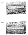
- Figs. 1A-1C Commercially available, woven materials that have been used to help repair soft tissue are illustrated in Figs. 1A-1C .
- the polypropylene mesh shown in the micrograph of Fig. 1A is Bard Mesh, a non-absorbable, knitted material produced by C.R. Bard, Inc. (Murray Hill, NJ); and the material shown in the micrograph of Fig. 1B is ProleneTM Mesh (Ethicon, Inc., Somerville, NJ); and the material shown in the micrograph of Fig. 1C is Gore-Tex Soft Tissue PatchTM, a non-absorbable implant of ePTFE produced by W.L. Gore & Associates, Inc. (Flagstaff, AZ);
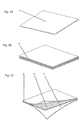
- Figs. 2A-2C are perspective views of materials that can be machined to produce a non-woven soft tissue implant of the present invention.
- Fig. 2A is a perspective view of non-woven biocompatible film 14.
- Film 14 has known or discernable dimensions (width, length, and thickness), which can be modified or left intact in the manufacture of a soft tissue implant.
- Film 14 is a single-layer, smooth-edged film.
- film 14 can be laminated to produce film 16, which can also be used, with or without further modification, to manufacture the implants of the present invention.
- Multiple layers of biocompatible film 14 can be added together to improve the mechanical properties (e.g ., tear resistance or burst strength) of the implant.
- a first film 14 can be thermally bonded to a second film 14 using hydraulic presses such as those manufactured by OEM Press Systems (Orange, CA).
- an implant can include laminated film 16, that includes two pieces of film 14 and tear resistant substrate 18. Tear resistant substrate 18 is placed between a first film 14 and a second film 14. Where tear resistant substrate 18 is thermally compatible with film 14, tear resistant substrate 18 and film 14 can be bonded using heat and/or pressure. If necessary, an adhesive or thermal attachment layer can be used between film 14 and tear resistant substrate 18. This may include a layer of material with a lower melting point, which can be achieved by reducing the crystallinity of a like material or by selecting a different material composition. Alternatively, tear resistant substrate 18 can be mechanically bonded to film 14 by sutures, clips, or the like.
- Biocompatible materials useful in film 14 or laminated film 16 can include non-absorbable polymers such as polypropylene, polyethylene, polyethylene terephthalate, polytetrafluoroethylene, polyaryletherketone, nylon, fluorinated ethylene propylene, polybutester, and silicone, or copolymers, thereof ( e.g ., a copolymer of polypropylene and polyethylene); absorbable polymers such as polyglycolic acid (PGA), polylactic acid (PLA), polycaprolactone, and polyhydroxyalkanoate, or copolymers thereof (e.g., a copolymer of PGA and PLA); and in addition tissue based materials (e.g., collagen or other biological material or tissue (e.g., mucosal or submucosal tissue) obtained from the patient who is to receive the implant or obtained from another person (e.g., a recently deceased person) or an animal (i.e., the implant can constitute a xenograft)
- the polymers can be of the D- isoform, the L-isoform, or a mixture of both.
- An example of a biocompatible film 14 suitable for producing the laminated film structure 16 is biaxially oriented polypropylene.
- AET Films (Peabody, MA) manufactures biaxially oriented films (AQS and OPB).
- Tear resistant substrate 18 can be spun bonded polypropylene, ePTFE, or a polymeric film compounded with impact modifiers.
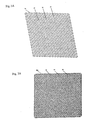
- Figs. 3A and 3B are perspective views of machined films 20 and 21, respectively.
- diamond-like cell pattern 22 has been machined into film 20 to impart porosity, which can support tissue ingrowth on high strength thin film substrates.
- Radius 24 has been applied to each cell pattern 22 corner to improve tear strength.
- Changing the dimensions of cell member 26 can alter the configuration of cell pattern 22.
- Different physical properties can be imparted along each axis of the film.
- Fig. 3B a perspective view of a machined film 21, tapered cell pattern 22 has been machined into the film to impart porosity, which can support tissue ingrowth. The ability to alter mechanical properties with tapered cell pattern 22 geometry is demonstrated.
- Manufacturing methods to impart patterns such as cell pattern 22 include, but are not limited to, laser machining, die punching, water jet cutting, and chemical etching.
- the lasers preferred for creating smooth edges on plastic films include, but are not limited to, CO 2 , diode ultraviolet, or excimer lasers.
- An implant having cell pattern 22 is expected to confer benefit to a patient in which it is implanted because of the substantially smooth edges of cell pattern 22.
- cell member 27 was created in biocompatible film 28.
- Atraumatic edge 29 lies at the interface between cell member 27 and biocompatible film 28.
- Cell member 27 was created using a 3.0-Watt Avia Q-switched Ultraviolet Laser (Coherent, Inc., Santa Clara, CA).
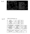
- a block diagram shows manufacturing steps for creating a non-woven soft tissue implant.
- the polymer used to construct the film is extruded using melt or paste extrusion techniques. After extrusion, the mechanical properties (e.g., tensile strength) can be improved through a biaxial stretching process (this is an optional step). Equipment that can be used to carry out this process can be purchased from Bruckner GmbH (Siegsdorf, Germany). If desired, the film can be laminated using heat, pressure, or adhesives to further improve the mechanical properties of the implant. Films with properties that may improve an implant (e.g ., films with increased tear strength) can be added at this step. A cell pattern (such as one described or illustrated herein) is machined into the film.
- the film can be annealed at elevated temperatures (e.g ., above the glass transition temperature for the polymer within the film) to relieve stresses caused by film stretching and the machining process.
- the material can then be cleaned, packaged, and sterilized.
- the packaging material can include instructions for use (i.e ., instructions can be printed on the packaging material); similarly, instructions can be provided on a separate material.
- FIG. 11 an exemplary pore having an opening of 2.54mm (0.100") and a wall thickness of 0.635mm (0.025 inches) is shown.
- a graph illustrates the percentage strain (x-axis) on various soft tissue implants including MarlexTM, ProleneTM, TrelexTM, Mesh2, and ePTFE.
- the cells within a soft tissue implant can be regularly shaped (as are the rectangular cells of Fig. 3A ) or irregularly shaped (i.e., they can have an irregularly shaped perimeter, as shown in Fig.6A , which mayor may not be symmetrical).
- the cell can be of a "regular" shape when it is essentially square, rectangular, or diamond-shaped, or essentially round or oval; the cell(s) can be of an "irregular" shape when at least one of the cell walls contains a sinusoidal element.
- each of the cells in the implant can have a plurality of undulating elements that form a repeating pattern (e.g., the undulations can be in phase with one another).
- the shape of the cells, their pattern, number, size, etc. can vary as described herein regardless of the film from which the implant is constructed (i.e., the cells can vary as described herein regardless of whether the film is non-porous or microporous; whether the implant contains a single film or multiple films; whether the film contains an absorbable or non-absorbable polymer; whether the implant contains a film to increase tear resistance; etc.).
- the length of an opening i.e., the distance between one part of the cell wall and another (e.g., the distance along the longest axis, the shortest axis, an intermediate axis; or the distance between two points that do not define an axis)
- the length of an opening can be between about 10 and about 10,000 microns (e.g., about 50-100 (e.g., about 75); about 10-1,000 (e.g., about 500); about 10-2,000 (e.g., about 1,200); about 10 - 5,000 (e.g., about 2,500); about 10-7,500 (e.g., about 4,500); about 100-1,000 (e.g., about 25 750); about 500-2,000 (e.g., about 1,750); about 1,000-3,000 (e.g., about 2,100); about 1,000 - 5,000 (e.g., about 3,500); about 1,500-5,000 (e.g., about 3,750) about 4,000-6,000 (e.g., about 4,750); about
- the cells of a soft tissue implant will be about 10-10,000 ⁇ ; about 1,500-5,000 ⁇ ; or about 50-100 ⁇ ( i.e ., the length across the longest axis of the cell can be about 100 ⁇ , 250 ⁇ , 500 ⁇ , 1,000 ⁇ or 2,000 ⁇ .
- Such implants e.g ., implants in which the longest length of a cellular opening is about 2,000 microns
- One or more of the cells in the plurality within an implant can have essentially the same shape as the cell shown herein as that of Mesh2, Mesh2C, Mesh3, or Mesh4.
- Finite element analysis can be used to design a cell or cell pattern that, when incorporated in a soft tissue implant, provides the implant with properties that approximate one or more of the properties of the soft tissue being repaired or replaced.
- Human skeletal muscle can exert 3-4 kg of tension per square centimeter of cross sectional area. Since many muscles in humans (or other animals, which may also be treated with a soft tissue implant described herein) have a relatively large cross-sectional area, the tension they develop is quite large.
- the gluteus maximus can exert a tension of 1200 kg, and the quadriceps can exert a tension of 360 kg. This difference is due to varying cross sectional areas.
- the non-woven soft tissue implants of the invention can be constructed so that their characteristics (e.g ., their strength characteristics) match those of the tissue(s) being replaced or repaired.
- the soft tissue implant can have force displacement characteristics that do not restrict tissue movement (e.g ., that do not restrict the contraction or stretching of a muscle to which the implant is attached) or that restrict such movement to a limited extent.
- a soft tissue implant can restrict tissue movement by less than 5%, less than 10%, less than 25%, or less than 50%.
- the force displacement character of a given implant can be calculated by measuring the percentage by which the implant is displaced (e.g ., the amount by which it "gives" relative to a resting configuration) under a given force.
- a soft tissue implant can be distended by about 25% (or more ( e.g ., 30, 35,40, 45, 50% or more)) at 16 N/cm (see Fig. 12 ).
- the number, shape, and arrangement of the plurality of cells and the thickness of the implant can be varied to impart force displacement characteristics that approximate those of the structure being repaired.
- the films can be made from a variety of polymers, including absorbable polymers.
- the rate at which one film (e.g ., a first film) is resorbed within a body can be different from the rate at which another film (e.g., a second film) is resorbed.
- a surface of the first film can adhere to a surface of the second film, and multi-layer implants can include a film that increases tear resistance (e.g., a porous biocompatible film).
- a soft tissue implant can also be defined by measured parameters such as the area of a cell (or pore; Ap (see the size ranges above), its perimeter (Pp), the area of a cell "unit” (Ac), and the surface area ratio (Asurf), which is less than 1.5.
- a method for calculating Asurf is shown in Fig. 6B , for example. Asurf is calculated by dividing Asu (the 3D surface area of a unit cell) by the area of the unit cell (Ac). Asu is determined by adding the top surface area (Atop), the bottom surface area (Abot; which can equal the top surface area), and the area of thickness (At).
- Atop is the difference between the area of a unit cell (Ac) and the area of space in a unit cell (As);
- Abot can equal Atop; and
- At equals the thickness of the film multiplied by (Pp + 4 (Pp/4)).
- Ap plus 4 (Ap/4) is equal to 2Ap).
- An implant can be made by a method that includes the steps of extruding a biocompatible polymer into a film and forming a plurality of cells in the film.
- the film can be of a thickness described above and have the material content described above, and the cells can have the characteristics of any of those described above.
- the extrusion process can be, for example, a melt or paste extrusion process, and the cells can be formed by, for example, laser ablation or machining (e.g., die punching).
- a soft tissue implant having more than one layer can be made by a method that includes the steps of (a) extruding a first biocompatible polymer to form a first film; (b) extruding a second biocompatible polymer to form a second film; (c) attaching the first film to the second film to produce a soft tissue implant and (d) forming pores in the soft tissue implant.
- a multi-layer implant can be made by a method including the steps of (a) extruding a first biocompatible polymer to form a first film; (b) forming pores or cell patterns in the first film; (c) extruding a second biocompatible polymer to form a second film; (d) forming pores in the second film; and (e) attaching the first film to the second film to produce a soft tissue implant.
- the films can be of a thickness described above and have the material content described above, and the cells can have the characteristics of any of those described above. Any of the soft tissue implants made by these methods can be further processed (e.
- edges can be modified to facilitate tissue placement and/or their shape can be changed (by, for example, stretching)).
- the implants can also be cleaned and/or sterilized and packaged, with or without instructions for use. Any of the soft tissue implants made by these methods can be used to repair, or in the course of repairing, a damaged tissue in a body (including, but not limited to, a human body).
- Medical implant applications for the soft tissue implant technology described above may include, but are not limited to, plastic reconstruction, urinary stress incontinence, hernia repair, gastric banding, and chest wall reconstruction. Accordingly, the invention may be used to treat a patient who has sustained an injury to a tissue, independent of the source of the injury (i.e ., the injury could arise from a traumatic injury, including an accidental injury or a surgical incision, or the injury may be associated with a disease, disorder, or condition).
- the method can include exposing, preferably under sterile conditions, the injured tissue (e.g ., a muscle, muscle group, or other tissue such as the intestine, liver, or kidney), and administering a soft tissue implant to the tissue.
- the implant can be further secured to the tissue by one or more sutures, staples, or other fasteners. Alternatively, or in addition, the implant can be secured by an adhesive.
- the surgical incision through which the implant was inserted can then be closed.
- the physician or surgeon performing the operative procedure can select an appropriate implant. For example, it will be readily apparent what size implant is required (generally, the implant should be large enough to cover the affected part of a tissue).
- the physician or surgeon can choose a non-absorbable implant when appropriate. For example, one may select a non-absorbable soft tissue implant for indications such as hernia repair that require long-term durability and strength.
- tissue augmentation during plastic reconstruction when one wants to avoid the potential complications associated with a permanent implant.
- Tissue-based materials are best suited for indications such as pelvic slings that require materials less prone to erosion into adjacent tissue structures.
- the soft tissue implant can be produced in more three-dimensional forms for certain indications, such as the plug and patch procedure for inguinal hernia repair.
- a three dimensional structure can be machined using a laser system incorporating a third axis for micromachining.
- the nonwoven soft tissue implant could be thermoformed into a three-dimensional shape after machining.
- Non-medical applications may include diagnostic testing, in biotechnology or other research, in automotive, electronics, aerospace, and home and commercial appliances.
- Example 1 A non-woven soft tissue implant was constructed using biaxially-oriented polymer films. The film is stretched in both the machine and transverse directions (relative to the extrusion direction) to orient the polymer chains. The stretching process can take place simultaneously or sequentially depending on the equipment that is available.
- the base film was SyncartaTM (AET Films, Peabody, MA).
- the base film was machined into Mesh Design 2 ("Mesh2”) using a 3.0-Watt Avia Q-switched Ultraviolet Laser produced by Coherent, Inc. (Santa Clara, CA).
- the design of a cell of the non-woven soft tissue implant is shown in Fig. 6A .
- the finished product was implanted, using standard surgical techniques, in the subcutaneous tissue of rats for 7, 14, and 28 days. Following sacrifice and retrieval of the specimens, histological evaluation was carried out to evaluate the inflammatory and wound healing response. Histology sections were obtained and stained with Haematoxylin and Eosin for cellularity of the implant site and Masson's Trichrome stain was used to evaluate the extent of fibrous capsule formation. The findings over a 28-day period indicate that the nonwoven soft tissue implant is biocompatible and undergoes a normal resolution of the inflammatory response, secondary to surgical injury, and development of a normal foreign body reaction at the material/tissue interface with fibrous capsule formation surrounding the entire implant and within the holes of the material. The results of histological analyses are shown in Figs. 7A and 7B .
- Example 2 A non-woven soft tissue implant was constructed using biaxially oriented polymer films. Two base films were used. The first film was a two-side sealable material OPB 95 (AET Films, Peabody, MA). The second film was a one-side sealable material AQS 90 (AET Films). Six sheets of the first film were placed between two sheets of the second film with the sealable side of the second in contact with the first film set. The sheet assembly was brought to 145° C at 27.586 bar (400 PSI) of constant pressure for 60 minutes under vacuum. The laminated assembly was machined into designs Mesh2 and Mesh4 (see Figs. 6A and 9A , respectively) using a 3.0-Watt Avia Q-switched Ultraviolet Laser produced by Coherent, Inc. (Santa Clara, CA).
- Example 3 A non-woven soft tissue implant was constructed using biaxially- oriented polymer films. Two base films were produced. The first film comprised a three layer extrusion in an A-B-A form. The "A” layer was made up from PKS409 resin (Solvay Polyolefins Europe, Brussels, Belgium) and the "B” layer was made up from HC312BF resin (Borealis Group, Kongens Lyngby, Denmark). The layers were melt extruded and oriented using a stenter .film process. The .film was oriented in the machine direction at a 5:1 1 ratio and in the transverse direction at a 10:1 1 ratio. The thickness of the film after stretching was 24 ⁇ .
- the second film included a three-layer extrusion in an AA-B form.
- the "A” layer was made up from HC312BF and the "B” layer was made from PKS409.
- the layers were melt extruded and oriented using a stenter film process.
- the film was oriented in the machine direction at a 5:1 ratio and in the transverse direction at a 10:1 1 ratio.
- the thickness of the film after stretching was 23 ⁇ .
- Six sheets of the first film were placed between two sheets of the second film with the "B" side in contact with the first film set.
- the sheet assembly was brought to 145° C at 27.586 bar (400 PSI) of constant pressure for 60 minutes under vacuum.
- the laminated assembly was machined into the design Mesh2C (see Fig.
- Example 4 Polyaryletherketone (PEEK; Invibio Inc., Lancashire, UK) is a polymer that has properties making it useful as an implant material for devices such as spine cages, bone screws, orthopedic stems, and dental implants. PEEK exhibits a desirable combination of strength, stiffness, and toughness, and it is biocompatible. Accordingly, a soft tissue implant was constructed using PEEK material. Westlake Plastics (Lenni, PA) supplies PEEK polymer films that range from about 0.0254mm (0.001 inches) to about 0.7366mm (0.029 inches) thick. These films can be used to fabricate biocompatible implants with lower profiles than commercially available textile based products.
- a film made of 0.127mm (0.005 inch) PEEK polymer was machined using an ultraviolet laser (more specifically, a 3.0-Watt Avia Q-switched Ultraviolet Laser (Coherent, Inc., Santa Clara, CA)) into the pattern shown in FIG. 6A using a CAD-CAM process.
- FIG. 4B shows a highly magnified image of a cell pattern edge created using the laser machining process.
- This soft tissue implant has an implant surface area ratio of 0.79, which reduces the amount of material available to provoke a foreign body reaction.
- the implant had a smooth surface with a low coefficient of friction.
- Example 5 Polytetrafluoroethylene (PTFE; Bard Vascular Systems (Tempe, AZ)) polymer also has properties that allow it to be used, as described herein, as an implant material for, for example, vascular grafts and patches.
- PTFE can be processed into a microporous form using an expansion procedure.
- expanded PTFE is strong, flexible, and biocompatible.
- Example 6 Yet another non-woven soft tissue implant was constructed using a biaxially-oriented polymer film.
- the film is stretched in both the machine and transverse directions (relative to the extrusion direction) to orient the polymer chains. As noted above, the stretching process can take place simultaneously or sequentially depending on the equipment that is available.
- the base film was SyncartaTM (AET Films, Peabody, MA).
- the base film was machined into Mesh Design 3 ("Mesh3") using a 3.0-Watt Avia Q-switched Ultraviolet Laser produced by Coherent, Inc. (Santa Clara, CA).
- the design of a cell of the non-woven soft tissue implant is shown in Fig. 10A .
Landscapes
- Health & Medical Sciences (AREA)
- Transplantation (AREA)
- Vascular Medicine (AREA)
- Life Sciences & Earth Sciences (AREA)
- Oral & Maxillofacial Surgery (AREA)
- Engineering & Computer Science (AREA)
- Biomedical Technology (AREA)
- Heart & Thoracic Surgery (AREA)
- Veterinary Medicine (AREA)
- Cardiology (AREA)
- Animal Behavior & Ethology (AREA)
- General Health & Medical Sciences (AREA)
- Public Health (AREA)
- Dermatology (AREA)
- Materials For Medical Uses (AREA)
- Prostheses (AREA)
- Separation Using Semi-Permeable Membranes (AREA)
Claims (32)
- Nicht gewebtes Weichgewebeimplantat, das einen porösen biokompatiblen Film umfasst, der ein Zellmuster aufweist, wobei das Zellmuster eine Vielzahl von Einheitszellen umfasst, wobei jede Einheitszelle eine Pore und einen Anteil des Films einschließt, der sich um den Umfang der Pore erstreckt, um die Pore zu definieren, und wobei der Film eine Dicke von weniger als etwa 0,381 mm (0,015 Zoll) aufweist, der poröse biokompatible Film ein nichtresorbierbares Polymer oder Copolymer umfasst oder der poröse biokompatible Film ein resorbierbares Polymer oder Copolymer umfasst, wobei das Polymer oder Copolymer biaxial orientierte Ketten hat, dadurch gekennzeichnet, dass das Implantat ein Oberflächenverhältnis von weniger als 1,5 aufweist,
wobei: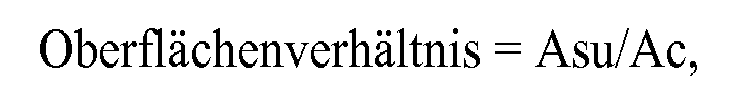
Asu = die dreidimensionale Oberfläche einer Einheitszelle,
Ac = die zweidimensionale Fläche der Einheitszelle. - Nicht gewebtes Weichgewebeimplantat nach Anspruch 1, worin das nichtresorbierbare Polymer oder Copolymer Folgende umfasst: Polypropylen, Polyethylenterephthalat, Polytetrafluorethylen, Polyaryletherketon, Nylon, fluoriertes Ethylenpropylen, Polybutester oder Silicon.
- Nicht gewebtes Weichgewebeimplantat nach Anspruch 1 oder 2, worin das resorbierbare Polymer oder Copolymer Folgende umfasst: Polyglycolsäure (PGA), Polymilchsäure (PLA), Polycaprolacton oder Polyhydroxyalkanoat.
- Nicht gewebtes Weichgewebeimplantat nach einem der Ansprüche 1 bis 3, worin eine oder mehrere der Zellen in der Vielzahl von Zellen einen Durchmesser, entlang der längsten Achse der Zelle gemessen, von etwa 10µ bis etwa 10 000µ haben.
- Nicht gewebtes Weichgewebeimplantat nach Anspruch 4, worin eine oder mehrere der Zellen in der Vielzahl von Zellen einen Durchmesser, entlang der längsten Achse der Zelle gemessen, von etwa 1 500 µ bis etwa 5 000 µ haben.
- Nicht gewebtes Weichgewebeimplantat nach Anspruch 4, worin eine oder mehrere der Zellen in der Vielzahl von Zellen einen Durchmesser, entlang der längsten Achse der Zelle gemessen, von etwa 50 µ bis etwa 100 µ haben.
- Nicht gewebtes Weichgewebeimplantat nach einem der Ansprüche 1 bis 6, worin eine oder mehrere der Zellen der Vielzahl im Wesentlichen quadratisch, rechteckig oder rautenförmig sind.
- Nicht gewebtes Weichgewebeimplantat nach einem der Ansprüche 1 bis 6, worin eine oder mehrere der Zellen der Vielzahl im Wesentlichen rund oder oval sind.
- Nicht gewebtes Weichgewebeimplantat nach einem der Ansprüche 1 bis 8, worin die Dicke des porösen biokompatiblen Films weniger als etwa 0,3556 mm (0,014 Zoll), weniger als etwa 0,3302 mm (0,013 Zoll), weniger als etwa 0,3048 mm (0,012 Zoll), weniger als etwa 0,2794 mm (0,011 Zoll), weniger als etwa 0,254 mm (0,010 Zoll), weniger als etwa 0,2286 mm (0,009 Zoll), weniger als etwa 0,2032 mm (0,008 Zoll), weniger als etwa 0,1778 mm (0,007 Zoll), weniger als etwa 0,1524 mm (0,006 Zoll), weniger als etwa 0,127 mm (0,005 Zoll), weniger als etwa 0,1016 mm (0,004 Zoll), weniger als etwa 0,0762 mm (0,003 Zoll), weniger als etwa 0,0508 mm (0,002 Zoll) beträgt oder etwa 0,0254 mm (0,001 Zoll) beträgt.
- Nicht gewebtes Weichgewebeimplantat nach einem der Ansprüche 1 bis 9, worin der poröse biokompatible Film atraumatische Kanten aufweist.
- Nicht gewebtes Weichgewebeimplantat nach einem der Ansprüche 1 bis 10, worin der poröse biokompatible Film längs einer ersten Seite mindestens etwa 2,5 cm lang und längs einer zweiten Seite nicht mehr als etwa 45,0 cm lang ist.
- Nicht gewebtes Weichgewebeimplantat nach einem der Ansprüche 1 bis 11, worin das Implantat entlang zweier Achsen flexibel ist.
- Nicht gewebtes Weichgewebeimplantat nach Anspruch 12, worin die Vielzahl von Zellen ein Zellmuster umfasst, das ein sinusförmiges Element enthält.
- Nicht gewebtes Weichgewebeimplantat nach Anspruch 12, worin jede der Zellen in der Vielzahl von Zellen eine Vielzahl von wellenförmigen Elementen in Form eines sich wiederholenden Musters aufweist, bevorzugt die wellenförmigen Elemente phasengleich sind.
- Nicht gewebtes Weichgewebeimplantat nach einem der Ansprüche 1 bis 14, worin die Zellen in der Vielzahl von Zellen einen Durchmesser von größer als 50 µ haben und das nicht gewebte Weichgewebeimplantat ein Kraft-Weg-Verhalten zeigt, das die Gewebebewegung nicht einschränkt, bevorzugt das Implantat um 25 % oder mehr bei 16 N/cm gedehnt werden kann, bevorzugt das Muster der Vielzahl von Zellen ein Kraft-Weg-Verhalten verleiht, das dem der zu reparierenden Struktur nahekommt.
- Nicht gewebtes Weichgewebeimplantat nach einem der Ansprüche 1 bis 15, worin der poröse biokompatible Film ein erster poröser biokompatibler Film ist und das Implantat einen zweiten porösen biokompatiblen Film umfasst, der ein Zellmuster aufweist, wobei das Zellmuster eine Vielzahl von Zellen umfasst, wobei jede Zelle eine Pore und einen Anteil des Films einschließt, der sich um den Umfang der Pore erstreckt, um die Pore zu definieren, wobei die Dicke des Implantats weniger als etwa 0,381 mm (0,015 Zoll) beträgt.
- Nicht gewebtes Weichgewebeimplantat nach Anspruch 16, worin der erste Film und der zweite Film aus dem gleichen Material oder den gleichen Materialien bestehen.
- Nicht gewebtes Weichgewebeimplantat nach Anspruch 16, worin der erste Film und der zweite Film aus unterschiedlichen Materialien bestehen, bevorzugt der erste Film oder der zweite Film ein bioresorbierbares Material einschließt, und die Rate, zu der der erste Film innerhalb eines Körpers resorbiert wird, verschieden von der Rate ist, zu der der zweite Film innerhalb des Körpers resorbiert wird.
- Nicht gewebtes Weichgewebeimplantat nach einem der Ansprüche 16 bis 18, worin der erste Film und der zweite Film im Wesentlichen von der gleichen Größe sind und eine Oberfläche des ersten Films an einer Oberfläche des zweiten Films haftet.
- Nicht gewebtes Weichgewebeimplantat nach einem der Ansprüche 16 bis 19, worin der zweite poröse biokompatible Film ein nichtresorbierbares Polymer oder Copolymer umfasst, bevorzugt das nichtresorbierbare Polymer oder Copolymer Folgende umfasst: Polypropylen, Polyethylenterephthalat, Polytetrafluorethylen, Polyaryletherketon, Nylon, fluoriertes Ethylenpropylen, Polybutester, Silicon, Polyethylen oder ein Copolymer aus Polyethylen und Polypropylen.
- Nicht gewebtes Weichgewebeimplantat nach einem der Ansprüche 16 bis 20, worin der zweite poröse biokompatible Film ein resorbierbares Polymer oder Copolymer umfasst, bevorzugt das resorbierbare Polymer oder Copolymer PGA, PLA, Polycaprolacton oder Polyhydroxyalkanoat ist.
- Nicht gewebtes Weichgewebeimplantat nach einem der Ansprüche 16 bis 21, worin der zweite poröse biokompatible Film ein biologisches Material umfasst, bevorzugt das biologische Material Kollagen ist.
- Nicht gewebtes Weichgewebeimplantat nach einem der Ansprüche 16 bis 22, worin der zweite Film eine Zelle umfasst, die einen Durchmesser, entlang der längsten Achse der Zelle gemessen, von etwa 10 µ bis etwa 10 000 µ, von etwa 1 500 µ bis etwa 5 000 µ oder von etwa 50 µ bis etwa 100 µ hat.
- Nicht gewebtes Weichgewebeimplantat nach einem der Ansprüche 16 bis 23, worin der zweite Film eine Zelle umfasst, die im Wesentlichen quadratisch, rechteckig oder rautenförmig ist.
- Nicht gewebtes Weichgewebeimplantat nach einem der Ansprüche 16 bis 23, worin der zweite Film eine Zelle umfasst, die im Wesentlichen rund oder oval ist.
- Nicht gewebtes Weichgewebeimplantat nach einem der Ansprüche 1 bis 25, worin die Dicke des Implantats weniger als etwa 0,3556 mm (0,014 Zoll), weniger als etwa 0,3302 mm (0,013 Zoll), weniger als etwa 0,3048 mm (0,012 Zoll), weniger als etwa 0,2794 mm (0,011 Zoll), weniger als etwa 0,254 mm (0,010 Zoll), weniger als etwa 0,2286 mm (0,009 Zoll), weniger als etwa 0,2032 mm (0,008 Zoll), weniger als etwa 0,1778 mm (0,007 Zoll), weniger als etwa 0,1524 mm (0,006 Zoll), weniger als etwa 0,127 mm (0,005 Zoll), weniger als etwa 0,1016 mm (0,004 Zoll), weniger als etwa 0,0762 mm (0,003 Zoll), weniger als etwa 0,0508 mm (0,002 Zoll) beträgt oder etwa 0,0254 mm (0,001 Zoll) beträgt.
- Nicht gewebtes Weichgewebeimplantat nach einem der Ansprüche 16 bis 26, worin der erste und zweite Film atraumatische Kanten aufweisen.
- Nicht gewebtes Weichgewebeimplantat nach einem der Ansprüche 1 bis 27, worin das Implantat längs einer ersten Seite mindestens 2,5 cm lang und längs einer zweiten Seite nicht mehr als 30,0 cm lang ist.
- Nicht gewebtes Weichgewebeimplantat nach einem der Ansprüche 1 bis 28, das weiterhin einen Film umfasst, der die Reißfestigkeit erhöht, wobei der Film, der die Reißfestigkeit erhöht, bevorzugt ein poröser biokompatibler Film ist.
- Nicht gewebtes Weichgewebeimplantat nach einem der Ansprüche 16 bis 29, worin die Vielzahl von Zellen im zweiten biokompatiblen Film ein Zellmuster umfasst, das ein sinusförmiges Element enthält.
- Nicht gewebtes Weichgewebeimplantat nach einem der Ansprüche 16 bis 30, worin jede der Zellen in der Vielzahl von Zellen im zweiten biokompatiblen Film eine Vielzahl von wellenförmigen Elementen in Form eines sich wiederholenden Musters aufweist, bevorzugt die wellenförmigen Elemente phasengleich sind.
- Nicht gewebtes Weichgewebeimplantat nach einem der Ansprüche 16 bis 31, worin die Zellen in der Vielzahl von Zellen im zweiten biokompatiblen Film einen Durchmesser von größer als 50 µ haben und das nicht gewebte Weichgewebeimplantat ein Kraft-Weg-Verhalten zeigt, das die Gewebebewegung nicht einschränkt, wenn es in einen Körper eingebracht wird, bevorzugt das Implantat um 25 % oder mehr bei 16 N/cm gedehnt werden kann, bevorzugt das Muster der Vielzahl von Zellen ein Kraft-Weg-Verhalten verleiht, das dem der zu reparierenden Struktur nahekommt.
Applications Claiming Priority (3)
| Application Number | Priority Date | Filing Date | Title |
|---|---|---|---|
| US39678102P | 2002-07-17 | 2002-07-17 | |
| US396781P | 2002-07-17 | ||
| PCT/US2003/022457 WO2004006808A2 (en) | 2002-07-17 | 2003-07-17 | Soft tissue implants and methods for making same |
Publications (3)
| Publication Number | Publication Date |
|---|---|
| EP1539044A2 EP1539044A2 (de) | 2005-06-15 |
| EP1539044A4 EP1539044A4 (de) | 2006-09-27 |
| EP1539044B1 true EP1539044B1 (de) | 2011-04-06 |
Family
ID=30116054
Family Applications (1)
| Application Number | Title | Priority Date | Filing Date |
|---|---|---|---|
| EP03764798A Expired - Lifetime EP1539044B1 (de) | 2002-07-17 | 2003-07-17 | Membrane für medizinische Implantation |
Country Status (7)
| Country | Link |
|---|---|
| US (3) | US20040059356A1 (de) |
| EP (1) | EP1539044B1 (de) |
| AT (1) | ATE504262T1 (de) |
| AU (1) | AU2003249310A1 (de) |
| DE (1) | DE60336658D1 (de) |
| ES (1) | ES2363319T3 (de) |
| WO (1) | WO2004006808A2 (de) |
Cited By (1)
| Publication number | Priority date | Publication date | Assignee | Title |
|---|---|---|---|---|
| US9974640B2 (en) | 2011-09-22 | 2018-05-22 | Boston Scientific Scimed, Inc. | Pelvic implant and treatment method |
Families Citing this family (97)
| Publication number | Priority date | Publication date | Assignee | Title |
|---|---|---|---|---|
| FR2811218B1 (fr) | 2000-07-05 | 2003-02-28 | Patrice Suslian | Dispositif implantable destine a corriger l'incontinence urinaire |
| US20060205995A1 (en) | 2000-10-12 | 2006-09-14 | Gyne Ideas Limited | Apparatus and method for treating female urinary incontinence |
| GB0025068D0 (en) | 2000-10-12 | 2000-11-29 | Browning Healthcare Ltd | Apparatus and method for treating female urinary incontinence |
| US8167785B2 (en) | 2000-10-12 | 2012-05-01 | Coloplast A/S | Urethral support system |
| GB0108088D0 (en) | 2001-03-30 | 2001-05-23 | Browning Healthcare Ltd | Surgical implant |
| DE10155842A1 (de) * | 2001-11-14 | 2003-05-28 | Ethicon Gmbh | Flächiges Implantat |
| ATE504262T1 (de) | 2002-07-17 | 2011-04-15 | Proxy Biomedical Ltd | Membrane für medizinische implantation |
| CA2492630C (en) | 2002-08-02 | 2009-01-13 | C.R. Bard, Inc. | Self anchoring sling and introducer system |
| ATE287307T1 (de) | 2002-11-08 | 2005-02-15 | Howmedica Osteonics Corp | Lasererzeugte poröse oberfläche |
| US20060147332A1 (en) | 2004-12-30 | 2006-07-06 | Howmedica Osteonics Corp. | Laser-produced porous structure |
| GB0307082D0 (en) | 2003-03-27 | 2003-04-30 | Gyne Ideas Ltd | Drug delivery device and method |
| US8298292B2 (en) | 2003-04-16 | 2012-10-30 | Howmedica Osteonics Corp. | Craniofacial implant |
| BRPI0409487A (pt) | 2003-04-16 | 2006-05-02 | Porex Surgical Inc | implante cirúrgico, processo para sua preparação e método de reconstrução de um defeito ósseo |
| US20050136764A1 (en) * | 2003-12-18 | 2005-06-23 | Sherman Michael C. | Designed composite degradation for spinal implants |
| GB0411360D0 (en) | 2004-05-21 | 2004-06-23 | Mpathy Medical Devices Ltd | Implant |
| US7297102B2 (en) * | 2004-07-28 | 2007-11-20 | Ethicon, Inc. | Minimally invasive medical implant and insertion device and method for using the same |
| US7641688B2 (en) * | 2004-09-16 | 2010-01-05 | Evera Medical, Inc. | Tissue augmentation device |
| US8796015B2 (en) | 2004-11-09 | 2014-08-05 | Proxy Biomedical Limited | Tissue scaffold |
| US20060235525A1 (en) * | 2005-04-19 | 2006-10-19 | Sdgi Holdings, Inc. | Composite structure for biomedical implants |
| EP1937183B1 (de) | 2005-09-12 | 2018-11-28 | Proxy Biomedical Limited | Weichgewebeimplantate |
| GB2430372B (en) * | 2005-09-19 | 2010-09-29 | Stephen George Edward Barker | Reinforcement device |
| US8728387B2 (en) | 2005-12-06 | 2014-05-20 | Howmedica Osteonics Corp. | Laser-produced porous surface |
| US7670762B2 (en) * | 2006-01-17 | 2010-03-02 | Brennen Medical, Llc | Biocompatible tissue graft material for implant and method of making |
| US8147861B2 (en) | 2006-08-15 | 2012-04-03 | Howmedica Osteonics Corp. | Antimicrobial implant |
| US7614258B2 (en) | 2006-10-19 | 2009-11-10 | C.R. Bard, Inc. | Prosthetic repair fabric |
| FR2908290B3 (fr) * | 2006-11-10 | 2009-01-09 | Symatese Soc Par Actions Simpl | Dispositif destine a la regeneration du derme humain et procede de fabrication dudit dispositif |
| FR2908289B1 (fr) * | 2006-11-10 | 2009-01-23 | Symatese Soc Par Actions Simpl | Dispositif destine a la regeneration du derme et procede de fabrication dudit dispositif. |
| US8932619B2 (en) * | 2007-06-27 | 2015-01-13 | Sofradim Production | Dural repair material |
| US20090004455A1 (en) * | 2007-06-27 | 2009-01-01 | Philippe Gravagna | Reinforced composite implant |
| DE102007037051A1 (de) * | 2007-07-24 | 2009-01-29 | Aesculap Ag | Flächiges Implantat |
| US8142886B2 (en) | 2007-07-24 | 2012-03-27 | Howmedica Osteonics Corp. | Porous laser sintered articles |
| US20090068250A1 (en) | 2007-09-07 | 2009-03-12 | Philippe Gravagna | Bioresorbable and biocompatible compounds for surgical use |
| EP2200672B1 (de) | 2007-09-11 | 2012-06-27 | Solvay Specialty Polymers USA, LLC. | Verbesserte prothesenvorrichtungen |
| WO2009057371A1 (ja) * | 2007-10-31 | 2009-05-07 | Yasuhiro Fukuhara | 歯科印象用フィルム及び歯科印象用フィルムの製造方法 |
| US9308068B2 (en) | 2007-12-03 | 2016-04-12 | Sofradim Production | Implant for parastomal hernia |
| US8758373B2 (en) | 2008-02-18 | 2014-06-24 | Covidien Lp | Means and method for reversibly connecting a patch to a patch deployment device |
| US9398944B2 (en) | 2008-02-18 | 2016-07-26 | Covidien Lp | Lock bar spring and clip for implant deployment device |
| US9393002B2 (en) | 2008-02-18 | 2016-07-19 | Covidien Lp | Clip for implant deployment device |
| WO2009104182A2 (en) | 2008-02-18 | 2009-08-27 | Polytouch Medical Ltd | A device and method for deploying and attaching a patch to a biological tissue |
| US8808314B2 (en) | 2008-02-18 | 2014-08-19 | Covidien Lp | Device and method for deploying and attaching an implant to a biological tissue |
| US9833240B2 (en) | 2008-02-18 | 2017-12-05 | Covidien Lp | Lock bar spring and clip for implant deployment device |
| US9301826B2 (en) | 2008-02-18 | 2016-04-05 | Covidien Lp | Lock bar spring and clip for implant deployment device |
| US9044235B2 (en) | 2008-02-18 | 2015-06-02 | Covidien Lp | Magnetic clip for implant deployment device |
| US8317808B2 (en) | 2008-02-18 | 2012-11-27 | Covidien Lp | Device and method for rolling and inserting a prosthetic patch into a body cavity |
| US9393093B2 (en) | 2008-02-18 | 2016-07-19 | Covidien Lp | Clip for implant deployment device |
| US9034002B2 (en) | 2008-02-18 | 2015-05-19 | Covidien Lp | Lock bar spring and clip for implant deployment device |
| US9242026B2 (en) * | 2008-06-27 | 2016-01-26 | Sofradim Production | Biosynthetic implant for soft tissue repair |
| AU2009305958B9 (en) | 2008-10-20 | 2013-07-11 | Covidien Lp | A device for attaching a patch to a biological tissue |
| US20120010636A1 (en) * | 2009-02-11 | 2012-01-12 | Nanyang Technological University | Multi-layered surgical prosthesis |
| US8906045B2 (en) | 2009-08-17 | 2014-12-09 | Covidien Lp | Articulating patch deployment device and method of use |
| US9999424B2 (en) | 2009-08-17 | 2018-06-19 | Covidien Lp | Means and method for reversibly connecting an implant to a deployment device |
| FR2949688B1 (fr) | 2009-09-04 | 2012-08-24 | Sofradim Production | Tissu avec picots revetu d'une couche microporeuse bioresorbable |
| US8992551B2 (en) * | 2010-01-28 | 2015-03-31 | Boston Scientific Scimed, Inc. | Composite surgical implants for soft tissue repair |
| FR2972626B1 (fr) | 2011-03-16 | 2014-04-11 | Sofradim Production | Prothese comprenant un tricot tridimensionnel et ajoure |
| FR2977790B1 (fr) | 2011-07-13 | 2013-07-19 | Sofradim Production | Prothese pour hernie ombilicale |
| FR2977789B1 (fr) | 2011-07-13 | 2013-07-19 | Sofradim Production | Prothese pour hernie ombilicale |
| CA2849052C (en) | 2011-09-30 | 2019-11-05 | Sofradim Production | Reversible stiffening of light weight mesh |
| CA2847615A1 (en) | 2011-09-30 | 2013-04-04 | Sofradim Production | Multilayer implants for delivery of therapeutic agents |
| FR2985271B1 (fr) | 2011-12-29 | 2014-01-24 | Sofradim Production | Tricot a picots |
| FR2985170B1 (fr) | 2011-12-29 | 2014-01-24 | Sofradim Production | Prothese pour hernie inguinale |
| US9364896B2 (en) | 2012-02-07 | 2016-06-14 | Medical Modeling Inc. | Fabrication of hybrid solid-porous medical implantable devices with electron beam melting technology |
| EP3517145A1 (de) | 2012-02-08 | 2019-07-31 | Boston Scientific Scimed, Inc. | Chirurgische gerüste |
| US9180010B2 (en) | 2012-04-06 | 2015-11-10 | Howmedica Osteonics Corp. | Surface modified unit cell lattice structures for optimized secure freeform fabrication |
| US9135374B2 (en) | 2012-04-06 | 2015-09-15 | Howmedica Osteonics Corp. | Surface modified unit cell lattice structures for optimized secure freeform fabrication |
| USD681813S1 (en) | 2012-05-24 | 2013-05-07 | Ams Research Corporation | Implant |
| USD694886S1 (en) | 2012-05-24 | 2013-12-03 | Ams Research Corporation | Implant |
| USD685476S1 (en) | 2012-05-25 | 2013-07-02 | Ams Research Corporation | Implant |
| USD680649S1 (en) | 2012-05-25 | 2013-04-23 | Ams Research Corporation | Implant |
| FR2994185B1 (fr) | 2012-08-02 | 2015-07-31 | Sofradim Production | Procede de preparation d’une couche poreuse a base de chitosane |
| FR2995779B1 (fr) | 2012-09-25 | 2015-09-25 | Sofradim Production | Prothese comprenant un treillis et un moyen de consolidation |
| FR2995788B1 (fr) | 2012-09-25 | 2014-09-26 | Sofradim Production | Patch hemostatique et procede de preparation |
| FR2995778B1 (fr) | 2012-09-25 | 2015-06-26 | Sofradim Production | Prothese de renfort de la paroi abdominale et procede de fabrication |
| CA2880380C (en) | 2012-09-28 | 2020-09-15 | Sofradim Production | Packaging for a hernia repair device |
| WO2014179720A1 (en) | 2013-05-02 | 2014-11-06 | University Of South Florida | Implantable sonic windows |
| FR3006581B1 (fr) | 2013-06-07 | 2016-07-22 | Sofradim Production | Prothese a base d’un textile pour voie laparoscopique |
| FR3006578B1 (fr) | 2013-06-07 | 2015-05-29 | Sofradim Production | Prothese a base d’un textile pour voie laparoscopique |
| US10343385B2 (en) * | 2013-07-18 | 2019-07-09 | Boston Scientific Scimed, Inc. | Bodily implant |
| EP3086820B1 (de) | 2013-12-26 | 2020-10-28 | Tepha, Inc. | Medizinische implantate mit laminaten aus poly-4-hydroxybutyrat und copolymeren davon |
| EP3000489B1 (de) | 2014-09-24 | 2017-04-05 | Sofradim Production | Verfahren zur Herstellung einer Antihaft-Sperrschicht |
| EP3000432B1 (de) | 2014-09-29 | 2022-05-04 | Sofradim Production | Textilbasierte Prothese für die Behandlung von Leistenbruch |
| EP3000433B1 (de) | 2014-09-29 | 2022-09-21 | Sofradim Production | Vorrichtung zur Einführung einer Prothese zur Hernie-Behandlung in einen Einschnitt und flexible textile Prothese |
| EP3029189B1 (de) | 2014-12-05 | 2021-08-11 | Sofradim Production | Prothetisches poröses Netz, sein Herstellungsverfahren und Hernienprothese |
| EP3059255B1 (de) | 2015-02-17 | 2020-05-13 | Sofradim Production | Verfahren zur Herstellung einer Matrix auf Chitosanbasis mit faseroptischem Verstärkungselement |
| EP3085337B1 (de) | 2015-04-24 | 2022-09-14 | Sofradim Production | Prothese zur unterstützung einer bruststruktur |
| EP3106185B1 (de) | 2015-06-19 | 2018-04-25 | Sofradim Production | Synthetische prothese mit einem gewirk und einem nichtporösen film und verfahren zur formung davon |
| US10130738B2 (en) | 2015-08-31 | 2018-11-20 | Ethicon Llc | Adjunct material to promote tissue growth |
| EP3195830B1 (de) | 2016-01-25 | 2020-11-18 | Sofradim Production | Prothese zur hernienreparatur |
| EP3312325B1 (de) | 2016-10-21 | 2021-09-22 | Sofradim Production | Verfahren zur herstellung eines netzes mit einem daran befestigten nahtmaterial mit widerhaken und dadurch erhaltenes netz |
| EP3398554B1 (de) | 2017-05-02 | 2025-06-25 | Sofradim Production | Prothese zur leistenbruch reparatur |
| US11298747B2 (en) | 2017-05-18 | 2022-04-12 | Howmedica Osteonics Corp. | High fatigue strength porous structure |
| CA3079532A1 (en) | 2017-10-19 | 2019-04-25 | C.R.Bard, Inc. | Self-gripping hernia prosthesis |
| EP3765102B1 (de) | 2018-03-13 | 2024-05-15 | Institut Químic de Sarrià CETS Fundació Privada | Gefässreparaturpflaster |
| CA3099695A1 (en) * | 2018-05-07 | 2019-11-14 | Mentor Worldwide Llc | 2d and 3d bioscaffold extracellular structural unit and tissue structure design and methods of manufacture |
| CN108721771B (zh) * | 2018-06-29 | 2021-12-28 | 四川大学 | 一种用于小动物软组织再生的皮下硅胶管装置及应用 |
| EP3653171B1 (de) | 2018-11-16 | 2024-08-21 | Sofradim Production | Implantate zur weichgewebereparatur |
| US12064330B2 (en) | 2020-04-28 | 2024-08-20 | Covidien Lp | Implantable prothesis for minimally invasive hernia repair |
| CN114748208B (zh) * | 2022-04-15 | 2024-01-12 | 柔脉医疗(深圳)有限公司 | 一种可原位检测多种化学、生物成分的组织工程支架 |
Family Cites Families (44)
| Publication number | Priority date | Publication date | Assignee | Title |
|---|---|---|---|---|
| US3463158A (en) * | 1963-10-31 | 1969-08-26 | American Cyanamid Co | Polyglycolic acid prosthetic devices |
| US4060081A (en) * | 1975-07-15 | 1977-11-29 | Massachusetts Institute Of Technology | Multilayer membrane useful as synthetic skin |
| JPS6037734B2 (ja) * | 1978-10-12 | 1985-08-28 | 住友電気工業株式会社 | 管状臓器補綴材及びその製造方法 |
| US4787906A (en) * | 1987-03-02 | 1988-11-29 | Haris Andras G | Controlled tissue growth and graft containment |
| NL8801741A (nl) * | 1988-07-08 | 1990-02-01 | Utermoehlen Nv | Kunsthuid. |
| US5011494A (en) * | 1988-09-16 | 1991-04-30 | Clemson University | Soft tissue implant with micron-scale surface texture to optimize anchorage |
| IT1248934B (it) * | 1990-06-01 | 1995-02-11 | Fidia Spa | Membrane forate biocompatibili,processi per la loro preparazione,loro impiego come supporto per la crescita in vitro di cellule epiteliali, pelli artificiali cosi' ottenute e loro impiego nei trapianti di pelle |
| US5356423A (en) * | 1991-01-04 | 1994-10-18 | American Medical Systems, Inc. | Resectable self-expanding stent |
| US5512600A (en) * | 1993-01-15 | 1996-04-30 | Massachusetts Institute Of Technology | Preparation of bonded fiber structures for cell implantation |
| US5423826A (en) * | 1993-02-05 | 1995-06-13 | Danek Medical, Inc. | Anterior cervical plate holder/drill guide and method of use |
| CA2155324C (en) | 1993-02-18 | 1999-03-16 | Layne L. Moen | Macroscopically perforated porous polytetrafluoroethylene materials |
| US5468242A (en) * | 1993-11-19 | 1995-11-21 | Leibinger Gmbh | Form-fitting mesh implant |
| US5634931A (en) * | 1994-09-29 | 1997-06-03 | Surgical Sense, Inc. | Hernia mesh patches and methods of their use |
| US5733337A (en) * | 1995-04-07 | 1998-03-31 | Organogenesis, Inc. | Tissue repair fabric |
| DE19613730C2 (de) * | 1996-03-26 | 2002-08-14 | Ethicon Gmbh | Flächiges Implantat zum Verstärken oder Verschließen von Körpergewebe |
| DE69702281T2 (de) * | 1996-04-16 | 2001-02-22 | Medtronic, Inc. | Geschweisster sinuswellenförmiger Stent |
| US5885829A (en) * | 1996-05-28 | 1999-03-23 | The Regents Of The University Of Michigan | Engineering oral tissues |
| US5766176A (en) * | 1996-09-11 | 1998-06-16 | Walter Lorenz Surgical, Inc. | Formable mesh |
| US5980540A (en) * | 1997-04-11 | 1999-11-09 | Kinamed, Inc. | Perforated cover for covering spaces in the cranium and conforming to the shape of the cranium |
| US6120539A (en) * | 1997-05-01 | 2000-09-19 | C. R. Bard Inc. | Prosthetic repair fabric |
| US6042592A (en) * | 1997-08-04 | 2000-03-28 | Meadox Medicals, Inc. | Thin soft tissue support mesh |
| DE19746396A1 (de) * | 1997-10-21 | 1999-05-06 | Howmedica Leibinger Gmbh & Co | Gitter für die Fixierung von Knochenteilen oder für die Überbrückung von Knochenfehlstellen |
| US6319264B1 (en) * | 1998-04-03 | 2001-11-20 | Bionx Implants Oy | Hernia mesh |
| US6547821B1 (en) * | 1998-07-16 | 2003-04-15 | Cardiothoracic Systems, Inc. | Surgical procedures and devices for increasing cardiac output of the heart |
| US6328765B1 (en) * | 1998-12-03 | 2001-12-11 | Gore Enterprise Holdings, Inc. | Methods and articles for regenerating living tissue |
| US6258099B1 (en) * | 1999-03-31 | 2001-07-10 | Scimed Life Systems, Inc. | Stent security balloon/balloon catheter |
| DE19942611C1 (de) * | 1999-08-31 | 2001-07-05 | Ethicon Gmbh | Verstärktes flächiges Implantat |
| US6306079B1 (en) * | 1999-12-07 | 2001-10-23 | Arnaldo F. Trabucco | Mesh pubovaginal sling |
| US6355058B1 (en) * | 1999-12-30 | 2002-03-12 | Advanced Cardiovascular Systems, Inc. | Stent with radiopaque coating consisting of particles in a binder |
| US6432138B1 (en) * | 2000-03-07 | 2002-08-13 | Promatrx, Inc. | Controlled porosity 3-D fabric breast prosthesis |
| US6425924B1 (en) * | 2000-03-31 | 2002-07-30 | Ethicon, Inc. | Hernia repair prosthesis |
| US6296607B1 (en) * | 2000-10-20 | 2001-10-02 | Praxis, Llc. | In situ bulking device |
| US6852330B2 (en) * | 2000-12-21 | 2005-02-08 | Depuy Mitek, Inc. | Reinforced foam implants with enhanced integrity for soft tissue repair and regeneration |
| US6574497B1 (en) * | 2000-12-22 | 2003-06-03 | Advanced Cardiovascular Systems, Inc. | MRI medical device markers utilizing fluorine-19 |
| US20050261780A1 (en) * | 2001-06-08 | 2005-11-24 | Harri Heino | Form-fitting bioabsorbable mesh implant |
| US20050147656A1 (en) * | 2001-06-14 | 2005-07-07 | Hemcon, Inc. | Tissue dressing assemblies, systems, and methods formed from hydrophilic polymer sponge structures such as chitosan |
| US6585755B2 (en) * | 2001-06-29 | 2003-07-01 | Advanced Cardiovascular | Polymeric stent suitable for imaging by MRI and fluoroscopy |
| WO2003049600A2 (en) * | 2001-12-06 | 2003-06-19 | Stx Medical, Inc. | Medical device |
| ATE504262T1 (de) | 2002-07-17 | 2011-04-15 | Proxy Biomedical Ltd | Membrane für medizinische implantation |
| EP1545390A4 (de) | 2002-08-23 | 2007-05-09 | Proxy Biomedical Ltd | Dreidimensionales implantat |
| CN100560439C (zh) * | 2003-04-16 | 2009-11-18 | 美佐公司 | 含β球晶的挤出聚丙烯片材 |
| US20050113849A1 (en) * | 2003-11-26 | 2005-05-26 | Nicholas Popadiuk | Prosthetic repair device |
| US20050149032A1 (en) * | 2003-12-30 | 2005-07-07 | Douglas Vaughen | Resorbable surgical fixation device |
| US7189263B2 (en) * | 2004-02-03 | 2007-03-13 | Vita Special Purpose Corporation | Biocompatible bone graft material |
-
2003
- 2003-07-17 AT AT03764798T patent/ATE504262T1/de not_active IP Right Cessation
- 2003-07-17 US US10/621,941 patent/US20040059356A1/en not_active Abandoned
- 2003-07-17 WO PCT/US2003/022457 patent/WO2004006808A2/en not_active Ceased
- 2003-07-17 ES ES03764798T patent/ES2363319T3/es not_active Expired - Lifetime
- 2003-07-17 AU AU2003249310A patent/AU2003249310A1/en not_active Abandoned
- 2003-07-17 EP EP03764798A patent/EP1539044B1/de not_active Expired - Lifetime
- 2003-07-17 DE DE60336658T patent/DE60336658D1/de not_active Expired - Lifetime
-
2013
- 2013-07-03 US US13/934,738 patent/US9788930B2/en not_active Expired - Fee Related
-
2017
- 2017-01-31 US US15/420,792 patent/US20170135799A1/en not_active Abandoned
Cited By (1)
| Publication number | Priority date | Publication date | Assignee | Title |
|---|---|---|---|---|
| US9974640B2 (en) | 2011-09-22 | 2018-05-22 | Boston Scientific Scimed, Inc. | Pelvic implant and treatment method |
Also Published As
| Publication number | Publication date |
|---|---|
| WO2004006808A3 (en) | 2004-03-11 |
| US20170135799A1 (en) | 2017-05-18 |
| WO2004006808A2 (en) | 2004-01-22 |
| ES2363319T3 (es) | 2011-07-29 |
| AU2003249310A1 (en) | 2004-02-02 |
| WO2004006808A8 (en) | 2004-07-29 |
| ATE504262T1 (de) | 2011-04-15 |
| DE60336658D1 (de) | 2011-05-19 |
| EP1539044A2 (de) | 2005-06-15 |
| US20140178997A1 (en) | 2014-06-26 |
| US9788930B2 (en) | 2017-10-17 |
| US20040059356A1 (en) | 2004-03-25 |
| EP1539044A4 (de) | 2006-09-27 |
Similar Documents
| Publication | Publication Date | Title |
|---|---|---|
| EP1539044B1 (de) | Membrane für medizinische Implantation | |
| EP1937183B1 (de) | Weichgewebeimplantate | |
| US11013587B2 (en) | Surgical implant | |
| RU2689763C2 (ru) | Хирургический имплантат | |
| KR102234626B1 (ko) | 외과용 임플란트 | |
| EP1771124B1 (de) | Behandeltes chirurgisches mesh aus monofilen fasern | |
| US8562633B2 (en) | Tissue repair device with a bioabsorbable support member | |
| US20060106419A1 (en) | Three dimensional implant | |
| US20120071707A1 (en) | Medical device | |
| WO2010141252A1 (en) | Incision closure device and method | |
| IE20050170A1 (en) | A soft tissue implant for the repair of injured or otherwise defective tissue | |
| IE20050171A1 (en) | An implant for treating urinary incontinence and/or pelvic floor prolapse |
Legal Events
| Date | Code | Title | Description |
|---|---|---|---|
| PUAI | Public reference made under article 153(3) epc to a published international application that has entered the european phase |
Free format text: ORIGINAL CODE: 0009012 |
|
| 17P | Request for examination filed |
Effective date: 20050128 |
|
| AK | Designated contracting states |
Kind code of ref document: A2 Designated state(s): AT BE BG CH CY CZ DE DK EE ES FI FR GB GR HU IE IT LI LU MC NL PT RO SE SI SK TR |
|
| AX | Request for extension of the european patent |
Extension state: AL LT LV MK |
|
| DAX | Request for extension of the european patent (deleted) | ||
| A4 | Supplementary search report drawn up and despatched |
Effective date: 20060829 |
|
| 17Q | First examination report despatched |
Effective date: 20071213 |
|
| R17C | First examination report despatched (corrected) |
Effective date: 20080709 |
|
| GRAP | Despatch of communication of intention to grant a patent |
Free format text: ORIGINAL CODE: EPIDOSNIGR1 |
|
| RTI1 | Title (correction) |
Free format text: FILM FOR MEDICAL IMPLANTATION |
|
| GRAS | Grant fee paid |
Free format text: ORIGINAL CODE: EPIDOSNIGR3 |
|
| GRAA | (expected) grant |
Free format text: ORIGINAL CODE: 0009210 |
|
| AK | Designated contracting states |
Kind code of ref document: B1 Designated state(s): AT BE BG CH CY CZ DE DK EE ES FI FR GB GR HU IE IT LI LU MC NL PT RO SE SI SK TR |
|
| REG | Reference to a national code |
Ref country code: GB Ref legal event code: FG4D |
|
| REG | Reference to a national code |
Ref country code: CH Ref legal event code: EP |
|
| REG | Reference to a national code |
Ref country code: IE Ref legal event code: FG4D |
|
| REF | Corresponds to: |
Ref document number: 60336658 Country of ref document: DE Date of ref document: 20110519 Kind code of ref document: P |
|
| REG | Reference to a national code |
Ref country code: DE Ref legal event code: R096 Ref document number: 60336658 Country of ref document: DE Effective date: 20110519 |
|
| REG | Reference to a national code |
Ref country code: NL Ref legal event code: VDEP Effective date: 20110406 |
|
| REG | Reference to a national code |
Ref country code: ES Ref legal event code: FG2A Ref document number: 2363319 Country of ref document: ES Kind code of ref document: T3 Effective date: 20110729 |
|
| PG25 | Lapsed in a contracting state [announced via postgrant information from national office to epo] |
Ref country code: SI Free format text: LAPSE BECAUSE OF FAILURE TO SUBMIT A TRANSLATION OF THE DESCRIPTION OR TO PAY THE FEE WITHIN THE PRESCRIBED TIME-LIMIT Effective date: 20110406 |
|
| PG25 | Lapsed in a contracting state [announced via postgrant information from national office to epo] |
Ref country code: SE Free format text: LAPSE BECAUSE OF FAILURE TO SUBMIT A TRANSLATION OF THE DESCRIPTION OR TO PAY THE FEE WITHIN THE PRESCRIBED TIME-LIMIT Effective date: 20110406 Ref country code: PT Free format text: LAPSE BECAUSE OF FAILURE TO SUBMIT A TRANSLATION OF THE DESCRIPTION OR TO PAY THE FEE WITHIN THE PRESCRIBED TIME-LIMIT Effective date: 20110808 |
|
| PG25 | Lapsed in a contracting state [announced via postgrant information from national office to epo] |
Ref country code: GR Free format text: LAPSE BECAUSE OF FAILURE TO SUBMIT A TRANSLATION OF THE DESCRIPTION OR TO PAY THE FEE WITHIN THE PRESCRIBED TIME-LIMIT Effective date: 20110707 Ref country code: CY Free format text: LAPSE BECAUSE OF FAILURE TO SUBMIT A TRANSLATION OF THE DESCRIPTION OR TO PAY THE FEE WITHIN THE PRESCRIBED TIME-LIMIT Effective date: 20110406 Ref country code: AT Free format text: LAPSE BECAUSE OF FAILURE TO SUBMIT A TRANSLATION OF THE DESCRIPTION OR TO PAY THE FEE WITHIN THE PRESCRIBED TIME-LIMIT Effective date: 20110406 Ref country code: FI Free format text: LAPSE BECAUSE OF FAILURE TO SUBMIT A TRANSLATION OF THE DESCRIPTION OR TO PAY THE FEE WITHIN THE PRESCRIBED TIME-LIMIT Effective date: 20110406 Ref country code: BE Free format text: LAPSE BECAUSE OF FAILURE TO SUBMIT A TRANSLATION OF THE DESCRIPTION OR TO PAY THE FEE WITHIN THE PRESCRIBED TIME-LIMIT Effective date: 20110406 |
|
| PG25 | Lapsed in a contracting state [announced via postgrant information from national office to epo] |
Ref country code: NL Free format text: LAPSE BECAUSE OF FAILURE TO SUBMIT A TRANSLATION OF THE DESCRIPTION OR TO PAY THE FEE WITHIN THE PRESCRIBED TIME-LIMIT Effective date: 20110406 |
|
| PG25 | Lapsed in a contracting state [announced via postgrant information from national office to epo] |
Ref country code: CZ Free format text: LAPSE BECAUSE OF FAILURE TO SUBMIT A TRANSLATION OF THE DESCRIPTION OR TO PAY THE FEE WITHIN THE PRESCRIBED TIME-LIMIT Effective date: 20110406 Ref country code: EE Free format text: LAPSE BECAUSE OF FAILURE TO SUBMIT A TRANSLATION OF THE DESCRIPTION OR TO PAY THE FEE WITHIN THE PRESCRIBED TIME-LIMIT Effective date: 20110406 |
|
| PLBE | No opposition filed within time limit |
Free format text: ORIGINAL CODE: 0009261 |
|
| STAA | Information on the status of an ep patent application or granted ep patent |
Free format text: STATUS: NO OPPOSITION FILED WITHIN TIME LIMIT |
|
| PG25 | Lapsed in a contracting state [announced via postgrant information from national office to epo] |
Ref country code: DK Free format text: LAPSE BECAUSE OF FAILURE TO SUBMIT A TRANSLATION OF THE DESCRIPTION OR TO PAY THE FEE WITHIN THE PRESCRIBED TIME-LIMIT Effective date: 20110406 Ref country code: RO Free format text: LAPSE BECAUSE OF FAILURE TO SUBMIT A TRANSLATION OF THE DESCRIPTION OR TO PAY THE FEE WITHIN THE PRESCRIBED TIME-LIMIT Effective date: 20110406 Ref country code: MC Free format text: LAPSE BECAUSE OF NON-PAYMENT OF DUE FEES Effective date: 20110731 Ref country code: SK Free format text: LAPSE BECAUSE OF FAILURE TO SUBMIT A TRANSLATION OF THE DESCRIPTION OR TO PAY THE FEE WITHIN THE PRESCRIBED TIME-LIMIT Effective date: 20110406 |
|
| REG | Reference to a national code |
Ref country code: CH Ref legal event code: PL |
|
| 26N | No opposition filed |
Effective date: 20120110 |
|
| PG25 | Lapsed in a contracting state [announced via postgrant information from national office to epo] |
Ref country code: LI Free format text: LAPSE BECAUSE OF NON-PAYMENT OF DUE FEES Effective date: 20110731 Ref country code: CH Free format text: LAPSE BECAUSE OF NON-PAYMENT OF DUE FEES Effective date: 20110731 |
|
| REG | Reference to a national code |
Ref country code: DE Ref legal event code: R097 Ref document number: 60336658 Country of ref document: DE Effective date: 20120110 |
|
| PG25 | Lapsed in a contracting state [announced via postgrant information from national office to epo] |
Ref country code: LU Free format text: LAPSE BECAUSE OF NON-PAYMENT OF DUE FEES Effective date: 20110717 |
|
| PG25 | Lapsed in a contracting state [announced via postgrant information from national office to epo] |
Ref country code: BG Free format text: LAPSE BECAUSE OF FAILURE TO SUBMIT A TRANSLATION OF THE DESCRIPTION OR TO PAY THE FEE WITHIN THE PRESCRIBED TIME-LIMIT Effective date: 20110706 |
|
| PG25 | Lapsed in a contracting state [announced via postgrant information from national office to epo] |
Ref country code: TR Free format text: LAPSE BECAUSE OF FAILURE TO SUBMIT A TRANSLATION OF THE DESCRIPTION OR TO PAY THE FEE WITHIN THE PRESCRIBED TIME-LIMIT Effective date: 20110406 |
|
| PG25 | Lapsed in a contracting state [announced via postgrant information from national office to epo] |
Ref country code: HU Free format text: LAPSE BECAUSE OF FAILURE TO SUBMIT A TRANSLATION OF THE DESCRIPTION OR TO PAY THE FEE WITHIN THE PRESCRIBED TIME-LIMIT Effective date: 20110406 |
|
| PG25 | Lapsed in a contracting state [announced via postgrant information from national office to epo] |
Ref country code: IT Free format text: LAPSE BECAUSE OF FAILURE TO SUBMIT A TRANSLATION OF THE DESCRIPTION OR TO PAY THE FEE WITHIN THE PRESCRIBED TIME-LIMIT Effective date: 20110406 |
|
| REG | Reference to a national code |
Ref country code: FR Ref legal event code: PLFP Year of fee payment: 14 |
|
| REG | Reference to a national code |
Ref country code: FR Ref legal event code: PLFP Year of fee payment: 15 |
|
| REG | Reference to a national code |
Ref country code: FR Ref legal event code: PLFP Year of fee payment: 16 |
|
| PGFP | Annual fee paid to national office [announced via postgrant information from national office to epo] |
Ref country code: GB Payment date: 20210610 Year of fee payment: 19 Ref country code: IE Payment date: 20210608 Year of fee payment: 19 |
|
| PGFP | Annual fee paid to national office [announced via postgrant information from national office to epo] |
Ref country code: ES Payment date: 20210819 Year of fee payment: 19 |
|
| PGFP | Annual fee paid to national office [announced via postgrant information from national office to epo] |
Ref country code: DE Payment date: 20220621 Year of fee payment: 20 |
|
| PGFP | Annual fee paid to national office [announced via postgrant information from national office to epo] |
Ref country code: FR Payment date: 20220725 Year of fee payment: 20 |
|
| GBPC | Gb: european patent ceased through non-payment of renewal fee |
Effective date: 20220717 |
|
| PG25 | Lapsed in a contracting state [announced via postgrant information from national office to epo] |
Ref country code: GB Free format text: LAPSE BECAUSE OF NON-PAYMENT OF DUE FEES Effective date: 20220717 |
|
| REG | Reference to a national code |
Ref country code: DE Ref legal event code: R071 Ref document number: 60336658 Country of ref document: DE |
|
| PG25 | Lapsed in a contracting state [announced via postgrant information from national office to epo] |
Ref country code: IE Free format text: LAPSE BECAUSE OF NON-PAYMENT OF DUE FEES Effective date: 20220717 |
|
| REG | Reference to a national code |
Ref country code: ES Ref legal event code: FD2A Effective date: 20230829 |
|
| PG25 | Lapsed in a contracting state [announced via postgrant information from national office to epo] |
Ref country code: ES Free format text: LAPSE BECAUSE OF NON-PAYMENT OF DUE FEES Effective date: 20220718 |