EP1531676B1 - Compositions et methodes de croissance et de protection du foie - Google Patents
Compositions et methodes de croissance et de protection du foie Download PDFInfo
- Publication number
- EP1531676B1 EP1531676B1 EP03757334.2A EP03757334A EP1531676B1 EP 1531676 B1 EP1531676 B1 EP 1531676B1 EP 03757334 A EP03757334 A EP 03757334A EP 1531676 B1 EP1531676 B1 EP 1531676B1
- Authority
- EP
- European Patent Office
- Prior art keywords
- flt
- agonist
- vegf
- liver
- kdr
- Prior art date
- Legal status (The legal status is an assumption and is not a legal conclusion. Google has not performed a legal analysis and makes no representation as to the accuracy of the status listed.)
- Expired - Lifetime
Links
- 210000004185 liver Anatomy 0.000 title claims description 146
- 238000000034 method Methods 0.000 title claims description 74
- 230000012010 growth Effects 0.000 title description 30
- 239000000203 mixture Substances 0.000 title description 26
- 230000004224 protection Effects 0.000 title description 11
- 210000004027 cell Anatomy 0.000 claims description 129
- 101100372762 Rattus norvegicus Flt1 gene Proteins 0.000 claims description 114
- 101100381481 Caenorhabditis elegans baz-2 gene Proteins 0.000 claims description 103
- 239000000556 agonist Substances 0.000 claims description 82
- 102100033177 Vascular endothelial growth factor receptor 2 Human genes 0.000 claims description 52
- 239000003795 chemical substances by application Substances 0.000 claims description 46
- 206010067125 Liver injury Diseases 0.000 claims description 44
- 108091008605 VEGF receptors Proteins 0.000 claims description 44
- 239000003814 drug Substances 0.000 claims description 33
- 239000013598 vector Substances 0.000 claims description 33
- 230000035755 proliferation Effects 0.000 claims description 30
- 231100000234 hepatic damage Toxicity 0.000 claims description 29
- 231100000334 hepatotoxic Toxicity 0.000 claims description 29
- 230000008818 liver damage Effects 0.000 claims description 29
- 230000003082 hepatotoxic effect Effects 0.000 claims description 28
- 102100035194 Placenta growth factor Human genes 0.000 claims description 22
- 150000007523 nucleic acids Chemical class 0.000 claims description 21
- 101000595923 Homo sapiens Placenta growth factor Proteins 0.000 claims description 20
- 108020004707 nucleic acids Proteins 0.000 claims description 20
- 102000039446 nucleic acids Human genes 0.000 claims description 20
- 230000006378 damage Effects 0.000 claims description 19
- 206010028980 Neoplasm Diseases 0.000 claims description 17
- 208000037265 diseases, disorders, signs and symptoms Diseases 0.000 claims description 15
- 230000001575 pathological effect Effects 0.000 claims description 15
- 101001001487 Homo sapiens Phosphatidylinositol-glycan biosynthesis class F protein Proteins 0.000 claims description 14
- 108010053096 Vascular Endothelial Growth Factor Receptor-1 Proteins 0.000 claims description 13
- 125000003275 alpha amino acid group Chemical group 0.000 claims description 13
- 238000006467 substitution reaction Methods 0.000 claims description 13
- 210000004978 chinese hamster ovary cell Anatomy 0.000 claims description 12
- 201000011510 cancer Diseases 0.000 claims description 10
- 201000010099 disease Diseases 0.000 claims description 10
- 208000007386 hepatic encephalopathy Diseases 0.000 claims description 10
- 210000004962 mammalian cell Anatomy 0.000 claims description 10
- 238000004519 manufacturing process Methods 0.000 claims description 10
- 238000002360 preparation method Methods 0.000 claims description 10
- 208000019425 cirrhosis of liver Diseases 0.000 claims description 9
- 231100000331 toxic Toxicity 0.000 claims description 9
- 230000002588 toxic effect Effects 0.000 claims description 9
- 108010073925 Vascular Endothelial Growth Factor B Proteins 0.000 claims description 8
- 239000004005 microsphere Substances 0.000 claims description 8
- 239000002870 angiogenesis inducing agent Substances 0.000 claims description 7
- 239000002246 antineoplastic agent Substances 0.000 claims description 7
- 208000006454 hepatitis Diseases 0.000 claims description 7
- 229940124597 therapeutic agent Drugs 0.000 claims description 7
- 229940127089 cytotoxic agent Drugs 0.000 claims description 6
- 238000001476 gene delivery Methods 0.000 claims description 6
- 231100000283 hepatitis Toxicity 0.000 claims description 6
- 238000012385 systemic delivery Methods 0.000 claims description 6
- 206010010075 Coma hepatic Diseases 0.000 claims description 5
- 206010019663 Hepatic failure Diseases 0.000 claims description 5
- 230000002491 angiogenic effect Effects 0.000 claims description 5
- 201000001059 hepatic coma Diseases 0.000 claims description 5
- 206010019692 hepatic necrosis Diseases 0.000 claims description 5
- 230000007866 hepatic necrosis Effects 0.000 claims description 5
- 239000002502 liposome Substances 0.000 claims description 5
- 208000007903 liver failure Diseases 0.000 claims description 5
- 231100000835 liver failure Toxicity 0.000 claims description 5
- 229920000642 polymer Polymers 0.000 claims description 5
- 239000011159 matrix material Substances 0.000 claims description 4
- 102220474981 Histidine-tRNA ligase, cytoplasmic_Q79A_mutation Human genes 0.000 claims description 3
- 102220469725 Voltage-dependent L-type calcium channel subunit beta-2_I46A_mutation Human genes 0.000 claims description 3
- 230000001270 agonistic effect Effects 0.000 claims description 3
- 102220602191 m7GpppX diphosphatase_I83A_mutation Human genes 0.000 claims description 3
- 239000003094 microcapsule Substances 0.000 claims description 3
- 239000013603 viral vector Substances 0.000 claims description 3
- 208000032484 Accidental exposure to product Diseases 0.000 claims description 2
- 108010082093 Placenta Growth Factor Proteins 0.000 claims description 2
- 231100000818 accidental exposure Toxicity 0.000 claims description 2
- 239000004530 micro-emulsion Substances 0.000 claims description 2
- 239000002088 nanocapsule Substances 0.000 claims description 2
- 239000002105 nanoparticle Substances 0.000 claims description 2
- 230000005855 radiation Effects 0.000 claims description 2
- 102000005789 Vascular Endothelial Growth Factors Human genes 0.000 claims 9
- 108010019530 Vascular Endothelial Growth Factors Proteins 0.000 claims 9
- 102000009521 Vascular Endothelial Growth Factor B Human genes 0.000 claims 3
- 102100033178 Vascular endothelial growth factor receptor 1 Human genes 0.000 claims 1
- 125000002091 cationic group Chemical group 0.000 claims 1
- 230000001177 retroviral effect Effects 0.000 claims 1
- 210000003494 hepatocyte Anatomy 0.000 description 74
- 102000003745 Hepatocyte Growth Factor Human genes 0.000 description 61
- 108090000100 Hepatocyte Growth Factor Proteins 0.000 description 61
- 108090000623 proteins and genes Proteins 0.000 description 60
- 230000000694 effects Effects 0.000 description 51
- 241001465754 Metazoa Species 0.000 description 44
- 210000002889 endothelial cell Anatomy 0.000 description 43
- VZGDMQKNWNREIO-UHFFFAOYSA-N tetrachloromethane Chemical compound ClC(Cl)(Cl)Cl VZGDMQKNWNREIO-UHFFFAOYSA-N 0.000 description 36
- 102000005962 receptors Human genes 0.000 description 35
- 108020003175 receptors Proteins 0.000 description 35
- 102000004169 proteins and genes Human genes 0.000 description 34
- 235000018102 proteins Nutrition 0.000 description 33
- -1 VEGF165 Proteins 0.000 description 31
- 230000014509 gene expression Effects 0.000 description 27
- 210000002966 serum Anatomy 0.000 description 25
- 210000001519 tissue Anatomy 0.000 description 25
- 238000003556 assay Methods 0.000 description 22
- 150000001875 compounds Chemical class 0.000 description 22
- 108010053099 Vascular Endothelial Growth Factor Receptor-2 Proteins 0.000 description 21
- 229940079593 drug Drugs 0.000 description 21
- 102000004190 Enzymes Human genes 0.000 description 20
- 108090000790 Enzymes Proteins 0.000 description 20
- 229940088598 enzyme Drugs 0.000 description 20
- 108020004414 DNA Proteins 0.000 description 19
- 230000002440 hepatic effect Effects 0.000 description 19
- 230000001225 therapeutic effect Effects 0.000 description 19
- FBOZXECLQNJBKD-ZDUSSCGKSA-N L-methotrexate Chemical compound C=1N=C2N=C(N)N=C(N)C2=NC=1CN(C)C1=CC=C(C(=O)N[C@@H](CCC(O)=O)C(O)=O)C=C1 FBOZXECLQNJBKD-ZDUSSCGKSA-N 0.000 description 18
- 108010022394 Threonine synthase Proteins 0.000 description 18
- 102000004419 dihydrofolate reductase Human genes 0.000 description 18
- 230000001965 increasing effect Effects 0.000 description 18
- 229960000485 methotrexate Drugs 0.000 description 18
- 241000699670 Mus sp. Species 0.000 description 16
- 229940024606 amino acid Drugs 0.000 description 16
- 230000006870 function Effects 0.000 description 16
- 241000282414 Homo sapiens Species 0.000 description 15
- 235000001014 amino acid Nutrition 0.000 description 15
- 238000002474 experimental method Methods 0.000 description 15
- 238000001727 in vivo Methods 0.000 description 15
- 108091008598 receptor tyrosine kinases Proteins 0.000 description 15
- 238000011282 treatment Methods 0.000 description 15
- 230000033115 angiogenesis Effects 0.000 description 14
- 239000003446 ligand Substances 0.000 description 14
- 108090000765 processed proteins & peptides Proteins 0.000 description 14
- 101000808011 Homo sapiens Vascular endothelial growth factor A Proteins 0.000 description 13
- 239000002299 complementary DNA Substances 0.000 description 13
- 239000003102 growth factor Substances 0.000 description 13
- 231100000753 hepatic injury Toxicity 0.000 description 13
- 102000058223 human VEGFA Human genes 0.000 description 13
- 239000003226 mitogen Substances 0.000 description 13
- 102000027426 receptor tyrosine kinases Human genes 0.000 description 13
- 239000000523 sample Substances 0.000 description 13
- WOVKYSAHUYNSMH-RRKCRQDMSA-N 5-bromodeoxyuridine Chemical compound C1[C@H](O)[C@@H](CO)O[C@H]1N1C(=O)NC(=O)C(Br)=C1 WOVKYSAHUYNSMH-RRKCRQDMSA-N 0.000 description 12
- 102100040681 Platelet-derived growth factor C Human genes 0.000 description 12
- FAPWRFPIFSIZLT-UHFFFAOYSA-M Sodium chloride Chemical compound [Na+].[Cl-] FAPWRFPIFSIZLT-UHFFFAOYSA-M 0.000 description 12
- 102000016548 Vascular Endothelial Growth Factor Receptor-1 Human genes 0.000 description 12
- 241000700605 Viruses Species 0.000 description 12
- 150000001413 amino acids Chemical class 0.000 description 12
- 230000007246 mechanism Effects 0.000 description 12
- 230000003076 paracrine Effects 0.000 description 12
- 210000004738 parenchymal cell Anatomy 0.000 description 12
- 108010017992 platelet-derived growth factor C Proteins 0.000 description 12
- 230000002792 vascular Effects 0.000 description 12
- 206010028851 Necrosis Diseases 0.000 description 11
- 208000027418 Wounds and injury Diseases 0.000 description 11
- 230000004913 activation Effects 0.000 description 11
- 210000004556 brain Anatomy 0.000 description 11
- 238000002347 injection Methods 0.000 description 11
- 239000007924 injection Substances 0.000 description 11
- 208000014674 injury Diseases 0.000 description 11
- 230000035772 mutation Effects 0.000 description 11
- 230000017074 necrotic cell death Effects 0.000 description 11
- 102000004196 processed proteins & peptides Human genes 0.000 description 11
- 241000701161 unidentified adenovirus Species 0.000 description 11
- BJHCYTJNPVGSBZ-YXSASFKJSA-N 1-[4-[6-amino-5-[(Z)-methoxyiminomethyl]pyrimidin-4-yl]oxy-2-chlorophenyl]-3-ethylurea Chemical compound CCNC(=O)Nc1ccc(Oc2ncnc(N)c2\C=N/OC)cc1Cl BJHCYTJNPVGSBZ-YXSASFKJSA-N 0.000 description 10
- 206010008635 Cholestasis Diseases 0.000 description 10
- 102100024520 Ficolin-3 Human genes 0.000 description 10
- WSFSSNUMVMOOMR-UHFFFAOYSA-N Formaldehyde Chemical compound O=C WSFSSNUMVMOOMR-UHFFFAOYSA-N 0.000 description 10
- 101001052749 Homo sapiens Ficolin-3 Proteins 0.000 description 10
- 241000700159 Rattus Species 0.000 description 10
- 102000016549 Vascular Endothelial Growth Factor Receptor-2 Human genes 0.000 description 10
- 230000004071 biological effect Effects 0.000 description 10
- 230000007870 cholestasis Effects 0.000 description 10
- 231100000359 cholestasis Toxicity 0.000 description 10
- 239000012634 fragment Substances 0.000 description 10
- 238000006366 phosphorylation reaction Methods 0.000 description 10
- 239000013612 plasmid Substances 0.000 description 10
- 230000002062 proliferating effect Effects 0.000 description 10
- 239000000243 solution Substances 0.000 description 10
- 210000003556 vascular endothelial cell Anatomy 0.000 description 10
- WOVKYSAHUYNSMH-UHFFFAOYSA-N BROMODEOXYURIDINE Natural products C1C(O)C(CO)OC1N1C(=O)NC(=O)C(Br)=C1 WOVKYSAHUYNSMH-UHFFFAOYSA-N 0.000 description 9
- 238000002965 ELISA Methods 0.000 description 9
- DHMQDGOQFOQNFH-UHFFFAOYSA-N Glycine Chemical compound NCC(O)=O DHMQDGOQFOQNFH-UHFFFAOYSA-N 0.000 description 9
- 102000009484 Vascular Endothelial Growth Factor Receptors Human genes 0.000 description 9
- 229950004398 broxuridine Drugs 0.000 description 9
- 238000011534 incubation Methods 0.000 description 9
- 210000000056 organ Anatomy 0.000 description 9
- 230000026731 phosphorylation Effects 0.000 description 9
- 230000008929 regeneration Effects 0.000 description 9
- 238000011069 regeneration method Methods 0.000 description 9
- 230000004044 response Effects 0.000 description 9
- 238000012384 transportation and delivery Methods 0.000 description 9
- 102100036475 Alanine aminotransferase 1 Human genes 0.000 description 8
- 108010082126 Alanine transaminase Proteins 0.000 description 8
- LFQSCWFLJHTTHZ-UHFFFAOYSA-N Ethanol Chemical compound CCO LFQSCWFLJHTTHZ-UHFFFAOYSA-N 0.000 description 8
- 102000004889 Interleukin-6 Human genes 0.000 description 8
- 108090001005 Interleukin-6 Proteins 0.000 description 8
- 241001529936 Murinae Species 0.000 description 8
- NKANXQFJJICGDU-QPLCGJKRSA-N Tamoxifen Chemical compound C=1C=CC=CC=1C(/CC)=C(C=1C=CC(OCCN(C)C)=CC=1)/C1=CC=CC=C1 NKANXQFJJICGDU-QPLCGJKRSA-N 0.000 description 8
- IQFYYKKMVGJFEH-XLPZGREQSA-N Thymidine Chemical compound O=C1NC(=O)C(C)=CN1[C@@H]1O[C@H](CO)[C@@H](O)C1 IQFYYKKMVGJFEH-XLPZGREQSA-N 0.000 description 8
- 238000004113 cell culture Methods 0.000 description 8
- 210000003038 endothelium Anatomy 0.000 description 8
- 239000013604 expression vector Substances 0.000 description 8
- 238000001415 gene therapy Methods 0.000 description 8
- 231100000304 hepatotoxicity Toxicity 0.000 description 8
- 238000003780 insertion Methods 0.000 description 8
- 230000037431 insertion Effects 0.000 description 8
- 229940100601 interleukin-6 Drugs 0.000 description 8
- 208000019423 liver disease Diseases 0.000 description 8
- 230000001404 mediated effect Effects 0.000 description 8
- GLVAUDGFNGKCSF-UHFFFAOYSA-N mercaptopurine Chemical compound S=C1NC=NC2=C1NC=N2 GLVAUDGFNGKCSF-UHFFFAOYSA-N 0.000 description 8
- 108020004999 messenger RNA Proteins 0.000 description 8
- 229920001606 poly(lactic acid-co-glycolic acid) Polymers 0.000 description 8
- 230000009467 reduction Effects 0.000 description 8
- 230000002829 reductive effect Effects 0.000 description 8
- 239000011780 sodium chloride Substances 0.000 description 8
- WYWHKKSPHMUBEB-UHFFFAOYSA-N tioguanine Chemical compound N1C(N)=NC(=S)C2=C1N=CN2 WYWHKKSPHMUBEB-UHFFFAOYSA-N 0.000 description 8
- 238000001890 transfection Methods 0.000 description 8
- XLYOFNOQVPJJNP-UHFFFAOYSA-N water Substances O XLYOFNOQVPJJNP-UHFFFAOYSA-N 0.000 description 8
- HTTJABKRGRZYRN-UHFFFAOYSA-N Heparin Chemical compound OC1C(NC(=O)C)C(O)OC(COS(O)(=O)=O)C1OC1C(OS(O)(=O)=O)C(O)C(OC2C(C(OS(O)(=O)=O)C(OC3C(C(O)C(O)C(O3)C(O)=O)OS(O)(=O)=O)C(CO)O2)NS(O)(=O)=O)C(C(O)=O)O1 HTTJABKRGRZYRN-UHFFFAOYSA-N 0.000 description 7
- 241000829100 Macaca mulatta polyomavirus 1 Species 0.000 description 7
- 102000004022 Protein-Tyrosine Kinases Human genes 0.000 description 7
- 108090000412 Protein-Tyrosine Kinases Proteins 0.000 description 7
- 240000004808 Saccharomyces cerevisiae Species 0.000 description 7
- 235000014680 Saccharomyces cerevisiae Nutrition 0.000 description 7
- 239000005557 antagonist Substances 0.000 description 7
- 230000004663 cell proliferation Effects 0.000 description 7
- 238000003501 co-culture Methods 0.000 description 7
- 238000001514 detection method Methods 0.000 description 7
- 238000009472 formulation Methods 0.000 description 7
- 229960002897 heparin Drugs 0.000 description 7
- 229920000669 heparin Polymers 0.000 description 7
- 230000006698 induction Effects 0.000 description 7
- 210000003734 kidney Anatomy 0.000 description 7
- 230000003902 lesion Effects 0.000 description 7
- 239000002609 medium Substances 0.000 description 7
- 239000008194 pharmaceutical composition Substances 0.000 description 7
- 229920001184 polypeptide Polymers 0.000 description 7
- 230000003389 potentiating effect Effects 0.000 description 7
- 230000001737 promoting effect Effects 0.000 description 7
- 230000010076 replication Effects 0.000 description 7
- 230000000638 stimulation Effects 0.000 description 7
- 238000012360 testing method Methods 0.000 description 7
- 230000001988 toxicity Effects 0.000 description 7
- 231100000419 toxicity Toxicity 0.000 description 7
- 230000009466 transformation Effects 0.000 description 7
- 108091032973 (ribonucleotides)n+m Proteins 0.000 description 6
- 238000010600 3H thymidine incorporation assay Methods 0.000 description 6
- BPYKTIZUTYGOLE-IFADSCNNSA-N Bilirubin Chemical compound N1C(=O)C(C)=C(C=C)\C1=C\C1=C(C)C(CCC(O)=O)=C(CC2=C(C(C)=C(\C=C/3C(=C(C=C)C(=O)N\3)C)N2)CCC(O)=O)N1 BPYKTIZUTYGOLE-IFADSCNNSA-N 0.000 description 6
- 102100031168 CCN family member 2 Human genes 0.000 description 6
- 108010039419 Connective Tissue Growth Factor Proteins 0.000 description 6
- AOJJSUZBOXZQNB-TZSSRYMLSA-N Doxorubicin Chemical compound O([C@H]1C[C@@](O)(CC=2C(O)=C3C(=O)C=4C=CC=C(C=4C(=O)C3=C(O)C=21)OC)C(=O)CO)[C@H]1C[C@H](N)[C@H](O)[C@H](C)O1 AOJJSUZBOXZQNB-TZSSRYMLSA-N 0.000 description 6
- 241000588724 Escherichia coli Species 0.000 description 6
- 108090000386 Fibroblast Growth Factor 1 Proteins 0.000 description 6
- 102100031706 Fibroblast growth factor 1 Human genes 0.000 description 6
- 206010016654 Fibrosis Diseases 0.000 description 6
- PEDCQBHIVMGVHV-UHFFFAOYSA-N Glycerine Chemical compound OCC(O)CO PEDCQBHIVMGVHV-UHFFFAOYSA-N 0.000 description 6
- WZUVPPKBWHMQCE-UHFFFAOYSA-N Haematoxylin Chemical compound C12=CC(O)=C(O)C=C2CC2(O)C1C1=CC=C(O)C(O)=C1OC2 WZUVPPKBWHMQCE-UHFFFAOYSA-N 0.000 description 6
- 241000699660 Mus musculus Species 0.000 description 6
- 108091028043 Nucleic acid sequence Proteins 0.000 description 6
- 108700020796 Oncogene Proteins 0.000 description 6
- 101800004564 Transforming growth factor alpha Proteins 0.000 description 6
- 230000009471 action Effects 0.000 description 6
- 238000004458 analytical method Methods 0.000 description 6
- 230000015572 biosynthetic process Effects 0.000 description 6
- 210000004369 blood Anatomy 0.000 description 6
- 239000008280 blood Substances 0.000 description 6
- 230000001413 cellular effect Effects 0.000 description 6
- 238000006243 chemical reaction Methods 0.000 description 6
- 238000002512 chemotherapy Methods 0.000 description 6
- 210000003989 endothelium vascular Anatomy 0.000 description 6
- 238000001990 intravenous administration Methods 0.000 description 6
- 230000002297 mitogenic effect Effects 0.000 description 6
- 230000000394 mitotic effect Effects 0.000 description 6
- 230000026341 positive regulation of angiogenesis Effects 0.000 description 6
- 239000000126 substance Substances 0.000 description 6
- VBEQCZHXXJYVRD-GACYYNSASA-N uroanthelone Chemical compound C([C@@H](C(=O)N[C@H](C(=O)N[C@@H](CS)C(=O)N[C@@H](CC(N)=O)C(=O)N[C@@H](CS)C(=O)N[C@H](C(=O)N[C@@H]([C@@H](C)CC)C(=O)NCC(=O)N[C@@H](CC=1C=CC(O)=CC=1)C(=O)N[C@@H](CO)C(=O)NCC(=O)N[C@@H](CC(O)=O)C(=O)N[C@@H](CCCNC(N)=N)C(=O)N[C@@H](CS)C(=O)N[C@@H](CCC(N)=O)C(=O)N[C@@H]([C@@H](C)O)C(=O)N[C@@H](CCCNC(N)=N)C(=O)N[C@@H](CC(O)=O)C(=O)N[C@@H](CC(C)C)C(=O)N[C@@H](CCCNC(N)=N)C(=O)N[C@@H](CC=1C2=CC=CC=C2NC=1)C(=O)N[C@@H](CC=1C2=CC=CC=C2NC=1)C(=O)N[C@@H](CCC(O)=O)C(=O)N[C@@H](CC(C)C)C(=O)N[C@@H](CCCNC(N)=N)C(O)=O)C(C)C)[C@@H](C)O)NC(=O)[C@H](CO)NC(=O)[C@H](CC(O)=O)NC(=O)[C@H](CC(C)C)NC(=O)[C@H](CO)NC(=O)[C@H](CCC(O)=O)NC(=O)[C@@H](NC(=O)[C@H](CC=1NC=NC=1)NC(=O)[C@H](CCSC)NC(=O)[C@H](CS)NC(=O)[C@@H](NC(=O)CNC(=O)CNC(=O)[C@H](CC(N)=O)NC(=O)[C@H](CC(C)C)NC(=O)[C@H](CS)NC(=O)[C@H](CC=1C=CC(O)=CC=1)NC(=O)CNC(=O)[C@H](CC(O)=O)NC(=O)[C@H](CC=1C=CC(O)=CC=1)NC(=O)[C@H](CO)NC(=O)[C@H](CO)NC(=O)[C@H]1N(CCC1)C(=O)[C@H](CS)NC(=O)CNC(=O)[C@H]1N(CCC1)C(=O)[C@H](CC=1C=CC(O)=CC=1)NC(=O)[C@H](CO)NC(=O)[C@@H](N)CC(N)=O)C(C)C)[C@@H](C)CC)C1=CC=C(O)C=C1 VBEQCZHXXJYVRD-GACYYNSASA-N 0.000 description 6
- 210000000264 venule Anatomy 0.000 description 6
- 230000003612 virological effect Effects 0.000 description 6
- 241000894006 Bacteria Species 0.000 description 5
- 230000006820 DNA synthesis Effects 0.000 description 5
- 101800003838 Epidermal growth factor Proteins 0.000 description 5
- 102400001368 Epidermal growth factor Human genes 0.000 description 5
- 108060003951 Immunoglobulin Proteins 0.000 description 5
- ZDXPYRJPNDTMRX-VKHMYHEASA-N L-glutamine Chemical compound OC(=O)[C@@H](N)CCC(N)=O ZDXPYRJPNDTMRX-VKHMYHEASA-N 0.000 description 5
- 241000699666 Mus <mouse, genus> Species 0.000 description 5
- 102100038217 Vascular endothelial growth factor B Human genes 0.000 description 5
- 230000001154 acute effect Effects 0.000 description 5
- 125000003295 alanine group Chemical group N[C@@H](C)C(=O)* 0.000 description 5
- LMEKQMALGUDUQG-UHFFFAOYSA-N azathioprine Chemical compound CN1C=NC([N+]([O-])=O)=C1SC1=NC=NC2=C1NC=N2 LMEKQMALGUDUQG-UHFFFAOYSA-N 0.000 description 5
- 229960002170 azathioprine Drugs 0.000 description 5
- 230000001684 chronic effect Effects 0.000 description 5
- 238000010276 construction Methods 0.000 description 5
- 231100000135 cytotoxicity Toxicity 0.000 description 5
- 230000003013 cytotoxicity Effects 0.000 description 5
- 238000011161 development Methods 0.000 description 5
- 230000018109 developmental process Effects 0.000 description 5
- 208000035475 disorder Diseases 0.000 description 5
- 231100000673 dose–response relationship Toxicity 0.000 description 5
- 230000003511 endothelial effect Effects 0.000 description 5
- 229940116977 epidermal growth factor Drugs 0.000 description 5
- 238000009396 hybridization Methods 0.000 description 5
- 238000003018 immunoassay Methods 0.000 description 5
- 102000018358 immunoglobulin Human genes 0.000 description 5
- 238000003364 immunohistochemistry Methods 0.000 description 5
- 238000000338 in vitro Methods 0.000 description 5
- 230000001939 inductive effect Effects 0.000 description 5
- 238000001802 infusion Methods 0.000 description 5
- 150000002632 lipids Chemical class 0.000 description 5
- 239000012528 membrane Substances 0.000 description 5
- 230000004060 metabolic process Effects 0.000 description 5
- 239000002207 metabolite Substances 0.000 description 5
- 238000011580 nude mouse model Methods 0.000 description 5
- 238000012753 partial hepatectomy Methods 0.000 description 5
- 230000008569 process Effects 0.000 description 5
- 239000000047 product Substances 0.000 description 5
- 230000001681 protective effect Effects 0.000 description 5
- 238000001525 receptor binding assay Methods 0.000 description 5
- 230000001105 regulatory effect Effects 0.000 description 5
- 230000019491 signal transduction Effects 0.000 description 5
- 150000003431 steroids Chemical class 0.000 description 5
- IJGRMHOSHXDMSA-UHFFFAOYSA-N Atomic nitrogen Chemical compound N#N IJGRMHOSHXDMSA-UHFFFAOYSA-N 0.000 description 4
- OYPRJOBELJOOCE-UHFFFAOYSA-N Calcium Chemical compound [Ca] OYPRJOBELJOOCE-UHFFFAOYSA-N 0.000 description 4
- 108091026890 Coding region Proteins 0.000 description 4
- GHASVSINZRGABV-UHFFFAOYSA-N Fluorouracil Chemical compound FC1=CNC(=O)NC1=O GHASVSINZRGABV-UHFFFAOYSA-N 0.000 description 4
- 239000004471 Glycine Substances 0.000 description 4
- 108090000288 Glycoproteins Proteins 0.000 description 4
- 102000003886 Glycoproteins Human genes 0.000 description 4
- 206010019851 Hepatotoxicity Diseases 0.000 description 4
- 206010020751 Hypersensitivity Diseases 0.000 description 4
- ROHFNLRQFUQHCH-YFKPBYRVSA-N L-leucine Chemical compound CC(C)C[C@H](N)C(O)=O ROHFNLRQFUQHCH-YFKPBYRVSA-N 0.000 description 4
- 102000019149 MAP kinase activity proteins Human genes 0.000 description 4
- 108040008097 MAP kinase activity proteins Proteins 0.000 description 4
- 102000006747 Transforming Growth Factor alpha Human genes 0.000 description 4
- 208000012346 Venoocclusive disease Diseases 0.000 description 4
- 241000251539 Vertebrata <Metazoa> Species 0.000 description 4
- 230000017531 blood circulation Effects 0.000 description 4
- 239000011575 calcium Substances 0.000 description 4
- 229910052791 calcium Inorganic materials 0.000 description 4
- 239000000969 carrier Substances 0.000 description 4
- 230000007882 cirrhosis Effects 0.000 description 4
- 230000001086 cytosolic effect Effects 0.000 description 4
- 238000012217 deletion Methods 0.000 description 4
- 230000037430 deletion Effects 0.000 description 4
- 229960002949 fluorouracil Drugs 0.000 description 4
- 230000004927 fusion Effects 0.000 description 4
- 210000002216 heart Anatomy 0.000 description 4
- 206010073071 hepatocellular carcinoma Diseases 0.000 description 4
- 230000007686 hepatotoxicity Effects 0.000 description 4
- FDGQSTZJBFJUBT-UHFFFAOYSA-N hypoxanthine Chemical compound O=C1NC=NC2=C1NC=N2 FDGQSTZJBFJUBT-UHFFFAOYSA-N 0.000 description 4
- 229940072221 immunoglobulins Drugs 0.000 description 4
- NOESYZHRGYRDHS-UHFFFAOYSA-N insulin Chemical compound N1C(=O)C(NC(=O)C(CCC(N)=O)NC(=O)C(CCC(O)=O)NC(=O)C(C(C)C)NC(=O)C(NC(=O)CN)C(C)CC)CSSCC(C(NC(CO)C(=O)NC(CC(C)C)C(=O)NC(CC=2C=CC(O)=CC=2)C(=O)NC(CCC(N)=O)C(=O)NC(CC(C)C)C(=O)NC(CCC(O)=O)C(=O)NC(CC(N)=O)C(=O)NC(CC=2C=CC(O)=CC=2)C(=O)NC(CSSCC(NC(=O)C(C(C)C)NC(=O)C(CC(C)C)NC(=O)C(CC=2C=CC(O)=CC=2)NC(=O)C(CC(C)C)NC(=O)C(C)NC(=O)C(CCC(O)=O)NC(=O)C(C(C)C)NC(=O)C(CC(C)C)NC(=O)C(CC=2NC=NC=2)NC(=O)C(CO)NC(=O)CNC2=O)C(=O)NCC(=O)NC(CCC(O)=O)C(=O)NC(CCCNC(N)=N)C(=O)NCC(=O)NC(CC=3C=CC=CC=3)C(=O)NC(CC=3C=CC=CC=3)C(=O)NC(CC=3C=CC(O)=CC=3)C(=O)NC(C(C)O)C(=O)N3C(CCC3)C(=O)NC(CCCCN)C(=O)NC(C)C(O)=O)C(=O)NC(CC(N)=O)C(O)=O)=O)NC(=O)C(C(C)CC)NC(=O)C(CO)NC(=O)C(C(C)O)NC(=O)C1CSSCC2NC(=O)C(CC(C)C)NC(=O)C(NC(=O)C(CCC(N)=O)NC(=O)C(CC(N)=O)NC(=O)C(NC(=O)C(N)CC=1C=CC=CC=1)C(C)C)CC1=CN=CN1 NOESYZHRGYRDHS-UHFFFAOYSA-N 0.000 description 4
- 230000003908 liver function Effects 0.000 description 4
- 210000005228 liver tissue Anatomy 0.000 description 4
- 238000005259 measurement Methods 0.000 description 4
- 229960001428 mercaptopurine Drugs 0.000 description 4
- 230000000813 microbial effect Effects 0.000 description 4
- 239000012188 paraffin wax Substances 0.000 description 4
- 230000000069 prophylactic effect Effects 0.000 description 4
- 150000003839 salts Chemical class 0.000 description 4
- 238000010186 staining Methods 0.000 description 4
- 230000004083 survival effect Effects 0.000 description 4
- 230000009885 systemic effect Effects 0.000 description 4
- 229960001603 tamoxifen Drugs 0.000 description 4
- 229960003087 tioguanine Drugs 0.000 description 4
- 238000012546 transfer Methods 0.000 description 4
- 102000002260 Alkaline Phosphatase Human genes 0.000 description 3
- 108020004774 Alkaline Phosphatase Proteins 0.000 description 3
- 108010003415 Aspartate Aminotransferases Proteins 0.000 description 3
- 102000004625 Aspartate Aminotransferases Human genes 0.000 description 3
- UXVMQQNJUSDDNG-UHFFFAOYSA-L Calcium chloride Chemical compound [Cl-].[Cl-].[Ca+2] UXVMQQNJUSDDNG-UHFFFAOYSA-L 0.000 description 3
- 108050006400 Cyclin Proteins 0.000 description 3
- 102000004127 Cytokines Human genes 0.000 description 3
- 108090000695 Cytokines Proteins 0.000 description 3
- WQZGKKKJIJFFOK-GASJEMHNSA-N Glucose Natural products OC[C@H]1OC(O)[C@H](O)[C@@H](O)[C@@H]1O WQZGKKKJIJFFOK-GASJEMHNSA-N 0.000 description 3
- 101150022655 HGF gene Proteins 0.000 description 3
- QIVBCDIJIAJPQS-VIFPVBQESA-N L-tryptophane Chemical compound C1=CC=C2C(C[C@H](N)C(O)=O)=CNC2=C1 QIVBCDIJIAJPQS-VIFPVBQESA-N 0.000 description 3
- OUYCCCASQSFEME-QMMMGPOBSA-N L-tyrosine Chemical compound OC(=O)[C@@H](N)CC1=CC=C(O)C=C1 OUYCCCASQSFEME-QMMMGPOBSA-N 0.000 description 3
- GUBGYTABKSRVRQ-QKKXKWKRSA-N Lactose Natural products OC[C@H]1O[C@@H](O[C@H]2[C@H](O)[C@@H](O)C(O)O[C@@H]2CO)[C@H](O)[C@@H](O)[C@H]1O GUBGYTABKSRVRQ-QKKXKWKRSA-N 0.000 description 3
- 108010000817 Leuprolide Proteins 0.000 description 3
- 102000018697 Membrane Proteins Human genes 0.000 description 3
- 108010052285 Membrane Proteins Proteins 0.000 description 3
- NWIBSHFKIJFRCO-WUDYKRTCSA-N Mytomycin Chemical compound C1N2C(C(C(C)=C(N)C3=O)=O)=C3[C@@H](COC(N)=O)[C@@]2(OC)[C@@H]2[C@H]1N2 NWIBSHFKIJFRCO-WUDYKRTCSA-N 0.000 description 3
- 102100036691 Proliferating cell nuclear antigen Human genes 0.000 description 3
- 108010029485 Protein Isoforms Proteins 0.000 description 3
- 102000001708 Protein Isoforms Human genes 0.000 description 3
- 108020004511 Recombinant DNA Proteins 0.000 description 3
- HEMHJVSKTPXQMS-UHFFFAOYSA-M Sodium hydroxide Chemical compound [OH-].[Na+] HEMHJVSKTPXQMS-UHFFFAOYSA-M 0.000 description 3
- 238000002105 Southern blotting Methods 0.000 description 3
- 108010090804 Streptavidin Proteins 0.000 description 3
- 239000004098 Tetracycline Substances 0.000 description 3
- 231100000644 Toxic injury Toxicity 0.000 description 3
- 108090000340 Transaminases Proteins 0.000 description 3
- ZMANZCXQSJIPKH-UHFFFAOYSA-N Triethylamine Chemical compound CCN(CC)CC ZMANZCXQSJIPKH-UHFFFAOYSA-N 0.000 description 3
- QIVBCDIJIAJPQS-UHFFFAOYSA-N Tryptophan Natural products C1=CC=C2C(CC(N)C(O)=O)=CNC2=C1 QIVBCDIJIAJPQS-UHFFFAOYSA-N 0.000 description 3
- 108010073923 Vascular Endothelial Growth Factor C Proteins 0.000 description 3
- 102100038232 Vascular endothelial growth factor C Human genes 0.000 description 3
- 239000002253 acid Substances 0.000 description 3
- 235000004279 alanine Nutrition 0.000 description 3
- FPIPGXGPPPQFEQ-OVSJKPMPSA-N all-trans-retinol Chemical compound OC\C=C(/C)\C=C\C=C(/C)\C=C\C1=C(C)CCCC1(C)C FPIPGXGPPPQFEQ-OVSJKPMPSA-N 0.000 description 3
- 125000000539 amino acid group Chemical group 0.000 description 3
- 239000003263 anabolic agent Substances 0.000 description 3
- 229940070021 anabolic steroids Drugs 0.000 description 3
- 230000008901 benefit Effects 0.000 description 3
- 210000000941 bile Anatomy 0.000 description 3
- 210000004204 blood vessel Anatomy 0.000 description 3
- 230000037396 body weight Effects 0.000 description 3
- 210000004899 c-terminal region Anatomy 0.000 description 3
- 239000001110 calcium chloride Substances 0.000 description 3
- 229910001628 calcium chloride Inorganic materials 0.000 description 3
- 230000002759 chromosomal effect Effects 0.000 description 3
- 230000000295 complement effect Effects 0.000 description 3
- 239000003636 conditioned culture medium Substances 0.000 description 3
- 239000003433 contraceptive agent Substances 0.000 description 3
- 230000002254 contraceptive effect Effects 0.000 description 3
- 229920001577 copolymer Polymers 0.000 description 3
- 230000002950 deficient Effects 0.000 description 3
- 230000001419 dependent effect Effects 0.000 description 3
- 230000004069 differentiation Effects 0.000 description 3
- 239000003937 drug carrier Substances 0.000 description 3
- 238000005516 engineering process Methods 0.000 description 3
- 238000011156 evaluation Methods 0.000 description 3
- 239000012091 fetal bovine serum Substances 0.000 description 3
- 108020004445 glyceraldehyde-3-phosphate dehydrogenase Proteins 0.000 description 3
- 239000001963 growth medium Substances 0.000 description 3
- BCQZXOMGPXTTIC-UHFFFAOYSA-N halothane Chemical compound FC(F)(F)C(Cl)Br BCQZXOMGPXTTIC-UHFFFAOYSA-N 0.000 description 3
- 229960003132 halothane Drugs 0.000 description 3
- 229940088597 hormone Drugs 0.000 description 3
- 239000005556 hormone Substances 0.000 description 3
- 206010020718 hyperplasia Diseases 0.000 description 3
- 238000003119 immunoblot Methods 0.000 description 3
- 238000011065 in-situ storage Methods 0.000 description 3
- MOFVSTNWEDAEEK-UHFFFAOYSA-M indocyanine green Chemical compound [Na+].[O-]S(=O)(=O)CCCCN1C2=CC=C3C=CC=CC3=C2C(C)(C)C1=CC=CC=CC=CC1=[N+](CCCCS([O-])(=O)=O)C2=CC=C(C=CC=C3)C3=C2C1(C)C MOFVSTNWEDAEEK-UHFFFAOYSA-M 0.000 description 3
- 229960004657 indocyanine green Drugs 0.000 description 3
- 239000004615 ingredient Substances 0.000 description 3
- 239000003112 inhibitor Substances 0.000 description 3
- 230000002401 inhibitory effect Effects 0.000 description 3
- 238000007912 intraperitoneal administration Methods 0.000 description 3
- 239000008101 lactose Substances 0.000 description 3
- 230000000670 limiting effect Effects 0.000 description 3
- 239000007788 liquid Substances 0.000 description 3
- 210000005229 liver cell Anatomy 0.000 description 3
- 230000007056 liver toxicity Effects 0.000 description 3
- 239000003550 marker Substances 0.000 description 3
- 239000000463 material Substances 0.000 description 3
- KKZJGLLVHKMTCM-UHFFFAOYSA-N mitoxantrone Chemical compound O=C1C2=C(O)C=CC(O)=C2C(=O)C2=C1C(NCCNCCO)=CC=C2NCCNCCO KKZJGLLVHKMTCM-UHFFFAOYSA-N 0.000 description 3
- 239000000178 monomer Substances 0.000 description 3
- 230000007935 neutral effect Effects 0.000 description 3
- 230000005305 organ development Effects 0.000 description 3
- 230000010412 perfusion Effects 0.000 description 3
- 230000036470 plasma concentration Effects 0.000 description 3
- 239000013600 plasmid vector Substances 0.000 description 3
- 238000001556 precipitation Methods 0.000 description 3
- 230000009979 protective mechanism Effects 0.000 description 3
- HXCHCVDVKSCDHU-PJKCJEBCSA-N s-[(2r,3s,4s,6s)-6-[[(2r,3s,4s,5r,6r)-5-[(2s,4s,5s)-5-(ethylamino)-4-methoxyoxan-2-yl]oxy-4-hydroxy-6-[[(2s,5z,9r,13e)-9-hydroxy-12-(methoxycarbonylamino)-13-[2-(methyltrisulfanyl)ethylidene]-11-oxo-2-bicyclo[7.3.1]trideca-1(12),5-dien-3,7-diynyl]oxy]-2-m Chemical compound C1[C@H](OC)[C@@H](NCC)CO[C@H]1O[C@H]1[C@H](O[C@@H]2C\3=C(NC(=O)OC)C(=O)C[C@@](C/3=C/CSSSC)(O)C#C\C=C/C#C2)O[C@H](C)[C@@H](NO[C@@H]2O[C@H](C)[C@@H](SC(=O)C=3C(=C(OC)C(O[C@H]4[C@@H]([C@H](OC)[C@@H](O)[C@H](C)O4)O)=C(I)C=3C)OC)[C@@H](O)C2)[C@@H]1O HXCHCVDVKSCDHU-PJKCJEBCSA-N 0.000 description 3
- 230000011664 signaling Effects 0.000 description 3
- 238000002741 site-directed mutagenesis Methods 0.000 description 3
- 230000004936 stimulating effect Effects 0.000 description 3
- 239000000725 suspension Substances 0.000 description 3
- 229960002180 tetracycline Drugs 0.000 description 3
- 229930101283 tetracycline Natural products 0.000 description 3
- 235000019364 tetracycline Nutrition 0.000 description 3
- 150000003522 tetracyclines Chemical class 0.000 description 3
- 238000002560 therapeutic procedure Methods 0.000 description 3
- 239000003053 toxin Substances 0.000 description 3
- 102000014898 transaminase activity proteins Human genes 0.000 description 3
- 230000004862 vasculogenesis Effects 0.000 description 3
- 239000003981 vehicle Substances 0.000 description 3
- OGWKCGZFUXNPDA-UHFFFAOYSA-N vincristine Natural products C1C(CC)(O)CC(CC2(C(=O)OC)C=3C(=CC4=C(C56C(C(C(OC(C)=O)C7(CC)C=CCN(C67)CC5)(O)C(=O)OC)N4C=O)C=3)OC)CN1CCC1=C2NC2=CC=CC=C12 OGWKCGZFUXNPDA-UHFFFAOYSA-N 0.000 description 3
- 229960004528 vincristine Drugs 0.000 description 3
- OGWKCGZFUXNPDA-XQKSVPLYSA-N vincristine Chemical compound C([N@]1C[C@@H](C[C@]2(C(=O)OC)C=3C(=CC4=C([C@]56[C@H]([C@@]([C@H](OC(C)=O)[C@]7(CC)C=CCN([C@H]67)CC5)(O)C(=O)OC)N4C=O)C=3)OC)C[C@@](C1)(O)CC)CC1=C2NC2=CC=CC=C12 OGWKCGZFUXNPDA-XQKSVPLYSA-N 0.000 description 3
- 235000019155 vitamin A Nutrition 0.000 description 3
- 239000011719 vitamin A Substances 0.000 description 3
- 229940045997 vitamin a Drugs 0.000 description 3
- YBJHBAHKTGYVGT-ZKWXMUAHSA-N (+)-Biotin Chemical compound N1C(=O)N[C@@H]2[C@H](CCCCC(=O)O)SC[C@@H]21 YBJHBAHKTGYVGT-ZKWXMUAHSA-N 0.000 description 2
- YKFCISHFRZHKHY-NGQGLHOPSA-N (2s)-2-amino-3-(3,4-dihydroxyphenyl)-2-methylpropanoic acid;trihydrate Chemical compound O.O.O.OC(=O)[C@](N)(C)CC1=CC=C(O)C(O)=C1.OC(=O)[C@](N)(C)CC1=CC=C(O)C(O)=C1 YKFCISHFRZHKHY-NGQGLHOPSA-N 0.000 description 2
- FDKXTQMXEQVLRF-ZHACJKMWSA-N (E)-dacarbazine Chemical compound CN(C)\N=N\c1[nH]cnc1C(N)=O FDKXTQMXEQVLRF-ZHACJKMWSA-N 0.000 description 2
- GVJHHUAWPYXKBD-UHFFFAOYSA-N (±)-α-Tocopherol Chemical compound OC1=C(C)C(C)=C2OC(CCCC(C)CCCC(C)CCCC(C)C)(C)CCC2=C1C GVJHHUAWPYXKBD-UHFFFAOYSA-N 0.000 description 2
- IPDRTIBDOPMMIQ-VAOFZXAKSA-N 1-[(2r,4s,5r)-4-hydroxy-5-(hydroxymethyl)-2-methyloxolan-2-yl]-5-methylpyrimidine-2,4-dione Chemical compound O=C1NC(=O)C(C)=CN1[C@]1(C)O[C@H](CO)[C@@H](O)C1 IPDRTIBDOPMMIQ-VAOFZXAKSA-N 0.000 description 2
- VBICKXHEKHSIBG-UHFFFAOYSA-N 1-monostearoylglycerol Chemical compound CCCCCCCCCCCCCCCCCC(=O)OCC(O)CO VBICKXHEKHSIBG-UHFFFAOYSA-N 0.000 description 2
- FPIPGXGPPPQFEQ-UHFFFAOYSA-N 13-cis retinol Natural products OCC=C(C)C=CC=C(C)C=CC1=C(C)CCCC1(C)C FPIPGXGPPPQFEQ-UHFFFAOYSA-N 0.000 description 2
- GCKMFJBGXUYNAG-UHFFFAOYSA-N 17alpha-methyltestosterone Natural products C1CC2=CC(=O)CCC2(C)C2C1C1CCC(C)(O)C1(C)CC2 GCKMFJBGXUYNAG-UHFFFAOYSA-N 0.000 description 2
- JKMHFZQWWAIEOD-UHFFFAOYSA-N 2-[4-(2-hydroxyethyl)piperazin-1-yl]ethanesulfonic acid Chemical compound OCC[NH+]1CCN(CCS([O-])(=O)=O)CC1 JKMHFZQWWAIEOD-UHFFFAOYSA-N 0.000 description 2
- OSJPPGNTCRNQQC-UWTATZPHSA-N 3-phospho-D-glyceric acid Chemical compound OC(=O)[C@H](O)COP(O)(O)=O OSJPPGNTCRNQQC-UWTATZPHSA-N 0.000 description 2
- ALYNCZNDIQEVRV-UHFFFAOYSA-N 4-aminobenzoic acid Chemical compound NC1=CC=C(C(O)=O)C=C1 ALYNCZNDIQEVRV-UHFFFAOYSA-N 0.000 description 2
- WUBBRNOQWQTFEX-UHFFFAOYSA-N 4-aminosalicylic acid Chemical compound NC1=CC=C(C(O)=O)C(O)=C1 WUBBRNOQWQTFEX-UHFFFAOYSA-N 0.000 description 2
- STQGQHZAVUOBTE-UHFFFAOYSA-N 7-Cyan-hept-2t-en-4,6-diinsaeure Natural products C1=2C(O)=C3C(=O)C=4C(OC)=CC=CC=4C(=O)C3=C(O)C=2CC(O)(C(C)=O)CC1OC1CC(N)C(O)C(C)O1 STQGQHZAVUOBTE-UHFFFAOYSA-N 0.000 description 2
- RZVAJINKPMORJF-UHFFFAOYSA-N Acetaminophen Chemical compound CC(=O)NC1=CC=C(O)C=C1 RZVAJINKPMORJF-UHFFFAOYSA-N 0.000 description 2
- 108010088751 Albumins Proteins 0.000 description 2
- 102000009027 Albumins Human genes 0.000 description 2
- GUBGYTABKSRVRQ-XLOQQCSPSA-N Alpha-Lactose Chemical compound O[C@@H]1[C@@H](O)[C@@H](O)[C@@H](CO)O[C@H]1O[C@@H]1[C@@H](CO)O[C@H](O)[C@H](O)[C@H]1O GUBGYTABKSRVRQ-XLOQQCSPSA-N 0.000 description 2
- QGZKDVFQNNGYKY-UHFFFAOYSA-N Ammonia Chemical compound N QGZKDVFQNNGYKY-UHFFFAOYSA-N 0.000 description 2
- DWRXFEITVBNRMK-UHFFFAOYSA-N Beta-D-1-Arabinofuranosylthymine Natural products O=C1NC(=O)C(C)=CN1C1C(O)C(O)C(CO)O1 DWRXFEITVBNRMK-UHFFFAOYSA-N 0.000 description 2
- 101100263579 Bos taurus VEGFA gene Proteins 0.000 description 2
- GAGWJHPBXLXJQN-UORFTKCHSA-N Capecitabine Chemical compound C1=C(F)C(NC(=O)OCCCCC)=NC(=O)N1[C@H]1[C@H](O)[C@H](O)[C@@H](C)O1 GAGWJHPBXLXJQN-UORFTKCHSA-N 0.000 description 2
- 108010022366 Carcinoembryonic Antigen Proteins 0.000 description 2
- 102100025475 Carcinoembryonic antigen-related cell adhesion molecule 5 Human genes 0.000 description 2
- DLGOEMSEDOSKAD-UHFFFAOYSA-N Carmustine Chemical compound ClCCNC(=O)N(N=O)CCCl DLGOEMSEDOSKAD-UHFFFAOYSA-N 0.000 description 2
- 102000003914 Cholinesterases Human genes 0.000 description 2
- 108090000322 Cholinesterases Proteins 0.000 description 2
- 108020004705 Codon Proteins 0.000 description 2
- 108060005980 Collagenase Proteins 0.000 description 2
- 102000029816 Collagenase Human genes 0.000 description 2
- 108010047041 Complementarity Determining Regions Proteins 0.000 description 2
- 102000004420 Creatine Kinase Human genes 0.000 description 2
- 108010042126 Creatine kinase Proteins 0.000 description 2
- 241000699802 Cricetulus griseus Species 0.000 description 2
- CMSMOCZEIVJLDB-UHFFFAOYSA-N Cyclophosphamide Chemical compound ClCCN(CCCl)P1(=O)NCCCO1 CMSMOCZEIVJLDB-UHFFFAOYSA-N 0.000 description 2
- UHDGCWIWMRVCDJ-CCXZUQQUSA-N Cytarabine Chemical compound O=C1N=C(N)C=CN1[C@H]1[C@@H](O)[C@H](O)[C@@H](CO)O1 UHDGCWIWMRVCDJ-CCXZUQQUSA-N 0.000 description 2
- 101150074155 DHFR gene Proteins 0.000 description 2
- 108010092160 Dactinomycin Proteins 0.000 description 2
- 206010013700 Drug hypersensitivity Diseases 0.000 description 2
- 102100031968 Ephrin type-B receptor 2 Human genes 0.000 description 2
- ULGZDMOVFRHVEP-RWJQBGPGSA-N Erythromycin Chemical compound O([C@@H]1[C@@H](C)C(=O)O[C@@H]([C@@]([C@H](O)[C@@H](C)C(=O)[C@H](C)C[C@@](C)(O)[C@H](O[C@H]2[C@@H]([C@H](C[C@@H](C)O2)N(C)C)O)[C@H]1C)(C)O)CC)[C@H]1C[C@@](C)(OC)[C@@H](O)[C@H](C)O1 ULGZDMOVFRHVEP-RWJQBGPGSA-N 0.000 description 2
- 241001646716 Escherichia coli K-12 Species 0.000 description 2
- JOYRKODLDBILNP-UHFFFAOYSA-N Ethyl urethane Chemical compound CCOC(N)=O JOYRKODLDBILNP-UHFFFAOYSA-N 0.000 description 2
- LYCAIKOWRPUZTN-UHFFFAOYSA-N Ethylene glycol Chemical compound OCCO LYCAIKOWRPUZTN-UHFFFAOYSA-N 0.000 description 2
- 102100024785 Fibroblast growth factor 2 Human genes 0.000 description 2
- 108090000379 Fibroblast growth factor 2 Proteins 0.000 description 2
- 102100037362 Fibronectin Human genes 0.000 description 2
- 108010067306 Fibronectins Proteins 0.000 description 2
- 238000012413 Fluorescence activated cell sorting analysis Methods 0.000 description 2
- 102000001390 Fructose-Bisphosphate Aldolase Human genes 0.000 description 2
- 108010068561 Fructose-Bisphosphate Aldolase Proteins 0.000 description 2
- 208000034826 Genetic Predisposition to Disease Diseases 0.000 description 2
- 102100031132 Glucose-6-phosphate isomerase Human genes 0.000 description 2
- 108010070600 Glucose-6-phosphate isomerase Proteins 0.000 description 2
- 239000007995 HEPES buffer Substances 0.000 description 2
- 101000898034 Homo sapiens Hepatocyte growth factor Proteins 0.000 description 2
- 208000001953 Hypotension Diseases 0.000 description 2
- UGQMRVRMYYASKQ-UHFFFAOYSA-N Hypoxanthine nucleoside Natural products OC1C(O)C(CO)OC1N1C(NC=NC2=O)=C2N=C1 UGQMRVRMYYASKQ-UHFFFAOYSA-N 0.000 description 2
- 102000004877 Insulin Human genes 0.000 description 2
- 108090001061 Insulin Proteins 0.000 description 2
- 102000012011 Isocitrate Dehydrogenase Human genes 0.000 description 2
- 108010075869 Isocitrate Dehydrogenase Proteins 0.000 description 2
- 108010009384 L-Iditol 2-Dehydrogenase Proteins 0.000 description 2
- WHUUTDBJXJRKMK-VKHMYHEASA-N L-glutamic acid Chemical compound OC(=O)[C@@H](N)CCC(O)=O WHUUTDBJXJRKMK-VKHMYHEASA-N 0.000 description 2
- 102000003855 L-lactate dehydrogenase Human genes 0.000 description 2
- 108700023483 L-lactate dehydrogenases Proteins 0.000 description 2
- FFEARJCKVFRZRR-BYPYZUCNSA-N L-methionine Chemical compound CSCC[C@H](N)C(O)=O FFEARJCKVFRZRR-BYPYZUCNSA-N 0.000 description 2
- 102000002704 Leucyl aminopeptidase Human genes 0.000 description 2
- 108010004098 Leucyl aminopeptidase Proteins 0.000 description 2
- 102000043136 MAP kinase family Human genes 0.000 description 2
- 108091054455 MAP kinase family Proteins 0.000 description 2
- 206010027476 Metastases Diseases 0.000 description 2
- GCKMFJBGXUYNAG-HLXURNFRSA-N Methyltestosterone Chemical compound C1CC2=CC(=O)CC[C@]2(C)[C@@H]2[C@@H]1[C@@H]1CC[C@](C)(O)[C@@]1(C)CC2 GCKMFJBGXUYNAG-HLXURNFRSA-N 0.000 description 2
- 229930192392 Mitomycin Natural products 0.000 description 2
- CMWTZPSULFXXJA-UHFFFAOYSA-N Naproxen Natural products C1=C(C(C)C(O)=O)C=CC2=CC(OC)=CC=C21 CMWTZPSULFXXJA-UHFFFAOYSA-N 0.000 description 2
- PVNIIMVLHYAWGP-UHFFFAOYSA-N Niacin Chemical compound OC(=O)C1=CC=CN=C1 PVNIIMVLHYAWGP-UHFFFAOYSA-N 0.000 description 2
- 206010030113 Oedema Diseases 0.000 description 2
- 102000007981 Ornithine carbamoyltransferase Human genes 0.000 description 2
- 101710113020 Ornithine transcarbamylase, mitochondrial Proteins 0.000 description 2
- 102000016979 Other receptors Human genes 0.000 description 2
- 201000004602 Peliosis Hepatis Diseases 0.000 description 2
- 229930182555 Penicillin Natural products 0.000 description 2
- JGSARLDLIJGVTE-MBNYWOFBSA-N Penicillin G Chemical compound N([C@H]1[C@H]2SC([C@@H](N2C1=O)C(O)=O)(C)C)C(=O)CC1=CC=CC=C1 JGSARLDLIJGVTE-MBNYWOFBSA-N 0.000 description 2
- CXOFVDLJLONNDW-UHFFFAOYSA-N Phenytoin Chemical compound N1C(=O)NC(=O)C1(C=1C=CC=CC=1)C1=CC=CC=C1 CXOFVDLJLONNDW-UHFFFAOYSA-N 0.000 description 2
- 108091000080 Phosphotransferase Proteins 0.000 description 2
- ZLMJMSJWJFRBEC-UHFFFAOYSA-N Potassium Chemical compound [K] ZLMJMSJWJFRBEC-UHFFFAOYSA-N 0.000 description 2
- KNAHARQHSZJURB-UHFFFAOYSA-N Propylthiouracile Chemical compound CCCC1=CC(=O)NC(=S)N1 KNAHARQHSZJURB-UHFFFAOYSA-N 0.000 description 2
- 102100027378 Prothrombin Human genes 0.000 description 2
- 108010094028 Prothrombin Proteins 0.000 description 2
- 102000004278 Receptor Protein-Tyrosine Kinases Human genes 0.000 description 2
- 108090000873 Receptor Protein-Tyrosine Kinases Proteins 0.000 description 2
- 102220493406 Sodium/calcium exchanger 3_I43F_mutation Human genes 0.000 description 2
- 102100026974 Sorbitol dehydrogenase Human genes 0.000 description 2
- 229920002472 Starch Polymers 0.000 description 2
- 101150006914 TRP1 gene Proteins 0.000 description 2
- FOCVUCIESVLUNU-UHFFFAOYSA-N Thiotepa Chemical compound C1CN1P(N1CC1)(=S)N1CC1 FOCVUCIESVLUNU-UHFFFAOYSA-N 0.000 description 2
- 102400001320 Transforming growth factor alpha Human genes 0.000 description 2
- 108060008682 Tumor Necrosis Factor Proteins 0.000 description 2
- 102000000852 Tumor Necrosis Factor-alpha Human genes 0.000 description 2
- 108010073919 Vascular Endothelial Growth Factor D Proteins 0.000 description 2
- 108010053100 Vascular Endothelial Growth Factor Receptor-3 Proteins 0.000 description 2
- 102100038234 Vascular endothelial growth factor D Human genes 0.000 description 2
- 208000036142 Viral infection Diseases 0.000 description 2
- FPIPGXGPPPQFEQ-BOOMUCAASA-N Vitamin A Natural products OC/C=C(/C)\C=C\C=C(\C)/C=C/C1=C(C)CCCC1(C)C FPIPGXGPPPQFEQ-BOOMUCAASA-N 0.000 description 2
- 238000009825 accumulation Methods 0.000 description 2
- 230000002378 acidificating effect Effects 0.000 description 2
- 150000007513 acids Chemical class 0.000 description 2
- RJURFGZVJUQBHK-UHFFFAOYSA-N actinomycin D Natural products CC1OC(=O)C(C(C)C)N(C)C(=O)CN(C)C(=O)C2CCCN2C(=O)C(C(C)C)NC(=O)C1NC(=O)C1=C(N)C(=O)C(C)=C2OC(C(C)=CC=C3C(=O)NC4C(=O)NC(C(N5CCCC5C(=O)N(C)CC(=O)N(C)C(C(C)C)C(=O)OC4C)=O)C(C)C)=C3N=C21 RJURFGZVJUQBHK-UHFFFAOYSA-N 0.000 description 2
- 239000004480 active ingredient Substances 0.000 description 2
- 206010064930 age-related macular degeneration Diseases 0.000 description 2
- 238000007818 agglutination assay Methods 0.000 description 2
- WQZGKKKJIJFFOK-PHYPRBDBSA-N alpha-D-galactose Chemical compound OC[C@H]1O[C@H](O)[C@H](O)[C@@H](O)[C@H]1O WQZGKKKJIJFFOK-PHYPRBDBSA-N 0.000 description 2
- 229960004909 aminosalicylic acid Drugs 0.000 description 2
- 229960000723 ampicillin Drugs 0.000 description 2
- AVKUERGKIZMTKX-NJBDSQKTSA-N ampicillin Chemical compound C1([C@@H](N)C(=O)N[C@H]2[C@H]3SC([C@@H](N3C2=O)C(O)=O)(C)C)=CC=CC=C1 AVKUERGKIZMTKX-NJBDSQKTSA-N 0.000 description 2
- 230000003321 amplification Effects 0.000 description 2
- 239000003708 ampul Substances 0.000 description 2
- 230000001548 androgenic effect Effects 0.000 description 2
- 239000003242 anti bacterial agent Substances 0.000 description 2
- 229940088710 antibiotic agent Drugs 0.000 description 2
- 239000000427 antigen Substances 0.000 description 2
- 108091007433 antigens Proteins 0.000 description 2
- 102000036639 antigens Human genes 0.000 description 2
- 229940045687 antimetabolites folic acid analogs Drugs 0.000 description 2
- QVGXLLKOCUKJST-UHFFFAOYSA-N atomic oxygen Chemical compound [O] QVGXLLKOCUKJST-UHFFFAOYSA-N 0.000 description 2
- 230000009286 beneficial effect Effects 0.000 description 2
- WQZGKKKJIJFFOK-VFUOTHLCSA-N beta-D-glucose Chemical compound OC[C@H]1O[C@@H](O)[C@H](O)[C@@H](O)[C@@H]1O WQZGKKKJIJFFOK-VFUOTHLCSA-N 0.000 description 2
- IQFYYKKMVGJFEH-UHFFFAOYSA-N beta-L-thymidine Natural products O=C1NC(=O)C(C)=CN1C1OC(CO)C(O)C1 IQFYYKKMVGJFEH-UHFFFAOYSA-N 0.000 description 2
- 210000000013 bile duct Anatomy 0.000 description 2
- 238000010876 biochemical test Methods 0.000 description 2
- 229960002685 biotin Drugs 0.000 description 2
- 239000011616 biotin Substances 0.000 description 2
- 230000036765 blood level Effects 0.000 description 2
- 239000000872 buffer Substances 0.000 description 2
- DQXBYHZEEUGOBF-UHFFFAOYSA-N but-3-enoic acid;ethene Chemical compound C=C.OC(=O)CC=C DQXBYHZEEUGOBF-UHFFFAOYSA-N 0.000 description 2
- RYYVLZVUVIJVGH-UHFFFAOYSA-N caffeine Chemical compound CN1C(=O)N(C)C(=O)C2=C1N=CN2C RYYVLZVUVIJVGH-UHFFFAOYSA-N 0.000 description 2
- 235000011148 calcium chloride Nutrition 0.000 description 2
- 239000001506 calcium phosphate Substances 0.000 description 2
- 229910000389 calcium phosphate Inorganic materials 0.000 description 2
- 235000011010 calcium phosphates Nutrition 0.000 description 2
- 229930195731 calicheamicin Natural products 0.000 description 2
- 125000003178 carboxy group Chemical group [H]OC(*)=O 0.000 description 2
- 230000001925 catabolic effect Effects 0.000 description 2
- 230000005779 cell damage Effects 0.000 description 2
- 230000010261 cell growth Effects 0.000 description 2
- 208000037887 cell injury Diseases 0.000 description 2
- 238000001516 cell proliferation assay Methods 0.000 description 2
- 239000006285 cell suspension Substances 0.000 description 2
- 210000002421 cell wall Anatomy 0.000 description 2
- 230000036755 cellular response Effects 0.000 description 2
- 229940083181 centrally acting adntiadrenergic agent methyldopa Drugs 0.000 description 2
- 230000008859 change Effects 0.000 description 2
- 239000003153 chemical reaction reagent Substances 0.000 description 2
- 229960004630 chlorambucil Drugs 0.000 description 2
- JCKYGMPEJWAADB-UHFFFAOYSA-N chlorambucil Chemical compound OC(=O)CCCC1=CC=C(N(CCCl)CCCl)C=C1 JCKYGMPEJWAADB-UHFFFAOYSA-N 0.000 description 2
- 229960004782 chlordiazepoxide Drugs 0.000 description 2
- ANTSCNMPPGJYLG-UHFFFAOYSA-N chlordiazepoxide Chemical compound O=N=1CC(NC)=NC2=CC=C(Cl)C=C2C=1C1=CC=CC=C1 ANTSCNMPPGJYLG-UHFFFAOYSA-N 0.000 description 2
- 229960001076 chlorpromazine Drugs 0.000 description 2
- ZPEIMTDSQAKGNT-UHFFFAOYSA-N chlorpromazine Chemical compound C1=C(Cl)C=C2N(CCCN(C)C)C3=CC=CC=C3SC2=C1 ZPEIMTDSQAKGNT-UHFFFAOYSA-N 0.000 description 2
- 230000001587 cholestatic effect Effects 0.000 description 2
- 229940048961 cholinesterase Drugs 0.000 description 2
- 230000009693 chronic damage Effects 0.000 description 2
- 230000004087 circulation Effects 0.000 description 2
- DQLATGHUWYMOKM-UHFFFAOYSA-L cisplatin Chemical compound N[Pt](N)(Cl)Cl DQLATGHUWYMOKM-UHFFFAOYSA-L 0.000 description 2
- 229960004316 cisplatin Drugs 0.000 description 2
- 238000010367 cloning Methods 0.000 description 2
- ZPUCINDJVBIVPJ-LJISPDSOSA-N cocaine Chemical compound O([C@H]1C[C@@H]2CC[C@@H](N2C)[C@H]1C(=O)OC)C(=O)C1=CC=CC=C1 ZPUCINDJVBIVPJ-LJISPDSOSA-N 0.000 description 2
- 229960002424 collagenase Drugs 0.000 description 2
- 230000002860 competitive effect Effects 0.000 description 2
- 238000002591 computed tomography Methods 0.000 description 2
- 230000002153 concerted effect Effects 0.000 description 2
- 230000001276 controlling effect Effects 0.000 description 2
- 230000000875 corresponding effect Effects 0.000 description 2
- 229960004397 cyclophosphamide Drugs 0.000 description 2
- 125000000151 cysteine group Chemical group N[C@@H](CS)C(=O)* 0.000 description 2
- 229960003901 dacarbazine Drugs 0.000 description 2
- STQGQHZAVUOBTE-VGBVRHCVSA-N daunorubicin Chemical compound O([C@H]1C[C@@](O)(CC=2C(O)=C3C(=O)C=4C=CC=C(C=4C(=O)C3=C(O)C=21)OC)C(C)=O)[C@H]1C[C@H](N)[C@H](O)[C@H](C)O1 STQGQHZAVUOBTE-VGBVRHCVSA-N 0.000 description 2
- 230000003247 decreasing effect Effects 0.000 description 2
- 230000001627 detrimental effect Effects 0.000 description 2
- 239000008121 dextrose Substances 0.000 description 2
- NIJJYAXOARWZEE-UHFFFAOYSA-N di-n-propyl-acetic acid Natural products CCCC(C(O)=O)CCC NIJJYAXOARWZEE-UHFFFAOYSA-N 0.000 description 2
- 230000029087 digestion Effects 0.000 description 2
- 238000010790 dilution Methods 0.000 description 2
- 239000012895 dilution Substances 0.000 description 2
- AUZONCFQVSMFAP-UHFFFAOYSA-N disulfiram Chemical compound CCN(CC)C(=S)SSC(=S)N(CC)CC AUZONCFQVSMFAP-UHFFFAOYSA-N 0.000 description 2
- 229960004679 doxorubicin Drugs 0.000 description 2
- VLCYCQAOQCDTCN-UHFFFAOYSA-N eflornithine Chemical compound NCCCC(N)(C(F)F)C(O)=O VLCYCQAOQCDTCN-UHFFFAOYSA-N 0.000 description 2
- 238000004520 electroporation Methods 0.000 description 2
- 230000002124 endocrine Effects 0.000 description 2
- 230000002708 enhancing effect Effects 0.000 description 2
- 238000009585 enzyme analysis Methods 0.000 description 2
- 210000002919 epithelial cell Anatomy 0.000 description 2
- AWMFUEJKWXESNL-JZBHMOKNSA-N erythromycin estolate Chemical compound CCCCCCCCCCCCOS(O)(=O)=O.O([C@@H]1[C@@H](C)C(=O)O[C@@H]([C@@]([C@H](O)[C@@H](C)C(=O)[C@H](C)C[C@@](C)(O)[C@H](O[C@H]2[C@@H]([C@H](C[C@@H](C)O2)N(C)C)OC(=O)CC)[C@H]1C)(C)O)CC)[C@H]1C[C@@](C)(OC)[C@@H](O)[C@H](C)O1 AWMFUEJKWXESNL-JZBHMOKNSA-N 0.000 description 2
- 229960003203 erythromycin estolate Drugs 0.000 description 2
- 239000005038 ethylene vinyl acetate Substances 0.000 description 2
- 239000013613 expression plasmid Substances 0.000 description 2
- 230000004761 fibrosis Effects 0.000 description 2
- 238000000684 flow cytometry Methods 0.000 description 2
- 239000012530 fluid Substances 0.000 description 2
- OVBPIULPVIDEAO-LBPRGKRZSA-N folic acid Chemical compound C=1N=C2NC(N)=NC(=O)C2=NC=1CNC1=CC=C(C(=O)N[C@@H](CCC(O)=O)C(O)=O)C=C1 OVBPIULPVIDEAO-LBPRGKRZSA-N 0.000 description 2
- 150000002224 folic acids Chemical class 0.000 description 2
- 230000003325 follicular Effects 0.000 description 2
- 229930182830 galactose Natural products 0.000 description 2
- CHPZKNULDCNCBW-UHFFFAOYSA-N gallium nitrate Chemical compound [Ga+3].[O-][N+]([O-])=O.[O-][N+]([O-])=O.[O-][N+]([O-])=O CHPZKNULDCNCBW-UHFFFAOYSA-N 0.000 description 2
- 239000000499 gel Substances 0.000 description 2
- 102000006602 glyceraldehyde-3-phosphate dehydrogenase Human genes 0.000 description 2
- LNEPOXFFQSENCJ-UHFFFAOYSA-N haloperidol Chemical compound C1CC(O)(C=2C=CC(Cl)=CC=2)CCN1CCCC(=O)C1=CC=C(F)C=C1 LNEPOXFFQSENCJ-UHFFFAOYSA-N 0.000 description 2
- 210000004024 hepatic stellate cell Anatomy 0.000 description 2
- 230000003494 hepatotrophic effect Effects 0.000 description 2
- 125000000487 histidyl group Chemical group [H]N([H])C(C(=O)O*)C([H])([H])C1=C([H])N([H])C([H])=N1 0.000 description 2
- 239000000710 homodimer Substances 0.000 description 2
- 102000057308 human HGF Human genes 0.000 description 2
- 239000000017 hydrogel Substances 0.000 description 2
- 230000036543 hypotension Effects 0.000 description 2
- 229960001101 ifosfamide Drugs 0.000 description 2
- HOMGKSMUEGBAAB-UHFFFAOYSA-N ifosfamide Chemical compound ClCCNP1(=O)OCCCN1CCCl HOMGKSMUEGBAAB-UHFFFAOYSA-N 0.000 description 2
- 230000001900 immune effect Effects 0.000 description 2
- 238000007901 in situ hybridization Methods 0.000 description 2
- CGIGDMFJXJATDK-UHFFFAOYSA-N indomethacin Chemical compound CC1=C(CC(O)=O)C2=CC(OC)=CC=C2N1C(=O)C1=CC=C(Cl)C=C1 CGIGDMFJXJATDK-UHFFFAOYSA-N 0.000 description 2
- 230000002757 inflammatory effect Effects 0.000 description 2
- 230000005764 inhibitory process Effects 0.000 description 2
- 229940125396 insulin Drugs 0.000 description 2
- 230000003834 intracellular effect Effects 0.000 description 2
- 230000002147 killing effect Effects 0.000 description 2
- 210000002414 leg Anatomy 0.000 description 2
- GFIJNRVAKGFPGQ-LIJARHBVSA-N leuprolide Chemical compound CCNC(=O)[C@@H]1CCCN1C(=O)[C@H](CCCNC(N)=N)NC(=O)[C@H](CC(C)C)NC(=O)[C@@H](CC(C)C)NC(=O)[C@@H](NC(=O)[C@H](CO)NC(=O)[C@H](CC=1C2=CC=CC=C2NC=1)NC(=O)[C@H](CC=1N=CNC=1)NC(=O)[C@H]1NC(=O)CC1)CC1=CC=C(O)C=C1 GFIJNRVAKGFPGQ-LIJARHBVSA-N 0.000 description 2
- 229960004338 leuprorelin Drugs 0.000 description 2
- 238000012317 liver biopsy Methods 0.000 description 2
- 230000031142 liver development Effects 0.000 description 2
- 238000007449 liver function test Methods 0.000 description 2
- HQKMJHAJHXVSDF-UHFFFAOYSA-L magnesium stearate Chemical compound [Mg+2].CCCCCCCCCCCCCCCCCC([O-])=O.CCCCCCCCCCCCCCCCCC([O-])=O HQKMJHAJHXVSDF-UHFFFAOYSA-L 0.000 description 2
- 238000012423 maintenance Methods 0.000 description 2
- 238000013507 mapping Methods 0.000 description 2
- 230000002503 metabolic effect Effects 0.000 description 2
- 230000009401 metastasis Effects 0.000 description 2
- 229930182817 methionine Natural products 0.000 description 2
- 229960001566 methyltestosterone Drugs 0.000 description 2
- 244000005700 microbiome Species 0.000 description 2
- 230000002438 mitochondrial effect Effects 0.000 description 2
- 229960004857 mitomycin Drugs 0.000 description 2
- 230000011278 mitosis Effects 0.000 description 2
- 229960001156 mitoxantrone Drugs 0.000 description 2
- 230000009456 molecular mechanism Effects 0.000 description 2
- 229960002009 naproxen Drugs 0.000 description 2
- CMWTZPSULFXXJA-VIFPVBQESA-M naproxen(1-) Chemical compound C1=C([C@H](C)C([O-])=O)C=CC2=CC(OC)=CC=C21 CMWTZPSULFXXJA-VIFPVBQESA-M 0.000 description 2
- 230000001338 necrotic effect Effects 0.000 description 2
- 239000013642 negative control Substances 0.000 description 2
- QZGIWPZCWHMVQL-UIYAJPBUSA-N neocarzinostatin chromophore Chemical compound O1[C@H](C)[C@H](O)[C@H](O)[C@@H](NC)[C@H]1O[C@@H]1C/2=C/C#C[C@H]3O[C@@]3([C@@H]3OC(=O)OC3)C#CC\2=C[C@H]1OC(=O)C1=C(O)C=CC2=C(C)C=C(OC)C=C12 QZGIWPZCWHMVQL-UIYAJPBUSA-N 0.000 description 2
- 210000002569 neuron Anatomy 0.000 description 2
- 238000006386 neutralization reaction Methods 0.000 description 2
- 230000003472 neutralizing effect Effects 0.000 description 2
- 229910052757 nitrogen Inorganic materials 0.000 description 2
- 238000003199 nucleic acid amplification method Methods 0.000 description 2
- 235000015097 nutrients Nutrition 0.000 description 2
- 229940127234 oral contraceptive Drugs 0.000 description 2
- 239000003539 oral contraceptive agent Substances 0.000 description 2
- 210000001672 ovary Anatomy 0.000 description 2
- 239000001301 oxygen Substances 0.000 description 2
- 229910052760 oxygen Inorganic materials 0.000 description 2
- 210000000496 pancreas Anatomy 0.000 description 2
- XQYZDYMELSJDRZ-UHFFFAOYSA-N papaverine Chemical compound C1=C(OC)C(OC)=CC=C1CC1=NC=CC2=CC(OC)=C(OC)C=C12 XQYZDYMELSJDRZ-UHFFFAOYSA-N 0.000 description 2
- 229940049954 penicillin Drugs 0.000 description 2
- 230000035699 permeability Effects 0.000 description 2
- 238000002823 phage display Methods 0.000 description 2
- 239000000546 pharmaceutical excipient Substances 0.000 description 2
- 229960002036 phenytoin Drugs 0.000 description 2
- 102000020233 phosphotransferase Human genes 0.000 description 2
- 230000035790 physiological processes and functions Effects 0.000 description 2
- 229940012957 plasmin Drugs 0.000 description 2
- BASFCYQUMIYNBI-UHFFFAOYSA-N platinum Chemical compound [Pt] BASFCYQUMIYNBI-UHFFFAOYSA-N 0.000 description 2
- 229920001200 poly(ethylene-vinyl acetate) Polymers 0.000 description 2
- 230000008488 polyadenylation Effects 0.000 description 2
- 229920000728 polyester Polymers 0.000 description 2
- 108091033319 polynucleotide Proteins 0.000 description 2
- 239000002157 polynucleotide Substances 0.000 description 2
- 102000040430 polynucleotide Human genes 0.000 description 2
- 210000003240 portal vein Anatomy 0.000 description 2
- 239000011591 potassium Substances 0.000 description 2
- 229910052700 potassium Inorganic materials 0.000 description 2
- 229960002662 propylthiouracil Drugs 0.000 description 2
- 229940039716 prothrombin Drugs 0.000 description 2
- 238000000746 purification Methods 0.000 description 2
- RXWNCPJZOCPEPQ-NVWDDTSBSA-N puromycin Chemical compound C1=CC(OC)=CC=C1C[C@H](N)C(=O)N[C@H]1[C@@H](O)[C@H](N2C3=NC=NC(=C3N=C2)N(C)C)O[C@@H]1CO RXWNCPJZOCPEPQ-NVWDDTSBSA-N 0.000 description 2
- LOUPRKONTZGTKE-LHHVKLHASA-N quinidine Chemical compound C([C@H]([C@H](C1)C=C)C2)C[N@@]1[C@H]2[C@@H](O)C1=CC=NC2=CC=C(OC)C=C21 LOUPRKONTZGTKE-LHHVKLHASA-N 0.000 description 2
- 229960004622 raloxifene Drugs 0.000 description 2
- GZUITABIAKMVPG-UHFFFAOYSA-N raloxifene Chemical compound C1=CC(O)=CC=C1C1=C(C(=O)C=2C=CC(OCCN3CCCCC3)=CC=2)C2=CC=C(O)C=C2S1 GZUITABIAKMVPG-UHFFFAOYSA-N 0.000 description 2
- 230000001172 regenerating effect Effects 0.000 description 2
- 108091008146 restriction endonucleases Proteins 0.000 description 2
- 210000000844 retinal pigment epithelial cell Anatomy 0.000 description 2
- 229930002330 retinoic acid Natural products 0.000 description 2
- 238000003757 reverse transcription PCR Methods 0.000 description 2
- 230000002441 reversible effect Effects 0.000 description 2
- 238000012552 review Methods 0.000 description 2
- 206010039073 rheumatoid arthritis Diseases 0.000 description 2
- 210000004116 schwann cell Anatomy 0.000 description 2
- 238000007423 screening assay Methods 0.000 description 2
- 230000035945 sensitivity Effects 0.000 description 2
- 238000012163 sequencing technique Methods 0.000 description 2
- 230000007781 signaling event Effects 0.000 description 2
- 150000003384 small molecules Chemical class 0.000 description 2
- 241000894007 species Species 0.000 description 2
- 230000006641 stabilisation Effects 0.000 description 2
- 238000011105 stabilization Methods 0.000 description 2
- 239000008107 starch Substances 0.000 description 2
- 235000019698 starch Nutrition 0.000 description 2
- UCSJYZPVAKXKNQ-HZYVHMACSA-N streptomycin Chemical compound CN[C@H]1[C@H](O)[C@@H](O)[C@H](CO)O[C@H]1O[C@@H]1[C@](C=O)(O)[C@H](C)O[C@H]1O[C@@H]1[C@@H](NC(N)=N)[C@H](O)[C@@H](NC(N)=N)[C@H](O)[C@H]1O UCSJYZPVAKXKNQ-HZYVHMACSA-N 0.000 description 2
- PVYJZLYGTZKPJE-UHFFFAOYSA-N streptonigrin Chemical compound C=1C=C2C(=O)C(OC)=C(N)C(=O)C2=NC=1C(C=1N)=NC(C(O)=O)=C(C)C=1C1=CC=C(OC)C(OC)=C1O PVYJZLYGTZKPJE-UHFFFAOYSA-N 0.000 description 2
- 238000007920 subcutaneous administration Methods 0.000 description 2
- 239000000758 substrate Substances 0.000 description 2
- 229940124530 sulfonamide Drugs 0.000 description 2
- 150000003456 sulfonamides Chemical class 0.000 description 2
- 239000000829 suppository Substances 0.000 description 2
- 238000013268 sustained release Methods 0.000 description 2
- 239000012730 sustained-release form Substances 0.000 description 2
- 208000024891 symptom Diseases 0.000 description 2
- 238000003786 synthesis reaction Methods 0.000 description 2
- 229960001196 thiotepa Drugs 0.000 description 2
- ZCUFMDLYAMJYST-UHFFFAOYSA-N thorium dioxide Chemical compound O=[Th]=O ZCUFMDLYAMJYST-UHFFFAOYSA-N 0.000 description 2
- 229940104230 thymidine Drugs 0.000 description 2
- 238000013417 toxicology model Methods 0.000 description 2
- 231100000765 toxin Toxicity 0.000 description 2
- 108700012359 toxins Proteins 0.000 description 2
- 230000002103 transcriptional effect Effects 0.000 description 2
- 238000000844 transformation Methods 0.000 description 2
- 230000014616 translation Effects 0.000 description 2
- QORWJWZARLRLPR-UHFFFAOYSA-H tricalcium bis(phosphate) Chemical compound [Ca+2].[Ca+2].[Ca+2].[O-]P([O-])([O-])=O.[O-]P([O-])([O-])=O QORWJWZARLRLPR-UHFFFAOYSA-H 0.000 description 2
- OUYCCCASQSFEME-UHFFFAOYSA-N tyrosine Natural products OC(=O)C(N)CC1=CC=C(O)C=C1 OUYCCCASQSFEME-UHFFFAOYSA-N 0.000 description 2
- 241001430294 unidentified retrovirus Species 0.000 description 2
- 230000003827 upregulation Effects 0.000 description 2
- MSRILKIQRXUYCT-UHFFFAOYSA-M valproate semisodium Chemical compound [Na+].CCCC(C(O)=O)CCC.CCCC(C([O-])=O)CCC MSRILKIQRXUYCT-UHFFFAOYSA-M 0.000 description 2
- 229960000604 valproic acid Drugs 0.000 description 2
- 231100000216 vascular lesion Toxicity 0.000 description 2
- 210000005166 vasculature Anatomy 0.000 description 2
- 210000003462 vein Anatomy 0.000 description 2
- 230000009385 viral infection Effects 0.000 description 2
- 238000005406 washing Methods 0.000 description 2
- NNJPGOLRFBJNIW-HNNXBMFYSA-N (-)-demecolcine Chemical compound C1=C(OC)C(=O)C=C2[C@@H](NC)CCC3=CC(OC)=C(OC)C(OC)=C3C2=C1 NNJPGOLRFBJNIW-HNNXBMFYSA-N 0.000 description 1
- SFLSHLFXELFNJZ-QMMMGPOBSA-N (-)-norepinephrine Chemical compound NC[C@H](O)C1=CC=C(O)C(O)=C1 SFLSHLFXELFNJZ-QMMMGPOBSA-N 0.000 description 1
- XMAYWYJOQHXEEK-OZXSUGGESA-N (2R,4S)-ketoconazole Chemical compound C1CN(C(=O)C)CCN1C(C=C1)=CC=C1OC[C@@H]1O[C@@](CN2C=NC=C2)(C=2C(=CC(Cl)=CC=2)Cl)OC1 XMAYWYJOQHXEEK-OZXSUGGESA-N 0.000 description 1
- VLPIATFUUWWMKC-SNVBAGLBSA-N (2r)-1-(2,6-dimethylphenoxy)propan-2-amine Chemical compound C[C@@H](N)COC1=C(C)C=CC=C1C VLPIATFUUWWMKC-SNVBAGLBSA-N 0.000 description 1
- WDQLRUYAYXDIFW-RWKIJVEZSA-N (2r,3r,4s,5r,6r)-4-[(2s,3r,4s,5r,6r)-3,5-dihydroxy-4-[(2r,3r,4s,5s,6r)-3,4,5-trihydroxy-6-(hydroxymethyl)oxan-2-yl]oxy-6-[[(2r,3r,4s,5s,6r)-3,4,5-trihydroxy-6-(hydroxymethyl)oxan-2-yl]oxymethyl]oxan-2-yl]oxy-6-(hydroxymethyl)oxane-2,3,5-triol Chemical compound O[C@@H]1[C@@H](CO)O[C@@H](O)[C@H](O)[C@H]1O[C@H]1[C@H](O)[C@@H](O[C@H]2[C@@H]([C@@H](O)[C@H](O)[C@@H](CO)O2)O)[C@H](O)[C@@H](CO[C@H]2[C@@H]([C@@H](O)[C@H](O)[C@@H](CO)O2)O)O1 WDQLRUYAYXDIFW-RWKIJVEZSA-N 0.000 description 1
- FLWWDYNPWOSLEO-HQVZTVAUSA-N (2s)-2-[[4-[1-(2-amino-4-oxo-1h-pteridin-6-yl)ethyl-methylamino]benzoyl]amino]pentanedioic acid Chemical compound C=1N=C2NC(N)=NC(=O)C2=NC=1C(C)N(C)C1=CC=C(C(=O)N[C@@H](CCC(O)=O)C(O)=O)C=C1 FLWWDYNPWOSLEO-HQVZTVAUSA-N 0.000 description 1
- XMQUEQJCYRFIQS-YFKPBYRVSA-N (2s)-2-amino-5-ethoxy-5-oxopentanoic acid Chemical compound CCOC(=O)CC[C@H](N)C(O)=O XMQUEQJCYRFIQS-YFKPBYRVSA-N 0.000 description 1
- WCDDVEOXEIYWFB-VXORFPGASA-N (2s,3s,4r,5r,6r)-3-[(2s,3r,5s,6r)-3-acetamido-5-hydroxy-6-(hydroxymethyl)oxan-2-yl]oxy-4,5,6-trihydroxyoxane-2-carboxylic acid Chemical compound CC(=O)N[C@@H]1C[C@H](O)[C@@H](CO)O[C@H]1O[C@@H]1[C@@H](C(O)=O)O[C@@H](O)[C@H](O)[C@H]1O WCDDVEOXEIYWFB-VXORFPGASA-N 0.000 description 1
- LJRDOKAZOAKLDU-UDXJMMFXSA-N (2s,3s,4r,5r,6r)-5-amino-2-(aminomethyl)-6-[(2r,3s,4r,5s)-5-[(1r,2r,3s,5r,6s)-3,5-diamino-2-[(2s,3r,4r,5s,6r)-3-amino-4,5-dihydroxy-6-(hydroxymethyl)oxan-2-yl]oxy-6-hydroxycyclohexyl]oxy-4-hydroxy-2-(hydroxymethyl)oxolan-3-yl]oxyoxane-3,4-diol;sulfuric ac Chemical compound OS(O)(=O)=O.N[C@@H]1[C@@H](O)[C@H](O)[C@H](CN)O[C@@H]1O[C@H]1[C@@H](O)[C@H](O[C@H]2[C@@H]([C@@H](N)C[C@@H](N)[C@@H]2O)O[C@@H]2[C@@H]([C@@H](O)[C@H](O)[C@@H](CO)O2)N)O[C@@H]1CO LJRDOKAZOAKLDU-UDXJMMFXSA-N 0.000 description 1
- CGMTUJFWROPELF-YPAAEMCBSA-N (3E,5S)-5-[(2S)-butan-2-yl]-3-(1-hydroxyethylidene)pyrrolidine-2,4-dione Chemical compound CC[C@H](C)[C@@H]1NC(=O)\C(=C(/C)O)C1=O CGMTUJFWROPELF-YPAAEMCBSA-N 0.000 description 1
- RUDATBOHQWOJDD-UHFFFAOYSA-N (3beta,5beta,7alpha)-3,7-Dihydroxycholan-24-oic acid Natural products OC1CC2CC(O)CCC2(C)C2C1C1CCC(C(CCC(O)=O)C)C1(C)CC2 RUDATBOHQWOJDD-UHFFFAOYSA-N 0.000 description 1
- MZOFCQQQCNRIBI-VMXHOPILSA-N (3s)-4-[[(2s)-1-[[(2s)-1-[[(1s)-1-carboxy-2-hydroxyethyl]amino]-4-methyl-1-oxopentan-2-yl]amino]-5-(diaminomethylideneamino)-1-oxopentan-2-yl]amino]-3-[[2-[[(2s)-2,6-diaminohexanoyl]amino]acetyl]amino]-4-oxobutanoic acid Chemical compound OC[C@@H](C(O)=O)NC(=O)[C@H](CC(C)C)NC(=O)[C@H](CCCN=C(N)N)NC(=O)[C@H](CC(O)=O)NC(=O)CNC(=O)[C@@H](N)CCCCN MZOFCQQQCNRIBI-VMXHOPILSA-N 0.000 description 1
- TVIRNGFXQVMMGB-OFWIHYRESA-N (3s,6r,10r,13e,16s)-16-[(2r,3r,4s)-4-chloro-3-hydroxy-4-phenylbutan-2-yl]-10-[(3-chloro-4-methoxyphenyl)methyl]-6-methyl-3-(2-methylpropyl)-1,4-dioxa-8,11-diazacyclohexadec-13-ene-2,5,9,12-tetrone Chemical compound C1=C(Cl)C(OC)=CC=C1C[C@@H]1C(=O)NC[C@@H](C)C(=O)O[C@@H](CC(C)C)C(=O)O[C@H]([C@H](C)[C@@H](O)[C@@H](Cl)C=2C=CC=CC=2)C/C=C/C(=O)N1 TVIRNGFXQVMMGB-OFWIHYRESA-N 0.000 description 1
- NMWKYTGJWUAZPZ-WWHBDHEGSA-N (4S)-4-[[(4R,7S,10S,16S,19S,25S,28S,31R)-31-[[(2S)-2-[[(1R,6R,9S,12S,18S,21S,24S,27S,30S,33S,36S,39S,42R,47R,53S,56S,59S,62S,65S,68S,71S,76S,79S,85S)-47-[[(2S)-2-[[(2S)-4-amino-2-[[(2S)-2-[[(2S)-2-[[(2S)-2-[[(2S)-2-[[(2S)-2-amino-3-methylbutanoyl]amino]-3-methylbutanoyl]amino]-3-hydroxypropanoyl]amino]-3-(1H-imidazol-4-yl)propanoyl]amino]-3-phenylpropanoyl]amino]-4-oxobutanoyl]amino]-3-carboxypropanoyl]amino]-18-(4-aminobutyl)-27,68-bis(3-amino-3-oxopropyl)-36,71,76-tribenzyl-39-(3-carbamimidamidopropyl)-24-(2-carboxyethyl)-21,56-bis(carboxymethyl)-65,85-bis[(1R)-1-hydroxyethyl]-59-(hydroxymethyl)-62,79-bis(1H-imidazol-4-ylmethyl)-9-methyl-33-(2-methylpropyl)-8,11,17,20,23,26,29,32,35,38,41,48,54,57,60,63,66,69,72,74,77,80,83,86-tetracosaoxo-30-propan-2-yl-3,4,44,45-tetrathia-7,10,16,19,22,25,28,31,34,37,40,49,55,58,61,64,67,70,73,75,78,81,84,87-tetracosazatetracyclo[40.31.14.012,16.049,53]heptaoctacontane-6-carbonyl]amino]-3-methylbutanoyl]amino]-7-(3-carbamimidamidopropyl)-25-(hydroxymethyl)-19-[(4-hydroxyphenyl)methyl]-28-(1H-imidazol-4-ylmethyl)-10-methyl-6,9,12,15,18,21,24,27,30-nonaoxo-16-propan-2-yl-1,2-dithia-5,8,11,14,17,20,23,26,29-nonazacyclodotriacontane-4-carbonyl]amino]-5-[[(2S)-1-[[(2S)-1-[[(2S)-3-carboxy-1-[[(2S)-1-[[(2S)-1-[[(1S)-1-carboxyethyl]amino]-4-methyl-1-oxopentan-2-yl]amino]-4-methyl-1-oxopentan-2-yl]amino]-1-oxopropan-2-yl]amino]-1-oxopropan-2-yl]amino]-3-(1H-imidazol-4-yl)-1-oxopropan-2-yl]amino]-5-oxopentanoic acid Chemical compound CC(C)C[C@H](NC(=O)[C@H](CC(C)C)NC(=O)[C@H](CC(O)=O)NC(=O)[C@H](C)NC(=O)[C@H](Cc1c[nH]cn1)NC(=O)[C@H](CCC(O)=O)NC(=O)[C@@H]1CSSC[C@H](NC(=O)[C@@H](NC(=O)[C@@H]2CSSC[C@@H]3NC(=O)[C@H](Cc4ccccc4)NC(=O)[C@H](CCC(N)=O)NC(=O)[C@@H](NC(=O)[C@H](Cc4c[nH]cn4)NC(=O)[C@H](CO)NC(=O)[C@H](CC(O)=O)NC(=O)[C@@H]4CCCN4C(=O)[C@H](CSSC[C@H](NC(=O)[C@@H](NC(=O)CNC(=O)[C@H](Cc4c[nH]cn4)NC(=O)[C@H](Cc4ccccc4)NC3=O)[C@@H](C)O)C(=O)N[C@@H](CCCNC(N)=N)C(=O)N[C@@H](Cc3ccccc3)C(=O)N[C@@H](CC(C)C)C(=O)N[C@@H](C(C)C)C(=O)N[C@@H](CCC(N)=O)C(=O)N[C@@H](CCC(O)=O)C(=O)N[C@@H](CC(O)=O)C(=O)N[C@@H](CCCCN)C(=O)N3CCC[C@H]3C(=O)N[C@@H](C)C(=O)N2)NC(=O)[C@H](CC(O)=O)NC(=O)[C@H](CC(N)=O)NC(=O)[C@H](Cc2ccccc2)NC(=O)[C@H](Cc2c[nH]cn2)NC(=O)[C@H](CO)NC(=O)[C@@H](NC(=O)[C@@H](N)C(C)C)C(C)C)[C@@H](C)O)C(C)C)C(=O)N[C@@H](Cc2c[nH]cn2)C(=O)N[C@@H](CO)C(=O)NCC(=O)N[C@@H](Cc2ccc(O)cc2)C(=O)N[C@@H](C(C)C)C(=O)NCC(=O)N[C@@H](C)C(=O)N[C@@H](CCCNC(N)=N)C(=O)N1)C(=O)N[C@@H](C)C(O)=O NMWKYTGJWUAZPZ-WWHBDHEGSA-N 0.000 description 1
- XRBSKUSTLXISAB-XVVDYKMHSA-N (5r,6r,7r,8r)-8-hydroxy-7-(hydroxymethyl)-5-(3,4,5-trimethoxyphenyl)-5,6,7,8-tetrahydrobenzo[f][1,3]benzodioxole-6-carboxylic acid Chemical compound COC1=C(OC)C(OC)=CC([C@@H]2C3=CC=4OCOC=4C=C3[C@H](O)[C@@H](CO)[C@@H]2C(O)=O)=C1 XRBSKUSTLXISAB-XVVDYKMHSA-N 0.000 description 1
- XRBSKUSTLXISAB-UHFFFAOYSA-N (7R,7'R,8R,8'R)-form-Podophyllic acid Natural products COC1=C(OC)C(OC)=CC(C2C3=CC=4OCOC=4C=C3C(O)C(CO)C2C(O)=O)=C1 XRBSKUSTLXISAB-UHFFFAOYSA-N 0.000 description 1
- AESVUZLWRXEGEX-DKCAWCKPSA-N (7S,9R)-7-[(2S,4R,5R,6R)-4-amino-5-hydroxy-6-methyloxan-2-yl]oxy-6,9,11-trihydroxy-9-(2-hydroxyacetyl)-4-methoxy-8,10-dihydro-7H-tetracene-5,12-dione iron(3+) Chemical compound [Fe+3].COc1cccc2C(=O)c3c(O)c4C[C@@](O)(C[C@H](O[C@@H]5C[C@@H](N)[C@@H](O)[C@@H](C)O5)c4c(O)c3C(=O)c12)C(=O)CO AESVUZLWRXEGEX-DKCAWCKPSA-N 0.000 description 1
- JXVAMODRWBNUSF-KZQKBALLSA-N (7s,9r,10r)-7-[(2r,4s,5s,6s)-5-[[(2s,4as,5as,7s,9s,9ar,10ar)-2,9-dimethyl-3-oxo-4,4a,5a,6,7,9,9a,10a-octahydrodipyrano[4,2-a:4',3'-e][1,4]dioxin-7-yl]oxy]-4-(dimethylamino)-6-methyloxan-2-yl]oxy-10-[(2s,4s,5s,6s)-4-(dimethylamino)-5-hydroxy-6-methyloxan-2 Chemical compound O([C@@H]1C2=C(O)C=3C(=O)C4=CC=CC(O)=C4C(=O)C=3C(O)=C2[C@@H](O[C@@H]2O[C@@H](C)[C@@H](O[C@@H]3O[C@@H](C)[C@H]4O[C@@H]5O[C@@H](C)C(=O)C[C@@H]5O[C@H]4C3)[C@H](C2)N(C)C)C[C@]1(O)CC)[C@H]1C[C@H](N(C)C)[C@H](O)[C@H](C)O1 JXVAMODRWBNUSF-KZQKBALLSA-N 0.000 description 1
- INAUWOVKEZHHDM-PEDBPRJASA-N (7s,9s)-6,9,11-trihydroxy-9-(2-hydroxyacetyl)-7-[(2r,4s,5s,6s)-5-hydroxy-6-methyl-4-morpholin-4-yloxan-2-yl]oxy-4-methoxy-8,10-dihydro-7h-tetracene-5,12-dione;hydrochloride Chemical compound Cl.N1([C@H]2C[C@@H](O[C@@H](C)[C@H]2O)O[C@H]2C[C@@](O)(CC=3C(O)=C4C(=O)C=5C=CC=C(C=5C(=O)C4=C(O)C=32)OC)C(=O)CO)CCOCC1 INAUWOVKEZHHDM-PEDBPRJASA-N 0.000 description 1
- RCFNNLSZHVHCEK-IMHLAKCZSA-N (7s,9s)-7-(4-amino-6-methyloxan-2-yl)oxy-6,9,11-trihydroxy-9-(2-hydroxyacetyl)-4-methoxy-8,10-dihydro-7h-tetracene-5,12-dione;hydrochloride Chemical compound [Cl-].O([C@H]1C[C@@](O)(CC=2C(O)=C3C(=O)C=4C=CC=C(C=4C(=O)C3=C(O)C=21)OC)C(=O)CO)C1CC([NH3+])CC(C)O1 RCFNNLSZHVHCEK-IMHLAKCZSA-N 0.000 description 1
- NOPNWHSMQOXAEI-PUCKCBAPSA-N (7s,9s)-7-[(2r,4s,5s,6s)-4-(2,3-dihydropyrrol-1-yl)-5-hydroxy-6-methyloxan-2-yl]oxy-6,9,11-trihydroxy-9-(2-hydroxyacetyl)-4-methoxy-8,10-dihydro-7h-tetracene-5,12-dione Chemical compound N1([C@H]2C[C@@H](O[C@@H](C)[C@H]2O)O[C@H]2C[C@@](O)(CC=3C(O)=C4C(=O)C=5C=CC=C(C=5C(=O)C4=C(O)C=32)OC)C(=O)CO)CCC=C1 NOPNWHSMQOXAEI-PUCKCBAPSA-N 0.000 description 1
- FPVKHBSQESCIEP-UHFFFAOYSA-N (8S)-3-(2-deoxy-beta-D-erythro-pentofuranosyl)-3,6,7,8-tetrahydroimidazo[4,5-d][1,3]diazepin-8-ol Natural products C1C(O)C(CO)OC1N1C(NC=NCC2O)=C2N=C1 FPVKHBSQESCIEP-UHFFFAOYSA-N 0.000 description 1
- IEXUMDBQLIVNHZ-YOUGDJEHSA-N (8s,11r,13r,14s,17s)-11-[4-(dimethylamino)phenyl]-17-hydroxy-17-(3-hydroxypropyl)-13-methyl-1,2,6,7,8,11,12,14,15,16-decahydrocyclopenta[a]phenanthren-3-one Chemical compound C1=CC(N(C)C)=CC=C1[C@@H]1C2=C3CCC(=O)C=C3CC[C@H]2[C@H](CC[C@]2(O)CCCO)[C@@]2(C)C1 IEXUMDBQLIVNHZ-YOUGDJEHSA-N 0.000 description 1
- LKJPYSCBVHEWIU-KRWDZBQOSA-N (R)-bicalutamide Chemical compound C([C@@](O)(C)C(=O)NC=1C=C(C(C#N)=CC=1)C(F)(F)F)S(=O)(=O)C1=CC=C(F)C=C1 LKJPYSCBVHEWIU-KRWDZBQOSA-N 0.000 description 1
- AGNGYMCLFWQVGX-AGFFZDDWSA-N (e)-1-[(2s)-2-amino-2-carboxyethoxy]-2-diazonioethenolate Chemical compound OC(=O)[C@@H](N)CO\C([O-])=C\[N+]#N AGNGYMCLFWQVGX-AGFFZDDWSA-N 0.000 description 1
- JKKFKPJIXZFSSB-UHFFFAOYSA-N 1,3,5(10)-estratrien-17-one 3-sulfate Natural products OS(=O)(=O)OC1=CC=C2C3CCC(C)(C(CC4)=O)C4C3CCC2=C1 JKKFKPJIXZFSSB-UHFFFAOYSA-N 0.000 description 1
- FONKWHRXTPJODV-DNQXCXABSA-N 1,3-bis[2-[(8s)-8-(chloromethyl)-4-hydroxy-1-methyl-7,8-dihydro-3h-pyrrolo[3,2-e]indole-6-carbonyl]-1h-indol-5-yl]urea Chemical compound C1([C@H](CCl)CN2C(=O)C=3NC4=CC=C(C=C4C=3)NC(=O)NC=3C=C4C=C(NC4=CC=3)C(=O)N3C4=CC(O)=C5NC=C(C5=C4[C@H](CCl)C3)C)=C2C=C(O)C2=C1C(C)=CN2 FONKWHRXTPJODV-DNQXCXABSA-N 0.000 description 1
- OZOMQRBLCMDCEG-CHHVJCJISA-N 1-[(z)-[5-(4-nitrophenyl)furan-2-yl]methylideneamino]imidazolidine-2,4-dione Chemical compound C1=CC([N+](=O)[O-])=CC=C1C(O1)=CC=C1\C=N/N1C(=O)NC(=O)C1 OZOMQRBLCMDCEG-CHHVJCJISA-N 0.000 description 1
- GZCWLCBFPRFLKL-UHFFFAOYSA-N 1-prop-2-ynoxypropan-2-ol Chemical compound CC(O)COCC#C GZCWLCBFPRFLKL-UHFFFAOYSA-N 0.000 description 1
- OWEGMIWEEQEYGQ-UHFFFAOYSA-N 100676-05-9 Natural products OC1C(O)C(O)C(CO)OC1OCC1C(O)C(O)C(O)C(OC2C(OC(O)C(O)C2O)CO)O1 OWEGMIWEEQEYGQ-UHFFFAOYSA-N 0.000 description 1
- VOXZDWNPVJITMN-ZBRFXRBCSA-N 17β-estradiol Chemical compound OC1=CC=C2[C@H]3CC[C@](C)([C@H](CC4)O)[C@@H]4[C@@H]3CCC2=C1 VOXZDWNPVJITMN-ZBRFXRBCSA-N 0.000 description 1
- SVUOLADPCWQTTE-UHFFFAOYSA-N 1h-1,2-benzodiazepine Chemical compound N1N=CC=CC2=CC=CC=C12 SVUOLADPCWQTTE-UHFFFAOYSA-N 0.000 description 1
- 101150028074 2 gene Proteins 0.000 description 1
- LDGWQMRUWMSZIU-LQDDAWAPSA-M 2,3-bis[(z)-octadec-9-enoxy]propyl-trimethylazanium;chloride Chemical compound [Cl-].CCCCCCCC\C=C/CCCCCCCCOCC(C[N+](C)(C)C)OCCCCCCCC\C=C/CCCCCCCC LDGWQMRUWMSZIU-LQDDAWAPSA-M 0.000 description 1
- BTOTXLJHDSNXMW-POYBYMJQSA-N 2,3-dideoxyuridine Chemical compound O1[C@H](CO)CC[C@@H]1N1C(=O)NC(=O)C=C1 BTOTXLJHDSNXMW-POYBYMJQSA-N 0.000 description 1
- BOMZMNZEXMAQQW-UHFFFAOYSA-N 2,5,11-trimethyl-6h-pyrido[4,3-b]carbazol-2-ium-9-ol;acetate Chemical compound CC([O-])=O.C[N+]1=CC=C2C(C)=C(NC=3C4=CC(O)=CC=3)C4=C(C)C2=C1 BOMZMNZEXMAQQW-UHFFFAOYSA-N 0.000 description 1
- MIJDSYMOBYNHOT-UHFFFAOYSA-N 2-(ethylamino)ethanol Chemical compound CCNCCO MIJDSYMOBYNHOT-UHFFFAOYSA-N 0.000 description 1
- QCXJFISCRQIYID-IAEPZHFASA-N 2-amino-1-n-[(3s,6s,7r,10s,16s)-3-[(2s)-butan-2-yl]-7,11,14-trimethyl-2,5,9,12,15-pentaoxo-10-propan-2-yl-8-oxa-1,4,11,14-tetrazabicyclo[14.3.0]nonadecan-6-yl]-4,6-dimethyl-3-oxo-9-n-[(3s,6s,7r,10s,16s)-7,11,14-trimethyl-2,5,9,12,15-pentaoxo-3,10-di(propa Chemical compound C[C@H]1OC(=O)[C@H](C(C)C)N(C)C(=O)CN(C)C(=O)[C@@H]2CCCN2C(=O)[C@H](C(C)C)NC(=O)[C@H]1NC(=O)C1=C(N=C2C(C(=O)N[C@@H]3C(=O)N[C@H](C(N4CCC[C@H]4C(=O)N(C)CC(=O)N(C)[C@@H](C(C)C)C(=O)O[C@@H]3C)=O)[C@@H](C)CC)=C(N)C(=O)C(C)=C2O2)C2=C(C)C=C1 QCXJFISCRQIYID-IAEPZHFASA-N 0.000 description 1
- VNBAOSVONFJBKP-UHFFFAOYSA-N 2-chloro-n,n-bis(2-chloroethyl)propan-1-amine;hydrochloride Chemical compound Cl.CC(Cl)CN(CCCl)CCCl VNBAOSVONFJBKP-UHFFFAOYSA-N 0.000 description 1
- XBBVURRQGJPTHH-UHFFFAOYSA-N 2-hydroxyacetic acid;2-hydroxypropanoic acid Chemical compound OCC(O)=O.CC(O)C(O)=O XBBVURRQGJPTHH-UHFFFAOYSA-N 0.000 description 1
- 108020005065 3' Flanking Region Proteins 0.000 description 1
- YIMDLWDNDGKDTJ-QLKYHASDSA-N 3'-deamino-3'-(3-cyanomorpholin-4-yl)doxorubicin Chemical compound N1([C@H]2C[C@@H](O[C@@H](C)[C@H]2O)O[C@H]2C[C@@](O)(CC=3C(O)=C4C(=O)C=5C=CC=C(C=5C(=O)C4=C(O)C=32)OC)C(=O)CO)CCOCC1C#N YIMDLWDNDGKDTJ-QLKYHASDSA-N 0.000 description 1
- NDMPLJNOPCLANR-UHFFFAOYSA-N 3,4-dihydroxy-15-(4-hydroxy-18-methoxycarbonyl-5,18-seco-ibogamin-18-yl)-16-methoxy-1-methyl-6,7-didehydro-aspidospermidine-3-carboxylic acid methyl ester Natural products C1C(CC)(O)CC(CC2(C(=O)OC)C=3C(=CC4=C(C56C(C(C(O)C7(CC)C=CCN(C67)CC5)(O)C(=O)OC)N4C)C=3)OC)CN1CCC1=C2NC2=CC=CC=C12 NDMPLJNOPCLANR-UHFFFAOYSA-N 0.000 description 1
- PWMYMKOUNYTVQN-UHFFFAOYSA-N 3-(8,8-diethyl-2-aza-8-germaspiro[4.5]decan-2-yl)-n,n-dimethylpropan-1-amine Chemical compound C1C[Ge](CC)(CC)CCC11CN(CCCN(C)C)CC1 PWMYMKOUNYTVQN-UHFFFAOYSA-N 0.000 description 1
- AOJJSUZBOXZQNB-VTZDEGQISA-N 4'-epidoxorubicin Chemical compound O([C@H]1C[C@@](O)(CC=2C(O)=C3C(=O)C=4C=CC=C(C=4C(=O)C3=C(O)C=21)OC)C(=O)CO)[C@H]1C[C@H](N)[C@@H](O)[C@H](C)O1 AOJJSUZBOXZQNB-VTZDEGQISA-N 0.000 description 1
- DODQJNMQWMSYGS-QPLCGJKRSA-N 4-[(z)-1-[4-[2-(dimethylamino)ethoxy]phenyl]-1-phenylbut-1-en-2-yl]phenol Chemical compound C=1C=C(O)C=CC=1C(/CC)=C(C=1C=CC(OCCN(C)C)=CC=1)/C1=CC=CC=C1 DODQJNMQWMSYGS-QPLCGJKRSA-N 0.000 description 1
- YRNWIFYIFSBPAU-UHFFFAOYSA-N 4-[4-(dimethylamino)phenyl]-n,n-dimethylaniline Chemical compound C1=CC(N(C)C)=CC=C1C1=CC=C(N(C)C)C=C1 YRNWIFYIFSBPAU-UHFFFAOYSA-N 0.000 description 1
- WZRJTRPJURQBRM-UHFFFAOYSA-N 4-amino-n-(5-methyl-1,2-oxazol-3-yl)benzenesulfonamide;5-[(3,4,5-trimethoxyphenyl)methyl]pyrimidine-2,4-diamine Chemical compound O1C(C)=CC(NS(=O)(=O)C=2C=CC(N)=CC=2)=N1.COC1=C(OC)C(OC)=CC(CC=2C(=NC(N)=NC=2)N)=C1 WZRJTRPJURQBRM-UHFFFAOYSA-N 0.000 description 1
- TVZGACDUOSZQKY-LBPRGKRZSA-N 4-aminofolic acid Chemical compound C1=NC2=NC(N)=NC(N)=C2N=C1CNC1=CC=C(C(=O)N[C@@H](CCC(O)=O)C(O)=O)C=C1 TVZGACDUOSZQKY-LBPRGKRZSA-N 0.000 description 1
- 108020005029 5' Flanking Region Proteins 0.000 description 1
- 102100022464 5'-nucleotidase Human genes 0.000 description 1
- IDPUKCWIGUEADI-UHFFFAOYSA-N 5-[bis(2-chloroethyl)amino]uracil Chemical compound ClCCN(CCCl)C1=CNC(=O)NC1=O IDPUKCWIGUEADI-UHFFFAOYSA-N 0.000 description 1
- NMUSYJAQQFHJEW-KVTDHHQDSA-N 5-azacytidine Chemical compound O=C1N=C(N)N=CN1[C@H]1[C@H](O)[C@H](O)[C@@H](CO)O1 NMUSYJAQQFHJEW-KVTDHHQDSA-N 0.000 description 1
- BSYNRYMUTXBXSQ-FOQJRBATSA-N 59096-14-9 Chemical compound CC(=O)OC1=CC=CC=C1[14C](O)=O BSYNRYMUTXBXSQ-FOQJRBATSA-N 0.000 description 1
- WYXSYVWAUAUWLD-SHUUEZRQSA-N 6-azauridine Chemical compound O[C@@H]1[C@H](O)[C@@H](CO)O[C@H]1N1C(=O)NC(=O)C=N1 WYXSYVWAUAUWLD-SHUUEZRQSA-N 0.000 description 1
- 229960005538 6-diazo-5-oxo-L-norleucine Drugs 0.000 description 1
- YCWQAMGASJSUIP-YFKPBYRVSA-N 6-diazo-5-oxo-L-norleucine Chemical compound OC(=O)[C@@H](N)CCC(=O)C=[N+]=[N-] YCWQAMGASJSUIP-YFKPBYRVSA-N 0.000 description 1
- ZGXJTSGNIOSYLO-UHFFFAOYSA-N 88755TAZ87 Chemical compound NCC(=O)CCC(O)=O ZGXJTSGNIOSYLO-UHFFFAOYSA-N 0.000 description 1
- HDZZVAMISRMYHH-UHFFFAOYSA-N 9beta-Ribofuranosyl-7-deazaadenin Natural products C1=CC=2C(N)=NC=NC=2N1C1OC(CO)C(O)C1O HDZZVAMISRMYHH-UHFFFAOYSA-N 0.000 description 1
- 229930008281 A03AD01 - Papaverine Natural products 0.000 description 1
- 108010051457 Acid Phosphatase Proteins 0.000 description 1
- 102000013563 Acid Phosphatase Human genes 0.000 description 1
- 108010059616 Activins Proteins 0.000 description 1
- 208000007788 Acute Liver Failure Diseases 0.000 description 1
- 206010000804 Acute hepatic failure Diseases 0.000 description 1
- 208000003200 Adenoma Diseases 0.000 description 1
- 206010067484 Adverse reaction Diseases 0.000 description 1
- 101710187573 Alcohol dehydrogenase 2 Proteins 0.000 description 1
- 101710133776 Alcohol dehydrogenase class-3 Proteins 0.000 description 1
- CEIZFXOZIQNICU-UHFFFAOYSA-N Alternaria alternata Crofton-weed toxin Natural products CCC(C)C1NC(=O)C(C(C)=O)=C1O CEIZFXOZIQNICU-UHFFFAOYSA-N 0.000 description 1
- RMMXTBMQSGEXHJ-UHFFFAOYSA-N Aminophenazone Chemical compound O=C1C(N(C)C)=C(C)N(C)N1C1=CC=CC=C1 RMMXTBMQSGEXHJ-UHFFFAOYSA-N 0.000 description 1
- ITPDYQOUSLNIHG-UHFFFAOYSA-N Amiodarone hydrochloride Chemical compound [Cl-].CCCCC=1OC2=CC=CC=C2C=1C(=O)C1=CC(I)=C(OCC[NH+](CC)CC)C(I)=C1 ITPDYQOUSLNIHG-UHFFFAOYSA-N 0.000 description 1
- QGZKDVFQNNGYKY-UHFFFAOYSA-O Ammonium Chemical compound [NH4+] QGZKDVFQNNGYKY-UHFFFAOYSA-O 0.000 description 1
- APKFDSVGJQXUKY-KKGHZKTASA-N Amphotericin-B Natural products O[C@H]1[C@@H](N)[C@H](O)[C@@H](C)O[C@H]1O[C@H]1C=CC=CC=CC=CC=CC=CC=C[C@H](C)[C@@H](O)[C@@H](C)[C@H](C)OC(=O)C[C@H](O)C[C@H](O)CC[C@@H](O)[C@H](O)C[C@H](O)C[C@](O)(C[C@H](O)[C@H]2C(O)=O)O[C@H]2C1 APKFDSVGJQXUKY-KKGHZKTASA-N 0.000 description 1
- 102000009840 Angiopoietins Human genes 0.000 description 1
- 108010009906 Angiopoietins Proteins 0.000 description 1
- 208000032467 Aplastic anaemia Diseases 0.000 description 1
- 108010039627 Aprotinin Proteins 0.000 description 1
- 102000004452 Arginase Human genes 0.000 description 1
- 108700024123 Arginases Proteins 0.000 description 1
- 102000014654 Aromatase Human genes 0.000 description 1
- 108010078554 Aromatase Proteins 0.000 description 1
- IIFDPDVJAHQFSR-WHFBIAKZSA-N Asn-Glu Chemical compound NC(=O)C[C@H](N)C(=O)N[C@H](C(O)=O)CCC(O)=O IIFDPDVJAHQFSR-WHFBIAKZSA-N 0.000 description 1
- JHFNSBBHKSZXKB-VKHMYHEASA-N Asp-Gly Chemical compound OC(=O)C[C@H](N)C(=O)NCC(O)=O JHFNSBBHKSZXKB-VKHMYHEASA-N 0.000 description 1
- 102000015790 Asparaginase Human genes 0.000 description 1
- 108010024976 Asparaginase Proteins 0.000 description 1
- 241000972773 Aulopiformes Species 0.000 description 1
- 208000023275 Autoimmune disease Diseases 0.000 description 1
- NOWKCMXCCJGMRR-UHFFFAOYSA-N Aziridine Chemical class C1CN1 NOWKCMXCCJGMRR-UHFFFAOYSA-N 0.000 description 1
- 244000063299 Bacillus subtilis Species 0.000 description 1
- 235000014469 Bacillus subtilis Nutrition 0.000 description 1
- 206010004269 Benign hepatic neoplasm Diseases 0.000 description 1
- OGBVRMYSNSKIEF-UHFFFAOYSA-N Benzylphosphonic acid Chemical class OP(O)(=O)CC1=CC=CC=C1 OGBVRMYSNSKIEF-UHFFFAOYSA-N 0.000 description 1
- VGGGPCQERPFHOB-MCIONIFRSA-N Bestatin Chemical compound CC(C)C[C@H](C(O)=O)NC(=O)[C@@H](O)[C@H](N)CC1=CC=CC=C1 VGGGPCQERPFHOB-MCIONIFRSA-N 0.000 description 1
- 108010006654 Bleomycin Proteins 0.000 description 1
- 102000004506 Blood Proteins Human genes 0.000 description 1
- 108010017384 Blood Proteins Proteins 0.000 description 1
- 241000283690 Bos taurus Species 0.000 description 1
- 108091003079 Bovine Serum Albumin Proteins 0.000 description 1
- 238000012756 BrdU staining Methods 0.000 description 1
- 208000007257 Budd-Chiari syndrome Diseases 0.000 description 1
- MBABCNBNDNGODA-LTGLSHGVSA-N Bullatacin Natural products O=C1C(C[C@H](O)CCCCCCCCCC[C@@H](O)[C@@H]2O[C@@H]([C@@H]3O[C@H]([C@@H](O)CCCCCCCCCC)CC3)CC2)=C[C@H](C)O1 MBABCNBNDNGODA-LTGLSHGVSA-N 0.000 description 1
- KGGVWMAPBXIMEM-ZRTAFWODSA-N Bullatacinone Chemical compound O1[C@@H]([C@@H](O)CCCCCCCCCC)CC[C@@H]1[C@@H]1O[C@@H]([C@H](O)CCCCCCCCCC[C@H]2OC(=O)[C@H](CC(C)=O)C2)CC1 KGGVWMAPBXIMEM-ZRTAFWODSA-N 0.000 description 1
- KGGVWMAPBXIMEM-JQFCFGFHSA-N Bullatacinone Natural products O=C(C[C@H]1C(=O)O[C@H](CCCCCCCCCC[C@H](O)[C@@H]2O[C@@H]([C@@H]3O[C@@H]([C@@H](O)CCCCCCCCCC)CC3)CC2)C1)C KGGVWMAPBXIMEM-JQFCFGFHSA-N 0.000 description 1
- 101000583080 Bunodosoma granuliferum Delta-actitoxin-Bgr2a Proteins 0.000 description 1
- COVZYZSDYWQREU-UHFFFAOYSA-N Busulfan Chemical compound CS(=O)(=O)OCCCCOS(C)(=O)=O COVZYZSDYWQREU-UHFFFAOYSA-N 0.000 description 1
- BHPQYMZQTOCNFJ-UHFFFAOYSA-N Calcium cation Chemical compound [Ca+2] BHPQYMZQTOCNFJ-UHFFFAOYSA-N 0.000 description 1
- KLWPJMFMVPTNCC-UHFFFAOYSA-N Camptothecin Natural products CCC1(O)C(=O)OCC2=C1C=C3C4Nc5ccccc5C=C4CN3C2=O KLWPJMFMVPTNCC-UHFFFAOYSA-N 0.000 description 1
- GAGWJHPBXLXJQN-UHFFFAOYSA-N Capecitabine Natural products C1=C(F)C(NC(=O)OCCCCC)=NC(=O)N1C1C(O)C(O)C(C)O1 GAGWJHPBXLXJQN-UHFFFAOYSA-N 0.000 description 1
- 108090000565 Capsid Proteins Proteins 0.000 description 1
- SHHKQEUPHAENFK-UHFFFAOYSA-N Carboquone Chemical compound O=C1C(C)=C(N2CC2)C(=O)C(C(COC(N)=O)OC)=C1N1CC1 SHHKQEUPHAENFK-UHFFFAOYSA-N 0.000 description 1
- 101800005309 Carboxy-terminal peptide Proteins 0.000 description 1
- AOCCBINRVIKJHY-UHFFFAOYSA-N Carmofur Chemical compound CCCCCCNC(=O)N1C=C(F)C(=O)NC1=O AOCCBINRVIKJHY-UHFFFAOYSA-N 0.000 description 1
- 102000053642 Catalytic RNA Human genes 0.000 description 1
- 108090000994 Catalytic RNA Proteins 0.000 description 1
- 102000000844 Cell Surface Receptors Human genes 0.000 description 1
- 108010001857 Cell Surface Receptors Proteins 0.000 description 1
- 102100023321 Ceruloplasmin Human genes 0.000 description 1
- JWBOIMRXGHLCPP-UHFFFAOYSA-N Chloditan Chemical compound C=1C=CC=C(Cl)C=1C(C(Cl)Cl)C1=CC=C(Cl)C=C1 JWBOIMRXGHLCPP-UHFFFAOYSA-N 0.000 description 1
- XCDXSSFOJZZGQC-UHFFFAOYSA-N Chlornaphazine Chemical compound C1=CC=CC2=CC(N(CCCl)CCCl)=CC=C21 XCDXSSFOJZZGQC-UHFFFAOYSA-N 0.000 description 1
- MKQWTWSXVILIKJ-LXGUWJNJSA-N Chlorozotocin Chemical compound OC[C@@H](O)[C@@H](O)[C@H](O)[C@H](C=O)NC(=O)N(N=O)CCCl MKQWTWSXVILIKJ-LXGUWJNJSA-N 0.000 description 1
- 206010008909 Chronic Hepatitis Diseases 0.000 description 1
- 206010053567 Coagulopathies Diseases 0.000 description 1
- 102000001187 Collagen Type III Human genes 0.000 description 1
- 108010069502 Collagen Type III Proteins 0.000 description 1
- RYGMFSIKBFXOCR-UHFFFAOYSA-N Copper Chemical compound [Cu] RYGMFSIKBFXOCR-UHFFFAOYSA-N 0.000 description 1
- 229930188224 Cryptophycin Natural products 0.000 description 1
- FBPFZTCFMRRESA-KVTDHHQDSA-N D-Mannitol Chemical compound OC[C@@H](O)[C@@H](O)[C@H](O)[C@H](O)CO FBPFZTCFMRRESA-KVTDHHQDSA-N 0.000 description 1
- 238000001712 DNA sequencing Methods 0.000 description 1
- WEAHRLBPCANXCN-UHFFFAOYSA-N Daunomycin Natural products CCC1(O)CC(OC2CC(N)C(O)C(C)O2)c3cc4C(=O)c5c(OC)cccc5C(=O)c4c(O)c3C1 WEAHRLBPCANXCN-UHFFFAOYSA-N 0.000 description 1
- NNJPGOLRFBJNIW-UHFFFAOYSA-N Demecolcine Natural products C1=C(OC)C(=O)C=C2C(NC)CCC3=CC(OC)=C(OC)C(OC)=C3C2=C1 NNJPGOLRFBJNIW-UHFFFAOYSA-N 0.000 description 1
- 241000702421 Dependoparvovirus Species 0.000 description 1
- 108010002156 Depsipeptides Proteins 0.000 description 1
- 229920002307 Dextran Polymers 0.000 description 1
- AUGQEEXBDZWUJY-ZLJUKNTDSA-N Diacetoxyscirpenol Chemical compound C([C@]12[C@]3(C)[C@H](OC(C)=O)[C@@H](O)[C@H]1O[C@@H]1C=C(C)CC[C@@]13COC(=O)C)O2 AUGQEEXBDZWUJY-ZLJUKNTDSA-N 0.000 description 1
- AUGQEEXBDZWUJY-UHFFFAOYSA-N Diacetoxyscirpenol Natural products CC(=O)OCC12CCC(C)=CC1OC1C(O)C(OC(C)=O)C2(C)C11CO1 AUGQEEXBDZWUJY-UHFFFAOYSA-N 0.000 description 1
- IIUZTXTZRGLYTI-UHFFFAOYSA-N Dihydrogriseofulvin Natural products COC1CC(=O)CC(C)C11C(=O)C(C(OC)=CC(OC)=C2Cl)=C2O1 IIUZTXTZRGLYTI-UHFFFAOYSA-N 0.000 description 1
- 108090000204 Dipeptidase 1 Proteins 0.000 description 1
- 206010061818 Disease progression Diseases 0.000 description 1
- 239000006144 Dulbecco’s modified Eagle's medium Substances 0.000 description 1
- 229930193152 Dynemicin Natural products 0.000 description 1
- KCXVZYZYPLLWCC-UHFFFAOYSA-N EDTA Chemical compound OC(=O)CN(CC(O)=O)CCN(CC(O)=O)CC(O)=O KCXVZYZYPLLWCC-UHFFFAOYSA-N 0.000 description 1
- AFMYMMXSQGUCBK-UHFFFAOYSA-N Endynamicin A Natural products C1#CC=CC#CC2NC(C=3C(=O)C4=C(O)C=CC(O)=C4C(=O)C=3C(O)=C3)=C3C34OC32C(C)C(C(O)=O)=C(OC)C41 AFMYMMXSQGUCBK-UHFFFAOYSA-N 0.000 description 1
- SAMRUMKYXPVKPA-VFKOLLTISA-N Enocitabine Chemical compound O=C1N=C(NC(=O)CCCCCCCCCCCCCCCCCCCCC)C=CN1[C@H]1[C@@H](O)[C@H](O)[C@@H](CO)O1 SAMRUMKYXPVKPA-VFKOLLTISA-N 0.000 description 1
- 241000588921 Enterobacteriaceae Species 0.000 description 1
- 102000010911 Enzyme Precursors Human genes 0.000 description 1
- 108010062466 Enzyme Precursors Proteins 0.000 description 1
- 206010014950 Eosinophilia Diseases 0.000 description 1
- 241001379910 Ephemera danica Species 0.000 description 1
- HTIJFSOGRVMCQR-UHFFFAOYSA-N Epirubicin Natural products COc1cccc2C(=O)c3c(O)c4CC(O)(CC(OC5CC(N)C(=O)C(C)O5)c4c(O)c3C(=O)c12)C(=O)CO HTIJFSOGRVMCQR-UHFFFAOYSA-N 0.000 description 1
- OBMLHUPNRURLOK-XGRAFVIBSA-N Epitiostanol Chemical compound C1[C@@H]2S[C@@H]2C[C@]2(C)[C@H]3CC[C@](C)([C@H](CC4)O)[C@@H]4[C@@H]3CC[C@H]21 OBMLHUPNRURLOK-XGRAFVIBSA-N 0.000 description 1
- 241001522878 Escherichia coli B Species 0.000 description 1
- 229930189413 Esperamicin Natural products 0.000 description 1
- 208000010201 Exanthema Diseases 0.000 description 1
- 108091092566 Extrachromosomal DNA Proteins 0.000 description 1
- 208000004930 Fatty Liver Diseases 0.000 description 1
- 108010049003 Fibrinogen Proteins 0.000 description 1
- 102000008946 Fibrinogen Human genes 0.000 description 1
- 102000018233 Fibroblast Growth Factor Human genes 0.000 description 1
- 108050007372 Fibroblast Growth Factor Proteins 0.000 description 1
- PLDUPXSUYLZYBN-UHFFFAOYSA-N Fluphenazine Chemical compound C1CN(CCO)CCN1CCCN1C2=CC(C(F)(F)F)=CC=C2SC2=CC=CC=C21 PLDUPXSUYLZYBN-UHFFFAOYSA-N 0.000 description 1
- 108010010803 Gelatin Proteins 0.000 description 1
- 108700039691 Genetic Promoter Regions Proteins 0.000 description 1
- 108700007698 Genetic Terminator Regions Proteins 0.000 description 1
- XITLYYAIPBBHPX-ZKWXMUAHSA-N Gln-Ile Chemical compound CC[C@H](C)[C@@H](C(O)=O)NC(=O)[C@@H](N)CCC(N)=O XITLYYAIPBBHPX-ZKWXMUAHSA-N 0.000 description 1
- PABVKUJVLNMOJP-WHFBIAKZSA-N Glu-Cys Chemical compound OC(=O)CC[C@H](N)C(=O)N[C@@H](CS)C(O)=O PABVKUJVLNMOJP-WHFBIAKZSA-N 0.000 description 1
- SXGAGTVDWKQYCX-BQBZGAKWSA-N Glu-Met Chemical compound CSCC[C@@H](C(O)=O)NC(=O)[C@@H](N)CCC(O)=O SXGAGTVDWKQYCX-BQBZGAKWSA-N 0.000 description 1
- 102000030595 Glucokinase Human genes 0.000 description 1
- 108010021582 Glucokinase Proteins 0.000 description 1
- 102100031181 Glyceraldehyde-3-phosphate dehydrogenase Human genes 0.000 description 1
- 229930186217 Glycolipid Natural products 0.000 description 1
- 108010015899 Glycopeptides Proteins 0.000 description 1
- 102000002068 Glycopeptides Human genes 0.000 description 1
- 108010069236 Goserelin Proteins 0.000 description 1
- BLCLNMBMMGCOAS-URPVMXJPSA-N Goserelin Chemical compound C([C@@H](C(=O)N[C@H](COC(C)(C)C)C(=O)N[C@@H](CC(C)C)C(=O)N[C@@H](CCCN=C(N)N)C(=O)N1[C@@H](CCC1)C(=O)NNC(N)=O)NC(=O)[C@H](CO)NC(=O)[C@H](CC=1C2=CC=CC=C2NC=1)NC(=O)[C@H](CC=1NC=NC=1)NC(=O)[C@H]1NC(=O)CC1)C1=CC=C(O)C=C1 BLCLNMBMMGCOAS-URPVMXJPSA-N 0.000 description 1
- UXWOXTQWVMFRSE-UHFFFAOYSA-N Griseoviridin Natural products O=C1OC(C)CC=C(C(NCC=CC=CC(O)CC(O)C2)=O)SCC1NC(=O)C1=COC2=N1 UXWOXTQWVMFRSE-UHFFFAOYSA-N 0.000 description 1
- 108010012029 Guanine Deaminase Proteins 0.000 description 1
- 102000013587 Guanine deaminase Human genes 0.000 description 1
- PSFDQSOCUJVVGF-UHFFFAOYSA-N Harman Natural products C12=CC=CC=C2NC2=C1C=CN=C2C PSFDQSOCUJVVGF-UHFFFAOYSA-N 0.000 description 1
- 102100031573 Hematopoietic progenitor cell antigen CD34 Human genes 0.000 description 1
- 206010073069 Hepatic cancer Diseases 0.000 description 1
- 206010019708 Hepatic steatosis Diseases 0.000 description 1
- 206010019713 Hepatic vein thrombosis Diseases 0.000 description 1
- 206010019837 Hepatocellular injury Diseases 0.000 description 1
- 102100022623 Hepatocyte growth factor receptor Human genes 0.000 description 1
- 208000009889 Herpes Simplex Diseases 0.000 description 1
- 102000005548 Hexokinase Human genes 0.000 description 1
- 108700040460 Hexokinases Proteins 0.000 description 1
- MDCTVRUPVLZSPG-BQBZGAKWSA-N His-Asp Chemical compound OC(=O)C[C@@H](C(O)=O)NC(=O)[C@@H](N)CC1=CNC=N1 MDCTVRUPVLZSPG-BQBZGAKWSA-N 0.000 description 1
- 241000282412 Homo Species 0.000 description 1
- 101000777663 Homo sapiens Hematopoietic progenitor cell antigen CD34 Proteins 0.000 description 1
- 101000972946 Homo sapiens Hepatocyte growth factor receptor Proteins 0.000 description 1
- 101000577540 Homo sapiens Neuropilin-1 Proteins 0.000 description 1
- 101000851007 Homo sapiens Vascular endothelial growth factor receptor 2 Proteins 0.000 description 1
- 108010001336 Horseradish Peroxidase Proteins 0.000 description 1
- 241000701109 Human adenovirus 2 Species 0.000 description 1
- VSNHCAURESNICA-UHFFFAOYSA-N Hydroxyurea Chemical compound NC(=O)NO VSNHCAURESNICA-UHFFFAOYSA-N 0.000 description 1
- 206010021067 Hypopituitarism Diseases 0.000 description 1
- MPBVHIBUJCELCL-UHFFFAOYSA-N Ibandronate Chemical compound CCCCCN(C)CCC(O)(P(O)(O)=O)P(O)(O)=O MPBVHIBUJCELCL-UHFFFAOYSA-N 0.000 description 1
- HEFNNWSXXWATRW-UHFFFAOYSA-N Ibuprofen Chemical compound CC(C)CC1=CC=C(C(C)C(O)=O)C=C1 HEFNNWSXXWATRW-UHFFFAOYSA-N 0.000 description 1
- XDXDZDZNSLXDNA-TZNDIEGXSA-N Idarubicin Chemical compound C1[C@H](N)[C@H](O)[C@H](C)O[C@H]1O[C@@H]1C2=C(O)C(C(=O)C3=CC=CC=C3C3=O)=C3C(O)=C2C[C@@](O)(C(C)=O)C1 XDXDZDZNSLXDNA-TZNDIEGXSA-N 0.000 description 1
- XDXDZDZNSLXDNA-UHFFFAOYSA-N Idarubicin Natural products C1C(N)C(O)C(C)OC1OC1C2=C(O)C(C(=O)C3=CC=CC=C3C3=O)=C3C(O)=C2CC(O)(C(C)=O)C1 XDXDZDZNSLXDNA-UHFFFAOYSA-N 0.000 description 1
- WMDZARSFSMZOQO-DRZSPHRISA-N Ile-Phe Chemical compound CC[C@H](C)[C@H](N)C(=O)N[C@H](C(O)=O)CC1=CC=CC=C1 WMDZARSFSMZOQO-DRZSPHRISA-N 0.000 description 1
- DGAQECJNVWCQMB-PUAWFVPOSA-M Ilexoside XXIX Chemical compound C[C@@H]1CC[C@@]2(CC[C@@]3(C(=CC[C@H]4[C@]3(CC[C@@H]5[C@@]4(CC[C@@H](C5(C)C)OS(=O)(=O)[O-])C)C)[C@@H]2[C@]1(C)O)C)C(=O)O[C@H]6[C@@H]([C@H]([C@@H]([C@H](O6)CO)O)O)O.[Na+] DGAQECJNVWCQMB-PUAWFVPOSA-M 0.000 description 1
- 108010021625 Immunoglobulin Fragments Proteins 0.000 description 1
- 102000008394 Immunoglobulin Fragments Human genes 0.000 description 1
- 102000016844 Immunoglobulin-like domains Human genes 0.000 description 1
- 108050006430 Immunoglobulin-like domains Proteins 0.000 description 1
- 238000012404 In vitro experiment Methods 0.000 description 1
- 102100026818 Inhibin beta E chain Human genes 0.000 description 1
- 238000012695 Interfacial polymerization Methods 0.000 description 1
- 108010083687 Ion Pumps Proteins 0.000 description 1
- LPHGQDQBBGAPDZ-UHFFFAOYSA-N Isocaffeine Natural products CN1C(=O)N(C)C(=O)C2=C1N(C)C=N2 LPHGQDQBBGAPDZ-UHFFFAOYSA-N 0.000 description 1
- 206010023126 Jaundice Diseases 0.000 description 1
- QNAYBMKLOCPYGJ-REOHCLBHSA-N L-alanine Chemical compound C[C@H](N)C(O)=O QNAYBMKLOCPYGJ-REOHCLBHSA-N 0.000 description 1
- CKLJMWTZIZZHCS-REOHCLBHSA-N L-aspartic acid Chemical compound OC(=O)[C@@H](N)CC(O)=O CKLJMWTZIZZHCS-REOHCLBHSA-N 0.000 description 1
- 229930182816 L-glutamine Natural products 0.000 description 1
- HNDVDQJCIGZPNO-YFKPBYRVSA-N L-histidine Chemical compound OC(=O)[C@@H](N)CC1=CN=CN1 HNDVDQJCIGZPNO-YFKPBYRVSA-N 0.000 description 1
- COLNVLDHVKWLRT-QMMMGPOBSA-N L-phenylalanine Chemical compound OC(=O)[C@@H](N)CC1=CC=CC=C1 COLNVLDHVKWLRT-QMMMGPOBSA-N 0.000 description 1
- JLERVPBPJHKRBJ-UHFFFAOYSA-N LY 117018 Chemical compound C1=CC(O)=CC=C1C1=C(C(=O)C=2C=CC(OCCN3CCCC3)=CC=2)C2=CC=C(O)C=C2S1 JLERVPBPJHKRBJ-UHFFFAOYSA-N 0.000 description 1
- 229920001491 Lentinan Polymers 0.000 description 1
- 241000713666 Lentivirus Species 0.000 description 1
- 108010052014 Liberase Proteins 0.000 description 1
- NNJVILVZKWQKPM-UHFFFAOYSA-N Lidocaine Chemical compound CCN(CC)CC(=O)NC1=C(C)C=CC=C1C NNJVILVZKWQKPM-UHFFFAOYSA-N 0.000 description 1
- GQYIWUVLTXOXAJ-UHFFFAOYSA-N Lomustine Chemical compound ClCCN(N=O)C(=O)NC1CCCCC1 GQYIWUVLTXOXAJ-UHFFFAOYSA-N 0.000 description 1
- JPNRPAJITHRXRH-BQBZGAKWSA-N Lys-Asn Chemical compound NCCCC[C@H](N)C(=O)N[C@H](C(O)=O)CC(N)=O JPNRPAJITHRXRH-BQBZGAKWSA-N 0.000 description 1
- 239000004907 Macro-emulsion Substances 0.000 description 1
- GUBGYTABKSRVRQ-PICCSMPSSA-N Maltose Natural products O[C@@H]1[C@@H](O)[C@H](O)[C@@H](CO)O[C@@H]1O[C@@H]1[C@@H](CO)OC(O)[C@H](O)[C@H]1O GUBGYTABKSRVRQ-PICCSMPSSA-N 0.000 description 1
- 229930195725 Mannitol Natural products 0.000 description 1
- VJRAUFKOOPNFIQ-UHFFFAOYSA-N Marcellomycin Natural products C12=C(O)C=3C(=O)C4=C(O)C=CC(O)=C4C(=O)C=3C=C2C(C(=O)OC)C(CC)(O)CC1OC(OC1C)CC(N(C)C)C1OC(OC1C)CC(O)C1OC1CC(O)C(O)C(C)O1 VJRAUFKOOPNFIQ-UHFFFAOYSA-N 0.000 description 1
- 229930126263 Maytansine Natural products 0.000 description 1
- IVDYZAAPOLNZKG-KWHRADDSSA-N Mepitiostane Chemical compound O([C@@H]1[C@]2(CC[C@@H]3[C@@]4(C)C[C@H]5S[C@H]5C[C@@H]4CC[C@H]3[C@@H]2CC1)C)C1(OC)CCCC1 IVDYZAAPOLNZKG-KWHRADDSSA-N 0.000 description 1
- DUGOZIWVEXMGBE-UHFFFAOYSA-N Methylphenidate Chemical compound C=1C=CC=CC=1C(C(=O)OC)C1CCCCN1 DUGOZIWVEXMGBE-UHFFFAOYSA-N 0.000 description 1
- VFKZTMPDYBFSTM-KVTDHHQDSA-N Mitobronitol Chemical compound BrC[C@@H](O)[C@@H](O)[C@H](O)[C@H](O)CBr VFKZTMPDYBFSTM-KVTDHHQDSA-N 0.000 description 1
- 206010066758 Mixed liver injury Diseases 0.000 description 1
- OVBPIULPVIDEAO-UHFFFAOYSA-N N-Pteroyl-L-glutaminsaeure Natural products C=1N=C2NC(N)=NC(=O)C2=NC=1CNC1=CC=C(C(=O)NC(CCC(O)=O)C(O)=O)C=C1 OVBPIULPVIDEAO-UHFFFAOYSA-N 0.000 description 1
- GXCLVBGFBYZDAG-UHFFFAOYSA-N N-[2-(1H-indol-3-yl)ethyl]-N-methylprop-2-en-1-amine Chemical compound CN(CCC1=CNC2=C1C=CC=C2)CC=C GXCLVBGFBYZDAG-UHFFFAOYSA-N 0.000 description 1
- ZDZOTLJHXYCWBA-VCVYQWHSSA-N N-debenzoyl-N-(tert-butoxycarbonyl)-10-deacetyltaxol Chemical compound O([C@H]1[C@H]2[C@@](C([C@H](O)C3=C(C)[C@@H](OC(=O)[C@H](O)[C@@H](NC(=O)OC(C)(C)C)C=4C=CC=CC=4)C[C@]1(O)C3(C)C)=O)(C)[C@@H](O)C[C@H]1OC[C@]12OC(=O)C)C(=O)C1=CC=CC=C1 ZDZOTLJHXYCWBA-VCVYQWHSSA-N 0.000 description 1
- DDUHZTYCFQRHIY-UHFFFAOYSA-N Negwer: 6874 Natural products COC1=CC(=O)CC(C)C11C(=O)C(C(OC)=CC(OC)=C2Cl)=C2O1 DDUHZTYCFQRHIY-UHFFFAOYSA-N 0.000 description 1
- 206010029113 Neovascularisation Diseases 0.000 description 1
- 102100028762 Neuropilin-1 Human genes 0.000 description 1
- 108090000772 Neuropilin-1 Proteins 0.000 description 1
- SYNHCENRCUAUNM-UHFFFAOYSA-N Nitrogen mustard N-oxide hydrochloride Chemical compound Cl.ClCC[N+]([O-])(C)CCCl SYNHCENRCUAUNM-UHFFFAOYSA-N 0.000 description 1
- KGTDRFCXGRULNK-UHFFFAOYSA-N Nogalamycin Natural products COC1C(OC)(C)C(OC)C(C)OC1OC1C2=C(O)C(C(=O)C3=C(O)C=C4C5(C)OC(C(C(C5O)N(C)C)O)OC4=C3C3=O)=C3C=C2C(C(=O)OC)C(C)(O)C1 KGTDRFCXGRULNK-UHFFFAOYSA-N 0.000 description 1
- 238000000636 Northern blotting Methods 0.000 description 1
- 229930187135 Olivomycin Natural products 0.000 description 1
- 108700026244 Open Reading Frames Proteins 0.000 description 1
- 241000283973 Oryctolagus cuniculus Species 0.000 description 1
- 240000007594 Oryza sativa Species 0.000 description 1
- 235000007164 Oryza sativa Nutrition 0.000 description 1
- 108700005081 Overlapping Genes Proteins 0.000 description 1
- 238000012408 PCR amplification Methods 0.000 description 1
- 229930012538 Paclitaxel Natural products 0.000 description 1
- VREZDOWOLGNDPW-ALTGWBOUSA-N Pancratistatin Chemical compound C1=C2[C@H]3[C@@H](O)[C@H](O)[C@@H](O)[C@@H](O)[C@@H]3NC(=O)C2=C(O)C2=C1OCO2 VREZDOWOLGNDPW-ALTGWBOUSA-N 0.000 description 1
- VREZDOWOLGNDPW-MYVCAWNPSA-N Pancratistatin Natural products O=C1N[C@H]2[C@H](O)[C@H](O)[C@H](O)[C@H](O)[C@@H]2c2c1c(O)c1OCOc1c2 VREZDOWOLGNDPW-MYVCAWNPSA-N 0.000 description 1
- 208000034038 Pathologic Neovascularization Diseases 0.000 description 1
- 208000037273 Pathologic Processes Diseases 0.000 description 1
- 235000019483 Peanut oil Nutrition 0.000 description 1
- 241001494479 Pecora Species 0.000 description 1
- 108010087702 Penicillinase Proteins 0.000 description 1
- QGMRQYFBGABWDR-UHFFFAOYSA-M Pentobarbital sodium Chemical compound [Na+].CCCC(C)C1(CC)C(=O)NC(=O)[N-]C1=O QGMRQYFBGABWDR-UHFFFAOYSA-M 0.000 description 1
- 108010057150 Peplomycin Proteins 0.000 description 1
- 108090000279 Peptidyltransferases Proteins 0.000 description 1
- RGCVKNLCSQQDEP-UHFFFAOYSA-N Perphenazine Chemical compound C1CN(CCO)CCN1CCCN1C2=CC(Cl)=CC=C2SC2=CC=CC=C21 RGCVKNLCSQQDEP-UHFFFAOYSA-N 0.000 description 1
- IEHDJWSAXBGJIP-RYUDHWBXSA-N Phe-Val Chemical compound CC(C)[C@@H](C([O-])=O)NC(=O)[C@@H]([NH3+])CC1=CC=CC=C1 IEHDJWSAXBGJIP-RYUDHWBXSA-N 0.000 description 1
- 229940122907 Phosphatase inhibitor Drugs 0.000 description 1
- 102000001105 Phosphofructokinases Human genes 0.000 description 1
- 108010069341 Phosphofructokinases Proteins 0.000 description 1
- 102000012288 Phosphopyruvate Hydratase Human genes 0.000 description 1
- 108010022181 Phosphopyruvate Hydratase Proteins 0.000 description 1
- KMSKQZKKOZQFFG-HSUXVGOQSA-N Pirarubicin Chemical compound O([C@H]1[C@@H](N)C[C@@H](O[C@H]1C)O[C@H]1C[C@@](O)(CC=2C(O)=C3C(=O)C=4C=CC=C(C=4C(=O)C3=C(O)C=21)OC)C(=O)CO)[C@H]1CCCCO1 KMSKQZKKOZQFFG-HSUXVGOQSA-N 0.000 description 1
- 102000013566 Plasminogen Human genes 0.000 description 1
- 108010051456 Plasminogen Proteins 0.000 description 1
- 102100024616 Platelet endothelial cell adhesion molecule Human genes 0.000 description 1
- HFVNWDWLWUCIHC-GUPDPFMOSA-N Prednimustine Chemical compound O=C([C@@]1(O)CC[C@H]2[C@H]3[C@@H]([C@]4(C=CC(=O)C=C4CC3)C)[C@@H](O)C[C@@]21C)COC(=O)CCCC1=CC=C(N(CCCl)CCCl)C=C1 HFVNWDWLWUCIHC-GUPDPFMOSA-N 0.000 description 1
- BEPSGCXDIVACBU-IUCAKERBSA-N Pro-His Chemical compound C([C@@H](C(=O)O)NC(=O)[C@H]1NCCC1)C1=CN=CN1 BEPSGCXDIVACBU-IUCAKERBSA-N 0.000 description 1
- ONIBWKKTOPOVIA-UHFFFAOYSA-N Proline Natural products OC(=O)C1CCCN1 ONIBWKKTOPOVIA-UHFFFAOYSA-N 0.000 description 1
- ZGUGWUXLJSTTMA-UHFFFAOYSA-N Promazinum Chemical compound C1=CC=C2N(CCCN(C)C)C3=CC=CC=C3SC2=C1 ZGUGWUXLJSTTMA-UHFFFAOYSA-N 0.000 description 1
- 229940124158 Protease/peptidase inhibitor Drugs 0.000 description 1
- 102000001253 Protein Kinase Human genes 0.000 description 1
- 108010076504 Protein Sorting Signals Proteins 0.000 description 1
- 241000589516 Pseudomonas Species 0.000 description 1
- 201000004681 Psoriasis Diseases 0.000 description 1
- 206010037660 Pyrexia Diseases 0.000 description 1
- 108010011939 Pyruvate Decarboxylase Proteins 0.000 description 1
- 102000013009 Pyruvate Kinase Human genes 0.000 description 1
- 108020005115 Pyruvate Kinase Proteins 0.000 description 1
- 239000012083 RIPA buffer Substances 0.000 description 1
- 108020005067 RNA Splice Sites Proteins 0.000 description 1
- AHHFEZNOXOZZQA-ZEBDFXRSSA-N Ranimustine Chemical compound CO[C@H]1O[C@H](CNC(=O)N(CCCl)N=O)[C@@H](O)[C@H](O)[C@H]1O AHHFEZNOXOZZQA-ZEBDFXRSSA-N 0.000 description 1
- 102000007056 Recombinant Fusion Proteins Human genes 0.000 description 1
- 108010008281 Recombinant Fusion Proteins Proteins 0.000 description 1
- 208000017442 Retinal disease Diseases 0.000 description 1
- 206010038923 Retinopathy Diseases 0.000 description 1
- 206010038934 Retinopathy proliferative Diseases 0.000 description 1
- 241000219061 Rheum Species 0.000 description 1
- OWPCHSCAPHNHAV-UHFFFAOYSA-N Rhizoxin Natural products C1C(O)C2(C)OC2C=CC(C)C(OC(=O)C2)CC2CC2OC2C(=O)OC1C(C)C(OC)C(C)=CC=CC(C)=CC1=COC(C)=N1 OWPCHSCAPHNHAV-UHFFFAOYSA-N 0.000 description 1
- 102000006382 Ribonucleases Human genes 0.000 description 1
- 108010083644 Ribonucleases Proteins 0.000 description 1
- NSFWWJIQIKBZMJ-YKNYLIOZSA-N Roridin A Chemical compound C([C@]12[C@]3(C)[C@H]4C[C@H]1O[C@@H]1C=C(C)CC[C@@]13COC(=O)[C@@H](O)[C@H](C)CCO[C@H](\C=C\C=C/C(=O)O4)[C@H](O)C)O2 NSFWWJIQIKBZMJ-YKNYLIOZSA-N 0.000 description 1
- WINXNKPZLFISPD-UHFFFAOYSA-M Saccharin sodium Chemical compound [Na+].C1=CC=C2C(=O)[N-]S(=O)(=O)C2=C1 WINXNKPZLFISPD-UHFFFAOYSA-M 0.000 description 1
- 241000235070 Saccharomyces Species 0.000 description 1
- 241000293869 Salmonella enterica subsp. enterica serovar Typhimurium Species 0.000 description 1
- 102000014105 Semaphorin Human genes 0.000 description 1
- 108050003978 Semaphorin Proteins 0.000 description 1
- 102000013008 Semaphorin-3A Human genes 0.000 description 1
- 108010090319 Semaphorin-3A Proteins 0.000 description 1
- 238000012300 Sequence Analysis Methods 0.000 description 1
- RZEQTVHJZCIUBT-WDSKDSINSA-N Ser-Arg Chemical compound OC[C@H](N)C(=O)N[C@H](C(O)=O)CCCNC(N)=N RZEQTVHJZCIUBT-WDSKDSINSA-N 0.000 description 1
- UJTZHGHXJKIAOS-WHFBIAKZSA-N Ser-Gln Chemical compound OC[C@H](N)C(=O)N[C@H](C(O)=O)CCC(N)=O UJTZHGHXJKIAOS-WHFBIAKZSA-N 0.000 description 1
- LZLREEUGSYITMX-JQWIXIFHSA-N Ser-Trp Chemical compound C1=CC=C2C(C[C@H](NC(=O)[C@H](CO)N)C(O)=O)=CNC2=C1 LZLREEUGSYITMX-JQWIXIFHSA-N 0.000 description 1
- 241000607720 Serratia Species 0.000 description 1
- VYPSYNLAJGMNEJ-UHFFFAOYSA-N Silicium dioxide Chemical compound O=[Si]=O VYPSYNLAJGMNEJ-UHFFFAOYSA-N 0.000 description 1
- 229920000519 Sizofiran Polymers 0.000 description 1
- 229930006000 Sucrose Natural products 0.000 description 1
- CZMRCDWAGMRECN-UGDNZRGBSA-N Sucrose Chemical compound O[C@H]1[C@H](O)[C@@H](CO)O[C@@]1(CO)O[C@@H]1[C@H](O)[C@@H](O)[C@H](O)[C@@H](CO)O1 CZMRCDWAGMRECN-UGDNZRGBSA-N 0.000 description 1
- BXFOFFBJRFZBQZ-QYWOHJEZSA-N T-2 toxin Chemical compound C([C@@]12[C@]3(C)[C@H](OC(C)=O)[C@@H](O)[C@H]1O[C@H]1[C@]3(COC(C)=O)C[C@@H](C(=C1)C)OC(=O)CC(C)C)O2 BXFOFFBJRFZBQZ-QYWOHJEZSA-N 0.000 description 1
- 102000012753 TIE-2 Receptor Human genes 0.000 description 1
- 108010090091 TIE-2 Receptor Proteins 0.000 description 1
- FEWJPZIEWOKRBE-UHFFFAOYSA-N Tartaric Acid Chemical class [H+].[H+].[O-]C(=O)C(O)C(O)C([O-])=O FEWJPZIEWOKRBE-UHFFFAOYSA-N 0.000 description 1
- CGMTUJFWROPELF-UHFFFAOYSA-N Tenuazonic acid Natural products CCC(C)C1NC(=O)C(=C(C)/O)C1=O CGMTUJFWROPELF-UHFFFAOYSA-N 0.000 description 1
- KLBQZWRITKRQQV-UHFFFAOYSA-N Thioridazine Chemical compound C12=CC(SC)=CC=C2SC2=CC=CC=C2N1CCC1CCCCN1C KLBQZWRITKRQQV-UHFFFAOYSA-N 0.000 description 1
- DSGIVWSDDRDJIO-ZXXMMSQZSA-N Thr-Thr Chemical compound C[C@@H](O)[C@H](N)C(=O)N[C@@H]([C@@H](C)O)C(O)=O DSGIVWSDDRDJIO-ZXXMMSQZSA-N 0.000 description 1
- 208000007536 Thrombosis Diseases 0.000 description 1
- JLRGJRBPOGGCBT-UHFFFAOYSA-N Tolbutamide Chemical compound CCCCNC(=O)NS(=O)(=O)C1=CC=C(C)C=C1 JLRGJRBPOGGCBT-UHFFFAOYSA-N 0.000 description 1
- IWEQQRMGNVVKQW-OQKDUQJOSA-N Toremifene citrate Chemical compound OC(=O)CC(O)(C(O)=O)CC(O)=O.C1=CC(OCCN(C)C)=CC=C1C(\C=1C=CC=CC=1)=C(\CCCl)C1=CC=CC=C1 IWEQQRMGNVVKQW-OQKDUQJOSA-N 0.000 description 1
- 102000004338 Transferrin Human genes 0.000 description 1
- 108090000901 Transferrin Proteins 0.000 description 1
- 108090001012 Transforming Growth Factor beta Proteins 0.000 description 1
- 102000009618 Transforming Growth Factors Human genes 0.000 description 1
- 108010009583 Transforming Growth Factors Proteins 0.000 description 1
- 102100030742 Transforming growth factor beta-1 proprotein Human genes 0.000 description 1
- UMILHIMHKXVDGH-UHFFFAOYSA-N Triethylene glycol diglycidyl ether Chemical compound C1OC1COCCOCCOCCOCC1CO1 UMILHIMHKXVDGH-UHFFFAOYSA-N 0.000 description 1
- 102000005924 Triose-Phosphate Isomerase Human genes 0.000 description 1
- 108700015934 Triose-phosphate isomerases Proteins 0.000 description 1
- 239000007983 Tris buffer Substances 0.000 description 1
- 102000004142 Trypsin Human genes 0.000 description 1
- 108090000631 Trypsin Proteins 0.000 description 1
- ZSXJENBJGRHKIG-UWVGGRQHSA-N Tyr-Ser Chemical compound OC[C@@H](C(O)=O)NC(=O)[C@@H](N)CC1=CC=C(O)C=C1 ZSXJENBJGRHKIG-UWVGGRQHSA-N 0.000 description 1
- JAQGKXUEKGKTKX-HOTGVXAUSA-N Tyr-Tyr Chemical compound C([C@H](N)C(=O)N[C@@H](CC=1C=CC(O)=CC=1)C(O)=O)C1=CC=C(O)C=C1 JAQGKXUEKGKTKX-HOTGVXAUSA-N 0.000 description 1
- XCTHZFGSVQBHBW-IUCAKERBSA-N Val-Leu Chemical compound CC(C)C[C@@H](C([O-])=O)NC(=O)[C@@H]([NH3+])C(C)C XCTHZFGSVQBHBW-IUCAKERBSA-N 0.000 description 1
- JKHXYJKMNSSFFL-IUCAKERBSA-N Val-Lys Chemical compound CC(C)[C@H](N)C(=O)N[C@H](C(O)=O)CCCCN JKHXYJKMNSSFFL-IUCAKERBSA-N 0.000 description 1
- 102000016663 Vascular Endothelial Growth Factor Receptor-3 Human genes 0.000 description 1
- 208000024248 Vascular System injury Diseases 0.000 description 1
- 102100033179 Vascular endothelial growth factor receptor 3 Human genes 0.000 description 1
- 208000012339 Vascular injury Diseases 0.000 description 1
- JXLYSJRDGCGARV-WWYNWVTFSA-N Vinblastine Natural products O=C(O[C@H]1[C@](O)(C(=O)OC)[C@@H]2N(C)c3c(cc(c(OC)c3)[C@]3(C(=O)OC)c4[nH]c5c(c4CCN4C[C@](O)(CC)C[C@H](C3)C4)cccc5)[C@@]32[C@H]2[C@@]1(CC)C=CCN2CC3)C JXLYSJRDGCGARV-WWYNWVTFSA-N 0.000 description 1
- 229930003427 Vitamin E Natural products 0.000 description 1
- YGCODSQDUUUKIV-UHFFFAOYSA-N Zoxazolamine Chemical compound ClC1=CC=C2OC(N)=NC2=C1 YGCODSQDUUUKIV-UHFFFAOYSA-N 0.000 description 1
- ZYVSOIYQKUDENJ-ASUJBHBQSA-N [(2R,3R,4R,6R)-6-[[(6S,7S)-6-[(2S,4R,5R,6R)-4-[(2R,4R,5R,6R)-4-[(2S,4S,5S,6S)-5-acetyloxy-4-hydroxy-4,6-dimethyloxan-2-yl]oxy-5-hydroxy-6-methyloxan-2-yl]oxy-5-hydroxy-6-methyloxan-2-yl]oxy-7-[(3S,4R)-3,4-dihydroxy-1-methoxy-2-oxopentyl]-4,10-dihydroxy-3-methyl-5-oxo-7,8-dihydro-6H-anthracen-2-yl]oxy]-4-[(2R,4R,5R,6R)-4-hydroxy-5-methoxy-6-methyloxan-2-yl]oxy-2-methyloxan-3-yl] acetate Chemical class COC([C@@H]1Cc2cc3cc(O[C@@H]4C[C@@H](O[C@@H]5C[C@@H](O)[C@@H](OC)[C@@H](C)O5)[C@H](OC(C)=O)[C@@H](C)O4)c(C)c(O)c3c(O)c2C(=O)[C@H]1O[C@H]1C[C@@H](O[C@@H]2C[C@@H](O[C@H]3C[C@](C)(O)[C@@H](OC(C)=O)[C@H](C)O3)[C@H](O)[C@@H](C)O2)[C@H](O)[C@@H](C)O1)C(=O)[C@@H](O)[C@@H](C)O ZYVSOIYQKUDENJ-ASUJBHBQSA-N 0.000 description 1
- SPJCRMJCFSJKDE-ZWBUGVOYSA-N [(3s,8s,9s,10r,13r,14s,17r)-10,13-dimethyl-17-[(2r)-6-methylheptan-2-yl]-2,3,4,7,8,9,11,12,14,15,16,17-dodecahydro-1h-cyclopenta[a]phenanthren-3-yl] 2-[4-[bis(2-chloroethyl)amino]phenyl]acetate Chemical compound O([C@@H]1CC2=CC[C@H]3[C@@H]4CC[C@@H]([C@]4(CC[C@@H]3[C@@]2(C)CC1)C)[C@H](C)CCCC(C)C)C(=O)CC1=CC=C(N(CCCl)CCCl)C=C1 SPJCRMJCFSJKDE-ZWBUGVOYSA-N 0.000 description 1
- HIHOWBSBBDRPDW-PTHRTHQKSA-N [(3s,8s,9s,10r,13r,14s,17r)-10,13-dimethyl-17-[(2r)-6-methylheptan-2-yl]-2,3,4,7,8,9,11,12,14,15,16,17-dodecahydro-1h-cyclopenta[a]phenanthren-3-yl] n-[2-(dimethylamino)ethyl]carbamate Chemical compound C1C=C2C[C@@H](OC(=O)NCCN(C)C)CC[C@]2(C)[C@@H]2[C@@H]1[C@@H]1CC[C@H]([C@H](C)CCCC(C)C)[C@@]1(C)CC2 HIHOWBSBBDRPDW-PTHRTHQKSA-N 0.000 description 1
- IFJUINDAXYAPTO-UUBSBJJBSA-N [(8r,9s,13s,14s,17s)-17-[2-[4-[4-[bis(2-chloroethyl)amino]phenyl]butanoyloxy]acetyl]oxy-13-methyl-6,7,8,9,11,12,14,15,16,17-decahydrocyclopenta[a]phenanthren-3-yl] benzoate Chemical compound C([C@@H]1[C@@H](C2=CC=3)CC[C@]4([C@H]1CC[C@@H]4OC(=O)COC(=O)CCCC=1C=CC(=CC=1)N(CCCl)CCCl)C)CC2=CC=3OC(=O)C1=CC=CC=C1 IFJUINDAXYAPTO-UUBSBJJBSA-N 0.000 description 1
- IHGLINDYFMDHJG-UHFFFAOYSA-N [2-(4-methoxyphenyl)-3,4-dihydronaphthalen-1-yl]-[4-(2-pyrrolidin-1-ylethoxy)phenyl]methanone Chemical compound C1=CC(OC)=CC=C1C(CCC1=CC=CC=C11)=C1C(=O)C(C=C1)=CC=C1OCCN1CCCC1 IHGLINDYFMDHJG-UHFFFAOYSA-N 0.000 description 1
- XZSRRNFBEIOBDA-CFNBKWCHSA-N [2-[(2s,4s)-4-[(2r,4s,5s,6s)-4-amino-5-hydroxy-6-methyloxan-2-yl]oxy-2,5,12-trihydroxy-7-methoxy-6,11-dioxo-3,4-dihydro-1h-tetracen-2-yl]-2-oxoethyl] 2,2-diethoxyacetate Chemical compound O([C@H]1C[C@](CC2=C(O)C=3C(=O)C4=CC=CC(OC)=C4C(=O)C=3C(O)=C21)(O)C(=O)COC(=O)C(OCC)OCC)[C@H]1C[C@H](N)[C@H](O)[C@H](C)O1 XZSRRNFBEIOBDA-CFNBKWCHSA-N 0.000 description 1
- SXEHKFHPFVVDIR-UHFFFAOYSA-N [4-(4-hydrazinylphenyl)phenyl]hydrazine Chemical compound C1=CC(NN)=CC=C1C1=CC=C(NN)C=C1 SXEHKFHPFVVDIR-UHFFFAOYSA-N 0.000 description 1
- 230000001594 aberrant effect Effects 0.000 description 1
- 230000002159 abnormal effect Effects 0.000 description 1
- 230000005856 abnormality Effects 0.000 description 1
- ZOZKYEHVNDEUCO-XUTVFYLZSA-N aceglatone Chemical compound O1C(=O)[C@H](OC(C)=O)[C@@H]2OC(=O)[C@@H](OC(=O)C)[C@@H]21 ZOZKYEHVNDEUCO-XUTVFYLZSA-N 0.000 description 1
- 229950002684 aceglatone Drugs 0.000 description 1
- VGZSUPCWNCWDAN-UHFFFAOYSA-N acetohexamide Chemical compound C1=CC(C(=O)C)=CC=C1S(=O)(=O)NC(=O)NC1CCCCC1 VGZSUPCWNCWDAN-UHFFFAOYSA-N 0.000 description 1
- 229960001466 acetohexamide Drugs 0.000 description 1
- 239000003929 acidic solution Substances 0.000 description 1
- 229930183665 actinomycin Natural products 0.000 description 1
- RJURFGZVJUQBHK-IIXSONLDSA-N actinomycin D Chemical compound C[C@H]1OC(=O)[C@H](C(C)C)N(C)C(=O)CN(C)C(=O)[C@@H]2CCCN2C(=O)[C@@H](C(C)C)NC(=O)[C@H]1NC(=O)C1=C(N)C(=O)C(C)=C2OC(C(C)=CC=C3C(=O)N[C@@H]4C(=O)N[C@@H](C(N5CCC[C@H]5C(=O)N(C)CC(=O)N(C)[C@@H](C(C)C)C(=O)O[C@@H]4C)=O)C(C)C)=C3N=C21 RJURFGZVJUQBHK-IIXSONLDSA-N 0.000 description 1
- 230000003213 activating effect Effects 0.000 description 1
- 239000013543 active substance Substances 0.000 description 1
- 239000000488 activin Substances 0.000 description 1
- 239000000654 additive Substances 0.000 description 1
- 239000002671 adjuvant Substances 0.000 description 1
- 229950004955 adozelesin Drugs 0.000 description 1
- BYRVKDUQDLJUBX-JJCDCTGGSA-N adozelesin Chemical compound C1=CC=C2OC(C(=O)NC=3C=C4C=C(NC4=CC=3)C(=O)N3C[C@H]4C[C@]44C5=C(C(C=C43)=O)NC=C5C)=CC2=C1 BYRVKDUQDLJUBX-JJCDCTGGSA-N 0.000 description 1
- 229940009456 adriamycin Drugs 0.000 description 1
- 230000006838 adverse reaction Effects 0.000 description 1
- 230000002776 aggregation Effects 0.000 description 1
- 238000004220 aggregation Methods 0.000 description 1
- 229940050528 albumin Drugs 0.000 description 1
- 229930013930 alkaloid Natural products 0.000 description 1
- 229940045714 alkyl sulfonate alkylating agent Drugs 0.000 description 1
- 150000008052 alkyl sulfonates Chemical class 0.000 description 1
- 229940100198 alkylating agent Drugs 0.000 description 1
- 239000002168 alkylating agent Substances 0.000 description 1
- SHGAZHPCJJPHSC-YCNIQYBTSA-N all-trans-retinoic acid Chemical compound OC(=O)\C=C(/C)\C=C\C=C(/C)\C=C\C1=C(C)CCCC1(C)C SHGAZHPCJJPHSC-YCNIQYBTSA-N 0.000 description 1
- OFCNXPDARWKPPY-UHFFFAOYSA-N allopurinol Chemical compound OC1=NC=NC2=C1C=NN2 OFCNXPDARWKPPY-UHFFFAOYSA-N 0.000 description 1
- 229960003459 allopurinol Drugs 0.000 description 1
- RNBGYGVWRKECFJ-ZXXMMSQZSA-N alpha-D-fructofuranose 1,6-bisphosphate Chemical compound O[C@H]1[C@H](O)[C@](O)(COP(O)(O)=O)O[C@@H]1COP(O)(O)=O RNBGYGVWRKECFJ-ZXXMMSQZSA-N 0.000 description 1
- 125000000337 alpha-glutamyl group Chemical group 0.000 description 1
- CJCSPKMFHVPWAR-JTQLQIEISA-N alpha-methyl-L-dopa Chemical compound OC(=O)[C@](N)(C)CC1=CC=C(O)C(O)=C1 CJCSPKMFHVPWAR-JTQLQIEISA-N 0.000 description 1
- 229960000473 altretamine Drugs 0.000 description 1
- 125000003277 amino group Chemical group 0.000 description 1
- 229960003437 aminoglutethimide Drugs 0.000 description 1
- ROBVIMPUHSLWNV-UHFFFAOYSA-N aminoglutethimide Chemical compound C=1C=C(N)C=CC=1C1(CC)CCC(=O)NC1=O ROBVIMPUHSLWNV-UHFFFAOYSA-N 0.000 description 1
- 229960002749 aminolevulinic acid Drugs 0.000 description 1
- 229960000212 aminophenazone Drugs 0.000 description 1
- 229960003896 aminopterin Drugs 0.000 description 1
- 229960005260 amiodarone Drugs 0.000 description 1
- 229910021529 ammonia Inorganic materials 0.000 description 1
- APKFDSVGJQXUKY-INPOYWNPSA-N amphotericin B Chemical compound O[C@H]1[C@@H](N)[C@H](O)[C@@H](C)O[C@H]1O[C@H]1/C=C/C=C/C=C/C=C/C=C/C=C/C=C/[C@H](C)[C@@H](O)[C@@H](C)[C@H](C)OC(=O)C[C@H](O)C[C@H](O)CC[C@@H](O)[C@H](O)C[C@H](O)C[C@](O)(C[C@H](O)[C@H]2C(O)=O)O[C@H]2C1 APKFDSVGJQXUKY-INPOYWNPSA-N 0.000 description 1
- 229960003942 amphotericin b Drugs 0.000 description 1
- RNLQIBCLLYYYFJ-UHFFFAOYSA-N amrinone Chemical compound N1C(=O)C(N)=CC(C=2C=CN=CC=2)=C1 RNLQIBCLLYYYFJ-UHFFFAOYSA-N 0.000 description 1
- 229960002105 amrinone Drugs 0.000 description 1
- 229960001220 amsacrine Drugs 0.000 description 1
- XCPGHVQEEXUHNC-UHFFFAOYSA-N amsacrine Chemical compound COC1=CC(NS(C)(=O)=O)=CC=C1NC1=C(C=CC=C2)C2=NC2=CC=CC=C12 XCPGHVQEEXUHNC-UHFFFAOYSA-N 0.000 description 1
- 229940035676 analgesics Drugs 0.000 description 1
- 238000000540 analysis of variance Methods 0.000 description 1
- BBDAGFIXKZCXAH-CCXZUQQUSA-N ancitabine Chemical compound N=C1C=CN2[C@@H]3O[C@H](CO)[C@@H](O)[C@@H]3OC2=N1 BBDAGFIXKZCXAH-CCXZUQQUSA-N 0.000 description 1
- 229950000242 ancitabine Drugs 0.000 description 1
- 239000003098 androgen Substances 0.000 description 1
- 229940030486 androgens Drugs 0.000 description 1
- 229940035674 anesthetics Drugs 0.000 description 1
- 230000006427 angiogenic response Effects 0.000 description 1
- 238000010171 animal model Methods 0.000 description 1
- 230000003042 antagnostic effect Effects 0.000 description 1
- 239000000730 antalgic agent Substances 0.000 description 1
- 230000002280 anti-androgenic effect Effects 0.000 description 1
- 229940046836 anti-estrogen Drugs 0.000 description 1
- 230000001833 anti-estrogenic effect Effects 0.000 description 1
- 230000000340 anti-metabolite Effects 0.000 description 1
- 230000000118 anti-neoplastic effect Effects 0.000 description 1
- 230000000692 anti-sense effect Effects 0.000 description 1
- 239000000051 antiandrogen Substances 0.000 description 1
- 229940030495 antiandrogen sex hormone and modulator of the genital system Drugs 0.000 description 1
- 229940125681 anticonvulsant agent Drugs 0.000 description 1
- 239000001961 anticonvulsive agent Substances 0.000 description 1
- 239000013059 antihormonal agent Substances 0.000 description 1
- 229940100197 antimetabolite Drugs 0.000 description 1
- 239000002256 antimetabolite Substances 0.000 description 1
- 239000004599 antimicrobial Substances 0.000 description 1
- 229940034982 antineoplastic agent Drugs 0.000 description 1
- 229940045719 antineoplastic alkylating agent nitrosoureas Drugs 0.000 description 1
- 238000013459 approach Methods 0.000 description 1
- 229960004405 aprotinin Drugs 0.000 description 1
- 239000012062 aqueous buffer Substances 0.000 description 1
- 150000008209 arabinosides Chemical class 0.000 description 1
- 125000000637 arginyl group Chemical group N[C@@H](CCCNC(N)=N)C(=O)* 0.000 description 1
- 229910052785 arsenic Inorganic materials 0.000 description 1
- 229940091658 arsenic Drugs 0.000 description 1
- RQNWIZPPADIBDY-UHFFFAOYSA-N arsenic atom Chemical compound [As] RQNWIZPPADIBDY-UHFFFAOYSA-N 0.000 description 1
- 229960003272 asparaginase Drugs 0.000 description 1
- DCXYFEDJOCDNAF-UHFFFAOYSA-M asparaginate Chemical compound [O-]C(=O)C(N)CC(N)=O DCXYFEDJOCDNAF-UHFFFAOYSA-M 0.000 description 1
- 229940009098 aspartate Drugs 0.000 description 1
- 108010047857 aspartylglycine Proteins 0.000 description 1
- 230000002238 attenuated effect Effects 0.000 description 1
- 230000001363 autoimmune Effects 0.000 description 1
- 230000035578 autophosphorylation Effects 0.000 description 1
- 229960002756 azacitidine Drugs 0.000 description 1
- VSRXQHXAPYXROS-UHFFFAOYSA-N azanide;cyclobutane-1,1-dicarboxylic acid;platinum(2+) Chemical compound [NH2-].[NH2-].[Pt+2].OC(=O)C1(C(O)=O)CCC1 VSRXQHXAPYXROS-UHFFFAOYSA-N 0.000 description 1
- 229950011321 azaserine Drugs 0.000 description 1
- 150000001541 aziridines Chemical class 0.000 description 1
- 244000052616 bacterial pathogen Species 0.000 description 1
- 230000004888 barrier function Effects 0.000 description 1
- 229940049706 benzodiazepine Drugs 0.000 description 1
- 102000006635 beta-lactamase Human genes 0.000 description 1
- GUBGYTABKSRVRQ-QUYVBRFLSA-N beta-maltose Chemical compound OC[C@H]1O[C@H](O[C@H]2[C@H](O)[C@@H](O)[C@H](O)O[C@@H]2CO)[C@H](O)[C@@H](O)[C@@H]1O GUBGYTABKSRVRQ-QUYVBRFLSA-N 0.000 description 1
- 229960000997 bicalutamide Drugs 0.000 description 1
- 125000002619 bicyclic group Chemical group 0.000 description 1
- 239000003613 bile acid Substances 0.000 description 1
- 208000027119 bilirubin metabolic disease Diseases 0.000 description 1
- 239000011230 binding agent Substances 0.000 description 1
- 230000000975 bioactive effect Effects 0.000 description 1
- 238000012742 biochemical analysis Methods 0.000 description 1
- 229920002988 biodegradable polymer Polymers 0.000 description 1
- 239000004621 biodegradable polymer Substances 0.000 description 1
- 239000003124 biologic agent Substances 0.000 description 1
- 230000033228 biological regulation Effects 0.000 description 1
- 238000001815 biotherapy Methods 0.000 description 1
- 235000020958 biotin Nutrition 0.000 description 1
- 229950008548 bisantrene Drugs 0.000 description 1
- 229950006844 bizelesin Drugs 0.000 description 1
- OYVAGSVQBOHSSS-UAPAGMARSA-O bleomycin A2 Chemical class N([C@H](C(=O)N[C@H](C)[C@@H](O)[C@H](C)C(=O)N[C@@H]([C@H](O)C)C(=O)NCCC=1SC=C(N=1)C=1SC=C(N=1)C(=O)NCCC[S+](C)C)[C@@H](O[C@H]1[C@H]([C@@H](O)[C@H](O)[C@H](CO)O1)O[C@@H]1[C@H]([C@@H](OC(N)=O)[C@H](O)[C@@H](CO)O1)O)C=1N=CNC=1)C(=O)C1=NC([C@H](CC(N)=O)NC[C@H](N)C(N)=O)=NC(N)=C1C OYVAGSVQBOHSSS-UAPAGMARSA-O 0.000 description 1
- 238000006664 bond formation reaction Methods 0.000 description 1
- 230000008468 bone growth Effects 0.000 description 1
- GHAFORRTMVIXHS-UHFFFAOYSA-L bromosulfophthalein sodium Chemical compound [Na+].[Na+].C1=C(S([O-])(=O)=O)C(O)=CC=C1C1(C=2C=C(C(O)=CC=2)S([O-])(=O)=O)C(C(Br)=C(Br)C(Br)=C2Br)=C2C(=O)O1 GHAFORRTMVIXHS-UHFFFAOYSA-L 0.000 description 1
- 229960005520 bryostatin Drugs 0.000 description 1
- MJQUEDHRCUIRLF-TVIXENOKSA-N bryostatin 1 Chemical compound C([C@@H]1CC(/[C@@H]([C@@](C(C)(C)/C=C/2)(O)O1)OC(=O)/C=C/C=C/CCC)=C\C(=O)OC)[C@H]([C@@H](C)O)OC(=O)C[C@H](O)C[C@@H](O1)C[C@H](OC(C)=O)C(C)(C)[C@]1(O)C[C@@H]1C\C(=C\C(=O)OC)C[C@H]\2O1 MJQUEDHRCUIRLF-TVIXENOKSA-N 0.000 description 1
- MUIWQCKLQMOUAT-AKUNNTHJSA-N bryostatin 20 Natural products COC(=O)C=C1C[C@@]2(C)C[C@]3(O)O[C@](C)(C[C@@H](O)CC(=O)O[C@](C)(C[C@@]4(C)O[C@](O)(CC5=CC(=O)O[C@]45C)C(C)(C)C=C[C@@](C)(C1)O2)[C@@H](C)O)C[C@H](OC(=O)C(C)(C)C)C3(C)C MUIWQCKLQMOUAT-AKUNNTHJSA-N 0.000 description 1
- MBABCNBNDNGODA-LUVUIASKSA-N bullatacin Chemical compound O1[C@@H]([C@@H](O)CCCCCCCCCC)CC[C@@H]1[C@@H]1O[C@@H]([C@H](O)CCCCCCCCCC[C@@H](O)CC=2C(O[C@@H](C)C=2)=O)CC1 MBABCNBNDNGODA-LUVUIASKSA-N 0.000 description 1
- 229960002092 busulfan Drugs 0.000 description 1
- 108700002839 cactinomycin Proteins 0.000 description 1
- 229950009908 cactinomycin Drugs 0.000 description 1
- 229960001948 caffeine Drugs 0.000 description 1
- VJEONQKOZGKCAK-UHFFFAOYSA-N caffeine Natural products CN1C(=O)N(C)C(=O)C2=C1C=CN2C VJEONQKOZGKCAK-UHFFFAOYSA-N 0.000 description 1
- 230000001201 calcium accumulation Effects 0.000 description 1
- 230000009460 calcium influx Effects 0.000 description 1
- 229910001424 calcium ion Inorganic materials 0.000 description 1
- 229950009823 calusterone Drugs 0.000 description 1
- IVFYLRMMHVYGJH-PVPPCFLZSA-N calusterone Chemical compound C1C[C@]2(C)[C@](O)(C)CC[C@H]2[C@@H]2[C@@H](C)CC3=CC(=O)CC[C@]3(C)[C@H]21 IVFYLRMMHVYGJH-PVPPCFLZSA-N 0.000 description 1
- 229940127093 camptothecin Drugs 0.000 description 1
- VSJKWCGYPAHWDS-FQEVSTJZSA-N camptothecin Chemical compound C1=CC=C2C=C(CN3C4=CC5=C(C3=O)COC(=O)[C@]5(O)CC)C4=NC2=C1 VSJKWCGYPAHWDS-FQEVSTJZSA-N 0.000 description 1
- 229960004117 capecitabine Drugs 0.000 description 1
- 210000001043 capillary endothelial cell Anatomy 0.000 description 1
- 239000002775 capsule Substances 0.000 description 1
- 150000001720 carbohydrates Chemical class 0.000 description 1
- 235000014633 carbohydrates Nutrition 0.000 description 1
- 229960004562 carboplatin Drugs 0.000 description 1
- 229960002115 carboquone Drugs 0.000 description 1
- VDTNNGKXZGSZIP-UHFFFAOYSA-N carbutamide Chemical compound CCCCNC(=O)NS(=O)(=O)C1=CC=C(N)C=C1 VDTNNGKXZGSZIP-UHFFFAOYSA-N 0.000 description 1
- 229960003362 carbutamide Drugs 0.000 description 1
- 229940125692 cardiovascular agent Drugs 0.000 description 1
- 239000002327 cardiovascular agent Substances 0.000 description 1
- 229930188550 carminomycin Natural products 0.000 description 1
- XREUEWVEMYWFFA-CSKJXFQVSA-N carminomycin Chemical compound C1[C@H](N)[C@H](O)[C@H](C)O[C@H]1O[C@@H]1C2=C(O)C(C(=O)C3=C(O)C=CC=C3C3=O)=C3C(O)=C2C[C@@](O)(C(C)=O)C1 XREUEWVEMYWFFA-CSKJXFQVSA-N 0.000 description 1
- XREUEWVEMYWFFA-UHFFFAOYSA-N carminomycin I Natural products C1C(N)C(O)C(C)OC1OC1C2=C(O)C(C(=O)C3=C(O)C=CC=C3C3=O)=C3C(O)=C2CC(O)(C(C)=O)C1 XREUEWVEMYWFFA-UHFFFAOYSA-N 0.000 description 1
- 229960003261 carmofur Drugs 0.000 description 1
- 229960005243 carmustine Drugs 0.000 description 1
- 230000022159 cartilage development Effects 0.000 description 1
- 229950001725 carubicin Drugs 0.000 description 1
- 229950007509 carzelesin Drugs 0.000 description 1
- BBZDXMBRAFTCAA-AREMUKBSSA-N carzelesin Chemical compound C1=2NC=C(C)C=2C([C@H](CCl)CN2C(=O)C=3NC4=CC=C(C=C4C=3)NC(=O)C3=CC4=CC=C(C=C4O3)N(CC)CC)=C2C=C1OC(=O)NC1=CC=CC=C1 BBZDXMBRAFTCAA-AREMUKBSSA-N 0.000 description 1
- 108010047060 carzinophilin Proteins 0.000 description 1
- 230000032823 cell division Effects 0.000 description 1
- 230000007910 cell fusion Effects 0.000 description 1
- 239000013592 cell lysate Substances 0.000 description 1
- 230000012292 cell migration Effects 0.000 description 1
- 230000032341 cell morphogenesis Effects 0.000 description 1
- 238000002737 cell proliferation kit Methods 0.000 description 1
- 230000030570 cellular localization Effects 0.000 description 1
- 230000019522 cellular metabolic process Effects 0.000 description 1
- 230000005754 cellular signaling Effects 0.000 description 1
- 239000001913 cellulose Substances 0.000 description 1
- 229920002678 cellulose Polymers 0.000 description 1
- 238000005119 centrifugation Methods 0.000 description 1
- 238000012512 characterization method Methods 0.000 description 1
- 239000013043 chemical agent Substances 0.000 description 1
- 230000000973 chemotherapeutic effect Effects 0.000 description 1
- RUDATBOHQWOJDD-BSWAIDMHSA-N chenodeoxycholic acid Chemical compound C([C@H]1C[C@H]2O)[C@H](O)CC[C@]1(C)[C@@H]1[C@@H]2[C@@H]2CC[C@H]([C@@H](CCC(O)=O)C)[C@@]2(C)CC1 RUDATBOHQWOJDD-BSWAIDMHSA-N 0.000 description 1
- 229960001091 chenodeoxycholic acid Drugs 0.000 description 1
- WIIZWVCIJKGZOK-RKDXNWHRSA-N chloramphenicol Chemical compound ClC(Cl)C(=O)N[C@H](CO)[C@H](O)C1=CC=C([N+]([O-])=O)C=C1 WIIZWVCIJKGZOK-RKDXNWHRSA-N 0.000 description 1
- 229960005091 chloramphenicol Drugs 0.000 description 1
- 229950008249 chlornaphazine Drugs 0.000 description 1
- YTRQFSDWAXHJCC-UHFFFAOYSA-N chloroform;phenol Chemical compound ClC(Cl)Cl.OC1=CC=CC=C1 YTRQFSDWAXHJCC-UHFFFAOYSA-N 0.000 description 1
- 229960001480 chlorozotocin Drugs 0.000 description 1
- TZFWDZFKRBELIQ-UHFFFAOYSA-N chlorzoxazone Chemical compound ClC1=CC=C2OC(O)=NC2=C1 TZFWDZFKRBELIQ-UHFFFAOYSA-N 0.000 description 1
- 229960003633 chlorzoxazone Drugs 0.000 description 1
- HVYWMOMLDIMFJA-DPAQBDIFSA-N cholesterol Chemical compound C1C=C2C[C@@H](O)CC[C@]2(C)[C@@H]2[C@@H]1[C@@H]1CC[C@H]([C@H](C)CCCC(C)C)[C@@]1(C)CC2 HVYWMOMLDIMFJA-DPAQBDIFSA-N 0.000 description 1
- 229960001231 choline Drugs 0.000 description 1
- OEYIOHPDSNJKLS-UHFFFAOYSA-N choline Chemical compound C[N+](C)(C)CCO OEYIOHPDSNJKLS-UHFFFAOYSA-N 0.000 description 1
- 210000000349 chromosome Anatomy 0.000 description 1
- 229960001380 cimetidine Drugs 0.000 description 1
- AQIXAKUUQRKLND-UHFFFAOYSA-N cimetidine Chemical compound N#C/N=C(/NC)NCCSCC=1N=CNC=1C AQIXAKUUQRKLND-UHFFFAOYSA-N 0.000 description 1
- LOUPRKONTZGTKE-UHFFFAOYSA-N cinchonine Natural products C1C(C(C2)C=C)CCN2C1C(O)C1=CC=NC2=CC=C(OC)C=C21 LOUPRKONTZGTKE-UHFFFAOYSA-N 0.000 description 1
- 238000003776 cleavage reaction Methods 0.000 description 1
- 229960002227 clindamycin Drugs 0.000 description 1
- KDLRVYVGXIQJDK-AWPVFWJPSA-N clindamycin Chemical compound CN1C[C@H](CCC)C[C@H]1C(=O)N[C@H]([C@H](C)Cl)[C@@H]1[C@H](O)[C@H](O)[C@@H](O)[C@@H](SC)O1 KDLRVYVGXIQJDK-AWPVFWJPSA-N 0.000 description 1
- 230000035602 clotting Effects 0.000 description 1
- 229940047766 co-trimoxazole Drugs 0.000 description 1
- 238000005354 coacervation Methods 0.000 description 1
- 229960003920 cocaine Drugs 0.000 description 1
- 238000004891 communication Methods 0.000 description 1
- 238000012875 competitive assay Methods 0.000 description 1
- 230000009918 complex formation Effects 0.000 description 1
- 238000010668 complexation reaction Methods 0.000 description 1
- 239000012141 concentrate Substances 0.000 description 1
- 230000001143 conditioned effect Effects 0.000 description 1
- 230000021615 conjugation Effects 0.000 description 1
- 229910052802 copper Inorganic materials 0.000 description 1
- 239000010949 copper Substances 0.000 description 1
- 229940108928 copper Drugs 0.000 description 1
- 230000002596 correlated effect Effects 0.000 description 1
- 239000003246 corticosteroid Substances 0.000 description 1
- 229960001334 corticosteroids Drugs 0.000 description 1
- 108010089438 cryptophycin 1 Proteins 0.000 description 1
- PSNOPSMXOBPNNV-VVCTWANISA-N cryptophycin 1 Chemical compound C1=C(Cl)C(OC)=CC=C1C[C@@H]1C(=O)NC[C@@H](C)C(=O)O[C@@H](CC(C)C)C(=O)O[C@H]([C@H](C)[C@@H]2[C@H](O2)C=2C=CC=CC=2)C/C=C/C(=O)N1 PSNOPSMXOBPNNV-VVCTWANISA-N 0.000 description 1
- 108010090203 cryptophycin 8 Proteins 0.000 description 1
- PSNOPSMXOBPNNV-UHFFFAOYSA-N cryptophycin-327 Natural products C1=C(Cl)C(OC)=CC=C1CC1C(=O)NCC(C)C(=O)OC(CC(C)C)C(=O)OC(C(C)C2C(O2)C=2C=CC=CC=2)CC=CC(=O)N1 PSNOPSMXOBPNNV-UHFFFAOYSA-N 0.000 description 1
- 239000012228 culture supernatant Substances 0.000 description 1
- 210000004748 cultured cell Anatomy 0.000 description 1
- 230000001351 cycling effect Effects 0.000 description 1
- 229960000684 cytarabine Drugs 0.000 description 1
- 210000000805 cytoplasm Anatomy 0.000 description 1
- 231100000433 cytotoxic Toxicity 0.000 description 1
- 230000001472 cytotoxic effect Effects 0.000 description 1
- 229960000640 dactinomycin Drugs 0.000 description 1
- POZRVZJJTULAOH-LHZXLZLDSA-N danazol Chemical compound C1[C@]2(C)[C@H]3CC[C@](C)([C@](CC4)(O)C#C)[C@@H]4[C@@H]3CCC2=CC2=C1C=NO2 POZRVZJJTULAOH-LHZXLZLDSA-N 0.000 description 1
- 229960000766 danazol Drugs 0.000 description 1
- 229960001987 dantrolene Drugs 0.000 description 1
- 229960000975 daunorubicin Drugs 0.000 description 1
- 230000007547 defect Effects 0.000 description 1
- 230000003413 degradative effect Effects 0.000 description 1
- 230000002939 deleterious effect Effects 0.000 description 1
- 229960005052 demecolcine Drugs 0.000 description 1
- CFCUWKMKBJTWLW-UHFFFAOYSA-N deoliosyl-3C-alpha-L-digitoxosyl-MTM Natural products CC=1C(O)=C2C(O)=C3C(=O)C(OC4OC(C)C(O)C(OC5OC(C)C(O)C(OC6OC(C)C(O)C(C)(O)C6)C5)C4)C(C(OC)C(=O)C(O)C(C)O)CC3=CC2=CC=1OC(OC(C)C1O)CC1OC1CC(O)C(O)C(C)O1 CFCUWKMKBJTWLW-UHFFFAOYSA-N 0.000 description 1
- 230000000994 depressogenic effect Effects 0.000 description 1
- 229950003913 detorubicin Drugs 0.000 description 1
- 238000001784 detoxification Methods 0.000 description 1
- 229960002086 dextran Drugs 0.000 description 1
- 229960000633 dextran sulfate Drugs 0.000 description 1
- 206010012601 diabetes mellitus Diseases 0.000 description 1
- AAOVKJBEBIDNHE-UHFFFAOYSA-N diazepam Chemical compound N=1CC(=O)N(C)C2=CC=C(Cl)C=C2C=1C1=CC=CC=C1 AAOVKJBEBIDNHE-UHFFFAOYSA-N 0.000 description 1
- 229960003529 diazepam Drugs 0.000 description 1
- WVYXNIXAMZOZFK-UHFFFAOYSA-N diaziquone Chemical compound O=C1C(NC(=O)OCC)=C(N2CC2)C(=O)C(NC(=O)OCC)=C1N1CC1 WVYXNIXAMZOZFK-UHFFFAOYSA-N 0.000 description 1
- 229950002389 diaziquone Drugs 0.000 description 1
- 229960001259 diclofenac Drugs 0.000 description 1
- DCOPUUMXTXDBNB-UHFFFAOYSA-N diclofenac Chemical compound OC(=O)CC1=CC=CC=C1NC1=C(Cl)C=CC=C1Cl DCOPUUMXTXDBNB-UHFFFAOYSA-N 0.000 description 1
- RGLYKWWBQGJZGM-ISLYRVAYSA-N diethylstilbestrol Chemical compound C=1C=C(O)C=CC=1C(/CC)=C(\CC)C1=CC=C(O)C=C1 RGLYKWWBQGJZGM-ISLYRVAYSA-N 0.000 description 1
- 229960000452 diethylstilbestrol Drugs 0.000 description 1
- 230000010339 dilation Effects 0.000 description 1
- 239000003085 diluting agent Substances 0.000 description 1
- 238000006471 dimerization reaction Methods 0.000 description 1
- 230000005750 disease progression Effects 0.000 description 1
- 235000021186 dishes Nutrition 0.000 description 1
- UVTNFZQICZKOEM-UHFFFAOYSA-N disopyramide Chemical compound C=1C=CC=NC=1C(C(N)=O)(CCN(C(C)C)C(C)C)C1=CC=CC=C1 UVTNFZQICZKOEM-UHFFFAOYSA-N 0.000 description 1
- 229960001066 disopyramide Drugs 0.000 description 1
- 230000009429 distress Effects 0.000 description 1
- 238000009826 distribution Methods 0.000 description 1
- 229960002563 disulfiram Drugs 0.000 description 1
- VSJKWCGYPAHWDS-UHFFFAOYSA-N dl-camptothecin Natural products C1=CC=C2C=C(CN3C4=CC5=C(C3=O)COC(=O)C5(O)CC)C4=NC2=C1 VSJKWCGYPAHWDS-UHFFFAOYSA-N 0.000 description 1
- 239000003534 dna topoisomerase inhibitor Substances 0.000 description 1
- AMRJKAQTDDKMCE-UHFFFAOYSA-N dolastatin Chemical compound CC(C)C(N(C)C)C(=O)NC(C(C)C)C(=O)N(C)C(C(C)C)C(OC)CC(=O)N1CCCC1C(OC)C(C)C(=O)NC(C=1SC=CN=1)CC1=CC=CC=C1 AMRJKAQTDDKMCE-UHFFFAOYSA-N 0.000 description 1
- 229930188854 dolastatin Natural products 0.000 description 1
- 239000002552 dosage form Substances 0.000 description 1
- ZWAOHEXOSAUJHY-ZIYNGMLESA-N doxifluridine Chemical compound O[C@@H]1[C@H](O)[C@@H](C)O[C@H]1N1C(=O)NC(=O)C(F)=C1 ZWAOHEXOSAUJHY-ZIYNGMLESA-N 0.000 description 1
- 229950005454 doxifluridine Drugs 0.000 description 1
- NOTIQUSPUUHHEH-UXOVVSIBSA-N dromostanolone propionate Chemical compound C([C@@H]1CC2)C(=O)[C@H](C)C[C@]1(C)[C@@H]1[C@@H]2[C@@H]2CC[C@H](OC(=O)CC)[C@@]2(C)CC1 NOTIQUSPUUHHEH-UXOVVSIBSA-N 0.000 description 1
- 229950004683 drostanolone propionate Drugs 0.000 description 1
- 201000005311 drug allergy Diseases 0.000 description 1
- 238000012377 drug delivery Methods 0.000 description 1
- 229960005501 duocarmycin Drugs 0.000 description 1
- VQNATVDKACXKTF-XELLLNAOSA-N duocarmycin Chemical compound COC1=C(OC)C(OC)=C2NC(C(=O)N3C4=CC(=O)C5=C([C@@]64C[C@@H]6C3)C=C(N5)C(=O)OC)=CC2=C1 VQNATVDKACXKTF-XELLLNAOSA-N 0.000 description 1
- 229930184221 duocarmycin Natural products 0.000 description 1
- AFMYMMXSQGUCBK-AKMKHHNQSA-N dynemicin a Chemical compound C1#C\C=C/C#C[C@@H]2NC(C=3C(=O)C4=C(O)C=CC(O)=C4C(=O)C=3C(O)=C3)=C3[C@@]34O[C@]32[C@@H](C)C(C(O)=O)=C(OC)[C@H]41 AFMYMMXSQGUCBK-AKMKHHNQSA-N 0.000 description 1
- 230000008143 early embryonic development Effects 0.000 description 1
- 229960002759 eflornithine Drugs 0.000 description 1
- XOPYFXBZMVTEJF-PDACKIITSA-N eleutherobin Chemical compound C(/[C@H]1[C@H](C(=CC[C@@H]1C(C)C)C)C[C@@H]([C@@]1(C)O[C@@]2(C=C1)OC)OC(=O)\C=C\C=1N=CN(C)C=1)=C2\CO[C@@H]1OC[C@@H](O)[C@@H](O)[C@@H]1OC(C)=O XOPYFXBZMVTEJF-PDACKIITSA-N 0.000 description 1
- XOPYFXBZMVTEJF-UHFFFAOYSA-N eleutherobin Natural products C1=CC2(OC)OC1(C)C(OC(=O)C=CC=1N=CN(C)C=1)CC(C(=CCC1C(C)C)C)C1C=C2COC1OCC(O)C(O)C1OC(C)=O XOPYFXBZMVTEJF-UHFFFAOYSA-N 0.000 description 1
- 229950000549 elliptinium acetate Drugs 0.000 description 1
- 230000013020 embryo development Effects 0.000 description 1
- 210000002257 embryonic structure Anatomy 0.000 description 1
- 239000003995 emulsifying agent Substances 0.000 description 1
- 239000000839 emulsion Substances 0.000 description 1
- 230000012202 endocytosis Effects 0.000 description 1
- 230000008497 endothelial barrier function Effects 0.000 description 1
- JPGQOUSTVILISH-UHFFFAOYSA-N enflurane Chemical compound FC(F)OC(F)(F)C(F)Cl JPGQOUSTVILISH-UHFFFAOYSA-N 0.000 description 1
- 229960000305 enflurane Drugs 0.000 description 1
- 229950011487 enocitabine Drugs 0.000 description 1
- 239000003344 environmental pollutant Substances 0.000 description 1
- 229960001904 epirubicin Drugs 0.000 description 1
- 229950002973 epitiostanol Drugs 0.000 description 1
- 229930013356 epothilone Natural products 0.000 description 1
- 150000003883 epothilone derivatives Chemical class 0.000 description 1
- 210000003743 erythrocyte Anatomy 0.000 description 1
- 229960003276 erythromycin Drugs 0.000 description 1
- 229950002017 esorubicin Drugs 0.000 description 1
- ITSGNOIFAJAQHJ-BMFNZSJVSA-N esorubicin Chemical compound O([C@H]1C[C@@](O)(CC=2C(O)=C3C(=O)C=4C=CC=C(C=4C(=O)C3=C(O)C=21)OC)C(=O)CO)[C@H]1C[C@H](N)C[C@H](C)O1 ITSGNOIFAJAQHJ-BMFNZSJVSA-N 0.000 description 1
- LJQQFQHBKUKHIS-WJHRIEJJSA-N esperamicin Chemical compound O1CC(NC(C)C)C(OC)CC1OC1C(O)C(NOC2OC(C)C(SC)C(O)C2)C(C)OC1OC1C(\C2=C/CSSSC)=C(NC(=O)OC)C(=O)C(OC3OC(C)C(O)C(OC(=O)C=4C(=CC(OC)=C(OC)C=4)NC(=O)C(=C)OC)C3)C2(O)C#C\C=C/C#C1 LJQQFQHBKUKHIS-WJHRIEJJSA-N 0.000 description 1
- 229960005309 estradiol Drugs 0.000 description 1
- 229930182833 estradiol Natural products 0.000 description 1
- 229960001842 estramustine Drugs 0.000 description 1
- FRPJXPJMRWBBIH-RBRWEJTLSA-N estramustine Chemical compound ClCCN(CCCl)C(=O)OC1=CC=C2[C@H]3CC[C@](C)([C@H](CC4)O)[C@@H]4[C@@H]3CCC2=C1 FRPJXPJMRWBBIH-RBRWEJTLSA-N 0.000 description 1
- 239000000328 estrogen antagonist Substances 0.000 description 1
- JKKFKPJIXZFSSB-CBZIJGRNSA-N estrone 3-sulfate Chemical compound OS(=O)(=O)OC1=CC=C2[C@H]3CC[C@](C)(C(CC4)=O)[C@@H]4[C@@H]3CCC2=C1 JKKFKPJIXZFSSB-CBZIJGRNSA-N 0.000 description 1
- QSRLNKCNOLVZIR-KRWDZBQOSA-N ethyl (2s)-2-[[2-[4-[bis(2-chloroethyl)amino]phenyl]acetyl]amino]-4-methylsulfanylbutanoate Chemical compound CCOC(=O)[C@H](CCSC)NC(=O)CC1=CC=C(N(CCCl)CCCl)C=C1 QSRLNKCNOLVZIR-KRWDZBQOSA-N 0.000 description 1
- 229960005237 etoglucid Drugs 0.000 description 1
- VJJPUSNTGOMMGY-MRVIYFEKSA-N etoposide Chemical compound COC1=C(O)C(OC)=CC([C@@H]2C3=CC=4OCOC=4C=C3[C@@H](O[C@H]3[C@@H]([C@@H](O)[C@@H]4O[C@H](C)OC[C@H]4O3)O)[C@@H]3[C@@H]2C(OC3)=O)=C1 VJJPUSNTGOMMGY-MRVIYFEKSA-N 0.000 description 1
- 210000003527 eukaryotic cell Anatomy 0.000 description 1
- 201000005884 exanthem Diseases 0.000 description 1
- 208000009866 extrahepatic cholestasis Diseases 0.000 description 1
- 229940043168 fareston Drugs 0.000 description 1
- 208000010706 fatty liver disease Diseases 0.000 description 1
- 210000005002 female reproductive tract Anatomy 0.000 description 1
- 229940012952 fibrinogen Drugs 0.000 description 1
- 235000013312 flour Nutrition 0.000 description 1
- 229960000961 floxuridine Drugs 0.000 description 1
- ODKNJVUHOIMIIZ-RRKCRQDMSA-N floxuridine Chemical compound C1[C@H](O)[C@@H](CO)O[C@H]1N1C(=O)NC(=O)C(F)=C1 ODKNJVUHOIMIIZ-RRKCRQDMSA-N 0.000 description 1
- 229960000390 fludarabine Drugs 0.000 description 1
- GIUYCYHIANZCFB-FJFJXFQQSA-N fludarabine phosphate Chemical compound C1=NC=2C(N)=NC(F)=NC=2N1[C@@H]1O[C@H](COP(O)(O)=O)[C@@H](O)[C@@H]1O GIUYCYHIANZCFB-FJFJXFQQSA-N 0.000 description 1
- 229960002690 fluphenazine Drugs 0.000 description 1
- DLEGDLSLRSOURQ-UHFFFAOYSA-N fluroxene Chemical compound FC(F)(F)COC=C DLEGDLSLRSOURQ-UHFFFAOYSA-N 0.000 description 1
- 229950010045 fluroxene Drugs 0.000 description 1
- 229960002074 flutamide Drugs 0.000 description 1
- MKXKFYHWDHIYRV-UHFFFAOYSA-N flutamide Chemical compound CC(C)C(=O)NC1=CC=C([N+]([O-])=O)C(C(F)(F)F)=C1 MKXKFYHWDHIYRV-UHFFFAOYSA-N 0.000 description 1
- 102000006815 folate receptor Human genes 0.000 description 1
- 229960000304 folic acid Drugs 0.000 description 1
- 235000019152 folic acid Nutrition 0.000 description 1
- 239000011724 folic acid Substances 0.000 description 1
- 230000037406 food intake Effects 0.000 description 1
- 239000012537 formulation buffer Substances 0.000 description 1
- 229960004783 fotemustine Drugs 0.000 description 1
- YAKWPXVTIGTRJH-UHFFFAOYSA-N fotemustine Chemical compound CCOP(=O)(OCC)C(C)NC(=O)N(CCCl)N=O YAKWPXVTIGTRJH-UHFFFAOYSA-N 0.000 description 1
- 229940025237 fructose 1,6-diphosphate Drugs 0.000 description 1
- 229960003883 furosemide Drugs 0.000 description 1
- ZZUFCTLCJUWOSV-UHFFFAOYSA-N furosemide Chemical compound C1=C(Cl)C(S(=O)(=O)N)=CC(C(O)=O)=C1NCC1=CC=CO1 ZZUFCTLCJUWOSV-UHFFFAOYSA-N 0.000 description 1
- 108020001507 fusion proteins Proteins 0.000 description 1
- 102000037865 fusion proteins Human genes 0.000 description 1
- 229940044658 gallium nitrate Drugs 0.000 description 1
- WIGCFUFOHFEKBI-UHFFFAOYSA-N gamma-tocopherol Natural products CC(C)CCCC(C)CCCC(C)CCCC1CCC2C(C)C(O)C(C)C(C)C2O1 WIGCFUFOHFEKBI-UHFFFAOYSA-N 0.000 description 1
- 210000001035 gastrointestinal tract Anatomy 0.000 description 1
- 238000012817 gel-diffusion technique Methods 0.000 description 1
- 239000008273 gelatin Substances 0.000 description 1
- 229920000159 gelatin Polymers 0.000 description 1
- 235000019322 gelatine Nutrition 0.000 description 1
- 235000011852 gelatine desserts Nutrition 0.000 description 1
- 229960005277 gemcitabine Drugs 0.000 description 1
- SDUQYLNIPVEERB-QPPQHZFASA-N gemcitabine Chemical compound O=C1N=C(N)C=CN1[C@H]1C(F)(F)[C@H](O)[C@@H](CO)O1 SDUQYLNIPVEERB-QPPQHZFASA-N 0.000 description 1
- 239000003193 general anesthetic agent Substances 0.000 description 1
- 230000002068 genetic effect Effects 0.000 description 1
- ZJJXGWJIGJFDTL-UHFFFAOYSA-N glipizide Chemical compound C1=NC(C)=CN=C1C(=O)NCCC1=CC=C(S(=O)(=O)NC(=O)NC2CCCCC2)C=C1 ZJJXGWJIGJFDTL-UHFFFAOYSA-N 0.000 description 1
- 229960001381 glipizide Drugs 0.000 description 1
- 102000018146 globin Human genes 0.000 description 1
- 108060003196 globin Proteins 0.000 description 1
- 239000008103 glucose Substances 0.000 description 1
- 229960002989 glutamic acid Drugs 0.000 description 1
- 125000000291 glutamic acid group Chemical group N[C@@H](CCC(O)=O)C(=O)* 0.000 description 1
- 108010079547 glutamylmethionine Proteins 0.000 description 1
- 150000004676 glycans Chemical class 0.000 description 1
- YQEMORVAKMFKLG-UHFFFAOYSA-N glycerine monostearate Natural products CCCCCCCCCCCCCCCCCC(=O)OC(CO)CO YQEMORVAKMFKLG-UHFFFAOYSA-N 0.000 description 1
- SVUQHVRAGMNPLW-UHFFFAOYSA-N glycerol monostearate Natural products CCCCCCCCCCCCCCCCC(=O)OCC(O)CO SVUQHVRAGMNPLW-UHFFFAOYSA-N 0.000 description 1
- 230000002414 glycolytic effect Effects 0.000 description 1
- 229930182470 glycoside Natural products 0.000 description 1
- PCHJSUWPFVWCPO-UHFFFAOYSA-N gold Chemical compound [Au] PCHJSUWPFVWCPO-UHFFFAOYSA-N 0.000 description 1
- 229960002913 goserelin Drugs 0.000 description 1
- 229960002867 griseofulvin Drugs 0.000 description 1
- DDUHZTYCFQRHIY-RBHXEPJQSA-N griseofulvin Chemical compound COC1=CC(=O)C[C@@H](C)[C@@]11C(=O)C(C(OC)=CC(OC)=C2Cl)=C2O1 DDUHZTYCFQRHIY-RBHXEPJQSA-N 0.000 description 1
- 230000010005 growth-factor like effect Effects 0.000 description 1
- 230000035931 haemagglutination Effects 0.000 description 1
- 230000003394 haemopoietic effect Effects 0.000 description 1
- 229960003878 haloperidol Drugs 0.000 description 1
- 230000009931 harmful effect Effects 0.000 description 1
- 238000007490 hematoxylin and eosin (H&E) staining Methods 0.000 description 1
- 230000011132 hemopoiesis Effects 0.000 description 1
- 210000002989 hepatic vein Anatomy 0.000 description 1
- 231100000844 hepatocellular carcinoma Toxicity 0.000 description 1
- 239000000833 heterodimer Substances 0.000 description 1
- UUVWYPNAQBNQJQ-UHFFFAOYSA-N hexamethylmelamine Chemical compound CN(C)C1=NC(N(C)C)=NC(N(C)C)=N1 UUVWYPNAQBNQJQ-UHFFFAOYSA-N 0.000 description 1
- HNDVDQJCIGZPNO-UHFFFAOYSA-N histidine Natural products OC(=O)C(N)CC1=CN=CN1 HNDVDQJCIGZPNO-UHFFFAOYSA-N 0.000 description 1
- 102000055590 human KDR Human genes 0.000 description 1
- 102000050920 human NRP1 Human genes 0.000 description 1
- 210000005260 human cell Anatomy 0.000 description 1
- 229940014041 hyaluronate Drugs 0.000 description 1
- 210000004408 hybridoma Anatomy 0.000 description 1
- 229940042795 hydrazides for tuberculosis treatment Drugs 0.000 description 1
- 125000001165 hydrophobic group Chemical group 0.000 description 1
- 229920001600 hydrophobic polymer Polymers 0.000 description 1
- 150000004679 hydroxides Chemical class 0.000 description 1
- WGCNASOHLSPBMP-UHFFFAOYSA-N hydroxyacetaldehyde Natural products OCC=O WGCNASOHLSPBMP-UHFFFAOYSA-N 0.000 description 1
- 229960001330 hydroxycarbamide Drugs 0.000 description 1
- 208000036796 hyperbilirubinemia Diseases 0.000 description 1
- 229940015872 ibandronate Drugs 0.000 description 1
- 229960001680 ibuprofen Drugs 0.000 description 1
- 229960000908 idarubicin Drugs 0.000 description 1
- 238000003384 imaging method Methods 0.000 description 1
- BCGWQEUPMDMJNV-UHFFFAOYSA-N imipramine Chemical compound C1CC2=CC=CC=C2N(CCCN(C)C)C2=CC=CC=C21 BCGWQEUPMDMJNV-UHFFFAOYSA-N 0.000 description 1
- 229960004801 imipramine Drugs 0.000 description 1
- 230000000951 immunodiffusion Effects 0.000 description 1
- 238000000760 immunoelectrophoresis Methods 0.000 description 1
- 238000010166 immunofluorescence Methods 0.000 description 1
- 230000005847 immunogenicity Effects 0.000 description 1
- 229940125721 immunosuppressive agent Drugs 0.000 description 1
- 238000002513 implantation Methods 0.000 description 1
- DBIGHPPNXATHOF-UHFFFAOYSA-N improsulfan Chemical compound CS(=O)(=O)OCCCNCCCOS(C)(=O)=O DBIGHPPNXATHOF-UHFFFAOYSA-N 0.000 description 1
- 229950008097 improsulfan Drugs 0.000 description 1
- 230000001976 improved effect Effects 0.000 description 1
- 238000003017 in situ immunoassay Methods 0.000 description 1
- 238000000099 in vitro assay Methods 0.000 description 1
- 238000012606 in vitro cell culture Methods 0.000 description 1
- 238000010874 in vitro model Methods 0.000 description 1
- 230000002779 inactivation Effects 0.000 description 1
- 229960000905 indomethacin Drugs 0.000 description 1
- 239000000411 inducer Substances 0.000 description 1
- 208000015181 infectious disease Diseases 0.000 description 1
- 210000004969 inflammatory cell Anatomy 0.000 description 1
- ZPNFWUPYTFPOJU-LPYSRVMUSA-N iniprol Chemical compound C([C@H]1C(=O)NCC(=O)NCC(=O)N[C@H]2CSSC[C@H]3C(=O)N[C@@H](CCCCN)C(=O)N[C@@H](C)C(=O)N[C@@H](CCCNC(N)=N)C(=O)N[C@H](C(N[C@H](C(=O)N[C@@H](CCCNC(N)=N)C(=O)N[C@@H](CC=4C=CC(O)=CC=4)C(=O)N[C@@H](CC=4C=CC=CC=4)C(=O)N[C@@H](CC=4C=CC(O)=CC=4)C(=O)N[C@@H](CC(N)=O)C(=O)N[C@@H](C)C(=O)N[C@@H](CCCCN)C(=O)N[C@@H](C)C(=O)NCC(=O)N[C@@H](CC(C)C)C(=O)N[C@@H](CSSC[C@H](NC(=O)[C@H](CC(O)=O)NC(=O)[C@H](CCC(O)=O)NC(=O)[C@H](C)NC(=O)[C@H](CO)NC(=O)[C@H](CCCCN)NC(=O)[C@H](CC=4C=CC=CC=4)NC(=O)[C@H](CC(N)=O)NC(=O)[C@H](CC(N)=O)NC(=O)[C@H](CCCNC(N)=N)NC(=O)[C@H](CCCCN)NC(=O)[C@H](C)NC(=O)[C@H](CCCNC(N)=N)NC2=O)C(=O)N[C@@H](CCSC)C(=O)N[C@@H](CCCNC(N)=N)C(=O)N[C@@H]([C@@H](C)O)C(=O)N[C@@H](CSSC[C@H](NC(=O)[C@H](CC=2C=CC=CC=2)NC(=O)[C@H](CC(O)=O)NC(=O)[C@H]2N(CCC2)C(=O)[C@@H](N)CCCNC(N)=N)C(=O)N[C@@H](CC(C)C)C(=O)N[C@@H](CCC(O)=O)C(=O)N2[C@@H](CCC2)C(=O)N2[C@@H](CCC2)C(=O)N[C@@H](CC=2C=CC(O)=CC=2)C(=O)N[C@@H]([C@@H](C)O)C(=O)NCC(=O)N2[C@@H](CCC2)C(=O)N3)C(=O)NCC(=O)NCC(=O)N[C@@H](C)C(O)=O)C(=O)N[C@@H](CCC(N)=O)C(=O)N[C@H](C(=O)N[C@@H](CC=2C=CC=CC=2)C(=O)N[C@H](C(=O)N1)C(C)C)[C@@H](C)O)[C@@H](C)CC)=O)[C@@H](C)CC)C1=CC=C(O)C=C1 ZPNFWUPYTFPOJU-LPYSRVMUSA-N 0.000 description 1
- 238000013383 initial experiment Methods 0.000 description 1
- 229910052500 inorganic mineral Inorganic materials 0.000 description 1
- 230000003993 interaction Effects 0.000 description 1
- 230000002452 interceptive effect Effects 0.000 description 1
- 238000001361 intraarterial administration Methods 0.000 description 1
- 230000031146 intracellular signal transduction Effects 0.000 description 1
- 208000001024 intrahepatic cholestasis Diseases 0.000 description 1
- 238000007918 intramuscular administration Methods 0.000 description 1
- 239000007927 intramuscular injection Substances 0.000 description 1
- 238000010255 intramuscular injection Methods 0.000 description 1
- 238000010253 intravenous injection Methods 0.000 description 1
- XMBWDFGMSWQBCA-UHFFFAOYSA-M iodide Chemical compound [I-] XMBWDFGMSWQBCA-UHFFFAOYSA-M 0.000 description 1
- 229940006461 iodide ion Drugs 0.000 description 1
- UWKQSNNFCGGAFS-XIFFEERXSA-N irinotecan Chemical compound C1=C2C(CC)=C3CN(C(C4=C([C@@](C(=O)OC4)(O)CC)C=4)=O)C=4C3=NC2=CC=C1OC(=O)N(CC1)CCC1N1CCCCC1 UWKQSNNFCGGAFS-XIFFEERXSA-N 0.000 description 1
- 208000028867 ischemia Diseases 0.000 description 1
- 230000000302 ischemic effect Effects 0.000 description 1
- 238000002955 isolation Methods 0.000 description 1
- 125000000741 isoleucyl group Chemical group [H]N([H])C(C(C([H])([H])[H])C([H])([H])C([H])([H])[H])C(=O)O* 0.000 description 1
- 229960003350 isoniazid Drugs 0.000 description 1
- QRXWMOHMRWLFEY-UHFFFAOYSA-N isoniazide Chemical compound NNC(=O)C1=CC=NC=C1 QRXWMOHMRWLFEY-UHFFFAOYSA-N 0.000 description 1
- JJWLVOIRVHMVIS-UHFFFAOYSA-N isopropylamine Chemical compound CC(C)N JJWLVOIRVHMVIS-UHFFFAOYSA-N 0.000 description 1
- 229950003188 isovaleryl diethylamide Drugs 0.000 description 1
- 230000000366 juvenile effect Effects 0.000 description 1
- 229960004125 ketoconazole Drugs 0.000 description 1
- 210000001865 kupffer cell Anatomy 0.000 description 1
- 238000012332 laboratory investigation Methods 0.000 description 1
- 229940115286 lentinan Drugs 0.000 description 1
- 125000001909 leucine group Chemical group [H]N(*)C(C(*)=O)C([H])([H])C(C([H])([H])[H])C([H])([H])[H] 0.000 description 1
- RGLRXNKKBLIBQS-XNHQSDQCSA-N leuprolide acetate Chemical compound CC(O)=O.CCNC(=O)[C@@H]1CCCN1C(=O)[C@H](CCCNC(N)=N)NC(=O)[C@H](CC(C)C)NC(=O)[C@@H](CC(C)C)NC(=O)[C@@H](NC(=O)[C@H](CO)NC(=O)[C@H](CC=1C2=CC=CC=C2NC=1)NC(=O)[C@H](CC=1N=CNC=1)NC(=O)[C@H]1NC(=O)CC1)CC1=CC=C(O)C=C1 RGLRXNKKBLIBQS-XNHQSDQCSA-N 0.000 description 1
- 229960004194 lidocaine Drugs 0.000 description 1
- 230000003859 lipid peroxidation Effects 0.000 description 1
- 230000005817 liver abnormality Effects 0.000 description 1
- 230000005976 liver dysfunction Effects 0.000 description 1
- 239000003589 local anesthetic agent Substances 0.000 description 1
- 230000004807 localization Effects 0.000 description 1
- 229960002247 lomustine Drugs 0.000 description 1
- 229960003538 lonidamine Drugs 0.000 description 1
- WDRYRZXSPDWGEB-UHFFFAOYSA-N lonidamine Chemical compound C12=CC=CC=C2C(C(=O)O)=NN1CC1=CC=C(Cl)C=C1Cl WDRYRZXSPDWGEB-UHFFFAOYSA-N 0.000 description 1
- 238000000464 low-speed centrifugation Methods 0.000 description 1
- 210000004072 lung Anatomy 0.000 description 1
- 229940087857 lupron Drugs 0.000 description 1
- 230000001926 lymphatic effect Effects 0.000 description 1
- 210000004698 lymphocyte Anatomy 0.000 description 1
- 239000008176 lyophilized powder Substances 0.000 description 1
- 125000003588 lysine group Chemical group [H]N([H])C([H])([H])C([H])([H])C([H])([H])C([H])([H])C([H])(N([H])[H])C(*)=O 0.000 description 1
- 229920002521 macromolecule Polymers 0.000 description 1
- 210000002540 macrophage Anatomy 0.000 description 1
- 208000002780 macular degeneration Diseases 0.000 description 1
- ZLNQQNXFFQJAID-UHFFFAOYSA-L magnesium carbonate Chemical compound [Mg+2].[O-]C([O-])=O ZLNQQNXFFQJAID-UHFFFAOYSA-L 0.000 description 1
- 239000001095 magnesium carbonate Substances 0.000 description 1
- 229910000021 magnesium carbonate Inorganic materials 0.000 description 1
- 235000019359 magnesium stearate Nutrition 0.000 description 1
- 238000002826 magnetic-activated cell sorting Methods 0.000 description 1
- 210000001161 mammalian embryo Anatomy 0.000 description 1
- 239000000594 mannitol Substances 0.000 description 1
- 235000010355 mannitol Nutrition 0.000 description 1
- MQXVYODZCMMZEM-ZYUZMQFOSA-N mannomustine Chemical compound ClCCNC[C@@H](O)[C@@H](O)[C@H](O)[C@H](O)CNCCCl MQXVYODZCMMZEM-ZYUZMQFOSA-N 0.000 description 1
- 229950008612 mannomustine Drugs 0.000 description 1
- WKPWGQKGSOKKOO-RSFHAFMBSA-N maytansine Chemical compound CO[C@@H]([C@@]1(O)C[C@](OC(=O)N1)([C@H]([C@@H]1O[C@@]1(C)[C@@H](OC(=O)[C@H](C)N(C)C(C)=O)CC(=O)N1C)C)[H])\C=C\C=C(C)\CC2=CC(OC)=C(Cl)C1=C2 WKPWGQKGSOKKOO-RSFHAFMBSA-N 0.000 description 1
- 229960003439 mebendazole Drugs 0.000 description 1
- BAXLBXFAUKGCDY-UHFFFAOYSA-N mebendazole Chemical compound [CH]1C2=NC(NC(=O)OC)=NC2=CC=C1C(=O)C1=CC=CC=C1 BAXLBXFAUKGCDY-UHFFFAOYSA-N 0.000 description 1
- 229960004961 mechlorethamine Drugs 0.000 description 1
- HAWPXGHAZFHHAD-UHFFFAOYSA-N mechlorethamine Chemical compound ClCCN(C)CCCl HAWPXGHAZFHHAD-UHFFFAOYSA-N 0.000 description 1
- PSGAAPLEWMOORI-PEINSRQWSA-N medroxyprogesterone acetate Chemical compound C([C@@]12C)CC(=O)C=C1[C@@H](C)C[C@@H]1[C@@H]2CC[C@]2(C)[C@@](OC(C)=O)(C(C)=O)CC[C@H]21 PSGAAPLEWMOORI-PEINSRQWSA-N 0.000 description 1
- 229960002985 medroxyprogesterone acetate Drugs 0.000 description 1
- 229960001924 melphalan Drugs 0.000 description 1
- SGDBTWWWUNNDEQ-LBPRGKRZSA-N melphalan Chemical compound OC(=O)[C@@H](N)CC1=CC=C(N(CCCl)CCCl)C=C1 SGDBTWWWUNNDEQ-LBPRGKRZSA-N 0.000 description 1
- 229950009246 mepitiostane Drugs 0.000 description 1
- XXYTXQGCRQLRHA-UHFFFAOYSA-N metahexamide Chemical compound C1=C(N)C(C)=CC=C1S(=O)(=O)NC(=O)NC1CCCCC1 XXYTXQGCRQLRHA-UHFFFAOYSA-N 0.000 description 1
- 229960005125 metahexamide Drugs 0.000 description 1
- 229910052751 metal Inorganic materials 0.000 description 1
- 239000002184 metal Substances 0.000 description 1
- TWXDDNPPQUTEOV-FVGYRXGTSA-N methamphetamine hydrochloride Chemical compound Cl.CN[C@@H](C)CC1=CC=CC=C1 TWXDDNPPQUTEOV-FVGYRXGTSA-N 0.000 description 1
- RTWNYYOXLSILQN-UHFFFAOYSA-N methanediamine Chemical compound NCN RTWNYYOXLSILQN-UHFFFAOYSA-N 0.000 description 1
- PMRYVIKBURPHAH-UHFFFAOYSA-N methimazole Chemical compound CN1C=CNC1=S PMRYVIKBURPHAH-UHFFFAOYSA-N 0.000 description 1
- RFKMCNOHBTXSMU-UHFFFAOYSA-N methoxyflurane Chemical compound COC(F)(F)C(Cl)Cl RFKMCNOHBTXSMU-UHFFFAOYSA-N 0.000 description 1
- 229960002455 methoxyflurane Drugs 0.000 description 1
- VJRAUFKOOPNFIQ-TVEKBUMESA-N methyl (1r,2r,4s)-4-[(2r,4s,5s,6s)-5-[(2s,4s,5s,6s)-5-[(2s,4s,5s,6s)-4,5-dihydroxy-6-methyloxan-2-yl]oxy-4-hydroxy-6-methyloxan-2-yl]oxy-4-(dimethylamino)-6-methyloxan-2-yl]oxy-2-ethyl-2,5,7,10-tetrahydroxy-6,11-dioxo-3,4-dihydro-1h-tetracene-1-carboxylat Chemical compound O([C@H]1[C@@H](O)C[C@@H](O[C@H]1C)O[C@H]1[C@H](C[C@@H](O[C@H]1C)O[C@H]1C[C@]([C@@H](C2=CC=3C(=O)C4=C(O)C=CC(O)=C4C(=O)C=3C(O)=C21)C(=O)OC)(O)CC)N(C)C)[C@H]1C[C@H](O)[C@H](O)[C@H](C)O1 VJRAUFKOOPNFIQ-TVEKBUMESA-N 0.000 description 1
- 229960001344 methylphenidate Drugs 0.000 description 1
- VAOCPAMSLUNLGC-UHFFFAOYSA-N metronidazole Chemical compound CC1=NC=C([N+]([O-])=O)N1CCO VAOCPAMSLUNLGC-UHFFFAOYSA-N 0.000 description 1
- 229960000282 metronidazole Drugs 0.000 description 1
- 229960003404 mexiletine Drugs 0.000 description 1
- HPNSFSBZBAHARI-UHFFFAOYSA-N micophenolic acid Natural products OC1=C(CC=C(C)CCC(O)=O)C(OC)=C(C)C2=C1C(=O)OC2 HPNSFSBZBAHARI-UHFFFAOYSA-N 0.000 description 1
- 239000011325 microbead Substances 0.000 description 1
- 238000000520 microinjection Methods 0.000 description 1
- 230000003228 microsomal effect Effects 0.000 description 1
- 235000010755 mineral Nutrition 0.000 description 1
- 239000011707 mineral Substances 0.000 description 1
- 239000002480 mineral oil Substances 0.000 description 1
- 235000010446 mineral oil Nutrition 0.000 description 1
- CFCUWKMKBJTWLW-BKHRDMLASA-N mithramycin Chemical compound O([C@@H]1C[C@@H](O[C@H](C)[C@H]1O)OC=1C=C2C=C3C[C@H]([C@@H](C(=O)C3=C(O)C2=C(O)C=1C)O[C@@H]1O[C@H](C)[C@@H](O)[C@H](O[C@@H]2O[C@H](C)[C@H](O)[C@H](O[C@@H]3O[C@H](C)[C@@H](O)[C@@](C)(O)C3)C2)C1)[C@H](OC)C(=O)[C@@H](O)[C@@H](C)O)[C@H]1C[C@@H](O)[C@H](O)[C@@H](C)O1 CFCUWKMKBJTWLW-BKHRDMLASA-N 0.000 description 1
- 229960005485 mitobronitol Drugs 0.000 description 1
- 210000003470 mitochondria Anatomy 0.000 description 1
- 229960003539 mitoguazone Drugs 0.000 description 1
- MXWHMTNPTTVWDM-NXOFHUPFSA-N mitoguazone Chemical compound NC(N)=N\N=C(/C)\C=N\N=C(N)N MXWHMTNPTTVWDM-NXOFHUPFSA-N 0.000 description 1
- VFKZTMPDYBFSTM-GUCUJZIJSA-N mitolactol Chemical compound BrC[C@H](O)[C@@H](O)[C@@H](O)[C@H](O)CBr VFKZTMPDYBFSTM-GUCUJZIJSA-N 0.000 description 1
- 229950010913 mitolactol Drugs 0.000 description 1
- 229960000350 mitotane Drugs 0.000 description 1
- 230000004048 modification Effects 0.000 description 1
- 238000012986 modification Methods 0.000 description 1
- 238000010369 molecular cloning Methods 0.000 description 1
- 125000002950 monocyclic group Chemical group 0.000 description 1
- 210000001616 monocyte Anatomy 0.000 description 1
- 230000028550 monocyte chemotaxis Effects 0.000 description 1
- FOYWNSCCNCUEPU-UHFFFAOYSA-N mopidamol Chemical compound C12=NC(N(CCO)CCO)=NC=C2N=C(N(CCO)CCO)N=C1N1CCCCC1 FOYWNSCCNCUEPU-UHFFFAOYSA-N 0.000 description 1
- 229950010718 mopidamol Drugs 0.000 description 1
- 230000004660 morphological change Effects 0.000 description 1
- 230000004899 motility Effects 0.000 description 1
- 210000003205 muscle Anatomy 0.000 description 1
- 229960000951 mycophenolic acid Drugs 0.000 description 1
- HPNSFSBZBAHARI-RUDMXATFSA-N mycophenolic acid Chemical compound OC1=C(C\C=C(/C)CCC(O)=O)C(OC)=C(C)C2=C1C(=O)OC2 HPNSFSBZBAHARI-RUDMXATFSA-N 0.000 description 1
- NJSMWLQOCQIOPE-OCHFTUDZSA-N n-[(e)-[10-[(e)-(4,5-dihydro-1h-imidazol-2-ylhydrazinylidene)methyl]anthracen-9-yl]methylideneamino]-4,5-dihydro-1h-imidazol-2-amine Chemical compound N1CCN=C1N\N=C\C(C1=CC=CC=C11)=C(C=CC=C2)C2=C1\C=N\NC1=NCCN1 NJSMWLQOCQIOPE-OCHFTUDZSA-N 0.000 description 1
- 229940086322 navelbine Drugs 0.000 description 1
- 229940105631 nembutal Drugs 0.000 description 1
- 230000001613 neoplastic effect Effects 0.000 description 1
- 229960003512 nicotinic acid Drugs 0.000 description 1
- 235000001968 nicotinic acid Nutrition 0.000 description 1
- 239000011664 nicotinic acid Substances 0.000 description 1
- 229960002653 nilutamide Drugs 0.000 description 1
- XWXYUMMDTVBTOU-UHFFFAOYSA-N nilutamide Chemical compound O=C1C(C)(C)NC(=O)N1C1=CC=C([N+]([O-])=O)C(C(F)(F)F)=C1 XWXYUMMDTVBTOU-UHFFFAOYSA-N 0.000 description 1
- 229960001420 nimustine Drugs 0.000 description 1
- VFEDRRNHLBGPNN-UHFFFAOYSA-N nimustine Chemical compound CC1=NC=C(CNC(=O)N(CCCl)N=O)C(N)=N1 VFEDRRNHLBGPNN-UHFFFAOYSA-N 0.000 description 1
- YMVWGSQGCWCDGW-UHFFFAOYSA-N nitracrine Chemical compound C1=CC([N+]([O-])=O)=C2C(NCCCN(C)C)=C(C=CC=C3)C3=NC2=C1 YMVWGSQGCWCDGW-UHFFFAOYSA-N 0.000 description 1
- 229950008607 nitracrine Drugs 0.000 description 1
- NXFQHRVNIOXGAQ-YCRREMRBSA-N nitrofurantoin Chemical compound O1C([N+](=O)[O-])=CC=C1\C=N\N1C(=O)NC(=O)C1 NXFQHRVNIOXGAQ-YCRREMRBSA-N 0.000 description 1
- 229960000564 nitrofurantoin Drugs 0.000 description 1
- 229950009266 nogalamycin Drugs 0.000 description 1
- KGTDRFCXGRULNK-JYOBTZKQSA-N nogalamycin Chemical compound CO[C@@H]1[C@@](OC)(C)[C@@H](OC)[C@H](C)O[C@H]1O[C@@H]1C2=C(O)C(C(=O)C3=C(O)C=C4[C@@]5(C)O[C@H]([C@H]([C@@H]([C@H]5O)N(C)C)O)OC4=C3C3=O)=C3C=C2[C@@H](C(=O)OC)[C@@](C)(O)C1 KGTDRFCXGRULNK-JYOBTZKQSA-N 0.000 description 1
- 230000036963 noncompetitive effect Effects 0.000 description 1
- 229960002748 norepinephrine Drugs 0.000 description 1
- SFLSHLFXELFNJZ-UHFFFAOYSA-N norepinephrine Natural products NCC(O)C1=CC=C(O)C(O)=C1 SFLSHLFXELFNJZ-UHFFFAOYSA-N 0.000 description 1
- 238000007899 nucleic acid hybridization Methods 0.000 description 1
- 239000002773 nucleotide Substances 0.000 description 1
- 125000003729 nucleotide group Chemical group 0.000 description 1
- 235000003715 nutritional status Nutrition 0.000 description 1
- 239000003921 oil Substances 0.000 description 1
- 235000019198 oils Nutrition 0.000 description 1
- 229920001542 oligosaccharide Polymers 0.000 description 1
- 150000002482 oligosaccharides Chemical class 0.000 description 1
- 239000004006 olive oil Substances 0.000 description 1
- 235000008390 olive oil Nutrition 0.000 description 1
- CZDBNBLGZNWKMC-MWQNXGTOSA-N olivomycin Chemical class O([C@@H]1C[C@@H](O[C@H](C)[C@@H]1O)OC=1C=C2C=C3C[C@H]([C@@H](C(=O)C3=C(O)C2=C(O)C=1)O[C@H]1O[C@@H](C)[C@H](O)[C@@H](OC2O[C@@H](C)[C@H](O)[C@@H](O)C2)C1)[C@H](OC)C(=O)[C@@H](O)[C@@H](C)O)[C@H]1C[C@H](O)[C@H](OC)[C@H](C)O1 CZDBNBLGZNWKMC-MWQNXGTOSA-N 0.000 description 1
- 229950011093 onapristone Drugs 0.000 description 1
- 238000011275 oncology therapy Methods 0.000 description 1
- 230000021368 organ growth Effects 0.000 description 1
- 150000002891 organic anions Chemical class 0.000 description 1
- 230000002018 overexpression Effects 0.000 description 1
- UWYHMGVUTGAWSP-JKIFEVAISA-N oxacillin Chemical compound N([C@@H]1C(N2[C@H](C(C)(C)S[C@@H]21)C(O)=O)=O)C(=O)C1=C(C)ON=C1C1=CC=CC=C1 UWYHMGVUTGAWSP-JKIFEVAISA-N 0.000 description 1
- 229960001019 oxacillin Drugs 0.000 description 1
- 230000003647 oxidation Effects 0.000 description 1
- 238000007254 oxidation reaction Methods 0.000 description 1
- SJDACOMXKWHBOW-UHFFFAOYSA-N oxyphenisatine Chemical compound C1=CC(O)=CC=C1C1(C=2C=CC(O)=CC=2)C2=CC=CC=C2NC1=O SJDACOMXKWHBOW-UHFFFAOYSA-N 0.000 description 1
- 229960003241 oxyphenisatine Drugs 0.000 description 1
- 239000006179 pH buffering agent Substances 0.000 description 1
- 229960001592 paclitaxel Drugs 0.000 description 1
- VREZDOWOLGNDPW-UHFFFAOYSA-N pancratistatine Natural products C1=C2C3C(O)C(O)C(O)C(O)C3NC(=O)C2=C(O)C2=C1OCO2 VREZDOWOLGNDPW-UHFFFAOYSA-N 0.000 description 1
- 210000000277 pancreatic duct Anatomy 0.000 description 1
- 229960001789 papaverine Drugs 0.000 description 1
- 229960005489 paracetamol Drugs 0.000 description 1
- FIKAKWIAUPDISJ-UHFFFAOYSA-L paraquat dichloride Chemical compound [Cl-].[Cl-].C1=C[N+](C)=CC=C1C1=CC=[N+](C)C=C1 FIKAKWIAUPDISJ-UHFFFAOYSA-L 0.000 description 1
- 230000036961 partial effect Effects 0.000 description 1
- 230000008506 pathogenesis Effects 0.000 description 1
- 230000009054 pathological process Effects 0.000 description 1
- 230000007170 pathology Effects 0.000 description 1
- 239000000312 peanut oil Substances 0.000 description 1
- 229960001639 penicillamine Drugs 0.000 description 1
- 229950009506 penicillinase Drugs 0.000 description 1
- 229960002340 pentostatin Drugs 0.000 description 1
- FPVKHBSQESCIEP-JQCXWYLXSA-N pentostatin Chemical compound C1[C@H](O)[C@@H](CO)O[C@H]1N1C(N=CNC[C@H]2O)=C2N=C1 FPVKHBSQESCIEP-JQCXWYLXSA-N 0.000 description 1
- QIMGFXOHTOXMQP-GFAGFCTOSA-N peplomycin Chemical compound N([C@H](C(=O)N[C@H](C)[C@@H](O)[C@H](C)C(=O)N[C@@H]([C@H](O)C)C(=O)NCCC=1SC=C(N=1)C=1SC=C(N=1)C(=O)NCCCN[C@@H](C)C=1C=CC=CC=1)[C@@H](O[C@H]1[C@H]([C@@H](O)[C@H](O)[C@H](CO)O1)O[C@@H]1[C@H]([C@@H](OC(N)=O)[C@H](O)[C@@H](CO)O1)O)C=1NC=NC=1)C(=O)C1=NC([C@H](CC(N)=O)NC[C@H](N)C(N)=O)=NC(N)=C1C QIMGFXOHTOXMQP-GFAGFCTOSA-N 0.000 description 1
- 229950003180 peplomycin Drugs 0.000 description 1
- 239000000137 peptide hydrolase inhibitor Substances 0.000 description 1
- 239000000816 peptidomimetic Substances 0.000 description 1
- 230000011340 peptidyl-tyrosine autophosphorylation Effects 0.000 description 1
- CYXKNKQEMFBLER-UHFFFAOYSA-N perhexiline Chemical compound C1CCCNC1CC(C1CCCCC1)C1CCCCC1 CYXKNKQEMFBLER-UHFFFAOYSA-N 0.000 description 1
- 229960000989 perhexiline Drugs 0.000 description 1
- 210000001322 periplasm Anatomy 0.000 description 1
- 229960000762 perphenazine Drugs 0.000 description 1
- 239000003208 petroleum Substances 0.000 description 1
- 229940124531 pharmaceutical excipient Drugs 0.000 description 1
- 239000002831 pharmacologic agent Substances 0.000 description 1
- DDBREPKUVSBGFI-UHFFFAOYSA-N phenobarbital Chemical compound C=1C=CC=CC=1C1(CC)C(=O)NC(=O)NC1=O DDBREPKUVSBGFI-UHFFFAOYSA-N 0.000 description 1
- 229960002695 phenobarbital Drugs 0.000 description 1
- COLNVLDHVKWLRT-UHFFFAOYSA-N phenylalanine Natural products OC(=O)C(N)CC1=CC=CC=C1 COLNVLDHVKWLRT-UHFFFAOYSA-N 0.000 description 1
- 125000000405 phenylalanyl group Chemical group 0.000 description 1
- 108010084572 phenylalanyl-valine Proteins 0.000 description 1
- 229960002895 phenylbutazone Drugs 0.000 description 1
- VYMDGNCVAMGZFE-UHFFFAOYSA-N phenylbutazonum Chemical compound O=C1C(CCCC)C(=O)N(C=2C=CC=CC=2)N1C1=CC=CC=C1 VYMDGNCVAMGZFE-UHFFFAOYSA-N 0.000 description 1
- 239000006187 pill Substances 0.000 description 1
- 229960000952 pipobroman Drugs 0.000 description 1
- NJBFOOCLYDNZJN-UHFFFAOYSA-N pipobroman Chemical compound BrCCC(=O)N1CCN(C(=O)CCBr)CC1 NJBFOOCLYDNZJN-UHFFFAOYSA-N 0.000 description 1
- NUKCGLDCWQXYOQ-UHFFFAOYSA-N piposulfan Chemical compound CS(=O)(=O)OCCC(=O)N1CCN(C(=O)CCOS(C)(=O)=O)CC1 NUKCGLDCWQXYOQ-UHFFFAOYSA-N 0.000 description 1
- 229950001100 piposulfan Drugs 0.000 description 1
- 229960001221 pirarubicin Drugs 0.000 description 1
- 230000001817 pituitary effect Effects 0.000 description 1
- 229910052697 platinum Inorganic materials 0.000 description 1
- 150000003057 platinum Chemical class 0.000 description 1
- 230000004983 pleiotropic effect Effects 0.000 description 1
- 229960003171 plicamycin Drugs 0.000 description 1
- 231100000719 pollutant Toxicity 0.000 description 1
- 229920000747 poly(lactic acid) Polymers 0.000 description 1
- 229920002401 polyacrylamide Polymers 0.000 description 1
- 229920002704 polyhistidine Polymers 0.000 description 1
- 229920002338 polyhydroxyethylmethacrylate Polymers 0.000 description 1
- 229920001282 polysaccharide Polymers 0.000 description 1
- 239000005017 polysaccharide Substances 0.000 description 1
- 229920002451 polyvinyl alcohol Polymers 0.000 description 1
- 239000011148 porous material Substances 0.000 description 1
- 239000000843 powder Substances 0.000 description 1
- 229960004694 prednimustine Drugs 0.000 description 1
- 125000002924 primary amino group Chemical group [H]N([H])* 0.000 description 1
- REQCZEXYDRLIBE-UHFFFAOYSA-N procainamide Chemical compound CCN(CC)CCNC(=O)C1=CC=C(N)C=C1 REQCZEXYDRLIBE-UHFFFAOYSA-N 0.000 description 1
- 229960000244 procainamide Drugs 0.000 description 1
- MFDFERRIHVXMIY-UHFFFAOYSA-N procaine Chemical compound CCN(CC)CCOC(=O)C1=CC=C(N)C=C1 MFDFERRIHVXMIY-UHFFFAOYSA-N 0.000 description 1
- 229960004919 procaine Drugs 0.000 description 1
- CPTBDICYNRMXFX-UHFFFAOYSA-N procarbazine Chemical compound CNNCC1=CC=C(C(=O)NC(C)C)C=C1 CPTBDICYNRMXFX-UHFFFAOYSA-N 0.000 description 1
- 229960000624 procarbazine Drugs 0.000 description 1
- 238000012545 processing Methods 0.000 description 1
- WIKYUJGCLQQFNW-UHFFFAOYSA-N prochlorperazine Chemical compound C1CN(C)CCN1CCCN1C2=CC(Cl)=CC=C2SC2=CC=CC=C21 WIKYUJGCLQQFNW-UHFFFAOYSA-N 0.000 description 1
- 229960003111 prochlorperazine Drugs 0.000 description 1
- 238000004393 prognosis Methods 0.000 description 1
- 230000007425 progressive decline Effects 0.000 description 1
- 230000000750 progressive effect Effects 0.000 description 1
- 229960003598 promazine Drugs 0.000 description 1
- 230000000644 propagated effect Effects 0.000 description 1
- 238000011321 prophylaxis Methods 0.000 description 1
- QQONPFPTGQHPMA-UHFFFAOYSA-N propylene Natural products CC=C QQONPFPTGQHPMA-UHFFFAOYSA-N 0.000 description 1
- 125000004805 propylene group Chemical group [H]C([H])([H])C([H])([*:1])C([H])([H])[*:2] 0.000 description 1
- 108010043671 prostatic acid phosphatase Proteins 0.000 description 1
- 238000013197 protein A assay Methods 0.000 description 1
- 108060006633 protein kinase Proteins 0.000 description 1
- 210000001938 protoplast Anatomy 0.000 description 1
- WOLQREOUPKZMEX-UHFFFAOYSA-N pteroyltriglutamic acid Chemical compound C=1N=C2NC(N)=NC(=O)C2=NC=1CNC1=CC=C(C(=O)NC(CCC(=O)NC(CCC(=O)NC(CCC(O)=O)C(O)=O)C(O)=O)C(O)=O)C=C1 WOLQREOUPKZMEX-UHFFFAOYSA-N 0.000 description 1
- 150000003212 purines Chemical class 0.000 description 1
- 229950010131 puromycin Drugs 0.000 description 1
- 125000004076 pyridyl group Chemical class 0.000 description 1
- 150000003230 pyrimidines Chemical class 0.000 description 1
- ADRDEXBBJTUCND-UHFFFAOYSA-N pyrrolizidine Chemical class C1CCN2CCCC21 ADRDEXBBJTUCND-UHFFFAOYSA-N 0.000 description 1
- 229930002356 pyrrolizidine alkaloid Natural products 0.000 description 1
- 238000004445 quantitative analysis Methods 0.000 description 1
- 125000002294 quinazolinyl group Chemical class N1=C(N=CC2=CC=CC=C12)* 0.000 description 1
- 229960001404 quinidine Drugs 0.000 description 1
- 238000003127 radioimmunoassay Methods 0.000 description 1
- 238000001959 radiotherapy Methods 0.000 description 1
- 229910052705 radium Inorganic materials 0.000 description 1
- HCWPIIXVSYCSAN-UHFFFAOYSA-N radium atom Chemical compound [Ra] HCWPIIXVSYCSAN-UHFFFAOYSA-N 0.000 description 1
- 238000002708 random mutagenesis Methods 0.000 description 1
- 229960002185 ranimustine Drugs 0.000 description 1
- 229960000620 ranitidine Drugs 0.000 description 1
- VMXUWOKSQNHOCA-LCYFTJDESA-N ranitidine Chemical compound [O-][N+](=O)/C=C(/NC)NCCSCC1=CC=C(CN(C)C)O1 VMXUWOKSQNHOCA-LCYFTJDESA-N 0.000 description 1
- 206010037844 rash Diseases 0.000 description 1
- 238000009790 rate-determining step (RDS) Methods 0.000 description 1
- BMKDZUISNHGIBY-UHFFFAOYSA-N razoxane Chemical compound C1C(=O)NC(=O)CN1C(C)CN1CC(=O)NC(=O)C1 BMKDZUISNHGIBY-UHFFFAOYSA-N 0.000 description 1
- 229960000460 razoxane Drugs 0.000 description 1
- 230000009257 reactivity Effects 0.000 description 1
- 230000010837 receptor-mediated endocytosis Effects 0.000 description 1
- 238000003259 recombinant expression Methods 0.000 description 1
- 238000010188 recombinant method Methods 0.000 description 1
- 238000011084 recovery Methods 0.000 description 1
- 230000007115 recruitment Effects 0.000 description 1
- 230000006884 regulation of angiogenesis Effects 0.000 description 1
- 230000001850 reproductive effect Effects 0.000 description 1
- OWPCHSCAPHNHAV-LMONGJCWSA-N rhizoxin Chemical compound C/C([C@H](OC)[C@@H](C)[C@@H]1C[C@H](O)[C@]2(C)O[C@@H]2/C=C/[C@@H](C)[C@]2([H])OC(=O)C[C@@](C2)(C[C@@H]2O[C@H]2C(=O)O1)[H])=C\C=C\C(\C)=C\C1=COC(C)=N1 OWPCHSCAPHNHAV-LMONGJCWSA-N 0.000 description 1
- 108091092562 ribozyme Proteins 0.000 description 1
- 235000009566 rice Nutrition 0.000 description 1
- 239000007320 rich medium Substances 0.000 description 1
- 229960001225 rifampicin Drugs 0.000 description 1
- JQXXHWHPUNPDRT-WLSIYKJHSA-N rifampicin Chemical compound O([C@](C1=O)(C)O/C=C/[C@@H]([C@H]([C@@H](OC(C)=O)[C@H](C)[C@H](O)[C@H](C)[C@@H](O)[C@@H](C)\C=C\C=C(C)/C(=O)NC=2C(O)=C3C([O-])=C4C)C)OC)C4=C1C3=C(O)C=2\C=N\N1CC[NH+](C)CC1 JQXXHWHPUNPDRT-WLSIYKJHSA-N 0.000 description 1
- 229950004892 rodorubicin Drugs 0.000 description 1
- MBABCNBNDNGODA-WPZDJQSSSA-N rolliniastatin 1 Natural products O1[C@@H]([C@@H](O)CCCCCCCCCC)CC[C@H]1[C@H]1O[C@@H]([C@H](O)CCCCCCCCCC[C@@H](O)CC=2C(O[C@@H](C)C=2)=O)CC1 MBABCNBNDNGODA-WPZDJQSSSA-N 0.000 description 1
- IMUQLZLGWJSVMV-UOBFQKKOSA-N roridin A Natural products CC(O)C1OCCC(C)C(O)C(=O)OCC2CC(=CC3OC4CC(OC(=O)C=C/C=C/1)C(C)(C23)C45CO5)C IMUQLZLGWJSVMV-UOBFQKKOSA-N 0.000 description 1
- VHXNKPBCCMUMSW-FQEVSTJZSA-N rubitecan Chemical compound C1=CC([N+]([O-])=O)=C2C=C(CN3C4=CC5=C(C3=O)COC(=O)[C@]5(O)CC)C4=NC2=C1 VHXNKPBCCMUMSW-FQEVSTJZSA-N 0.000 description 1
- 150000003873 salicylate salts Chemical class 0.000 description 1
- 235000019515 salmon Nutrition 0.000 description 1
- 229930182947 sarcodictyin Natural products 0.000 description 1
- 230000007017 scission Effects 0.000 description 1
- 238000012216 screening Methods 0.000 description 1
- 230000028327 secretion Effects 0.000 description 1
- 239000006152 selective media Substances 0.000 description 1
- 150000003346 selenoethers Chemical class 0.000 description 1
- 238000000926 separation method Methods 0.000 description 1
- 238000013207 serial dilution Methods 0.000 description 1
- 125000003607 serino group Chemical group [H]N([H])[C@]([H])(C(=O)[*])C(O[H])([H])[H] 0.000 description 1
- 239000008159 sesame oil Substances 0.000 description 1
- 235000011803 sesame oil Nutrition 0.000 description 1
- 239000000741 silica gel Substances 0.000 description 1
- 229910002027 silica gel Inorganic materials 0.000 description 1
- 238000001542 size-exclusion chromatography Methods 0.000 description 1
- 229950001403 sizofiran Drugs 0.000 description 1
- 235000020183 skimmed milk Nutrition 0.000 description 1
- 210000000783 smooth endoplasmic reticulum Anatomy 0.000 description 1
- 239000011734 sodium Substances 0.000 description 1
- 229910052708 sodium Inorganic materials 0.000 description 1
- RYYKJJJTJZKILX-UHFFFAOYSA-M sodium octadecanoate Chemical compound [Na+].CCCCCCCCCCCCCCCCCC([O-])=O RYYKJJJTJZKILX-UHFFFAOYSA-M 0.000 description 1
- 239000007787 solid Substances 0.000 description 1
- 239000002904 solvent Substances 0.000 description 1
- 239000003549 soybean oil Substances 0.000 description 1
- 235000012424 soybean oil Nutrition 0.000 description 1
- 238000001228 spectrum Methods 0.000 description 1
- 229950006315 spirogermanium Drugs 0.000 description 1
- ICXJVZHDZFXYQC-UHFFFAOYSA-N spongistatin 1 Natural products OC1C(O2)(O)CC(O)C(C)C2CCCC=CC(O2)CC(O)CC2(O2)CC(OC)CC2CC(=O)C(C)C(OC(C)=O)C(C)C(=C)CC(O2)CC(C)(O)CC2(O2)CC(OC(C)=O)CC2CC(=O)OC2C(O)C(CC(=C)CC(O)C=CC(Cl)=C)OC1C2C ICXJVZHDZFXYQC-UHFFFAOYSA-N 0.000 description 1
- 238000012453 sprague-dawley rat model Methods 0.000 description 1
- 238000010561 standard procedure Methods 0.000 description 1
- 238000007619 statistical method Methods 0.000 description 1
- 231100000240 steatosis hepatitis Toxicity 0.000 description 1
- 239000008223 sterile water Substances 0.000 description 1
- 238000003860 storage Methods 0.000 description 1
- 229960005322 streptomycin Drugs 0.000 description 1
- 229960001052 streptozocin Drugs 0.000 description 1
- ZSJLQEPLLKMAKR-GKHCUFPYSA-N streptozocin Chemical compound O=NN(C)C(=O)N[C@H]1[C@@H](O)O[C@H](CO)[C@@H](O)[C@@H]1O ZSJLQEPLLKMAKR-GKHCUFPYSA-N 0.000 description 1
- 125000005504 styryl group Chemical group 0.000 description 1
- 238000010254 subcutaneous injection Methods 0.000 description 1
- 239000007929 subcutaneous injection Substances 0.000 description 1
- 239000005720 sucrose Substances 0.000 description 1
- 229960001734 sulfobromophthalein Drugs 0.000 description 1
- 230000008093 supporting effect Effects 0.000 description 1
- 230000002459 sustained effect Effects 0.000 description 1
- 230000008961 swelling Effects 0.000 description 1
- 201000000596 systemic lupus erythematosus Diseases 0.000 description 1
- 231100000057 systemic toxicity Toxicity 0.000 description 1
- 239000003826 tablet Substances 0.000 description 1
- 239000000454 talc Substances 0.000 description 1
- 229910052623 talc Inorganic materials 0.000 description 1
- 230000008685 targeting Effects 0.000 description 1
- 235000002906 tartaric acid Nutrition 0.000 description 1
- RCINICONZNJXQF-MZXODVADSA-N taxol Chemical compound O([C@@H]1[C@@]2(C[C@@H](C(C)=C(C2(C)C)[C@H](C([C@]2(C)[C@@H](O)C[C@H]3OC[C@]3([C@H]21)OC(C)=O)=O)OC(=O)C)OC(=O)[C@H](O)[C@@H](NC(=O)C=1C=CC=CC=1)C=1C=CC=CC=1)O)C(=O)C1=CC=CC=C1 RCINICONZNJXQF-MZXODVADSA-N 0.000 description 1
- RCINICONZNJXQF-XAZOAEDWSA-N taxol® Chemical compound O([C@@H]1[C@@]2(CC(C(C)=C(C2(C)C)[C@H](C([C@]2(C)[C@@H](O)C[C@H]3OC[C@]3(C21)OC(C)=O)=O)OC(=O)C)OC(=O)[C@H](O)[C@@H](NC(=O)C=1C=CC=CC=1)C=1C=CC=CC=1)O)C(=O)C1=CC=CC=C1 RCINICONZNJXQF-XAZOAEDWSA-N 0.000 description 1
- 229940063683 taxotere Drugs 0.000 description 1
- 235000013616 tea Nutrition 0.000 description 1
- NRUKOCRGYNPUPR-QBPJDGROSA-N teniposide Chemical compound COC1=C(O)C(OC)=CC([C@@H]2C3=CC=4OCOC=4C=C3[C@@H](O[C@H]3[C@@H]([C@@H](O)[C@@H]4O[C@@H](OC[C@H]4O3)C=3SC=CC=3)O)[C@@H]3[C@@H]2C(OC3)=O)=C1 NRUKOCRGYNPUPR-QBPJDGROSA-N 0.000 description 1
- 229960001278 teniposide Drugs 0.000 description 1
- 229960005353 testolactone Drugs 0.000 description 1
- BPEWUONYVDABNZ-DZBHQSCQSA-N testolactone Chemical compound O=C1C=C[C@]2(C)[C@H]3CC[C@](C)(OC(=O)CC4)[C@@H]4[C@@H]3CCC2=C1 BPEWUONYVDABNZ-DZBHQSCQSA-N 0.000 description 1
- 229960002178 thiamazole Drugs 0.000 description 1
- 125000003396 thiol group Chemical group [H]S* 0.000 description 1
- 229960002784 thioridazine Drugs 0.000 description 1
- 125000000341 threoninyl group Chemical group [H]OC([H])(C([H])([H])[H])C([H])(N([H])[H])C(*)=O 0.000 description 1
- YFTWHEBLORWGNI-UHFFFAOYSA-N tiamiprine Chemical compound CN1C=NC([N+]([O-])=O)=C1SC1=NC(N)=NC2=C1NC=N2 YFTWHEBLORWGNI-UHFFFAOYSA-N 0.000 description 1
- 229950011457 tiamiprine Drugs 0.000 description 1
- 230000036964 tight binding Effects 0.000 description 1
- 239000003104 tissue culture media Substances 0.000 description 1
- 230000009772 tissue formation Effects 0.000 description 1
- 229960005371 tolbutamide Drugs 0.000 description 1
- 229960001017 tolmetin Drugs 0.000 description 1
- UPSPUYADGBWSHF-UHFFFAOYSA-N tolmetin Chemical compound C1=CC(C)=CC=C1C(=O)C1=CC=C(CC(O)=O)N1C UPSPUYADGBWSHF-UHFFFAOYSA-N 0.000 description 1
- 229940044693 topoisomerase inhibitor Drugs 0.000 description 1
- 229960000303 topotecan Drugs 0.000 description 1
- UCFGDBYHRUNTLO-QHCPKHFHSA-N topotecan Chemical compound C1=C(O)C(CN(C)C)=C2C=C(CN3C4=CC5=C(C3=O)COC(=O)[C@]5(O)CC)C4=NC2=C1 UCFGDBYHRUNTLO-QHCPKHFHSA-N 0.000 description 1
- XFCLJVABOIYOMF-QPLCGJKRSA-N toremifene Chemical compound C1=CC(OCCN(C)C)=CC=C1C(\C=1C=CC=CC=1)=C(\CCCl)C1=CC=CC=C1 XFCLJVABOIYOMF-QPLCGJKRSA-N 0.000 description 1
- 229960005026 toremifene Drugs 0.000 description 1
- 235000013619 trace mineral Nutrition 0.000 description 1
- 239000011573 trace mineral Substances 0.000 description 1
- 238000013518 transcription Methods 0.000 description 1
- 230000035897 transcription Effects 0.000 description 1
- 239000012581 transferrin Substances 0.000 description 1
- 238000011830 transgenic mouse model Methods 0.000 description 1
- 238000013519 translation Methods 0.000 description 1
- 102000027257 transmembrane receptors Human genes 0.000 description 1
- 108091008578 transmembrane receptors Proteins 0.000 description 1
- 229950001353 tretamine Drugs 0.000 description 1
- IUCJMVBFZDHPDX-UHFFFAOYSA-N tretamine Chemical compound C1CN1C1=NC(N2CC2)=NC(N2CC2)=N1 IUCJMVBFZDHPDX-UHFFFAOYSA-N 0.000 description 1
- 229960001727 tretinoin Drugs 0.000 description 1
- 150000003626 triacylglycerols Chemical class 0.000 description 1
- 229960004560 triaziquone Drugs 0.000 description 1
- PXSOHRWMIRDKMP-UHFFFAOYSA-N triaziquone Chemical compound O=C1C(N2CC2)=C(N2CC2)C(=O)C=C1N1CC1 PXSOHRWMIRDKMP-UHFFFAOYSA-N 0.000 description 1
- 229930013292 trichothecene Natural products 0.000 description 1
- 150000003327 trichothecene derivatives Chemical class 0.000 description 1
- 230000001960 triggered effect Effects 0.000 description 1
- 229960001670 trilostane Drugs 0.000 description 1
- KVJXBPDAXMEYOA-CXANFOAXSA-N trilostane Chemical compound OC1=C(C#N)C[C@]2(C)[C@H]3CC[C@](C)([C@H](CC4)O)[C@@H]4[C@@H]3CC[C@@]32O[C@@H]31 KVJXBPDAXMEYOA-CXANFOAXSA-N 0.000 description 1
- NOYPYLRCIDNJJB-UHFFFAOYSA-N trimetrexate Chemical compound COC1=C(OC)C(OC)=CC(NCC=2C(=C3C(N)=NC(N)=NC3=CC=2)C)=C1 NOYPYLRCIDNJJB-UHFFFAOYSA-N 0.000 description 1
- 229960001099 trimetrexate Drugs 0.000 description 1
- 229950000212 trioxifene Drugs 0.000 description 1
- LENZDBCJOHFCAS-UHFFFAOYSA-N tris Chemical compound OCC(N)(CO)CO LENZDBCJOHFCAS-UHFFFAOYSA-N 0.000 description 1
- IHIXIJGXTJIKRB-UHFFFAOYSA-N trisodium vanadate Chemical compound [Na+].[Na+].[Na+].[O-][V]([O-])([O-])=O IHIXIJGXTJIKRB-UHFFFAOYSA-N 0.000 description 1
- 229960000875 trofosfamide Drugs 0.000 description 1
- UMKFEPPTGMDVMI-UHFFFAOYSA-N trofosfamide Chemical compound ClCCN(CCCl)P1(=O)OCCCN1CCCl UMKFEPPTGMDVMI-UHFFFAOYSA-N 0.000 description 1
- 238000003211 trypan blue cell staining Methods 0.000 description 1
- 239000012588 trypsin Substances 0.000 description 1
- HDZZVAMISRMYHH-LITAXDCLSA-N tubercidin Chemical compound C1=CC=2C(N)=NC=NC=2N1[C@@H]1O[C@@H](CO)[C@H](O)[C@H]1O HDZZVAMISRMYHH-LITAXDCLSA-N 0.000 description 1
- 210000004881 tumor cell Anatomy 0.000 description 1
- 108010003137 tyrosyltyrosine Proteins 0.000 description 1
- 229950009811 ubenimex Drugs 0.000 description 1
- 238000002604 ultrasonography Methods 0.000 description 1
- 210000003606 umbilical vein Anatomy 0.000 description 1
- 238000011870 unpaired t-test Methods 0.000 description 1
- 229960001055 uracil mustard Drugs 0.000 description 1
- 229960005088 urethane Drugs 0.000 description 1
- 125000002987 valine group Chemical group [H]N([H])C([H])(C(*)=O)C([H])(C([H])([H])[H])C([H])([H])[H] 0.000 description 1
- 108010036320 valylleucine Proteins 0.000 description 1
- 108010073969 valyllysine Proteins 0.000 description 1
- 230000004218 vascular function Effects 0.000 description 1
- 230000008728 vascular permeability Effects 0.000 description 1
- 230000024883 vasodilation Effects 0.000 description 1
- 235000013311 vegetables Nutrition 0.000 description 1
- 210000001631 vena cava inferior Anatomy 0.000 description 1
- 229960003048 vinblastine Drugs 0.000 description 1
- JXLYSJRDGCGARV-XQKSVPLYSA-N vincaleukoblastine Chemical compound C([C@@H](C[C@]1(C(=O)OC)C=2C(=CC3=C([C@]45[C@H]([C@@]([C@H](OC(C)=O)[C@]6(CC)C=CCN([C@H]56)CC4)(O)C(=O)OC)N3C)C=2)OC)C[C@@](C2)(O)CC)N2CCC2=C1NC1=CC=CC=C21 JXLYSJRDGCGARV-XQKSVPLYSA-N 0.000 description 1
- 229960004355 vindesine Drugs 0.000 description 1
- UGGWPQSBPIFKDZ-KOTLKJBCSA-N vindesine Chemical compound C([C@@H](C[C@]1(C(=O)OC)C=2C(=CC3=C([C@]45[C@H]([C@@]([C@H](O)[C@]6(CC)C=CCN([C@H]56)CC4)(O)C(N)=O)N3C)C=2)OC)C[C@@](C2)(O)CC)N2CCC2=C1N=C1[C]2C=CC=C1 UGGWPQSBPIFKDZ-KOTLKJBCSA-N 0.000 description 1
- GBABOYUKABKIAF-IELIFDKJSA-N vinorelbine Chemical compound C1N(CC=2C3=CC=CC=C3NC=22)CC(CC)=C[C@H]1C[C@]2(C(=O)OC)C1=CC([C@]23[C@H]([C@@]([C@H](OC(C)=O)[C@]4(CC)C=CCN([C@H]34)CC2)(O)C(=O)OC)N2C)=C2C=C1OC GBABOYUKABKIAF-IELIFDKJSA-N 0.000 description 1
- 229960002066 vinorelbine Drugs 0.000 description 1
- CILBMBUYJCWATM-PYGJLNRPSA-N vinorelbine ditartrate Chemical compound OC(=O)[C@H](O)[C@@H](O)C(O)=O.OC(=O)[C@H](O)[C@@H](O)C(O)=O.C1N(CC=2C3=CC=CC=C3NC=22)CC(CC)=C[C@H]1C[C@]2(C(=O)OC)C1=CC([C@]23[C@H]([C@@]([C@H](OC(C)=O)[C@]4(CC)C=CCN([C@H]34)CC2)(O)C(=O)OC)N2C)=C2C=C1OC CILBMBUYJCWATM-PYGJLNRPSA-N 0.000 description 1
- 229940088594 vitamin Drugs 0.000 description 1
- 229930003231 vitamin Natural products 0.000 description 1
- 235000013343 vitamin Nutrition 0.000 description 1
- 239000011782 vitamin Substances 0.000 description 1
- 235000019165 vitamin E Nutrition 0.000 description 1
- 229940046009 vitamin E Drugs 0.000 description 1
- 239000011709 vitamin E Substances 0.000 description 1
- 238000001262 western blot Methods 0.000 description 1
- 238000009736 wetting Methods 0.000 description 1
- 239000000080 wetting agent Substances 0.000 description 1
- 230000029663 wound healing Effects 0.000 description 1
- 229940053867 xeloda Drugs 0.000 description 1
- 239000002676 xenobiotic agent Substances 0.000 description 1
- 230000002034 xenobiotic effect Effects 0.000 description 1
- HBOMLICNUCNMMY-XLPZGREQSA-N zidovudine Chemical compound O=C1NC(=O)C(C)=CN1[C@@H]1O[C@H](CO)[C@@H](N=[N+]=[N-])C1 HBOMLICNUCNMMY-XLPZGREQSA-N 0.000 description 1
- 229960002555 zidovudine Drugs 0.000 description 1
- 229950009268 zinostatin Drugs 0.000 description 1
- 229960000641 zorubicin Drugs 0.000 description 1
- FBTUMDXHSRTGRV-ALTNURHMSA-N zorubicin Chemical compound O([C@H]1C[C@@](O)(CC=2C(O)=C3C(=O)C=4C=CC=C(C=4C(=O)C3=C(O)C=21)OC)C(\C)=N\NC(=O)C=1C=CC=CC=1)[C@H]1C[C@H](N)[C@H](O)[C@H](C)O1 FBTUMDXHSRTGRV-ALTNURHMSA-N 0.000 description 1
- 229950006967 zoxazolamine Drugs 0.000 description 1
Images
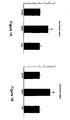
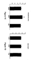
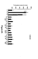


Classifications
-
- A—HUMAN NECESSITIES
- A61—MEDICAL OR VETERINARY SCIENCE; HYGIENE
- A61K—PREPARATIONS FOR MEDICAL, DENTAL OR TOILETRY PURPOSES
- A61K38/00—Medicinal preparations containing peptides
- A61K38/16—Peptides having more than 20 amino acids; Gastrins; Somatostatins; Melanotropins; Derivatives thereof
- A61K38/17—Peptides having more than 20 amino acids; Gastrins; Somatostatins; Melanotropins; Derivatives thereof from animals; from humans
- A61K38/18—Growth factors; Growth regulators
- A61K38/1858—Platelet-derived growth factor [PDGF]
- A61K38/1866—Vascular endothelial growth factor [VEGF]
-
- A—HUMAN NECESSITIES
- A61—MEDICAL OR VETERINARY SCIENCE; HYGIENE
- A61P—SPECIFIC THERAPEUTIC ACTIVITY OF CHEMICAL COMPOUNDS OR MEDICINAL PREPARATIONS
- A61P1/00—Drugs for disorders of the alimentary tract or the digestive system
- A61P1/16—Drugs for disorders of the alimentary tract or the digestive system for liver or gallbladder disorders, e.g. hepatoprotective agents, cholagogues, litholytics
-
- A—HUMAN NECESSITIES
- A61—MEDICAL OR VETERINARY SCIENCE; HYGIENE
- A61P—SPECIFIC THERAPEUTIC ACTIVITY OF CHEMICAL COMPOUNDS OR MEDICINAL PREPARATIONS
- A61P43/00—Drugs for specific purposes, not provided for in groups A61P1/00-A61P41/00
-
- A—HUMAN NECESSITIES
- A61—MEDICAL OR VETERINARY SCIENCE; HYGIENE
- A61K—PREPARATIONS FOR MEDICAL, DENTAL OR TOILETRY PURPOSES
- A61K48/00—Medicinal preparations containing genetic material which is inserted into cells of the living body to treat genetic diseases; Gene therapy
-
- C—CHEMISTRY; METALLURGY
- C12—BIOCHEMISTRY; BEER; SPIRITS; WINE; VINEGAR; MICROBIOLOGY; ENZYMOLOGY; MUTATION OR GENETIC ENGINEERING
- C12N—MICROORGANISMS OR ENZYMES; COMPOSITIONS THEREOF; PROPAGATING, PRESERVING, OR MAINTAINING MICROORGANISMS; MUTATION OR GENETIC ENGINEERING; CULTURE MEDIA
- C12N2799/00—Uses of viruses
- C12N2799/02—Uses of viruses as vector
- C12N2799/021—Uses of viruses as vector for the expression of a heterologous nucleic acid
- C12N2799/022—Uses of viruses as vector for the expression of a heterologous nucleic acid where the vector is derived from an adenovirus
Definitions
- the present invention relates to the diagnostic and therapeutic uses of VEGFR modulating agents, including methods of utilizing VEGFR agonists for promoting liver growth, treating liver pathological conditions, and protecting liver from damage.
- the liver is the major metabolic control organ of the human body that comprises thousands of minute lobules (lobuli hepatis), the functional units of the organ. Liver tissue contains two major differentiated cell types: parenchymal cells (i.e., hepatocytes) and non-parenchymal cells.
- parenchymal cells i.e., hepatocytes
- non-parenchymal cells such as Kupffer cells, Ito cells and liver sinusoidal endothelial cells (LSEC) play important roles in supporting and providing supplies to hepatocytes.
- LSEC liver sinusoidal endothelial cells
- the liver acts as a guardian interposed between the digestive tract and the rest of the body.
- a major hepatic function involves effective uptake, storage, metabolism and distribution to blood and bile large amounts of substances such as carbohydrates, lipids, amino acids, vitamins and trace elements.
- Another function of the liver is the detoxification of xenobiotic pollutants, drugs and endogenous metabolites, through both phase I (oxidation/reduction) and phase II (conjugation) mechanisms.
- liver dysfunction and diseases are often debilitating and life threatening.
- a number of acute or chronic pathological conditions are associated with structural and/or functional abnormalities of the liver. These include, but are not limited to, liver failure, hepatitis (acute, chronic or alcohol), liver cirrhosis, toxic liver damage, medicamentary liver damage, hepatic encephalopathy, hepatic coma or hepatic necrosis.
- liver damages can induce liver damages and thus are hepatotoxic.
- the susceptibility of the liver to damage by hepatotoxic agents may be related to its primary role in metabolism or is a consequence of hypersensitivity reactions. Up to 25% of cases of fulminant hepatic failure may be the result of adverse reactions to medical agents.
- Hepatotoxic compounds are also an important cause of chronic liver disease including fatty liver, hepatitis, cirrhosis and vascular and neoplastic lesions of the liver. ( Sinclair et al., Textbook of Internal Medicine, 569-575 (1992) (editor, Kelley; Publisher, J. B. Lippincott Co. ).
- Hepatotoxic agents may induce liver damage by cytotoxicity to the liver directly or through the production of toxic metabolites (this category includes the hypersensitivity reaction which mimics a drug allergy); cholestasis, an arrest in the flow of bile due to obstruction of the bile ducts; and vascular lesions, such as in veno occlusive disease (VOD), where injury to the vascular endothelium results in hepatic vein thrombosis.
- VOD veno occlusive disease
- Individual susceptibility to liver damage induced by hepatotoxic agents is influenced by genetic factors, age, sex, nutritional status, exposure to other drugs, and systemic diseases ( Sinclair et al., Textbook of Internal Medicine , Supra).
- liver tissue has a unique ability to regenerate at adult stage.
- Liver regeneration after the loss of hepatic tissue is a fundamental component of the recovery process in response to various forms of liver injury such as hepatotoxicity, viral infection, vascular injury and partial hepatectomy. Following partial hepatectomy, for example, the liver size is usually restored to its original mass within about six days.
- Liver growth and regeneration involves proliferation of both hepatocytes and non-parenchymal cells such as sinusoidal endothelial cells.
- hepatocytes are the first to proliferate, and other cells of the liver enter into DNA synthesis about 24 hours after the hepatocytes. Michalopoulos and DeFrances (1997) Science 276:60-65 .
- HGF hepatocyte growth factor
- EGF epidermal growth factor
- TGF- ⁇ transforming growth factor- ⁇
- IL-6 interleukin-6
- TGF- ⁇ tumor necrosis factor- ⁇
- CTGF basic and acidic fibroblast growth factors
- HGF liver mitogens
- HGF/SF is synthesized as an inactive, single-chain zymogen that is subsequently cleaved to produce an active, dimeric glycoprotein composed of a 69-kDa ⁇ -subunit and a 34-kDa ⁇ -subunit held together by a single disulfide bond.
- HGF/SF acts predominantly on Met-expressing epithelial cells in an endocrine and /or paracrine fashion, to mediate such diverse biological activities as proliferation, branching, cell migration, morphogenesis and lumen formation. van der Voort et al. Adv. Cancer Res. 79:39-90 (2000 ). In the liver, HGF is expressed in non-hepatocyte cells such as Ito cells and LSECs, whereas met transcripts are strongly expressed in hepatocytes. Hu et al. Am. J. Pathol. 142:1823-1830 (1993 ).
- HGF levels sharply increase, leading to a strong hepatocyte proliferation.
- Livers from transgenic mice with liver-specific overexpression of HGF are twice the size of livers of control animals and they regenerate much faster after partial hepatectomy.
- Sakata et al. (1996) Cell Growth Differ. 7:1513-1523 ; Shiota et al. (1994) Hepatol. 19:962-972 .
- HGF null mutant mouse embryos fail to develop a fully functional liver, demonstrating the essential role of HGF during liver development. Schmidt et al. (1995) Nature 373:699-702 .
- HGF substantially high HGF plasma concentrations may be required in order to promote liver growth in vivo ( Roos et al. (1995) Am. J. Physiol. 268:G380-6 ).
- HGF by virtue of its strong heparin-binding properties, is largely sequestered in extrahepatic tissues following intravenous administration ( Zioncheck et al. (1994) Endocrinology 134:1879-87 ) and the coadministration of dextran sulfate is required for an effective liver-promoting action (Roos et al., 1995).
- Angiogenesis is an important cellular event in which vascular endothelial cells proliferate, prune and reorganize to form new vessels from preexisting vascular network.
- the vascular endothelium may have an inductive effect on liver ( Matsumoto et al. (2001) Science 294:559-563 ) and pancreas organogenesis ( Lammert et al. (2001) Science 294:564-567 ), even prior to the establishment of a blood flow.
- the mechanism of such induction is unknown.
- Angiogenesis is also implicated in the pathogenesis of a variety of disorders, including but not limited to, proliferative retinopathies, age-related macular degeneration, tumors, rheumatoid arthritis (RA), and psoriasis.
- RA rheumatoid arthritis
- Folkman (1995) Nat Med 1:27-31 Regenerating liver, in analogy to rapidly growing tumors, must synthesize new stroma and blood vessels.
- many studies have focused on angiogenesis in liver development and regeneration, as well as the roles of many known angiogenic factors therein. Michalopoulos and DeFrances (1997) supra; Mochida et al. (1996).
- VEGF Vascular endothelial cell growth factor
- Ferrara and Davis-Smyth (1997) Endocrine Rev. 18:4-25 ; Ferrara (1999) J. Mol. Med. 77:527-543 .
- VEGF is unique in its high specificity for endothelial cells within the vascular system.
- Recent evidence indicates that VEGF is essential for embryonic vasculogenesis and angiogenesis. Carmeliet et al. (1996) Nature 380:435-439 ; Ferrara et al. (1996) Nature 380:439-442 .
- VEGF is required for the cyclical blood vessel proliferation in the female reproductive tract and for bone growth and cartilage formation.
- Ferrara et al. (1998) Nature Med. 4:336-340 ; Gerber et al. (1999) Nature Med. 5:623-628 .
- VEGF vascular endothelial growth factor
- VEGF as a pleiotropic growth factor, exhibits multiple biological effects in other physiological processes, such as endothelial cell survival, vessel permeability and vasodilation, monocyte chemotaxis and calcium influx. Ferrara and Davis-Smyth (1997), supra.
- mitogenic effects of VEGF on a few non-endothelial cell types such as retinal pigment epithelial cells, pancreatic duct cells and Schwann cells. Guerrin et al. (1995) J. Cell Physiol. 164:385-394 ; Oberg-Welsh et al. (1997) Mol. Cell. Endocrinol. 126:125-132 ; Sondell et al. (1999) J. Neurosci. 19:5731-5740 .
- VEGF vascular endothelial growth factor
- the VEGF mRNA is overexpressed by the majority of human tumors examined ( Berkman et al. J Clin Invest 91:153-159 (1993 ); Brown et al. Human Pathol.. 26:86-91 (1995 ); Brown et al. Cancer Res. 53:4727-4735 (1993 ); Mattern et al. Brit. J. Cancer. 73:931-934 (1996 ); and Dvorak et al. Am J. Pathol. 146:1029-1039 (1995 )).
- VEGF vascular endothelial growth factor
- concentration of VEGF in eye fluids are highly correlated to the presence of active proliferation of blood vessels in patients with diabetic and other ischemia-related retinopathies ( Aiello et al. N. Engl. J. Med. 331:1480-1487 (1994 )).
- recent studies have demonstrated the localization of VEGF in choroidal neovascular membranes in patients affected by AMD ( Lopez et al. Invest. Ophtalmo. Vis. Sci. 37:855-868 (1996 )).
- Anti-VEGF neutralizing antibodies suppress the growth of a variety of human tumor cell lines in nude mice ( Kim et al. Nature 362:841-844 (1993 ); Warren et al. J. Clin. Invest.
- Human VEGF was obtained by first screening a cDNA library prepared from human cells, using bovine VEGF cDNA as a hybridization probe. Leung et al. (1989) Science, 246:1306 . One cDNA identified thereby encodes a 165-amino acid protein having greater than 95% homology to bovine VEGF; this 165-amino acid protein is typically referred to as human VEGF (hVEGF) or VEGF 165 . The mitogenic activity of human VEGF was confirmed by expressing the human VEGF cDNA in mammalian host cells. Media conditioned by cells transfected with the human VEGF cDNA promoted the proliferation of capillary endothelial cells, whereas control cells did not. Leung et al. (1989) Science , supra.
- VEGF vascular endothelial cell growth factor
- VEGF is expressed in a variety of tissues as multiple homodimeric forms (121, 145, 165, 189, and 206 amino acids per monomer) resulting from alternative RNA splicing.
- VEGF 121 is a soluble mitogen that does not bind heparin; the longer forms of VEGF bind heparin with progressively higher affinity.
- the heparin-binding forms of VEGF can be cleaved in the carboxy terminus by plasmin to release a diffusible form(s) of VEGF. Amino acid sequencing of the carboxy terminal peptide identified after plasmin cleavage is Arg 110 -Ala 111 .
- Amino terminal "core” protein VEGF (1-110) isolated as a homodimer, binds neutralizing monoclonal antibodies (such as the antibodies referred to as 4.6.1 and 3.2E3.1.1) and soluble forms of VEGF receptors with similar affinity compared to the intact VEGF 165 homodimer.
- VEGF-B placenta growth factor
- VEGF-C vascular endothelial growth factor
- VEGF-D vascular endothelial growth factor-E
- Ferrara and Davis-Smyth (1987) Endocr. Rev., supra Ogawa et al. (1998) J. Biological Chem. 273:31273-31281 ; Meyer et al. (1999) EMBO J., 18:363-374 .
- a receptor tyrosine kinase, Flt-4 (VEGFR-3) has been identified as the receptor for VEGF-C and VEGF-D. Joukov et al. (1996) EMBO. J. 15:1751 ; Lee et al.
- VEGF-C has recently been shown to be involved in the regulation of lymphatic angiogenesis. Jeltsch et al. (1997) Science 276:1423-1425 .
- Flt-1 also called VEGFR-1
- KDR also called VEGFR-2
- Neuropilin-1 has been shown to be a selective VEGF receptor, able to bind the heparin-binding VEGF isoforms ( Soker et al. (1998) Cell 92:735-45 ).
- Both Flt-I and KDR belong to the family of receptor tyrosine kinases (RTKs).
- the RTKs comprise a large family of transmembrane receptors with diverse biological activities. At present, at least nineteen (19) distinct RTK subfamilies have been identified.
- the receptor tyrosine kinase (RTK) family includes receptors that are crucial for the growth and differentiation of a variety of cell types ( Yarden and Ullrich, Ann. Rev. Biochem. 57:433-478, 1988 ; Ullrich and Schlessinger, Cell 61:243-254, 1990 ).
- RTKs The intrinsic function of RTKs is activated upon ligand binding, which results in phosphorylation of the receptor and multiple cellular substrates, and subsequently in a variety of cellular responses ( Ullrich & Schlessinger, 1990, Cell 61:203-212 ).
- receptor tyrosine kinase mediated signal transduction is initiated by extracellular interaction with a specific growth factor (ligand), typically followed by receptor dimerization, stimulation of the intrinsic protein tyrosine kinase activity and receptor trans-phosphorylation. Binding sites are thereby created for intracellular signal transduction molecules and lead to the formation of complexes with a spectrum of cytoplasmic signaling molecules that facilitate the appropriate cellular response.
- both Flt-1 and KDR have seven immunoglobulin-like domains in the extracellular domain, a single transmembrane region, and a consensus tyrosine kinase sequence which is interrupted by a kinase-insert domain.
- Flt-1 and KDR have different signal transduction properties and possibly mediate different functions.
- the signals mediated through Flt-1 and KDR appear to be cell type specific.
- KDR is the major mediator of the mitogenic, angiogenic and permeability-enhancing effects of VEGF ( Ferrara (1999) Kidney Int. 56:794-814 ).
- VEGF stimulation leads to a robust auto-phosphorylation of KDR and activation of the MAPK cascade, which may directly contribute to endothelial cell proliferation ( Kroll and Waltenberger (1997) J. Biol. Chem. 272:32521-7 ).
- VEGFR-1 has been less clear, and many apparently conflicting reports on its function exist in the literature.
- This molecule displays a very weak or undetectable tyrosine autophosphorylation in endothelial cells in response to VEGF ( Gille et al. (2000) EMBO J. 19:4064-4073 ).
- Flt-1 has been shown to have inhibitory effects on endothelial mitogenesis in several biological contexts, including early embryonic development, either by acting as a "decoy" receptor that prevents VEGF binding to VEGFR-2 or by directly inhibiting VEGFR-2 activities. Park et al. (1994) J. Biol. Chem. 269:25646-54 ; U.S. Patent No.
- VEGFR-1 may mediate recruitment of monocytes and endothelial cell progenitors to the tumor vasculature ( Barleon et al. (1996) Blood 87:3336-43 ) ( Lyden et al. (2001) Nat. Med. 7:1194-201 ).
- VEGFR-1 signaling in the vascular endothelium is largely unclear.
- VEGF expressed in and secreted from hepatocytes acts as a stimulatory factor that binds to its receptors (KDR and Flt-1) on sinusoidal endothelial cells, thereby stimulating the proliferation and maintenance of the sinusoidal endothelial cells in the liver.
- KDR and Flt-1 its receptors
- Oncogene 9:2683-2690 observed that the endogenous expression of VEGF and VEGF receptors (Flt and KDR) as well as HGF and c-Met are strictly regulated in a cell-type specific manner in liver: using a flt-1 cDNA as a probe, flt-1 mRNA was found to be expressed at very high levels in sinusoidal endothelial cells in normal rat liver, but was hardly detectable in hepatocytes. Similar expression pattern was found for KDR, although the expression level was much lower. Yamane et al. further observed that, in an in vitro cell culture system, VEGF demonstrated a remarkably specific growth-stimulatory activity as well as maintenance activity on the sinusoidal endothelial cells.
- the present invention provides means, as defined in the claims, for treating a pathological liver condition in a subject, by administering to the subject a Flt-1 agonist in a manner effective to alleviate the pathological liver condition.
- Pathological liver conditions that can be treated by the present invention include, but not limited to, liver failure, hepatitis, liver cirrhosis, toxic liver damage, medicamentary liver damage, hepatic encephalopathy, hepatic coma or hepatic necrosis.
- the Flt-1 agonist is optionally in combination with an angiogenic agent.
- Also provided in the present invention are means, as defined in the claims, for protecting liver in a subject from damage due to exposure to a hepatotoxic agent, by administering to the subject a Flt-1 agonist, which effectively protects liver from damage.
- the Flt-1 agonist is optionally in combination with an angiogenesis agent.
- the Flt-1 agonist is administered prior to or concurrent with the exposure of said subject to the hepatotoxic agent, said hepatotoxic agent being a therapeutic agent such as a chemotherapeutic or radiation agent for treating cancers.
- the means serve to enhance the efficacy of the treatment by permitting the subject tolerance to high doses of the therapeutic agents.
- the Flt-1 agonist is administered after the exposure of the subject to a hepatotoxic agent but prior to any detectable liver damage in the subject. Such methods are especially useful for treating liver damages due to accidental exposure of the subject to hepatotoxic agent.
- the subject agents can be administered to the subject through a systemic delivery system, such as a cell preparation comprising mammalian cells (e.g., CHO cells) expressing a recombinant form of the subject agent.
- the systemic delivery system can comprise a slow release preparation comprising purified agent and a polymer matrix.
- the subject agent of the invention can be administered via a liver-targeted gene delivery vector comprising a nucleic acid encoding the agent.
- Well established viral or nonviral vectors for gene therapy can be used as the liver-targeted gene delivery vector in the present invention.
- An article of manufacture and a kit comprising a VEGFR modulating agent are also provided.
- the present invention provides, for the first time, a concerted system in which systemically delivered VEGFR modulating agents act in a paracrine fashion to promote liver growth.
- the current system may create a local cascade of signaling events originating in sinusoidal endothelial cells following VEGF receptor activation, which is much more potent and beneficial in promoting hepatocyte proliferation and liver growth than systemic delivery of the principal liver mitogen, HGF.
- the vasculature has been long thought to be necessary, but not sufficient, for proliferative processes, through the delivery of nutrients and oxygen and removal of catabolic products.
- the present invention demonstrates that, following an appropriate instructive signal, the vascular endothelium can be sufficient to initiate and amplify a growth/survival process that may overcome the set point for final organ size and protect the parenchyma from injury.
- VEGFR-1 VEGFR-1 (Flt-1) RTK in regulating key paracrine activities of LSEC, which in turn leads to liver proliferation and protection. Strikingly, in spite of the lack of stimulation of angiogenesis, activation of VEGFR-1 was sufficient to substantially protect the liver parenchyma from toxic injury. Indeed, the present invention provides the first evidence that protective effects on parenchymal cells mediated by the endothelium can be uncoupled from stimulation of angiogenesis.
- VEGF vascular endothelial growth factor
- KDR activation Kliche and Waltenberger (2001) IUBMB Life 52:61-6
- a Flt-1 agonist such as a Flt-selective VEGF variant
- a KDR agonist or other angiogenic factor at a lower ratio may result in a maximal therapeutic benefit, by providing stimulation of angiogenesis.
- VEGF variant that preferentially activates Flt-1 versus KDR might combine optimal characteristics of safety and efficacy.
- the potential indications include acute liver damage induced by various drugs, chemotherapy, or toxins as well as chronic injury, including cirrhosis.
- VEGF receptor or "VEGFR” as used herein refers to a cellular receptor for VEGF, ordinarily a cell-surface receptor found on vascular endothelial cells, as well as fragments and variants thereof which retain the ability to bind VEGF (such as fragments or truncated forms of the extracellular domain).
- VEGFR include the protein kinase receptors referred to in the literature as Flt-1 and KDR/Flk-1. DeVries et al. Science, 255:989 (1992 ); Shibuya et al. Oncogene, 5:519 (1990 ); Matthews et al.
- Flt-1 fms -like-tyrosine kinase
- KDR kinase domain region
- Both Flt-1 and KDR/Flk-1 have seven immunoglobulin (Ig)-like domains in the extracellular domain (ECD), a single transmembrane region and a consensus tyrosine kinase (TK) sequence, which is interrupted by a kinase-insert domain.
- Flt-1 has the highest affinity for rhVEGF 165 , with a Kd of approximately 10 to 20 pM.
- KDR has a lower affinity for VEGF, with a Kd of approximately 75 to 125 pM.
- VEGF receptors include those that can be cross-link labeled with VEGF, or that can be co-immunoprecipitated with KDR or Flt-1.
- An additional VEGF receptor that binds VEGF 165 but not VEGF 121 has been identified. Soker et al (1998) Cell 92:735-45 .
- the isoform-specific VEGF binding site is identical to human neuropilin-1, a receptor for the collapsin/semaphorin family that mediates neuronal cell guidance.
- the Flt-1 and KDR receptors mainly exist as a bound receptor on the surface of vascular endothelial cells, although they can also be present in non-endothelial cells.
- Some soluble forms of VEGFR have also been found.
- a cDNA coding an alternatively spliced soluble form of Flt-1 (sFlt-1), lacking the seventh Ig-like domain, transmembrane sequence, and the cytoplasmic domain has been identified in human umbilical vein endothelial cells (HUVECs).
- sFlt-1 alternatively spliced soluble form of Flt-1
- HAVECs human umbilical vein endothelial cells
- agent or, alternatively, “compound” as used herein refers broadly to any substance with identifiable molecular structure and physiochemical property.
- agents capable of modulating VEGFR activities include antibodies, proteins, peptides, glycoproteins, glycopeptides, glycolipids, polysaccharides, oligosaccharides, nucleic acids, bioorganic molecules, peptidomimetics, pharmacological agents and their metabolites, transcriptional and translation control sequences, and the like.
- the VEGFR modulating agents encompassed by the invention can be either an agonist or an antagonist of a VEGFR.
- An "agonist” is an agent that mediates or activate the biological activity of its target.
- a VEGFR agonist can be a growth factor ligand or an antibody that binds to the VEGFR's extracellular domain and triggers its signal transduction activity.
- a VEGFR agonist can be a small molecule compound that binds to the VEGFR's cytoplasmic domain and mediates its tyrosine phosphorylation.
- An "antagonist”, on the other hand, is one which blocks, inhibits or reduces biological activity of its target. Such inhibition can occur by any means, e.g.
- tyrosine kinase activity of a tyrosine kinase receptor in a receptor complex by interfering with: ligand binding to the receptor, receptor complex formation, tyrosine kinase activity of a tyrosine kinase receptor in a receptor complex and/or phosphorylation of tyrosine kinase residue(s) in or by the receptor.
- the agonist or antagonist of the invention is "selective" or “specific” to Flt-1, i.e., it exclusively or preferably modulates Flt-1 over other receptor tyrosine kinases such as KDR.
- the agonist or antagonist of the invention is "selective" or “specific” to KDR, i.e., it exclusively or preferably modulates KDR over other receptor tyrosine kinases such as Flt-1.
- the VEGFR agonist of the invention comprises a VEGF variant polypeptide capable of selectively binding to Flt-1 (referred hereinafter as "Flt-1 selective VEGF variant", or “Flt-sel”, or “Flt sel ").
- Flt-1 selective VEGF variant or “Flt-sel”, or “Flt sel”
- VEGF refers to the 165-amino acid vascular endothelial cell growth factor and related 121-, 189-, and 206- amino acid vascular endothelial cell growth factors, as described by Leung et al. Science, 246:1306 (1989 ), and Houck et al. Mol. Endocrin., 5:1806 (1991 ), together with the naturally occurring allelic and processed forms thereof.
- VEGF is also used to refer to truncated forms of the polypeptide comprising amino acids 8 to 109 or 1 to 109 of the 165-amino acid human vascular endothelial cell growth factor. Reference to any such forms of VEGF may be identified in the present application, e.g., by "VEGF (8-109),” “VEGF (1-109)” or “VEGF 165 .”
- the amino acid positions for a "truncated" native VEGF are numbered as indicated in the native VEGF sequence. For example, amino acid position 17 (methionine) in truncated native VEGF is also position 17 (methionine) in native VEGF.
- the truncated native VEGF has binding affinity for the KDR and Flt-1 receptors comparable to native VEGF.
- VEGF variant refers to a VEGF polypeptide which includes one or more amino acid mutations in the native VEGF sequence.
- the one or more amino acid mutations include amino acid substitution(s).
- numbers refer to the amino acid residue position along the amino acid sequence of the putative native VEGF (provided in Leung et al., supra and Houck et al., supra.).
- VEGF and variants thereof for use in the present invention can be prepared by a variety of methods well known in the art.
- the VEGF employed in the methods of the present invention comprises recombinant VEGF 165 .
- Amino acid sequence variants of VEGF can be prepared by mutations in the VEGF DNA. Such variants include, for example, deletions from, insertions into or substitutions of residues within the amino acid sequence shown in Leung et al., supra and Houck et al., supra. Any combination of deletion, insertion, and substitution may be made to arrive at the final construct having the desired activity.
- the mutations that will be made in the DNA encoding the variant must not place the sequence out of reading frame and preferably will not create complementary regions that could produce secondary mRNA structure. EP 75,444A .
- the VEGF variants optionally are prepared by site-directed mutagenesis of nucleotides in the DNA encoding the native VEGF or phage display techniques, thereby producing DNA encoding the variant, and thereafter expressing the DNA in recombinant cell culture.
- the mutation per se need not be predetermined.
- random mutagenesis may be conducted at the target codon or region and the expressed VEGF variants screened for the optimal combination of desired activity.
- Techniques for making substitution mutations at predetermined sites in DNA having a known sequence are well-known, such as, for example, site-specific mutagenesis.
- VEGF variants described herein are preferably achieved by phage display techniques, such as those described in the PCT publication WO 00/63380 .
- the mutated protein region may be removed and placed in an appropriate vector for protein production, generally an expression vector of the type that may be employed for transformation of an appropriate host.
- Amino acid sequence deletions generally range from about 1 to 30 residues, more preferably 1 to 10 residues, and typically are contiguous.
- Amino acid sequence insertions include amino- and/or carboxyl-terminal fusions of from one residue to polypeptides of essentially unrestricted length as well as intrasequence insertions of single or multiple amino acid residues.
- Intrasequence insertions i.e., insertions within the native VEGF sequence
- An example of a terminal insertion includes a fusion of a signal sequence, whether heterologous or homologous to the host cell, to the N-terminus to facilitate the secretion from recombinant hosts.
- Additional VEGF variants are those in which at least one amino acid residue in the native VEGF has been removed and a different residue inserted in its place. Such substitutions may be made in accordance with those shown in Table 1.
- Changes in function or immunological identity may be made by selecting substitutions that are less conservative than those in Table 1, i.e., selecting residues that differ more significantly in their effect on maintaining (a) the structure of the polypeptide backbone in the area of the substitution, for example, as a sheet or helical conformation, (b) the charge or hydrophobicity of the molecule at the target site, or (c) the bulk of the side chain.
- substitutions that in general are expected to produce the greatest changes in the VEGF variant properties will be those in which (a) glycine and/or proline (P) is substituted by another amino acid or is deleted or inserted; (b) a hydrophilic residue, e.g., seryl or threonyl, is substituted for (or by) a hydrophobic residue, e.g., leucyl, isoleucyl, phenylalanyl, valyl, or alanyl; (c) a cysteine residue is substituted for (or by) any other residue; (d) a residue having an electropositive side chain, e.g., lysyl, arginyl, or histidyl, is substituted for (or by) a residue having an electronegative charge, e.g., glutamyl or aspartyl; (e) a residue having an electronegative side chain is substituted for (or by) a residue having an electropositive charge; or (
- a phage display-selected VEGF variant may be expressed in recombinant cell culture, and, optionally, purified from the cell culture.
- the VEGF variant may then be evaluated for KDR or Flt-1 receptor binding affinity and other biological activities, such as those disclosed in the present application.
- the binding properties or activities of the cell lysate or purified VEGF variant can be screened in a suitable screening assay for a desirable characteristic. For example, a change in the immunological character of the VEGF variant as compared to native VEGF, such as affinity for a given antibody, may be desirable.
- VEGF variants of the invention will also exhibit activity in KIRA assays (such as described in the Examples) reflective of the capability to induce phosphorylation of the KDR receptor.
- Preferred VEGF variants of the invention will additionally or alternatively induce endothelial cell proliferation (which can be determined by known art methods such as the HUVEC proliferation assay in the Examples).
- endothelial cell proliferation which can be determined by known art methods such as the HUVEC proliferation assay in the Examples.
- the VEGF variants described in Keyt et al. (1996) J. Biol. Chem. 271:5638-5646 are also contemplated for use in the present invention.
- Flt-sel and methods of making the same have been known and are described in the Example sections below. Additional disclosures relating to Flt-sel can be found in, for example, the PCT publication WO 00/63380 and Li et al. (2000) J. Biol. Chem. 275:29823-29828 .
- Preferred Flt-sel variants include one or more amino acid mutations and exhibit binding affinity to the Flt-1 receptor which is equal to or greater ( ⁇ ) than the binding affinity of native VEGF to the Flt-1 receptor, and even more preferably, such VEGF variants exhibit less binding affinity ( ⁇ ) to the KDR receptor than the binding affinity exhibited by native VEGF to KDR.
- the binding affinity of such VEGF variant to the Flt-1 receptor is considered "selective" for the Flt-1 receptor.
- Preferred Flt-1 selective VEGF variants of the invention will have at least 10-fold less binding affinity to KDR receptor (as compared to native VEGF), and even more preferably, will have at least 100-fold less binding affinity to KDR receptor (as compared to native VEGF).
- the respective binding affinity of the VEGF variant may be determined by ELISA, RIA, and/or BIAcore assays, known in the art and described in the PCT publication WO 00/63380 .
- various methods for liver treatment further comprise administering an agent capable of modulating KDR activities.
- a KDR agonist can be administered in combination with a Flt-1 agonist to promote liver growth or liver regeneration.
- KDR has been identified as the major receptor tyrosine kinase that mediates VEGF's activities in endothelial cell proliferation.
- agonists of KDR and Flt-1 will induce concerted proliferation of both the SECs and hepatocytes, thereby promoting a coordinated growth of liver.
- the KDR agonist comprises a VEGF variant polypeptide capable of selectively binding to KDR (referred hereinafter as "KDR selective VEGF variant", or “KDR-sel”, or “KDR sel ").
- KDR selective VEGF variant or "KDR-sel”
- KDR sel KDR sel VEGF variants and methods of making the same are described in detail in the Example sections below. Additional disclosures relating to KDR-sel can be found in, for example, the PCT publication WO 00/63380 and Li et al. (2000) J. Biol. Chem. 275:29823-29828 .
- Preferred KDR-sel include one or more amino acid mutations and exhibit binding affinity to the KDR receptor which is equal to or greater ( ⁇ ) than the binding affinity of native VEGF to the KDR receptor, and even more preferably, the VEGF variants exhibit less binding affinity ( ⁇ ) to the flt-1 receptor than the binding affinity exhibited by native VEGF to Flt-1.
- the binding affinity of such VEGF variant to the KDR receptor is considered "selective" for the KDR receptor.
- Preferred KDR-sel of the invention will have at least 10-fold less binding affinity to Flt-1 receptor (as compared to native VEGF), and even more preferably, will have at least 100-fold less binding affinity to Flt-1 receptor (as compared to native VEGF).
- the respective binding affinity of the VEGF variant may be determined by ELISA, RIA, and/or BIAcore assays that are known in the art.
- Preferred KDR-sel of the invention will also exhibit activity in KIRA assays reflective of the capability to induce phosphorylation of the KDR receptor.
- Preferred KDR selective VEGF variants of the invention will additionally or alternatively induce endothelial cell proliferation (which can be determined by known methods such as the HUVEC proliferation assay).
- the VEGFR modulating agents of the invention are produced by recombinant methods.
- Isolated DNA used in these methods is understood herein to mean chemically synthesized DNA, cDNA, chromosomal, or extrachromosomal DNA with or without the 3'- and/or 5'-flanking regions.
- the VEGF and variants thereof herein are made by synthesis in recombinant cell culture.
- DNA encoding a VEGF molecule may be obtained from bovine pituitary follicular cells by (a) preparing a cDNA library from these cells, (b) conducting hybridization analysis with labeled DNA encoding the VEGF or fragments thereof (up to or more than 100 base pairs in length) to detect clones in the library containing homologous sequences, and (c) analyzing the clones by restriction enzyme analysis and nucleic acid sequencing to identify full-length clones.
- full-length clones are not present in a cDNA library, then appropriate fragments may be recovered from the various clones using the nucleic acid sequence information disclosed herein for the first time and ligated at restriction sites common to the clones to assemble a full-length clone encoding the VEGF.
- genomic libraries will provide the desired DNA.
- this DNA Once this DNA has been identified and isolated from the library, it is ligated into a replicable vector for further cloning or for expression.
- a VEGF-encoding gene is expressed in a cell system by transformation with an expression vector comprising DNA encoding the VEGF. It is preferable to transform host cells capable of accomplishing such processing so as to obtain the VEGF in the culture medium or periplasm of the host cell, i.e., obtain a secreted molecule.
- Transfection refers to the taking up of an expression vector by a host cell whether or not any coding sequences are in fact expressed. Numerous methods of transfection are known to the ordinarily skilled artisan, for example, CaPO 4 and electroporation. Successful transfection is generally recognized when any indication of the operation of this vector occurs within the host cell.
- Transformation refers to introducing DNA into an organism so that the DNA is replicable, either as an extrachromosomal element or by chromosomal integrant. Depending on the host cell used, transformation is done using standard techniques appropriate to such cells.
- the calcium treatment employing calcium chloride as described by Cohen, Proc. Natl. Acad. Sci. (USA), 69: 2110 (1972 ) and Mandel et al. J. Mol. Biol., 53: 154 (1970 ), is generally used for prokaryotes or other cells that contain substantial cell-wall barriers. For mammalian cells without such cell walls, the calcium phosphate precipitation method of Graham and van der Eb, Virology, 52: 456-457 (1978 ), is preferred.
- Transformations into yeast are typically carried out according to the method of Van Solingen et al. J. Bact., 130: 946 (1977 ) and Hsiao et al. Proc. Natl. Acad. Sci. (USA), 76: 3829 (1979 ).
- other methods for introducing DNA into cells such as by nuclear injection or by protoplast fusion may also be used.
- vectors and methods disclosed herein are suitable for use in host cells over a wide range of prokaryotic and eukaryotic organisms.
- E. coli K12 strain MM 294 (ATCC No. 31,446) is particularly useful.
- Other microbial strains that may be used include E. coli strains such as E. coli B and E. coli X1776 (ATCC No. 31,537). These examples are, of course, intended to be illustrative rather than limiting.
- Prokaryotes may also be used for expression.
- the aforementioned strains, as well as E. coli strains W3110 (F-, lambda-, prototrophic, ATCC No. 27,325), K5772 (ATCC No. 53,635), and SR101, bacilli such as Bacillus subtilis, and other enterobacteriaceae such as Salmonella typhimurium or Serratia marcesans, and various pseudomonas species, may be used.
- plasmid vectors containing replicon and control sequences that are derived from species compatible with the host cell are used in connection with these hosts.
- the vector ordinarily carries a replication site as well as marking sequences that are capable of providing phenotypic selection in transformed cells.
- E. coli is typically transformed using pBR322, a plasmid derived from an E. coli species (see, e.g., Bolivar et al. Gene, 2:95 (1977 ).
- pBR322 contains genes for ampicillin and tetracycline resistance and thus provides easy means for identifying transformed cells.
- the pBR322 plasmid, or other microbial plasmid or phage must also contain, or be modified to contain, promoters that can be used by the microbial organism for expression of its own proteins.
- promoters most commonly used in recombinant DNA construction include the ⁇ -lactamase (penicillinase) and lactose promoter systems Chang et al. Nature, 375:615 (1978 ); Itakura et al. Science, 198:1056 (1977 ); Goeddel et al. Nature, 281:544 (1979 )) and a tryptophan (trp) promoter system ( Goeddel et al. Nucleic Acids Res., 8:4057 (1980 ); EPO Appl. Publ. No. 0036,776 ).
- eukaryotic microbes such as yeast cultures
- Saccharomyces cerevisiae or common baker's yeast, is the most commonly used among eukaryotic microorganisms, although a number of other strains are commonly available.
- the plasmid YRp7 for example ( Stinchcomb et al. Nature, 282:39 (1979 ); Kingsman et al. Gene, 7:141 (1979 ); Tschemper et al. Gene, 10:157 (1980 )), is commonly used.
- This plasmid already contains the trp1 gene that provides a selection marker for a mutant strain of yeast lacking the ability to grow in tryptophan, for example, ATCC No. 44,076 or PEP4-1 ( Jones, Genetics, 85:12 (1977 )).
- the presence of the trp1 lesion as a characteristic of the yeast host cell genome then provides an effective environment for detecting transformation by growth in the absence of tryptophan.
- Suitable promoting sequences in yeast vectors include the promoters for 3-phosphoglycerate kinase ( Hitzeman et al. J. Biol. Chem., 255:2073 (1980 )) or other glycolytic enzymes ( Hess et al. J. Adv. Enzyme Reg., 7:149 (1968 ); Holland et al.
- enolase such as enolase, glyceraldehyde-3-phosphate dehydrogenase, hexokinase, pyruvate decarboxylase, phosphofructo-kinase, glucose-6-phosphate isomerase, 3-phosphoglycerate mutase, pyruvate kinase, triosephosphate isomerase, phosphoglucose isomerase, and glucokinase.
- the termination sequences associated with these genes are also ligated into the expression vector 3' of the sequence desired to be expressed to provide polyadenylation of the mRNA and termination.
- promoters which have the additional advantage of transcription controlled by growth conditions, are the promoter region for alcohol dehydrogenase 2, isocytochrome C, acid phosphatase, degradative enzymes associated with nitrogen metabolism, and the aforementioned glyceraldehyde-3-phosphate dehydrogenase, and enzymes responsible for maltose and galactose utilization.
- Any plasmid vector containing yeast-compatible promoter, origin of replication and termination sequences is suitable.
- cultures of cells derived from multicellular organisms may also be used as hosts.
- any such cell culture is workable, whether from vertebrate or invertebrate culture.
- interest has been greatest in vertebrate cells, and propagation of vertebrate cells in culture (tissue culture) has become a routine procedure in recent years ( Tissue Culture, Academic Press, Kruse and Patterson, editors (1973 )).
- useful host cell lines are VERO and HeLa cells, Chinese hamster ovary (CHO) cell lines, and W138, BHK, COS-7, 293, and MDCK cell lines.
- Expression vectors for such cells ordinarily include (if necessary) an origin of replication, a promoter located in front of the gene to be expressed, along with any necessary ribosome binding sites, RNA splice sites, polyadenylation sites, and transcriptional terminator sequences.
- control functions on the expression vectors are often provided by viral material.
- promoters are derived from polyoma, Adenovirus2, and most frequently Simian Virus 40 (SV40).
- the early and late promoters of SV40 virus are particularly useful because both are obtained easily from the virus as a fragment that also contains the SV40 viral origin of replication ( Fiers et al. Nature, 273:113 (1978 )). Smaller or larger SV40 fragments may also be used, provided there is included the approximately 250-bp sequence extending from the HindIII site toward the BgII site located in the viral origin of replication.
- promoter or control sequences normally associated with the desired gene sequence provided such control sequences are compatible with the host cell systems.
- An origin of replication may be provided either by construction of the vector to include an exogenous origin, such as may be derived from SV40 or other viral (e.g., Polyoma, Adeno, VSV, BPV) source, or may be provided by the host cell chromosomal replication mechanism. If the vector is integrated into the host cell chromosome, the latter is often sufficient.
- an exogenous origin such as may be derived from SV40 or other viral (e.g., Polyoma, Adeno, VSV, BPV) source, or may be provided by the host cell chromosomal replication mechanism. If the vector is integrated into the host cell chromosome, the latter is often sufficient.
- One secondary coding sequence comprises dihydrofolate reductase (DHFR) that is affected by an externally controlled parameter, such as methotrexate (MTX), thus permitting control of expression by control of the methotrexate concentration.
- DHFR dihydrofolate reductase
- MTX methotrexate
- a preferred host cell for transfection by the vectors of the invention that comprise DNA sequences encoding both VEGF and DHFR protein it is appropriate to select the host according to the type of DHFR protein employed. If wild-type DHFR protein is employed, it is preferable to select a host cell that is deficient in DHFR, thus permitting the use of the DHFR coding sequence as a marker for successful transfection in selective medium that lacks hypoxanthine, glycine, and thymidine.
- An appropriate host cell in this case is the Chinese hamster ovary (CHO) cell line deficient in DHFR activity, prepared and propagated as described by Urlaub and Chasin, Proc. Natl. Acad. Sci. (USA), 77:4216 (1980 ).
- DHFR protein with low binding affinity for MTX is used as the controlling sequence, it is not necessary to use DHFR-deficient cells. Because the mutant DHFR is resistant to methotrexate, MTX-containing media can be used as a means of selection provided that the host cells are themselves methotrexate sensitive. Most eukaryotic cells that are capable of absorbing MTX appear to be methotrexate sensitive.
- One such useful cell line is a CHO line, CHO-K1 (ATCC No. CCL 61).
- Plasmids containing the desired coding and control sequences employs standard ligation techniques. Isolated plasmids or DNA fragments are cleaved, tailored, and religated in the form desired to prepare the plasmids required.
- the preparation may be treated for 15 minutes at 15°C with 10 units of Polymerase I (Klenow), phenol-chloroform extracted, and ethanol precipitated.
- Size separation of the cleaved fragments may be performed using, by way of example, 6 percent polyacrylamide gel described by Goeddel et al. Nucleic Acids Res., 8:4057 (1980 ).
- the ligation mixtures are typically used to transform E. coli K12 strain 294 (ATCC 31,446) or other suitable E. coli strains, and successful transformants selected by ampicillin or tetracycline resistance where appropriate. Plasmids from the transformants are prepared and analyzed by restriction mapping and/or DNA sequencing by the method of Messing et al. Nucleic Acids Res., 9:309 (1981 ) or by the method of Maxam et al. Methods of Enzymology, 65:499 (1980 ).
- amplification of DHFR-protein-coding sequences is effected by growing host cell cultures in the presence of approximately 20,000-500,000 nM concentrations of methotrexate (MTX), a competitive inhibitor of DHFR activity.
- MTX methotrexate
- concentrations of folic acid analogs or other compounds that inhibit DHFR could also be used.
- MTX itself is, however, convenient, readily available, and effective.
- the Flt-1 agonist comprises a growth factor that selectively binds to and activates Flt-1.
- VEGF placental growth factor
- VEGF-B vascular endothelial growth factor
- PIGF has an amino acid sequence that shares 53% identity with the platelet-derived growth factor-like domain of VEGF. Park et al. (1994) J. Biol. Chem. 269:25646-54 ; Maglione et al. (1993) Oncogene 8:925-31 .
- PlGF placental growth factor
- PlGF placental growth factor
- VEGF-B is produced as two isoforms (167 and 185 residues) that also appear to specifically bind Flt-1. Pepper et al. (1998) Proc. Natl. Acad. Sci. USA 95:11709-11714 . Similar to the long forms of VEGF, VEGF-B is expressed as a membrane-bound protein that can be released in a soluble form after addition of heparin. VEGF-B and VEGF are also able to form heterodimers, when coexpressed. Olofsson et al. (1996) Proc. Natl. Acad. Sci. USA 93:2576-2581 .
- Compounds useful in the invention include small oraganic molecules that exert their modulating functions at the intracellular tyrosine kinase domain of the RTKs.
- small molecule agonists are employed to stimulate tyrosine phosphorylation, thereby activating the corresponding signaling pathway.
- small molecule inhibitors or antagonists are used to block and/or deactivate the RTK activities.
- Many small molecule compounds can be used for the purpose of this invention. These include, but not limited to, bis monocyclic, bicyclic or heterocyclic aryl compounds, vinylene-azaindole derivatives ( PCT WO 94/14808 ) and 1-cycloproppyl-4-pyridyl-quinolones ( U.S.
- Antibodies of the present invention can be either specific against a receptor (such as Flt-1), or specific against a ligand of the receptor, so long as they exert the necessary agonistic or antagonistic activity.
- Preferred antibodies of the invention include anti-Flt-1 antibodies. More preferably, the anti-Flt-1 antibody selectively binds to and modulate Flt-1, without affecting the KDR function.
- antibody is used in the broadest sense and includes monoclonal antibodies (including full length or intact monoclonal antibodies), polyclonal antibodies, multivalent antibodies, multispecific antibodies (e.g ., bispecific antibodies), and antibody fragments so long as they exhibit the desired biological activity. It is also contemplated that non-human antibodies, chimeric antibodies, humanized antibodies or human antibodies can be used for the purpose of the invention. Methods of preparing various antibodies suitable for the invention are known to the skilled artisan.
- a naturally occurring antibody comprises four polypeptide chains, two identical heavy (H) chains and two identical light (L) chains inter-connected by disulfide bonds.
- Each heavy chain is comprised of a heavy chain variable region (V H ) and a heavy chain constant region, which in its native form is comprised of three domains, CH1, CH2 and CH3.
- Each light chain is comprised of a light chain variable region (V L ) and a light chain constant region.
- the light chain constant region is comprised of one domain, C L .
- the V H and V L regions can be further subdivided into regions of hypervariability, termed complementarity determining regions (CDR), interspersed with regions that are more conserved, termed framework regions (FR).
- CDR complementarity determining regions
- Each V H and V L is composed of three CDRs and four FRs, arranged from amino-terminus to carboxy-terminus in the following order: FR1, CDR1, FR2, CDR2, FR3, CDR3, FR4.
- the light chains of antibodies from any vertebrate species can be assigned to one of two clearly distinct types, called kappa ( ⁇ ) and lambda ( ⁇ ), based on the amino acid sequences of their constant domains.
- ⁇ kappa
- ⁇ lambda
- antibodies immunoglobulins
- immunoglobulins There are five major classes of immunoglobulins: IgA, IgD, IgE, IgG and IgM, and several of these may be further divided into subclasses (isotypes), e.g., IgG-1, IgG-2, IgA-1, IgA-2, and etc.
- the heavy chain constant domains that correspond to the different classes of immunoglobulins are called ⁇ , ⁇ , ⁇ , ⁇ , and ⁇ , respectively.
- the subunit structures and three-dimensional configurations of different classes of immunoglobulins are well known and described generally in, for example, Abbas et al. Cellular and Mol. Immunology, 4th ed. (2000 ).
- An antibody may be part of a larger fusion molecule, formed by covalent or noncovalent association of the antibody or antibody portion with one or more other proteins or peptides.
- fusion proteins include use of the streptavidin core region to make a tetrameric scFv molecule ( Kipriyanov et al. (1995) Human Antibodies and Hybridomas 6:93-101 ) and use of a cysteine residue, a marker peptide and a C-terminal polyhistidine tag to make bivalent and biotinylated scFv molecules ( Kipriyanov, S. M., et al. (1994) Mol. Immunol. 31:1047-1058 ).
- agents capable of modulating Flt-1 or KDR activities include, for example but not limited to, soluble extracellular domain peptides of Flt-1 or KDR, Flt-1 or KDR binding peptides, Flt-1 or KDR specific ribozymes, antisense polynucleotides and RNA ligands.
- soluble Flt-1 extracellular fragments as antagonists are described in U.S. Pat. NO. 6,100,071 .
- the invention provides methods of using VEGFR agonists to upregulate gene expressions of factors that are important in regulating liver activities.
- the expression of HGF in nonparenchymal cells is upregulated.
- Methods and techniques for detecting levels of mRNA expression or protein expression in target cells/tissues are known to those skill in the art.
- the HGF gene expression level can be detected by known nucleic acid hybridization assays, using probes capable of hybridizing to HGF polynucleotides, under conditions suitable for the hybridization and subsequent detection and measurement. Methods useful for detecting HGF gene expression include but not limited to southern hybridization ( Southern (1975) J. Mol. Biol.
- the HGF protein levels can be detected by immunoassays using antibodies specific to HGF.
- immunoassays known in the art can be used, including but not limited to competitive and non-competitive assay systems using techniques such as radioimmunoassay, ELISA (enzyme linked immunosorbent assay), "sandwich” immunoassays, immunoradiometric assays, gel diffusion precipitin reactions, immunodiffusion assays, in situ immunoassays (using colloidal gold, enzyme or radioisotope labels), western blot analysis, precipitation reactions, agglutination assays (e.g., gel agglutination assays, hemagglutination assays), complement fixation assays, immunofluorescence assays, protein A assays, immunoelectrophoresis assays, etc.
- antibody binding is detected by detecting a label on the primary antibody.
- the primary antibody is detected by detecting binding of a secondary antibody or reagent to the primary antibody.
- the secondary antibody is labeled. Many means are known in the art for detecting binding in an immunoassay and are within the scope of the present invention.
- the invention provides methods for promoting liver growth and hepatocyte cell proliferation by administering an effective amount of VEGFR agonists.
- the promoting effects of the present invention can be assessed either in vitro or in vivo, using methods known in the art. Drakes et al. (1997) J. Immunol. 159:4268 ; Omori et al. (1997) Hepatology 26:720 ; U.S. Patent No. 5,227,158 .
- hepatocytes and other nonparenchymal liver cells are isolated from the target livers and resuspended in appropriate tissue culture medium to induce cell adherence. If necessary, different cell fractions can be further separated (e.g., parenchymal cells from nonparenchymal cells) by centrifugation at different speeds for different length of time. Cell proliferation is assessed during culture using methods known in the art, including but not limited to, measuring the rate of DNA synthesis (see, e.g., Nakamura et al. (1984) supra), trypan blue dye exclusion/hemacytometer counting (see, e.g., Omiri et al. (1997) supra), or flow cytometry (see, e.g., Drakes (1997) supra).
- the proliferative effect of a VEGFR agonist on hepatic cells and liver organ as a whole is measured in vivo using, for example, histochemistry assays of the liver tissue samples.
- in vivo proliferation of hepatic cells is assessed by reactivity to an antibody directed against a protein known to be present in higher concentrations in proliferating cells than in non-proliferating cells, such as proliferating cell nuclear antigen (PCNA or cyclin).
- PCNA proliferating cell nuclear antigen
- the invention provides methods for treating a pathological liver condition in a subject.
- treatment refers to clinical intervention in an attempt to alter the natural course of the individual or cell being treated, and can be performed either for prophylaxis or during the course of clinical pathology. Desirable effects of treatment include preventing occurrence or recurrence of disease, alleviation of symptoms, diminishment of any direct or indirect pathological consequences of the disease, preventing metastasis, decreasing the rate of disease progression, amelioration or palliation of the disease state, and remission or improved prognosis.
- an “effective amount” refers to an amount effective, at dosages and for periods of time necessary, to achieve the desired therapeutic or prophylactic result.
- a “therapeutically effective amount” of the antibody may vary according to factors such as the disease state, age, sex, and weight of the individual, and the ability of the antibody to elicit a desired response in the individual.
- a therapeutically effective amount is also one in which any toxic or detrimental effects of the antibody are outweighed by the therapeutically beneficial effects.
- a “prophylactically effective amount” refers to an amount effective, at dosages and for periods of time necessary, to achieve the desired prophylactic result. Typically, since a prophylactic dose is used in subjects prior to or at an earlier stage of disease, the prophylactically effective amount will be less than the therapeutically effective amount.
- pathological liver condition is used interchangeably with "liver disorder” or “liver disease” to indicate any structural and/or functional liver abnormalities.
- pathological liver condition include those conditions associated with liver failure, hepatitis (acute, chronic or alcohol), liver cirrhosis, toxic liver damage, medicamentary liver damage, hepatic encephalopathy, hepatic coma or hepatic necrosis.
- the invention provides methods for protecting liver from damage in a subject susceptible to conditions or factors causative of liver damage.
- liver damage is used herein in the broadest sense, and indicates any structural or functional liver injury resulting, directly or indirectly, from internal or external factors or their combinations. Liver damage can be induced by a number of factors including, but not limited to, exposure to hepatotoxic compounds, radiation exposure, mechanical liver injuries, genetic predisposition, viral infections, autoimmune disease, such as, autoimmune chronic hepatitis and as a result of elevated in vivo levels of proteins, such as activin and TGF-.beta..
- Liver damage induced by hepatotoxic compounds includes direct cytotoxicity including drug hypersensitivity reactions, cholestasis, and injury to the vascular endothelium.
- Hepatotoxic compounds can produce liver cytotoxicity by direct chemical attack or by the production of a toxic metabolite.
- the products of reductive metabolism are highly reactive species that bind to cellular macromolecules and cause lipid peroxidation and inactivation of drug metabolizing and other enzymes.
- the membrane injury provokes release of calcium from mitochondria and smooth endoplasmic reticulum and appears to interfere with the calcium ion pump, which normally prevents cytosolic accumulation of calcium.
- the deleterious effect on cell metabolism with resultant calcium accumulation, the loss of potassium and enzymes from the cytoplasm, and the loss of essential energy that results from mitochondrial injury all contribute to the necrosis of hepatic tissue.
- liver damage is referred to as a hypersensitivity reaction and is like that of a drug reaction, where the patient presents with fever, rash and eosinophilia and has a recurrence of symptoms upon rechallenge of the drug.
- the mechanism for injury is unknown and may represent aberrant metabolism in susceptible patients that permits the production or accumulation of hepatotoxic metabolites.
- drugs inducing cytotoxicity by direct chemical attack include the following: Anesthetics, such as Enflurane, Fluroxene, Halothane, and Methoxyflurane; Neuropsychotropics, such as, Cocaine, Hydrazides, Methylphenidate, and Tricyclics; Anticonvulsants, such as, Phenytoin and Valproic acid; Analgesics, such as, Acetaminophen, Chlorzoxazone, Dantrolene, Diclofenac, Ibuprofen, Indomethacin, Salicylates, Tolmetin, and Zoxazolamine; Hormones, such as, Acetohexamide, Carbutamide, Glipizide, Metahexamide, Propylthiouracil, Tamoxifen, Diethylstilbestrol; Antimicrobials, such as, Amphotericin B, Clindamycin, Ketoconazole, Mebendazole, Metronidazole, Oxacillin
- Those hepatotoxic compounds producing hypersensitivity reaction in the liver include the following: Phenytoin, Paraamino salicylic acid, Chlorpromazine, Sulfonamides, Erythromycin estolate, Isoniazid, Halothane, Methyldopa, and Valproic acid.
- Hepatotoxic compounds including cholestasis may take several forms. Centribular cholestasis is accompanied by portal inflammatory changes. Bile duct changes have been reported with some drugs such as erythromycin, while pure canalicular cholestasis is characteristic of other drugs such as the anabolic steroids. Chronic cholestasis has been linked to such drugs as methyltestosterone and estradiol.
- Those hepatotoxic compounds inducing cholestatic disease include the following: Contraceptive steroids, androgenic steroids, anabolic steroids, Acetylsalicylic acid, Azathioprine, Benzodiazepine, Chenodeoxycholic acid, Chlordiazepoxide, Erythromycin estolate, Fluphenazine, Furosemide, Griseofulvin, Haloperidol, Imipramine, 6-Mercaptopurine, Methimazole, Methotrexate, Methyldopa, Methylenediamine, Methyltestosterone, Naproxen, Nitrofurantoin, Penicillamine, Perphenazine, Prochlorperazine, Promazine, Thiobendazole, Thioridazine, Tolbutamide, Trimethoprimsulfamethoxazole, Arsenic, Copper, and Paraquat.
- the drugs causing mixed liver injury include, for example, the following: Chlorpromazine, Phenylbutazone, Halothane, Chlordiazepoxide, Diazepam, Allopurinol, Phenobarbital, Naproxen, Propylthiouracil, Chloramphenicol, Trimethoprimsulfamethoxazxole, Amrinone, Disopyramide, Azathioprine, Cimetidine, and Ranitidine.
- Vascular lesions of the liver including thrombosis of the hepatic veins, occlusion of the hepatic venules or veno occlusive disease (VOD), and peliosis hepatitis, can be produced by drugs.
- lesions including sinusoidal dilation, perisinusoidal fibrosis, and hepatoportal selerosis can occur.
- Midzonal and pericentral sinusoidal dilatation was first reported as a complication of oral contraceptive therapy.
- Peliosis hepatitis is a condition consisting of large blood-filled cavities that results from leakage of red blood cells through the endothelial barrier, followed by perisinusoidal fibrosis.
- Adenomas a lesion restricted to women in the childbearing years, is related to the use of contraceptive steroids and the risk increases with duration of use. Hepatocellular carcinoma may also be seen in patients taking androgenic hormones for aplastic anemia or hypopituitarism.
- Hepatotoxic compounds known to cause hepatic liesons include the following: Contraceptive steroids, Pyrriolizidine alkaloids, Urethane, Azathioprine, 6-Mercaptopurine, 6-Thioguanine, Mitomycin, BCNU, Vincristine, Adriamycin, Intravenous Vitamin E, Anabolic-androgenic steroids, Azathioprine, Medroxyprogesterone acetate, Estrone sulfate, Tamoxifen, inorganic arsenicals, Thorium dioxide, Vitamin A, methotrexate, Methylamphetamine hydrochloride, Vitamin A, Corticosteroids, Thorium dioxide, and Radium therapy.
- Liver damage caused by other factors usually takes similar forms. Liver damage, whether caused by the hepatotoxicity of a compound, radiation therapy, genetic predisposition, mechanical injury or any combination of such and other factors, can be detected by several means. Biochemical tests have been used clinically for many years as the standard measure of hepatotoxicity. Most biochemical tests generally fall into two categories: tests which measure specific liver markers, for example, prothrombin clotting time, and/or hepatic blood flow, or tests which analyze serum markers, for detection of necrosis, cholestasis, progressive fibrogenesis, or hepatoma ( Cornelius, C. in Hepatotoxicology, Meeks et al. eds., pgs. 181-185 (1991 )). The importance of such tests lies in their simplicity and the fact that they are non-invasive. The rationale for the use of serum enzymes in assessing liver damage is that these enzymes, normally contained in the liver cells, gain entry into the general circulation when liver cells are injured.
- Elevated serum enzyme activity suggests nercrosis and/or cholestasis. Elevated levels of serum bilirubin conjugates suggest intra or extra hepatic cholestasis.
- serum enzyme levels may increase as a result of leakage from cells with altered permeability due to systemic effects of an agent rather than specific liver injury caused by a chemical. Histopathological examination of the liver is the next logical step in identifying and quantifying the nature and extent of liver injury.
- the serum enzymes as markers of liver injury can be divided into four groups based on specificity and sensitivity to liver damage (Kodavanti et al., Supra).
- Group I these enzymes indicate more selectively hepatic cholestasis when elevated, e.g. alkaline phosphatase (AP), 5'-nucleotidase (5'-ND), and ⁇ -glutamyl transpeptidase (G-GT) and leucine aminopeptidase (LAP).
- AP alkaline phosphatase
- 5'-ND 5'-nucleotidase
- G-GT ⁇ -glutamyl transpeptidase
- LAP leucine aminopeptidase
- Group II These enzymes indicate parenchymal injury when elevated, e.g., aspartate transaminase (AST), alanine transaminase (ALT), fructose-1,6-diphosphate aldolase (ALD), lactate dehydrogenase (LDH), isocitrate dehydrogenase (ICDH), ornithine-carbamoyl-transferase (OCT), and sorbitol dehydrogenase (SDH) arginase and guanase.
- AST aspartate transaminase
- ALT alanine transaminase
- ALDH lactate dehydrogenase
- ICDH isocitrate dehydrogenase
- OCT sorbitol dehydrogenase
- SDH sorbitol dehydrogenase arginase and guanase.
- Group III These enzymes represent injury of other tissue when elevated e.g., creatine phosphokinase (CPK).
- CPK creatine phosphokinase
- Group IV These enzymes are depressed in hepatic injury, e.g., cholinesterase (ChE).
- ChE cholinesterase
- Other serum markers include, procollagen type III peptide levels (PIIIP) to assess if hepatic fibrogenesis is active; ammonia blood levels in hepatoencephalopathies; ligand in levels in necrosis and hepatoma; hyaluronate levels due to hepatic endothelial cell damage; a-1-fetoprotein (AFP) levels to detect hepatoma; carcinoembryonic antigen (CEA) levels to detect cancer metastasis to the liver; elevations of antibodies against a variety of cellular components, such as, mitochondrial, and nuclear and specific liver membrane protein; and detection of proteins, such as, albumin, globin, amino acids, cholestrol, and other lipids.
- biochemical analysis of a variety of minerals, metabolites, and enzymes obtained from liver biopsies can be useful in studying specific biochemical defects in inherited, acquired, and experimentally induced liver disorders.
- Liver function tests can be performed to assess liver injury. Liver function tests include the following:
- liver injury may lead to decreased blood levels of albumin, prothrombin, fibrinogen, and other proteins synthesized exclusively by hepatocytes, these protein levels may be measured as indicators of liver injury.
- serum protein levels reflect liver synthetic function rather than just cell injury ( Podolsky, D. Principles of Internal Medicine, 12th edition, Wilson et al. eds., 2: 1308-1311 (1991 )).
- CT computed tomography
- ultrasound ultrasound
- scintiscans or liver biopsy may be needed to determine the nature of the liver disease (Isselbacher, K, Supra and Friedman, L. and Needleman, L. in Harrison's Principles of Internal Medicine, 12th edition, Wilson et al. eds., 2: 1303-1307 (1991 )).
- the present invention provides methods for enhancing the effect of chemotherapy of cancer in a subject, said methods comprising administering to the subject a VEGFR modulating agent in a manner effective to protect the liver of the subject from damage caused by a hepatoxic compound prior to, or simultaneous with, the chemotherapy, thereby increasing the subject's tolerance to the chemotherapy.
- the chemotherapeutic agents used during the course of chemotherapy can have cytotoxic effects upon hepatic cells, therefore limiting the dosage and/or duration of the chemotherapeutic agent being administered to the patient.
- a composition comprising a VEGFR agonist such as VEGF, Flt-sel or KDR-sel, such toxic effects can be prevented or reduced.
- the dosage of the chemotherapeutic agents can be increased, thereby enhancing the efficacy of the cancer therapy.
- chemotherapeutic agent is a chemical compound useful in the treatment of cancer.
- examples of chemotherapeutic agents include alkylating agents such as thiotepa and cyclosphosphamide (CYTOXANTM); alkyl sulfonates such as busulfan, improsulfan and piposulfan; aziridines such as benzodopa, carboquone, meturedopa, and uredopa; ethylenimines and methylamelamines including altretamine, triethylenemelamine, trietylenephosphoramide, triethylenethiophosphaoramide and trimethylolomelamine; acetogenins (especially bullatacin and bullatacinone); a camptothecin (including the synthetic analogue topotecan); bryostatin; callystatin; CC-1065 (including its adozelesin, carzelesin and bizelesin synthetic analogues); cryptophycin
- calicheamicin especially calicheamicin ⁇ 1 I and calicheamicin ⁇ I 1 , see, e.g., Agnew Chem Intl. Ed. Engl. 33:183-186 (1994 ); dynemicin, including dynemicin A; an esperamicin; as well as neocarzinostatin chromophore and related chromoprotein enediyne antiobiotic chromomophores), aclacinomysins, actinomycin, authramycin, azaserine, bleomycins, cactinomycin, carabicin, carminomycin, carzinophilin, chromomycins, dactinomycin, daunorubicin, detorubicin, 6-diazo-5-oxo-L-norleucine, doxorubicin (including morpholino-doxorubicin, cyanomorpholino-doxorubicin,
- paclitaxel TAXOL ® , Bristol-Myers Squibb Oncology, Princeton, NJ
- doxetaxel TAXOTERE ® , Rhone-Poulenc Rorer, Antony, France
- chlorambucil gemcitabine
- 6-thioguanine mercaptopurine
- methotrexate platinum analogs such as cisplatin and carboplatin; vinblastine; platinum; etoposide (VP-16); ifosfamide; mitomycin C; mitoxantrone; vincristine; vinorelbine; navelbine; novantrone; teniposide; daunomycin; aminopterin; xeloda; ibandronate; CPT-11; topoisomerase inhibitor RFS 2000; difluoromethylornithine (DMFO); retinoic acid; capecitabine; and pharmaceutically acceptable salts, acids or derivatives of any of the above.
- anti-hormonal agents that act to regulate or inhibit hormone action on tumors
- anti-estrogens including for example tamoxifen, raloxifene, aromatase inhibiting 4(5)-imidazoles, 4-hydroxytamoxifen, trioxifene, keoxifene, LY117018, onapristone, and toremifene (Fareston); and anti-androgens such as flutamide, nilutamide, bicalutamide, leuprolide, and goserelin; and pharmaceutically acceptable salts, acids or derivatives of any of the above.
- compositions and Therapeutic / prophylactic Administration
- a therapeutic compound of the invention is administered to a subject using methods and techniques known in the art and suitable for the particular use.
- the compound is administered in the form of pharmaceutical compositions at a pharmaceutically acceptable dosage.
- the invention contemplates the use of mammalian cell preparations for the administration of a therapeutic protein agent (such as VEGF, Flt-sel or KDR-sel).
- a therapeutic protein agent such as VEGF, Flt-sel or KDR-sel.
- the mammalian cells used herein have been transfected with the heterologous gene encoding the protein, as described in detail above.
- the host cells used for the administration are CHO cells.
- the therapeutic agent can be entrapped in microcapsules prepared, for example, by coacervation techniques or by interfacial polymerization, in colloidal drug delivery systems (e.g., liposomes, microspheres, microemulsions, nanoparticles and nanocapsules), or in macroemulsions.
- colloidal drug delivery systems e.g., liposomes, microspheres, microemulsions, nanoparticles and nanocapsules
- macroemulsions e.g., liposomes, microspheres, microemulsions, nanoparticles and nanocapsules
- the therapeutic agent can be administered in vivo in slow-release preparations.
- slow-release preparations include semipermeable matrices of solid hydrophobic polymers containing the multivalent antibody, which matrices are in the form of shaped articles, e.g., films, or microcapsule.
- sustained-release matrices include polyesters, hydrogels (for example, poly(2-hydroxyethyl-methacrylate), or poly(vinylalcohol)), polylactides ( U.S. Pat. No.
- copolymers of L-glutamic acid and ⁇ ethyl-L-glutamate copolymers of L-glutamic acid and ⁇ ethyl-L-glutamate, non-degradable ethylene-vinyl acetate, degradable lactic acid-glycolic acid copolymers such as the LUPRON DEPOTTM (injectable microspheres composed of lactic acid-glycolic acid copolymer and leuprolide acetate), and poly-D-(-)-3-hydroxybutyric acid. While polymers such as ethylene-vinyl acetate and lactic acid-glycolic acid enable release of molecules for over 100 days, certain hydrogels release proteins for shorter time periods.
- encapsulated antibodies When encapsulated antibodies remain in the body for a long time, they may denature or aggregate as a result of exposure to moisture at 37°C, resulting in a loss of biological activity and possible changes in immunogenicity. Rational strategies can be devised for stabilization depending on the mechanism involved. For example, if the aggregation mechanism is discovered to be intermolecular S-S bond formation through thio-disulfide interchange, stabilization may be achieved by modifying sulfhydryl residues, lyophilizing from acidic solutions, controlling moisture content, using appropriate additives, and developing specific polymer matrix compositions.
- the therapeutic composition of the invention can be administered by any suitable means, including but not limited to, parenteral, subcutaneous, intraperitoneal, intrapulmonary, and intranasal administration.
- Parenteral infusions include intramuscular, intravenous, intraarterial, intraperitoneal, or subcutaneous administration.
- the therapeutic composition is suitably administered by pulse infusion, particularly with declining doses of the antibody.
- the therapeutic composition is given by injections, most preferably intravenous or subcutaneous injections, depending in part on whether the administration is brief or chronic.
- a therapeutic protein agent of the invention can be introduced to a subject by gene therapy.
- Gene therapy refers to therapy performed by the administration of a nucleic acid to a subject.
- Gene therapy refers to therapy performed by the administration of a nucleic acid to a subject.
- nucleic acid (optionally contained in a vector) into the subject's cells
- in vivo and ex vivo the nucleic acid is injected directly into the subject, usually at the site where the protein is required.
- ex vivo treatment the subject's cells are removed, the nucleic acid is introduced into these isolated cells and the modified cells are administered to the subject either directly or, for example, encapsulated within porous membranes which are implanted into the subject (see, e.g. U.S. Patent Nos. 4,892,538 and 5,283,187 ).
- techniques available for introducing nucleic acids into viable cells There are a variety of techniques available for introducing nucleic acids into viable cells.
- the techniques vary depending upon whether the nucleic acid is transferred into cultured cells ex vivo, or in vivo in the cells of the intended host.
- Techniques suitable for the transfer of nucleic acid into mammalian cells ex vivo include the use of liposomes, electroporation, microinjection, cell fusion, DEAE-dextran, the calcium phosphate precipitation method, etc.
- a commonly used vector for ex vivo delivery of the gene is a retrovirus.
- the currently preferred in vivo nucleic acid transfer techniques include transfection with viral vectors (such as adenovirus, Herpes simplex I virus, lentivirus, retrovirus, or adeno-associated virus) and lipid-based systems (useful lipids for lipid-mediated transfer of the gene are DOTMA, DOPE and DC-Chol, for example).
- viral vectors such as adenovirus, Herpes simplex I virus, lentivirus, retrovirus, or adeno-associated virus
- lipid-based systems useful lipids for lipid-mediated transfer of the gene are DOTMA, DOPE and DC-Chol, for example.
- examples of using viral vectors in gene therapy can be found in Clowes et al. (1994) J. Clin. Invest. 93:644-651 ; Kiem et al. (1994) Blood 83:1467-1473 ; Salmons and Gunzberg (1993) Human Gene Therapy 4:129-141 ; Grossman and Wilson (1993) Curr.
- the nucleic acid source with an agent that targets the target cells, such as an antibody specific for a cell surface membrane protein on the target cell, a ligand for a receptor on the target cell, etc.
- an agent that targets the target cells such as an antibody specific for a cell surface membrane protein on the target cell, a ligand for a receptor on the target cell, etc.
- proteins which bind to a cell surface membrane protein associated with endocytosis may be used for targeting and/or to facilitate uptake, e.g. capsid proteins or fragments thereof tropic for a particular cell type, antibodies for proteins which undergo internalization in cycling, and proteins that target intracellular localization and enhance intracellular half-life.
- the technique of receptor-mediated endocytosis is described, for example, by Wu et al. J. Biol. Chem.
- compositions comprise a therapeutically effective amount of a VEGFR modulating agent, and a pharmaceutically acceptable carrier.
- pharmaceutically acceptable means approved by a regulatory agency of the Federal or a state government or listed in the U.S. Pharmacopeia or other generally recognized pharmacopeia for use in animals, and more particularly, in humans.
- carrier refers to a diluent, adjuvant, excipient, or vehicle with which the therapeutic is administered.
- Such pharmaceutical carriers can be sterile liquids, such as water and oils, including those of petroleum, animal, vegetable or synthetic origin, including but not limited to peanut oil, soybean oil, mineral oil, sesame oil and the like.
- Water is a preferred carrier when the pharmaceutical composition is administered orally.
- Saline and aqueous dextrose are preferred carriers when the pharmaceutical composition is administered intravenously.
- Saline solutions and aqueous dextrose and glycerol solutions are preferably employed as liquid carriers for injectable solutions.
- Suitable pharmaceutical excipients include starch, glucose, lactose, sucrose, gelatin, malt, rice, flour, chalk, silica gel, sodium stearate, glycerol monostearate, talc, sodium chloride, dried skim milk, glycerol, propylene, glycol, water, ethanol and the like.
- the composition if desired, can also contain minor amounts of wetting or emulsifying agents, or pH buffering agents.
- compositions can take the form of solutions suspensions, emulsions, tablets, pills, capsules, powders, sustained- release formulations and the like.
- the composition can be formulated as a suppository, with traditional binders and carriers such as triglycerides.
- Oral formulation can include standard carriers such as pharmaceutical grades of mannitol, lactose, starch, magnesium stearate, sodium saccharine, cellulose, magnesium carbonate, etc. Examples of suitable pharmaceutical carriers are described in "Remington's Pharmaceutical Sciences" by E.W. Martin.
- Such compositions will contain a therapeutically effective amount of the Therapeutic, preferably in purified form, together with a suitable amount of carrier so as to provide the form for proper administration to the patient.
- the formulation should suit the mode of administration.
- the composition is formulated, in accordance with routine procedures, as a pharmaceutical composition adapted for intravenous administration to human beings.
- compositions for intravenous administration are solutions in sterile isotonic aqueous buffer.
- the composition may also include a solubilizing agent and a local anesthetic such as lignocaine to ease pain at the site of the injection.
- the ingredients are supplied either separately or mixed together in unit dosage form, for example, as a dry lyophilized powder or water-free concentrate in a hermetically sealed container such as an ampoule or sachette indicating the quantity of active agent.
- composition is to be administered by infusion, it can be dispensed with an infusion bottle containing sterile pharmaceutical grade water or saline.
- an ampoule of sterile water or saline for injection can be provided so that the ingredients may be mixed prior to administration.
- the therapeutics of the invention can be formulated as neutral or salt forms.
- Pharmaceutically acceptable salts include those formed with free carboxyl groups such as those derived from hydrochloric, phosphoric, acetic, oxalic, tartaric acids, etc., those formed with free amine groups such as those derived from isopropylamine, triethylamine, 2- ethylamino ethanol, histidine, procaine, etc., and those derived from sodium, potassium, ammonium, calcium, and ferric hydroxides, etc.
- the amount of the Therapeutic of the invention which will be effective in the treatment of a particular disorder or condition will depend on the nature of the disorder or condition, and can be determined by standard clinical techniques.
- in vitro assays may optionally be employed to help identify optimal dosage ranges.
- the precise dose to be employed in the formulation will also depend on the route of administration, and the seriousness of the disease or disorder, and should be decided according to the judgment of the practitioner and each patient's circumstances.
- suitable dosage ranges for intravenous administration are generally about 20-500 micrograms of active compound per kilogram body weight.
- Suitable dosage ranges for intranasal administration are generally about 0.01 pg/kg body weight to I mg/kg body weight.
- Effective doses may be extrapolated from dose-response curves derived from in vitro or animal model test systems.
- Suppositories generally contain active ingredient in the range of 0.5% to 10% by weight; oral formulations preferably contain 10% to 95 % active ingredient.
- the invention also provides a pharmaceutical pack or kit comprising one or more containers filled with one or more of the ingredients of the pharmaceutical compositions of the invention.
- a pharmaceutical pack or kit comprising one or more containers filled with one or more of the ingredients of the pharmaceutical compositions of the invention.
- Optionally associated with such container(s) can be a notice in the form prescribed by a governmental agency regulating the manufacture, use or sale of pharmaceuticals or biological products, which notice reflects approval by the agency of manufacture, use or sale for human administration.
- VEGF variants that selectively bind and activate a specific VEGF receptor (such as KDR or Flt-1) have been known in the art and described in, for example, Li et al. (2000) J. Biol. Chem. 275:29823 ; Gille et al. (2001) J. Biol. Chem. 276:3222-3230 ; PCT publications WO 00/63380 and 97/08313 ; and U.S. Pat. No. 6,057,428 , the disclosure of which are expressly incorporated herein by reference.
- a specific VEGF receptor such as KDR or Flt-1
- a VEGF variant with high selectivity for the Flt-1 receptor was generated by combining four mutations that greatly affected KDR but not Flt-1 binding. Mutation to alanine of Ile 43, Ile 46, Gln 79 or Ile 83 showed that the side chains of these residues are critical for tight binding to KDR but unimportant for Flt-1-binding. Li et al. (2000) supra.
- a Flt-sel variant was constructed with alanine substitutions at positions Ile 43, Ile 46, Gln 79 and Ile 83, using site directed mutagenesis methods described by Kunkel et al.(1991) Methods Enzymol. 204:125-139 .
- This particular Flt-sel variant can also be represented by the identifier, I43A/I46A/Q79A/I83A.
- the corresponding codons for these four alanine substitutions at positions 43, 46, 79 and 83 are GCC/GCC/GCG/GCC, respectively.
- cDNA encoding for human VEGF 165 was inserted into the dicistronic dihydrofolate reductase (DHFR) intron expression vector SV.DI ( Lucas et al. (1996) Nucleic Acids Res. 24:1774-9 ), resulting in the construct SV.DI.VEGF 165 .H8.
- HAg is a serum glycoprotein which has been recently shown to be an autoantigen in systemic lupus erythematosus patients. Sugimoto et al. (1998) J. Biol. Chem. 273:20721-7 . This protein has structural homology to the angiopoietin family, although it does not interact with the Tie-2 receptor.
- a dicistronic mRNA was transcribed under the control of the SV40 early gene promoter and encoded both the DHFR gene and the inserted gene of interest, which were thereby expressed in a fixed equimolar ratio. Both open reading frames were also fused with a DNA sequence encoding for a short carboxyl terminal peptide tag consisting of 8 histidine residues allowing easy detection and purification.
- the linearized expression plasmids, SV.DI.VEGF 165 .H8, and SV.DI.HAg.H8 as well as an empty vector, SV.DI.H8, were electroporated into DHFR - CHO cells.
- VEGF 165 protein by VEGF 165 transfected CHO cells (CHO-VEGF 165 ) was confirmed by immunoblotting and ELISA.
- HGF is a key mitogen for hepatocytes and has sequence similarity to plasminogen. Nakamura et al. (1987) FEBS Lett. 224:311-6 .
- HGF is secreted as a single-chain 82-84 kDa promitogen which is endoproteolytically processed into the bioactive HGF heterodimer, consisting of a 69 kDa ⁇ -chain and a 32 kDa ⁇ -chain.
- FEBS Lett 224:311-6 A band consistent in molecular weight with the 69 kDa HGF ⁇ -chain was detected in conditioned culture media of CHO-HGF cells but not in CHO-DHFR controls.
- SV.DI.HAg.H8 another stably transfected CHO cell pool was also generated, which secretes high amounts of HAg. Under reducing conditions, the HAg monomer was detected in CHO-HAg conditioned culture media as a band of ⁇ 32 kDa.
- VEGF-PLGA microspheres were used as a slow release formulation of VEGF.
- VEGF-PLGA microspheres were generated by encapsulating VEGF 165 protein into poly(lactic-co-glycolic acid) biodegradable polymer (provided by J. Cleland and A. Daugherty, Genentech). About 10% of the total VEGF protein contained in the microspheres was released into the circulation.
- Juvenile (3-4 week old) or adult athymic beige nude xid mice (Hsd:NIHS-bg-nu-xid, Harlan Sprague Dawley (Indianapolis, IN)) were anesthetized by isofluorane inhalation. Each group consisted of five animals. Suspensions of stably transfected or control CHO cells in complete culture medium were intramuscularly injected into both legs of the animal. A total volume of 100 ⁇ l cell suspensions containing about 3 x 10 6 cells was injected into 3 different sites of the anterior femoral muscles of each leg. On day 14 after injection, the mice were sacrificed, and serum samples were isolated and further analyzed.
- VEGF-PLGA treated animals received a total of 3 intramuscular injections of 100 ⁇ l of PLGA-VEGF microspheres per leg at days 1, 7, and 10. The dose of released VEGF per injection was about 4.5 mg/kg.
- the VEGF-PLGA injected animals were sacrificed on day 15. One hour before sacrifice, all animals were intraperitoneally injected with 100 mg/kg bromodeoxyuridine (BrdU) (Sigma, St. Louis, MO). Livers, kidneys, heart, legs, and brains were removed and weighed. The collected tissues were immersed in formalin for histological evaluation.
- This antigen is not expressed by lymphocytes or polymorphonuclear cells.
- T o demonstrate specificity, rat IgG 2B (Pharmingen, San Diego, CA) was used as a negative control. Biotinylated rabbit anti-rat IgG was used as a secondary antibody. The antibody was detected using Vectastain Elite ABC reagent (Vector Labs, Burlingame, CA) followed by Metal Enhanced DAB (Pierce, Rockford, IL). The sections were counterstained with Mayer's hematoxylin.
- Nude mice that have been intramuscularly injected with CHO cells were anesthetized by intraperitoneally injecting 100 ⁇ l Nembutal (Abbott Laboratories, Chicago, IL) and perfused with 10 ml of perfusion buffer (NaCl 142 mM, KCl 6.7 mM, HEPES 10 mM). After sacrificing the animals, the livers were removed and minced into fine pieces.
- Collagenase digestion of the tissue pieces was carried out at 37°C for 30 min in digestion buffer (67 mM NaCl, 6.7 mM KCl, HEPES 100 mM, CaCl 2 5 mM) supplemented with 50 ⁇ g/ml Liberase RH (Roche Molecular Biochemicals, Indianapolis, IN).
- digestion buffer 67 mM NaCl, 6.7 mM KCl, HEPES 100 mM, CaCl 2 5 mM
- 50 ⁇ g/ml Liberase RH Roche Molecular Biochemicals, Indianapolis, IN
- Single cell suspensions were obtained after passing the digested liver suspension through a 70 ⁇ m cell strainer (Falcon, Bedford, MA).
- the in situ Cell Proliferation Kit was used according to the manufacturer's recommendations (Roche Molecular Biochemicals, Indianapolis, IN). In short, cells were washed, ethanol-fixed and the DNA denatured by HCl treatment.
- DNA-incorporated BrdU into was detected by staining with an anti-BrdU-FLUOS antibody (anti-BrdU-F(ab') 2 -FITC conjugate).
- anti-BrdU-FLUOS antibody anti-BrdU-F(ab') 2 -FITC conjugate.
- For flow cytometry brightly autofluorescent monocytic cells were rejected from the analysis by forward/side scatter-gating. 10,000 gated cells were acquired and analyzed on a Becton Dickinson (San Jose, CA) FACSCalilbur flow cytometer.
- a fluorimetric anti-VEGF enzyme-linked immunosorbant assay (ELISA) was performed as described before. Rodriguez et al. (1998) J. Immunol. Methods 219:45-55 .
- the limit of sensitivity of the assay in the presence of mouse serum was 200 pg/ml.
- the assay is specific for human VEGF and does not cross-react with murine VEGF.
- ELISA For detection of recombinant human HGF, a previously described ELISA was used ( Koch et al. (1996) Arthr. Rheum. 39: 1566-75 ), with some modifications.
- an anti-human HGF monoclonal antibody was used to coat 96-well microtiter plates. After a 1-hour incubation at room temperature, the wells were washed and serial dilutions of serum samples were added. rHGF was used as a reference standard. Following a 2-hour incubation and a wash, biotinylated sheep anti-HGF was added and incubated for 1 hour. After washing, horseradish peroxidase conjugated streptavidin (Amdex) was added and incubated for 30 minutes.
- Amdex horseradish peroxidase conjugated streptavidin
- Immunoreactivities were visualized by the avidin-biotin complex technique using Vectastain Elite ABC kit (Vector Laboratories, Burlingame, CA) with diaminobenzidine as chromogen. Hematoxylin was used as counterstain. BrdU immunohistochemoistry was performed as previously described (Gerber et al., 1999).
- CHO-VEGF cells were injected intramuscularly into both legs of 3-4 week, 6-8 week or 12-14 week old beige nude mice.
- An identical number of CHO-DHFR cells, CHO-HAg cells, or CHO-HGF cells was injected into control animals.
- the serum concentrations of human VEGF in the CHO-VEGF animals were 3.3 ⁇ 1.7 ng/ml, (range 0.8-5.4 ng/ml).
- hVEGF was undetectable in the sera of CHO-DHFR and CHO-HAg control animals.
- the HGF levels in the serum of CHO-HGF animals were 1.25 ⁇ 0.87 ng/ml (range 0.50-2.00).
- liver/brain ratio i.e. the relative liver mass, of the CHO-VEGF group (4.73 ⁇ 0.39) was highly significantly increased when compared to the CHO-DHFR (3.18 ⁇ 0.25, p ⁇ 0.0001) and the CHO-HAg (3.00 ⁇ 0.45, p ⁇ 0.0001) controls ( Fig. 1A, B ). This reflects an increase of relative liver masses of 49 and 59%, respectively.
- the heart/brain ratio was also significantly increased in the CHO-VEGF group (0.376 ⁇ 0.052 compared to 0.304 ⁇ 0.022 and 0.304 ⁇ 0.017 in the CHO-DHFR and the CHO-HAg groups, respectively; CHO-VEGF versus CHO-DHFR, p ⁇ 0.05).
- the kidney/brain ratio (0.976 ⁇ 0.071 compared to 0.860 ⁇ 0.070, p ⁇ 0.05 and 0.822 ⁇ 0.097, p ⁇ 0.02 in the CHO-DHFR and the CHO-HAg groups) was significantly increased ( Fig. 1C ).
- VEGF in slow release preparation also promotes liver growth
- proliferating hepatocyte compartment represented 6.44 ⁇ 0.96 % (CHO-DHFR 1.02 ⁇ 0.74 and CHO-HGF 1.55 ⁇ 1.48 %) as quantified by FACS analysis of hepatocytes isolated from BrdU-injected CHO-VEGF mice.
- VEGF vascular permeability
- Hepatocytes were isolated from nude mice following retrograde liver perfusion, according to a previously described procedure. Harman et al. (1987) J. Pharmacol. Methods 17:157-63 .
- the inferior vena cava was cannulated with a 22G Abbocath T catheter (Abbott Lab) and the portal vein was severed.
- the livers were perfused in situ with a 0.1 % collagenase, 2 mM CaCl2 in PBS solution at a flow rate of 3 ml/min for 10-15 minutes.
- Hepatocytes were seeded in 24-well plates at a density of 5x10 4 cells per well in William's E medium (GIBCO BRL) supplemented with 10% heat-inactivated fetal bovine serum (GIBCO BRL), 1 ⁇ g/ml insulin, 10 ⁇ g/ml transferrin, 1 ⁇ g/ml aprotinin (Sigma), 2 mM 1-glutamine, 100 U/ml penicillin and 100 ⁇ g/ml streptomycin (GIBCO BRL) and allowed to adhere overnight.
- GEBCO BRL William's E medium
- GEBCO BRL heat-inactivated fetal bovine serum
- 1 ⁇ g/ml insulin 10 ⁇ g/ml transferrin
- 1 ⁇ g/ml aprotinin Sigma
- 2 mM 1-glutamine 100 U/ml penicillin and 100 ⁇ g/ml streptomycin (GIBCO BRL) and allowed to adhere overnight.
- the medium was then carefully removed from each well and medium containing growth factors (murine EGF, murine HGF, recombinant human VEGF, murine PIGF, VEGF-E, KDR sel or Flt sel ) was added. After a 24-hour incubation, cells were pulsed with 1 ⁇ Ci/well methyl- 3 H thymidine (47 Ci/mmol, Amersham Pharmacia Biotech), and incubated overnight. The following day, plates were harvested by rinsing in cold PBS followed by a 15 minute incubation in cold 10% TCA. Wells were subsequently rinsed with water and 200 _1 of 0.2 N NaOH was added. This volume was then added to scintillation fluid and analyzed in a Beckman Liquid Scintillation System.
- growth factors murine EGF, murine HGF, recombinant human VEGF, murine PIGF, VEGF-E, KDR sel or Flt sel
- hepatocytes were placed in the upper chamber on a 0.4 _m pore size polyester membrane (Costar), and 1x10 5 LSEC were seeded in the lower chamber of the 24 well plate that was previously coated with a 0.002 % solution of fibronectin (F1141, Sigma). Cells were allowed to adhere overnight. Media was removed and growth factors were added in CSC media (Cell Systems) supplemented with 0.2% FCS and 0.1% BSA. After a 24-hour incubation, cells were pulsed with 1 ⁇ Ci/well methyl-3 H thymidine and incubated overnight. Incorporated counts were assessed as described above.
- Sinusoidal endothelial cells were isolated from nude or C57B16 mice. Following retrograde perfusion as above described, the liver was removed, minced and parenchymal cells were depleted following 2 low-speed centrifugations. The remaining non-parenchymal cells were incubated with endothelial cell-specific anti-CD31 antibody conjugated to biotin (Pharmingen MEC13.3) in PBS, 2 mM EDTA and 0.5% BSA, on ice for 5-10 minutes, washed, and then incubated with 25 ⁇ l of streptavidin-conjugated magnetic microbeads (Milteny biotech).
- the streptavidin-decorated endothelial cells were then captured on LS+NS+ columns placed in the magnetic field of a Vairo MACS separator (Milteny biotech). Cells were washed and then eluted by removing the column from the magnetic field. The identity and purity of endothelial cells was verified by FACS analysis for CD31, CD34 (Pharmingen) and Flk-1 (Genentech), and uptake of DiI-labeled Ac-LDL (Biomedical Technologies Inc., Stoughton, MA).
- the purified endothelial cells were plated in 6-well or 24-well dishes previously coated with a 0.002% solution of fibronectin in CSC-medium with serum and growth factors (Cell Systems), supplemented with an additional 5 ng/ml recombinant human VEGF.
- LSEC passage 1 Primary LSEC passage 1 were plated at a density of 1x10 6 / well in 6-well plates. After an overnight incubation cells were starved in CSC media containing 0.2% FCS, 0.1% BSA for 12-18h. Media was changed to CSC media containing 0.1%BSA for 90 minutes and then factors (20ng/ml) were added for a 5 minute incubation. Samples were quickly rinsed in cold PBS and lysed in 0.8 ml RIPA buffer (150 mm NaCl, 1 % Nonidet P-40, 0.5% sodium orthovanadate, 50 mM Tris pH8.0) containing a protease inhibitor mixture (Roche MB 1836145) and phosphatase inhibitor cocktail (Sigma). Anti-phospho-ERK antiserum was purchased from Cell Signaling Technology and pan-ERK antiserum, from Signal Transduction Laboratories.
- VEGF is not a mitogen for hepatocytes
- VEGF has been characterized as a mitogen with a target selectivity largely restricted to vascular endothelial cells (Conn et al., 1990; Ferrara and Henzel, 1989; Plouet et al., 1989).
- vascular endothelial cells Conn et al., 1990; Ferrara and Henzel, 1989; Plouet et al., 1989.
- mitogenic effects of VEGF also on certain non-endothelial cell types, including retinal pigment epithelial cells (Guerrin et al., 1995), and Schwann cells (Sondell et al., 1999). Therefore, it was important to test whether VEGF has any direct mitogenic effect on hepatocytes.
- VEGF tested over a broad concentration range failed to induce any increase in 3 H-thymidine incorporation.
- VEGFR-selective VEGF variants Fltsel and KDRsel nor the naturally occurring VEGFR-selective agonists PIGF (for VEGFR-1) and VEGF-E (for VEGFR-2) induced hepatocyte proliferation.
- HGF induced a dose-dependent stimulation, with a maximal increase at ⁇ 50 ng/ml.
- EGF tested at the concentration of 10 ng/ml, also induced a significant increase in 3 H-thymidine uptake.
- VEGF binding sites are localized to endothelial cells, but not hepatocytes, in liver sections.
- the hepatocyte growth-promoting effects of VEGF require the action of endothelial cell-derived paracrine mediator(s).
- hepatocyte mitosis cultures of primary hepatocytes and primary sinusoidal endothelial cells (LSEC) were established either in isolation or in a co-culture system in a trans-well format.
- LSEC primary sinusoidal endothelial cells
- VEGF, KDR sel and VEGF-E induced a 2-2.4-fold increase in 3 H-thymidine incorporation in primary LSEC cultures ( Figures 2B and 2C ), and a robust activation of ERK1/2 phosphorylation as assessed with the phospho-specific antibody ( Figure 3 , upper panel).
- Flt sel or PlGF were indistinguishable from negative control in LSEC proliferation ( Figures 2B and 2C ) and ERK1/2 phosphorylation ( Fig 3 , upper panel). HGF had little effect on LSEC proliferation ( Figure 2B ).
- HGF is a more potent mitogen for hepatocytes compared to nonparenchymal cells such as LSEC ( Patijn et al. (1998) Hepatol. 28:707-16 ), although in other biological contexts HGF is highly effective endothelial cell mitogen (Rosen and Goldberg (1997) Rosen, E, Goldberg, ID, Eds. Springer Verlag. pp. 193-208).
- HGF is a key hepatocyte mitogen and in initial experiments, strong upregulation of HGF mRNA expression were seen by in situ hybridization within the sinusoidal endothelium of mice implanted with CHO-VEGF relative to controls. Thus, HGF was tested as one of the potential paracrine mediators of the VEGF effect in LSEC-hepatocyte co-cultures.
- RNA transcripts for a number of cytokines and receptors were examined in primary LSEC.
- Confluent primary endothelial cell cultures were dissociated by exposure to trypsin and seeded in 6-well plates at the density of 2x10 6 cells/well in growth.factor-free CSC medium with 2.5 % FBS. After 10-12 hours, media were changed and cells were incubated for 24 hours with recombinant factor, including rhVEGF, KDR sel , Flt sel , mPIGF and VEGF-E, all at 10 ng/ml.
- FIG. 4 shows differential induction of hepatotrophic genes by VEGF or VEGFR selective agonists.
- the panel of potential gene targets analyzed included HGF, heparin-binding EGF (HB-EGF), IL-6, connective tissue growth factor (CTGF), TGF ⁇ , TGF ⁇ , aFGF, bFGF, PlGF, Flt-1, Flk-1, and c-Met.
- HGF heparin-binding EGF
- IL-6 connective tissue growth factor
- CTGF connective tissue growth factor
- TGF ⁇ TGF ⁇
- TGF ⁇ aFGF
- bFGF connective tissue growth factor
- PlGF connective tissue growth factor
- Flt-1 Flk-1
- c-Met c-Met
- IL-6 also appeared to be a selective target of Flt-1 signaling, induced 3.3 ⁇ 0.6 -fold above non-treated LSEC cultures.
- HB-EGF and CTGF were induced to equivalent levels by VEGF, Flt sel or KDR sel , and therefore may represent overlapping targets of VEGFR-1 and VEGFR-2 signals.
- TGF ⁇ , PIGF and Flk-1 were higher in KDR sel treated cultures. These genes may therefore represent VEGFR-2 responsive targets.
- the expression levels of other transcripts including TGF ⁇ , acidic and basic FGF were not increased by any of the treatments, the levels of these transcripts were substantial in the cultured LSEC (i.e. Taqman threshold concentration values less than 23).
- adenoviral vectors were used to introduce wt-VEGF (Av-VEGF) or receptor-selective agonists (Av-KDR sel or Av-Flt sel ) (Gille et al., 2001).
- Av-VEGF wt-VEGF
- Av-KDR sel or Av-Flt sel receptor-selective agonists
- the liver represents the major organ responsible for blood clearance of Adenovirus and is a natural site of infection via the IV route of administration.
- Ad-VEGF Av-Flt sel and Av-KDR sel and Ad-lacZ were generated using the AdEasy adenoviral vector system (Stratagene) essentially as described by the manufacturer. The coding regions were cloned between the NotI and HindIII sites of the pShuttleCMV vector. These vectors, along with the supplied pShuttleCMV-lacZ, were recombined, in BJ5183 electrocompetent bacteria (Stratagene), with the AdEasy vector containing the Ad5 genome deleted for E1 and E3 regions. Primary viral stocks were prepared by transiently transfecting the recombined AdEasy plasmids into host HEK293 cells.
- Adenovirus stocks were further amplified in HEK293 cells and purified using the Virakit Adeno purification kit (Virapur; Carlsbad, CA). Adenovirus titers were obtained by agarose-overlayed plaque assays.
- Adenovirus was directly injected into the tail vein of mice. Virus was stored in Kit Formulation Buffer supplied by Virapur and the appropriate dilutions were made with PBS. The volume of virus and PBS injected was 100 ⁇ l for each animal. Doses of virus administered were as follows: Av-VEGF 10 7 , Av-LacZ 5x10 8 , Av-Flt sel 5x10 8 and Av-KDR sel 5x10 8 . Serum was collected when experiments were terminated, 7 or 14 days with virus alone, and 6 or 10 days for the CCl 4 experiments described below. One hour prior to sacrifice, all animals were intraperitoneally injected with 100 mg/kg bromodeoxyuridine (BrdU) (Sigma, St. Louis, MO). Livers, kidneys, heart, legs, and brains were removed and weighed. The collected tissues were immersed in formalin for histological evaluation. Statistical analysis was performed by ANOVA.
- the Flt sel was able to stimulate hepatocyte proliferation in vitro, without an accompanying increase in the vascular compartment in vivo , the overall organ growth was substantially attenuated or constrained, indicating that stimulation of angiogenesis may be necessary for maximal growth of adult liver.
- No liver growth was associated with tail vein injection of the control virus, Av-LacZ, at any of the doses tested (up to 10 9 pfu) and the liver histology was essentially normal.
- proliferating cells were counted by assessing bromodeoxyuridine (BrdU) immunohistochemistry, in liver sections 10 days after Av-KDR sel or Av-Flt sel delivery.
- BrdU bromodeoxyuridine
- FIGs 5A and 5B Av-Flt sel promoted a significant increase in hepatocyte proliferation compared with Av-KDR sel and Av-LacZ ( Figure 5A ).
- Av-KDR sel induced the greatest proliferation of sinusoidal cells ( Figure 5B ).
- the Av-Flt sel -treated livers showed few proliferating sinusoidal cells and in this respect were almost indistinguishable from the Av-LacZ controls ( Figure 5B ).
- the sections were stained with Gil's Hematoxylin only and coverslipped before conducting a blinded analysis.
- a Hamamatsu Digital Camera attached to a Nikon Eclipse TE300 microscope captured bright field images and total tissue area and necrotic area for each sample were measured using MetaMorph imaging software (Universal Imaging Corp, West Chester, PA).
- Total tissue area was defined as the total area of the section analyzed minus the area of the vascular lumens and was approximately 20 mm 2 in each sample analyzed. The data is expressed as the ratio of necrotic area to total tissue area +/- SE.
- CHO-DHFR animals exhibited features typical of CCl 4 toxicity, with extensive necrosis of hepatocytes around terminal hepatic venules. In CHO-VEGF animals, the extent of necrosis was substantially reduced. Serum levels of alanine aminotransferase (ALT) and aspartate aminotranferase (AST), which are key indicators of the extent of liver damage, were reduced respectively 85.3 % and 66.3 % in the CHO-VEGF group relative to CHO-DHFR (Table 3). Table 3.
- ALT alanine aminotransferase
- AST aspartate aminotranferase
- CCl 4 experiments were replicated in the Adenovirus-treated mice in several studies. Adenoviral vector construction and transfection are described above. The Adenovirus was given 4 or 8 days prior to CCl 4 administration. In Av-VEGF treated livers, ALT levels were reduced an average 36.5 % in one experiment, relative to Av-LacZ. However, it was difficult to reproduce such finding and in one experiment ALT levels in the Av-VEGF group treated with CCl 4 levels were even higher than in the Av-LacZ group. In this case, VEGF delivery was associated with profound vascular changes, with extensive sinusoidal dilatation and accompanied by high mortality.
- Av-Flt sel did not induce endothelial cell proliferation or a substantial increase in liver mass in adult animals, it was essentially as effective as KDR sel in preserving liver function during acute liver damage. Furthermore, Av-Flt sel displayed no apparent toxicities even when the virus was administered at a dose of 10 9 with animals monitored over a 14 day period.
- Flt sel was equally effective at both time points, consistent with a protective mechanism based on the release of survival/mitogenic factors from nonproliferating LSECs, with much reduced or absent dependence on angiogenesis and the release of survival factors from non-proliferating LSEC.
- VEGF vascular endothelial growth factor
- Av- KDR sel was toxic when given at the highest doses (10 9 ), it showed a better safety profile than VEGF, and even plasma concentrations significantly higher than those achieved with Av-VEGF were associated with less toxicity. It is conceivable that the inability of KDR sel to induce the full complement of VEGF target genes, which includes inflammatory cytokines like IL-6, may account for such difference. Conversely, Flt sel did not induce angiogenesis or significant growth of the liver, although in the acute liver toxicity model, this molecule salvaged hepatocytes and organ function to an impressive extent.
- VEGFR-1 agonists such as Flt sel are useful in forming the basis of a therapeutic scheme aimed toward liver protection.
- the addition of a VEGFR-2 agonist or other angiogenic factor at a lower ratio may result in a maximal therapeutic benefit, by providing stimulation of angiogenesis.
- a VEGF mutant that preferentially activates VEGFR-1 versus VEGFR-2 might combine optimal characteristics of safety and efficacy.
- the potential indications include acute liver damage induced by various drugs, chemotherapy, or toxins as well as chronic injury, including cirrhosis.
Landscapes
- Health & Medical Sciences (AREA)
- Life Sciences & Earth Sciences (AREA)
- General Health & Medical Sciences (AREA)
- Bioinformatics & Cheminformatics (AREA)
- Chemical & Material Sciences (AREA)
- Veterinary Medicine (AREA)
- Medicinal Chemistry (AREA)
- Engineering & Computer Science (AREA)
- Public Health (AREA)
- Pharmacology & Pharmacy (AREA)
- Animal Behavior & Ethology (AREA)
- Gastroenterology & Hepatology (AREA)
- Nuclear Medicine, Radiotherapy & Molecular Imaging (AREA)
- Proteomics, Peptides & Aminoacids (AREA)
- General Chemical & Material Sciences (AREA)
- Vascular Medicine (AREA)
- Zoology (AREA)
- Chemical Kinetics & Catalysis (AREA)
- Immunology (AREA)
- Organic Chemistry (AREA)
- Epidemiology (AREA)
- Medicines That Contain Protein Lipid Enzymes And Other Medicines (AREA)
- Medicinal Preparation (AREA)
- Medicines Containing Material From Animals Or Micro-Organisms (AREA)
- Peptides Or Proteins (AREA)
- Micro-Organisms Or Cultivation Processes Thereof (AREA)
- Pharmaceuticals Containing Other Organic And Inorganic Compounds (AREA)
- Medicines Containing Antibodies Or Antigens For Use As Internal Diagnostic Agents (AREA)
Claims (24)
- Agoniste de Flt-1, pour une utilisation dans un procédé (1) pour traiter un état hépatique pathologique chez un sujet, l'état hépatique pathologique étant une insuffisance hépatique, une hépatite, une cirrhose du foie, une altération hépatique toxique, une altération hépatique médicamenteuse, une encéphalopathie hépatique, un coma hépatique ou une nécrose hépatique, ou (2) pour protéger le foie contre une altération chez un sujet due à l'exposition à un agent hépatotoxique, l'agoniste de Flt-1 étant choisi entre :(i) un variant du VEGF sélectif vis-à-vis de Flt-1 (Fit-sel) qui se lie sélectivement à Flt-1 ;(ii) le PIGF ou le VEGF-B ; et(iii) un anticorps agoniste anti-Flt-1.
- Utilisation d'un agoniste de Flt-1 pour la production d'un médicament (1) pour traiter un état hépatique pathologique chez un sujet, l'état hépatique pathologique étant une insuffisance hépatique, une hépatite, une cirrhose du foie, une altération hépatique toxique, une altération hépatique médicamenteuse, une encéphalopathie hépatique, un coma hépatique ou une nécrose hépatique, ou (2) pour protéger le foie contre une altération chez un sujet due à l'exposition à un agent hépatotoxique, l'agoniste de Flt-1 étant choisi entre :(i) un variant du VEGF sélectif vis-à-vis de Flt-1 (Fit-sel) qui se lie sélectivement à Flt-1 ;(ii) le PIGF ou le VEGF-B ; et(iii) un anticorps agoniste anti-Flt-1.
- Agoniste de Flt-1 pour une utilisation suivant la revendication 1 ou utilisation suivant la revendication 2, l'agoniste de Flt-1 comprenant un variant du VEGF sélectif vis-à-vis de Flt-1 (Flt-sel) qui se lie sélectivement à Flt-1.
- Agoniste de Flt-1 pour une utilisation suivant la revendication 3 ou utilisation suivant la revendication 3, le variant du VEGF Flt-sel comprenant les substitutions d'aminoacides suivantes par rapport au VEGF de type sauvage : I43A, I46A, Q79A et I83A.
- Agoniste de Flt-1 pour une utilisation suivant la revendication 1 ou utilisation suivant la revendication 2, l'agoniste de Flt-1 comprenant le PIGF ou le VEGF-B.
- Agoniste de Flt-1 pour une utilisation suivant la revendication 1 ou utilisation suivant la revendication 2, le procédé comprenant en outre l'administration d'un, ou le médicament étant destiné à l'administration avec un, agent angiogénique en une quantité efficace pour favoriser la prolifération de cellules non parenchymateuses dans le foie.
- Agoniste de Flt-1 pour une utilisation suivant la revendication 6 ou utilisation suivant la revendication 6, l'agent angiogénique étant un agoniste de KDR, l'agoniste de KDR étant le VEGF, un variant du VEGF sélectif vis-à-vis de KDR (KDR-sel), ou un anticorps agoniste anti-KDR.
- Agoniste de Flt-1 pour une utilisation suivant la revendication 1 ou utilisation suivant la revendication 2, l'agoniste de Flt-1 étant administré au sujet par un système d'administration systémique.
- Agoniste de Flt-1 pour une utilisation suivant la revendication 8 ou utilisation suivant la revendication 8, le système d'administration systémique comprenant une préparation cellulaire comprenant des cellules de mammifère exprimant un agoniste de FLT-1 recombinant.
- Agoniste de Flt-1 pour une utilisation suivant la revendication 9 ou utilisation suivant la revendication 9, les cellules de mammifère étant des cellules CHO.
- Agoniste de Flt-1 pour une utilisation suivant la revendication 8 ou utilisation suivant la revendication 8, le système d'administration systémique comprenant une préparation à libération lente comprenant un agoniste de Flt-1 purifié et une matrice polymère.
- Agoniste de Flt-1 pour une utilisation suivant la revendication 9 ou utilisation suivant la revendication 9, la matrice polymère étant une microcapsule choisie dans le groupe consistant en un liposome, une microsphère, une microémulsion, une nanoparticule et une nanocapsule.
- Agoniste de Flt-1 pour une utilisation suivant la revendication 1 ou utilisation suivant la revendication 2, l'agoniste de Flt-1 étant administré par un vecteur d'administration de gènes à ciblage hépatique comprenant un acide nucléique codant pour l'agoniste de Flt-1.
- Agoniste de Flt-1 pour une utilisation suivant la revendication 13 ou utilisation suivant la revendication 13, le vecteur d'administration de gènes à ciblage hépatique étant un vecteur rétroviral, un vecteur adénoviral, un vecteur viral adéno-associé ou un vecteur lentiviral.
- Agoniste de Flt-1 pour une utilisation suivant la revendication 13 ou utilisation suivant la revendication 13, le vecteur d'administration de gènes à ciblage hépatique étant un vecteur non viral comprenant un liposome cationique.
- Agoniste de Flt-1 pour une utilisation suivant la revendication 1, partie (2), ou utilisation suivant la revendication 2, partie (2), l'agoniste de Flt-1 étant administré en association avec un facteur angiogénique.
- Agoniste de Flt-1 pour une utilisation suivant la revendication 16 ou utilisation suivant la revendication 16, le facteur angiogénique étant un agoniste de KDR, l'agoniste de KDR étant le VEGF, un variant du VEGF sélectif vis-à-vis de KDR (KDR-sel) ou un anticorps agoniste anti-KDR.
- Agoniste de Flt-1 pour une utilisation suivant la revendication 1, partie (2), ou utilisation suivant la revendication 2, partie (2), l'agent modulateur du VEGFR étant administré avant ou conjointement avec l'exposition dudit sujet à l'agent hépatotoxique.
- Agoniste de Flt-1 pour une utilisation suivant la revendication 18 ou utilisation suivant la revendication 18, l'agent hépatotoxique étant un agent thérapeutique efficace pour le traitement d'une maladie.
- Agoniste de Flt-1 pour une utilisation suivant la revendication 19 ou utilisation suivant la revendication 19, l'agent thérapeutique étant un agent chimiothérapeutique pour le traitement du cancer.
- Agoniste de Flt-1 pour une utilisation suivant la revendication 19 ou utilisation suivant la revendication 19, l'agent thérapeutique étant un agent radiothérapeutique pour le traitement du cancer.
- Agoniste de Flt-1 pour une utilisation suivant la revendication 1, partie (2), ou utilisation suivant la revendication 2, partie (2), l'agoniste de Flt-1 étant administré après l'exposition dudit sujet audit agent hépatotoxique mais avant une quelconque altération hépatique détectable chez le sujet.
- Agoniste de Flt-1 pour une utilisation suivant la revendication 22 ou utilisation suivant la revendication 22, ledit sujet étant exposé accidentellement audit agent hépatotoxique.
- Agoniste de Flt-1 pour une utilisation suivant la revendication 23 ou utilisation suivant la revendication 23, l'agoniste de Flt-1 étant administré dans approximativement les 10 à approximativement les 20 heures après l'exposition accidentelle.
Priority Applications (1)
| Application Number | Priority Date | Filing Date | Title |
|---|---|---|---|
| EP11180450A EP2430923A1 (fr) | 2002-06-05 | 2003-06-05 | Compositions et procédés pour la croissance du foie et la protection du foie |
Applications Claiming Priority (3)
| Application Number | Priority Date | Filing Date | Title |
|---|---|---|---|
| US38663702P | 2002-06-05 | 2002-06-05 | |
| US386637P | 2002-06-05 | ||
| PCT/US2003/017591 WO2003103581A2 (fr) | 2002-06-05 | 2003-06-05 | Compositions et methodes de croissance et de protection du foie |
Related Child Applications (1)
| Application Number | Title | Priority Date | Filing Date |
|---|---|---|---|
| EP11180450A Division-Into EP2430923A1 (fr) | 2002-06-05 | 2003-06-05 | Compositions et procédés pour la croissance du foie et la protection du foie |
Publications (3)
| Publication Number | Publication Date |
|---|---|
| EP1531676A2 EP1531676A2 (fr) | 2005-05-25 |
| EP1531676A4 EP1531676A4 (fr) | 2009-06-17 |
| EP1531676B1 true EP1531676B1 (fr) | 2015-07-22 |
Family
ID=29736189
Family Applications (2)
| Application Number | Title | Priority Date | Filing Date |
|---|---|---|---|
| EP11180450A Withdrawn EP2430923A1 (fr) | 2002-06-05 | 2003-06-05 | Compositions et procédés pour la croissance du foie et la protection du foie |
| EP03757334.2A Expired - Lifetime EP1531676B1 (fr) | 2002-06-05 | 2003-06-05 | Compositions et methodes de croissance et de protection du foie |
Family Applications Before (1)
| Application Number | Title | Priority Date | Filing Date |
|---|---|---|---|
| EP11180450A Withdrawn EP2430923A1 (fr) | 2002-06-05 | 2003-06-05 | Compositions et procédés pour la croissance du foie et la protection du foie |
Country Status (8)
| Country | Link |
|---|---|
| US (2) | US7700571B2 (fr) |
| EP (2) | EP2430923A1 (fr) |
| JP (2) | JP5111729B2 (fr) |
| AU (1) | AU2003251397B2 (fr) |
| CA (1) | CA2486270C (fr) |
| HK (1) | HK1076227A1 (fr) |
| IL (2) | IL165392A0 (fr) |
| WO (1) | WO2003103581A2 (fr) |
Families Citing this family (27)
| Publication number | Priority date | Publication date | Assignee | Title |
|---|---|---|---|---|
| EP1289565B1 (fr) | 2000-06-02 | 2015-04-22 | Bracco Suisse SA | Composes pour le ciblage des cellules endotheliales |
| US8263739B2 (en) | 2000-06-02 | 2012-09-11 | Bracco Suisse Sa | Compounds for targeting endothelial cells, compositions containing the same and methods for their use |
| US20050123925A1 (en) | 2002-11-15 | 2005-06-09 | Genentech, Inc. | Compositions and methods for the diagnosis and treatment of tumor |
| US7261876B2 (en) | 2002-03-01 | 2007-08-28 | Bracco International Bv | Multivalent constructs for therapeutic and diagnostic applications |
| AU2003278807A1 (en) | 2002-03-01 | 2004-08-13 | Bracco International B.V. | Kdr and vegf/kdr binding peptides and their use in diagnosis and therapy |
| US7794693B2 (en) | 2002-03-01 | 2010-09-14 | Bracco International B.V. | Targeting vector-phospholipid conjugates |
| US8623822B2 (en) | 2002-03-01 | 2014-01-07 | Bracco Suisse Sa | KDR and VEGF/KDR binding peptides and their use in diagnosis and therapy |
| AU2003251397B2 (en) | 2002-06-05 | 2009-10-01 | Genentech, Inc. | Compositions and methods for liver growth and liver protection |
| JP5588587B2 (ja) * | 2004-01-14 | 2014-09-10 | ノーバヘップ アーベー | ヒトの肝臓前駆細胞およびその使用方法 |
| US8070703B2 (en) * | 2004-03-10 | 2011-12-06 | Vision Quest Industries Incorporated | Electrically stimulating orthotic device and segmented liner |
| RU2380411C2 (ru) | 2004-07-20 | 2010-01-27 | Дженентек, Инк. | Способ ингибирования пролиферации гепатоцитов, способ ингибирования клеточной адгезии гепатоцитов и способ ингибирования биологической активности angptl4 в гепатоцитах или предшественниках гепатоцитов |
| EP2447281B1 (fr) | 2005-03-24 | 2015-12-16 | ThromboGenics N.V. | Nouvel anticorps anti-PLGF |
| AU2006230436B2 (en) | 2005-03-31 | 2011-11-24 | Calando Pharmaceuticals, Inc. | Inhibitors of ribonucleotide reductase subunit 2 and uses thereof |
| WO2007003609A1 (fr) * | 2005-06-30 | 2007-01-11 | Vib Vzw | Traitement de la cirrhose hepatique et de ses complications |
| KR101067525B1 (ko) | 2005-12-22 | 2011-09-27 | 제넨테크, 인크. | 헤파린 결합 단백질의 재조합 생산 |
| AU2007230580A1 (en) * | 2006-03-27 | 2007-10-04 | Genentech, Inc. | Methods for treating kidney disorders |
| US8685730B2 (en) * | 2006-05-02 | 2014-04-01 | Wisconsin Alumni Research Foundation | Methods and devices for differentiating pluripotent stem cells into cells of the pancreatic lineage |
| CN101250498B (zh) * | 2008-04-07 | 2010-06-09 | 南通大学附属医院 | 用载肝细胞生长因子聚乳酸-o-羧甲基壳聚糖纳米粒子的肝细胞培养方法 |
| ES2592902T3 (es) | 2008-10-02 | 2016-12-02 | Vib Vzw | Inhibición del PlGF para el tratamiento de la leucemia positiva para el cromosoma Filadelfia |
| AU2011326562B2 (en) | 2010-11-09 | 2017-02-23 | Cornell University | Methods for organ regeneration |
| PL2508207T3 (pl) * | 2011-03-31 | 2013-09-30 | Bioalliance Pharma | Nanocząsteczki zawierające chemioterapeutyczny lek przeciwnowotworowy |
| US9901616B2 (en) | 2011-08-31 | 2018-02-27 | University Of Georgia Research Foundation, Inc. | Apoptosis-targeting nanoparticles |
| DK2785739T3 (en) | 2011-12-01 | 2017-07-03 | Thrombogenics Nv | IMPROVING THE RESULT OF TREASURE CULECTOMY |
| US10416167B2 (en) | 2012-02-17 | 2019-09-17 | University Of Georgia Research Foundation, Inc. | Nanoparticles for mitochondrial trafficking of agents |
| WO2015138992A1 (fr) | 2014-03-14 | 2015-09-17 | University Of Georgia Research Foundation, Inc. | Administration de 3-bromopyruvate dans les mitochondries |
| KR102461927B1 (ko) * | 2016-02-17 | 2022-10-31 | 차의과학대학교 산학협력단 | 이식용 줄기세포 및 이를 포함하는 혈관신생의 유도 또는 촉진을 위한 이식용 약학 조성물 |
| CN114224893B (zh) * | 2022-01-25 | 2023-06-06 | 贵州省中国科学院天然产物化学重点实验室(贵州医科大学天然产物化学重点实验室) | 喹唑啉衍生物在制备防治砷致肝损伤药物中的用途 |
Family Cites Families (27)
| Publication number | Priority date | Publication date | Assignee | Title |
|---|---|---|---|---|
| US3773919A (en) | 1969-10-23 | 1973-11-20 | Du Pont | Polylactide-drug mixtures |
| US4399216A (en) | 1980-02-25 | 1983-08-16 | The Trustees Of Columbia University | Processes for inserting DNA into eucaryotic cells and for producing proteinaceous materials |
| ZA811368B (en) | 1980-03-24 | 1982-04-28 | Genentech Inc | Bacterial polypedtide expression employing tryptophan promoter-operator |
| NZ201918A (en) | 1981-09-18 | 1987-04-30 | Genentech Inc | N-terminal methionyl analogues of bovine growth hormone |
| US4683195A (en) | 1986-01-30 | 1987-07-28 | Cetus Corporation | Process for amplifying, detecting, and/or-cloning nucleic acid sequences |
| US4683202A (en) | 1985-03-28 | 1987-07-28 | Cetus Corporation | Process for amplifying nucleic acid sequences |
| US4889818A (en) | 1986-08-22 | 1989-12-26 | Cetus Corporation | Purified thermostable enzyme |
| US4892538A (en) | 1987-11-17 | 1990-01-09 | Brown University Research Foundation | In vivo delivery of neurotransmitters by implanted, encapsulated cells |
| US5283187A (en) | 1987-11-17 | 1994-02-01 | Brown University Research Foundation | Cell culture-containing tubular capsule produced by co-extrusion |
| US5217999A (en) | 1987-12-24 | 1993-06-08 | Yissum Research Development Company Of The Hebrew University Of Jerusalem | Styryl compounds which inhibit EGF receptor protein tyrosine kinase |
| DE69105495T2 (de) | 1990-04-02 | 1995-04-06 | Pfizer | Benzylphosphonsäure-tyrosinkinaseinhibitoren. |
| US5302606A (en) | 1990-04-16 | 1994-04-12 | Rhone-Poulenc Rorer Pharmaceuticals Inc. | Styryl-substituted pyridyl compounds which inhibit EGF receptor tyrosine kinase |
| EP0586608A1 (fr) | 1991-05-29 | 1994-03-16 | Pfizer Inc. | Composes tricycliques polyhydroxyliques inhibiteurs de la tyrosine-kinase |
| US5227158A (en) | 1991-06-10 | 1993-07-13 | Genentech, Inc. | Method of stimulating hepatocyte proliferation by administration of hepatocyte growth factor and gamma-interferon |
| AU661533B2 (en) | 1992-01-20 | 1995-07-27 | Astrazeneca Ab | Quinazoline derivatives |
| CA2140440A1 (fr) | 1992-08-06 | 1994-02-17 | Ellen M. Dobrusin | 2-thioindoles (selenoindoles) et disulfides appparentes (selenides) qui inhibent les proteine-tyrosine-kinases et qui ont des proprietes antitumorales |
| US6107046A (en) | 1992-10-09 | 2000-08-22 | Orion Corporation | Antibodies to Flt4, a receptor tyrosine kinase and uses thereof |
| US5330992A (en) | 1992-10-23 | 1994-07-19 | Sterling Winthrop Inc. | 1-cyclopropyl-4-pyridyl-quinolinones |
| GB9226855D0 (en) | 1992-12-23 | 1993-02-17 | Erba Carlo Spa | Vinylene-azaindole derivatives and process for their preparation |
| US6020473A (en) | 1995-08-25 | 2000-02-01 | Genentech, Inc. | Nucleic acids encoding variants of vascular endothelial cell growth factor |
| EP0848755B2 (fr) | 1995-09-08 | 2011-02-09 | Genentech, Inc. | Proteine apparentee au vegf |
| US6100071A (en) | 1996-05-07 | 2000-08-08 | Genentech, Inc. | Receptors as novel inhibitors of vascular endothelial growth factor activity and processes for their production |
| ES2265931T3 (es) | 1999-04-16 | 2007-03-01 | Genentech, Inc. | Variantes del factor de crecimiento celular endotelial vascular (vegf) y sus usos. |
| WO2000071713A1 (fr) | 1999-05-20 | 2000-11-30 | Scios Inc. | Variants du facteur de croissance endotheliale |
| US20020164310A1 (en) * | 2001-03-02 | 2002-11-07 | Mgvs Ltd. | Nucleic acid constructs, cells transformed therewith and methods utilizing same for inducing liver regeneration and alleviation of portal hypertension |
| US6659223B2 (en) * | 2001-10-05 | 2003-12-09 | Collins & Aikman Products Co. | Sound attenuating material for use within vehicles and methods of making same |
| AU2003251397B2 (en) * | 2002-06-05 | 2009-10-01 | Genentech, Inc. | Compositions and methods for liver growth and liver protection |
-
2003
- 2003-06-05 AU AU2003251397A patent/AU2003251397B2/en not_active Ceased
- 2003-06-05 EP EP11180450A patent/EP2430923A1/fr not_active Withdrawn
- 2003-06-05 US US10/455,470 patent/US7700571B2/en not_active Expired - Fee Related
- 2003-06-05 JP JP2004510702A patent/JP5111729B2/ja not_active Expired - Fee Related
- 2003-06-05 IL IL16539203A patent/IL165392A0/xx unknown
- 2003-06-05 EP EP03757334.2A patent/EP1531676B1/fr not_active Expired - Lifetime
- 2003-06-05 CA CA2486270A patent/CA2486270C/fr not_active Expired - Fee Related
- 2003-06-05 WO PCT/US2003/017591 patent/WO2003103581A2/fr active Application Filing
-
2004
- 2004-11-25 IL IL165392A patent/IL165392A/en active IP Right Grant
-
2005
- 2005-10-31 HK HK05109684.7A patent/HK1076227A1/xx not_active IP Right Cessation
-
2006
- 2006-09-25 US US11/535,049 patent/US7709455B2/en not_active Expired - Fee Related
-
2010
- 2010-02-24 JP JP2010038622A patent/JP2010159266A/ja not_active Withdrawn
Also Published As
| Publication number | Publication date |
|---|---|
| AU2003251397B2 (en) | 2009-10-01 |
| JP5111729B2 (ja) | 2013-01-09 |
| EP2430923A1 (fr) | 2012-03-21 |
| IL165392A0 (en) | 2006-01-15 |
| JP2010159266A (ja) | 2010-07-22 |
| CA2486270C (fr) | 2015-07-28 |
| JP2005534648A (ja) | 2005-11-17 |
| CA2486270A1 (fr) | 2003-12-18 |
| WO2003103581A3 (fr) | 2004-04-15 |
| AU2003251397A1 (en) | 2003-12-22 |
| HK1076227A1 (en) | 2006-01-13 |
| US7709455B2 (en) | 2010-05-04 |
| US7700571B2 (en) | 2010-04-20 |
| EP1531676A4 (fr) | 2009-06-17 |
| WO2003103581A2 (fr) | 2003-12-18 |
| IL165392A (en) | 2015-11-30 |
| EP1531676A2 (fr) | 2005-05-25 |
| US20040170613A1 (en) | 2004-09-02 |
| US20070122394A1 (en) | 2007-05-31 |
Similar Documents
| Publication | Publication Date | Title |
|---|---|---|
| US7709455B2 (en) | Compositions and methods for liver growth and liver protection | |
| Warren et al. | Regulation by vascular endothelial growth factor of human colon cancer tumorigenesis in a mouse model of experimental liver metastasis. | |
| O'Neill et al. | Notch2 signaling induces apoptosis and inhibits human MDA-MB-231 xenograft growth | |
| Mise et al. | Clinical significance of vascular endothelial growth factor and basic fibroblast growth factor gene expression in liver tumor | |
| EP0661993B1 (fr) | Protection contre des lesions hepatiques au moyen du facteur de croissance d'hepatocytes (hgf) | |
| Ferrara | The role of VEGF in the regulation of physiological and pathological angiogenesis | |
| Niu et al. | Vascular endothelial growth factor as an anti-angiogenic target for cancer therapy | |
| Giavazzi et al. | Distinct role of fibroblast growth factor-2 and vascular endothelial growth factor on tumor growth and angiogenesis | |
| Hsu et al. | Clinical implications of growth factors in flexor tendon wound healing | |
| Hicklin et al. | Role of the vascular endothelial growth factor pathway in tumor growth and angiogenesis | |
| Arcasoy et al. | Functional significance of erythropoietin receptor expression in breast cancer | |
| List et al. | Vascular endothelial growth factor receptor-1 and receptor-2 initiate a phosphatidylinositide 3-kinase–dependent clonogenic response in acute myeloid leukemia cells | |
| US20020102260A1 (en) | Methods for treating neoplastic disease characterized by vascular endothelial growth factor D expression, for screening for neoplastic disease or metastatic risk, and for maintaining vascularization of tissue | |
| US6352975B1 (en) | Methods of treating hypertension and compositions for use therein | |
| US20040126375A1 (en) | Bone morphogenetic protein-2 in the treatment and diagnosis of cancer | |
| Lyons III et al. | The role of VEGF pathways in human physiologic and pathologic angiogenesis | |
| Bardella et al. | The therapeutic potential of hepatocyte growth factor to sensitize ovarian cancer cells to cisplatin and paclitaxel in vivo | |
| Staton et al. | Current status and future prospects for anti-angiogenic therapies in cancer | |
| Ribatti | From the discovery of Vascular Endothelial Growth Factor to the introduction of Avastin in clinical trials | |
| Los et al. | VEGF as a target of therapy in gastrointestinal oncology | |
| KR20040004681A (ko) | 혈관재생 요법 | |
| Sarkar et al. | Angiogenesis, Its Importance, and Implication in Tumor Growth and Progression and in Anticancer Therapy: An Update | |
| Donovan | Vascular endothelial growth factor in breast cancer and novel anti-angiogenic mechanisms of tamoxifen | |
| Ferrara | Mechanisms of angiogenesis 209 Edited by M. Clauss and G. Breier© 2005 Birkhäuser Verlag/Switzerland | |
| Brown et al. | Regulation of Angiogenesis ed. by ID Goldberg & EM Rosen© 1997 Birkhäuser Verlag Basel/Switzerland |
Legal Events
| Date | Code | Title | Description |
|---|---|---|---|
| PUAI | Public reference made under article 153(3) epc to a published international application that has entered the european phase |
Free format text: ORIGINAL CODE: 0009012 |
|
| 17P | Request for examination filed |
Effective date: 20041224 |
|
| AK | Designated contracting states |
Kind code of ref document: A2 Designated state(s): AT BE BG CH CY CZ DE DK EE ES FI FR GB GR HU IE IT LI LU MC NL PT RO SE SI SK TR |
|
| AX | Request for extension of the european patent |
Extension state: AL LT LV MK |
|
| DAX | Request for extension of the european patent (deleted) | ||
| REG | Reference to a national code |
Ref country code: HK Ref legal event code: DE Ref document number: 1076227 Country of ref document: HK |
|
| A4 | Supplementary search report drawn up and despatched |
Effective date: 20090520 |
|
| 17Q | First examination report despatched |
Effective date: 20090902 |
|
| GRAP | Despatch of communication of intention to grant a patent |
Free format text: ORIGINAL CODE: EPIDOSNIGR1 |
|
| INTG | Intention to grant announced |
Effective date: 20150211 |
|
| GRAS | Grant fee paid |
Free format text: ORIGINAL CODE: EPIDOSNIGR3 |
|
| GRAA | (expected) grant |
Free format text: ORIGINAL CODE: 0009210 |
|
| AK | Designated contracting states |
Kind code of ref document: B1 Designated state(s): AT BE BG CH CY CZ DE DK EE ES FI FR GB GR HU IE IT LI LU MC NL PT RO SE SI SK TR |
|
| REG | Reference to a national code |
Ref country code: GB Ref legal event code: FG4D |
|
| REG | Reference to a national code |
Ref country code: CH Ref legal event code: EP |
|
| REG | Reference to a national code |
Ref country code: IE Ref legal event code: FG4D |
|
| REG | Reference to a national code |
Ref country code: AT Ref legal event code: REF Ref document number: 737351 Country of ref document: AT Kind code of ref document: T Effective date: 20150815 |
|
| REG | Reference to a national code |
Ref country code: CH Ref legal event code: NV Representative=s name: F. HOFFMANN-LA ROCHE AG, CH |
|
| REG | Reference to a national code |
Ref country code: DE Ref legal event code: R096 Ref document number: 60347844 Country of ref document: DE |
|
| REG | Reference to a national code |
Ref country code: AT Ref legal event code: MK05 Ref document number: 737351 Country of ref document: AT Kind code of ref document: T Effective date: 20150722 |
|
| REG | Reference to a national code |
Ref country code: NL Ref legal event code: MP Effective date: 20150722 |
|
| PG25 | Lapsed in a contracting state [announced via postgrant information from national office to epo] |
Ref country code: GR Free format text: LAPSE BECAUSE OF FAILURE TO SUBMIT A TRANSLATION OF THE DESCRIPTION OR TO PAY THE FEE WITHIN THE PRESCRIBED TIME-LIMIT Effective date: 20151023 Ref country code: FI Free format text: LAPSE BECAUSE OF FAILURE TO SUBMIT A TRANSLATION OF THE DESCRIPTION OR TO PAY THE FEE WITHIN THE PRESCRIBED TIME-LIMIT Effective date: 20150722 |
|
| REG | Reference to a national code |
Ref country code: HK Ref legal event code: GR Ref document number: 1076227 Country of ref document: HK |
|
| PG25 | Lapsed in a contracting state [announced via postgrant information from national office to epo] |
Ref country code: SE Free format text: LAPSE BECAUSE OF FAILURE TO SUBMIT A TRANSLATION OF THE DESCRIPTION OR TO PAY THE FEE WITHIN THE PRESCRIBED TIME-LIMIT Effective date: 20150722 Ref country code: ES Free format text: LAPSE BECAUSE OF FAILURE TO SUBMIT A TRANSLATION OF THE DESCRIPTION OR TO PAY THE FEE WITHIN THE PRESCRIBED TIME-LIMIT Effective date: 20150722 Ref country code: AT Free format text: LAPSE BECAUSE OF FAILURE TO SUBMIT A TRANSLATION OF THE DESCRIPTION OR TO PAY THE FEE WITHIN THE PRESCRIBED TIME-LIMIT Effective date: 20150722 Ref country code: PT Free format text: LAPSE BECAUSE OF FAILURE TO SUBMIT A TRANSLATION OF THE DESCRIPTION OR TO PAY THE FEE WITHIN THE PRESCRIBED TIME-LIMIT Effective date: 20151123 |
|
| REG | Reference to a national code |
Ref country code: DE Ref legal event code: R097 Ref document number: 60347844 Country of ref document: DE |
|
| PG25 | Lapsed in a contracting state [announced via postgrant information from national office to epo] |
Ref country code: DK Free format text: LAPSE BECAUSE OF FAILURE TO SUBMIT A TRANSLATION OF THE DESCRIPTION OR TO PAY THE FEE WITHIN THE PRESCRIBED TIME-LIMIT Effective date: 20150722 Ref country code: CZ Free format text: LAPSE BECAUSE OF FAILURE TO SUBMIT A TRANSLATION OF THE DESCRIPTION OR TO PAY THE FEE WITHIN THE PRESCRIBED TIME-LIMIT Effective date: 20150722 Ref country code: SK Free format text: LAPSE BECAUSE OF FAILURE TO SUBMIT A TRANSLATION OF THE DESCRIPTION OR TO PAY THE FEE WITHIN THE PRESCRIBED TIME-LIMIT Effective date: 20150722 Ref country code: EE Free format text: LAPSE BECAUSE OF FAILURE TO SUBMIT A TRANSLATION OF THE DESCRIPTION OR TO PAY THE FEE WITHIN THE PRESCRIBED TIME-LIMIT Effective date: 20150722 Ref country code: IT Free format text: LAPSE BECAUSE OF FAILURE TO SUBMIT A TRANSLATION OF THE DESCRIPTION OR TO PAY THE FEE WITHIN THE PRESCRIBED TIME-LIMIT Effective date: 20150722 |
|
| PLBE | No opposition filed within time limit |
Free format text: ORIGINAL CODE: 0009261 |
|
| STAA | Information on the status of an ep patent application or granted ep patent |
Free format text: STATUS: NO OPPOSITION FILED WITHIN TIME LIMIT |
|
| PG25 | Lapsed in a contracting state [announced via postgrant information from national office to epo] |
Ref country code: RO Free format text: LAPSE BECAUSE OF FAILURE TO SUBMIT A TRANSLATION OF THE DESCRIPTION OR TO PAY THE FEE WITHIN THE PRESCRIBED TIME-LIMIT Effective date: 20150722 |
|
| REG | Reference to a national code |
Ref country code: FR Ref legal event code: PLFP Year of fee payment: 14 |
|
| 26N | No opposition filed |
Effective date: 20160425 |
|
| PG25 | Lapsed in a contracting state [announced via postgrant information from national office to epo] |
Ref country code: SI Free format text: LAPSE BECAUSE OF FAILURE TO SUBMIT A TRANSLATION OF THE DESCRIPTION OR TO PAY THE FEE WITHIN THE PRESCRIBED TIME-LIMIT Effective date: 20150722 |
|
| PG25 | Lapsed in a contracting state [announced via postgrant information from national office to epo] |
Ref country code: BE Free format text: LAPSE BECAUSE OF FAILURE TO SUBMIT A TRANSLATION OF THE DESCRIPTION OR TO PAY THE FEE WITHIN THE PRESCRIBED TIME-LIMIT Effective date: 20150722 |
|
| PG25 | Lapsed in a contracting state [announced via postgrant information from national office to epo] |
Ref country code: MC Free format text: LAPSE BECAUSE OF FAILURE TO SUBMIT A TRANSLATION OF THE DESCRIPTION OR TO PAY THE FEE WITHIN THE PRESCRIBED TIME-LIMIT Effective date: 20150722 |
|
| REG | Reference to a national code |
Ref country code: IE Ref legal event code: MM4A |
|
| REG | Reference to a national code |
Ref country code: FR Ref legal event code: PLFP Year of fee payment: 15 |
|
| PG25 | Lapsed in a contracting state [announced via postgrant information from national office to epo] |
Ref country code: IE Free format text: LAPSE BECAUSE OF NON-PAYMENT OF DUE FEES Effective date: 20160605 |
|
| PG25 | Lapsed in a contracting state [announced via postgrant information from national office to epo] |
Ref country code: NL Free format text: LAPSE BECAUSE OF FAILURE TO SUBMIT A TRANSLATION OF THE DESCRIPTION OR TO PAY THE FEE WITHIN THE PRESCRIBED TIME-LIMIT Effective date: 20150722 |
|
| REG | Reference to a national code |
Ref country code: FR Ref legal event code: PLFP Year of fee payment: 16 |
|
| PG25 | Lapsed in a contracting state [announced via postgrant information from national office to epo] |
Ref country code: CY Free format text: LAPSE BECAUSE OF FAILURE TO SUBMIT A TRANSLATION OF THE DESCRIPTION OR TO PAY THE FEE WITHIN THE PRESCRIBED TIME-LIMIT Effective date: 20150722 Ref country code: HU Free format text: LAPSE BECAUSE OF FAILURE TO SUBMIT A TRANSLATION OF THE DESCRIPTION OR TO PAY THE FEE WITHIN THE PRESCRIBED TIME-LIMIT; INVALID AB INITIO Effective date: 20030605 |
|
| PG25 | Lapsed in a contracting state [announced via postgrant information from national office to epo] |
Ref country code: LU Free format text: LAPSE BECAUSE OF NON-PAYMENT OF DUE FEES Effective date: 20160605 Ref country code: TR Free format text: LAPSE BECAUSE OF FAILURE TO SUBMIT A TRANSLATION OF THE DESCRIPTION OR TO PAY THE FEE WITHIN THE PRESCRIBED TIME-LIMIT Effective date: 20150722 |
|
| PG25 | Lapsed in a contracting state [announced via postgrant information from national office to epo] |
Ref country code: BG Free format text: LAPSE BECAUSE OF FAILURE TO SUBMIT A TRANSLATION OF THE DESCRIPTION OR TO PAY THE FEE WITHIN THE PRESCRIBED TIME-LIMIT Effective date: 20150722 |
|
| PGFP | Annual fee paid to national office [announced via postgrant information from national office to epo] |
Ref country code: DE Payment date: 20180608 Year of fee payment: 16 Ref country code: CH Payment date: 20180425 Year of fee payment: 16 |
|
| PGFP | Annual fee paid to national office [announced via postgrant information from national office to epo] |
Ref country code: FR Payment date: 20180516 Year of fee payment: 16 |
|
| PGFP | Annual fee paid to national office [announced via postgrant information from national office to epo] |
Ref country code: GB Payment date: 20180403 Year of fee payment: 16 |
|
| REG | Reference to a national code |
Ref country code: DE Ref legal event code: R119 Ref document number: 60347844 Country of ref document: DE |
|
| REG | Reference to a national code |
Ref country code: CH Ref legal event code: PL |
|
| GBPC | Gb: european patent ceased through non-payment of renewal fee |
Effective date: 20190605 |
|
| PG25 | Lapsed in a contracting state [announced via postgrant information from national office to epo] |
Ref country code: GB Free format text: LAPSE BECAUSE OF NON-PAYMENT OF DUE FEES Effective date: 20190605 Ref country code: DE Free format text: LAPSE BECAUSE OF NON-PAYMENT OF DUE FEES Effective date: 20200101 |
|
| PG25 | Lapsed in a contracting state [announced via postgrant information from national office to epo] |
Ref country code: LI Free format text: LAPSE BECAUSE OF NON-PAYMENT OF DUE FEES Effective date: 20190630 Ref country code: CH Free format text: LAPSE BECAUSE OF NON-PAYMENT OF DUE FEES Effective date: 20190630 |
|
| PG25 | Lapsed in a contracting state [announced via postgrant information from national office to epo] |
Ref country code: FR Free format text: LAPSE BECAUSE OF NON-PAYMENT OF DUE FEES Effective date: 20190630 |