EP1526370B1 - Klassifikation von Patientenproben auf Basis von Kleinwinkelstreuung - Google Patents
Klassifikation von Patientenproben auf Basis von Kleinwinkelstreuung Download PDFInfo
- Publication number
- EP1526370B1 EP1526370B1 EP04256535A EP04256535A EP1526370B1 EP 1526370 B1 EP1526370 B1 EP 1526370B1 EP 04256535 A EP04256535 A EP 04256535A EP 04256535 A EP04256535 A EP 04256535A EP 1526370 B1 EP1526370 B1 EP 1526370B1
- Authority
- EP
- European Patent Office
- Prior art keywords
- sample
- sample container
- recited
- moving
- container
- Prior art date
- Legal status (The legal status is an assumption and is not a legal conclusion. Google has not performed a legal analysis and makes no representation as to the accuracy of the status listed.)
- Expired - Lifetime
Links
- 239000013610 patient sample Substances 0.000 title claims abstract description 10
- 238000001374 small-angle light scattering Methods 0.000 title description 8
- 239000000523 sample Substances 0.000 claims abstract description 92
- 230000004520 agglutination Effects 0.000 claims abstract description 42
- 238000005259 measurement Methods 0.000 claims abstract description 39
- 239000002245 particle Substances 0.000 claims abstract description 39
- 238000009582 blood typing Methods 0.000 claims abstract description 19
- 238000000926 separation method Methods 0.000 claims abstract description 17
- 239000007788 liquid Substances 0.000 claims abstract description 15
- 230000007246 mechanism Effects 0.000 claims abstract description 7
- 238000000034 method Methods 0.000 claims description 60
- 238000006243 chemical reaction Methods 0.000 claims description 31
- 238000001514 detection method Methods 0.000 claims description 25
- 239000003153 chemical reaction reagent Substances 0.000 claims description 20
- 238000005119 centrifugation Methods 0.000 claims description 19
- 239000012530 fluid Substances 0.000 claims description 18
- 239000000203 mixture Substances 0.000 claims description 15
- 230000007704 transition Effects 0.000 claims description 8
- 238000002156 mixing Methods 0.000 claims description 7
- 238000013019 agitation Methods 0.000 claims description 6
- 210000004369 blood Anatomy 0.000 claims description 6
- 239000008280 blood Substances 0.000 claims description 6
- 239000000427 antigen Substances 0.000 claims description 5
- 102000036639 antigens Human genes 0.000 claims description 5
- 108091007433 antigens Proteins 0.000 claims description 5
- 230000005484 gravity Effects 0.000 claims description 5
- 210000004027 cell Anatomy 0.000 description 35
- 210000003743 erythrocyte Anatomy 0.000 description 16
- 239000000463 material Substances 0.000 description 16
- 210000002966 serum Anatomy 0.000 description 11
- 238000012360 testing method Methods 0.000 description 11
- 239000011159 matrix material Substances 0.000 description 9
- 238000004458 analytical method Methods 0.000 description 6
- 230000008569 process Effects 0.000 description 6
- 230000008901 benefit Effects 0.000 description 5
- 239000000376 reactant Substances 0.000 description 5
- 241000700605 Viruses Species 0.000 description 3
- 238000012545 processing Methods 0.000 description 3
- 239000007787 solid Substances 0.000 description 3
- 239000000126 substance Substances 0.000 description 3
- 230000000007 visual effect Effects 0.000 description 3
- 238000000149 argon plasma sintering Methods 0.000 description 2
- 239000011324 bead Substances 0.000 description 2
- 230000000694 effects Effects 0.000 description 2
- 238000012986 modification Methods 0.000 description 2
- 230000004048 modification Effects 0.000 description 2
- 238000002360 preparation method Methods 0.000 description 2
- 102000004169 proteins and genes Human genes 0.000 description 2
- 108090000623 proteins and genes Proteins 0.000 description 2
- 238000012956 testing procedure Methods 0.000 description 2
- XLYOFNOQVPJJNP-UHFFFAOYSA-N water Substances O XLYOFNOQVPJJNP-UHFFFAOYSA-N 0.000 description 2
- 241000894006 Bacteria Species 0.000 description 1
- 241001116459 Sequoia Species 0.000 description 1
- 208000027418 Wounds and injury Diseases 0.000 description 1
- 238000010521 absorption reaction Methods 0.000 description 1
- 238000007818 agglutination assay Methods 0.000 description 1
- -1 antibodies Proteins 0.000 description 1
- 238000003556 assay Methods 0.000 description 1
- 210000000601 blood cell Anatomy 0.000 description 1
- 239000000969 carrier Substances 0.000 description 1
- 230000008859 change Effects 0.000 description 1
- 238000007596 consolidation process Methods 0.000 description 1
- 230000006378 damage Effects 0.000 description 1
- 230000007812 deficiency Effects 0.000 description 1
- 230000001419 dependent effect Effects 0.000 description 1
- 238000013461 design Methods 0.000 description 1
- 239000006185 dispersion Substances 0.000 description 1
- 230000008030 elimination Effects 0.000 description 1
- 238000003379 elimination reaction Methods 0.000 description 1
- 230000002708 enhancing effect Effects 0.000 description 1
- 239000008240 homogeneous mixture Substances 0.000 description 1
- 238000005286 illumination Methods 0.000 description 1
- 238000011534 incubation Methods 0.000 description 1
- 230000000977 initiatory effect Effects 0.000 description 1
- 208000014674 injury Diseases 0.000 description 1
- 239000011859 microparticle Substances 0.000 description 1
- 230000007935 neutral effect Effects 0.000 description 1
- 230000003287 optical effect Effects 0.000 description 1
- 239000011236 particulate material Substances 0.000 description 1
- 230000001737 promoting effect Effects 0.000 description 1
- 239000011541 reaction mixture Substances 0.000 description 1
- 150000003384 small molecules Chemical class 0.000 description 1
- 230000001360 synchronised effect Effects 0.000 description 1
Images


Classifications
-
- G—PHYSICS
- G01—MEASURING; TESTING
- G01N—INVESTIGATING OR ANALYSING MATERIALS BY DETERMINING THEIR CHEMICAL OR PHYSICAL PROPERTIES
- G01N15/00—Investigating characteristics of particles; Investigating permeability, pore-volume or surface-area of porous materials
- G01N15/04—Investigating sedimentation of particle suspensions
- G01N15/042—Investigating sedimentation of particle suspensions by centrifuging and investigating centrifugates
-
- G—PHYSICS
- G01—MEASURING; TESTING
- G01N—INVESTIGATING OR ANALYSING MATERIALS BY DETERMINING THEIR CHEMICAL OR PHYSICAL PROPERTIES
- G01N15/00—Investigating characteristics of particles; Investigating permeability, pore-volume or surface-area of porous materials
- G01N15/04—Investigating sedimentation of particle suspensions
- G01N15/05—Investigating sedimentation of particle suspensions in blood
-
- G—PHYSICS
- G01—MEASURING; TESTING
- G01N—INVESTIGATING OR ANALYSING MATERIALS BY DETERMINING THEIR CHEMICAL OR PHYSICAL PROPERTIES
- G01N33/00—Investigating or analysing materials by specific methods not covered by groups G01N1/00 - G01N31/00
- G01N33/48—Biological material, e.g. blood, urine; Haemocytometers
- G01N33/483—Physical analysis of biological material
- G01N33/487—Physical analysis of biological material of liquid biological material
- G01N33/49—Blood
- G01N33/4905—Determining clotting time of blood
-
- G—PHYSICS
- G01—MEASURING; TESTING
- G01N—INVESTIGATING OR ANALYSING MATERIALS BY DETERMINING THEIR CHEMICAL OR PHYSICAL PROPERTIES
- G01N15/00—Investigating characteristics of particles; Investigating permeability, pore-volume or surface-area of porous materials
- G01N15/02—Investigating particle size or size distribution
- G01N15/0205—Investigating particle size or size distribution by optical means
- G01N15/0211—Investigating a scatter or diffraction pattern
-
- G—PHYSICS
- G01—MEASURING; TESTING
- G01N—INVESTIGATING OR ANALYSING MATERIALS BY DETERMINING THEIR CHEMICAL OR PHYSICAL PROPERTIES
- G01N33/00—Investigating or analysing materials by specific methods not covered by groups G01N1/00 - G01N31/00
- G01N33/48—Biological material, e.g. blood, urine; Haemocytometers
- G01N33/50—Chemical analysis of biological material, e.g. blood, urine; Testing involving biospecific ligand binding methods; Immunological testing
- G01N33/80—Chemical analysis of biological material, e.g. blood, urine; Testing involving biospecific ligand binding methods; Immunological testing involving blood groups or blood types or red blood cells
Definitions
- the invention is directed to the field of clinical chemistry and more particularly to apparatus and a related method for blood typing patient samples.
- agglutination strength is determined in a manner that is optimized for a subjective visual read. This determination is based upon a perceived distribution of red color from red blood cells, RBCs, in an elongated column, with an agglutinated sample being characterized by red color that is distributed above the bottom of the column, with a non-agglutinated sample having red color localized at the bottom thereof.
- a centrifugation step of relatively long duration e.g., approximately 15 to 20 minutes
- a centrifugation step of relatively long duration e.g., approximately 15 to 20 minutes
- US 2002/196435 discusses a rotating apparatus that has a separation zone structure having solid components spaced part to form gaps.
- the gaps are large enough to allow disperse particles to change position relative to the center of rotation by passing through the separation zone structure.
- the gaps are too small to allow particle agglutinants to pass through the separation zone structure.
- US 2002/149763 discusses an analytical method having both flow cytometery and cytodiagnosis functions comprising the steps of: preparing a sample containing particulate substances such as cells and viruses; injecting the sample into a plate-like sample container; centrifuging the sample container; and using the sample container in which a distribution of the particulate substances has been formed as a preparation for analysis. The preparation is scanned with laser beam to obtain analytical data. Analytical device for this method is also provided.
- the mixture is moved by means of a vertical (e.g., up and down) movement of the sample within a reaction vessel serving as the sample container in order to mix the sample and to move the agglutinated cells through at least one defined transition zone of the vessel, the transition zone having a smaller inside diameter than the adjacent portion through which the sample is moved under the force of gravity.
- agglutinated material is separated from non-agglutinated material.
- the sample container can be centrifuged using at least one fluid having a viscosity and specific gravity that effectively excludes the sample prior to centrifugation, but which allows the particles to enter when the centrifugal force is applied, while excluding small molecules and controlling the rate at which the material moves within the fluid under the centrifugal field.
- a cloud of cells are formed in the sample container.
- a low-angle particle measurement system comprising at least one light emitter and at least one light detector is used to detect the agglutination in the cloud of cells at specified times after initiating the reaction.
- the sample container is elongated in configuration and includes a flat planar wall defining the measurement window for permitting the light beam to be effectively scattered by the particles contained in the moving flow field.
- an apparatus for classifying a patient sample said apparatus including:
- the light source emits a beam that scatters at low angles based upon the number and size of particles in a scanning measurement volume wherein the aligned detector receives the scattered light and through processing logic contained therein is able to detect the amount of agglutination based on the detected particle distribution, so as to perform blood typing or other detection analyses of a patient sample.
- an advantage of the herein described measurement system and method is that blood typing and other detection analyses can be performed in a much more time effective manner than previously known techniques.
- the newly described technique does not require the use of an inert bead matrix or other similar means as typically required, for example, in agglutination detection processes that require spatial separation of agglutinated and unagglutinated cells, such as those described, for example, in U.S. Patent Nos. 5,512,432 and 6,203,706B1 , among others that require spatial separation in order to effectuate a visual determination of agglutination strength.
- An advantage provided by the apparatus and method of the present invention is that blood typing and other forms of classification by reaction can be performed in an extremely fast and efficient manner without requiring spatial separation.
- Yet another advantage of the present invention is that the herein described system and method can be added to already existing equipment without significant modification thereto.
- Yet another advantage of the present invention is that the above described method provides an effective qualitative determination of agglutination strength.
- Fig. 1 is a cross-sectional view of a prior art test tube cartridge containing multiple samples showing the patterns occurring in positive, weakly positive, and negative agglutination reactions for column agglutination assays;
- Fig. 2 is a diagrammatical view illustrating the effects of light scattering in a liquid medium from an incident beam
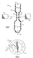
- Fig. 3 depicts a diagrammatic view of a low angle light scattering/particle detection system for use in the present invention, the system being depicted to shown unscattered light through a liquid medium;
- Fig. 4 depicts a partial, diagrammatic view of the low angle light scattering/ particle detection system of Fig. 3 , as taken during scattering events such as those previously depicted in Fig. 2 ;
- Fig. 5 is a perspective view of a measurement system in accordance with a first embodiment of the present invention.
- Fig 6 is a diagrammatic view of a measurement system in accordance with a second embodiment of the present invention.
- Fig. 7 is a side elevational view, taken in section of a sample container made in accordance with a preferred embodiment of the present invention.
- Fig. 7(a) is a side elevational view, also taken in section, of a sample container according to an alternative embodiment of the invention.
- Fig. 8 is the side elevational view of the sample container of Fig.7 , depicting the movement of a flow field in relation to the measurement system of Fig. 3 ;
- Fig. 9 is a side elevational view of a sample container made in accordance with the present invention.
- Fig. 10 is a side elevational view of the sample container of Fig. 9 ;
- Fig. 11 is a flow chart illustrating a method for performing antibody detection in accordance with another preferred embodiment of the invention using the measurement system of Fig. 5 .
- a series of tubular sample containers 14, 16, 18, 20, 22 are disposed within a cartridge 10.
- Each of the tubular sample containers includes a suspended matrix 30 of substantially noncompressible inert microparticles that permit movement of nonagglutinated reactants, such as red blood cells, while constraining agglutinated reactants.
- the matrix 30 is typically suspended within a gel in a lower portion 25 of the container, the gel having a density that is slightly lower than of the red blood cells to promote movement therethrough
- a sample and agglutination reactant is first received within an upper portion 23 or chamber of each container in the form of serum and cells that are incubated prior to movement through the matrix 30 of suspended particles.
- the upper portion 23 is separated from the lower portion 25 by means of a separating orifice having a diameter permitting the passage of cells therethrough, the serum and cell reagent being applied so as to create an air pocket or bubble between the upper portion and the reminder of the container.
- a user then applies a sufficient force, typically centrifugation, to effect this movement wherein a band of agglutinated reactants is formed above the matrix 30 for visual or automated detection.
- the container may also have an initial reaction zone 21.
- FIG. 1 illustrates a range of spatial separation patterns that are representative of positive, weakly positive, and negative agglutinations.
- Tubular sample container 12 demonstrates a strong positive reaction with a firm band 19 of agglutinates
- Container 14 shows a positive reaction that is somewhat weaker than that shown in container 12, as the agglutinate band has broken apart into smaller agglutinates.
- Container 16 demonstrates a weaker positive reaction with a smaller quantity of agglutinates being distributed throughout the middle portion of the matrix, and even settling on the bottom, as in container 18.
- Container 20 depicts a very weak positive reaction, with most of the cells being collected on the bottom.
- tubular sample container 22 depicts a clear negative reaction, with a button of cells 26 located on the bottom of the container, and no agglutinates dispersed within the matrix 30.
- the present invention relates to use of technique of low-angle light scattering to provide a more accurate and repeatable determination of agglutination without requiring spatial separation
- the basic theory of this technique follows:
- Low-angle light scattering is generally based on the principle that the absorption coefficient and the volume scattering function (VSF) completely characterize how a beam of laser light will propagate through water or other fluid.
- VSF volume scattering function
- this technique measures the intensity of light that is scattered through a range of small angles from the original direction of propagation as a result of particles in a detection area.
- the pick up angle of the incident e.g., the scattering angle
- the scattering angle varies between 0.1 to about 10 degrees.
- VSF Volume Scattering Function
- a monochromatic light beam 33 is formed by collimating the output of a diode or other form of laser or other type of light emitter 32. Scattered light is detected in this particular instance by means of a detector 36 that is defined by a predetermined number of coaxially arranged rings 38 allowing the passage of unscattered light.
- Fig. 3 illustrates an unscattered light beam 33 that is initially collimated by means of lens 33 and is focused onto the center of the detector 36 through a corresponding lens 37. As shown more clearly in Fig.
- each detector ring 38 is disposed so as to collect the portion of the light beam that is scattered into a particular solid angle ⁇ and defined by a narrow range of scattering angles ( ⁇ ) relative to the medium, typically on the order of about 0.1 to about 20 degrees.
- the herein described system is from the LISST Series Particle Measurement Systems, manufactured by Sequoia Scientific, Inc. of Redmond, Washington.
- VSF Volume Scattering Function
- the above-described technique is facilitated if the entrance and exit surfaces of a sample container are preferably approximately planar, made from a light transmissive material, and are substantially perpendicular to the angle of incident illumination in order to reduce artifacts that may result from scatter at the container surfaces.
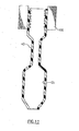
- FIGs. 5 and 6 two different embodiments are herein described for facilitating agglutination and localizing the particulate materials preferably in conjunction with the low angle scattering/particle detection system of Figs. 3 and 4 . It should be readily discernible, however, that other techniques can be employed within the inventive scope of the present invention.
- a low angle light scattering/particle measuring system having a laser emitter 32 and a detector 36, shown only schematically, but as described with regard to Figs. 3 and 4 , is aligned relative to a sample container 43 and more specifically to a measurement window 45 of the container.
- the lenses 35, 37, Fig. 3 , of the measuring system are not shown in this view for reasons of clarity.
- the sample container 43 for purposes of this embodiment is defined by three adjacent chambers 51, 53, 55 each linked by a common wall, preferably, each of the chambers being defined by a different inside diameter D1, D2 and D3, respectively. As shown, each of D 2 and D 3 are substantially larger than D 1 .
- transition zones 44, 48 are created between adjacent chambers so as to promote an agglutination reaction and more directly so as to create a cloud of cells when agitation of the sample container 43 occurs.
- a fluid having a predetermined density and viscosity is not required, as in the instance of centrifugation, in order to create mixing.
- mixing can be sufficiently accomplished, for example, through vertical (e.g., up and down) movement of the sample container 43 causing the mixture (not shown) to move, arrows 61, 63 between the adjacent chambers 51, 53, 55 and the transition zones 44, 48 therebetween of the sample container and producing the agglutination reaction as well as the moving field of cells.
- the movement could be made at an angle other than vertical.
- pumps or other aggressive liquid moving means capable of moving a quantity of liquid sample between the adjacent chambers for mixing can be used to draw the fluid between the chambers in a similar manner.
- a single pump can be used to draw the liquid upwardly into the chambers 51, 53 from chamber 55 wherein the force of gravity will cause the moving flow field to move past the measurement window.
- the sample container, as well as the various movement mechanisms that can be used in conjunction therewith, are described in greater detail in U.S. Publication Numbers US 2002/0076826 and US 2002/0081747 .
- a centrifuge shown only partially as 74, is used to support a plurality of tubular sample containers 70 into which patient sample is metered.
- the sample containers are generally as those described with regard to Fig. 1 , with some notable exceptions as described in greater detail below.
- each sample container 70 is a tube that includes a planar measurement window such as shown in Fig. 5 , to permit detection.
- Reactant material e.g., red blood cells
- a receiving fluid having a specified density and viscosity is provided in a lower portion (not shown) of the container.
- This material can include at least one liquid having a higher specific gravity than that of the blood cells.
- the other properties herein contained of this at least one additional fluid are designed to produce an environment that maintains the integrity of the red blood cells.
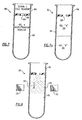
- Figs. 7-10 herein describing a proposed testing procedure for blood antibody detection ( Figs. 7 and 8 ) and an proposed testing procedure for A-B-O blood typing ( Figs. 9, 10 ), each using the presently described method.
- the sample containers 80 used in each of the above tests are somewhat similar to those described with regard to Fig. 1 .
- the restriction orifice 88 located between the upper portion 84 of the container and the lower potion 86 thereof is optional since spatial separation is not the preferred mode by which the cells are classified following reaction.
- a restriction orifice is added that promotes a dispersion of cells into a moving flow field.
- This restriction orifice is sized to be smaller than those of previously known systems requiring both chemical separation and spatial separation.
- the restriction orifice for this embodiment for example is approximately 1 millimeter as opposed to a 4 millimeter diameter required in known containers.
- a receiving fluid containing an agglutination reagent is added to the lower portion of the sample container, either as filled within a clinical analyzer (not shown) or sold as a prefilled quantity, the agglutination reagent being mixed with the receiving fluid to form a homogenous mixture.
- a patient serum is first added, followed by a RBC (red blood cell) or other suitable reagent, the serum and reagent being added preferably by means of a metering mechanism or pipette tip (not shown).
- RBC red blood cell
- the edges of the restriction orifice 88 provide a latch point for the metered serum material, thereby forming an air bubble 90 between the gel/reagent mixture and the serum/RBC reagent prior to the application of the centrifugal force.
- a sugar-based or other material having a density chosen to permit the red blood cells to move through it at a specified rate under centrifugal force is preferred.
- the sample container 80 can include two or more materials designated 94 and 96, respectively having differing material (density) properties.
- a higher density material can be provided in the lowest portion of the container while a lower density material can be provided in an intermediate portion of the container.
- the red blood cells would not settle at the bottom of the test container, but rather would cease migrating when they reach a neutral buoyancy rate. The latter is less sensitive to centrifugation, force and time and therefore may enable more rapid centrifugation of the sample container.
- the sample container 80 is incubated wherein the bound antibody is bound to the red blood cells.
- the sample container 80 is then centrifuged and test spun in order to group the cells and force same through the restriction orifice 88 where the cells are dispensed into a moving flow field into the reagent matrix below.
- the centrifuge promotes the agglutination reaction vis a vis the contained reagent.
- the moving cell field is pushed as a "cloud" into the measurement windows 87, 89 of the container 80 wherein the particle detection system 32, 36 determines the size distribution of particles in order to determine the strength of agglutination.
- the above read process can occur in several ways: First, the read can occur during centrifugation if synchronized with the rotation of the centrifuge. This read technique provides real time data relating to the state of the cells moving through the media, thereby making the measurement insensitive to the final position and minimizing the time to result, especially for strong agglutination reactions.
- the read process can also occur after the completion of centrifugation, but can proceed while the container (e.g. tube) is still within the centrifuge.
- This enables consolidation of hardware and uses the centrifuge as the location device that holds the tube(s) in fixed position.
- the light emitter 32 and detector 36 of the light scattering/particle detection system are arranged in a fixed location relative to the centrifuge, as the centrifuge rotates the tubes therebetween, per arrow 76.
- Each of the above read process steps can therefore be accomplished using this type of system station positioning.
- the above read/detection process can also be performed as a separate or off line procedure in which the tubes 70 can be placed in a separate device/apparatus (not shown) following the centrifugation process. This latter process may be required if there are space/size or other unique requirements that are not available internally to the centrifuge.
- the sample container can be removed or remain in the centrifuge for either an end point measurement or alternately an "on the fly” measurement can be made in which at least one image can be obtained while the centrifuge is slowing down or through use of a strobe.
- centrifugation techniques for example, involving a fixed angled centrifuge can be similarly utilized. This technique has been shown to create a "smear" of cells along the wall of the tubular sample container. A high-resolution vision system or a low-angle particle analysis system such as that shown in Figs. 3 and 4 can then inspect these cells. As with a swinging bucket centrifuge, it is possible to read the test tubes in the centrifuge, during or after centrifugation, or after the tubes have been removed from the centrifuge in a separate apparatus.
- a similar sample container 80 is shown for A-B-O blood typing preferably having a smaller restriction orifice, as compared to those of the prior art, for example Fig. 1 , in order to promote disposition of a moving flow field.
- whole blood and a liquid serum reagent are each added to the upper portion 84 of a sample container 80.
- a gel is added to the bottom portion 86 of the sample container 80, the gel having a density and viscosity that is lower than that of the agglutinated red blood cells to promote movement therethrough as a flow field.
- the sample container 80 is incubated sufficiently and centrifugation forces intimation of the cells, enhancing the agglutination reaction as the cells are dispensed through the restriction orifice 88 into the gel matrix and past the measurement windows 87, 89 permitting scanning by the detection system 32, 36.
- an A-B-O blood typing procedure/testing is performed by aspirating whole blood and serum reagent into the confines of the container and then promoting movement between the transition zones of the tip, thereby creating the agglutination reaction.
- An air bubble interface between the compartments of the container is optional for this type of testing.
- Antibody detection can also be performed using a sample container 100 similar in design to that depicted according to Fig. 5 .
- serum cell material is initially aspirated into the container 100.
- RBC reagent is then additionally aspirated into the container 100.
- the serum and the RBC reagent are then mixed through means of vertical agitation using a pump or other means in order to create bound antibodies.
- the sample container 100 is then axially spun by centrifuge or other means, the container preferably having a compartment (not shown) within same into which the cells are trapped as described in U.S. Patent No. 4,933,291 to Uaiss et al.
- Surplus material (reagent, serum, unbound antibodies) is then discharged from the sample container 100 and a wash fluid is aspirated into the container to wash the cells wherein the wash fluid and cells are suspended and the sample container is axially spun.
- the wash fluid is then discarded and agglutination reagent is then aspirated into the sample container.
- the cell/reagent mixture is resuspended and mixed by vertical agitation to create an agglutination reaction by means of the transition zones after which the passing mixture is read using the particle detection system and determining a size distribution of the passing particles.
- a magnetic bead or particle can be alternately attached to each cell/bound antibody.
- the sample container 43 can then be placed within a toroidal magnet 100, suspending the cells, permitting discharge of surplus material and use of a wash fluid to first wash unbound antibody from the cells and then adding an agglutination reagent, the magnet being used to retain the bound cells in the container 43 between washes and other processing steps.
Landscapes
- Health & Medical Sciences (AREA)
- Life Sciences & Earth Sciences (AREA)
- Chemical & Material Sciences (AREA)
- Engineering & Computer Science (AREA)
- Physics & Mathematics (AREA)
- Biomedical Technology (AREA)
- Hematology (AREA)
- General Physics & Mathematics (AREA)
- Immunology (AREA)
- Pathology (AREA)
- General Health & Medical Sciences (AREA)
- Biochemistry (AREA)
- Analytical Chemistry (AREA)
- Dispersion Chemistry (AREA)
- Biophysics (AREA)
- Ecology (AREA)
- Molecular Biology (AREA)
- Urology & Nephrology (AREA)
- Food Science & Technology (AREA)
- Medicinal Chemistry (AREA)
- Investigating Or Analysing Biological Materials (AREA)
- Investigating Or Analysing Materials By Optical Means (AREA)
- Optical Measuring Cells (AREA)
- Automatic Analysis And Handling Materials Therefor (AREA)
Claims (17)
- Verfahren zum Klassifizieren einer Agglutinationsreaktion ohne eine räumliche Trennung von agglutinierten und nicht agglutinierten Teilchen zu erfordern, wobei das Verfahren folgende Schritte umfasst:Bereitstellen einer Mischung aus einer Blutprobe und einem Agglutinationsreagens im Innern eines Probenbehälters (43);Bewegen der Mischung innerhalb des Probenbehälters, so dass eine Agglutinationsreaktion innerhalb des Behälters erfolgt, und dass die agglutinierten Zellen als Strömungsfeld an einem Messfenster (45) des Probenbehälters vorbei bewegt werden;Richten einer Lichtquelle eines Kleinwinkel-Teilchenmesssystems in den Probenbehälter und Abtasten des sich bewegenden Strömungsfeldes mit einem emittierten monochromatischen Lichtstrahl (33); undErkennen der Anzahl und Größenverteilung der Teilchen, basierend auf Streulicht, das auf die Teilchen fällt, um den Agglutinationsgrad der Mischung zu bestimmen, wobei das Licht über einen Winkelbereich zwischen ungefähr 0,1 Grad und 10 Grad von dem emittierten Lichtstrahl aus gestreut wird.
- Verfahren nach Anspruch 1, wobei der Schritt des Bewegens durch Zentrifugieren ausgeführt wird.
- Verfahren nach Anspruch 1, wobei der Schritt des Bewegens durch eine senkrechte Bewegung der Mischung innerhalb eines Reaktionsgefäßes ausgeführt wird, das als Probenbehälter dient.
- Verfahren nach Anspruch 3, wobei das Reaktionsgefäß (43) mindestens eine Übergangszone (44, 48) umfasst, die einen kleineren Innendurchmesser (D1) aufweist als ein angrenzender Abschnitt (D1, D2), durch den die Mischung bewegt wird.
- Verfahren nach Anspruch 2, wobei der Probenbehälter mindestens ein Fluid enthält, das eine spezifische Viskosität und spezifische Dichte aufweist, um die Probe vor dem Zentrifugieren desselben effektiv auszuschließen.
- Verfahren nach Anspruch 1, umfassend den Schritt des Klassifizierens der Probenflüssigkeit, basierend auf dem Schritt des Erkennens.
- Verfahren nach Anspruch 1, wobei der Schritt des Bewegens unter Verwendung eines Rührvorgangs des Probenbehälters ausgeführt wird.
- Verfahren nach Anspruch 7, wobei der Probenbehälter mindestens eine Kammer (51) umfasst, die einen kleineren Innendurchmesser (D1) als eine angrenzende Kammer (D2, D3) des Behälters aufweist, durch welche die Mischung bewegt wird.
- Verfahren nach Anspruch 1, wobei das Verfahren den Schritt des Blutgruppenbestimmens, basierend auf den Teilcheneigenschaften der Mischung, umfasst.
- Verfahren nach Anspruch 1, wobei der Schritt des Erkennens die Schritte des Erkennens von Antigenen in der Mischung umfasst.
- Gerät zum Klassifizieren einer Patientenprobe, wobei das Gerät Folgendes umfasst:mindestens einen Probenbehälter (43);Mittel zum Bewegen einer Menge von Probenfluid und mindestens eines Reagens, das innerhalb des mindestens einen Probenbehälters enthalten ist, um eine Agglutinationsreaktion zu schaffen, die ein sich bewegendes Strömungsfeld aufweist; undeinen Mechanismus zum Teilchenmessen, wobei der Mechanismus einen Lichtsender (32), der auf ein Messfenster (45) des mindestens einen Probenbehälters ausgerichtet ist, und einen Kleinwinkel-Lichtdetektor (36), der im Verhältnis zum Messfenster gegenüber angeordnet ist und Streulicht von einem emittierten Lichtstrahl erkennt, der den Probenbehälter abtastet, umfasst, wobei der Lichtsender einen monochromatischen Lichtstrahl emittiert, der ein Messvolumen abtastet und auf Teilchen in dem sich bewegenden Strömungsfeld fällt, und der Lichtdetektor basierend auf Licht, das über einen Bereich von kleinen Winkeln aus einer ursprünglichen Richtung des abgetasteten emittierten Lichtstrahls auf Grund von Teilchen, die in dem Messvolumen vorhanden sind, gestreut wird, die Größenverteilung und Konzentration von suspendierten Teilchen in dem sich bewegenden Strömungsfeld erkennt, um das Probenfluid effektiv zu klassifizieren, und wobei der Bereich kleiner Streuwinkel im Verhältnis zu dem emittierten Lichtstrahl zwischen ungefähr 0,1 Grad und ungefähr 10 Grad liegt,dadurch gekennzeichnet, dass das Bewegungsmittel eine Zentrifuge (74) oder ein senkrechtes Rührmittel umfasst.
- Gerät nach Anspruch 11, wobei das Bewegungsmittel das senkrechte Rührmittel umfasst, und der Probenbehälter (43) mindestens eine Kammer (51) umfasst, die einen Innendurchmesser (D1) aufweist, der kleiner ist als eine angrenzende Kammer (52, 53), um das Mischen zu fördern.
- Gerät nach Anspruch 11, wobei das Bewegungsmittel und das Messsystem nicht einstückig an dem Gerät bereitgestellt sind, wobei das Messsystem verwendbar ist, um Streulicht zu erkennen, nachdem der mindestens eine Probenbehälter aus dem Bewegungsmittel entfernt wurde.
- Gerät nach Anspruch 11, wobei das Bewegungsmittel und das Messsystem einstückig an dem Gerät bereitgestellt sind, so dass Messungen rechtzeitig im Anschluss an den Schritt des Mischens vorgenommen werden können.
- Gerät nach Anspruch 11, wobei die Klassifizierung das Erkennen des Agglutinationsgrades einer Patientenprobe umfasst.
- Gerät nach Anspruch 11, wobei die Klassifizierung das Erkennen der Menge mindestens eines Antigens in einer Patientenprobe umfasst.
- Gerät nach Anspruch 15, wobei die Klassifizierung ferner eine Blutgruppenbestimmung umfasst.
Applications Claiming Priority (4)
| Application Number | Priority Date | Filing Date | Title |
|---|---|---|---|
| US51375303P | 2003-10-23 | 2003-10-23 | |
| US513753P | 2003-10-23 | ||
| US968524 | 2004-10-19 | ||
| US10/968,524 US20070054405A1 (en) | 2003-10-23 | 2004-10-19 | Patient sample classification based upon low angle light scattering |
Publications (3)
| Publication Number | Publication Date |
|---|---|
| EP1526370A2 EP1526370A2 (de) | 2005-04-27 |
| EP1526370A3 EP1526370A3 (de) | 2005-05-04 |
| EP1526370B1 true EP1526370B1 (de) | 2012-03-21 |
Family
ID=34396608
Family Applications (1)
| Application Number | Title | Priority Date | Filing Date |
|---|---|---|---|
| EP04256535A Expired - Lifetime EP1526370B1 (de) | 2003-10-23 | 2004-10-22 | Klassifikation von Patientenproben auf Basis von Kleinwinkelstreuung |
Country Status (4)
| Country | Link |
|---|---|
| US (2) | US20070054405A1 (de) |
| EP (1) | EP1526370B1 (de) |
| JP (1) | JP4712345B2 (de) |
| AT (1) | ATE550639T1 (de) |
Cited By (1)
| Publication number | Priority date | Publication date | Assignee | Title |
|---|---|---|---|---|
| CN104884954A (zh) * | 2012-12-31 | 2015-09-02 | 高丽大学校产学协力团 | 测试基于离心式微流控法的血小板的多功能和药物反应的设备和方法 |
Families Citing this family (25)
| Publication number | Priority date | Publication date | Assignee | Title |
|---|---|---|---|---|
| KR101285643B1 (ko) * | 2006-09-25 | 2013-07-12 | 토루 오바타 | 겔화 측정 장치 및 시료 셀 |
| US8986456B2 (en) * | 2006-10-10 | 2015-03-24 | Asm America, Inc. | Precursor delivery system |
| US7850917B2 (en) * | 2008-03-11 | 2010-12-14 | Ortho-Clinical Diagnostics, Inc. | Particle agglutination in a tip |
| US20090274348A1 (en) | 2008-04-30 | 2009-11-05 | Ortho-Clinical Diagnostics, Inc. | Immunodiagnostic test apparatus having at least one imager to provide agglutination evaluations during centrifugration cycle |
| US9394608B2 (en) | 2009-04-06 | 2016-07-19 | Asm America, Inc. | Semiconductor processing reactor and components thereof |
| US8877655B2 (en) | 2010-05-07 | 2014-11-04 | Asm America, Inc. | Systems and methods for thin-film deposition of metal oxides using excited nitrogen-oxygen species |
| US8883270B2 (en) * | 2009-08-14 | 2014-11-11 | Asm America, Inc. | Systems and methods for thin-film deposition of metal oxides using excited nitrogen—oxygen species |
| US9096931B2 (en) | 2011-10-27 | 2015-08-04 | Asm America, Inc | Deposition valve assembly and method of heating the same |
| US9202727B2 (en) | 2012-03-02 | 2015-12-01 | ASM IP Holding | Susceptor heater shim |
| US9389229B2 (en) | 2012-07-18 | 2016-07-12 | Theranos, Inc. | Methods for detecting and measuring aggregation |
| US9558931B2 (en) | 2012-07-27 | 2017-01-31 | Asm Ip Holding B.V. | System and method for gas-phase sulfur passivation of a semiconductor surface |
| US9021985B2 (en) | 2012-09-12 | 2015-05-05 | Asm Ip Holdings B.V. | Process gas management for an inductively-coupled plasma deposition reactor |
| US10714315B2 (en) | 2012-10-12 | 2020-07-14 | Asm Ip Holdings B.V. | Semiconductor reaction chamber showerhead |
| US8894870B2 (en) | 2013-02-01 | 2014-11-25 | Asm Ip Holding B.V. | Multi-step method and apparatus for etching compounds containing a metal |
| US9484191B2 (en) | 2013-03-08 | 2016-11-01 | Asm Ip Holding B.V. | Pulsed remote plasma method and system |
| US9589770B2 (en) | 2013-03-08 | 2017-03-07 | Asm Ip Holding B.V. | Method and systems for in-situ formation of intermediate reactive species |
| US9018111B2 (en) | 2013-07-22 | 2015-04-28 | Asm Ip Holding B.V. | Semiconductor reaction chamber with plasma capabilities |
| US10167557B2 (en) | 2014-03-18 | 2019-01-01 | Asm Ip Holding B.V. | Gas distribution system, reactor including the system, and methods of using the same |
| US11015245B2 (en) | 2014-03-19 | 2021-05-25 | Asm Ip Holding B.V. | Gas-phase reactor and system having exhaust plenum and components thereof |
| US9404587B2 (en) | 2014-04-24 | 2016-08-02 | ASM IP Holding B.V | Lockout tagout for semiconductor vacuum valve |
| US10858737B2 (en) | 2014-07-28 | 2020-12-08 | Asm Ip Holding B.V. | Showerhead assembly and components thereof |
| US9890456B2 (en) | 2014-08-21 | 2018-02-13 | Asm Ip Holding B.V. | Method and system for in situ formation of gas-phase compounds |
| US10941490B2 (en) | 2014-10-07 | 2021-03-09 | Asm Ip Holding B.V. | Multiple temperature range susceptor, assembly, reactor and system including the susceptor, and methods of using the same |
| CN105223366B (zh) * | 2015-09-18 | 2017-01-18 | 深圳市达科为生物工程有限公司 | 用于血型检测的微管、一体式ABO/RhD血型定型检测卡及制备 |
| US10393643B2 (en) | 2016-09-07 | 2019-08-27 | Rochester Institute Of Technology | Optical vortex coronagraph scatterometer |
Family Cites Families (27)
| Publication number | Priority date | Publication date | Assignee | Title |
|---|---|---|---|---|
| DE2126393A1 (de) * | 1970-11-23 | 1971-12-02 | Talbot J | Verfahren und Vorrichtung zur Analyse von geometrischen Eigenschaften der Komponenten eines Systems |
| US4252538A (en) * | 1979-03-02 | 1981-02-24 | Engineering & Research Associates, Inc. | Apparatus and method for antibody screening, typing and compatibility testing of red blood cells |
| US4452902A (en) * | 1981-11-19 | 1984-06-05 | Labsystems Oy | Method and equipment for the measurement of properties of a liquid |
| US4762413A (en) * | 1984-09-07 | 1988-08-09 | Olympus Optical Co., Ltd. | Method and apparatus for measuring immunological reaction with the aid of fluctuation in intensity of scattered light |
| US4720465A (en) * | 1985-05-14 | 1988-01-19 | Fisher Scientific Company | Centrifugal kinetic agglutination assay |
| US4933291A (en) * | 1986-12-22 | 1990-06-12 | Eastman Kodak Company | Centrifugable pipette tip and pipette therefor |
| US5338689A (en) * | 1987-08-24 | 1994-08-16 | Stiftung Fur Diagnostische Forschung | Method and card for detecting antigens and/or antibodies |
| US5100805A (en) * | 1989-01-26 | 1992-03-31 | Seradyn, Inc. | Quantitative immunoassay system and method for agglutination assays |
| JPH03110473A (ja) * | 1989-09-26 | 1991-05-10 | Tokuyama Soda Co Ltd | 血液型自動判定方法及びその装置 |
| US5286452A (en) * | 1991-05-20 | 1994-02-15 | Sienna Biotech, Inc. | Simultaneous multiple assays |
| JP3283078B2 (ja) * | 1992-12-04 | 2002-05-20 | 興和株式会社 | 免疫学的測定装置 |
| US5552064A (en) * | 1993-02-26 | 1996-09-03 | Ortho Diagnostic Systems, Inc. | Column agglutination assay and device using biphasic centrifugation |
| US5594808A (en) * | 1993-06-11 | 1997-01-14 | Ortho Diagnostic Systems Inc. | Method and system for classifying agglutination reactions |
| US5656499A (en) * | 1994-08-01 | 1997-08-12 | Abbott Laboratories | Method for performing automated hematology and cytometry analysis |
| ES2156610T3 (es) * | 1996-12-18 | 2001-07-01 | Stiftung Fur Diagnostische For | Particulas sinteticas como reactivos de aglutinacion. |
| US5872860A (en) * | 1997-03-13 | 1999-02-16 | Ortho Diagnostic Systems, Inc. | Calibration cassette for use in calibrating an automated agglutination reaction analyzing system |
| JP3957864B2 (ja) * | 1998-03-02 | 2007-08-15 | 株式会社アイディエス | 凝固検体判定方法およびその装置 |
| US6153148A (en) * | 1998-06-15 | 2000-11-28 | Becton, Dickinson And Company | Centrifugal hematology disposable |
| TW499825B (en) * | 1999-02-23 | 2002-08-21 | Samsung Electro Mech | Method and device for coupling PCB sheet |
| DE19948587A1 (de) * | 1999-10-08 | 2001-04-12 | Dade Behring Marburg Gmbh | Spektralphotometrische und nephelometrische Detektionseinheit |
| JP4054500B2 (ja) * | 1999-12-27 | 2008-02-27 | オリンパス株式会社 | 抗原、抗体及びdna等の核酸を検出用のプローブとして用いた多項目検査方法 |
| US7452508B2 (en) * | 2000-02-22 | 2008-11-18 | Jacobs Merrit N | Aspirating and mixing of liquids within a probe tip |
| US20020196435A1 (en) * | 2000-11-22 | 2002-12-26 | Cohen David Samuel | Apparatus and methods for separating agglutinants and disperse particles |
| JP2002310886A (ja) * | 2001-04-11 | 2002-10-23 | Canon Inc | ディスクサイトメトリーによる分析方法及び装置 |
| WO2003043403A2 (en) * | 2001-11-19 | 2003-05-30 | Burstein Technologies, Inc. | Methods and apparatus for blood typing with optical bio-discs |
| JP2005017254A (ja) * | 2003-06-30 | 2005-01-20 | Sysmex Corp | 血小板凝集能測定装置 |
| WO2005099408A2 (en) * | 2004-04-10 | 2005-10-27 | Michael Trainer | Methods and apparatus for determining particle characterics by measuring scattered light |
-
2004
- 2004-10-19 US US10/968,524 patent/US20070054405A1/en not_active Abandoned
- 2004-10-22 EP EP04256535A patent/EP1526370B1/de not_active Expired - Lifetime
- 2004-10-22 AT AT04256535T patent/ATE550639T1/de active
- 2004-10-22 JP JP2004308556A patent/JP4712345B2/ja not_active Expired - Fee Related
-
2010
- 2010-05-13 US US12/779,358 patent/US9310286B2/en active Active
Cited By (2)
| Publication number | Priority date | Publication date | Assignee | Title |
|---|---|---|---|---|
| CN104884954A (zh) * | 2012-12-31 | 2015-09-02 | 高丽大学校产学协力团 | 测试基于离心式微流控法的血小板的多功能和药物反应的设备和方法 |
| CN104884954B (zh) * | 2012-12-31 | 2018-07-24 | 高丽大学校产学协力团 | 测试基于离心式微流控法的血小板的多功能和药物反应的设备和方法 |
Also Published As
| Publication number | Publication date |
|---|---|
| US9310286B2 (en) | 2016-04-12 |
| US20100221757A1 (en) | 2010-09-02 |
| EP1526370A3 (de) | 2005-05-04 |
| ATE550639T1 (de) | 2012-04-15 |
| JP2005134389A (ja) | 2005-05-26 |
| EP1526370A2 (de) | 2005-04-27 |
| US20070054405A1 (en) | 2007-03-08 |
| JP4712345B2 (ja) | 2011-06-29 |
Similar Documents
| Publication | Publication Date | Title |
|---|---|---|
| US9310286B2 (en) | Patient sample classification based upon low angle light scattering | |
| US5256376A (en) | Agglutination detection apparatus | |
| US10073086B2 (en) | Device and method for carrying out haematological and biochemical measurements from a biological sample | |
| US4197088A (en) | Method for qualitative and quantitative determination of immunological reactions | |
| US6277641B1 (en) | Methods for analyzing the presence and concentration of multiple analytes using a diffusion-based chemical sensor | |
| US5858648A (en) | Assays using reference microparticles | |
| JP3593315B2 (ja) | 血球計数を行うための使い捨て装置 | |
| CN101454653B (zh) | 用于分析荧光标记颗粒的基于芯片的流式细胞器类系统 | |
| US4400353A (en) | Electro-optical system for use in evaluating immunological reactions | |
| WO2017035539A1 (en) | Fluid holding and dispensing micro-feature | |
| CA2921647C (en) | Method for determining the result of an agglutination reaction and microplate for determining products of agglutination reactions | |
| US9568488B2 (en) | Biological sample analyzing apparatus | |
| EP0269526B1 (de) | Verfahren zur quantitativen Bestimmung der Antigene und Antikörper | |
| EP4354144A1 (de) | Mikrofluidischer chip zum testen von proteinantigen sowie verfahren, kit und system | |
| EP0190019B1 (de) | Verfahren und Vorrichtung zur Handhabung von Flüssigkeiten | |
| CA2529362A1 (en) | Method for measuring substance having affinity | |
| US6342397B1 (en) | Homogeneous biospecific assay using a solid phase, two-photon excitation and confocal fluorescence detection | |
| KR20220159382A (ko) | 라텍스 응집법에 의한 목적 물질의 측정 방법 및 그 시약 | |
| EP1084408B1 (de) | Homogener biospezifischer versuchsansatz mit festphase, zwei-photonen-anregung und konfokaler fluoreszenzbestimmung | |
| CN100594382C (zh) | 搅拌方法、反应容器以及使用该反应容器的测定装置、测定方法 | |
| EP0021236B1 (de) | Verfahren zur optischen Prüfung flüssiger Proben | |
| JP7327944B2 (ja) | ラテックス凝集法による目的物質の測定方法、およびその試薬 | |
| CA1139126A (en) | Apparatus and method for quantitative determination of immunological reactions | |
| JP2001183377A (ja) | 免疫学的多項目検査方法 | |
| JP2002286722A (ja) | 凝集反応分析装置 |
Legal Events
| Date | Code | Title | Description |
|---|---|---|---|
| PUAI | Public reference made under article 153(3) epc to a published international application that has entered the european phase |
Free format text: ORIGINAL CODE: 0009012 |
|
| PUAL | Search report despatched |
Free format text: ORIGINAL CODE: 0009013 |
|
| AK | Designated contracting states |
Kind code of ref document: A2 Designated state(s): AT BE BG CH CY CZ DE DK EE ES FI FR GB GR HU IE IT LI LU MC NL PL PT RO SE SI SK TR |
|
| AX | Request for extension of the european patent |
Extension state: AL HR LT LV MK |
|
| AK | Designated contracting states |
Kind code of ref document: A3 Designated state(s): AT BE BG CH CY CZ DE DK EE ES FI FR GB GR HU IE IT LI LU MC NL PL PT RO SE SI SK TR |
|
| AX | Request for extension of the european patent |
Extension state: AL HR LT LV MK |
|
| 17P | Request for examination filed |
Effective date: 20051103 |
|
| AKX | Designation fees paid |
Designated state(s): AT BE BG CH CY CZ DE DK EE ES FI FR GB GR HU IE IT LI LU MC NL PL PT RO SE SI SK TR |
|
| 17Q | First examination report despatched |
Effective date: 20080401 |
|
| GRAP | Despatch of communication of intention to grant a patent |
Free format text: ORIGINAL CODE: EPIDOSNIGR1 |
|
| GRAS | Grant fee paid |
Free format text: ORIGINAL CODE: EPIDOSNIGR3 |
|
| GRAA | (expected) grant |
Free format text: ORIGINAL CODE: 0009210 |
|
| AK | Designated contracting states |
Kind code of ref document: B1 Designated state(s): AT BE BG CH CY CZ DE DK EE ES FI FR GB GR HU IE IT LI LU MC NL PL PT RO SE SI SK TR |
|
| REG | Reference to a national code |
Ref country code: GB Ref legal event code: FG4D |
|
| REG | Reference to a national code |
Ref country code: CH Ref legal event code: EP |
|
| REG | Reference to a national code |
Ref country code: IE Ref legal event code: FG4D |
|
| REG | Reference to a national code |
Ref country code: CH Ref legal event code: NV Representative=s name: E. BLUM & CO. AG PATENT- UND MARKENANWAELTE VSP |
|
| REG | Reference to a national code |
Ref country code: AT Ref legal event code: REF Ref document number: 550639 Country of ref document: AT Kind code of ref document: T Effective date: 20120415 |
|
| REG | Reference to a national code |
Ref country code: DE Ref legal event code: R096 Ref document number: 602004036976 Country of ref document: DE Effective date: 20120516 |
|
| REG | Reference to a national code |
Ref country code: NL Ref legal event code: VDEP Effective date: 20120321 |
|
| PG25 | Lapsed in a contracting state [announced via postgrant information from national office to epo] |
Ref country code: FI Free format text: LAPSE BECAUSE OF FAILURE TO SUBMIT A TRANSLATION OF THE DESCRIPTION OR TO PAY THE FEE WITHIN THE PRESCRIBED TIME-LIMIT Effective date: 20120321 Ref country code: GR Free format text: LAPSE BECAUSE OF FAILURE TO SUBMIT A TRANSLATION OF THE DESCRIPTION OR TO PAY THE FEE WITHIN THE PRESCRIBED TIME-LIMIT Effective date: 20120622 |
|
| REG | Reference to a national code |
Ref country code: AT Ref legal event code: MK05 Ref document number: 550639 Country of ref document: AT Kind code of ref document: T Effective date: 20120321 |
|
| PG25 | Lapsed in a contracting state [announced via postgrant information from national office to epo] |
Ref country code: CY Free format text: LAPSE BECAUSE OF FAILURE TO SUBMIT A TRANSLATION OF THE DESCRIPTION OR TO PAY THE FEE WITHIN THE PRESCRIBED TIME-LIMIT Effective date: 20120321 |
|
| PG25 | Lapsed in a contracting state [announced via postgrant information from national office to epo] |
Ref country code: SE Free format text: LAPSE BECAUSE OF FAILURE TO SUBMIT A TRANSLATION OF THE DESCRIPTION OR TO PAY THE FEE WITHIN THE PRESCRIBED TIME-LIMIT Effective date: 20120321 Ref country code: RO Free format text: LAPSE BECAUSE OF FAILURE TO SUBMIT A TRANSLATION OF THE DESCRIPTION OR TO PAY THE FEE WITHIN THE PRESCRIBED TIME-LIMIT Effective date: 20120321 Ref country code: PL Free format text: LAPSE BECAUSE OF FAILURE TO SUBMIT A TRANSLATION OF THE DESCRIPTION OR TO PAY THE FEE WITHIN THE PRESCRIBED TIME-LIMIT Effective date: 20120321 Ref country code: EE Free format text: LAPSE BECAUSE OF FAILURE TO SUBMIT A TRANSLATION OF THE DESCRIPTION OR TO PAY THE FEE WITHIN THE PRESCRIBED TIME-LIMIT Effective date: 20120321 Ref country code: SI Free format text: LAPSE BECAUSE OF FAILURE TO SUBMIT A TRANSLATION OF THE DESCRIPTION OR TO PAY THE FEE WITHIN THE PRESCRIBED TIME-LIMIT Effective date: 20120321 Ref country code: BE Free format text: LAPSE BECAUSE OF FAILURE TO SUBMIT A TRANSLATION OF THE DESCRIPTION OR TO PAY THE FEE WITHIN THE PRESCRIBED TIME-LIMIT Effective date: 20120321 Ref country code: CZ Free format text: LAPSE BECAUSE OF FAILURE TO SUBMIT A TRANSLATION OF THE DESCRIPTION OR TO PAY THE FEE WITHIN THE PRESCRIBED TIME-LIMIT Effective date: 20120321 |
|
| PG25 | Lapsed in a contracting state [announced via postgrant information from national office to epo] |
Ref country code: SK Free format text: LAPSE BECAUSE OF FAILURE TO SUBMIT A TRANSLATION OF THE DESCRIPTION OR TO PAY THE FEE WITHIN THE PRESCRIBED TIME-LIMIT Effective date: 20120321 Ref country code: PT Free format text: LAPSE BECAUSE OF FAILURE TO SUBMIT A TRANSLATION OF THE DESCRIPTION OR TO PAY THE FEE WITHIN THE PRESCRIBED TIME-LIMIT Effective date: 20120723 |
|
| PLBE | No opposition filed within time limit |
Free format text: ORIGINAL CODE: 0009261 |
|
| STAA | Information on the status of an ep patent application or granted ep patent |
Free format text: STATUS: NO OPPOSITION FILED WITHIN TIME LIMIT |
|
| PG25 | Lapsed in a contracting state [announced via postgrant information from national office to epo] |
Ref country code: NL Free format text: LAPSE BECAUSE OF FAILURE TO SUBMIT A TRANSLATION OF THE DESCRIPTION OR TO PAY THE FEE WITHIN THE PRESCRIBED TIME-LIMIT Effective date: 20120321 Ref country code: AT Free format text: LAPSE BECAUSE OF FAILURE TO SUBMIT A TRANSLATION OF THE DESCRIPTION OR TO PAY THE FEE WITHIN THE PRESCRIBED TIME-LIMIT Effective date: 20120321 Ref country code: DK Free format text: LAPSE BECAUSE OF FAILURE TO SUBMIT A TRANSLATION OF THE DESCRIPTION OR TO PAY THE FEE WITHIN THE PRESCRIBED TIME-LIMIT Effective date: 20120321 |
|
| 26N | No opposition filed |
Effective date: 20130102 |
|
| REG | Reference to a national code |
Ref country code: DE Ref legal event code: R097 Ref document number: 602004036976 Country of ref document: DE Effective date: 20130102 |
|
| PG25 | Lapsed in a contracting state [announced via postgrant information from national office to epo] |
Ref country code: ES Free format text: LAPSE BECAUSE OF FAILURE TO SUBMIT A TRANSLATION OF THE DESCRIPTION OR TO PAY THE FEE WITHIN THE PRESCRIBED TIME-LIMIT Effective date: 20120702 |
|
| PG25 | Lapsed in a contracting state [announced via postgrant information from national office to epo] |
Ref country code: MC Free format text: LAPSE BECAUSE OF NON-PAYMENT OF DUE FEES Effective date: 20121031 |
|
| REG | Reference to a national code |
Ref country code: IE Ref legal event code: MM4A |
|
| PG25 | Lapsed in a contracting state [announced via postgrant information from national office to epo] |
Ref country code: BG Free format text: LAPSE BECAUSE OF FAILURE TO SUBMIT A TRANSLATION OF THE DESCRIPTION OR TO PAY THE FEE WITHIN THE PRESCRIBED TIME-LIMIT Effective date: 20120621 Ref country code: IE Free format text: LAPSE BECAUSE OF NON-PAYMENT OF DUE FEES Effective date: 20121022 |
|
| PG25 | Lapsed in a contracting state [announced via postgrant information from national office to epo] |
Ref country code: TR Free format text: LAPSE BECAUSE OF FAILURE TO SUBMIT A TRANSLATION OF THE DESCRIPTION OR TO PAY THE FEE WITHIN THE PRESCRIBED TIME-LIMIT Effective date: 20120321 |
|
| PG25 | Lapsed in a contracting state [announced via postgrant information from national office to epo] |
Ref country code: LU Free format text: LAPSE BECAUSE OF NON-PAYMENT OF DUE FEES Effective date: 20121022 |
|
| PG25 | Lapsed in a contracting state [announced via postgrant information from national office to epo] |
Ref country code: HU Free format text: LAPSE BECAUSE OF FAILURE TO SUBMIT A TRANSLATION OF THE DESCRIPTION OR TO PAY THE FEE WITHIN THE PRESCRIBED TIME-LIMIT Effective date: 20041022 |
|
| REG | Reference to a national code |
Ref country code: FR Ref legal event code: PLFP Year of fee payment: 12 |
|
| PGFP | Annual fee paid to national office [announced via postgrant information from national office to epo] |
Ref country code: FR Payment date: 20150908 Year of fee payment: 12 |
|
| REG | Reference to a national code |
Ref country code: FR Ref legal event code: ST Effective date: 20170630 |
|
| PG25 | Lapsed in a contracting state [announced via postgrant information from national office to epo] |
Ref country code: FR Free format text: LAPSE BECAUSE OF NON-PAYMENT OF DUE FEES Effective date: 20161102 |
|
| PGFP | Annual fee paid to national office [announced via postgrant information from national office to epo] |
Ref country code: DE Payment date: 20171018 Year of fee payment: 14 |
|
| PGFP | Annual fee paid to national office [announced via postgrant information from national office to epo] |
Ref country code: CH Payment date: 20171013 Year of fee payment: 14 Ref country code: GB Payment date: 20171018 Year of fee payment: 14 Ref country code: IT Payment date: 20171024 Year of fee payment: 14 |
|
| REG | Reference to a national code |
Ref country code: DE Ref legal event code: R119 Ref document number: 602004036976 Country of ref document: DE |
|
| REG | Reference to a national code |
Ref country code: CH Ref legal event code: PL |
|
| GBPC | Gb: european patent ceased through non-payment of renewal fee |
Effective date: 20181022 |
|
| PG25 | Lapsed in a contracting state [announced via postgrant information from national office to epo] |
Ref country code: DE Free format text: LAPSE BECAUSE OF NON-PAYMENT OF DUE FEES Effective date: 20190501 |
|
| PG25 | Lapsed in a contracting state [announced via postgrant information from national office to epo] |
Ref country code: CH Free format text: LAPSE BECAUSE OF NON-PAYMENT OF DUE FEES Effective date: 20181031 Ref country code: LI Free format text: LAPSE BECAUSE OF NON-PAYMENT OF DUE FEES Effective date: 20181031 |
|
| PG25 | Lapsed in a contracting state [announced via postgrant information from national office to epo] |
Ref country code: GB Free format text: LAPSE BECAUSE OF NON-PAYMENT OF DUE FEES Effective date: 20181022 Ref country code: IT Free format text: LAPSE BECAUSE OF NON-PAYMENT OF DUE FEES Effective date: 20181022 |