EP1514112B1 - Method for identifying an allosteric effector of a receptor - Google Patents
Method for identifying an allosteric effector of a receptor Download PDFInfo
- Publication number
- EP1514112B1 EP1514112B1 EP03748170A EP03748170A EP1514112B1 EP 1514112 B1 EP1514112 B1 EP 1514112B1 EP 03748170 A EP03748170 A EP 03748170A EP 03748170 A EP03748170 A EP 03748170A EP 1514112 B1 EP1514112 B1 EP 1514112B1
- Authority
- EP
- European Patent Office
- Prior art keywords
- receptor
- ligand
- dissociation
- effector
- amplitude
- Prior art date
- Legal status (The legal status is an assumption and is not a legal conclusion. Google has not performed a legal analysis and makes no representation as to the accuracy of the status listed.)
- Expired - Lifetime
Links
Images


Classifications
-
- G—PHYSICS
- G01—MEASURING; TESTING
- G01N—INVESTIGATING OR ANALYSING MATERIALS BY DETERMINING THEIR CHEMICAL OR PHYSICAL PROPERTIES
- G01N33/00—Investigating or analysing materials by specific methods not covered by groups G01N1/00 - G01N31/00
- G01N33/48—Biological material, e.g. blood, urine; Haemocytometers
- G01N33/50—Chemical analysis of biological material, e.g. blood, urine; Testing involving biospecific ligand binding methods; Immunological testing
- G01N33/53—Immunoassay; Biospecific binding assay; Materials therefor
- G01N33/566—Immunoassay; Biospecific binding assay; Materials therefor using specific carrier or receptor proteins as ligand binding reagents where possible specific carrier or receptor proteins are classified with their target compounds
-
- C—CHEMISTRY; METALLURGY
- C07—ORGANIC CHEMISTRY
- C07K—PEPTIDES
- C07K5/00—Peptides containing up to four amino acids in a fully defined sequence; Derivatives thereof
- C07K5/04—Peptides containing up to four amino acids in a fully defined sequence; Derivatives thereof containing only normal peptide links
- C07K5/06—Dipeptides
- C07K5/06086—Dipeptides with the first amino acid being basic
-
- G—PHYSICS
- G01—MEASURING; TESTING
- G01N—INVESTIGATING OR ANALYSING MATERIALS BY DETERMINING THEIR CHEMICAL OR PHYSICAL PROPERTIES
- G01N33/00—Investigating or analysing materials by specific methods not covered by groups G01N1/00 - G01N31/00
- G01N33/48—Biological material, e.g. blood, urine; Haemocytometers
- G01N33/50—Chemical analysis of biological material, e.g. blood, urine; Testing involving biospecific ligand binding methods; Immunological testing
- G01N33/53—Immunoassay; Biospecific binding assay; Materials therefor
- G01N33/536—Immunoassay; Biospecific binding assay; Materials therefor with immune complex formed in liquid phase
- G01N33/542—Immunoassay; Biospecific binding assay; Materials therefor with immune complex formed in liquid phase with steric inhibition or signal modification, e.g. fluorescent quenching
-
- G—PHYSICS
- G01—MEASURING; TESTING
- G01N—INVESTIGATING OR ANALYSING MATERIALS BY DETERMINING THEIR CHEMICAL OR PHYSICAL PROPERTIES
- G01N33/00—Investigating or analysing materials by specific methods not covered by groups G01N1/00 - G01N31/00
- G01N33/48—Biological material, e.g. blood, urine; Haemocytometers
- G01N33/50—Chemical analysis of biological material, e.g. blood, urine; Testing involving biospecific ligand binding methods; Immunological testing
- G01N33/53—Immunoassay; Biospecific binding assay; Materials therefor
- G01N33/557—Immunoassay; Biospecific binding assay; Materials therefor using kinetic measurement, i.e. time rate of progress of an antigen-antibody interaction
Definitions
- the present invention relates to a method for demonstrating an allosteric effector of a receptor.
- allosteric effectors or modulators exerts its effect by binding to sites, present on the surface of the receptors, but topologically distinct from the endogenous ligand binding site. Their interaction with the receptor is uncompetitive with respect to that of the endogenous ligand. By binding to their regulatory sites, the effectors have little or no effect on receptor responses in the absence of endogenous ligand. On the other hand, they modify the binding properties of competitive endogenous ligands and alter the biological responses evoked by said ligand.
- Allosteric effectors are of particular interest in the therapeutic field in that their action is not "constitutive” but only occurs when the endogenous ligand stimulates the receptor. In cases where such molecules have been identified and placed on the market, patient comfort and effect selectivity have always been greatly improved, for example in comparison with the observed effect of administering a drug. agonist (Pan et al., 2001).
- the authors have indifferently used either agonists or antagonists as radioligands, and defined the positive or negative character of the modulator by its ability to accelerate (positive) or slow down (negative) the dissociation rate of the radioligand used. .
- the second most common approach to identifying an allosteric effector is to measure the amount of radioligand / receptor complex at a subset radioligand concentration. Under these conditions, the fact that a third molecule causes an increase in the amount of radioligand / receptor complex is interpreted in terms of potentiation of the binding by an allosteric effect.
- the third experimental approach is to establish the saturation curve of a radoligand in the presence and absence of allosteric effector.
- the 5HT-modulin effector reduces the amount of receptor sites without affecting the apparent affinity determined for the radioligand used.
- FRET fluorescence energy transfer
- the receptor can be made fluorescent by means of a fluorescent protein, for example a protein derived from the jellyfish Aequorea victoria whose corresponding gene has been sequenced, cloned (Prasher et al., 1992) and optimized for good expression in higher eukaryotes ( Cormack et al., 1996). It can be fused with the gene encoding another protein, including a G-protein coupled receptor (Galzi & Alix, 1997, Weill et al., 1999, Vollmer et al., 1999, Valenzuela et al., 2001). to allow the expression of a fluorescent receptor protein.
- a fluorescent protein for example a protein derived from the jellyfish Aequorea victoria whose corresponding gene has been sequenced, cloned (Prasher et al., 1992) and optimized for good expression in higher eukaryotes ( Cormack et al., 1996). It can be fused with the gene encoding another protein, including a
- the combination of the fluorescent receptor protein with one of its fluorescent ligands allows the formation of a non-covalent receptor-ligand complex that is detected by fluorescence energy transfer (Galzi & Alix, 1997; Vollmer et al., 1999). Valenzuela et al., 2001) and whose kinetics of formation and dissociation can be measured (Palanché et al., 2001, Valenzuela et al., 2001).
- One of the aims of the invention is to provide a method that is simple to implement, rapid and sensitive, making it possible to carry out quantitative measurements of the interactions between a receptor and a ligand marker of the site of the endogenous ligand, and making it possible to detect the presence of a possible allosteric effector.
- Another object of the invention is to enable real-time measurements of the interactions of a marker ligand of the site of the endogenous ligand with its receptor, making it possible to detect the presence of a possible allosteric effector.
- Another object of the invention is to allow a time-resolved measurement of the kinetics of association and dissociation of a fluorescent ligand, marker of the site of the endogenous ligand, with its receptor, and to detect the presence of a possible allosteric effector.
- Another object of the invention is to enable the detection of the interaction between a receptor and its endogenous ligand by fluorescence energy transfer in fluorescent endogenous ligand concentration ranges considerably greater (from nanomolar to micromolar) than when a radioligand (nanomolar domain) is used.
- the present invention makes it possible to detect by energy transfer a greater number of conformational states of the receptor, which greatly facilitates the identification and interpretation of the effects of supposed modulator agents.
- One of the aims of the invention is to provide an experimental measurement of the constants of association speed and dissociation of a fluorescent ligand, marker of the site of the endogenous ligand, with its receptor, allowing the detection of the presence of a possible allosteric effector.
- the invention also aims to provide a method of detecting an allosteric effector of a receptor, generalizable to many receptor proteins and their ligands.
- the invention also aims to provide a method of detecting an allosteric effector of a receptor, automatable, requiring neither the purification of the receptor nor that of the ligand.
- the invention also aims to provide a method for detecting an allosteric effector of a receptor, non-polluting since it does not use radioactivity, which is economical since it uses visible light and can be used on existing equipment.
- the invention answers the technical problem posed by the determination of the competitive or non-competitive nature of the interaction between a receptor and a pharmacological agent that is supposed to act as an effector of a receptor or as a competitor of the endogenous ligand of a receptor, by measurements kinetics of association and dissociation.
- the allosteric effector of a receptor will alter the kinetics of association or dissociation of a receptor while an endogenous ligand competing agent will have no effect on these kinetics.
- ligand, endogenous ligand site marker refers to a ligand that binds to the site of the endogenous ligand as opposed to an eventual allosteric effector, which binds to a site distinct from that of the endogenous ligand.
- the endogenous ligand site marker competitively interacts with the endogenous ligand (mutually exclusive binding), whereas the allosteric effector interacts noncompetitively with the endogenous ligand.
- an “allosteric effector” or “allosteric modulator” is a molecule or ligand or biologically active substance that modulates the binding properties of the ligand as well as the functional properties of the receptor, without competing with said ligand.
- ligand any molecule capable of binding to a receptor in a non-covalent manner by binding to the same site as the endogenous ligand, and that a ligand can be an agonist or an antagonist, an agonist being capable, by binding, of triggering a biological response and an antagonist not triggering a biological response and being able to block the effect of the agonist.
- An allosteric effector is also a ligand of the receptor but it binds with the receptor on another binding site.
- the term "allosteric effector” or “allosteric modulator” will be used and not "ligand”.
- FIG. 3 An equivalent version of this scheme for a G protein-coupled receptor capable of activating a Gs protein, itself responsible for the activation of an adenylate cyclase, is described in Tucek and Proska (1995) and in Hall (2000).
- the protein particularly the receptor, exists in two conformations that are in spontaneous equilibrium with each other.
- This equilibrium is described by the constant L 0 whose value is given by the ratio of fractional concentration [R] / [A], R being the state of rest and A, the active state.
- receptor denotes any molecule of a protein nature capable of interacting noncovalently with a pharmacological agent.
- a neurotransmitter receptor, hormone, growth factor, etc. capable of producing, after interaction with a pharmacological ligand, a measurable signal transduction response in vivo and in vitro is used. .
- competitive interaction is meant an interaction of two molecules with a receptor taking place at a single site, and a ligand is said to be “competitive” when it interferes with the binding of another ligand in a steric manner.
- non-competitive interaction means an interaction of two molecules with a receptor taking place at topologically distinct sites, and a ligand is said to be “non-competitive” when it interferes with the binding of another ligand, the two ligands interacting with sites topologically distinct from the receptor.
- association in the expression “association of the complex” is the action for a ligand to bind to a receptor protein.
- the receptor protein can adopt several conformations or states, it is sometimes possible to discern the binding of a ligand to different conformations by differences in kinetics of association.
- association kinetics refers to the time course of an association reaction.
- the kinetics can be either monoexponential or pluriexponential. In the case where they are pluriexponential, they are decomposed into a sum of monoexponential relaxations each characterized by a speed of association and an amplitude.
- ⁇ the amplitude
- k app the apparent rate constant of the reaction
- T the time.
- dissociation in the expression “dissociation of the complex” is the action for a ligand to leave a receptor site.
- Dissociation can be achieved in at least two ways: a) by strongly diluting the receptor-ligand mixture to favor dissociation over the association, or 2) by adding a highly competitive ligand to a very large extent. a site left vacant by the dissociated ligand.
- the receptor protein can adopt several conformations or states, it is sometimes possible to discern the dissociation between a ligand and its receptor having different conformations, by differences in kinetics of dissociation between the different conformations.
- the kinetics of dissociation is the time course of a dissociation reaction.
- the kinetics can be either monoexponential or pluriexponential. In the case where they are pluriexponential, they are decomposed into a sum of monoexponential relaxations characterized by a dissociation rate and an amplitude.
- ⁇ the amplitude
- T the time.
- biological response refers to any variation in the metabolism of cells, tissues or organisms.
- cells for example, it will be possible to determine variations in pH, ion concentration, formation of metabolites such as GTP or cAMP, gene expression, cell morphology (measurement of the percentage of cells that have changed of form), cell proliferation, among others. Examples are given below.
- appropriate physiological conditions all conditions of pH, ionic concentrations and composition, and nutrient supplements as close as possible to those encountered in the whole organism. These conditions will be chosen so that the experiment carried out is carried out under conditions as close as possible to those which could be obtained by carrying out the measurement in the whole organism.
- the expression "capable of modulating at least one of the responses” means that in a set of measurable responses that can be stimulated by a receiver, at least one must be modified by the effector.
- the modification or modulation can relate to the time of establishment of the response, its frequency, its amplitude, its duration, its rate of extinction, as well as its sensitivity to the agonist.
- the general test is to determine the activation of G protein by measuring GTP binding (Befort et al., 1996).
- Other more specific measurements involve, for example, determinations of intracellular concentrations of cAMP, inositol phosphates, calcium, activation measures of gene transcription or oncogenic activity, depending on the type of specific coupling of the receiver considered.
- the most direct measurements are ion current determinations (Hille, 1992).
- Other measures may, for example, involve gene transcription determinations or enzyme activations.
- the general tests are those of proliferation, differentiation or cell survival, frequently also phosphorylations of specific substrates (Honneger et al., 1988) of each receptor and detection by antibodies. specific phosphoamino acids.
- the signal transduction tests are those of the transcription of genes in which reporter genes, for example "chromogenic" genes are placed under the control of specific promoters of the transduction pathways of the receptor studied (Ko et al. 2002).
- cAMP accumulation responses various protocols may be employed, including a radioimmunoassay as described in Palanché et al. (2001) or Hausdorff et al. (1990).
- the binding of GTP to the G protein may be recorded for example as described in Fawzi et al. (2001) or Vuong and Chabre (1990).
- Cell proliferation will for example be analyzed according to the protocol described in Burstein et al (1997).
- protein kinases may for example be studied as described in Vollmer et al. (1999) or Yuan et al. (2000).
- the pH variation can be measured (Nicolini et al., 1995).
- an “autofluorescent protein” is a natural or synthetic protein in which the chromophore is formed by an autocatalytic reaction between amino acids of the protein without requiring the addition of a prosthetic group (chromophore), and whose fluorescence properties are intrinsic to the monomer.
- fluorescence energy transfer refers to a physical, distance-dependent process by which energy is transmitted non-radiatively from one excited chromophore, the donor, to another chromophore. acceptor, by dipole-dipole interaction (Förster, 1951, Wu and Brand, 1994, Clegg, 1995).
- the energy transfer can be observed either by a decrease in the amplitude of the emission of the donor, or by an increase in the amplitude of the excitation and the emission of the acceptor. If the acceptor is not fluorescent, but has a spectrum of excitation at least partly covering the donor's emission spectrum, the energy transfer can be detected in the form of a reduction in the amplitude of the emission of the donor.
- the transfer signal can not persist if the experimental conditions do not allow interaction between the fluorescent ligand and the fluorescent receptor. Similarly, if one of the two interacting partners does not carry a suitable chromophore, the possible variations in fluorescence observed for the other partner can not be attributed to a process of energy transfer.
- change or “fluorescence variation”, defined in the context of energy transfer, refer to any modifications of 1) the amplitude of the acceptor fluorescence signal, 2) the amplitude of the spectrum. excitation or 3) the amplitude of the donor emission signal. Variations or changes in fluorescence should not be observed if one of the two partners does not carry a suitable chromophore or fluorophore (see below) or if the interaction between the fluorescent partners is inhibited, for example by an excess of a competing agent.
- the first condition results in the fact that the excitation of the donor then leads concomitantly a decrease in the amplitude of the emission of the donor and the appearance of an emission signal of the acceptor. This makes it possible to detect the interactions between the donor and the acceptor and / or to measure their distance.
- the existence of the variation of the aforesaid kinetic is also detected.
- optimal codons indicates the codon substitution of the wild-type protein by their preferred counterparts of the host organism, with no change of code and therefore no protein sequence change.
- UV GFP with the following mutations: F99S, M153T, V163A of wavelength, excitation and emission respectively of 395-510 is described in Crameri et al. (1996), or with the mutation T203I and the excitation and emission wavelength 400-512 respectively is described in Ehrig et al. (1995).
- EGFP has the following mutations: F64L S65T H231L
- the EYFP has the following mutations: S65G V68L S72A T203Y
- the ECFP presents the following mutations: F64L S65T Y66W N146I M153T V163A N212K
- GFP indicates a protein that when expressed in cells emits fluorescence.
- GFPs with amino acid substitutions, additions or deletions influencing either the fluorescence properties or the level of expression of GFP are termed GFP mutants.
- the autofluorescent protein BFP is preferably excluded because it does not meet the conditions defined here for the autofluorescent proteins of cnidaires, namely molecular extinction coefficient greater than 14000 M -1 cm -1 and fluorescence quantum yield greater than 0.38. .
- the receptor is chosen from membrane receptors coupled to the G protein.
- receptor denotes any molecule of protein nature capable of interacting noncovalently with a pharmacological agent, and preferably a neurotransmitter, hormone, growth factor receptor. etc., capable of producing, after interaction with a pharmacological ligand, a measurable signal transduction response in vivo and / or in vitro.
- signal transduction response any response, or response inhibition, measurable in vivo and / or in vitro, resulting from the interaction of a receptor with its specific pharmacological agents and leading to activations or inhibitions of cellular metabolism. by effect on secondary messengers, enzymes, or ionic currents.
- membrane receptors coupled to G proteins include receptors purines and nucleotides, biogenic amines, peptides and proteins, eicosanoids, lipids and derivatives, excitatory amino acids and ions, molecules as well as orphan receptors (hereinafter a rather exhaustive list).
- growth factor receptors examples include cytokines, epidermal growth factor, insulin, platelet-derived growth factor, transformation growth factor.
- channel receptors mention may be made in particular of ATP receptors, serotonin, GABA, glycine, acetylcholine and glutamate receptors.
- nuclear receptors As examples of nuclear receptors, mention may in particular be made of thyroid hormone receptors, estrogens, glucocorticoids and retinoids.
- the advantage of observing only the dissociation kinetics lies in the fact that at high (saturating) concentrations of fluorescent ligand, it is possible that no amplitude variation is detectable because all the binding sites are saturated. .
- the present invention relates to a method as defined above, characterized in that it determines only the amplitude of the bond formed between the aforesaid receptor and one of its ligands in the presence of said allosteric effector, relative to the amplitude of the bond formed between said receptor and said ligand, in the absence of said effector.
- the measurement of amplitude alone is sufficient to detect an allosteric effector, while in others it must be coupled with the measurement of kinetic variation.
- the process as defined above is characterized in that the ligand is an antagonist.
- antagonist means any molecule inhibiting the effect of the agonist by binding on the same receptor as the latter.
- the process as defined above is characterized in that the ligand is an agonist.
- agonist means any molecule binding to the site of the natural endogenous ligand and capable of activating the biological response.
- a partial model comprising a number of reduced states sufficient to describe the observed phenomenon.
- a partial model is used comprising two states: the state of rest (R) and the active state (A) of the receiver.
- the ligands have distinct affinities for the R and A states.
- an agonist shifts the equilibrium R ⁇ A in favor of A because its affinity is better for A than for R.
- the competitive antagonist shifts the equilibrium towards R.
- a pallet of agonist molecules (c ⁇ 1) or antagonists (c> 1) of variable efficiency for a set of ligands of a target.
- the association of a ligand at a receptor site is controlled by the diffusion of this ligand in the biological medium. Differences in affinity for a given ligand, and for a given site, result from a different dissociation rate of said ligand for the site in each of the conformations that it can adopt. It is the same for agonists, competitive antagonists and allosteric effectors. It follows from this that the allosteric effector, by stabilizing for example a high affinity conformation for the agonist, will increase the proportion of the receptors in the high affinity conformation, and thus cause a decrease in the dissociation rate of the agonist linked to his site.
- this difference in affinity between R and A can be revealed by a dissociation kinetics exhibiting a monoexponential time course (0.01 ⁇ L 0 c ⁇ 100) or bsexponential for L 0 c> 100 or L 0 c ⁇ 0.01.
- the kinetics is biexponential, it is described using 2 constants of velocity and 2 amplitudes, and the ratio of the 2 amplitudes corresponds to the fractional concentration of each of the states.
- the dissociation kinetics is biexponential, the sum of the dissociation events of each of the conformational states is recorded.
- the present invention relates to a method as defined above, characterized in that the kinetics of dissociation of the complex formed between said receptor and one of its ligands (agonist), in the presence of said effector, is slower than the kinetics of dissociation of the complex formed between said receptor and said ligand, in the absence of said effector, which implies that the allosteric effector is a positive effector.
- the ligand is an agonist: it therefore shifts the equilibrium R ⁇ A to A (active state of the receptor) for which it has a higher affinity and therefore a slower dissociation rate.
- the allosteric effector also shifts the equilibrium to A, which has the effect of increasing the amplitude of the slow dissociation and decreasing the amplitude of the rapid dissociation.
- the present invention relates to a method as defined above, characterized in that the kinetics of dissociation of the complex formed between said receptor and the one of its ligands (agonist), in the presence of said effector, is faster than the kinetics of dissociation of the complex formed between said receptor and said ligand, in the absence of said effector, which implies that the allosteric effector is a negative effector .
- the ligand is an agonist: it therefore shifts the equilibrium R ⁇ A to A (active state of the receptor) for which it has a higher affinity and therefore a slower dissociation rate.
- the allosteric effector also shifts the equilibrium towards R, which has the effect of decreasing the amplitude of the slow dissociation and of increasing the amplitude of the rapid dissociation.
- the present invention relates to a method as defined above, characterized in that the kinetics of dissociation of the complex formed between said receptor and the one of its ligands, in the presence of said effector, is slower than the kinetics of dissociation of the complex formed between said receptor and said ligand, in the absence of said effector, which implies that the allosteric effector is a negative effector.
- the ligand is an antagonist: it therefore moves the equilibrium R ⁇ A to R (state of rest of the receptor) for which it has a higher affinity and therefore a dissociation rate more slow, while the allosteric effector stabilizes the resting state, which has the effect of increasing the amplitude of the slow dissociation, while decreasing the amplitude of the rapid dissociation.
- the present invention relates to a method as defined above, characterized in that the kinetics of dissociation of the complex formed between said receptor and the one of its ligands, in the presence of said effector, is faster than the kinetics of dissociation of the complex formed between said receptor and said ligand, in the absence of said effector, which implies that the allosteric effector is a positive effector.
- the ligand is an antagonist: it therefore moves the equilibrium R ⁇ A to R (state of rest of the receptor) for which it has a higher affinity and therefore a dissociation rate more slow, while the allosteric effector stabilizes the active state, which has the effect of decreasing the amplitude of the slow dissociation, and increasing the amplitude of the rapid dissociation.
- the receptor can adopt a number of conformations greater than two, it is necessary to identify the conformations occupied by the fluorescent ligand in order to determine which conformational equilibrium carries the effect of the supposed allosteric agent.
- the agonist can occupy states A and D while the antagonist will preferentially bind to state R or in state D.
- This will result in effects different in nature from the allosteric agent depending on whether a fluorescent agonist or a fluorescent antagonist will be used in the binding measurement experiments.
- the positive effector which stabilizes A will reduce the binding of the agonist to the D state. There will be an increase in the rate of dissociation by disappearance of a fraction of the receptors in the D state which generally binds the agonist with greater affinity than state A.
- a stabilizing effector A1 will have the effect of reducing the binding to A2 (and decreasing the associated responses) while increasing the binding to A1 (and will potentiate the answers associated with it). In this case, an increase in the dissociation rate of the fluorescent agonist will be observed.
- ligand kinetics of association is more complex. It depends in fact on the experimental system analyzed. For certain receptors, and under certain experimental conditions, it is possible to observe multiexponential association kinetics. In these cases, the fastest kinetics reflect the bimolecular interaction of the ligand (agonist or antagonist) with the receptor, while the slower kinetics may reflect conformational interconversions that take place more slowly. If this is observed experimentally, it is then possible to analyze the amplitudes of the slow kinetics and to observe variations of these amplitudes in the presence of an allosteric effector. These variations will reflect the differential stabilization of the various conformational states by the allosteric effector.
- conformational interconversions are kinetically invisible because they are faster than the kinetics of bimolecular association of the ligand with the receptor.
- the effect of an allosteric effector on the speed of association is then not detectable experimentally.
- the active state is defined as a conformational state with the ability to produce the biological response.
- the analysis of the answers must relate to several parameters:
- One of the simplest parameters to evaluate is the amplitude of the response. This will be greater in the presence of a positive and lower allosteric effector when the allosteric effector is negative.
- the second parameter is that of the delay: it is sometimes possible to detect a shorter establishment time for potentiated responses, and conversely a longer delay for inhibited responses, in comparison with a control response. This is indeed observable when the receiver activates secondary effectors themselves responsible for the response. In this case, which is that of the G protein-coupled receptors, the transient accumulation of activated relay proteins (the G protein) or secondary messengers (inositol triphosphates, cAMP, etc.), the appearance of the response can occur. with less delay when there is potentiation.
- the comparative analysis of the amplitudes (and / or delays) of the various responses can provide indications on the nature of the modulator effect.
- the present invention relates to a method as defined above, characterized in that one only determines the variation of the amplitude of the bond formed between the aforesaid receptor and one of its ligands in the presence of said allosteric effector, by relative to the amplitude of the bond formed between said receptor and said ligand, in the absence of said effector, when said variation is positive.
- This embodiment thus makes it possible to distinguish an allosteric effector from a competitive agent.
- the method of the invention is characterized in that said variation in dissociation kinetics is positive or negative, which implies that the test compound is an allosteric effector. If said variation in dissociation kinetics is zero, this implies that the test compound is a competitor.
- the effector can modulate an unknown response of the receiver.
- the present invention makes it possible to search for other responses of the receiver.
- the column “code” corresponds to the usage name
- the “EC50” column corresponds to the concentration of compound causing a 50% increase (relative to the maximum value) of the percentage of the amplitude of the fast dissociation rate
- the column “% a1 (max)” corresponds to the maximum value observed for the amplitude increase of the fast dissociation rate
- the “IC50” column corresponds to the concentration of compound for which there is a 50% decrease in the binding of 20 nM NKA-Bo to the EGFP-NK2R receptor
- the "Rep Ca” column corresponds to the ability of each compound to evoke a calcium response on cells not expressing the EGFP-NK2R receptor
- the "cAMP inhibitor” column indicates the ability of each compound to inhibit the production response. AMPc evoked by the NKA.
- 805 was the subject of a study of the potentiation of calcium responses evoked by truncated NKA or NKA on wild-type human NK2R, wild-type NK1R, or EGFP-NK2R receptors.
- Table 1 ⁇ / b> Code Structure IUPAC name EC50 ( ⁇ M) % al (max) IC50 ( ⁇ M) Rep.
- family I family II
- family II family II
- the family I groups the compounds A11, G11, H10, 801, 803, 804, 805, 806, 808, 807, CV1-80, CV1-81, CV1-84, CV1-85, CV1-93, CV1-97, CV1-122, CV1-123 and CV1-135.
- n 0 or 1
- Ar 1 and Ar 2 are substituted unicyclic or bicyclic aromatic groups and R is an electron withdrawing group such as CN, COOMe or CONR 1 R 2 or an unsaturated group such as an alkene group or alkyne.
- the compounds H6 and H3 are synthetic intermediates making it possible to obtain the compounds of the aforementioned family I.
- Ar 1 and Ar 2 represent aromatic groups, R represents either a hydrogen atom or a -CH 2 CN group; and R 1 is a hydrogen atom or a CH 2 CO ⁇ group.
- the secondary amine thus obtained has the following formula: wherein Ar 1 and Ar 2 are as defined above.
- the secondary amine (1.1 eq) obtained in the previous step is dissolved in dimethylformamide, and alkyl halide (chloroacetonitrile, propargyl chloride, etc.) (see below) is added.
- alkyl halide chloroacetonitrile, propargyl chloride, etc.
- the solution is refluxed for 12 hours and then allowed to warm to room temperature. It is then extracted with ether (10 times more by volume than DMF) and washed with water and then with saturated NaCl solution. The organic phase is dried with Na 2 SO 4 and then evaporated to dryness.
- the crude is purified by chromatography on silica gel (AcOEt 1 / Hex 9).
- the hydrochlorides are prepared by bubbling gaseous HCl into AcOEt.
- this second step corresponds to the following reaction scheme:
- the secondary amines are therefore reacted with different commercial halides.
- 4-Bromobenzonitrile is dissolved in acetonitrile of formula: as well as the catalyst PdCl 2 (PPh 3 ) 2 , copper iodide CuI, the base NEt 3 and phenylacetylene.
- the reaction medium is heated overnight at 50-60 ° C. It is evaporated at the end of the reaction and purified by chromatography on a silica column.
- 5-Phenyl-2-benzylaminothiazole is obtained from 5-phenyl-2-aminothiazole by condensation with benzaldehyde, followed by hydrogenation. The compound obtained and then alkylated with chloroacetonitrile (see procedure I).
- compound 809 (((5-phenyl-thiazol-2-yl) -N- benzyl) acetonitrile C 18 H 15 N 3 S (305.40 g mol -1 ) is obtained in the form of a beige powder, according to the following reaction scheme: 1 H NMR (CDCl 3 , 200 MHz): 4.42 (s, 2H); 4.70 (s, 2H); 7.30-7.51 (m, 11H)
- the development of the invention uses the cDNA coding for the green fluorescent protein (Prasher et al., 1992) of the jellyfish aequorea victoria, preferentially the EYFP, EGFP and ECPF mutants of this protein, optimized for their expression. in the preferred host organisms, mammalian cells.
- the cDNA can be modified to encode a variant in which one or more amino acids are substituted, inserted or deleted to permit its N- or C-terminal fusion with the gene encoding a receptor.
- Any gene encoding a fluorescent protein, including GFP, coupled to a receptor, and derived from GFP expressing organisms or similar proteins could be used in this invention.
- the DNA sequences encoding GFP and the target proteins, including receptors may be of genomic origin or be cDNAs, and may be obtained from the DNA of any animal or plant species, eukaryotic or prokaryotic, for example by preparing genomic libraries or cDNA libraries and screening these libraries to identify coding sequences by hybridization with oligonucleotide probes by standard techniques (Current Protocols in Molecular Biology, op cit).
- the DNA constructs encoding GFP and the target proteins can also be obtained by total synthesis by standard methods, including the phosphoramidite method (Beaucage and Caruthers, 1981) and the use of automated DNA synthesis apparatus. the obtained polynucleotides are then purified, ligated and cloned into the appropriate vectors.
- the genes coding for GFP and the target proteins will preferably be obtained by screening libraries, while the spacer arms and oligonucleotides required for mutagenesis will preferably be obtained by synthesis.
- the DNA constructs may be of a mixed, synthetic and genomic nature, by ligation of synthetic fragments with genomic DNA elements, according to standard procedures (Current Protocols in Molecular Biology, op.cit.).
- DNA constructs can also be obtained by PCR ("polymerase chain reaction”) using specific primers, as for example described in PCR protocol 1990, Academic Press, San Diego, California, USA.
- DNA constructs can be modified by other methods including, for example, chemical reactions, random or directed mutagenesis, by insertion, deletion or substitution of nucleotides, these modifications being able to alter properties of one or the other. other protein, including GFP and the target proteins.
- the DNA constructs can be inserted into a recombinant vector.
- This vector may be any suitable vector for the procedures employed with recombinant vectors. The choice of the vector will often be made according to the host cell in which the DNA construct will want to be introduced.
- the vector may thus be a vector capable of replicating autonomously, that is to say extrachromosomal, and independent of chromosomal replication, for example a plasmid.
- the vector may be designed to integrate all or part of the DNA it contains into the genome of the host cell, and will replicate at the same time as the chromosome (s) in which it will be integrated .
- the vector is preferably an expression vector in which GFP fused to the receptor or GFP fused to the ligand is under the control of other DNA segments required for transcription.
- the expression vector is derived from plasmid or viral DNA or may contain elements of both.
- the term "under the control" indicates that the DNA segments are arranged on the vector so as to work together to serve the intended purpose, for example, transcription is initiated in the promoter and continues throughout the sequence encoding the GFP-fused receptor or the ligand fused to GFP.
- the promoter may be any DNA sequence capable of promoting transcriptional activity in the selected host cell and may be derived from homologous or heterologous genes to the host cell.
- promoters suitable for the expression of the GFP-fused receptor or the GFP-fused ligand in mammalian cells are SV40 simian virus promoter (Subramani et al., 1981), the Rous sarcoma virus (RSV) promoter, the cytomegalovirus (CMV) promoter or the adenovirus major late promoter (AdMLP).
- RSV Rous sarcoma virus
- CMV cytomegalovirus
- AdMLP adenovirus major late promoter
- yeast promoters examples include yeast promoters, yeast promoters, and yeast promoters.
- promoters for bacteria are examples of promoters for bacteria:
- promoters for expression in the bacterium may be constitutive promoters such as the T7 polymerase promoter, or inducible promoters such as, for example, the phage lambda pL promoter (Current Protocols in Molecular Biology, op.cit.).
- Suitable promoters are, for example, the ADH3 promoter (McKnight et al., 1985) or the tpiA promoter.
- Other useful promoters may be derived from the genes encoding Rhizomucor miehei aspartate proteinase , Aspergillus niger neutral alpha-amylase , Aspergillus nidulans acetamidase , Aspergillus oryzae TAKA amylase or the promoter of the invention.
- Aspergillus awamori glucoamylase Aspergillus awamori glucoamylase.
- the host cell may be any cell capable of expressing the inserted DNA construct in a suitable vector.
- the cells may especially be bacteria, yeasts, fungi and higher eukaryotic cells, for example mammalian cells.
- COS cells eg ATCC CRL 1650
- BHK eg ATCC CRL 1632
- CHO eg ATCC CCL 61
- yeast cells examples include yeast cells
- Transformed cells are selected by a phenotype determined by a resistance marker, usually a drug, or by their ability to proliferate in the absence of a particular nutrient.
- filamentous fungi Examples of filamentous fungi:
- Ligands interacting with the receptor can be of any origin (natural, synthetic, semi-synthetic, recombinant), and of any structure (chemical, peptide, protein). They can be naturally fluorescent (or carry a chromophore) or may require either a chemical reaction allowing the grafting of a fluorescent group (or a fluorescent group precursor) or a chromophore, or a DNA construct leading to the fusion of the ligand with GFP and allowing the expression of the ligand thus made fluorescent.
- the fluorescence of the transformed cells can be measured in a spectrofluorometer by means of which the spectral properties of the cells, in suspension or adherent, can be determined by the acquisition of their excitation and excitation spectra. 'program. Interactions with the fluorescent ligand are then detected by changes in the excitation and / or emission spectra of the donor and the energy acceptor, and the ligands are defined pharmacologically. significant if their interactions with the receptor are inhibited by the addition of an excess of non-fluorescent ligand preventing interaction between the fluorescent receptor and the fluorescent ligand.
- the measurements of the kinetics of association and / or dissociation are carried out by any means making it possible to record the formation or dissociation of the complex between the labeled ligand and the labeled receptor, continuously or discontinuously, so that the the kinetic parameters of the association and dissociation, namely the rate constant (s), as well as the relative amplitude (s) associated with each kinetic step of association and / or dissociation, are determined.
- the kinetics whose apparent speed constants are greater than 0.1 s -1 will be recorded using a fast mixing apparatus connected to a device for excitation and fluorescence detection (FIG. ).
- the samples to be mixed will be placed in mixing syringes (or other containers) and mixed rapidly.
- “rapidly” means that the contents of the syringes are propelled with a flow rate greater than or equal to 4 ml / sec for an observation chamber of 100 ⁇ l, as described for example in Palanché et al. (2001).
- the association measurements are carried out by mixing the fluorescent ligand with cells, membranes or extracts, containing the fluorescent receptor with the fluorescent ligand, following the fluorescence variation due to the energy transfer.
- the effect of the effector can be studied after pre-mixing with the receptor or in a simultaneous mixing protocol with the fluorescent ligand.
- Mixing can be carried out by means of a rapid mixing apparatus or by means of manual mixing in a spectrofluorimeter cup.
- an allosteric effector is defined by its ability to potentiate (positive effector) or to depress (negative effector) a physiological response specific to the receptor studied.
- the positive allosteric effector will be considered as such, regardless of its effect (acceleration or deceleration) on the kinetics of association and / or dissociation of the fluorescent marker ligand, whether the latter is agonist or antagonist.
- All parameters of a response may be affected by a positive allosteric effector. These parameters include, among others, the speed of establishing response, amplitude, duration, frequency, sensitivity to agonist and baseline level of response.
- the method is for the study of G-protein coupled receptors.
- G-protein coupled receptors For these receptors, the responses that can be studied are varied because they are coupled to multiple signal transduction pathways. For example, GTP binding to G protein, production of cAMP, inositol phosphates, arachidonic acid, protein phosphorylation, intracellular calcium release, cellular pH modification, cell proliferation rate, alteration of cell morphology, actin polymerization, or ion channel regulation or gene expression.
- a calcium sensitive optical probe or electrophysiological recording of current regulation may be used.
- the use of a calcium probe makes it possible to determine the delay in establishing the response, the duration of the response, its intensity or sensitivity to the agonist.
- Electrophysiological recording also allows the analysis of the channel opening frequency (Mulle et al., 1992).
- a positive allosteric effector may decrease the response establishment time and increase the other parameters (amplitude, duration, sensitivity ). The negative allosteric effector will have contrary effects.
- Figure 1 shows a diagram of a rapid mixing device for performing rapid kinetics measurements.
- the apparatus consists of two syringes, a mixing chamber and an observation chamber.
- the advance of the syringe pistons allows the mixing of the contents of the syringes at the level of the mixing chamber.
- the mixture ends in the observation chamber provided with an excitation device and fluorescence detection. Stopping the thrust of the pistons, or the abutment stop of the stop syringe, stops the mixing step at the end of which the evolution of the mixture is recorded.
- Figure 2 (borrowed from Monod, Wyman and Changeux, 1965) shows a regulatory protein pattern existing in two conformational states or two oligomerization states.
- the protein can exist in several discrete states, in finite number, which correspond to thermodynamically stable states which differ from each other by their tertiary and quaternary structure. Interconversion between each state can occur spontaneously and is described by the isomerization parameter.
- the conformations differ in their pharmacological properties.
- the ligands stabilize the conformations for which they exhibit the highest affinity.
- the conformations differ in their functional properties.
- the agonists preferentially stabilize the active state, while the antagonists preferentially stabilize the inactive state.
- the relaxed state corresponds to the "active" conformational state of the protein.
- This conformation can bind with high affinity agonists (A) if it is a receptor or substrates (S) if it is an enzyme.
- A high affinity agonists
- S receptor or substrates
- the constrained and / or monomeric states are less (or not) active compared to the relaxed state, which is the most active.
- the constrained state binds with high affinity the inhibitors or antagonists.
- the monomeric state binds agonists or substrates with moderate affinity.
- Figures 3A and 3B show a quantified diagram of the relationships between conformational states and interactions with ligands.
- the protein exists in two conformations corresponding to a resting state (non-biologically active) and an active state.
- the ligands of the protein bind to the R state with a dissociation constant equal to K R and the A state with a dissociation constant equal to K A.
- the equilibrium between ligand-related R and A conformations is described by the product of L 0 by c.
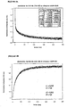
- Figure 4 shows a real-time recording of the kinetics of association (A) and dissociation (B) of NKA-Bo (Bodipy-labeled NKA) with the EGFP-NK2R receptor (GFP-labeled NK2R). Registration of the kinetics of association is carried out after rapid mixing of HEK 293 cells expressing the EGFP-NK2R receptor. The kinetics of dissociation are recorded after manual mixing of the cells expressing the receptor with the NKA-bo (preincubation of 15 minutes, 100 nanomolar) and then with the competitive antagonist SR 4896 (10 ⁇ M final).
- the x-axis represents the time in seconds and the y-axis represents the fluorescence (arbitrary units) at 510 nm.
- the values ⁇ n and ⁇ n respectively correspond to the amplitudes and the rate constants determined by a least squares adjustment with two exponentials.
- Figures 5A, 5B, 5C and 5D indicate the procedure for identifying a ligand competitively interacting with the fluorescent ligand at a receptor site.
- the complex formed between the EGFP-NK2R receptor and the NKA-Bo is reversed by increasing concentrations of the G6 molecule (see Table 1) and then by the non-fluorescent NKA.
- FIG. 5A indicates that a part of the receptor-ligand complex is reversed during each addition of G6 molecule (10 or 20 ⁇ M), as well as during the addition of a saturation concentration of NKA (10 ⁇ M).
- the abscissa axis corresponds to the time in seconds and the ordinate axis to the fluorescence measured at 510 nm.
- FIG. 5B shows that the dissociation kinetics of NKA-Bo, determined after addition of a saturating concentration of NKA, is not modified by the presence of the G6 molecule.
- the two experimental traces are adjusted using two fast exponential (0.04 s -1) and slow (0.014 s- 1) whose amplitudes (66% for the fast and slow 33%) are not affected by the presence of 50 ⁇ moles of molecule G6.
- the abscissa axis corresponds to the time in seconds and the ordinate axis to the fluorescence measured at 510 nm.
- Figure 5C shows a quantitative study of the dissociation kinetics of NKA-Bo in the presence of a known competitive antagonist, SR 48968.
- the dissociation of NKA-Bo is recorded in the presence of 0.2; 1 and 5 nM of SR 48968.
- the dissociation rate can be described as a sum of two exponentials whose velocities are 0.04 sec -1 and 0.008 sec -1 , and whose relative amplitudes remain constant at different doses of SR 48968 (45% rapid dissociation (0.04 s -1 ) and 55% slow dissociation (0.008 s -1 )).
- the abscissa axis corresponds to the time in seconds and the ordinate axis to the fluorescence measured at 510 nm.
- Figure 5D shows the values of the fast dissociation (square) and slow (upward triangles) amplitudes of NKA-Bo in the presence of SR 48968, as well as the total amplitude of the NKA-Bo bond (triangles downwards). ).
- the abscissa axis corresponds to the logarithm of the concentration of SR 48968 and the ordinate axis to the amplitude of the normalized fluorescence:
- FIG. 5D represents the ratio of the amplitude of the slow dissociation (A2) with respect to the total dissociation amplitude (total A) (curve with the diamonds).
- A2 the amplitude of the slow dissociation
- total A total dissociation amplitude
- FIGS. 6A, 6B and 6C show the procedure for identifying a ligand interacting noncompetitively with the fluorescent ligand at a receptor site.
- the fluorescence measured at 510 nm is represented as a function of time in seconds.
- the complex formed between the EGFP-NK2R receptor and the NKA-Bo is reversed by increasing concentrations of the A11 molecule and then by the non-fluorescent NKA.
- Figure 6A indicates that a small portion of the receptor-ligand complex is reversed at each addition of A11 molecule, while NKA (10 ⁇ M) reverses the majority of the complex.
- the kinetics of dissociation by NKA is not of the same shape as in the control.
- FIG. 6B the fluorescence measured at 510 nm is represented as a function of time in seconds.
- Figure 6B illustrates the change in dissociation kinetics in the presence of A11 molecule.
- NKA-Bo dissociation recordings of its EGFP-NK2R receptor are performed in the presence and absence of A11. Dissociation is initiated by the addition of non-fluorescent NKA at a final concentration of 20 ⁇ M.
- the analysis of the dissociation kinetics reveals two monoexponential relaxations whose rate constants (k1 and k2) are identical in the presence and absence of A11 but whose relative amplitudes change so that the slow dissociation represents 59% of the total signal in the control and 22% of the total signal in the presence of A11.
- FIG. 6C shows the association of NKABo (20 nanomolar) with the EGFP-NK2R receptor as a function of time in seconds.
- Figure 6C shows time-resolved binding (association) recordings of NKA-Bo to its EGFP-NK2R receptor, in the absence and presence of the A11 molecule.
- Analysis of the binding kinetics reveals two monoexponential relaxations characterized by rate constants (k1 and k2) that do not change in the presence of A11, but with variable amplitudes depending on whether the cells are incubated with A11 or not.
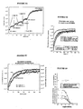
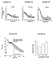
- FIGS. 7A to 7I illustrate the analysis of the effects of various molecules on the dissociation of NKA-Bo linked to its fluorescent EGFP-NK2R receptor.
- Figure 7A shows dissociation records of NKA-Bo in the presence of varying concentrations (1, 10 and 50 ⁇ M) of the 805 molecule (see Table 1).
- the fluorescence measured at 510 nm is represented as a function of time in seconds.
- the quantitative analysis of the dissociation kinetics is obtained using two exponentials fast (0.04 sec -1 ) and slow (0.008 sec -1 ) whose relative amplitudes (fast / slow) vary from 30/70 to 80 / When the concentration of 805 increases.
- FIGS. 7B - 7I show the percentage of amplitude of fast dissociation as a function of the logarithm of the concentration of the compounds tested. These figures give the results of the quantitative analysis of the dissociation of NKA-Bo in the presence of the molecules 805 (FIG. 7B), NP246 (FIG. 7C), A11 (FIG. 7D), H10 (FIG. 7E), G11 (FIG. 7F). , F7 ( Figure 7G), F3 ( Figure 7H) and NP 234 ( Figure 7I).
- the dissociation of NKA-Bo was determined in the presence of each molecule at the concentrations indicated on the abscissa axis. The effect of increasing the dissociation rate is represented by the relative increase in the amplitude (% ⁇ 1) of the fast dissociation.
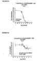
- FIGS. 8A and 8B show a quantification of the displacement of [ 3 H] SR 48968, bound to the EGFP-NK2R receptor, by the NKA and the molecule 805.
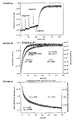
- Figures 9A, 9B and 9C show the cAMP production stimulation results by NKA in HEK 293 cells expressing the EGFP-NK2R receptor.
- Figure 9A shows the effect of the presence of competitive antagonist (H8565) and allosteric effector (805).
- the accumulation of intracellular cAMP (in pmoles / well) is represented as a function of the logarithm of the concentration of NKA.
- the curve with black squares corresponds to white; the curve with the black triangles corresponds to the H8565 antagonist at a concentration of 1 ⁇ M; the curve with the gray squares corresponds to the ligand 805 at a concentration of 10 ⁇ M and the curve with the gray triangles corresponds to the ligand 805 at a concentration of 50 ⁇ M.
- Intracellular cAMP accumulation is determined at different concentrations of NKA.
- the H8565 antagonist shifts the concentration-response curve of NKA to higher values without affecting the intensity of the maximal response (black triangles).
- the 805 ligand (gray squares and triangles) decreases the intensity of the maximal response without affecting the EC 50 (50% effective concentration) of the NKA for response.
- Figure 9B shows the 50% effective concentration of the maximum cyclic AMP response to NKA. This EC 50 increases in the presence of competitive antagonist while it is not significantly modified in the presence of 10 and 50 ⁇ M of 805.
- FIG. 9C shows the inhibition of the NKA-induced maximal cAMP response in the presence of increasing concentration of 805.
- the abscissa represents the logarithm of the 805 concentration and the ordinate the maximum value of the MPA production. cyclic.
- the inhibition constant KI is of the order of 6 ⁇ M.
- Figures 10A, 10B, 10C, 10D and 10E illustrate the effect of 805 on calcium responses associated with the EGFP-NK2R receptor in HEK 293 cells.
- FIGS. 10A, 10B and 10C the fluorescence of INDO-1 is shown at 400 nm as a function of time in seconds.
- the responses are recorded using the INDO-1 calcium-sensitive fluorescent probe.
- NKA agonist 1 nM, 5 nM and 1 ⁇ M
- a control response white circle
- a response in the presence of 50 ⁇ M of 805 black circle
- FIGGS. 10A, 10B and 10C there is an increase in the duration of the calcium response in the presence of 805, without significant effect on the amplitude or on the kinetics of setting up the response.
- FIG. 10D shows the variations of calcium responses obtained for a fixed agonist concentration (5 nM NKA) and variable concentrations (10 and 50 ⁇ M) of 805.
- the normalized calcium concentration as a function of time in seconds is shown.
- the curve with the white circles corresponds to a measurement carried out in the presence of 5 nM of NKA and without 805;
- the curve with the black circles corresponds to a measurement carried out in the presence of 5 nM NKA and 10 ⁇ M of 805;
- the curve with the black squares corresponds to a measurement carried out in the presence of 5 nM of NKA and 50 ⁇ M of 805.
- FIG. 10E shows a quantification of the half-return time (time required for the calcium concentration to be half of its maximum value) to the initial value of the calcium response recorded in the presence of 0, 10 and 50 ⁇ M of 805.
- Figures 11A, 11B, 11C, 11D, 11E, 11F and 11G illustrate the effect of 805 on fluorescent truncated neurokinin binding (NKA4-10 TR7) labeled with the Texas red fluorescent group and the associated calcium response.
- FIGS. 11A to 11D the calcium responses at 106 (FIG. 11A), 50.3 (FIG. 11B), 26.5 (FIG. 11C) and 10.6 nM (FIG. 11D) of NKA4-10 TR7 (NKA4-10 marked in position 7 by the Texas Red) are recorded in duplicate, in absence (curves with the white circles) and in the presence of 20 ⁇ M of 805 (curves with the black circles).
- the normalized calcium concentration as a function of time in seconds is shown. It is noted that 805 causes an increase in the amplitude of the calcium response, and a persistence of the signal.
- Figure 11E shows the relationship between the amplitude of the calcium response and the agonist concentration.
- the amplitude of the normalized calcium peak with respect to the extracellular calcium concentration is shown as a function of the concentration of NKA4-10 TR7.
- the curve with the black squares corresponds to the control measure and (without 805) and the curve with the triangles corresponds to the measurement carried out in the presence of 20 ⁇ M of 805.
- Figure 11F shows the dissociation of the NKA 4-10 TR7 bond.
- the x-axis represents the time in seconds and the y-axis represents the fluorescence.
- the complex formed between the EGFP-NK2R receptor and the NKA4-10 TR7 (106 nM) is inverted (at 10 ° C.) by the non-fluorescent NKA (20 ⁇ M) in the absence and in the presence of 10 ⁇ M of 805.
- dissociation kinetics reveals two monoexponential relaxations whose rate constants (0.05 sec -1 and 0.013 sec -1 ) are identical in the presence and absence of 805, but whose relative amplitudes change so that the Slow dissociation accounts for 42% of the total signal in the control and 12% of the total signal in the presence of 805.
- FIG. 11G shows the percentage of amplitude of the rapid dissociation of NKA 4-10 TR7 as a function of the logarithm of the 805 concentration.
- the amplitude of the rapid dissociation of NKA 4-10 TR7 is measured in the presence of variable concentrations of 805. Rapid dissociation accounts for about 40% of the total dissociation in the absence of 805 and up to 90% of the dissociation amplitude at 100 ⁇ M.
- the apparent affinity for the amplitude variation is of the order of 20 ⁇ M.
- FIGS. 12A, 12B, 12C and 12D represent the substance P (endogenous ligand) -regulated calcium responses on the human NK1 receptor (FIGS. 12A and 12B) and by neurokinin A on the human NK2 receptor (FIGS. 12C and 12D), in the presence and absence of 805.
- substance P endogenous ligand
- FIGS. 12A and 12B a suspension (1 ⁇ 10 6 cells / ml) of HEK 293 cells expressing the wild-type human NK1 receptor and loaded with indo-1 (3 ⁇ M) is stimulated with 0.1 nM (FIG. 12A). or 10 nM (FIG. 12B) of substance P (SP) in the presence (black circles) and in the absence (white circles) of 805 molecules (20 ⁇ M).
- SP substance P
- FIGS. 12C and 12D a suspension (1 ⁇ 10 6 cells / ml) of HEK 293 cells expressing the wild-type human NK2 receptor and loaded with indo-1 (3 ⁇ M) is stimulated with 10 nM (FIG. 12C) or 100 nM (FIG. 12D) of neurokinin A (NKA) in the presence (black circles) and in absence (white circles) of 805 molecules (20 ⁇ M).
- the molecule 805 causes an increase in the amplitude and duration of the calcium response evoked by each agonist.
- HEK 293 cells expressing the EGFP-NK2R receptor are suspended at a concentration of 2,000,000 cells / ml in physiological Hepes buffer (in mM: 137.5 NaCl) 1.25 MgCl 2 , 1.25 CaCl 2 , 6 KCl, 5.6 glucose, 10 Hepes, 0.4 NaH 2 PO 4 , 1% BSA (w / v), pH 7.4) and arranged in one of the tanks of the rapid mixing apparatus.
- a solution of NKA-BO (Bodipy-labeled neurokinin A) (200 nM) in the same buffer is placed in the other reservoir of the rapid mixing apparatus.
- the observation chamber is placed in an SPEX fluorolog 3 spectrofluorometer.
- the temperature of the reservoir syringes as well as of the mixing and observation chamber is set at 21 ° C.
- the excitation wavelength of the observation chamber is set at 470 nm, and the fluorescence emission wavelength is set at 510 nm.
- the sampling frequency of the experimental points is fixed at 50 Hz (1 point every 20 msec).
- the contents of the tanks are propelled towards the mixing and observation chamber by means of a pneumatic device allowing a flow of 4 ml / sec and the evolution of the mixture is recorded continuously from the moment of stopping of the thrust of the pistons of the tanks.
- the NKA-Bo association reaction with the EGFP-NK2R receptor is detected as a decrease in the fluorescence intensity of the GFP carried by the NK2R receptor.
- the HEK 293 cells expressing the EGFP-NK2R receptor are suspended at a concentration of 1,000,000 cells / ml in Hepes physiological buffer (in mM: 137.5 NaCl, 1.25 MgCl 2 1.25 CaCl 2, 6 KCl, 5.6 glucose, 10 Hepes, 0.4 NaH 2 PO 4 , 1% BSA (w / v), pH 7.4) and placed in a 1 ml cuvette provided with a magnetic stir bar on the cell holder of a SPEX fluorolog 3 spectrofluorometer. 20 nM (final concentration) of NKA-Bo is added thereto and the combination is allowed to reach equilibrium for 10 minutes.
- Hepes physiological buffer in mM: 137.5 NaCl, 1.25 MgCl 2 1.25 CaCl 2, 6 KCl, 5.6 glucose, 10 Hepes, 0.4 NaH 2 PO 4 , 1% BSA (w / v), pH 7.4
- the measurement of the dissociation kinetics of NKA-Bo is initiated by the manual addition of 10 ⁇ M (final concentration) of the competitive antagonist SR 48968 and recorded at 510 nm (excitation: 470 nm) at a rate of 1 point every 100 msec.
- EXAMPLE 2 Identification of the G6 molecule acting as a competitive ligand, by fluorescence energy transfer between the NKA-Bo and the EGFP-NK2R receptor.
- the control experiment presented in FIG. 5C indicates that a known molecule, competitive for the interaction of NKA-Bo with its site, ie 5R48968, provokes a dissociation of NKA-Bo with kinetics identical to that recorded when NKA-Bo is displaced by NKA.
- the relative amplitude of each step remains constant for each concentration of SR 48968, as attested by the fast / slow amplitude ratios close to 45/55.
- HEK 293 cells expressing the EGFP-NKR2 receptor are incubated, in one milliliter of Hepes buffer, with 20 nM NKA-Bo for 15 minutes.
- the mixture is then placed in a spectrofluorimetric cuvette and the reversion of the interaction between EGFP-NK2R and NKA-Bo is evaluated after addition of molecule A11 in increasing concentration (FIG. 6A).
- the estimate of the reversed complex fraction is then measured after the addition of a saturating amount of NKA (10 ⁇ M). It can be seen that the A11 molecule displaces only a small fraction (29%) of the NKA-Bo linked to its site.
- the amplitude of each relaxation is then given by the adjustment of the experimental traces.
- fast relaxation represents 41% and slow 59%.
- A1 50 ⁇ M
- the fast relaxation represents 78% of the signal and the slow 22%.
- the A11 molecule thus has the effect of stabilizing a fraction of the receptors in a state of lower affinity than that in which the NKA-Bo alone stabilizes it.
- the A11 molecule binds preferentially to the conformation of the receptor which has a more modest affinity for NKA-Bo.
- the binding of NKA-Bo is not inhibited by A11. It occurs on the same conformational states of the receptor, but the proportions of these two conformational states are modified by the presence of A11.
- the dissociation kinetics of NKA-Bo is faster in the presence of A11 than in its absence. This acceleration of the dissociation kinetics of NKA-Bo does not result from a change in the intrinsic affinities of the conformations for NKA-Bo, but from a modification of the relative proportions of the high and low affinity states.
- the receptor exists in at least two conformational states, one of which pre-exists agonist addition and the other occurs slowly over time in the presence of agonist.
- the A11 molecule by stabilizing the low affinity states of the receptor, increases the proportion of fast (low affinity) binding to the detriment of the slow binding which reflects the stabilization of a higher affinity state
- the identification of other allosteric effectors of the NK2R receptor of the tachykinins is done by analyzing the kinetics of dissociation of NKA-Bo bound to the receptor.
- NKA-Bo 10 6 HEK 293 cells expressing the EGFP-NK2R receptor are suspended in Hepes buffer (see above) and incubated with NKA-Bo (20 nM) and one of the following molecules: 805, NP246, A11, H10, G11, F7, F3 and NP 234.
- the A11 molecules and the known competitive antagonist SR 48968 are introduced into the experiment as positive controls.
- the dissociation of NKA-Bo is recorded in a spectrofluorometer cuvette equipped with magnetic stirring for 700 seconds, after adding 10 ⁇ M of non-fluorescent NKA to expel the bound NKA-Bo.
- the dissociation kinetics of NKA-Bo is recorded in the presence of increasing concentrations of each of the aforementioned molecules.
- FIG. 7A shows a typical experiment for measuring the dissociation rate of NKA-Bo in the presence of increasing concentrations of the molecule 805.
- FIGS. 7B to 7I show, for each molecule tested, the variation of the NKA-Bo binding amplitude and the amplitude variation of the fast dissociation. These figures show that all the indicated molecules behave like allosteric effectors because they cause an increase in the amplitude of the rapid dissociation of NKA-Bo.
- the amplitude increase in fast dissociation reflects the ability of molecules to stabilize a larger fraction of the receptors in the low-affinity state. It is thus seen that the initial fraction of receptors in the low affinity state goes from about 25% in the absence of effector to 60-75% in the presence of the molecules 805, NP234, A11, G11, F7, F3, and H10. (at 100 ⁇ M effector) and nearly 100% in the presence of A11 and NP246.
- 25,000 cells expressing the EGFP-NK2R receptor are incubated for 3 hours at 4 ° C. in a final volume of 500 ⁇ l of Hepes buffer in the presence of 1 nM of [ 3 H] SR48968 and variable concentrations of NKA or 805. incubation period, the reaction mixtures are filtered through GF / C (Whatmann) fiberglass filters previously incubated in Hepes buffer supplemented with 1% (W / V) semi-skimmed milk powder, and rinsed three times. 5 ml of hepes buffer at 4 ° C. The filters are then placed in scintillation counting flasks in which 3 ml of scintillation cocktail is added. Radioactivity is measured after 16 hours in a radioactivity counter.
- GF / C Whatmann
- the effect of 805 on the binding of SR 48968 does not reflect a competitive but rather non-competitive interaction.
- 30-well 6-well cell culture dishes are treated with collagen and then inoculated with 50,000 cells expressing the EGFP-NK2R receptor. After two to three days of culture, the medium is replaced with hepes buffer containing the inhibitor of phosphodiesterase IBMX (isobutyl-methyl-xhantine) and incubated for 30 minutes with increasing concentrations of NKA in the presence or absence of i) d competitive inhibitor H8565 (1 ⁇ M) or ii) effector 805 (10 or 50 ⁇ M).
- the quantity of cAMP produced (expressed in pmols / well) is determined according to a radioimmunological method described in Gicquiaux et al. (2002). This results in a measurement of the concentration-effect curve of NKA (FIG. 9A).
- NKA alone is able to stimulate the production of cAMP in cells expressing the EGFP-NK2R receptor with an EC 50 close to 100 nM.
- FIGS. 10A, 10B and 10C show that compound 805 (50 ⁇ M) increases the duration of the evoked calcium responses by 1, 5 and 1000 nM of NKA. In the same way, compound 805, at concentrations of 10 and 50 ⁇ M, increases the duration of the evoked calcium response by 5 nM NKA (FIG. 10D).
- Compound 805 therefore behaves as a positive allosteric effector of the calcium responses evoked by the NKA-Bo agonist which increases the duration of the response. This is indicated in FIG. 10E where it can be seen that the time required to return to 50% of the maximum response goes from 91 seconds in the absence of 805 to 105 then 115 s when the concentration of 805 is 10 and then 50 ⁇ M.
- the calcium responses evoked by the truncated agonist NKA4-10 TR7 on cells expressing the EGFP-NK2R receptor are recorded after loading the cells with the calcium-sensitive fluorescent probe: indo-1 according to the protocol described in Vollmer et al. (1999).
- the cells are suspended at 500,000 cells / ml in Hepes buffer and placed in a spectrofluorimeter tank.
- the excitation wavelength is set at 355 nm and the emission is recorded at 375 and 405 nm.
- the responses plotted are the 375 nm / 405 nm signal ratios.
- 11A to 11D show that compound 805 (20 ⁇ M) increases the amplitude of the response evoked by NKA 4-10 TR7 (106, 50.3, 26.5 and 10.6 nM). Amplitude variation by 805 is shown in panel B of Figure 11 for concentrations of 5, 10, 25, 50 and 106 nM of NKA 4-10 TR7. Compound 805 is shown to be a positive allosteric effector of the calcium responses evoked by NKA 4-10 TR7 which decreases the 50% effective dose from about 20 nM to about 15 nM.
- NKA 4-10 is a truncated form of NKA found in middle gut tumor fluids. Fluorescent NKA 4-10 is modified at position 7 to introduce a cysteine which is then labeled with the Texas Red fluorescent group.
- the fluorescent NKA 4-10 is, like the fluorescent NKA, an agonist of the EGFP-NK2R receptor, except that it is unable to evoke cAMP accumulation responses (Palanche et al., 2001).
- the binding of fluorescent NKA 4-10 is in a monoexponential process with which the development of a calcium response in HEK 293 cells expressing the EGFP-NK2R receptor is associated (Palanche et al., 2001).
- respective amplitudes of 58% for fast dissociation and 42% for slow dissociation Figure 11F.
- an acceleration of the dissociation characterized by a modification of the amplitudes of the fast (88%) and slow (12%) dissociation stages is observed.
- a relationship between the dissociation rate increase of NKA4-10 TR7 and the concentration of 805 is noted ( Figure 11G).
- the concentration of 805 which causes a 50% increase in the amplitude of the rapid dissociation of NKA4-10 TR7 is of the order of 20
- the calcium responses evoked by the following agonists: substance P (SP) and neurokinin A (NKA), on cells expressing the wild-type human NK1 receptor or wild-type human NK2 are recorded after loading of the cells with the calcium-sensitive fluorescent probe: indole 1 (3 ⁇ M) according to the protocol described in Vollmer et al. (1999).
- the cells are suspended at 1,000,000 cells / ml in Hepes buffer (op cit) and placed in a spectrofluorimeter vat.
- the excitation wavelength is set at 338 nm and the emission is recorded at 400 nm.
- the answers plotted are the signals recorded at 400 nm.
- FIGS. 12A and 12B show that compound 805 (20 ⁇ M) increases the amplitude of the evoked response by substance P (0.1 or 10 nM).
- Figures 12C and 12D show that compound 805 (20 ⁇ M) increases the amplitude of the response evoked by neurokinin A (10 or 100 nM).
- Compound 805 is shown to be a positive allosteric effector of the neurokinin A-evoked calcium responses on the wild-type human NK2 receptor and substance P-evoked responses on the wild-type human NK1 receptor.
- the M1 fluorescent muscarinic receptor is obtained by fusion of its cDNA with that of EGFP or YPFP as described in Ilien et al. (2003) and stably expressed in HEK 293 cells.
- the cells are suspended at a concentration of 1,000,000 cells / ml in Hepes physiological buffer (in mM: 137.5 NaCl, 1.25 MgCl 2 ; 1.25 CaCl 2 , 6 KCl, 5.6 glucose, 10 Hepes, 0.4 NaH 2 PO 4 , 1% BSA (w / v), pH 7.4) and arranged in a 1 ml fluorescence cuvette, itself placed in the rack waving a spectrofluorimeter.
- Hepes physiological buffer in mM: 137.5 NaCl, 1.25 MgCl 2 ; 1.25 CaCl 2 , 6 KCl, 5.6 glucose, 10 Hepes, 0.4 NaH 2 PO 4 , 1% BSA (w / v), pH 7.4
- the cell suspension is excited at 470 nm and the fluorescence emission is recorded at 510 nm (EGFP) or at 530 nm (EYFP).
- the addition of 70 nM bodipy (558-568) -pirenzepine causes a decrease in fluorescence emission of the receptor-borne fluorescent protein that reflects the step of ligand-receptor association. The time course of this step is followed by recording the emission at 510 or 530 nm.
- the complex between the receptor and the bodipy pirenzepine is formed in a fluorescence cuvette placed in the spectrofluorimeter before adding an excess (5 ⁇ M) of atropine to initiate the dissociation step of the ligand receptor complex.
- the time course of this dissociation step is recorded at 510 or 530 nm.
- the cells are preincubated with one or more known or unknown molecules, and the time course of the acetylcholine receptor is recorded. step of association and / or dissociation step.
- An allosteric effector is accelerated, either slows down the time course or the stage association, either of the dissociation step, or both. This results in a relative change in the fast and slow association (or dissociation) amplitudes compared to the control experimental condition (without effector).
- the fluorescent CXCR4 receptor is obtained by fusion of its cDNA with that of EGFP as described in Valenzuela et al. (2001) and in Palanché et al. (2001) and stably expressed in HEK 293 cells.
- the cells are suspended at a concentration of 1,000,000 cells / ml in Hepes physiological buffer (in mM: 137.5 NaCl, 1.25 MgCl 2 ; 1.25 CaCl 2 , 6 KCl, 5.6 glucose, 10 Hepes, 0.4 NaH 2 PO 4 , 1% BSA (w / v), pH 7.4) and arranged in a 1 ml fluorescence cuvette. even placed in the rack waving a spectrofluorimeter.
- the cell suspension is excited at 470 nm and the fluorescence emission is recorded at 510 nm.
- the addition of 50 nM Texas Red-SDF1 ⁇ causes a decrease in fluorescence emission of the receptor-borne fluorescent protein that reflects the step of ligand-receptor association.
- the time course of this step is followed by recording the emission at 510 nm.
- the complex between the receptor and the Texas Red-SDF1 ⁇ is formed in a fluorescence cuvette placed in the spectrofluorimeter before adding an excess (500 nM) of SDF1 ⁇ to initiate the dissociation step of the ligand receptor complex.
- the time course of this dissociation step is recorded at 510 nm.
- the cells are pre-incubated with one or more known or unknown molecules, and the time course of the step is recorded. association and / or dissociation step.
- An allosteric effector either accelerates or slows down the time course of either the association step, the dissociation step, or both. This results in a relative change in the fast and slow association (or dissociation) amplitudes compared to the control experimental condition (without effector).
Landscapes
- Health & Medical Sciences (AREA)
- Life Sciences & Earth Sciences (AREA)
- Immunology (AREA)
- Chemical & Material Sciences (AREA)
- Engineering & Computer Science (AREA)
- Molecular Biology (AREA)
- Biomedical Technology (AREA)
- Hematology (AREA)
- Urology & Nephrology (AREA)
- Medicinal Chemistry (AREA)
- General Health & Medical Sciences (AREA)
- Biochemistry (AREA)
- Microbiology (AREA)
- Food Science & Technology (AREA)
- Physics & Mathematics (AREA)
- Analytical Chemistry (AREA)
- Cell Biology (AREA)
- Biotechnology (AREA)
- General Physics & Mathematics (AREA)
- Pathology (AREA)
- Organic Chemistry (AREA)
- Proteomics, Peptides & Aminoacids (AREA)
- Genetics & Genomics (AREA)
- Biophysics (AREA)
- Chemical Kinetics & Catalysis (AREA)
- Investigating Or Analysing Biological Materials (AREA)
- Peptides Or Proteins (AREA)
- Plural Heterocyclic Compounds (AREA)
- Measuring Or Testing Involving Enzymes Or Micro-Organisms (AREA)
- Auxiliary Devices For Music (AREA)
- Pyrane Compounds (AREA)
- Electrotherapy Devices (AREA)
- Measuring Pulse, Heart Rate, Blood Pressure Or Blood Flow (AREA)
Abstract
Description
La présente invention a pour objet un procédé de mise en évidence d'un effecteur allostérique d'un récepteur.The present invention relates to a method for demonstrating an allosteric effector of a receptor.
De nombreux médicaments et substances naturelles exercent leur action en interagissant avec des protéines régulatrices, appelées récepteurs, impliquées dans de nombreuses fonctions physiologiques des organismes, et les altérations de leurs fonctions sont la cause de nombreuses pathologies.Many drugs and natural substances exert their action by interacting with regulatory proteins, called receptors, involved in many physiological functions of organisms, and alterations of their functions are the cause of many pathologies.
Parmi les molécules qui agissent en se liant à des récepteurs, une catégorie particulière, appelée effecteurs allostériques ou modulateurs, exerce son effet en se liant à des sites, présents à la surface des récepteurs, mais topologiquement distincts du site de liaison du ligand endogène. Leur interaction avec le récepteur est de nature non compétitive par rapport à celle du ligand endogène. En se liant à leurs sites régulateurs, les effecteurs n'ont pas ou peu d'effet sur les réponses du récepteur en absence de ligand endogène. En revanche, ils modifient les propriétés de liaison des ligands endogènes compétitifs et altèrent les réponses biologiques évoquées par ledit ligand.Among the molecules that act by binding to receptors, a particular class, called allosteric effectors or modulators, exerts its effect by binding to sites, present on the surface of the receptors, but topologically distinct from the endogenous ligand binding site. Their interaction with the receptor is uncompetitive with respect to that of the endogenous ligand. By binding to their regulatory sites, the effectors have little or no effect on receptor responses in the absence of endogenous ligand. On the other hand, they modify the binding properties of competitive endogenous ligands and alter the biological responses evoked by said ligand.
Les effecteurs allostériques présentent un intérêt particulier dans le domaine thérapeutique par le fait que leur action n'est pas "constitutive" mais se révèle uniquement lorsque le ligand endogène vient stimuler le récepteur. Dans les cas où de telles molécules ont pu être identifiées et mises sur le marché, le confort du patient et la sélectivité de l'effet se sont toujours trouvés grandement améliorés par exemple en comparaison avec l'effet observé de l'administration d'un agoniste (Pan et al., 2001).Allosteric effectors are of particular interest in the therapeutic field in that their action is not "constitutive" but only occurs when the endogenous ligand stimulates the receptor. In cases where such molecules have been identified and placed on the market, patient comfort and effect selectivity have always been greatly improved, for example in comparison with the observed effect of administering a drug. agonist (Pan et al., 2001).
Afin d'identifier de nouveaux effecteurs allostériques de récepteurs, plusieurs approches expérimentales ont été développées à ce jour, notamment les mesures de liaison d'un ligand à son récepteur et les mesures fonctionnelles de réponses physiologiques.In order to identify novel allosteric receptor effectors, several experimental approaches have been developed to date, including ligand binding to its receptor and functional measurements of physiological responses.
Parmi les méthodes de mesure de liaison, il y a l'étude de la cinétique de dissociation d'un radioligand. Comme décrit dans Kostenis et Mohr (1996), la modulation allostérique est le plus généralement mise en évidence par un effet de l'effecteur allostérique sur la cinétique de dissociation d'un radioligand. Cette méthode a été mise en oeuvre pour démontrer le caractère non compétitif (et donc allostérique) de molécules régulant les récepteurs M1 à M5 muscariniques de l'acétylcholine (Tucek et Poska, 1995), A1 de l'adénosine (Bruns & Ferguns, 1990), alpha2 de l'adrénaline (Nunnari et al., 1987), ou D2 de la dopamine (Hoare & Strange, 1995). Dans ces différents exemples, les auteurs ont indifféremment utilisé soit des agonistes, soit des antagonistes comme radioligands, et ont défini le caractère positif ou négatif du modulateur par sa capacité à accélérer (positif) ou ralentir (négatif) la vitesse de dissociation du radioligand utilisé.Among the binding measurement methods, there is the study of the kinetics of dissociation of a radioligand. As described in Kostenis and Mohr (1996), allosteric modulation is most generally evidenced by an effect of the allosteric effector on the dissociation kinetics of a radioligand. This method was used to demonstrate the non-competitive (and therefore allosteric) nature of molecules regulating M1 to M5 muscarinic acetylcholine receptors (Tucek and Poska, 1995), adenosine A1 (Bruns & Ferguns, 1990). ), adrenaline alpha2 (Nunnari et al., 1987), or D2 of dopamine (Hoare & Strange, 1995). In these various examples, the authors have indifferently used either agonists or antagonists as radioligands, and defined the positive or negative character of the modulator by its ability to accelerate (positive) or slow down (negative) the dissociation rate of the radioligand used. .
La deuxième approche la plus répandue d'identification d'un effecteur allostérique consiste à mesurer la quantité de complexe radioligand / récepteur à une concentration subsaturante de radioligand. Dans ces conditions, le fait qu'une tierce molécule provoque une augmentation de la quantité de complexe radioligand / récepteur s'interprète en termes de potentialisation de la liaison par un effet allostérique. Le travail de Bruns et Ferguns, cité plus haut, illustre ce point lorsqu'un radioligand agoniste est utilisé, tandis que l'article de Jakubik et al. (1997) utilise un antagoniste radioactif.The second most common approach to identifying an allosteric effector is to measure the amount of radioligand / receptor complex at a subset radioligand concentration. Under these conditions, the fact that a third molecule causes an increase in the amount of radioligand / receptor complex is interpreted in terms of potentiation of the binding by an allosteric effect. The work of Bruns and Ferguns, cited above, illustrates this point when an agonist radioligand is used, while the article by Jakubik et al. (1997) uses a radioactive antagonist.
La troisième approche expérimentale consiste à établir la courbe de saturation d'un radoligand en présence et en absence d'effecteur allostérique. Dans ce cas, qui est illustré dans l'article de Massot et al. (1996) pour le récepteur 5HT1B de la sérotonine, l'effecteur 5HT-moduline réduit la quantité de sites récepteurs sans affecter l'affinité apparente déterminée pour le radioligand utilisé.The third experimental approach is to establish the saturation curve of a radoligand in the presence and absence of allosteric effector. In this case, which is illustrated in the article by Massot et al. (1996) for the 5HT1B serotonin receptor, the 5HT-modulin effector reduces the amount of receptor sites without affecting the apparent affinity determined for the radioligand used.
Les mesures de la réponse biologique évoquée par un ligand de récepteur comprennent soit son activation (par les agonistes) soit son inhibition (par les antagonistes). Ces mesures estiment généralement une variation d'amplitude des réponses qui sont soit potentialisées par les effecteurs positifs ou inhibées par les effecteurs négatifs de la réponse, et incluent :
- l'enregistrement de courants ioniques par des méthodes électrophysiologiques (Krause et al., 1997 ; Galzi et al., 1996) ou par des méthodes optiques utilisant des sondes spécifiques d'ions (Birdsall et al., 1999),
- la mesure de la production de messagers secondaires tels que l'AMPc (Bruns & Ferguns, 1990) ou les inositols phosphates (Waugh et al., 1999),
- la liaison de GTP à la protéine G associée au récepteur (Birdsall et al., 1999; Lazareno & Birdsall, 1995 ; Hoare et al., 2000) ou l'hydrolyse de GTP par la protéine G associée au récepteur (Birdsall et al., 1999). Rees et al., 2002, vol. 8, no. 5-6 pages 261-268 présente un résumé des différentes techniques d'identification d'un effecteur allostérique.
- the recording of ionic currents by electrophysiological methods (Krause et al., 1997, Galzi et al., 1996) or by optical methods using ion-specific probes (Birdsall et al., 1999),
- measuring the production of secondary messengers such as cAMP (Bruns & Ferguns, 1990) or inositol phosphates (Waugh et al., 1999),
- GTP binding to receptor-associated G protein (Birdsall et al., 1999; Lazareno & Birdsall, 1995; Hoare et al., 2000) or GTP hydrolysis by receptor-associated G protein (Birdsall et al. , 1999). Rees et al., 2002, vol. 8, no. 5-6 pages 261-268 presents a summary of the different techniques for identifying an allosteric effector.
La technique du transfert d'énergie de fluorescence (FRET) en combinaison avec l'utilisation d'un récepteur rendu fluorescent et de l'un de ses ligands fluorescents permet de mesurer, comme décrit dans la demande internationale WO 98/55873, des interactions non covalentes entre ledit récepteur et ledit ligand.The technique of fluorescence energy transfer (FRET) in combination with the use of a fluorescent rendered receptor and one of its fluorescent ligands allows to measure, as described in international application WO 98/55873, non-covalent interactions between said receptor and said ligand.
Le récepteur peut être rendu fluorescent grâce à une protéine fluorescente, par exemple une protéine issue de la méduse Aequorea victoria dont le gène correspondant a été séquencé, cloné (Prasher et al., 1992) et optimisé pour une bonne expression chez les eucaryotes supérieurs (Cormack et al., 1996). Il peut être fusionné avec le gène codant pour une autre protéine, notamment un récepteur couplé à une protéine G. (Galzi & Alix, 1997 ; Weill et al., 1999 ; Vollmer et al., 1999 ; Valenzuela et al., 2001) pour permettre l'expression d'une protéine réceptrice fluorescente. La combinaison de la protéine réceptrice fluorescente avec l'un de ses ligands fluorescents permet la formation d'un complexe récepteur-ligand non covalent qui est détecté par transfert d'énergie de fluorescence (Galzi & Alix, 1997 ; Vollmer et al., 1999 ; Valenzuela et al., 2001) et dont on peut mesurer la cinétique de formation et de dissociation (Palanché et al., 2001 ; Valenzuela et al., 2001).The receptor can be made fluorescent by means of a fluorescent protein, for example a protein derived from the jellyfish Aequorea victoria whose corresponding gene has been sequenced, cloned (Prasher et al., 1992) and optimized for good expression in higher eukaryotes ( Cormack et al., 1996). It can be fused with the gene encoding another protein, including a G-protein coupled receptor (Galzi & Alix, 1997, Weill et al., 1999, Vollmer et al., 1999, Valenzuela et al., 2001). to allow the expression of a fluorescent receptor protein. The combination of the fluorescent receptor protein with one of its fluorescent ligands allows the formation of a non-covalent receptor-ligand complex that is detected by fluorescence energy transfer (Galzi & Alix, 1997; Vollmer et al., 1999). Valenzuela et al., 2001) and whose kinetics of formation and dissociation can be measured (Palanché et al., 2001, Valenzuela et al., 2001).
L'un des buts de l'invention est de fournir un procédé simple à mettre en oeuvre, rapide, sensible, permettant d'effectuer des mesures quantitatives des interactions entre un récepteur et un ligand marqueur du site du ligand endogène, et permettant de détecter la présence d'un éventuel effecteur allostérique.One of the aims of the invention is to provide a method that is simple to implement, rapid and sensitive, making it possible to carry out quantitative measurements of the interactions between a receptor and a ligand marker of the site of the endogenous ligand, and making it possible to detect the presence of a possible allosteric effector.
Un autre but de l'invention est de permettre des mesures en temps réel des interactions d'un ligand marqueur du site du ligand endogène avec son récepteur, permettant de détecter la présence d'un éventuel effecteur allostérique.Another object of the invention is to enable real-time measurements of the interactions of a marker ligand of the site of the endogenous ligand with its receptor, making it possible to detect the presence of a possible allosteric effector.
Un autre but de l'invention est de permettre une mesure en temps résolu de la cinétique d'association et de dissociation d'un ligand fluorescent, marqueur du site du ligand endogène, avec son récepteur, et de détecter la présence d'un éventuel effecteur allostérique.Another object of the invention is to allow a time-resolved measurement of the kinetics of association and dissociation of a fluorescent ligand, marker of the site of the endogenous ligand, with its receptor, and to detect the presence of a possible allosteric effector.
Un autre but de l'invention est de permettre la détection de l'interaction entre un récepteur et son ligand endogène par transfert d'énergie de fluorescence dans des gammes de concentrations de ligand endogène fluorescent considérablement plus étendues (du nanomolaire au micromolaire) que lorsqu'on utilise un radioligand (domaine du nanomolaire). Ainsi, la présente invention permet de détecter par transfert d'énergie un plus grand nombre d'états conformationnels du récepteur, ce qui facilite grandement l'identification et l'interprétation des effets d'agents modulateurs supposés.Another object of the invention is to enable the detection of the interaction between a receptor and its endogenous ligand by fluorescence energy transfer in fluorescent endogenous ligand concentration ranges considerably greater (from nanomolar to micromolar) than when a radioligand (nanomolar domain) is used. Thus, the present invention makes it possible to detect by energy transfer a greater number of conformational states of the receptor, which greatly facilitates the identification and interpretation of the effects of supposed modulator agents.
L'un des buts de l'invention est de fournir une mesure expérimentale des constantes de vitesse d'association et de dissociation d'un ligand fluorescent, marqueur du site du ligand endogène, avec son récepteur, en permettant la détection de la présence d'un éventuel effecteur allostérique.One of the aims of the invention is to provide an experimental measurement of the constants of association speed and dissociation of a fluorescent ligand, marker of the site of the endogenous ligand, with its receptor, allowing the detection of the presence of a possible allosteric effector.
L'invention a également pour but de fournir un procédé de détection d'un effecteur allostérique d'un récepteur, généralisable à de nombreuses protéines réceptrices et à leurs ligands.The invention also aims to provide a method of detecting an allosteric effector of a receptor, generalizable to many receptor proteins and their ligands.
L'invention a également pour but de fournir un procédé de détection d'un effecteur allostérique d'un récepteur, automatisable, ne nécessitant ni la purification du récepteur, ni celle du ligand.The invention also aims to provide a method of detecting an allosteric effector of a receptor, automatable, requiring neither the purification of the receptor nor that of the ligand.
L'invention a également pour but de fournir un procédé de détection d'un effecteur allostérique d'un récepteur, non polluant puisqu'il n'utilise pas de radioactivité, économique puisqu'il utilise de la lumière visible et peut être mis en oeuvre sur des équipements existants.The invention also aims to provide a method for detecting an allosteric effector of a receptor, non-polluting since it does not use radioactivity, which is economical since it uses visible light and can be used on existing equipment.
La présente invention concerne un procédé de mise en évidence d'un effecteur allostérique d'un récepteur, par détermination de la variation :
- de la cinétique de dissociation et/ou d'association du complexe formé entre le susdit récepteur et l'un de ses ligands en présence dudit effecteur allostérique, par rapport à la cinétique de dissociation et/ou d'association du complexe formé entre ledit récepteur et ledit ligand, en l'absence dudit effecteur,
- et/ou de l'amplitude de la liaison formée entre le susdit récepteur et l'un de ses ligands en présence dudit effecteur allostérique, par rapport à l'amplitude de la liaison formée entre ledit récepteur et ledit ligand, en l'absence dudit effecteur,
ledit récepteur et ledit ligand étant impliqués dans au moins une réponse biologique dans des conditions physiologiques appropriées, et l'effecteur allostérique étant capable de moduler au moins l'une des réponses,
ledit récepteur étant marqué par une protéine fluorescente choisie parmi les protéines fluorescentes issues ou dérivées de protéines autofluorescentes de cnidaires, dont le coefficient d'extinction moléculaire est supérieur à environ 14000M-1.cm-1 et le rendement quantique de fluorescence est supérieur à environ 0,38,
ledit ligand étant marqué par un marqueur constitué :- soit par une molécule susceptible d'absorber la lumière émise par la protéine fluorescente,
- soit par une substance fluorescente,
- entre la protéine fluorescente susmentionnée et la susdite substance fluorescente, la substance fluorescente étant telle que soit elle est excitable à la longueur d'onde d'émission de la protéine fluorescente susmentionnée, soit elle émet à la longueur d'excitation de la protéine fluorescente susmentionnée, ou
- entre la protéine fluorescente susmentionnée et la susdite molécule susceptible d'absorber la lumière émise par la protéine fluorescente.
- the kinetics of dissociation and / or association of the complex formed between the aforesaid receptor and one of its ligands in the presence of said allosteric effector, with respect to the kinetics of dissociation and / or association of the complex formed between said receptor and said ligand, in the absence of said effector,
- and / or the amplitude of the bond formed between the aforesaid receptor and one of its ligands in the presence of said allosteric effector, with respect to the amplitude of the bond formed between said receptor and said ligand, in the absence of said effector
said receptor and said ligand being involved in at least one biological response under appropriate physiological conditions, and the allosteric effector being capable of modulating at least one of the responses,
said receptor being labeled with a fluorescent protein selected from fluorescent proteins derived from or derived from autofluorescent cnidarian proteins, whose molecular extinction coefficient is greater than about 14000M -1 .cm -1 and the fluorescence quantum yield is greater than about 0.38
said ligand being labeled with a label consisting of:- by a molecule capable of absorbing the light emitted by the fluorescent protein,
- by a fluorescent substance,
- between the aforementioned fluorescent protein and the aforesaid fluorescent substance, the fluorescent substance being such that it is excitable at the emission wavelength of the aforementioned fluorescent protein, or it emits at the excitation length of the aforementioned fluorescent protein , or
- between the aforementioned fluorescent protein and the aforesaid molecule capable of absorbing the light emitted by the fluorescent protein.
La présente invention concerne un procédé de mise en évidence d'un effecteur allostérique d'un récepteur, par détermination de la variation :
- de la cinétique de dissociation et/ou d'association du complexe formé entre le susdit récepteur et l'un de ses ligands en présence dudit effecteur allostérique, par rapport à la cinétique de dissociation et/ou d'association du complexe formé entre ledit récepteur et ledit ligand, en l'absence dudit effecteur,
- et/ou de l'amplitude de la liaison formée entre le susdit récepteur et l'un de ses ligands en présence dudit effecteur allostérique, par rapport à l'amplitude de la liaison formée entre ledit récepteur et ledit ligand, en l'absence dudit effecteur, sous réserve que lorsque la variation de la susdite amplitude est négative, on détecte également l'existence de la variation de la susdite cinétique,
ledit récepteur et ledit ligand étant impliqués dans au moins une réponse biologique dans des conditions physiologiques appropriées, et l'effecteur allostérique étant capable de moduler au moins l'une des réponses,
ledit récepteur étant marqué par une protéine fluorescente choisie parmi les protéines fluorescentes issues ou dérivées de protéines autofluorescentes de cnidaires, dont le coefficient d'extinction moléculaire est supérieur à environ 14000M-1.cm-1 et le rendement quantique de fluorescence est supérieur àenviron 0,38,
ledit ligand étant marqué par un marqueur constitué :- soit par une molécule susceptible d'absorber la lumière émise par la protéine fluorescente,
- soit par une substance fluorescente,
- entre la protéine fluorescente susmentionnée et la susdite substance fluorescente, la substance fluorescente étant telle que soit elle est excitable à la longueur d'onde d'émission de la protéine fluorescente susmentionnée, soit elle émet à la longueur d'excitation de la protéine fluorescente susmentionnée, ou
- entre la protéine fluorescente susmentionnée et la susdite molécule susceptible d'absorber la lumière émise par la protéine fluorescente.
- the kinetics of dissociation and / or association of the complex formed between the aforesaid receptor and one of its ligands in the presence of said allosteric effector, with respect to the kinetics of dissociation and / or association of the complex formed between said receptor and said ligand, in the absence of said effector,
- and / or the amplitude of the bond formed between the aforesaid receptor and one of its ligands in the presence of said allosteric effector, with respect to the amplitude of the bond formed between said receptor and said ligand, in the absence of said effector, provided that when the variation of the aforesaid amplitude is negative, the existence of the variation of the aforesaid kinetics is also detected,
said receptor and said ligand being involved in at least one biological response under appropriate physiological conditions, and the allosteric effector being capable of modulating at least one of the responses,
said receptor being labeled with a fluorescent protein selected from fluorescent proteins derived from or derived from autofluorescent cnidarian proteins, whose molecular extinction coefficient is greater than about 14000M -1 .cm -1 and the fluorescence quantum yield is greater than about 0.38
said ligand being labeled with a label consisting of:- by a molecule capable of absorbing the light emitted by the fluorescent protein,
- by a fluorescent substance,
- between the aforementioned fluorescent protein and the aforesaid fluorescent substance, the fluorescent substance being such that it is excitable at the emission wavelength of the aforementioned fluorescent protein, or it emits at the excitation length of the aforementioned fluorescent protein , or
- between the aforementioned fluorescent protein and the aforesaid molecule capable of absorbing the light emitted by the fluorescent protein.
L'invention répond au problème technique posé par la détermination du caractère compétitif ou non compétitif de l'interaction entre un récepteur et un agent pharmacologique supposé agir comme effecteur d'un récepteur ou comme compétiteur du ligand endogène d'un récepteur, par des mesures de cinétiques d'association et de dissociation. De manière simple, l'effecteur allostérique d'un récepteur altérera la cinétique d'association ou de dissociation d'un récepteur tandis qu'un agent compétiteur du ligand endogène n'aura pas d'effet sur ces cinétiques.The invention answers the technical problem posed by the determination of the competitive or non-competitive nature of the interaction between a receptor and a pharmacological agent that is supposed to act as an effector of a receptor or as a competitor of the endogenous ligand of a receptor, by measurements kinetics of association and dissociation. In a simple manner, the allosteric effector of a receptor will alter the kinetics of association or dissociation of a receptor while an endogenous ligand competing agent will have no effect on these kinetics.
On appelle "ligand, marqueur du site du ligand endogène" un ligand qui se lie au site du ligand endogène par opposition à un éventuel effecteur allostérique, qui se lie à un site distinct de celui du ligand endogène. Le marqueur du site du ligand endogène interagit de manière compétitive avec le ligand endogène (liaison mutuellement exclusive), tandis que l'effecteur allostérique interagit de manière non compétitive avec le ligand endogène.The term "ligand, endogenous ligand site marker" refers to a ligand that binds to the site of the endogenous ligand as opposed to an eventual allosteric effector, which binds to a site distinct from that of the endogenous ligand. The endogenous ligand site marker competitively interacts with the endogenous ligand (mutually exclusive binding), whereas the allosteric effector interacts noncompetitively with the endogenous ligand.
On appelle "effecteur allostérique" ou "modulateur allostérique" une molécule ou un ligand ou une substance biologiquement active qui module les propriétés de liaison du ligand ainsi que les propriétés fonctionnelles du récepteur, sans entrer en compétition avec ledit ligand.An "allosteric effector" or "allosteric modulator" is a molecule or ligand or biologically active substance that modulates the binding properties of the ligand as well as the functional properties of the receptor, without competing with said ligand.
Par définition, on rappelle qu'on nomme ligand toute molécule susceptible de se lier à un récepteur de manière non covalente en se liant au même site que le ligand endogène, et qu'un ligand peut être un agoniste ou un antagoniste, un agoniste étant capable, en se liant, de déclencher une réponse biologique et un antagoniste ne déclenchant pas de réponse biologique et étant capable de bloquer l'effet de l'agoniste.By definition, it is recalled that the name ligand any molecule capable of binding to a receptor in a non-covalent manner by binding to the same site as the endogenous ligand, and that a ligand can be an agonist or an antagonist, an agonist being capable, by binding, of triggering a biological response and an antagonist not triggering a biological response and being able to block the effect of the agonist.
Un effecteur allostérique est aussi un ligand du récepteur mais celui-ci se lie avec le récepteur sur un autre site de liaison. Dans le présent contexte, sera utilisé le terme "effecteur allostérique" ou "modulateur allostérique" et non pas "ligand".An allosteric effector is also a ligand of the receptor but it binds with the receptor on another binding site. In the present context, the term "allosteric effector" or "allosteric modulator" will be used and not "ligand".
La modulation allostérique est le mécanisme par lequel une molécule, appelée effecteur allostérique ou modulateur allostérique, augmente ou diminue la réponse d'un récepteur activé par un agoniste. L'effecteur allostérique interagit avec le récepteur au niveau d'un site de liaison qui est distinct de celui auquel le ligand compétitif se lie sur le récepteur. Le fait qu'une modulation allostérique soit possible réside dans :
- 1) l'existence de différentes conformations ou états du récepteur. Chaque conformation est dotée de propriétés pharmacologiques et fonctionnelles distinctes. Il existe ainsi des conformations actives et inactives sur le plan biologique. Chaque conformation lie ses ligands avec des affinités différentes. Par exemple, un agoniste, tout comme un effecteur allostérique positif, exhibe une affinité plus grande pour la (les) conformation(s) biologiquement active(s) du récepteur, tandis qu'un antagoniste, tout comme un effecteur allostérique négatif, présente une affinité plus élevée pour la (les) conformation(s) inactive(s) du récepteur ;
Les propriétés pharmacologiques d'une conformation sont la capacité de lier des ligands et modulateurs avec une affinité définie. Chaque conformation lie différents ligands avec une affinité intrinsèque propre à chaque ligand. L'ensemble des ligands se liant à une conformation avec une affinité propre constitue ce que l'on appelle profil pharmacologique. Les propriétés pharmacologiques d'une conformation sont assimilables à son profil pharmacologique.
Les propriétés fonctionnelles d'une conformation d'un récepteur correspondent à sa capacité ou non de stimuler une réponse biologique. On distinguera plusieurs propriétés fonctionnelles d'un récepteur :- l'état de repos, par définition non actif, car non couplé à une réponse, mais activable, c'est à dire, capable de s'interconvertir dans une conformation active ou couplée à une réponse ;
- le ou les état(s) actif(s), responsable(s) d'une réponse biologique ; dans le cas où il existe plusieurs états actifs, l'une au moins des propriétés de la réponse sera différente (par exemple, réponse calcique versus cAMP, ou alors même réponse mais sensibilité différente aux agonistes, ou encore durée de réponses différentes etc....) ; d'autre part, chaque état actif peut donner lieu à une ou plusieurs réponses, distinctes par leur nature ou d'un point de vue quantitatif; de plus, un récepteur peut adopter plusieurs conformations correspondant à plusieurs états actifs auxquels sont associées des réponses de nature différente ;
- le(s) état(s) désensibilisé(s) qui sont non actifs et non activables ;
- 2) l'existence, sur les protéines, de sites multiples qui permettent l'interaction simultanée de plusieurs molécules avec un seul récepteur ; les ligands compétitifs interagissent avec un site commun (de manière mutuellement exclusive) qui lui-même est distinct du (des) site(s) d'interaction des effecteurs ou modulateurs allostériques ;
- 3) les transitions conformationnelles du récepteur impliquent la totalité de la molécule de récepteur et ne sont pas restreintes à la région de la molécule qui lie les ligands ; elles affectent toute la structure tertiaire, voire quaternaire, de la protéine, et sont discrètes ; ainsi, lors de la transition conformationnelle de l'état de repos vers l'état actif du récepteur, la totalité des sites de liaison ainsi que celle du site "biologiquement actif" voient leur structure changer d'une manière concertée.
- 1) the existence of different conformations or states of the receiver. Each conformation has distinct pharmacological and functional properties. There are thus active and inactive conformations on a biological level. Each conformation binds its ligands with different affinities. For example, an agonist, like a positive allosteric effector, exhibits a greater affinity for the biologically active conformation (s) of the receptor, whereas an antagonist, like a negative allosteric effector, has a higher affinity for the inactive conformation (s) of the receptor;
The pharmacological properties of a conformation are the ability to bind ligands and modulators with defined affinity. Each conformation binds different ligands with intrinsic affinity specific to each ligand. The set of ligands binding to a conformation with a specific affinity constitutes what is called the pharmacological profile. The pharmacological properties of a conformation are comparable to its pharmacological profile.
The functional properties of a conformation of a receptor correspond to its ability or not to stimulate a biological response. We will distinguish several functional properties of a receiver:- the state of rest, by definition not active, because not coupled to a response, but activatable, that is to say, capable of interconverting into an active conformation or coupled to a response;
- the active state (s) responsible for a biological response; in the case where there are several active states, at least one of the properties of the response will be different (for example, calcium response versus cAMP, or even the same response but different sensitivity to agonists, or duration of different responses etc .. ..); on the other hand, each active state may give rise to one or more responses, distinct in their nature or in a quantitative point of view; moreover, a receiver can adopt several conformations corresponding to several active states with which responses of different nature are associated;
- desensitized state (s) that are non-active and non-activatable;
- 2) the existence, on proteins, of multiple sites that allow the simultaneous interaction of several molecules with a single receptor; the competitive ligands interact with a common (mutually exclusive) site which itself is distinct from the interaction site (s) of the allosteric effectors or modulators;
- 3) conformational transitions of the receptor involve the entire receptor molecule and are not restricted to the region of the molecule that binds the ligands; they affect the entire tertiary or even quaternary structure of the protein, and are discrete; thus, during the conformational transition from the rest state to the active state of the receptor, all the binding sites as well as that of the "biologically active" site have their structure change in a concerted manner.
Ces différents aspects de la régulation fonctionnelle d'une protéine sont illustrés dans la figure 1 (Monod et al., 1965 ; Galzi et al., 1996 ; Rubin et Changeux, 1966). La description du mode de fonctionnement d'un récepteur pouvant exister dans deux conformations (état de repos et état actif) est illustrée en figure 3 pour un récepteur. Une version équivalente de ce schéma pour un récepteur couplé aux protéines G pouvant activer une protéine Gs, elle-même responsable de l'activation d'une adénylate cyclase, est décrite dans Tucek et Proska (1995) et dans Hall (2000).These different aspects of the functional regulation of a protein are illustrated in Figure 1 (Monod et al., 1965, Galzi et al., 1996, Rubin and Changeux, 1966). The description of the mode of operation of a receiver that can exist in two conformations (state of rest and active state) is illustrated in FIG. 3 for a receiver. An equivalent version of this scheme for a G protein-coupled receptor capable of activating a Gs protein, itself responsible for the activation of an adenylate cyclase, is described in Tucek and Proska (1995) and in Hall (2000).
Dans ces schémas, la protéine, en particulier le récepteur, existe dans deux conformations qui sont en équilibre spontané l'une avec l'autre. Cet équilibre est décrit par la constante L0 dont la valeur est donnée par le rapport de concentration fractionnelle [R]/[A], R étant l'état de repos et A, l'état actif. Les ligands se lient à l'état R et à l'état A avec des affinités décrites par les constantes de dissociation KR et KA, respectivement. Si le rapport c = KA/KR est < 1, c'est à dire que l'affinité du ligand est meilleure pour A que pour R, ledit ligand se comporte comme un agoniste du récepteur et la concentration fractionnelle de récepteur dans l'état A sera décrite par le produit L0c qui sera lui-même plus petit que L0. Inversement, si le rapport d'affinité c est > 1, le ligand se liera préférentiellement à l'état R et aura tendance à se comporter comme un antagoniste. Le produit L0c sera plus grand que L0. Les effecteurs allostériques, en se liant à des sites distincts de celui des ligands compétitifs, se comportent de la même manière que les ligands compétitifs en ce qu'ils modifient la valeur de la constante d'équilibre entre les états R et A. Le formalisme mathématique de l'effet des effecteurs allostériques est donné dans Rubin et Changeux (1966). La présence d'un effecteur allostérique modifie la valeur de la constante L0 en L'o selon L'0 = L0 [(1+βd)/(1+β)]n avec β = KA/KR et d = F/KA, F correspondant à la concentration d'effecteur allostérique. L'ajout d'un agoniste ou d'un antagoniste entraîne alors le changement des concentrations fractionnelles des états du récepteur en L'0c.In these schemes, the protein, particularly the receptor, exists in two conformations that are in spontaneous equilibrium with each other. This equilibrium is described by the constant L 0 whose value is given by the ratio of fractional concentration [R] / [A], R being the state of rest and A, the active state. The ligands bind to the R state and the A state with affinities described by the dissociation constants K R and K A , respectively. If the ratio c = K A / K R is <1, i.e. the ligand affinity is better for A than for R, said ligand behaves as a receptor agonist and the fractional receptor concentration in the state A will be described by the product L 0 c which will itself be smaller than L 0 . Conversely, if the affinity ratio c is> 1, the ligand will bind preferentially to the R-state and tend to behave as an antagonist. The product L 0 c will be larger than L 0 . Allosteric effectors, by binding to sites distinct from that of competitive ligands, behave in the same way as competitive ligands in that they modify the value of the equilibrium constant between R and A states. mathematical effect of effectors allosteric is given in Rubin and Changeux (1966). The presence of an allosteric effector modifies the value of the constant L 0 in L'o according to L ' 0 = L 0 [(1 + βd) / (1 + β)] n with β = K A / K R and d = F / K A , F corresponding to the allosteric effector concentration. The addition of an agonist or an antagonist then causes the fractional concentrations of the receptor states to change to L ' 0 c.
On rappelle les définitions suivantes :The following definitions are recalled:
On désigne par "récepteur" toute molécule de nature protéique susceptible d'entrer en interaction non covalente avec un agent pharmacologique. Préférentiellement, dans l'invention, on utilise un récepteur de neurotransmetteur, d'hormone, de facteur de croissance etc.... capable de produire, après interaction avec un ligand pharmacologique, une réponse de transduction de signal mesurable in vivo et in vitro. The term "receptor" denotes any molecule of a protein nature capable of interacting noncovalently with a pharmacological agent. Preferably, in the invention, a neurotransmitter receptor, hormone, growth factor, etc., capable of producing, after interaction with a pharmacological ligand, a measurable signal transduction response in vivo and in vitro is used. .
On désigne par "interaction compétitive" une interaction de deux molécules avec un récepteur prenant place au niveau d'un site unique, et un ligand est dit "compétitif' lorsqu'il interfère avec la liaison d'un autre ligand de manière stérique.By "competitive interaction" is meant an interaction of two molecules with a receptor taking place at a single site, and a ligand is said to be "competitive" when it interferes with the binding of another ligand in a steric manner.
Parallèlement, on désigne par "interaction non compétitive" une interaction de deux molécules avec un récepteur prenant place au niveau de sites topologiquement distincts, et un ligand est dit "non compétitif' lorsqu'il interfère avec la liaison d'un autre ligand, les deux ligands interagissant avec des sites topologiquement distincts du récepteur.In parallel, the term "non-competitive interaction" means an interaction of two molecules with a receptor taking place at topologically distinct sites, and a ligand is said to be "non-competitive" when it interferes with the binding of another ligand, the two ligands interacting with sites topologically distinct from the receptor.
On rappelle également que l' "association" dans l'expression "association du complexe" est l'action pour un ligand de se lier à une protéine réceptrice.It is also recalled that the "association" in the expression "association of the complex" is the action for a ligand to bind to a receptor protein.
Si la protéine réceptrice peut adopter plusieurs conformations ou états, il est parfois possible de discerner la liaison d'un ligand aux différentes conformations par des différences de cinétiques d'association.If the receptor protein can adopt several conformations or states, it is sometimes possible to discern the binding of a ligand to different conformations by differences in kinetics of association.
On désigne par "cinétique d'association" le décours temporel d'une réaction d'association. Les cinétiques peuvent être soit monoexponentielles, soit pluriexponentielles. Dans le cas où elles sont pluriexponentielles, elles se décomposent en une somme de relaxations monoexponentielles caractérisées chacune par une vitesse d'association et une amplitude."Association kinetics" refers to the time course of an association reaction. The kinetics can be either monoexponential or pluriexponential. In the case where they are pluriexponential, they are decomposed into a sum of monoexponential relaxations each characterized by a speed of association and an amplitude.
La "vitesse d'association" est mesurée au moyen de la constante de vitesse de la réaction d'association obtenue par ajustement d'une courbe expérimentale à l'aide d'une expression monoexponentielle de la forme y = λ exp (-kapp × T) où λ est l'amplitude, kapp est la constante de vitesse apparente de la réaction et T est le temps. Dans le cas ou l'association reflète plusieurs évènements de liaison simultanés, on utilise une expression multiexponentielle de type y = λ1 exp (-k1app × T) + λ2 exp (-k2app × T)+ ..."Association rate" is measured using the rate constant of the association reaction obtained by fitting an experimental curve using a monoexponential expression of the form y = λ exp (-k app × T) where λ is the amplitude, k app is the apparent rate constant of the reaction and T is the time. In the case where the association reflects several simultaneous binding events, we use a multiexponential expression of type y = λ 1 exp (-k1 app × T) + λ 2 exp (-k2 app × T) + ...
On rappelle que l'expression mathématique de la constante de vitesse apparente (kapp) dépend de la nature linéaire ou non de la variation de kapp avec la concentration de ligand [L]. Ainsi, si kapp varie de manière linéaire avec [L], la réaction est bimoléculaire du type R + L ↔ RL avec k1 pour la formation et k-1 pour la dissociation du complexe. kapp est alors égale à k1 × [L] + k-1. Si la relation est non linéaire, on applique le schéma réactionnel suivant : R + L ↔ RL ↔ R'L où KD est la constante de dissociation du complexe RL, k2 est la vitesse d'interconversion RL → R'L et k-2 est la constante de vitesse de la conversion R'L → RL, kapp est alors égal à k2 × ([L]/([L] + KD))+ k-2.It is recalled that the mathematical expression of the apparent speed constant (k app ) depends on the linear nature or not of the variation of k app with the ligand concentration [L]. Thus, if k app varies linearly with [L], the reaction is bimolecular of the type R + L ↔ RL with k 1 for the formation and k -1 for the dissociation of the complex. k app is then equal to k 1 × [L] + k -1 . If the relation is nonlinear, one applies the following reaction scheme: R + L ↔ RL ↔ R'L where K D is the dissociation constant of the complex RL, k 2 is the speed of interconversion RL → R'L and k - 2 is the speed constant of the conversion R'L → RL, k app is then equal to k 2 × ([L] / ([L] + K D )) + k -2 .
On rappelle également que la "dissociation" dans l'expression "dissociation du complexe" est l'action pour un ligand de quitter un site récepteur. La dissociation peut être obtenue d'au moins deux manières : a) en diluant fortement le mélange récepteur-ligand de façon à privilégier la dissociation par rapport à l'association ou 2) en ajoutant en fort excès un ligand compétitif qui occupera de manière privilégiée un site laissé vacant par le ligand qui s'est dissocié.It is also recalled that "dissociation" in the expression "dissociation of the complex" is the action for a ligand to leave a receptor site. Dissociation can be achieved in at least two ways: a) by strongly diluting the receptor-ligand mixture to favor dissociation over the association, or 2) by adding a highly competitive ligand to a very large extent. a site left vacant by the dissociated ligand.
Si la protéine réceptrice peut adopter plusieurs conformations ou états, il est parfois possible de discerner la dissociation entre un ligand et son récepteur présentant différentes conformations, par des différences de cinétique de dissociation entre les différentes conformations.If the receptor protein can adopt several conformations or states, it is sometimes possible to discern the dissociation between a ligand and its receptor having different conformations, by differences in kinetics of dissociation between the different conformations.
On désigne par cinétique de dissociation le décours temporel d'une réaction de dissociation. Les cinétiques peuvent être soit monoexponentielles, soit pluriexponentielles. Dans le cas où elles sont pluriexponentielles, elles se décomposent en un somme de relaxations monoexponentielles caractérisées par une vitesse de dissociation et une amplitude.The kinetics of dissociation is the time course of a dissociation reaction. The kinetics can be either monoexponential or pluriexponential. In the case where they are pluriexponential, they are decomposed into a sum of monoexponential relaxations characterized by a dissociation rate and an amplitude.
La "vitesse de dissociation" est mesurée au moyen de la constante de vitesse de la réaction de dissociation obtenue par ajustement d'une courbe expérimentale à l'aide d'une expression exponentielle de la forme y = λ exp(-k × T) où λ est l'amplitude, k est la constante de vitesse intrinsèque de la réaction (k = k-1) et T est le temps. Dans le cas ou la dissociation reflète plusieurs évènements de dissociation simultanés, on utilise une expression multiexponentielle de type y = λ1 exp (-k1 × T) + λ2 exp (-k2 × T)+ ..."Dissociation rate" is measured using the dissociation reaction rate constant obtained by fitting an experimental curve using an exponential expression of the form y = λ exp (-k × T) where λ is the amplitude, k is the intrinsic rate constant of the reaction (k = k -1 ) and T is the time. In the case where the dissociation reflects several simultaneous dissociation events, we use a multiexponential expression of type y = λ 1 exp (-k 1 × T) + λ 2 exp (-k 2 × T) + ...
On rappelle que l'expression "amplitude de la liaison" désigne l'amplitude du signal enregistré, qui est lui-même proportionnel au taux d'occupation des sites récepteurs. À concentration saturante de ligand, l'amplitude de liaison est constante. En deçà, elle évolue de manière non linéaire, et selon la loi d'action de masse. Elle peut être décrite par l'équation empirique de Hill : RL = R0/(1+KD/L)n où R0 est la quantité totale de sites récepteurs présents dans l'essai, KD est la constante de dissociation, L est la concentration du ligand, et où n est le coefficient de proportionnalité aussi appelé coefficient de Hill.Remember that the term "amplitude of the link" refers to the amplitude of the recorded signal, which is itself proportional to the occupancy rate of the receptor sites. At saturating ligand concentration, the binding amplitude is constant. Below, it evolves in a nonlinear way, and according to the law of mass action. It can be described by the empirical Hill equation: R 0 = R 0 / (1 + K D / L) n where R 0 is the total amount of receptor sites present in the assay, K D is the dissociation constant, L is the concentration of the ligand, and where n is the coefficient of proportionality also called Hill coefficient.
On appelle "réponse biologique" toute variation de métabolisme de cellules, tissus ou organismes. Pour des cellules, par exemple, on pourra déterminer des variations de pH, de concentration ionique, de formation de métabolites tels que le GTP ou l'AMPc, d'expression de gènes, de morphologie cellulaire (mesure du pourcentage de cellules qui ont changé de forme), de prolifération cellulaire, entre autres. Des exemples sont donnés plus loin.The term "biological response" refers to any variation in the metabolism of cells, tissues or organisms. For cells, for example, it will be possible to determine variations in pH, ion concentration, formation of metabolites such as GTP or cAMP, gene expression, cell morphology (measurement of the percentage of cells that have changed of form), cell proliferation, among others. Examples are given below.
Par "conditions physiologiques appropriées", on désigne toutes conditions de pH, de concentrations et de composition ioniques, de compléments nutritifs aussi proches que possibles de celles qui sont rencontrées dans l'organisme entier. Ces conditions seront choisies de manière à ce que l'expérience effectuée soit réalisée dans des conditions aussi proches que possibles de celles que l'on pourrait obtenir en effectuant la mesure chez l'organisme entier.By "appropriate physiological conditions" is meant all conditions of pH, ionic concentrations and composition, and nutrient supplements as close as possible to those encountered in the whole organism. These conditions will be chosen so that the experiment carried out is carried out under conditions as close as possible to those which could be obtained by carrying out the measurement in the whole organism.
L'expression "capable de moduler au moins l'une des réponses" signifie que dans un ensemble de réponses mesurables et stimulables par un récepteur, l'une au moins doit être modifiée par l'effecteur. La modification ou modulation peut porter sur le délai d'établissement de la réponse, sa fréquence, son amplitude, sa durée, sa vitesse d'extinction, ainsi que sa sensibilité à l'agoniste.The expression "capable of modulating at least one of the responses" means that in a set of measurable responses that can be stimulated by a receiver, at least one must be modified by the effector. The modification or modulation can relate to the time of establishment of the response, its frequency, its amplitude, its duration, its rate of extinction, as well as its sensitivity to the agonist.
S'agissant de la réponse de transduction de signal pour les récepteurs couplés aux protéines G, le test général consiste à déterminer l'activation de la protéine G par mesure de la fixation de GTP (Befort et al., 1996). D'autres mesures plus spécifiques font par exemple intervenir des déterminations de concentrations intracellulaires d'AMPc, d'inositol phosphates, de calcium, des mesures d'activation de la transcription de gènes ou d'activité oncogénique, suivant le type de couplage spécifique du récepteur considéré.With respect to the signal transduction response for G-protein coupled receptors, the general test is to determine the activation of G protein by measuring GTP binding (Befort et al., 1996). Other more specific measurements involve, for example, determinations of intracellular concentrations of cAMP, inositol phosphates, calcium, activation measures of gene transcription or oncogenic activity, depending on the type of specific coupling of the receiver considered.
Pour les récepteurs-canaux, les mesures les plus directes sont des déterminations de courants ioniques (Hille, 1992). D'autres mesures peuvent, par exemple, faire intervenir des déterminations de transcription de gènes ou des activations d'enzymes.For channel-receivers, the most direct measurements are ion current determinations (Hille, 1992). Other measures may, for example, involve gene transcription determinations or enzyme activations.
Pour les récepteurs de facteurs de croissance, les tests généraux sont ceux de la prolifération, de différentiation ou de la survie cellulaire, fréquemment aussi des tests de phosphorylations de substrats spécifiques (Honneger et al., 1988) de chaque récepteur et repérage par des anticorps spécifiques de phosphoaminoacides.For growth factor receptors, the general tests are those of proliferation, differentiation or cell survival, frequently also phosphorylations of specific substrates (Honneger et al., 1988) of each receptor and detection by antibodies. specific phosphoamino acids.
Pour les récepteurs nucléaires, les tests de transduction de signal sont ceux de la transcription de gènes dans lesquels des gènes rapporteurs, par exemple "chromogéniques" sont placés sous le contrôle de promoteurs spécifiques des voies de transduction du récepteur étudié (Ko et al., 2002).For nuclear receptors, the signal transduction tests are those of the transcription of genes in which reporter genes, for example "chromogenic" genes are placed under the control of specific promoters of the transduction pathways of the receptor studied (Ko et al. 2002).
Ainsi, pour les réponses d'accumulation d'AMPc, on pourra employer divers protocoles, notamment une mesure radioimmunologique telle que décrite dans Palanché et al. (2001) ou Hausdorff et al. (1990).Thus, for cAMP accumulation responses, various protocols may be employed, including a radioimmunoassay as described in Palanché et al. (2001) or Hausdorff et al. (1990).
La liaison de GTP à la protéine G pourra être enregistrée par exemple comme décrit dans Fawzi et al. (2001) ou Vuong et Chabre (1990).The binding of GTP to the G protein may be recorded for example as described in Fawzi et al. (2001) or Vuong and Chabre (1990).
La prolifération cellulaire sera par exemple analysée selon le protocole décrit dans Burstein et al (1997).Cell proliferation will for example be analyzed according to the protocol described in Burstein et al (1997).
La régulation de l'expression de gènes pourra par exemple être étudiée selon le protocole décrit dans Baulmann et al. (2000).The regulation of gene expression could for example be studied according to the protocol described in Baulmann et al. (2000).
L'activation de protéines kinases pourra par exemple être étudiée comme décrit dans Vollmer et al. (1999) ou Yuan et al. (2000).The activation of protein kinases may for example be studied as described in Vollmer et al. (1999) or Yuan et al. (2000).
La variation de pH pourra être mesurée (Nicolini et al., 1995).The pH variation can be measured (Nicolini et al., 1995).
Une "protéine autofluorescente" est une protéine naturelle ou synthétique dans laquelle le chromophore se forme par une réaction autocatalytique entre des acides aminés de la protéine sans nécessiter l'addition d'un groupe prosthétique (chromophore), et dont les propriétés de fluorescence sont intrinsèques au monomère.An "autofluorescent protein" is a natural or synthetic protein in which the chromophore is formed by an autocatalytic reaction between amino acids of the protein without requiring the addition of a prosthetic group (chromophore), and whose fluorescence properties are intrinsic to the monomer.
L'expression "transfert d'énergie de fluorescence" correspond à un processus physique, dépendant de la distance, par lequel de l'énergie est transmise de manière non radiative d'un chromophore excité, le donneur, à un autre chromophore, l'accepteur, par interaction dipôle-dipôle (Förster, 1951 ; Wu and Brand, 1994; Clegg, 1995). Le transfert d'énergie peut être observé soit par une diminution de l'amplitude de l'émission du donneur, soit par une augmentation de l'amplitude de l'excitation et de l'émission de l'accepteur. Si l'accepteur n'est pas fluorescent, mais présente un spectre d'excitation recouvrant au moins en partie le spectre d'émission du donneur, le transfert d'énergie pourra être détecté sous la forme d'une réduction d'amplitude de l'émission du donneur.The term "fluorescence energy transfer" refers to a physical, distance-dependent process by which energy is transmitted non-radiatively from one excited chromophore, the donor, to another chromophore. acceptor, by dipole-dipole interaction (Förster, 1951, Wu and Brand, 1994, Clegg, 1995). The energy transfer can be observed either by a decrease in the amplitude of the emission of the donor, or by an increase in the amplitude of the excitation and the emission of the acceptor. If the acceptor is not fluorescent, but has a spectrum of excitation at least partly covering the donor's emission spectrum, the energy transfer can be detected in the form of a reduction in the amplitude of the emission of the donor.
Dans le cas de l'application du transfert d'énergie à des échantillons biologiques en interaction non covalentes, le signal de transfert ne peut pas persister si les conditions expérimentales ne permettent pas l'interaction entre le ligand fluorescent et le récepteur fluorescent. De même, si l'un des deux partenaires interagissant n'est pas porteur d'un chromophore adéquat, les éventuelles variations de fluorescence observées pour l'autre partenaire ne pourront pas être attribuées à un processus de transfert d'énergie.In the case of the application of energy transfer to non-covalently interacting biological samples, the transfer signal can not persist if the experimental conditions do not allow interaction between the fluorescent ligand and the fluorescent receptor. Similarly, if one of the two interacting partners does not carry a suitable chromophore, the possible variations in fluorescence observed for the other partner can not be attributed to a process of energy transfer.
Les termes "changement" ou "variation de fluorescence", définis dans le contexte du transfert d'énergie, se réfèrent à toutes modifications de 1) l'amplitude du signal de fluorescence de l'accepteur, de 2) l'amplitude du spectre d'excitation ou 3) l'amplitude du signal d'émission du donneur. Les variations ou changements de fluorescence ne doivent pas être observés si l'un des deux partenaires ne porte pas de chromophore ou de fluorophore adéquat (voir infra) ou si l'interaction entre les partenaires fluorescents est inhibée, par exemple par un excès d'un agent compétiteur.The terms "change" or "fluorescence variation", defined in the context of energy transfer, refer to any modifications of 1) the amplitude of the acceptor fluorescence signal, 2) the amplitude of the spectrum. excitation or 3) the amplitude of the donor emission signal. Variations or changes in fluorescence should not be observed if one of the two partners does not carry a suitable chromophore or fluorophore (see below) or if the interaction between the fluorescent partners is inhibited, for example by an excess of a competing agent.
De façon plus précise, la réaction de transfert d'énergie de fluorescence requiert deux groupes, l'un appelé donneur, nécessairement fluorescent, et l'autre accepteur, soit fluorescent, soit colorant. Cette réaction se produit lorsque deux conditions sont réunies :
- 1) le spectre d'absorption de l'accepteur et le spectre d'émission du donneur doivent se recouvrir, au moins en partie ; le recouvrement se calcule à partir de données expérimentales et d'une équation donnant une valeur en cm3M-1 (Lakey et al., 1991) ;
- 2) le donneur et l'accepteur doivent être proches dans l'espace (
de 10 à 100 angströms) afin que le transfert d'énergie puisse avoir lieu.
- 1) the absorption spectrum of the acceptor and the emission spectrum of the donor must overlap, at least in part; the recovery is calculated from experimental data and an equation giving a value in cm 3 M -1 (Lakey et al., 1991);
- 2) the donor and the acceptor must be close in space (10 to 100 angstroms) so that the energy transfer can take place.
La première condition a pour conséquence le fait que l'excitation du donneur entraîne alors de manière concomitante une diminution de l'amplitude de l'émission du donneur et l'apparition d'un signal d'émission de l'accepteur. Ceci permet de détecter les interactions entre le donneur et l'accepteur et/ou de mesurer leur distance.The first condition results in the fact that the excitation of the donor then leads concomitantly a decrease in the amplitude of the emission of the donor and the appearance of an emission signal of the acceptor. This makes it possible to detect the interactions between the donor and the acceptor and / or to measure their distance.
L'expression "proches dans l'espace" signifie que la distance entre le donneur et l'accepteur est inférieure à 2 Ro, Ro représentant le rayon de Forster (op.cit.) (Lakey et al., 1991).The expression "close in space" means that the distance between the donor and the acceptor is less than 2 Ro, Ro being the Forster radius (op.cit.) (Lakey et al., 1991).
Selon un mode de réalisation avantageux de l' invention, lorsque la variation de l'amplitude de la liaison formée entre le susdit récepteur et l'un de ses ligands en présence dudit effecteur allostérique, par rapport à l'amplitude de la liaison formé entre ledit récepteur et ledit ligand, en l'absence dudit effecteur, est négative, il convient de détecter l'existence d' une variation de la cinétique de dissociation et/ ou d' association du complexe formé entre le susdit récepteur et l'un de ses ligands en présence dudit effecteur allostérique, par rapport à la cinétique de dissociation et/ou d' association du complexe formé entre ledit récepteur et ledit ligand, en l'absence dudit effecteur, et avantageusement de quantifier cette variation de cinétique, afin de distinguer un effecteur allostérique d'un agent compétitif.According to an advantageous embodiment of the invention, when the variation of the amplitude of the bond formed between the aforesaid receptor and one of its ligands in the presence of said allosteric effector, with respect to the amplitude of the bond formed between said receptor and said ligand, in the absence of said effector, is negative, it is necessary to detect the existence of a variation in the kinetics of dissociation and / or association of the complex formed between the aforesaid receptor and one of its ligands in the presence of said allosteric effector, with respect to the kinetics of dissociation and / or association of the complex formed between said receptor and said ligand, in the absence of said effector, and advantageously to quantify this kinetic variation, in order to distinguish an allosteric effector of a competitive agent.
La présente invention concerne un procédé de mise en évidence d'un effecteur allostérique d'un récepteur, par détermination de la variation :
- de la cinétique de dissociation et/ou d'association du complexe formé entre le susdit récepteur et l'un de ses ligands en présence dudit effecteur allostérique, par rapport à la cinétique de dissociation et/ou d'association du complexe formé entre ledit récepteur et ledit ligand, en l'absence dudit effecteur,
- et/ou de l'amplitude de la liaison formée entre le susdit récepteur et l'un de ses ligands en présence dudit effecteur allostérique, par rapport à l'amplitude de la liaison formée entre ledit récepteur et ledit ligand, en l'absence dudit effecteur,
ledit récepteur et ledit ligand étant impliqués dans au moins une réponse biologique dans des conditions physiologiques appropriées, et l'effecteur allostérique étant capable de moduler au moins l'une des réponses,
ledit récepteur étant marqué par une protéine fluorescente choisie parmi :- la protéine fluorescente verte (GFP ou EGFP), la protéine fluorescente cyan (CFP ou ECFP), la protéine fluorescente jaune (YFP ou EYFP) ou GFPUV,
- des variants dérivés de la GFP, de la CFP, de la YFP ou de la GFPUV, par addition, délétion ou substitution d'un ou plusieurs acides aminés, sous réserve que ces variants conservent la propriété de fluorescence,
- ou des fragments de la GFP, de la CFP, de la YFP ou de la GFPUV, ou de fragments des susdits variants, sous réserve que ces fragments conservent la propriété de fluorescence,
- soit par une molécule susceptible d'absorber la lumière émise par la protéine fluorescente,
- soit par une substance fluorescente,
- entre la protéine fluorescente telle que définie ci-dessus et la susdite substance fluorescente, la substance fluorescente étant telle que soit elle est excitable à la longueur d'onde d'émission de la protéine fluorescente, soit elle émet à la longueur d'onde d'excitation de la protéine fluorescente, ou
- entre la protéine fluorescente telle que définie ci-dessus et la susdite molécule susceptible d'absorber la lumière émise par la protéine fluorescente.
- the kinetics of dissociation and / or association of the complex formed between the aforesaid receptor and one of its ligands in the presence of said allosteric effector, with respect to the kinetics of dissociation and / or association of the complex formed between said receptor and said ligand, in the absence of said effector,
- and / or the amplitude of the bond formed between the aforesaid receptor and one of its ligands in the presence of said allosteric effector, with respect to the amplitude of the bond formed between said receptor and said ligand, in the absence of said effector
said receptor and said ligand being involved in at least one biological response under appropriate physiological conditions, and the allosteric effector being capable of modulating at least one of the responses,
said receptor being labeled with a fluorescent protein selected from:- green fluorescent protein (GFP or EGFP), cyan fluorescent protein (CFP or ECFP), yellow fluorescent protein (YFP or EYFP) or GFPUV,
- variants derived from GFP, CFP, YFP or GFPUV by addition, deletion or substitution of one or more amino acids, provided that these variants retain the fluorescence property,
- or fragments of GFP, PSC, YFP or GFPUV, or fragments of the above variants, provided that these fragments retain the fluorescence property,
- by a molecule capable of absorbing the light emitted by the fluorescent protein,
- by a fluorescent substance,
- between the fluorescent protein as defined above and the above-mentioned fluorescent substance, the fluorescent substance being such that it is either excitable at the emission wavelength of the fluorescent protein, or it emits at the wavelength of excitation of the fluorescent protein, or
- between the fluorescent protein as defined above and the aforesaid molecule capable of absorbing the light emitted by the fluorescent protein.
Selon un mode de réalisation préféré de l'invention, lorsque la variation de la susdite amplitude de la liaison formée entre le susdit récepteur et l'un de ses ligands en présence dudit effecteur allostérique, par rapport à l'amplitude de la liaison formée entre ledit récepteur et ledit ligand, en l'absence dudit effecteur, est négative, on détecte également l'existence de la variation de la susdite cinétique.According to a preferred embodiment of the invention, when the variation of the aforesaid amplitude of the bond formed between the aforesaid receptor and one of its ligands in the presence of said allosteric effector, with respect to the amplitude of the bond formed between said receptor and said ligand, in the absence of said effector, is negative, the existence of the variation of the aforesaid kinetic is also detected.
Le récepteur est marqué par voie génétique par une protéine fluorescente choisie parmi :
- la protéine fluorescente verte (GFP) (Ward et al., 1980 ; Chalfie, 1995), ou EGFP (Heim & Tsien, 1996 ; Miyawaki et al., 1997),
- la protéine fluorescente cyan (CFP ou ECFP) (Heim & Tsien, 1996 ; Miyawaki et al., 1997),
- la protéine fluorescente jaune (YFP ou EYFP) (Cormack et al., 1995 ; Heim, Cubitt et Tsien, 1995 ; Ehrig et al., 1995) (Miyawaki et al., 1997),
- GFPUV (Crameri et al., 1996 ; Ehrig et al., 1995),
ou leurs mutants présentant des longueurs d'excitation ou d'émission plus élevées ou plus faibles que celles associées aux protéines définies ci-dessus, sous réserve que leur coefficient d'extinction moléculaire soit supérieur à environ 14000M-1cm-1 et. leur rendement quantique de fluorescence soit supérieur à
- green fluorescent protein (GFP) (Ward et al., 1980, Chalfie, 1995), or EGFP (Heim & Tsien, 1996, Miyawaki et al., 1997),
- cyan fluorescent protein (CFP or ECFP) (Heim & Tsien, 1996, Miyawaki et al., 1997),
- yellow fluorescent protein (YFP or EYFP) (Cormack et al., 1995, Heim, Cubitt and Tsien, 1995, Ehrig et al., 1995) (Miyawaki et al., 1997),
- GFPUV (Crameri et al., 1996, Ehrig et al., 1995),
or mutants thereof having excitation or emission lengths higher or lower than those associated with the proteins defined above, provided that their molecular extinction coefficient is greater than about 14000M -1 cm -1 and. their fluorescence quantum yield is greater than about 0; 38.
L'expression "codons optimisés" indique la substitution de codons de la protéine sauvage par leurs homologues préférés de l'organisme hôte, sans changement de code donc sans changement de séquence protéique.The term "optimized codons" indicates the codon substitution of the wild-type protein by their preferred counterparts of the host organism, with no change of code and therefore no protein sequence change.
La GFP sauvage (WT) de longueur d'onde, d'excitation et d'émission 395/470-509 est décrite dans Ward et al. (1980) et Chalfie (1995).The wavelength, excitation and emission wild type GFP (WT) 395 / 470-509 is described in Ward et al. (1980) and Chalfie (1995).
La GFP UV présentant les mutations suivantes: F99S, M153T, V163A de longueur d'onde, d'excitation et d'émission respectivement de 395-510 est décrite dans Crameri et al. (1996), ou avec la mutation T203I et la longueur d'onde respectivement d'excitation et d'émission 400-512 est décrite dans Ehrig et al. (1995).The UV GFP with the following mutations: F99S, M153T, V163A of wavelength, excitation and emission respectively of 395-510 is described in Crameri et al. (1996), or with the mutation T203I and the excitation and emission wavelength 400-512 respectively is described in Ehrig et al. (1995).
La EGFP présente les mutations suivantes :
La EYFP présente les mutations suivantes :
La ECFP présente les mutations suivantes :
Les différents mutants de GFP peuvent en outre être optimisés (par l'introduction de mutations silencieuses optimisant l'usage de codons spécifiques à chaque espèce) pour l'expression dans des cellules :
- humaines (Haas et al., 1996 ; Yan et al., 1996 ; Zolotukhin et al., 1996)
- bactériennes (Crameri et al., 1996 ; Cormack et al., 1996, pour Escherichia coli),
- végétales (Reichel et al., 1996).
- (Haas et al., 1996, Yan et al., 1996, Zolotukhin et al., 1996)
- Bacterial (Crameri et al., 1996, Cormack et al., 1996, for Escherichia coli ),
- plants (Reichel et al., 1996).
Le terme GFP indique une protéine qui une fois exprimée dans des cellules émet une fluorescence. Les GFPs présentant des substitutions, additions ou délétions d'acides aminés influençant soit les propriétés de fluorescence, soit le taux d'expression de la GFP sont appelés mutants de GFP.The term GFP indicates a protein that when expressed in cells emits fluorescence. GFPs with amino acid substitutions, additions or deletions influencing either the fluorescence properties or the level of expression of GFP are termed GFP mutants.
On donne ci-après les principales caractéristiques des protéines fluorescentes avantageusement utilisées dans le procédé de l'invention :
La protéine autofluorescente BFP est de préférence exclue car elle ne répond pas aux conditions définies ici pour les protéines autofluorescentes de cnidaires, à savoir coefficient d'extinction moléculaire supérieur à 14000 M-1cm-1 et rendement quantique de fluorescence supérieur à 0,38.The autofluorescent protein BFP is preferably excluded because it does not meet the conditions defined here for the autofluorescent proteins of cnidaires, namely molecular extinction coefficient greater than 14000 M -1 cm -1 and fluorescence quantum yield greater than 0.38. .
Selon un mode de réalisation avantageux de l'invention, le récepteur est marqué par une protéine fluorescente (n° 1) et le ligand est marqué
- * soit par une substance fluorescente, le marquage étant :
- soit effectué par voie chimique, la substance fluorescente étant alors un composé chimique,
- soit effectué par voie recombinante, la substance fluorescente étant alors un peptide ou une protéine fluorescente (n° 2), et pouvant être notamment choisie parmi les protéines fluorescentes issues ou dérivées de protéines autofluorescentes de cnidaires, dont le coefficient d'extinction moléculaire est supérieur à environ 14000M-1cm-1 et le rendement quantique de fluorescence est supérieur à
environ 0,38, notamment choisie parmi :- la protéine fluorescente verte (GFP), ou
- des variants dérivés de la GFP, par addition, délétion ou substitution d'un ou plusieurs acides aminés, sous réserve que ces variants conservent la propriété de fluorescence,
- ou des fragments de la GFP, ou de fragments des susdits variants, sous réserve que ces fragments conservent la propriété de fluorescence,
- * soit par une substance non fluorescente appartenant au groupe des acides violets [
Acid Violet 5, CAS 10130-48-0 ;Acid Violet 7, CAS 4321-69-1 ; Acid Violet 17, CAS 4129-84-4], acides rouges [Acid Red 1, CAS 3734-67-6 ;Acid Red 8, CAS 4787-93-3 ; Acid Red 37, CAS 6360-07-2 ;Acid Red 40, CAS 12167-45-2 ;Acid Red 106, CAS 6844-74-2; Acid Red 114, CAS 6459-94-5], les alizarines, l'aluminon, l'azocarmine B [CAS 25360-72-9], la fuschine basique [Basic Red 9, CAS 569-61-9], le Bordeaux R [Acid Red 17, CAS 5858-33-3], 1a Carmine [CAS 1390-65-4].
- * by a fluorescent substance, the marking being:
- is carried out chemically, the fluorescent substance then being a chemical compound,
- is carried out recombinantly, the fluorescent substance then being a peptide or a fluorescent protein (No. 2), and may be chosen in particular from fluorescent proteins derived or derived from autofluorescent proteins of cnidarians, whose molecular extinction coefficient is greater at about 14000M -1 cm -1 and the fluorescence quantum yield is greater than about 0.38, in particular chosen from:
- green fluorescent protein (GFP), or
- variants derived from GFP, by addition, deletion or substitution of one or more amino acids, provided that these variants retain the fluorescence property,
- or fragments of GFP, or fragments of the above variants, provided that these fragments retain the fluorescence property,
- or by a non-fluorescent substance belonging to the group of violet acids [
Acid Violet 5, CAS 10130-48-0;Acid Violet 7, CAS 4321-69-1; Acid Violet 17, CAS 4129-84-4], red acids [Acid Red 1, CAS 3734-67-6;Acid Red 8, CAS 4787-93-3; Acid Red 37, CAS 6360-07-2;Acid Red 40, CAS 12167-45-2;Acid Red 106, CAS 6844-74-2; Acid Red 114, CAS 6459-94-5], alizarinas, aluminon, azocarmine B [CAS 25360-72-9], basic fuchsin [Basic Red 9, CAS 569-61-9], Bordeaux R [Acid Red 17, CAS 5858-33-3], 1a Carmine [CAS 1390-65-4].
"CAS" correspond à Chemical Abstracts."CAS" corresponds to Chemical Abstracts.
Par marquage d'un récepteur ou d'un ligand, on entend :
- pour le récepteur, la fusion de son gène ou ADNc, ou partie du gène ou de l'ADNc, avec le gène ou ADNc, ou partie du gène ou ADNc, de la GFP;
- pour le ligand, il peut s'agir d'un couplage chimique entre le ligand et un groupe fluorescent, ou bien de la fusion de son gène ou ADNc, ou partie du gène ou de l'ADNc, avec le gène ou ADNc, ou partie du gène ou ADNc, de la GFP.
- for the receptor, the fusion of its gene or cDNA, or part of the gene or cDNA, with the gene or cDNA, or part of the gene or cDNA, of GFP;
- for the ligand, it may be a chemical coupling between the ligand and a fluorescent group, or the fusion of its gene or cDNA, or part of the gene or cDNA, with the gene or cDNA, or part of the gene or cDNA, GFP.
L'invention concerne l'utilisation d'une protéine fluorescente selon l'invention dans laquelle le récepteur et le ligand sont marqués par voie génétique, la protéine fluorescente et la substance fluorescente étant respectivement choisies parmi les couples de composés suivants :
- GFPUV - EYFP
- EYFP - GFPUV
- ECFP - EYFP
- EYFP - ECFP
- ECFP - EGFP
- EGFP - ECFP
- EGFP - EYFP
- EYFP - EGFP
- GFPUV - EYFP
- EYFP - GFPUV
- ECFP - EYFP
- EYFP - ECFP
- ECFP - EGFP
- EGFP - ECFP
- EGFP - EYFP
- EYFP - EGFP
Selon un mode de réalisation avantageux de l'invention, la protéine fluorescente est EGFP et :
- soit la EGFP est donneur d'énergie de fluorescence et le marqueur absorbant la lumière émise par la EGFP est une substance fluorescente ou non, et le marqueur étant choisi parmi des substances, dont le spectre d'excitation chevauche le spectre d'émission de la EGFP,
et, dans le cas où le marqueur est une substance fluorescente, il est choisi parmi:le 4,4-difluoro-4-bora-3a,4a-diaza-s-indacene (Bodipy), l'éosine, la lissamine, l'érythrosine, la tétraméthylrhodamine, la sulforhodamine 101 commercialisée par Molecular probe sous la dénomination Texas Red, et leurs dérivés permettant d'une part le greffage et, d'autre part, dont le spectre d'excitation recouvre le spectre d'émission de EGFP,
et, dans le cas où le marqueur n'est pas une substance fluorescente, il est choisi parmi le groupe des acides violets [Acid Violet 5, CAS 10130-48-0 ;Acid Violet 7, CAS 4321-69-1 ; Acid Violet 17, CAS 4129-84-4], des acides rouges [Acid Red 1, CAS 3734-67-6 ;Acid Red 8, CAS 4787-93-3 ; Acid Red 37, CAS 6360-07-2 ;Acid Red 40, CAS 12167-45-2 ;Acid Red 106, CAS 6844-74-2 ; Acid Red 114, CAS 6459-94-5], les alizarines, l'aluminon, l'azocarmine B [CAS 25360-72-9], la fuschine basique [Basic Red 9, CAS 569-61-9], le Bordeaux R [Acid Red 17, CAS 5858-33-3], la Carmine [CAS 1390-65-4], - soit la EGFP est accepteur d'énergie de fluorescence et la substance fluorescente est donneur d'énergie de fluorescence et est choisie parmi des substances, dont le spectre d'émission chevauche le spectre d'excitation de la EGFP, et notamment parmi : les coumarines, la fluorescamine, le 6-(N-méthylanilino)naphtalène, (mansyl) et leurs dérivés permettent d'une part le greffage et, d'autre part, dont le spectre d'émission recouvre le spectre d'excitation de EGFP,
- either the EGFP is a fluorescence energy donor and the light absorbing marker emitted by the EGFP is a fluorescent or non-fluorescent substance, and the marker is selected from substances whose excitation spectrum overlaps the emission spectrum of the EGFP
and, in the case where the marker is a fluorescent substance, it is chosen from: 4,4-difluoro-4-bora-3a, 4a-diaza-s-indacene (Bodipy), eosin, lissamine, erythrosine, tetramethylrhodamine, sulforhodamine 101 marketed by Molecular probe under the name Texas Red, and derivatives thereof on the one hand grafting and on the other hand, whose excitation spectrum covers the spectrum of EGFP issue,
and in the case where the label is not a fluorescent substance, it is selected from the group of purple acids [Acid Violet 5, CAS 10130-48-0;Acid Violet 7, CAS 4321-69-1; Acid Violet 17, CAS 4129-84-4], red acids [Acid Red 1, CAS 3734-67-6;Acid Red 8, CAS 4787-93-3; Acid Red 37, CAS 6360-07-2;Acid Red 40, CAS 12167-45-2;Acid Red 106, CAS 6844-74-2; Acid Red 114, CAS 6459-94-5], alizarinas, aluminon, azocarmine B [CAS 25360-72-9], basic fuchsin [Basic Red 9, CAS 569-61-9], Bordeaux R [Acid Red 17, CAS 5858-33-3], Carmine [CAS 1390-65-4], - EGFP is a fluorescence energy acceptor and the fluorescent substance is a fluorescence energy donor and is selected from substances whose emission spectrum overlaps the excitation spectrum of EGFP, and in particular among: coumarines , fluorescamine, 6- (N-methylanilino) naphthalene, (mansyl) and their derivatives allow on the one hand grafting and, on the other hand, whose emission spectrum covers the excitation spectrum of EGFP,
Selon un mode de réalisation avantageux de l'invention, la protéine fluorescente est ECFP et :
- soit la ECFP est donneur d'énergie de fluorescence et la substance fluorescente est accepteur d'énergie et est choisie parmi la fluoresceïne et le 7-nitrobenz-2-oxa-1,3-diazole,
- soit la ECFP est accepteur d'énergie de fluorescence et la substance fluorescente est donneur d'énergie et est choisie parmi le pyrène ou la coumarine ou leurs dérivés permettant d'une part le greffage, et, d'autre part, dont le spectre d'émission chevauche le spectre d'excitation de la ECFP.
- the ECFP is a fluorescence energy donor and the fluorescent substance is an energy acceptor and is selected from fluorescein and 7-nitrobenz-2-oxa-1,3-diazole,
- the ECFP is a fluorescence energy acceptor and the fluorescent substance is an energy donor and is chosen from pyrene or coumarin or derivatives thereof, on the one hand grafting, and on the other hand, whose spectrum of The emission overlaps the excitation spectrum of the ECFP.
S'agissant du récepteur, il peut être choisi parmi :
- les récepteurs membranaires couplés à la protéine G, notamment dans Supplément Trends in Pharmacological Sciences, 1997 (Receptor and ion Channel Nomenclature) et dans les bases de données ensembl.org et GPCRdb,
- les récepteurs des facteurs de croissance, notamment ceux qui sont structurellement reliés au récepteur de l'insuline (Yarden, Y. and Ullrich, A., 1988) ou au récepteur de l'interféron γ (Brisco et al., 1996 ; Ihle, 1995) et ceux décrits dans les bases de données ensembl.org et GPCRdb,
- les récepteurs canaux, notamment dans Supplement Trends in Pharmacological Sciences, 1997 (Receptor and ion Charcnel Nomenclature) et ceux décrits dans les bases de données ensembl.org et GPCRdb,
- les récepteurs nucléaires intracellulaires, notamment ceux qui sont structurellement reliés au récepteur des stéroïdes (Mangelsdorf et al., 1995 ; Wurtz et al., 1996) et ceux décrits dans les bases de données ensembl.org et GPCRdb.
- G protein-coupled membrane receptors, particularly in the Trends in Pharmacological Sciences Supplement, 1997 ( Receptor and ion Channel Nomenclature ) and in the ensembl.org and GPCRdb databases,
- growth factor receptors, particularly those structurally related to the insulin receptor (Yarden, Y. and Ullrich, A., 1988) or to the interferon γ receptor (Brisco et al., 1996, Ihle, 1995) and those described in the ensembl.org and GPCRdb databases,
- channel receptors, notably in Supplement Trends in Pharmacological Sciences, 1997 ( Receptor and ion Charcnel Nomenclature ) and those described in the ensembl.org and GPCRdb databases,
- intracellular nuclear receptors, particularly those structurally related to the steroid receptor (Mangelsdorf et al., 1995, Wurtz et al., 1996) and those described in the ensembl.org and GPCRdb databases.
Selon un mode de réalisation avantageux, le récepteur est choisi parmi les récepteurs membranaires couplés à la protéine G.According to an advantageous embodiment, the receptor is chosen from membrane receptors coupled to the G protein.
Comme défini ci-dessus, on rappelle que par "récepteur" on désigne toute molécule de nature protéique susceptible d'entrer en interaction non covalente avec un agent pharmacologique, et, préférentiellement, un récepteur de neurotransmetteur, d'hormones, de facteur de croissance etc..., capable de produire, après interaction avec un ligand pharmacologique, une réponse de transduction de signal mesurable in vivo et/ou in vitro. As defined above, it is recalled that "receptor" denotes any molecule of protein nature capable of interacting noncovalently with a pharmacological agent, and preferably a neurotransmitter, hormone, growth factor receptor. etc., capable of producing, after interaction with a pharmacological ligand, a measurable signal transduction response in vivo and / or in vitro.
Par réponse de transduction de signal, on désigne toute réponse, ou inhibition de réponse, mesurable in vivo et/ou in vitro, résultant de l'interaction d'un récepteur avec ses agents pharmacologiques spécifiques et conduisant à des activations ou inhibitions du métabolisme cellulaire par effet sur des messagers secondaires, des enzymes, ou des courants ioniques.By signal transduction response is meant any response, or response inhibition, measurable in vivo and / or in vitro, resulting from the interaction of a receptor with its specific pharmacological agents and leading to activations or inhibitions of cellular metabolism. by effect on secondary messengers, enzymes, or ionic currents.
A titre d'exemples de récepteurs membranaires couplés aux protéines G, on peut citer les récepteurs de purines et nucléotides, des amines biogéniques, des peptides et protéines, des eicosanoides, des lipides et dérivés, des acides aminés excitateurs et des ions, des molécules olfactives ainsi que les récepteurs orphelins (ci-après une liste assez exhaustive).Examples of membrane receptors coupled to G proteins include receptors purines and nucleotides, biogenic amines, peptides and proteins, eicosanoids, lipids and derivatives, excitatory amino acids and ions, molecules as well as orphan receptors (hereinafter a rather exhaustive list).
A titre d'exemple de récepteurs de facteurs de croissance, on peut citer les cytokines, le facteur de croissance épidermique, l'insuline, le facteur de croissance dérivé des plaquettes, le facteur de croissance de transformation.Examples of growth factor receptors include cytokines, epidermal growth factor, insulin, platelet-derived growth factor, transformation growth factor.
A titre de récepteurs canaux, on peut citer notamment les récepteurs de l'ATP, de la sérotonine, du GABA, de la glycine, de l'acétylcholine, du glutamate.As channel receptors, mention may be made in particular of ATP receptors, serotonin, GABA, glycine, acetylcholine and glutamate receptors.
A titre d'exemple de récepteurs nucléaires, on peut citer notamment les récepteurs des hormones thyroïdiennes, des oestrogènes, des glucocorticoïdes, des rétinoïdes.As examples of nuclear receptors, mention may in particular be made of thyroid hormone receptors, estrogens, glucocorticoids and retinoids.
A titre de ligands des récepteurs couplés à la protéine G on peut citer :
- Purines et Nucléotides
- Adénosine
- cAMP
- ATP
- UTP
- ADP
- Amines Biogéniques (et ligands naturels reliés).
- 5-hydroxytryptamine
- Acétylcholine
- Dopamine
- Adrénaline
- Histamine
- Mélatonine
- Noradrénaline
- Tyramine/Octopamine
- autres composés reliés
- Peptides
- Hormone adrénocorticotrophique (ACTH)
- Hormone stimulatrice de mélanocyte (MSH)
- Mélanocortines
- Neurotensine (NT)
- Bombésine et peptides voisins
- Endothélines
- Cholecystokinine
- Gastrine
- Neurokinine B (NKB)
- Récepteur des tachykinines
- Substance K (NKA)
- Substance P (SP)
- Neuropeptide Y (NPY)
- Facteur de libération de la thyrotropine
- Nociceptine
- Bradykinine
- Angiotensine II
- Beta-endorphine
- C5a anaphalatoxine
- Calcitonine
- Chemokines (également appelés intercrines)
- Facteur de libération corticotrophique (CRF)
- Dynorphine
- Endorphine
- Peptides formylés
- Follitropine (FSH)
- Phéromones de maturation fongique
- Galanine
- Récepteur du polypeptide inhibiteur gastrique (GIP)
- Peptides analogues du glucagon (GLPs)
- Glucagon
- Hormone de libération de gonadotropine (GmRH)
- Hormone de libération de l'hormone de croissance (GHRM)
- Hormone diurétique d'insecte
- Interleukine
- Leutropine (LH/HCG)
- MET -enképhaline
- Peptides opioïdes
- Oxytocine
- Hormone parathyroïde (PTH) et (PTHrP)
- Peptides activant l'adényl cyclase pituitaire (PACAP)
- Secretine
- Somatostatine
- Thrombine
- Thyrotropine (TSH)
- Peptide instestinal vasoactif (VIP)
- Vasopressine
- Vasotocine
- Eicosanoïdes
- IP - Prostacyclines
- PG - Prostaglandines
- TX - Thromboxanes
- Composés à base de rétinal
- 11-cis rétinal de vertébré
- 11-cis rétinal d'invertébré
- Lipides et composés à bases de lipides
- Cannabinoïdes
- Anandamide
- Acide lysophosphatidique
- Facteur d'activation des plaquettes
- Leukotriènes
- Amino acides excitateurs et ions
- Ion Calcium
- Glutamate
- Récepteurs orphelins
- Purines and Nucleotides
- adenosine
- camp
- ATP
- UTP
- ADP
- Biogenic amines (and bound natural ligands).
- 5-hydroxytryptamine
- acetylcholine
- dopamine
- Adrenaline
- histamine
- melatonin
- norepinephrine
- Tyramine / Octopamine
- other related compounds
- peptides
- Adrenocorticotrophic hormone (ACTH)
- Melanocyte stimulating hormone (MSH)
- melanocortin
- Neurotensin (NT)
- Bombesin and neighboring peptides
- endothelin
- cholecystokinin
- gastrin
- Neurokinin B (NKB)
- Receptor of tachykinins
- Substance K (NKA)
- Substance P (SP)
- Neuropeptide Y (NPY)
- Thyrotropin release factor
- nociceptine
- bradykinin
- Angiotensin II
- Beta-endorphin
- C5a anaphalatoxin
- calcitonin
- Chemokines (also called intercrines)
- Corticotrophic release factor (CRF)
- dynorphin
- endorphin
- Formylated peptides
- Follitropin (FSH)
- Pheromones of fungal ripening
- galanin
- Gastric Inhibitory Polypeptide (GIP) Receptor
- Glucagon Anal Peptides (GLPs)
- glucagon
- Gonadotropin releasing hormone (GmRH)
- Growth Hormone Release Hormone (GHRM)
- Diuretic insect hormone
- interleukin
- Leutropine (LH / HCG)
- MET -enkephalin
- Opioid peptides
- oxytocin
- Parathyroid hormone (PTH) and (PTHrP)
- Peptides Activating Pituitary Adenyl Cyclase (PACAP)
- secretin
- somatostatin
- thrombin
- Thyrotropin (TSH)
- Vasoactive instital peptide (VIP)
- vasopressin
- vasotocin
- eicosanoids
- IP - Prostacyclines
- PG - Prostaglandins
- TX - Thromboxanes
- Retinal-based compounds
- 11-cis retinal vertebrate
- 11-cis invertebrate retinal
- Lipids and lipid-based compounds
- cannabinoids
- anandamide
- Lysophosphatidic acid
- Activation factor of platelets
- leukotrienes
- Amino excitatory acids and ions
- Ion Calcium
- glutamate
- Orphan receivers
La présente invention concerne un procédé tel que défini ci-dessus, caractérisé en ce que l'on détermine la variation :
- de la cinétique de dissociation du complexe formé entre le susdit récepteur et l'un de ses ligands en présence dudit effecteur allostérique, par rapport à la cinétique de dissociation du complexe formé entre ledit récepteur et ledit ligand, en l'absence dudit effecteur,
- et/ou de l'amplitude de la liaison formée entre le susdit récepteur et l'un de ses ligands en présence dudit effecteur allostérique, par rapport à l'amplitude de la liaison formée entre ledit récepteur et ledit ligand, en l'absence dudit effecteur.
La présente invention concerne un procédé tel que défini ci-dessus, caractérisé en ce que l'on détermine uniquement la cinétique de dissociation du complexe formé entre le susdit récepteur et l'un de ses ligands en présence dudit effecteur allostérique, par rapport à la cinétique de dissociation du complexe formé entre ledit récepteur et ledit ligand, en l'absence dudit effecteur.
Dans tous les cas, la mesure de la variation de la cinétique est suffisante afin de détecter un effecteur allostérique.
- the kinetics of dissociation of the complex formed between the aforesaid receptor and one of its ligands in the presence of said allosteric effector, with respect to the kinetics of dissociation of the complex formed between said receptor and said ligand, in the absence of said effector,
- and / or the amplitude of the bond formed between the aforesaid receptor and one of its ligands in the presence of said allosteric effector, with respect to the amplitude of the bond formed between said receptor and said ligand, in the absence of said effector.
The present invention relates to a process as defined above, characterized in that one only determines the kinetics of dissociation of the complex formed between the aforesaid receptor and one of its ligands in the presence of said allosteric effector, relative to the kinetics of dissociation of the complex formed between said receptor and said ligand, in the absence of said effector.
In all cases, the measurement of the kinetic variation is sufficient to detect an allosteric effector.
- L'intérêt d'observer seulement la cinétique de dissociation réside dans le fait qu'à des concentrations élevées (saturantes) de ligand fluorescent, il est possible qu'aucune variation d'amplitude ne soit détectable car tous les sites de liaison sont saturés.The advantage of observing only the dissociation kinetics lies in the fact that at high (saturating) concentrations of fluorescent ligand, it is possible that no amplitude variation is detectable because all the binding sites are saturated. .
La présente invention concerne un procédé tel que défini ci-dessus, caractérisé en ce que l'on détermine uniquement l'amplitude de la liaison formée entre le susdit récepteur et l'un de ses ligands en présence dudit effecteur allostérique, par rapport à l'amplitude de la liaison formée entre ledit récepteur et ledit ligand, en l'absence dudit effecteur.The present invention relates to a method as defined above, characterized in that it determines only the amplitude of the bond formed between the aforesaid receptor and one of its ligands in the presence of said allosteric effector, relative to the amplitude of the bond formed between said receptor and said ligand, in the absence of said effector.
Dans certains cas, la mesure seule de l'amplitude est suffisante afin de détecter un effecteur alllostérique, tandis que dans d'autres, elle doit être couplée à la mesure de la variation de la cinétique.In some cases, the measurement of amplitude alone is sufficient to detect an allosteric effector, while in others it must be coupled with the measurement of kinetic variation.
Selon un mode de réalisation avantageux de la présente invention, le procédé tel que défini ci-dessus est caractérisé en ce que le ligand est un antagoniste.According to an advantageous embodiment of the present invention, the process as defined above is characterized in that the ligand is an antagonist.
Comme défini ci-dessus, on rappelle que par "antagoniste", on entend toute molécule inhibant l'effet de l'agoniste en se liant sur le même récepteur que ce dernier.As defined above, it is recalled that "antagonist" means any molecule inhibiting the effect of the agonist by binding on the same receptor as the latter.
Selon un mode de réalisation avantageux de la présente invention, le procédé tel que défini ci-dessus est caractérisé en ce que le ligand est un agoniste.According to an advantageous embodiment of the present invention, the process as defined above is characterized in that the ligand is an agonist.
Comme défini ci-dessus, on rappelle que par "agoniste", on entend toute molécule se liant au site du ligand endogène naturel et capable d'activer la réponse biologique.As defined above, it is recalled that "agonist" means any molecule binding to the site of the natural endogenous ligand and capable of activating the biological response.
Tout système biologique complexe peut être décrit par un modèle partiel comprenant un nombre d'états réduits suffisants pour décrire le phénomène observé. Dans le cas des modulateurs ou effecteurs allostériques, on emploie un modèle partiel comprenant deux états : l'état de repos (R) et l'état actif (A) du récepteur.Any complex biological system can be described by a partial model comprising a number of reduced states sufficient to describe the observed phenomenon. In the case of modulators or allosteric effectors, a partial model is used comprising two states: the state of rest (R) and the active state (A) of the receiver.
On a ainsi un équilibre R ↔ A.We thus have a balance R ↔ A.
Dans ce modèle, les ligands présentent des affinités distinctes pour les états R et A. De manière qualitative, on peut décrire les affinités relatives KR et KA pour respectivement R et A de la manière suivante, avec c = KA / KR :
Dans cette description, un agoniste déplace l'équilibre R ↔ A en faveur de A car son affinité est meilleure pour A que pour R. Inversement, l'antagoniste compétitif déplace l'équilibre vers R. Les écarts d'affinité entre l'état R et l'état A varient de 1 fois (c = 1)(pas de différence d'affinité) à plus de 100 fois selon le ligand (c ≥ 100 ou c ≤ 0,01). On obtient ainsi, pour un ensemble de ligands d'une cible, une palette de molécules agonistes (c < 1) ou antagonistes (c > 1) d'efficacité variable.In this description, an agonist shifts the equilibrium R ↔ A in favor of A because its affinity is better for A than for R. Conversely, the competitive antagonist shifts the equilibrium towards R. The affinity differences between the state R and the state A vary from 1 time (c = 1) (no difference in affinity) to more than 100 times depending on the ligand (c ≥ 100 or c ≤ 0.01). Thus, for a set of ligands of a target, a pallet of agonist molecules (c <1) or antagonists (c> 1) of variable efficiency.
En pratique, l'association d'un ligand à un site récepteur est contrôlée par la diffusion de ce ligand dans le milieu biologique. Des différences d'affinité pour un ligand donné, et pour un site donné, résultent d'une vitesse de dissociation différente dudit ligand pour le site dans chacune des conformations qu'il peut adopter. Il en est de même pour les agonistes, les antagonistes compétitifs et les effecteurs allostériques. Il résulte de ceci que l'effecteur allostérique, en stabilisant par exemple une conformation de haute affinité pour l'agoniste, va augmenter la proportion des récepteurs dans la conformation de haute affinité, et donc entraîner une diminution de la vitesse de dissociation de l'agoniste lié à son site. Dans une mesure expérimentale de vitesse de dissociation, on obtiendra une courbe de dissociation en fonction du temps dont l'allure générale sera plus lente si l'effecteur stabilise des états de haute affinité pour le ligand fluorescent. Ces courbes sont correctement analysées selon un modèle multiexponentiel dans lequel les vitesses de dissociation sont identiques en présence et en absence d'effecteur, mais dont les amplitudes (et donc les concentrations fractionnelles des divers états conformationnels) différent. On obtiendra ainsi des rapports de concentration [Repos]/[Actif] qui différeront lorsque l'agoniste seul est présent et lorsque l'agoniste et l'effecteur allostérique sont présents tous deux. De la même manière, l'expérience peut être conduite avec un antagoniste : dans ce cas, l'effet des effecteurs allostériques sera de signe opposé à celui qui est observé pour un agoniste.In practice, the association of a ligand at a receptor site is controlled by the diffusion of this ligand in the biological medium. Differences in affinity for a given ligand, and for a given site, result from a different dissociation rate of said ligand for the site in each of the conformations that it can adopt. It is the same for agonists, competitive antagonists and allosteric effectors. It follows from this that the allosteric effector, by stabilizing for example a high affinity conformation for the agonist, will increase the proportion of the receptors in the high affinity conformation, and thus cause a decrease in the dissociation rate of the agonist linked to his site. In an experimental measure of dissociation rate, a dissociation versus time curve will be obtained whose overall speed will be slower if the effector stabilizes high affinity states for the fluorescent ligand. These curves are correctly analyzed according to a multiexponential model in which the dissociation rates are identical in the presence and absence of effector, but whose amplitudes (and thus the fractional concentrations of the various conformational states) differ. This will result in [Repos] / [Active] concentration ratios that will differ when the agonist alone is present and when both the agonist and the allosteric effector are present. In the same way, the experiment can be carried out with an antagonist: in this case, the effect of the allosteric effectors will be of sign opposite to that which is observed for an agonist.
Si on effectue une dissociation après association à l'équilibre, on pourra révéler cette différence d'affinité entre R et A par une cinétique de dissociation présentant un décours temporel monoexponentiel (0,01 ≤ L0c ≤ 100) ou biexponentiel pour L0c > 100 ou L0c < 0,01. Dans le cas où la cinétique est biexponentielle, elle est décrite à l'aide de 2 constantes de vitesse et de 2 amplitudes, et le rapport des 2 amplitudes correspond à la concentration fractionnelle de chacun des états. Lorsque la cinétique de dissociation est biexponentielle, on enregistre la somme des évènements de dissociation de chacun des états conformationnels.If dissociation is performed after association with equilibrium, this difference in affinity between R and A can be revealed by a dissociation kinetics exhibiting a monoexponential time course (0.01 ≤ L 0 c ≤ 100) or bsexponential for L 0 c> 100 or L 0 c <0.01. In the case where the kinetics is biexponential, it is described using 2 constants of velocity and 2 amplitudes, and the ratio of the 2 amplitudes corresponds to the fractional concentration of each of the states. When the dissociation kinetics is biexponential, the sum of the dissociation events of each of the conformational states is recorded.
De même que précédemment, dans le cas des effecteurs, on peut décrire les affinités relatives KR et KA pour respectivement R et A de la manière suivante, avec c = KA / KR:
Dans cette description, un effecteur positif déplace l'équilibre R ↔ A en faveur de A car son affinité est meilleure pour A que pour R. Inversement, l'effecteur négatif déplace l'équilibre vers R.In this description, a positive effector shifts the equilibrium R ↔ A in favor of A because its affinity is better for A than for R. Conversely, the negative effector shifts the equilibrium to R.
Ainsi, dans le cas où il existe deux états, à savoir un état actif et un état de repos, la présente invention concerne un procédé tel que défini ci-dessus, caractérisé en ce que la cinétique de dissociation du complexe formé entre ledit récepteur et l'un de ses ligands (agoniste), en présence dudit effecteur, est plus lente que la cinétique de dissociation du complexe formé entre ledit récepteur et ledit ligand, en l'absence dudit effecteur, ce qui implique que l'effecteur allostérique est un effecteur positif. Dans ce mode de réalisation préféré de l'invention, le ligand est un agoniste : il déplace donc l'équilibre R ↔ A vers A (état actif du récepteur) pour lequel il a une affinité plus élevée et donc une vitesse de dissociation plus lente, tandis que l'effecteur allostérique déplace aussi l'équilibre vers A, ce qui a pour effet d'augmenter l'amplitude de la dissociation lente et de diminuer l'amplitude de la dissociation rapide.Thus, in the case where there are two states, namely an active state and a state of rest, the present invention relates to a method as defined above, characterized in that the kinetics of dissociation of the complex formed between said receptor and one of its ligands (agonist), in the presence of said effector, is slower than the kinetics of dissociation of the complex formed between said receptor and said ligand, in the absence of said effector, which implies that the allosteric effector is a positive effector. In this preferred embodiment of the invention, the ligand is an agonist: it therefore shifts the equilibrium R ↔ A to A (active state of the receptor) for which it has a higher affinity and therefore a slower dissociation rate. , while the allosteric effector also shifts the equilibrium to A, which has the effect of increasing the amplitude of the slow dissociation and decreasing the amplitude of the rapid dissociation.
Dans le cas où il existe deux états, à savoir un état actif et un état de repos, la présente invention concerne un procédé tel que défini ci-dessus, caractérisé en ce que la cinétique de dissociation du complexe formé entre ledit récepteur et l'un de ses ligands (agoniste), en présence dudit effecteur, est plus rapide que la cinétique de dissociation du complexe formé entre ledit récepteur et ledit ligand, en l'absence dudit effecteur, ce qui implique que l'effecteur allostérique est un effecteur négatif. Dans ce mode de réalisation préféré de l'invention, le ligand est un agoniste : il déplace donc l'équilibre R ↔ A vers A (état actif du récepteur) pour lequel il a une affinité plus élevée et donc une vitesse de dissociation plus lente, tandis que l'effecteur allostérique déplace aussi l'équilibre vers R, ce qui a pour effet de diminuer l'amplitude de la dissociation lente et d'augmenter l'amplitude de la dissociation rapide.In the case where there are two states, namely an active state and a state of rest, the present invention relates to a method as defined above, characterized in that the kinetics of dissociation of the complex formed between said receptor and the one of its ligands (agonist), in the presence of said effector, is faster than the kinetics of dissociation of the complex formed between said receptor and said ligand, in the absence of said effector, which implies that the allosteric effector is a negative effector . In this preferred embodiment of the invention, the ligand is an agonist: it therefore shifts the equilibrium R ↔ A to A (active state of the receptor) for which it has a higher affinity and therefore a slower dissociation rate. , while the allosteric effector also shifts the equilibrium towards R, which has the effect of decreasing the amplitude of the slow dissociation and of increasing the amplitude of the rapid dissociation.
Dans le cas où il existe deux états, à savoir un état actif et un état de repos, la présente invention concerne un procédé tel que défini ci-dessus, caractérisé en ce que la cinétique de dissociation du complexe formé entre ledit récepteur et l'un de ses ligands, en présence dudit effecteur, est plus lente que la cinétique de dissociation du complexe formé entre ledit récepteur et ledit ligand, en l'absence dudit effecteur, ce qui implique que l'effecteur allostérique est un effecteur négatif. Dans ce mode de réalisation préféré de l'invention, le ligand est un antagoniste : il déplace donc l'équilibre R ↔ A vers R (état de repos du récepteur) pour lequel il a une affinité plus élevée et donc une vitesse de dissociation plus lente, tandis que l'effecteur allostérique stabilise l'état de repos, ce qui a pour effet d'augmenter l'amplitude de la dissociation lente, tout en diminuant l'amplitude de la dissociation rapide.In the case where there are two states, namely an active state and a state of rest, the present invention relates to a method as defined above, characterized in that the kinetics of dissociation of the complex formed between said receptor and the one of its ligands, in the presence of said effector, is slower than the kinetics of dissociation of the complex formed between said receptor and said ligand, in the absence of said effector, which implies that the allosteric effector is a negative effector. In this preferred embodiment of the invention, the ligand is an antagonist: it therefore moves the equilibrium R ↔ A to R (state of rest of the receptor) for which it has a higher affinity and therefore a dissociation rate more slow, while the allosteric effector stabilizes the resting state, which has the effect of increasing the amplitude of the slow dissociation, while decreasing the amplitude of the rapid dissociation.
Dans le cas où il existe deux états, à savoir un état actif et un état de repos, la présente invention concerne un procédé tel que défini ci-dessus, caractérisé en ce que la cinétique de dissociation du complexe formé entre ledit récepteur et l'un de ses ligands, en présence dudit effecteur, est plus rapide que la cinétique de dissociation du complexe formé entre ledit récepteur et ledit ligand, en l'absence dudit effecteur, ce qui implique que l'effecteur allostérique est un effecteur positif. Dans ce mode de réalisation préféré de l'invention, le ligand est un antagoniste : il déplace donc l'équilibre R ↔ A vers R (état de repos du récepteur) pour lequel il a une affinité plus élevée et donc une vitesse de dissociation plus lente, tandis que l'effecteur allostérique stabilise l'état actif, ce qui a pour effet de diminuer l'amplitude de la dissociation lente, et d'augmenter l'amplitude de la dissociation rapide.In the case where there are two states, namely an active state and a state of rest, the present invention relates to a method as defined above, characterized in that the kinetics of dissociation of the complex formed between said receptor and the one of its ligands, in the presence of said effector, is faster than the kinetics of dissociation of the complex formed between said receptor and said ligand, in the absence of said effector, which implies that the allosteric effector is a positive effector. In this preferred embodiment of the invention, the ligand is an antagonist: it therefore moves the equilibrium R ↔ A to R (state of rest of the receptor) for which it has a higher affinity and therefore a dissociation rate more slow, while the allosteric effector stabilizes the active state, which has the effect of decreasing the amplitude of the slow dissociation, and increasing the amplitude of the rapid dissociation.
Dans le cas où le récepteur peut adopter un nombre de conformations supérieur à deux, il convient d'identifier les conformations occupées par le ligand fluorescent afin de déterminer sur quels équilibres conformationnels porte l'effet de l'agent allostérique supposé.In the case where the receptor can adopt a number of conformations greater than two, it is necessary to identify the conformations occupied by the fluorescent ligand in order to determine which conformational equilibrium carries the effect of the supposed allosteric agent.
Ainsi, s'il existe trois états conformationnels R (repos), A (actif) et D (désensibilisé), l'agoniste peut occuper les états A et D tandis que l'antagoniste se liera de manière préférentielle à l'état R ou à l'état D. Il en résultera des effets de nature différente de l'agent allostérique selon que l'on utilisera un agoniste fluorescent ou une antagoniste fluorescent dans les expériences de mesure de liaison. L'effecteur positif (qui stabilise A) réduira la liaison de l'agoniste à l'état D. Il y aura augmentation de la vitesse de dissociation par disparition d'une fraction des récepteurs dans l'état D qui généralement lie l'agoniste avec une affinité plus grande que l'état A.Thus, if there are three conformational states R (rest), A (active) and D (desensitized), the agonist can occupy states A and D while the antagonist will preferentially bind to state R or in state D. This will result in effects different in nature from the allosteric agent depending on whether a fluorescent agonist or a fluorescent antagonist will be used in the binding measurement experiments. The positive effector (which stabilizes A) will reduce the binding of the agonist to the D state. There will be an increase in the rate of dissociation by disappearance of a fraction of the receptors in the D state which generally binds the agonist with greater affinity than state A.
De la même manière, si il existe deux états actifs A1 et A2, un effecteur stabilisant A1 aura pour effet de réduire la liaison à A2 (et diminuera les réponses qui y sont associées) tandis qu'il augmentera la liaison à A1 (et potentialisera les réponses qui y sont associées). Dans ce cas, on observera une augmentation de la vitesse de dissociation de l'agoniste fluorescent.Similarly, if there are two active states A1 and A2, a stabilizing effector A1 will have the effect of reducing the binding to A2 (and decreasing the associated responses) while increasing the binding to A1 (and will potentiate the answers associated with it). In this case, an increase in the dissociation rate of the fluorescent agonist will be observed.
L'analyse des cinétiques d'association du ligand est plus complexe. Elle dépend en effet du système expérimental analysé. Pour certains récepteurs, et dans certaines conditions expérimentales, il est possible d'observer des cinétiques d'association multiexponentielles. Dans ces cas, la cinétique la plus rapide reflète l'interaction bimoléculaire du ligand (agoniste ou antagoniste) avec le récepteur, tandis que les cinétiques plus lentes peuvent refléter des interconversions conformationnelles qui prennent place plus lentement. Si cela est observé expérimentalement, il est alors possible d'analyser les amplitudes des cinétiques lentes et d'observer des variations de ces amplitudes en présence d'un effecteur allostérique. Ces variations reflèteront la stabilisation différentielle des divers états conformationnels par l'effecteur allostérique. Dans d'autres cas défavorables, les interconversions conformationnelles sont cinétiquement invisibles car plus rapides que la cinétique d'association bimoléculaire du ligand avec le récepteur. L'effet d'un effecteur allostérique sur la vitesse d'association n'est alors pas détectable expérimentalement.The analysis of ligand kinetics of association is more complex. It depends in fact on the experimental system analyzed. For certain receptors, and under certain experimental conditions, it is possible to observe multiexponential association kinetics. In these cases, the fastest kinetics reflect the bimolecular interaction of the ligand (agonist or antagonist) with the receptor, while the slower kinetics may reflect conformational interconversions that take place more slowly. If this is observed experimentally, it is then possible to analyze the amplitudes of the slow kinetics and to observe variations of these amplitudes in the presence of an allosteric effector. These variations will reflect the differential stabilization of the various conformational states by the allosteric effector. In other unfavorable cases, conformational interconversions are kinetically invisible because they are faster than the kinetics of bimolecular association of the ligand with the receptor. The effect of an allosteric effector on the speed of association is then not detectable experimentally.
L'analyse des réponses est une manière de suivre l'état actif du récepteur. En effet, on définit l'état actif comme un état conformationnel doté de la capacité de produire la réponse biologique. L'analyse des réponses doit porter sur plusieurs paramètres :Response analysis is a way to track the active state of the receiver. Indeed, the active state is defined as a conformational state with the ability to produce the biological response. The analysis of the answers must relate to several parameters:
L'un des paramètres les plus simplés à évaluer est l'amplitude de la réponse. Celle-ci sera plus grande en présence d'un effecteur allostérique positif et plus faible lorsque l'effecteur allostérique est négatif.One of the simplest parameters to evaluate is the amplitude of the response. This will be greater in the presence of a positive and lower allosteric effector when the allosteric effector is negative.
Le deuxième paramètre est celui du délai : il est parfois possible de détecter un délai d'établissement plus court pour des réponses potentialisées, et inversement un délai plus long pour des réponses inhibées, en comparaison à une réponse contrôle. Ceci est en effet observable lorsque le récepteur active des effecteurs secondaires eux-mêmes responsables de la réponse. Dans ce cas, qui est celui des récepteurs couplés aux protéines G, l'accumulation transitoire de protéines de relais activées (la protéine G) ou de messagers secondaires (inositols triphosphates, AMPc...), l'apparition de la réponse peut survenir avec un délai moindre lorsqu'il y a potentialisation.The second parameter is that of the delay: it is sometimes possible to detect a shorter establishment time for potentiated responses, and conversely a longer delay for inhibited responses, in comparison with a control response. This is indeed observable when the receiver activates secondary effectors themselves responsible for the response. In this case, which is that of the G protein-coupled receptors, the transient accumulation of activated relay proteins (the G protein) or secondary messengers (inositol triphosphates, cAMP, etc.), the appearance of the response can occur. with less delay when there is potentiation.
Enfin, lorsque plusieurs réponses peuvent être déclenchées par un récepteur, l'analyse comparative des amplitudes (et/ou des délais) des diverses réponses peut apporter des indications sur la nature de l'effet modulateur.Finally, when several responses can be triggered by a receiver, the comparative analysis of the amplitudes (and / or delays) of the various responses can provide indications on the nature of the modulator effect.
La présente invention concerne également un procédé tel que défini ci-dessus, par détermination de :
- la variation de l'amplitude de la liaison formée entre le susdit récepteur et l'un de ses ligands en présence dudit effecteur allostérique, par rapport à l'amplitude de la liaison formé entre ledit récepteur et ledit ligand, en l'absence dudit effecteur,
- et éventuellement de la variation de la cinétique de dissociation du complexe formé entre le susdit récepteur et l'un de ses ligands en présence dudit effecteur allostérique, par rapport à la cinétique de dissociation du complexe formé entre ledit récepteur et ledit ligand, en l'absence dudit effecteur.
- the variation of the amplitude of the bond formed between the aforesaid receptor and one of its ligands in the presence of said allosteric effector, with respect to the amplitude of the bond formed between said receptor and said ligand, in the absence of said effector ,
- and optionally the variation of the dissociation kinetics of the complex formed between the aforesaid receptor and one of its ligands in the presence of said allosteric effector, with respect to the kinetics of dissociation of the complex formed between said receptor and said ligand, in absence of said effector.
La présente invention concerne un procédé tel que défini ci-dessus, caractérisé en ce que l'on détermine uniquement la variation de l'amplitude de la liaison formée entre le susdit récepteur et l'un de ses ligands en présence dudit effecteur allostérique, par rapport à l'amplitude de la liaison formé entre ledit récepteur et ledit ligand, en l'absence dudit effecteur, lorsque ladite variation est positive.The present invention relates to a method as defined above, characterized in that one only determines the variation of the amplitude of the bond formed between the aforesaid receptor and one of its ligands in the presence of said allosteric effector, by relative to the amplitude of the bond formed between said receptor and said ligand, in the absence of said effector, when said variation is positive.
La présente invention concerne un procédé tel que défini ci-dessus, caractérisé en ce que l'on détermine :
- la variation de l'amplitude de la liaison formée entre le susdit récepteur et l'un de ses ligands en présence dudit effecteur allostérique, par rapport à l'amplitude de la liaison formé entre ledit récepteur et ledit ligand, en l'absence dudit effecteur,
- la variation de la cinétique de dissociation du complexe formé entre le susdit récepteur et l'un de ses ligands en présence dudit effecteur allostérique, par rapport à la cinétique de dissociation du complexe formé entre ledit récepteur et ledit ligand, en l'absence dudit effecteur.
- the variation of the amplitude of the bond formed between the aforesaid receptor and one of its ligands in the presence of said allosteric effector, with respect to the amplitude of the bond formed between said receptor and said ligand, in the absence of said effector ,
- the variation of the kinetics of dissociation of the complex formed between the aforesaid receptor and one of its ligands in the presence of said allosteric effector, with respect to the kinetics of dissociation of the complex formed between said receptor and said ligand, in the absence of said effector .
Ce mode de réalisation permet ainsi de distinguer un effecteur allostérique d'un agent compétitif.This embodiment thus makes it possible to distinguish an allosteric effector from a competitive agent.
Selon un mode de réalisation avantageux de la présente invention, le procédé de l'invention est caractérisé en ce que ladite variation de cinétique de dissociation est positive ou négative, ce qui implique que le composé testé est un effecteur allostérique. Si ladite variation de cinétique de dissociation est nulle, cela implique que le composé testé est un compétiteur.According to an advantageous embodiment of the present invention, the method of the invention is characterized in that said variation in dissociation kinetics is positive or negative, which implies that the test compound is an allosteric effector. If said variation in dissociation kinetics is zero, this implies that the test compound is a competitor.
Dans le cas des récepteurs couplés aux protéines G on discerne des cas complexes car il peut exister plusieurs états actifs d'un récepteur (Palanché et al., 2001 ; Lefkowitz, 1998). Dans ce cas, les différents états actifs sont pourvus de propriétés fonctionnelles distinctes, c'est-à-dire qu'ils régulent des réponses qui peuvent différer les unes des autres. L'effecteur allostérique peut alors avoir un comportement discriminant les états actifs entre eux. Ainsi, en privilégiant la stabilisation d'un état actif pris parmi plusieurs, l'effecteur peut se comporter comme modulateur positif d'une réponse donnée tout en se comportant comme un modulateur négatif des réponses associées aux autres états actifs. On définira alors le caractère positif et/ou négatif par rapport à une réponse biologique donnée.In the case of G protein-coupled receptors, complex cases are discerned because there may be several active states of a receptor (Palanché et al., 2001, Lefkowitz, 1998). In this case, the different active states are provided with distinct functional properties, that is, they regulate responses that may differ from one another. The allosteric effector can then have a behavior discriminating the states active between them. Thus, by favoring the stabilization of one of several active states, the effector can behave as a positive modulator of a given response while behaving as a negative modulator of the responses associated with the other active states. We will then define the positive and / or negative character with respect to a given biological response.
L'effecteur peut moduler une réponse inconnue du récepteur. Ainsi, dans le cas où on n'observe pas d'effet sur une réponse connue sur le récepteur, la présente invention permet de rechercher d'autres réponses du récepteur.The effector can modulate an unknown response of the receiver. Thus, in the case where no effect is observed on a known response on the receiver, the present invention makes it possible to search for other responses of the receiver.
Des produits répondant à l'une des formules suivantes ont été identifiés :

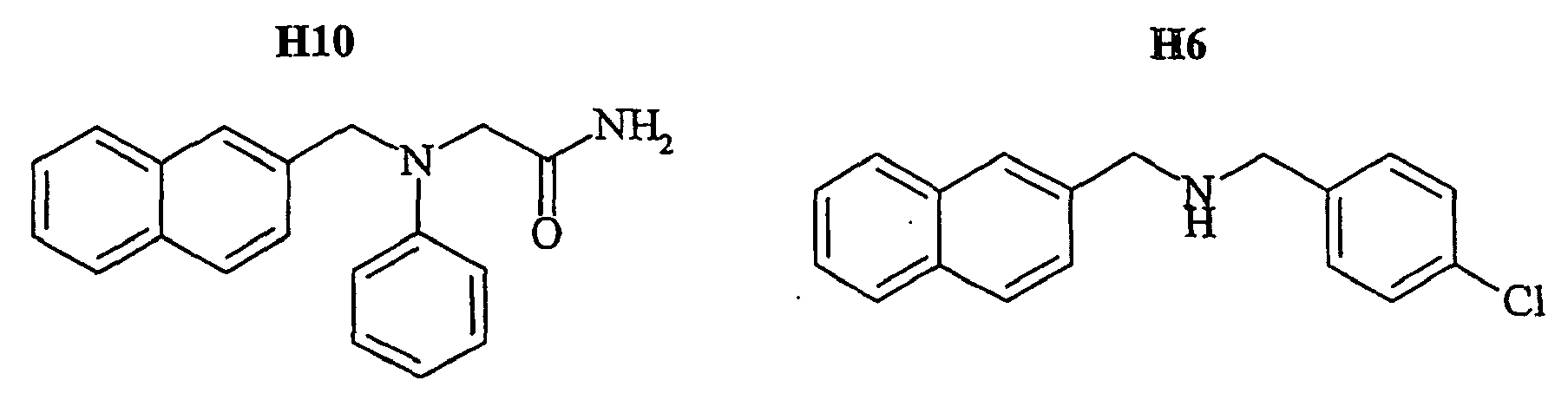
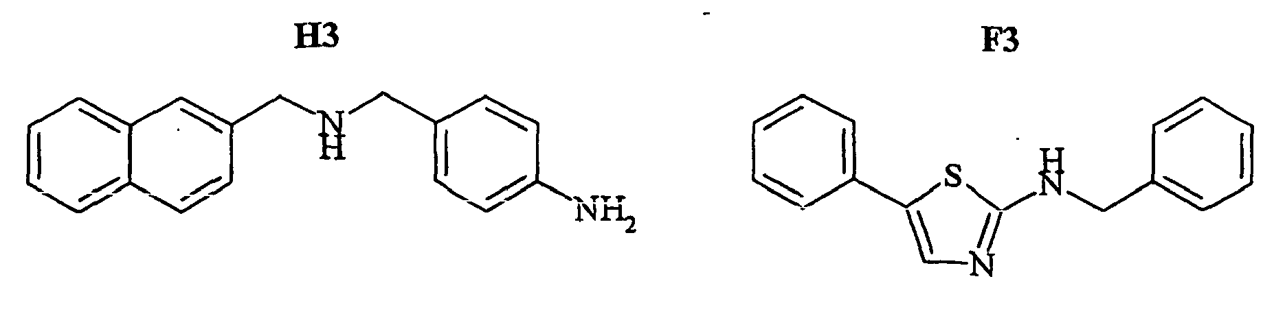


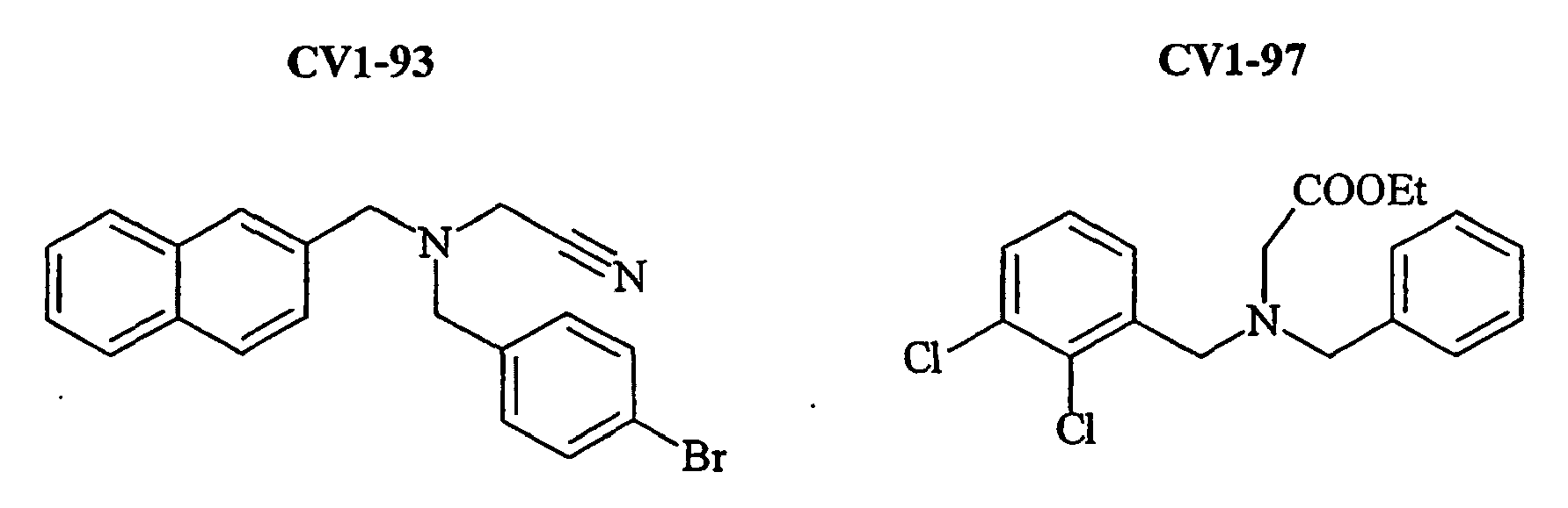


Les produits de formule A11, G11, H10, H6, H3, F3, 801, 802, 803, 804, 805, 806, 807, 808, 809, CV1-80, CV1-81, CV1-84, CV1-85, CV1-93, CV1-97, CV1-122, CV1-123, CV1-131 et CV1-135 sont nouveaux en tant que tels.Products of formula A11, G11, H10, H6, H3, F3, 801, 802, 803, 804, 805, 806, 807, 808, 809, CV1-80, CV1-81, CV1-84, CV1-85, CV1-93, CV1-97, CV1-122, CV1-123, CV1-131 and CV1-135 are novel as such.
Les tableaux 1 et 2 suivants résument l'ensemble des données expérimentales acquises pour les molécules G6, A11, G11, F3, H6, F9, H3, F7, H10, NP234 (ou 801), NP 246 (ou 803), 804, 805, 806, 808, 807, 809, CV1-80, CV1-81, CV1-84, CV1-85, CV1-93, CV1-97, CV1-122, CV1-123, et CV1-135, dont les structures sont indiquées en colonnes.The following tables 1 and 2 summarize all the experimental data acquired for the molecules G6, A11, G11, F3, H6, F9, H3, F7, H10, NP234 (or 801), NP 246 (or 803), 804, 805, 806, 808, 807, 809, CV1-80, CV1-81, CV1-84, CV1-85, CV1-93, CV1-97, CV1-122, CV1-123, and CV1-135, whose structures are indicated in columns.
Dans le tableau 1, la colonne "code" correspond au nom d'usage ; la colonne "EC50" correspond à la concentration de composé provoquant une augmentation de 50% (par rapport à la valeur maximale) du pourcentage de l'amplitude de la vitesse de dissociation rapide ; la colonne "%a1 (max) " correspond à la valeur maximale observée pour l'augmentation d'amplitude de la vitesse de dissociation rapide ; la colonne "IC50" correspond à la concentration de composé pour laquelle on observe une diminution de 50% de la liaison de 20 nM de NKA-Bo au récepteur EGFP-NK2R ; la colonne "Rep Ca" correspond à la capacité de chaque composé à évoquer une réponse calcique sur des cellules n'exprimant pas le récepteur EGFP-NK2R et la colonne "inhib AMPc" indique la capacité de chaque composé à inhiber la réponse de production d'AMPc évoquée par la NKA.In Table 1, the column "code" corresponds to the usage name; the "EC50" column corresponds to the concentration of compound causing a 50% increase (relative to the maximum value) of the percentage of the amplitude of the fast dissociation rate; the column "% a1 (max)" corresponds to the maximum value observed for the amplitude increase of the fast dissociation rate; the "IC50" column corresponds to the concentration of compound for which there is a 50% decrease in the binding of 20 nM NKA-Bo to the EGFP-NK2R receptor; the "Rep Ca" column corresponds to the ability of each compound to evoke a calcium response on cells not expressing the EGFP-NK2R receptor and the "cAMP inhibitor" column indicates the ability of each compound to inhibit the production response. AMPc evoked by the NKA.
Dans le tableau 2, la colonne "code" correspond au nom d'usage ; la colonne "EC50" correspond à la concentration de composé provoquant une augmentation de 50% (par rapport à la valeur maximale) du pourcentage de l'amplitude de la vitesse de dissociation rapide ; la colonne "% al (à 10 µM) " signifie que les composés ont été testés uniquement à 10 µM et comparés aux valeurs déterminées pour 805 (T = 22 ± 1 représente l'amplitude de dissociation rapide en absence d'effecteur) ; la colonne "% Réversion de liaison (à 10 µM) " signifie que les composés ont été testés uniquement à 10 µM et comparés aux valeurs déterminées pour 805.In Table 2, the column "code" corresponds to the usage name; the "EC50" column corresponds to the concentration of compound causing a 50% increase (relative to the maximum value) of the percentage of the amplitude of the fast dissociation rate; the "% al (at 10 μM)" column means that the compounds were tested only at 10 μM and compared with the values determined for 805 (T = 22 ± 1 represents the rapid dissociation amplitude in the absence of an effector); the column "% Bond Reversion (at 10 μM)" means that the compounds were tested only at 10 μM and compared to the values determined for 805.
Parmi tous les composés, 805 a fait l'objet d'une étude de la potentialisation des réponses calciques évoquées par la NKA ou la NKA tronquée sur les récepteurs NK2R humain sauvage, NK1R humain sauvage, ou EGFP-NK2R.

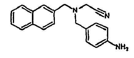


![]()

![]()


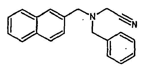
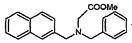


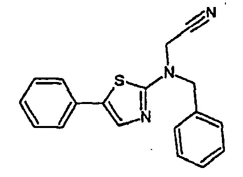


![]()
![]()
- Les composés susmentionnés sont regroupés dans deux grandes familles : famille I et famille II.The aforementioned compounds are grouped into two large families: family I and family II.
La famille I regroupe les composés A11, G11, H10, 801, 803, 804, 805, 806, 808, 807, CV1-80, CV1-81, CV1-84, CV1-85, CV1-93, CV1-97, CV1-122, CV1-123 et CV1-135.The family I groups the compounds A11, G11, H10, 801, 803, 804, 805, 806, 808, 807, CV1-80, CV1-81, CV1-84, CV1-85, CV1-93, CV1-97, CV1-122, CV1-123 and CV1-135.
Ces composés répondent à la formule générale suivante :


Les composés H6 et H3 sont des intermédiaires de synthèse permettant l'obtention des composés de la famille I susmentionnée.The compounds H6 and H3 are synthetic intermediates making it possible to obtain the compounds of the aforementioned family I.
La famille II regroupe les composés F3, F7 et 809.Family II groups the compounds F3, F7 and 809.
Ces composés répondent à la formule générale suivante :

Les composés F7 et F9 sont déjà connus et sont décrits dans l'article de Moutou et al. (1994).The compounds F7 and F9 are already known and are described in the article by Moutou et al. (1994).
Par ailleurs, le composé G6 du tableau 1 est déjà connu et décrit dans la demande internationale WO 02/24192.Moreover, the compound G6 of Table 1 is already known and described in the international application WO 02/24192.
A une solution de l'aldéhyde aromatique approprié (1 éq) dans le méthanol, on ajoute l'amine primaire (1 éq)(voir tableau ci-après). Le milieu réactionnel est chauffé à 60°C pendant deux heures puis refroidi rapidement à 0°C. On ajoute ensuite du NaBH4 (2 éq) et on agite pendant quelques minutes puis on remet le milieu réactionnel à température ambiante pendant une heure. Après évaporation de la solution, on extrait avec de l'AcOEt en lavant avec de l'eau puis avec une solution saturée en NaCl. On sèche avec Na2SO4, on filtre sur fritté et on évapore à sec. L'amine secondaire, ainsi isolée, le plus généralement sous la forme d'une huile, est utilisée telle quelle sans aucune purification.To a solution of the appropriate aromatic aldehyde (1 eq) in methanol, the primary amine (1 eq) is added (see table below). The reaction medium is heated at 60 ° C for two hours and then rapidly cooled to 0 ° C. NaBH 4 (2 eq) is then added and the mixture is stirred for a few minutes and then the reaction medium is returned to ambient temperature for one hour. After evaporation of the solution, it is extracted with AcOEt, washing with water and then with saturated NaCl solution. It is dried with Na 2 SO 4 , sintered and evaporated to dryness. The secondary amine, thus isolated, most generally in the form of an oil, is used as it is without any purification.
L'amine secondaire ainsi obtenue répond à la formule suivante :

Un exemple de réaction correspondant à l'étape 1 (ici obtention du composé H3) est :

Tableau récapitulatif des produits de départ :
L'amine secondaire (1,1 éq) obtenue à l'étape précédente est dissoute dans le diméthylformamide, et on ajoute de l'halogénure d'alkyle (chloroacétonitrile, chlorure de propargyle, etc.)(voir plus loin). La solution est chauffée à reflux pendant 12 heures puis on la laisse revenir à température ambiante. On extrait ensuite avec de l'éther (10 fois plus en volume que le DMF) et on lave avec de l'eau puis avec une solution saturée de NaCl. La phase organique est séchée avec Na2SO4 puis elle est évaporée à sec. Le brut est purifié par chromatographie sur gel de silice (AcOEt 1/Hex 9). Les chlorhydrates sont préparés par barbotage de HCl gazeux dans l'AcOEt.The secondary amine (1.1 eq) obtained in the previous step is dissolved in dimethylformamide, and alkyl halide (chloroacetonitrile, propargyl chloride, etc.) (see below) is added. The solution is refluxed for 12 hours and then allowed to warm to room temperature. It is then extracted with ether (10 times more by volume than DMF) and washed with water and then with saturated NaCl solution. The organic phase is dried with Na 2 SO 4 and then evaporated to dryness. The crude is purified by chromatography on silica gel (
On obtient alors les composés susmentionnés, sous forme de chlorhydrate, de formule telle que définie ci-dessous :

Plus précisément, cette deuxième étape correspond au schéma réactionnel suivant :

Les amines secondaires sont donc mises à réagir avec différents halogénures commerciaux.The secondary amines are therefore reacted with different commercial halides.
Pour les composés A11, G11, 801, 803, 804, 805, CV1-80, CV1-81, CV1-84, CV1-85, CV1-93, CV1-122, CV 1-123 et CV1-135, on utilise du chloroacétonitrile.For the compounds A11, G11, 801, 803, 804, 805, CV1-80, CV1-81, CV1-84, CV1-85, CV1-93, CV1-122, CV 1-123 and CV1-135, use is made of chloroacetonitrile.
Pour les composés 806 et CV1-97, on utilise du bromo ou du chloroacétate d'éthyle ou de méthyle.For
Pour les composés H4, H10 et 807, on utilise du bromo ou du chloroacétamide.For the compounds H4, H10 and 807, bromo or chloroacetamide is used.
Pour le composé 808, on utilise du bromure ou du chlorure de propargyle.For compound 808, bromide or propargyl chloride is used.
Concernant le composé CV1-135, il est obtenu selon un mode opératoire particulier, tel que décrit ci-après :Regarding the compound CV1-135, it is obtained according to a particular procedure, as described below:
On dissout dans l'acétonitrile le 4-bromobenzonitrile de formule :

Ce mode opératoire correspond au schéma réactionnel suivant :
- 1)

- 2)

- 1)
- 2)
Le 5-phényl 2-benzylaminothiazole est obtenu au départ du 5-phényl-2-aminothiazole par condensation avec le benzaldéhyde, suivie d'une hydrogénation. Le composé obtenu et ensuite alkylé par le chloroacétonitrile (voir mode opératoire I).5-Phenyl-2-benzylaminothiazole is obtained from 5-phenyl-2-aminothiazole by condensation with benzaldehyde, followed by hydrogenation. The compound obtained and then alkylated with chloroacetonitrile (see procedure I).
Le composé F3 est obtenu selon le schéma réactionnel suivant :

A une solution de 5-phényl-2-aminobenzylthiazole (1 éq) dans le DMF refroidi à 0°C est ajouté du NaH (1,3 éq). Le milieu réactionnel est agité pendant 15 minutes puis le chloroacétonitrile (1 éq) est ajouté gouttes à gouttes. L'agitation est poursuivie à température ambiante pendant 8 heures, la solution est ensuite diluée par l'eau. Après extraction à l'acétate d'éthyle, la phase organique est séchée sur sulfate de sodium anhydre et est évaporée. Le brut réactionnel est purifié par chromatographie sur gel de silice (AcOEt 1 / Hexane 2).To a solution of 5-phenyl-2-aminobenzylthiazole (1 eq) in DMF cooled at 0 ° C. is added NaH (1.3 eq). The reaction medium is stirred for 15 minutes and then the chloroacetonitrile (1 eq) is added dropwise. Stirring is continued at room temperature for 8 hours, the solution is then diluted with water. After extraction with ethyl acetate, the organic phase is dried over anhydrous sodium sulphate and evaporated. The crude reaction product is purified by chromatography on silica gel (
On obtient donc ainsi par exemple le composé 809 : ((5-phényl-thiazol-2-yl)-N-benzyl)acétonitrile C18H15N3S (305,40 g.mol-1) sous la forme d'une poudre beige, selon le schéma réactionnel suivant :

Dans sa réalisation préférée, le développement de l'invention utilise l'ADNc codant pour la protéine fluorescente verte (Prasher et al., 1992) de la méduse aequorea victoria, préférentiellement les mutants EYFP, EGFP et ECPF de cette protéine optimisée pour leur expression dans les organismes hôtes préférés, les cellules mammifères.In its preferred embodiment, the development of the invention uses the cDNA coding for the green fluorescent protein (Prasher et al., 1992) of the jellyfish aequorea victoria, preferentially the EYFP, EGFP and ECPF mutants of this protein, optimized for their expression. in the preferred host organisms, mammalian cells.
L'ADNc peut être modifié pour coder pour un variant dans lequel un ou plusieurs acides aminés sont substitués, insérés ou délétés afin de permettre sa fusion, en N ou en C-terminal avec le gène codant pour un récepteur.The cDNA can be modified to encode a variant in which one or more amino acids are substituted, inserted or deleted to permit its N- or C-terminal fusion with the gene encoding a receptor.
Le récepteur peut être choisi parmi :
- 1) les récepteurs de neurotransmetteurs couplés à des protéines G structurellement reliés aux récepteurs adrénergiques et récepteurs métabotropiques du glutamate tels que présentés dans la liste actualisées annuellement et éditée dans les bases de données GPCRdb (http://www.gcrdb.uthscsa.edu ou http://www.gpcr.org), ensembl (http://www.ensembl.org) entre autres.
- 2) les récepteurs-canaux structurellement reliés aux récepteurs nicotiniques, aux récepteurs de glutamate et aux récepteurs de l'ATP, tels que présentés dans la liste actualisées annuellement et éditée dans les bases de données GPCRdb (http://www.gcrdb.uthscsa.edu ou http://www.gpcr.org), ensembl (http://www.ensembl.org) entre autres.
- 3) les récepteurs nucléaires possédant un domaine d'interaction avec l'ADN structurellement reliés au récepteur des stéroïdes (Mangelsdorf et al., 1995 ; Wurtz et al., 1996).
- 4) les récepteurs de la membrane plasmique à activité tyrosine kinase structurellement reliés au récepteur de l'insuline (Yarden, Y. and Ullrich, A., 1988).
- 5) les récepteurs membranaires couplés aux protéines tyrosine kinases (STATs, TYK2, Jak) structurellement reliés au récepteur de l'interféron γ (Brisco et al., 1996 ; Ihle, 1995).
- 1) neurotransmitter receptors coupled to G proteins structurally related to adrenergic receptors and metabotropic glutamate receptors as presented in the list updated annually and published in the GPCRdb databases (http://www.gcrdb.uthscsa.edu or http://www.gpcr.org), together (http://www.ensembl.org) among others.
- 2) receptors-channels structurally related to nicotinic receptors, glutamate receptors and ATP receptors, as presented in the list updated annually and published in GPCRdb databases (http: //www.gcrdb.uthscsa .edu or http://www.gpcr.org), together (http://www.ensembl.org) among others.
- 3) nuclear receptors possessing a domain of interaction with the DNA structurally related to the steroid receptor (Mangelsdorf et al., 1995; Wurtz et al., 1996).
- 4) tyrosine kinase-active plasma membrane receptors structurally related to the insulin receptor (Yarden, Y. and Ullrich, A., 1988).
- 5) membrane receptors coupled to protein tyrosine kinases (STATs, TYK2, Jak) structurally related to the interferon γ receptor (Brisco et al., 1996, Ihle, 1995).
Dans le cas où la fusion est effectuée entre la EGFP et un récepteur couplé aux protéines G (groupe 1), la fusion peut être notamment effectuée :
- 1) du coté N-terminal du récepteur, et donc du côté C-terminal de la EGFP,
- 2) du coté C-terminal du récepteur et donc du côté N-terminal de la EGFP,
- 3) dans la séquence du récepteur, en particulier dans la première ou la troisième boucle intracellulaire, éventuellement en introduisant une ou plusieurs copies d'une séquence espaceur, notamment -GGGGS-.
- 1) on the N-terminal side of the receiver, and therefore on the C-terminal side of the EGFP,
- 2) on the C-terminal side of the receiver and therefore on the N-terminal side of the EGFP,
- 3) in the receptor sequence, in particular in the first or third intracellular loop, optionally introducing one or more copies of a spacer sequence, in particular -GGGGS-.
Dans le cas où la fusion est effectuée entre la EGFP et un récepteur-canal (groupe 2), la fusion peut être notamment effectuée :
- 1) dans la région homologue à la "région immunogénique majeure" de la sous-unité α du récepteur nicotinique de Torpille (résidus 67-76), éventuellement en introduisant une ou plusieurs copies d'une séquence espaceur, notamment -GGGGS-.
- 1) in the region homologous to the "major immunogenic region" of the α subunit of the nicotinic Torpille receptor (residues 67-76), optionally introducing one or more copies of a spacer sequence, in particular -GGGGS-.
Dans le cas ou la fusion est effectuée entre la EGFP et un récepteur nucléaire (groupe 3), la fusion peut être notamment effectuée :
- 1) du coté N-terminal du récepteur, et donc du côté C-terminal de la EGFP,
- 2) du coté N-terminal du récepteur, tronqué dans sa partie N-terminale en amont du domaine de liaison à l'ADN, et donc du côté C-terminal de la EGFP.
- 1) on the N-terminal side of the receiver, and therefore on the C-terminal side of the EGFP,
- 2) on the N-terminal side of the receptor, truncated in its N-terminal portion upstream of the DNA binding domain, and therefore on the C-terminal side of EGFP.
Dans le cas où la fusion est effectuée entre la EGFP et un récepteur soit à activité tyrosine kinase, soit couplé à une tyrosine kinase (groupes 4 et 5), la fusion peut être notamment effectuée :
- 1) du coté N-terminal du récepteur, et donc du coté C-terminal de la EGFP.
- 1) on the N-terminal side of the receiver, and therefore on the C-terminal side of the EGFP.
Tout gène codant pour une protéine fluorescente, notamment la GFP, couplée à un récepteur, et dérivant d'organismes exprimant la GFP ou des protéines similaires pourrait être utilisé dans cette invention.Any gene encoding a fluorescent protein, including GFP, coupled to a receptor, and derived from GFP expressing organisms or similar proteins could be used in this invention.
Les séquences d'ADN codant pour la GFP et les protéines cibles, notamment des récepteurs, peuvent être d'origine génomique ou être des ADNc, et peuvent être obtenus à partir de l'ADN de n'importe quelle espèce animale ou végétale, eucaryote ou procaryote, par exemple en préparant des banques génomiques ou des banques d'ADNc et en criblant ces banques pour identifier les séquences codantes par hybridation avec des sondes oligonucléotidiques par les techniques standard (Current Protocols in Molecular Biology, op. cit.).The DNA sequences encoding GFP and the target proteins, including receptors, may be of genomic origin or be cDNAs, and may be obtained from the DNA of any animal or plant species, eukaryotic or prokaryotic, for example by preparing genomic libraries or cDNA libraries and screening these libraries to identify coding sequences by hybridization with oligonucleotide probes by standard techniques (Current Protocols in Molecular Biology, op cit).
Les constructions d'ADN codant pour la GFP et les protéines cibles peuvent aussi être obtenues par synthèse totale par les méthodes standards, notamment la méthode des phosphoramidites (Beaucage and Caruthers, 1981) et l'emploi d'appareils de synthèse d'ADN automatisés, les polynucléotides obtenus étant ensuite purifiés, ligués et clonés dans les vecteurs appropriés. Pour la plupart des applications, les gènes codant pour la GFP et les protéines cibles seront préférentiellement obtenus par criblage de banques, tandis que les bras espaceurs ainsi que les oligonucléotides requis pour la mutagenèse seront préférentiellement obtenus par synthèse.The DNA constructs encoding GFP and the target proteins can also be obtained by total synthesis by standard methods, including the phosphoramidite method (Beaucage and Caruthers, 1981) and the use of automated DNA synthesis apparatus. the obtained polynucleotides are then purified, ligated and cloned into the appropriate vectors. For most applications, the genes coding for GFP and the target proteins will preferably be obtained by screening libraries, while the spacer arms and oligonucleotides required for mutagenesis will preferably be obtained by synthesis.
Les constructions d'ADN pourront être de nature mixte, synthétique et génomique, par ligation de fragments synthétiques avec des élément d'ADN génomique, selon des procédures standard (Current Protocols in Molecular Biology, op.cit.).The DNA constructs may be of a mixed, synthetic and genomic nature, by ligation of synthetic fragments with genomic DNA elements, according to standard procedures (Current Protocols in Molecular Biology, op.cit.).
Les constructions d'ADN peuvent aussi être obtenues par PCR ("polymerase chain reaction") en employant des amorces spécifiques, comme par exemple décrit dans PCR protocol 1990, Academic press, San Diego, California, USA.DNA constructs can also be obtained by PCR ("polymerase chain reaction") using specific primers, as for example described in PCR protocol 1990, Academic Press, San Diego, California, USA.
Enfin, les constructions d'ADN peuvent être modifiées par d'autres méthodes incluant par exemple des réactions chimiques, de la mutagenèse aléatoire ou dirigée, par insertion, délétion ou substitution de nucléotides, ces modifications pouvant altérer des propriétés de l'une ou l'autre protéine, notamment, la GFP et les protéines cibles.Finally, the DNA constructs can be modified by other methods including, for example, chemical reactions, random or directed mutagenesis, by insertion, deletion or substitution of nucleotides, these modifications being able to alter properties of one or the other. other protein, including GFP and the target proteins.
Les constructions d'ADN peuvent être insérées dans un vecteur recombinant. Ce vecteur peut être n'importe quel vecteur approprié pour les procédures employées avec des vecteurs recombinants. Le choix du vecteur sera souvent effectué en fonction de la cellule hôte dans laquelle la construction d'ADN voudra être introduite. Le vecteur pourra ainsi être un vecteur capable de se répliquer de manière autonome, c'est-à-dire extrachromosomal, et indépendante de la réplication chromosomale, par exemple un plasmide. Alternativement, le vecteur pourra être élaboré de manière à intégrer tout ou partie de l'ADN qu'il contient dans le génome de la cellule hôte, et se répliquera en même temps que le(s) chromosome(s) dans lequel il sera intégré.The DNA constructs can be inserted into a recombinant vector. This vector may be any suitable vector for the procedures employed with recombinant vectors. The choice of the vector will often be made according to the host cell in which the DNA construct will want to be introduced. The vector may thus be a vector capable of replicating autonomously, that is to say extrachromosomal, and independent of chromosomal replication, for example a plasmid. Alternatively, the vector may be designed to integrate all or part of the DNA it contains into the genome of the host cell, and will replicate at the same time as the chromosome (s) in which it will be integrated .
Le vecteur est préférentiellement un vecteur d'expression dans lequel la GFP fusionnée au récepteur ou la GFP fusionnée au ligand est sous le contrôle d'autres segments d'ADN requis pour la transcription. En général, le vecteur d'expression dérive d'ADN plasmidique ou viral ou peut contenir des éléments de l'un et de l'autre.The vector is preferably an expression vector in which GFP fused to the receptor or GFP fused to the ligand is under the control of other DNA segments required for transcription. In general, the expression vector is derived from plasmid or viral DNA or may contain elements of both.
Le terme "sous le contrôle" indique que les segments d'ADN sont disposés sur le vecteur de manière à fonctionner de concert pour servir l'objectif voulu, par exemple, la transcription est initiée dans le promoteur et se poursuit tout au long de la séquence codant pour le récepteur fusionné à la GFP ou le ligand fusionné à la GFP.The term "under the control" indicates that the DNA segments are arranged on the vector so as to work together to serve the intended purpose, for example, transcription is initiated in the promoter and continues throughout the sequence encoding the GFP-fused receptor or the ligand fused to GFP.
Le promoteur peut être n'importe quelle séquence d'ADN susceptible de promouvoir une activité transcriptionnelle dans la cellule hôte choisie et peut être dérivé de gène homologues ou hétérologues à la cellule hôte.The promoter may be any DNA sequence capable of promoting transcriptional activity in the selected host cell and may be derived from homologous or heterologous genes to the host cell.
Des exemples de promoteurs convenant pour l'expression du récepteur fusionné avec le GFP ou du ligand fusionné avec la GFP dans des cellules mammifères sont le promoteur du virus simien SV40 (Subramani et al., 1981), le promoteur du virus du sarcome de Rous (RSV), le promoteur du cytomegalovirus (CMV) ou le promoteur tardif majeur de l'adénovirus (AdMLP).Examples of promoters suitable for the expression of the GFP-fused receptor or the GFP-fused ligand in mammalian cells are SV40 simian virus promoter (Subramani et al., 1981), the Rous sarcoma virus (RSV) promoter, the cytomegalovirus (CMV) promoter or the adenovirus major late promoter (AdMLP).
Promoteur de la polyhédrine (US 4,745,051 ; Vasuvedan et al., 1992) le promoteur P10 (Vlack et al., 1988), le promoteur du gène précoce 1 du baculovirus (US 5,155,037; US 5,162,222).Promoter of polyhedrin (US 4,745,051, Vasuvedan et al., 1992) the P10 promoter (Vlack et al., 1988), the early gene promoter of baculovirus (US 5,155,037; US 5,162,222).
Promoteurs des gènes de la glycolyse (Hitzeman et al., 1980 ; Alber et Kawasaki, 1982), des gènes des alcool deshydrogénases (Young et al., 1982).Promoters of the genes for glycolysis (Hitzeman et al., 1980, Alber and Kawasaki, 1982), genes for alcohol dehydrogenases (Young et al., 1982).
Des exemples de promoteurs pour l'expression dans la bactérie peuvent être des promoteurs constitutifs comme le promoteur de la polymérase T7, ou des promoteurs inductibles comme par exemple le promoteur pL du phage lambda (Current Protocols in Molecular Biology, op.cit.).Examples of promoters for expression in the bacterium may be constitutive promoters such as the T7 polymerase promoter, or inducible promoters such as, for example, the phage lambda pL promoter (Current Protocols in Molecular Biology, op.cit.).
Les promoteurs utilisables sont par exemple le promoteur ADH3 (McKnight et al., 1985) ou le promoteur tpiA. D'autres promoteurs utiles peuvent être dérivés des gènes codant pour l'aspartate protéinase de Rhizomucor miehei, l'alpha-amylase neutre de Aspergillus niger, de l'acétamidase de Aspergillus nidulans, de la TAKA amylase de Aspergillus oryzae ou le promoteur de la glucoamylase de Aspergillus awamori. Suitable promoters are, for example, the ADH3 promoter (McKnight et al., 1985) or the tpiA promoter. Other useful promoters may be derived from the genes encoding Rhizomucor miehei aspartate proteinase , Aspergillus niger neutral alpha-amylase , Aspergillus nidulans acetamidase , Aspergillus oryzae TAKA amylase or the promoter of the invention. Aspergillus awamori glucoamylase.
Le vecteur pourra par ailleurs contenir :
- des séquences de polyadénylation, comme par exemple celles de SV40 ou de la région
Elb 5 de l'adénovirus, - des séquences activatrices (enhancer) de la transcription (l'activateur de SV40),
- des séquences de réplication comme par exemple les séquences de réplication de SV40 ou du virus Epstein Barr, pour les cellules mammifères ou l'origine et les gènes de réplication REP 1-3 du plasmide 2 µ, pour les levures,
- des marqueurs de sélection, à savoir des gènes conférant une résistance à un antibiotique (néomycine, zéocine, hygromycine, ampicilline, kanamycine, tétracycline, chloramphénicol...) ou permettant la compensation d'un défaut (gène codant pour la dihydrofolate réductase permettant la résistance au méthotrexate, ou gène TPI de S. pombe décrit par Russell (1985).
- polyadenylation sequences, for example those of SV40 or of the Elb region of the adenovirus,
- enhancer sequences (enhancer) of the transcription (SV40 activator),
- replication sequences, for example the SV40 or Epstein Barr virus replication sequences, for the mammalian cells or the origin and the REP 1-3 replication genes of the
plasmid 2 μ, for the yeasts, - selection markers, namely genes conferring resistance to an antibiotic (neomycin, zeocin, hygromycin, ampicillin, kanamycin, tetracycline, chloramphenicol ...) or for the compensation of a defect (gene encoding dihydrofolate reductase for resistance to methotrexate, or S. pombe TPI gene described by Russell (1985).
La cellule hôte peut être n'importe quelle cellule capable d'exprimer la construction d'ADN insérée dans un vecteur approprié.The host cell may be any cell capable of expressing the inserted DNA construct in a suitable vector.
Les cellules peuvent être notamment des bactéries, des levures, des champignons et des cellules eucaryotes supérieures comme par exemple des cellules mammifères.The cells may especially be bacteria, yeasts, fungi and higher eukaryotic cells, for example mammalian cells.
Des exemples de cellules bactériennes capables d'exprimer les constructions d'ADN sont :
- des bactéries gram-positives telles que les souches de Bacillus comme B. subtilis, B. licheniformis, B. lentus, B. brevis, B. strearothermophilus, B. thurigiensis ou des souches de Streptomyces comme S. lividans, S murinus,
- des bactéries gram-négatives telles Escherichia coli.
La transformation des bactéries peut être effectuée par transformation protoplastique ou par transformation de bactéries compétentes (Current Protocols in Molecular Biology, op.cit.).
- gram-positive bacteria such as Bacillus strains such as B. subtilis, B. licheniformis, B. lentus, B. brevis, B. strearothermophilus, B. thurigiensis or strains of Streptomyces such as S. lividans, S. murinus,
- Gram-negative bacteria such as Escherichia coli.
Bacterial transformation can be effected by protoplast transformation or transformation of competent bacteria (Current Protocols in Molecular Biology, op.cit.).
Lignées cellulaires HEK 293, HeLa, cultures primaires, cellules COS (par exemple ATCC CRL 1650), BHK (par exemple ATCC CRL 1632) CHO (par exemple ATCC CCL 61).HEK 293, HeLa cell lines, primary cultures, COS cells (eg ATCC CRL 1650), BHK (eg ATCC CRL 1632) CHO (eg ATCC CCL 61).
Les méthodes d'introduction de l'ADN dans ces cellules (transfection, lipofection, électroporation etc.) sont décrites dans Current Protocols in Molecular Biology, op.cit.Methods of introducing DNA into these cells (transfection, lipofection, electroporation etc.) are described in Current Protocols in Molecular Biology, op.cit.
- Saccharomyces, S. cerevisiae, S. kluyveri,Saccharomyces, S. cerevisiae, S. kluyveri,
- Kluiveromyces, K. lactis,Kluiveromyces, K. lactis,
- Hansenula, H. polymorpha,Hansenula, H. polymorpha,
- Pichia, P. pastoris,Pichia, P. pastoris,
- transformées par introduction d'ADN hétérologue selon les protocoles décrits dans Current Protocols in Molecular Biology, op.cit.transformed by introduction of heterologous DNA according to the protocols described in Current Protocols in Molecular Biology, op.cit.
Les cellules transformées sont sélectionnées par un phénotype déterminé par un marqueur de résistance, généralement à une drogue, ou par leur capacité à proliférer en l'absence d'un nutriment particulier.Transformed cells are selected by a phenotype determined by a resistance marker, usually a drug, or by their ability to proliferate in the absence of a particular nutrient.
Les souches Aspergillus (A. oryzae, A. nidulans, A. niger), Neurospora, Fusarium, Trichoderma. L'utilisation de Aspergillus pour l'expression de protéines est décrite dans EP 272 277 ou EP 230 023 ou EP 184 438.Strains Aspergillus ( A. oryzae, A. nidulans, A. niger ), Neurospora, Fusarium, Trichoderma. The use of Aspergillus for the expression of proteins is described in EP 272 277 or EP 230 023 or EP 184 438.
On peut citer les lignées de Lepidoptera e.g. Spodoptera frugiperda (Sf9) ou Trichoplusia ni (Tni). Les méthodes de transformation (infection en particulier) sont décrites dans Current Protocols in Molecular Biology (op.cit.).We can mention the lines of Lepidoptera eg Spodoptera frugiperda (Sf9) or Trichoplusia ni (Tni). Transformation methods (infection in particular) are described in Current Protocols in Molecular Biology (op.cit.).
Les ligands interagissant avec le récepteur peuvent être de n'importe quelle origine (naturelle, synthétique, semi-synthétique, recombinante), et de n'importe quelle structure (chimique, peptidique, protéique). Ils peuvent être naturellement fluorescents (ou porteurs d'un chromophore) ou peuvent nécessiter soit une réaction chimique permettant le greffage d'un groupe fluorescent (ou d'un précurseur de groupe fluorescent) ou d'un chromophore, soit une construction d'ADN conduisant à la fusion du ligand avec la GFP et permettant l'expression du ligand ainsi rendu fluorescent.Ligands interacting with the receptor can be of any origin (natural, synthetic, semi-synthetic, recombinant), and of any structure (chemical, peptide, protein). They can be naturally fluorescent (or carry a chromophore) or may require either a chemical reaction allowing the grafting of a fluorescent group (or a fluorescent group precursor) or a chromophore, or a DNA construct leading to the fusion of the ligand with GFP and allowing the expression of the ligand thus made fluorescent.
Des exemples de réactions chimiques sont :
- couplage d'amines ou de thiols avec des réactifs de type halogénure d'alkyle, halogénures d'aryles, halogénures d'acyles, halogénures d'acides, le groupe isothiocyanate, le groupe maléimide, les époxydes, dans un solvant organique en présence d'une base ou en milieu aqueux,
- couplage d'acides avec des amines activées par des groupes tels que les succinimides.
- coupling of amines or thiols with alkyl halide, aryl halide, acyl halide, acid halide, isothiocyanate, maleimide and epoxide reactants in an organic solvent in the presence of a base or in an aqueous medium,
- coupling of acids with amines activated by groups such as succinimides.
Selon le procédé de l'invention, la fluorescence des cellules transformées peut être mesurée dans un spectrofluorimètre à l'aide duquel les propriétés spectrales des cellules, en suspension ou adhérentes, peuvent être déterminées par l'acquisition de leurs spectres d'excitation et d'émission. Les interactions avec le ligand fluorescent sont ensuite détectées par les changements des spectres d'excitation et/ou d'émission du donneur et de l'accepteur d'énergie, et les ligands sont définis comme pharmacologiquement significatifs si leurs interactions avec le récepteur sont inhibées par l'addition d'un excès de ligand non fluorescent empêchant l'interaction entre le récepteur fluorescent et le ligand fluorescent.According to the method of the invention, the fluorescence of the transformed cells can be measured in a spectrofluorometer by means of which the spectral properties of the cells, in suspension or adherent, can be determined by the acquisition of their excitation and excitation spectra. 'program. Interactions with the fluorescent ligand are then detected by changes in the excitation and / or emission spectra of the donor and the energy acceptor, and the ligands are defined pharmacologically. significant if their interactions with the receptor are inhibited by the addition of an excess of non-fluorescent ligand preventing interaction between the fluorescent receptor and the fluorescent ligand.
Les mesures des cinétiques d'association et/ou de dissociation sont effectuées par tous les moyens permettant d'enregistrer la formation ou la dissociation du complexe entre le ligand marqué et le récepteur marqué, de manière continue ou discontinue, de telle sorte que l'on parvient à déterminer les paramètres cinétiques de l'association et de la dissociation, à savoir la (les) constantes de vitesse(s), ainsi que la ou les amplitudes relatives associées à chaque étape cinétique d'association et/ou de dissociation.The measurements of the kinetics of association and / or dissociation are carried out by any means making it possible to record the formation or dissociation of the complex between the labeled ligand and the labeled receptor, continuously or discontinuously, so that the the kinetic parameters of the association and dissociation, namely the rate constant (s), as well as the relative amplitude (s) associated with each kinetic step of association and / or dissociation, are determined.
Dans sa réalisation préférée, les cinétiques dont les constantes de vitesse apparente sont supérieures à 0,1 s-1 seront enregistrées à l'aide d'un appareil de mélange rapide connecté à un dispositif d'excitation et de détection de fluorescence (figure 1). Les échantillons à mélanger seront disposés dans des seringues (ou autres conteneurs) de mélange, puis mélangés rapidement. Dans ce contexte, "rapidement" veut dire que le contenu des seringues est propulsé avec un débit supérieur ou égal à 4 ml/sec pour une chambre d'observation de 100 µl, ainsi que décrit par exemple dans Palanché et al. (2001).In its preferred embodiment, the kinetics whose apparent speed constants are greater than 0.1 s -1 will be recorded using a fast mixing apparatus connected to a device for excitation and fluorescence detection (FIG. ). The samples to be mixed will be placed in mixing syringes (or other containers) and mixed rapidly. In this context, "rapidly" means that the contents of the syringes are propelled with a flow rate greater than or equal to 4 ml / sec for an observation chamber of 100 μl, as described for example in Palanché et al. (2001).
Dans le cas où les constantes de vitesse apparentes sont inférieures à 0,1 s-1, l'utilisation d'un appareil de mélange rapide n'est pas indispensable. On pourra dans ce cas enregistrer les variations de fluorescence du donneur et/ou de l'accepteur dans un spectrofluorimètre standard pourvu d'un dispositif d'agitation magnétique de l'échantillon, d'une régulation de température et d'une fonction d'enregistrement en temps. Les échantillons sont alors disposés dans une cuvette munie d'un agitateur et le mélange sera obtenu par addition manuelle des composants ou solutions désirés. Les échantillons contenant le récepteur peuvent être soit des cellules, soit des fragments de cellules. Ils seront préférentiellement disposés en premier dans la cuvette et les agents modulateurs ainsi que le ligand marqué seront ajoutés à cette solution. L'effet de ces ajouts pourra ainsi être enregistré et toute variation de fluorescence pourra être caractérisée au plan physique et pharmacologique pour déterminer si elles relèvent ou non du processus de transfert d'énergie et répondent ou non aux critères de la spécificité pharmacologique recherchée. Dans sa réalisation préférée, une mesure de dissociation est obtenue par addition d'un excès de ligand compétitif. Ceci peut être obtenu :
- par addition d'un volume faible (≤ 5% du volume final) d'une solution concentrée de ligand compétitif (environ 500-1000 fois son Kd dans la solution finale, le Ka correspondant à la concentration conduisant à 50% d'occupation des sites) dans une cuvette contenant un mélange de ligand fluorescent, de récepteur fluorescent et éventuellement d'effecteur allostérique, ou
- par mélange rapide du contenu d'une seringue contenant le ligand fluorescent, le récepteur fluorescent et éventuellement l'effecteur allostérique avec celui d'une seringue contenant le ligand compétitif à la concentration désirée ainsi que l'effecteur allostérique à une concentration égale à celle de l'autre seringue (pour un mélange volume à volume) afin d'éviter de le diluer au cours de l'opération de mélange.
- by adding a small volume (≤5% of the final volume) of a concentrated solution of competitive ligand (about 500-1000 times its K d in the final solution, the K a corresponding to the concentration leading to 50% of occupation of the sites) in a cuvette containing a mixture of fluorescent ligand, fluorescent receptor and optionally allosteric effector, or
- by rapidly mixing the contents of a syringe containing the fluorescent ligand, the fluorescent receptor and optionally the allosteric effector with that of a syringe containing the competitive ligand at the desired concentration and the allosteric effector at a concentration equal to that of the other syringe (for volume-to-volume mixing) in order to avoid diluting it during the mixing operation.
Dans la réalisation préférée du procédé, les mesures d'association sont effectuées par mélange du ligand fluorescent avec des cellules, membranes ou extraits, contenant le récepteur fluorescent avec le ligand fluorescent, en suivant la variation de fluorescence due au transfert d'énergie. L'effet de l'effecteur peut être étudié après mélange préalable avec le récepteur ou dans un protocole de mélange simultané avec le ligand fluorescent. Le mélange peut être effectué au moyen d'un appareil de mélange rapide ou au moyen d'un mélange manuel dans une cuvette de spectrofluorimètre.In the preferred embodiment of the method, the association measurements are carried out by mixing the fluorescent ligand with cells, membranes or extracts, containing the fluorescent receptor with the fluorescent ligand, following the fluorescence variation due to the energy transfer. The effect of the effector can be studied after pre-mixing with the receptor or in a simultaneous mixing protocol with the fluorescent ligand. Mixing can be carried out by means of a rapid mixing apparatus or by means of manual mixing in a spectrofluorimeter cup.
Etant donné la diversité des récepteurs et le nombre variable des états conformationnels qu'ils peuvent adopter, c'est la mesure de la modulation des réponses qui permet de définir le caractère positif ou négatif de l'effecteur allostérique.Given the diversity of the receptors and the variable number of conformational states they can adopt, it is the measurement of the modulation of the responses that makes it possible to define the positive or negative character of the allosteric effector.
On rappelle que dans ce contexte, le caractère positif ou négatif d'un effecteur allostérique est défini par sa capacité à potentialiser (effecteur positif) ou à déprimer (effecteur négatif) une réponse physiologique propre au récepteur étudié. L'effecteur allostérique positif sera considéré comme tel, quel que soit son effet (accélération ou ralentissement) sur la cinétique d'association et/ou de dissociation du ligand fluorescent marqueur, que ce dernier soit agoniste ou antagoniste.It is recalled that in this context, the positive or negative character of an allosteric effector is defined by its ability to potentiate (positive effector) or to depress (negative effector) a physiological response specific to the receptor studied. The positive allosteric effector will be considered as such, regardless of its effect (acceleration or deceleration) on the kinetics of association and / or dissociation of the fluorescent marker ligand, whether the latter is agonist or antagonist.
Les différents tests utilisés pour mesurer les réponses ont été donnés plus haut.The different tests used to measure the responses were given above.
Tous les paramètres d'une réponse peuvent être affectés par un effecteur allostérique positif. Ces paramètres incluent entre autres, la vitesse d'établissement de ia réponse, son amplitude, sa durée, sa fréquence, sa sensibilité à l'agoniste et le niveau de base de la réponse.All parameters of a response may be affected by a positive allosteric effector. These parameters include, among others, the speed of establishing response, amplitude, duration, frequency, sensitivity to agonist and baseline level of response.
Dans sa réalisation préférée, le procédé est destiné à l'étude des récepteurs couplés aux protéines G. Pour ces récepteurs, les réponses pouvant faire l'objet d'une étude sont variées car ils sont couplés à de multiples voies de transduction de signal. On peut citer par exemple la liaison de GTP à la protéine G, la production d'AMPc, d'inositols phosphates, d'acide arachidonique, la phosphorylation de protéines, la libération de calcium intracellulaire, la modification de pH cellulaire, la modification de la vitesse de prolifération cellulaire, l'altération de la morphologie cellulaire, la polymérisation d'actine, ou encore la régulation de canaux ioniques ou celle de l'expression de gènes.In its preferred embodiment, the method is for the study of G-protein coupled receptors. For these receptors, the responses that can be studied are varied because they are coupled to multiple signal transduction pathways. For example, GTP binding to G protein, production of cAMP, inositol phosphates, arachidonic acid, protein phosphorylation, intracellular calcium release, cellular pH modification, cell proliferation rate, alteration of cell morphology, actin polymerization, or ion channel regulation or gene expression.
Chacune de ces réponses peut être enregistrée et les effets de l'effecteur peuvent être déterminés.Each of these responses can be recorded and the effects of the effector can be determined.
Dans les cas les plus répandus, on se focalisera en premier sur les réponses calciques et la production d'AMPc.In the most common cases, the focus will be on calcium responses and cAMP production.
Pour les réponses calciques, on pourra employer une sonde optique sensible au calcium ou un enregistrement électrophysiologique de la régulation des courants. L'utilisation d'une sonde calcique permet de déterminer le délai d'établissement de la réponse, la durée de la réponse, son intensité ou sa sensibilité à l'agoniste. L'enregistrement électrophysiologique permet en outre l'analyse de la fréquence d'ouverture de canaux (Mulle et al., 1992). Un effecteur allostérique positif pourra diminuer le délai d'établissement de la réponse et augmenter les autres paramètres (amplitude, durée, sensibilité ...). L'effecteur allostérique négatif aura des effets contraires.For calcium responses, a calcium sensitive optical probe or electrophysiological recording of current regulation may be used. The use of a calcium probe makes it possible to determine the delay in establishing the response, the duration of the response, its intensity or sensitivity to the agonist. Electrophysiological recording also allows the analysis of the channel opening frequency (Mulle et al., 1992). A positive allosteric effector may decrease the response establishment time and increase the other parameters (amplitude, duration, sensitivity ...). The negative allosteric effector will have contrary effects.
La figure 1 montre le schéma d'un appareil de mélange rapide permettant d'effectuer les mesures de cinétique rapide. L'appareil est constitué de deux seringues, d'une chambre de mélange et d'une chambre d'observation. L'avancée des pistons des seringues permet le mélange du contenu des seringues au niveau de la chambre de mélange. Après un temps mort, le mélange aboutit dans la chambre d'observation munie d'un dispositif d'excitation et de détection de la fluorescence. L'arrêt de la poussée des pistons, ou l'arrivée en butée de la seringue d'arrêt, stoppe l'étape de mélange au terme de laquelle on enregistre l'évolution du mélange. Figure 1 shows a diagram of a rapid mixing device for performing rapid kinetics measurements. The apparatus consists of two syringes, a mixing chamber and an observation chamber. The advance of the syringe pistons allows the mixing of the contents of the syringes at the level of the mixing chamber. After a dead time, the mixture ends in the observation chamber provided with an excitation device and fluorescence detection. Stopping the thrust of the pistons, or the abutment stop of the stop syringe, stops the mixing step at the end of which the evolution of the mixture is recorded.
La figure 2 (empruntée à Monod, Wyman et Changeux, 1965) représente un schéma de protéine régulatrice existant dans deux états conformationnels ou deux états d'oligomérisation. Dans ce modèle, la protéine peut exister dans plusieurs états discrets, en nombre fini, qui correspondent à des états thermodynamiquement stables qui diffèrent entre eux par leur structure tertiaire et quaternaire. L'interconversion entre chaque état peut s'opérer spontanément et est décrite par le paramètre d'isomérisation. Figure 2 (borrowed from Monod, Wyman and Changeux, 1965) shows a regulatory protein pattern existing in two conformational states or two oligomerization states. In this model, the protein can exist in several discrete states, in finite number, which correspond to thermodynamically stable states which differ from each other by their tertiary and quaternary structure. Interconversion between each state can occur spontaneously and is described by the isomerization parameter.
Les conformations diffèrent par leurs propriétés pharmacologiques. Ainsi, les ligands stabilisent les conformations pour lesquelles ils exhibent l'affinité la plus élevée. Les conformations diffèrent par leurs propriétés fonctionnelles. Ainsi les agonistes stabilisent préférentiellement l'état actif, tandis que les antagonistes stabilisent préférentiellement l'état inactif.The conformations differ in their pharmacological properties. Thus, the ligands stabilize the conformations for which they exhibit the highest affinity. The conformations differ in their functional properties. Thus, the agonists preferentially stabilize the active state, while the antagonists preferentially stabilize the inactive state.
Dans ce schéma, l'état relâché correspond à l'état conformationnel "actif" de la protéine. Cette conformation peut lier avec une haute affinité les agonistes (A) si c'est un récepteur ou les substrats (S) si c'est une enzyme. Les états contraint et/ou monomère sont moins (ou pas) actifs en comparaison à l'état relâché, qui est le plus actif. L'état contraint lie avec une haute affinité les inhibiteurs ou antagonistes. L'état monomérique lie avec une affinité modérée les agonistes ou les substrats.In this scheme, the relaxed state corresponds to the "active" conformational state of the protein. This conformation can bind with high affinity agonists (A) if it is a receptor or substrates (S) if it is an enzyme. The constrained and / or monomeric states are less (or not) active compared to the relaxed state, which is the most active. The constrained state binds with high affinity the inhibitors or antagonists. The monomeric state binds agonists or substrates with moderate affinity.
Les figures 3A et 3B présentent un schéma quantifié des relations entre les états conformationnels et les interactions avec les ligands. Figures 3A and 3B show a quantified diagram of the relationships between conformational states and interactions with ligands.
Dans la figure 3A, la protéine existe dans deux conformations correspondant à un état de repos (non actif au plan biologique) et un état actif. L'isomérisation spontanée entre les états R et A est décrite par la constante d'isomérisation L0 égale au rapport des concentrations relatives de chaque état (L0 = [R]/[A]). Les ligands de la protéine se lient à l'état R avec une constante de dissociation égale à KR et à l'état A avec une constante de dissociation égale à KA. Le rapport c = KA/KR décrit le rapport d'affinité pour un ligand donné entre les conformations. Pour c est inférieur à 1, le ligand est agoniste (il augmente la concentration fractionnelle de A). Si c est inférieur ou égal à 1, le ligand est antagoniste (il augmente la concentration fractionnelle de R). L'équilibre entre les conformations R et A liées au ligand (RF ↔ AF) est décrit par le produit de L0 par c.In Figure 3A , the protein exists in two conformations corresponding to a resting state (non-biologically active) and an active state. Spontaneous isomerization between the R and A states is described by the isomerization constant L 0 equal to the ratio of relative concentrations of each state (L 0 = [R] / [A]). The ligands of the protein bind to the R state with a dissociation constant equal to K R and the A state with a dissociation constant equal to K A. The ratio c = K A / K R describes the affinity ratio for a given ligand between conformations. For c is less than 1, the ligand is agonist (it increases the fractional concentration of A). If c is less than or equal to 1, the ligand is antagonistic (it increases the fractional concentration of R). The equilibrium between ligand-related R and A conformations (RF ↔ AF) is described by the product of L 0 by c.
En présence d'un effecteur allostérique (voir figure 3B), la constante d'isomérisation L0 est altérée en L0' selon L0' = L[(1+(βd) / (1+β)]n où β (homologue de c) décrit le rapport d'affinité de l'effecteur pour les deux conformations (β = KA/KR) et d est la concentration d'effecteur normalisée par rapport à l'affinité pour l'état A (d = [effecteur]/ KA). Les effecteurs dont β>1 sont inhibiteurs, ceux dont β<1 sont potentialisateurs.In the presence of an allosteric effector (see FIG. 3B ), the isomerization constant L 0 is altered at L 0 'according to L 0 ' = L [(1+ (βd) / (1 + β)] n where β ( homologue of c) describes the affinity ratio of the effector for both conformations (β = K A / K R ) and d is the effector concentration normalized to the affinity for state A (d = [effector] / K A ) The effectors of which β> 1 are inhibitory, those of which β <1 are potentiating.
La figure 4 représente un enregistrement en temps réel de la cinétique d'association (A) et de dissociation (B) de NKA-Bo (NKA marquée par Bodipy) au récepteur EGFP-NK2R (NK2R marqué par la GFP). L'enregistrement de la cinétique d'association est effectué après mélange rapide de cellules HEK 293 exprimant le récepteur EGFP-NK2R. La cinétique de dissociation est enregistrée après mélange manuel des cellules exprimant le récepteur avec la NKA-bo (pré-incubation de 15 minutes, 100 nanomolaires) puis avec l'antagoniste compétitif SR 4896 (10 µM final). Figure 4 shows a real-time recording of the kinetics of association (A) and dissociation (B) of NKA-Bo (Bodipy-labeled NKA) with the EGFP-NK2R receptor (GFP-labeled NK2R). Registration of the kinetics of association is carried out after rapid mixing of HEK 293 cells expressing the EGFP-NK2R receptor. The kinetics of dissociation are recorded after manual mixing of the cells expressing the receptor with the NKA-bo (preincubation of 15 minutes, 100 nanomolar) and then with the competitive antagonist SR 4896 (10 μM final).
L'axe des abscisses représente le temps en secondes et l'axe des ordonnées représente la fluorescence (unités arbitraires) à 510 nm. Les valeurs λn et κn correspondent respectivement aux amplitudes et aux constantes de vitesses déterminées par un ajustement aux moindres carrés à deux exponentielles.The x-axis represents the time in seconds and the y-axis represents the fluorescence (arbitrary units) at 510 nm. The values λn and κn respectively correspond to the amplitudes and the rate constants determined by a least squares adjustment with two exponentials.
Les figures 5A, 5B, 5C et 5D indiquent la procédure d'identification d'un ligand interagissant de manière compétitive avec le ligand fluorescent sur un site récepteur. Figures 5A, 5B, 5C and 5D indicate the procedure for identifying a ligand competitively interacting with the fluorescent ligand at a receptor site.
Le complexe formé entre le récepteur EGFP-NK2R et la NKA-Bo est renversé par des concentrations croissantes de la molécule G6 (voir tableau 1) puis par la NKA non fluorescente.The complex formed between the EGFP-NK2R receptor and the NKA-Bo is reversed by increasing concentrations of the G6 molecule (see Table 1) and then by the non-fluorescent NKA.
La figure 5A indique qu'une partie du complexe récepteur-ligand est renversé lors de chaque ajout de molécule G6 (10 ou 20 µM), ainsi que lors de l'addition d'une concentration saturante de NKA (10 µM). Dans cette figure, l'axe des abscisses correspond au temps en secondes et l'axe des ordonnées à la fluorescence mesurée à 510 nm. FIG. 5A indicates that a part of the receptor-ligand complex is reversed during each addition of G6 molecule (10 or 20 μM), as well as during the addition of a saturation concentration of NKA (10 μM). In this figure, the abscissa axis corresponds to the time in seconds and the ordinate axis to the fluorescence measured at 510 nm.
La figure 5B montre que la cinétique de dissociation de la NKA-Bo, déterminée après addition d'une concentration saturante de NKA n'est pas modifiée par la présence de la molécule G6. FIG. 5B shows that the dissociation kinetics of NKA-Bo, determined after addition of a saturating concentration of NKA, is not modified by the presence of the G6 molecule.
Les deux traces expérimentales sont ajustées à l'aide de 2 exponentielles rapide (0,04 s-1) et lente (0,014 s-1) dont les amplitudes (66% pour la rapide et 33% pour la lente) ne sont pas affectées par la présence de 50 µmoles de molécule G6.The two experimental traces are adjusted using two fast exponential (0.04 s -1) and slow (0.014 s- 1) whose amplitudes (66% for the fast and slow 33%) are not affected by the presence of 50 μmoles of molecule G6.
Dans cette figure, l'axe des abscisses correspond au temps en secondes et l'axe des ordonnées à la fluorescence mesurée à 510 nm.In this figure, the abscissa axis corresponds to the time in seconds and the ordinate axis to the fluorescence measured at 510 nm.
La figure 5C représente une étude quantitative de la cinétique de dissociation de NKA-Bo en présence d'un antagoniste compétitif connu, le SR 48968. La dissociation de NKA-Bo est enregistrée en présence de 0,2 ; 1 et 5 nM de SR 48968. Dans les trois conditions expérimentales, la vitesse de dissociation peut être décrite comme une somme de deux exponentielles dont les vitesses sont 0.04 sec-1 et 0.008 sec-1, et dont les amplitudes relatives restent constantes aux différentes doses de SR 48968 (45% de dissociation rapide (0,04 s-1) et 55% de dissociation lente (0,008 s-1)). Figure 5C shows a quantitative study of the dissociation kinetics of NKA-Bo in the presence of a known competitive antagonist, SR 48968. The dissociation of NKA-Bo is recorded in the presence of 0.2; 1 and 5 nM of SR 48968. In the three experimental conditions, the dissociation rate can be described as a sum of two exponentials whose velocities are 0.04 sec -1 and 0.008 sec -1 , and whose relative amplitudes remain constant at different doses of SR 48968 (45% rapid dissociation (0.04 s -1 ) and 55% slow dissociation (0.008 s -1 )).
Dans cette figure, l'axe des abscisses correspond au temps en secondes et l'axe des ordonnées à la fluorescence mesurée à 510 nm.In this figure, the abscissa axis corresponds to the time in seconds and the ordinate axis to the fluorescence measured at 510 nm.
La figure 5D représente les valeurs des amplitudes de dissociation rapide (carrés) et lente (triangles vers le haut) de NKA-Bo en présence de SR 48968, ainsi que l'amplitude totale de la liaison de NKA-Bo (triangles vers le bas). Figure 5D shows the values of the fast dissociation (square) and slow (upward triangles) amplitudes of NKA-Bo in the presence of SR 48968, as well as the total amplitude of the NKA-Bo bond (triangles downwards). ).
Dans cette figure, l'axe des abscisses correspond au logarithme de la concentration en SR 48968 et l'axe des ordonnées à l'amplitude de la fluorescence normalisée:In this figure, the abscissa axis corresponds to the logarithm of the concentration of SR 48968 and the ordinate axis to the amplitude of the normalized fluorescence:
La partie supérieure de la figure 5D représente le rapport de l'amplitude de la dissociation lente (A2) par rapport à l'amplitude totale de dissociation (A totale)(courbe avec les losanges). On note que ce rapport est constant. Dans ce graphique, l'axe des abscisses correspond au logarithme de la concentration en SR 48968 et l'axe des ordonnées au pourcentage de l'amplitude de la fluorescence.The upper part of FIG. 5D represents the ratio of the amplitude of the slow dissociation (A2) with respect to the total dissociation amplitude (total A) (curve with the diamonds). We note that this report is constant. In this graph, the abscissa axis corresponds to the logarithm of the concentration of SR 48968 and the ordinate axis to the percentage of the fluorescence amplitude.
- Les figures 6A, 6B et 6C indiquent la procédure d'identification d'un ligand interagissant de manière non compétitive avec le ligand fluorescent sur un site récepteur. FIGS. 6A, 6B and 6C show the procedure for identifying a ligand interacting noncompetitively with the fluorescent ligand at a receptor site.
Dans la figure 6A, on a représenté la fluorescence mesurée à 510 nm en fonction du temps en secondes.In FIG. 6A , the fluorescence measured at 510 nm is represented as a function of time in seconds.
Le complexe formé entre le récepteur EGFP-NK2R et la NKA-Bo est renversé par des concentrations croissantes de la molécule A11 puis par la NKA non fluorescente. La figure 6A indique qu'une faible portion du complexe récepteur - ligand est renversé lors de chaque ajout de molécule A11, tandis que la NKA (10 µM) renverse la majorité du complexe. La cinétique de la dissociation par NKA n'est pas de même forme que dans le contrôle.The complex formed between the EGFP-NK2R receptor and the NKA-Bo is reversed by increasing concentrations of the A11 molecule and then by the non-fluorescent NKA. Figure 6A indicates that a small portion of the receptor-ligand complex is reversed at each addition of A11 molecule, while NKA (10 μM) reverses the majority of the complex. The kinetics of dissociation by NKA is not of the same shape as in the control.
Dans la figure 6B, on a représenté la fluorescence mesurée à 510 nm en fonction du temps en secondes. La figure 6B illustre la modification de cinétique de dissociation en présence de molécule A11. Les enregistrements de dissociation de NKA-Bo de son récepteur EGFP-NK2R sont effectués en présence et en absence de A11. La dissociation est initiée par l'ajout de NKA non fluorescente à une concentration de 20 µM finale. L'analyse de la cinétique de dissociation fait apparaître deux relaxations monoexponentielles dont les constantes de vitesses (k1 et k2) sont identiques en présence et en absence de A11 mais dont les amplitudes relatives changent de manière à ce que la dissociation lente représente 59% du signal total dans le contrôle et 22% du signal total en présence de A11.In FIG. 6B, the fluorescence measured at 510 nm is represented as a function of time in seconds. Figure 6B illustrates the change in dissociation kinetics in the presence of A11 molecule. NKA-Bo dissociation recordings of its EGFP-NK2R receptor are performed in the presence and absence of A11. Dissociation is initiated by the addition of non-fluorescent NKA at a final concentration of 20 μM. The analysis of the dissociation kinetics reveals two monoexponential relaxations whose rate constants (k1 and k2) are identical in the presence and absence of A11 but whose relative amplitudes change so that the slow dissociation represents 59% of the total signal in the control and 22% of the total signal in the presence of A11.
Dans la figure 6C, on a représenté l'association de NKABo (20 nanomolaires) au récepteur EGFP-NK2R, en fonction du temps en secondes. La figure 6C représente des enregistrements de liaison (association) résolue dans le temps de NKA-Bo à son récepteur EGFP-NK2R, en absence et en présence de la molécule A11. L'analyse de la cinétique de liaison fait apparaître deux relaxations monoexponentielles caractérisées par des constantes de vitesses (k1 et k2) ne changeant pas en présence de A11, mais d'amplitudes variables selon que les cellules sont incubées ou non avec A11. FIG. 6C shows the association of NKABo (20 nanomolar) with the EGFP-NK2R receptor as a function of time in seconds. Figure 6C shows time-resolved binding (association) recordings of NKA-Bo to its EGFP-NK2R receptor, in the absence and presence of the A11 molecule. Analysis of the binding kinetics reveals two monoexponential relaxations characterized by rate constants (k1 and k2) that do not change in the presence of A11, but with variable amplitudes depending on whether the cells are incubated with A11 or not.
Les figures 7A à 7I illustrent l'analyse des effets de diverses molécules sur la dissociation de NKA-Bo liée à son récepteur fluorescent EGFP-NK2R. FIGS. 7A to 7I illustrate the analysis of the effects of various molecules on the dissociation of NKA-Bo linked to its fluorescent EGFP-NK2R receptor.
La figure 7A représente les enregistrements de dissociation de NKA-Bo en présence de concentrations variables (1, 10 et 50 µM) de la molécule 805 (voir tableau 1). On a représenté la fluorescence mesurée à 510 nm en fonction du temps en secondes. L'analyse quantitative de la cinétique de dissociation est obtenue à l'aide de deux exponentielles rapide (0.04 sec-1) et lente (0.008 sec-1) dont les amplitudes relatives (rapide/lente) varient de 30/70 à 80/20 quand la concentration de 805 augmente. Figure 7A shows dissociation records of NKA-Bo in the presence of varying concentrations (1, 10 and 50 μM) of the 805 molecule (see Table 1). The fluorescence measured at 510 nm is represented as a function of time in seconds. The quantitative analysis of the dissociation kinetics is obtained using two exponentials fast (0.04 sec -1 ) and slow (0.008 sec -1 ) whose relative amplitudes (fast / slow) vary from 30/70 to 80 / When the concentration of 805 increases.
Dans les figures 7B à 7I, on a représenté le pourcentage d'amplitude de la dissociation rapide en fonction du logarithme de la concentration des composés testés. Ces figures donnent les résultats de l'analyse quantitative de la dissociation de NKA-Bo en présence des molécules 805 (figure 7B), NP246 (figure 7C), A11 (figure 7D), H10 (figure 7E), G11(figure 7F), F7 (figure 7G), F3 (figure 7H) et NP 234 (figure 7I). La dissociation de NKA-Bo a été déterminée en présence de chaque molécule aux concentrations indiquées sur l'axe des abscisses. L'effet d'augmentation de la vitesse de dissociation est représenté par l'augmentation relative de l'amplitude (%λ1) de la dissociation rapide. FIGS. 7B - 7I show the percentage of amplitude of fast dissociation as a function of the logarithm of the concentration of the compounds tested. These figures give the results of the quantitative analysis of the dissociation of NKA-Bo in the presence of the molecules 805 (FIG. 7B), NP246 (FIG. 7C), A11 (FIG. 7D), H10 (FIG. 7E), G11 (FIG. 7F). , F7 (Figure 7G), F3 (Figure 7H) and NP 234 (Figure 7I). The dissociation of NKA-Bo was determined in the presence of each molecule at the concentrations indicated on the abscissa axis. The effect of increasing the dissociation rate is represented by the relative increase in the amplitude (% λ1) of the fast dissociation.
Les figures 8A et 8B présentent une quantification du déplacement de [3H]SR 48968, lié au récepteur EGFP-NK2R, par la NKA et la molécule 805. FIGS. 8A and 8B show a quantification of the displacement of [ 3 H] SR 48968, bound to the EGFP-NK2R receptor, by the NKA and the
Dans ces figures, on a représenté la liaison de 1 nanomolaire [3H]SR 48968 (Amersham, Life Sciences) en fonction du logarithme de la concentration en NKA (figure 8A) ou en fonction de la concentration en molécule 805 (figure 8B).In these figures, there is shown the 1 nanomolar bond [ 3 H] SR 48968 (Amersham, Life Sciences) as a function of the logarithm of the concentration of NKA (FIG. 8A) or as a function of the concentration of the molecule 805 (FIG. 8B). .
On note que la NKA (ligand compétitif) renverse 100% de la liaison de [3H]SR 48968 tandis que 805 (ligand non compétitif) ne renverse qu'environ 35% de la liaison de SR 48968.It is noted that the NKA (competitive ligand) reverses 100% of [ 3 H] SR 48968 binding whereas 805 (uncompetitive ligand) reverses only about 35% of the SR 48968 binding.
Les Figures 9A, 9B et 9C présentent les résultats de stimulation de production d'AMPc par la NKA dans des cellules HEK 293 exprimant le récepteur EGFP-NK2R. Figures 9A, 9B and 9C show the cAMP production stimulation results by NKA in HEK 293 cells expressing the EGFP-NK2R receptor.
La figure 9A représente l'effet de la présence d'antagoniste compétitif (H8565) et d'effecteur allostérique (805). Dans cette figure, on a représenté l'accumulation d'AMPc intracellulaire (en pmoles/puits) en fonction du logarithme de la concentration en NKA. La courbe avec les carrés noirs correspond au blanc ; la courbe avec les triangles noirs correspond à l'antagoniste H8565 à une concentration de 1 µM; la courbe avec les carrés gris correspond au ligand 805 à une concentration de 10 µM et la courbe avec les triangles gris correspond au ligand 805 à une concentration de 50 µM. Figure 9A shows the effect of the presence of competitive antagonist (H8565) and allosteric effector (805). In this figure, the accumulation of intracellular cAMP (in pmoles / well) is represented as a function of the logarithm of the concentration of NKA. The curve with black squares corresponds to white; the curve with the black triangles corresponds to the H8565 antagonist at a concentration of 1 μM; the curve with the gray squares corresponds to the
L'accumulation d'AMPc intracellulaire est déterminée à différentes concentrations de NKA. L'antagoniste H8565 déplace la courbe concentration-réponse de NKA vers des valeurs plus élevées sans affecter l'intensité de la réponse maximale (triangles noirs). Le ligand 805 (carrés et triangles gris) diminue l'intensité de la réponse maximale sans affecter l'EC50 (concentration efficace à 50%) de la NKA pour la réponse.Intracellular cAMP accumulation is determined at different concentrations of NKA. The H8565 antagonist shifts the concentration-response curve of NKA to higher values without affecting the intensity of the maximal response (black triangles). The 805 ligand (gray squares and triangles) decreases the intensity of the maximal response without affecting the EC 50 (50% effective concentration) of the NKA for response.
La figure 9B représente la concentration efficace à 50% de la réponse maximale d'AMP cyclique à la NKA. Cette EC50 augmente en présence d'antagoniste compétitif tandis qu'elle n'est pas modifiée de manière significative en présence de 10 et 50 µM de 805. Figure 9B shows the 50% effective concentration of the maximum cyclic AMP response to NKA. This EC 50 increases in the presence of competitive antagonist while it is not significantly modified in the presence of 10 and 50 μM of 805.
La figure 9C représente l'inhibition de la réponse maximale d'AMPc induite par la NKA en présence de concentration croissante de 805. L'abscisse représente le logarithme de la concentration en 805 et l'ordonnée la valeur maximale de la production d'AMP cyclique. La constante d'inhibition KI est de l'ordre de 6 µM. FIG. 9C shows the inhibition of the NKA-induced maximal cAMP response in the presence of increasing concentration of 805. The abscissa represents the logarithm of the 805 concentration and the ordinate the maximum value of the MPA production. cyclic. The inhibition constant KI is of the order of 6 μM.
Les Figures 10A, 10B, 10C, 10D et 10E illustrent l'effet de 805 sur les réponses calciques associée au récepteur EGFP-NK2R dans les cellules HEK 293. Figures 10A, 10B, 10C, 10D and 10E illustrate the effect of 805 on calcium responses associated with the EGFP-NK2R receptor in HEK 293 cells.
Dans les figures 10A, 10B et 10C, on a représenté la fluorescence de l'INDO-1 à 400 nm en fonction du temps en secondes.In FIGS. 10A, 10B and 10C, the fluorescence of INDO-1 is shown at 400 nm as a function of time in seconds.
Les réponses sont enregistrées à l'aide de la sonde fluorescente sensible au calcium INDO-1. Pour chaque concentration d'agoniste NKA (1nM, 5 nM et 1 µM) on mesure une réponse contrôle (cercle blanc) et une réponse en présence de 50 µM de 805 (cercle noir)(figures 10A, 10B et 10C). Dans tous les cas, on note une augmentation de la durée de la réponse calcique en présence de 805, sans effet significatif sur l'amplitude ou sur la cinétique de mise en place de la réponse.The responses are recorded using the INDO-1 calcium-sensitive fluorescent probe. For each concentration of NKA agonist (1 nM, 5 nM and 1 μM) a control response (white circle) and a response in the presence of 50 μM of 805 (black circle) are measured (FIGS. 10A, 10B and 10C). In all cases, there is an increase in the duration of the calcium response in the presence of 805, without significant effect on the amplitude or on the kinetics of setting up the response.
La figure 10D indique les variations de réponses calciques obtenues pour une concentration d'agoniste fixe (5 nM de NKA) et des concentrations variables (10 et 50 µM) de 805. On a représenté la concentration en calcium normalisée en fonction du temps en secondes. La courbe avec les cercles blancs correspond à une mesure effectuée en présence de 5 nM de NKA et sans 805 ; la courbe avec les cercles noirs correspond à une mesure effectuée en présence de 5 nM de NKA et de 10 µM de 805 ; et la courbe avec les carrés noirs correspond à une mesure effectuée en présence de 5 nM de NKA et de 50 µM de 805. FIG. 10D shows the variations of calcium responses obtained for a fixed agonist concentration (5 nM NKA) and variable concentrations (10 and 50 μM) of 805. The normalized calcium concentration as a function of time in seconds is shown. . The curve with the white circles corresponds to a measurement carried out in the presence of 5 nM of NKA and without 805; the curve with the black circles corresponds to a measurement carried out in the presence of 5 nM NKA and 10 μM of 805; and the curve with the black squares corresponds to a measurement carried out in the presence of 5 nM of NKA and 50 μM of 805.
Dans tous les cas on note une augmentation de la durée de la réponse calcique en présence de 805.In all cases there is an increase in the duration of the calcium response in the presence of 805.
- La figure 10E présente une quantification du temps de demi retour (temps requis pour que la concentration de calcium soit la moitié de sa valeur maximale) à la valeur initiale de la réponse calcique enregistrée en présence de 0, 10 et 50 µM de 805. FIG. 10E shows a quantification of the half-return time (time required for the calcium concentration to be half of its maximum value) to the initial value of the calcium response recorded in the presence of 0, 10 and 50 μM of 805.
Les figures 11A, 11B, 11C, 11D, 11E, 11F et 11G illustrent l'effet de 805 sur la liaison de neurokinine tronquée fluorescente (NKA4-10 TR7) marquée avec le groupe fluorescent Texas red et la réponse calcique associée. Figures 11A, 11B, 11C, 11D, 11E, 11F and 11G illustrate the effect of 805 on fluorescent truncated neurokinin binding (NKA4-10 TR7) labeled with the Texas red fluorescent group and the associated calcium response.
Dans les figures 11A à 11D, les réponses calciques à 106 (figure 11A), 50,3 (figure 11B), 26,5 (figure 11C) et 10,6 nM (figure 11D) de NKA4-10 TR7 (NKA4-10 marquée en position 7 par le Texas Red) sont enregistrées en double, en absence (courbes avec les cercles blancs) et en présence de 20 µM de 805 (courbes avec les cercles noirs). On a représenté la concentration en calcium normalisée en fonction du temps en secondes. On note que 805 provoque une augmentation de l'amplitude de la réponse calcique, et une persistance du signal.In FIGS. 11A to 11D, the calcium responses at 106 (FIG. 11A), 50.3 (FIG. 11B), 26.5 (FIG. 11C) and 10.6 nM (FIG. 11D) of NKA4-10 TR7 (NKA4-10 marked in
Le figure 11E représente la relation entre l'amplitude de la réponse calcique et la concentration d'agoniste. On a représenté l'amplitude du pic calcium normalisé par rapport à la concentration de calcium extracellulaire, en fonction de la concentration en NKA4-10 TR7. La courbe avec les carrés noirs correspond à la mesure témoin et (sans 805) et la courbe avec les triangles correspond à la mesure effectuée en présence de 20 µM de 805. Figure 11E shows the relationship between the amplitude of the calcium response and the agonist concentration. The amplitude of the normalized calcium peak with respect to the extracellular calcium concentration is shown as a function of the concentration of NKA4-10 TR7. The curve with the black squares corresponds to the control measure and (without 805) and the curve with the triangles corresponds to the measurement carried out in the presence of 20 μM of 805.
La figure 11F représente la dissociation de la liaison de NKA 4-10 TR7. L'axe des abscisses représente le temps en secondes et l'axe des ordonnées représente la fluorescence. Le complexe formé entre le récepteur EGFP-NK2R et la NKA4-10 TR7 (106 nM) est renversé (à 10°C) par la NKA non fluorescente (20 µM) en absence et en présence de 10 µM de 805. L'analyse de la cinétique de dissociation fait apparaître deux relaxations monoexponentielles dont les constantes de vitesses (0,05 sec-1 et 0,013 sec-1) sont identiques en présence et en absence de 805, mais dont les amplitudes relatives changent de manière à ce que la dissociation lente représente 42% du signal total dans le contrôle et 12% du signal total en présence de 805. Figure 11F shows the dissociation of the NKA 4-10 TR7 bond. The x-axis represents the time in seconds and the y-axis represents the fluorescence. The complex formed between the EGFP-NK2R receptor and the NKA4-10 TR7 (106 nM) is inverted (at 10 ° C.) by the non-fluorescent NKA (20 μM) in the absence and in the presence of 10 μM of 805. dissociation kinetics reveals two monoexponential relaxations whose rate constants (0.05 sec -1 and 0.013 sec -1 ) are identical in the presence and absence of 805, but whose relative amplitudes change so that the Slow dissociation accounts for 42% of the total signal in the control and 12% of the total signal in the presence of 805.
Dans la figure 11G, on a représenté le pourcentage d'amplitude de la dissociation rapide de NKA 4-10 TR7 en fonction du logarithme de la concentration en 805. L'amplitude de la dissociation rapide de NKA 4-10 TR7 est mesurée en présence de concentrations variables de 805. La dissociation rapide représente environ 40% de la dissociation totale en absence de 805 et jusqu'à 90% de l'amplitude de dissociation à 100 µM. L'affinité apparente pour la variation d'amplitude est de l'ordre de 20 µM.FIG. 11G shows the percentage of amplitude of the rapid dissociation of NKA 4-10 TR7 as a function of the logarithm of the 805 concentration. The amplitude of the rapid dissociation of NKA 4-10 TR7 is measured in the presence of variable concentrations of 805. Rapid dissociation accounts for about 40% of the total dissociation in the absence of 805 and up to 90% of the dissociation amplitude at 100 μM. The apparent affinity for the amplitude variation is of the order of 20 μM.
Les figures 12A, 12B, 12C et 12D représentent les réponses calciques évoquées par la substance P (ligand endogène) sur le récepteur NK1 humain (figure 12A et 12B) et par la neurokinine A sur le récepteur NK2 humain (figures 12C et 12D), en présence et en absence de 805. FIGS. 12A, 12B, 12C and 12D represent the substance P (endogenous ligand) -regulated calcium responses on the human NK1 receptor (FIGS. 12A and 12B) and by neurokinin A on the human NK2 receptor (FIGS. 12C and 12D), in the presence and absence of 805.
Dans les figures 12A et 12B, une suspension (1 × 10⁶ cellules/ml) de cellules HEK 293 exprimant le récepteur NK1 humain sauvage, et chargées en indo-1 (3 µM) est stimulée par 0,1 nM (figure 12A) ou 10 nM (figure 12B) de substance P (SP) en présence (cercles noirs) et en absence (cercles blancs) de molécules 805 (20 µM).In FIGS. 12A and 12B, a suspension (1 × 10 6 cells / ml) of HEK 293 cells expressing the wild-type human NK1 receptor and loaded with indo-1 (3 μM) is stimulated with 0.1 nM (FIG. 12A). or 10 nM (FIG. 12B) of substance P (SP) in the presence (black circles) and in the absence (white circles) of 805 molecules (20 μM).
Dans les figures 12C et 12D, une suspension (1 × 10⁶ cellules/ml) de cellules HEK 293 exprimant le récepteur NK2 humain sauvage, et chargées en indo-1 (3 µM) est stimulée par 10 nM (figure 12C) ou 100 nM (figure 12D) de neurokinine A (NKA) en présence (cercles noirs) et en absence (cercles blancs) de molécules 805 (20 µM).In FIGS. 12C and 12D , a suspension (1 × 10 6 cells / ml) of HEK 293 cells expressing the wild-type human NK2 receptor and loaded with indo-1 (3 μM) is stimulated with 10 nM (FIG. 12C) or 100 nM (FIG. 12D) of neurokinin A (NKA) in the presence (black circles) and in absence (white circles) of 805 molecules (20 μM).
Dans tous les cas, on note que la molécule 805 provoque une augmentation de l'amplitude et de la durée de la réponse calcique évoquée par chaque agoniste.In all cases, it is noted that the
Association (Figure 4A) : les cellules HEK 293 exprimant le récepteur EGFP-NK2R (NK2R marqué avec la GFP) sont mises en suspension à une concentration de 2 000 000 cellules/ml dans du tampon physiologique Hepes (en mM : 137,5 NaCl ; 1,25 MgCl2; 1,25 CaCl2 ; 6 KCl ; 5,6 glucose ; 10 Hepes ; 0,4 NaH2PO4; 1% BSA (w/v) ; pH 7,4) et disposées dans l'un des réservoirs de l'appareil de mélange rapide. Une solution de NKA-BO (neurokinine A marquée avec le Bodipy)(200 nM) dans le même tampon est disposée dans l'autre réservoir de l'appareil de mélange rapide. La chambre d'observation est disposée dans un spectrofluorimètre SPEX fluorolog 3. La température des seringues réservoir ainsi que de la chambre de mélange et d'observation est fixée à 21°C. La longueur d'onde d'excitation de la chambre d'observation est fixée à 470 nm, et la longueur d'onde d'émission de fluorescence est fixée à 510 nm. La fréquence d'échantillonnage des points expérimentaux est fixée à 50 Hz (1 point toutes les 20 msec). Le contenu des réservoirs est propulsé vers la chambre de mélange et d'observation à l'aide d'un dispositif pneumatique permettant un débit de 4 ml/sec et l'évolution du mélange est enregistrée en continu à partir du moment d'arrêt de la poussée des pistons des réservoirs. La réaction d'association de NKA-Bo avec le récepteur EGFP-NK2R est détectée sous la forme d'une diminution de l'intensité de fluorescence de la GFP porte par le récepteur NK2R. La trace expérimentale est ajustée par une courbe multiexponentielle de type y = λ1 exp (-k1app × T) + λ2 exp (-k2app × T) + "droite", où λ1 et λ2 sont les amplitudes des relaxations rapide et lente, k1app et k2app sont les constantes de vitesse apparente d'association rapide et lente, T est le temps et "droite" est une correction mathématique de la dérive du signal. Les valeurs de λ1, λ2, k1app, k2app pour une concentration de NKA-BO finale de 100 nM sont respectivement 67% ; 32% ; 0,6 sec-1 et 0,03 sed-1. Le coefficient de corrélation de l'ajustement de la courbe expérimentale est R = 0,989. Combination (FIG. 4A): HEK 293 cells expressing the EGFP-NK2R receptor (GFP-labeled NK2R) are suspended at a concentration of 2,000,000 cells / ml in physiological Hepes buffer (in mM: 137.5 NaCl) 1.25 MgCl 2 , 1.25 CaCl 2 , 6 KCl, 5.6 glucose, 10 Hepes, 0.4 NaH 2 PO 4 , 1% BSA (w / v), pH 7.4) and arranged in one of the tanks of the rapid mixing apparatus. A solution of NKA-BO (Bodipy-labeled neurokinin A) (200 nM) in the same buffer is placed in the other reservoir of the rapid mixing apparatus. The observation chamber is placed in an
- Dissociation (figure 4B) : Les cellules HEK 293 exprimant le récepteur EGFP-NK2R sont mises en suspension à une concentration de 1 000 000 cellules/ml dans du tampon physiologique Hepes (en mM : 137,5 NaCl ; 1,25 MgCl2 ; 1,25 CaCl2 ; 6 KCl ; 5,6 glucose ; 10 Hepes ; 0,4 NaH2PO4 ; 1% BSA (w/v) ; pH 7,4) et disposées dans une cuvette de 1 ml munie d'un barreau d'agitation magnétique sur le porte cuve d'un spectrofluorimètre SPEX fluorolog 3. On y ajoute 20 nM (concentration finale) de NKA-Bo et on laisse l'association atteindre l'équilibre pendant 10 minutes. La mesure de la cinétique de dissociation de NKA-Bo est initiée par l'ajout manuel de 10 µM (concentration finale) de l'antagoniste compétitif SR 48968 et enregistrée à 510 nm (excitation : 470 nm) à raison de 1 point toutes les 100 msec. L'ajustement de la courbe expérimentale est obtenu à l'aide d'une somme de deux exponentielles ayant pour constantes de vitesse k1 = 0,052 sec-1 et k2 = 0,011 sec-1 et pour amplitudes λ1 = 40% et λ2 = 58%, respectivement. Dissociation (FIG. 4B): The HEK 293 cells expressing the EGFP-NK2R receptor are suspended at a concentration of 1,000,000 cells / ml in Hepes physiological buffer (in mM: 137.5 NaCl, 1.25 MgCl 2 1.25
106 cellules HEK 293 exprimant le récepteur EGFP-NKR2 sont incubées, dans un millilitre de tampon Hepes, avec 20 nm de NKA-Bo pendant 15 minutes. Le mélange est ensuite placé dans une cuvette de spectrofluorimétrie et la réversion de l'interaction entre EGFP-NK2R et NKA-Bo est évaluée après addition de molécule G6 (voir tableau 1) en concentration croissante (Figure 5A). La quantification de la fraction de complexe renversé est ensuite effectuée après addition d'une quantité saturante de NKA (10 µM) qui donne accès à la valeur de 100% de réversion de la liaison.10 6 HEK 293 cells expressing the EGFP-NKR2 receptor are incubated, in one milliliter of Hepes buffer, with 20 nm of NKA-Bo for 15 minutes. The mixture is then placed in a spectrofluorimetric cuvette and the reversion of the interaction between EGFP-NK2R and NKA-Bo is evaluated after addition of G6 molecule (see Table 1) in increasing concentration (FIG. 5A). Quantification of the reversed complex fraction is then carried out after addition of a saturating amount of NKA (10 μM) which gives access to the value of 100% reversion of the bond.
La comparaison de la cinétique de dissociation de NKA-Bo par la NKA, en présence et en absence de molécule G6 (50 µM), montre que la molécule G6 n'altère pas la vitesse de dissociation de NKA-Bo (figure 5B). Les deux courbes expérimentales sont correctement ajustées à l'aide de deux exponentielles avec une constante de vitesse rapide k1 = 0,04sec-1 et une constante de vitesse lente k2 = 0,01 sec-1.The comparison of the NKA-Bo dissociation kinetics by the NKA, in the presence and absence of G6 molecule (50 μM), shows that the G6 molecule does not alter the dissociation rate of NKA-Bo (FIG. 5B). The two experimental curves are correctly adjusted using two exponentials with a fast rate constant k 1 = 0.04sec -1 and a slow speed constant k 2 = 0.01 sec -1 .
L'expérience contrôle présentée en figure 5C indique qu'une molécule connue, compétitive pour l'interaction de la NKA-Bo avec son site, ie 5R48968, provoque une dissociation de NKA-Bo avec une cinétique identique à celle qui est enregistrée lorsque la NKA-Bo est déplacée par le NKA. Aux différentes concentrations testées de SR 48968, on note que la dissociation de NKA-Bo se fait selon un processus biphasique incluant une étape de dissociation rapide (k1 = 0,04 sec-1) et lente (k2 = 0,008 sec-1). L'amplitude relative de chaque étape reste constante pour chaque concentration de SR 48968, ainsi qu'en attestent les rapports d'amplitudes rapide/lent voisins de 45/55.The control experiment presented in FIG. 5C indicates that a known molecule, competitive for the interaction of NKA-Bo with its site, ie 5R48968, provokes a dissociation of NKA-Bo with kinetics identical to that recorded when NKA-Bo is displaced by NKA. At the different tested concentrations of SR 48968, it is noted that the dissociation of NKA-Bo is carried out according to a biphasic process including a step of fast dissociation (k 1 = 0.04 sec -1 ) and slow (k 2 = 0.008 sec -1 ). The relative amplitude of each step remains constant for each concentration of SR 48968, as attested by the fast / slow amplitude ratios close to 45/55.
L'analyse quantitative de la dissociation de NKA-Bo en présence d'une gamme de concentrations croissantes de SR 48968 révèle que pour chaque concentration de SR 48968, la dissociation se produit non seulement selon un processus biphasique avec deux constantes de vitesses (voir figure 5C) mais en outre que le rapport d'amplitude de la dissociation rapide et de la dissociation lente reste constant (figure 5D). La dissociation lente représente toujours environ 43% du signal "dissociation rapide + dissociation lente".Quantitative analysis of the dissociation of NKA-Bo in the presence of a rising concentration range of SR 48968 reveals that for each concentration of SR 48968, dissociation occurs not only in a biphasic process with two rate constants (see figure 5C) but in addition that the amplitude ratio of the rapid dissociation and the slow dissociation remains constant (FIG. 5D). Slow dissociation still accounts for about 43% of the signal "fast dissociation + slow dissociation".
106 cellules HEK 293 exprimant le récepteur EGFP-NKR2 sont incubées, dans un millilitre de tampon Hepes, avec 20 nM de NKA-Bo pendant 15 minutes. Le mélange est ensuite placé dans une cuvette de spectrofluorimétrie et la réversion de l'interaction entre EGFP-NK2R et NKA-Bo est évaluée après addition de molécule A11 en concentration croissante (Figure 6A). L'estimation de la fraction de complexe renversé est ensuite mesurée après addition d'une quantité saturante de NKA (10 µM). On voit que la molécule A11 ne déplace qu'une faible fraction (29%) de la NKA-Bo liée à son site. Par contre, on constate que lors de la dissociation de NKA-Bo par la NKA non fluorescente, la présence de A11 a pour effet d'accélérer la vitesse de dissociation de NKA-Bo. Ce phénomène est illustré sur la figure 6B où la cinétique de dissociation de NKA-BO par la NKA est mesurée en présence et en absence de molécule A11. Les deux cinétiques de dissociation sont mieux décrites par une somme de deux relaxations exponentielles dont les vitesses sont respectivement k1 = 0,052 sec-1 pour l'étape rapide et k2 = 0,011 sec-1 pour l'étape lente. Compte tenu de la vitesse d'association de NKA-Bo à son récepteur (kon = 5 × 10⁶ M-1 sec-1) on déduit que la NKA-Bo se lie à deux populations de sites dont les constantes de dissociation (KD= koff/kon) sont respectivement, 10 nM (dissociation rapide - 0,011 s-1) et 2 nM (dissociation lente - 0,052 s-1). L'amplitude de chaque relaxation est alors donnée par l'ajustement des traces expérimentales. Pour la dissociation contrôle (sans A11), la relaxation rapide représente 41% et la lente 59%. Après addition de A1 (50 µM), la relaxation rapide représente 78% du signal et la lente 22%. La molécule A11 a donc pour effet de stabiliser une fraction des récepteurs dans un état de plus faible affinité que celui dans lequel la NKA-Bo seule le stabilise. La molécule A11 quant à elle se lie de manière préférentielle à la conformation du récepteur qui présente une affinité plus modeste pour la NKA-Bo. La liaison de NKA-Bo n'est pas inhibée par A11. Elle se produit sur les mêmes états conformationnels du récepteur, mais les proportions de ces deux états conformationnels sont modifiées par la présence de A11. Il en résulte que la cinétique de dissociation de NKA-Bo est plus rapide en présence de A11 qu'en son absence. Cette accélération de la cinétique de dissociation de NKA-Bo ne résulte pas d'un changement des affinités intrinsèques des conformations pour la NKA-Bo, mais d'une modification des proportions relatives des états de haute et de faible affinité.10 6 HEK 293 cells expressing the EGFP-NKR2 receptor are incubated, in one milliliter of Hepes buffer, with 20 nM NKA-Bo for 15 minutes. The mixture is then placed in a spectrofluorimetric cuvette and the reversion of the interaction between EGFP-NK2R and NKA-Bo is evaluated after addition of molecule A11 in increasing concentration (FIG. 6A). The estimate of the reversed complex fraction is then measured after the addition of a saturating amount of NKA (10 μM). It can be seen that the A11 molecule displaces only a small fraction (29%) of the NKA-Bo linked to its site. On the other hand, it is found that during the dissociation of NKA-Bo by the non-fluorescent NKA, the presence of A11 has the effect of accelerating the dissociation rate of NKA-Bo. This phenomenon is illustrated in FIG. 6B where the NKA-BO dissociation kinetics by the NKA is measured in the presence and in the absence of A11 molecule. The two kinetics of dissociation are better described by a sum of two exponential relaxations whose velocities are respectively k 1 = 0.052 sec -1 for the fast stage and k 2 = 0.011 sec -1 for the slow stage. Given the speed of association of NKA-Bo with its receptor (k on = 5 × 10 6 M -1 sec -1 ) it is deduced that NKA-Bo binds to two site populations whose dissociation constants ( K D = k off / k on ) are respectively, 10 nM (rapid dissociation - 0.011 s -1 ) and 2 nM (slow dissociation - 0.052 s -1 ). The amplitude of each relaxation is then given by the adjustment of the experimental traces. For control dissociation (without A11), fast relaxation represents 41% and slow 59%. After the addition of A1 (50 μM), the fast relaxation represents 78% of the signal and the slow 22%. The A11 molecule thus has the effect of stabilizing a fraction of the receptors in a state of lower affinity than that in which the NKA-Bo alone stabilizes it. As for the A11 molecule, it binds preferentially to the conformation of the receptor which has a more modest affinity for NKA-Bo. The binding of NKA-Bo is not inhibited by A11. It occurs on the same conformational states of the receptor, but the proportions of these two conformational states are modified by the presence of A11. As a result, the dissociation kinetics of NKA-Bo is faster in the presence of A11 than in its absence. This acceleration of the dissociation kinetics of NKA-Bo does not result from a change in the intrinsic affinities of the conformations for NKA-Bo, but from a modification of the relative proportions of the high and low affinity states.
L'analyse de l'effet de la molécule A11 sur la cinétique d'association de NKA-Bo (Figure 6C) révèle une accélération modeste mais détectable. La liaison rapide de NKA-Bo, enregistrée à une concentration de 20 nM à l'aide de l'équipement de mélange rapide, se décompose en une somme de deux exponentielles dont les vitesses (kapp) différent d'un facteur 10. La liaison rapide de NKA-Bo (kapp = 0,095 sec-1) représente 44% du signal total de liaison, en absence de A11 (50 µM), tandis qu'elle représente 52% du signal total en présence de A11. La liaison lente s'effectue dans les deux cas avec une vitesse apparente (kapp) égale à 0,0095 sec-1, et une amplitude égale à 66% en absence de A11, et 48% en présence de A11. Ces résultats s'interprètent selon un modèle décrit dans Palanché et al. (2001) dans les termes suivants :
- la liaison rapide tout comme la liaison lente s'effectuent d'une manière qui est contrôlée par la diffusion de NKA-Bo dans le milieu,
- la vitesse apparente de la liaison rapide augmente de manière linéaire avec la concentration de NKA-Bo. Il s'agit donc d'une liaison contrôlée exclusivement par la diffusion,
- la vitesse apparente de la liaison lente augmente de manière non linéaire avec la concentration de NKA-Bo et sature à des concentrations de ligand de l'ordre de 500 nM. Cette cinétique lente reflète l'interconversion lente d'un état conformationnel de faible affinité pour la NKA-BO vers un état de plus haute affinité dont on détecte l'occupation.
- the fast link as the slow link is carried out in a way that is controlled by the diffusion of NKA-Bo in the medium,
- the apparent speed of the fast bond increases linearly with the concentration of NKA-Bo. It is therefore a link controlled exclusively by broadcasting,
- the apparent speed of the slow bond increases non-linearly with the concentration of NKA-Bo and saturates at ligand concentrations of the order of 500 nM. This slow kinetics reflects the slow interconversion of a conformational state of low affinity for NKA-BO to a higher affinity state whose occupancy is detected.
Ainsi, le récepteur existe dans au moins deux états conformationnels dont l'un préexiste à l'addition d'agoniste et l'autre apparaît lentement au cours du temps en présence d'agoniste.Thus, the receptor exists in at least two conformational states, one of which pre-exists agonist addition and the other occurs slowly over time in the presence of agonist.
La molécule A11, en stabilisant les états de faible affinité du récepteur, augmente la proportion de liaison rapide (de faible affinité) au détriment de la liaison lente qui reflète la stabilisation d'un état de plus haute affinitéThe A11 molecule, by stabilizing the low affinity states of the receptor, increases the proportion of fast (low affinity) binding to the detriment of the slow binding which reflects the stabilization of a higher affinity state
L'identification d'autres effecteurs allostériques du récepteur NK2R des tachykinines est faite par l'analyse de la cinétique de dissociation de NKA-Bo liée au récepteur.The identification of other allosteric effectors of the NK2R receptor of the tachykinins is done by analyzing the kinetics of dissociation of NKA-Bo bound to the receptor.
106 cellules HEK 293 exprimant le récepteur EGFP-NK2R sont mises en suspension dans du tampon Hepes (voir plus haut) et incubées avec de la NKA-Bo (20 nM) et l'une des molécules suivantes : 805, NP246, A11, H10, G11, F7, F3 et NP 234. Les molécules A11 et l'antagoniste compétitif connu SR 48968 sont introduits dans l'expérience comme contrôles positifs. La dissociation de NKA-Bo est enregistrée, dans une cuvette de spectrofluorimètre munie d'une agitation magnétique pendant 700 secondes, après avoir ajouté 10 µM de NKA non fluorescente pour chasser la NKA-Bo liée. La cinétique de dissociation de NKA-Bo est enregistrée en présence de concentrations croissantes de chacune des molécules précitées.10 6 HEK 293 cells expressing the EGFP-NK2R receptor are suspended in Hepes buffer (see above) and incubated with NKA-Bo (20 nM) and one of the following molecules: 805, NP246, A11, H10, G11, F7, F3 and NP 234. The A11 molecules and the known competitive antagonist SR 48968 are introduced into the experiment as positive controls. The dissociation of NKA-Bo is recorded in a spectrofluorometer cuvette equipped with magnetic stirring for 700 seconds, after adding 10 μM of non-fluorescent NKA to expel the bound NKA-Bo. The dissociation kinetics of NKA-Bo is recorded in the presence of increasing concentrations of each of the aforementioned molecules.
La figure 7A montre une expérience typique de mesure de la vitesse de dissociation de NKA-Bo en présence de concentrations croissantes de la molécule 805. Ces déterminations permettent de mesurer plusieurs paramètres expérimentaux : i) la vitesse de dissociation de NKA-Bo qui sera ensuite décomposée en une somme de relaxations exponentielles et ii) l'amplitude relative de chaque relaxation exponentielle qui donnera la mesure de la concentration fractionnelle de chacun des états conformationnels détectés dans l'expérience. On détermine ainsi que la dissociation de NKA-Bo se produit en deux étapes caractérisées par des constantes de vitesse k1 = 0,04 sec-1 et k2 = 0,008 sec-1 et des amplitudes λ1/λ2 variables : λ1/λ2= 30/70 (sans -805) ; λ1/λ2= 35/65 (1 µM 805) ; λ1/λ2= 9/31 (10 µM 805) et λ1/λ2= 80/20 (50 µM 805).FIG. 7A shows a typical experiment for measuring the dissociation rate of NKA-Bo in the presence of increasing concentrations of the
Les figures 7B à 7I montrent, pour chaque molécule testée, la variation d'amplitude de liaison de NKA-Bo et la variation d'amplitude de la dissociation rapide. Ces figures montrent que toutes les molécules indiquées se comportent comme des effecteurs allostériques car elles provoquent une augmentation de l'amplitude de la dissociation rapide de NKA-Bo.FIGS. 7B to 7I show, for each molecule tested, the variation of the NKA-Bo binding amplitude and the amplitude variation of the fast dissociation. These figures show that all the indicated molecules behave like allosteric effectors because they cause an increase in the amplitude of the rapid dissociation of NKA-Bo.
L'augmentation d'amplitude de la dissociation rapide reflète la capacité des molécules à stabiliser une fraction plus importante des récepteurs dans l'état de faible affinité. On voit ainsi que la fraction initiale de récepteurs dans l'état de faible affinité passe d'environ 25% en absence d'effecteur à 60-75% en présence des molécules 805, NP234, A11, G11, F7, F3, et H10 (à 100 µM d'effecteur) et à près de 100% en présence de A11 et NP246.The amplitude increase in fast dissociation reflects the ability of molecules to stabilize a larger fraction of the receptors in the low-affinity state. It is thus seen that the initial fraction of receptors in the low affinity state goes from about 25% in the absence of effector to 60-75% in the presence of the
25 000 cellules exprimant le récepteur EGFP-NK2R sont incubées pendant 3 heures à 4°C dans un volume final de 500 µl de tampon Hepes en présence de 1 nM de [3H] SR48968 et de concentrations variables de NKA ou de 805. Au terme de l'incubation, les mélanges réactionnels sont filtrés sur des filtres GF/C (Whatmann) en fibre de verre préalablement incubés dans du tampon Hepes complémenté de 1 % (W/V) de lait en poudre semi-écrémé, et rincés trois fois par 5 ml de tampon hepes à 4°C. Les filtres sont ensuite disposés dans des fioles de comptage à scintillation dans lesquelles on ajoute 3 ml de cocktail scintillant. La radioactivité est mesurée après 16 h dans un compteur de radioactivité. La figure 8A montre que l'agoniste NKA déplace quantitativement la liaison de SR 48968 avec une affinité apparente Ki = 22 nM. En revanche, l'effecteur 805 est incapable de déplacer la totalité du SR 48968 lié, même lorsque sa concentration est de l'ordre du millimolaire. Il renverse seulement 27-33% de la liaison du radioligand avec une affinité apparente Ki = 5 µM. L'effet de 805 sur la liaison de SR 48968 ne reflète pas une interaction compétitive mais plutôt de nature non-compétitive.25,000 cells expressing the EGFP-NK2R receptor are incubated for 3 hours at 4 ° C. in a final volume of 500 μl of Hepes buffer in the presence of 1 nM of [ 3 H] SR48968 and variable concentrations of NKA or 805. incubation period, the reaction mixtures are filtered through GF / C (Whatmann) fiberglass filters previously incubated in Hepes buffer supplemented with 1% (W / V) semi-skimmed milk powder, and rinsed three times. 5 ml of hepes buffer at 4 ° C. The filters are then placed in scintillation counting flasks in which 3 ml of scintillation cocktail is added. Radioactivity is measured after 16 hours in a radioactivity counter. Figure 8A shows that the NKA agonist quantitatively shifts the binding of SR 48968 with apparent affinity K i = 22 nM. On the other hand, the
Des boîtes de culture cellulaire à 6 puits de 30 mm sont traitées au collagène puis ensemencées avec 50 000 cellules exprimant le récepteur EGFP-NK2R. Après deux à trois jours de culture, le milieu est remplacé par du tampon hepes contenant de l'inhibiteur de phosphodiestérase IBMX (isobutyl-methyl-xhantine) et incubées pendant 30 minutes avec des concentrations croissantes de NKA en présence ou en absence i) d'inhibiteur compétitif H8565 (1 µM) ou ii) d'effecteur 805 (10 ou 50 µM). La quantité de cAMP produit (exprimé en pmoles/puits) est déterminée selon un procédé radioimmunologique décrit dans Gicquiaux et al. (2002). Il en résulte une mesure de la courbe concentration-effet de NKA (Figure 9A).30-well 6-well cell culture dishes are treated with collagen and then inoculated with 50,000 cells expressing the EGFP-NK2R receptor. After two to three days of culture, the medium is replaced with hepes buffer containing the inhibitor of phosphodiesterase IBMX (isobutyl-methyl-xhantine) and incubated for 30 minutes with increasing concentrations of NKA in the presence or absence of i) d competitive inhibitor H8565 (1 μM) or ii) effector 805 (10 or 50 μM). The quantity of cAMP produced (expressed in pmols / well) is determined according to a radioimmunological method described in Gicquiaux et al. (2002). This results in a measurement of the concentration-effect curve of NKA (FIG. 9A).
La NKA seule est capable de stimuler la production de cAMP dans les cellules exprimant le récepteur EGFP-NK2R avec une EC50 voisine de 100 nM.NKA alone is able to stimulate the production of cAMP in cells expressing the EGFP-NK2R receptor with an EC 50 close to 100 nM.
En présence d'une concentration fixe de l'antagoniste compétitif H8565, on note une diminution de l'affinité apparente de la NKA (EC50 = 300 nM) sans que le niveau maximal de la réponse ne soit affecté (réponse max. = 100%).In the presence of a fixed concentration of the competitive antagonist H8565, there is a decrease in the apparent affinity of the NKA (EC 50 = 300 nM) without the maximum level of the response being affected (max response = 100 %).
En présence de l'effecteur 805 à 10 ou 50 µM, on ne détecte aucune variation de l'EC50 de la NKA (EC50 =100 nM) mais la réponse maximale n'atteint jamais la valeur du contrôle (Rmax = 0,7 pour [805]= 10 µM et Rmax = 0,3 pour [805] = 50 µM). La valeur de la concentration de 805 qui conduit à 50% de diminution de la réponse maximale d'AMPc est de l'ordre de 6 µM (figure 9C). Ainsi, l'affinité apparente pour la réponse, mais pas son amplitude maximale, est diminuée en présence d'antagoniste compétitif, tandis que l'amplitude maximale de la réponse, mais pas son affinité apparente, est diminuée par un effecteur allostérique négatif.In the presence of the
Les réponses calciques évoquées par l'agoniste NKA-Bo sur des cellules exprimant le récepteur EGFP-NK2R sont enregistrées après chargement des cellules avec la sonde fluorescente sensible au calcium : indo-1, selon le protocole décrit dans Vollmer et al. (1999). Les cellules sont mises en suspension à raison de 500 000 cellules/ml dans du tampon Hepes et disposées dans une cuve de spectrofluorimètre. La longueur d'onde d'excitation est fixée à 338 nm et l'émission est enregistrée à 400 et 470 nm. Les réponses tracées sont les rapports des signaux 400/470. Les figures 10A, 10B et 10C montrent que le composé 805 (50 µM) augmente la durée des réponses calciques évoquées par 1, 5 et 1000 nM de NKA. De la même manière, le composé 805, aux concentrations de 10 et 50 µM, augmente la durée de la réponse calcique évoquée par 5 nM de NKA (figure 10D).The calcium responses evoked by the NKA-Bo agonist on cells expressing the EGFP-NK2R receptor are recorded after loading the cells with the calcium-sensitive fluorescent probe: indo-1, according to the protocol described in Vollmer et al. (1999). The cells are suspended at 500,000 cells / ml in Hepes buffer and placed in a spectrofluorimeter tank. The excitation wavelength is set at 338 nm and the emission is recorded at 400 and 470 nm. The answers plotted are the 400/470 signal ratios. FIGS. 10A, 10B and 10C show that compound 805 (50 μM) increases the duration of the evoked calcium responses by 1, 5 and 1000 nM of NKA. In the same way,
Le composé 805 se comporte donc comme un effecteur allostérique positif des réponses calciques évoquées par l'agoniste NKA-Bo qui augmente la durée de la réponse. Ceci est indiqué en figure 10E où l'on peut voir que le temps nécessaire pour retourner à 50% de la réponse maximale passe de 91 secondes en absence de 805 à 105 puis 115 s lorsque la concentration de 805 est de 10 puis 50 µM.
Les réponses calciques évoquées par l'agoniste tronqué NKA4-10 TR7 sur des cellules exprimant le récepteur EGFP-NK2R sont enregistrées après chargement des cellules avec la sonde fluorescente sensible au calcium : indo-1 selon le protocole décrit dans Vollmer et al. (1999). Les cellules sont mises en suspension à raison de 500 000 cellules/ml dans du tampon Hepes et disposées dans une cuve de spectrofluorimètre. La longueur d'onde d'excitation est fixée à 355 nm et l'émission est enregistrée à 375 et 405 nm. Les réponses tracées sont les rapports des signaux 375 nm/405 nm. Les figures 11A à 11D montrent que le composé 805 (20 µM) augmente l'amplitude de la réponse évoquée par NKA 4-10 TR7 (106, 50.3, 26.5 et 10.6 nM). La variation d'amplitude par 805 est illustrée dans le panneau B de la figure 11 pour des concentrations de 5, 10, 25, 50 et 106 nM de NKA 4-10 TR7. Le composé 805 s'avère être un effecteur allostérique positif des réponses calciques évoquées par NKA 4-10 TR7 qui diminue la dose efficace à 50 % d'environ 20 nM à environ 15 nM.The calcium responses evoked by the truncated agonist NKA4-10 TR7 on cells expressing the EGFP-NK2R receptor are recorded after loading the cells with the calcium-sensitive fluorescent probe: indo-1 according to the protocol described in Vollmer et al. (1999). The cells are suspended at 500,000 cells / ml in Hepes buffer and placed in a spectrofluorimeter tank. The excitation wavelength is set at 355 nm and the emission is recorded at 375 and 405 nm. The responses plotted are the 375 nm / 405 nm signal ratios. FIGS. 11A to 11D show that compound 805 (20 μM) increases the amplitude of the response evoked by NKA 4-10 TR7 (106, 50.3, 26.5 and 10.6 nM). Amplitude variation by 805 is shown in panel B of Figure 11 for concentrations of 5, 10, 25, 50 and 106 nM of NKA 4-10 TR7.
La NKA 4-10 correspond à une forme tronquée de la NKA que l'on trouve dans les fluides des tumeurs de l'intestin moyen. La NKA 4-10 fluorescente est modifiée en position 7 pour introduire une cystéine qui est ensuite marquée par le groupe fluorescent Texas Red. La NKA 4-10 fluorescente est, tout comme la NKA fluorescente, un agoniste du récepteur EGFP-NK2R, à ceci près qu'elle est incapable d'évoquer des réponses d'accumulation d'AMPc (Palanche et al., 2001).NKA 4-10 is a truncated form of NKA found in middle gut tumor fluids. Fluorescent NKA 4-10 is modified at
La liaison de NKA 4-10 fluorescente se fait selon un processus monoexponentiel auquel est associé le développement d'une réponse calcique dans les cellules HEK 293 exprimant le récepteur EGFP-NK2R (Palanche et al., 2001).The binding of fluorescent NKA 4-10 is in a monoexponential process with which the development of a calcium response in HEK 293 cells expressing the EGFP-NK2R receptor is associated (Palanche et al., 2001).
A 10°C, la dissociation de NKA 4-10 rendue fluorescente à l'aide du groupe Texas red introduit en position 7 du peptide (NKA4-10 TR7) se déroule selon un processus biphasique caractérisé par les constantes de vitesse rapide et lente égales à k1 = 0,5 sec-1 et k2 = 0,015 sec-1, et des amplitudes respectives de 58% pour la dissociation rapide et 42 % pour la dissociation lente (figure 11F). En présence de 10 µM de 805, on observe une accélération de la dissociation caractérisée par une modification des amplitudes des étapes rapides (88%) et lentes (12%) de dissociation. On note une relation entre l'augmentation de vitesse de dissociation de NKA4-10 TR7 et la concentration de 805 (figure 11G). La concentration de 805 qui provoque une augmentation de 50 % de l'amplitude de la dissociation rapide de NKA4-10 TR7 est de l'ordre de 20 µM.At 10 ° C., the dissociation of NKA 4-10 rendered fluorescent using the Texas red group introduced at
Les réponses calciques évoquées par les agonistes suivants : substance P (SP) et neurokinine A (NKA), sur des cellules exprimant le récepteur NK1 humain sauvage ou NK2 humain sauvage sont enregistrées après chargement des cellules avec la sonde fluorescente sensible au calcium : indo-1 (3 µM) selon le protocole décrit dans Vollmer et al. (1999). Les cellules sont mises en suspension à raison de 1.000.000 cellules/ml dans du tampon Hepes (op. cit.) et disposées dans une cuve de spectrofluorimètre. La longueur d'onde d'excitation est fixée a 338 nm et l'émission est enregistrée à 400 nm. Les réponses tracées sont les signaux enregistrés à 400 nm. Les figure 12A et 12B montrent que le composé 805 (20 µM) augmente l'amplitude de la réponse évoquée par la substance P (0.1 ou 10 nM). Les figures 12C et 12D montrent que le composé 805 (20 µM) augmente l'amplitude de la réponse évoquée par la neurokinine A (10 ou 100 nM).The calcium responses evoked by the following agonists: substance P (SP) and neurokinin A (NKA), on cells expressing the wild-type human NK1 receptor or wild-type human NK2 are recorded after loading of the cells with the calcium-sensitive fluorescent probe: indole 1 (3 μM) according to the protocol described in Vollmer et al. (1999). The cells are suspended at 1,000,000 cells / ml in Hepes buffer (op cit) and placed in a spectrofluorimeter vat. The excitation wavelength is set at 338 nm and the emission is recorded at 400 nm. The answers plotted are the signals recorded at 400 nm. FIGS. 12A and 12B show that compound 805 (20 μM) increases the amplitude of the evoked response by substance P (0.1 or 10 nM). Figures 12C and 12D show that compound 805 (20 μM) increases the amplitude of the response evoked by neurokinin A (10 or 100 nM).
Le composé 805 s'avère être un effecteur allostérique positif des réponses calciques évoquées par la neurokinine A sur le récepteur NK2 humain sauvage et des réponses évoquées par la substance P sur le récepteur NK1 humain sauvage.
Le récepteur muscarinique M1 fluorescent est obtenu par fusion de son ADNc avec celui de la EGFP ou de la YPFP ainsi que décrit dans Ilien et al. (2003) et exprimé de manière stable dans des cellules HEK 293. Les cellules sont mises en suspension à une concentration de 1 000 000 cellules/ml dans du tampon physiologique Hepes (en mM: 137,5 NaCl ; 1,25 MgCl2 ; 1,25 CaCl2 ; 6 KCl; 5,6 glucose; 10 Hepes; 0,4 NaH2PO4 ; 1% BSA (w/v) ; pH 7,4) et disposées dans une cuvette de fluorescence de 1 ml, elle-même placée dans le portoir agitant d'un spectrofluorimètre. La suspension de cellules est excitée à 470 nm et l'émission de fluorescence est enregistrée à 510 nm (EGFP) ou à 530 nm (EYFP). L'addition de 70 nM de bodipy(558-568)-pirenzepine provoque une diminution d'émission de fluorescence de la protéine fluorescente portée par le récepteur qui reflète l'étape d'association du ligand au récepteur. Le décours temporel de cette étape est suivi par enregistrement de l'émission à 510 ou 530 nm. Alternativement, le complexe entre le récepteur et la bodipy-pirenzepine est formé dans une cuvette de fluorescence placée dans le spectrofluorimètre avant d'ajouter un excès (5 µM) d'atropine pour initier l'étape de dissociation du complexe récepteur ligand. Le décours temporel de cette étape de dissociation est enregistré à 510 ou 530 nm. Pour identifier un effecteur allostérique du récepteur muscarinique M1 de l'acétylcholine, on pré incube les cellules avec une, ou plusieurs, molécule(s), connue(s) ou inconnue(s), et l'on enregistre le décours temporel de l'étape d'association et/ou de l'étape de dissociation. Les cinétiques enregistrées sont ensuite ajustées par une courbe multiexponentielle de type y = λ1 exp(-k1app × T) + λ2 exp(-k2app × T)+ "droite", où λ1 et λ2 sont les amplitudes des relaxations rapide et lente, k1app et k2app sont les constantes de vitesse apparente d'association ou de dissociation rapide et lente, T est le temps et "droite" est une correction mathématique de la dérive du signal. Un effecteur allostérique soit accélère, soit en ralentit le décours temporel soit de l'étape d'association, soit de l'étape de dissociation, soit des deux. Cela se traduit par une variation relative des amplitudes d'association (ou de dissociation) rapide et lente, comparativement à la condition expérimentale contrôle (sans effecteur).The M1 fluorescent muscarinic receptor is obtained by fusion of its cDNA with that of EGFP or YPFP as described in Ilien et al. (2003) and stably expressed in HEK 293 cells. The cells are suspended at a concentration of 1,000,000 cells / ml in Hepes physiological buffer (in mM: 137.5 NaCl, 1.25 MgCl 2 ; 1.25 CaCl 2 , 6 KCl, 5.6 glucose, 10 Hepes, 0.4 NaH 2 PO 4 , 1% BSA (w / v), pH 7.4) and arranged in a 1 ml fluorescence cuvette, itself placed in the rack waving a spectrofluorimeter. The cell suspension is excited at 470 nm and the fluorescence emission is recorded at 510 nm (EGFP) or at 530 nm (EYFP). The addition of 70 nM bodipy (558-568) -pirenzepine causes a decrease in fluorescence emission of the receptor-borne fluorescent protein that reflects the step of ligand-receptor association. The time course of this step is followed by recording the emission at 510 or 530 nm. Alternatively, the complex between the receptor and the bodipy pirenzepine is formed in a fluorescence cuvette placed in the spectrofluorimeter before adding an excess (5 μM) of atropine to initiate the dissociation step of the ligand receptor complex. The time course of this dissociation step is recorded at 510 or 530 nm. To identify an allosteric effector of the muscarinic M1 acetylcholine receptor, the cells are preincubated with one or more known or unknown molecules, and the time course of the acetylcholine receptor is recorded. step of association and / or dissociation step. The recorded kinetics are then adjusted by a multiexponential curve of type y = λ1 exp (-k1 × × T app ) + λ 2 exp (-k2 app × T) + "right", where λ1 and λ2 are the amplitudes of the fast relaxations and slow, k1 app and k2 app are constants of apparent speed of association or fast and slow dissociation, T is time and "right" is a mathematical correction of signal drift. An allosteric effector is accelerated, either slows down the time course or the stage association, either of the dissociation step, or both. This results in a relative change in the fast and slow association (or dissociation) amplitudes compared to the control experimental condition (without effector).
Le récepteur CXCR4 fluorescent est obtenu par fusion de son ADNc avec celui de la EGFP ainsi que décrit dans Valenzuela et al. (2001) et dans Palanché et al. (2001) et exprimé de manière stable dans des cellules HEK 293. Les cellules sont mises en suspension à une concentration de 1 000 000 cellules/ml dans du tampon physiologique Hepes (en mM : 137,5 NaCl ; 1,25 MgCl2 ; 1,25 CaCl2 ; 6 KCl ; 5,6 glucose ; 10 Hepes ; 0,4 NaH2PO4; 1% BSA (w/v) ; pH 7,4) et disposées dans une cuvette de fluorescence de 1 ml elle même placée dans le portoir agitant d'une spectrofluorimètre. La suspension de cellules est excitée à 470 nm et l'émission de fluorescence est enregistrée à 510 nm. L'addition de 50 nM de Texas Red-SDF1α provoque une diminution d'émission de fluorescence de la protéine fluorescente portée par le récepteur qui reflète l'étape d'association du ligand au récepteur. Le décours temporel de cette étape est suivi par enregistrement de l'émission à 510 nm. Alternativement, le complexe entre le récepteur et la Texas Red-SDF1α est formé dans une cuvette de fluorescence placée dans le spectrofluorimètre avant d'ajouter un excès (500 nM) de SDF1α pour initier l'étape de dissociation du complexe récepteur ligand. Le décours temporel de cette étape de dissociation est enregistré à 510 nm. Pour identifier un effecteur allostérique du récepteur CXCR4, on pré-incube les cellules avec une, ou plusieurs, molécule(s), connue(s) ou inconnue(s), et l'on enregistre le décours temporel de l'étape d'association et/ou de l'étape de dissociation. Les cinétiques enregistrées sont ensuite ajustées par une courbe multiexponentielle de type y = λ1 exp(-k1app × T) + λ2 exp(-k2app × T)+ "droite", où λ1 et λ2 sont les amplitudes des relaxations rapide et lente, k1app et k2app sont les constantes de vitesse apparente d'association ou de dissociation rapide et lente, T est le temps et "droite" est une correction mathématique de la dérive du signal. Un effecteur allostérique soit accélère, soit en ralentit, le décours temporel soit de l'étape d'association, soit de l'étape de dissociation, soit des deux. Cela se traduit par une variation relative des amplitudes d'association (ou de dissociation) rapide et lente, comparativement à la condition expérimentale contrôle (sans effecteur).The fluorescent CXCR4 receptor is obtained by fusion of its cDNA with that of EGFP as described in Valenzuela et al. (2001) and in Palanché et al. (2001) and stably expressed in HEK 293 cells. The cells are suspended at a concentration of 1,000,000 cells / ml in Hepes physiological buffer (in mM: 137.5 NaCl, 1.25 MgCl 2 ; 1.25 CaCl 2 , 6 KCl, 5.6 glucose, 10 Hepes, 0.4 NaH 2 PO 4 , 1% BSA (w / v), pH 7.4) and arranged in a 1 ml fluorescence cuvette. even placed in the rack waving a spectrofluorimeter. The cell suspension is excited at 470 nm and the fluorescence emission is recorded at 510 nm. The addition of 50 nM Texas Red-SDF1α causes a decrease in fluorescence emission of the receptor-borne fluorescent protein that reflects the step of ligand-receptor association. The time course of this step is followed by recording the emission at 510 nm. Alternatively, the complex between the receptor and the Texas Red-SDF1α is formed in a fluorescence cuvette placed in the spectrofluorimeter before adding an excess (500 nM) of SDF1α to initiate the dissociation step of the ligand receptor complex. The time course of this dissociation step is recorded at 510 nm. To identify an allosteric effector of the CXCR4 receptor, the cells are pre-incubated with one or more known or unknown molecules, and the time course of the step is recorded. association and / or dissociation step. The recorded kinetics are then adjusted by a multiexponential curve of type y = λ 1 exp (-k1 app × T) + λ 2 exp (-
- Alber et Kawasaki (1982) J. Mol. Appl. Gen., 1, 419-434,Alber and Kawasaki (1982) J. Mol. Appl. Gen., 1 , 419-434,
- Baulmann et al. (2000) Neuroscience, 95, 813-820,Baulmann et al. (2000) Neuroscience, 95 , 813-820,
- Beaucage and Caruthers (1981) Tett. Lett., 22, 1859-1869,Beaucage and Caruthers (1981) Tett. Lett., 22, 1859-1869,
- Befort et al. (1996) Neurochem. Res., 11, 1301-1307,Befort et al. (1996) Neurochem. Res., 11, 1301-1307,
- Birdsall et al. (1999) Mol. Pharmacol., 55, 778-786,Birdsall et al. (1999) Mol. Pharmacol., 55, 778-786,
- Brisco, J. et al. (1996) Phylos. Trans. R. Soc. Lond. B. Biol. Sci., 351, 167-171,Brisco, J. et al. (1996) Phylos. Trans. R. Soc. Lond. B. Biol. Sci., 351, 167-171,
- Bruns & Ferguns (1990) Mol Pharmacol, 38, 939-949,Bruns & Ferguns (1990) Mol Pharmacol, 38, 939-949,
- Burstein et al (1997) Mol. Pharmacol. 51, 312-319,Burstein et al (1997) Mol. Pharmacol. 51, 312-319,
- Chalfie (1995) Photochem. Photobiol., 62, 651-656,Chalfie (1995) Photochem. Photobiol., 62, 651-656,
- Clegg (1995) Current Opinion in Biotechnol., 6, 103-110,Clegg (1995) Current Opinion in Biotechnol., 6 , 103-110,
- Cormack et al. (1995) Gene, 173, 33-38,Cormack et al. (1995) Gene , 173 , 33-38,
- Cormack et al. (1996) Gene, 173, 1246-1251,Cormack et al. (1996) Gene, 173 , 1246-1251,
- Crameri et al. (1996) Nature Biotechnol., 14, 315-319,Crameri et al. (1996) Nature Biotechnol., 14 , 315-319,
- Ehrig et al. (1995) FEBS Lett. 367, 163-166Ehrig et al. (1995) FEBS Lett. 367, 163-166
- Fawzi et al. (2001) Mol. Pharmacol. 59, 30-37,Fawzi et al. (2001) Mol. Pharmacol. 59 , 30-37,
- Förster (1951) in Fluoreszenz organischer Verbindung. Vandenhoek and Rupprecht, Göttingen,Förster (1951) in Fluoreszenz organizer Verbindung. Vandenhoek and Rupprecht, Göttingen,
- Galzi & Alix (1997), demande internationale WO 98/55873,Galzi & Alix (1997), International Application WO 98/55873,
- Galzi et al. (1996) EMBO J., 15, 5824-5832,Galzi et al. (1996) EMBO J., 15 , 5824-5832,
- Galzi et al. (1996) PNAS, 93, 1853-1858,Galzi et al. (1996) PNAS, 93 , 1853-1858,
- Gicquiaux et al. (2002) J. Biol. Chem. 277, 6645-6655,Gicquiaux et al. (2002) J. Biol. Chem. 277 , 6645-6655,
- Haas et al. (1996) Curr. Biol., 6, 315-323,Haas et al. (1996) Curr. Biol., 6 , 315-323,
- Hall (2000) Mol. Pharmacol., 58, 1412-1423,Hall (2000) Mol. Pharmacol., 58 , 1412-1423,
- Hausdorff et al. (1990) J. Biol. Chem 265, 1388-1393,Hausdorff et al. (1990) J. Biol. Chem 265, 1388-1393,
- Heim & Tsien (1996) Current Biology, vol. n° 6, p178-182,Heim & Tsien (1996) Current Biology, vol. No. 6, p178-182,
- Heim, Cubitt et Tsien (1995) Nature, 373, 663-664,Heim, Cubitt and Tsien (1995) Nature, 373 , 663-664,
- Hille, B. (1992) in Ion channels of excitable membranes, Sinauer Associates, Sunderlands, Massachussets,Hille, B. (1992) in Ion channels of excitable membranes, Sinauer Associates, Sunderlands, Massachussets,
- Hitzeman et al. (1980) J. Biol. Chem., 255, 12073-12080,Hitzeman et al. (1980) J. Biol. Chem., 255, 12073-12080,
- Hoare & Strange (1995) Biochem. Soc. Trans., 23, 92S,Hoare & Strange (1995) Biochem. Soc. Trans., 23, 92S,
- Hoare et al. (2000) British J. Pharmacol. 130, 1045-1059,Hoare et al. (2000) British J. Pharmacol. 130, 1045-1059,
- Honneger et al. (1988) EMBO J. 7, 3053-3060, Honneger et al. (1988) EMBO J. 7, 3053-3060,
- Ihle, J.N. (1995) Nature 377, 591-594,Ihle, JN (1995) Nature 377 , 591-594,
-
Ilien et al. (2003) J. Neurochem 85, 768-778,Ilien et al. (2003)
J. Neurochem 85, 768-778, - Jakubik et al. (1997) Mol. Pharmacol., 52, 172-179,Jakubik et al. (1997) Mol. Pharmacol., 52, 172-179,
- Ko et al. (2002) Mol. Cell. Biol., 22, 357-369,Ko et al. (2002) Mol. Cell. Biol., 22, 357-369,
- Kostenis et Mohr (1996), Trends in Pharmacol. Sci., 17, 280-283,Kostenis and Mohr (1996), Trends in Pharmacol. Sci., 17 , 280-283,
- Krause et al. (1997) Mol Pharmacol., 53, 283-294Krause et al. (1997) Mol Pharmacol., 53 , 283-294
- Lakey et al. (1991) J. Mol. Biol., 218, 639-653Lakey et al. (1991) J. Mol. Biol., 218 , 639-653
- Lazareno & Birdsall (1995) Mol. Pharmacol., 48, 362-378Lazareno & Birdsall (1995) Mol. Pharmacol., 48, 362-378
- Lefkowitz (1998) J. Biol. Chem., 273, 18677-18680,Lefkowitz (1998) J. Biol. Chem., 273, 18677-18680,
- Mangelsdorf et al. (1995), Cell, 83, 835-839,Mangelsdorf et al. (1995), Cell, 83, 835-839,
- Massot et al. (1996), Mol. Pharmacol., 50, 752-762,Massot et al. (1996), Mol. Pharmacol., 50, 752-762,
- McKnight et al. (1985) EMBO J., 4, 2093-2099,McKnight et al. (1985) EMBO J., 4 , 2093-2099,
- Miyawaki et al. (1997) Nature, vol. 388, p882-887,Miyawaki et al. (1997) Nature, vol. 388, p882-887,
- Monod et al. (1965) J. Mol. Biol., 12, 88-118,Monod et al. (1965) J. Mol. Biol. , 12 , 88-118,
- Moutou et al. (1994) Tet. Lett., 35, 6883-6886,Moutou et al. (1994) Tet. Lett., 35 , 6883-6886,
- Mulle et al. (1992) Neuron., 8, 937-945,Mulle et al. (1992) Neuron., 8, 937-945,
- Nicolini et al. (1995) Biosens. Bioelection., 10, 723-733,Nicolini et al. (1995) Biosens. Bioelection., 10 , 723-733,
- Nunnari et al. (1987) J. Biol. Chem., 262, 12387-12392,Nunnari et al. (1987) J. Biol. Chem., 262, 12387-12392,
- Palanché et al. (2001) J. Biol. Chem., 276, 34853-34861,Palanche et al. (2001) J. Biol. Chem., 276, 34853-34861,
- Pan et al. (2001) Aneastesiology, 95, 416-420,Pan et al. (2001) Aneastesiology, 95, 416-420,
- Prasher et al. (1992) Gene, 111, 229-233,Prasher et al. (1992) Gene, 111, 229-233,
- Reichel et al. (1996) Proc. Natl. Acad. Sci., 93, 5888-5893,Reichel et al. (1996) Proc. Natl. Acad. Sci., 93, 5888-5893,
- Rubin et Changeux (1966) J. Mol. Biol., 21, 265-274,Rubin and Changeux (1966) J. Mol. Biol., 21, 265-274,
- Russell (1985) Gene, 40,125-130,Russell (1985) Gene, 40 , 125-130,
- Subramani et al. (1981) Mol Cell. Biol., 1, 854-864,Subramani et al. (1981) Mol Cell. Biol., 1 , 854-864,
- Tucek et Proska (1995) TIPS, 16, 205-212,Tucek and Proska (1995) TIPS, 16 , 205-212,
- Valenzuela et al. (2001) J. Biol. Chem., 276, 26550-26558,Valenzuela et al. (2001) J. Biol. Chem., 276 , 26550-26558,
- Vasuvedan et al. (1992) FEBS Lett., 311, 7-11,Vasuvedan et al. (1992) FEBS Lett., 311, 7-11,
- Vlack et al. (1988) J. Gen. Virol., 69, 765-776,Vlack et al. (1988) J. Gen. Virol., 69, 765-776,
- Vollmer et al (1999) J. Biol. Chem., 274, 37915-37922,Vollmer et al (1999) J. Biol. Chem., 274, 37915-37922,
- Vuong et Chabre (1990) Nature, 346, 71-74,Vuong and Chabre (1990) Nature, 346, 71-74,
- Ward et al. (1980) Photochem. Photobiol., 31, 611-615,Ward et al. (1980) Photochem. Photobiol., 31, 611-615,
- Waugh et al. (1999) J. Pharm. Exp. Ther, 291, 1164-1171,Waugh et al. (1999) J. Pharm. Exp. Ther, 291, 1164-1171,
- Weill et al. (1999) J. Neuro. Chem., 73, 791-801,Weill et al. (1999) J. Neuro. Chem., 73, 791-801,
- Wu and Brand (1994) Anal. Biochem., 218, 1-13,Wu and Brand (1994) Anal. Biochem., 218, 1-13,
- Wurtz, J.L. et al. (1996) Nature Struct. Biol., 3, 206,Wurtz, JL et al. (1996) Nature Struct. Biol., 3 , 206,
- Yan et al. (1996) Nucleic Ac. Res., 24, 4592-4593,Yan et al. (1996) Nucleic Ac. Res., 24, 4592-4593,
- Yarden, Y. and Ullrich, A. (1988) Biochemistry, 27, 3113-3119,Yarden, Y. and Ullrich, A. (1988) Biochemistry, 27, 3113-3119,
- Young et al. (1982) dans Genetic Engineering of microorganisms for chemicals (Hollaender et al. eds), Plenum Press, NY 1982,Young et al. (1982) in Genetic Engineering of Microorganisms for Chemicals (Hollaender et al., Eds), Plenum Press, NY 1982,
- Yuan et al. (2000) J. Biol. Chem., 275, 2157-2164,Yuan et al. (2000) J. Biol. Chem., 275, 2157-2164,
- Zolotukhin et al. (1996) J. Virol., 70, 4646-4654.Zolotukhin et al. (1996) J. Virol., 70, 4646-4654.
Claims (6)
- Process for detecting an allosteric effector of a receptor, by determination of the variation:- in the dissociation and/or association kinetics of the complex formed between the abovementioned receptor and one of its ligands in the presence of said allosteric effector, relative to the dissociation and/or association kinetics of the complex formed between said receptor and said ligand, in the absence of said effector,- and/or in the amplitude of the bond formed between the abovementioned receptor and one of its ligands in the presence of said allosteric effector, relative to the amplitude of the bond formed between said receptor and said ligand, in the absence of said effector,provided that when the variation in the abovementioned amplitude is negative, the existence of the variation in the abovementioned kinetics is also detected,
said receptor and said ligand being involved in at least one biological response under appropriate physiological conditions, and the allosteric effector being capable of modulating at least one of the responses,
said receptor being marked by a fluorescent protein chosen from the fluorescent proteins obtained or derived from autofluorescent proteins of cnidaria, the molecular extinction coefficient of which is greater than approximately 14000M-1.cm-1 and the fluorescence quantum efficiency is greater than approximately 0.38,
said ligand being marked by a marker constituted:- either by a molecule capable of absorbing the light emitted by the fluorescent protein,- or by a fluorescent substance,said determinations of variation in dissociation and/or association kinetics and of variation in amplitude being carried out by fluorescence energy transfer:• between the abovementioned fluorescent protein and the abovementioned fluorescent substance, the fluorescent substance being such that either it is excitable at the emission wavelength of the abovementioned fluorescent protein, or it emits at the excitation wavelength of the abovementioned fluorescent protein, or• between the abovementioned fluorescent protein and the abovementioned molecule capable of absorbing the light emitted by the fluorescent protein. - Process according to claim 1, wherein said receptor is marked by a fluorescent protein chosen from:- green fluorescent protein (GFP or EGFP), cyan fluorescent protein (CFP or ECFP), yellow fluorescent protein (YFP or EYFP) or GFPUV, or,- variants derived from GFP, CFP, YFP or GFPUV, by addition, deletion or substitution of one or more amino acids, provided that these variants preserve the property of fluorescence,- or fragments of GFP, CFP, YFP or GFPUV, or fragments of the abovementioned variants, provided that these fragments preserve the property of fluorescence,said ligand being marked by a marker constituted:- either by a molecule capable of absorbing the light emitted by the fluorescent protein,- or by a fluorescent substance.
- Process according to one of claims 1 to 2, characterized in that the ligand is an antagonist.
- Process according to one of claims 1 to 2, characterized in that the ligand is an agonist.
- Process according to one of claims 1 to 2, characterized in that:- the variation in the amplitude of the bond formed between the abovementioned receptor and one of its ligands in the presence of said allosteric effector, relative to the amplitude of the bond formed between said receptor and said ligand, in the absence of said effector is determined,and that said variation is negative, which requires the determination of:- the variation in the dissociation kinetics of the complex formed between the abovementioned receptor and one of its ligands in the presence of said allosteric effector, relative to the dissociation kinetics of the complex formed between said receptor and said ligand, in the absence of said effector.
- Process according to claim 5, characterized in that said variation in dissociation kinetics is positive or negative, which implies that the compound tested is an allosteric effector.
Applications Claiming Priority (3)
| Application Number | Priority Date | Filing Date | Title |
|---|---|---|---|
| FR0207436A FR2840993B1 (en) | 2002-06-17 | 2002-06-17 | METHOD FOR DISPLAYING AN ALLOSTERIC EFFECTOR OF A RECEPTOR |
| FR0207436 | 2002-06-17 | ||
| PCT/FR2003/001817 WO2003107004A2 (en) | 2002-06-17 | 2003-06-16 | Method for isolating an allosteric effector of a receptor |
Publications (2)
| Publication Number | Publication Date |
|---|---|
| EP1514112A2 EP1514112A2 (en) | 2005-03-16 |
| EP1514112B1 true EP1514112B1 (en) | 2006-09-06 |
Family
ID=29595303
Family Applications (1)
| Application Number | Title | Priority Date | Filing Date |
|---|---|---|---|
| EP03748170A Expired - Lifetime EP1514112B1 (en) | 2002-06-17 | 2003-06-16 | Method for identifying an allosteric effector of a receptor |
Country Status (11)
| Country | Link |
|---|---|
| US (1) | US20060194259A1 (en) |
| EP (1) | EP1514112B1 (en) |
| JP (1) | JP2005530158A (en) |
| AT (1) | ATE338950T1 (en) |
| AU (1) | AU2003267483A1 (en) |
| CA (1) | CA2489389A1 (en) |
| DE (1) | DE60308182T2 (en) |
| DK (1) | DK1514112T3 (en) |
| ES (1) | ES2273026T3 (en) |
| FR (1) | FR2840993B1 (en) |
| WO (1) | WO2003107004A2 (en) |
Cited By (1)
| Publication number | Priority date | Publication date | Assignee | Title |
|---|---|---|---|---|
| DE102007022915A1 (en) * | 2007-05-14 | 2008-11-20 | Stiftung Caesar Center Of Advanced European Studies And Research | Method for analyzing polymerization of analyte in fluid, particularly organic monomer, involves overflowing reaction medium before beginning of analyzing sensor surface |
Family Cites Families (2)
| Publication number | Priority date | Publication date | Assignee | Title |
|---|---|---|---|---|
| FR2764387B1 (en) * | 1997-06-05 | 1999-07-23 | Centre Nat Rech Scient | USE OF A FLUORESCENT PROTEIN FOR THE DETECTION OF INTERACTIONS BETWEEN A TARGET PROTEIN AND ITS LIGAND |
| AU2030000A (en) * | 1998-11-25 | 2000-06-13 | Cadus Pharmaceutical Corporation | Methods and compositions for identifying receptor effectors |
-
2002
- 2002-06-17 FR FR0207436A patent/FR2840993B1/en not_active Expired - Fee Related
-
2003
- 2003-06-16 AT AT03748170T patent/ATE338950T1/en not_active IP Right Cessation
- 2003-06-16 ES ES03748170T patent/ES2273026T3/en not_active Expired - Lifetime
- 2003-06-16 US US10/518,298 patent/US20060194259A1/en not_active Abandoned
- 2003-06-16 CA CA002489389A patent/CA2489389A1/en not_active Abandoned
- 2003-06-16 WO PCT/FR2003/001817 patent/WO2003107004A2/en not_active Ceased
- 2003-06-16 DE DE60308182T patent/DE60308182T2/en not_active Expired - Fee Related
- 2003-06-16 DK DK03748170T patent/DK1514112T3/en active
- 2003-06-16 JP JP2004513772A patent/JP2005530158A/en active Pending
- 2003-06-16 AU AU2003267483A patent/AU2003267483A1/en not_active Abandoned
- 2003-06-16 EP EP03748170A patent/EP1514112B1/en not_active Expired - Lifetime
Cited By (1)
| Publication number | Priority date | Publication date | Assignee | Title |
|---|---|---|---|---|
| DE102007022915A1 (en) * | 2007-05-14 | 2008-11-20 | Stiftung Caesar Center Of Advanced European Studies And Research | Method for analyzing polymerization of analyte in fluid, particularly organic monomer, involves overflowing reaction medium before beginning of analyzing sensor surface |
Also Published As
| Publication number | Publication date |
|---|---|
| DE60308182T2 (en) | 2007-08-23 |
| ES2273026T3 (en) | 2007-05-01 |
| EP1514112A2 (en) | 2005-03-16 |
| US20060194259A1 (en) | 2006-08-31 |
| FR2840993A1 (en) | 2003-12-19 |
| JP2005530158A (en) | 2005-10-06 |
| AU2003267483A1 (en) | 2003-12-31 |
| FR2840993B1 (en) | 2005-04-08 |
| DE60308182D1 (en) | 2006-10-19 |
| ATE338950T1 (en) | 2006-09-15 |
| DK1514112T3 (en) | 2007-01-15 |
| WO2003107004A2 (en) | 2003-12-24 |
| CA2489389A1 (en) | 2003-12-24 |
| WO2003107004A3 (en) | 2004-06-24 |
Similar Documents
| Publication | Publication Date | Title |
|---|---|---|
| Mercier et al. | Quantitative assessment of β1-and β2-adrenergic receptor homo-and heterodimerization by bioluminescence resonance energy transfer | |
| Lakowicz | Fluorophores | |
| Machleidt et al. | NanoBRET a novel BRET platform for the analysis of protein–protein interactions | |
| Coast et al. | Mosquito natriuretic peptide identified as a calcitonin-like diuretic hormone in Anopheles gambiae (Giles) | |
| Harikumar et al. | Constitutive formation of oligomeric complexes between family BG protein-coupled vasoactive intestinal polypeptide and secretin receptors | |
| Lao et al. | Single-molecule imaging demonstrates ligand regulation of the oligomeric status of CXCR4 in living cells | |
| EP0986759B1 (en) | Use of a fluorescent protein for detecting the interaction between a membrane receptor and its ligand | |
| Choi et al. | Constitutive and agonist-induced dimerizations of the P2Y1 receptor: relationship to internalization and scaffolding | |
| Corcelli et al. | Detection of explosives by olfactory sensory neurons | |
| Nørager et al. | Development of potent fluorescent polyamine toxins and application in labeling of ionotropic glutamate receptors in hippocampal neurons | |
| Stefanucci et al. | Developing cyclic opioid analogues: fluorescently labeled bioconjugates of biphalin | |
| EP1514112B1 (en) | Method for identifying an allosteric effector of a receptor | |
| Lisenbee et al. | Secretin receptor oligomers form intracellularly during maturation through receptor core domains | |
| McFarlane et al. | Differential functional coupling in Gp130-JAK complexes expands the plasticity of the interleukin-6 signaling axis | |
| Dahoun et al. | Recombinant expression and functional characterization of mouse olfactory receptor mOR256-17 in mammalian cells | |
| US20250197799A1 (en) | Cells derived from polypedilum vanderplanki and odor sensor equipped therewith | |
| JP3459523B2 (en) | Method for producing physiologically active substance | |
| KR102176538B1 (en) | Biosensor based on fluorescence resonance energy transfer for detecting of antipsychotic medicine and detection method for using same | |
| Zhao et al. | Cell active and functionally-relevant small-molecule agonists of calcitonin receptor | |
| EP2828659B1 (en) | Method for determining the ability of an antibody to keep cells close to one another | |
| Nakamura et al. | Current techniques for studying oligomer formations of G-protein-coupled receptors using mammalian and yeast cells | |
| Maria Tindara et al. | Light‐Based Molecular Tools to Precisely Monitor and Operate β‐Adrenoceptors | |
| EP4490267A1 (en) | Cells derived from polypedilum vanderplanki and odor sensor equipped therewith | |
| Varela et al. | Programming olfaction into Pichia pastoris for high-throughput detection of odors that influence mosquito behavior | |
| Tindara et al. | Light‐Based Molecular Tools to Precisely Monitor and Operate β‐Adrenoceptors |
Legal Events
| Date | Code | Title | Description |
|---|---|---|---|
| PUAI | Public reference made under article 153(3) epc to a published international application that has entered the european phase |
Free format text: ORIGINAL CODE: 0009012 |
|
| 17P | Request for examination filed |
Effective date: 20041112 |
|
| AK | Designated contracting states |
Kind code of ref document: A2 Designated state(s): AT BE BG CH CY CZ DE DK EE ES FI FR GB GR HU IE IT LI LU MC NL PT RO SE SI SK TR |
|
| AX | Request for extension of the european patent |
Extension state: AL LT LV MK |
|
| DAX | Request for extension of the european patent (deleted) | ||
| GRAP | Despatch of communication of intention to grant a patent |
Free format text: ORIGINAL CODE: EPIDOSNIGR1 |
|
| RTI1 | Title (correction) |
Free format text: METHOD FOR IDENTIFYING AN ALLOSTERIC EFFECTOR OF A RECEPTOR |
|
| GRAS | Grant fee paid |
Free format text: ORIGINAL CODE: EPIDOSNIGR3 |
|
| GRAA | (expected) grant |
Free format text: ORIGINAL CODE: 0009210 |
|
| AK | Designated contracting states |
Kind code of ref document: B1 Designated state(s): AT BE BG CH CY CZ DE DK EE ES FI FR GB GR HU IE IT LI LU MC NL PT RO SE SI SK TR |
|
| PG25 | Lapsed in a contracting state [announced via postgrant information from national office to epo] |
Ref country code: CZ Free format text: LAPSE BECAUSE OF FAILURE TO SUBMIT A TRANSLATION OF THE DESCRIPTION OR TO PAY THE FEE WITHIN THE PRESCRIBED TIME-LIMIT Effective date: 20060906 Ref country code: SI Free format text: LAPSE BECAUSE OF FAILURE TO SUBMIT A TRANSLATION OF THE DESCRIPTION OR TO PAY THE FEE WITHIN THE PRESCRIBED TIME-LIMIT Effective date: 20060906 Ref country code: SK Free format text: LAPSE BECAUSE OF FAILURE TO SUBMIT A TRANSLATION OF THE DESCRIPTION OR TO PAY THE FEE WITHIN THE PRESCRIBED TIME-LIMIT Effective date: 20060906 Ref country code: RO Free format text: LAPSE BECAUSE OF FAILURE TO SUBMIT A TRANSLATION OF THE DESCRIPTION OR TO PAY THE FEE WITHIN THE PRESCRIBED TIME-LIMIT Effective date: 20060906 Ref country code: IT Free format text: LAPSE BECAUSE OF FAILURE TO SUBMIT A TRANSLATION OF THE DESCRIPTION OR TO PAY THE FEE WITHIN THE PRESCRIBED TIME-LIMIT;WARNING: LAPSES OF ITALIAN PATENTS WITH EFFECTIVE DATE BEFORE 2007 MAY HAVE OCCURRED AT ANY TIME BEFORE 2007. THE CORRECT EFFECTIVE DATE MAY BE DIFFERENT FROM THE ONE RECORDED. Effective date: 20060906 |
|
| REG | Reference to a national code |
Ref country code: GB Ref legal event code: FG4D Free format text: NOT ENGLISH |
|
| REG | Reference to a national code |
Ref country code: CH Ref legal event code: EP |
|
| GBT | Gb: translation of ep patent filed (gb section 77(6)(a)/1977) |
Effective date: 20060907 |
|
| REG | Reference to a national code |
Ref country code: IE Ref legal event code: FG4D Free format text: LANGUAGE OF EP DOCUMENT: FRENCH |
|
| REF | Corresponds to: |
Ref document number: 60308182 Country of ref document: DE Date of ref document: 20061019 Kind code of ref document: P |
|
| REG | Reference to a national code |
Ref country code: CH Ref legal event code: NV Representative=s name: ABREMA AGENCE BREVETS ET MARQUES GANGUILLET & HUMP |
|
| PG25 | Lapsed in a contracting state [announced via postgrant information from national office to epo] |
Ref country code: BG Free format text: LAPSE BECAUSE OF FAILURE TO SUBMIT A TRANSLATION OF THE DESCRIPTION OR TO PAY THE FEE WITHIN THE PRESCRIBED TIME-LIMIT Effective date: 20061206 |
|
| REG | Reference to a national code |
Ref country code: SE Ref legal event code: TRGR |
|
| REG | Reference to a national code |
Ref country code: DK Ref legal event code: T3 |
|
| PG25 | Lapsed in a contracting state [announced via postgrant information from national office to epo] |
Ref country code: PT Free format text: LAPSE BECAUSE OF FAILURE TO SUBMIT A TRANSLATION OF THE DESCRIPTION OR TO PAY THE FEE WITHIN THE PRESCRIBED TIME-LIMIT Effective date: 20070219 |
|
| REG | Reference to a national code |
Ref country code: ES Ref legal event code: FG2A Ref document number: 2273026 Country of ref document: ES Kind code of ref document: T3 |
|
| PLBE | No opposition filed within time limit |
Free format text: ORIGINAL CODE: 0009261 |
|
| STAA | Information on the status of an ep patent application or granted ep patent |
Free format text: STATUS: NO OPPOSITION FILED WITHIN TIME LIMIT |
|
| 26N | No opposition filed |
Effective date: 20070607 |
|
| PG25 | Lapsed in a contracting state [announced via postgrant information from national office to epo] |
Ref country code: GR Free format text: LAPSE BECAUSE OF FAILURE TO SUBMIT A TRANSLATION OF THE DESCRIPTION OR TO PAY THE FEE WITHIN THE PRESCRIBED TIME-LIMIT Effective date: 20061207 |
|
| PG25 | Lapsed in a contracting state [announced via postgrant information from national office to epo] |
Ref country code: EE Free format text: LAPSE BECAUSE OF FAILURE TO SUBMIT A TRANSLATION OF THE DESCRIPTION OR TO PAY THE FEE WITHIN THE PRESCRIBED TIME-LIMIT Effective date: 20060906 |
|
| PGFP | Annual fee paid to national office [announced via postgrant information from national office to epo] |
Ref country code: CH Payment date: 20080527 Year of fee payment: 6 Ref country code: DK Payment date: 20080523 Year of fee payment: 6 Ref country code: ES Payment date: 20080606 Year of fee payment: 6 Ref country code: LU Payment date: 20080528 Year of fee payment: 6 |
|
| PGFP | Annual fee paid to national office [announced via postgrant information from national office to epo] |
Ref country code: AT Payment date: 20080526 Year of fee payment: 6 |
|
| PGFP | Annual fee paid to national office [announced via postgrant information from national office to epo] |
Ref country code: MC Payment date: 20080526 Year of fee payment: 6 Ref country code: BE Payment date: 20080528 Year of fee payment: 6 Ref country code: IT Payment date: 20080614 Year of fee payment: 6 Ref country code: FI Payment date: 20080526 Year of fee payment: 6 |
|
| PGFP | Annual fee paid to national office [announced via postgrant information from national office to epo] |
Ref country code: DE Payment date: 20080617 Year of fee payment: 6 Ref country code: IE Payment date: 20080527 Year of fee payment: 6 Ref country code: NL Payment date: 20080626 Year of fee payment: 6 Ref country code: SE Payment date: 20080526 Year of fee payment: 6 |
|
| PGFP | Annual fee paid to national office [announced via postgrant information from national office to epo] |
Ref country code: FR Payment date: 20080630 Year of fee payment: 6 |
|
| PGFP | Annual fee paid to national office [announced via postgrant information from national office to epo] |
Ref country code: GB Payment date: 20080527 Year of fee payment: 6 |
|
| PG25 | Lapsed in a contracting state [announced via postgrant information from national office to epo] |
Ref country code: CY Free format text: LAPSE BECAUSE OF FAILURE TO SUBMIT A TRANSLATION OF THE DESCRIPTION OR TO PAY THE FEE WITHIN THE PRESCRIBED TIME-LIMIT Effective date: 20060906 |
|
| PG25 | Lapsed in a contracting state [announced via postgrant information from national office to epo] |
Ref country code: HU Free format text: LAPSE BECAUSE OF FAILURE TO SUBMIT A TRANSLATION OF THE DESCRIPTION OR TO PAY THE FEE WITHIN THE PRESCRIBED TIME-LIMIT Effective date: 20070307 Ref country code: TR Free format text: LAPSE BECAUSE OF FAILURE TO SUBMIT A TRANSLATION OF THE DESCRIPTION OR TO PAY THE FEE WITHIN THE PRESCRIBED TIME-LIMIT Effective date: 20060906 |
|
| BERE | Be: lapsed |
Owner name: *CENTRE NATIONAL DE LA RECHERCHE SCIENTIFIQUE Effective date: 20090630 Owner name: *UNIVERSITE LOUIS PASTEUR DE STRASBOURG Effective date: 20090630 |
|
| PG25 | Lapsed in a contracting state [announced via postgrant information from national office to epo] |
Ref country code: MC Free format text: LAPSE BECAUSE OF NON-PAYMENT OF DUE FEES Effective date: 20090630 Ref country code: FI Free format text: LAPSE BECAUSE OF NON-PAYMENT OF DUE FEES Effective date: 20090616 |
|
| REG | Reference to a national code |
Ref country code: CH Ref legal event code: PL |
|
| REG | Reference to a national code |
Ref country code: DK Ref legal event code: EBP |
|
| GBPC | Gb: european patent ceased through non-payment of renewal fee |
Effective date: 20090616 |
|
| NLV4 | Nl: lapsed or anulled due to non-payment of the annual fee |
Effective date: 20100101 |
|
| REG | Reference to a national code |
Ref country code: FR Ref legal event code: ST Effective date: 20100226 |
|
| PG25 | Lapsed in a contracting state [announced via postgrant information from national office to epo] |
Ref country code: IE Free format text: LAPSE BECAUSE OF NON-PAYMENT OF DUE FEES Effective date: 20090616 Ref country code: LI Free format text: LAPSE BECAUSE OF NON-PAYMENT OF DUE FEES Effective date: 20090630 Ref country code: FR Free format text: LAPSE BECAUSE OF NON-PAYMENT OF DUE FEES Effective date: 20090630 Ref country code: CH Free format text: LAPSE BECAUSE OF NON-PAYMENT OF DUE FEES Effective date: 20090630 |
|
| PG25 | Lapsed in a contracting state [announced via postgrant information from national office to epo] |
Ref country code: GB Free format text: LAPSE BECAUSE OF NON-PAYMENT OF DUE FEES Effective date: 20090616 |
|
| PG25 | Lapsed in a contracting state [announced via postgrant information from national office to epo] |
Ref country code: AT Free format text: LAPSE BECAUSE OF NON-PAYMENT OF DUE FEES Effective date: 20090616 Ref country code: BE Free format text: LAPSE BECAUSE OF NON-PAYMENT OF DUE FEES Effective date: 20090630 Ref country code: DE Free format text: LAPSE BECAUSE OF NON-PAYMENT OF DUE FEES Effective date: 20100101 |
|
| PG25 | Lapsed in a contracting state [announced via postgrant information from national office to epo] |
Ref country code: DK Free format text: LAPSE BECAUSE OF NON-PAYMENT OF DUE FEES Effective date: 20090630 Ref country code: NL Free format text: LAPSE BECAUSE OF NON-PAYMENT OF DUE FEES Effective date: 20100101 |
|
| REG | Reference to a national code |
Ref country code: ES Ref legal event code: FD2A Effective date: 20090617 |
|
| PG25 | Lapsed in a contracting state [announced via postgrant information from national office to epo] |
Ref country code: ES Free format text: LAPSE BECAUSE OF NON-PAYMENT OF DUE FEES Effective date: 20090617 |
|
| PG25 | Lapsed in a contracting state [announced via postgrant information from national office to epo] |
Ref country code: IT Free format text: LAPSE BECAUSE OF NON-PAYMENT OF DUE FEES Effective date: 20090616 |
|
| PG25 | Lapsed in a contracting state [announced via postgrant information from national office to epo] |
Ref country code: LU Free format text: LAPSE BECAUSE OF NON-PAYMENT OF DUE FEES Effective date: 20090616 |
|
| PG25 | Lapsed in a contracting state [announced via postgrant information from national office to epo] |
Ref country code: SE Free format text: LAPSE BECAUSE OF NON-PAYMENT OF DUE FEES Effective date: 20090617 |