EP1514112B1 - Verfahren zum nachweis von allosteren effektoren eines rezeptors - Google Patents
Verfahren zum nachweis von allosteren effektoren eines rezeptors Download PDFInfo
- Publication number
- EP1514112B1 EP1514112B1 EP03748170A EP03748170A EP1514112B1 EP 1514112 B1 EP1514112 B1 EP 1514112B1 EP 03748170 A EP03748170 A EP 03748170A EP 03748170 A EP03748170 A EP 03748170A EP 1514112 B1 EP1514112 B1 EP 1514112B1
- Authority
- EP
- European Patent Office
- Prior art keywords
- receptor
- ligand
- dissociation
- effector
- amplitude
- Prior art date
- Legal status (The legal status is an assumption and is not a legal conclusion. Google has not performed a legal analysis and makes no representation as to the accuracy of the status listed.)
- Expired - Lifetime
Links
Images


Classifications
-
- G—PHYSICS
- G01—MEASURING; TESTING
- G01N—INVESTIGATING OR ANALYSING MATERIALS BY DETERMINING THEIR CHEMICAL OR PHYSICAL PROPERTIES
- G01N33/00—Investigating or analysing materials by specific methods not covered by groups G01N1/00 - G01N31/00
- G01N33/48—Biological material, e.g. blood, urine; Haemocytometers
- G01N33/50—Chemical analysis of biological material, e.g. blood, urine; Testing involving biospecific ligand binding methods; Immunological testing
- G01N33/53—Immunoassay; Biospecific binding assay; Materials therefor
- G01N33/566—Immunoassay; Biospecific binding assay; Materials therefor using specific carrier or receptor proteins as ligand binding reagents where possible specific carrier or receptor proteins are classified with their target compounds
-
- C—CHEMISTRY; METALLURGY
- C07—ORGANIC CHEMISTRY
- C07K—PEPTIDES
- C07K5/00—Peptides containing up to four amino acids in a fully defined sequence; Derivatives thereof
- C07K5/04—Peptides containing up to four amino acids in a fully defined sequence; Derivatives thereof containing only normal peptide links
- C07K5/06—Dipeptides
- C07K5/06086—Dipeptides with the first amino acid being basic
-
- G—PHYSICS
- G01—MEASURING; TESTING
- G01N—INVESTIGATING OR ANALYSING MATERIALS BY DETERMINING THEIR CHEMICAL OR PHYSICAL PROPERTIES
- G01N33/00—Investigating or analysing materials by specific methods not covered by groups G01N1/00 - G01N31/00
- G01N33/48—Biological material, e.g. blood, urine; Haemocytometers
- G01N33/50—Chemical analysis of biological material, e.g. blood, urine; Testing involving biospecific ligand binding methods; Immunological testing
- G01N33/53—Immunoassay; Biospecific binding assay; Materials therefor
- G01N33/536—Immunoassay; Biospecific binding assay; Materials therefor with immune complex formed in liquid phase
- G01N33/542—Immunoassay; Biospecific binding assay; Materials therefor with immune complex formed in liquid phase with steric inhibition or signal modification, e.g. fluorescent quenching
-
- G—PHYSICS
- G01—MEASURING; TESTING
- G01N—INVESTIGATING OR ANALYSING MATERIALS BY DETERMINING THEIR CHEMICAL OR PHYSICAL PROPERTIES
- G01N33/00—Investigating or analysing materials by specific methods not covered by groups G01N1/00 - G01N31/00
- G01N33/48—Biological material, e.g. blood, urine; Haemocytometers
- G01N33/50—Chemical analysis of biological material, e.g. blood, urine; Testing involving biospecific ligand binding methods; Immunological testing
- G01N33/53—Immunoassay; Biospecific binding assay; Materials therefor
- G01N33/557—Immunoassay; Biospecific binding assay; Materials therefor using kinetic measurement, i.e. time rate of progress of an antigen-antibody interaction
Definitions
- the present invention relates to a method for demonstrating an allosteric effector of a receptor.
- allosteric effectors or modulators exerts its effect by binding to sites, present on the surface of the receptors, but topologically distinct from the endogenous ligand binding site. Their interaction with the receptor is uncompetitive with respect to that of the endogenous ligand. By binding to their regulatory sites, the effectors have little or no effect on receptor responses in the absence of endogenous ligand. On the other hand, they modify the binding properties of competitive endogenous ligands and alter the biological responses evoked by said ligand.
- Allosteric effectors are of particular interest in the therapeutic field in that their action is not "constitutive” but only occurs when the endogenous ligand stimulates the receptor. In cases where such molecules have been identified and placed on the market, patient comfort and effect selectivity have always been greatly improved, for example in comparison with the observed effect of administering a drug. agonist (Pan et al., 2001).
- the authors have indifferently used either agonists or antagonists as radioligands, and defined the positive or negative character of the modulator by its ability to accelerate (positive) or slow down (negative) the dissociation rate of the radioligand used. .
- the second most common approach to identifying an allosteric effector is to measure the amount of radioligand / receptor complex at a subset radioligand concentration. Under these conditions, the fact that a third molecule causes an increase in the amount of radioligand / receptor complex is interpreted in terms of potentiation of the binding by an allosteric effect.
- the third experimental approach is to establish the saturation curve of a radoligand in the presence and absence of allosteric effector.
- the 5HT-modulin effector reduces the amount of receptor sites without affecting the apparent affinity determined for the radioligand used.
- FRET fluorescence energy transfer
- the receptor can be made fluorescent by means of a fluorescent protein, for example a protein derived from the jellyfish Aequorea victoria whose corresponding gene has been sequenced, cloned (Prasher et al., 1992) and optimized for good expression in higher eukaryotes ( Cormack et al., 1996). It can be fused with the gene encoding another protein, including a G-protein coupled receptor (Galzi & Alix, 1997, Weill et al., 1999, Vollmer et al., 1999, Valenzuela et al., 2001). to allow the expression of a fluorescent receptor protein.
- a fluorescent protein for example a protein derived from the jellyfish Aequorea victoria whose corresponding gene has been sequenced, cloned (Prasher et al., 1992) and optimized for good expression in higher eukaryotes ( Cormack et al., 1996). It can be fused with the gene encoding another protein, including a
- the combination of the fluorescent receptor protein with one of its fluorescent ligands allows the formation of a non-covalent receptor-ligand complex that is detected by fluorescence energy transfer (Galzi & Alix, 1997; Vollmer et al., 1999). Valenzuela et al., 2001) and whose kinetics of formation and dissociation can be measured (Palanché et al., 2001, Valenzuela et al., 2001).
- One of the aims of the invention is to provide a method that is simple to implement, rapid and sensitive, making it possible to carry out quantitative measurements of the interactions between a receptor and a ligand marker of the site of the endogenous ligand, and making it possible to detect the presence of a possible allosteric effector.
- Another object of the invention is to enable real-time measurements of the interactions of a marker ligand of the site of the endogenous ligand with its receptor, making it possible to detect the presence of a possible allosteric effector.
- Another object of the invention is to allow a time-resolved measurement of the kinetics of association and dissociation of a fluorescent ligand, marker of the site of the endogenous ligand, with its receptor, and to detect the presence of a possible allosteric effector.
- Another object of the invention is to enable the detection of the interaction between a receptor and its endogenous ligand by fluorescence energy transfer in fluorescent endogenous ligand concentration ranges considerably greater (from nanomolar to micromolar) than when a radioligand (nanomolar domain) is used.
- the present invention makes it possible to detect by energy transfer a greater number of conformational states of the receptor, which greatly facilitates the identification and interpretation of the effects of supposed modulator agents.
- One of the aims of the invention is to provide an experimental measurement of the constants of association speed and dissociation of a fluorescent ligand, marker of the site of the endogenous ligand, with its receptor, allowing the detection of the presence of a possible allosteric effector.
- the invention also aims to provide a method of detecting an allosteric effector of a receptor, generalizable to many receptor proteins and their ligands.
- the invention also aims to provide a method of detecting an allosteric effector of a receptor, automatable, requiring neither the purification of the receptor nor that of the ligand.
- the invention also aims to provide a method for detecting an allosteric effector of a receptor, non-polluting since it does not use radioactivity, which is economical since it uses visible light and can be used on existing equipment.
- the invention answers the technical problem posed by the determination of the competitive or non-competitive nature of the interaction between a receptor and a pharmacological agent that is supposed to act as an effector of a receptor or as a competitor of the endogenous ligand of a receptor, by measurements kinetics of association and dissociation.
- the allosteric effector of a receptor will alter the kinetics of association or dissociation of a receptor while an endogenous ligand competing agent will have no effect on these kinetics.
- ligand, endogenous ligand site marker refers to a ligand that binds to the site of the endogenous ligand as opposed to an eventual allosteric effector, which binds to a site distinct from that of the endogenous ligand.
- the endogenous ligand site marker competitively interacts with the endogenous ligand (mutually exclusive binding), whereas the allosteric effector interacts noncompetitively with the endogenous ligand.
- an “allosteric effector” or “allosteric modulator” is a molecule or ligand or biologically active substance that modulates the binding properties of the ligand as well as the functional properties of the receptor, without competing with said ligand.
- ligand any molecule capable of binding to a receptor in a non-covalent manner by binding to the same site as the endogenous ligand, and that a ligand can be an agonist or an antagonist, an agonist being capable, by binding, of triggering a biological response and an antagonist not triggering a biological response and being able to block the effect of the agonist.
- An allosteric effector is also a ligand of the receptor but it binds with the receptor on another binding site.
- the term "allosteric effector” or “allosteric modulator” will be used and not "ligand”.
- FIG. 3 An equivalent version of this scheme for a G protein-coupled receptor capable of activating a Gs protein, itself responsible for the activation of an adenylate cyclase, is described in Tucek and Proska (1995) and in Hall (2000).
- the protein particularly the receptor, exists in two conformations that are in spontaneous equilibrium with each other.
- This equilibrium is described by the constant L 0 whose value is given by the ratio of fractional concentration [R] / [A], R being the state of rest and A, the active state.
- receptor denotes any molecule of a protein nature capable of interacting noncovalently with a pharmacological agent.
- a neurotransmitter receptor, hormone, growth factor, etc. capable of producing, after interaction with a pharmacological ligand, a measurable signal transduction response in vivo and in vitro is used. .
- competitive interaction is meant an interaction of two molecules with a receptor taking place at a single site, and a ligand is said to be “competitive” when it interferes with the binding of another ligand in a steric manner.
- non-competitive interaction means an interaction of two molecules with a receptor taking place at topologically distinct sites, and a ligand is said to be “non-competitive” when it interferes with the binding of another ligand, the two ligands interacting with sites topologically distinct from the receptor.
- association in the expression “association of the complex” is the action for a ligand to bind to a receptor protein.
- the receptor protein can adopt several conformations or states, it is sometimes possible to discern the binding of a ligand to different conformations by differences in kinetics of association.
- association kinetics refers to the time course of an association reaction.
- the kinetics can be either monoexponential or pluriexponential. In the case where they are pluriexponential, they are decomposed into a sum of monoexponential relaxations each characterized by a speed of association and an amplitude.
- ⁇ the amplitude
- k app the apparent rate constant of the reaction
- T the time.
- dissociation in the expression “dissociation of the complex” is the action for a ligand to leave a receptor site.
- Dissociation can be achieved in at least two ways: a) by strongly diluting the receptor-ligand mixture to favor dissociation over the association, or 2) by adding a highly competitive ligand to a very large extent. a site left vacant by the dissociated ligand.
- the receptor protein can adopt several conformations or states, it is sometimes possible to discern the dissociation between a ligand and its receptor having different conformations, by differences in kinetics of dissociation between the different conformations.
- the kinetics of dissociation is the time course of a dissociation reaction.
- the kinetics can be either monoexponential or pluriexponential. In the case where they are pluriexponential, they are decomposed into a sum of monoexponential relaxations characterized by a dissociation rate and an amplitude.
- ⁇ the amplitude
- T the time.
- biological response refers to any variation in the metabolism of cells, tissues or organisms.
- cells for example, it will be possible to determine variations in pH, ion concentration, formation of metabolites such as GTP or cAMP, gene expression, cell morphology (measurement of the percentage of cells that have changed of form), cell proliferation, among others. Examples are given below.
- appropriate physiological conditions all conditions of pH, ionic concentrations and composition, and nutrient supplements as close as possible to those encountered in the whole organism. These conditions will be chosen so that the experiment carried out is carried out under conditions as close as possible to those which could be obtained by carrying out the measurement in the whole organism.
- the expression "capable of modulating at least one of the responses” means that in a set of measurable responses that can be stimulated by a receiver, at least one must be modified by the effector.
- the modification or modulation can relate to the time of establishment of the response, its frequency, its amplitude, its duration, its rate of extinction, as well as its sensitivity to the agonist.
- the general test is to determine the activation of G protein by measuring GTP binding (Befort et al., 1996).
- Other more specific measurements involve, for example, determinations of intracellular concentrations of cAMP, inositol phosphates, calcium, activation measures of gene transcription or oncogenic activity, depending on the type of specific coupling of the receiver considered.
- the most direct measurements are ion current determinations (Hille, 1992).
- Other measures may, for example, involve gene transcription determinations or enzyme activations.
- the general tests are those of proliferation, differentiation or cell survival, frequently also phosphorylations of specific substrates (Honneger et al., 1988) of each receptor and detection by antibodies. specific phosphoamino acids.
- the signal transduction tests are those of the transcription of genes in which reporter genes, for example "chromogenic" genes are placed under the control of specific promoters of the transduction pathways of the receptor studied (Ko et al. 2002).
- cAMP accumulation responses various protocols may be employed, including a radioimmunoassay as described in Palanché et al. (2001) or Hausdorff et al. (1990).
- the binding of GTP to the G protein may be recorded for example as described in Fawzi et al. (2001) or Vuong and Chabre (1990).
- Cell proliferation will for example be analyzed according to the protocol described in Burstein et al (1997).
- protein kinases may for example be studied as described in Vollmer et al. (1999) or Yuan et al. (2000).
- the pH variation can be measured (Nicolini et al., 1995).
- an “autofluorescent protein” is a natural or synthetic protein in which the chromophore is formed by an autocatalytic reaction between amino acids of the protein without requiring the addition of a prosthetic group (chromophore), and whose fluorescence properties are intrinsic to the monomer.
- fluorescence energy transfer refers to a physical, distance-dependent process by which energy is transmitted non-radiatively from one excited chromophore, the donor, to another chromophore. acceptor, by dipole-dipole interaction (Förster, 1951, Wu and Brand, 1994, Clegg, 1995).
- the energy transfer can be observed either by a decrease in the amplitude of the emission of the donor, or by an increase in the amplitude of the excitation and the emission of the acceptor. If the acceptor is not fluorescent, but has a spectrum of excitation at least partly covering the donor's emission spectrum, the energy transfer can be detected in the form of a reduction in the amplitude of the emission of the donor.
- the transfer signal can not persist if the experimental conditions do not allow interaction between the fluorescent ligand and the fluorescent receptor. Similarly, if one of the two interacting partners does not carry a suitable chromophore, the possible variations in fluorescence observed for the other partner can not be attributed to a process of energy transfer.
- change or “fluorescence variation”, defined in the context of energy transfer, refer to any modifications of 1) the amplitude of the acceptor fluorescence signal, 2) the amplitude of the spectrum. excitation or 3) the amplitude of the donor emission signal. Variations or changes in fluorescence should not be observed if one of the two partners does not carry a suitable chromophore or fluorophore (see below) or if the interaction between the fluorescent partners is inhibited, for example by an excess of a competing agent.
- the first condition results in the fact that the excitation of the donor then leads concomitantly a decrease in the amplitude of the emission of the donor and the appearance of an emission signal of the acceptor. This makes it possible to detect the interactions between the donor and the acceptor and / or to measure their distance.
- the existence of the variation of the aforesaid kinetic is also detected.
- optimal codons indicates the codon substitution of the wild-type protein by their preferred counterparts of the host organism, with no change of code and therefore no protein sequence change.
- UV GFP with the following mutations: F99S, M153T, V163A of wavelength, excitation and emission respectively of 395-510 is described in Crameri et al. (1996), or with the mutation T203I and the excitation and emission wavelength 400-512 respectively is described in Ehrig et al. (1995).
- EGFP has the following mutations: F64L S65T H231L
- the EYFP has the following mutations: S65G V68L S72A T203Y
- the ECFP presents the following mutations: F64L S65T Y66W N146I M153T V163A N212K
- GFP indicates a protein that when expressed in cells emits fluorescence.
- GFPs with amino acid substitutions, additions or deletions influencing either the fluorescence properties or the level of expression of GFP are termed GFP mutants.
- the autofluorescent protein BFP is preferably excluded because it does not meet the conditions defined here for the autofluorescent proteins of cnidaires, namely molecular extinction coefficient greater than 14000 M -1 cm -1 and fluorescence quantum yield greater than 0.38. .
- the receptor is chosen from membrane receptors coupled to the G protein.
- receptor denotes any molecule of protein nature capable of interacting noncovalently with a pharmacological agent, and preferably a neurotransmitter, hormone, growth factor receptor. etc., capable of producing, after interaction with a pharmacological ligand, a measurable signal transduction response in vivo and / or in vitro.
- signal transduction response any response, or response inhibition, measurable in vivo and / or in vitro, resulting from the interaction of a receptor with its specific pharmacological agents and leading to activations or inhibitions of cellular metabolism. by effect on secondary messengers, enzymes, or ionic currents.
- membrane receptors coupled to G proteins include receptors purines and nucleotides, biogenic amines, peptides and proteins, eicosanoids, lipids and derivatives, excitatory amino acids and ions, molecules as well as orphan receptors (hereinafter a rather exhaustive list).
- growth factor receptors examples include cytokines, epidermal growth factor, insulin, platelet-derived growth factor, transformation growth factor.
- channel receptors mention may be made in particular of ATP receptors, serotonin, GABA, glycine, acetylcholine and glutamate receptors.
- nuclear receptors As examples of nuclear receptors, mention may in particular be made of thyroid hormone receptors, estrogens, glucocorticoids and retinoids.
- the advantage of observing only the dissociation kinetics lies in the fact that at high (saturating) concentrations of fluorescent ligand, it is possible that no amplitude variation is detectable because all the binding sites are saturated. .
- the present invention relates to a method as defined above, characterized in that it determines only the amplitude of the bond formed between the aforesaid receptor and one of its ligands in the presence of said allosteric effector, relative to the amplitude of the bond formed between said receptor and said ligand, in the absence of said effector.
- the measurement of amplitude alone is sufficient to detect an allosteric effector, while in others it must be coupled with the measurement of kinetic variation.
- the process as defined above is characterized in that the ligand is an antagonist.
- antagonist means any molecule inhibiting the effect of the agonist by binding on the same receptor as the latter.
- the process as defined above is characterized in that the ligand is an agonist.
- agonist means any molecule binding to the site of the natural endogenous ligand and capable of activating the biological response.
- a partial model comprising a number of reduced states sufficient to describe the observed phenomenon.
- a partial model is used comprising two states: the state of rest (R) and the active state (A) of the receiver.
- the ligands have distinct affinities for the R and A states.
- an agonist shifts the equilibrium R ⁇ A in favor of A because its affinity is better for A than for R.
- the competitive antagonist shifts the equilibrium towards R.
- a pallet of agonist molecules (c ⁇ 1) or antagonists (c> 1) of variable efficiency for a set of ligands of a target.
- the association of a ligand at a receptor site is controlled by the diffusion of this ligand in the biological medium. Differences in affinity for a given ligand, and for a given site, result from a different dissociation rate of said ligand for the site in each of the conformations that it can adopt. It is the same for agonists, competitive antagonists and allosteric effectors. It follows from this that the allosteric effector, by stabilizing for example a high affinity conformation for the agonist, will increase the proportion of the receptors in the high affinity conformation, and thus cause a decrease in the dissociation rate of the agonist linked to his site.
- this difference in affinity between R and A can be revealed by a dissociation kinetics exhibiting a monoexponential time course (0.01 ⁇ L 0 c ⁇ 100) or bsexponential for L 0 c> 100 or L 0 c ⁇ 0.01.
- the kinetics is biexponential, it is described using 2 constants of velocity and 2 amplitudes, and the ratio of the 2 amplitudes corresponds to the fractional concentration of each of the states.
- the dissociation kinetics is biexponential, the sum of the dissociation events of each of the conformational states is recorded.
- the present invention relates to a method as defined above, characterized in that the kinetics of dissociation of the complex formed between said receptor and one of its ligands (agonist), in the presence of said effector, is slower than the kinetics of dissociation of the complex formed between said receptor and said ligand, in the absence of said effector, which implies that the allosteric effector is a positive effector.
- the ligand is an agonist: it therefore shifts the equilibrium R ⁇ A to A (active state of the receptor) for which it has a higher affinity and therefore a slower dissociation rate.
- the allosteric effector also shifts the equilibrium to A, which has the effect of increasing the amplitude of the slow dissociation and decreasing the amplitude of the rapid dissociation.
- the present invention relates to a method as defined above, characterized in that the kinetics of dissociation of the complex formed between said receptor and the one of its ligands (agonist), in the presence of said effector, is faster than the kinetics of dissociation of the complex formed between said receptor and said ligand, in the absence of said effector, which implies that the allosteric effector is a negative effector .
- the ligand is an agonist: it therefore shifts the equilibrium R ⁇ A to A (active state of the receptor) for which it has a higher affinity and therefore a slower dissociation rate.
- the allosteric effector also shifts the equilibrium towards R, which has the effect of decreasing the amplitude of the slow dissociation and of increasing the amplitude of the rapid dissociation.
- the present invention relates to a method as defined above, characterized in that the kinetics of dissociation of the complex formed between said receptor and the one of its ligands, in the presence of said effector, is slower than the kinetics of dissociation of the complex formed between said receptor and said ligand, in the absence of said effector, which implies that the allosteric effector is a negative effector.
- the ligand is an antagonist: it therefore moves the equilibrium R ⁇ A to R (state of rest of the receptor) for which it has a higher affinity and therefore a dissociation rate more slow, while the allosteric effector stabilizes the resting state, which has the effect of increasing the amplitude of the slow dissociation, while decreasing the amplitude of the rapid dissociation.
- the present invention relates to a method as defined above, characterized in that the kinetics of dissociation of the complex formed between said receptor and the one of its ligands, in the presence of said effector, is faster than the kinetics of dissociation of the complex formed between said receptor and said ligand, in the absence of said effector, which implies that the allosteric effector is a positive effector.
- the ligand is an antagonist: it therefore moves the equilibrium R ⁇ A to R (state of rest of the receptor) for which it has a higher affinity and therefore a dissociation rate more slow, while the allosteric effector stabilizes the active state, which has the effect of decreasing the amplitude of the slow dissociation, and increasing the amplitude of the rapid dissociation.
- the receptor can adopt a number of conformations greater than two, it is necessary to identify the conformations occupied by the fluorescent ligand in order to determine which conformational equilibrium carries the effect of the supposed allosteric agent.
- the agonist can occupy states A and D while the antagonist will preferentially bind to state R or in state D.
- This will result in effects different in nature from the allosteric agent depending on whether a fluorescent agonist or a fluorescent antagonist will be used in the binding measurement experiments.
- the positive effector which stabilizes A will reduce the binding of the agonist to the D state. There will be an increase in the rate of dissociation by disappearance of a fraction of the receptors in the D state which generally binds the agonist with greater affinity than state A.
- a stabilizing effector A1 will have the effect of reducing the binding to A2 (and decreasing the associated responses) while increasing the binding to A1 (and will potentiate the answers associated with it). In this case, an increase in the dissociation rate of the fluorescent agonist will be observed.
- ligand kinetics of association is more complex. It depends in fact on the experimental system analyzed. For certain receptors, and under certain experimental conditions, it is possible to observe multiexponential association kinetics. In these cases, the fastest kinetics reflect the bimolecular interaction of the ligand (agonist or antagonist) with the receptor, while the slower kinetics may reflect conformational interconversions that take place more slowly. If this is observed experimentally, it is then possible to analyze the amplitudes of the slow kinetics and to observe variations of these amplitudes in the presence of an allosteric effector. These variations will reflect the differential stabilization of the various conformational states by the allosteric effector.
- conformational interconversions are kinetically invisible because they are faster than the kinetics of bimolecular association of the ligand with the receptor.
- the effect of an allosteric effector on the speed of association is then not detectable experimentally.
- the active state is defined as a conformational state with the ability to produce the biological response.
- the analysis of the answers must relate to several parameters:
- One of the simplest parameters to evaluate is the amplitude of the response. This will be greater in the presence of a positive and lower allosteric effector when the allosteric effector is negative.
- the second parameter is that of the delay: it is sometimes possible to detect a shorter establishment time for potentiated responses, and conversely a longer delay for inhibited responses, in comparison with a control response. This is indeed observable when the receiver activates secondary effectors themselves responsible for the response. In this case, which is that of the G protein-coupled receptors, the transient accumulation of activated relay proteins (the G protein) or secondary messengers (inositol triphosphates, cAMP, etc.), the appearance of the response can occur. with less delay when there is potentiation.
- the comparative analysis of the amplitudes (and / or delays) of the various responses can provide indications on the nature of the modulator effect.
- the present invention relates to a method as defined above, characterized in that one only determines the variation of the amplitude of the bond formed between the aforesaid receptor and one of its ligands in the presence of said allosteric effector, by relative to the amplitude of the bond formed between said receptor and said ligand, in the absence of said effector, when said variation is positive.
- This embodiment thus makes it possible to distinguish an allosteric effector from a competitive agent.
- the method of the invention is characterized in that said variation in dissociation kinetics is positive or negative, which implies that the test compound is an allosteric effector. If said variation in dissociation kinetics is zero, this implies that the test compound is a competitor.
- the effector can modulate an unknown response of the receiver.
- the present invention makes it possible to search for other responses of the receiver.
- the column “code” corresponds to the usage name
- the “EC50” column corresponds to the concentration of compound causing a 50% increase (relative to the maximum value) of the percentage of the amplitude of the fast dissociation rate
- the column “% a1 (max)” corresponds to the maximum value observed for the amplitude increase of the fast dissociation rate
- the “IC50” column corresponds to the concentration of compound for which there is a 50% decrease in the binding of 20 nM NKA-Bo to the EGFP-NK2R receptor
- the "Rep Ca” column corresponds to the ability of each compound to evoke a calcium response on cells not expressing the EGFP-NK2R receptor
- the "cAMP inhibitor” column indicates the ability of each compound to inhibit the production response. AMPc evoked by the NKA.
- 805 was the subject of a study of the potentiation of calcium responses evoked by truncated NKA or NKA on wild-type human NK2R, wild-type NK1R, or EGFP-NK2R receptors.
- Table 1 ⁇ / b> Code Structure IUPAC name EC50 ( ⁇ M) % al (max) IC50 ( ⁇ M) Rep.
- family I family II
- family II family II
- the family I groups the compounds A11, G11, H10, 801, 803, 804, 805, 806, 808, 807, CV1-80, CV1-81, CV1-84, CV1-85, CV1-93, CV1-97, CV1-122, CV1-123 and CV1-135.
- n 0 or 1
- Ar 1 and Ar 2 are substituted unicyclic or bicyclic aromatic groups and R is an electron withdrawing group such as CN, COOMe or CONR 1 R 2 or an unsaturated group such as an alkene group or alkyne.
- the compounds H6 and H3 are synthetic intermediates making it possible to obtain the compounds of the aforementioned family I.
- Ar 1 and Ar 2 represent aromatic groups, R represents either a hydrogen atom or a -CH 2 CN group; and R 1 is a hydrogen atom or a CH 2 CO ⁇ group.
- the secondary amine thus obtained has the following formula: wherein Ar 1 and Ar 2 are as defined above.
- the secondary amine (1.1 eq) obtained in the previous step is dissolved in dimethylformamide, and alkyl halide (chloroacetonitrile, propargyl chloride, etc.) (see below) is added.
- alkyl halide chloroacetonitrile, propargyl chloride, etc.
- the solution is refluxed for 12 hours and then allowed to warm to room temperature. It is then extracted with ether (10 times more by volume than DMF) and washed with water and then with saturated NaCl solution. The organic phase is dried with Na 2 SO 4 and then evaporated to dryness.
- the crude is purified by chromatography on silica gel (AcOEt 1 / Hex 9).
- the hydrochlorides are prepared by bubbling gaseous HCl into AcOEt.
- this second step corresponds to the following reaction scheme:
- the secondary amines are therefore reacted with different commercial halides.
- 4-Bromobenzonitrile is dissolved in acetonitrile of formula: as well as the catalyst PdCl 2 (PPh 3 ) 2 , copper iodide CuI, the base NEt 3 and phenylacetylene.
- the reaction medium is heated overnight at 50-60 ° C. It is evaporated at the end of the reaction and purified by chromatography on a silica column.
- 5-Phenyl-2-benzylaminothiazole is obtained from 5-phenyl-2-aminothiazole by condensation with benzaldehyde, followed by hydrogenation. The compound obtained and then alkylated with chloroacetonitrile (see procedure I).
- compound 809 (((5-phenyl-thiazol-2-yl) -N- benzyl) acetonitrile C 18 H 15 N 3 S (305.40 g mol -1 ) is obtained in the form of a beige powder, according to the following reaction scheme: 1 H NMR (CDCl 3 , 200 MHz): 4.42 (s, 2H); 4.70 (s, 2H); 7.30-7.51 (m, 11H)
- the development of the invention uses the cDNA coding for the green fluorescent protein (Prasher et al., 1992) of the jellyfish aequorea victoria, preferentially the EYFP, EGFP and ECPF mutants of this protein, optimized for their expression. in the preferred host organisms, mammalian cells.
- the cDNA can be modified to encode a variant in which one or more amino acids are substituted, inserted or deleted to permit its N- or C-terminal fusion with the gene encoding a receptor.
- Any gene encoding a fluorescent protein, including GFP, coupled to a receptor, and derived from GFP expressing organisms or similar proteins could be used in this invention.
- the DNA sequences encoding GFP and the target proteins, including receptors may be of genomic origin or be cDNAs, and may be obtained from the DNA of any animal or plant species, eukaryotic or prokaryotic, for example by preparing genomic libraries or cDNA libraries and screening these libraries to identify coding sequences by hybridization with oligonucleotide probes by standard techniques (Current Protocols in Molecular Biology, op cit).
- the DNA constructs encoding GFP and the target proteins can also be obtained by total synthesis by standard methods, including the phosphoramidite method (Beaucage and Caruthers, 1981) and the use of automated DNA synthesis apparatus. the obtained polynucleotides are then purified, ligated and cloned into the appropriate vectors.
- the genes coding for GFP and the target proteins will preferably be obtained by screening libraries, while the spacer arms and oligonucleotides required for mutagenesis will preferably be obtained by synthesis.
- the DNA constructs may be of a mixed, synthetic and genomic nature, by ligation of synthetic fragments with genomic DNA elements, according to standard procedures (Current Protocols in Molecular Biology, op.cit.).
- DNA constructs can also be obtained by PCR ("polymerase chain reaction”) using specific primers, as for example described in PCR protocol 1990, Academic Press, San Diego, California, USA.
- DNA constructs can be modified by other methods including, for example, chemical reactions, random or directed mutagenesis, by insertion, deletion or substitution of nucleotides, these modifications being able to alter properties of one or the other. other protein, including GFP and the target proteins.
- the DNA constructs can be inserted into a recombinant vector.
- This vector may be any suitable vector for the procedures employed with recombinant vectors. The choice of the vector will often be made according to the host cell in which the DNA construct will want to be introduced.
- the vector may thus be a vector capable of replicating autonomously, that is to say extrachromosomal, and independent of chromosomal replication, for example a plasmid.
- the vector may be designed to integrate all or part of the DNA it contains into the genome of the host cell, and will replicate at the same time as the chromosome (s) in which it will be integrated .
- the vector is preferably an expression vector in which GFP fused to the receptor or GFP fused to the ligand is under the control of other DNA segments required for transcription.
- the expression vector is derived from plasmid or viral DNA or may contain elements of both.
- the term "under the control" indicates that the DNA segments are arranged on the vector so as to work together to serve the intended purpose, for example, transcription is initiated in the promoter and continues throughout the sequence encoding the GFP-fused receptor or the ligand fused to GFP.
- the promoter may be any DNA sequence capable of promoting transcriptional activity in the selected host cell and may be derived from homologous or heterologous genes to the host cell.
- promoters suitable for the expression of the GFP-fused receptor or the GFP-fused ligand in mammalian cells are SV40 simian virus promoter (Subramani et al., 1981), the Rous sarcoma virus (RSV) promoter, the cytomegalovirus (CMV) promoter or the adenovirus major late promoter (AdMLP).
- RSV Rous sarcoma virus
- CMV cytomegalovirus
- AdMLP adenovirus major late promoter
- yeast promoters examples include yeast promoters, yeast promoters, and yeast promoters.
- promoters for bacteria are examples of promoters for bacteria:
- promoters for expression in the bacterium may be constitutive promoters such as the T7 polymerase promoter, or inducible promoters such as, for example, the phage lambda pL promoter (Current Protocols in Molecular Biology, op.cit.).
- Suitable promoters are, for example, the ADH3 promoter (McKnight et al., 1985) or the tpiA promoter.
- Other useful promoters may be derived from the genes encoding Rhizomucor miehei aspartate proteinase , Aspergillus niger neutral alpha-amylase , Aspergillus nidulans acetamidase , Aspergillus oryzae TAKA amylase or the promoter of the invention.
- Aspergillus awamori glucoamylase Aspergillus awamori glucoamylase.
- the host cell may be any cell capable of expressing the inserted DNA construct in a suitable vector.
- the cells may especially be bacteria, yeasts, fungi and higher eukaryotic cells, for example mammalian cells.
- COS cells eg ATCC CRL 1650
- BHK eg ATCC CRL 1632
- CHO eg ATCC CCL 61
- yeast cells examples include yeast cells
- Transformed cells are selected by a phenotype determined by a resistance marker, usually a drug, or by their ability to proliferate in the absence of a particular nutrient.
- filamentous fungi Examples of filamentous fungi:
- Ligands interacting with the receptor can be of any origin (natural, synthetic, semi-synthetic, recombinant), and of any structure (chemical, peptide, protein). They can be naturally fluorescent (or carry a chromophore) or may require either a chemical reaction allowing the grafting of a fluorescent group (or a fluorescent group precursor) or a chromophore, or a DNA construct leading to the fusion of the ligand with GFP and allowing the expression of the ligand thus made fluorescent.
- the fluorescence of the transformed cells can be measured in a spectrofluorometer by means of which the spectral properties of the cells, in suspension or adherent, can be determined by the acquisition of their excitation and excitation spectra. 'program. Interactions with the fluorescent ligand are then detected by changes in the excitation and / or emission spectra of the donor and the energy acceptor, and the ligands are defined pharmacologically. significant if their interactions with the receptor are inhibited by the addition of an excess of non-fluorescent ligand preventing interaction between the fluorescent receptor and the fluorescent ligand.
- the measurements of the kinetics of association and / or dissociation are carried out by any means making it possible to record the formation or dissociation of the complex between the labeled ligand and the labeled receptor, continuously or discontinuously, so that the the kinetic parameters of the association and dissociation, namely the rate constant (s), as well as the relative amplitude (s) associated with each kinetic step of association and / or dissociation, are determined.
- the kinetics whose apparent speed constants are greater than 0.1 s -1 will be recorded using a fast mixing apparatus connected to a device for excitation and fluorescence detection (FIG. ).
- the samples to be mixed will be placed in mixing syringes (or other containers) and mixed rapidly.
- “rapidly” means that the contents of the syringes are propelled with a flow rate greater than or equal to 4 ml / sec for an observation chamber of 100 ⁇ l, as described for example in Palanché et al. (2001).
- the association measurements are carried out by mixing the fluorescent ligand with cells, membranes or extracts, containing the fluorescent receptor with the fluorescent ligand, following the fluorescence variation due to the energy transfer.
- the effect of the effector can be studied after pre-mixing with the receptor or in a simultaneous mixing protocol with the fluorescent ligand.
- Mixing can be carried out by means of a rapid mixing apparatus or by means of manual mixing in a spectrofluorimeter cup.
- an allosteric effector is defined by its ability to potentiate (positive effector) or to depress (negative effector) a physiological response specific to the receptor studied.
- the positive allosteric effector will be considered as such, regardless of its effect (acceleration or deceleration) on the kinetics of association and / or dissociation of the fluorescent marker ligand, whether the latter is agonist or antagonist.
- All parameters of a response may be affected by a positive allosteric effector. These parameters include, among others, the speed of establishing response, amplitude, duration, frequency, sensitivity to agonist and baseline level of response.
- the method is for the study of G-protein coupled receptors.
- G-protein coupled receptors For these receptors, the responses that can be studied are varied because they are coupled to multiple signal transduction pathways. For example, GTP binding to G protein, production of cAMP, inositol phosphates, arachidonic acid, protein phosphorylation, intracellular calcium release, cellular pH modification, cell proliferation rate, alteration of cell morphology, actin polymerization, or ion channel regulation or gene expression.
- a calcium sensitive optical probe or electrophysiological recording of current regulation may be used.
- the use of a calcium probe makes it possible to determine the delay in establishing the response, the duration of the response, its intensity or sensitivity to the agonist.
- Electrophysiological recording also allows the analysis of the channel opening frequency (Mulle et al., 1992).
- a positive allosteric effector may decrease the response establishment time and increase the other parameters (amplitude, duration, sensitivity ). The negative allosteric effector will have contrary effects.
- Figure 1 shows a diagram of a rapid mixing device for performing rapid kinetics measurements.
- the apparatus consists of two syringes, a mixing chamber and an observation chamber.
- the advance of the syringe pistons allows the mixing of the contents of the syringes at the level of the mixing chamber.
- the mixture ends in the observation chamber provided with an excitation device and fluorescence detection. Stopping the thrust of the pistons, or the abutment stop of the stop syringe, stops the mixing step at the end of which the evolution of the mixture is recorded.
- Figure 2 (borrowed from Monod, Wyman and Changeux, 1965) shows a regulatory protein pattern existing in two conformational states or two oligomerization states.
- the protein can exist in several discrete states, in finite number, which correspond to thermodynamically stable states which differ from each other by their tertiary and quaternary structure. Interconversion between each state can occur spontaneously and is described by the isomerization parameter.
- the conformations differ in their pharmacological properties.
- the ligands stabilize the conformations for which they exhibit the highest affinity.
- the conformations differ in their functional properties.
- the agonists preferentially stabilize the active state, while the antagonists preferentially stabilize the inactive state.
- the relaxed state corresponds to the "active" conformational state of the protein.
- This conformation can bind with high affinity agonists (A) if it is a receptor or substrates (S) if it is an enzyme.
- A high affinity agonists
- S receptor or substrates
- the constrained and / or monomeric states are less (or not) active compared to the relaxed state, which is the most active.
- the constrained state binds with high affinity the inhibitors or antagonists.
- the monomeric state binds agonists or substrates with moderate affinity.
- Figures 3A and 3B show a quantified diagram of the relationships between conformational states and interactions with ligands.
- the protein exists in two conformations corresponding to a resting state (non-biologically active) and an active state.
- the ligands of the protein bind to the R state with a dissociation constant equal to K R and the A state with a dissociation constant equal to K A.
- the equilibrium between ligand-related R and A conformations is described by the product of L 0 by c.
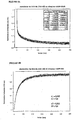
- Figure 4 shows a real-time recording of the kinetics of association (A) and dissociation (B) of NKA-Bo (Bodipy-labeled NKA) with the EGFP-NK2R receptor (GFP-labeled NK2R). Registration of the kinetics of association is carried out after rapid mixing of HEK 293 cells expressing the EGFP-NK2R receptor. The kinetics of dissociation are recorded after manual mixing of the cells expressing the receptor with the NKA-bo (preincubation of 15 minutes, 100 nanomolar) and then with the competitive antagonist SR 4896 (10 ⁇ M final).
- the x-axis represents the time in seconds and the y-axis represents the fluorescence (arbitrary units) at 510 nm.
- the values ⁇ n and ⁇ n respectively correspond to the amplitudes and the rate constants determined by a least squares adjustment with two exponentials.
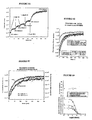
- Figures 5A, 5B, 5C and 5D indicate the procedure for identifying a ligand competitively interacting with the fluorescent ligand at a receptor site.
- the complex formed between the EGFP-NK2R receptor and the NKA-Bo is reversed by increasing concentrations of the G6 molecule (see Table 1) and then by the non-fluorescent NKA.
- FIG. 5A indicates that a part of the receptor-ligand complex is reversed during each addition of G6 molecule (10 or 20 ⁇ M), as well as during the addition of a saturation concentration of NKA (10 ⁇ M).
- the abscissa axis corresponds to the time in seconds and the ordinate axis to the fluorescence measured at 510 nm.
- FIG. 5B shows that the dissociation kinetics of NKA-Bo, determined after addition of a saturating concentration of NKA, is not modified by the presence of the G6 molecule.
- the two experimental traces are adjusted using two fast exponential (0.04 s -1) and slow (0.014 s- 1) whose amplitudes (66% for the fast and slow 33%) are not affected by the presence of 50 ⁇ moles of molecule G6.
- the abscissa axis corresponds to the time in seconds and the ordinate axis to the fluorescence measured at 510 nm.
- Figure 5C shows a quantitative study of the dissociation kinetics of NKA-Bo in the presence of a known competitive antagonist, SR 48968.
- the dissociation of NKA-Bo is recorded in the presence of 0.2; 1 and 5 nM of SR 48968.
- the dissociation rate can be described as a sum of two exponentials whose velocities are 0.04 sec -1 and 0.008 sec -1 , and whose relative amplitudes remain constant at different doses of SR 48968 (45% rapid dissociation (0.04 s -1 ) and 55% slow dissociation (0.008 s -1 )).
- the abscissa axis corresponds to the time in seconds and the ordinate axis to the fluorescence measured at 510 nm.
- Figure 5D shows the values of the fast dissociation (square) and slow (upward triangles) amplitudes of NKA-Bo in the presence of SR 48968, as well as the total amplitude of the NKA-Bo bond (triangles downwards). ).
- the abscissa axis corresponds to the logarithm of the concentration of SR 48968 and the ordinate axis to the amplitude of the normalized fluorescence:
- FIG. 5D represents the ratio of the amplitude of the slow dissociation (A2) with respect to the total dissociation amplitude (total A) (curve with the diamonds).
- A2 the amplitude of the slow dissociation
- total A total dissociation amplitude
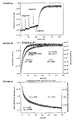
- FIGS. 6A, 6B and 6C show the procedure for identifying a ligand interacting noncompetitively with the fluorescent ligand at a receptor site.
- the fluorescence measured at 510 nm is represented as a function of time in seconds.
- the complex formed between the EGFP-NK2R receptor and the NKA-Bo is reversed by increasing concentrations of the A11 molecule and then by the non-fluorescent NKA.
- Figure 6A indicates that a small portion of the receptor-ligand complex is reversed at each addition of A11 molecule, while NKA (10 ⁇ M) reverses the majority of the complex.
- the kinetics of dissociation by NKA is not of the same shape as in the control.
- FIG. 6B the fluorescence measured at 510 nm is represented as a function of time in seconds.
- Figure 6B illustrates the change in dissociation kinetics in the presence of A11 molecule.
- NKA-Bo dissociation recordings of its EGFP-NK2R receptor are performed in the presence and absence of A11. Dissociation is initiated by the addition of non-fluorescent NKA at a final concentration of 20 ⁇ M.
- the analysis of the dissociation kinetics reveals two monoexponential relaxations whose rate constants (k1 and k2) are identical in the presence and absence of A11 but whose relative amplitudes change so that the slow dissociation represents 59% of the total signal in the control and 22% of the total signal in the presence of A11.
- FIG. 6C shows the association of NKABo (20 nanomolar) with the EGFP-NK2R receptor as a function of time in seconds.
- Figure 6C shows time-resolved binding (association) recordings of NKA-Bo to its EGFP-NK2R receptor, in the absence and presence of the A11 molecule.
- Analysis of the binding kinetics reveals two monoexponential relaxations characterized by rate constants (k1 and k2) that do not change in the presence of A11, but with variable amplitudes depending on whether the cells are incubated with A11 or not.
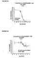
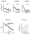
- FIGS. 7A to 7I illustrate the analysis of the effects of various molecules on the dissociation of NKA-Bo linked to its fluorescent EGFP-NK2R receptor.
- Figure 7A shows dissociation records of NKA-Bo in the presence of varying concentrations (1, 10 and 50 ⁇ M) of the 805 molecule (see Table 1).
- the fluorescence measured at 510 nm is represented as a function of time in seconds.
- the quantitative analysis of the dissociation kinetics is obtained using two exponentials fast (0.04 sec -1 ) and slow (0.008 sec -1 ) whose relative amplitudes (fast / slow) vary from 30/70 to 80 / When the concentration of 805 increases.
- FIGS. 7B - 7I show the percentage of amplitude of fast dissociation as a function of the logarithm of the concentration of the compounds tested. These figures give the results of the quantitative analysis of the dissociation of NKA-Bo in the presence of the molecules 805 (FIG. 7B), NP246 (FIG. 7C), A11 (FIG. 7D), H10 (FIG. 7E), G11 (FIG. 7F). , F7 ( Figure 7G), F3 ( Figure 7H) and NP 234 ( Figure 7I).
- the dissociation of NKA-Bo was determined in the presence of each molecule at the concentrations indicated on the abscissa axis. The effect of increasing the dissociation rate is represented by the relative increase in the amplitude (% ⁇ 1) of the fast dissociation.
- FIGS. 8A and 8B show a quantification of the displacement of [ 3 H] SR 48968, bound to the EGFP-NK2R receptor, by the NKA and the molecule 805.
- Figures 9A, 9B and 9C show the cAMP production stimulation results by NKA in HEK 293 cells expressing the EGFP-NK2R receptor.
- Figure 9A shows the effect of the presence of competitive antagonist (H8565) and allosteric effector (805).
- the accumulation of intracellular cAMP (in pmoles / well) is represented as a function of the logarithm of the concentration of NKA.
- the curve with black squares corresponds to white; the curve with the black triangles corresponds to the H8565 antagonist at a concentration of 1 ⁇ M; the curve with the gray squares corresponds to the ligand 805 at a concentration of 10 ⁇ M and the curve with the gray triangles corresponds to the ligand 805 at a concentration of 50 ⁇ M.
- Intracellular cAMP accumulation is determined at different concentrations of NKA.
- the H8565 antagonist shifts the concentration-response curve of NKA to higher values without affecting the intensity of the maximal response (black triangles).
- the 805 ligand (gray squares and triangles) decreases the intensity of the maximal response without affecting the EC 50 (50% effective concentration) of the NKA for response.
- Figure 9B shows the 50% effective concentration of the maximum cyclic AMP response to NKA. This EC 50 increases in the presence of competitive antagonist while it is not significantly modified in the presence of 10 and 50 ⁇ M of 805.
- FIG. 9C shows the inhibition of the NKA-induced maximal cAMP response in the presence of increasing concentration of 805.
- the abscissa represents the logarithm of the 805 concentration and the ordinate the maximum value of the MPA production. cyclic.
- the inhibition constant KI is of the order of 6 ⁇ M.
- Figures 10A, 10B, 10C, 10D and 10E illustrate the effect of 805 on calcium responses associated with the EGFP-NK2R receptor in HEK 293 cells.
- FIGS. 10A, 10B and 10C the fluorescence of INDO-1 is shown at 400 nm as a function of time in seconds.
- the responses are recorded using the INDO-1 calcium-sensitive fluorescent probe.
- NKA agonist 1 nM, 5 nM and 1 ⁇ M
- a control response white circle
- a response in the presence of 50 ⁇ M of 805 black circle
- FIGGS. 10A, 10B and 10C there is an increase in the duration of the calcium response in the presence of 805, without significant effect on the amplitude or on the kinetics of setting up the response.
- FIG. 10D shows the variations of calcium responses obtained for a fixed agonist concentration (5 nM NKA) and variable concentrations (10 and 50 ⁇ M) of 805.
- the normalized calcium concentration as a function of time in seconds is shown.
- the curve with the white circles corresponds to a measurement carried out in the presence of 5 nM of NKA and without 805;
- the curve with the black circles corresponds to a measurement carried out in the presence of 5 nM NKA and 10 ⁇ M of 805;
- the curve with the black squares corresponds to a measurement carried out in the presence of 5 nM of NKA and 50 ⁇ M of 805.
- FIG. 10E shows a quantification of the half-return time (time required for the calcium concentration to be half of its maximum value) to the initial value of the calcium response recorded in the presence of 0, 10 and 50 ⁇ M of 805.
- Figures 11A, 11B, 11C, 11D, 11E, 11F and 11G illustrate the effect of 805 on fluorescent truncated neurokinin binding (NKA4-10 TR7) labeled with the Texas red fluorescent group and the associated calcium response.
- FIGS. 11A to 11D the calcium responses at 106 (FIG. 11A), 50.3 (FIG. 11B), 26.5 (FIG. 11C) and 10.6 nM (FIG. 11D) of NKA4-10 TR7 (NKA4-10 marked in position 7 by the Texas Red) are recorded in duplicate, in absence (curves with the white circles) and in the presence of 20 ⁇ M of 805 (curves with the black circles).
- the normalized calcium concentration as a function of time in seconds is shown. It is noted that 805 causes an increase in the amplitude of the calcium response, and a persistence of the signal.
- Figure 11E shows the relationship between the amplitude of the calcium response and the agonist concentration.
- the amplitude of the normalized calcium peak with respect to the extracellular calcium concentration is shown as a function of the concentration of NKA4-10 TR7.
- the curve with the black squares corresponds to the control measure and (without 805) and the curve with the triangles corresponds to the measurement carried out in the presence of 20 ⁇ M of 805.
- Figure 11F shows the dissociation of the NKA 4-10 TR7 bond.
- the x-axis represents the time in seconds and the y-axis represents the fluorescence.
- the complex formed between the EGFP-NK2R receptor and the NKA4-10 TR7 (106 nM) is inverted (at 10 ° C.) by the non-fluorescent NKA (20 ⁇ M) in the absence and in the presence of 10 ⁇ M of 805.
- dissociation kinetics reveals two monoexponential relaxations whose rate constants (0.05 sec -1 and 0.013 sec -1 ) are identical in the presence and absence of 805, but whose relative amplitudes change so that the Slow dissociation accounts for 42% of the total signal in the control and 12% of the total signal in the presence of 805.
- FIG. 11G shows the percentage of amplitude of the rapid dissociation of NKA 4-10 TR7 as a function of the logarithm of the 805 concentration.
- the amplitude of the rapid dissociation of NKA 4-10 TR7 is measured in the presence of variable concentrations of 805. Rapid dissociation accounts for about 40% of the total dissociation in the absence of 805 and up to 90% of the dissociation amplitude at 100 ⁇ M.
- the apparent affinity for the amplitude variation is of the order of 20 ⁇ M.
- FIGS. 12A, 12B, 12C and 12D represent the substance P (endogenous ligand) -regulated calcium responses on the human NK1 receptor (FIGS. 12A and 12B) and by neurokinin A on the human NK2 receptor (FIGS. 12C and 12D), in the presence and absence of 805.
- substance P endogenous ligand
- FIGS. 12A and 12B a suspension (1 ⁇ 10 6 cells / ml) of HEK 293 cells expressing the wild-type human NK1 receptor and loaded with indo-1 (3 ⁇ M) is stimulated with 0.1 nM (FIG. 12A). or 10 nM (FIG. 12B) of substance P (SP) in the presence (black circles) and in the absence (white circles) of 805 molecules (20 ⁇ M).
- SP substance P
- FIGS. 12C and 12D a suspension (1 ⁇ 10 6 cells / ml) of HEK 293 cells expressing the wild-type human NK2 receptor and loaded with indo-1 (3 ⁇ M) is stimulated with 10 nM (FIG. 12C) or 100 nM (FIG. 12D) of neurokinin A (NKA) in the presence (black circles) and in absence (white circles) of 805 molecules (20 ⁇ M).
- the molecule 805 causes an increase in the amplitude and duration of the calcium response evoked by each agonist.
- HEK 293 cells expressing the EGFP-NK2R receptor are suspended at a concentration of 2,000,000 cells / ml in physiological Hepes buffer (in mM: 137.5 NaCl) 1.25 MgCl 2 , 1.25 CaCl 2 , 6 KCl, 5.6 glucose, 10 Hepes, 0.4 NaH 2 PO 4 , 1% BSA (w / v), pH 7.4) and arranged in one of the tanks of the rapid mixing apparatus.
- a solution of NKA-BO (Bodipy-labeled neurokinin A) (200 nM) in the same buffer is placed in the other reservoir of the rapid mixing apparatus.
- the observation chamber is placed in an SPEX fluorolog 3 spectrofluorometer.
- the temperature of the reservoir syringes as well as of the mixing and observation chamber is set at 21 ° C.
- the excitation wavelength of the observation chamber is set at 470 nm, and the fluorescence emission wavelength is set at 510 nm.
- the sampling frequency of the experimental points is fixed at 50 Hz (1 point every 20 msec).
- the contents of the tanks are propelled towards the mixing and observation chamber by means of a pneumatic device allowing a flow of 4 ml / sec and the evolution of the mixture is recorded continuously from the moment of stopping of the thrust of the pistons of the tanks.
- the NKA-Bo association reaction with the EGFP-NK2R receptor is detected as a decrease in the fluorescence intensity of the GFP carried by the NK2R receptor.
- the HEK 293 cells expressing the EGFP-NK2R receptor are suspended at a concentration of 1,000,000 cells / ml in Hepes physiological buffer (in mM: 137.5 NaCl, 1.25 MgCl 2 1.25 CaCl 2, 6 KCl, 5.6 glucose, 10 Hepes, 0.4 NaH 2 PO 4 , 1% BSA (w / v), pH 7.4) and placed in a 1 ml cuvette provided with a magnetic stir bar on the cell holder of a SPEX fluorolog 3 spectrofluorometer. 20 nM (final concentration) of NKA-Bo is added thereto and the combination is allowed to reach equilibrium for 10 minutes.
- Hepes physiological buffer in mM: 137.5 NaCl, 1.25 MgCl 2 1.25 CaCl 2, 6 KCl, 5.6 glucose, 10 Hepes, 0.4 NaH 2 PO 4 , 1% BSA (w / v), pH 7.4
- the measurement of the dissociation kinetics of NKA-Bo is initiated by the manual addition of 10 ⁇ M (final concentration) of the competitive antagonist SR 48968 and recorded at 510 nm (excitation: 470 nm) at a rate of 1 point every 100 msec.
- EXAMPLE 2 Identification of the G6 molecule acting as a competitive ligand, by fluorescence energy transfer between the NKA-Bo and the EGFP-NK2R receptor.
- the control experiment presented in FIG. 5C indicates that a known molecule, competitive for the interaction of NKA-Bo with its site, ie 5R48968, provokes a dissociation of NKA-Bo with kinetics identical to that recorded when NKA-Bo is displaced by NKA.
- the relative amplitude of each step remains constant for each concentration of SR 48968, as attested by the fast / slow amplitude ratios close to 45/55.
- HEK 293 cells expressing the EGFP-NKR2 receptor are incubated, in one milliliter of Hepes buffer, with 20 nM NKA-Bo for 15 minutes.
- the mixture is then placed in a spectrofluorimetric cuvette and the reversion of the interaction between EGFP-NK2R and NKA-Bo is evaluated after addition of molecule A11 in increasing concentration (FIG. 6A).
- the estimate of the reversed complex fraction is then measured after the addition of a saturating amount of NKA (10 ⁇ M). It can be seen that the A11 molecule displaces only a small fraction (29%) of the NKA-Bo linked to its site.
- the amplitude of each relaxation is then given by the adjustment of the experimental traces.
- fast relaxation represents 41% and slow 59%.
- A1 50 ⁇ M
- the fast relaxation represents 78% of the signal and the slow 22%.
- the A11 molecule thus has the effect of stabilizing a fraction of the receptors in a state of lower affinity than that in which the NKA-Bo alone stabilizes it.
- the A11 molecule binds preferentially to the conformation of the receptor which has a more modest affinity for NKA-Bo.
- the binding of NKA-Bo is not inhibited by A11. It occurs on the same conformational states of the receptor, but the proportions of these two conformational states are modified by the presence of A11.
- the dissociation kinetics of NKA-Bo is faster in the presence of A11 than in its absence. This acceleration of the dissociation kinetics of NKA-Bo does not result from a change in the intrinsic affinities of the conformations for NKA-Bo, but from a modification of the relative proportions of the high and low affinity states.
- the receptor exists in at least two conformational states, one of which pre-exists agonist addition and the other occurs slowly over time in the presence of agonist.
- the A11 molecule by stabilizing the low affinity states of the receptor, increases the proportion of fast (low affinity) binding to the detriment of the slow binding which reflects the stabilization of a higher affinity state
- the identification of other allosteric effectors of the NK2R receptor of the tachykinins is done by analyzing the kinetics of dissociation of NKA-Bo bound to the receptor.
- NKA-Bo 10 6 HEK 293 cells expressing the EGFP-NK2R receptor are suspended in Hepes buffer (see above) and incubated with NKA-Bo (20 nM) and one of the following molecules: 805, NP246, A11, H10, G11, F7, F3 and NP 234.
- the A11 molecules and the known competitive antagonist SR 48968 are introduced into the experiment as positive controls.
- the dissociation of NKA-Bo is recorded in a spectrofluorometer cuvette equipped with magnetic stirring for 700 seconds, after adding 10 ⁇ M of non-fluorescent NKA to expel the bound NKA-Bo.
- the dissociation kinetics of NKA-Bo is recorded in the presence of increasing concentrations of each of the aforementioned molecules.
- FIG. 7A shows a typical experiment for measuring the dissociation rate of NKA-Bo in the presence of increasing concentrations of the molecule 805.
- FIGS. 7B to 7I show, for each molecule tested, the variation of the NKA-Bo binding amplitude and the amplitude variation of the fast dissociation. These figures show that all the indicated molecules behave like allosteric effectors because they cause an increase in the amplitude of the rapid dissociation of NKA-Bo.
- the amplitude increase in fast dissociation reflects the ability of molecules to stabilize a larger fraction of the receptors in the low-affinity state. It is thus seen that the initial fraction of receptors in the low affinity state goes from about 25% in the absence of effector to 60-75% in the presence of the molecules 805, NP234, A11, G11, F7, F3, and H10. (at 100 ⁇ M effector) and nearly 100% in the presence of A11 and NP246.
- 25,000 cells expressing the EGFP-NK2R receptor are incubated for 3 hours at 4 ° C. in a final volume of 500 ⁇ l of Hepes buffer in the presence of 1 nM of [ 3 H] SR48968 and variable concentrations of NKA or 805. incubation period, the reaction mixtures are filtered through GF / C (Whatmann) fiberglass filters previously incubated in Hepes buffer supplemented with 1% (W / V) semi-skimmed milk powder, and rinsed three times. 5 ml of hepes buffer at 4 ° C. The filters are then placed in scintillation counting flasks in which 3 ml of scintillation cocktail is added. Radioactivity is measured after 16 hours in a radioactivity counter.
- GF / C Whatmann
- the effect of 805 on the binding of SR 48968 does not reflect a competitive but rather non-competitive interaction.
- 30-well 6-well cell culture dishes are treated with collagen and then inoculated with 50,000 cells expressing the EGFP-NK2R receptor. After two to three days of culture, the medium is replaced with hepes buffer containing the inhibitor of phosphodiesterase IBMX (isobutyl-methyl-xhantine) and incubated for 30 minutes with increasing concentrations of NKA in the presence or absence of i) d competitive inhibitor H8565 (1 ⁇ M) or ii) effector 805 (10 or 50 ⁇ M).
- the quantity of cAMP produced (expressed in pmols / well) is determined according to a radioimmunological method described in Gicquiaux et al. (2002). This results in a measurement of the concentration-effect curve of NKA (FIG. 9A).
- NKA alone is able to stimulate the production of cAMP in cells expressing the EGFP-NK2R receptor with an EC 50 close to 100 nM.
- FIGS. 10A, 10B and 10C show that compound 805 (50 ⁇ M) increases the duration of the evoked calcium responses by 1, 5 and 1000 nM of NKA. In the same way, compound 805, at concentrations of 10 and 50 ⁇ M, increases the duration of the evoked calcium response by 5 nM NKA (FIG. 10D).
- Compound 805 therefore behaves as a positive allosteric effector of the calcium responses evoked by the NKA-Bo agonist which increases the duration of the response. This is indicated in FIG. 10E where it can be seen that the time required to return to 50% of the maximum response goes from 91 seconds in the absence of 805 to 105 then 115 s when the concentration of 805 is 10 and then 50 ⁇ M.
- the calcium responses evoked by the truncated agonist NKA4-10 TR7 on cells expressing the EGFP-NK2R receptor are recorded after loading the cells with the calcium-sensitive fluorescent probe: indo-1 according to the protocol described in Vollmer et al. (1999).
- the cells are suspended at 500,000 cells / ml in Hepes buffer and placed in a spectrofluorimeter tank.
- the excitation wavelength is set at 355 nm and the emission is recorded at 375 and 405 nm.
- the responses plotted are the 375 nm / 405 nm signal ratios.
- 11A to 11D show that compound 805 (20 ⁇ M) increases the amplitude of the response evoked by NKA 4-10 TR7 (106, 50.3, 26.5 and 10.6 nM). Amplitude variation by 805 is shown in panel B of Figure 11 for concentrations of 5, 10, 25, 50 and 106 nM of NKA 4-10 TR7. Compound 805 is shown to be a positive allosteric effector of the calcium responses evoked by NKA 4-10 TR7 which decreases the 50% effective dose from about 20 nM to about 15 nM.
- NKA 4-10 is a truncated form of NKA found in middle gut tumor fluids. Fluorescent NKA 4-10 is modified at position 7 to introduce a cysteine which is then labeled with the Texas Red fluorescent group.
- the fluorescent NKA 4-10 is, like the fluorescent NKA, an agonist of the EGFP-NK2R receptor, except that it is unable to evoke cAMP accumulation responses (Palanche et al., 2001).
- the binding of fluorescent NKA 4-10 is in a monoexponential process with which the development of a calcium response in HEK 293 cells expressing the EGFP-NK2R receptor is associated (Palanche et al., 2001).
- respective amplitudes of 58% for fast dissociation and 42% for slow dissociation Figure 11F.
- an acceleration of the dissociation characterized by a modification of the amplitudes of the fast (88%) and slow (12%) dissociation stages is observed.
- a relationship between the dissociation rate increase of NKA4-10 TR7 and the concentration of 805 is noted ( Figure 11G).
- the concentration of 805 which causes a 50% increase in the amplitude of the rapid dissociation of NKA4-10 TR7 is of the order of 20
- the calcium responses evoked by the following agonists: substance P (SP) and neurokinin A (NKA), on cells expressing the wild-type human NK1 receptor or wild-type human NK2 are recorded after loading of the cells with the calcium-sensitive fluorescent probe: indole 1 (3 ⁇ M) according to the protocol described in Vollmer et al. (1999).
- the cells are suspended at 1,000,000 cells / ml in Hepes buffer (op cit) and placed in a spectrofluorimeter vat.
- the excitation wavelength is set at 338 nm and the emission is recorded at 400 nm.
- the answers plotted are the signals recorded at 400 nm.
- FIGS. 12A and 12B show that compound 805 (20 ⁇ M) increases the amplitude of the evoked response by substance P (0.1 or 10 nM).
- Figures 12C and 12D show that compound 805 (20 ⁇ M) increases the amplitude of the response evoked by neurokinin A (10 or 100 nM).
- Compound 805 is shown to be a positive allosteric effector of the neurokinin A-evoked calcium responses on the wild-type human NK2 receptor and substance P-evoked responses on the wild-type human NK1 receptor.
- the M1 fluorescent muscarinic receptor is obtained by fusion of its cDNA with that of EGFP or YPFP as described in Ilien et al. (2003) and stably expressed in HEK 293 cells.
- the cells are suspended at a concentration of 1,000,000 cells / ml in Hepes physiological buffer (in mM: 137.5 NaCl, 1.25 MgCl 2 ; 1.25 CaCl 2 , 6 KCl, 5.6 glucose, 10 Hepes, 0.4 NaH 2 PO 4 , 1% BSA (w / v), pH 7.4) and arranged in a 1 ml fluorescence cuvette, itself placed in the rack waving a spectrofluorimeter.
- Hepes physiological buffer in mM: 137.5 NaCl, 1.25 MgCl 2 ; 1.25 CaCl 2 , 6 KCl, 5.6 glucose, 10 Hepes, 0.4 NaH 2 PO 4 , 1% BSA (w / v), pH 7.4
- the cell suspension is excited at 470 nm and the fluorescence emission is recorded at 510 nm (EGFP) or at 530 nm (EYFP).
- the addition of 70 nM bodipy (558-568) -pirenzepine causes a decrease in fluorescence emission of the receptor-borne fluorescent protein that reflects the step of ligand-receptor association. The time course of this step is followed by recording the emission at 510 or 530 nm.
- the complex between the receptor and the bodipy pirenzepine is formed in a fluorescence cuvette placed in the spectrofluorimeter before adding an excess (5 ⁇ M) of atropine to initiate the dissociation step of the ligand receptor complex.
- the time course of this dissociation step is recorded at 510 or 530 nm.
- the cells are preincubated with one or more known or unknown molecules, and the time course of the acetylcholine receptor is recorded. step of association and / or dissociation step.
- An allosteric effector is accelerated, either slows down the time course or the stage association, either of the dissociation step, or both. This results in a relative change in the fast and slow association (or dissociation) amplitudes compared to the control experimental condition (without effector).
- the fluorescent CXCR4 receptor is obtained by fusion of its cDNA with that of EGFP as described in Valenzuela et al. (2001) and in Palanché et al. (2001) and stably expressed in HEK 293 cells.
- the cells are suspended at a concentration of 1,000,000 cells / ml in Hepes physiological buffer (in mM: 137.5 NaCl, 1.25 MgCl 2 ; 1.25 CaCl 2 , 6 KCl, 5.6 glucose, 10 Hepes, 0.4 NaH 2 PO 4 , 1% BSA (w / v), pH 7.4) and arranged in a 1 ml fluorescence cuvette. even placed in the rack waving a spectrofluorimeter.
- the cell suspension is excited at 470 nm and the fluorescence emission is recorded at 510 nm.
- the addition of 50 nM Texas Red-SDF1 ⁇ causes a decrease in fluorescence emission of the receptor-borne fluorescent protein that reflects the step of ligand-receptor association.
- the time course of this step is followed by recording the emission at 510 nm.
- the complex between the receptor and the Texas Red-SDF1 ⁇ is formed in a fluorescence cuvette placed in the spectrofluorimeter before adding an excess (500 nM) of SDF1 ⁇ to initiate the dissociation step of the ligand receptor complex.
- the time course of this dissociation step is recorded at 510 nm.
- the cells are pre-incubated with one or more known or unknown molecules, and the time course of the step is recorded. association and / or dissociation step.
- An allosteric effector either accelerates or slows down the time course of either the association step, the dissociation step, or both. This results in a relative change in the fast and slow association (or dissociation) amplitudes compared to the control experimental condition (without effector).
Landscapes
- Health & Medical Sciences (AREA)
- Life Sciences & Earth Sciences (AREA)
- Immunology (AREA)
- Chemical & Material Sciences (AREA)
- Engineering & Computer Science (AREA)
- Molecular Biology (AREA)
- Urology & Nephrology (AREA)
- Biomedical Technology (AREA)
- Hematology (AREA)
- General Health & Medical Sciences (AREA)
- Biochemistry (AREA)
- Medicinal Chemistry (AREA)
- Analytical Chemistry (AREA)
- Food Science & Technology (AREA)
- Microbiology (AREA)
- Physics & Mathematics (AREA)
- Cell Biology (AREA)
- Biotechnology (AREA)
- General Physics & Mathematics (AREA)
- Pathology (AREA)
- Organic Chemistry (AREA)
- Proteomics, Peptides & Aminoacids (AREA)
- Genetics & Genomics (AREA)
- Biophysics (AREA)
- Chemical Kinetics & Catalysis (AREA)
- Investigating Or Analysing Biological Materials (AREA)
- Electrotherapy Devices (AREA)
- Measuring Pulse, Heart Rate, Blood Pressure Or Blood Flow (AREA)
- Measuring Or Testing Involving Enzymes Or Micro-Organisms (AREA)
- Peptides Or Proteins (AREA)
- Auxiliary Devices For Music (AREA)
- Pyrane Compounds (AREA)
- Plural Heterocyclic Compounds (AREA)
Claims (6)
- Verfahren zum Nachweis eines allosterischen Effektors eines Rezeptors durch Bestimmen der Änderung:- der Dissoziations- und/oder Assoziationskinetik des zwischen dem Rezeptor und einem seiner Liganden gebildeten Komplexes in Gegenwart dieses allosterischen Effektors im Vergleich zur Dissoziations- und/oder Assoziationskinetik des zwischen dem Rezeptor und dem Liganden gebildeten Komplexes in Abwesenheit des Effektors,- und/oder der Amplitude der zwischen dem Rezeptor und einem seiner Liganden gebildeten Bindung in Gegenwart dieses allosterischen Effektors im Vergleich zur Amplitude der zwischen dem Rezeptor und dem Liganden gebildeten Bindung in Abwesenheit des Effektors,mit der Maßgabe, dass man, wenn die Änderung dieser Amplitude negativ ist, das Vorhandensein der Änderung der genannten Kinetik auch nachweist,
wobei der Rezeptor und der Ligand unter geeigneten physiologischen Bedingungen in mindestens eine biologische Reaktion involviert sind und der allosterische Effektor in der Lage ist, mindestens eine der Reaktionen zu modulieren,
wobei der Rezeptor durch ein fluoreszierendes Protein markiert ist, das aus den fluoreszierenden Proteinen ausgewählt ist, die von autofluoreszierenden Proteinen von Cnidaria stammen oder abgeleitet sind, deren molekularer Extinktionskoeffizient größer als etwa 14 000 M-1.cm-1 und deren Fluoreszenzquantenausbeute größer als etwa 0,38 ist,
wobei der Ligand durch einen Marker markiert ist, bestehend aus:- einem Molekül, das in der Lage ist, das von dem fluoreszierenden Protein emittierte Licht zu absorbieren, oder- einer fluoreszierenden Substanz,wobei die Bestimmungen der Änderung der Dissoziations- und/oder Assoziationskinetik und der Änderung der Amplitude durch Übertragung von Fluoreszenzenergie erfolgen:- zwischen dem fluoreszierenden Protein und der fluoreszierenden Substanz, wobei die fluoreszierende Substanz entweder bei der Emissionswellenlänge des fluoreszierenden Proteins anregbar ist oder bei der Anregungswellenlänge des fluoreszierenden Proteins emittiert, oder- zwischen dem fluoreszierenden Protein und dem Molekül, das in der Lage ist, das von dem fluoreszierenden Protein emittierte Licht zu absorbieren. - Verfahren nach Anspruch 1, wobei der Rezeptor durch ein fluoreszierendes Protein markiert ist, welches ausgewählt ist aus:- grün fluoreszierendem Protein (GFP oder EGFP), cyan fluoreszierendem Protein (CFP oder ECFP), gelb fluoreszierendem Protein (YFP oder EYFP) oder GFPUV oder- Varianten, die von GFP, CFP, YFP oder GFPUV durch Addition, Deletion oder Substitution einer oder mehrerer Aminosäuren abgeleitet sind, mit der Maßgabe, dass die Varianten die Fluoreszenzeigenschaft beibehalten,- oder Fragmenten von GFP, CFP, YFP oder GFPUV oder Fragmenten der oben genannten Varianten, mit der Maßgabe, dass die Fragmente die Fluoreszenzeigenschaft beibehalten,
wobei der Ligand durch einen Marker markiert ist, bestehend aus:- einem Molekül, das in der Lage ist, das von dem fluoreszierenden Protein emittierte Licht zu absorbieren, oder- einer fluoreszierenden Substanz. - Verfahren nach einem der Ansprüche 1 bis 2, dadurch gekennzeichnet, dass der Ligand ein Antagonist ist.
- Verfaluen nach einem der Ansprüche 1 bis 2, dadurch gekennzeichnet, dass der Ligand ein Agonist ist.
- Verfahren nach einem der Ansprüche 1 bis 2, dadurch gekennzeichnet, dass- die Änderung der Amplitude der zwischen dem Rezeptor und einem seiner Liganden gebildeten Bindung in Gegenwart des allosterischen Effektors im Vergleich zur Amplitude der zwischen dem Rezeptor und dem Liganden gebildeten Bindung in Abwesenheit des Effektors bestimmt wirdund dass diese Änderung negativ ist, was die Bestimmung- der Änderung der Dissoziationskinetik des zwischen dem Rezeptor und einem seiner Liganden gebildeten Komplexes in Gegenwart des allosterischen Effektors im Vergleich zur Dissoziationskinetik des zwischen dem Rezeptor und dem Liganden gebildeten Komplexes in Abwesenheit des Effektors erfordert.
- Verfahren nach Anspruch 5, dadurch gekennzeichnet, dass die Änderung der Dissoziationskinetik positiv oder negativ ist, was impliziert, dass die getestete Verbindung ein allosterischer Effektor ist.
Applications Claiming Priority (3)
| Application Number | Priority Date | Filing Date | Title |
|---|---|---|---|
| FR0207436A FR2840993B1 (fr) | 2002-06-17 | 2002-06-17 | Procede de mise en evidence d'un effecteur allosterique d'un recepteur |
| FR0207436 | 2002-06-17 | ||
| PCT/FR2003/001817 WO2003107004A2 (fr) | 2002-06-17 | 2003-06-16 | Procede de mise en evidence d'un effecteur allosterique d'un recepteur |
Publications (2)
| Publication Number | Publication Date |
|---|---|
| EP1514112A2 EP1514112A2 (de) | 2005-03-16 |
| EP1514112B1 true EP1514112B1 (de) | 2006-09-06 |
Family
ID=29595303
Family Applications (1)
| Application Number | Title | Priority Date | Filing Date |
|---|---|---|---|
| EP03748170A Expired - Lifetime EP1514112B1 (de) | 2002-06-17 | 2003-06-16 | Verfahren zum nachweis von allosteren effektoren eines rezeptors |
Country Status (11)
| Country | Link |
|---|---|
| US (1) | US20060194259A1 (de) |
| EP (1) | EP1514112B1 (de) |
| JP (1) | JP2005530158A (de) |
| AT (1) | ATE338950T1 (de) |
| AU (1) | AU2003267483A1 (de) |
| CA (1) | CA2489389A1 (de) |
| DE (1) | DE60308182T2 (de) |
| DK (1) | DK1514112T3 (de) |
| ES (1) | ES2273026T3 (de) |
| FR (1) | FR2840993B1 (de) |
| WO (1) | WO2003107004A2 (de) |
Cited By (1)
| Publication number | Priority date | Publication date | Assignee | Title |
|---|---|---|---|---|
| DE102007022915A1 (de) * | 2007-05-14 | 2008-11-20 | Stiftung Caesar Center Of Advanced European Studies And Research | Polymerisationssensor |
Family Cites Families (2)
| Publication number | Priority date | Publication date | Assignee | Title |
|---|---|---|---|---|
| FR2764387B1 (fr) * | 1997-06-05 | 1999-07-23 | Centre Nat Rech Scient | Utilisation d'une proteine fluorescente pour la detection d'interactions entre une proteine cible et son ligand |
| AU2030000A (en) * | 1998-11-25 | 2000-06-13 | Cadus Pharmaceutical Corporation | Methods and compositions for identifying receptor effectors |
-
2002
- 2002-06-17 FR FR0207436A patent/FR2840993B1/fr not_active Expired - Fee Related
-
2003
- 2003-06-16 DE DE60308182T patent/DE60308182T2/de not_active Expired - Fee Related
- 2003-06-16 ES ES03748170T patent/ES2273026T3/es not_active Expired - Lifetime
- 2003-06-16 JP JP2004513772A patent/JP2005530158A/ja active Pending
- 2003-06-16 AU AU2003267483A patent/AU2003267483A1/en not_active Abandoned
- 2003-06-16 AT AT03748170T patent/ATE338950T1/de not_active IP Right Cessation
- 2003-06-16 EP EP03748170A patent/EP1514112B1/de not_active Expired - Lifetime
- 2003-06-16 CA CA002489389A patent/CA2489389A1/fr not_active Abandoned
- 2003-06-16 DK DK03748170T patent/DK1514112T3/da active
- 2003-06-16 US US10/518,298 patent/US20060194259A1/en not_active Abandoned
- 2003-06-16 WO PCT/FR2003/001817 patent/WO2003107004A2/fr not_active Ceased
Cited By (1)
| Publication number | Priority date | Publication date | Assignee | Title |
|---|---|---|---|---|
| DE102007022915A1 (de) * | 2007-05-14 | 2008-11-20 | Stiftung Caesar Center Of Advanced European Studies And Research | Polymerisationssensor |
Also Published As
| Publication number | Publication date |
|---|---|
| FR2840993A1 (fr) | 2003-12-19 |
| WO2003107004A3 (fr) | 2004-06-24 |
| DE60308182D1 (de) | 2006-10-19 |
| ES2273026T3 (es) | 2007-05-01 |
| US20060194259A1 (en) | 2006-08-31 |
| FR2840993B1 (fr) | 2005-04-08 |
| DK1514112T3 (da) | 2007-01-15 |
| WO2003107004A2 (fr) | 2003-12-24 |
| ATE338950T1 (de) | 2006-09-15 |
| CA2489389A1 (fr) | 2003-12-24 |
| EP1514112A2 (de) | 2005-03-16 |
| DE60308182T2 (de) | 2007-08-23 |
| JP2005530158A (ja) | 2005-10-06 |
| AU2003267483A1 (en) | 2003-12-31 |
Similar Documents
| Publication | Publication Date | Title |
|---|---|---|
| Mercier et al. | Quantitative assessment of β1-and β2-adrenergic receptor homo-and heterodimerization by bioluminescence resonance energy transfer | |
| Lakowicz | Fluorophores | |
| Machleidt et al. | NanoBRET a novel BRET platform for the analysis of protein–protein interactions | |
| Coast et al. | Mosquito natriuretic peptide identified as a calcitonin-like diuretic hormone in Anopheles gambiae (Giles) | |
| Harikumar et al. | Constitutive formation of oligomeric complexes between family BG protein-coupled vasoactive intestinal polypeptide and secretin receptors | |
| Lao et al. | Single-molecule imaging demonstrates ligand regulation of the oligomeric status of CXCR4 in living cells | |
| EP0986759B1 (de) | Verwendung eines fluoreszenzproteins zum nachweis von membrane rezeptoren-ligand interaktionen | |
| Choi et al. | Constitutive and agonist-induced dimerizations of the P2Y1 receptor: relationship to internalization and scaffolding | |
| Carpenter et al. | Protein proximity observed using fluorogen activating protein and dye activated by proximal anchoring (FAP–DAPA) system | |
| WO2019062744A1 (zh) | 融合多肽 | |
| Nørager et al. | Development of potent fluorescent polyamine toxins and application in labeling of ionotropic glutamate receptors in hippocampal neurons | |
| Stefanucci et al. | Developing cyclic opioid analogues: fluorescently labeled bioconjugates of biphalin | |
| EP1514112B1 (de) | Verfahren zum nachweis von allosteren effektoren eines rezeptors | |
| Lisenbee et al. | Secretin receptor oligomers form intracellularly during maturation through receptor core domains | |
| McFarlane et al. | Differential functional coupling in Gp130-JAK complexes expands the plasticity of the interleukin-6 signaling axis | |
| Dahoun et al. | Recombinant expression and functional characterization of mouse olfactory receptor mOR256-17 in mammalian cells | |
| WO2023172727A1 (en) | Cells derived from polypedilum vanderplanki and odor sensor equipped therewith | |
| JP3459523B2 (ja) | 生理活性物質の製造方法 | |
| KR102176538B1 (ko) | 항정신병성 약물 검출용 fret 바이오센서 및 이를 이용한 검출 방법 | |
| EP1756579B1 (de) | Sammlungen aus verfolgbaren verbindungen und verwendung dafür | |
| Zhao et al. | Cell active and functionally-relevant small-molecule agonists of calcitonin receptor | |
| EP2828659B1 (de) | Verfahren zur bestimmung der fähigkeit eines antikörpers zur aufrechterhaltung der nähe von zellen zueinander | |
| Nakamura et al. | Current techniques for studying oligomer formations of G-protein-coupled receptors using mammalian and yeast cells | |
| Calebiro et al. | GPCR Signaling in Nanodomains: Lessons from Single‐Molecule Microscopy | |
| Maria Tindara et al. | Light‐Based Molecular Tools to Precisely Monitor and Operate β‐Adrenoceptors |
Legal Events
| Date | Code | Title | Description |
|---|---|---|---|
| PUAI | Public reference made under article 153(3) epc to a published international application that has entered the european phase |
Free format text: ORIGINAL CODE: 0009012 |
|
| 17P | Request for examination filed |
Effective date: 20041112 |
|
| AK | Designated contracting states |
Kind code of ref document: A2 Designated state(s): AT BE BG CH CY CZ DE DK EE ES FI FR GB GR HU IE IT LI LU MC NL PT RO SE SI SK TR |
|
| AX | Request for extension of the european patent |
Extension state: AL LT LV MK |
|
| DAX | Request for extension of the european patent (deleted) | ||
| GRAP | Despatch of communication of intention to grant a patent |
Free format text: ORIGINAL CODE: EPIDOSNIGR1 |
|
| RTI1 | Title (correction) |
Free format text: METHOD FOR IDENTIFYING AN ALLOSTERIC EFFECTOR OF A RECEPTOR |
|
| GRAS | Grant fee paid |
Free format text: ORIGINAL CODE: EPIDOSNIGR3 |
|
| GRAA | (expected) grant |
Free format text: ORIGINAL CODE: 0009210 |
|
| AK | Designated contracting states |
Kind code of ref document: B1 Designated state(s): AT BE BG CH CY CZ DE DK EE ES FI FR GB GR HU IE IT LI LU MC NL PT RO SE SI SK TR |
|
| PG25 | Lapsed in a contracting state [announced via postgrant information from national office to epo] |
Ref country code: CZ Free format text: LAPSE BECAUSE OF FAILURE TO SUBMIT A TRANSLATION OF THE DESCRIPTION OR TO PAY THE FEE WITHIN THE PRESCRIBED TIME-LIMIT Effective date: 20060906 Ref country code: SI Free format text: LAPSE BECAUSE OF FAILURE TO SUBMIT A TRANSLATION OF THE DESCRIPTION OR TO PAY THE FEE WITHIN THE PRESCRIBED TIME-LIMIT Effective date: 20060906 Ref country code: SK Free format text: LAPSE BECAUSE OF FAILURE TO SUBMIT A TRANSLATION OF THE DESCRIPTION OR TO PAY THE FEE WITHIN THE PRESCRIBED TIME-LIMIT Effective date: 20060906 Ref country code: RO Free format text: LAPSE BECAUSE OF FAILURE TO SUBMIT A TRANSLATION OF THE DESCRIPTION OR TO PAY THE FEE WITHIN THE PRESCRIBED TIME-LIMIT Effective date: 20060906 Ref country code: IT Free format text: LAPSE BECAUSE OF FAILURE TO SUBMIT A TRANSLATION OF THE DESCRIPTION OR TO PAY THE FEE WITHIN THE PRESCRIBED TIME-LIMIT;WARNING: LAPSES OF ITALIAN PATENTS WITH EFFECTIVE DATE BEFORE 2007 MAY HAVE OCCURRED AT ANY TIME BEFORE 2007. THE CORRECT EFFECTIVE DATE MAY BE DIFFERENT FROM THE ONE RECORDED. Effective date: 20060906 |
|
| REG | Reference to a national code |
Ref country code: GB Ref legal event code: FG4D Free format text: NOT ENGLISH |
|
| REG | Reference to a national code |
Ref country code: CH Ref legal event code: EP |
|
| GBT | Gb: translation of ep patent filed (gb section 77(6)(a)/1977) |
Effective date: 20060907 |
|
| REG | Reference to a national code |
Ref country code: IE Ref legal event code: FG4D Free format text: LANGUAGE OF EP DOCUMENT: FRENCH |
|
| REF | Corresponds to: |
Ref document number: 60308182 Country of ref document: DE Date of ref document: 20061019 Kind code of ref document: P |
|
| REG | Reference to a national code |
Ref country code: CH Ref legal event code: NV Representative=s name: ABREMA AGENCE BREVETS ET MARQUES GANGUILLET & HUMP |
|
| PG25 | Lapsed in a contracting state [announced via postgrant information from national office to epo] |
Ref country code: BG Free format text: LAPSE BECAUSE OF FAILURE TO SUBMIT A TRANSLATION OF THE DESCRIPTION OR TO PAY THE FEE WITHIN THE PRESCRIBED TIME-LIMIT Effective date: 20061206 |
|
| REG | Reference to a national code |
Ref country code: SE Ref legal event code: TRGR |
|
| REG | Reference to a national code |
Ref country code: DK Ref legal event code: T3 |
|
| PG25 | Lapsed in a contracting state [announced via postgrant information from national office to epo] |
Ref country code: PT Free format text: LAPSE BECAUSE OF FAILURE TO SUBMIT A TRANSLATION OF THE DESCRIPTION OR TO PAY THE FEE WITHIN THE PRESCRIBED TIME-LIMIT Effective date: 20070219 |
|
| REG | Reference to a national code |
Ref country code: ES Ref legal event code: FG2A Ref document number: 2273026 Country of ref document: ES Kind code of ref document: T3 |
|
| PLBE | No opposition filed within time limit |
Free format text: ORIGINAL CODE: 0009261 |
|
| STAA | Information on the status of an ep patent application or granted ep patent |
Free format text: STATUS: NO OPPOSITION FILED WITHIN TIME LIMIT |
|
| 26N | No opposition filed |
Effective date: 20070607 |
|
| PG25 | Lapsed in a contracting state [announced via postgrant information from national office to epo] |
Ref country code: GR Free format text: LAPSE BECAUSE OF FAILURE TO SUBMIT A TRANSLATION OF THE DESCRIPTION OR TO PAY THE FEE WITHIN THE PRESCRIBED TIME-LIMIT Effective date: 20061207 |
|
| PG25 | Lapsed in a contracting state [announced via postgrant information from national office to epo] |
Ref country code: EE Free format text: LAPSE BECAUSE OF FAILURE TO SUBMIT A TRANSLATION OF THE DESCRIPTION OR TO PAY THE FEE WITHIN THE PRESCRIBED TIME-LIMIT Effective date: 20060906 |
|
| PGFP | Annual fee paid to national office [announced via postgrant information from national office to epo] |
Ref country code: CH Payment date: 20080527 Year of fee payment: 6 Ref country code: DK Payment date: 20080523 Year of fee payment: 6 Ref country code: ES Payment date: 20080606 Year of fee payment: 6 Ref country code: LU Payment date: 20080528 Year of fee payment: 6 |
|
| PGFP | Annual fee paid to national office [announced via postgrant information from national office to epo] |
Ref country code: AT Payment date: 20080526 Year of fee payment: 6 |
|
| PGFP | Annual fee paid to national office [announced via postgrant information from national office to epo] |
Ref country code: MC Payment date: 20080526 Year of fee payment: 6 Ref country code: BE Payment date: 20080528 Year of fee payment: 6 Ref country code: IT Payment date: 20080614 Year of fee payment: 6 Ref country code: FI Payment date: 20080526 Year of fee payment: 6 |
|
| PGFP | Annual fee paid to national office [announced via postgrant information from national office to epo] |
Ref country code: DE Payment date: 20080617 Year of fee payment: 6 Ref country code: IE Payment date: 20080527 Year of fee payment: 6 Ref country code: NL Payment date: 20080626 Year of fee payment: 6 Ref country code: SE Payment date: 20080526 Year of fee payment: 6 |
|
| PGFP | Annual fee paid to national office [announced via postgrant information from national office to epo] |
Ref country code: FR Payment date: 20080630 Year of fee payment: 6 |
|
| PGFP | Annual fee paid to national office [announced via postgrant information from national office to epo] |
Ref country code: GB Payment date: 20080527 Year of fee payment: 6 |
|
| PG25 | Lapsed in a contracting state [announced via postgrant information from national office to epo] |
Ref country code: CY Free format text: LAPSE BECAUSE OF FAILURE TO SUBMIT A TRANSLATION OF THE DESCRIPTION OR TO PAY THE FEE WITHIN THE PRESCRIBED TIME-LIMIT Effective date: 20060906 |
|
| PG25 | Lapsed in a contracting state [announced via postgrant information from national office to epo] |
Ref country code: HU Free format text: LAPSE BECAUSE OF FAILURE TO SUBMIT A TRANSLATION OF THE DESCRIPTION OR TO PAY THE FEE WITHIN THE PRESCRIBED TIME-LIMIT Effective date: 20070307 Ref country code: TR Free format text: LAPSE BECAUSE OF FAILURE TO SUBMIT A TRANSLATION OF THE DESCRIPTION OR TO PAY THE FEE WITHIN THE PRESCRIBED TIME-LIMIT Effective date: 20060906 |
|
| BERE | Be: lapsed |
Owner name: *CENTRE NATIONAL DE LA RECHERCHE SCIENTIFIQUE Effective date: 20090630 Owner name: *UNIVERSITE LOUIS PASTEUR DE STRASBOURG Effective date: 20090630 |
|
| PG25 | Lapsed in a contracting state [announced via postgrant information from national office to epo] |
Ref country code: MC Free format text: LAPSE BECAUSE OF NON-PAYMENT OF DUE FEES Effective date: 20090630 Ref country code: FI Free format text: LAPSE BECAUSE OF NON-PAYMENT OF DUE FEES Effective date: 20090616 |
|
| REG | Reference to a national code |
Ref country code: CH Ref legal event code: PL |
|
| REG | Reference to a national code |
Ref country code: DK Ref legal event code: EBP |
|
| GBPC | Gb: european patent ceased through non-payment of renewal fee |
Effective date: 20090616 |
|
| NLV4 | Nl: lapsed or anulled due to non-payment of the annual fee |
Effective date: 20100101 |
|
| REG | Reference to a national code |
Ref country code: FR Ref legal event code: ST Effective date: 20100226 |
|
| PG25 | Lapsed in a contracting state [announced via postgrant information from national office to epo] |
Ref country code: IE Free format text: LAPSE BECAUSE OF NON-PAYMENT OF DUE FEES Effective date: 20090616 Ref country code: LI Free format text: LAPSE BECAUSE OF NON-PAYMENT OF DUE FEES Effective date: 20090630 Ref country code: FR Free format text: LAPSE BECAUSE OF NON-PAYMENT OF DUE FEES Effective date: 20090630 Ref country code: CH Free format text: LAPSE BECAUSE OF NON-PAYMENT OF DUE FEES Effective date: 20090630 |
|
| PG25 | Lapsed in a contracting state [announced via postgrant information from national office to epo] |
Ref country code: GB Free format text: LAPSE BECAUSE OF NON-PAYMENT OF DUE FEES Effective date: 20090616 |
|
| PG25 | Lapsed in a contracting state [announced via postgrant information from national office to epo] |
Ref country code: AT Free format text: LAPSE BECAUSE OF NON-PAYMENT OF DUE FEES Effective date: 20090616 Ref country code: BE Free format text: LAPSE BECAUSE OF NON-PAYMENT OF DUE FEES Effective date: 20090630 Ref country code: DE Free format text: LAPSE BECAUSE OF NON-PAYMENT OF DUE FEES Effective date: 20100101 |
|
| PG25 | Lapsed in a contracting state [announced via postgrant information from national office to epo] |
Ref country code: DK Free format text: LAPSE BECAUSE OF NON-PAYMENT OF DUE FEES Effective date: 20090630 Ref country code: NL Free format text: LAPSE BECAUSE OF NON-PAYMENT OF DUE FEES Effective date: 20100101 |
|
| REG | Reference to a national code |
Ref country code: ES Ref legal event code: FD2A Effective date: 20090617 |
|
| PG25 | Lapsed in a contracting state [announced via postgrant information from national office to epo] |
Ref country code: ES Free format text: LAPSE BECAUSE OF NON-PAYMENT OF DUE FEES Effective date: 20090617 |
|
| PG25 | Lapsed in a contracting state [announced via postgrant information from national office to epo] |
Ref country code: IT Free format text: LAPSE BECAUSE OF NON-PAYMENT OF DUE FEES Effective date: 20090616 |
|
| PG25 | Lapsed in a contracting state [announced via postgrant information from national office to epo] |
Ref country code: LU Free format text: LAPSE BECAUSE OF NON-PAYMENT OF DUE FEES Effective date: 20090616 |
|
| PG25 | Lapsed in a contracting state [announced via postgrant information from national office to epo] |
Ref country code: SE Free format text: LAPSE BECAUSE OF NON-PAYMENT OF DUE FEES Effective date: 20090617 |