EP1501604B1 - Verfahren zur durchführung von dosimetrie in vivo - Google Patents
Verfahren zur durchführung von dosimetrie in vivo Download PDFInfo
- Publication number
- EP1501604B1 EP1501604B1 EP03723593A EP03723593A EP1501604B1 EP 1501604 B1 EP1501604 B1 EP 1501604B1 EP 03723593 A EP03723593 A EP 03723593A EP 03723593 A EP03723593 A EP 03723593A EP 1501604 B1 EP1501604 B1 EP 1501604B1
- Authority
- EP
- European Patent Office
- Prior art keywords
- phantom
- dose
- extdet
- measurements
- treatment
- Prior art date
- Legal status (The legal status is an assumption and is not a legal conclusion. Google has not performed a legal analysis and makes no representation as to the accuracy of the status listed.)
- Expired - Lifetime
Links
- 238000000034 method Methods 0.000 title claims abstract description 39
- 238000004980 dosimetry Methods 0.000 title description 7
- 238000001727 in vivo Methods 0.000 title description 7
- 238000011282 treatment Methods 0.000 claims abstract description 56
- 238000005259 measurement Methods 0.000 claims abstract description 36
- 238000012795 verification Methods 0.000 claims abstract description 18
- 230000005855 radiation Effects 0.000 claims abstract description 15
- 238000004590 computer program Methods 0.000 claims abstract description 6
- 238000011002 quantification Methods 0.000 claims abstract description 6
- 230000001678 irradiating effect Effects 0.000 claims abstract description 3
- 238000001959 radiotherapy Methods 0.000 claims description 15
- 229920003266 Leaf® Polymers 0.000 claims description 7
- 230000001419 dependent effect Effects 0.000 claims description 3
- 238000007493 shaping process Methods 0.000 claims description 2
- 206010028980 Neoplasm Diseases 0.000 description 8
- 238000002721 intensity-modulated radiation therapy Methods 0.000 description 5
- 238000003908 quality control method Methods 0.000 description 5
- 238000003384 imaging method Methods 0.000 description 4
- 230000033001 locomotion Effects 0.000 description 4
- 210000003484 anatomy Anatomy 0.000 description 3
- 230000008901 benefit Effects 0.000 description 3
- 238000004364 calculation method Methods 0.000 description 3
- 238000002203 pretreatment Methods 0.000 description 3
- 231100000987 absorbed dose Toxicity 0.000 description 2
- 238000004422 calculation algorithm Methods 0.000 description 2
- 201000011510 cancer Diseases 0.000 description 2
- 238000011443 conventional therapy Methods 0.000 description 2
- 238000013461 design Methods 0.000 description 2
- 238000012623 in vivo measurement Methods 0.000 description 2
- 238000012545 processing Methods 0.000 description 2
- 206010027476 Metastases Diseases 0.000 description 1
- 206010060862 Prostate cancer Diseases 0.000 description 1
- 208000000236 Prostatic Neoplasms Diseases 0.000 description 1
- 230000003044 adaptive effect Effects 0.000 description 1
- 238000004458 analytical method Methods 0.000 description 1
- 230000005540 biological transmission Effects 0.000 description 1
- 239000012141 concentrate Substances 0.000 description 1
- 238000005538 encapsulation Methods 0.000 description 1
- 238000005516 engineering process Methods 0.000 description 1
- 230000001788 irregular Effects 0.000 description 1
- 230000005055 memory storage Effects 0.000 description 1
- 239000004065 semiconductor Substances 0.000 description 1
- 230000003595 spectral effect Effects 0.000 description 1
- 230000001360 synchronised effect Effects 0.000 description 1
- 238000003325 tomography Methods 0.000 description 1
- 238000012876 topography Methods 0.000 description 1
- 238000011144 upstream manufacturing Methods 0.000 description 1
- 210000001835 viscera Anatomy 0.000 description 1
Images


Classifications
-
- A—HUMAN NECESSITIES
- A61—MEDICAL OR VETERINARY SCIENCE; HYGIENE
- A61N—ELECTROTHERAPY; MAGNETOTHERAPY; RADIATION THERAPY; ULTRASOUND THERAPY
- A61N5/00—Radiation therapy
- A61N5/10—X-ray therapy; Gamma-ray therapy; Particle-irradiation therapy
- A61N5/1048—Monitoring, verifying, controlling systems and methods
-
- A—HUMAN NECESSITIES
- A61—MEDICAL OR VETERINARY SCIENCE; HYGIENE
- A61N—ELECTROTHERAPY; MAGNETOTHERAPY; RADIATION THERAPY; ULTRASOUND THERAPY
- A61N5/00—Radiation therapy
- A61N5/10—X-ray therapy; Gamma-ray therapy; Particle-irradiation therapy
- A61N5/1048—Monitoring, verifying, controlling systems and methods
- A61N5/1075—Monitoring, verifying, controlling systems and methods for testing, calibrating, or quality assurance of the radiation treatment apparatus
- A61N2005/1076—Monitoring, verifying, controlling systems and methods for testing, calibrating, or quality assurance of the radiation treatment apparatus using a dummy object placed in the radiation field, e.g. phantom
-
- A—HUMAN NECESSITIES
- A61—MEDICAL OR VETERINARY SCIENCE; HYGIENE
- A61N—ELECTROTHERAPY; MAGNETOTHERAPY; RADIATION THERAPY; ULTRASOUND THERAPY
- A61N5/00—Radiation therapy
- A61N5/10—X-ray therapy; Gamma-ray therapy; Particle-irradiation therapy
- A61N5/1042—X-ray therapy; Gamma-ray therapy; Particle-irradiation therapy with spatial modulation of the radiation beam within the treatment head
-
- A—HUMAN NECESSITIES
- A61—MEDICAL OR VETERINARY SCIENCE; HYGIENE
- A61N—ELECTROTHERAPY; MAGNETOTHERAPY; RADIATION THERAPY; ULTRASOUND THERAPY
- A61N5/00—Radiation therapy
- A61N5/10—X-ray therapy; Gamma-ray therapy; Particle-irradiation therapy
- A61N5/1048—Monitoring, verifying, controlling systems and methods
- A61N5/1071—Monitoring, verifying, controlling systems and methods for verifying the dose delivered by the treatment plan
Definitions
- the present invention relates to a method for calibrating detectors intended for use in an apparatus for radiotherapy during treatment of a patient to verify the accuracy of the delivered dose to the patient.
- Radiotherapy has been used to treat cancer in the human body since early 1900. Even though radiation of cancer tumours is known to be efficient, mortality rate for many cancers remained virtually unchanged for a long time. The major reasons for this have been the inability to control the primary tumour or the occurrence of metastases. Only by improving the local control may the treatment be more effective.
- Treatment Planning Systems in Radiation Therapy have developed extensively and is now able to take into account the anatomy of the specific patient and in a time efficient way plan a more optimised treatment for each individual patient, homogenous dose to the target and minimum dose to risk-organs.
- the treatment technique to deliver this optimised treatment is more complicated than conventional treatments because each field must be modulated laterally in intensity and thereby compensate for the heterogeneity and contour of the patient, the technique is called IMRT-Intensity Modulated Radiation Therapy.
- the delivery can be done using compensators, filters that reduce the intensity to a predefined level in each part of the field due to attenuation of the primary photon beam.
- compensators filters that reduce the intensity to a predefined level in each part of the field due to attenuation of the primary photon beam.
- this technique is time consuming and requires a lot of effort. Additionally the attenuation of the beam also causes unwanted change of the spectral distribution in the beam, thereby complicating the whole process.
- MLC Multi Leaf Collimator
- the latter has the benefit of providing data that is more easily understandable.
- measurement down-streams the patient alone will always be less accurate than combined with measurements up-streams the patient and will thereby not distinguish if the deviation was caused due to incorrect dose delivery by the treatment machine or due to positioning errors or change in anatomy of the patient (the patient might loose weight etc. from original diagnostics).
- the latter is important not least in order to analyse the root of the deviation and thereby to prevent it from occurring in the next treatment-fraction (normally a patient receives 30 fractions before the treatment is completed).
- the aim of the present invention is to separate the dose verification from the patient-positioning verification during Radio Therapy treatment of a patient and provide a method to perform the dose verification.
- the invention is thereby a method to calibrate the detectors to be used In Vivo (during treatment) in a time-efficient and accurate way to achieve high quality, reliable dose measurements during treatment.
- the invention is characterised by the steps of irradiation of a phantom, measurement in said phantom, measurement with detectors (ExtDet) between the phantom and the radiation source, wherein said measurements are divided in time-intervals, and analysing the measurements for obtaining information regarding the relationship between the measurements in the phantom and between the phantom and the treatment source at each time-interval.
- the relationship between the measurements may be utilised in different ways.
- the verification of the patient positioning can then be done in a traditional way using an EPID or other methods could be used e.g. using a diagnostic x-ray source and transmission detector in a projection out of the treatment beam (called image guided radio Therapy).
- image guided radio Therapy a diagnostic x-ray source and transmission detector in a projection out of the treatment beam
- diagnostic x-ray source would have the benefit of extensive improvement in image-contrast and thereby position accuracy, as is well known in the art.
- An alternative may be a fluence verification where a reference value for each time-interval is obtained for the ExtDet comparing the integrated value for all time intervals with an integrated measurement in the phantom.
- a combination with back projection from the EPID-images or as an input to the treatment planning system could give quantitative dose data in the patient.
- a second step can be to combine the two and thereby predict the dose distribution in the patient for more precise checks of dose to the tumour, risk-organs etc.
- This data from one fraction or accumulated for several fractions can be used to modify the treatment plan for the remaining treatment fractions and thereby compensate for the earlier deviations.
- Such an adaptive treatment technology can be updated after each fraction if required.
- ExtDet Another alternative to measure the fluence with the detector up-streams the patient, ExtDet, is to calculate the fluence using any information of the MLC positions as input and then calibrate that fluence using the described method, eg. calibrate the fluence for each time interval to dose measured in the phantom during the pre treatment verification.
- Such a determination of the dose in the phantom will be limited in accuracy and verification compared to the use of an ExtDet but still very useful because it enables quantification of deviations during treatment as dose in the phantom as if it was in place.
- a radiotherapy device utilised for treating tumours with radiation is shown schematically in Figs. 1-2 and is generally denoted with reference numeral 10.
- the device comprises a radiotherapy system capable of emitting a beam 12 of electrons or photons from a treatment head.
- the radiotherapy system is provided with conventional field-shaping device (not shown), for example an MLC, for allowing the lateral shape of the beam to be altered so as to shield off non-affected areas of the body and concentrate the beam to the tumour.
- Control means are provided for the radiotherapy system.
- a table 22 is arranged for a patient 20 to lie on.
- the table is rotatable around a vertical axis, and movable horizontally and vertically in order to place the area to be treated of the patient in the area of the beam.
- the method according to the invention utilises different detectors for measuring the radiation emitted from the radiotherapy device. They may for example comprise real-time detectors for measurement on surface/skin 14, such as semiconductor detector, gas detector, scintillator detector etc.
- the detector device might be thin or including a build-up to reduce the dependency on scatter radiation. It might also be designed in a way that it is evenly thick measured in g/ cm2 over its entire area thereby taking into account the various density in encapsulation and the detector itself at a typical beam modality.
- the detectors may also be detectors for measurement in-between radiation source and phantom/patient like for example imaging systems such as film or EPID.
- the detectors are connected to suitable signal processing means (not shown).
- the method according to the present invention is further aimed at preferably obtaining calibration factors for the ExtDet.
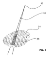
- the said calibration factors are obtained for each ExtDet per point in the defined segment in the phantom 36, Fig. 3 , and said definable time-interval for each field simultaneously irradiating the ExtDet and said phantom including detectors to measure the absorbed dose using the said patient-specific treatment without patient (from now on called off-line).
- Deviations between the measured and calculated dose values can be analysed by using the data for each time-interval and thereby simplifying the analysing phase.
- the calculated dose value in the phantom might be slightly incorrect and in such a case the exact value can be verified using a phantom measurement simulating the motion during the miss-delivered treatment.
- An alternative to measure the fluence with the detector up-streams the patient, ExtDet is to calculate the fluence using any information of the MLC positions as input and then calibrate that fluence using the described method, eg. calibrate the fluence for each time interval to dose measured in the phantom during the pre treatment verification.
- Such a determination of the dose in the phantom will be limited in accuracy and verification compared to the use of an ExtDet but still very useful because it enables quantification of deviations during treatment as dose in the phantom as if it was in place.
- the information regarding the MLC positions is easily obtainable since there is already provided means in the radio therapy device for controlling the position of the MLC leaves. This information can then be used in the comparison with the measurements inside the phantom.
- the method according to the invention may be implemented in the control and measurement system of the radiotherapy device, and thereby using the processor and storage means available there. It may of course be implemented in a stand-alone unit comprising the necessary equipment such as a central processing unit CPU performing the steps of the method according to the invention. This is performed with the aid of a dedicated computer program, which is stored in the program memory. It is to be understood that the computer program may also be run on a general purpose industrial computer instead of a specially adapted computer.
- the software includes computer program code elements or software code portions that make the computer perform the method using equations, algorithms, data and calculations previously described.
- a part of the program may be stored in a processor as above, but also in a ROM, RAM, PROM or EPROM chip or similar.
- the program in part or in whole may also be stored on, or in, other suitable computer readable medium such as a magnetic disk, CD-ROM or DVD disk, hard disk, magneto-optical memory storage means, in volatile memory, in flash memory, as firmware, or stored on a data server.
Landscapes
- Health & Medical Sciences (AREA)
- Engineering & Computer Science (AREA)
- Biomedical Technology (AREA)
- Pathology (AREA)
- Nuclear Medicine, Radiotherapy & Molecular Imaging (AREA)
- Radiology & Medical Imaging (AREA)
- Life Sciences & Earth Sciences (AREA)
- Animal Behavior & Ethology (AREA)
- General Health & Medical Sciences (AREA)
- Public Health (AREA)
- Veterinary Medicine (AREA)
- Radiation-Therapy Devices (AREA)
- Surgical Instruments (AREA)
Claims (16)
- Verfahren zum Ermöglichen einer Quantifizierung der Dosisbelastung bei der Strahlenbehandlung, dadurch gekennzeichnet, dass es die folgenden Schritte umfasst:- Bestrahlung eines Phantoms nach einem Behandlungsplan eines Patienten,- Messung der Bestrahlung im Phantom,- Sammeln von Informationen bezüglich der Bestrahlung durch ein Informationsmittel, das zwischen dem Phantom und der Strahlungsquelle angeordnet ist, wobei die Messungen in Zeitintervalle unterteil sind, und- Analysieren der Messungen zum Erhalten von Informationen bezüglich der Beziehung zwischen den Messungen im Phantom und Messungen im Informationsmittel zwischen dem Phantom und der Strahlungsquelle in jedem Zeitintervall- unter Verwendung der Beziehungsinformationen während der Überprüfung der Behandlung des Patienten.
- Verfahren nach Anspruch 1, dadurch gekennzeichnet, dass das Informationsmittel die Position der Lamellen des Multi-Leaf Collimators (MLC) (Tiefenblende) umfasst, der zur Formung des Bestrahlungsbündels der Strahlungsquelle angeordnet ist.
- Verfahren nach Anspruch 2, dadurch gekennzeichnet, dass die Messungen im Phantom und die Bestimmung der Positionen der Lamellen des Multi-Leaf Collimators gleichzeitig ausgeführt sind.
- Verfahren nach Anspruch 1, dadurch gekennzeichnet, dass das Informationsmittel Detektoren (ExtDet) umfasst.
- Verfahren nach Anspruch 4, dadurch gekennzeichnet, dass die Messungen im Phantom und mit den Detektoren (ExtDet) gleichzeitig ausgeführt sind.
- Verfahren nach Anspruch 2 bis 5, gekennzeichnet durch das Berechnen von Kalibrierfaktoren aus den erhaltenen Beziehungsinformationen als Verhältnis der Ablesung von Informationen aus dem Informationsmittel und den Messungen entlang des Bestrahlungsbündels im Phantom.
- Verfahren nach einem der vorherigen Ansprüche, gekennzeichnet durch den weiteren Schritt des Speicherns der Daten für jedes spezielle Zeitintervall sowohl für Messungen im Phantom wie auch Informationen aus dem Informationsmittel zwischen dem Phantom und der Behandlungsquelle.
- Verfahren nach Anspruch 6, wenn von Anspruch 4 oder 5 abhängig, dadurch gekennzeichnet, dass die Kalibrierfaktoren nach folgender Formel berechnet sind

wobeiDseg-n,f,p,t(i),t(i+1) Die Dosis in Punkt p im Phantomsegment, das durch das ExtDet-Detektorelement, n und das Feld (Projektion), f integriert von Zeit t(i) bis t(i+1), definiert ist.Sn,f,t(i),t(i+1) Das Signal vom ExtDet-Detektorelement, n, im Feld, f, integriert von Zeit t(i) bis t(i+1).Caln,f,seg-n,f,p,t(i),t(i+1) Der Kalibrierfaktor, der beim ExtDet- Detektorelement n, im Feld, f, zu verwenden ist. Zum Konvertieren des Signals, das von Zeit t(i) bis t(i+1) integriert ist, um die Dosis in Punkt p im Phantomsegment zu erreichen, das durch das ExtDet-Detektorelement definiert ist, n und Feld (Projektion), f integriert von Zeit t(i) bis t(i+1). - Verfahren nach einem der Ansprüche 4 bis 8, dadurch gekennzeichnet, dass die Detektoren (ExtDet) auf der Oberfläche des Phantoms angeordnet sind.
- Verfahren nach einem der Ansprüche 4 bis 8, dadurch gekennzeichnet, dass die Detektoren (ExtDet) zwischen der Strahlungsquelle und der Oberfläche des Phantoms angeordnet sind.
- Verfahren nach einem der Ansprüche 5 bis 7, wobei die Detektoren (ExtDet) im Phantom angeordnet sind.
- Verfahren nach Anspruch 6, wenn von Anspruch 2 oder 3 abhängig, dadurch gekennzeichnet, dass die Kalibrierfaktoren nach folgender Formel berechnet sind

wobeiDf,p,t(i),t(i+1) Die Dosis in Punkt p im Phantom im Feld (Projektion), f integriert von Zeit t(i) bis t(i+1).Fn,f,t(i),t(i+1) Der Strahlungsfluss im Feld, zwischen dem Phantom und der Quelle entlang des Bündels, das den Punkt p im Phantom, integriert von Zeit t(i) bis t(i+1), schneidet.Caln,f,p,t(i),t(i+1) Der Kalibrierfaktor, der die Beziehung zwischen dem Fluss zwischen dem Patienten und der Quelle und der Dosis im Phantom beschreibt. - Verfahren nach einem der vorherigen Ansprüche, gekennzeichnet durch die Verwendung eines patientenspezifischen Behandlungsplans während der Bestrahlung des Phantoms und Überprüfung der Genauigkeit der Bestrahlung des Phantoms durch Vergleichen der gemessenen Dosis im Phantom mit dem Behandlungsplan.
- Verfahren nach Anspruch 4, das den weiteren Schritt des Bestimmens der Position der ExtDet in der transversalen Ebene unter Verwendung der Detektoren oder Marker umfasst, die für die ExtDet definiert sind, wobei ein Bild von einer Bildvorrichtung unterhalb des Phantoms genutzt wird, z.B. EPID oder Röntgenfilm.
- Computerprogrammprodukt, das einem Computer ermöglichen kann, das Verfahren nach einem der Ansprüche 1 bis 14 auszuführen.
- Computerprogrammprodukt nach Anspruch 15, das auf einem computerlesbaren Medium gespeichert ist.
Applications Claiming Priority (5)
| Application Number | Priority Date | Filing Date | Title |
|---|---|---|---|
| US37758802P | 2002-05-06 | 2002-05-06 | |
| SE0201371A SE522162C2 (sv) | 2002-05-06 | 2002-05-06 | Metod att utföra in vivo-dosimetri vid IMRT-behandling |
| US377588P | 2002-05-06 | ||
| SE0201371 | 2002-05-06 | ||
| PCT/SE2003/000725 WO2003092813A1 (en) | 2002-05-06 | 2003-05-06 | Method for performing in vivo dosimetry |
Publications (2)
| Publication Number | Publication Date |
|---|---|
| EP1501604A1 EP1501604A1 (de) | 2005-02-02 |
| EP1501604B1 true EP1501604B1 (de) | 2009-12-02 |
Family
ID=29405461
Family Applications (1)
| Application Number | Title | Priority Date | Filing Date |
|---|---|---|---|
| EP03723593A Expired - Lifetime EP1501604B1 (de) | 2002-05-06 | 2003-05-06 | Verfahren zur durchführung von dosimetrie in vivo |
Country Status (9)
| Country | Link |
|---|---|
| US (2) | US7345274B2 (de) |
| EP (1) | EP1501604B1 (de) |
| JP (2) | JP5086523B2 (de) |
| CN (1) | CN1649643B (de) |
| AT (1) | ATE450292T1 (de) |
| AU (1) | AU2003230522A1 (de) |
| DE (1) | DE60330331D1 (de) |
| SE (1) | SE522162C2 (de) |
| WO (1) | WO2003092813A1 (de) |
Cited By (12)
| Publication number | Priority date | Publication date | Assignee | Title |
|---|---|---|---|---|
| US10456600B2 (en) | 2017-07-26 | 2019-10-29 | Reflexion Medical, Inc. | Graphical representation of radiation therapy |
| US10500416B2 (en) | 2015-06-10 | 2019-12-10 | Reflexion Medical, Inc. | High bandwidth binary multi-leaf collimator design |
| US10603515B2 (en) | 2017-08-09 | 2020-03-31 | Reflexion Medical, Inc. | Systems and methods for fault detection in emission-guided radiotherapy |
| US10688320B2 (en) | 2017-05-30 | 2020-06-23 | Reflexion Medical, Inc. | Methods for real-time image guided radiation therapy |
| US10695586B2 (en) | 2016-11-15 | 2020-06-30 | Reflexion Medical, Inc. | System for emission-guided high-energy photon delivery |
| US10795037B2 (en) | 2017-07-11 | 2020-10-06 | Reflexion Medical, Inc. | Methods for pet detector afterglow management |
| US10959686B2 (en) | 2008-03-14 | 2021-03-30 | Reflexion Medical, Inc. | Method and apparatus for emission guided radiation therapy |
| US11369806B2 (en) | 2017-11-14 | 2022-06-28 | Reflexion Medical, Inc. | Systems and methods for patient monitoring for radiotherapy |
| US11504550B2 (en) | 2017-03-30 | 2022-11-22 | Reflexion Medical, Inc. | Radiation therapy systems and methods with tumor tracking |
| US11648418B2 (en) | 2017-06-22 | 2023-05-16 | Reflexion Medical, Inc. | Systems and methods for biological adaptive radiotherapy |
| US11896848B2 (en) | 2019-01-16 | 2024-02-13 | Reflexion Medical, Inc. | Methods for setup corrections in radiation therapy |
| US12115386B2 (en) | 2019-07-12 | 2024-10-15 | Reflexion Medical, Inc. | Multi-target treatment planning and delivery and virtual localization for radiation therapy |
Families Citing this family (43)
| Publication number | Priority date | Publication date | Assignee | Title |
|---|---|---|---|---|
| US7399977B2 (en) | 2004-07-23 | 2008-07-15 | University Health Network | Apparatus and method for determining radiation dose |
| WO2007098164A2 (en) * | 2006-02-21 | 2007-08-30 | The University Of Toledo | Unfiltered radiation therapy |
| US7496174B2 (en) | 2006-10-16 | 2009-02-24 | Oraya Therapeutics, Inc. | Portable orthovoltage radiotherapy |
| US7620147B2 (en) | 2006-12-13 | 2009-11-17 | Oraya Therapeutics, Inc. | Orthovoltage radiotherapy |
| EP1917999A1 (de) * | 2006-11-03 | 2008-05-07 | Ion Beam Applications S.A. | Verifikationsverfahren und Verifikationsystem für IMRT |
| EP1917998A1 (de) * | 2006-11-03 | 2008-05-07 | Ion Beam Applications S.A. | On-line Verifikationsverfahren und Verifikationsystem für IMRT |
| SE531789C2 (sv) * | 2006-12-22 | 2009-08-04 | Micropos Medical Ab | Metod och system för att spåra en position hos en positioneringsanordning samt metod för kalibrering av system |
| DE102007014723A1 (de) * | 2007-03-23 | 2008-11-27 | Gesellschaft für Schwerionenforschung mbH (GSI) | Bestimmung eines Planungsvolumens für eine Bestrahlung eines Körpers |
| US8363783B2 (en) | 2007-06-04 | 2013-01-29 | Oraya Therapeutics, Inc. | Method and device for ocular alignment and coupling of ocular structures |
| US8920406B2 (en) | 2008-01-11 | 2014-12-30 | Oraya Therapeutics, Inc. | Device and assembly for positioning and stabilizing an eye |
| ES2313844B1 (es) * | 2007-07-27 | 2010-02-11 | Sociedad Española De Fisica Medica | Procedimiento para el control de calidad de los sistemas de planificacion dosimetrica y maniqui de imagen utilizado en dicho procedimiento. |
| EP2231277B1 (de) | 2007-12-23 | 2017-08-30 | Carl Zeiss Meditec, Inc. | Vorrichtungen für den nachweis, die kontrolle und die vorhersage von strahlenabgabe |
| US7801271B2 (en) | 2007-12-23 | 2010-09-21 | Oraya Therapeutics, Inc. | Methods and devices for orthovoltage ocular radiotherapy and treatment planning |
| EP2116277A1 (de) | 2008-05-06 | 2009-11-11 | Ion Beam Applications S.A. | Vorrichtung und Verfahren zur Überwachung und Überprüfung von Partikeltherapie |
| EP2116278A1 (de) | 2008-05-06 | 2009-11-11 | Ion Beam Applications S.A. | Vorrichtung für 3D-Dosisverfolgung in der Strahlungstherapie |
| US8827554B2 (en) | 2010-04-13 | 2014-09-09 | Carestream Health, Inc. | Tube alignment for mobile radiography system |
| US8824634B2 (en) | 2010-04-13 | 2014-09-02 | Carestream Health, Inc. | Configurable AEC sensor for an X-ray system |
| GB2479717B (en) * | 2010-04-13 | 2015-03-11 | Mirada Medical | Method for estimating radiation exposure of a patient and radiation exposure monitoring system therefor |
| US8821017B2 (en) | 2010-04-13 | 2014-09-02 | Carestream Health, Inc. | Projector as collimator light |
| US8873712B2 (en) | 2010-04-13 | 2014-10-28 | Carestream Health, Inc. | Exposure control using digital radiography detector |
| WO2012004757A2 (en) | 2010-07-07 | 2012-01-12 | University Health Network | Fiber optic radiochromic dosimeter probe and method to make the same |
| WO2012024448A2 (en) * | 2010-08-17 | 2012-02-23 | Board Of Regents, The University Of Texas System | Automated treatment planning for radiation therapy |
| US10165992B2 (en) | 2010-10-18 | 2019-01-01 | Carestream Health, Inc. | X-ray imaging systems and devices |
| DE102010061121B4 (de) * | 2010-12-08 | 2013-03-07 | Gsi Helmholtzzentrum Für Schwerionenforschung Gmbh | Bestrahlungsphantom mit zumindest einer Bewegungsvorrichtung zur Bewegung eines ersten Teilbereichs |
| US8753171B2 (en) * | 2011-02-24 | 2014-06-17 | Elizabeth Chabner Thompson | Radiation treatment brassiere |
| US9956429B2 (en) * | 2011-03-28 | 2018-05-01 | Varian Medical Systems International Ag | Method and system for automated evaluation of multiple portal dose images in radiation therapy |
| EP2816956B1 (de) | 2012-02-22 | 2018-01-17 | Carestream Health, Inc. | Mobile radiografische vorrichtung/verfahren mit tomosynthesekapazität |
| JP5801004B2 (ja) | 2012-03-09 | 2015-10-28 | シーメンス・ヘルスケア・ダイアグノスティックス・インコーポレーテッドSiemens Healthcare Diagnostics Inc. | 試薬カード分析器の較正方法 |
| US20130304409A1 (en) * | 2012-05-10 | 2013-11-14 | Board Of Regents, The University Of Texas System | Methods for validating plastic scintillating detectors and applications of same |
| US9295432B2 (en) * | 2012-10-19 | 2016-03-29 | Koninklijke Philips N.V. | Method of determining distribution of a dose in a body |
| WO2014194194A1 (en) * | 2013-05-31 | 2014-12-04 | Board Of Regents, The University Of Texas System | Large-volume scintillator detector for rapid real-time 3-d dose imaging of advanced radiation therapy modalities |
| GB2516282B (en) * | 2013-07-17 | 2017-07-26 | Vision Rt Ltd | Method of calibration of a stereoscopic camera system for use with a radio therapy treatment apparatus |
| EP2893956B1 (de) * | 2014-01-09 | 2017-02-22 | PTW - Freiburg Physikalisch-Technische Werkstätten Dr. Pychlau GmbH | Strahlungsplanungssystem |
| WO2015167980A1 (en) * | 2014-04-28 | 2015-11-05 | Brigham And Women's Hospital, Inc. | Real-time margin adaptation |
| KR102117680B1 (ko) | 2014-07-18 | 2020-06-01 | 사회복지법인 삼성생명공익재단 | 방사선 치료기 및 방사선 치료기의 정도 관리 방법 |
| GB2543731A (en) * | 2015-06-12 | 2017-05-03 | Elekta ltd | Improvements in dosimetry techniques for radiotherapy |
| GB2539261A (en) * | 2015-06-12 | 2016-12-14 | Elekta ltd | Improvements in Dosimetry Techniques for Radiotherapy |
| DE102017009040A1 (de) | 2017-09-27 | 2019-03-28 | Städtisches Klinikum Dessau | Verfahren zur EPID-basierten Überprüfung, Korrektur und Minimierung des Isozentrums eines Strahlentherapiegeräts |
| US11738212B2 (en) * | 2020-06-02 | 2023-08-29 | Vanderbilt University | Rigid phantom for end-to-end verification of adaptive radiotherapy systems |
| EP3932481B1 (de) * | 2020-06-30 | 2023-12-27 | Ion Beam Applications | Behandlungsplanungssystem für multimodale protonentherapie |
| JP7662344B2 (ja) * | 2021-01-25 | 2025-04-15 | 株式会社日立ハイテク | ワークフロー管理システム、放射線治療システム及びワークフロー管理方法 |
| CN117350965A (zh) * | 2023-10-07 | 2024-01-05 | 中国原子能科学研究院 | 对象内放射性微球的指标预估装置 |
| CN119942896B (zh) * | 2024-12-27 | 2025-11-07 | 中国辐射防护研究院 | 用于头骨内照射活体测量装置校准的头颅模型及制造方法 |
Family Cites Families (34)
| Publication number | Priority date | Publication date | Assignee | Title |
|---|---|---|---|---|
| JPH05506177A (ja) * | 1991-02-15 | 1993-09-16 | イーストマン・コダック・カンパニー | 電子計算機処理放射線写真患者識別システム |
| US5533508A (en) * | 1991-10-31 | 1996-07-09 | Pdt Systems, Inc. | Vivo dosimeter for photodynamic therapy |
| DE19503647C2 (de) * | 1995-02-06 | 1999-12-16 | Forschungszentrum Juelich Gmbh | Meßvorrichtung zur in-vivo und on-line-Bestimmung der gewebeäquivalenten Dosis bei der Strahlentherapie |
| DE69530752T2 (de) * | 1995-02-17 | 2004-03-11 | Agfa-Gevaert | System und Verfahren der Identifizierung zur Verwendung im Felde der digitalen Röntgenphotographie |
| US5769779A (en) * | 1995-04-27 | 1998-06-23 | Radiology Support Devices, Inc. | Method and apparatus for accurate radiation dosage control in breast cancer treatment |
| US5635709A (en) * | 1995-10-12 | 1997-06-03 | Photoelectron Corporation | Method and apparatus for measuring radiation dose distribution |
| US5818902A (en) * | 1996-03-01 | 1998-10-06 | Elekta Ab | Intensity modulated arc therapy with dynamic multi-leaf collimation |
| US5844241A (en) * | 1996-07-19 | 1998-12-01 | City Of Hope | System and method for determining internal radioactivity and absorbed dose estimates |
| US6047257A (en) * | 1997-03-01 | 2000-04-04 | Agfa-Gevaert | Identification of medical images through speech recognition |
| JP4212128B2 (ja) * | 1997-07-02 | 2009-01-21 | 株式会社東芝 | 放射線治療装置 |
| US6271536B1 (en) * | 1997-10-08 | 2001-08-07 | Agfa-Gevaert | Radiographic image identification method |
| US6225622B1 (en) * | 1998-07-31 | 2001-05-01 | Daniel Navarro | Dynamic radiation scanning device |
| US6668073B1 (en) * | 1998-11-12 | 2003-12-23 | The University Of British Columbia | Anthropomorphic film phantom for three-dimensional dosimetry |
| US6811079B1 (en) * | 1998-12-22 | 2004-11-02 | Eastman Kodak Company | Sheet media package having radio-frequency identification transponder |
| DE19907774A1 (de) * | 1999-02-19 | 2000-08-31 | Schwerionenforsch Gmbh | Verfahren zum Verifizieren der berechneten Bestrahlungsdosis eines Ionenstrahl-Therapiesystems |
| US6318146B1 (en) * | 1999-07-14 | 2001-11-20 | Wisconsin Alumni Research Foundation | Multi-imaging modality tissue mimicking materials for imaging phantoms |
| US6298115B1 (en) * | 2000-01-13 | 2001-10-02 | Scanditronix Medical Ab | Method for calibrating a detector means |
| US20020021830A1 (en) * | 2000-08-17 | 2002-02-21 | Ritt Daniel M. | Interrupted treatment quality assurance |
| JP2002174879A (ja) * | 2000-09-18 | 2002-06-21 | Eastman Kodak Co | 無線周波数識別トランスポンダを有するシート媒体パッケージ |
| WO2002024278A1 (en) * | 2000-09-22 | 2002-03-28 | Numerix Llc | Improved radiation therapy treatment method |
| US6493574B1 (en) * | 2000-09-28 | 2002-12-10 | Koninklijke Philips Electronics, N.V. | Calibration phantom and recognition algorithm for automatic coordinate transformation in diagnostic imaging |
| US6364529B1 (en) * | 2000-10-20 | 2002-04-02 | Med-Tec Iowa, Inc. | Radiation phantom |
| US6800870B2 (en) * | 2000-12-20 | 2004-10-05 | Michel Sayag | Light stimulating and collecting methods and apparatus for storage-phosphor image plates |
| US6697452B2 (en) * | 2001-02-16 | 2004-02-24 | The Board Of Trustees Of The Leland Stanford Junior University | Verification method of monitor units and fluence map in intensity modulated radiation therapy |
| JP4115675B2 (ja) * | 2001-03-14 | 2008-07-09 | 三菱電機株式会社 | 強度変調療法用吸収線量測定装置 |
| US6907105B2 (en) * | 2001-09-25 | 2005-06-14 | Bc Cancer Agency | Methods and apparatus for planning and delivering intensity modulated radiation fields with a rotating multileaf collimator |
| US7557353B2 (en) * | 2001-11-30 | 2009-07-07 | Sicel Technologies, Inc. | Single-use external dosimeters for use in radiation therapies |
| SE0301508D0 (sv) * | 2003-05-23 | 2003-05-23 | Goergen Nilsson | Method for pre treatment verification in intensity modulates radiation therapy |
| EP1544672B1 (de) * | 2003-12-17 | 2008-11-19 | Agfa-Gevaert HealthCare GmbH | Bildträger zur Speicherung von Röntgeninformation sowie System und Verfahren zur Bearbeitung eines solchen Bildträgers |
| AU2005216070B2 (en) * | 2004-02-20 | 2012-01-19 | University Of Florida Research Foundation, Inc. | System for delivering conformal radiation therapy while simultaneously imaging soft tissue |
| US7399977B2 (en) * | 2004-07-23 | 2008-07-15 | University Health Network | Apparatus and method for determining radiation dose |
| US20060027756A1 (en) * | 2004-08-09 | 2006-02-09 | Ian Thomson | Dosimeter having an array of sensors for measuring ionizing radiation, and dosimetry system and method using such a dosimeter |
| US7024026B1 (en) * | 2005-01-20 | 2006-04-04 | Radiological Imaging Technology, Inc. | Relative calibration for dosimetric devices |
| US7252434B2 (en) * | 2005-03-16 | 2007-08-07 | Wisconsin Alumni Research Foundation | Radiation therapy machine calibration apparatus providing multiple angle measurements |
-
2002
- 2002-05-06 SE SE0201371A patent/SE522162C2/sv unknown
-
2003
- 2003-05-06 AU AU2003230522A patent/AU2003230522A1/en not_active Abandoned
- 2003-05-06 CN CN038101726A patent/CN1649643B/zh not_active Expired - Lifetime
- 2003-05-06 AT AT03723593T patent/ATE450292T1/de not_active IP Right Cessation
- 2003-05-06 US US10/513,240 patent/US7345274B2/en not_active Expired - Lifetime
- 2003-05-06 DE DE60330331T patent/DE60330331D1/de not_active Expired - Lifetime
- 2003-05-06 EP EP03723593A patent/EP1501604B1/de not_active Expired - Lifetime
- 2003-05-06 WO PCT/SE2003/000725 patent/WO2003092813A1/en not_active Ceased
- 2003-05-06 JP JP2004500991A patent/JP5086523B2/ja not_active Expired - Lifetime
-
2007
- 2007-12-28 US US11/966,383 patent/US20080191141A1/en not_active Abandoned
-
2012
- 2012-04-03 JP JP2012084649A patent/JP5567058B2/ja not_active Expired - Lifetime
Cited By (31)
| Publication number | Priority date | Publication date | Assignee | Title |
|---|---|---|---|---|
| US10959686B2 (en) | 2008-03-14 | 2021-03-30 | Reflexion Medical, Inc. | Method and apparatus for emission guided radiation therapy |
| US12167922B2 (en) | 2008-03-14 | 2024-12-17 | Reflexion Medical, Inc. | Method and apparatus for emission guided radiation therapy |
| US11627920B2 (en) | 2008-03-14 | 2023-04-18 | Reflexion Medical, Inc. | Method and apparatus for emission guided radiation therapy |
| US10500416B2 (en) | 2015-06-10 | 2019-12-10 | Reflexion Medical, Inc. | High bandwidth binary multi-leaf collimator design |
| US11878185B2 (en) | 2015-06-10 | 2024-01-23 | Reflexion Medical, Inc. | High bandwidth binary multi-leaf collimator design |
| US11285340B2 (en) | 2015-06-10 | 2022-03-29 | Reflexion Medical, Inc. | High bandwidth binary multi-leaf collimator design |
| US11975220B2 (en) | 2016-11-15 | 2024-05-07 | Reflexion Medical, Inc. | System for emission-guided high-energy photon delivery |
| US10695586B2 (en) | 2016-11-15 | 2020-06-30 | Reflexion Medical, Inc. | System for emission-guided high-energy photon delivery |
| US11504550B2 (en) | 2017-03-30 | 2022-11-22 | Reflexion Medical, Inc. | Radiation therapy systems and methods with tumor tracking |
| US11904184B2 (en) | 2017-03-30 | 2024-02-20 | Reflexion Medical, Inc. | Radiation therapy systems and methods with tumor tracking |
| US12303717B2 (en) | 2017-03-30 | 2025-05-20 | Reflexion Medical, Inc. | Radiation therapy systems and methods with tumor tracking |
| US11033757B2 (en) | 2017-05-30 | 2021-06-15 | Reflexion Medical, Inc. | Methods for real-time image guided radiation therapy |
| US12521571B2 (en) | 2017-05-30 | 2026-01-13 | Reflexion Medical, Inc. | Methods for real-time image guided radiation therapy |
| US10688320B2 (en) | 2017-05-30 | 2020-06-23 | Reflexion Medical, Inc. | Methods for real-time image guided radiation therapy |
| US11633626B2 (en) | 2017-05-30 | 2023-04-25 | Reflexion Medical, Inc. | Methods for real-time image guided radiation therapy |
| US12214219B2 (en) | 2017-06-22 | 2025-02-04 | Reflexion Medical, Inc. | Systems and methods for biological adaptive radiotherapy |
| US11648418B2 (en) | 2017-06-22 | 2023-05-16 | Reflexion Medical, Inc. | Systems and methods for biological adaptive radiotherapy |
| US12032107B2 (en) | 2017-07-11 | 2024-07-09 | Reflexion Medical, Inc. | Methods for PET detector afterglow management |
| US10795037B2 (en) | 2017-07-11 | 2020-10-06 | Reflexion Medical, Inc. | Methods for pet detector afterglow management |
| US11675097B2 (en) | 2017-07-11 | 2023-06-13 | Reflexion Medical, Inc. | Methods for PET detector afterglow management |
| US11287540B2 (en) | 2017-07-11 | 2022-03-29 | Reflexion Medical, Inc. | Methods for PET detector afterglow management |
| US10456600B2 (en) | 2017-07-26 | 2019-10-29 | Reflexion Medical, Inc. | Graphical representation of radiation therapy |
| US12303718B2 (en) | 2017-07-26 | 2025-05-20 | Reflexion Medical, Inc. | Graphical representation of radiation therapy |
| US11007384B2 (en) | 2017-08-09 | 2021-05-18 | Reflexion Medical, Inc. | Systems and methods for fault detection in emission-guided radiotherapy |
| US11511133B2 (en) | 2017-08-09 | 2022-11-29 | Reflexion Medical, Inc. | Systems and methods for fault detection in emission-guided radiotherapy |
| US12023523B2 (en) | 2017-08-09 | 2024-07-02 | Reflexion Medical, Inc. | Systems and methods for fault detection in emission-guided radiotherapy |
| US10603515B2 (en) | 2017-08-09 | 2020-03-31 | Reflexion Medical, Inc. | Systems and methods for fault detection in emission-guided radiotherapy |
| US12029921B2 (en) | 2017-11-14 | 2024-07-09 | Reflexion Medical, Inc. | Systems and methods for patient monitoring for radiotherapy |
| US11369806B2 (en) | 2017-11-14 | 2022-06-28 | Reflexion Medical, Inc. | Systems and methods for patient monitoring for radiotherapy |
| US11896848B2 (en) | 2019-01-16 | 2024-02-13 | Reflexion Medical, Inc. | Methods for setup corrections in radiation therapy |
| US12115386B2 (en) | 2019-07-12 | 2024-10-15 | Reflexion Medical, Inc. | Multi-target treatment planning and delivery and virtual localization for radiation therapy |
Also Published As
| Publication number | Publication date |
|---|---|
| ATE450292T1 (de) | 2009-12-15 |
| SE522162C2 (sv) | 2004-01-20 |
| US20050151071A1 (en) | 2005-07-14 |
| AU2003230522A1 (en) | 2003-11-17 |
| JP5567058B2 (ja) | 2014-08-06 |
| CN1649643B (zh) | 2010-05-05 |
| JP2012157706A (ja) | 2012-08-23 |
| JP2005524449A (ja) | 2005-08-18 |
| DE60330331D1 (de) | 2010-01-14 |
| WO2003092813A1 (en) | 2003-11-13 |
| SE0201371L (sv) | 2003-11-07 |
| SE0201371D0 (sv) | 2002-05-06 |
| JP5086523B2 (ja) | 2012-11-28 |
| US20080191141A1 (en) | 2008-08-14 |
| CN1649643A (zh) | 2005-08-03 |
| US7345274B2 (en) | 2008-03-18 |
| EP1501604A1 (de) | 2005-02-02 |
Similar Documents
| Publication | Publication Date | Title |
|---|---|---|
| US7345274B2 (en) | Method for performing in vivo dosimetry | |
| US7371007B2 (en) | Method for pre treatment verification in radiation therapy | |
| Pfeiler et al. | Experimental validation of a 4D dose calculation routine for pencil beam scanning proton therapy | |
| Polf et al. | Imaging particle beams for cancer treatment | |
| EP2086639B1 (de) | On-line verifikationsystem für imrt | |
| CN104548372A (zh) | 放射治疗计划、剂量确定、质量保证的方法及装置 | |
| Rinaldin et al. | Quality assurance of rapid arc treatments: performances and pre-clinical verifications of a planar detector (MapCHECK2) | |
| CN103127623A (zh) | 一种放射治疗中在线验证加速器出束准确性的方法 | |
| Schweins et al. | Detection of an internal density change in an anthropomorphic head phantom via tracking of charged nuclear fragments in carbon‐ion radiotherapy | |
| Ali et al. | Quantitative assessment of the accuracy of dose calculation using pencil beam and Monte Carlo algorithms and requirements for clinical quality assurance | |
| US9227083B2 (en) | Device and method for radiation dosimetry | |
| Schwarz et al. | Importance of accurate dose calculations outside segment edges in intensity modulated radiotherapy treatment planning | |
| US12527974B2 (en) | Method to determine a patient dose distribution and corresponding radiation dosimetry apparatus | |
| Dorje | Limitation of pencil beam convolution (PBC) algorithm for photon dose calculation in inhomogeneous medium | |
| Huang et al. | An experimental validation of a filtering approach for prompt gamma prediction in a research proton treatment planning system | |
| Huntelerslag | Modelling Dosimetric Variations Due to Beam Delivery Parameter Perturbations | |
| Potter | Feasibility Study of the Flattening Filter Removal Design for Conventional Treatments Using a Linear Accelerator | |
| Savatović et al. | A GEANT4 Monte Carlo simulation study for the computation of output factors and out-of-field dosimetry in breast IOERT | |
| Cowell | Dose delivery accuracy of visually gated SBRT: a stress test for clinical acceptance | |
| Bogaert et al. | Adapted Delta4 phantom for EBT3 film based pre-treatment QA for lung SBRT VMAT: proof of concept | |
| JP2023023437A (ja) | 粒子線治療システム、および治療計画装置 | |
| De Marzi et al. | Treatment Planning Systems and Hadron Therapy Practice in France | |
| Cufflin et al. | DEVELOPMENT OF A MONTE CARLO BASED TECHNIQUE TO IMPROVE ACCURACY OF PORTAL DOSIMETRY | |
| Nakada et al. | DOSIMETRIC PERFORMANCE OF A 2D DIODE ARRAY FOR VERIFICATION OF VOLUMETRIC MODULATED ARC THERAPY | |
| Tateoka et al. | EVALUATION OF DOSE FROM IMRT USING IONIZATION CHAMBER-THE EFFECT OF IONIZATION CHAMBER VOLUME AND SENSITIVITY IN RESPONSE |
Legal Events
| Date | Code | Title | Description |
|---|---|---|---|
| PUAI | Public reference made under article 153(3) epc to a published international application that has entered the european phase |
Free format text: ORIGINAL CODE: 0009012 |
|
| 17P | Request for examination filed |
Effective date: 20041006 |
|
| AK | Designated contracting states |
Kind code of ref document: A1 Designated state(s): AT BE BG CH CY CZ DE DK EE ES FI FR GB GR HU IE IT LI LU MC NL PT RO SE SI SK TR |
|
| AX | Request for extension of the european patent |
Extension state: AL LT LV MK |
|
| DAX | Request for extension of the european patent (deleted) | ||
| 17Q | First examination report despatched |
Effective date: 20070410 |
|
| GRAP | Despatch of communication of intention to grant a patent |
Free format text: ORIGINAL CODE: EPIDOSNIGR1 |
|
| GRAS | Grant fee paid |
Free format text: ORIGINAL CODE: EPIDOSNIGR3 |
|
| GRAA | (expected) grant |
Free format text: ORIGINAL CODE: 0009210 |
|
| AK | Designated contracting states |
Kind code of ref document: B1 Designated state(s): AT BE BG CH CY CZ DE DK EE ES FI FR GB GR HU IE IT LI LU MC NL PT RO SE SI SK TR |
|
| REG | Reference to a national code |
Ref country code: GB Ref legal event code: FG4D |
|
| REG | Reference to a national code |
Ref country code: CH Ref legal event code: EP |
|
| REG | Reference to a national code |
Ref country code: IE Ref legal event code: FG4D |
|
| REF | Corresponds to: |
Ref document number: 60330331 Country of ref document: DE Date of ref document: 20100114 Kind code of ref document: P |
|
| REG | Reference to a national code |
Ref country code: NL Ref legal event code: VDEP Effective date: 20091202 |
|
| PG25 | Lapsed in a contracting state [announced via postgrant information from national office to epo] |
Ref country code: FI Free format text: LAPSE BECAUSE OF FAILURE TO SUBMIT A TRANSLATION OF THE DESCRIPTION OR TO PAY THE FEE WITHIN THE PRESCRIBED TIME-LIMIT Effective date: 20091202 Ref country code: SE Free format text: LAPSE BECAUSE OF FAILURE TO SUBMIT A TRANSLATION OF THE DESCRIPTION OR TO PAY THE FEE WITHIN THE PRESCRIBED TIME-LIMIT Effective date: 20091202 |
|
| PG25 | Lapsed in a contracting state [announced via postgrant information from national office to epo] |
Ref country code: SI Free format text: LAPSE BECAUSE OF FAILURE TO SUBMIT A TRANSLATION OF THE DESCRIPTION OR TO PAY THE FEE WITHIN THE PRESCRIBED TIME-LIMIT Effective date: 20091202 Ref country code: CY Free format text: LAPSE BECAUSE OF FAILURE TO SUBMIT A TRANSLATION OF THE DESCRIPTION OR TO PAY THE FEE WITHIN THE PRESCRIBED TIME-LIMIT Effective date: 20091202 |
|
| PG25 | Lapsed in a contracting state [announced via postgrant information from national office to epo] |
Ref country code: AT Free format text: LAPSE BECAUSE OF FAILURE TO SUBMIT A TRANSLATION OF THE DESCRIPTION OR TO PAY THE FEE WITHIN THE PRESCRIBED TIME-LIMIT Effective date: 20091202 |
|
| PG25 | Lapsed in a contracting state [announced via postgrant information from national office to epo] |
Ref country code: PT Free format text: LAPSE BECAUSE OF FAILURE TO SUBMIT A TRANSLATION OF THE DESCRIPTION OR TO PAY THE FEE WITHIN THE PRESCRIBED TIME-LIMIT Effective date: 20100402 Ref country code: RO Free format text: LAPSE BECAUSE OF FAILURE TO SUBMIT A TRANSLATION OF THE DESCRIPTION OR TO PAY THE FEE WITHIN THE PRESCRIBED TIME-LIMIT Effective date: 20091202 Ref country code: ES Free format text: LAPSE BECAUSE OF FAILURE TO SUBMIT A TRANSLATION OF THE DESCRIPTION OR TO PAY THE FEE WITHIN THE PRESCRIBED TIME-LIMIT Effective date: 20100313 Ref country code: EE Free format text: LAPSE BECAUSE OF FAILURE TO SUBMIT A TRANSLATION OF THE DESCRIPTION OR TO PAY THE FEE WITHIN THE PRESCRIBED TIME-LIMIT Effective date: 20091202 Ref country code: BG Free format text: LAPSE BECAUSE OF FAILURE TO SUBMIT A TRANSLATION OF THE DESCRIPTION OR TO PAY THE FEE WITHIN THE PRESCRIBED TIME-LIMIT Effective date: 20100302 Ref country code: NL Free format text: LAPSE BECAUSE OF FAILURE TO SUBMIT A TRANSLATION OF THE DESCRIPTION OR TO PAY THE FEE WITHIN THE PRESCRIBED TIME-LIMIT Effective date: 20091202 |
|
| PG25 | Lapsed in a contracting state [announced via postgrant information from national office to epo] |
Ref country code: CZ Free format text: LAPSE BECAUSE OF FAILURE TO SUBMIT A TRANSLATION OF THE DESCRIPTION OR TO PAY THE FEE WITHIN THE PRESCRIBED TIME-LIMIT Effective date: 20091202 Ref country code: BE Free format text: LAPSE BECAUSE OF FAILURE TO SUBMIT A TRANSLATION OF THE DESCRIPTION OR TO PAY THE FEE WITHIN THE PRESCRIBED TIME-LIMIT Effective date: 20091202 Ref country code: SK Free format text: LAPSE BECAUSE OF FAILURE TO SUBMIT A TRANSLATION OF THE DESCRIPTION OR TO PAY THE FEE WITHIN THE PRESCRIBED TIME-LIMIT Effective date: 20091202 |
|
| PLBI | Opposition filed |
Free format text: ORIGINAL CODE: 0009260 |
|
| PLAX | Notice of opposition and request to file observation + time limit sent |
Free format text: ORIGINAL CODE: EPIDOSNOBS2 |
|
| 26 | Opposition filed |
Opponent name: IBA DOSIMETRY GMBH Effective date: 20100901 |
|
| PG25 | Lapsed in a contracting state [announced via postgrant information from national office to epo] |
Ref country code: GR Free format text: LAPSE BECAUSE OF FAILURE TO SUBMIT A TRANSLATION OF THE DESCRIPTION OR TO PAY THE FEE WITHIN THE PRESCRIBED TIME-LIMIT Effective date: 20100303 |
|
| PG25 | Lapsed in a contracting state [announced via postgrant information from national office to epo] |
Ref country code: MC Free format text: LAPSE BECAUSE OF NON-PAYMENT OF DUE FEES Effective date: 20100531 |
|
| REG | Reference to a national code |
Ref country code: CH Ref legal event code: PL |
|
| PG25 | Lapsed in a contracting state [announced via postgrant information from national office to epo] |
Ref country code: DK Free format text: LAPSE BECAUSE OF FAILURE TO SUBMIT A TRANSLATION OF THE DESCRIPTION OR TO PAY THE FEE WITHIN THE PRESCRIBED TIME-LIMIT Effective date: 20091202 |
|
| PLBB | Reply of patent proprietor to notice(s) of opposition received |
Free format text: ORIGINAL CODE: EPIDOSNOBS3 |
|
| PG25 | Lapsed in a contracting state [announced via postgrant information from national office to epo] |
Ref country code: CH Free format text: LAPSE BECAUSE OF NON-PAYMENT OF DUE FEES Effective date: 20100531 Ref country code: LI Free format text: LAPSE BECAUSE OF NON-PAYMENT OF DUE FEES Effective date: 20100531 |
|
| PG25 | Lapsed in a contracting state [announced via postgrant information from national office to epo] |
Ref country code: IT Free format text: LAPSE BECAUSE OF FAILURE TO SUBMIT A TRANSLATION OF THE DESCRIPTION OR TO PAY THE FEE WITHIN THE PRESCRIBED TIME-LIMIT Effective date: 20091202 |
|
| PG25 | Lapsed in a contracting state [announced via postgrant information from national office to epo] |
Ref country code: IE Free format text: LAPSE BECAUSE OF NON-PAYMENT OF DUE FEES Effective date: 20100506 |
|
| RDAF | Communication despatched that patent is revoked |
Free format text: ORIGINAL CODE: EPIDOSNREV1 |
|
| APAH | Appeal reference modified |
Free format text: ORIGINAL CODE: EPIDOSCREFNO |
|
| APBM | Appeal reference recorded |
Free format text: ORIGINAL CODE: EPIDOSNREFNO |
|
| APBP | Date of receipt of notice of appeal recorded |
Free format text: ORIGINAL CODE: EPIDOSNNOA2O |
|
| PG25 | Lapsed in a contracting state [announced via postgrant information from national office to epo] |
Ref country code: HU Free format text: LAPSE BECAUSE OF FAILURE TO SUBMIT A TRANSLATION OF THE DESCRIPTION OR TO PAY THE FEE WITHIN THE PRESCRIBED TIME-LIMIT Effective date: 20100603 Ref country code: LU Free format text: LAPSE BECAUSE OF NON-PAYMENT OF DUE FEES Effective date: 20100506 |
|
| PG25 | Lapsed in a contracting state [announced via postgrant information from national office to epo] |
Ref country code: TR Free format text: LAPSE BECAUSE OF FAILURE TO SUBMIT A TRANSLATION OF THE DESCRIPTION OR TO PAY THE FEE WITHIN THE PRESCRIBED TIME-LIMIT Effective date: 20091202 |
|
| REG | Reference to a national code |
Ref country code: FR Ref legal event code: PLFP Year of fee payment: 13 |
|
| PLAB | Opposition data, opponent's data or that of the opponent's representative modified |
Free format text: ORIGINAL CODE: 0009299OPPO |
|
| R26 | Opposition filed (corrected) |
Opponent name: IBA DOSIMETRY GMBH Effective date: 20100901 |
|
| REG | Reference to a national code |
Ref country code: FR Ref legal event code: PLFP Year of fee payment: 14 |
|
| REG | Reference to a national code |
Ref country code: FR Ref legal event code: PLFP Year of fee payment: 15 |
|
| PLAB | Opposition data, opponent's data or that of the opponent's representative modified |
Free format text: ORIGINAL CODE: 0009299OPPO |
|
| R26 | Opposition filed (corrected) |
Opponent name: IBA DOSIMETRY GMBH Effective date: 20100901 |
|
| APBU | Appeal procedure closed |
Free format text: ORIGINAL CODE: EPIDOSNNOA9O |
|
| REG | Reference to a national code |
Ref country code: FR Ref legal event code: PLFP Year of fee payment: 16 |
|
| PLCK | Communication despatched that opposition was rejected |
Free format text: ORIGINAL CODE: EPIDOSNREJ1 |
|
| REG | Reference to a national code |
Ref country code: DE Ref legal event code: R100 Ref document number: 60330331 Country of ref document: DE |
|
| PLBN | Opposition rejected |
Free format text: ORIGINAL CODE: 0009273 |
|
| STAA | Information on the status of an ep patent application or granted ep patent |
Free format text: STATUS: OPPOSITION REJECTED |
|
| 27O | Opposition rejected |
Effective date: 20180804 |
|
| PGFP | Annual fee paid to national office [announced via postgrant information from national office to epo] |
Ref country code: GB Payment date: 20220517 Year of fee payment: 20 Ref country code: FR Payment date: 20220518 Year of fee payment: 20 Ref country code: DE Payment date: 20220520 Year of fee payment: 20 |
|
| REG | Reference to a national code |
Ref country code: DE Ref legal event code: R071 Ref document number: 60330331 Country of ref document: DE |
|
| REG | Reference to a national code |
Ref country code: GB Ref legal event code: PE20 Expiry date: 20230505 |
|
| PG25 | Lapsed in a contracting state [announced via postgrant information from national office to epo] |
Ref country code: GB Free format text: LAPSE BECAUSE OF EXPIRATION OF PROTECTION Effective date: 20230505 |