EP1498490A1 - Process for producing antibody composition - Google Patents
Process for producing antibody composition Download PDFInfo
- Publication number
- EP1498490A1 EP1498490A1 EP03723096A EP03723096A EP1498490A1 EP 1498490 A1 EP1498490 A1 EP 1498490A1 EP 03723096 A EP03723096 A EP 03723096A EP 03723096 A EP03723096 A EP 03723096A EP 1498490 A1 EP1498490 A1 EP 1498490A1
- Authority
- EP
- European Patent Office
- Prior art keywords
- cell
- seq
- dna
- antibody
- protein
- Prior art date
- Legal status (The legal status is an assumption and is not a legal conclusion. Google has not performed a legal analysis and makes no representation as to the accuracy of the status listed.)
- Withdrawn
Links
Images




















Classifications
-
- A—HUMAN NECESSITIES
- A61—MEDICAL OR VETERINARY SCIENCE; HYGIENE
- A61K—PREPARATIONS FOR MEDICAL, DENTAL OR TOILETRY PURPOSES
- A61K39/00—Medicinal preparations containing antigens or antibodies
- A61K39/395—Antibodies; Immunoglobulins; Immune serum, e.g. antilymphocytic serum
-
- A—HUMAN NECESSITIES
- A61—MEDICAL OR VETERINARY SCIENCE; HYGIENE
- A61P—SPECIFIC THERAPEUTIC ACTIVITY OF CHEMICAL COMPOUNDS OR MEDICINAL PREPARATIONS
- A61P29/00—Non-central analgesic, antipyretic or antiinflammatory agents, e.g. antirheumatic agents; Non-steroidal antiinflammatory drugs [NSAID]
-
- A—HUMAN NECESSITIES
- A61—MEDICAL OR VETERINARY SCIENCE; HYGIENE
- A61P—SPECIFIC THERAPEUTIC ACTIVITY OF CHEMICAL COMPOUNDS OR MEDICINAL PREPARATIONS
- A61P31/00—Antiinfectives, i.e. antibiotics, antiseptics, chemotherapeutics
- A61P31/04—Antibacterial agents
-
- A—HUMAN NECESSITIES
- A61—MEDICAL OR VETERINARY SCIENCE; HYGIENE
- A61P—SPECIFIC THERAPEUTIC ACTIVITY OF CHEMICAL COMPOUNDS OR MEDICINAL PREPARATIONS
- A61P31/00—Antiinfectives, i.e. antibiotics, antiseptics, chemotherapeutics
- A61P31/12—Antivirals
-
- A—HUMAN NECESSITIES
- A61—MEDICAL OR VETERINARY SCIENCE; HYGIENE
- A61P—SPECIFIC THERAPEUTIC ACTIVITY OF CHEMICAL COMPOUNDS OR MEDICINAL PREPARATIONS
- A61P31/00—Antiinfectives, i.e. antibiotics, antiseptics, chemotherapeutics
- A61P31/12—Antivirals
- A61P31/14—Antivirals for RNA viruses
-
- A—HUMAN NECESSITIES
- A61—MEDICAL OR VETERINARY SCIENCE; HYGIENE
- A61P—SPECIFIC THERAPEUTIC ACTIVITY OF CHEMICAL COMPOUNDS OR MEDICINAL PREPARATIONS
- A61P35/00—Antineoplastic agents
-
- A—HUMAN NECESSITIES
- A61—MEDICAL OR VETERINARY SCIENCE; HYGIENE
- A61P—SPECIFIC THERAPEUTIC ACTIVITY OF CHEMICAL COMPOUNDS OR MEDICINAL PREPARATIONS
- A61P37/00—Drugs for immunological or allergic disorders
- A61P37/02—Immunomodulators
-
- A—HUMAN NECESSITIES
- A61—MEDICAL OR VETERINARY SCIENCE; HYGIENE
- A61P—SPECIFIC THERAPEUTIC ACTIVITY OF CHEMICAL COMPOUNDS OR MEDICINAL PREPARATIONS
- A61P37/00—Drugs for immunological or allergic disorders
- A61P37/02—Immunomodulators
- A61P37/04—Immunostimulants
-
- A—HUMAN NECESSITIES
- A61—MEDICAL OR VETERINARY SCIENCE; HYGIENE
- A61P—SPECIFIC THERAPEUTIC ACTIVITY OF CHEMICAL COMPOUNDS OR MEDICINAL PREPARATIONS
- A61P37/00—Drugs for immunological or allergic disorders
- A61P37/08—Antiallergic agents
-
- A—HUMAN NECESSITIES
- A61—MEDICAL OR VETERINARY SCIENCE; HYGIENE
- A61P—SPECIFIC THERAPEUTIC ACTIVITY OF CHEMICAL COMPOUNDS OR MEDICINAL PREPARATIONS
- A61P43/00—Drugs for specific purposes, not provided for in groups A61P1/00-A61P41/00
-
- A—HUMAN NECESSITIES
- A61—MEDICAL OR VETERINARY SCIENCE; HYGIENE
- A61P—SPECIFIC THERAPEUTIC ACTIVITY OF CHEMICAL COMPOUNDS OR MEDICINAL PREPARATIONS
- A61P9/00—Drugs for disorders of the cardiovascular system
-
- C—CHEMISTRY; METALLURGY
- C07—ORGANIC CHEMISTRY
- C07K—PEPTIDES
- C07K16/00—Immunoglobulins [IG], e.g. monoclonal or polyclonal antibodies
-
- C—CHEMISTRY; METALLURGY
- C07—ORGANIC CHEMISTRY
- C07K—PEPTIDES
- C07K16/00—Immunoglobulins [IG], e.g. monoclonal or polyclonal antibodies
- C07K16/18—Immunoglobulins [IG], e.g. monoclonal or polyclonal antibodies against material from animals or humans
- C07K16/28—Immunoglobulins [IG], e.g. monoclonal or polyclonal antibodies against material from animals or humans against receptors, cell surface antigens or cell surface determinants
- C07K16/2866—Immunoglobulins [IG], e.g. monoclonal or polyclonal antibodies against material from animals or humans against receptors, cell surface antigens or cell surface determinants against receptors for cytokines, lymphokines, interferons
-
- C—CHEMISTRY; METALLURGY
- C07—ORGANIC CHEMISTRY
- C07K—PEPTIDES
- C07K16/00—Immunoglobulins [IG], e.g. monoclonal or polyclonal antibodies
- C07K16/18—Immunoglobulins [IG], e.g. monoclonal or polyclonal antibodies against material from animals or humans
- C07K16/28—Immunoglobulins [IG], e.g. monoclonal or polyclonal antibodies against material from animals or humans against receptors, cell surface antigens or cell surface determinants
- C07K16/2896—Immunoglobulins [IG], e.g. monoclonal or polyclonal antibodies against material from animals or humans against receptors, cell surface antigens or cell surface determinants against molecules with a "CD"-designation, not provided for elsewhere
-
- C—CHEMISTRY; METALLURGY
- C12—BIOCHEMISTRY; BEER; SPIRITS; WINE; VINEGAR; MICROBIOLOGY; ENZYMOLOGY; MUTATION OR GENETIC ENGINEERING
- C12N—MICROORGANISMS OR ENZYMES; COMPOSITIONS THEREOF; PROPAGATING, PRESERVING, OR MAINTAINING MICROORGANISMS; MUTATION OR GENETIC ENGINEERING; CULTURE MEDIA
- C12N15/00—Mutation or genetic engineering; DNA or RNA concerning genetic engineering, vectors, e.g. plasmids, or their isolation, preparation or purification; Use of hosts therefor
- C12N15/09—Recombinant DNA-technology
- C12N15/11—DNA or RNA fragments; Modified forms thereof; Non-coding nucleic acids having a biological activity
- C12N15/113—Non-coding nucleic acids modulating the expression of genes, e.g. antisense oligonucleotides; Antisense DNA or RNA; Triplex- forming oligonucleotides; Catalytic nucleic acids, e.g. ribozymes; Nucleic acids used in co-suppression or gene silencing
- C12N15/1137—Non-coding nucleic acids modulating the expression of genes, e.g. antisense oligonucleotides; Antisense DNA or RNA; Triplex- forming oligonucleotides; Catalytic nucleic acids, e.g. ribozymes; Nucleic acids used in co-suppression or gene silencing against enzymes
-
- C—CHEMISTRY; METALLURGY
- C12—BIOCHEMISTRY; BEER; SPIRITS; WINE; VINEGAR; MICROBIOLOGY; ENZYMOLOGY; MUTATION OR GENETIC ENGINEERING
- C12N—MICROORGANISMS OR ENZYMES; COMPOSITIONS THEREOF; PROPAGATING, PRESERVING, OR MAINTAINING MICROORGANISMS; MUTATION OR GENETIC ENGINEERING; CULTURE MEDIA
- C12N9/00—Enzymes; Proenzymes; Compositions thereof; Processes for preparing, activating, inhibiting, separating or purifying enzymes
- C12N9/10—Transferases (2.)
- C12N9/1048—Glycosyltransferases (2.4)
- C12N9/1051—Hexosyltransferases (2.4.1)
-
- C—CHEMISTRY; METALLURGY
- C12—BIOCHEMISTRY; BEER; SPIRITS; WINE; VINEGAR; MICROBIOLOGY; ENZYMOLOGY; MUTATION OR GENETIC ENGINEERING
- C12Y—ENZYMES
- C12Y204/00—Glycosyltransferases (2.4)
- C12Y204/01—Hexosyltransferases (2.4.1)
- C12Y204/01068—Glycoprotein 6-alpha-L-fucosyltransferase (2.4.1.68), i.e. FUT8
-
- C—CHEMISTRY; METALLURGY
- C07—ORGANIC CHEMISTRY
- C07K—PEPTIDES
- C07K2317/00—Immunoglobulins specific features
- C07K2317/20—Immunoglobulins specific features characterized by taxonomic origin
- C07K2317/24—Immunoglobulins specific features characterized by taxonomic origin containing regions, domains or residues from different species, e.g. chimeric, humanized or veneered
-
- C—CHEMISTRY; METALLURGY
- C07—ORGANIC CHEMISTRY
- C07K—PEPTIDES
- C07K2317/00—Immunoglobulins specific features
- C07K2317/40—Immunoglobulins specific features characterized by post-translational modification
- C07K2317/41—Glycosylation, sialylation, or fucosylation
-
- C—CHEMISTRY; METALLURGY
- C07—ORGANIC CHEMISTRY
- C07K—PEPTIDES
- C07K2317/00—Immunoglobulins specific features
- C07K2317/70—Immunoglobulins specific features characterized by effect upon binding to a cell or to an antigen
- C07K2317/73—Inducing cell death, e.g. apoptosis, necrosis or inhibition of cell proliferation
- C07K2317/732—Antibody-dependent cellular cytotoxicity [ADCC]
-
- C—CHEMISTRY; METALLURGY
- C12—BIOCHEMISTRY; BEER; SPIRITS; WINE; VINEGAR; MICROBIOLOGY; ENZYMOLOGY; MUTATION OR GENETIC ENGINEERING
- C12N—MICROORGANISMS OR ENZYMES; COMPOSITIONS THEREOF; PROPAGATING, PRESERVING, OR MAINTAINING MICROORGANISMS; MUTATION OR GENETIC ENGINEERING; CULTURE MEDIA
- C12N2310/00—Structure or type of the nucleic acid
- C12N2310/10—Type of nucleic acid
- C12N2310/14—Type of nucleic acid interfering nucleic acids [NA]
-
- C—CHEMISTRY; METALLURGY
- C12—BIOCHEMISTRY; BEER; SPIRITS; WINE; VINEGAR; MICROBIOLOGY; ENZYMOLOGY; MUTATION OR GENETIC ENGINEERING
- C12N—MICROORGANISMS OR ENZYMES; COMPOSITIONS THEREOF; PROPAGATING, PRESERVING, OR MAINTAINING MICROORGANISMS; MUTATION OR GENETIC ENGINEERING; CULTURE MEDIA
- C12N2310/00—Structure or type of the nucleic acid
- C12N2310/50—Physical structure
- C12N2310/53—Physical structure partially self-complementary or closed
Definitions
- the present invention relates to a process for producing an antibody composition, and a cell used for producing the antibody composition.
- An inhibitor against an enzyme relating to the modification of a sugar chain includes tunicamycin which selectively inhibits formation of GlcNAc-P-P-Dol which is the first step of the formation of a core oligosaccharide which is a precursor of an N -glycoside-linked sugar chain, castanospermin and N -methyl-1-deoxynojirimycin which are inhibitors of glycosidase I, bromocondulitol which is an inhibitor of glycosidase II, 1-deoxynojirimycin and 1,4-dioxy-1,4-imino-D-mannitol which are inhibitors of mannosidase I, swainsonine which is an inhibitor of mannosidase II and the like.
- glycosyltransferase As an inhibitor specific for a glycosyltransferase, deoxy derivatives of substrates against N -acetylglucosamine transferase V (GnTV) and the like are being developed [ Glycobiology Series 2 - Destiny of Sugar Chain in Cell (Kodan-sha Scientific), edited by Katsutaka Nagai, Senichiro Hakomori and Akira Kobata (1993)]. Also, it is known that 1-deoxynojirimycin inhibits synthesis of a complex type sugar chain and increases the ratio of high mannose type and hybrid type sugar chains.
- glycoprotein having changed sugar chain structure can be produced when a mutant in which the activity of a gene encoding an enzyme relating to the modification of a sugar chain is used as a host cell.
- an antibody having a high mannose type sugar chain structure can be produced by using a CHO cell mutant cell line deficient in the activity of N -acetylglucosamine transferase I (GnTI) [ J . Immuno ., 160 , 3393 (1998)].
- the present invention relates to the following (1) to (81).
- the process for producing an antibody composition using the cell of the present invention includes a process for producing a monoclonal antibody using a hybridoma cell, a process for producing a human antibody and a humanized antibody using a host cell into which a gene encoding an antibody is introduced, a process for producing a human antibody using a transgenic non-human animal which is developed after transplanting a non-human embryonic stem cell or fertilized egg cell into which a gene encoding an antibody is introduced into a non-human animal early stage embryo, and the like.
- any cell can be used, so long as it is a cell such as yeast, an animal cell, an insect cell or a plant cell which can be used for producing an antibody composition and is a cell resistant to a lectin which recognizes a sugar chain in which 1-position of fucose is bound to 6-position of N -acetylglucosamine in the reducing end through ⁇ -bond in a complex N -glycoside-linked sugar chain.
- Examples include a hybridoma cell, a host cell for producing a human antibody and humanized antibody, an embryonic stem cell and fertilized egg cell for producing a transgenic non-human animal which produces a human antibody, a plant callus cell for producing a transgenic plant which produces a human antibody, a myeloma cell, a cell derived from a transgenic non-human animal and the like which are resistant to lectin which recognizes a sugar chain in which 1-position of fucose is bound to 6-position of N -acetylglucosamine in the reducing end through ⁇ -bond in a complex N -glycoside-linked sugar chain.
- the myeloma cell can be used as a fusion cell for producing a hybridoma cell.
- a hybridoma cell can be produced by immunizing a transgenic non-human animal with an antigen and using spleen cells of the animal.
- the lectin-resistant cell is a cell of which growth is not inhibited when a lectin is applied at an effective concentration.
- the effective concentration of a lectin which does not inhibit the growth can be adjusted depending on the cell line, and is generally 10 ⁇ g/ml to 10.0 mg/ml, preferably 0.5 to 2.0 mg/ml.
- the effective concentration in the case where mutation is introduced into a parent cell is a concentration in which the parent cell cannot normally grow or higher than the concentration, and is a concentration which is preferably similar to, more preferably 2 to 5 times, still more preferably at least 10 times, and most preferably at least 20 times, higher than the concentration in which the parent cell cannot normally grow.
- the parent cell means a cell before a certain treatment is applied, namely a cell before the step for selecting a lectin-resistant cell used in the present invention is carried out or a cell before genetic engineering techniques for decreasing or deleting the above enzyme activity are carried out.
- parent cell is not particularly limited, parent cells of various cell lines are exemplified below.
- NS0 cell The parent cell of NS0 cell includes NS0 cells described in literatures such as BIO / TECHNOLOGY , 10 , 169 (1992) and Biotechnol. Bioeng., 73 , 261 (2001), NS0 cell line (RCB 0213) registered at RIKEN Cell Bank, The Institute of Physical and Chemical Research, sub-cell lines obtained by acclimating these cell lines to media in which they can grow, and the like.
- the parent cell of SP2/0-Ag14 cell includes SP2/0-Ag14 cells described in literatures such as J. Immunol ., 126 , 317 (1981), Nature , 276 , 269 (1978) and Human Antibodies and Hybridomas , 3 , 129 (1992), SP2/0-Ag14 cell (ATCC CRL-1581) registered at ATCC, sub-cell lines obtained by acclimating these cell lines to media in which they can grow (ATCC CRL-1581.1), and the like.
- the parent cell of CHO cell derived from Chinese hamster ovary tissue includes CHO cells described in literatures such as Journal of Experimental Medicine (Jikken Igaku), 108 , 945 (1958), Proc. Natl . Acad . Sci. USA , 60 , 1275 (1968), Genetics , 55 , 513 (1968), Chromosoma , 41 , 129 (1973), Methods in Cell Science , 18 , 115 (1996), Radiation Research, 148 , 260 (1997), Proc. Natl. Acad Sci. USA, 77 , 4216 (1980), Proc. Natl. Acad Sci.
- cell line CHO-K1 ATCC CCL-61
- cell line DLTXB11 ATCC CRL-9096
- cell line Pro-5 ATCC CRL-1781 registered at ATCC
- commercially available cell line CHO-S Cat # 11619 of Life Technologies
- sub-cell lines obtained by acclimating these cell lines to media in which they can grow, and the like.
- the parent cell of a rat myeloma cell line YB2/3HL.P2.G11.16Ag.20 cell includes cell lines established from Y3/Ag1.2.3 cell (ATCC CRL-1631) such as YB2/3HL.P2.G11.16Ag.20 cell described in literatures such as J . Cell. Biol ., 93 , 576 (1982) and Methods Enzymol ., 73B , 1 (1981), YB2/3HL.P2.G11.16Ag.20 cell (ATCC CRL-1662) registered at ATCC, sub-lines obtained by acclimating these cell lines to media in which they can grow, and the like.
- ATCC CRL-1631 YB2/3HL.P2.G11.16Ag.20 cell described in literatures such as J . Cell. Biol ., 93 , 576 (1982) and Methods Enzymol ., 73B , 1 (1981)
- ATCC CRL-1662 registered at ATCC, sub-
- any lectin can be used, so long as it can recognize the sugar chain structure.
- Examples include a Lens culinaris lectin LCA (lentil agglutinin derived from Lens culinaris ), a pea lectin PSA (pea lectin derived from Pisum sativum ), a broad bean lectin VFA (agglutinin derived from Vicia faba ), an Aleuria aurantia lectin AAL (lectin derived from Aleuria aurantia ) and the like.
- LCA lentil agglutinin derived from Lens culinaris
- pea lectin PSA pea lectin derived from Pisum sativum
- a broad bean lectin VFA agglutinin derived from Vicia faba
- an Aleuria aurantia lectin AAL lectin derived from Aleuria aurantia
- the lectin-resistant cell may be any cell, so long as growth of the cell is not inhibited in the presence of a lectin at a definite effective concentration.
- examples include cells in which the activity of at least one protein shown below is decreased or deleted, and the like.
- the GDP-fucose synthase may be any enzyme, so long as it is an enzyme relating to the synthesis of the intracellular sugar nucleotide, GDP-fucose, as a supply source of fucose to a sugar chain, and includes an enzyme which has influence on the synthesis of the intracellular sugar nucleotide, GDP-fucose, and the like.
- the intracellular sugar nucleotide, GDP-fucose is supplied by a de novo synthesis pathway or a salvage synthesis pathway.
- all enzymes relating to the synthesis pathways are included in the GDP-fucose synthase.
- the GDP-fucose synthase relating to the de novo synthesis pathway includes GDP-mannose 4-dehydratase (hereinafter referred to as "GMD”), GDP-keto-6-deoxymannose 3,5-epimerase, 4-reductase (hereinafter referred to as "Fx”) and the like.
- GMD GDP-mannose 4-dehydratase
- Fx 4-reductase
- the GDP-fucose synthase relating to the salvage synthesis pathway includes GDP-beta-L-fucose pyrophosphorylase (hereinafter referred to as "GFPP”), fucokinase and the like.
- GFPP GDP-beta-L-fucose pyrophosphorylase
- the GMD includes:
- the DNA encoding the amino acid sequence of GMD includes a DNA comprising the nucleotide sequence represented by SEQ ID NO:85 and a DNA which hybridizes with the DNA comprising the nucleotide sequence represented by SEQ ID NO:85 under stringent conditions and encodes an amino acid sequence having GMD activity.
- the Fx includes:
- the DNA encoding the amino acid sequence of Fx includes a DNA comprising the nucleotide sequence represented by SEQ ID NO:87 and a DNA which hybridizes with the DNA comprising the nucleotide sequence represented by SEQ ID NO:87 under stringent conditions and encodes an amino acid sequence having Fx activity.
- the GFPP includes:
- the DNA encoding the amino acid sequence of GFPP includes a DNA comprising the nucleotide sequence represented by SEQ ID NO:89 and a DNA which hybridizes with the DNA comprising the nucleotide sequence represented by SEQ ID NO:89 under stringent conditions and encodes an amino acid sequence having GFPP activity.
- the ⁇ 1,6-fucose modifying enzyme includes any enzyme, so long as it is an enzyme relating to the reaction of binding of 1-position of fucose to 6-position of N -acetylglucosamine in the reducing end through ⁇ -bond in the complex N -glycoside-linked sugar chain.
- the enzyme relating to the reaction of binding of 1-position of fucose to 6-position of N -acetylglucosamine in the reducing end through ⁇ -bond in the complex N -glycoside-linked sugar chain includes an enzyme which has influence on the reaction of binding of 1-position of fucose to 6-position of N -acetylglucosamine in the reducing end through ⁇ -bond in the complex N -glycoside-linked sugar chain. Examples include ⁇ 1,6-fucosyltransferase, ⁇ -L-fucosidase and the like.
- the enzyme relating to the reaction of binding of 1-position of fucose to 6-position of N -acetylglucosamine in the reducing end through ⁇ -bond in the complex N -glycoside-linked sugar chain includes an enzyme which has influence on the activity of the enzyme relating to the reaction of binding of 1-position of fucose to 6-position of N -acetylglucosamine in the reducing end through ⁇ -bond in the complex N -glycoside-linked sugar chain and an enzyme which has influence on the structure of substances as the substrate of the enzyme.
- the ⁇ 1,6-fucosyltransferase includes:
- the DNA encoding the amino acid sequence of ⁇ 1,6-fucosyltransferase include a DNA having the nucleotide sequence represented by SEQ ID NO:79, 80, 81 or 82 and a DNA which hybridizes with the DNA having the nucleotide sequence represented by SEQ ID NO:79, 80, 81 or 82 under stringent conditions and encodes an amino acid sequence having ⁇ 1,6-fucosyltransferase activity.
- the GDP-fucose transport protein may be any protein, so long as it is a protein relating to the transport of the intracellular sugar nucleotide, GDP-fucose, to the Golgi body, and includes a GDP-fucose transporter and the like.
- the GDP-fucose transporter includes:
- the GDP-fucose transporte of the present invention further includes the proteins selected from the following (i) to (t):
- the GDP-fucose transporter protein furthermore includes a protein which has influence on the reaction to transport the intracellular sugar nucleotide, GDP-fucose, to the Golgi body, and examples thereof include a protein which has an influence on the activity of the above protein relating to the transport of the intracellular sugar nucleotide, GDP-fucose, to the Golgi body or has influence on the expression thereof
- a DNA which is hybridizable under stringent conditions is a DNA obtained, e.g., by a method such as colony hybridization, plaque hybridization or Southern blot hybridization using a DNA such as the DNA having the nucleotide sequence represented by any one of the above SEQ ID NOs or a partial fragment thereof as the probe, and examples thereof include a DNA which can be identified by carrying out hybridization at 65°C in the presence of 0.7 to 1.0 M sodium chloride using a filter to which colony- or plaque-derived DNA fragments are immobilized, and then washing the filter at 65°C using 0.1 to 2 ⁇ SSC solution (composition of the 1 ⁇ SSC solution comprising 150 mM sodium chloride and 15 mM sodium citrate).
- the hybridization can be carried out in accordance with the methods described, e.g., in Molecular Cloning, A Laboratory Manual, Second Edition, Cold Spring Harbor Laboratory Press (1989) (hereinafter referred to as " Molecular Cloning, Second Edition"), Current Protocols in Molecular Biology, John Wiley & Sons, 1987-1997 (hereinafter referred to as " Current Protocols in Molecular Biology” ); DNA Cloning I: Core Techniques, A Practical Approach, Second Edition, Oxford University (1995); and the like.
- the hybridizable DNA includes a DNA having at least 60% or more, preferably 70% or more, more preferably 80% or more, still more preferably 90% or more, far more preferably 95% or more, and most preferably 98% or more, of homology with the nucleotide sequence represented by any one of the above SEQ ID NOs.
- the protein which comprises an amino acid sequence in which at least one amino acid is deleted, substituted, inserted and/or added in the amino acid sequence represented by any one of the above SEQ ID NOs and has the above activity can be obtained, e.g., by introducing a site-directed mutation into a DNA encoding a protein having the amino acid sequence represented by any one of the above SEQ ID NOs using the site-directed mutagenesis described, e.g., in Molecular Cloning, Second Edition; Current Protocols in Molecular Biology; Nucleic Acids Research, 10 , 6487 (1982); Proc. Natl . Acad Sci.
- the number of amino acids to be deleted, substituted, inserted and/or added is one or more, and the number is not particularly limited, but is a number which can be deleted, substituted or added by a known technique such as the site-directed mutagenesis, e.g., it is 1 to several tens, preferably 1 to 20, more preferably 1 to 10, and most preferably 1 to 5.
- the protein to be used in the present invention has at least 80% or more, preferably 85% or more, more preferably 90% or more, still more preferably 95% or more, far more preferably 97% or more, and most preferably 99% or more, of homology with the amino acid sequence represented by the above SEQ ID NOs, when calculated using an analyzing soft such as BLAST [ J . Mol. Biol ., 215 , 403 (1990)] or FASTA [ Methods in Enzymology, 183 , 63 (1990)].
- BLAST J . Mol. Biol ., 215 , 403 (1990)
- FASTA Methods in Enzymology, 183 , 63 (1990)
- any technique can be used, so long as it is a technique which can select a cell resistant to a lectin which recognizes a sugar chain in which 1-position of fucose is bound to 6-position of N -acetylglucosamine in the reducing end through ⁇ -bond in a complex N -glycoside-linked sugar chain.
- the method includes a technique for decreasing or deleting the activity of the above protein.
- the technique for decreasing or deleting includes:
- the antibody composition is a composition which comprises an antibody molecule having a complex N -glycoside-linked sugar chain in the Fc region.
- the antibody is a tetramer in which two molecules of each of two polypeptide chains, a heavy chain and a light chain (hereinafter referred to as "H chain” and “L chain”, respectively), are respectively associated.
- H chain heavy chain and a light chain
- V region which is rich in diversity and directly relates to the binding with an antigen.
- the greater part of the moiety other than the V region is called a constant region (hereinafter referred to as "C region").
- C region constant region
- the IgG class is further classified into subclasses IgG1 to IgG4 based on homology with the C region.
- the H chain is divided into four immunoglobulin domains, VH, CH1, CH2 and CH3, from its N -terminal side, and a highly flexible peptide region called hinge region is present between CH1 and CH2 to divide CH1 and CH2.
- a structural unit comprising CH2 and CH3 under the downstream of the hinge region is called Fc region to which a complex N -glycoside-linked sugar chain is bound.
- Fc region is a region to which an Fc receptor, a complement and the like are bound ( Immunology Illustrated , the Original, 5th edition, published on February 10, 2000, by Nankodo; Handbook of Antibody Technology (Kotai Kogaku Nyumon) , 1st edition on January 25, 1994, by Chijin Shokan).
- sugar chains of glycoproteins such as an antibody are roughly classified into two types, namely a sugar chain which binds to asparagine ( N -glycoside-linked sugar chain) and a sugar chain which binds to other amino acid such as serine, threonine ( O -glycoside-linked sugar chain), based on the binding form to the protein moiety.
- the N -glycoside-linked sugar chains have a basic common core structure shown by the following structural formula (I) [ Biochemical Experimentation Method 23 - Method for Studying Glycoprotein Sugar Chain (Gakujutsu Shuppan Center), edited by Reiko Takahashi (1989)]:
- the sugar chain terminus which binds to asparagine is called a reducing end, and the opposite side is called a non-reducing end.
- the N -glycoside-linked sugar chain may be any N -glycoside-linked sugar chain, so long as it comprises the core structure of formula (I).
- Examples include a high mannose type in which mannose alone binds to the non-reducing end of the core structure; a complex type in which the non-reducing end side of the core structure comprises at least one parallel branches of galactose- N -acetylglucosamine (hereinafter referred to as "Gal-GlcNAc”) and the non-reducing end side of Gal-GlcNAc comprises a structure of sialic acid, bisecting N -acetylglucosamine or the like; a hybrid type in which the non-reducing end side of the core structure comprises branches of both of the high mannose type and complex type; and the like.
- Gal-GlcNAc galactose- N -acetylglucosamine
- the Fc region in the antibody molecule comprises positions to which N -glycoside-linked sugar chains are separately bound, two sugar chains are bound per one antibody molecule. Since the N -glycoside-linked sugar chain which binds to an antibody molecule includes any sugar chain having the core structure represented by formula (I), there are a number of combinations of sugar chains for the two N -glycoside-linked sugar chains which bind to the antibody.
- the antibody composition of the present invention which is prepared by using a lectin-resistant cell of the present invention may comprise an antibody having the same sugar chain structure or an antibody having different sugar chain structures, so long as the effect of the present invention is obtained from the composition.
- the ratio of a sugar chain in which fucose is not bound to N -acetylglucosamine in the reducing end in the sugar chain among the total complex N -glycoside-linked sugar chains bound to the Fc region contained in the antibody composition is a ratio of the number of a sugar chain in which fucose is not bound to N -acetylglucosamine in the reducing end in the sugar chain to the total number of the complex N -glycoside-linked sugar chains bound to the Fc region contained in the composition.
- the sugar chain in which fucose is not bound to N -acetylglucosamine in the reducing end in the complex N -glycoside-linked sugar chain is a sugar chain in which fucose is not bound to N -acetylglucosamine in the reducing end through ⁇ -bond in the complex N -glycoside-linked sugar chain.
- it is a complex N -glycoside-linked sugar chain in which 1-position of fucose is not bound to 6-position of N -acetylglucosamine through ⁇ -bond.
- the antibody composition having higher ADCC activity includes an antibody composition in which a ratio of a sugar chain in which fucose is not bound to N -acetylglucosamine in the reducing end in the sugar chain among the total complex N -glycoside-linked sugar chains bound to the Fc region contained in the antibody composition is preferably at 20% or more, more preferably 30% or more, still more preferably 40% or more, most preferably 50% or more, and far most preferably 100%.
- the present invention relates to a process for producing an antibody composition having higher ADCC activity than an antibody composition produced by its parent cell.
- the ratio of a sugar chain in which fucose is not bound to N -acetylglucosamine in the reducing end among the total complex N -glycoside-linked sugar chains bound to the Fc region contained in the antibody composition is higher than that of an antibody composition produced by its parent cell, the antibody composition has higher ADCC activity than the antibody composition produced by its parent cell.
- the process for producing an antibody composition having higher ADCC activity than an antibody composition produced by its parent cell includes a process for producing an antibody composition using the above lectin-resistant clone.
- the ADCC activity is a cytotoxic activity in which an antibody bound to a cell surface antigen on a tumor cell in the living body activate an effector cell through an Fc receptor existing on the antibody Fc region and effector cell surface and thereby obstruct the tumor cell and the like [ Monoclonal Antibodies: Principles and Applications, Wiley-Liss, Inc., Chapter 2.1 (1995)].
- the effector cell includes a killer cell, a natural killer cell, an activated macrophage and the like.
- the ratio of a sugar chain in which fucose is not bound to N -acetylglucosamine in the reducing end in the sugar chain contained in the composition which comprises an antibody molecule having complex N -glycoside-linked sugar chains in the Fc region can be determined by releasing the sugar chain from the antibody molecule, carrying out fluorescence labeling or radioisotope labeling of the released sugar chain and then separating the labeled sugar chain by chromatography using a known method such as hydrazinolysis or enzyme digestion [ Biochemical Experimentation Methods 23 - Method for Studying Glycoprotein Sugar Chain (Japan Scientific Societies Press), edited by Reiko Takahashi (1989)]. Also, the released sugar chain can also be determined by analyzing it with the HPAED-PAD method [ J . Liq. Chromatogr ., 6 , 1577 (1983)].
- the antibody molecule may be any antibody molecule, so long as it comprises the Fc region of an antibody. Examples include an antibody, an antibody fragment, a fusion protein comprising an Fc region, and the like.
- the antibody examples include an antibody secreted by a hybridoma cell prepared from a spleen cell of an animal immunized with an antigen; an antibody prepared by a genetic recombination technique, namely an antibody obtained by introducing an antibody gene-inserted antibody expression vector into a host cell; and the like.
- Specific examples include an antibody produced by a hybridoma, a humanized antibody, a human antibody and the like.
- a hybridoma is a cell which is obtained by cell fusion between a B cell obtained by immunizing a non-human mammal with an antigen and a myeloma cell derived from mouse or the like and which can produce a monoclonal antibody having the antigen specificity of interest.
- the humanized antibody includes a human chimeric antibody, a human CDR-grafted antibody and the like.
- a human chimeric antibody is an antibody which comprises H chain V region (hereinafter referred to as "HV” or “VH”) and L chain V region (hereinafter referred to as “LV” or “VL”), both of a non-human animal antibody, a human antibody H chain C region (hereinafter also referred to as "CH”) and a human antibody L chain C region (hereinafter also referred to as "CL”).
- the non-human animal may be any animal such as mouse, rat, hamster or rabbit, so long as a hybridoma can be prepared therefrom.
- the human chimeric antibody can be produced by obtaining cDNAs encoding VH and VL from a monoclonal antibody-producing hybridoma, inserting them into an expression vector for host cell having genes encoding human antibody CH and human antibody CL to thereby construct a human chimeric antibody expression vector, and then introducing the vector into a host cell to express the antibody.
- any CH can be used, so long as it belongs to human immunoglobulin (hereinafter referred to as "hIg") can be used, and those belonging to the hIgG class are preferred, and any one of the subclasses belonging to the hIgG class, such as hIgG1, hIgG2, hIgG3 and hIgG4, can be used.
- hIg human immunoglobulin
- CL the CL of human chimeric antibody
- any CL can be used, so long as it belongs to the hIg class, and those belonging to the ⁇ class or ⁇ class can be used.
- a human CDR-grafted antibody is an antibody in which amino acid sequences of CDRs of VH and VL of a non-human animal antibody are grafted into appropriate positions of VH and VL of a human antibody.
- the human CDR-grafted antibody can be produced by constructing cDNAs encoding V regions in which CDRs of VH and VL of a non-human animal antibody are grafted into CDRs of VH and VL of a human antibody, inserting them into an expression vector for host cell having genes encoding human antibody CH and human antibody CL to thereby construct a human CDR-grafted antibody expression vector, and then introducing the expression vector into a host cell to express the human CDR-grafted antibody.
- any CH can be used, so long as it belongs to the hIg, and those of the hIgG class are preferred and any one of the subclasses belonging to the hIgG class, such as hIgG1, hIgG2, hIgG3 and hIgG4, can be used.
- any CL can be used, so long as it belongs to the hIg class, and those belonging to the ⁇ class or ⁇ class can be used.
- a human antibody is originally an antibody naturally existing in the human body, but it also includes antibodies obtained from a human antibody phage library, a human antibody-producing transgenic non-transgenic animal and a human antibody-producing transgenic plant, which are prepared based on the recent advance in genetic engineering, cell engineering and developmental engineering techniques.
- the antibody existing in the human body can be prepared by isolating a human peripheral blood lymphocyte, immortalizing it by its infection with EB virus or the like and then cloning it to thereby obtain lymphocytes capable of producing the antibody, culturing the lymphocytes thus obtained, and collecting the antibody from the culture.
- the human antibody phage library is a library in which antibody fragments such as Fab and single chain antibody are expressed on the phage surface by inserting a gene encoding an antibody prepared from a human B cell into a phage gene.
- a phage expressing an antibody fragment having the desired antigen binding activity can be recovered from the library, using its activity to bind to an antigen-immobilized substrate as the marker.
- the antibody fragment can be converted further into a human antibody molecule comprising two full H chains and two full L chains by genetic engineering techniques.
- a human antibody-producing transgenic non-human animal is an animal in which a human antibody gene is introduced into cells.
- a human antibody-producing transgenic non-human animal can be prepared by introducing a human antibody gene into ES cell of a mouse, transplanting the ES cell into an early stage embryo of other mouse and then developing it. By introducing a human chimeric antibody gene into a fertilized egg and developing it, the transgenic non-human animal can be also prepared.
- a human antibody is prepared from the human antibody-producing transgenic non-human animal by obtaining a human antibody-producing hybridoma by a hybridoma preparation method usually carried out in non-human mammals, culturing the obtained hybridoma and accumulating the human antibody in the culture.
- the transgenic non-human animal includes cattle, sheep, goat, pig, horse, mouse, rat, fowl, monkey, rabbit and the like.
- an antibody which recognizes a tumor-related antigen preferred are an antibody which recognizes an allergy- or inflammation-related antigen, an antibody which recognizes cardiovascular disease-related antigen, an antibody which recognizes an autoimmune disease-related antigen or an antibody which recognizes a viral or bacterial infection-related antigen, and a human antibody which belongs to the IgG class is preferred.
- An antibody fragment is a fragment which comprises at least a part of the Fc region of an antibody.
- the Fc region is a region at the C-terminal of H chain of an antibody, CH2 region and CH3 region, and includes a natural type and a mutant type.
- "A part of the Fc region” is preferably a fragment containing CH2 region, more preferably a region containing Asp at position 1 in CH2 region.
- the Fc region of the IgG class is from Cys at position 226 to the C-terminal or from Pro at position 230 to the C-terminal according to the numbering of EU Index of Kabat et al. [ Sequences of Proteins of Immunological Interest , 5 th Ed., Public Health Service, National Institutes of Health, Bethesda, MD. (1991)].
- the antibody fragment includes an H chain monomer, an H chain dimer and the like.
- a fusion protein comprising a part of an Fc region is a protein which is obtained by fusing an antibody comprising the Fc region of an antibody or the antibody fragment with a protein such as an enzyme or a cytokine (hereinafter referred to as "Fc fusion protein").
- the cell used for producing the antibody composition of the present invention includes yeast, an animal cell, an insect cell, a plant cell and the like.
- the cell used for producing the antibody composition of the present invention can be prepared by a technique for selecting a cell resistant to a lectin which recognizes a sugar chain in which 1-position of fucose is bound to 6-position of N -acetylglucosamine in the reducing end through ⁇ -bond in a complex N -glycoside-linked sugar chain.
- the cell of the present invention can be prepared by selecting a transformant which is resistant to a lectin which recognizes a sugar chain in which 1-position of fucose is bound to 6-position of N -acetylglucosamine in the reducing end through ⁇ -bond in a complex N -glycoside-linked sugar chain from transformants into which a gene encoding an antibody molecule is introduced in advance.
- the method for selecting a clone resistant to a lectin which recognizes a sugar chain structure in which 1-position of fucose is bound to 6-position of N -acetylglucosamine in the reducing end through ⁇ -bond in the N -glycoside-linked sugar chain includes the method using lectin described in Somatic Cell Mol . Genet., 12 , 51 (1986) and the like.
- any lectin can be used, so long as it recognizes a sugar chain structure in which 1-position of fucose is bound to 6-position of N -acetylglucosamine in the reducing end through ⁇ -bond in the N -glycoside-linked sugar chain.
- Examples include a Lens culinaris lectin LCA (lentil agglutinin derived from Lens culinaris ), a pea lectin PSA (pea lectin derived from Pisum sativum ), a broad bean lectin VFA (agglutinin derived from Vicia faba ), an Aleuria aurantia lectin AAL (lectin derived from Aleuria aurantia ) and the like.
- LCA lentil agglutinin derived from Lens culinaris
- pea lectin PSA pea lectin derived from Pisum sativum
- a broad bean lectin VFA agglutinin derived from Vicia faba
- an Aleuria aurantia lectin AAL lectin derived from Aleuria aurantia
- the cell resistant to a lectin which recognizes a sugar chain structure in which 1-position of fucose is bound to 6-position of N -acetylglucosamine in the reducing end through ⁇ -bond in the N -glycoside-linked sugar chain can be selected by culturing cells for 1 day to 2 weeks, preferably from 3 days to 1 week in a medium comprising the lectin at a concentration of 10 ⁇ g/ml to 10 mg/ml, preferably 0.5 to 2.0 ⁇ g/ml, and then by subjecting the surviving cells to subculturing or by picking up a colony and transferring it into a culture vessel, and subsequently continuing the culturing using the lectin-containing medium.
- the lectin-resistant cell may be selected from a resistant clone obtained by a mutation-inducing treatment of a cell or a spontaneously generated clone.
- any treatment can be used, so long as it can induce a point mutation or a deletion or frame shift mutation in the DNA of cells of the parent cell line.
- mutation treatment examples include treatment with ethyl nitrosourea, nitrosoguanidine, benzopyrene or an acridine pigment and treatment with radiation.
- various alkylating agents and carcinogens can be used as mutagens.
- the method for allowing a mutagen to act upon cells includes the method described in Tissue Culture Techniques , 3rd edition (Asakura Shoten), edited by Japanese Tissue Culture Association (1996), Nature Genet., 24 , 314 (2000) and the like.
- the spontaneously generated mutant includes mutants which are naturally resistant to lectin, mutants which are spontaneously formed by continuing subculture under general cell culture conditions without applying special mutation-inducing treatment, and the like.
- the cell of the present invention can also be prepared by using a gene disruption technique, a technique for introducing a dominant negative mutant of the gene, or a technique for inhibiting transcription and/or translation of the gene while targeting a gene encoding GDP-fucose synthase, ⁇ 1,6-fucose modifying enzyme or encoding a GDP-fucose transport protein.
- the gene disruption method may be any method, so long as it can disrupt the gene of the target enzyme is included. Examples include a homologous recombination method, an RNA-DNA oligonucleotide (RDO) method, a method using retrovirus, a method using transposon, an antisense method, a ribozyme method, an RNA interference (RNAi) method and the like.
- RDO RNA-DNA oligonucleotide
- RNAi RNA interference
- the cell of the present invention can be prepared by the antisense method or the ribozyme method described in Cell Technology, 12 , 239 (1993); BIO / TECHNOLOGY, 17 , 1097 (1999); Hum. Mol. Genet., 5 , 1083 (1995); Cell Technology, 13 , 255 (1994); Proc. Natl . Acad . Sci. USA , 96 , 1886 (1999); or the like, e.g., in the following manner while targeting the above gene.
- a cDNA or genomic DNA of the target gene is prepared.
- the nucleotide sequence of the prepared cDNA or genomic DNA is determined.
- an antisense gene or ribozyme construct of an appropriate length comprising a part of a translation region, a part of an untranslated region or a part of an intron of the target gene is designed.
- a recombinant vector is prepared by inserting a fragment or total length of the prepared DNA into downstream of the promoter of an appropriate expression vector.
- a transformant is obtained by introducing the recombinant vector into a host cell suitable for the expression vector.
- the cell of the present invention can be obtained by selecting a transformant based on the activity of the protein encoded by the target gene.
- the cell of the present invention can also be obtained by selecting a transformant based on the sugar chain structure of a glycoprotein on the cell membrane or the sugar chain structure of the produced antibody molecule.
- any cell such as yeast, an animal cell, an insect cell or a plant cell can be used, so long as it has the target gene, and examples include host cells described in the following item 3.
- a vector which is autonomously replicable in the host cell or can be integrated into the chromosome and comprises a promoter at such a position that the designed antisense gene or ribozyme can be transferred is used.
- Examples include expression vectors described in the following item 3.
- the methods for introducing a gene into various host cells can be used.
- a total RNA or mRNA is prepared from various host cells.
- a cDNA library is prepared from the prepared total RNA or mRNA.
- Degenerative primers are prepared based on a known amino acid sequence, such as a human amino acid sequence, of a protein encoded by the target gene, and a gene fragment of the target gene is obtained by PCR using the prepared cDNA library as the template.
- a cDNA of the target gene can be obtained by screening the cDNA library using the obtained gene fragment as a probe.
- the mRNA of various host cells may be a commercially available product (e.g., manufactured by Clontech) or may be prepared from various host cells as follows.
- the method for preparing total mRNA from various host cells include the guanidine thiocyanate-cesium trifluoroacetate method [ Methods in Enzymology , 154 , 3 (1987)], the acidic guanidine thiocyanate phenol chloroform (AGPC) method [ Analytical Biochemistry , 162 , 156 (1987); Experimental Medicine (Jikken Igaku), 9 , 1937 (1991)] and the like.
- a method for preparing mRNA as poly(A) + RNA from a total RNA includes the oligo(dT)-immobilized cellulose column method ( Molecular Cloning, Second Edition)
- mRNA can be prepared using a kit, such as Fast Track mRNA Isolation Kit (manufactured by Invitrogen) or Quick Prep mRNA Purification Kit (manufactured by Pharmacia).
- kit such as Fast Track mRNA Isolation Kit (manufactured by Invitrogen) or Quick Prep mRNA Purification Kit (manufactured by Pharmacia).
- a cDNA library is prepared from the prepared mRNA of various host cells.
- the method for preparing cDNA libraries includes the methods described in Molecular Cloning, Second Edition, Current Protocols in Molecular Biology, and the like, or methods using a commercially available kit, such as SuperScript Plasmid System for cDNA Synthesis and Plasmid Cloning (manufactured by Life Technologies) or ZAP-cDNA Synthesis Kit (manufactured by STRATAGENE).
- any vector such as a phage vector or a plasmid vector can be used, so long as it is autonomously replicable in Escherichia coli K12.
- Examples include ZAP Express [manufactured by STRATAGENE, Strategies, 5 , 58 (1992)], pBluescript II SK(+) [ Nucleic Acids Research, 17 , 9494 (1989)], Lambda ZAP II (manufactured by STRATAGENE), ⁇ gt10 and ⁇ gt11 [DNA Cloning, A Practical Approach, 1 , 49 (1985)], ⁇ TriplEx (manufactured by Clontech), ⁇ ExCell (manufactured by Pharmacia), pT7T318U (manufactured by Pharmacia), pcD2 [ Mol. Cell. Biol ., 3 , 280 (1983)], pUC18 [ Gene, 33 , 103 (1985)] and the like.
- Escherichia coli is preferably used.
- Escherichia coli XL1-Blue MRF' manufactured by STRATAGENE, Strategies, 5 , 81 (1992)]
- Escherichia coli C600 [ Genetics, 39 , 440 (1954)]
- Escherichia coli Y1088 [ Science, 222 , 778 (1983)]
- Escherichia coli Y1090 [ Science, 222 , 778 (1983)]
- Escherichia coli NM522 [ J. Mol. Biol ., 166 , 1 (1983)]
- Escherichia coli K802 [ J. Mol. Biol ., 16 , 118 (1966)]
- the cDNA library may be used as such in the subsequent analysis, and in order to obtain a full length cDNA as efficient as possible by decreasing the ratio of an infull length cDNA, a cDNA library prepared using the oligo cap method developed by Sugano et al. [ Gene, 138 , 171 (1994); Gene, 200 , 149 (1997); Protein , Nucleic Acid and Protein, 41 , 603 (1996); Experimental Medicine (Jikken Igaku) , 11 , 2491 (1993); cDNA Cloning (Yodo-sha) (1996); Methods for Preparing Gene Libraries (Yodo-sha) (1994)] may be used in the subsequent analysis.
- degenerative primers specific for the 5'-terminal and 3'-terminal nucleotide sequences of a nucleotide sequence presumed to encode the amino acid sequence are prepared, and DNA is amplified by PCR [ PCR Protocols , Academic Press (1990)] using the prepared cDNA library as the template to obtain a gene fragment of the target gene.
- the obtained gene fragment is the target gene by a method generally used for analyzing a nucleotide such as the dideoxy method of Sanger et al. [ Proc. Natl. Acad Sci. USA , 74 , 5463 (1977)], or a nucleotide sequence analyzer such as ABIPRISM 377 DNA Sequencer (manufactured by PE Biosystems).
- a method generally used for analyzing a nucleotide such as the dideoxy method of Sanger et al. [ Proc. Natl. Acad Sci. USA , 74 , 5463 (1977)], or a nucleotide sequence analyzer such as ABIPRISM 377 DNA Sequencer (manufactured by PE Biosystems).
- a cDNA of the target gene can be obtained by carrying out colony hybridization or plaque hybridization ( Molecular Cloning, Second Edition) for the cDNA or cDNA library synthesized from the mRNA contained in various host cells, using the gene fragment as a DNA probe.
- a cDNA of the target gene can also be obtained by carrying out screening by PCR using the primers for obtaining a gene fragment of the target gene and using the cDNA or cDNA library synthesized from the mRNA contained in various host cells as the template.
- the nucleotide sequence of the obtained DNA of the target gene is analyzed from its terminus and determined by a method generally used for analyzing a nucleotide such as the dideoxy method of Sanger et al . [ Proc. Natl . Acad Sci. USA, 74 , 5463 (1977)], or a nucleotide sequence analyzer such as ABIPRISM 377 DNA Sequencer (manufactured by PE Biosystems).
- a gene encoding a protein having a function similar to the protein encoded by the target gene can also be determined from genes in a data base by searching a nucleotide sequence data base such as GenBank, EMBL or DDBJ using a homology retrieving program such as BLAST based on the determined cDNA nucleotide sequence.
- the cDNA of the target gene can also be obtained by chemically synthesizing it with a DNA synthesizer such as DNA Synthesizer model 392 manufactured by Perkin Elmer using the phosphoamidite method, based on the determined DNA nucleotide sequence.
- a DNA synthesizer such as DNA Synthesizer model 392 manufactured by Perkin Elmer using the phosphoamidite method, based on the determined DNA nucleotide sequence.
- genomic DNA of the target gene can also be isolated using a kit such as Genome DNA Library Screening System (manufactured by Genome Systems) or Universal GenomeWalkerTM Kits (manufactured by CLONTECH).
- the following method can be exemplified as the method for selecting a transformant based on the activity of a protein encoded by the target gene.
- the method for selecting a cell in which activity of a protein encoded by the target gene is decreased includes biochemical methods or genetic engineering techniques described in New Biochemical Experimentation Series 3-Saccharides I , Glycoprotein (Tokyo Kagaku Dojin), edited by Japanese Biochemical society (1988); Cell Engineering, Supplement , Experimental Protocol Series, Glycobiology Experimental Protocol, Glycoprotein , Glycolipid and Proteoglycan (Shujun-sha), edited by Naoyuki Taniguchi, Akemi Suzuki, Kiyoshi Furukawa and Kazuyuki Sugawara (1996); Molecular Cloning, Second Edition; Current Protocols in Molecular Biology; and the like.
- the biochemical method includes a method in which the function of the protein encoded by the target gene is evaluated.
- the genetic engineering technique includes the Northern analysis, RT-PCR and the like which measures the amount of mRNA of the target gene.
- the method for selecting a cell based on morphological change caused by decrease of the activity of a protein encoded by the target gene includes a method for selecting a transformant based on the sugar structure of a produced antibody molecule as a marker, a method for selecting a transformant based on the sugar structure of a glycoprotein on a cell membrane, and the like.
- the method for selecting a transformant based on the sugar structure of an antibody-producing molecule includes methods described in the item 5 below.
- the method for selecting a transformant based on the sugar structure of a glycoprotein on a cell membrane includes the above method selecting a clone resistant to a lectin which recognizes a sugar chain structure wherein 1-position of fucose is bound to 6-position of N -acetylglucosamine in the reducing end through ⁇ -bond in the complex N -glycoside-linked sugar chain. Examples include a method using a lectin described in Somatic Cell Mol. Genet ., 12 , 51 (1986).
- the cell of the present invention can also be obtained without using an expression vector, by directly introducing an antisense oligonucleotide or ribozyme which is designed based on the nucleotide sequence of the target gene into a host cell.
- the antisense oligonucleotide or ribozyme can be prepared in the usual method or using a DNA synthesizer. Specifically, it can be prepared based on the sequence information of an oligonucleotide having a corresponding sequence of continued 5 to 150 bases, preferably 5 to 60 bases, and more preferably 10 to 40 bases, among nucleotide sequences of a cDNA and a genomic DNA of the target gene by synthesizing an oligonucleotide which corresponds to a sequence complementary to the oligonucleotide (antisense oligonucleotide) or a ribozyme comprising the oligonucleotide sequence.
- the oligonucleotide includes oligo RNA and derivatives of the oligonucleotide (hereinafter referred to as "oligonucleotide derivatives").
- the oligonucleotide derivatives includes oligonucleotide derivatives in which a phosphodiester bond in the oligonucleotide is converted into a phosphorothioate bond, an oligonucleotide derivative in which a phosphodiester bond in the oligonucleotide is converted into an N3'-P5' phosphoamidate bond, an oligonucleotide derivative in which ribose and a phosphodiester bond in the oligonucleotide are converted into a peptide-nucleic acid bond, an oligonucleotide derivative in which uracil in the oligonucleotide is substituted with C-5 propynyluracil, an oligonucleotide derivative in which uracil in the oligonucleotide is substituted with C-5 thiazoleuracil, an oligonucleotide derivative in which cytosine in the oligon
- the cell of the present invention can be prepared by targeting a gene encoding the GDP-fucose synthase, the ⁇ 1,6-fucose modifying enzyme or the fucose transport protein and modifying the target gene on chromosome with a homologous recombination technique.
- the target gene on the chromosome can be modified by using a method described in Manipulating the Mouse Embryo, A Laboratory Manual, Second Edition, Cold Spring Harbor Laboratory Press (1994) (hereinafter referred to as " Manipulating the Mouse Embryo, A Laboratory Manual” ); Gene Targeting, A Practical Approach, IRL Press at Oxford University Press (1993); Biomanual Series 8, Gene Targeting, Preparation of Mutant Mice using ES Cells, Yodo-sha (1995) (hereinafter referred to as " Preparation of Mutant Mice using ES Cells” ); or the like, for example, as follows.
- a genomic DNA of the target gene is prepared.
- a target vector is prepared for homologous recombination of a target gene to be modified (e.g., structural gene of the GDP-fucose synthase, the ⁇ 1,6-fucose modifying enzyme or the fucose transport protein, or a promoter gene).
- a target gene to be modified e.g., structural gene of the GDP-fucose synthase, the ⁇ 1,6-fucose modifying enzyme or the fucose transport protein, or a promoter gene.
- the cell of the present invention can be prepared by introducing the prepared target vector into a host cell and selecting a cell in which homologous recombination occurred between the target gene and target vector.
- any cell such as yeast, an animal cell, an insect cell or a plant cell can be used, so long as it has the target gene.
- Examples include the host cells described in the following item 3.
- the method for preparing a genomic DNA of the target gene includes the methods described in "Preparation method of genomic DNA" in the item 1(1) and the like.
- the target vector for the homologous recombination of the target gene can be prepared in accordance with a method described in Gene Targeting, A Practical Approach, IRL Press at Oxford University Press (1993); Biomanual Series 8, Gene Targeting, Preparation of Mutant Mice using ES Cells, Yodo-sha (1995) or the like.
- the target vector can be used as any of a replacement type, an insertion type and a gene trap type.
- the method for introducing recombinant vectors suitable for various host cells described in the following item 3 can be used.
- the method for efficiently selecting a homologous recombinant includes a method such as the positive selection, promoter selection, negative selection or polyA selection described in Gene Targeting, A Practical Approach, IRL Press at Oxford University Press (1993); Biomanual Series 8, Gene Targeting, Preparation of Mutant Mice using ES Cells, Yodo-sha (1995); or the like.
- the method for selecting the homologous recombinant of interest from the selected clones includes the Southern hybridization method for genomic DNA ( Molecular Cloning, Second Edition), PCR [ PCR Protocols , Academic Press (1990)], and the like.
- the cell of the present invention can be prepared by an RDO (RNA-DNA oligonucleotide) method by targeting a gene encoding the GDP-fucose synthase, the ⁇ 1,6-fucose modifying enzyme or the fucose transport protein, for example, as follows.
- RDO RNA-DNA oligonucleotide
- a cDNA or genomic DNA of the target gene is prepared.
- the nucleotide sequence of the prepared cDNA or genomic DNA is determined.
- an RDO construct of an appropriate length comprising a part of a translation region, a part of a untranslated region or a part of intron of the target gene, is designed and synthesized.
- the cell of the present invention can be obtained by introducing the synthesized RDO into a host cell and then selecting a transformant in which a mutation occurred in the target protein.
- any cell such as yeast, an animal cell, an insect cell or a plant cell can be used, so long as it has the target gene.
- Examples include the host cells described in the following item 3.
- the method for introducing RDO into various host cells includes the method for introducing recombinant vectors suitable for various host cells, described in the following item 3.
- the method for preparing the cDNA of the target gene includes the method described in "Preparation method of cDNA" in the item 1(1) and the like.
- the method for preparing the genomic DNA of the target gene includes the method in "Preparation method of genomic DNA" described in the item 1(1) and the like.
- the nucleotide sequence of the DNA can be determined by digesting it with appropriate restriction enzymes, cloning the fragments into a plasmid such as pBluescript SK(-) (manufactured by Stratagene), subjecting the clones to the reaction generally used as a method for analyzing a nucleotide sequence such as the dideoxy method of Sanger et al. [ Proc. Natl. Acad Sci. USA, 74 , 5463 (1977)] or the like, and then analyzing the clones using an automatic nucleotide sequence analyzer such as A.L.F. DNA Sequencer (manufactured by Pharmacia) or the like.
- a plasmid such as pBluescript SK(-) (manufactured by Stratagene)
- an automatic nucleotide sequence analyzer such as A.L.F. DNA Sequencer (manufactured by Pharmacia) or the like.
- the RDO can be prepared by a usual method or using a DNA synthesizer.
- the method for selecting a cell in which a mutation occurred in the gene encoding the GDP-fucose synthase, the ⁇ 1,6-fucose modifying enzyme or the fucose transportation enzyme by introducing the RDO into the host cell includes the methods for directly detecting mutations in chromosomal genes described in Molecular Cloning, Second Edition, Current Protocols in Molecular Biology and the like.
- the method described in the item 1(1) for selecting a transformant based on the activity of the protein encoded by the target gene, the method for selecting a transformant based on the sugar chain structure of a glycoprotein on the cell membrane, and the method for selecting a transformant based on the sugar structure of a produced antibody molecule described in the following item 5 can also be used.
- the construct of the RDO can be designed in accordance with the methods described in Science, 273 , 1386 (1996); Nature Medicine, 4 , 285 (1998); Hepatology, 25 , 1462 (1997); Gene Therapy, 5 , 1960 (1999); Gene Therapy, 5 , 1960 (1999); J. Mol. Med., 75 , 829 (1997); Proc. Natl. Acad Sci. USA , 96 , 8774 (1999); Proc. Natl . Acad . Sci. USA , 96 , 8768 (1999); Nuc. Acids. Res., 27 , 1323 (1999); Invest. Dematol., 111 , 1172 (1998); Nature Biotech., 16 , 1343 (1998); Nature Biotech., 18 , 43 (2000); Nature Biotech., 18 , 555 (2000); and the like.
- the cell of the present invention can be prepared by an RNAi (RNA interference) method by targeting a gene encoding the GDP-fucose synthase, the ⁇ 1,6-fucose modifying enzyme or the fucose transportation protein, for example, as follows.
- the RNAi method means a method in which double stranded RNA is introduced into a cell and mRNA present in the cell homologous to the sequence of the RNA is decomposed and destroyed to thereby inhibit gene expression.
- a cDNA of the target gene is prepared.
- the nucleotide sequence of the prepared cDNA is determined.
- RNAi gene construct of an appropriate length comprising a part of a translation region or a part of an untranslated region of the target gene, is designed.
- a recombinant vector is prepared by inserting a fragment or full length of the prepared DNA into downstream of the promoter of an appropriate expression vector.
- a transformant is obtained by introducing the recombinant vector into a host cell suitable for the expression vector.
- the cell of the present invention can be obtained by selecting a transformant based on the activity of the protein encoded by the target gene or the sugar chain structure of the produced antibody molecule or of a glycoprotein on the cell membrane.
- any cell such as yeast, an animal cell, an insect cell or a plant cell can be used, so long as it has the target gene.
- Examples include the host cells described in the following item 3.
- RNAi gene a vector which is autonomously replicable in the host cell or can be integrated into the chromosome and comprises a promoter at such a position that the designed RNAi gene can be transferred is used.
- Examples include expression vectors in which transcription is carried out by polymerase III and the expression vectors described in the following item 3.
- the methods for introducing a gene into various host cells can be used.
- the method for selecting a transformant based on the activity of the protein encoded by the target gene or the method for selecting a transformant based on the sugar chain structure of a glycoprotein on the cell membrane includes the method described in the item 1(1).
- the method for selecting a transformant based on the sugar chain structure of a produced antibody molecule includes the method described in the following item 5.
- the method for preparing cDNA of the protein encoded by the target gene includes the method described in "Preparation method of cDNA" in the item 1(1) and the like.
- the cell of the present invention can also be obtained without using an expression vector, by directly introducing an RNAi gene designed based on the nucleotide sequence of the target gene.
- the RNAi gene can be prepared in the usual method or by using a DNA synthesizer.
- RNAi gene construct can be designed in accordance with the methods described in Nature , 391 , 806 (1998); Proc. Natl. Acad Sci. USA, 95 , 15502 (1998); Nature, 395 , 854 (1998); Proc. Natl. Acad. Sci. USA , 96 , 5049 (1999); Cell, 95 , 1017 (1998); Proc. Natl. Acad. Sci. USA, 96 , 1451 (1999); Proc. Natl. Acad. Sci. USA, 95 , 13959 (1998); Nature Cell Biol., 2 , 70 (2000); and the like.
- RNA used in the RNAi method of the present invention includes RNA corresponding to DNA encoding:
- the RNA is preferably RNA corresponding to DNA of ⁇ 1,6-fucosyltransferase in the ⁇ 1,6-fucose modifying enzyme.
- the DNA encoding ⁇ 1,6-fucosyltransferase includes:
- the RNA used in the RNAi method of the present invention may be any double stranded RNA consisting of RNA and its complementary RNA and capable of decreasing the amount of mRNA of ⁇ 1,6-fucosyltransferase.
- the RNA is a continuous RNA of preferably 1 to 30, more preferably 5 to 29, still more preferably 10 to 29, and most preferably 15 to 29. Examples include:
- RNA having substantially the same RNAi activity as the RNA represented by SEQ ID NO:83 or 84 may be any RNA having RNAi activity to ⁇ 1,6-fucosyltransferase similar to the RNA represented by SEQ ID NO:83 or 84, and the quantitative element such as the length of the RNA may be different.
- the nucleotide sequence in which one or a few nucleotides are deleted or added means a nucleotide sequence in which one or a few nucleotides are deleted and/or added at both terminals of SEQ ID NO:83 or 84.
- the nucleotide sequence is a continuous RNA of preferably 1 to 30, more preferably 5 to 29, still more preferably 10 to 29, and most preferably 15 to 29.
- a DNA corresponding to the RNA and its complementary DNA are within the scope of the present invention, and the DNA corresponding to the RNA includes a DNA comprising the nucleotide sequence represented by SEQ ID NO:63 or 64.
- a recombinant DNA comprising a vector into the DNA and its complementary DNA are introduced and a transformant obtained by introducing the recombinant DNA into a cell are also within the scope of the present invention, and can be used for expressing the double stranded RNA.
- the cell of the present invention can be prepared by selecting a mutant by using a transposon system described in Nature Genet., 25 , 35 (2000) or the like, based on the activity of the GDP-fucose synthase, the ⁇ 1,6-fucose modifying enzyme or the fucose transport protein, or the sugar chain structure of a produced antibody molecule or of a glycoprotein on the cell membrane.
- the transposon system is a system in which a mutation is induced by randomly inserting an exogenous gene into chromosome, wherein an exogenous gene interposed between transposons is generally used as a vector for inducing a mutation, and a transposase expression vector for randomly inserting the gene into chromosome is introduced into the cell at the same time.
- transposase Any transposase can be used, so long as it is suitable for the sequence of the transposon to be used.
- any gene can be used, so long as it can induce a mutation in the DNA of a host cell.
- any cell such as yeast, an animal cell, an insect cell or a plant cell can be used, so long as it has the target gene (gene encoding the GDP-fucose synthase, gene encoding the ⁇ 1,6-fucose modifying enzyme, or gene encoding the fucose transport protein).
- Examples include the host cells described in the following item 3.
- the method for introducing recombinant vectors suitable for various host cells described in the following item 3 can be used.
- the method for selecting a mutant based on the activity of the protein encoded by the target gene or the method for selecting a mutant based on the sugar chain structure of a glycoprotein on the cell membrane includes the method described in the above item 1(1).
- the method for selecting a mutant based on the sugar chain structure of a produced antibody molecule includes the method described in the following item 5.
- the cell of the present invention can be prepared by targeting the GDP-fucose synthase, the ⁇ 1,6-fucose modifying enzyme or the fucose transport protein using a technique for introducing a dominant negative mutant of the protein.
- the fucose transport protein is exemplified.
- a transporter of an intracellular sugar nucleotide functions in the form of a dimer on the membrane of endoplasmic reticulum or the Golgi body [ J . Biol . Chem ., 275 , 17718 (2000)]. Also, it is reported that, when a mutant of a transporter of an intracellular sugar nucleotide is compulsorily expressed intracellularly, a heterodimer is formed with a wild type transporter, and the formed heterodimer has an activity to inhibit a wild type homodimer [ J . Biol . Chem ., 275 , 17718 (2000)].
- a mutant of a transporter of an intracellular sugar nucleotide is prepared and introduced into a cell so that it can function as a dominant negative mutant.
- the mutant can be prepared using site-directed mutagenesis method described in Molecular Cloning, Second Edition, Current Protocols in Molecular Biology and the like.
- the cell of the present invention can be prepared by using the prepared dominant negative mutant gene of the target enzyme in accordance with the method described in Molecular Cloning, Second Edition, Current Protocols in Molecular Biology, Manipulating the Mouse Embryo or the like, for example, as follows.
- a dominant negative mutant gene of the target protein is prepared.
- a DNA fragment of an appropriate length containing a moiety encoding the protein is prepared, if necessary.
- a recombinant vector is prepared by inserting the DNA fragment or full length DNA into downstream of the promoter of an appropriate expression vector.
- a transformant is obtained by introducing the recombinant vector into a host cell suitable for the expression vector.
- the host cell of the present invention can be prepared by selecting a transformant based on the activity of the target protein or based on the sugar chain structure of a produced antibody molecule or of a glycoprotein on the cell membrane.
- any cell such as yeast, an animal cell, an insect cell or a plant cell can be used, so long as it has the target gene.
- Examples include the host cells described in the following item 3.
- a vector which is autonomously replicable in the host cell or can be integrated into the chromosome and comprises a promoter at a position where transcription of the DNA encoding the dominant negative mutant of interest can be effected is used.
- Examples include the expression vectors described in the following item 3.
- the method for introducing recombinant vectors suitable for various host cells described in the following item 3 can be used.
- the method for selecting a mutant based on the activity of the target protein or the method for selecting a mutant based on the sugar chain structure of a glycoprotein on the cell membrane as a marker includes the method described in the item 1(1).
- the method for selecting a mutant based on the sugar chain structure of a produced antibody molecule as a marker includes the methods described in the following item 5.
- the transgenic non-human animal or plant or the progenies thereof of the present invention can be prepared according to a conventional method from the embryonic stem cell, the fertilized egg cells or the plant cells of the present invention which is resistant to a lectin which recognizes a sugar chain structure wherein 1-position of fucose is bound to 6-position of N -acetylglucosamine in the reducing end through ⁇ -bond in the complex N -glycoside-linked sugar chain according to the known method, and which is prepared by the method described in the item 1.
- the embryonic stem cell of the present invention which is resistant to a lectin which recognizes a sugar chain structure wherein 1-position of fucose is bound to 6-position of N -acetylglucosamine in the reducing end through ⁇ -bond in the complex N -glycoside-linked sugar chain can be prepared by applying the method similar to that in the item 1 to an embryonic stem cell of a non-human animal of interest such as cattle, sheep, goat, pig, horse, mouse, rat, fowl, monkey or rabbit.
- a non-human animal of interest such as cattle, sheep, goat, pig, horse, mouse, rat, fowl, monkey or rabbit.
- the embryotic stem cell As the embryotic stem cell, mentioned is a mutant clone in which a gene encoding the enzyme relating to the synthesis of an intracellular sugar nucleotide, GDP-fucose, and/or the enzyme relating to the modification of a sugar chain wherein 1-position of fucose is bound to 6-position of N -acetylglucosamine in the reducing end through ⁇ -bond in the complex N -glycoside-linked sugar chain is inactivated or substituted with any sequence, by a known homologous recombination technique [e.g., Nature, 326 , 6110, 295 (1987); Cell, 51 , 3, 503 (1987); or the like].
- a homologous recombination technique e.g., Nature, 326 , 6110, 295 (1987); Cell, 51 , 3, 503 (1987); or the like.
- a chimeric individual comprising the embryonic stem cell clone and a normal cell can be prepared by an injection chimera method into blastocyst of fertilized egg of an animal or by an aggregation chimera method.
- the chimeric individual is crossed with a normal individual to thereby obtain a transgenic non-human animal in which all the cells in the body ware resistant to a lectin which recognize the modification of a sugar chain wherein 1-position of fucose is bound to 6-position of N -acetylglucosamine in the reducing end through ⁇ -bond in the complex N -glycoside-linked sugar chain.
- the target vector for the homologous recombination of the target gene can be prepared in accordance with a method described in Gene Targeting, A Practical Approach, IRL Press at Oxford University Press (1993); Preparation of Mutant Mice using ES Cells, or the like.
- the target vector can be used as any of a replacement type, an insertion type and a gene trap type.
- any method can be used, so long as it can introduce DNA into an animal cell. Examples include electroporation [ Cytotechnology , 3 , 133 (1990)], the calcium phosphate method (Japanese Published Unexamined Patent Application No. 227075/90), the lipofection method [ Proc. Natl . Acad Sci . USA , 84 , 7413 (1987)], the injection method ( Manipulating Mouse Embryo , Second Edition), a method using particle gun (gene gun) (Japanese Patent No. 2606856, Japanese Patent No.
- the method for efficiently selecting a homologous recombinant includes a method such as the positive selection, promoter selection, negative selection or polyA selection described in Gene Targeting , A Practical Approach, IRL Press at Oxford University Press (1993); or the like.
- positive selection which selects the homologous recombinant of the hprt gene can be carried out by introducing the target vector into the hprt gene-defected embryonic stem cell, culturing the embryonic stem cell in a medium containing aminopterin, hypoxanthine and thymidine, and selecting an aminopterin-resistant clone.
- positive selection which selects a homologous recombinant containing neomycin-resistant gene can be carried out by culturing the vector-introduced embryonic stem cell in a medium containing G418, and selecting a G418-resistant gene.
- negative selection which selects a DT gene-free homologous recombinant clone can be carried out by culturing the vector-introduced embryonic stem cell, and selecting the grown clone.
- the method for selecting the homogenous recombinant of interest among the selected clones include the Southern hybridization for genomic DNA ( Molecular Cloning, Second Edition), PCR [ PCR Protocols, Academic Press (1990)] and the like.
- a fertilized egg at the development stage before 8-cell stage is preferably used.
- a fertilized egg at the development stage from 8-cell stage to batstocyst stage is preferably used.
- the fertilized egg When the fertilized egg is transplanted into a female mouse, it is preferred to artificially transplant or implant a fertilized egg obtained from a pseudopregnant female mouse in which fertility is induced by mating with a male non-human mammal which is subjected to vasoligation.
- a fertilized egg obtained from a pseudopregnant female mouse in which fertility is induced by mating with a male non-human mammal which is subjected to vasoligation.
- the pseudopregnant female mouse in which fertility is induced can also be obtained by mating with a male mouse after administration of a luteinizing hormone-releasing hormone (hereinafter referred to as "LHRH”) or its analogue thereof.
- LHRH luteinizing hormone-releasing hormone
- the analogue of LHRH includes [3,5-Dil-Tyr5]-LHRH, [Gln8]-LHRH, [D-Ala6]-LHRH, des-Gly10-[D-His(Bzl)6]-LHRH ethylamide and the like.
- a fertilized egg cell of the present invention which is resistant to a lectin which recognizes a sugar chain in which 1-position of fucose is bound to 6-position of N -acetylglucosamine in the reducing end through ⁇ -bond in a complex N -glycoside-linked sugar chain can be prepared by applying the method described in item 1 to fertilized egg of a non-human animal of interest such as cattle, sheep, goat, pig, horse, mouse, rat, fowl, monkey or rabbit.
- a non-human animal of interest such as cattle, sheep, goat, pig, horse, mouse, rat, fowl, monkey or rabbit.
- a transgenic non-human animal in which all the cells in the body are resistant to a lectin which recognizes a sugar chain in which 1-position of fucose is bound to 6-position of N -acetylglucosamine in the reducing end through ⁇ -bond in a complex N -glycoside-linked sugar chain can be prepared by transplanting the prepared fertilized egg cell into the oviduct or uterus of a pseudopregnant female using the embryo transplantation method described in Manipulating Mouse Embryo , Second Edition or the like, followed by childbirth.
- the callus containing plant cells which are resistant to a lectin which recognizes a sugar chain in which 1-position of fucose is bound to 6-position of N -acetylglucosamine in the reducing end through ⁇ -bond in a complex N -glycoside-linked sugar chain can be prepared by applying the method described in item 1 to a callus or cell of the plant of interest.
- a transgenic plant which is resistant to a lectin which recognizes a sugar chain in which 1-position of fucose is bound to 6-position of N -acetylglucosamine in the reducing end through ⁇ -bond in a complex N -glycoside-linked sugar chain can be prepared by culturing the prepared callus using a medium comprising auxin and cytokinin to redifferentiate it in accordance with a known method [ Tissue Culture (Soshiki Baiyo), 20 (1994); Tissue Culture (Soshiki Baiyo), 21 (1995); Trends in Biotechnology , 15 , 45 (1997)].
- a gene encoding an antibody molecule can be introduced into a cell by the method described in Molecular Cloning , Second Edition; Current Protocols in Molecular Biology; Antibodies, A Laboratory Manual , Cold Spring Harbor Laboratory, 1988 (hereinafter sometimes referred to as “Antibodies” ); Monoclonal Antibodies: Principles and Practice, Third Edition, Acad. Press, 1993 (hereinafter referred to as “ Monoclonal Antibodies “); or Antibody Engineering, A Practical Approach, IRL Press at Oxford University Press (hereinafter sometimes referred to as " Antibody Engineering "), for example, as follows.
- a cDNA encoding an antibody molecule is prepared.
- a DNA fragment of an appropriate length comprising a moiety encoding the protein is prepared, if necessary.
- a recombinant vector is prepared by inserting the DNA fragment or the full length cDNA into downstream of the promoter of an appropriate expression vector.
- a transformant of the present invention can be obtained by introducing the recombinant vector into a host cell suitable for the expression vector.
- cDNA can be prepared from a tissue or cell of a human or non-human animal by using a probe primer specific to the antibody molecule of interest.
- any of yeast, an animal cell, an insect cell, a plant cell or the like can be used, so long as it can express the gene encoding the antibody of interest.
- a cell such as a yeast, an animal cell, an insect cell, a plant cell or the like into which an enzyme relating to the modification of an N -glycoside-linked sugar chain which binds to the Fc region of the antibody molecule is introduced by a genetic engineering technique can also be used as the host cell.
- the host cell used for producing the antibody composition of the present invention includes the above-described various host cells resistant to a lectin which recognizes a sugar chain in which 1-position of fucose is bound to 6-position of N -acetylglucosamine in the reducing end through ⁇ -bond in a complex N -glycoside-linked sugar chain.
- a vector which is autonomously replicable in the host cell or can be integrated into the chromosome and comprises a promoter at such a position that the DNA encoding the antibody molecule of interest can be transferred is used.
- the expression vector When a yeast is used as the host cell, the expression vector includes YEP13 (ATCC 37115), YEp24 (ATCC 37051), YCp50 (ATCC 37419) and the like.
- Any promoter can be used, so long as it can function in yeast.
- Examples include a promoter of a gene of the glycolytic pathway such as a hexose kinase, PHO5 promoter, PGK promoter, GAP promoter, ADH promoter, gal 1 promoter, gal 10 promoter, heat shock protein promoter, MF ⁇ 1 promoter, CUP 1 promoter and the like.
- the host cell includes yeasts belonging to the genus Saccharomyces , the genus Schizosaccharomyces , the genus Kluyveromyces , the genus Trichosporon , the genus Schwanniomyces and the like, such as Saccharomyces cerevisiae , Schizosaccharomyces pombe , Kluyveromyces lactis , Trichosporon pullulans and Schwanniomyces alluvius.
- any method can be used, so long as it can introduce DNA into yeast.
- Examples include electroporation [ Methods in Enzymology , 194 , 182 (1990)], the spheroplast method [ Proc. Natl . Acad . Sci . USA , 84 , 1929 (1978)], the lithium acetate method [ J . Bacteriol ., 153 , 163 (1983)], the method described in Proc . Natl . Acad. Sci . USA , 75 , 1929 (1978) and the like.
- the expression vector includes pcDNAI, pcDM8 (available from Funakoshi), pAGE107 [Japanese Published Unexamined Patent Application No. 22979/91; Cytotechnology , 3 , 133 (1990)], pAS3-3 (Japanese Published Unexamined Patent Application No. 227075/90), pCDM8 [ Nature , 329 , 840 (1987)], pcDNAI/Amp (manufactured by Invitrogen), pREP4 (manufactured by Invitrogen), pAGE103 [ J . Biochemisny , 101 , 1307 (1987)], pAGE210 and the like.
- Any promoter can be used, so long as it can function in an animal cell.
- Examples include a promoter of IE (immediate early) gene of cytomegalovirus (CMV), an early promoter of SV40, a promoter of retrovirus, a promoter of metallothionein, a heat shock promoter, an SR ⁇ promoter and the like.
- an enhancer of the IE gene of human CMV can be used together with the promoter.
- the host cell includes a human cell such as Namalwa cell, a monkey cell such as COS cell, a Chinese hamster cell such as CHO cell or HBT5637 (Japanese Published Unexamined Patent Application No. 299/88), a rat myeloma cell, a mouse myeloma cell, a cell derived from syrian hamster kidney, an embryonic stem cell, a fertilized egg cell and the like.
- a human cell such as Namalwa cell
- a monkey cell such as COS cell
- a Chinese hamster cell such as CHO cell or HBT5637 (Japanese Published Unexamined Patent Application No. 299/88)
- a rat myeloma cell a mouse myeloma cell
- a cell derived from syrian hamster kidney an embryonic stem cell
- a fertilized egg cell and the like a fertilized egg cell and the like.
- any method can be used, so long as it can introduce DNA into an animal cell.
- Examples include electroporation [ Cytotechnology , 3 , 133 (1990)], the calcium phosphate method (Japanese Published Unexamined Patent Application No. 227075/90), the lipofection method [ Proc . Natl . Acad Sci. USA , 84 , 7413 (1987)], the injection method [ Manipulating the Mouse Embryo , A Laboratory Manual, Second Edition, Cold Spring Harbor Laboratory Press (1994)], a method using a particle gun (gene gun) (Japanese Patent No. 2606856, Japanese Patent No.
- the protein When an insect cell is used as the host cell, the protein can be expressed by the method described in Current Protocols in Molecular Biology, Baculovirus Expression Vectors , A Laboratory Manual, W.H. Freeman and Company, New York (1992), Bio / Technology , 6 , 47 (1988) or the like.
- the protein can be expressed by co-introducing a recombinant gene-introducing vector and a baculovirus into an insect cell to obtain a recombinant virus in an insect cell culture supernatant and then infecting the insect cell with the recombinant virus.
- the gene-introducing vector used in the method includes pVL1392, pVL1393, pBlueBacIII (all manufactured by Invitrogen) and the like.
- the baculovirus includes Autographa californica nuclear polyhedrosis virus which is infected by an insect of the family Barathra.
- the insect cell includes Spodoptera frugiperda oocytes Sf9 and Sf21 [ Current Protocols in Molecular Biology, Baculovirus Expression Vectors, A Laboratory Manual, W.H. Freeman and Company, New York (1992)], a Trichoplusia ni oocyte High 5 (manufactured by Invitrogen) and the like.
- the method for the co-introducing the recombinant gene-introducing vector and the baculovirus for preparing the recombinant virus includes the calcium phosphate method (Japanese Published Unexamined Patent Application No. 227075/90), the lipofection method [ Proc. Natl. Acad. Sci. USA , 84 , 7413 (1987)] and the like.
- the expression vector includes Ti plasmid, tobacco mosaic virus vector and the like.
- any promoter can be used, so long as it can function in a plant cell.
- Examples include cauliflower mosaic virus (CaMV) 35S promoter, rice actin 1 promoter and the like.
- the host cell includes plant cells of tobacco, potato, tomato, carrot, soybean, rape, alfalfa, rice, wheat, barley, etc ., and the like.
- any method can be used, so long as it can introduce DNA into a plant cell.
- Examples include a method using Agrobacterium (Japanese Published Unexamined Patent Application No. 140885/84, Japanese Published Unexamined Patent Application No. 70080/85, WO 94/00977), electroporation (Japanese Published Unexamined Patent Application No. 251887/85), a method using a particle gun (gene gun) (Japanese Patent No. 2606856, Japanese Patent No. 2517813) and the like.
- An antibody composition of interest can be obtained by culturing the obtained transformant in a medium to produce and accumulate the antibody molecule of interest in the culture and then recovering it from the resulting culture.
- the method for culturing the transformant using a medium can be carried out in accordance with a general method which is used for the culturing of host cells.
- the medium for culturing a transformant obtained using a eukaryote, such as yeast, as the host cell may be either a natural medium or a synthetic medium, so long as it comprises materials such as a carbon source, a nitrogen source, an inorganic salt and the like which can be assimilated by the organism and culturing of the transformant can be efficiently carried out.
- carbon source those which can be assimilated by the organism can be used.
- examples include carbohydrates such as glucose, fructose, sucrose, molasses containing them, starch and starch hydrolysate; organic acids such as acetic acid and propionic acid; alcohols such as ethanol and propanol; and the like.
- the nitrogen source includes ammonia; ammonium salts of inorganic acid or organic acid such as ammonium chloride, ammonium sulfate, ammonium acetate and ammonium phosphate; other nitrogen-containing compounds; peptone; meat extract; yeast extract; corn steep liquor; casein hydrolysate; soybean meal; soybean meal hydrolysate; various fermented cells and hydrolysates thereof; and the like.
- the inorganic material includes potassium dihydrogen phosphate, dipotassium hydrogen phosphate, magnesium phosphate, magnesium sulfate, sodium chloride, ferrous sulfate, manganese sulfate, copper sulfate, calcium carbonate, and the like.
- the culturing is carried out generally under aerobic conditions such as shaking culture or submerged-aeration stirring culture.
- the culturing temperature is preferably 15 to 40°C, and the culturing time is generally 16 hours to 7 days.
- the pH is maintained at 3.0 to 9.0.
- the pH is adjusted using an inorganic or organic acid, an alkali solution, urea, calcium carbonate, ammonia or the like.
- an antibiotic such as ampicillin or tetracycline can be added to the medium during the culturing.
- an inducer can be added to the medium, if necessary.
- an inducer can be added to the medium, if necessary.
- a microorganism transformed with a recombinant vector obtained using lac promoter is cultured, isopropyl- ⁇ -D-thiogalactopyranoside can be added to the medium, and when a microorganism transformed with a recombinant vector obtained using trp promoter is cultured, indoleacrylic acid can be added to the medium.
- the medium When a transformant obtained using an animal cell as the host cell is cultured, the medium includes generally used RPMI 1640 medium [ The Journal of the American Medical Association, 199 , 519 (1967)], Eagle's MEM medium [ Science, 122 , 501 (1952)], Dulbecco's modified MEM medium [ Virology , 8 , 396 (1959)], 199 medium [ Proceeding of the Society for the Biological Medicine, 73 , 1 (1950)] and Whitten's medium [ Developmental Engineering Experimentation Manual-Preparation of Transgenic Mice (Kodan-sha), edited by M. Katsuki (1987)], the media to which fetal calf serum, etc. is added, and the like.
- the culturing is carried out generally at a pH of 6 to 8 and 30 to 40°C for 1 to 7 days in the presence of 5% CO 2 .
- Culturing can also be carried out by culturing a method such as fed-batch culturing or hollo-fiber culturing for several days to several months.
- the medium for use in the culturing of a transformant obtained using an insect cell as the host cell includes generally used TNM-FH medium (manufactured by Pharmingen), Sf-900 II SFM medium (manufactured by Life Technologies), ExCell 400 and ExCell 405 (both manufactured by JRH Biosciences), Grace's Insect Medium [ Nature , 195 , 788 (1962)] and the like.
- the culturing is carried out generally at a medium pH of 6 to 7 and 25 to 30°C for 1 to 5 days.
- antibiotics such as gentamicin can be added to the medium during the culturing, if necessary.
- a transformant obtained using a plant cell as the host cell can be cultured as a cell or after differentiating it into a plant cell or organ.
- the medium for culturing the transformant includes generally used Murashige and Skoog (MS) medium and White medium, the media to which a plant hormone such as auxin or cytokinin is added, and the like.
- the culturing is carried out generally at a pH of 5 to 9 and 20 to 40°C for 3 to 60 days.
- an antibiotic such as kanamycin or hygromycin can be added to the medium during the culturing.
- an antibody composition can be produced by culturing a transformant derived from yeast, an animal cell, an insect cell or a plant cell, which comprises a recombinant vector into which a DNA encoding an antibody molecule is inserted, in accordance with a general culturing method, to thereby produce and accumulate the antibody composition, and then recovering the antibody composition from the culture.
- the process for producing an antibody composition includes a method of intracellular expression in a host cell, a method of extracellular secretion from a host cell, and a method of production on a host cell membrane outer envelope.
- the method can be selected by changing the host cell used or the structure of an antibody composition produced.
- the antibody composition of the present invention When the antibody composition of the present invention is produced in a host cell or on a host cell membrane outer envelope, it can be positively secreted extracellularly in accordance with the method of Paulson et al. [ J. Biol. Chem ., 264 , 17619 (1989)], the method of Lowe et al . [ Proc. Natl . Acad Sci. USA , 86 , 8227 (1989), Genes Develop., 4 , 1288 (1990)], the methods described in Japanese Published Unexamined Patent Application No. 336963/93 and Japanese Published Unexamined Patent Application No. 823021/94 and the like.
- an antibody molecule of interest can be positively secreted extracellularly from a host cell by inserting a DNA encoding the antibody molecule and a DNA encoding a signal peptide suitable for the expression of the antibody molecule into an expression vector using a gene recombination technique, and introducing the expression vector into the host cell.
- the production can be increased in accordance with the method described in Japanese Published Unexamined Patent Application No. 227075/90 using a gene amplification system using a dihydrofolate reductase gene.
- the antibody composition can also be produced using a gene-introduced animal individual (transgenic non-human animal) or a plant individual (transgenic plant) which is constructed by the redifferentiation of an animal or plant cell into which the gene is introduced.
- an antibody composition can be produced in accordance with a general method by rearing or cultivating it to thereby produce and accumulate the antibody composition and then recovering the antibody composition from the animal or plant individual.
- the process for producing an antibody composition using an animal individual includes a method in which the antibody composition of interest is produced in an animal constructed by introducing a gene in accordance with a known method [ American Journal of Clinical Nutrition , 63 , 639S (1996); American Journal of Clinical Nutrition , 63 , 627S (1996); Bio / Technology , 9 , 830 (1991)].
- an antibody composition in the case of an animal individual, can be produced by rearing a transgenic non-human animal into which a DNA encoding an antibody molecule is introduced to thereby produce and accumulate the antibody composition in the animal, and then recovering the antibody composition from the animal.
- the place in the animal where the composition is produced and accumulated includes milk (Japanese Published Unexamined Patent Application No. 309192/88) and eggs of the animal.
- the promoter used in this case any promoter can be used, so long as it can function in an animal.
- Preferred examples include mammary gland cell-specific promoters such as ⁇ casein promoter, ⁇ casein promoter, ⁇ lactoglobulin promoter, whey acidic protein promoter and the like.
- the process for producing an antibody composition using a plant individual includes a method in which an antibody composition is produced by cultivating a transgenic plant into which a DNA encoding an antibody molecule is introduced by a known method [ Tissue Culture (Soshiki Baiyo), 20 (1994); Tissue Culture (Soshiki Baiyo), 21 (1995); Trends in Biotechnology , 15 , 45 (1997)] to produce and accumulate the antibody composition in the plant, and then recovering the antibody composition from the plant.
- an antibody composition produced by a transformant into which a gene encoding an antibody molecule is introduced for example, when the antibody composition is intracellularly expressed in a dissolved state, the cells after culturing are recovered by centrifugation, suspended in an aqueous buffer and then disrupted using ultrasonic oscillator, French press, Manton Gaulin homogenizer, dynomill or the like to obtain a cell-free extract, which is centrifuged to obtain a supernatant, and a purified product of the antibody composition can be obtained by subjecting the supernatant to a general enzyme isolation and purification techniques such as solvent extraction; salting out and desalting with ammonium sulfate, etc.; precipitation with an organic solvent; anion exchange chromatography using a resin such as DIAION HPA-75 (manufactured by Mitsubishi Chemical); cation exchange chromatography using a resin such as S-Sepharose FF (manufactured by Pharmacia); hydrophobic
- the cells When the antibody composition is expressed intracellularly by forming an insoluble body, the cells are recovered, disrupted and centrifuged in the same manner, and the insoluble body of the antibody composition is recovered as a precipitation fraction.
- the recovered insoluble body of the antibody composition is solubilized with a protein denaturing agent.
- the antibody composition is made into a normal three-dimensional structure by diluting or dialyzing the solubilized solution, and then a purified product of the antibody composition is obtained by the same isolation purification method.
- the antibody composition or derivatives thereof can be recovered from the culture supernatant. That is, the culture is treated by a technique such as centrifugation to obtain a soluble fraction, and a purified preparation of the antibody composition can be obtained from the soluble fraction by the same isolation purification method.
- the antibody composition thus obtained includes an antibody, the fragment of the antibody, a fusion protein comprising the Fc region of the antibody, and the like.
- a vector for expression of humanized antibody is an expression vector for animal cell into which genes encoding CH and CL of a human antibody are inserted, which can be constructed by cloning each of genes encoding CH and CL of a human antibody into an expression vector for animal cell.
- the C regions of a human antibody may be CH or CL of any human antibody.
- Examples include the C region belonging to IgG1 subclass in the H chain of a human antibody (hereinafter referred to as "hC ⁇ 1”), the C region belonging to ⁇ class in the L chain of a human antibody (hereinafter referred to as "hC ⁇ ”), and the like.
- a chromosomal DNA comprising an exon and an intron
- a cDNA can also be used.
- any vector can be used, so long as a gene encoding the C region of a human antibody can be inserted thereinto and expressed therein.
- Examples include pAGE107 [ Cytotechnology , 3 , 133 (1990)], pAGE103 [ J . Biochem., 101 , 1307 (1987)], pHSG274 [ Gene, 27 , 223 (1984)], pKCR [ Proc. Natl . Acad . Sci. USA , 78 , 1527 (1981), pSG1 ⁇ d2-4 [ Cytotechnology , 4 , 173 (1990)] and the like.
- the promoter and enhancer in the expression vector for animal cell include SV40 early promoter and enhancer [ J .
- the vector for expression of humanized antibody may be either of a type in which genes encoding the H chain and L chain of an antibody exist on separate vectors or of a type in which both genes exist on the same vector (tandem type).
- tandem type of the vector for expression of humanized antibody is more preferred [ J . Immunol . Methods, 167 , 271 (1994)].
- the constructed vector for expression of humanized antibody can be used for expression of a human chimeric antibody and a human CDR-grafted antibody in animal cells.
- cDNAs encoding VH and VL of a non-human animal antibody such as a mouse antibody can be obtained in the following manner.
- a cDNA is synthesized from mRNA extracted from a hybridoma cell which produces the mouse antibody of interest.
- the synthesized cDNA is cloned into a vector such as a phage or a plasmid to obtain a cDNA library.
- a recombinant phage or recombinant plasmid comprising a cDNA encoding VH and a recombinant phage or recombinant plasmid comprising a cDNA encoding VL is isolated from the library by using a C region part or a V region part of an existing mouse antibody as the probe.
- VH and VL of the mouse antibody of interest on the recombinant phage or recombinant plasmid are determined, and full length amino acid sequences of VH and VL are deduced from the nucleotide sequences.
- any animal such as mouse, rat, hamster, rabbit or the like can be used so long as a hybridoma cell can be produced therefrom.
- the method for preparing total RNA from a hybridoma cell includes the guanidine thiocyanate-cesium trifluoroacetate method [ Methods in Enzymology , 154 , 3 (1987)] and the like, and the method for preparing mRNA from total RNA includes an oligo(dT)-immobilized cellulose column method ( Molecular Cloning, Second Edition) and the like.
- a kit for preparing mRNA from a hybridoma cell includes Fast Track mRNA Isolation Kit (manufactured by Invitrogen), Quick Prep mRNA Purification Kit (manufactured by Pharmacia) and the like.
- the method for synthesizing cDNA and preparing a cDNA library includes the usual methods ( Molecular Cloning, Second Edition, Current Protocols in Molecular Biology, Supplement 1-34), methods using a commercially available kit such as SuperScriptTM, Plasmid System for cDNA Synthesis and Plasmid Cloning (manufactured by GIBCO BRL) or ZAP-cDNA Synthesis Kit (manufactured by Stratagene), and the like.
- the vector into which a cDNA synthesized by using mRNA extracted from a hybridoma cell as the template is inserted may be any vector so long as the cDNA can be inserted.
- Examples include ZAP Express [ Strategies , 5 , 58 (1992)], pBluescript II SK(+) [ Nucleic Acids Research , 17 , 9494 (1989)], ⁇ zapII (manufactured by Stratagene), ⁇ gt10 and ⁇ gt11 [ DNA Cloning, A Practical Approach, I , 49 (1985)], Lambda BlueMid (manufactured by Clontech), ⁇ ExCell, pT7T3 18U (manufactured by Pharmacia), pcD2 [ Mol . Cell. Biol ., 3 , 280 (1983)], pUC18 [ Gene, 33 , 103 (1985)] and the like.
- any Escherichia coli can be used, so long as the cDNA library can be introduced, expressed and maintained.
- Examples include XL1-Blue MRF' [ Strategies , 5 , 81 (1992)], C600 [ Genetics, 39 , 440 (1954)], Y1088 and Y1090 [ Science , 222 , 778 (1983)], NM522 [ J . Mol . Biol ., 166 , 1 (1983)], K802 [ J . Mol . Biol ., 16 , 118 (1966)], JM105 [ Gene , 38 , 275 (1985)] and the like.
- a colony hybridization or a plaque hybridization using an isotope- or fluorescence-labeled probe can be used ( Molecular Cloning, Second Edition).
- the cDNA encoding VH and VL can also be prepared by preparing primers and carrying out polymerase chain reaction (hereinafter referred to as "PCR"; Molecular Cloning, Second Edition; Current Protocols in Molecular Biology, Supplement 1-34) using a cDNA synthesized from mRNA or a cDNA library as the template.
- the nucleotide sequences of the cDNAs can be determined by digesting the selected cDNAs with appropriate restriction enzymes, cloning the fragments into a plasmid such as pBluescript SK(-) (manufactured by Stratagene), carrying out the reaction of a generally used nucleotide sequence analyzing method such as the dideoxy method of Sanger et al. [ Proc. Natl. Acad Sci. USA , 74 , 5463 (1977)] or the like and then analyzing the clones using an automatic nucleotide sequence analyzer such as A.L.F. DNA Sequencer (manufactured by Pharmacia) or the like.
- the obtained cDNAs encode the full length amino acid sequences of VH and VL of the antibody containing a secretory signal sequence can be confirmed by deducing the full length amino acid sequences of VH and VL from the determined nucleotide sequence and comparing them with the full length amino acid sequences of VH and VL of known antibodies [ Sequences of Proteins of Immunological Interest , US Dep. Health and Human Services (1991) (hereinafter referred to as " Sequences of Proteins of Immunological Interest "].
- the length of the secretory signal sequence and the N -terminal amino acid sequences can be deduced and subgroups to which they belong can also be found, by comparing them with the full length amino acid sequences of VH and VL of known antibodies ( Sequences of Proteins of Immunological Interest ).
- the amino acid sequences of each CDR pf VH and VL can also be found by comparing them with the amino acid sequences of VH and VL of known antibodies ( Sequences of Proteins of Immunological Interest ).
- a human chimeric antibody expression vector can be constructed by cloning cDNAs encoding VH and VL of a non-human animal antibody into upstream of genes encoding CH and CL of a human antibody in the vector for expression of humanized antibody described in the item 3(1).
- a human chimeric antibody expression vector can be constructed by linking each of cDNAs encoding VH and VL of a non-human animal antibody to a synthetic DNA comprising nucleotide sequences at the 3'-terminals of VH and VL of a non-human animal antibody and nucleotide sequences at the 5'-terminals of CH and CL of a human antibody and also having a recognition sequence of an appropriate restriction enzyme at both terminals, and by cloning them into upstream of genes encoding CH and CL of a human antibody contained in the vector for expression of humanized antibody described in the item 3(1) in such a manner that they can be expressed in a suitable form.
- cDNAs encoding VH and VL of a human CDR-grafted antibody can be obtained as follows. First, amino acid sequences of the frameworks (hereinafter referred to as "FR") of VH and VL of a human antibody for grafting CDR of VH and VL of a non-human animal antibody is selected. As the amino acid sequences of FRs of VH and VL of a human antibody, any amino acid sequences can be used so long as they are derived from a human antibody.
- Examples include amino acid sequences of FRs of VH and VL of human antibodies registered at databases such as Protein Data Bank, amino acid sequences common in each subgroup of FRs of VH and VL of human antibodies ( Sequences of Proteins of Immunological Interest ) and the like.
- amino acid sequences of FRs of VH and VL of human antibodies registered at databases such as Protein Data Bank, amino acid sequences common in each subgroup of FRs of VH and VL of human antibodies ( Sequences of Proteins of Immunological Interest ) and the like.
- the amino acid sequences of CDRs of VH and VL of the non-human animal antibody of interest are grafted to the selected amino acid sequences of FRs of VH and VL of a human antibody to design amino acid sequences of VH and VL of the human CDR-grafted antibody.
- the designed amino acid sequences are converted into DNA sequences by considering the frequency of codon usage found in nucleotide sequences of antibody genes ( Sequences of Proteins of Immunological Interest ), and the DNA sequences encoding the amino acid sequences of VH and VL of the human CDR-grafted antibody are designed. Based on the designed DNA sequences, several synthetic DNAs having a length of about 100 bases are synthesized, and PCR is carried out by using them. In this case, it is preferred in each of the H chain and the L chain that 6 synthetic DNAs are designed in view of the reaction efficiency of PCR and the lengths of DNAs which can be synthesized.
- the amplified product is cloned into a plasmid such as pBluescript SK(-) (manufactured by Stratagene) and the nucleotide sequences are determined by the method in the item 3(2) to thereby obtain a plasmid having DNA sequences encoding the amino acid sequences of VH and VL of the desired human CDR-grafted antibody.
- a plasmid such as pBluescript SK(-) (manufactured by Stratagene) and the nucleotide sequences are determined by the method in the item 3(2) to thereby obtain a plasmid having DNA sequences encoding the amino acid sequences of VH and VL of the desired human CDR-grafted antibody.
- an amino acid residue which directly relates to binding to an antigen, or an amino acid residue which indirectly relates to binding to an antigen by interacting with an amino acid residue in CDR or by maintaining the three-dimensional structure of an antibody is identified and modified to an amino acid residue which is found in the original non-human animal antibody to thereby increase the antigen binding activity which has been decreased [ BIO / TECHNOLOGY , 9 , 266 (1991)].
- the amino acid sequence of FRs in VH and VL of a human antibody can be modified using various synthetic DNA for modification according to PCR as described in the item 3(5).
- the nucleotide sequence is determined according to the method as described in the item 3(2) to thereby confirm whether the objective modification has been carried out.
- a human CDR-grafted antibody expression vector can be constructed by cloning the cDNAs encoding VH and VL of the human CDR-grafted antibody constructed in the items 3(5) and (6) into upstream of the gene encoding CH and CL of a human antibody in the vector for expression of humanized antibody described in the item 3(1).
- recognizing sequences of an appropriate restriction enzyme are introduced into the 5'-terminals of both terminals of a synthetic DNA fragment, among the synthetic DNA fragments which are used in the items 3(5) and (6) for constructing the VH and VL of the human CDR-grafted antibody, so that they are cloned into upstream of the genes encoding CH and CL of a human antibody in the vector for expression of humanized antibody described in the item 3(1) in such a manner that they can be expressed in a suitable form, to thereby construct the human CDR-grafted antibody expression vector.
- a transformant capable of stably producing a human chimeric antibody and a human CDR-grafted antibody can be obtained by introducing the vectors for humanized antibody expression described in the items 3(4) and (7) into an appropriate animal cell.
- the method for introducing a humanized antibody expression vector into an animal cell includes electroporation [Japanese Published Unexamined Patent Application No. 257891/90, Cytotechnology , 3 , 133 (1990)] and the like.
- any cell can be used so long as it is an animal cell which can produce the humanized antibody.
- mice such as NS0 cell and SP2/0 cell
- Chinese hamster ovary cells such as CHO/dhfr - cell and CHO/DG44 cell
- rat myeloma such as YB2/0 cell and IR983F cell
- BHK cell derived from a syrian hamster kidney a human myeloma cell such as Namalwa cell, and the like, and the host cells of the present invention described in the item 1 are preferred.
- a transformant capable of stably producing the humanized antibody can be selected using a medium for animal cell culture comprising an agent such as G418 sulfate (hereinafter referred to as "G418"; manufactured by SIGMA) and the like in accordance with the method disclosed in Japanese Published Unexamined Patent Application No. 257891/90.
- G418 G418 sulfate
- the medium to culture animal cells includes RPMI 1640 medium (manufactured by Nissui Pharmaceutical), GIT medium (manufactured by Nihon Pharmaceutical), EX-CELL 302 medium (manufactured by JRH), IMDM medium (manufactured by GIBCO BRL), Hybridoma-SFM medium (manufactured by GIBCO BRL), media obtained by adding various additives such as fetal bovine serum (hereinafter referred to as "FBS”) to these media, and the like.
- FBS fetal bovine serum
- the amount of production and antigen binding activity of the humanized antibody in the culture supernatant can be measured by a method such as enzyme-linked immunosorbent assay (hereinafter referred to as "ELISA"; Antibodies, Chapter 14; Monoclonal Antibodies ) or the like. Also, the amount of the humanized antibody produced by the transformant can be increased by using a DHFR gene amplification system in accordance with the method disclosed in Japanese Published Unexamined Patent Application No. 257891/90.
- ELISA enzyme-linked immunosorbent assay
- the humanized antibody can be purified from a culture supernatant of the transformant using a protein A column ( Antibodies, Chapter 8; Monoclonal Antibodies ).
- purification methods generally used for the purification of proteins can also be used.
- the purification can be carried out through the combination of a gel filtration, an ion exchange chromatography and an ultrafiltration.
- the molecular weight of the H chain, L chain or antibody molecule as a whole of the purified humanized antibody can be measured, e.g., by polyacrylamide gel electrophoresis [hereinafter referred to as "SDS-PAGE"; Nature, 227 , 680 (1970)], Western blotting ( Antibodies, Chapter 12; Monoclonal Antibodies ) or the like.
- SDS-PAGE polyacrylamide gel electrophoresis
- An Fc fusion protein expression vector is an expression vector for animal cell into which genes encoding the Fc region of a human antibody and a protein to be fused are inserted, which can be constructed by cloning each of genes encoding the Fc region of a human antibody and the protein to be fused into an expression vector for animal cell.
- the Fc region of a human antibody includes those containing a part of a hinge region and/or CH1 in addition to regions containing CH2 and CH3 regions. Also, it can be any Fc region so long as at least one amino acid of CH2 or CH3 may be deleted, substituted, added or inserted, and substantially has the binding activity to the Fc ⁇ receptor.
- a chromosomal DNA comprising an exon and an intron
- a cDNA can also be used.
- the method for linking the genes and the Fc region includes PCR using each of the gene sequences as the template ( Molecular Cloning, Second Edition; Current Protocols in Molecular Biology, Supplement 1-34).
- any vector can be used, so long as a gene encoding the C region of a human antibody can be inserted thereinto and expressed therein.
- Examples include pAGE107 [ Cytotechnology, 3 , 133 (1990)], pAGE103 [ J. Biochem., 101 , 1307 (1987)), pHSG274 [ Gene , 27 , 223 (1984)], pKCR [ Proc . Natl . Acad . Sci. USA , 78 , 1527 (1981), pSG1 ⁇ d2-4 [ Cytotechnology , 4 , 173 (1990)] and the like.
- the promoter and enhancer in the expression vector for animal cell include SV40 early promoter and enhancer [ J.
- a DNA encoding the Fc region of a human antibody and the protein to be fused can be obtained in the following manner.
- a cDNA is synthesized from mRNA extracted from a cell or tissue which expresses the protein of interest to be fused with Fc.
- the synthesized cDNA is cloned into a vector such as a phage or a plasmid to obtain a cDNA library.
- a recombinant phage or recombinant plasmid comprising cDNA encoding the protein of interest is isolated from the library by using the gene sequence part of the protein of interest as the probe.
- a full nucleotide sequence of the antibody of interest on the recombinant phage or recombinant plasmid is determined, and a full length amino acid sequence is deduced from the nucleotide sequence.
- any animal such as mouse, rat, hamster or rabbit can be used so long as a cell or tissue can be removed therefrom.
- the method for preparing a total RNA from a cell or tissue includes the guanidine thiocyanate-cesium trifluoroacetate method [ Methods in Enzymology, 154 , 3 (1987)] and the like, and the method for preparing mRNA from total RNA includes an oligo (dT)-immobilized cellulose column method ( Molecular Cloning, Second Edition) and the like.
- a kit for preparing mRNA from a cell or tissue includes Fast Track mRNA Isolation Kit (manufactured by Invitrogen), Quick Prep mRNA Purification Kit (manufactured by Pharmacia) and the like.
- the method for synthesizing a cDNA and preparing a cDNA library includes the usual methods ( Molecular Cloning, Second Edition; Current Protocols in Molecular Biology, Supplement 1-34); methods using a commercially available kit such as SuperScriptTM, Plasmid System for cDNA Synthesis and Plasmid Cloning (manufactured by GIBCO BRL) or ZAP-cDNA Synthesis Kit (manufactured by Stratagene); and the like.
- the vector into which a cDNA synthesized by using mRNA extracted from a cell or tissue as the template is inserted may be any vector so long as the cDNA can be inserted.
- Examples include ZAP Express [ Strategies, 5 , 58 (1992)], pBluescript II SK(+) [ Nucleic Acids Research , 17 , 9494 (1989)], ⁇ zapII (manufactured by Stratagene), ⁇ gt10 and ⁇ gt11 [ DNA Cloning, A Practical Approach , I , 49 (1985)], Lambda BlueMid (manufactured by Clontech), ⁇ ExCell, pT7T3 18U (manufactured by Pharmacia), pcD2 [ Mol . Cell . Biol ., 3 , 280 (1983)], pUC18 [ Gene, 33 , 103 (1985)] and the like.
- any Escherichia coli can be used, so long as the cDNA library can be introduced, expressed and maintained.
- Examples include XL1-Blue MRF' [ Strategies, 5 , 81 (1992)], C600 [ Genetics, 39 , 440 (1954)], Y1088 and Y1090 [ Science, 222, 778 (1983)], NM522 [ J. Mol. Biol ., 166 , 1 (1983)], K802 [ J . Mol. Biol., 16 , 118 (1966)], JM105 [ Gene, 38 , 275 (1985)] and the like.
- a colony hybridization or a plaque hybridization using an isotope- or fluorescence-labeled probe can be used ( Molecular Cloning, Second Edition).
- the cDNA encoding the protein of interest can also be prepared by preparing primers and using a cDNA synthesized from mRNA or a cDNA library as the template according to PCR.
- the method for fusing the protein of interest with the Fc region of a human antibody includes PCR.
- synthesized oligo DNAs are designed at the 5'-terminal and 3'-terminal of the gene sequence encoding the protein of interest, and PCR is carried out to prepare a PCR product.
- primers are designed for the gene sequence encoding the Fc region of a human antibody to be fused to prepare a PCR product.
- the primers are designed in such a manner that the same restriction enzyme site or the same gene sequence is present between the 3'-terminal of the PCR product of the protein to be fused and the 5'-terminal of the PCR product of the Fc region.
- mutation is introduced by using the primer into which the mutation is introduced.
- PCR is further carried out by using the two kinds of the obtained PCR fragments to link the genes. Also, they can be linked by carrying out ligation after treatment with the same restriction enzyme.
- the nucleotide sequence of the DNA can be determined by digesting the gene sequence linked by the above method with appropriate restriction enzymes, cloning the fragments into a plasmid such as pBluescript SK(-) (manufactured by Stratagene), carrying out analysis by using a generally used nucleotide sequence analyzing method such as the dideoxy method of Sanger et al . [ Proc. Natl . Acad Sci. USA , 74 , 5463 (1977)] or an automatic nucleotide sequence analyzer such as A.L.F. DNA Sequencer (manufactured by Pharmacia).
- the obtained cDNA encodes the full length amino acid sequences of the Fc fusion protein containing a secretory signal sequence can be confirmed by deducing the full length amino acid sequence of the Fc fusion protein from the determined nucleotide sequence and comparing it with the amino acid sequence of interest.
- a transformant capable of stably producing an Fc fusion protein can be obtained by introducing the Fc fusion protein expression vector described in the item (1) into an appropriate animal cell.
- the method for introducing the Fc fusion protein expression vector into an animal cell include electroporation [Japanese Published Unexamined Patent Application No. 257891/90, Cylotechnology , 3 , 133 (1990)] and the like.
- any cell can be used, so long as it is an animal cell which can produce the Fc fusion protein.
- mice myeloma cells such as NS0 cell and SP2/0 cell
- Chinese hamster ovary cells such as CHO/dhfr - cell and CHO/DG44 cell
- rat myeloma such as YB2/0 cell and IR983F cell
- BHK cell derived from a syrian hamster kidney a human myeloma cell such as Namalwa cell, and the like
- a Chinese hamster ovary cell CHO/DG44 cell, a rat myeloma YB2/0 cell and the host cells used in the method of the present invention described in the item 1 are preferred.
- a transformant capable of stably producing the Fc fusion protein expression vector can be selected using a medium for animal cell culture comprising an agent such as G418 and the like in accordance with the method disclosed in Japanese Published Unexamined Patent Application No. 257891/90.
- the medium to culture animal cells includes RPMI 1640 medium (manufactured by Nissui Pharmaceutical), GIT medium (manufactured by Nihon Pharmaceutical), EX-CELL 302 medium (manufactured by JRH), IMDM medium (manufactured by GIBCO BRL), Hybridoma-SFM medium (manufactured by GIBCO BRL), media obtained by adding various additives such as fetal bovine serum to these media, and the like.
- the Fc fusion protein can be produced and accumulated in the culture supernatant by culturing the obtained transformant in a medium.
- the amount of production and antigen binding activity of the Fc fusion protein in the culture supernatant can be measured by a method such as ELISA. Also, the amount of the Fc fusion protein produced by the transformant can be increased by using a dhfr gene amplification system in accordance with the method disclosed in Japanese Published Unexamined Patent Application No. 257891/90.
- the Fc fusion protein can be purified from a culture supernatant culturing the transformant by using a protein A column or a protein G column ( Antibodies, Chapter 8; Monoclonal Antibodies ).
- purification methods generally used for the purification of proteins can also be used.
- the purification can be carried out through the combination of a gel filtration, an ion exchange chromatography and an ultrafiltration.
- the molecular weight as a whole of the purified Fc fusion protein molecule can be measured by SDS-PAGE [ Nature , 227 , 680 (1970)], Western blotting ( Antibodies , Chapter 12, Monoclonal Antibodies ) or the like.
- the antibody composition can also be produced by bacteria, yeast, an insect cell, a plant cell, an animal individual or a plant individual as described above.
- a host cell When a host cell has a gene capable of expressing an antibody molecule in the host cell, the host cell is converted to a cell which is resistant to a lectin which recognizes a sugar chain in which 1-position of fucose is bound to 6-position of N -acetylglucosamine in the reducing end through ⁇ -bond in a complex N -glycoside-linked sugar chain according to the method described in the item 1, the cell is cultured, and the antibody composition of interest is purified from the culture to obtain the antibody composition of the present invention.
- the method for measuring the amount of the purified antibody composition the activity to bind to an antibody and the effector function of the purified antibody composition, the known method described in Monoclonal Antibodies, Antibody Engineering and the like can be used.
- the binding activity with an antigen and the binding activity with an antigen-positive cultured clone can be measured by ELISA, the immunofluorescent method [ Cancer Immunol. Immunother., 36 , 373 (1993)] or the like.
- the cytotoxic activity against an antigen-positive cultured clone can be evaluated by measuring CDC activity, ADCC activity [ Cancer Immunol. Immunother., 36 , 373 (1993)] and the like.
- safety and therapeutic effect of the antibody composition in human can be evaluated using an appropriate model of animal species relatively close to human, such as Macaca fascicularis or the like.
- the sugar chain structure binding to an antibody molecule expressed in a host cell can be analyzed in accordance with the general analysis of the sugar chain structure of a glycoprotein.
- the sugar chain which is bound to IgG molecule comprises a neutral sugar such as galactose, mannose or fucose, an amino sugar such as N -acetylglucosamine and an acidic sugar such as sialic acid, and can be analyzed according to a method such as a sugar chain structure analysis by using sugar composition analysis, two dimensional sugar chain mapping or the like.
- the sugar chain composition binding to an antibody molecule can be analyzed by carrying out acid hydrolysis of sugar chains with an acid such as trifluoroacetic acid or the like to release a neutral sugar or an amino sugar and measuring the composition ratio.
- BioLC sugar composition analyzer
- HPAEC-PAD high performance anion-exchange chromatography-pulsed amperometric detection
- the composition ratio can also be analyzed by a fluorescence labeling method using 2-aminopyridine. Specifically, the composition ratio can be calculated in accordance with a known method [ Agric. Biol. Chem ., 55(1) , 283-284 (1991)], by labeling an acid-hydrolyzed sample with a fluorescence with 2-aminopyridylation and then analyzing the composition by HPLC.
- the sugar chain structure binding to an antibody molecule can be analyzed by the two dimensional sugar chain mapping method [ Anal . Biochem., 171 , 73 (1988), Biochemical Experimentation Methods 23 - Methods for Studying Glycoprotein Sugar Chains (Japan Scientific Societies Press) edited by Reiko Takahashi (1989)].
- the two dimensional sugar chain mapping method is a method for deducing a sugar chain structure by, e.g., plotting the retention time or elution position of a sugar chain by reverse phase chromatography as the X axis and the retention time or elution position of the sugar chain by normal phase chromatography as the Y axis, respectively, and comparing them with those of known sugar chains.
- sugar chains are released from an antibody by subjecting the antibody to hydrazinolysis, and the released sugar chain is subjected to fluorescence labeling with 2-aminopyridine (hereinafter referred to as "PA") [ J. Biochem ., 95 , 197 (1984)], and then the sugar chains are separated from an excess PA-treating reagent by gel filtration, and subjected to reverse phase chromatography. Thereafter, each peak of the separated sugar chains are subjected to normal phase chromatography.
- the sugar chain structure can be deduced by plotting the results on a two dimensional sugar chain map and comparing them with the spots of a sugar chain standard (manufactured by Takara Shuzo) or a literature [ Anal . Biochem ., 171 , 73 (1988)].
- the structure deduced by the two dimensional sugar chain mapping method can be confirmed by further carrying out mass spectrometry such as MALDI-TOF-MS of each sugar chain.
- An antibody composition comprises an antibody molecule in which sugar chains binding to the Fc region of the antibody are different in structure.
- the antibody composition of the present invention has a higher ratio of a sugar chain in which fucose is not bound to N -acetylglucosamine in the reducing end in the sugar chain among the total complex N -glycoside-linked sugar chains binding to the Fc region than that of the antibody composition produced by a parent cell line NS0 cell, and has high ADCC activity.
- the antibody composition can be identified by using the method for analyzing the sugar chain structure binding to an antibody molecule described in the item 4. Also, it can also be identified by an immunological determination method using a lectin.
- the sugar chain structure binding to an antibody molecule can be identified by the immunological determination method using a lectin in accordance with the known immunological determination method such as Western staining, IRA (radioimmunoassay), VIA (viroimmunoassay), EIA (enzymoimmunoassay), FIA (fluoroimmunoassay) or MIA (metalloimmunoassay) described in Monoclonal Antibodies: Principles and Applications, Wiley-Liss, Inc. (1995); Immunoassay, 3rd Ed., Igakushoin (1987); Enzyme Antibody Method, Revised Edition, Gakusai Kikaku (1985); and the like.
- IRA radioimmunoassay
- VIA viraloimmunoassay
- EIA enzymoimmunoassay
- FIA fluoroimmunoassay
- MIA metaloimmunoassay
- a lectin which recognizes the sugar chain structure binding to an antibody molecule comprised in an antibody composition is labeled, and the labeled lectin is allowed to react with a sample antibody composition. Then, the amount of the complex of the labeled lectin with the antibody molecule is measured.
- the lectin used for identifying the sugar chain structure binding to an antibody molecule includes WGA (wheat-germ agglutinin derived from T . vulgaris ), ConA (cocanavalin A derived from C. ensiformis ), RIC (a toxin derived from R . communis ), L-PHA (leucoagglutinin derived from P.
- WGA wheat-germ agglutinin derived from T . vulgaris
- ConA cocanavalin A derived from C. ensiformis
- RIC a toxin derived from R . communis
- L-PHA leucoagglutinin derived from P.
- Whether or not the type of N -glycoside-linked sugar chain bound in the Fc region of the antibody is a complex type can be judged by using these lectins.
- the sugar chain structure can be analyzed in detail by using a lectin which specifically recognizes a sugar chain structure wherein fucose binds to the N -acetylglucosamine in the reducing end in the complex N -glycoside-linked sugar chain.
- a lectin which specifically recognizes a sugar chain structure wherein fucose binds to the N -acetylglucosamine in the reducing end in the complex N -glycoside-linked sugar chain.
- Examples include Lens culinaris lectin LCA (lentil agglutinin derived from Lens culinaris ), pea lectin PSA (pea lectin derived from Pisum sativum ), broad bean lectin VFA (agglutinin derived from Vicia faba ) and Aleuria aurantia lectin AAL (lectin derived from Aleuria aurantia ).
- the antibody composition obtained by the present invention has high ADCC activity.
- An antibody having high ADCC activity is useful for preventing and treating various diseases including cancers, inflammatory diseases, immune diseases such as autoimmune diseases and allergies, cardiovascular diseases and viral or bacterial infections.
- cancers namely malignant tumors
- General anti-tumor agents inhibit the growth of cancer cells.
- an antibody having high ADCC activity can treat cancers by injuring cancer cells through its cell killing effect, and therefore, it is more effective as a therapeutic agent than the general anti-tumor agents.
- an anti-tumor effect of an antibody medicament alone is insufficient, so that combination therapy with chemotherapy has been carried out [ Science, 280 , 1197 (1998)]. If higher anti-tumor effect is found by the antibody composition of the present invention alone, the dependency on chemotherapy will be decreased and side effects will be reduced.
- the cardiovascular diseases include arteriosclerosis and the like.
- the arteriosclerosis is treated using balloon catheter at present, but cardiovascular diseases can be prevented and treated by inhibiting growth of arterial cells in restricture after treatment using an antibody having high ADCC activity.
- Various diseases including viral and bacterial infections can be prevented and treated by inhibiting proliferation of cells infected with a virus or bacterium using an antibody having high ADCC activity.
- An antibody which recognizes a tumor-related antigen, an antibody which recognizes an allergy- or inflammation-related antigen, an antibody which recognizes cardiovascular disease-related antigen and an antibody which recognizes a viral or bacterial infection-related antigen are exemplified below.
- the antibody which recognizes a tumor-related antigen includes anti-GD2 antibody [ Anticancer Res., 13 , 331-336 (1993)], anti-GD3 antibody [ Cancer Immunol . Immunother., 36 , 260-266 (1993)], anti-GM2 antibody [ Cancer Res., 54 , 1511-1516 (1994)], anti-HER2 antibody [ Proc. Natl . Acad. Sci. USA , 89 , 4285-4289 (1992)], anti-CD52 antibody [ Nature, 332 , 323-327 (1992)], anti-MAGE antibody [ British J.
- the antibody which recognizes an allergy- or inflammation-related antigen includes anti-interleukin 6 antibody [ Immunol . Rev., 127 , 5-24 (1992)], anti-interleukin 6 receptor antibody [ Molecular Immunol ., 31 , 371-381 (1994)], anti-interleukin 5 antibody [ Immunol . Rev., 127 , 5-24 (1992)], anti-interleukin 5 receptor antibody and anti-interleukin 4 antibody [ Cytokine , 3 , 562-567 (1991)], anti-interleukin 4 antibody [ J . Immunol .
- the antibody which recognizes a cardiovascular disease-related antigen includes anti-GpIIb/IIIa antibody [ J .
- the antibody which recognizes an antigen relating to autoimmune diseases includes an anti-auto-DNA antibody [ Immunol . Letters, 72 , 61-68 (2000)] and the like.
- the antibody which recognizes a viral or bacterial infection-related antigen includes anti-gp120 antibody [ Structure , 8 , 385-395 (2000)], anti-CD4 antibody [ J . Rheumatology, 25 , 2065-2076 (1998)], anti-CCR4 antibody and anti-Vero toxin antibody [ J . Clin . Microbiol ., 37 , 396-399 (1999)] and the like.
- the medicament comprising the antibody composition obtained in the present invention can be administered as a therapeutic agent alone, but generally, it is preferred to provide it as a pharmaceutical formulation produced by an appropriate method well known in the technical field of manufacturing pharmacy, by mixing it with at least one pharmaceutically acceptable carrier.
- a route of administration which is most effective in treatment.
- routes of administration include oral administration and parenteral administration, such as buccal, tracheal, rectal, subcutaneous, intramuscular or intravenous.
- parenteral administration such as buccal, tracheal, rectal, subcutaneous, intramuscular or intravenous.
- intravenous administration is preferred.
- the dosage form includes sprays, capsules, tablets, granules, syrups, emulsions, suppositories, injections, ointments, tapes and the like.
- the pharmaceutical preparation suitable for oral administration includes emulsions, syrups, capsules, tablets, powders, granules and the like.
- Liquid preparations such as emulsions and syrups, can be produced using, as additives, water; sugars such as sucrose, sorbitol and fructose; glycols, such as polyethylene glycol and propylene glycol; oils, such as sesame oil, olive oil and soybean oil; antiseptics, such as p-hydroxybenzoic acid esters; flavors, such as strawberry flavor and peppermint; and the like.
- sugars such as sucrose, sorbitol and fructose
- glycols such as polyethylene glycol and propylene glycol
- oils such as sesame oil, olive oil and soybean oil
- antiseptics such as p-hydroxybenzoic acid esters
- flavors such as strawberry flavor and peppermint; and the like.
- Capsules, tablets, powders, granules and the like can be produced using, as additive, fillers, such as lactose, glucose, sucrose and mannitol; disintegrating agents, such as starch and sodium alginate; lubricants, such as magnesium stearate and talc; binders, such as polyvinyl alcohol, hydroxypropylcellulose and gelatin; surfactants, such as fatty acid ester; plasticizers, such as glycerine; and the like.
- fillers such as lactose, glucose, sucrose and mannitol
- disintegrating agents such as starch and sodium alginate
- lubricants such as magnesium stearate and talc
- binders such as polyvinyl alcohol, hydroxypropylcellulose and gelatin
- surfactants such as fatty acid ester
- plasticizers such as glycerine
- the pharmaceutical preparation suitable for parenteral administration includes injections, suppositories, sprays and the like.
- Injections can be prepared using a carrier, such as a salt solution, a glucose solution, a mixture of both thereof or the like. Also, powdered injections can be prepared by freeze-drying the antibody composition in the usual way and adding sodium chloride thereto.
- a carrier such as a salt solution, a glucose solution, a mixture of both thereof or the like.
- Suppositories can be prepared using a carrier such as cacao butter, hydrogenated fat, carboxylic acid or the like.
- Sprays can be prepared using the antibody composition as such or using the antibody composition together with a carrier which does not stimulate the buccal or airway mucous membrane of the patient and can facilitate absorption of the antibody composition by dispersing it as fine particles.
- the carrier includes lactose, glycerol and the like. Depending on the properties of the antibody composition and the carrier, it is possible to produce pharmaceutical preparations such as aerosols, dry powders and the like. In addition, the components exemplified as additives for oral preparations can also be added to the parenteral preparations.
- the clinical dose or the frequency of administration varies depending on the objective therapeutic effect, administration method, treating period, age, body weight and the like, it is usually 10 ⁇ g/kg to 20 mg/kg per day and per adult.
- in vitro tests include CDC activity measuring method, ADCC activity measuring method and the like
- in vivo tests include antitumor experiments using a tumor system in an experimental animal such as a mouse, and the like.
- CDC activity and ADCC activity measurements and antitumor experiments can be carried out in accordance with the methods described in Cancer Immunology Immunotherapy , 36 , 373 (1993); Cancer Research, 54 , 1511 (1994) and the like.
- a mouse myeloma NS0 cell (RCB 0213) was cultured in a suspension culture flask 75 cm 2 (manufactured by Iwaki Glass) using RPMI 1640 medium (manufactured by Invitrogen) to which fetal bovine serum (manufactured by Invitrogen) (hereinafter referred to as "RPMI-FBS(10) medium”) had been added at 10% volume ratio, and allowed to proliferate until just before confluent.
- RPMI 1640 medium manufactured by Invitrogen
- fetal bovine serum manufactured by Invitrogen
- the cells in this culture were suspended in RPMI-FBS(10) medium to give a density of 1 ⁇ 10 5 cell/ml, and then 0.1 ⁇ g/ml of N -methyl-N'-nitro- N -nitrosoguanidin which was an alkylating agent (hereinafter referred to as "MNNG", manufactured by Sigma) was added or not added. After allowing to stand at 37°C for 3 days in a 5% CO 2 incubator (manufactured by TABAI), the cells were inoculated into a suspension culture 96 well plate (manufactured by Iwaki Glass) at a density of 1,000 cells/well.
- MNNG alkylating agent
- a Lens culinaris agglutinin (hereinafter referred to as "LCA", manufactured by Vector) was added to each well at a final concentration in medium of 1 mg/ml. After culturing at 37°C for 2 weeks in a 5% CO 2 incubator, the growing lectin-resistant colonies were obtained as lectin-resistant NS0 clones.
- the clone was named clone NS0-LCA.
- a cell which stably produces an anti-CCR4 chimeric antibody was prepared using the anti-CCR4 chimeric antibody tandem expression vector pKANTEX2160 described in WO 01/64754 as follows.
- Culture supernatants were recovered from wells where colonies of transformants showing G418 resistance were formed and their growth was confirmed, and an accumulated amount of the anti-CCR4 chimeric antibody in the culture supernatant was measured by the ELISA described in the item (3) of Example 1.
- Transformants in wells where production of an anti-CCR4 chimeric antibody was detected in the culture supernatant the cells were suspended to give a density of 1 to 2 ⁇ 10 5 cells/ml in the RPMI-FBS(10) medium containing 0.5 mg/ml G418 and 50 nM MTX in order to increase the antibody production using the DHFR gene amplification system, and the suspension was dispensed at 2 ml into a 24 well plate (manufactured by Greiner). After culturing at 37°C for 1 to 2 weeks in a 5% CO 2 incubator, transformants showing 50 nM MTX resistance were induced.
- Expressed level of the anti-CCR4 chimeric antibody in the culture supernatants of wells where growth of transformants were detected was measured by the ELISA described in the item (3) of Example 1.
- the MTX concentration was increased to 200 nM and then to 500 nM by a method similar to the above to finally obtain transformants which can grow in the RPMI-FBS(10) medium containing 0.5 mg/ml G418 and 500 nM MTX and also can highly produce the anti-CCR4 chimeric antibody.
- One clone of transformants was selected from each of the clone NS0 and clone NS0-LCA.
- the clone NS0-derived transformant was named clone NS0/CCR4, and the NS0-LCA-derived transformant was named clone NS0/LCA-CCR4. Also, the clone NS0/LCA-CCR4 has been deposited on March 14, 2002, as FERM BP-7964 in International Patent Organism Depositary, National Institute of Advanced Industrial Science and Technology (Tsukuba Central 6, 1, Higashi 1-Chome Tsukuba-shi, Ibaraki-ken 305-8566 Japan).
- the concentration of the anti-CCR4 chimeric antibody in the culture supernatant was measured as follows.
- IgG anti-human immunoglobulin G
- Greiner a 96 well plate for ELISA (manufactured by Greiner)
- 50 ⁇ l of the solution was dispensed and allowed to stand at 4°C overnight, and the solution in each well was discarded, and a 1% bovine serum albumin (hereinafter referred to as "BSA”; manufactured by SIGMA)-containing PBS (hereinafter referred to as "1% BSA-PBS”
- BSA bovine serum albumin
- Tween-PBS Tween 20-containing PBS
- an ABTS substrate solution [a solution prepared by dissolving 0.55 g of 2,2'-azino-bis(3-ethylbenzothiazoline-6-sulfonic acid)ammonium in 1 liter of 0.1 M citrate buffer (pH 4.2), and adding 1 ⁇ l/ml hydrogen peroxide just before use] was added at 50 ⁇ l/well for coloration and the absorbance at 415 nm (hereinafter referred to as "OD415") was measured using Microplate Reader (manufactured by BIO-RAD).
- the transformant cell expressing an anti-CCR4 chimeric antibody obtained in the item (2) of Example 1 was suspended in Hybridoma-SFM (manufactured by Invitrogen) medium comprising MTX and bovine serum albumin at final concentrations 500 nM and 0.2%, respectively, to give a density of 2 ⁇ 10 5 cells/ml and inoculated into a flask for suspension culture (manufactured by Iwaki Glass). After culturing at 37°C for 7 days in a 5% CO 2 incubator, the anti-CCR4 chimeric antibody was purified from the culture supernatant recovered using Prosep-A (manufactured by Millipore) column and gel filtration. Regarding the obtained antibody, the antibodies produced by the clone NS0/LCA-CCR4 and the clone NS0/CCR4 was named NS0/LCA-CCR4 antibody and NS0/CCR4 antibody.
- Compound 1 (SEQ ID NO:1) was selected as a human CCR4 (hCCR4) extracellular domain peptide which reacts with an anti-CCR4 chimeric antibody and synthesized as follows.
- step (a) the condensation reaction of step (a) was carried out by using Fmoc-Asn(Trt)-OH, the washing step of (b) was carried out, the condensation reaction of step (a) using Fmoc-Asn(Trt)-OH and the washing step of (b) were repeated and then the deprotection steps of (c) and (d) were carried out.
- a mixed solution (1 ml) of TFA (90%), thioanisole (5%) and 1,2-ethanedithiol (5%) was added thereto, and allowed to stand at room temperature for 2 hours to thereby remove the side chain protecting groups and simultaneously cutting out the peptide from the resin. After removing the resin by filtration, about 10 ml of ether was added to the resulting solution, and the formed precipitate was recovered by centrifugation and decantation and dried under a reduced pressure to obtain 63.7 mg of crude peptide.
- the crude product was dissolved in 2 ml of DMF in the presence of 60 mg of DTT and then purified by HPLC using a reverse phase column (CAPCELL PAK C18, 30 mm I.D. ⁇ 25 mm, manufactured by Shiseido). Elution was carried out according to a linear density gradient method in which 90% acetonitrile aqueous solution containing 0.1% TFA was added to 0.1% TFA aqueous solution and detecting at 220 nm was carried out to obtain a fraction containing Compound 1. After freeze-drying of this fraction, 2.5 mg of Compound 1 was obtained.
- Compound 1 (SEQ ID NO:1) was selected as a human CCR4 extracellular region peptide capable of reacting with the anti-CCR4 chimeric antibody.
- a conjugate with BSA bovine serum albumin
- Nacalai Tesque was prepared by the following method and used as the antigen.
- DMSO solution comprising 25 mg/ml SMCC [4-( N -maleimidomethyl)-cyclohexane-1-carboxylic acid N -hydroxysuccinimide ester] (manufactured by Sigma)-DMSO solution was added dropwise to 900 ml of a 10 mg BSA-containing PBS solution under stirring using a vortex, followed by gently stirring for 30 minutes.
- SMCC 4-( N -maleimidomethyl)-cyclohexane-1-carboxylic acid N -hydroxysuccinimide ester
- BSA-SMCC solution BSA concentration was calculated based on A 280 measurement.
- 250 ml of PBS was added to 0.5 mg of Compound 1 and then completely dissolved by adding 250 ml of DMF, and the BSA-SMCC solution was added thereto under vortex, followed by gently stirring for 3 hours.
- reaction solution was dialyzed against PBS at 4°C overnight, sodium azide was added thereto to give a final concentration of 0.05%, and the mixture was filtered through a 0.22 mm filter to be used as a BSA-compound 1 solution.
- the prepared conjugate was dispensed at 0.05 ⁇ g/ml and 50 ⁇ l/well into a 96 well EIA plate (manufactured by Greiner) and incubated for immobilization at 4°C overnight. After washing each well with PBS, 1% BSA-PBS was added thereto in 100 ⁇ l/well and allowed to react at room temperature for one hour to block the remaining active residues. After washing each well with PBS containing 0.05% Tween 20 (hereinafter referred to as "Tween-PBS”), a culture supernatant of a transformant was added at 50 ⁇ l/well and allowed to react at room temperature for 1 hour.
- Tween-PBS 0.05% Tween 20
- each well was washed with Tween-PBS, and then a peroxidase-labeled goat anti-human IgG( ⁇ ) antibody solution (manufactured by American Qualex) diluted 6000-fold with 1% BSA-PBS as the secondary antibody was added at 50 ⁇ l/well and allowed to react at room temperature for 1 hour.
- the ABTS substrate solution was added at 50 ⁇ l/well for color development, and 20 minutes thereafter, the reaction was stopped by adding a 5% SDS solution at 50 ⁇ l/well. Thereafter, the absorbance at OD415 was measured.
- Fig. 1 shows results of binding activities by changing concentrations of the added anti-CCR4 chimeric antibodies. As shown in Fig. 1, the two anti-CCR4 chimeric antibodies show similar binding activity to the CCR4 peptide.
- the ADCC activity of the anti-CCR4 chimeric antibody was measured by using a highly human CCR4 expressing clone CCR4/EL-4 (WO 01/64754) as a target cell as follows.
- CCR4-EL4 clone was cultured in the RPMI1640-FBS(10) medium containing 500 ⁇ g/ml G418 sulfate (manufactured by Nacalai Tesque) to prepare 1 ⁇ 10 6 cells, and the cells were radioisotope-labeled by reacting them with 3.7 MBq equivalents of a radioactive substance Na 2 51 CrO 4 at 37°C for 90 minutes. After the reaction, the cells were washed three times through their suspension in the RPMI1640-FBS(10) medium and centrifugation, re-suspended in the medium and then incubated at 4°C for 30 minutes on ice for spontaneous dissolution of the radioactive substance. After centrifugation, the precipitate was adjusted to 2 ⁇ 10 5 cells/ml by adding 5 ml of the RPMI1640-FBS(10) medium to obtain the target cell solution.
- each well of a 96 well U-shaped bottom plate (manufactured by Falcon), 50 ⁇ l of the target cell solution prepared in the item (2-2-1) (1 ⁇ 10 4 cells/well) was dispensed. Next, 100 ⁇ l of the effector cell solution prepared in the item (2-2-2) was added thereto (2 ⁇ 10 5 cells/well, the ratio of effector cells to target cells becomes 25:1). Subsequently, each of the anti-CCR4 chimeric antibodies obtained in the item (1) of Example 2 was added thereto to give a final concentration 0.025 to 2.5 ⁇ g/ml, followed by reaction at 37°C for 4 hours.
- the sample sugar chain solution was dried by using a centrifugal evaporator and then subjected to fluorescence labeling with 2-aminopyridine [ J. Biochem ., 95 , 197 (1984)].
- the 2-aminopyridine solution was prepared by adding 760 ⁇ l of HCl per g of 2-aminopyridine (1 ⁇ PA solution) and diluting the solution 10-fold with reverse osmosis purified water (10-folds diluted PA solution).
- the sodium cyanoborohydride solution was prepared by adding 20 ⁇ l of 1 ⁇ PA solution and 430 ⁇ l of reverse osmosis purified water per 10 mg of sodium cyanoborohydride.
- This step was carried out by using 10 mM ammonium bicarbonate as the eluent at a flow rate of 0.5 ml/min and at a column temperature of room temperature, and using a fluorescence detector of 320 nm excitation wavelength and 400 nm fluorescence wavelength.
- the eluate was recovered 20 to 30 minutes after addition of the sample and dried by using a centrifugal evaporator obtain purified PA-treated sugar chains.
- Each of the PA-treated sugar chain was identified by post source decay analysis of each peak of the separated PA-treated sugar chains by using matrix-assisted laser ionization time of flight mass spectrometry (MALDI-TOF-MS analysis), comparison of elution positions with standards of PA-treated sugar chain manufactured by Takara Shuzo, and reverse phase HPLC analysis after digestion of each PA-treated sugar chain by using various enzymes.
- the analysis chart by HPLC is shown in Fig. 3. Using a 10 mM sodium phosphate buffer (pH 3.8) as buffer A and a 10 mM sodium phosphate buffer (pH 3.8) + 0.5% 1-butanol as buffer B, the analysis was carried out by the following gradient. Time (minute) 0 80 90 90.1 120 Buffer B (%) 0 60 60 0 0 0
- peaks 1 ⁇ to 8 ⁇ show the following structures (1) to (8).
- GlcNAc, Gal, Man, Fuc and PA indicate N -acetylglucosamine, galactose, mannose, fucose and a pyridylamino group, respectively.
- a sugar content is calculated based on the peak area of each PA-treated sugar chain in the reverse phase HPLC analysis.
- a PA-treated sugar chain whose reducing end is not N -acetylglucosamine was excluded from the peak area calculation, because it is an impurity or a by-product during preparation of PA-treated sugar chain.
- ⁇ 1,6-fucose-free sugar chain group the ratio of a sugar chain group in which fucose is not bound to 6-position of N -acetylglucosamine in the reducing end in the complex N -glycoside-linked sugar chain (hereinafter referred to as " ⁇ 1,6-fucose-free sugar chain group") was calculated from the area occupied by the peaks 1 ⁇ to 4 ⁇ among 1 ⁇ to 8 ⁇ , and the ratio of a sugar chain group in which fucose is bound to 6-position of N -acetylglucosamine in the reducing end in the complex N -glycoside-linked sugar chain (hereinafter referred to as " ⁇ 1,6-fucose-bound sugar chain group”) from the area occupied by the peaks 5 ⁇ to 8 ⁇ among 1 ⁇ to 8 ⁇ .
- Table 1 Antibody Ratio of ⁇ 1,6-fucose-free complex biantennary sugar chain NS0/CCR4
- the ratio of the ⁇ 1,6-fucose-free sugar chain of the antibody produced by the clone NS0/CCR4 was 25%, whereas the ratio of the ⁇ 1,6-fucose-free sugar chain of the antibody produced by the clone NS0/CCR4-LCA was significantly increased to 48%.
- a anti-GD3 chimeric antibody-producing transformed cell clone KM-872 (PERM BP-3512) of mouse myeloma SP2/0 cell (ATCC CRL-1581) described in Japanese Published Unexamined Patent Application No. 304989/93 was cultured in a suspension culture flask 75 cm 2 (manufactured by Iwaki Glass) using RPMI 1640 medium (manufactured by Invitrogen) to which fetal bovine serum (manufactured by PAA Laboratories) (hereinafter referred to as "RPMI-FBS(10) medium”) had been added at 10% volume ratio and allowed to proliferate until just before confluent.
- RPMI 1640 medium manufactured by Invitrogen
- fetal bovine serum manufactured by PAA Laboratories
- the cells in this culture medium were suspended to give a density of 1 ⁇ 10 5 cells/ml by adding the RPMI-FBS(10) medium, and then 0.1 ⁇ g/ml of N -methyl- N '-nitro- N nitrosoguanidin which was an alkylating agent (MNNG, manufactured by Sigma) was added. After allowing to stand at 37°C for 3 days in a 5% CO 2 incubator (manufactured by NATIONAL APPLIANCE COMPANY), the cells were inoculated into a suspension culture 96 well plate (manufactured by Iwaki Glass) at a density of 1,000 cells/well.
- MNNG alkylating agent
- a Lens culinaris agglutinin (hereinafter referred to as "LCA", manufactured by Vector) was added to each well at a final concentration in the medium of 2 mg/ml. After culturing at 37°C for 2 weeks in a 5% CO 2 incubator, the formed lectin-resistant colony was obtained as lectin-resistant clone SP2/0. This clone was named clone SP2/0/GD3-LCA.
- the clone SP2/0/GD3-LCA as a cell name of SP2/0/GD3-LCA, has been deposited on March 26, 2002, as FERM BP-7975 in International Patent Organism Depositary, National Institute of Advanced Industrial Science and Technology (Tsukuba Central 6, 1, Higashi 1-Chome Tsukuba-shi, Ibaraki-ken 305-8566 Japan).
- Each of the anti-GD3 chimeric antibody-producing transformant clone SP2/0/GD3-LCA obtained in the item 1 of Example 4 and the clone KM-871 (PERM BP-3512) as its parent clone was suspended in Hybridoma-SFM medium (manufactured by Invitrogen) containing fetal bovine serum (manufactured by Invitrogen) at a final concentration of 0.2% to give a density of 3 ⁇ 10 5 cells/ml, and dispensed at 200 ml into 175 cm 2 flasks (manufactured by Greiner). After culturing at 37°C for 7 days in a 5% CO 2 incubator, culture supernatants were recovered.
- Each of the anti-GD3 chimeric antibodies was purified from the culture supernatants using Prosep-A (manufactured by Bioprocessing) column according to the manufacture's instructions.
- the antibody produced by the clone SP2/0/GD3-LCA was named SP2/0/GD3-LCA antibody and the antibody produced by the clone KM-871 was named SP2/0/GD3 antibody.
- Binding activities of the purified anti-ganglioside GD3 antibodies obtained in the item 2 of Example 4 were measured according to the method described below.
- 1% bovine serum albumin (hereinafter referred to as "BSA”; manufactured by SIGMA)-containing PBS (hereinafter referred to as "1% BSA-PBS") was added thereto at 100 ⁇ l/well and allowed to react at room temperature for 1 hour to block the remaining active groups.
- BSA-PBS bovine serum albumin-containing PBS
- variously diluted solutions of culture supernatant of a transformant or a purified chimeric antibody were added thereto at 50 ⁇ l/well and allowed to react at room temperature for 1 hour.
- Tween-PBS peroxidase-labeled goat anti-human IgG (H & L) antibody solution
- an ABTS substrate solution [a solution prepared by dissolving 0.55 g of 2,2'-azino-bis(3-ethylbenzothiazoline-6-sulfonic acid)ammonium in 1 liter of 0.1 M citrate buffer (pH 4.2), and adding hydrogen peroxide at a final concentration of 1 ⁇ l/ml just before use] was dispensed at 50 ⁇ l/well for coloration and the absorbance at 490 nm was measured.
- a human melanoma culture cell line G-361 was cultured in RPMI1640 medium containing 10% in volume ratio of FBS (hereinafter referred to as "RPMI1640-FBS(10) medium") to prepare 1 ⁇ 10 6 cells, and 3.7 MBq equivalents of a radioactive substance Na 2 51 CrO 4 were added thereto and the cells were isotope-labeled by carrying out the reaction at 37°C for 1 hour. After the reaction, the cells were washed three times by repeating their suspension in PRMI1640-FBS(10) medium and subsequent centrifugation, re-suspended in the medium and then allowed to stand at 4°C for 30 minutes on ice for spontaneous dissociation of the radioactive substance. After centrifugation, the cells were adjusted to 2 ⁇ 10 5 cells/ml by adding 5 ml of the PRMI1640-FBS(10) medium to obtain the target cell suspension.
- the target cell suspension prepared in the item (2-1) was dispensed at 50 ⁇ l (1 ⁇ 10 4 cells/well) into wells of a 96 well U-bottom plate (manufactured by Falcon).
- the effector cell suspension prepared in the item (2-2) was added thereto at 100 ⁇ l (2 ⁇ 10 5 cells/well, the ratio of effector cells to target cells becomes 20:1).
- various anti-GD3 chimeric antibodies were added thereto to a respective final concentration of 2.5 to 2,500 ng/ml and allowed to react at 37°C for 4 hours. After the reaction, the plate was centrifuged, and the amount of 51 Cr in the supernatant was measured using a ⁇ -counter (manufactured by Packard).
- the amount of the spontaneously dissociated 51 Cr was calculated by the same procedure using the medium alone instead of the effector cell suspension and antibody solution and measuring the amount of 51 Cr in the supernatant.
- the amount of the total dissociated 51 Cr was calculated by the same procedure using the medium alone instead of the antibody solution, and 1 N hydrochloric acid instead of the effector cell suspension, and measuring the amount of 51 Cr in the supernatant.
- the ADCC activity was calculated by equation (1) described above.
- the SP2/0GD3-LCA antibody has higher activity than that of the SP2/0/GD3 antibody.
- sugar chain structure of the purified anti-GD3 antibody obtained in the item 2 of Example 4 was analyzed according to the method described in Example 3. As a result, it was found that the ratio of a sugar chain in which 1-position of fucose is bound to 6-position of N -acetylglucosamine in the reducing end through ⁇ -bond in the complex N -glycoside-linked sugar chain is significantly decreased in the SP2/0GD3-LCA antibody in comparison with the SP2/0/GD3 antibody.
- CHO/DG44 cells were grown until they reached a stage of just before confluent, by culturing in a 75 cm 2 flask for adhesion culture (manufactured by Greiner) using IMDM-FBS(10) medium [IMDM medium comprising 10% of fetal bovine serum (FBS) and 1 ⁇ concentration of HT supplement (manufactured by GIBCO BRL)]. After washing the cells with 5 ml of Dulbecco PBS (manufactured by Invitrogen), 1.5 ml of 0.05% trypsin (manufactured by Invitrogen) diluted with Dulbecco PBS was added thereto and incubated at 37°C for 5 minutes for peel the cells from the flask bottom.
- Dulbecco PBS manufactured by Invitrogen
- trypsin manufactured by Invitrogen
- the peeled cells were recovered by a centrifugation operation generally used in cell culture, and suspended in IMDM-FBS(10) medium to give a density of 1 ⁇ 10 5 cells/ml, and then 0.1 ⁇ g/ml of an alkylating agent N -methyl-N'-nitro- N -nitrosoguanidine (hereinafter referred to as "MNNG", manufactured by Sigma) was added or not added thereto.
- MNNG an alkylating agent N -methyl-N'-nitro- N -nitrosoguanidine
- L-PHA Phaseolus vulgaris leucoagglutinin
- an LCA-resistant clone was named CHO-LCA
- an AAL-resistant clone was named CHO-AAL
- an L-PHA-resistant clone was named CHO-PHA.
- the CHO-LCA and CHO-AAL also showed a resistance to a lectin which recognizes a sugar chain structure identical to the sugar chain structure recognized by LCA and AAL, namely a lectin which recognizes a sugar chain structure in which 1-position of fucose is bound to 6-position of N -acetylglucosamine residue in the reducing end through ⁇ -bond in the N -glycoside-linked sugar chain.
- the CHO-LCA and CHO-AAL can show resistance and survive even in a medium supplemented with 1 mg/ml at a final concentration of a pea agglutinin ( Pisum sativum agglutinin; hereinafter referred to as "PSA", manufactured by Vector).
- PSA Pisum sativum agglutinin
- An anti-CCR4 human chimeric antibody expression plasmid pKANTEX2160 was introduced into each of the three lectin-resistant clones obtained in the item (1) by the method described in Reference Example 1, and gene amplification by a drug MTX was carried out to prepare an anti-CCR4 human chimeric antibody-producing clone.
- antibody-expressing transformants were obtained from each of the CHO-LCA, CHO-AAL and CHO-PHA.
- a transformant derived from CHO-LCA was named clone CHO/CCR4-LCA
- a transformant derived from CHO-AAL was named clone CHO/CCR4-AAL
- a transformant derived from CHO-PHA was named clone CHO/CCR4-PHA.
- the clone CHO/CCR4-LCA as a name of clone Nega-13, has been deposited on September 26, 2001, as FERM BP-7756 in International Patent Organism Depositary, National Institute of Advanced Industrial Science and Technology (Tsukuba Central 6, 1, Higashi 1-Chome Tsukuba-shi, Ibaraki-ken 305-8566 Japan).
- purified antibodies were obtained by the method described in the item 3 of Reference Example 1.
- the antigen binding activity of each of the purified anti-CCR4 human chimeric antibodies was evaluated using the ELISA described in the item 2 of Reference Example 1.
- the antibodies produced by all transformants showed an antigen binding activity identical to that of the antibody produced by a recombinant clone (clone 5-03) prepared in Reference Example 1 using general CHO/DG44 cell as the host.
- ADCC activity of each of the purified anti-CCR4 human chimeric antibodies was evaluated in accordance with the method described in the item 7 of Reference Example 1. The results are shown in Fig. 4.
- Peaks (i) to (viii) in Fig. 6 correspond to the structures (1) to (8), respectively, shown in Example 3.
- the ratio of the ⁇ 1,6-fucose-free sugar chains was increased from 9% to 48% in the antibody produced by the clone CHO/CCR4-LCA when calculated from the analyzed peak area.
- the ratio of ⁇ 1,6-fucose-free sugar chains was increased from 9% to 27% in the antibody produced by the clone CHO/CCR4-AAL.
- changes in the sugar chain pattern and ratio of the ⁇ 1,6-fucose-free sugar chains were hardly found in the PHA-resistant clone when compared with the clone 5-03.
- Each of the CHO/DG44 cell, the anti-CCR4 human chimeric antibody-producing clone 5-03 obtained in the item 1(2) of Reference Example 1 and the anti-CCR4 human chimeric antibody-producing clone CHO/CCR4-LCA obtained in the item (2) of Example 5 was subcultured at 37°C in a 5% CO 2 incubator, followed by culturing for 4 days. After culturing, total RNA was prepared from 1 ⁇ 10 7 cells of each clone using RNeasy Protect Mini Kit (manufactured by QIAGEN) in accordance with the manufacture's instructions.
- single-stranded cDNA was synthesized from 5 ⁇ g of each RNA in a 20 ⁇ l of a reaction solution using SUPER SCRIPT First-Strand Synthesis System for RT-PCR (manufactured by GIBCO BRL) in accordance with the manufacture's instructions.
- a 24 mer synthetic DNA primer having the nucleotide sequence shown by SEQ ID NO:2 and a 26 mer synthetic DNA primer having the nucleotide sequence shown by SEQ ID NO:3 were prepared based on the CHO cell-derived GMD cDNA sequence shown in the item 1 of Reference Example 3.
- a 27 mer synthetic DNA primer having the nucleotide sequence shown by SEQ ID NO:4 and a 23 mer synthetic DNA primer having the nucleotide sequence shown by SEQ ID NO:5 were prepared based on the CHO cell-derived GFPP cDNA sequence obtained in the item 2 of Reference Example 2.
- a 28 mer synthetic DNA primer having the nucleotide sequence shown by SEQ ID NO:6 and a 28 mer synthetic DNA primer having the nucleotide sequence shown by SEQ ID NO:7 were prepared based on the CHO cell-derived FX cDNA sequence shown in the item 1 of Reference Example 2.
- the amount of the PCR-amplified fragment of each gene in the clone 5-03 and the clone CHO/CCR4-LCA was calculated by dividing values of the amounts of PCR-amplified fragments derived from GMD, GFPP, FX and FUT cDNA in each clone measured in the items (2) to (5) by the value of the amount of PCR-amplified fragment derived from ⁇ -actin cDNA in each clone, and the values were represented as the ratio against the amount of the PCR-amplified fragments in CHO/DG44 cell. The results are shown in Table 3.
- the amount of production of GMD gene in the clone CHO/CCR4-LCA was decreased to about 1/10 in comparison with other clones.
- the test was independently carried out twice, and the average value was used.
- a 28 mer primer having the nucleotide sequence shown by SEQ ID NO:8 and a 29 mer primer having the nucleotide sequence shown by SEQ ID NO:9 were prepared.
- the PCR reaction solution was subjected to agarose electrophoresis, and then a DNA fragment of about 600 bp was recovered using Gene Clean II Kit (manufactured by BIO 101) in accordance with the manufacture's instructions.
- the recovered DNA fragment was subjected to pT7Blue(R) vector (manufactured by Novagen) using DNA Ligation Kit (manufactured by Takara Shuzo), and E. coli DH5 ⁇ (manufactured by Toyobo) was transformed using the obtained recombinant plasmid DNA to obtain a plasmid mt-C (cf. Fig. 7).
- the PCR reaction solution was subjected to agarose electrophoresis, and then a DNA fragment of about 150 bp was recovered using Gene Clean II Kit (manufactured by BIO 101) in accordance with the manufacture's instructions.
- the recovered DNA fragment was subcloned to pT7Blue(R) vector (manufactured by Novagen) using DNA Ligation Kit (manufactured by Takara Shuzo), and E. coli DH5 ⁇ (manufactured by Toyobo) was transformed using the obtained recombinant plasmid DNA to obtain a plasmid ATG (cf. Fig. 8).
- the plasmid mt-C (1.4 ⁇ g) was allowed to react with a restriction enzyme Sac I (manufactured by Takara Shuzo) at 37°C for 16 hours, DNA was recovered by carrying out phenol/chloroform extraction and ethanol precipitation and allowed to react with a restriction enzyme Eco RI (manufactured by Takara Shuzo) at 37°C for 16 hours, the digest was subjected to agarose electrophoresis and then a DNA fragment of about 3.1 kbp was recovered using Gene Clean II Kit (manufactured by BIO 101) in accordance with the manufacture's instructions. The recovered DNA fragments were ligated using DNA Ligation Kit (manufactured by Takara Shuzo), and E . coli DH5 ⁇ was transformed using the obtained recombinant plasmid DNA to obtain a plasmid WT-N(-) (cf. Fig. 9).
- the plasmid pBluescript SK(-) (3 ⁇ g; manufactured by Stratagene) was allowed to react with a restriction enzyme Bam HI (manufactured by Takara Shuzo) at 37°C for 16 hours, DNA was recovered by carrying out phenol/chloroform extraction and ethanol precipitation and allowed to react with a restriction enzyme Eco RI (manufactured by Takara Shuzo) at 37°C for 16 hours, the digest was subjected to agarose electrophoresis and then a DNA fragment of about 3 kbp was recovered using Gene Clean II Kit (manufactured by BIO 101) in accordance with the manufacture's instructions.
- the recovered respective DNA fragments were ligated using DNA Ligation Kit (manufactured by Takara Shuzo), and E. coli DH5 ⁇ was transformed using the obtained recombinant plasmid DNA to obtain a plasmid WT-N(-) in pBS (cf. Fig. 10).
- the CHO cell-derived GMD gene expression vector pAGE249GMD (5 ⁇ g) made into linear form by digesting it with a restriction enzyme Fsp I (manufactured by NEW ENGLAND BIOLABS), which was introduced into 1.6 ⁇ 10 6 cells of CHO/CCR4-LCA by electroporation [ Cytotechnology , 3 , 133 (1990)].
- IMDM-dFBS(10) medium [IMDM medium (manufactured by GIBCO BRL) supplemented with 10% of dFBS] containing 200 nM MTX (manufactured by SIGMA), and cultured in a 182 cm 2 flask (manufactured by Greiner) at 37°C for 24 hours in a 5% CO 2 incubator.
- the medium was changed to IMDM-dFBS(10) medium containing 0.5 mg/ml hygromycin and 200 nM MTX (manufactured by SIGMA), followed by culturing for 19 days to obtain colonies of hygromycin-resistant transformants.
- the pAGE249 vector was introduced into the clone CHO/CCR4-LCA by the same method to obtain colonies of hygromycin-resistant transformants.
- the GMD-expressing transformant cells obtained in the item (2) were cultured in a 182 cm 2 flask (manufactured by Greiner) at 37°C in a 5% CO 2 incubator. Several days thereafter, when the cell density reached confluent, the culture supernatant was discarded, and the cells were washed with 25 ml of PBS buffer (manufactured by GIBCO BRL) and mixed with 35 ml of EXCELL301 medium (manufactured by JRH).
- the pAGE249 vector-introduced transformant cells were cultured by the same method, and then anti-CCR4 chimeric antibody was recovered and purified from the culture supernatant.
- the GMD-expressing transformant cells obtained in the item (2) were suspended in IMDM-dFBS(10) medium comprising 200 nM MTX (manufactured by SIGMA) and 0.5 mg/ml hygromycin to give a density of 6 ⁇ 10 4 cells/ml, and the suspension was dispensed at 50 ⁇ l/well into a 96 well culture plate (manufactured by Iwaki Glass).
- a medium prepared by suspending at concentrations of 0 mg/ml, 0.4 mg/ml, 1.6 mg/ml or 4 mg/ml LCA Lens culinaris agglutinin: manufactured by Vector Laboratories
- IMDM-dFBS(10) medium containing 200 nM MTX (manufactured by SIGMA) and 0.5 mg/ml hygromycin was added to the plate at 50 ⁇ l/well, followed by culturing at 37°C for 96 hours in a 5% CO 2 incubator.
- WST-I manufactured by Boehringer
- OD 450 absorbance at 450 nm and 595 nm
- OD 595 absorbance at 450 nm and 595 nm
- Fig. 13 shows the percentage of survived cells in each well calculated by subtracting OD 595 from OD 450 measured in the above as the number of survived cells in each of the LCA-free wells is defined as 100%.
- decrease in the LCA-resistance was observed in the GMD-expressed CHO/CCR4-LCA, and the survival ratio was about 40% in the presence of 0.2 mg/ml LCA and the survival ratio was about 20% in the presence of 0.8 mg/ml LCA.
- the survival ratio was 100% in the presence of 0.2 mg/ml LCA and the survival ratio was about 80% even in the presence of 0.8 mg/ml LCA. Based on these results, it was suggested that amount of production of GMD gene in the clone CHO/CCR4-LCA was decreased and, as a result, the cell obtained the resistance against LCA.
- the ADCC activity was measured in accordance with the following methods.
- the target cell suspension prepared in the i) was dispensed at 50 ⁇ l (1 ⁇ 10 4 cells/well) into each well of a 96 well U-bottom plate (manufactured by Falcon). Next, 100 ⁇ l of the effector cell suspension prepared in the ii) was added thereto (2 ⁇ 10 5 cells/well, ratio of the effector cells to the target cells was 25 : 1).
- Each of various anti-CCR4 chimeric antibodies (the anti-CCR4 chimeric antibody purified in the item (3), and KM2760-1 and KM3060 described in the item 3 of Reference Example 1) was further added thereto to give a final concentration of 0.0025 to 2.5 ⁇ g/ml, followed by reaction at 37°C for 4 hours.
- the plate was centrifuged and the amount of 51 Cr in the supernatant was measured using a ⁇ -counter.
- the amount of the spontaneously dissociated 51 Cr was calculated by carrying out the same procedure using the medium alone instead of the effector cell suspension and antibody solution, and measuring the amount of 51 Cr in the supernatant.
- the amount of the total dissociated 51 Cr was calculated by carrying out the same procedure using the medium alone instead of the antibody solution and adding 1 N hydrochloric acid instead of the effector cell suspension and measuring the amount of 51 Cr in the supernatant.
- the ADCC activity was calculated based on the formula (I) shown above.
- ADCC activity of the purified anti-CCR4 chimeric antibody obtained from the GMD-expressed CHO/CCR4-LCA was decreased to a similar degree to that of the KM3060 obtained in Reference Example 1.
- ADCC activity of the purified anti-CCR4 chimeric antibody obtained from the pAGE249 vector-introduced CHO/CCR4-LCA showed a similar degree of ADCC activity to that of the purified anti-CCR4 chimeric antibody obtained from the clone CHO/CCR4-LCA.
- a plasmid pChi641LGM40 was constructed by ligating a fragment of about 4.03 kb fragment containing an L chain cDNA, obtained by digesting an L chain expression vector, pChi641LGM4 [ J . Immunol.
- anti-ganglioside GD3 human chimeric antibody (hereinafter referred to as "anti-GD3 chimeric antibody”) with restriction enzymes Mlu I (manufactured by Takara Shuzo) and Sal I (manufactured by Takara Shuzo) with a fragment of about 3.40 kb containing a G418-resistant gene and a splicing signal obtained by digesting an expression vector for animal cell pAGE107 [ Cytotechnology , 3 , 133 (1990)] with restriction enzymes Mlu I (manufactured by Takara Shuzo) and Sal I (manufactured by Takara Shuzo), using DNA Ligation Kit (manufactured by Takara Shuzo), and then transforming E. coli HB101 ( Molecular Cloning, Second Edition) with the ligated product.
- the anti-GD3 chimeric antibody expression vector pChi641LHGM4 prepared in the item 1 of Example 7 was introduced by electroporation [ Cytotechnology , 3 , 133 (1990)], and the cells were suspended in 10 ml of IMDM medium (manufactured by Invitrogen, to be referred to as IMDM-dFBS(10) medium) containing dialyzed fetal bovine serum (manufactured by Invitrogen) at 10% volume ratio and dispensed at 200 ⁇ l/well into a 96 well culture plate (manufactured by Iwaki Glass).
- IMDM medium manufactured by Invitrogen, to be referred to as IMDM-dFBS(10) medium
- dialyzed fetal bovine serum manufactured by Invitrogen
- the cells were cultured for 2 weeks in a 5% CO 2 incubator. Culture supernatants were recovered from wells where colonies of transformants showing medium nucleic acid component-independent growth were formed and their growth was confirmed, and then, antigen-binding activity of the anti-GD3 chimeric antibody in the culture supernatant was measured by the ELISA shown in the item 3(1) of Example 4.
- transformants in wells where production of an anti-GD3 chimeric antibody were detected in the culture supernatant were suspended to give a density of 1 ⁇ 10 5 cells/ml in the IIVVIDM-dFBS(10) medium containing 50 nM methotrexate (manufactured by Sigma, hereinafter referred to as "MTX"), and the suspension was dispensed at 0.5 ml into a 24 well plate (manufactured by Iwaki Glass). After culturing at 37°C for 2 weeks in a 5% CO 2 incubator, transformants showing 50 nM MTX resistance grew up.
- transformants in wells where their growth was observed were cultured at 37°C for 2 weeks by increasing the MTX concentration to 200 nM by a method similar to the above to induce transformants showing 200 nM MTX resistance.
- the transformants in wells where their growth was observed were cultured at 37°C for 2 weeks by increasing the MTX concentration to 500 nM by a method similar to the above to induce transformants showing 500 nM MTX resistance.
- stable transformants which can grow in the IMDM-dFBS(10) medium containing 500 nM MTX and also can highly produce the anti-GD3 chimeric antibody were obtained.
- cloned clones were obtained by carrying out single cell isolation (cloning) by a limiting dilution method.
- Cloned clones obtained using the clone CHO-LCA as the host cell for gene introduction were named clone CHO/GD3-LCA-1 and clone CHO/GD3-LCA-2.
- a clone obtained using the clone CHO-DG44 as the host cell was named clone CHO/GD3.
- the clone CHO/GD3-LCA-1 has been deposited on November 11, 2002, as FERM BP-8236 in International Patent Organism Depositary, National Institute of Advanced Industrial Science and Technology (Tsukuba Central 6, 1, Higashi 1-Chome Tsukuba-shi, Ibaraki-ken, Japan).
- Each of the anti-GD3 chimeric antibody-producing transformant cell clone, the clone CHO/GD3-LCA-1 and the clone CHO/GD3-LCA-2 obtained in the item 2 of Example 7 was suspended in a commercially available serum-free medium, EX-CELL 301 medium (manufactured by JRH) to give a density of 1 ⁇ 10 6 cells/ml and dispensed at 35 ml into 175 cm 2 flasks (manufactured by Greiner). After culturing at 37°C for 7 days in a 5% CO 2 incubator, culture supernatants were recovered.
- EX-CELL 301 medium manufactured by JRH
- Each of the anti-GD3 chimeric antibodies was purified from the culture supernatants by using Prosep-A (manufactured by Bioprocessing) column according to the manufacture's instructions.
- the antibody produced by the clone CHO/GD3-LCA-1 was named CHO/GD3-LCA-1 antibody
- the antibody produced by the clone CHO/GD3-LCA-2 was named CHO/GD3-LCA-2 antibody.
- the antibody used as a control the usual antibody produced by the clone CHO/DG44 was named CHO/GD3 antibody.
- the molecular weights almost coincided with the molecular weights deduced from the cDNA nucleotide sequences of H chain and L chain of the antibody (H chain: about 49 Kd, L chain: about 23 Kd, whole molecule: about 144 Kd), and also coincided with the reports stating that the IgG antibody has a molecular weight of about 150 Kd under non-reducing conditions and is degraded into H chains having a molecular weight of about 50 Kd and L chains having a molecular weight of about 25 Kd under reducing conditions due to cutting of the disulfide bond (hereinafter referred to as "S-S bond”) in the molecule [ Antibodies, Chapter 14; Monoclonal Antibodies ], so that it was confirmed that each anti-GD3 chimeric antibody was expressed and purified as an antibody molecule having the correct structure.
- S-S bond disulfide bond
- the ratio of ⁇ 1,6-fucose-free complex biantennary sugar chain was increased from 9% to 42% in the CHO/GD3-LCA-1 antibody in comparison with that in the control CHO/GD3 antibody. Also, the ratio of complex biantennary sugar chains was increased from 9% to 80% in the CHO/GD3-LCA-2 antibody.
- Fig. 17 shows results of binding activity tested by changing a concentration of each anti-GD3 chimeric antibody to be added. As shown in Fig. 17, the three anti-GD3 chimeric antibodies showed almost the same binding activity against the ganglioside GD3. This result shows that the antigen-binding activities of antibodies produced by LCA lectin-resistant CHO/DG44 cells are the same as that of the antibody produced by the control CHO/DG44 cell.
- ADCC activity In vitro cytotoxic activity of anti-GD3 chimeric antibodies
- Example 7 In order to evaluate in vitro cytotoxic activity of the three purified anti-GD3 chimeric antibodies obtained in the item 3 of Example 7, the ADCC activities were measured according to the method shown in Example 4-3(2).
- Fig. 18 As shown in Fig. 18, among the three anti-GD3 chimeric antibodies, the CHO/GD3-LCA-2 antibody showed the highest ADCC activity, followed by the CHO/GD3-LCA-1 antibody and the CHO/GD3 antibody. The above results shows that the ADCC activities of produced antibodies are increased in LCA lectin-resistant clone CHO/DG44.
- a cDNA (described in SEQ ID NO:46) encoding the amino acid sequence of VL of the anti-CD20 mouse monoclonal antibody 2B8 described in WO 94/11026 was constructed using PCR as follows.
- PCR primer binding nucleotide sequences (also containing a restriction enzyme recognizing sequence for cloning into a vector for humanized antibody expression) for amplification by PCR were added to the 5'-terminal and 3'-terminal in the nucleotide sequence of the VL described in WO 94/11026.
- a designed nucleotide sequence was divided from the 5'-terminal side into a total of 6 nucleotide sequences each having about 100 nucleotides (wherein adjacent nucleotide sequences were designed in such a manner that their terminals have an overlapping sequence of about 20 nucleotides), and 6 synthetic DNA fragments, actually those represented by SEQ ID NOs:48, 49, 50, 51, 52 and 53, were prepared from them in alternate order of a sense chain and an antisense chain (consigned to GENSET).
- Each oligonucleotide was added to 50 ⁇ l of a reaction solution [PCR Buffer #1 attached to KOD DNA Polymerase (manufactured by TOYOBO), 0.2 mM dNTPs, 1 mM magnesium chloride, 0.5 ⁇ M M13 primer M4 (manufactured by Takara Shuzo) and 0.5 ⁇ M M13 primer RV (manufactured by Takara Shuzo)] to give a final concentration of 0.1 ⁇ M, and using a DNA thermal cycler GeneAmp PCR System 9600 (manufactured by Perkin Elmer), the reaction was carried out by heating at 94°C for 3 minutes, adding 2.5 units of KOD DNA Polymerase (manufactured by TOYOBO), and then carrying out 25 cycles of heating at 94°C for 30 seconds, 55°C for 30 seconds and 74°C for 1 minutes as one cycle, and then further heating at 72°C for 10 minutes. After 25 ⁇ l of the reaction solution was subjected to aga
- 0.1 ⁇ g of a DNA fragment obtained by digesting a plasmid pBluescript II SK(-) (manufactured by Stratagene) with a restriction enzyme Sma I (manufactured by Takara Shuzo) and about 0.1 ⁇ g of the PCR product obtained in the above were added to sterile water to adjust the total volume to 7.5 ⁇ l, and then 7.5 ⁇ l of the solution I of TAKARA ligation kit ver. 2 (manufactured by Takara Shuzo) and 0.3 ⁇ l of a restriction enzyme Sma I (manufactured by Takara Shuzo) were added thereto to carry out the reaction at 22°C for 2 hours.
- Escherichia coli DH5 ⁇ strain (manufactured by TOYOBO) was transformed.
- Each plasmid DNA was prepared from the transformant clones and allowed to react by using BigDye Terminator Cycle Sequencing Ready Reaction Kit v2.0 (manufactured by Applied Biosystems) according to the manufacture's instructions, and then the nucleotide sequence was analyzed by DNA sequencer ABI PRISM 377 manufactured by the same company. In this manner, the plasmid pBS-2B8L shown in Fig. 19 having the nucleotide sequence of interest was obtained.
- a cDNA (described in SEQ ID NO:47) encoding the amino acid sequence of VH of the anti-CD20 mouse monoclonal antibody 2B8 described in WO 94/11026 was constructed using PCR as follows.
- PCR binding nucleotide sequences (also containing a restriction enzyme recognizing sequence for cloning into a vector for humanized antibody expression) for amplification by PCR were added to the 5'-terminal and 3'-terminal of the nucleotide sequence of the VH described in WO 94/11026.
- a designed nucleotide sequence was divided from the 5'-terminal side into a total of 6 nucleotide sequences each having about 100 nucleotides (adjacent nucleotide sequences are designed in such a manner that their terminals have an overlapping sequence of about 20 nucleotides), and 6 synthetic DNA fragments, actually those represented by SEQ ID NOs:54, 55, 56, 57, 58 and 59, were prepared from them in alternate order of sense chain and antisense chain (consigned to GENSET).
- Each oligonucleotide was added to 50 ⁇ l of a reaction solution [PCR Buffer #1 attached to KOD DNA Polymerase (manufactured by TOYOBO), 0.2 mM dNTPs, 1 mM magnesium chloride, 0.5 ⁇ M M13 primer M4 (manufactured by Takara Shuzo) and 0.5 ⁇ M M13 primer RV (manufactured by Takara Shuzo)] to give a final concentration of 0.1 ⁇ M, and using a DNA thermal cycler GeneAmp PCR System 9600 (manufactured by Perkin Elmer), the reaction was carried out by heating at 94°C for 3 minutes, adding 2.5 units of KOD DNA Polymerase (manufactured by TOYOBO), and then carrying out 25 cycles of heating at 94°C for 30 seconds, 55°C for 30 seconds and 74°C for 1 minute as one cycle, and then further heating at 72°C for 10 minutes.
- PCR Buffer #1 attached to KOD DNA Polymerase manufactured
- 0.1 ⁇ g of a DNA fragment obtained by digesting the plasmid pBluescript II SK(-) (manufactured by Stratagene) with a restriction enzyme Sma I (manufactured by Takara Shuzo) and about 0.1 ⁇ g of the PCR product obtained in the above were added to sterile water to adjust the total volume to 7.5 ⁇ l, and then 7.5 ⁇ l of the solution I of TAKARA ligation kit ver. 2 (manufactured by Takara Shuzo) and 0.3 ⁇ l of a restriction enzyme Sma I (manufactured by Takara Shuzo) were added thereto to carry out the reaction at 22°C overnight.
- Escherichia coli DH5 ⁇ (manufactured by TOYOBO) was transformed.
- Each plasmid DNA was prepared from the transformant clones and allowed to react by using BigDye Terminator Cycle Sequencing Ready Reaction Kit v2.0 (manufactured by Applied Biosystems) according to the manufacture's instructions, and then the nucleotide sequence was analyzed by the DNA sequencer ABI PRISM 377 manufactured by the same company. In this manner, the plasmid pBS-2B8H shown in Fig. 20 having the nucleotide sequence of interest was obtained.
- the synthetic DNA represented by SEQ ED NO:60 was designed and the nucleotide was substituted as follows according to PCR by using LA PCR in vitro Mutagenesis Primer Set for pBluescript II (manufactured by Takara Shuzo).
- PCR was carried out by using 50 ⁇ l of a reaction solution [LA PCR Buffer II (manufactured by Takara Shuzo), 2.5 units of TaKaRa LA Taq, 0.4 mM dNTPs, 2.5 mM magnesium chloride, 50 nM T7 BcaBEST Sequencing primer (manufactured by Takara Shuzo) and 50 nM MUT B1 primer (manufactured by Takara Shuzo)] containing 1 ng of the plasmid pBS-2B8H.
- LA PCR Buffer II manufactured by Takara Shuzo
- 5 units of TaKaRa LA Taq 2.5 units
- 0.4 mM dNTPs 2.5 mM magnesium chloride
- 50 nM T7 BcaBEST Sequencing primer manufactured by Takara Shuzo
- 50 nM MUT B1 primer manufactured by Takara Shuzo
- the obtained 0.44 kb PCR product and 0.63 kb PCR product were added at 0.5 ⁇ l to 47.5 ⁇ l of a reaction solution [LA PCR Buffer II (manufactured by Takara Shuzo), 0.4 mM dNTPs, and 2.5 mM magnesium chloride], and annealing of the DNA was carried out by heating the reaction solution at 90°C for 10 minutes, cooling it to 37°C spending 60 minutes and then keeping it at 37°C for 15 minutes by using a DNA thermal cycler GeneAmp PCR System 9600 (manufactured by Perkin Elmer).
- the obtained PCR product-derived Kpn I -Sac I fragment and plasmid pBluescript II SK(-)-derived Kpn I -Sac I fragment were ligated by using Solution I of DNA Ligation Kit Ver. 2 (manufactured by Takara Shuzo) according to the manufacture's instructions.
- Escherichia coli DH5 ⁇ (manufactured by TOYOBO) was transformed, each plasmid DNA was prepared from the transformant clones and allowed to react by using BigDye Terminator Cycle Sequencing Ready Reaction Kit v2.0 (manufactured by Applied Biosystems) according to the manufacture's instructions, and then the nucleotide sequence was analyzed by the DNA sequencer ABI PRISM 377 manufactured by the same company.
- an anti-CD20 human chimeric antibody (hereinafter referred to as "anti-CD20 chimeric antibody") expression vector pKANTEX2B8P was constructed as follows.
- the obtained plasmid pBS-2B8L-derived Bsi WI- Eco RI fragment and plasmid pKANTEX93-derived Bsi WI- Eco RI fragment were ligated by using the Solution I of DNA Ligation Kit Ver. 2 (manufactured by Takara Shuzo) according to the manufacture's instructions.
- Escherichia coli DH5 ⁇ manufactured by TOYOBO was transformed to obtain the plasmid pKANTEX2B8-L shown in Fig. 21.
- the clone CHO-LCA produced in the item (1) of Example 5 was used.
- 4 ⁇ g of the anti-CD20 human chimeric antibody expression vector pKANTEX2B8P was introduced by electroporation [ Cytotechnology , 3 , 133 (1990)], and the cells were suspended in 10 ml of IMDM-dFBS(10)-HT(1) medium [IMDM medium (manufactured by Invitrogen) containing 10% dFBS (manufactured by Invitrogen) and HT supplement (manufactured by Invitrogen) at 1 ⁇ concentration] and dispensed at 100 ⁇ l/well into a 96 well microtiter plate (manufactured by Sumitomo Bakelite).
- IMDM-dFBS(10) medium IMDM medium containing 10% dialized FBS
- a produced amount of an antibody was increased by using a dhfr gene amplification system. Specifically, they were suspended to give a density of 1 to 2 ⁇ 10 5 cells/ml in IMDM-FBS(10) medium containing 50 nM MTX, and the suspension was dispensed at 1 ml/well into a 24 well plate (manufactured by Greiner).
- the clone R92-3-1 has been deposited on March 26, 2002, as FERM BP-7976 in International Patent Organism Depositary, National Institute of Advanced Industrial Science and Technology (AIST Tsukuba Central 6, 1-1, Higashi 1-Chome Tsukuba-shi, Ibaraki-ken 305-8566 Japan).
- a goat anti-human IgG (H & L) antibody (manufactured by American Qualex) was diluted with phosphate buffered saline (hereinafter referred to as "PBS") to give a concentration of 1 ⁇ g/ml, dispensed at 50 ⁇ l/well into a 96 well plate for ELISA (manufactured by Greiner) and then allowed to stand at 4°C overnight for immobilization.
- PBS phosphate buffered saline
- 1% bovine serum albumin (hereinafter referred to as "BSA”; manufactured by AMPC)-containing PBS (hereinafter referred to as "1% BSA-PBS”) was added thereto at 100 ⁇ l/well and allowed to react at room temperature for 1 hour to block the remaining active residues.
- BSA-PBS 1% bovine serum albumin-containing PBS
- culture supernatant of a transformant and variously diluted solutions of a purified human chimeric antibody were added thereto at 50 ⁇ l/well and allowed to react at room temperature for 2 hours.
- Tween-PBS Tween 20-containing PBS
- H & L peroxidase-labeled goat anti-human IgG
- an ABTS substrate solution [a solution prepared by dissolving 0.55 g of 2,2'-azino-bis(3-ethylbenzothiazoline-6-sulfonic acid)ammonium in 1 liter of 0.1 M citrate buffer (pH 4.2), and adding 1 ⁇ l/ml hydrogen peroxide just before use] was dispensed at 50 ⁇ l/well for coloration and the absorbance at 415 nm (hereinafter referred to as OD 415 ) was measured.
- Binding activity of the purified CD20 chimeric antibody obtained in the item 2(3) of Example 9 was evaluated by a immunofluorescence technique.
- a human lymphoma cell line Raji cell (JCRB 9012) which was a CD20-positive cell was dispensed at 2 ⁇ 10 5 cells into a 96 well U-shape plate (manufactured by Falcon).
- the ADCC activity was measured in accordance with the following method.
- each well of a 96 well U-shaped bottom plate 50 ⁇ l of the target cell solution prepared in the item (1) (1 ⁇ 10 4 cells/well) was dispensed.
- 50 ⁇ l of the effector cell solution prepared in the item (2) was added thereto (2 ⁇ 10 5 cells/well, the ratio of effector cells to target cells becomes 20:1).
- each of the anti-CD20 chimeric antibodies was added to give a final concentration from 0.3 to 3000 ⁇ g/ml, followed by reaction at 37°C for 4 hours.
- the plate was centrifuged, and the lactic acid dehydrogenase (LDH) activity in the supernatant was measured by obtaining absorbance data using CytoTox96 NonRadioactive Cytotoxicity Assay (manufactured by Promega) according to the manufacture's instructions.
- Absorbance data at spontaneously release from target cells were obtained by using the medium alone without using the effector cell solution and the antibody solution, and absorbance data at spontaneously release from effector cells were obtained by using the medium alone without using the target cell solution and the antibody solution, in the sama manner as the above.
- Fig. 24 shows results in which the three clones were used as the target.
- Fig. 24A, Fig. 24B and Fig. 25C show the results using Raji cell (JCRB9012), Ramos cell (ATCC CRL1596) and WIL2-S cell (ATCC CRL8885), respectively.
- the R92-3-1 antibody has higher ADCC activity than RituxanTM at all antibody concentrations, and has the highest maximum cytotoxic activity value.
- sugar chains of the anti-CD20 antibodies purified in the item 2(3) of Example 9 were analyzed.
- the sugar chains were cleaved from the proteins by subjecting the R92-3-1 antibody and RituxanTM to hydrazinolysis [ Method of Enzymology, 83 , 263 (1982)].
- N -acetylation was carried out by adding an aqueous ammonium acetate solution and acetic anhydride.
- fluorescence labeling by 2-aminopyridine was carrying out [ Journal of Biochemistry , 95 , 197 (1984)].
- a fluorescence-labeled sugar chain group (PA-treated sugar chain group) was separated from excess reagents using Superdex Peptide HR 10/30 column (manufactured by Pharmacia). The sugar chain fractions were dried using a centrifugation concentrator and used as a purified PA-treated sugar chain group. Next, the purified PA-treated sugar chain group was subjected to reverse phase HPLC analysis using a CLC-ODS column (manufactured by Shimadzu).
- Fig. 25 shows elution patterns obtained by carrying out reverse phase HPLC analysis of each of PA-treated sugar chains prepared from the anti-CD20 chimeric antibodies.
- Fig. 25A and Fig. 25B show elution patterns of the R92-3-1 antibody and RituxanTM, respectively. The analysis was carried out by gradient similar to that in Example 3.
- Peaks 1 ⁇ to 8 ⁇ shown in Fig. 25 show the structures (1) to (8), respectively, shown in Example 3.
- GlcNAc, Gal, Man, Fuc and PA indicate N -acetylglucosamine, galactose, mannose, fucose and a pyridylamino group, respectively.
- the ratio of the ⁇ 1,6-fucose-free sugar chain group was calculated from the area occupied by the peaks 1 ⁇ to 4 ⁇ among 1 ⁇ to 8 ⁇ , and the ratio of the ⁇ 1,6-fucose-bound sugar chain group from the area occupied by the peaks 5 ⁇ to 8 ⁇ among 1 ⁇ to 8 ⁇ .
- the nucleotide sequence of a Chinese hamster-derived FUT8 cDNA was determined from a single-strand cDNA prepared from CHO/DG44 cells by the following method.
- a polymerase chain reaction (PCR) was carried out by using a primer specific for the 5'-terminal of the untranslated region (SEQ ID NO:61) and another primer specific for the 3'-terminal of the untranslated region (SEQ ID NO:62) designed from the cDNA sequence of mouse FUT8 (GenBank No. AB025198) to amplify the full length Chinese hamster-derived FUT8 cDNA gene.
- the PCR solution was extracted with phenol/chloroform, and the PCR amplified fragment was purified by ethanol precipitation.
- the nucleotide sequence of the PCR amplified fragment was determined by using a DNA sequencer ABI PRISM377 (manufactured by Perkin Elmer) according to the conventional method.
- the nucleotide sequences of PCR amplified fragments (totally, 10 clones) were determined by carrying out the experiment independently, and the nucleotide sequence of Chinese hamster-derived PUT8 cDNA was determined after removal of the nucleotide mutation caused by PCR.
- nucleotide sequence of Chinese hamster-derived FUT8 cDNA Based on the nucleotide sequence of Chinese hamster-derived FUT8 cDNA, the nucleotide sequences represented by SEQ ID NOs:63 and 64 were used as the target sequences in RNAi. Method for constructing an expression plasmid U6_FUT8_B_puro of the siRNA molecule having SEQ ID NO:63 and an expression plasmid U6_FUT8_R_puro of the siRNA molecule having SEQ ID NO:64 are described below. The basic structure of the siRNA expression plasmid was designed according to the method of Miyagishi [ Nature Biotechnology , 20 , 5 (2002)].
- Plasmid U6_pre_sense was constructed according to the following method (Fig. 26). A polymerase chain reaction (PCR) was carried out by using primers (SEQ ID NOs:65 and 66) designed from the gene sequence of human U6 snRNP registered in GenBank (GenBank Nos. X07425 and M14486) to amplify the promoter region of the human U6 snRNP gene.
- PCR polymerase chain reaction
- the PCR solution was extracted with phenol/chloroform, and the PCR amplified fragment was recovered by ethanol precipitation.
- the amplified fragment was digested with Xba I (manufactured by Takara Shuzo), extracted with phenol/chloroform, and subjected to ethanol precipitation to recover a DNA fragment.
- the DNA fragment was then digested with Bam HI (manufactured by Takara Shuzo), and the reaction mixture was subjected to agarose gel electrophoresis.
- the DNA fragment of about 300 bp was purified by Gel Extraction Kit (manufactured by QIAGEN).
- the recovered DNA fragment was linked with pBluescript SK(-) vector (STRATAGENE) which had been digested in advance with Xba I (manufactured by Takara Shuzo) and Bam HI (manufactured by Takara Shuzo) by using DNA Ligation Kit (manufactured by Takara Shuzo).
- Escherichia coli DH5 ⁇ strain manufactured by Toyobo was transformed according to the method of Cohen et al. [ Proc. Natl. Acad Sci. USA., 69 , 2110 (1972)] (hereinafter, this method was used in transformation of Escherichia coli ).
- a recombinant plasmid DNA was isolated from the resulting multiple ampicillin resistant colonies by using QIAprep Spin Miniprep Kit (manufactured by QIAGEN).
- the nucleotide sequence of U6 promoter contained in the plasmid was determined by using DNA sequencer ABI PRISM 377 (manufactured by Perkin Elmer) according to the conventional method.
- the plasmid in which no mutation was occurred in the nucleotides during the PCR was selected and named U6_pre_sense.
- Plasmid pBS_BglII was constructed according to the following method (Fig. 27). In distilled water, 10 pmol of syntheticoligo DNAs (each phosphorylated at the 5' terminals) represented by SEQ ID NOs:67 and 68 was dissolved, followed by heating at 90°C for 10 minutes, and the mixture was allowed to stand to room temperature for annealing. The annealed syntheticoligomer (0.2 pmol) isolated from the reaction solution was linked with pBluescript SK(-) vector (manufactured by STRATAGENE) which had been digested in advance with Sac I (manufactured by Takara Shuzo) by using DNA Ligation Kit (manufactured by Takara Shuzo).
- Escherichia coli DH5 ⁇ strain (manufactured by Toyobo) was transformed with the resulting recombinant plasmid DNA.
- the recombinant plasmid DNA was isolated from the resulting multiple ampicillin-resistant colonies by using QIAprep Spin Miniprep Kit (manufactured by QIAGEN).
- the plasmid which was digested with Bgl II (manufactured by Takara Shuzo) was selected from the respective clones and named pBS_BglII.
- Plasmid U6_pre_antisense was constructed according to the following method (Fig. 28).
- a polymerase chain reaction (PCR) was carried out by using primers (SEQ ID NOs:69 and 70) designed from the gene sequence of human U6 snRNP registered in GenBank (GenBank Accession Nos. X07425 and M14486) to amplify the promoter region of the human U6 snRNP gene.
- the PCR solution was extracted with phenol/chloroform, and the PCR amplified fragment was recovered by ethanol precipitation.
- the amplified fragment was digested with Bam HI (manufactured by Takara Shuzo), extracted with phenol/chloroform, and subjected to ethanol precipitation to recover a DNA fragment.
- the DNA fragment was then digested with Eco RI (manufactured by Takara Shuzo), and the reaction mixture was subjected to agarose gel electrophoresis.
- the DNA fragment of about 300 bp was purified by using Gel Extraction Kit (manufactured by QIAGEN).
- the recovered DNA fragment was linked with plasmid pBS_BglII (manufactured by Takara Shuzo) which had been digested in advance with Bam HI (manufactured by Takara Shuzo) and Eco RI (manufactured by Takara Shuzo) by using DNA Ligation Kit (manufactured by Takara Shuzo).
- Escherichia coli DH5 ⁇ strain (manufactured by Toyobo) was transformed with the resulting recombinant plasmid DNA.
- the recombinant plasmid DNA was isolated from the resulting multiple ampicillin resistant colonies by using a QIAprep Spin Miniprep Kit (manufactured by QIAGEN).
- the nucleotide sequence of U6 promoter contained in the plasmid was determined by using DNA sequencer ABI PRISM 377 (manufactured by Perkin Elmer) according to the conventional method.
- the plasmid in which no mutation was occurred in the nucleotides during the PCR was selected from the determined clones and named U6_pre_antisense.
- Plasmid U6_sense_B was constructed according to the following method (Fig. 29). In distilled water, 10 pmol of each of syntheticoligo DNAs (each phosphorylated at the 5' terminal) represented by SEQ ID NOs:71 and 72 was dissolved, followed by heating at 90°C for 10 minutes, and the mixture was allowed to stand to room temperature for annealing.
- the annealed syntheticoligomer (0.2 pmol) isolated from the reaction solution was linked with a plasmid U6_pre_sense which had been digested in advance with Pma CI (manufactured by Takara Shuzo) and Bam HI (manufactured by Takara Shuzo) by using DNA Ligation Kit (manufactured by Takara Shuzo).
- Escherichia coli DH5 ⁇ strain (manufactured by Toyobo) was transformed with the resulting recombinant plasmid DNA.
- a recombinant plasmid DNA was isolated from the resulting multiple ampicillin resistant colonies by using QIAprep Spin Miniprep Kit (manufactured by QIAGEN).
- the nucleotide sequence derived from the syntheticoligomer contained in the plasmid was determined by using DNA sequencer ABI PRISM 377 (manufactured by Perkin Elmer) according to the conventional method.
- the plasmid into which the nucleotide sequences represented by SEQ ID NOs: 71 and 72 were correctly introduced was selected from the determined clones and named U6_sense_B.
- Plasmid U6_sense_R was constructed according to the following method (Fig. 30). In distilled water, 10 pmol of each of syntheticoligo DNAs (each phosphorylated at the 5' terminals) represented by SEQ ID NOs:73 and 74 was dissolved, followed by heating at 90°C for 10 minutes, and the mixture was allowed to stand to room temperature for annealing.
- the annealed syntheticoligomer (0.2 pmol) isolated from the reaction solution was linked with plasmid U6_pre_sense which had been digested in advance with Pma CI (manufactured by Takara Shuzo) and Bam HI (manufactured by Takara Shuzo) by using DNA Ligation Kit (manufactured by Takara Shuzo).
- Escherichia coli DH5 ⁇ strain (manufactured by Toyobo) was transformed with the resulting recombinant plasmid DNA.
- the recombinant plasmid DNA was isolated from the resulting multiple ampicillin resistant colonies by using QIAprep Spin Miniprep Kit (manufactured by QIAGEN).
- the nucleotide sequence derived from the syntheticoligomer contained in the plasmid was determined by using a DNA sequencer ABI PRISM 377 (manufactured by Perkin Elmer) according to the conventional method.
- the plasmid into which the nucleotide sequences represented by SEQ ID NOs: 73 and 74 were correctly introduced was selected from the determined clones and designated as U6_sense_R.
- Plasmid U6_antisense_B was constructed according to the following method (Fig. 31). In distilled water, 10 pmol of each of syntheticoligo DNAs (each phosphorylated at the 5' terminals) represented by SEQ ID NOs:75 and 76 was dissolved, followed by heating at 90°C for 10 minutes, and the mixture was allowed to cool to room temperature for annealing.
- the annealed syntheticoligomer (0.2 pmol) isolated from the reaction solution was linked with plasmid U6_pre_antisense which had been digested in advance with Pma CI (manufactured by Takara Shuzo) and Eco RI (manufactured by Takara Shuzo) by using DNA Ligation Kit (manufactured by Takara Shuzo).
- Escherichia coli DH5 ⁇ strain (manufactured by Toyobo) was transformed with the resulting recombinant plasmid DNA.
- the recombinant plasmid DNA was isolated from the resulting multiple ampicillin resistant colonies by using QIAprep Spin Miniprep Kit (manufactured by QIAGEN).
- the nucleotide sequence derived from the syntheticoligomer contained in the plasmid was determined by using DNA sequencer ABI PRISM 377 (manufactured by Perkin Elmer) according to the conventional method.
- the plasmid into which the nucleotide sequences represented by SEQ ID NOs:75 and 76 were correctly introduced was selected from the determined clones and named U6_antisense_B.
- Plasmid U6_antisense_R was constructed according to the following method (Fig. 32). In distilled water, 10 pmol of each of syntheticoligo DNAs (each phosphorylated at the 5' terminal) represented by SEQ ID NOs:77 and 78 was dissolved in distilled water, followed by heating at 90°C for 10 minutes, and the mixture was allowed to stand to room temperature for annealing.
- the annealed syntheticoligomer (0.2 pmol) isolated from the reaction mixture was linked with plasmid U6_pre_antisense which had been digested in advance with Pma CI (manufactured by Takara Shuzo) and Eco RI (manufactured by Takara Shuzo) by using DNA Ligation Kit (manufactured by Takara Shuzo).
- Escherichia coli DH5 ⁇ strain (manufactured by Toyobo) was transformed with the resulting recombinant plasmid DNA.
- the recombinant plasmid DNA was isolated from the resulting multiple ampicillin resistant colonies by using QIAprep Spin Miniprep Kit (manufactured by QIAGEN).
- the nucleotide sequence derived from the syntheticoligomer contained in the plasmid was determined by using DNA sequencer ABI PRISM 377 (manufactured by Perkin Elmer) according to the conventional method.
- the plasmid into which the nucleotide sequences represented by SEQ ID NOs: 77 and 78 were correctly introduced was selected from the determined clones and named U6_antisense_R.
- Plasmid U6_FUT8_B was constructed according to the following method (Fig. 33). Plasmid U6_antisense_B was digested with Sal I (manufactured by Takara Shuzo) and extracted with phenol/chloroform. The DNA fragment was recovered by ethanol precipitation and then digested with Bgl II (manufactured by Takara Shuzo). The reaction solution was subjected to agarose gel electrophoresis and the DNA fragment of about 370 bp was purified by using Gel Extraction Kit (manufactured by QIAGEN).
- the recovered DNA fragment was linked with plasmid U6_sense_B which had been digested in advance with Sal I (manufactured by Takara Shuzo) and Bam HI (manufactured by Takara Shuzo) by using DNA Ligation Kit (manufactured by Takara Shuzo).
- Escherichia coli DH5 ⁇ strain (manufactured by Toyobo) was transformed with the resulting recombinant plasmid DNA.
- the recombinant plasmid DNA was isolated from the resulting multiple ampicillin resistant colonies by using QIAprep Spin Miniprep Kit (manufactured by QIAGEN).
- the nucleotide sequence contained in the plasmid was determined by using DNA sequencer ABI PRISM 377 (manufactured by Perkin Elmer) according to the conventional method.
- the plasmid having the nucleotide sequence of interest was selected from the determined clones and named U6_FUT8_B.
- Plasmid U6_FUT8_R was constructed according to the following method (Fig. 34). Plasmid U6_antisense_R was digested with Sal I (manufactured by Takara Shuzo) and extracted with phenol/chloroform. The DNA fragment was recovered by ethanol precipitation and then digested with Bgl II (manufactured by Takara Shuzo). The reaction solution was subjected to agarose gel electrophoresis and the DNA fragment of about 370 bp was purified by using Gel Extraction Kit (manufactured by QIAGEN).
- the recovered DNA fragment was linked with plasmid U6_sense_R which had been digested in advance with Sal I (manufactured by Takara Shuzo) and Bam HI (manufactured by Takara Shuzo) by using DNA Ligation Kit (manufactured by Takara Shuzo).
- Escherichia coli DH5 ⁇ strain (manufactured by Toyobo) was transformed with the resulting recombinant plasmid DNA.
- the recombinant plasmid DNA was isolated from the resulting multiple ampicillin resistant colonies by using QIAprep Spin Miniprep Kit (manufactured by QIAGEN).
- the nucleotide sequence contained in the plasmid was determined by using a DNA sequencer ABI PRISM 377 (manufactured by Perkin Elmer) according to the conventional method.
- the plasmid having the nucleotide sequence of interest was selected from the determined clones and named U6_FUT8_R.
- Plasmid U6_FUT8_B_puro was constructed according to the following method (Fig. 35). Plasmid U6_FUT8_B was digested with Pvu II (manufactured by Takara Shuzo) and the reaction solution was subjected to agarose gel electrophoresis. The DNA fragment of about 1150 bp was purified by using Gel Extraction Kit (manufactured by QIAGEN). The recovered DNA fragment was linked plasmid pPUR (manufactured by Clontech) which had been digested in advance with Pvu II (manufactured by Takara Shuzo) by using DNA Ligation Kit (manufactured by Takara Shuzo).
- Escherichia coli DH5 ⁇ strain (manufactured by Toyobo) was transformed with the resulting recombinant plasmid DNA.
- the recombinant plasmid DNA was isolated from the resulting multiple ampicillin resistant colonies by using QIAprep Spin Miniprep Kit (manufactured by QIAGEN).
- the nucleotide sequence contained in the plasmid was determined by using DNA sequencer ABI PRISM 377 (manufactured by Perkin Elmer) according to the conventional method.
- the plasmid having the nucleotide sequence of interest was selected from the determined clones and named U6_FUT8_B_puro.
- Plasmid U6_FUT8_R_puro was constructed according to the following method (Fig. 36). Plasmid U6_FUT8_R was digested with Pvu II (manufactured by Takara Shuzo) and the reaction solution was subjected to agarose gel electrophoresis. The DNA fragment of about 1150 bp was purified by using Gel Extraction Kit (manufactured by QIAGEN). The recovered DNA fragment was linked with plasmid pPUR (manufactured by Clontech) which had been digested in advance with Pvu II (manufactured by Takara Shuzo) by using DNA Ligation Kit (manufactured by Takara Shuzo).
- Escherichia coli DH5 ⁇ strain (manufactured by Toyobo) was transformed with the resulting recombinant plasmid DNA.
- the recombinant plasmid DNA was isolated from the resulting multiple ampicillin resistant colonies by using QIAprep Spin Miniprep Kit (manufactured by QIAGEN).
- the nucleotide sequence contained in the plasmid was determined by using DNA sequencer ABI PRISM 377 (manufactured by Perkin Elmer) according to the conventional method.
- the plasmid having the nucleotide sequence of interest was selected from the determined clones and named U6_FUT8_R_puro.
- Plasmids U6_FUT8_B_puro and U6_FUT8_R_puro were digested with a restriction enzyme Fsp I (manufactured by NEW ENGLAND BIOLABS) for linearization. After the digestion, the reaction solution was subjected to agarose gel electrophoresis to confirm that the plasmids were correctly linearized.
- Fsp I restriction enzyme
- the siRNA expression plasmid constructed in the item 1 was introduced into the clone 32-05-09 or the clone 32-05-12 to obtain LCA, ⁇ 1,6-fucose specific lectin-resistant clones.
- the clone 32-05-09 and the clone 32-05-12 are clones of CHO/DG44 cell producing an anti-CCR4 humanized antibody, which has been obtained according to the method as described in the item 1(2) of Reference Example 1.
- the siRNA expression plasmid into the clone 32-05-09 or the clone 32-05-12 was introduced by electroporation [ Cytotechnology , 3 , 133 (1990)] according to the following method.
- the clone 32-05-09 and the clone 32-05-12 were suspended into a K-PBS buffer solution (137 mmol/L KCl, 2.7 mmol/L NaCl, 8.1 mmol/L Na 2 HPO 4 , 1.5 mmol/L KH 2 PO 4 , 4.0 mmol/L MgCl 2 ) at 8 ⁇ 10 6 cells/ml.
- the cell suspension (200 ⁇ l) (1.8 ⁇ 10 6 cells) was mixed with 10 ⁇ g of the linearized plasmid U6_FUT8_B_puro or U6_FUT8_R_puro prepared in the item 1 of Example 12.
- the resulting cell/DNA mixture was moved into Gene Pulser Cuvette (2 mm in distance between the electrodes) (manufactured by BIO-RAD) and subjected to gene transduction at 0.35 KV of pulse voltage and 250 ⁇ F of electric capacity on a cell fusion apparatus, Gene Pulser (manufactured by BIO-RAD).
- the cell suspension was added to 30 ml of a basic culture medium [Iscove's Modified Dulbecco's Medium (manufactured by Life Technologies) supplemented with 10% fetal bovine dialyzed serum (manufactured by Life Technologies) and 50 ⁇ g/ml gentamicin (manufactured by Nacalai Tesque)] and then inoculated at 10 ml on a 10 cm dish for cell adhesion (manufactured by Iwaki Glass) and cultured in 5% CO 2 at 37°C for 24 hours.
- a basic culture medium [Iscove's Modified Dulbecco's Medium (manufactured by Life Technologies) supplemented with 10% fetal bovine dialyzed serum (manufactured by Life Technologies) and 50 ⁇ g/ml gentamicin (manufactured by Nacalai Tesque)] and then inoculated at 10 ml on a 10 cm dish for cell adhesion (
- the formed lectin-resistant colonies were collected according to the following method. First, the supernatant was removed from the 10 cm dish, 7 ml of a phosphate buffer saline was then added thereto, and the dish was placed under a stereoscopic microscope. Then, the colony was scratched and sucked up with Pipetteman (manufactured by GILSON) and placed in a round-bottom 96 well plate (manufactured by Falcon).
- each clone was inoculated on a flat-bottom 96 well plate for adhesion cells (manufactured by Iwaki Glass) and cultured with a basic medium supplemented with 12 mg/ml puromycin (manufactured by SIGMA) for 1 week. After completion of the incubation, each clone cultured on the above plate was further cultured on a basic medium supplemented with 12 ⁇ g/ml puromycin (manufactured by SIGMA) for scale-up culture.
- lectin-resistant clone 9R-3 into which U6_FUT8_R_puro was introduced in the clone 32-05-09 and lectin-resistant clone 12B-5 in which U6_FUT8_B_puro was expressed in the clone 32-05-12 were obtained.
- These clones were adapted to the analyses as described in the items 3 and 4 described below and frozen vials in which 1 ⁇ 10 7 cells were suspended in 1 ml of 10% DMSO (manufactured by SIGMA)/FBS (manufactured by Life Technologies) per vial were prepared.
- the clone 9R-3 and the clone 12B-5 have been deposited in the names of CHO/9R-3 and CHO/12B-5, respectively, on March 4, 2003, as PERM BP-8311 and FERM BP-8312, respectively, in International Patent Organism Depositary, National Institute of Advanced Industrial Science and Technology (AIST Tsukuba Central 6, 1-1, Higashi 1-Chome Tsukuba-shi, Ibaraki-ken 305-8566 Japan).
- the lectin-resistant clone 9R-3 or 12B-5 obtained in the item 2 was suspended in a basic culture medium supplemented with 12 ⁇ g/ml puromycin at a cell density of 3 ⁇ 10 5 cells/ml, inoculated in a T25 flask for adhesion cells (manufactured by Greiner), and cultured for 3 days. Each cell suspension was recovered by trypsin treatment and centrifuged at 3000 rpm at 4°C for 5 minutes to remove the supernatant. The cells were suspended in a PBS buffer (manufactured by GIBCO), again centrifuged at 3000 rpm at 4°C twice for 5 minutes, and frozen at -80°C. The parent clones 32-05-09 and 32-05-12 were also treated in the same manner by using the basic medium.
- RNAeasy manufactured by QIAGEN
- the cells were thawed at room temperature and total RNA was extracted by using RNAeasy (manufactured by QIAGEN) according to the attached manufacture's instruction.
- the total RNA was dissolved in 45 ⁇ l of sterilized water, 1 ⁇ l of RQ1 RNase-Free DNase (manufactured by Promega), 5 ⁇ l of the attached 10 ⁇ DNase buffer, and 0.5 ⁇ l of RNasin Ribonuclease inhibitor (manufactured by Promega) were added thereto, and the mixture was allowed to react at 37°C for 30 minutes to decompose genomic DNA contaminated in the sample. After the reaction, the total RNA was purified again using RNAeasy (manufactured by QIAGEN) and dissolved in 40 ⁇ l of sterilized water.
- a reverse transcription reaction was carried out by using oligo(dT) as a primer in a 20 ⁇ l system with SUPERSCRIPTTM Preamplification System for First Strand cDNA Synthesis (manufactured by Life Technologies) according to the attached manufacture's instruction to synthesize a single-strand cDNA.
- SUPERSCRIPTTM Preamplification System for First Strand cDNA Synthesis manufactured by Life Technologies
- each of the reaction solution diluted 50-folds with water was used and stocked at -80°C before use.
- a competitive PCR was carried out by using the total cDNA of each of the cell clones to determine the amount of FUT8 mRNA and ⁇ -actin mRNA in the total RNA of each of the cell clones. Considering that the amount of transcription of ⁇ -actin is even between different cells, the relative value of the amount of FUT8 mRNA was calculated for that of ⁇ -actin mRNA. Fig. 37 shows the results of the comparison of these values.
- An anti-CCR4 humanized antibody produced by the lectin-resistant clone obtained above was prepared according to the following method. First, the lectin-resistant clone 9R-3 or 12B-5 obtained in the item 2 was suspended in a basic culture medium supplemented with 12 ⁇ g/ml puromycin at a density of 3 ⁇ 10 5 cells/ml, and 30 ml of the mixture was inoculated in a T182 flask for culturing adhesion cell (manufactured by Greiner) and cultured to become 100% confluent.
- the supernatant (30 ml) of the above cultured medium was applied to the column, and then the column was washed with 3.0 ml of 0.2 mol/L borate-0.15 mol/L NaCl buffer (pH 7.5). After washing, the antibody adsorbed on the column was eluted with 1.25 ml of 0.1 mol/L citrate buffer (pH 3.5). A fraction of 250 ⁇ l first eluted was discarded, and the second fraction (1 ml) was collected and neutralized with 200 ml of 2 mol/L Tris-HCl (pH 8.5).
- the recovered eluate was dialyzed in 10 mol/L citrate-0.15 mol/L NaCl buffer (pH 6.0) at 4°C overnight. After the dialysis, the antibody solution was recovered and subjected to sterile filtration by using Millex GV (manufactured by MILLIPORE).
- the monosaccharide composition was analyzed according to a known method [ Journal of Liquid Chromatography, 6 , 1577 (1983)].
- Table 5 shows the ratio of fucose-free complex sugar chains in the total complex sugar chains, calculated from the monosaccharide composition ratio contained in each of the antibodies. The results show that the ratios of the fucose-free sugar chains in the antibodies produced by the parent clones 32-05-09 and 32-05-12 used in the siRNA introduction were 8% and 12%, respectively, whereas those in the siRNA-introduced lectin-resistant stains 9R-3 and 12B-5 were markedly increased up to 56% and 65%, respectively.
- siRNA targeting-FUT8 can control the content of ⁇ 1,6-fucose in the antibody produced by the host cell.
- Transformant clone (KM968) from mouse myeloma SP2/0 cell (ATCC CRL-1581) producing an anti-GM2 chimeric antibody as described in Japanese Published Unexamined Patent Application No. 205694/94 was cultured on an RPMI1640 medium (manufactured by Invitrogen) containing 10 v/v% fetal bovine serum (manufactured by JRH Biosciences), 500 ⁇ g/mL G418 and 500 nM MTX (hereinafter referred to as "RPMI-FBS(10) medium") in a 25 cm 2 flask for suspension culture (manufactured by Iwaki Glass), and multiplied just before confluent state.
- RPMI1640 medium manufactured by Invitrogen
- 10 v/v% fetal bovine serum manufactured by JRH Biosciences
- 500 ⁇ g/mL G418 and 500 nM MTX hereinafter referred to as "RPMI-FBS(10) medium
- RPMI-FBS(10) medium was suspended to give a cell density of 1 ⁇ 10 5 cells/ml, and then 0.1 ⁇ g/ml of an alkylating agent, N -methyl-N'-nitro- N -nitrosoguanidine (hereinafter referred to as "MNNG”; manufactured by Sigma) was added thereto.
- MNNG N -methyl-N'-nitro- N -nitrosoguanidine
- the mixture was allowed to stand at 37°C in a CO 2 incubator (manufactured by Tabai ESPEC) and then inoculated on a 96-well plate for adhesion culture (manufactured by Greiner) at a density of 1 ⁇ 10 4 cells/well.
- LCA Lens culinaris agglutinin
- the mixture was cultured at 37°C in a CO 2 incubator for 2 weeks, and the formed lectin-resistant colony was recovered as lectin-resistant clone SP2/0n.
- the clone was hereinafter named clone SP2/0/GM2-LCA.
- the clone SP2/0/GM2-LCA has been deposited in the names of SP2/0/GM2-LCA on March 4, 2003, as FERM BP-8313 in International Patent Organism Depositary, National Institute of Advanced Industrial Science and Technology (AIST Tsukuba Central 6, 1-1, Higashi 1-Chome Tsukuba-shi, Ibaraki-ken 305-8566 Japan).
- the transformant cell SP2/0/GM2-LCA clone producing an anti-GM2 chimeric antibody obtained in the item 1 of Example 13 or clone KM968 of the parent clone was suspended into Hybridoma-SFM medium containing 5% Ultra-Low IgG fetal bovine serum (manufactured by Invitrogen) as the final concentration, 500 ⁇ g/ml G418 and 500 nM MTX, to give the density of 3 ⁇ 10 5 cells/ml and was distributed into 225 cm 2 flasks for suspension culture (manufactured by Iwaki Glass) at 50 ml. The mixture was cultured at 37°C in a CO 2 incubator for 7 days, and the cultured supernatant was recovered.
- Hybridoma-SFM medium containing 5% Ultra-Low IgG fetal bovine serum (manufactured by Invitrogen) as the final concentration, 500 ⁇ g/ml G418 and 500 nM MTX, to
- An anti-GM2 chimeric antibody was purified from the supernatant by using Prosep-A column (Millipore) according to the attached manufacture's instruction.
- the purified anti-GM2 chimeric antibody produced by the clone SP2/0/GM2-LCA was named antibody SP2/0/GM2-LCA, and that produced by clone KM968 was named as antibody SP2/0/GM2.
- the sugar chain structure of the purified anti-GM2 antibody obtained in the item 2 of Example 13 was analyzed in the same manner as in Example 3. As a result, it was found that the ratio of ⁇ 1,6-fucose-bound sugar chain in the antibody SP2/0/GM2-LCA was significantly reduced to less than 10% in comparison with that in the antibody SP2/0/GM2.
- Cells capable of stably producing an anti-CCR4 chimeric antibody were prepared as follows using a tandem type expression vector pKANTEX2160 for an anti-CCR4 chimeric antibody described in WO 01/64754.
- Hybridoma-SFM-FBS Hybridoma-SFM medium (manufactured by Invitrogen) comprising 5% FBS (manufactured by PAA Laboratories)] and dispensed in 200 ⁇ l/well into 96 well culture plates (manufactured by Sumitomo Bakelite).
- the MTX concentration was increased by the same method, and a transformant capable of growing in the Hybridoma-SFM-FBS(5) medium comprising 200 nM MTX and of producing the anti-CCR4 chimeric antibody in a large amount was finally obtained.
- the obtained transformant was made into a single cell (cloning) by limiting dilution twice, and the obtained cloned clone was named KM2760#58-35-16.
- a clone producing a relatively small amount of the transcription product was selected and used as a suitable clone.
- IMDM-dFBS(10)-HT(1) IMDM medium (manufactured by Invitrogen) comprising 10% dFBS (manufactured by Invitrogen) and 1 ⁇ concentration of HT supplement (manufactured by Invitrogen)] and dispensed in 100 ⁇ l/well into 96 well culture plates (manufactured by Iwaki Glass).
- IMDM-dFBS(10) IMDM medium comprising 10% of dialyzed FBS
- Culture supernatant was recovered from wells in which the growth was observed due to formation of a transformant showing HT-independent growth, and an amount of production of the anti-CCR4 chimeric antibody in the supernatant was measured by the ELISA described in the item 2 of Reference Example 1.
- each of them was suspended in the IMDM-dFBS(10) medium comprising 50 nM MTX to give a density of 1 to 2 ⁇ 10 5 cells/ml, and the suspension was dispensed in 0.5 ml into wells of 24 well plates (manufactured by Iwaki Glass). After culturing at 37°C for 1 to 2 weeks in a 5% CO 2 incubator, transformants showing 50 nM MTX resistance were induced.
- the MTX concentration was increased to 200 nM by the same method, and a transformant capable of growing in the IMDM-dFBS(10) medium comprising 200 nM MTX and of producing the anti-CCR4 chimeric antibody in a large amount was finally obtained.
- the obtained transformant was named clone 5-03.
- Compound 1 (SEQ ID NO:1) was selected as a human CCR4 extracellular region peptide capable of reacting with the anti-CCR4 chimeric antibody.
- a conjugate with BSA bovine serum albumin
- Nacalai Tesque was prepared by the following method and used as the antigen.
- a DMSO solution comprising 25 mg/ml SMCC [4-( N -maleimidomethyl)-cyclohexane-1-carboxylic acid N -hydroxysuccinimide ester] (manufactured by Sigma) was added dropwise to 900 ml of a 10 mg BSA-containing PBS solution under stirring using a vortex, followed by gently stirring for 30 minutes.
- SMCC 4-( N -maleimidomethyl)-cyclohexane-1-carboxylic acid N -hydroxysuccinimide ester
- BSA-SMCC solution BSA concentration was calculated based on A 280 measurement.
- 250 ml of PBS was added to 0.5 mg of Compound 1 and then completely dissolved by adding 250 ml of DMF, and the BSA-SMCC solution was added thereto under vortex, followed by gently stirring for 3 hours.
- the reaction solution was dialyzed against PBS at 4°C overnight, sodium azide was added thereto to give a final concentration of 0.05%, and the mixture was filtered through a 0.22 mm filter to be used as a BSA-compound 1 solution.
- the prepared conjugate was dispensed at 0.05 ⁇ g/ml and 50 ⁇ l/well into 96 well EIA plates (manufactured by Greiner) and allowed to stand for adhesion at 4°C overnight. After washing each well with PBS, 1% BSA-PBS was added thereto in 100 ⁇ l/well and allowed to react at room temperature to block the remaining active groups. After washing each well with PBS containing 0.05% Tween 20 (hereinafter referred to as "Tween-PBS”), a culture supernatant of a transformant was added at 50 ⁇ l/well and allowed to react at room temperature for 1 hour.
- Tween-PBS 0.05% Tween 20
- each well was washed with Tween-PBS, and then a peroxidase-labeled goat anti-human IgG( ⁇ ) antibody solution (manufactured by American Qualex) diluted 6000 times with 1% BSA-PBS as the secondary antibody was added at 50 ⁇ l/well and allowed to react at room temperature for 1 hour.
- the ABTS substrate solution was added at 50 ⁇ l/well for color development, and 20 minutes thereafter, the reaction was stopped by adding a 5% SDS solution at 50 ⁇ l/well. Thereafter, the absorbance at OD 415 was measured.
- the anti-CCR4 chimeric antibody obtained in the item 1 of Reference Example 1 showed the binding activity to CCR4.
- the anti-CCR4 chimeric antibody-expressing transformant cell clone KM2760#58-35-16 obtained in the item 1(1) of Reference Example 1 was suspended in Hybridoma-SFM (manufactured by Invitrogen) medium comprising 200 nM MTX and 5% of Daigo's GF21 (manufactured by Wako Pure Chemical Industries) to give a density of 2 ⁇ 10 5 cells/ml and subjected to fed-batch shaking culturing using a spinner bottle (manufactured by Iwaki Glass) in a constant temperature chamber of 37°C.
- Hybridoma-SFM manufactured by Invitrogen
- Daigo's GF21 manufactured by Wako Pure Chemical Industries
- the anti-CCR4 chimeric antibody was purified from the culture supernatant recovered using Prosep-A (manufactured by Millipore) column and gel filtration.
- the purified anti-CCR4 chimeric antibody was named KM2760-1.
- the anti-CCR4 chimeric antibody-producing transformant clone 5-03 obtained in the item 1(2) of Reference Example 1 was cultured at 37°C in a 5% CO 2 incubator using IMDM-dFBS(10) medium in a 182 cm 2 flask (manufactured by Greiner). When the cell density reached confluent after several days, the culture supernatant was discarded, and the cells were washed with 25 ml of PBS buffer and then mixed with 35 ml of EXCELL 301 medium (manufactured by JRH). After culturing at 37°C for 7 days in a 5% CO 2 incubator, the culture supernatant was recovered.
- the anti-CCR4 chimeric antibody was purified from the culture supernatant by using Prosep-A (manufactured by Millipore) column in accordance with the manufacture's instructions.
- the purified anti-CCR4 chimeric antibody was named KM3060.
- Each (4 ⁇ g) of the two kinds of the anti-CCR4 chimeric antibodies produced by and purified from in different animal cells, obtained in the item 1 of Reference Example 1 was subjected to SDS-PAGE in accordance with a known method [ Nature , 227 , 680 (1970)], and the molecular weight and purity were analyzed.
- SDS-PAGE SDS-PAGE in accordance with a known method [ Nature , 227 , 680 (1970)]
- a single band corresponding to the molecular weight of about 150 Kd was found under non-reducing conditions, and two bands of about 50 Kd and about 25 Kd were found under reducing conditions.
- Sugar chains of these samples were analyzed in accordance with the method of Example 3.
- the ratio of ⁇ 1,6-fucose-free sugar chains was 9%, 18%, 27%, 39% and 46%, respectively.
- anti-CCR4 chimeric antibody 9%
- anti-CCR4 chimeric antibody 18%
- anti-CCR4 chimeric antibody 27%
- anti-CCR4 chimeric antibody 39%)
- anti-CCR4 chimeric antibody 46%
- Binding activity of the six kinds of the different anti-CCR4 chimeric antibodies having a different ⁇ 1,6-fucose-free sugar chain ratio prepared in the item 5 of Reference Example 1 to a CCR4 partial peptide was measured in accordance with the method described in the item 2 of Reference Example 1.
- the six kinds of the anti-CCR4 chimeric antibodies showed almost the same CCR4-binding activity, it was found that the ratio of ⁇ 1,6-fucose-free sugar chains does not have influence on the antigen-binding activity of the antibody.
- the ADCC activity of the anti-CCR4 chimeric antibodies against a human CCR4-high expressing cell CCR4/EL-4 cell was measured as follows.
- Cells (1.5 ⁇ 10 6 ) of a human CCR4-expressing cell, CCR4/EL-4 cell, described in WO 01/64754 were prepared and a 5.55 MBq equivalent of a radioactive substance Na 2 51 CrO 4 was added thereto, followed by reaction at 37°C for 1.5 hours to thereby label the cells with a radioisotope. After the reaction, the cells were washed three times by suspension in a medium and subsequent centrifugation, resuspended in the medium and then allowed to stand at 4°C for 30 minutes on ice for spontaneous dissociation of the radioactive substance. After centrifugation, the cells were adjusted to give a density of 2 ⁇ 10 5 cells/ml by adding 7.5 ml of the medium and used as a target cell suspension.
- the target cell suspension prepared in the (1) was dispensed at 50 ⁇ l (1 ⁇ 10 4 cells/well) into each well of a 96 well U-bottom plate (manufactured by Falcon). Next, 100 ⁇ l of the human effector cell suspension prepared in the (2) was added thereto (5 ⁇ 10 5 cells/well, ratio of the human effector cells to the target cells was 50: 1). Furthermore, each of the anti-CCR4 chimeric antibodies was added thereto to give a final concentration of 0.0001 to 10 ⁇ g/ml, followed by reaction at 37°C for 4 hours. After the reaction, the plate was centrifuged and the amount of 51 Cr in the supernatant was measured by using a ⁇ -counter.
- An amount of the spontaneously dissociated 51 Cr was calculated by carrying out the same procedure by using the medium alone instead of the human effector cell suspension and antibody solution, and measuring the amount of 51 Cr in the supernatant.
- An amount of the total dissociated 51 Cr was calculated by carrying out the same procedure by using a 1 mol/L hydrochloric acid solution instead of the antibody solution and human effector cell suspension, and measuring the amount of 51 Cr in the supernatant.
- the ADCC activity (%) was calculated based on equation (1).
- Figs. 40 and 41 show results of the measurement of ADCC activity of the anti-CCR4 chimeric antibodies having a different ratio of ⁇ 1,6-fucose-free sugar chains at various concentrations (0.001 to 10 ⁇ g/ml) by using effector cells of two healthy donors (A and B), respectively.
- the ADCC activity of the anti-CCR4 chimeric antibodies showed a tendency to increase in proportion to the ratio of ⁇ 1,6-fucose-free sugar chains at each antibody concentration.
- the ADCC activity decreases when the antibody concentration is low.
- the antibody in which the ⁇ 1,6-fucose-free sugar chains is 27%, 39% or 46% showed almost the same high ADCC activity but the ADCC activity was low in the antibody in which the ratio of ⁇ 1,6-fucose-free sugar chains is less than 20%.
- the results were the same as the case when the effector cell donor was changed.
- CHO/DG44 cells were suspended in IMDM medium containing 10% fetal bovine serum (manufactured by Life Technologies) and 1 ⁇ concentration HT supplement (manufactured by Life Technologies), and 15 ml of the suspension was inoculated into a T75 flask for adhesion cell culture use (manufactured by Greiner) to give a density of 2 ⁇ 10 5 cells/ml.
- RNAeasy manufactured by QIAGEN
- the total RNA prepared in the (1) was dissolved in 45 ⁇ l of sterile water, and 1 ⁇ l of RQ1 RNase-Free DNase (manufactured by Promega), 5 ⁇ l of the attached 10 ⁇ DNase buffer and 0.5 ⁇ l of RNasin Ribonuclease Inhibitor (manufactured by Promega) were added thereto, followed by reaction at 37°C for 30 minutes to degrade genomic DNA contaminated in the sample. After the reaction, the total RNA was purified again by using RNAeasy (manufactured by QIAGEN) and dissolved in 50 ⁇ l of sterile water.
- RNAeasy manufactured by QIAGEN
- FX cDNA partial fragment derived from Chinese hamster was prepared by the following procedure.
- primers represented by SEQ ID NOs:12 and 13
- SEQ ID NOs:12 and 13 specific for common nucleotide sequences registered at a public data base, namely a human FX cDNA (Genebank Accession No. U58766) and a mouse cDNA (Genebank Accession No. M30127), were designed.
- a reaction solution [ExTaq buffer (manufactured by Takara Shuzo), 0.2 mmol/l dNTPs and 0.5 ⁇ mol/l gene-specific primers (SEQ ID NOs:12 and 13)] containing 1 ⁇ l of the CHO/DG44-derived single-stranded cDNA prepared in the item (2) was prepared, and a polymerase chain reaction (PCR) was carried out by using a DNA polymerase ExTaq (manufactured by Takara Shuzo). The PCR was carried out by heating at 94°C for 5 minutes, subsequent 30 cycles of heating at 94°C for 1 minute, 58°C for 2 minutes and 72°C for 3 minutes as one cycle, and final heating at 72°C for 10 minutes.
- PCR polymerase chain reaction
- the reaction solution was subjected to 2% agarose gel electrophoresis, and a specific amplified fragment of 301 bp was purified by using QuiaexII Gel Extraction Kit (manufactured by QIAGEN) and eluted with 20 ⁇ l of sterile water (hereinafter, the method was used for the purification of DNA fragments from agarose gel).
- QuiaexII Gel Extraction Kit manufactured by QIAGEN
- the method was used for the purification of DNA fragments from agarose gel.
- 4 ⁇ l of the amplified fragment was employed to insert in accordance with the instructions attached to TOPO TA Cloning Kit (manufactured by Invitrogen), and E. coli DH5 ⁇ was transformed with the reaction solution by the method of Cohen et al . [ Proc .
- Plasmid DNA was isolated in accordance with a known method [ Nucleic Acids Research, 7 , 1513 (1979)] (hereinafter, the method was used for the isolation of plasmid) from the obtained several kanamycin-resistant colonies to obtain 2 clones into which FX cDNA partial fragments were respectively inserted. They are referred to as pCRFX clone 8 and pCRFX clone 12.
- the nucleotide sequence of the cDNA inserted into each of the FX clone 8 and FX clone 12 was determined by using DNA Sequencer 377 (manufactured by Parkin Elmer) and BigDye Terminator Cycle Sequencing FS Ready Reaction kit (manufactured by Parkin Elmer) in accordance with the method of the manufacture's instructions. It was confirmed that each of the inserted cDNA whose sequence was determined encodes open reading frame (ORF) partial sequence of the Chinese hamster FX.
- ORF open reading frame
- Single-stranded cDNA samples for 5' and 3' RACE were prepared from the CHO/DG44 total RNA extracted in the item (1) using SMARTTM RACE cDNA Amplification Kit (manufactured by CLONTECH) in accordance with the manufacture's instructions. In the case, PowerScriptTM Reverse Transcriptase (manufactured by CLONTECH) was used as the reverse transcriptase.
- SMARTTM RACE cDNA Amplification Kit manufactured by CLONTECH
- PowerScriptTM Reverse Transcriptase manufactured by CLONTECH
- Each single-stranded cDNA after the preparation was diluted 10-fold with the Tricin-EDTA buffer attached to the kit and used as the template of PCR.
- primers FXGSP1-1 (SEQ ID NO:14) and FXGSP1-2 (SEQ ID NO:15) for the Chinese hamster FX-specific 5' RACE and primers FXGSP2-1 (SEQ ID NO:16) and FXGSP2-2 (SEQ ID NO:17) for the Chinese hamster FX-specific 3' RACE were designed.
- PCR polymerase chain reaction
- the PCR was carried out by repeating 20 cycles of heating at 94°C for 5 seconds, 68°C for 10 seconds and 72°C for 2 minutes as one cycle.
- reaction solution was diluted 50-folds with the Tricin-EDTA buffer, and 1 ⁇ l of the diluted solution was used as a template.
- the reaction solution was again prepared and the PCR was carried out under the same conditions.
- the templates, the combination of primers used in the first and second PCRs and the length of amplified DNA fragments by the PCRs are shown in Table 6.
- the reaction solution was subjected to 1% agarose gel electrophoresis, and the specific amplified fragment of interest was purified by using QiaexIl Gel Extraction Kit (manufactured by QIAGEN) and eluted with 20 ⁇ l of sterile water.
- QiaexIl Gel Extraction Kit manufactured by QIAGEN
- E. coli DH5 ⁇ was transformed by using the reaction solution in accordance with the instructions attached to TOPO TA Cloning Kit (manufactured by Invitrogen).
- Plasmid DNAs were isolated from the appeared several kanamycin-resistant colonies to obtain 5 cDNA clones containing Chinese hamster FX 5' region. They are referred to as FX5' clone 25, FX5' clone 26, FX5' clone 27, FX5' clone 28, FX5' clone 31 and FX5' clone 32.
- FX3' clones containing Chinese hamster FX 3' region were obtained. These FX3' clones are referred to as FX3' clone 1, FX3' clone 3, FX3' clone 6, FX3' clone 8 and FX3' clone 9.
- the nucleotide sequence of the cDNA moiety of each of the clones obtained by the 5' and 3' RACE was determined by using DNA Sequencer 377 (manufactured by Parkin Elmer) in accordance with the method described in the manufacture's instructions. By comparing the cDNA nucleotide sequences determined by the method, reading errors of bases due to PCR were excluded and the full length nucleotide sequence of Chinese hamster FX cDNA was determined. The determined sequence is represented by SEQ ID NO:18.
- a GFPP cDNA partial fragment derived from Chinese hamster was prepared by the following procedure.
- nucleotide sequences of a human GFPP cDNA (Genebank Accession No. AF017445), mouse EST sequences having high homology with the sequence (Genebank Accession Nos. AI467195, AA422658, BE304325 and AI466474) and rat EST sequences (Genebank Accession Nos. BF546372, AI058400 and AW144783), registered at public data bases, were compared, and primers GFPP FW9 and GFPP RV9 (SEQ ID NOs:19 and 20) specific for rat GFPP were designed on a highly preserved region among these three species.
- PCR polymerase chain reaction
- a reaction solution [ExTaq buffer (manufactured by Takara Shuzo), 0.2 mM dNTPs and 0.5 ⁇ mol/l GFPP-specific primers GFPP FW9 and GFPP RV9 (SEQ ID NOs:19 and 20)] containing 1 ⁇ l of the CHO/DG44-derived single-stranded cDNA prepared in the item 1(2).
- the PCR was carried out by heating at 94°C for 5 minutes, subsequent 30 cycles of heating at 94°C for 1 minute, 58°C for 2 minutes and 72°C for 3 minutes as one cycle, and final heating at 72°C for 10 minutes.
- the reaction solution was subjected to 2% agarose gel electrophoresis, and a specific amplified fragment of 1.4 Kbp was purified by using QuiaexII Gel Extraction Kit (manufactured by QIAGEN) and eluted with 20 ⁇ l of sterile water.
- QuiaexII Gel Extraction Kit manufactured by QIAGEN
- Plasmid DNAs were isolated from the appeared several kanamycin-resistant colonies to obtain 3 clones into which GFPP cDNA partial fragments were respectively integrated. They are referred to as GFPP clone 8, GFPP clone 11 and GFPP clone 12.
- the nucleotide sequence of the cDNA inserted into each of the GFPP clone 8, GFPP clone 11 or GFPP clone 12 was determined by using DNA Sequencer 377 (manufactured by Parkin Elmer) and BigDye Terminator Cycle Sequencing FS Ready Reaction Kit (manufactured by Parkin Elmer) in accordance with the method described in the manufacture's instructions. It was confirmed that the inserted cDNA whose sequence was determined encodes a partial sequence of the open reading frame (ORF) of the Chinese hamster GFPP.
- ORF open reading frame
- primers GFPP GSP1-1 (SEQ ID NO:22) and GFPP GSP1-2 (SEQ ID NO:23) for the Chinese hamster FX-specific 5' RACE and primers GFPP GSP2-1 (SEQ ID NO:24) and GFPP GSP2-2 (SEQ ID NO:25) for the Chinese hamster GFPP-specific 3' RACE were designed.
- PCR polymerase chain reaction
- the PCR was carried out by repeating 20 cycles of heating at 94°C for 5 seconds, 68°C for 10 seconds and 72°C for 2 minutes as one cycle.
- the reaction solution was subjected to 1% agarose gel electrophoresis, and the specific amplified fragment of interest was purified by using QiaexII Gel Extraction Kit (manufactured by QIAGEN) and eluted with 20 ⁇ l of sterile water.
- QiaexII Gel Extraction Kit manufactured by QIAGEN
- E . coli DH5 ⁇ was transformed with the reaction solution in accordance with the instructions attached to TOPO TA Cloning Kit (manufactured by Invitrogen).
- Plasmid DNAs were isolated from the obtained several kanamycin-resistant colonies to obtain 4 cDNA clones containing Chinese hamster GFPP 5' region. They are referred to as GFPP5' clone 1, GFPP5' clone 2, GFPP5' clone 3 and GFPP5' clone 4.
- GFPP3' clone 10 5 cDNA clones containing Chinese hamster GFPP 3' region were obtained. They are referred to as GFPP3' clone 10, GFPP3' clone 16 and GFPP3' clone 20.
- the nucleotide sequence of the cDNA of each of the clones obtained by the 5' and 3' RACE was determined by using DNA Sequencer 377 (manufactured by Parkin Elmer) in accordance with the method described in the manufacture's instructions. By comparing the cDNA nucleotide sequences after the nucleotide sequence determination, reading errors of bases due to PCR were excluded and the full length nucleotide sequence of Chinese hamster GFPP cDNA was determined. The determined sequence is represented by SEQ ID NO:21.
- a rodents-derived GMD cDNA was searched in a public data base (BLAST) using a human-derived GMD cDNA sequence (GenBank Accession No. AF042377) registered at GenBank as a query, and three kinds of mouse EST sequences were obtained (GenBank Accession Nos. BE986856, BF158988 and BE284785). By ligating these EST sequences, a deduced mouse GMD cDNA sequence was determined.
- a 28 mer primer having the sequence represented by SEQ ID NO:26, a 27 mer primer having the sequence represented by SEQ ID NO:27, a 25 mer primer having the sequence represented by SEQ ID NO:28, a 24 mer primer having the sequence represented by SEQ ID NO:29 and a 25 mer primer having the sequence represented by SEQ ID NO:30 were generated.
- PCR was carried out by the following method.
- combinations represented by SEQ ID NO:26 with SEQ ID NO:27, SEQ ID NO:28 with SEQ ID NO:27, SEQ ID NO:26 with SEQ ID NO:29 and SEQ ID NO:26 with SEQ ID NO:30 were used as the synthetic DNA primers.
- the reaction of heating at 94°C for 5 minutes and subsequent 30 cycles of heating at 94°C for 1 minute and 68°C for 2 minutes as one cycle was carried out by DNA Thermal Cycler 480 (manufactured by Perkin Elmer).
- the PCR reaction solution was subjected to agarose electrophoresis to find that a DNA fragment of about 1.2 kbp was amplified in the PCR product when synthetic DNA primers represented by SEQ ID NOs:26 and 27 were used, a fragment of about 1.1 kbp was amplified in the PCR product when synthetic DNA primers represented by SEQ ID NOs:27 and 29 were used, a fragment of about 350 bp was amplified in the PCR product when synthetic DNA primers represented by SEQ ID NOs:26 and 29 were used and a fragment of about 1 kbp was amplified in the PCR product when synthetic DNA primers represented by SEQ ID NOs:26 and 30 were used.
- the DNA fragments were recovered by using Gene Clean II Kit (manufactured by BIO 101) in accordance with the manufacture's instructions.
- the recovered DNA fragments were ligated to a pT7Blue(R) vector (manufactured by Novagen) by using DNA Ligation Kit (manufactured by Takara Shuzo), and E .
- coli DH (manufactured by Toyobo) was transformed by using the obtained recombinant plasmid DNA samples to thereby obtain plasmids 22-8 (having a DNA fragment of about 1.2 kbp amplified from synthetic DNA primers represented by SEQ ID NO:26 and SEQ ID NO:27), 23-3 (having a DNA fragment of about 1.1 kbp amplified from synthetic DNA primers represented by SEQ ID NO:28 and SEQ ID NO:27), 31-5 (a DNA fragment of about 350 bp amplified from synthetic DNA primers represented by SEQ ID NO:26 and SEQ ID NO:29) and 34-2 (having a DNA fragment of about 1 kbp amplified from synthetic DNA primers represented by SEQ ID NO:26 and SEQ ID NO:30).
- the CHO cell-derived GMD cDNA sequence contained in these plasmids was determined in the usual way by using a DNA sequencer ABI PRISM 377 (manufactured by Parkin Elmer) (since a sequence of 28 bases in downstream of the initiation codon methionine in the 5'-terminal side and a sequence of 27 bases in upstream of the termination codon in the 3'-terminal side are originated from syntheticoligo DNA sequences, they are mouse GMD cDNA sequences).
- the following steps were carried out in order to prepare a plasmid in which the CHO cell-derived GMD cDNA fragments contained in the plasmids 22-8 and 34-2 are combined.
- the plasmid 22-8 (1 ⁇ g) was allowed to react with a restriction enzyme Eco RI (manufactured by Takara Shuzo) at 37°C for 16 hours, the digest was subjected to agarose electrophoresis and then a DNA fragment of about 4 kbp was recovered by using Gene Clean II Kit (manufactured by BIO 101) in accordance with the manufacture's instructions.
- the plasmid 34-2 (2 ⁇ g) was allowed to react with a restriction enzyme Eco RI at 37°C for 16 hours, the digest was subjected to agarose electrophoresis and then a DNA fragment of about 150 bp was recovered by using Gene Clean II Kit (manufactured by BIO 101) in accordance with the manufacture's instructions.
- the recovered DNA fragments were respectively subjected to terminal dephosphorylation by using Calf Intestine Alkaline Phosphatase (manufactured by Takara Shuzo) and then ligated by using DNA Ligation Kit (manufactured by Takara Shuzo), and E. coli DH5 ⁇ (manufactured by Toyobo) was transformed by using the obtained recombinant plasmid DNA to obtain a plasmid CHO-GMD (cf. Fig. 42).
- a 24 mer primer having the nucleotide sequence represented by SEQ ID NO:31 was prepared from 5'-terminal side non-coding region nucleotide sequences of CHO cell-derived human and mouse GMD cDNA, and a 32 mer primer having the nucleotide sequence represented by SEQ ID NO:32 from CHO cell-derived GMD cDNA sequence were prepared, and PCR was carried out by the following method to amplify cDNA.
- a DNA fragment of about 300 bp was recovered by using Gene Clean II Kit (manufactured by BIO 101) in accordance with the manufacture's instructions.
- the recovered DNA fragment was ligated to a pT7Blue(R) vector (manufactured by Novagen) by using DNA Ligation Kit (manufactured by Takara Shuzo), and E . coli DH5 ⁇ (manufactured by Toyobo) was transformed by using the obtained recombinant plasmid DNA samples to thereby obtain a plasmid 5'GMD.
- DNA Sequencer 377 manufactured by Parkin Elmer
- the nucleotide sequence of 28 bases in downstream of the initiation methionine of CHO cell-derived GMD cDNA contained in the plasmid was determined.
- a single-stranded cDNA for 3' RACE was prepared from the CHO cell-derived RNA obtained in the item 1(1) of Example 6 by using SMARTTM RACE cDNA Amplification Kit (manufactured by CLONTECH) in accordance with the manufacture's instructions. In the case, PowerScriptTM Reverse Transcriptase (manufactured by CLONTECH) was used as the reverse transcriptase.
- the single-stranded cDNA after the preparation was diluted 10-fold with the Tricin-EDTA buffer attached to the kit and used as the template of PCR.
- the PCR reaction solution was subjected to agarose electrophoresis and then a DNA fragment of about 700 bp was recovered by using Gene Clean II Kit (manufactured by BIO 101) in accordance with the manufacture's instructions.
- the recovered DNA fragment was ligated to a pT7Blue(R) vector (manufactured by Novagen) by using DNA Ligation Kit (manufactured by Takara Shuzo), and E . coli DH5 ⁇ (manufactured by Toyobo) was transformed by using the obtained recombinant plasmid DNA to obtain a plasmid 3'GMD.
- DNA Sequencer 377 manufactured by Parkin Elmer
- the full length cDNA sequence of the CHO-derived GMD gene determined by the items (1), (2) and (3) and the corresponding amino acid sequence are represented by SEQ ID NOs:35 and 41, respectively.
- a 25 mer primer having the nucleotide sequence represented by SEQ ID NO:36 was prepared from the mouse GMD cDNA sequence determined in the item 1 of Reference Example 3.
- a CHO cell-derived genomic DNA was obtained by the following method.
- a CHO/DG44 cell-derived KC861 was suspended in IMDM-dFBS(10)-HT(1) medium [IMDM-dFBS(10) medium comprising 1 ⁇ concentration of HT supplement (manufactured by Invitrogen)] to give a density of 3 ⁇ 10 5 cells/ml, and the suspension was dispensed at 2 ml/well into a 6 well flat bottom plate for adhesion cell use (manufactured by Greiner).
- genomic DNA was prepared from the cells on the plate by a known method [ Nucleic Acids Research , 3 , 2303 (1976)] and dissolved overnight in 150 ⁇ l of TE-RNase buffer (pH 8.0) (10 mmol/l Tris-HCl, 1 mmol/l EDTA, 200 ⁇ g/ml RNase A).
- the PCR reaction solution was subjected to agarose electrophoresis and then a DNA fragment of about 100 bp was recovered by using Gene Clean II Kit (manufactured by BIO 101) in accordance with the manufacture's instructions.
- the recovered DNA fragment was ligated to a pT7Blue(R) vector (manufactured by Novagen) by using DNA Ligation Kit (manufactured by Takara Shuzo), and E . coli DH5 ⁇ (manufactured by Toyobo) was transformed by using the obtained recombinant plasmid DNA to obtain a plasmid ex3.
- DNA Sequencer 377 manufactured by Parkin Elmer
- the nucleotide sequence of CHO cell-derived genomic DNA contained in the plasmid was determined. The result is represented by SEQ ID NO:37.
- a 25 mer primer having the nucleotide sequence represented by SEQ ID NO:38 and a 25 mer primer having the nucleotide sequence represented by SEQ ID NO:39 were generated on the base of the CHO cell-derived GMD cDNA sequence determined in the item 1 of Reference Example 3.
- a reaction solution [1 ⁇ Ex Taq buffer (manufactured by Takara Shuzo), 0.2 mM dNTPs, 0.5 unit of EX Taq polymerase (manufactured by Takara Shuzo) and 0.5 ⁇ M of the synthetic DNA primers represented by SEQ ID NO:38 and SEQ ID NO:39] containing 100 ng of the CHO/DG44-derived genomic DNA was prepared, and PCR was carried out by using DNA Thermal Cycler 480 (manufactured by Perkin Elmer) by heating at 94°C for 5 minutes and subsequent 30 cycles of heating at 94°C for 1 minute and 68°C for 2 minutes as one cycle.
- DNA Thermal Cycler 480 manufactured by Perkin Elmer
- the PCR reaction solution was subjected to agarose electrophoresis and then a DNA fragment of about 200 bp was recovered by using Gene Clean II Kit (manufactured by BIO 101) in accordance with the manufacture's instructions.
- the recovered DNA fragment was ligated to a pT7Blue(R) vector (manufactured by Novagen) by using DNA Ligation Kit (manufactured by Takara Shuzo), and E. coli DH5 ⁇ (manufactured by Toyobo) was transformed by using the obtained recombinant plasmid DNA to obtain a plasmid ex4.
- DNA Sequencer 377 manufactured by Parkin Elmer
- the nucleotide sequence of CHO cell-derived genomic DNA contained in the plasmid was determined, and represented by SEQ ID NO:40.
- the present invention can provide a process for producing an antibody composition using a cell, which comprises using a cell resistant to a lectin which recognizes a sugar chain in which 1-position of fucose is bound to 6-position of N -acetylglucosamine in the reducing end through ⁇ -bond in a complex N -glycoside-linked sugar chain.
Landscapes
- Health & Medical Sciences (AREA)
- Chemical & Material Sciences (AREA)
- Life Sciences & Earth Sciences (AREA)
- Organic Chemistry (AREA)
- General Health & Medical Sciences (AREA)
- Medicinal Chemistry (AREA)
- Engineering & Computer Science (AREA)
- Genetics & Genomics (AREA)
- Immunology (AREA)
- Bioinformatics & Cheminformatics (AREA)
- Molecular Biology (AREA)
- Veterinary Medicine (AREA)
- Biochemistry (AREA)
- Pharmacology & Pharmacy (AREA)
- Animal Behavior & Ethology (AREA)
- Public Health (AREA)
- Chemical Kinetics & Catalysis (AREA)
- Nuclear Medicine, Radiotherapy & Molecular Imaging (AREA)
- Wood Science & Technology (AREA)
- General Chemical & Material Sciences (AREA)
- Zoology (AREA)
- General Engineering & Computer Science (AREA)
- Biophysics (AREA)
- Biomedical Technology (AREA)
- Microbiology (AREA)
- Biotechnology (AREA)
- Proteomics, Peptides & Aminoacids (AREA)
- Virology (AREA)
- Communicable Diseases (AREA)
- Oncology (AREA)
- Mycology (AREA)
- Plant Pathology (AREA)
- Physics & Mathematics (AREA)
- Epidemiology (AREA)
- Pulmonology (AREA)
- Heart & Thoracic Surgery (AREA)
- Cardiology (AREA)
- Pain & Pain Management (AREA)
- Rheumatology (AREA)
- Micro-Organisms Or Cultivation Processes Thereof (AREA)
Abstract
Description
or,
or,
or,

- (a) an RNA comprising the nucleotide sequence represented by SEQ ID NO:83;
- (b) an RNA comprising the nucleotide sequence represented by SEQ ID NO:84;
- (c) an RNA which comprises a nucleotide sequence in which one or a few nucleotides are deleted or added in the nucleotide sequence represented by SEQ ID NO:83 and has substantially the same RNAi activity as the RNA represented by SEQ ID NO:83; and
- (d) an RNA which comprises a nucleotide sequence in which one or a few nucleotides are deleted or added in the nucleotide sequence represented by SEQ ID NO:84 and has substantially the same RNAi activity as the RNA represented by SEQ ID NO:84.
- Ala:
- L-Alanine
- Asn:
- L-Asparagine
- Asp:
- L-Aspartic acid
- Asx:
- L-Aspartic acid or L-asparagine
- Cys:
- L-Cysteine
- Gln:
- L-Glutamine
- Glu:
- L-Glutamic acid
- Glx:
- L-Glutamic acid or L-glutamine
- Gly:
- Glycine
- Ile:
- L-Isoleucine
- Leu:
- L-Leucine
- Lys:
- L-Lysine
- Met:
- L-Methionine
- Phe:
- L-Phenylalanine
- Pro:
- L-Proline
- Ser:
- L-Serine
- Thr:
- L-Threonine
- Tyr:
- L-Tyrosine
- Val:
- L-Valine
- Fmoc:
- 9-Fluorenylmethyloxycarbonyl
- tBu:
- t-Butyl
- Trt:
- Trityl
- Boc:
- t-Butyloxycarbonyl
- Fmoc-Thr(tBu)-OH:
- Nα-9-Fluorenylmethyloxycarbonyl-O-t-butyl-L-threonine
- Fmoc-Ser(tBu)-OH:
- Nα-9-Fluorenylmethyloxycarbonyl-O-t-butyl-L-serine
- Fmoc-Tyr(tBu)-OH:
- Nα-9-Fluorenylmethyloxycarbonyl-O-t-butyl-L-tyrosine
- Fmoc-Lys(Boc)-OH:
- Nα-9-Fluorenylmethyloxycarbonyl-Nε-t-butyloxycarbonyl-L-lysine
- Fmoc-Asn(Trt)-OH:
- Nα-9-Fluorenylmethyloxycarbonyl-Nγ-trityl-L-asparagine
- Fmoc-Gln(Trt)-OH:
- Nα-9-Fluorenylmethyloxycarbonyl-Nδ-trityl-L-glutamine
- Fmoc-Asp(OtBu)-OH:
- Nα-9-Fluorenylmethyloxycarbonyl-L-aspartic acid β-t-butyl ester
- Fmoc-Glu(OtBu)-OH:
- Nα-9-Fluorenylmethyloxycarbonyl-L-glutamic acid γ-t-butyl ester
- Fmoc-Cys(Trt)-OH:
- Nα-9-Fluorenylmethyloxycarbonyl-S-trityl-L-cysteine
- PyBOP:
- Benzotriazol-1-yloxytripyrrolidinophosphonium hexafluorophosphate
- HOBt:
- N-Hydroxybenzotriazole
- DMF:
- N,N-Dimethylformamide
- DCM:
- Dichloromethane
- TFA:
- Trifluoroacetic acid
- NMM:
- N-Methylmorpholine
- DTT:
- Dithiothreitol
Mass spectrometry (FAB MS): m/z = 3227.5 (M+H+)
Amino acid analysis: Asx 1.2(1), Glx 2.7(2), Ser 3.1(3), Pro 2.2(2), Tyr 3.8(4), Leu 1.2(1), Lys 1.2 (1), Ile 2.0(2), Cys 1.2(1)
- E1mol/l:
- absorption coefficient at 280 nm (mg-1 ml cm-1)
- E1mol/ml:
- molar absorption coefficient at 280 nm (M-1 cm-1)
- A:
- molar absorption coefficient of tryptophan at 280 nm = 5550 (M-1 cm-1)
- B:
- molar absorption coefficient oftyrosine at 280 nm = 1340 (M-1 cm-1)
- C:
- molar absorption coefficient of cystine at 280 nm = 200 (M-1 cm-1)
- n1:
- the number of tryptophan per 1 antibody molecule
- n2:
- the number of tyrosine per 1 antibody molecule
- n3:
- the number of cystine per 1 antibody molecule
- MW:
- molecular weight of antibody (g/mol)
| Time (minute) | 0 | 80 | 90 | 90.1 | 120 |
| Buffer B (%) | 0 | 60 | 60 | 0 | 0 |

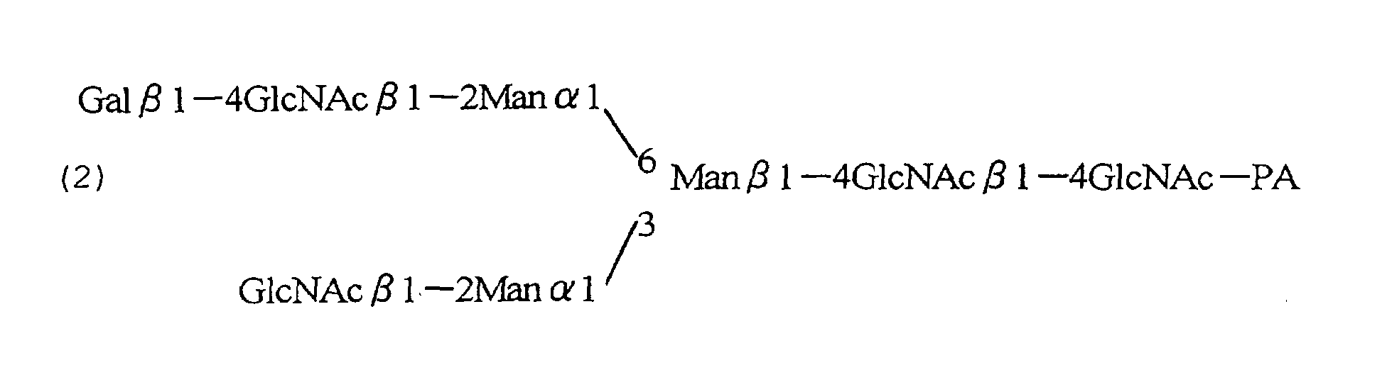


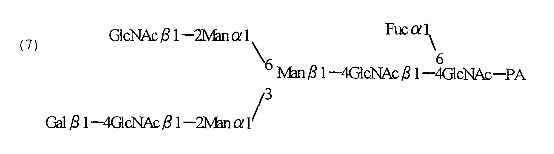
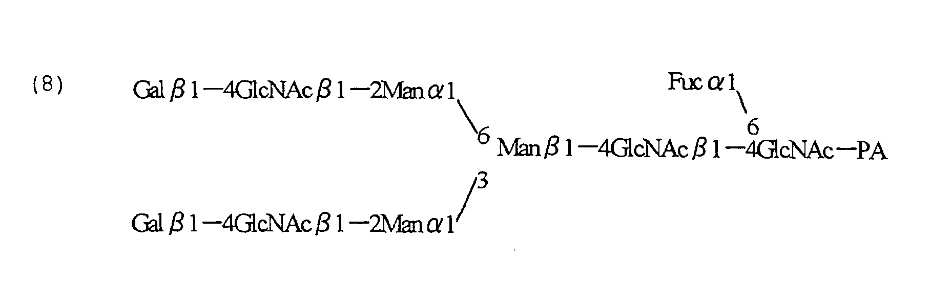
| Antibody | Ratio of α1,6-fucose-free complex biantennary sugar chain |
| NS0/ | 25% |
| NS/CCR4- | 48% |
| Antibody producing cells | α-1,6-Fucose-free complex biantennary sugar chain (%) |
| Clone 5-03 | 9 |
| Clone CHO/CCR4- | 48 |
| Clone CHO/CCR4- | 27 |
| Clone CHO/CCR4- | 8 |
| GMD | GEPP | FX | FUT8 | |
| Clone CHO/ | 1 | 1 | 1 | 1 |
| Clone 5-03 | 1.107 | 0.793 | 1.093 | 0.901 |
| Clone 5-03-derived LCA-resistant cell CHO/CCR4-LCA | 0.160 | 0.886 | 0.920 | 0.875 |
| Antibody producing cells | α1,6-Fucose-free sugar chain (%) |
| CHO/ | 9 |
| CHO/GD3-LCA-1 antibody | 42 |
| CHO/GD3-LCA-1 | 80 |


| Combination of primers used in Chinese hamster FX cDNA RACE PCR and size of PCR product | |||
| 5' RACE | FX-specific primers | Common primers | Size of PCR-amplified product |
| First | FXGSP1-1 | UPM (Universal primer mix) | |
| Second | FXGSP1-2 | NUP (Nested Universal primer) | 300 bp |
| 3' RACE | FX-specific primers | Common primers | Size of PCR-amplified product |
| First | FXGSP2-1 | UPM (Universal primer mix) | |
| Second | FXGSP2-2 | NUP (Nested Universal primer) | 1,100 bp |
| Combination of primers used in Chinese hamster GFPP cDNA RACE PCR and size of PCR products | |||
| 5'RACE | GFPP-specific primers | Common primers | Size of PCR-amplified product |
| First | GFPPGSP1-1 | UPM (Universal primer mix) | |
| Second | GFPPGSP1-2 | NUP (Nested Universal primer) | 1,100 bp |
| 3' RACE | GFPP-specific primers | Common primers | Size of PCR-amplified product |
| First | GFPPGSP2-1 | UPM (Universal primer mix) | |
| Second | GFPPGSP2-2 | NUP (Nested Universal primer) | 1,400 bp |


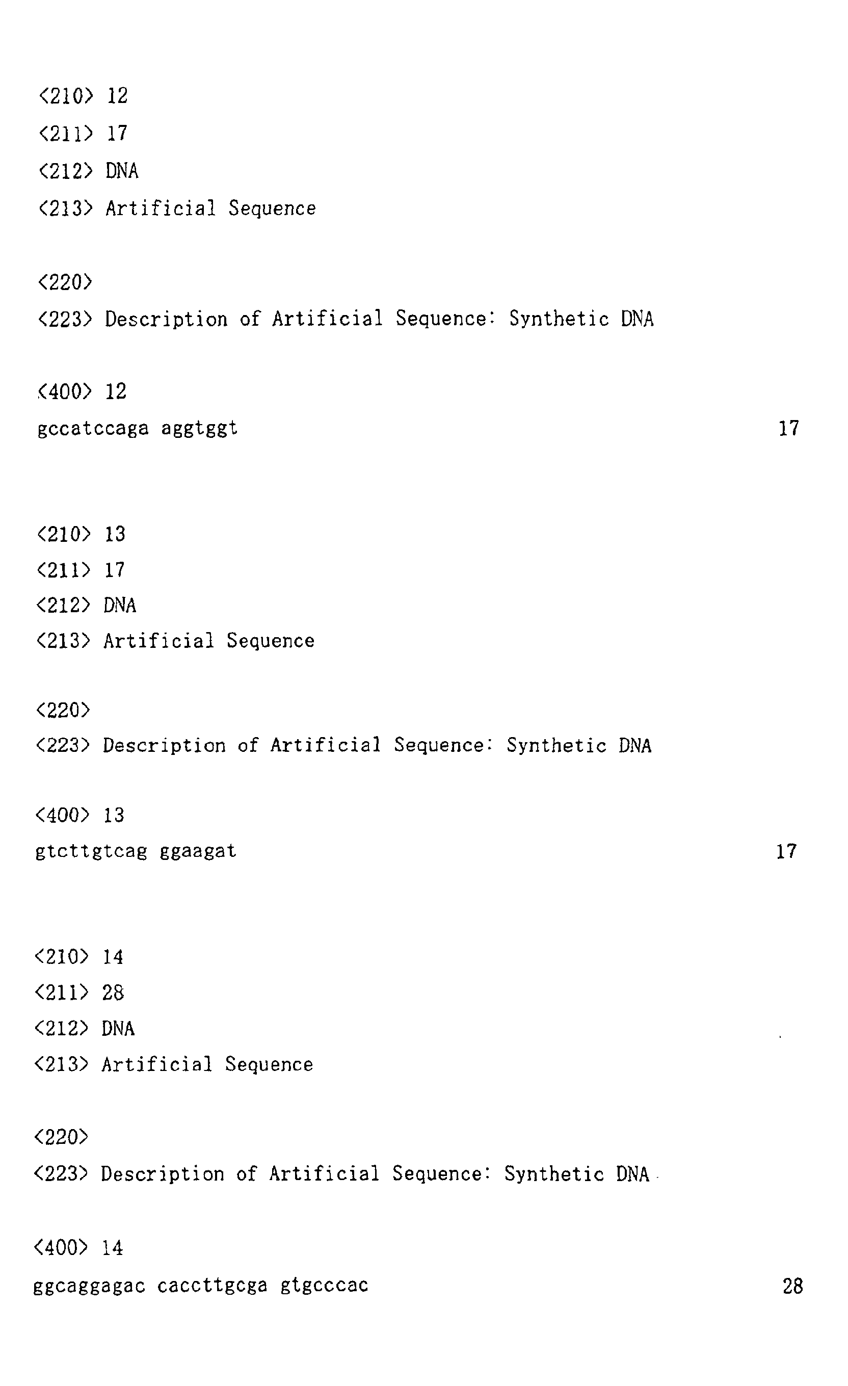
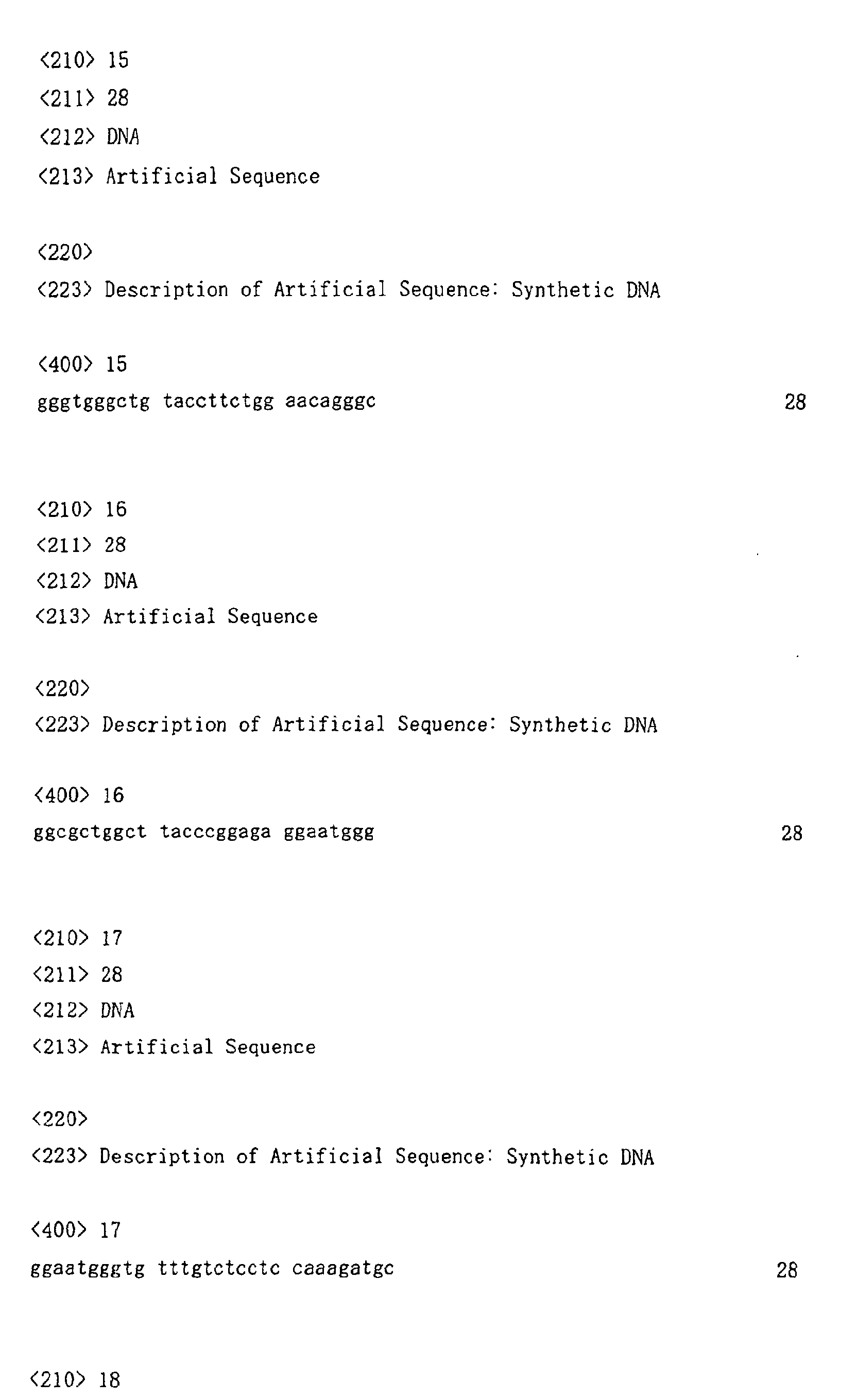
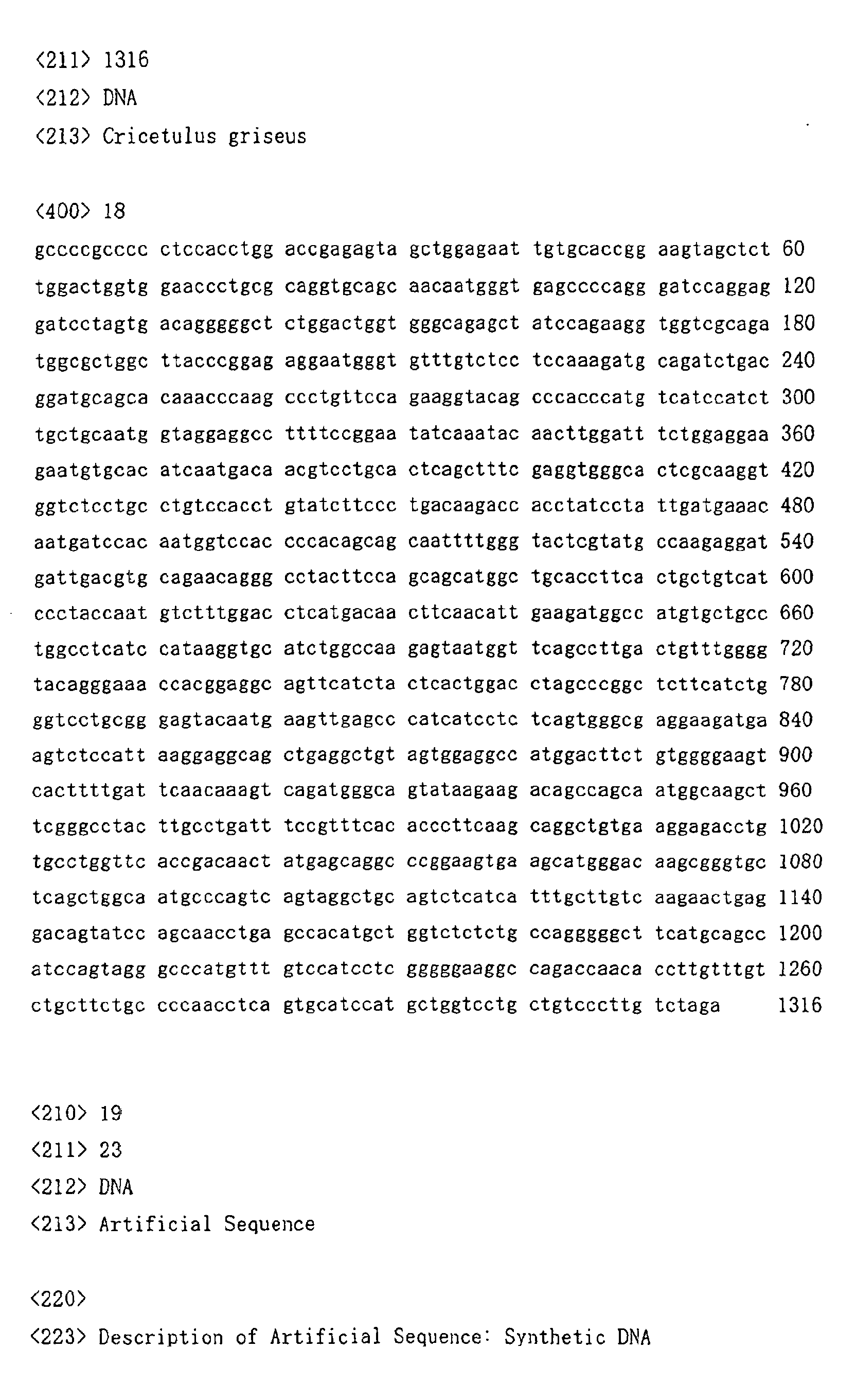

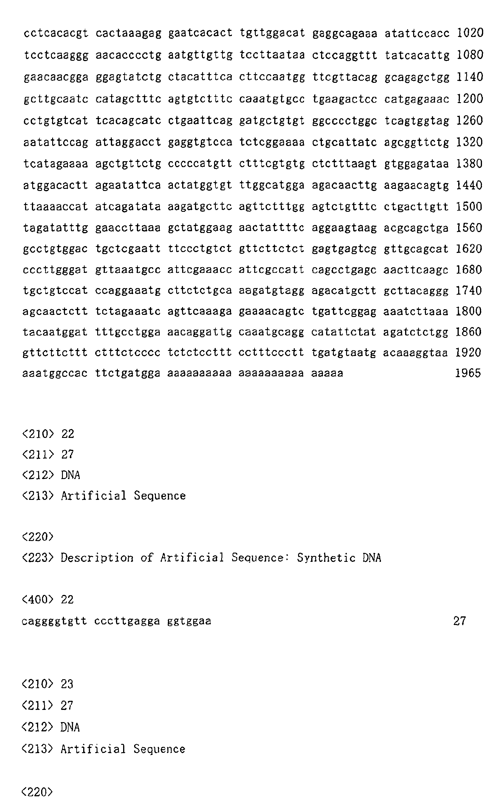
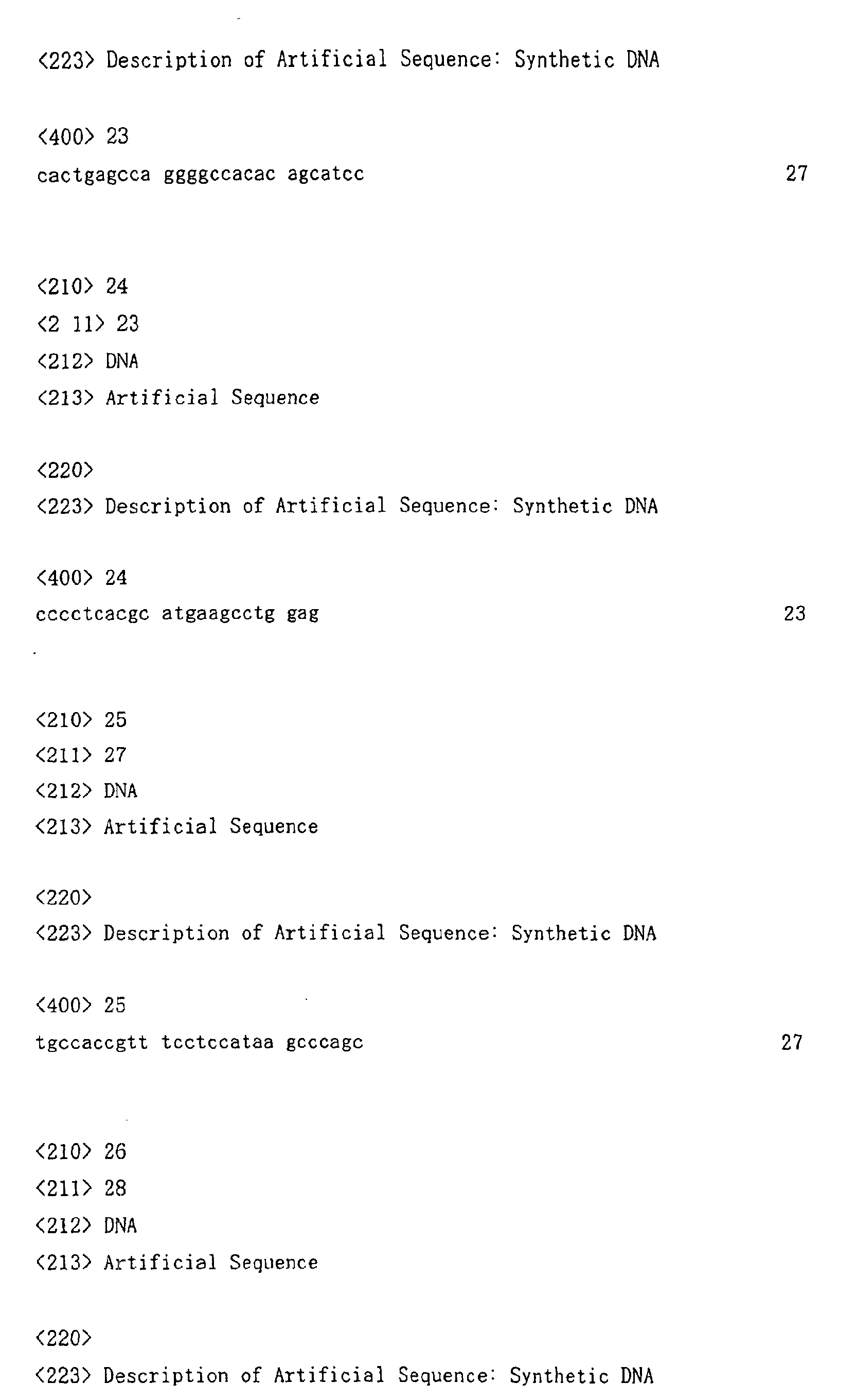
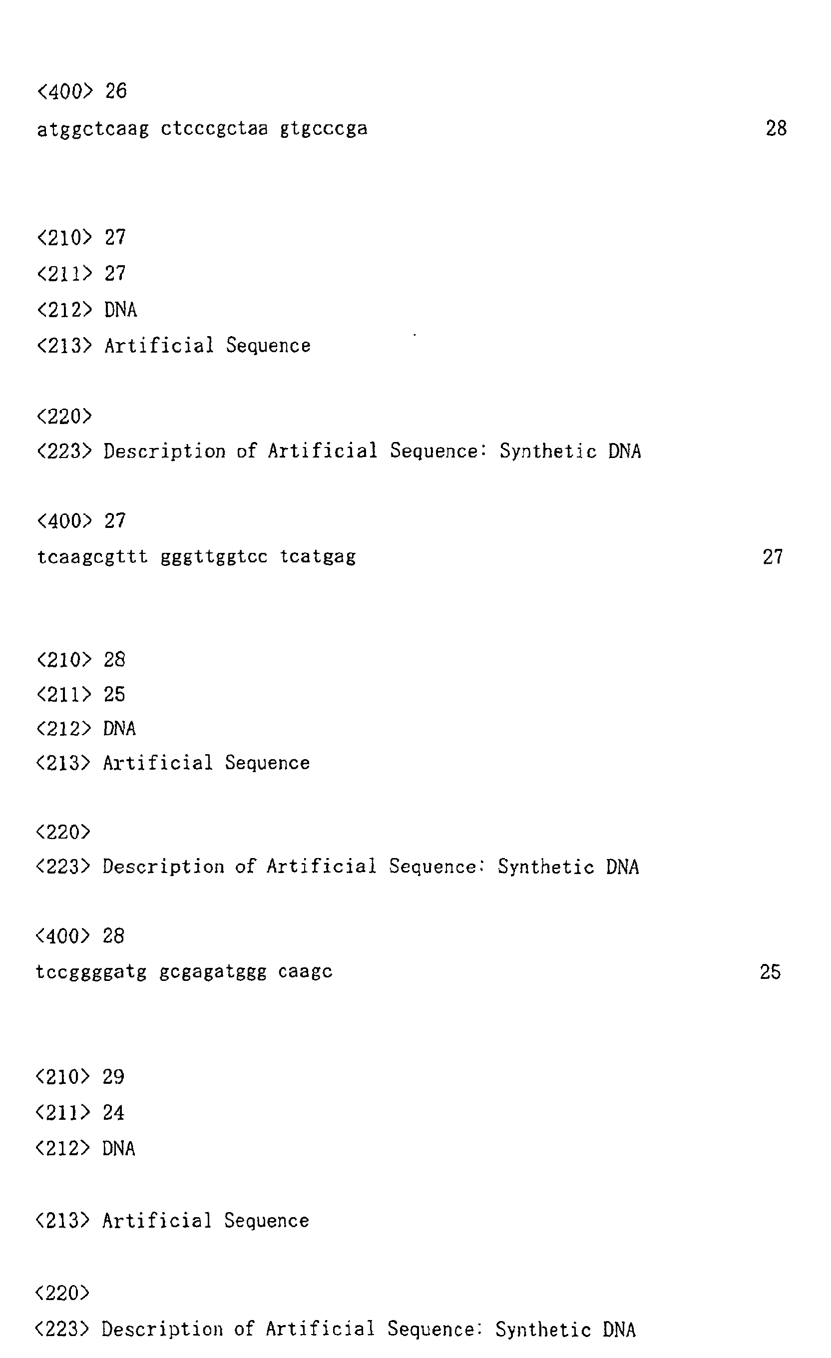
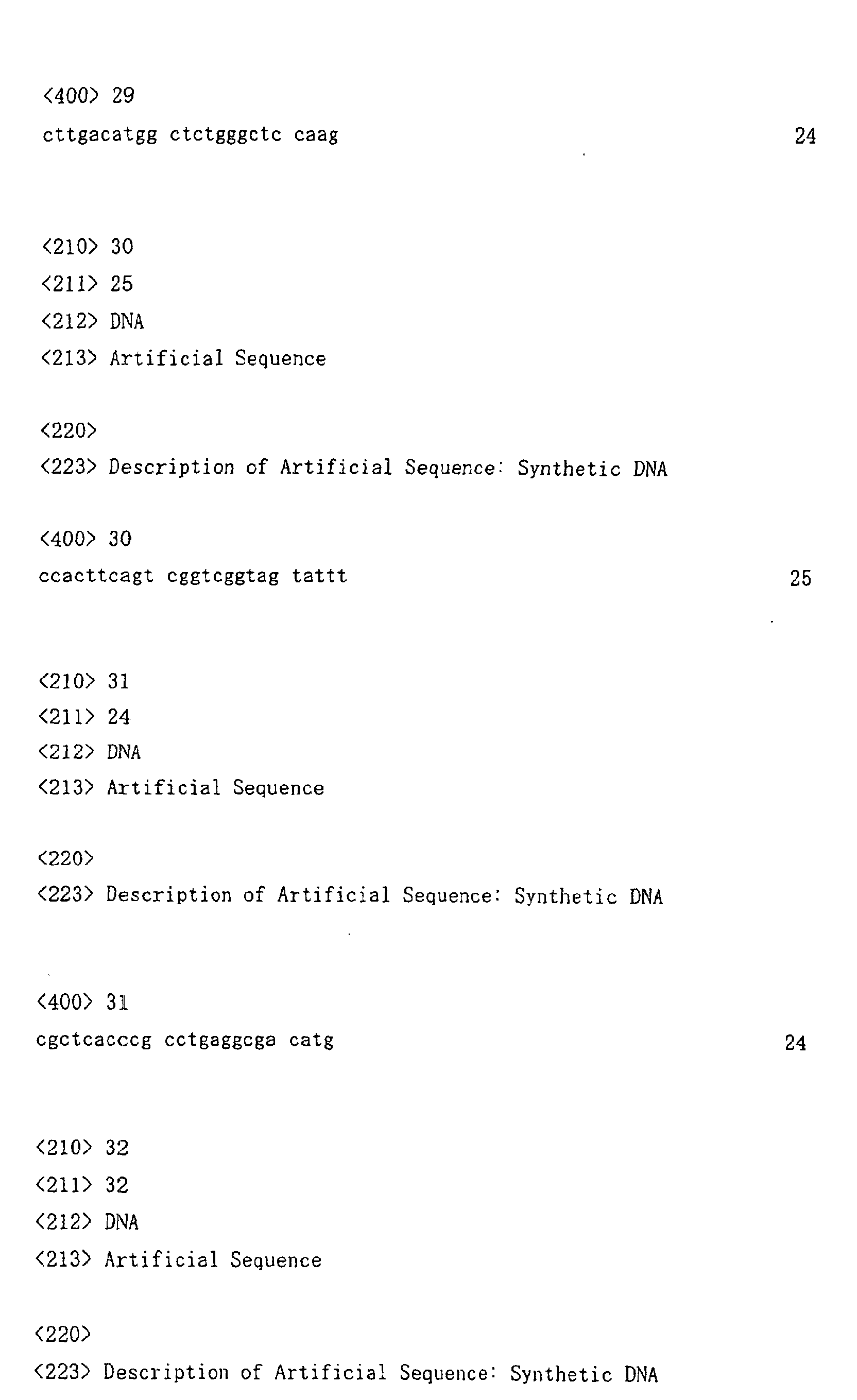

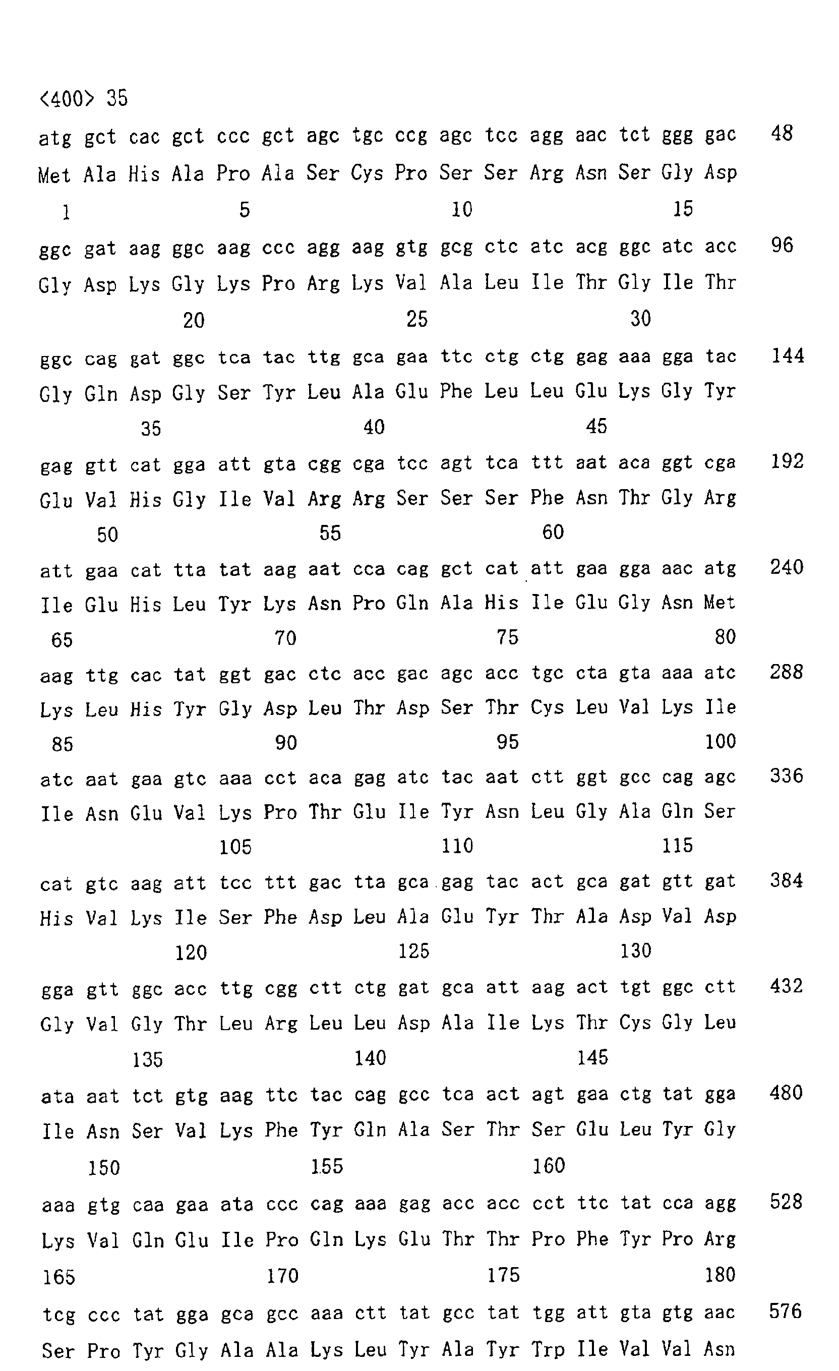
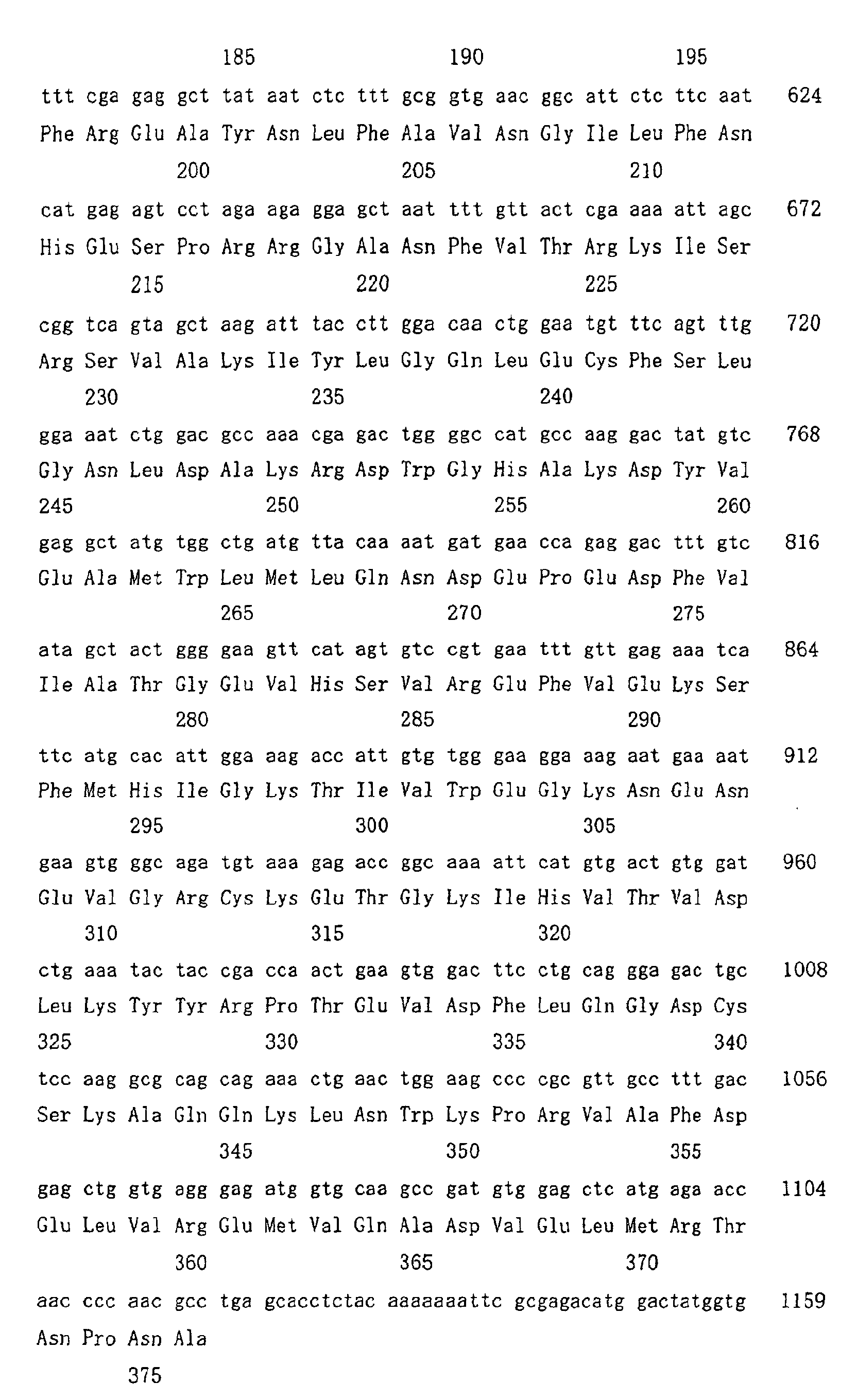
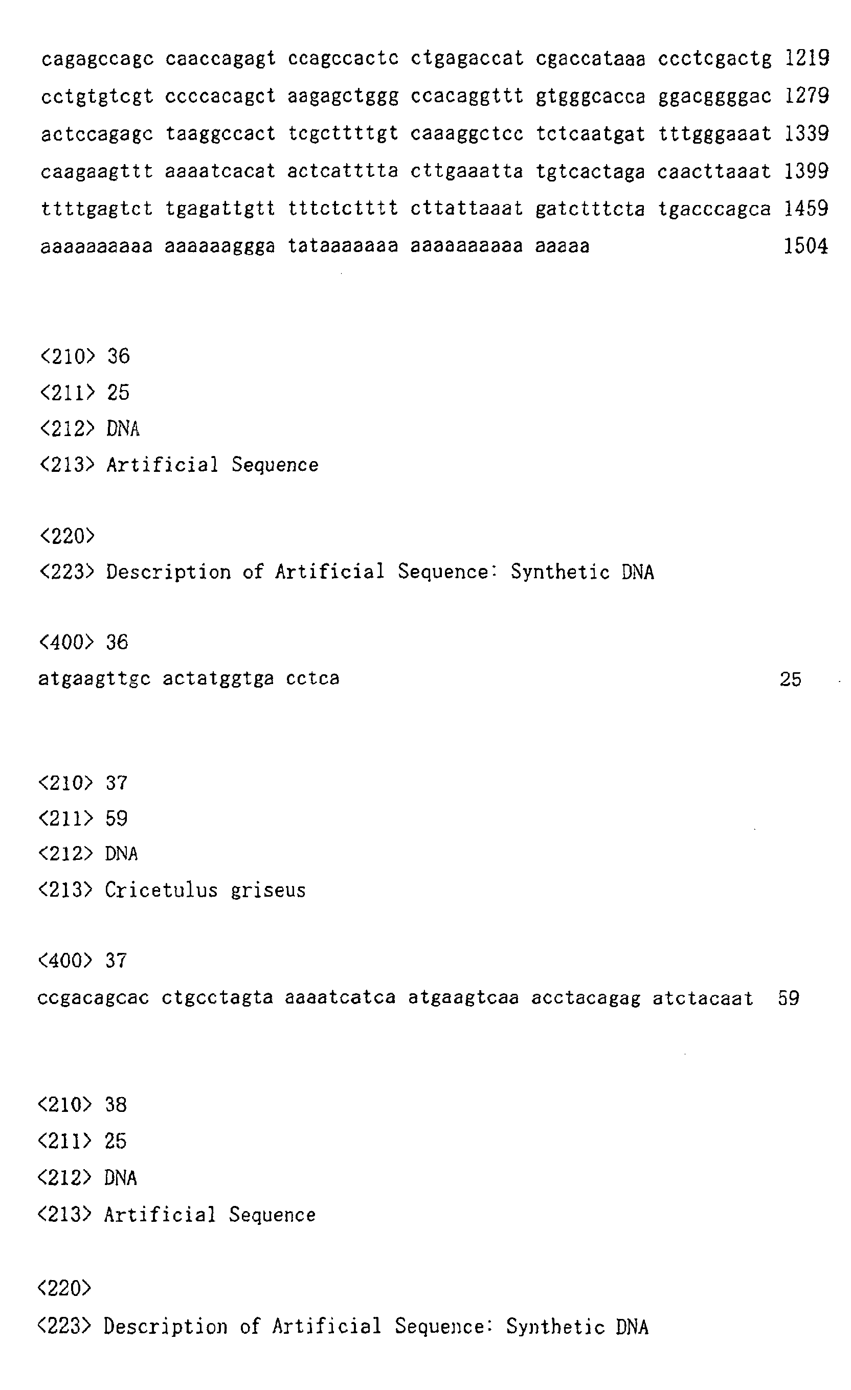


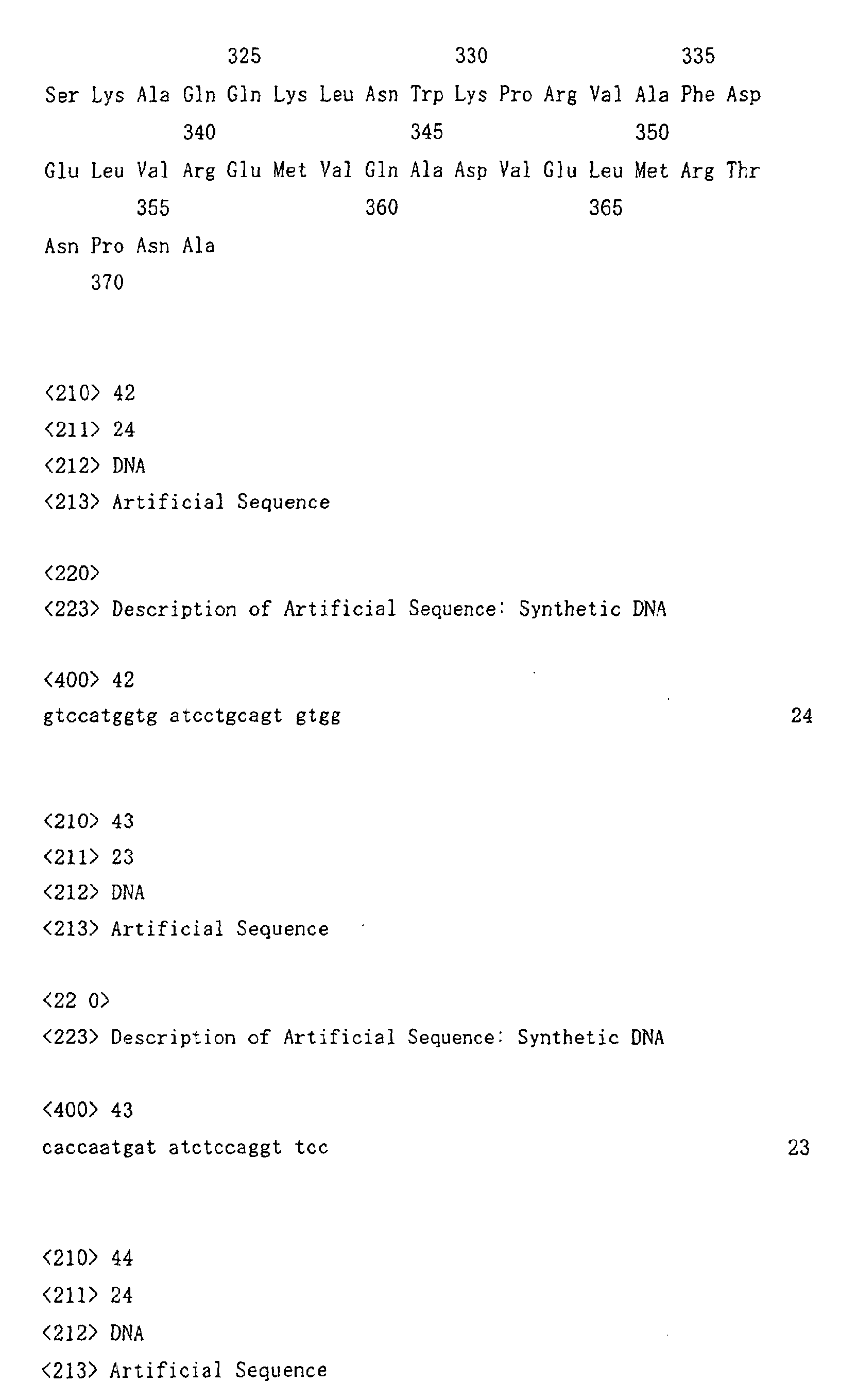
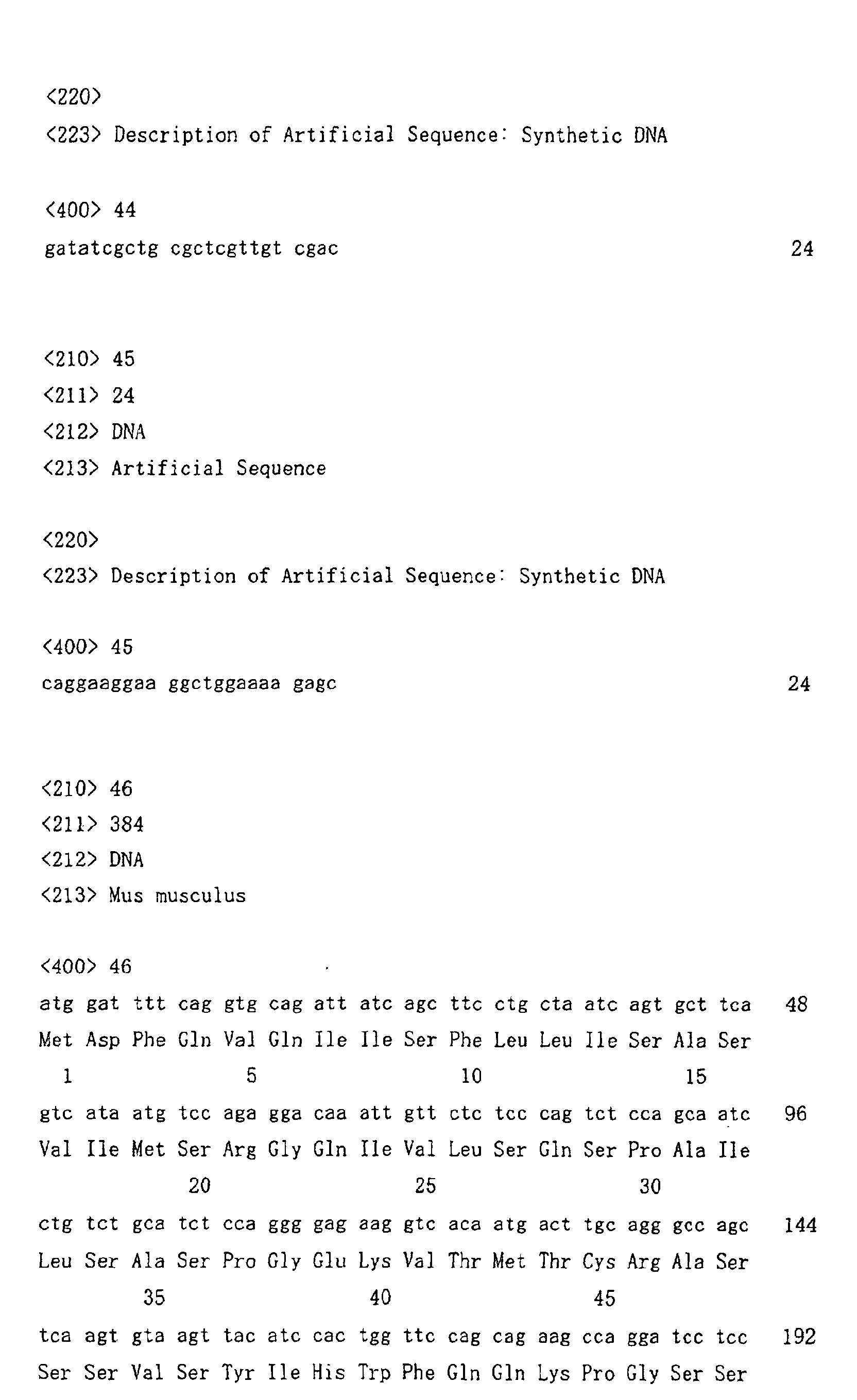

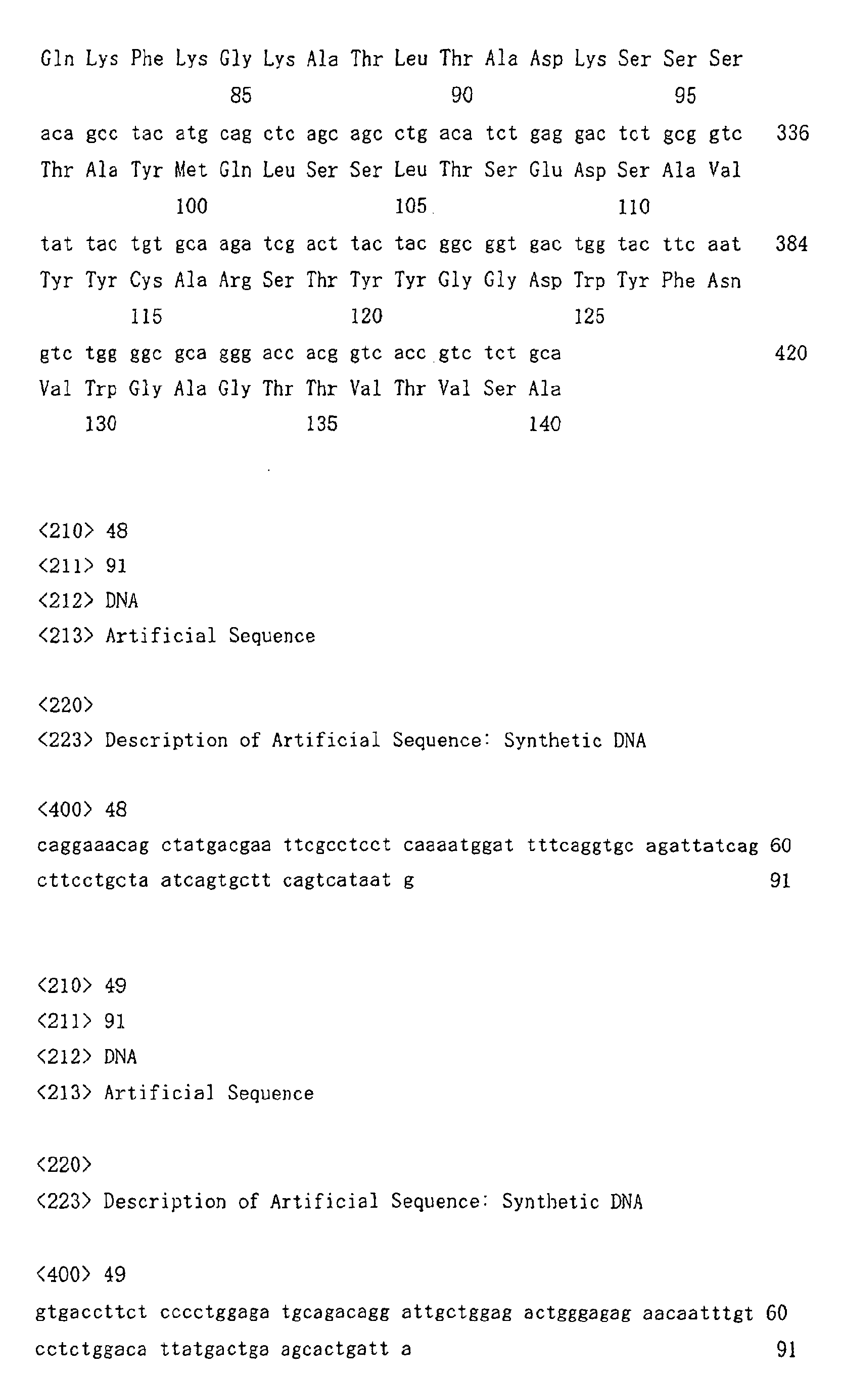
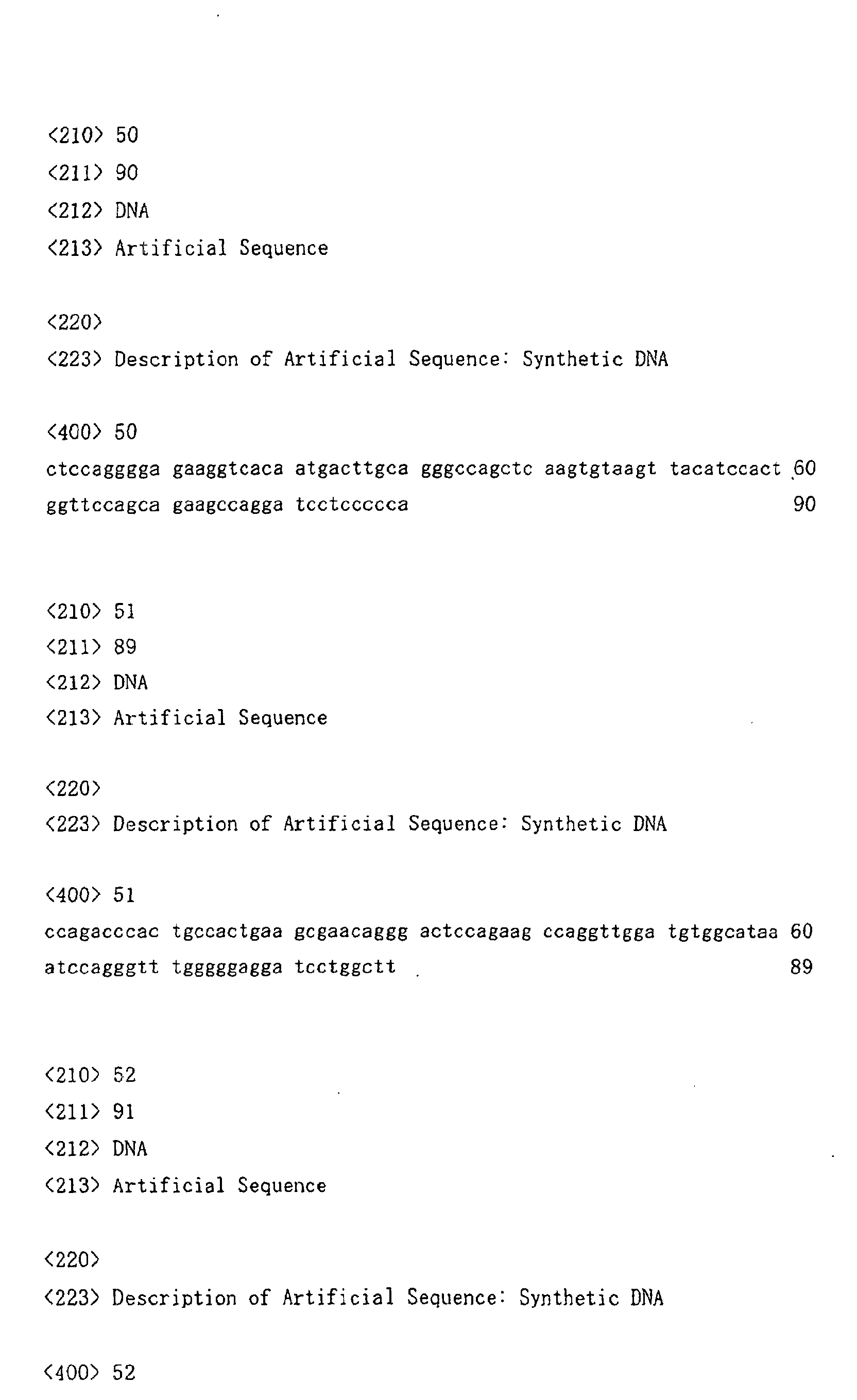
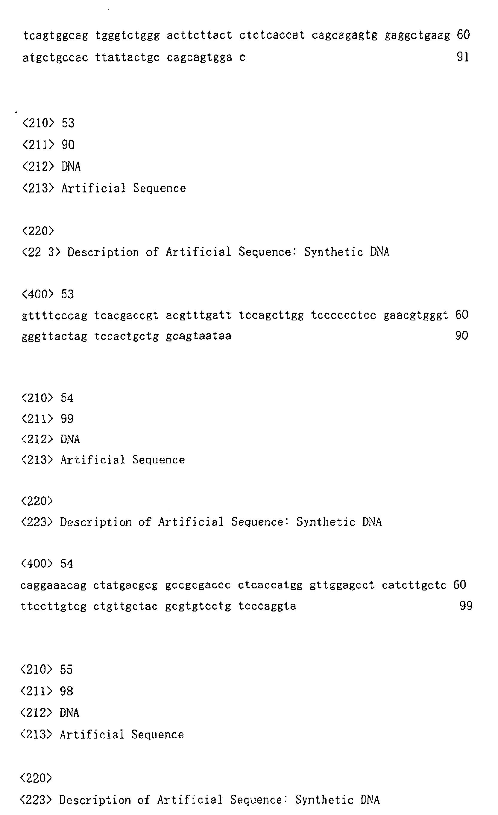

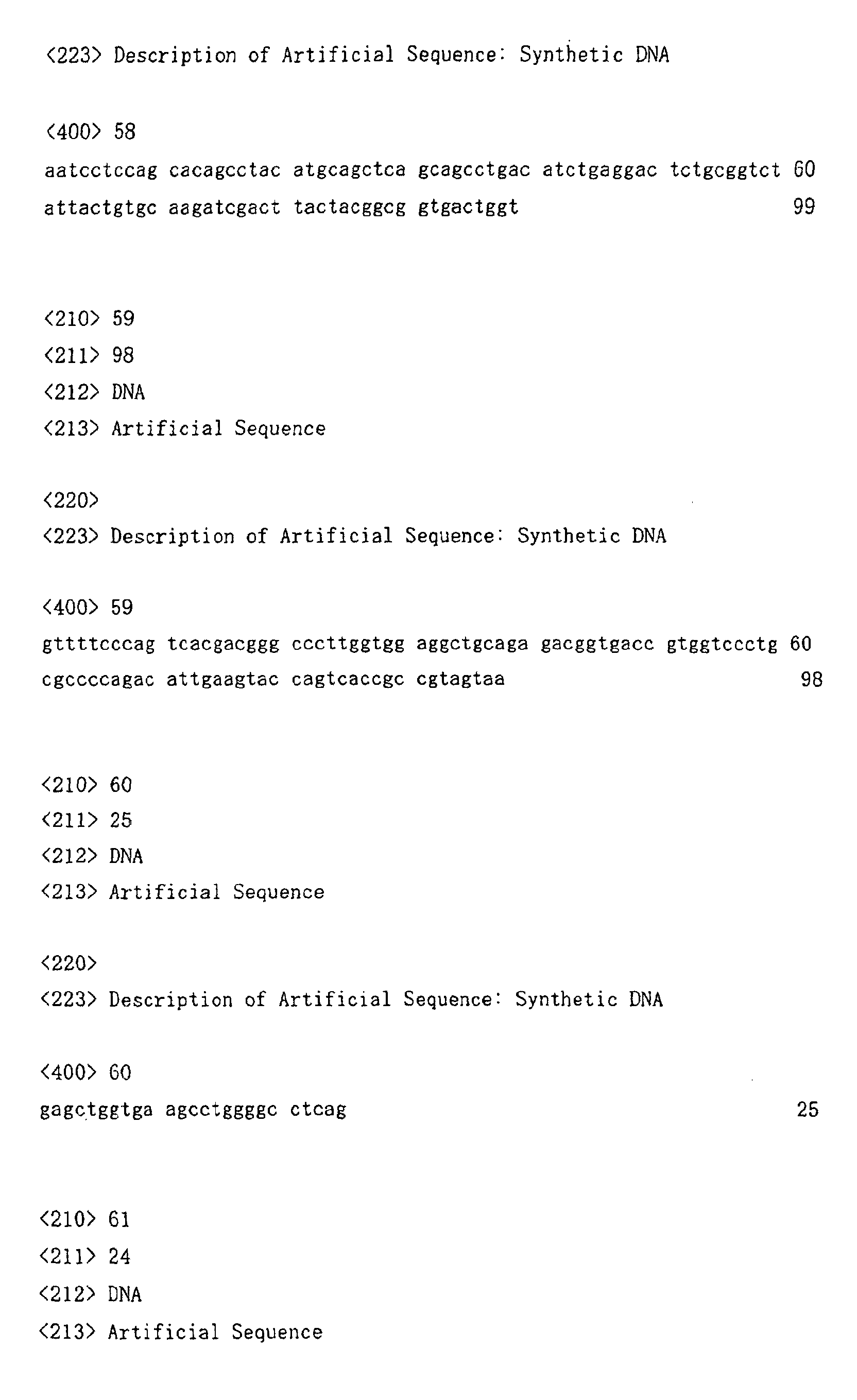
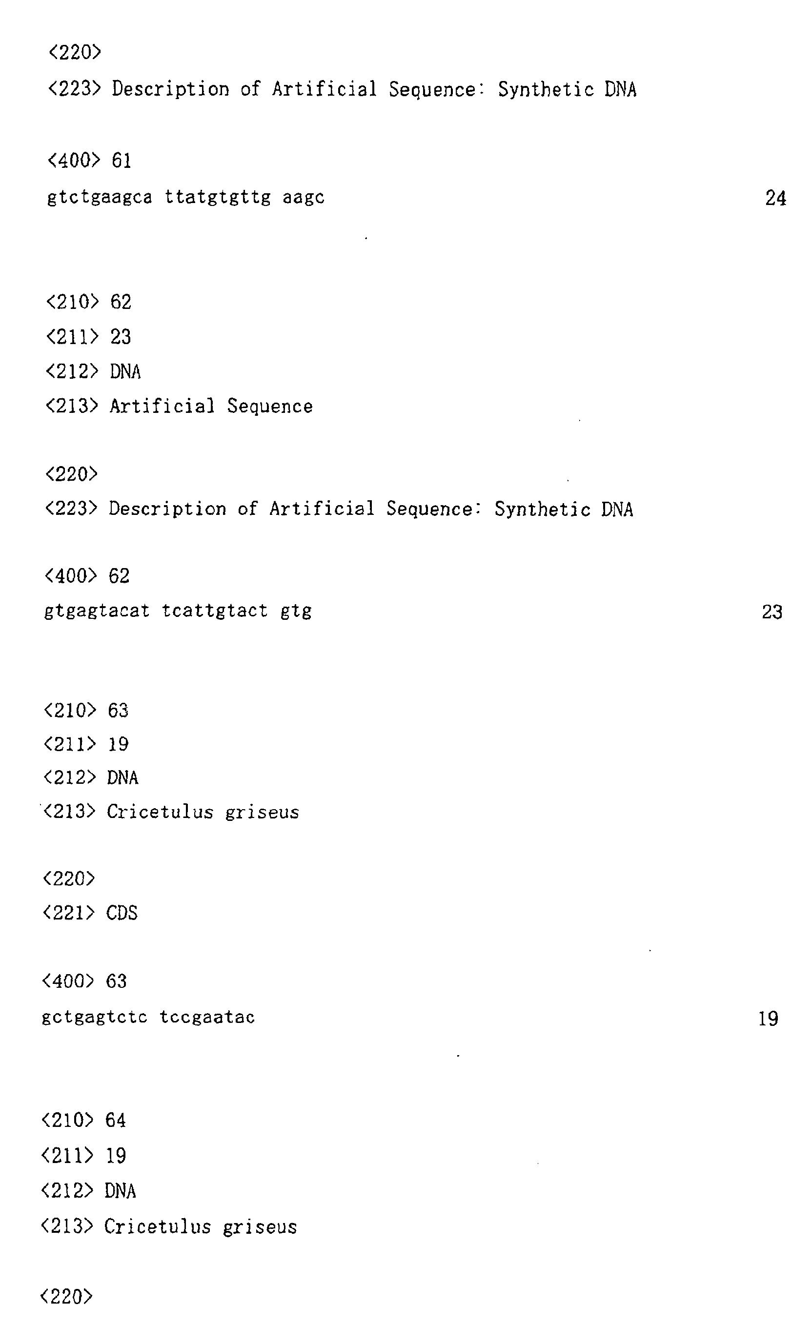


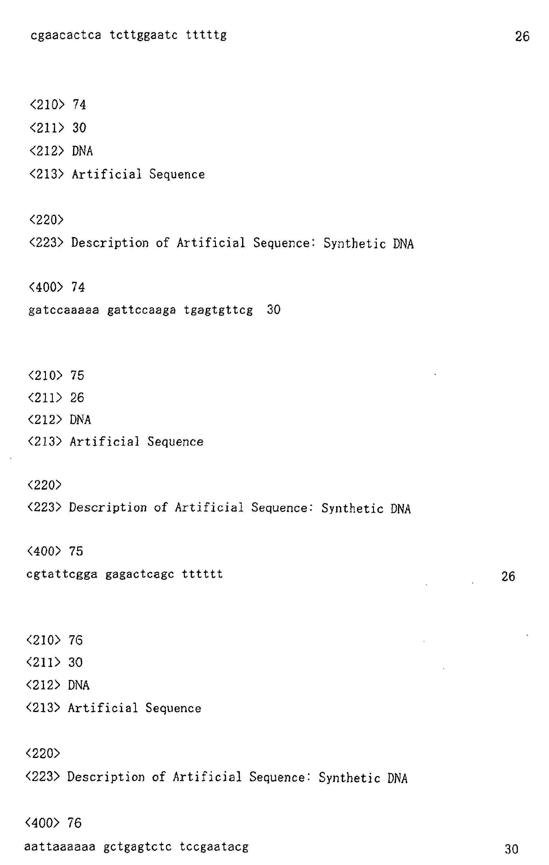


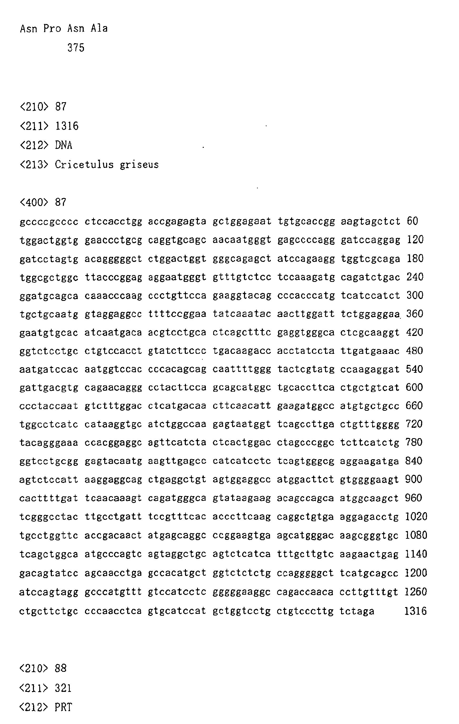


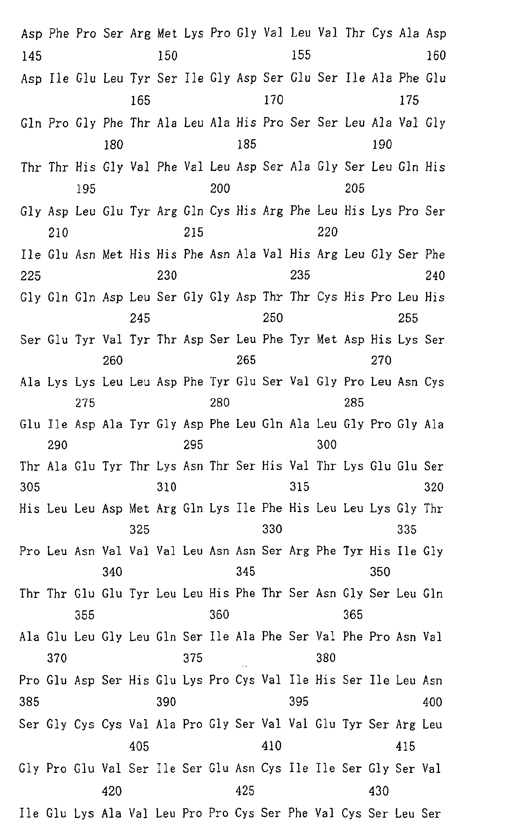

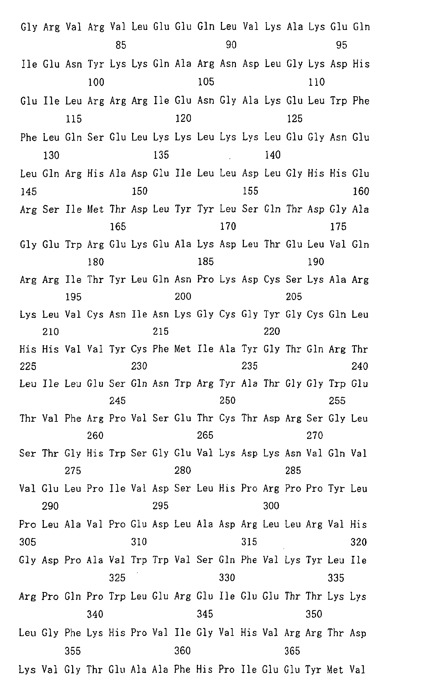

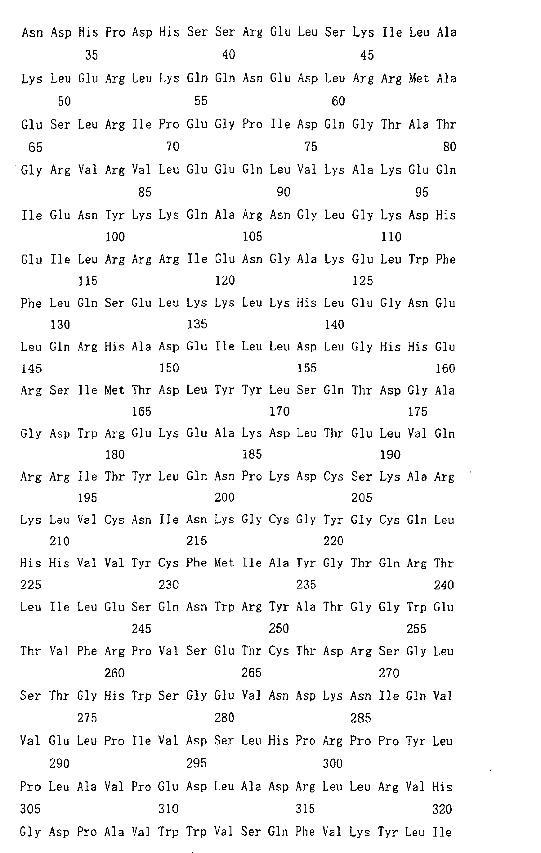

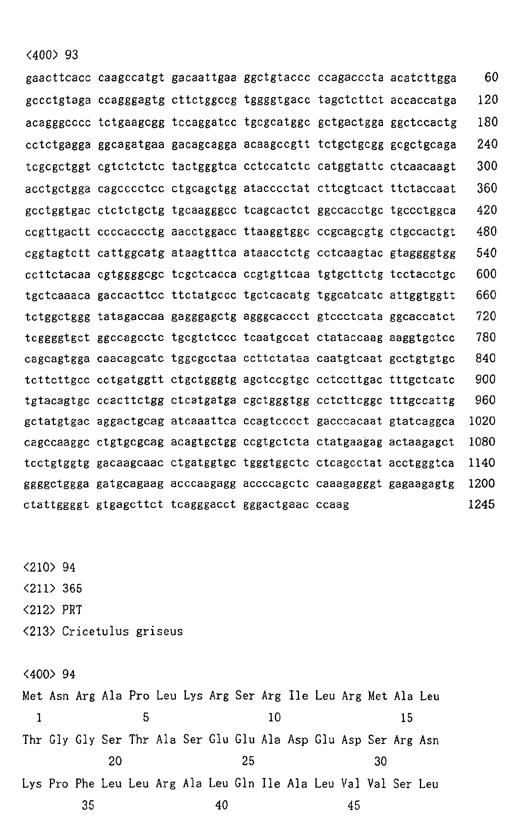

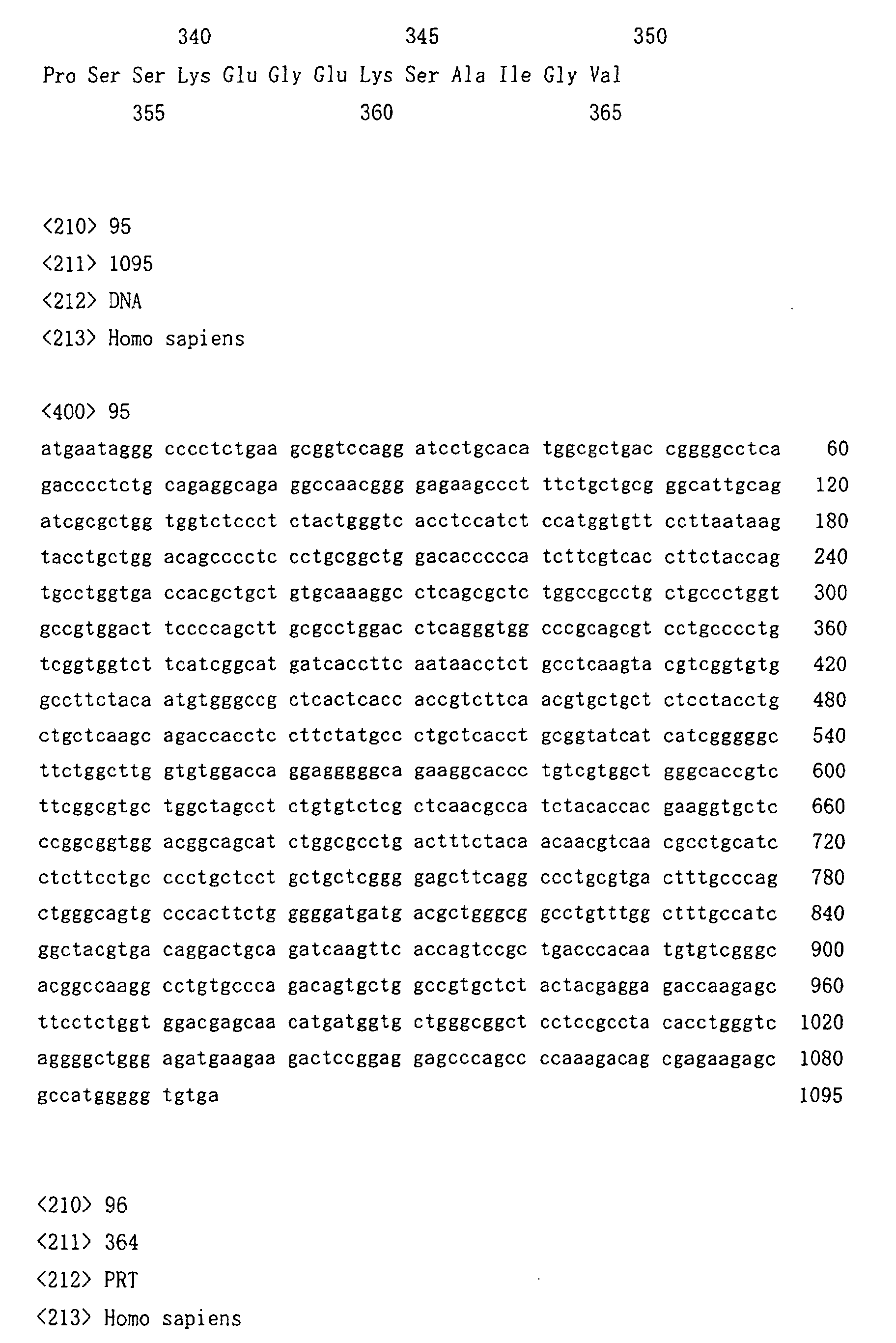

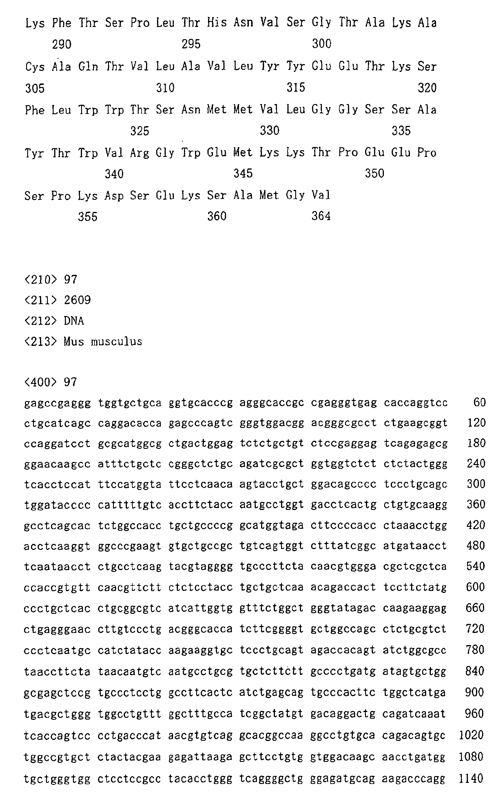

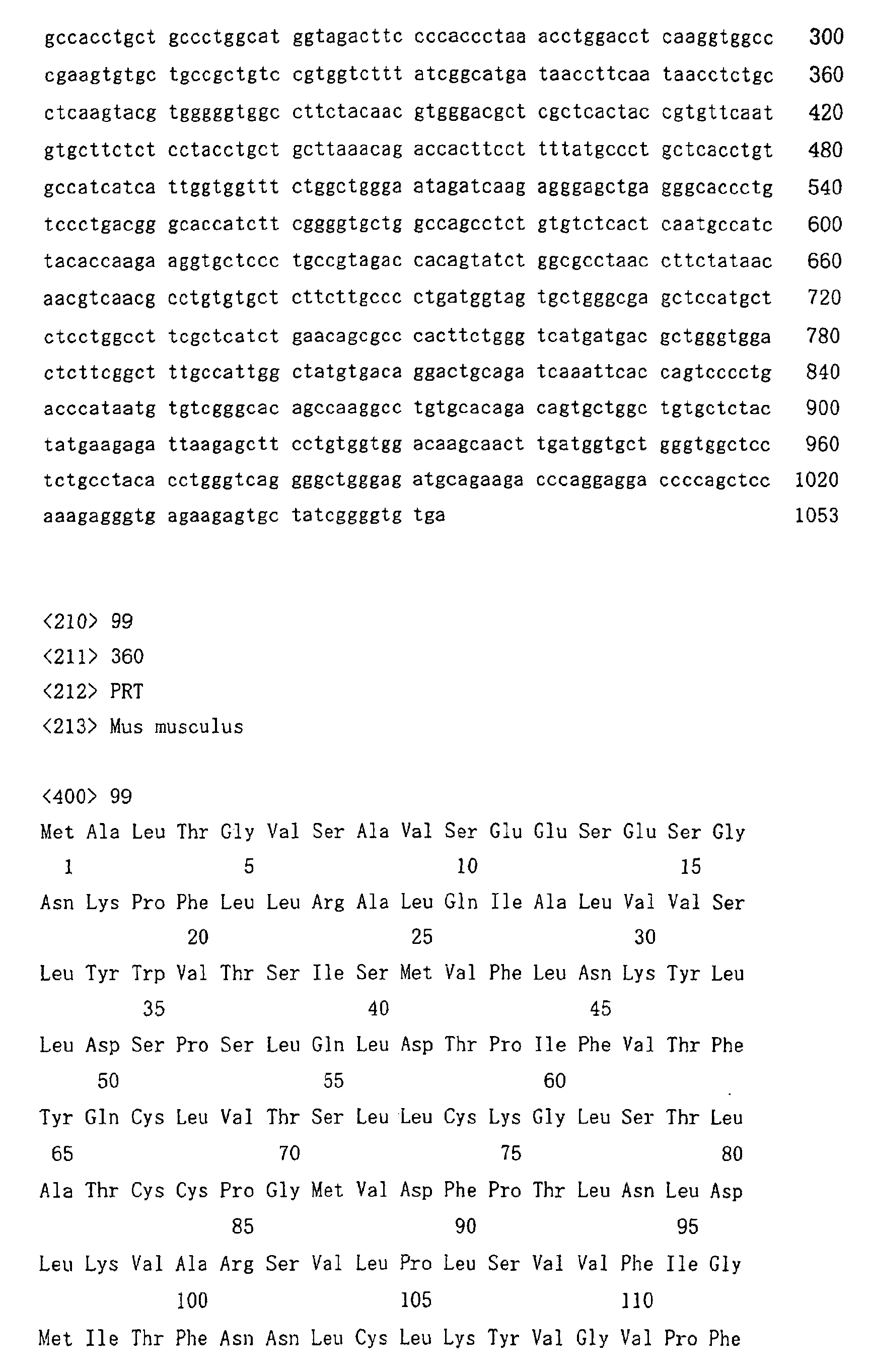
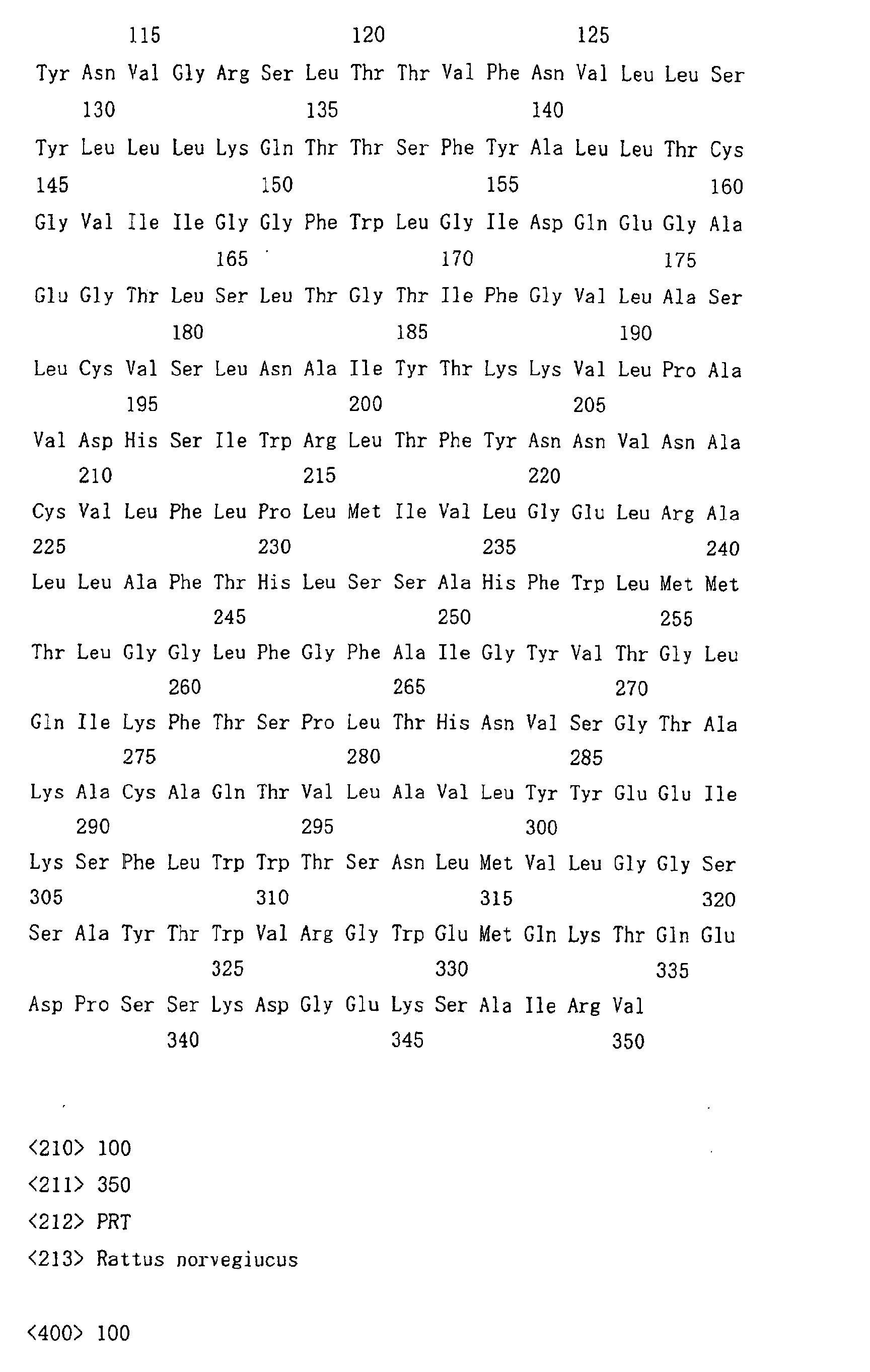
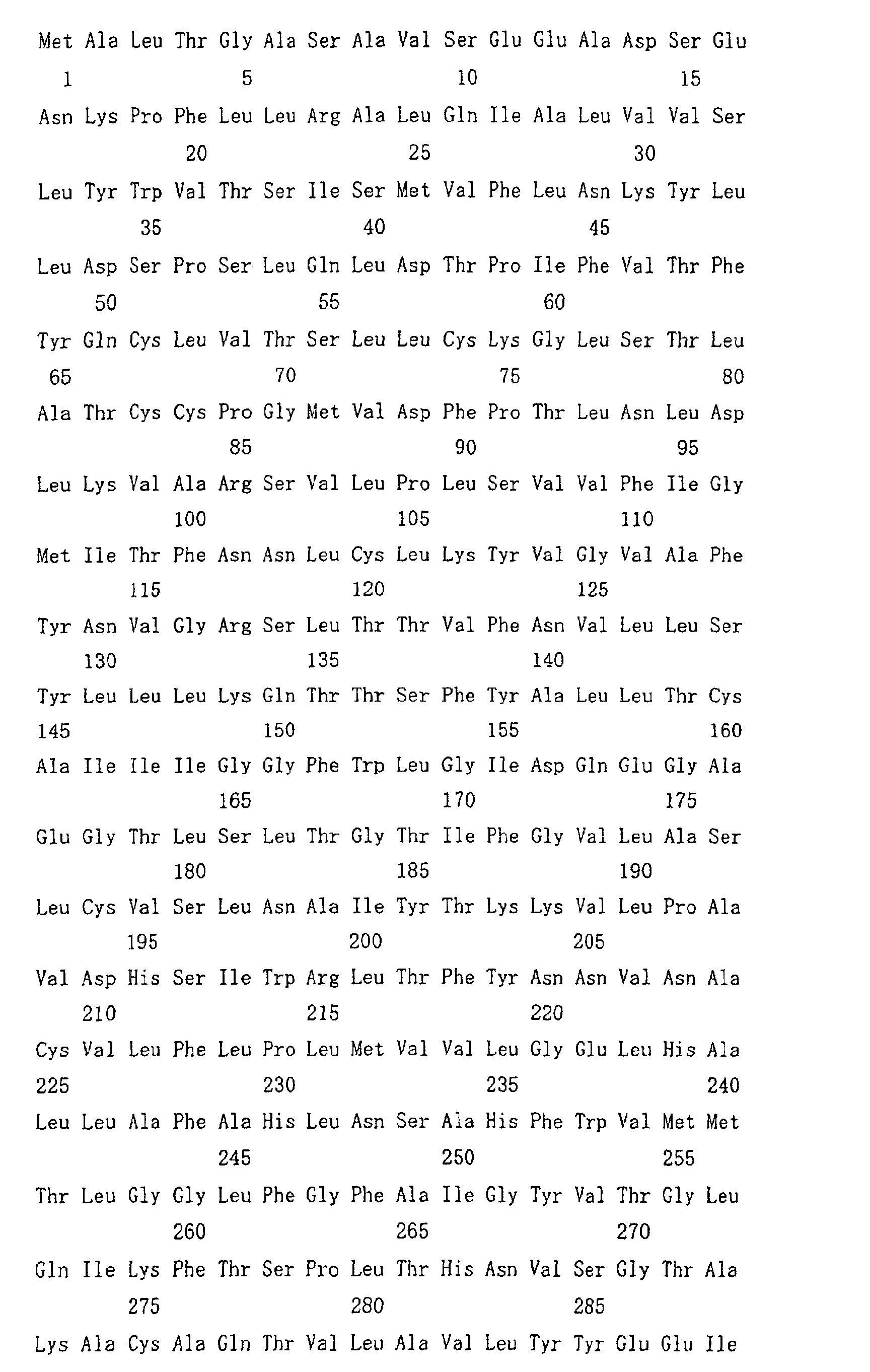

Claims (81)
- A process for producing an antibody composition using a cell, which comprises using a cell resistant to a lectin which recognizes a sugar chain in which 1-position of fucose is bound to 6-position of N-acetylglucosamine in the reducing end through α-bond in a complex N-glycoside-linked sugar chain.
- The process according to claim 1, wherein the cell is a cell in which the activity of a protein selected from the group consisting of the following (a), (b) and (c) is decreased or deleted:(a) an enzyme protein relating to synthesis of an intracellular sugar nucleotide, GDP-fucose;(b) an enzyme protein relating to modification of a sugar chain in which 1-position of fucose is bound to 6-position of N-acetylglucosamine in the reducing end through α-bond in a complex N-glycoside-linked sugar chain;(c) a protein relating to transport of an intracellular sugar nucleotide, GDP-fucose, to the Golgi body.
- The process according to claim 2, wherein the enzyme protein relating to modification of a sugar chain in which 1-position of fucose is bound to 6-position of N-acetylglucosamine in the reducing end through α-bond in a complex N-glycoside-linked sugar chain is α1,6-fucosyltransferase.
- The process according to claim 2 or 3, wherein the activity of the protein is decreased or deleted by a technique selected from the group consisting of (a) to (d):(a) a gene disruption technique which comprises targeting a gene encoding the protein;(b) a technique for introducing a dominant negative mutant of a gene encoding the protein;(c) a technique for introducing mutation into the protein;(d) a technique for suppressing transcription and/or translation of a gene encoding the protein.
- The process according to claim 4), wherein the technique for suppressing transcription and/or translation of a gene encoding the protein is an RNAi method.
- The process according to any one of claims 3 to 5, wherein the α1,6-fucosyltransferase is a protein encoded by a DNA selected from the group consisting of (a) to (h) or a protein selected from the group consisting of the following (i) to (n):(a) a DNA comprising the nucleotide sequence represented by SEQ ID NO:79;(b) a DNA comprising the nucleotide sequence represented by SEQ ID NO:80;(c) a DNA comprising the nucleotide sequence represented by SEQ ID NO:81;(d) a DNA comprising the nucleotide sequence represented by SEQ ID NO:82;(e) a DNA which hybridizes with a DNA comprising the nucleotide sequence represented by SEQ ID NO:79 under stringent conditions and encodes a protein having an α1,6-fucosyltransferase activity;(f) a DNA which hybridizes with a DNA comprising the nucleotide sequence represented by SEQ ID NO:80 under stringent conditions and encodes a protein having an α1,6-fucosyltransferase activity;(g) a DNA which hybridizes with a DNA comprising the nucleotide sequence represented by SEQ ID NO:81 under stringent conditions and encodes a protein having an α1,6-fucosyltransferase activity;(h) a DNA which hybridizes with a DNA comprising the nucleotide sequence represented by SEQ ID NO:82 under stringent conditions and encodes a protein having an α1,6-fucosyltransferase activity;(i) a protein comprising the amino acid sequence represented by SEQ ID NO:91;(j) a protein comprising the amino acid sequence represented by SEQ ID NO:92;(k) a protein which comprises an amino acid sequence in which at least one amino acid is deleted, substituted, inserted and/or added in the amino acid sequence represented by SEQ ID NO:91 and has an α1,6-fucosyltransferase activity;(l) a protein which comprises an amino acid sequence in which at least one amino acid is deleted, substituted, inserted and/or added in the amino acid sequence represented by SEQ ID NO:92 and has an α1,6-fucosyltransferase activity;(m) a protein which comprises an amino acid sequence having a homology of at least 80% with the amino acid sequence represented by SEQ ID NO:91 and has an α1,6-fucosyltransferase activity;(n) a protein which comprises an amino acid sequence having a homology of at least 80% with the amino acid sequence represented by SEQ ID NO:92 and has an α1,6-fucosyltransferase activity.
- The process according to claim 5 or 6, wherein the RNAi method is a method in which a double-stranded RNA comprising an RNA selected from the group consisting of (a) to (d) and its complementary RNA and being capable of decreasing the amount of mRNA of α1,6-fucosyltransferase is introduced into or expressed in the cell:(a) an RNA corresponding to a DNA comprising a nucleotide sequence of continuous 10 to 30 nucleotides in the nucleotide sequence represented by SEQ ID NO:79;(b) an RNA corresponding to a DNA comprising a nucleotide sequence of continuous 10 to 30 nucleotides in the nucleotide sequence represented by SEQ ID NO:80;(c) an RNA corresponding to a DNA comprising a nucleotide sequence of continuous 10 to 30 nucleotides in the nucleotide sequence represented by SEQ ID NO:81;(d) an RNA corresponding to a DNA comprising a nucleotide sequence of continuous 10 to 30 nucleotides in the nucleotide sequence represented by SEQ ID NO:82.
- The process according to claim 7, wherein the double-stranded RNA comprising an RNA selected from the group consisting of (a) to (d) and its complementary RNA is introduced into or expressed in the cell to thereby decrease the amount of mRNA of α1,6-fucosyltransferase and provide the cell with the resistance to a lectin which recognizes a sugar chain in which 1-position of fucose is bound to 6-position of N-acetylglucosamine in the reducing end through α-bond in a complex N-glycoside-linked sugar chain:(a) an RNA comprising the nucleotide sequence represented by SEQ ID NO:83;(b) an RNA comprising the nucleotide sequence represented by SEQ ID NO:84;(c) an RNA which comprises a nucleotide sequence in which one or a few nucleotides are deleted or added in the nucleotide sequence represented by SEQ ID NO:83 and has substantially the same RNAi activity as the RNA represented by SEQ ID NO:83;(d) an RNA which comprises a nucleotide sequence in which one or a few nucleotides are deleted or added in the nucleotide sequence represented by SEQ ID NO:84 and has substantially the same RNAi activity as the RNA represented by SEQ ID NO:94.
- The process according to claim 7 or 8, wherein the double-stranded RNA is introduced into the cell by using a vector into which a DNA corresponding to the RNA according to claim 7 or 8 and its complementary DNA are inserted.
- The process according to any one of claims 1 to 9, wherein the cell is resistant to at least one lectin selected from the group consisting of the following (a) to (d):(a) a Lens culinaris lectin;(b) a Pisum sativum lectin;(c) a Vicia faba lectin;(d) an Aleuria aurantia lectin.
- The process according to any one of claims 1 to 10, wherein the cell is a cell into which a gene encoding an antibody molecule is introduced.
- The process according to claim 11, wherein the antibody molecule is selected from the group consisting of the following (a) to (d):(a) a human antibody;(b) a humanized antibody;(c) an antibody fragment comprising the Fc region of (a) or (b);(d) a fusion protein comprising the Fc region of (a) or (b).
- The process according to claim 11 or 12, wherein the antibody molecule belongs to an IgG class.
- The process according to any one of claims 1 to 13, wherein the antibody composition has a higher antibody-dependent cell-mediated cytotoxic activity than an antibody composition produced by its parent cell.
- The process according to claim 14, wherein the antibody composition having a higher antibody-dependent cell-mediated cytotoxic activity has a higher ratio of a sugar chain in which fucose is not bound to N-acetylglucosamine in the reducing end in the sugar chain among total complex N-glycoside-linked sugar chains bound to the Fc region in the antibody composition than an antibody composition produced by its parent cell.
- The process according to claim 15, wherein the ratio of a sugar chain in which fucose is not bound to N-acetylglucosamine in the reducing end through α-bond is 20% or more of total complex N-glycoside-linked sugar chains bound to the Fc region in the antibody composition.
- The process according to claim 15 or 16, wherein the sugar chain in which fucose is not bound is a sugar chain in which 1-position of fucose is not bound to 6-position of N-acetylglucosamine in the reducing end in the complex N-glycoside-linked sugar chain through α-bond.
- The process according to any one of claims 1 to 17, wherein the cell is selected from the group consisting of a yeast, an animal cell, an insect cell and a plant cell.
- The process according to any one of claims 1 to 18, wherein the cell is a mouse myeloma cell.
- The process according to claim 19, wherein the mouse myeloma cell is NS0 cell or SP2/0-Ag14 cell.
- The process according to any one of claims 1 to 18, wherein the cell is selected from the group consisting of the following (a) to (g):(a) a CHO cell derived from a Chinese hamster ovary tissue;(b) a rat myeloma cell line YB2/3HL.P2.G11.16Ag.20 cell;(c) a BHK cell derived from a Syrian hamster kidney tissue;(d) a hybridoma cell which produces an antibody;(e) a human leukemic cell line Namalwa cell;(f) an embryonic stem cell;(g) a fertilized egg cell.
- A cell into which a gene encoding an antibody composition is introduced, which is resistant to a lectin which recognizes a sugar chain in which 1-position of fucose is bound to 6-position of N-acetylglucosamine in the reducing end through α-bond in a complex N-glycoside-linked sugar chain.
- The cell according to claim 22, wherein the activity of a protein selected from the group consisting of the following (a), (b) and (c) is more decreased or deleted than its parent cell:(a) an enzyme protein relating to synthesis of an intracellular sugar nucleotide, GDP-fucose;(b) an enzyme protein relating to modification of a sugar chain in which 1-position of fucose is bound to 6-position of N-acetylglucosamine in the reducing end through α-bond in a complex N-glycoside-linked sugar chain;(c) a protein relating to transport of an intracellular sugar nucleotide, GDP-fucose, to the Golgi body.
- The cell according to claim 23, wherein the enzyme protein relating to modification of a sugar chain in which 1-position of fucose is bound to 6-position of N-acetylglucosamine in the reducing end through α-bond in a complex N-glycoside-linked sugar chain is α1,6-fucosyltransferase.
- The cell according to claim 23 or 24, wherein the activity of the protein is decreased or deleted by a technique selected from the group consisting of (a) to (d):(a) a gene disruption technique which comprises targeting a gene encoding the protein;(b) a technique for introducing a dominant negative mutant of a gene encoding the protein;(c) a technique for introducing mutation into the protein;(d) a technique for suppressing transcription and/or translation of a gene encoding the protein.
- The cell according to claim 25, wherein the technique for suppressing transcription and/or translation of a gene encoding the protein is an RNAi method.
- The cell according to any one of claims 24 to 26, wherein the α1,6-fucosyltransferase is a protein encoded by a DNA selected from the group consisting of (a) to (h) or a protein selected from the group consisting of the following (i) to (n):(a) a DNA comprising the nucleotide sequence represented by SEQ ID NO:79;(b) a DNA comprising the nucleotide sequence represented by SEQ ID NO:80;(c) a DNA comprising the nucleotide sequence represented by SEQ ID NO:81;(d) a DNA comprising the nucleotide sequence represented by SEQ ID NO:82;(e) a DNA which hybridizes with a DNA comprising the nucleotide sequence represented by SEQ ID NO:79 under stringent conditions and encodes a protein having an α1,6-fucosyltransferase activity;(f) a DNA which hybridizes with a DNA comprising the nucleotide sequence represented by SEQ ID NO:80 under stringent conditions and encodes a protein having an α1,6-fucosyltransferase activity;(g) a DNA which hybridizes with a DNA comprising the nucleotide sequence represented by SEQ ID NO:81 under stringent conditions and encodes a protein having an α1,6-fucosyltransferase activity;(h) a DNA which hybridizes with a DNA comprising the nucleotide sequence represented by SEQ ID NO:82 under stringent conditions and encodes a protein having an α1,6-fucosyltransferase activity;(i) a protein comprising the amino acid sequence represented by SEQ ID NO:91;(j) a protein comprising the amino acid sequence represented by SEQ ID NO:92;(k) a protein which comprises an amino acid sequence in which at least one amino acid is deleted, substituted, inserted and/or added in the amino acid sequence represented by SEQ ID NO:91 and has an α1,6-fucosyltransferase activity;(I) a protein which comprises an amino acid sequence in which at least one amino acid is deleted, substituted, inserted and/or added in the amino acid sequence represented by SEQ ID NO:92 and has an α1,6-fucosyltransferase activity;(m) a protein which comprises an amino acid sequence having a homology of at least 80% with the amino acid sequence represented by SEQ ID NO:91 and has an α1,6-fucosyltransferase activity;(n) a protein which comprises an amino acid sequence having a homology of at least 80% with the amino acid sequence represented by SEQ ID NO:92 and has an α1,6-fucosyltransferase activity.
- The cell according to claim 26 or 27, wherein the RNAi method is a method in which a double-stranded RNA comprising an RNA selected from the group consisting of (a) to (d) and its complementary RNA and being capable of decreasing the amount of mRNA of α1,6-fucosyltransferase is introduced into or expressed in the cell:(a) an RNA corresponding to a DNA comprising a nucleotide sequence of continuous 10 to 30 nucleotides in the nucleotide sequence represented by SEQ ID NO:79;(b) an RNA corresponding to a DNA comprising a nucleotide sequence of continuous 10 to 30 nucleotides in the nucleotide sequence represented by SEQ ID NO:80;(c) an RNA corresponding to a DNA comprising a nucleotide sequence of continuous 10 to 30 nucleotides in the nucleotide sequence represented by SEQ ID NO:81;(d) an RNA corresponding to a DNA comprising a nucleotide sequence of continuous 10 to 30 nucleotides in the nucleotide sequence represented by SEQ ID NO:82.
- The cell according to claim 28, wherein the double-stranded RNA comprising an RNA selected from the group consisting of (a) to (d) and its complementary RNA is introduced into or expressed in the cell to thereby decrease the amount of mRNA of α1,6-fucosyltransferase and provide the cell with the resistance to a lectin which recognizes a sugar chain in which 1-position of fucose is bound to 6-position of N-acetylglucosamine in the reducing end through α-bond in a complex N-glycoside-linked sugar chain:(a) an RNA comprising the nucleotide sequence represented by SEQ ID NO:83;(b) an RNA comprising the nucleotide sequence represented by SEQ ID NO:84;(c) an RNA which comprises a nucleotide sequence in which one or a few nucleotides are deleted or added in the nucleotide sequence represented by SEQ ID NO:83 and has substantially the same RNAi activity as the RNA represented by SEQ ID NO:83;(d) an RNA which comprises a nucleotide sequence in which one or a few nucleotides are deleted or added in the nucleotide sequence represented by SEQ ID NO:84 and has substantially the same RNAi activity as the RNA represented by SEQ ID NO:84.
- The process according to claim 28 or 29, wherein the double-stranded RNA is introduced into the cell by using a vector into which a DNA corresponding to the RNA according to claim 28 or 29 and its complementary DNA are inserted.
- The cell according to any one of claims 22 to 30, wherein the cell is resistant to at least one lectin selected from the group consisting of the following (a) to (d):(a) a Lens culinaris lectin;(b) a Pisum sativum lectin;(c) a Vicia faba lectin;(d) an Aleuria auranlia lectin.
- The cell according to any one of claims 22 to 31, wherein the antibody molecule is selected from the group consisting of the following (a), (b), (c) and (d):(a) a human antibody;(b) a humanized antibody;(c) an antibody fragment comprising the Fc region of (a) or (b);(d) a fusion protein comprising the Fc region of (a) or (b).
- The cell according to any one of claims 22 to 32, wherein the antibody molecule belongs to an IgG class.
- The cell according to any one of claims 22 to 33, wherein the antibody composition has a higher antibody-dependent cell-mediated cytotoxic activity than an antibody composition produced by its parent cell.
- The cell according to claim 34, wherein the antibody composition having a higher antibody-dependent cell-mediated cytotoxic activity has a higher ratio of a sugar chain in which fucose is not bound to N-acetylglucosamine in the reducing end in the sugar chain among total complex N-glycoside-linked sugar chains bound to the Fc region in the antibody composition than an antibody composition produced by its parent cell.
- The cell according to claim 35, wherein the ratio of a sugar chain in which fucose is not bound to N-acetylglucosamine in the reducing end through α-bond is 20% or more of total complex N-glycoside-linked sugar chains bound to the Fc region in the antibody composition.
- The cell according to claim 35, wherein the sugar chain in which fucose is not bound is a sugar chain in which 1-position of fucose is not bound to 6-position of N-acetylglucosamine in the reducing end in the complex N-glycoside-linked sugar chain through α-bond.
- The cell according to any one of claims 22 to 37, wherein the cell is selected from the group consisting of a yeast, an animal cell, an insect cell and a plant cell.
- The cell according to any one of claims 22 to 37, wherein the cell is a mouse myeloma cell.
- The cell according to claim 39, wherein the mouse myeloma cell is NS0 cell or SP2/0-Ag14 cell.
- The cell according to any one of claims 22 to 38, wherein the cell is selected from the group consisting of the following (a) to (g):(a) a CHO cell derived from a Chinese hamster ovary tissue;(b) a rat myeloma cell line YB2/3HL.P2.G11.16Ag.20 cell;(c) a BHK cell derived from a Syrian hamster kidney tissue;(d) a hybridoma cell which produces an antibody;(e) a human leukemic cell line Namalwa cell;(f) an embryonic stem cell;(g) a fertilized egg cell.
- A process for producing an antibody composition, which comprises culturing the cell according to any one of claims 22 to 41 in a medium to form and accumulate an antibody composition in a culture; and recovering the antibody composition from the culture.
- The process according to claim 42, which produces an antibody composition having a higher antibody-dependent cell-mediated cytotoxic activity than an antibody composition produced by its parent cell.
- The process according to claim 43, wherein the antibody composition having a higher antibody-dependent cell-mediated cytotoxic activity has a higher ratio of a sugar chain in which fucose is not bound to N-acetylglucosamine in the reducing end in the sugar chain among total complex N-glycoside-linked sugar chains bound to the Fc region in the antibody composition than an antibody composition produced by its parent cell.
- The process according to claim 44, wherein the ratio of a sugar chain in which fucose is not bound to N-acetylglucosamine in the reducing end through α-bond is 20% or more of total complex N-glycoside-linked sugar chains bound to the Fc region in the antibody composition.
- The process according to claim 44 or 45, wherein the sugar chain in which fucose is not bound is a sugar chain in which 1-position of the fucose is not bound to 6-position of N-acetylglucosamine in the reducing end through α-bond in a complex N-glycoside-linked sugar chain.
- A mouse myeloma cell resistant to a lectin which recognizes a sugar chain in which 1-position of fucose is bound to 6-position of N-acetylglucosamine in the reducing end through α-bond in a complex N-glycoside-linked sugar chain.
- The mouse myeloma cell according to claim 47, wherein the activity of a protein selected from the group consisting of the following (a), (b) and (c) is more decreased or deleted than its parent cell:(a) an enzyme protein relating to synthesis of an intracellular sugar nucleotide, GDP-fucose;(b) an enzyme protein relating to modification of a sugar chain in which 1-position of fucose is bound to 6-position of N-acetylglucosamine in the reducing end through α-bond in a complex N-glycoside-linked sugar chain;(c) a protein relating to transport of an intracellular sugar nucleotide, GDP-fucose, to the Golgi body.
- The mouse myeloma cell according to claim 48, wherein the enzyme protein relating to modification of a sugar chain in which 1-position of fucose is bound to 6-position of N-acetylglucosamine in the reducing end through α-bond in a complex N-glycoside-linked sugar chain is α1,6-fucosyltransferase.
- The mouse myeloma cell according to claim 48 or 49, wherein the activity of the protein is decreased or deleted by a technique selected from the group consisting of (a) to (d):(a) a gene disruption technique which comprises targeting a gene encoding the protein;(b) a technique for introducing a dominant negative mutant of a gene encoding the protein;(c) a technique for introducing mutation into the protein;(d) a technique for suppressing transcription and/or translation of a gene encoding the protein.
- The mouse myeloma cell according to claim 50, wherein the technique for suppressing transcription and/or translation of a gene encoding the protein is an RNAi method.
- The mouse myeloma cell according to any one of claims 49 to 51, wherein the α1,6-fucosyltransferase is a protein encoding a DNA selected from the group consisting of (a) to (h) or a protein selected from the group consisting of the following (i) to (n):(a) a DNA comprising the nucleotide sequence represented by SEQ ID NO:79;(b) a DNA comprising the nucleotide sequence represented by SEQ ID NO:80;(c) a DNA comprising the nucleotide sequence represented by SEQ ID NO:81;(d) a DNA comprising the nucleotide sequence represented by SEQ ID NO:82;(e) a DNA which hybridizes with a DNA comprising the nucleotide sequence represented by SEQ ID NO:79 under stringent conditions and encodes a protein having an α1,6-fucosyltransferase activity;(f) a DNA which hybridizes with a DNA comprising the nucleotide sequence represented by SEQ ID NO:80 under stringent conditions and encodes a protein having an α1,6-fucosyltransferase activity;(g) a DNA which hybridizes with a DNA comprising the nucleotide sequence represented by SEQ ID NO:81 under stringent conditions and encodes a protein having an α1,6-fucosyltransferase activity;(h) a DNA which hybridizes with a DNA comprising the nucleotide sequence represented by SEQ ID NO:82 under stringent conditions and encodes a protein having an α1,6-fucosyltransferase activity;(i) a protein comprising the amino acid sequence represented by SEQ ID NO:91;(j) a protein comprising the amino acid sequence represented by SEQ ID NO:92;(k) a protein which comprises an amino acid sequence in which at least one amino acid is deleted, substituted, inserted and/or added in the amino acid sequence represented by SEQ ID NO:91 and has an α1,6-fucosyltransferase activity;(l) a protein which comprises an amino acid sequence in which at least one amino acid is deleted, substituted, inserted and/or added in the amino acid sequence represented by SEQ ID NO:92 and has an α1,6-fucosyltransferase activity;(m) a protein which comprises an amino acid sequence having a homology of at least 80% with the amino acid sequence represented by SEQ ID NO:91 and has an α1,6-fucosyltransferase activity;(n) a protein which comprises an amino acid sequence having a homology of at least 80% with the amino acid sequence represented by SEQ ID NO:92 and has an α1,6-fucosyltransferase activity.
- The mouse myeloma cell according to claim 51 or 52, wherein the RNAi method is a method in which a double-stranded RNA comprising a RNA selected from the group consisting of (a) to (d) and its complementary RNA and being capable of decreasing the amount of mRNA of α1,6-fucosyltransferase is introduced into or expressed in the cell:(a) an RNA corresponding to a DNA comprising a nucleotide sequence of continuous 10 to 30 nucleotides in the nucleotide sequence represented by SEQ ID NO:79;(b) an RNA corresponding to a DNA comprising a nucleotide sequence of continuous 10 to 30 nucleotides in the nucleotide sequence represented by SEQ ID NO:80;(c) an RNA corresponding to a DNA comprising a nucleotide sequence of continuous 10 to 30 nucleotides in the nucleotide sequence represented by SEQ ID NO:81;(d) an RNA corresponding to a DNA comprising a nucleotide sequence of continuous 10 to 30 nucleotides in the nucleotide sequence represented by SEQ ID NO:82.
- The mouse myeloma cell according to claim 51, wherein the double-stranded RNA comprising an RNA selected from the group consisting of (a) to (d) and its complementary RNA is introduced into or expressed in the cell to thereby decrease the amount of mRNA of α1,6-fucosyltransferase and provide the cell with the resistance to a lectin which recognizes a sugar chain in which 1-position of fucose is bound to 6-position of N-acetylglucosamine in the reducing end through α-bond in a complex N-glycoside-linked sugar chain:(a) an RNA comprising the nucleotide sequence represented by SEQ ID NO:83;(b) an RNA comprising the nucleotide sequence represented by SEQ ID NO:84;(c) an RNA which comprises a nucleotide sequence in which one or a few nucleotides are deleted or added in the nucleotide sequence represented by SEQ ID NO:83 and has substantially the same RNAi activity as the RNA represented by SEQ ID NO:83;(d) an RNA which comprises a nucleotide sequence in which one or a few nucleotides are deleted or added in the nucleotide sequence represented by SEQ ID NO:84 and has substantially the same RNAi activity as the RNA represented by SEQ ID NO:84.
- The mouse myeloma cell according to claim 50 or 51, wherein the double-stranded RNA is introduced into the cell by using a vector into which a DNA corresponding to the RNA according to claim 53 or 54 and its complementary DNA are inserted.
- The mouse myeloma cell according to any one of claims 47 to 55, wherein the cell is resistant to at least one lectin selected from the group consisting of the following (a) to (d):(a) a Lens culinaris lectin;(b) a Pisum sativum lectin;(c) a Vicia faba lectin;(d) an Aleuria aurantia lectin.
- The mouse myeloma cell according to any one of claims 47 to 56, wherein the mouse myeloma cell is selected from the group consisting of the following (a) and (b):(a) NS0 cell;(b) SP2/0-Ag14 cell.
- The cell according to any one of claims 47 to 57, wherein the mouse myeloma cell is a cell into which a gene encoding an antibody molecule is introduced.
- The cell according to claim 58, wherein the antibody molecule is selected from the group consisting of the following (a), (b), (c) and (d):(a) a human antibody;(b) a humanized antibody;(c) an antibody fragment comprising the Fc region of (a) or (b);(d) a fusion protein comprising the Fc region of (a) or (b).
- The cell according to claim 58 or 59, wherein the antibody molecule belongs to an IgG class.
- The cell according to any one of claims 58 to 60, wherein the antibody composition has a higher antibody-dependent cell-mediated cytotoxic activity than an antibody composition produced by its parent cell.
- The cell according to claim 61, wherein the antibody composition having a higher antibody-dependent cell-mediated cytotoxic activity has a higher ratio of a sugar chain in which fucose is not bound to N-acetylglucosamine in the reducing end in the sugar chain among total complex N-glycoside-linked sugar chains bound to the Fc region in the antibody composition than an antibody composition produced by its parent cell.
- The cell according to claim 62, wherein the ratio of a sugar chain in which fucose is not bound to N-acetylglucosamine in the reducing end through α-bond is 20% or more of total complex N-glycoside-linked sugar chains bound to the Fc region in the antibody composition.
- The cell according to claim 62, wherein the sugar chain in which fucose is not bound is a sugar chain in which 1-position of fucose is not bound to 6-position of N-acetylglucosamine in the reducing end in the complex N-glycoside-linked sugar chain through α-bond.
- A process for producing an antibody composition, which comprises culturing the cell according to any one of claims 58 to 64 in a medium to form and accumulate an antibody composition in the culture; and recovering the antibody composition from the culture.
- The process according to claim 65, which produces an antibody composition having a higher antibody-dependent cell-mediated cytotoxic activity than an antibody composition produced by its parent cell.
- The process according to claim 66, wherein the antibody composition having a higher antibody-dependent cell-mediated cytotoxic activity has a higher ratio of a sugar chain in which fucose is not bound to N-acetylglucosamine in the reducing end in the sugar chain among total complex N-glycoside-linked sugar chains bound to the Fc region in the antibody composition than an antibody composition produced by its parent cell.
- The process according to claim 67, wherein the ratio of a sugar chain in which fucose is not bound to N-acetylglucosamine in the reducing end through α-bond is 20% or more of total complex N-glycoside-linked sugar chains bound to the Fc region in the antibody composition.
- The process according to claim 67 or 68, wherein the sugar chain in which fucose is not bound is a sugar chain in which 1-position of fucose is not bound to 6-position of N-acetylglucosamine in the reducing end in the complex N-glycoside-linked sugar chain through α-bond.
- An antibody composition produced by the process according to any one of claims 1 to 21.
- An antibody composition produced by the process according to any one of claims 42 to 46.
- An antibody composition produced by the process according to any one of claims 65 to 69.
- A medicament comprising as an active ingredient the antibody composition according to any one of claims 70 to 72.
- The medicament according to claim 73, which is a diagnostic agent, an preventing agent or a treating agent for tumor-accompanied diseases, allergy-accompanied diseases, inflammatory-accompanied diseases, autoimmune diseases, cardiovascular diseases, viral infection-accompanied diseases or bacterial infection-accompanied diseases.
- A double-stranded RNA comprising an RNA selected from the group consisting of (a) to (d) and its complementary RNA:(a) an RNA comprising the nucleotide sequence represented by SEQ ID NO:83;(b) an RNA comprising the nucleotide sequence represented by SEQ ID NO:84;(c) an RNA which comprises a nucleotide sequence in which one or a few nucleotides are deleted or added in the nucleotide sequence represented by SEQ ID NO:83 and has substantially the same RNAi activity as the RNA represented by SEQ ID NO:83;(d) an RNA which comprises a nucleotide sequence in which one or a few nucleotides are deleted or added in the nucleotide sequence represented by SEQ ID NO:84 and has substantially the same RNAi activity as the RNA represented by SEQ ID NO:84.
- A DNA corresponding to the RNA according to claim 75 and its complementary DNA.
- The DNA according to claim 76, the DNA corresponding to the RNA is the nucleotide sequence represented by SEQ ID NO:63 or 64.
- A recombinant DNA comprising the DNA according to claim 76 or 77 and its complementary DNA.
- The recombinant DNA according to claim 78, which is constructed for expressing the double-stranded RNA according to claim 75.
- A transformant obtainable by introducing the recombinant DNA according to claim 78 or 79 into a cell.
- Use of the antibody composition according to any one of claims 70 to 72 in the manufacture of the medicament according to claim 73 or 74.
Applications Claiming Priority (5)
| Application Number | Priority Date | Filing Date | Title |
|---|---|---|---|
| JP2002106820 | 2002-04-09 | ||
| JP2002106820 | 2002-04-09 | ||
| JP2003024685 | 2003-01-31 | ||
| JP2003024685 | 2003-01-31 | ||
| PCT/JP2003/004502 WO2003085118A1 (en) | 2002-04-09 | 2003-04-09 | Process for producing antibody composition |
Publications (2)
| Publication Number | Publication Date |
|---|---|
| EP1498490A1 true EP1498490A1 (en) | 2005-01-19 |
| EP1498490A4 EP1498490A4 (en) | 2006-11-29 |
Family
ID=28793555
Family Applications (1)
| Application Number | Title | Priority Date | Filing Date |
|---|---|---|---|
| EP03723096A Withdrawn EP1498490A4 (en) | 2002-04-09 | 2003-04-09 | PROCESS FOR PRODUCING ANTIBODY COMPOSITION |
Country Status (6)
| Country | Link |
|---|---|
| US (1) | US20040132140A1 (en) |
| EP (1) | EP1498490A4 (en) |
| JP (1) | JPWO2003085118A1 (en) |
| AU (1) | AU2003236015A1 (en) |
| CA (1) | CA2481837A1 (en) |
| WO (1) | WO2003085118A1 (en) |
Cited By (12)
| Publication number | Priority date | Publication date | Assignee | Title |
|---|---|---|---|---|
| EP1500698A4 (en) * | 2002-04-09 | 2006-02-15 | Kyowa Hakko Kogyo Kk | CELL WITH INHIBITION OR SUPPRESSION OF PROTEIN ACTIVITY PARTICIPATING IN THE TRANSPORT OF GDP-FUCOSE |
| EP1500400A4 (en) * | 2002-04-09 | 2006-10-11 | Kyowa Hakko Kogyo Kk | MEDICAMENT CONTAINING ANTIBODY COMPOSITION |
| WO2008065543A2 (en) | 2006-11-28 | 2008-06-05 | Centelion | Modified soluble fgf receptor fc fusions with improved biological activity |
| EP1937313A2 (en) | 2005-08-31 | 2008-07-02 | Centocor, Inc. | Host cell lines for production of antibody constant region with enhanced effector function |
| WO2008077545A1 (en) * | 2006-12-22 | 2008-07-03 | F. Hoffmann-La Roche Ag | Selection method |
| WO2009114641A1 (en) * | 2008-03-11 | 2009-09-17 | Genentech, Inc. | Antibodies with enhanced adcc function |
| US7651688B2 (en) | 1999-04-09 | 2010-01-26 | Kyowa Hakko Kirin Co., Ltd | Method of modulating the activity of functional immune molecules to CD52 |
| WO2011039724A1 (en) | 2009-10-02 | 2011-04-07 | Sanofi-Aventis | Antibodies that specifically bind to the epha2 receptor |
| US8153765B2 (en) | 2006-10-19 | 2012-04-10 | Sanof Aventis | Anti-CD38 antibodies for the treatment of cancer |
| USRE47123E1 (en) | 2006-07-18 | 2018-11-13 | Sanofi | EPHA2 receptor antagonist antibodies |
| US10233475B2 (en) | 2000-10-06 | 2019-03-19 | Kyowa Hakko Kirin Co., Ltd | Antibody composition-producing cell |
| WO2019207159A1 (en) | 2018-04-27 | 2019-10-31 | Fondazione Ebri Rita Levi-Montalcini | Antibody directed against a tau-derived neurotoxic peptide and uses thereof |
Families Citing this family (785)
| Publication number | Priority date | Publication date | Assignee | Title |
|---|---|---|---|---|
| FR2807767B1 (en) | 2000-04-12 | 2005-01-14 | Lab Francais Du Fractionnement | MONOCLONAL ANTIBODIES ANTI-D |
| ES2348008T3 (en) | 2003-06-18 | 2010-11-26 | Chugai Seiyaku Kabushiki Kaisha | FUCOSA TRANSPORTER. |
| AU2004279742A1 (en) * | 2003-10-08 | 2005-04-21 | Kyowa Hakko Kirin Co., Ltd. | Fused protein composition |
| AU2004279740A1 (en) * | 2003-10-08 | 2005-04-21 | Kyowa Hakko Kirin Co., Ltd. | Antibody composition specifically binding to IL-5 receptor |
| JPWO2005035577A1 (en) * | 2003-10-08 | 2007-11-22 | 協和醗酵工業株式会社 | Antibody composition that specifically binds to ganglioside GD3 |
| EP1688436A4 (en) * | 2003-10-08 | 2009-12-02 | Kyowa Hakko Kirin Co Ltd | Composition of antibody capable of specifically binding ccr4 |
| US20060021071A1 (en) * | 2003-10-09 | 2006-01-26 | Kyowa Hakko Kogyo Co., Ltd. | Cell in which genome is modified |
| AU2004279736A1 (en) * | 2003-10-09 | 2005-04-21 | Kyowa Hakko Kirin Co., Ltd. | Antibody composition specifically binding to ganglioside GM2 |
| JPWO2005035581A1 (en) * | 2003-10-09 | 2007-11-22 | 協和醗酵工業株式会社 | Antibody composition that specifically binds to human VEGF receptor Flt-1 |
| JPWO2005035741A1 (en) * | 2003-10-09 | 2006-12-21 | 協和醗酵工業株式会社 | Genome-modified cells |
| AU2004280064A1 (en) * | 2003-10-09 | 2005-04-21 | Kyowa Hakko Kirin Co., Ltd. | Genomically modified cell neutralized to serum-free system |
| EP1676860B1 (en) * | 2003-10-09 | 2012-04-18 | Kyowa Hakko Kirin Co., Ltd. | Process for producing antithrombin III composition |
| JPWO2005035778A1 (en) * | 2003-10-09 | 2006-12-21 | 協和醗酵工業株式会社 | Method for producing antibody composition using RNA that suppresses function of α1,6-fucosyltransferase |
| JPWO2005053742A1 (en) * | 2003-12-04 | 2007-06-28 | 協和醗酵工業株式会社 | Medicament containing antibody composition |
| DE602004029934D1 (en) | 2003-12-04 | 2010-12-16 | Kyowa Hakko Kirin Co Ltd | A GENETICALLY MODIFIED ANTIBODY AGAINST CHEMOCIN RECEPTOR-CCR4 CONTAINING MEDICAMENT |
| EP1753455A2 (en) | 2004-06-04 | 2007-02-21 | Genentech, Inc. | Method for treating multiple sclerosis |
| US20060223147A1 (en) * | 2004-08-05 | 2006-10-05 | Kyowa Hakko Kogyo Co., Ltd., | Process for producing glycoprotein composition |
| RU2411956C2 (en) | 2004-10-05 | 2011-02-20 | Дженентек, Инк. | Method of treating vasculitis |
| JPWO2006067847A1 (en) * | 2004-12-22 | 2008-06-12 | 中外製薬株式会社 | Antibody production method using cells in which fucose transporter function is inhibited |
| ES2755976T3 (en) | 2005-02-07 | 2020-04-24 | Roche Glycart Ag | Antigen-binding molecules that bind to EGFR, vectors that encode them, and uses thereof |
| MX2007009878A (en) * | 2005-02-18 | 2007-10-03 | Medarex Inc | Human monoclonal antibodies to prostate specific membrane antigen (psma). |
| CN101160324B (en) * | 2005-02-18 | 2013-04-24 | 米德列斯公司 | Anti-Prostate-Specific Membrane Antigen (PSMA) Monoclonal Antibody Lacking Fucose Residues |
| US20060223096A1 (en) * | 2005-03-25 | 2006-10-05 | Glycart Biotechnology Ag | Antigen binding molecules directed to MCSP and having increased Fc receptor binding affinity and effector function |
| JP2008539753A (en) * | 2005-05-09 | 2008-11-20 | グリクアート バイオテクノロジー アクチェンゲゼルシャフト | Antigen binding molecule having modified FC region and altered binding to FC receptor |
| WO2006133148A2 (en) * | 2005-06-03 | 2006-12-14 | Genentech, Inc. | Method of producing antibodies with modified fucosylation level |
| MY149159A (en) | 2005-11-15 | 2013-07-31 | Hoffmann La Roche | Method for treating joint damage |
| AU2006342792A1 (en) | 2005-12-02 | 2007-11-08 | Genentech, Inc. | Compositions and methods for the treatment of diseases and disorders associated with cytokine signaling involving antibodies that bind to IL-22 and IL-22R |
| EP1973950B1 (en) | 2006-01-05 | 2014-09-17 | Genentech, Inc. | Anti-ephb4 antibodies and methods using the same |
| WO2007100091A1 (en) * | 2006-03-03 | 2007-09-07 | Osaka University | Composition for suppressing the expression of fucosyltransferase |
| AR059851A1 (en) | 2006-03-16 | 2008-04-30 | Genentech Inc | ANTIBODIES OF EGFL7 AND METHODS OF USE |
| WO2007140371A2 (en) | 2006-05-30 | 2007-12-06 | Genentech, Inc. | Antibodies and immunoconjugates and uses therefor |
| WO2007143689A2 (en) * | 2006-06-06 | 2007-12-13 | Genentech, Inc. | Compositions and methods for modulating vascular development |
| AR061246A1 (en) * | 2006-06-06 | 2008-08-13 | Genentech Inc | ANTI-DILL4 ANTIBODIES AND METHODS THAT USE THEM |
| CA2657934C (en) | 2006-07-19 | 2017-04-04 | The Trustees Of The University Of Pennsylvania | Wsx-1/p28 as a target for anti-inflammatory responses |
| AR062223A1 (en) * | 2006-08-09 | 2008-10-22 | Glycart Biotechnology Ag | MOLECULES OF ADHESION TO THE ANTIGEN THAT ADHER TO EGFR, VECTORS THAT CODE THEM, AND THEIR USES OF THESE |
| MX353340B (en) | 2006-09-13 | 2018-01-09 | Abbvie Inc | Cell culture improvements. |
| US8911964B2 (en) | 2006-09-13 | 2014-12-16 | Abbvie Inc. | Fed-batch method of making human anti-TNF-alpha antibody |
| NZ576032A (en) * | 2006-10-12 | 2012-03-30 | Genentech Inc | Antibodies to lymphotoxin-alpha |
| AU2007308792B2 (en) | 2006-10-27 | 2013-01-24 | Genentech, Inc. | Antibodies and immunoconjugates and uses therefor |
| JP2010512766A (en) * | 2006-12-22 | 2010-04-30 | エフ.ホフマン−ラ ロシュ アーゲー | SHRNA-mediated suppression of α-1,6-fucosyltransferase expression |
| JP2010517944A (en) * | 2007-01-26 | 2010-05-27 | バイオインヴェント インターナショナル アーベー | DLL4 signaling inhibitor and use thereof |
| BRPI0806403A2 (en) | 2007-02-09 | 2011-09-06 | Genentech Inc | anti-robo4 antibody, use an antibody and imaging method |
| EP2703011A3 (en) | 2007-05-07 | 2014-03-26 | MedImmune, LLC | Anti-icos antibodies and their use in treatment of oncology, transplantation and autoimmune disease |
| DK3072525T3 (en) | 2007-05-14 | 2018-04-30 | Astrazeneca Ab | PROCEDURES FOR REDUCING BASOFILE CELL LEVELS |
| HUE037162T2 (en) | 2007-07-09 | 2018-08-28 | Genentech Inc | Prevention of reduction of disulfide bond during recombinant production of polypeptides |
| EP3415529B1 (en) | 2007-09-26 | 2020-11-04 | Chugai Seiyaku Kabushiki Kaisha | Modified antibody constant region |
| ES2637918T3 (en) | 2007-09-28 | 2017-10-17 | Janssen Biotech, Inc. | Methods and structural conformations of antibody preparations with increased protease resistance |
| RU2503460C2 (en) | 2007-11-07 | 2014-01-10 | Дженентек Инк. | Using antimicrobial polypeptide for treating microbial disorders |
| TWI580694B (en) | 2007-11-30 | 2017-05-01 | 建南德克公司 | Anti-vegf antibodies |
| JP6161233B2 (en) * | 2008-03-31 | 2017-07-12 | ジェネンテック, インコーポレイテッド | Compositions and methods for the treatment and diagnosis of asthma |
| CN102076865B (en) | 2008-05-02 | 2016-03-16 | 西雅图基因公司 | The antibody reduced for the manufacture of core fucosylation and the method and composition of antibody derivatives |
| TW201438738A (en) | 2008-09-16 | 2014-10-16 | 建南德克公司 | Method for treating progressive multiple sclerosis |
| KR101834999B1 (en) | 2008-10-14 | 2018-03-06 | 제넨테크, 인크. | Immunoglobulin variants and uses thereof |
| TW201024318A (en) * | 2008-10-20 | 2010-07-01 | Abbott Lab | Isolation and purification of antibodies using protein A affinity chromatography |
| SG195577A1 (en) | 2008-10-20 | 2013-12-30 | Abbott Lab | Viral inactivation during purification of antibodies |
| WO2010075249A2 (en) | 2008-12-22 | 2010-07-01 | Genentech, Inc. | A method for treating rheumatoid arthritis with b-cell antagonists |
| EP2382234A2 (en) | 2008-12-23 | 2011-11-02 | Genentech, Inc. | Immunoglobulin variants with altered binding to protein a |
| SG10201402742YA (en) | 2009-03-20 | 2014-08-28 | Genentech Inc | Bispecific anti-her antibodies |
| HRP20150965T1 (en) * | 2009-03-25 | 2015-11-06 | Genentech, Inc. | ANTI-FGR3 ANTIBODIES AND METHODS USED |
| JP6051048B2 (en) | 2009-03-25 | 2016-12-21 | ジェネンテック, インコーポレイテッド | Novel anti-α5β1 antibody and use thereof |
| PT2417156E (en) | 2009-04-07 | 2015-04-29 | Roche Glycart Ag | Trivalent, bispecific antibodies |
| US9676845B2 (en) | 2009-06-16 | 2017-06-13 | Hoffmann-La Roche, Inc. | Bispecific antigen binding proteins |
| WO2010146059A2 (en) | 2009-06-16 | 2010-12-23 | F. Hoffmann-La Roche Ag | Biomarkers for igf-1r inhibitor therapy |
| TW201106972A (en) | 2009-07-27 | 2011-03-01 | Genentech Inc | Combination treatments |
| KR20120089253A (en) | 2009-07-31 | 2012-08-09 | 제넨테크, 인크. | Inhibition of tumor metastasis using bv8- or g-csf-antagonists |
| WO2011019619A1 (en) | 2009-08-11 | 2011-02-17 | Genentech, Inc. | Production of proteins in glutamine-free cell culture media |
| AU2010288469A1 (en) | 2009-08-31 | 2012-03-01 | Roche Glycart Ag | Affinity-matured humanized anti CEA monoclonal antibodies |
| DK2473522T3 (en) | 2009-09-02 | 2016-11-28 | Genentech Inc | Smoothened MUTANT AND METHODS OF USING THE SAME |
| BR112012009412A2 (en) | 2009-10-22 | 2016-11-29 | Genentech Inc | 'isolated anti-hepsin antibody, nucleic acid molecule, host cell, method of making an antibody, immunoconjugate, pharmaceutical formulation, use of the antibody and method of making an anti-serine protease antibody' |
| WO2011056494A1 (en) | 2009-10-26 | 2011-05-12 | Genentech, Inc. | Activin receptor-like kinase-1 antagonist and vegfr3 antagonist combinations |
| WO2011056502A1 (en) | 2009-10-26 | 2011-05-12 | Genentech, Inc. | Bone morphogenetic protein receptor type ii compositions and methods of use |
| WO2011056497A1 (en) | 2009-10-26 | 2011-05-12 | Genentech, Inc. | Activin receptor type iib compositions and methods of use |
| AU2010318542B2 (en) | 2009-10-29 | 2015-08-27 | Janssen Biotech Inc. | Antibody glycosylation variants |
| EP2496600A1 (en) | 2009-11-04 | 2012-09-12 | Fabrus LLC | Methods for affinity maturation-based antibody optimization |
| SI2496601T1 (en) | 2009-11-05 | 2017-09-29 | F. Hoffmann-La Roche Ag | Methods and composition for secretion of heterologous polypeptides |
| TWI505836B (en) | 2009-12-11 | 2015-11-01 | Genentech Inc | Anti-vegf-c antibodies and methods using same |
| NZ600826A (en) | 2009-12-23 | 2014-09-26 | Genentech Inc | Anti-bv8 antibodies and uses thereof |
| US9173961B2 (en) | 2010-02-10 | 2015-11-03 | Immunogen, Inc. | CD20 antibodies and uses thereof |
| KR20130000384A (en) | 2010-02-18 | 2013-01-02 | 더 보드 어브 트러스티스 어브 더 리랜드 스탠포드 주니어 유니버시티 | Neuregulin antagonists and use thereof in treating cancer |
| WO2011101328A2 (en) | 2010-02-18 | 2011-08-25 | Roche Glycart Ag | Treatment with a humanized igg class anti egfr antibody and an antibody against insulin like growth factor 1 receptor |
| BR112012022044A2 (en) | 2010-03-24 | 2020-08-25 | Genentech Inc | ''antibody, immunoconjugate, pharmaceutical formulation, antibody use, treatment method, isolated bispecific antibody and host cell''. |
| WO2011147834A1 (en) | 2010-05-26 | 2011-12-01 | Roche Glycart Ag | Antibodies against cd19 and uses thereof |
| BR112012029866A2 (en) | 2010-06-03 | 2017-03-07 | Genentech Inc | method for determining the presence of a steap-1 protein |
| CA2794731C (en) | 2010-06-18 | 2019-03-19 | Genentech, Inc. | Anti-axl antibodies and methods of use |
| WO2011161119A1 (en) | 2010-06-22 | 2011-12-29 | F. Hoffmann-La Roche Ag | Antibodies against insulin-like growth factor i receptor and uses thereof |
| WO2011161189A1 (en) | 2010-06-24 | 2011-12-29 | F. Hoffmann-La Roche Ag | Anti-hepsin antibodies and methods of use |
| WO2012006503A1 (en) | 2010-07-09 | 2012-01-12 | Genentech, Inc. | Anti-neuropilin antibodies and methods of use |
| WO2012010582A1 (en) | 2010-07-21 | 2012-01-26 | Roche Glycart Ag | Anti-cxcr5 antibodies and methods of use |
| BR112013002535A2 (en) | 2010-08-03 | 2019-09-24 | F. Hoffmann - La Roche Ag | biomarkers of chronic lymphocytic leukemia (cll) |
| EP3513794A3 (en) | 2010-08-05 | 2019-10-16 | Seattle Genetics, Inc. | Fucose analogs for inhibition of protein fucosylation in vivo |
| MX2013001305A (en) | 2010-08-05 | 2013-03-20 | Hoffmann La Roche | Anti-mhc antibody anti-viral cytokine fusion protein. |
| SG187746A1 (en) | 2010-08-13 | 2013-03-28 | Roche Glycart Ag | Anti-fap antibodies and methods of use |
| KR101653030B1 (en) | 2010-08-13 | 2016-08-31 | 로슈 글리카트 아게 | Anti-tenascin-c a2 antibodies and methods of use |
| AR082518A1 (en) | 2010-08-25 | 2012-12-12 | Hoffmann La Roche | ANTIBODIES AGAINST IL-18R1 AND USES OF THE SAME |
| SG10201408229WA (en) | 2010-08-31 | 2015-02-27 | Genentech Inc | Biomarkers and methods of treatment |
| AU2011312205B2 (en) | 2010-10-05 | 2015-08-13 | Curis, Inc. | Mutant smoothened and methods of using the same |
| TW201300417A (en) | 2010-11-10 | 2013-01-01 | Genentech Inc | Method and composition for neurological immunotherapy |
| EP2652498B1 (en) | 2010-12-16 | 2018-04-18 | F.Hoffmann-La Roche Ag | Diagnosis and treatments relating to th2 inhibition |
| PE20170917A1 (en) | 2010-12-20 | 2017-07-12 | Genentech Inc | ANTI-MESOTHELIN ANTIBODIES AND IMMUNOCONJUGATES |
| BR112013015687A2 (en) | 2010-12-22 | 2016-10-11 | Genentech Inc | anti-pcsk9 antibody or an antibody fragment that binds to pcsk9, isolated nucleic acid, vector, host cell, method for making an anti-pcsk9 antibody, pharmaceutical composition, method of lowering ldl cholesterol level in a subject and method of treatment of hypercholesterolemia in a subject |
| WO2012092539A2 (en) | 2010-12-31 | 2012-07-05 | Takeda Pharmaceutical Company Limited | Antibodies to dll4 and uses thereof |
| RU2013139267A (en) | 2011-02-10 | 2015-03-20 | Рош Гликарт Аг | IMPROVED IMMUNOTHERAPY |
| AR085403A1 (en) | 2011-02-28 | 2013-09-25 | Hoffmann La Roche | MONOVALENT PROTEINS THAT JOIN ANTIGENS |
| EP2681239B8 (en) | 2011-02-28 | 2015-09-09 | F. Hoffmann-La Roche AG | Antigen binding proteins |
| ES2657856T3 (en) | 2011-03-02 | 2018-03-07 | Roche Glycart Ag | CEA antibodies |
| BR112013024717A2 (en) | 2011-04-07 | 2017-08-08 | Genentech Inc | isolated antibody, isolated nucleic acid, host cell, immunoconjugate, pharmaceutical formulation, method of treating an individual who has cancer and method of inhibiting cell proliferation in an individual |
| US9062106B2 (en) | 2011-04-27 | 2015-06-23 | Abbvie Inc. | Methods for controlling the galactosylation profile of recombinantly-expressed proteins |
| EA201892619A1 (en) | 2011-04-29 | 2019-04-30 | Роше Гликарт Аг | IMMUNOCONJUGATES CONTAINING INTERLEUKIN-2 MUTANT POLYPETIPS |
| WO2012146630A1 (en) | 2011-04-29 | 2012-11-01 | F. Hoffmann-La Roche Ag | N-terminal acylated polypeptides, methods for their production and uses thereof |
| US8679767B2 (en) | 2011-05-12 | 2014-03-25 | Genentech, Inc. | Multiple reaction monitoring LC-MS/MS method to detect therapeutic antibodies in animal samples using framework signature peptides |
| KR101623246B1 (en) | 2011-05-16 | 2016-05-20 | 제넨테크, 인크. | Fgfr1 agonists and methods of use |
| MY166974A (en) | 2011-06-15 | 2018-07-27 | Hoffmann La Roche | Anti-human epo receptor antibodies and method of use |
| PE20141212A1 (en) | 2011-06-22 | 2014-09-19 | Hoffmann La Roche | ELIMINATION OF TARGET CELLS BY VIRUS SPECIFIC CYTOTOXIC T CELLS USING COMPLEXES INCLUDING CLASS 1 MHC |
| WO2013003680A1 (en) | 2011-06-30 | 2013-01-03 | Genentech, Inc. | Anti-c-met antibody formulations |
| US20140363438A1 (en) | 2011-08-17 | 2014-12-11 | Genentech, Inc. | Neuregulin antibodies and uses thereof |
| MX2014001736A (en) | 2011-08-17 | 2014-03-31 | Genentech Inc | Inhibition of angiogenesis in refractory tumors. |
| BR112014003999A2 (en) | 2011-08-23 | 2017-06-13 | Roche Glycart Ag | isolated antibody that binds to a near epitope near the human mcsp membrane, isolated nucleic acid, host cell, method of producing an antibody, immunoconjugate, pharmaceutical formulation, use of the antibody, method of treating cancer patients, unduction method of cell lysis in individuals and mcsp immunohistochemical test |
| MY171038A (en) | 2011-08-23 | 2019-09-23 | Roche Glycart Ag | Bispecific antigen binding molecules |
| WO2013040433A1 (en) | 2011-09-15 | 2013-03-21 | Genentech, Inc. | Methods of promoting differentiation |
| WO2013043715A1 (en) | 2011-09-19 | 2013-03-28 | Genentech, Inc. | Combination treatments comprising c-met antagonists and b-raf antagonists |
| CA2849011A1 (en) | 2011-10-05 | 2013-04-11 | Genentech, Inc. | Methods of treating liver conditions using notch2 antagonists |
| MX390891B (en) | 2011-10-14 | 2025-03-19 | Genentech Inc | ANTI-HtrA1 ANTIBODIES AND METHODS OF USE. |
| MX2014004426A (en) | 2011-10-15 | 2014-07-09 | Genentech Inc | Scd1 antagonists for treating cancer. |
| WO2013059531A1 (en) | 2011-10-20 | 2013-04-25 | Genentech, Inc. | Anti-gcgr antibodies and uses thereof |
| WO2013059740A1 (en) | 2011-10-21 | 2013-04-25 | Foundation Medicine, Inc. | Novel alk and ntrk1 fusion molecules and uses thereof |
| PH12014500925A1 (en) | 2011-10-28 | 2014-06-09 | Genentech Inc | Therapeutic combinations and methods of treating melanoma |
| MX2014005885A (en) | 2011-11-21 | 2014-09-04 | Genentech Inc | Purification of anti-c-met antibodies. |
| WO2013083497A1 (en) | 2011-12-06 | 2013-06-13 | F. Hoffmann-La Roche Ag | Antibody formulation |
| HRP20200744T1 (en) | 2011-12-22 | 2020-07-24 | F. Hoffmann - La Roche Ag | ORGANIZATION OF EXPRESSION VECTOR, NEW PROCEDURES FOR GENERATION OF CELL PRODUCTION AND THEIR USE FOR RECOMBINANT POLYPEPTIDE PRODUCTION |
| CN104011080B (en) | 2011-12-22 | 2017-10-20 | 弗·哈夫曼-拉罗切有限公司 | Full-length antibody display system for eukaryotic cells and use thereof |
| RU2756910C2 (en) | 2011-12-22 | 2021-10-06 | Ф. Хоффманн-Ля Рош Аг | Combinations of expression vector elements, new methods for producing producer cells and their application for recombinant production of polypeptides |
| WO2013096791A1 (en) | 2011-12-23 | 2013-06-27 | Genentech, Inc. | Process for making high concentration protein formulations |
| WO2013101771A2 (en) | 2011-12-30 | 2013-07-04 | Genentech, Inc. | Compositions and method for treating autoimmune diseases |
| US10774132B2 (en) | 2012-01-09 | 2020-09-15 | The Scripps Research Instittue | Ultralong complementarity determining regions and uses thereof |
| WO2013106489A1 (en) | 2012-01-09 | 2013-07-18 | The Scripps Research Institute | Humanized antibodies with ultralong cdr3s |
| US9200072B2 (en) | 2012-01-18 | 2015-12-01 | Genentech Inc. | Anti-LRP5 antibodies and methods of use |
| CA2862424A1 (en) | 2012-01-18 | 2013-07-25 | Genentech, Inc. | Methods of using fgf19 modulators |
| US20130243750A1 (en) | 2012-01-31 | 2013-09-19 | Genentech, Inc. | Anti-ige antibodies and methods using same |
| HK1211037A1 (en) | 2012-02-11 | 2016-05-13 | 霍夫曼-拉罗奇有限公司 | R-spondin translocations and methods using the same |
| PL2814587T3 (en) | 2012-02-15 | 2018-10-31 | F.Hoffmann-La Roche Ag | Fc-receptor based affinity chromatography |
| RU2014136886A (en) | 2012-03-27 | 2016-05-20 | Дженентек, Инк. | DIAGNOSTIC AND TREATMENT TYPES RELATED TO HER3 INHIBITORS |
| AR090549A1 (en) | 2012-03-30 | 2014-11-19 | Genentech Inc | ANTI-LGR5 AND IMMUNOCATE PLAYERS |
| US9067990B2 (en) | 2013-03-14 | 2015-06-30 | Abbvie, Inc. | Protein purification using displacement chromatography |
| WO2013158279A1 (en) | 2012-04-20 | 2013-10-24 | Abbvie Inc. | Protein purification methods to reduce acidic species |
| WO2013158273A1 (en) | 2012-04-20 | 2013-10-24 | Abbvie Inc. | Methods to modulate c-terminal lysine variant distribution |
| BR112014027166A2 (en) | 2012-05-01 | 2017-06-27 | Genentech Inc | antibody, nucleic acid, host cell, method for producing an antibody, immunoconjugate, pharmaceutical formulation, treatment method, proliferation inhibiting method and detection methods. |
| WO2013170191A1 (en) | 2012-05-11 | 2013-11-14 | Genentech, Inc. | Methods of using antagonists of nad biosynthesis from nicotinamide |
| MX2014014086A (en) | 2012-05-23 | 2015-01-26 | Genentech Inc | Selection method for therapeutic agents. |
| US9249182B2 (en) | 2012-05-24 | 2016-02-02 | Abbvie, Inc. | Purification of antibodies using hydrophobic interaction chromatography |
| RU2015101113A (en) | 2012-06-15 | 2016-08-10 | Дженентек, Инк. | ANTIBODIES AGAINST PCSK9, COMPOSITIONS, DOSES AND METHODS OF APPLICATION |
| BR112014029403A2 (en) | 2012-07-04 | 2018-10-09 | F. Hoffmann-La Roche Ag | conjugates, antibody and pharmaceutical formulation |
| JP6324376B2 (en) | 2012-07-04 | 2018-05-16 | エフ.ホフマン−ラ ロシュ アーゲーF. Hoffmann−La Roche Aktiengesellschaft | Anti-theophylline antibodies and methods of use |
| BR112014030844A2 (en) | 2012-07-04 | 2019-10-15 | Hoffmann La Roche | humanized anti-biotin antibody, pharmaceutical formulation and antibody use |
| PL2870247T3 (en) | 2012-07-05 | 2019-10-31 | Hoffmann La Roche | Expression and secretion system |
| EP2869847B1 (en) | 2012-07-09 | 2017-12-06 | Genentech, Inc. | Immunoconjugates comprising anti-cd79b antibodies |
| AR091700A1 (en) | 2012-07-09 | 2015-02-25 | Genentech Inc | ANTI-CD79B ANTIBODIES AND IMMUNOCATION |
| CN104540524A (en) | 2012-07-09 | 2015-04-22 | 基因泰克公司 | Immunoconjugates comprising anti-CD22 antibodies |
| CN104428007B (en) | 2012-07-09 | 2018-03-16 | 基因泰克公司 | Immunoconjugates comprising anti-CD22 antibody |
| AR092044A1 (en) | 2012-08-07 | 2015-03-18 | Roche Glycart Ag | IMPROVED IMMUNOTHERAPY |
| US20140212409A1 (en) * | 2012-08-13 | 2014-07-31 | Kyowa Hakko Kirin Co., Ltd. | METHOD FOR INCREASING DEPOSITION OF COMPLEMENT C3b ON BACTERIAL SURFACE AND PHAGOCYTOSIS BY PHAGOCYTE AND A THERAPEUTIC METHOD AND A THERAPEUTIC AGENT FOR BACTERIAL INFECTIONS |
| US10350228B2 (en) | 2012-08-23 | 2019-07-16 | Seattle Genetics, Inc. | Treatment of sickle cell disease and inflammatory conditions |
| KR20150043523A (en) | 2012-09-02 | 2015-04-22 | 애브비 인코포레이티드 | Methods to control protein heterogeneity |
| US9512214B2 (en) | 2012-09-02 | 2016-12-06 | Abbvie, Inc. | Methods to control protein heterogeneity |
| WO2014071419A2 (en) | 2012-11-05 | 2014-05-08 | Foundation Medicine, Inc. | Novel fusion molecules and uses thereof |
| AU2013337277B2 (en) | 2012-11-05 | 2018-03-08 | Foundation Medicine, Inc. | Novel NTRK1 fusion molecules and uses thereof |
| WO2014072306A1 (en) | 2012-11-08 | 2014-05-15 | F. Hoffmann-La Roche Ag | Her3 antigen binding proteins binding to the beta-hairpin of her3 |
| JP6386465B2 (en) | 2012-11-13 | 2018-09-05 | ジェネンテック, インコーポレイテッド | Anti-hemagglutinin antibodies and methods of use |
| WO2014096015A1 (en) | 2012-12-21 | 2014-06-26 | F. Hoffmann-La Roche Ag | Disulfide-linked multivalent mhc class i comprising multi-function proteins |
| WO2014113729A2 (en) | 2013-01-18 | 2014-07-24 | Foundation Mecicine, Inc. | Methods of treating cholangiocarcinoma |
| WO2014116749A1 (en) | 2013-01-23 | 2014-07-31 | Genentech, Inc. | Anti-hcv antibodies and methods of using thereof |
| KR20150118159A (en) | 2013-02-22 | 2015-10-21 | 에프. 호프만-라 로슈 아게 | Methods of treating cancer and preventing drug resistance |
| JP2016512489A (en) | 2013-02-26 | 2016-04-28 | ロシュ グリクアート アーゲー | Anti-MCSP antibody |
| KR20150123250A (en) | 2013-03-06 | 2015-11-03 | 제넨테크, 인크. | Methods of treating and preventing cancer drug resistance |
| SG11201507230PA (en) | 2013-03-12 | 2015-10-29 | Abbvie Inc | Human antibodies that bind human tnf-alpha and methods of preparing the same |
| AU2014239903A1 (en) | 2013-03-14 | 2015-09-17 | Genentech, Inc. | Combinations of a MEK inhibitor compound with an HER3/EGFR inhibitor compound and methods of use |
| EP3299391B1 (en) | 2013-03-14 | 2019-12-04 | Genentech, Inc. | Anti-b7-h4 antibodies and immunoconjugates |
| US8921526B2 (en) | 2013-03-14 | 2014-12-30 | Abbvie, Inc. | Mutated anti-TNFα antibodies and methods of their use |
| WO2014151878A2 (en) | 2013-03-14 | 2014-09-25 | Abbvie Inc. | Methods for modulating protein glycosylation profiles of recombinant protein therapeutics using monosaccharides and oligosacharides |
| US9562099B2 (en) | 2013-03-14 | 2017-02-07 | Genentech, Inc. | Anti-B7-H4 antibodies and immunoconjugates |
| KR20150127216A (en) | 2013-03-14 | 2015-11-16 | 제넨테크, 인크. | Methods of treating cancer and preventing cancer drug resistance |
| US9017687B1 (en) | 2013-10-18 | 2015-04-28 | Abbvie, Inc. | Low acidic species compositions and methods for producing and using the same using displacement chromatography |
| RU2015143437A (en) | 2013-03-15 | 2017-04-27 | Дженентек, Инк. | METHODS FOR TREATING CANCER AND PREVENTING RESISTANCE TO MEDICINES FOR TREATING CANCER |
| WO2014151006A2 (en) | 2013-03-15 | 2014-09-25 | Genentech, Inc. | Biomarkers and methods of treating pd-1 and pd-l1 related conditions |
| AU2014236815B2 (en) | 2013-03-15 | 2019-04-04 | Genentech, Inc. | Compositions and methods for diagnosis and treatment of hepatic cancers |
| WO2014144865A2 (en) | 2013-03-15 | 2014-09-18 | Genentech, Inc. | Anti-crth2 antibodies and methods of use |
| PE20151673A1 (en) | 2013-03-15 | 2015-11-27 | Genentech Inc | IL-22 POLYPEPTIDES AND IL-22 Fc FUSION PROTEINS AND METHODS OF USE |
| BR112015023262B8 (en) | 2013-03-15 | 2024-02-06 | Ac Immune Sa | Isolated antibody, immunoconjugate, pharmaceutical formulation and uses of antibody |
| UA118028C2 (en) | 2013-04-03 | 2018-11-12 | Рош Глікарт Аг | Bispecific antibodies specific for fap and dr5, antibodies specific for dr5 and methods of use |
| SG10201810481UA (en) | 2013-04-29 | 2018-12-28 | Hoffmann La Roche | Fcrn-binding abolished anti-igf-1r antibodies and their use in the treatment of vascular eye diseases |
| SG10201800492PA (en) | 2013-04-29 | 2018-03-28 | Hoffmann La Roche | Human fcrn-binding modified antibodies and methods of use |
| EP2992010B1 (en) | 2013-04-29 | 2021-03-24 | F.Hoffmann-La Roche Ag | Fc-receptor binding modified asymmetric antibodies and methods of use |
| EA038367B1 (en) | 2013-05-20 | 2021-08-16 | Дженентек, Инк. | ANTI-TRANSFERRIN RECEPTOR (TfR) ANTIBODIES AND METHODS OF USE THEREOF |
| MA38686A1 (en) | 2013-05-31 | 2017-10-31 | Genentech Inc | Teichoic acid antibodies to the cell wall and associated conjugates |
| BR112015029754A2 (en) | 2013-05-31 | 2017-09-26 | Genentech Inc | anti-theoretical wall antibodies and conjugates |
| WO2015017146A2 (en) | 2013-07-18 | 2015-02-05 | Fabrus, Inc. | Antibodies with ultralong complementarity determining regions |
| US10640574B2 (en) | 2013-07-18 | 2020-05-05 | Taurus Biosciences, Llc | Humanized antibodies with ultralong complementary determining regions |
| CN105452296B (en) | 2013-08-01 | 2020-09-25 | 戊瑞治疗有限公司 | Afucosylated anti-FGFR 2IIIB antibodies |
| CR20160132A (en) | 2013-08-12 | 2016-08-25 | Genentech Inc | COMPOSITIONS AND METHOD TO TREAT CONDITIONS ASSOCIATED WITH THE COMPLEMENT |
| MX2016003248A (en) | 2013-09-17 | 2016-06-07 | Genentech Inc | Methods of using anti-lgr5 antibodies. |
| EP3052640A2 (en) | 2013-10-04 | 2016-08-10 | AbbVie Inc. | Use of metal ions for modulation of protein glycosylation profiles of recombinant proteins |
| RU2016115866A (en) | 2013-10-11 | 2017-11-16 | Ф. Хоффманн-Ля Рош Аг | MULTI-SPECIFIC ANTIBODIES WITH EXCHANGED DOMAINS AND SAME VARIABLE DOMAINS OF EASY CHAIN |
| ES2764833T3 (en) | 2013-10-11 | 2020-06-04 | The United States Of America Represented By The Sec Dep Of Health And Human Services | TEM8 antibodies and their use |
| EP3055328A1 (en) | 2013-10-11 | 2016-08-17 | F. Hoffmann-La Roche AG | Nsp4 inhibitors and methods of use |
| US9085618B2 (en) | 2013-10-18 | 2015-07-21 | Abbvie, Inc. | Low acidic species compositions and methods for producing and using the same |
| TW201940514A (en) | 2013-10-18 | 2019-10-16 | 美商建南德克公司 | Anti-RSPO antibodies and methods of use |
| US8946395B1 (en) | 2013-10-18 | 2015-02-03 | Abbvie Inc. | Purification of proteins using hydrophobic interaction chromatography |
| US9181337B2 (en) | 2013-10-18 | 2015-11-10 | Abbvie, Inc. | Modulated lysine variant species compositions and methods for producing and using the same |
| BR112016008694A2 (en) | 2013-10-23 | 2017-10-03 | Genentech Inc | METHODS FOR PREDICTING THE REACTION OF PATIENTS WITH ASTHMA, FOR PREDICTING THE ABILITY TO REACT IN PATIENTS WITH ASTHMA, FOR IDENTIFYING PATIENTS WITH ASTHMA, FOR TREATMENT OF PATIENTS WITH ASTHMA AND FOR TREATMENT OF ASTHMA, USE OF A KIT AND KIT |
| WO2015073884A2 (en) | 2013-11-15 | 2015-05-21 | Abbvie, Inc. | Glycoengineered binding protein compositions |
| CA2924268C (en) | 2013-11-21 | 2021-05-18 | F. Hoffmann-La Roche Ag | Anti-alpha-synuclein antibodies and methods of use |
| EP3611191A1 (en) | 2013-12-09 | 2020-02-19 | Allakos Inc. | Anti-siglec-8 antibodies and methods of use thereof |
| CA2931340A1 (en) | 2013-12-13 | 2015-06-18 | Genentech, Inc. | Anti-cd33 antibodies and immunoconjugates |
| WO2015095423A2 (en) | 2013-12-17 | 2015-06-25 | Genentech, Inc. | Combination therapy comprising ox40 binding agonists and pd-1 axis binding antagonists |
| BR112016013741A2 (en) | 2013-12-17 | 2017-10-03 | Genentech Inc | USES OF PD-1 AXIS LEADING ANTAGONISTS AND AN ANTI-CD20 ANTIBODY, AND KITS COMPRISING THEM |
| SG10202002334PA (en) | 2013-12-17 | 2020-05-28 | Genentech Inc | Methods of treating cancers using pd-1 axis binding antagonists and taxanes |
| MX379030B (en) | 2013-12-17 | 2025-03-10 | Genentech Inc | Anti-cd3 antibodies and methods of use |
| TWI670283B (en) | 2013-12-23 | 2019-09-01 | 美商建南德克公司 | Antibodies and methods of use |
| EP3089758B1 (en) | 2014-01-03 | 2021-01-27 | F.Hoffmann-La Roche Ag | Covalently linked helicar-anti-helicar antibody conjugates and uses thereof |
| EP3089759B1 (en) | 2014-01-03 | 2018-12-05 | F. Hoffmann-La Roche AG | Covalently linked polypeptide toxin-antibody conjugates |
| WO2015103549A1 (en) | 2014-01-03 | 2015-07-09 | The United States Of America, As Represented By The Secretary Department Of Health And Human Services | Neutralizing antibodies to hiv-1 env and their use |
| ES2895752T3 (en) | 2014-01-03 | 2022-02-22 | Hoffmann La Roche | Bispecific anti-hapten/anti-blood brain barrier receptor antibodies, complexes thereof and their use as shuttles across the blood brain barrier |
| JP6557664B2 (en) | 2014-01-06 | 2019-08-07 | エフ.ホフマン−ラ ロシュ アーゲーF. Hoffmann−La Roche Aktiengesellschaft | Monovalent blood-brain barrier shuttle module |
| EP3835318B1 (en) | 2014-01-15 | 2025-10-29 | F. Hoffmann-La Roche AG | Fc-region variants with modified fcrn- and maintained protein a-binding properties |
| CN106413756A (en) | 2014-01-24 | 2017-02-15 | 豪夫迈·罗氏有限公司 | Methods of using anti-STEAP1 antibodies and immunoconjugates |
| ES2694857T3 (en) | 2014-02-04 | 2018-12-27 | Genentech, Inc. | Smoothened mutant and methods of using it |
| SG10201901076WA (en) | 2014-02-08 | 2019-03-28 | Genentech Inc | Methods of treating alzheimer's disease |
| RU2724190C2 (en) | 2014-02-08 | 2020-06-23 | Дженентек, Инк. | Methods of treating alzheimer's disease |
| EP3105253B1 (en) | 2014-02-12 | 2018-06-27 | F. Hoffmann-La Roche AG | Anti-jagged1 antibodies and methods of use |
| UA117608C2 (en) | 2014-02-21 | 2018-08-27 | Дженентек, Інк. | Anti-il-13/il-17 bispecific antibodies and uses thereof |
| JP6825909B2 (en) | 2014-02-28 | 2021-02-03 | アラコス インコーポレイテッド | Methods and Compositions for Treating SIGLEC-8 Related Diseases |
| PL3116999T3 (en) | 2014-03-14 | 2021-12-27 | F.Hoffmann-La Roche Ag | Methods and compositions for secretion of heterologous polypeptides |
| US20170107294A1 (en) | 2014-03-21 | 2017-04-20 | Nordlandssykehuset Hf | Anti-cd14 antibodies and uses thereof |
| EP3119490B1 (en) | 2014-03-21 | 2021-09-08 | F. Hoffmann-La Roche AG | In vitro prediction of in vivo half-life of antibodies |
| MX2016012285A (en) | 2014-03-24 | 2017-01-23 | Genentech Inc | Cancer treatment with c-met antagonists and correlation of the latter with hgf expression. |
| MA40682B1 (en) | 2014-03-31 | 2020-01-31 | Hoffmann La Roche | Anti-ox40 antibodies and methods of use thereof |
| WO2015153514A1 (en) | 2014-03-31 | 2015-10-08 | Genentech, Inc. | Combination therapy comprising anti-angiogenesis agents and ox40 binding agonists |
| WO2015164615A1 (en) | 2014-04-24 | 2015-10-29 | University Of Oslo | Anti-gluten antibodies and uses thereof |
| EP3145952A2 (en) | 2014-05-22 | 2017-03-29 | Genentech, Inc. | Anti-gpc3 antibodies and immunoconjugates |
| MX2016015163A (en) | 2014-05-23 | 2017-03-03 | Genentech Inc | Mit biomarkers and methods using the same. |
| KR20170010785A (en) | 2014-06-11 | 2017-02-01 | 제넨테크, 인크. | Anti-lgr5 antibodies and uses thereof |
| EP3154589A1 (en) | 2014-06-13 | 2017-04-19 | Genentech, Inc. | Methods of treating and preventing cancer drug resistance |
| CN106687476B (en) | 2014-06-26 | 2020-11-13 | 豪夫迈·罗氏有限公司 | Anti-BRDU antibody and method of use |
| BR112017000497B1 (en) | 2014-07-11 | 2023-12-26 | Ventana Medical Systems, Inc | ISOLATED ANTIBODY, PROKARYOTIC HOST CELL, IMMUNOCONJUGATE AND METHOD FOR DETECTING THE PRESENCE OR LEVEL OF PD-L1 EXPRESSION |
| BR112017000130A2 (en) | 2014-07-11 | 2018-01-09 | Genentech Inc | method for mitigating toxicity associated with notch pathway inhibition and cancer treatment method |
| TWI805109B (en) | 2014-08-28 | 2023-06-11 | 美商奇諾治療有限公司 | Antibodies and chimeric antigen receptors specific for cd19 |
| ES3034398T3 (en) | 2014-08-28 | 2025-08-18 | Bioatla Inc | Conditionally active chimeric antigen receptors for modified t-cells |
| MY186334A (en) | 2014-09-12 | 2021-07-12 | Genentech Inc | Anti-her2 antibodies and immunoconjugates |
| CN113698488A (en) | 2014-09-12 | 2021-11-26 | 基因泰克公司 | Anti-B7-H4 antibodies and immunoconjugates |
| MA40579A (en) | 2014-09-12 | 2016-03-17 | Genentech Inc | ANTI-CLL-1 ANTIBODIES AND IMMUNOCONJUGATES |
| CN107124870A (en) | 2014-09-17 | 2017-09-01 | 基因泰克公司 | Immunoconjugates comprising an anti-HER2 antibody and a pyrrolobenzodiazepine* |
| DK3262071T3 (en) | 2014-09-23 | 2020-06-15 | Hoffmann La Roche | Method of using anti-CD79b immune conjugates |
| US9732148B2 (en) | 2014-10-16 | 2017-08-15 | Genentech, Inc. | Anti-α-synuclein antibodies and methods of use |
| WO2016070001A1 (en) | 2014-10-31 | 2016-05-06 | Jounce Therapeutics, Inc. | Methods of treating conditions with antibodies that bind b7-h4 |
| KR20170086540A (en) | 2014-11-03 | 2017-07-26 | 제넨테크, 인크. | Assays for detecting t cell immune subsets and methods of use thereof |
| EP3215637B1 (en) | 2014-11-03 | 2019-07-03 | F. Hoffmann-La Roche AG | Methods and biomarkers for predicting efficacy and valuation of an ox40 agonist treatment |
| CN107108740B (en) | 2014-11-05 | 2021-09-17 | 豪夫迈·罗氏有限公司 | anti-FGFR 2/3 antibodies and methods of use thereof |
| WO2016071377A1 (en) | 2014-11-06 | 2016-05-12 | F. Hoffmann-La Roche Ag | Fc-region variants with modified fcrn- and protein a-binding properties |
| EP3215528B1 (en) | 2014-11-06 | 2019-08-07 | F.Hoffmann-La Roche Ag | Fc-region variants with modified fcrn-binding and methods of use |
| KR20170072343A (en) | 2014-11-06 | 2017-06-26 | 제넨테크, 인크. | Combination therapy comprising ox40 binding agonists and tigit inhibitors |
| EP3552488A1 (en) | 2014-11-10 | 2019-10-16 | F. Hoffmann-La Roche AG | Animal model for nephropathy and agents for treating the same |
| AU2015346460A1 (en) | 2014-11-10 | 2017-03-23 | Genentech, Inc. | Anti-interleukin-33 antibodies and uses thereof |
| WO2016077789A1 (en) | 2014-11-14 | 2016-05-19 | The Usa, As Represented By The Secretary, Department Of Health And Human Services | Neutralizing antibodies to ebola virus glycoprotein and their use |
| EP3221360A1 (en) | 2014-11-17 | 2017-09-27 | F. Hoffmann-La Roche AG | Combination therapy comprising ox40 binding agonists and pd-1 axis binding antagonists |
| CN107108745B (en) | 2014-11-19 | 2021-01-12 | 基因泰克公司 | Antibodies against BACE1 and their use for immunotherapy of neurological diseases |
| WO2016081640A1 (en) | 2014-11-19 | 2016-05-26 | Genentech, Inc. | Anti-transferrin receptor / anti-bace1 multispecific antibodies and methods of use |
| JP6779876B2 (en) | 2014-11-19 | 2020-11-04 | ジェネンテック, インコーポレイテッド | Anti-transferrin receptor antibody and how to use it |
| FI4141032T3 (en) | 2014-11-20 | 2024-07-31 | Hoffmann La Roche | Combination therapy of t cell activating bispecific antigen binding molecules and pd-1 axis binding antagonists |
| JP6751393B2 (en) | 2014-12-03 | 2020-09-02 | ジェネンテック, インコーポレイテッド | Anti-STAPHYLOCOCCUS AUREUS antibody rifamycin conjugate and use thereof |
| CN107001482B (en) | 2014-12-03 | 2021-06-15 | 豪夫迈·罗氏有限公司 | multispecific antibody |
| KR20170086536A (en) | 2014-12-03 | 2017-07-26 | 제넨테크, 인크. | Anti-staphylococcus aureus antibody rifamycin conjugates and uses thereof |
| AR102918A1 (en) | 2014-12-05 | 2017-04-05 | Genentech Inc | ANTI-CD79B ANTIBODIES AND METHODS OF USE |
| CA2966365A1 (en) | 2014-12-10 | 2016-06-16 | Genentech, Inc. | Blood brain barrier receptor antibodies and methods of use |
| SG10201710322VA (en) | 2014-12-19 | 2018-02-27 | Chugai Pharmaceutical Co Ltd | Anti-c5 antibodies and methods of use |
| NZ730607A (en) | 2014-12-19 | 2022-07-01 | Chugai Pharmaceutical Co Ltd | Anti-myostatin antibodies, polypeptides containing variant fc regions, and methods of use |
| US20160200815A1 (en) | 2015-01-05 | 2016-07-14 | Jounce Therapeutics, Inc. | Antibodies that inhibit tim-3:lilrb2 interactions and uses thereof |
| AR103442A1 (en) | 2015-01-16 | 2017-05-10 | Juno Therapeutics Inc | ANTIBODIES AND RECEIVERS OF SPECIFIC CHEMICAL ANTIGENS OF ROR1 |
| CN107428823B (en) | 2015-01-22 | 2021-10-26 | 中外制药株式会社 | Combinations and methods of use of two or more anti-C5 antibodies |
| WO2016126972A1 (en) | 2015-02-04 | 2016-08-11 | Genentech, Inc. | Mutant smoothened and methods of using the same |
| WO2016125495A1 (en) | 2015-02-05 | 2016-08-11 | Chugai Seiyaku Kabushiki Kaisha | Antibodies comprising an ion concentration dependent antigen-binding domain, fc region variants, il-8-binding antibodies, and uses therof |
| US10301377B2 (en) | 2015-02-24 | 2019-05-28 | The United States Of America, As Represented By The Secretary, Department Of Health And Human Services | Middle east respiratory syndrome coronavirus immunogens, antibodies, and their use |
| AU2016233398A1 (en) | 2015-03-16 | 2017-09-07 | F. Hoffmann-La Roche Ag | Methods of detecting and quantifying IL-13 and uses in diagnosing and treating Th2-associated diseases |
| WO2016146833A1 (en) | 2015-03-19 | 2016-09-22 | F. Hoffmann-La Roche Ag | Biomarkers for nad(+)-diphthamide adp ribosyltransferase resistance |
| SMT202000255T1 (en) | 2015-03-20 | 2020-07-08 | The United States Of America As Represented By The Secretary Department Of Health And Human Services | Neutralizing antibodies to gp120 and their use |
| RS60614B1 (en) | 2015-03-23 | 2020-08-31 | Jounce Therapeutics Inc | Antibodies to icos |
| WO2016153572A1 (en) | 2015-03-25 | 2016-09-29 | The United States Of America, As Represented By The Secretary, Department Of Health And Human Services | Bispecific multivalent fusion proteins |
| WO2016161390A1 (en) | 2015-04-03 | 2016-10-06 | Eureka Therapeutics, Inc. | Constructs targeting afp peptide/mhc complexes and uses thereof |
| EP3991748A3 (en) | 2015-04-07 | 2022-08-24 | Alector LLC | Anti-sortilin antibodies and methods of use thereof |
| CN115932273A (en) | 2015-04-24 | 2023-04-07 | 豪夫迈·罗氏有限公司 | Method for identifying bacteria comprising a binding polypeptide |
| HK1250997A1 (en) | 2015-05-01 | 2019-01-18 | 基因泰克公司 | Masked anti-cd3 antibodies and methods of use |
| WO2016179194A1 (en) | 2015-05-04 | 2016-11-10 | Jounce Therapeutics, Inc. | Lilra3 and method of using the same |
| CN107592812A (en) | 2015-05-11 | 2018-01-16 | 豪夫迈·罗氏有限公司 | Compositions and methods for treating lupus nephritis |
| IL255312B (en) | 2015-05-12 | 2022-08-01 | Genentech Inc | Therapeutic and diagnostic methods for cancer comprising a pd-l1 binding antagonist |
| WO2016196343A1 (en) | 2015-05-29 | 2016-12-08 | Genentech, Inc. | Humanized anti-ebola virus glycoprotein antibodies and methods of use |
| HK1248773A1 (en) | 2015-05-29 | 2018-10-19 | 豪夫迈‧罗氏有限公司 | Therapeutic and diagnostic methods for cancer |
| ES2810700T3 (en) | 2015-05-29 | 2021-03-09 | Hoffmann La Roche | PD-L1 promoter methylation in cancer |
| JP2018516933A (en) | 2015-06-02 | 2018-06-28 | ジェネンテック, インコーポレイテッド | Compositions and methods for treating neurological disorders using anti-IL-34 antibodies |
| WO2016196975A1 (en) | 2015-06-03 | 2016-12-08 | The United States Of America, As Represented By The Secretary Department Of Health & Human Services | Neutralizing antibodies to hiv-1 env and their use |
| UA126272C2 (en) | 2015-06-05 | 2022-09-14 | Дженентек, Інк. | Anti-tau antibodies and methods of use |
| KR20180011839A (en) | 2015-06-08 | 2018-02-02 | 제넨테크, 인크. | Treatment of Cancer Using Anti-OX40 Antibody |
| HK1251474A1 (en) | 2015-06-08 | 2019-02-01 | 豪夫迈‧罗氏有限公司 | Methods of treating cancer using anti-ox40 antibodies and pd-1 axis binding antagonists |
| US10017577B2 (en) | 2015-06-15 | 2018-07-10 | Genentech, Inc. | Antibodies and immunoconjugates |
| CN107847568B (en) | 2015-06-16 | 2022-12-20 | 豪夫迈·罗氏有限公司 | anti-CLL-1 antibodies and methods of use |
| HRP20231134T1 (en) | 2015-06-16 | 2024-01-05 | F. Hoffmann - La Roche Ag | HUMANIZED AND AFFINITY MATURED ANTIBODIES TO FCRH5 AND METHODS OF USE |
| WO2016204966A1 (en) | 2015-06-16 | 2016-12-22 | Genentech, Inc. | Anti-cd3 antibodies and methods of use |
| CA2986263A1 (en) | 2015-06-17 | 2016-12-22 | Genentech, Inc. | Methods of treating locally advanced or metastatic breast cancers using pd-1 axis binding antagonists and taxanes |
| US10774145B2 (en) | 2015-06-17 | 2020-09-15 | Allakos Inc. | Methods and compositions for treating fibrotic diseases |
| HUE073235T2 (en) | 2015-06-17 | 2026-01-28 | Hoffmann La Roche | Anti-her2 antibodies and methods of use |
| CR20210429A (en) | 2015-06-24 | 2021-09-24 | Hoffmann La Roche | TRANSFERRIN ANTI-RECEPTOR ANTIBODIES WITH DESIGNED AFFINITY (Divisional 2017-0562) |
| JP7021955B2 (en) | 2015-06-24 | 2022-03-03 | エフ・ホフマン-ラ・ロシュ・アクチェンゲゼルシャフト | Trispecific antibodies specific for HER2 and blood-brain barrier receptors and how to use them |
| EP3313885A1 (en) | 2015-06-29 | 2018-05-02 | H. Hoffnabb-La Roche Ag | Type ii anti-cd20 antibody for use in organ transplantation |
| EP3514174B1 (en) | 2015-06-29 | 2021-03-31 | Ventana Medical Systems, Inc. | Materials and methods for performing histochemical assays for human pro-epiregulin and amphiregulin |
| CN105384825B (en) | 2015-08-11 | 2018-06-01 | 南京传奇生物科技有限公司 | A kind of bispecific chimeric antigen receptor and its application based on single domain antibody |
| WO2017040342A1 (en) | 2015-08-28 | 2017-03-09 | Genentech, Inc. | Anti-hypusine antibodies and uses thereof |
| AU2016328357B2 (en) | 2015-09-22 | 2023-03-02 | Ventana Medical Systems, Inc. | Anti-OX40 antibodies and diagnostic uses thereof |
| JP6959912B2 (en) | 2015-09-23 | 2021-11-05 | ジェネンテック, インコーポレイテッド | Optimized variant of anti-VEGF antibody |
| PH12018500645B1 (en) | 2015-09-24 | 2022-10-21 | Abvitro Llc | Hiv antibody compositions and methods of use |
| PH12022553648A1 (en) | 2015-09-25 | 2024-03-11 | Genentech Inc | Anti-tigit antibodies and methods of use |
| AR106189A1 (en) | 2015-10-02 | 2017-12-20 | Hoffmann La Roche | BIESPECTIFIC ANTIBODIES AGAINST HUMAN A-b AND THE HUMAN TRANSFERRINE RECEIVER AND METHODS OF USE |
| EP3150636A1 (en) | 2015-10-02 | 2017-04-05 | F. Hoffmann-La Roche AG | Tetravalent multispecific antibodies |
| KR102111338B1 (en) | 2015-10-02 | 2020-05-15 | 제넨테크, 인크. | Pyrrolobenzodiazepine antibody drug conjugates and methods of use thereof |
| TWI819458B (en) | 2015-10-02 | 2023-10-21 | 瑞士商赫孚孟拉羅股份公司 | Bispecific anti-human cd20/human transferrin receptor antibodies and methods of use |
| IL324412A (en) | 2015-10-06 | 2026-01-01 | Genentech Inc | Method for treating multiple sclerosis |
| CA2997809A1 (en) | 2015-10-07 | 2017-04-13 | The United States Of America, As Represented By The Secretary, Department Of Health And Human Services | Il-7r-alpha specific antibodies for treating acute lymphoblastic leukemia |
| CA2998208A1 (en) | 2015-10-22 | 2017-04-27 | Jounce Therapeutics, Inc. | Gene signatures for determining icos expression |
| US10604577B2 (en) | 2015-10-22 | 2020-03-31 | Allakos Inc. | Methods and compositions for treating systemic mastocytosis |
| EP3184547A1 (en) | 2015-10-29 | 2017-06-28 | F. Hoffmann-La Roche AG | Anti-tpbg antibodies and methods of use |
| CN114891102A (en) | 2015-10-29 | 2022-08-12 | 豪夫迈·罗氏有限公司 | Anti-variant Fc region antibodies and methods of use |
| EP3368074A2 (en) | 2015-10-30 | 2018-09-05 | Hoffmann-La Roche AG | Anti-factor d antibodies and conjugates |
| SG11201803356SA (en) | 2015-10-30 | 2018-05-30 | Genentech Inc | Anti-htra1 antibodies and methods of use thereof |
| CA3003878A1 (en) | 2015-11-03 | 2017-05-11 | The United States Of America, As Represented By The Secretary, Department Of Health And Human Services | Neutralizing antibodies to hiv-1 gp41 and their use |
| CN108602884B (en) | 2015-11-08 | 2024-06-25 | 豪夫迈·罗氏有限公司 | Methods for screening multispecific antibodies |
| RU2745707C2 (en) | 2015-11-23 | 2021-03-30 | Файв Прайм Терапьютикс, Инк. | Fgfr2 inhibitors separately or in combination with immunostimulating agents in cancer treatment |
| EP3178848A1 (en) | 2015-12-09 | 2017-06-14 | F. Hoffmann-La Roche AG | Type ii anti-cd20 antibody for reducing formation of anti-drug antibodies |
| AU2016368469B2 (en) | 2015-12-09 | 2023-11-02 | F. Hoffmann-La Roche Ag | Type II anti-CD20 antibody for reducing formation of anti-drug antibodies |
| PL3390442T3 (en) | 2015-12-18 | 2024-03-18 | Chugai Seiyaku Kabushiki Kaisha | Anti-c5 antibodies and methods of use |
| EP3390443A4 (en) | 2015-12-18 | 2019-11-13 | Chugai Seiyaku Kabushiki Kaisha | ANTI-MYOSTATIN ANTIBODIES, POLYPEPTIDES CONTAINING VARIANTS OF FC REGIONS, AND METHODS OF USE |
| EP3397287B8 (en) | 2015-12-30 | 2025-07-23 | F. Hoffmann-La Roche AG | Formulations with reduced degradation of polysorbate |
| AR107303A1 (en) | 2016-01-08 | 2018-04-18 | Hoffmann La Roche | METHODS OF TREATMENT OF POSITIVE CANCER FOR ACE USING ANTAGONISTS OF AXISION TO AXIS PD-1 AND ANTI-ACE / ANTI-CD3, USE, COMPOSITION, KIT |
| CN114019170A (en) | 2016-01-20 | 2022-02-08 | 基因泰克公司 | High dose treatment for alzheimer's disease |
| AU2017213826A1 (en) | 2016-02-04 | 2018-08-23 | Curis, Inc. | Mutant smoothened and methods of using the same |
| CN109196121B (en) | 2016-02-29 | 2022-01-04 | 基因泰克公司 | Methods for treatment and diagnosis of cancer |
| US11767362B1 (en) | 2016-03-15 | 2023-09-26 | Chugai Seiyaku Kabushiki Kaisha | Methods of treating cancers using PD-1 axis binding antagonists and anti-GPC3 antibodies |
| US20170315132A1 (en) | 2016-03-25 | 2017-11-02 | Genentech, Inc. | Multiplexed total antibody and antibody-conjugated drug quantification assay |
| EP3443004A1 (en) | 2016-04-14 | 2019-02-20 | H. Hoffnabb-La Roche Ag | Anti-rspo3 antibodies and methods of use |
| AU2017248644B2 (en) | 2016-04-15 | 2019-10-31 | Bioatla, Llc | Anti-Axl antibodies, antibody fragments and their immunoconjugates and uses thereof |
| MX384141B (en) | 2016-04-15 | 2025-03-14 | Genentech Inc | METHODS FOR MONITORING AND THERAPIES FOR USE IN TREATING CANCER. |
| JP2019515670A (en) | 2016-04-15 | 2019-06-13 | ジェネンテック, インコーポレイテッド | Methods for monitoring and treating cancer |
| WO2017192589A1 (en) | 2016-05-02 | 2017-11-09 | The United States Of America, As Represented By The Secretary, Department Of Health And Human Services | Neutralizing antibodies to influenza ha and their use and identification |
| SG11201809620UA (en) | 2016-05-02 | 2018-11-29 | Hoffmann La Roche | The contorsbody - a single chain target binder |
| CN109071640B (en) | 2016-05-11 | 2022-10-18 | 豪夫迈·罗氏有限公司 | Modified anti-tenascin antibodies and methods of use |
| TWI851939B (en) | 2016-05-13 | 2024-08-11 | 美商拜奧亞特拉公司 | Anti-ror2 antibodies, antibody fragments, their immunoconjugates and uses thereof |
| CN118436801A (en) | 2016-05-20 | 2024-08-06 | 豪夫迈·罗氏有限公司 | PROTAC antibody conjugates and methods of use thereof |
| JP7022080B2 (en) | 2016-05-27 | 2022-02-17 | ジェネンテック, インコーポレイテッド | Biochemical analytical methods for the characterization of site-specific antibody-drug conjugates |
| EP3252078A1 (en) | 2016-06-02 | 2017-12-06 | F. Hoffmann-La Roche AG | Type ii anti-cd20 antibody and anti-cd20/cd3 bispecific antibody for treatment of cancer |
| MX2018014375A (en) | 2016-06-17 | 2019-04-22 | Chugai Pharmaceutical Co Ltd | Anti-myostatin antibodies and methods of use. |
| EP3475298A1 (en) | 2016-06-24 | 2019-05-01 | H. Hoffnabb-La Roche Ag | Anti-polyubiquitin multispecific antibodies |
| WO2018014260A1 (en) | 2016-07-20 | 2018-01-25 | Nanjing Legend Biotech Co., Ltd. | Multispecific antigen binding proteins and methods of use thereof |
| WO2018023100A2 (en) | 2016-07-29 | 2018-02-01 | Juno Therapeutics, Inc. | Anti-idiotypic antibodies and related methods |
| CN109415444B (en) | 2016-07-29 | 2024-03-01 | 中外制药株式会社 | Bispecific antibodies showing increased functional activity of alternative FVIII cofactors |
| BR112019001902A2 (en) | 2016-08-05 | 2019-07-09 | Chugai Seiyaku Kabushiki Kaisha | prophylaxis or treatment composition for il-8-related diseases |
| JP7250674B2 (en) | 2016-08-08 | 2023-04-03 | エフ・ホフマン-ラ・ロシュ・アクチェンゲゼルシャフト | CANCER TREATMENT AND DIAGNOSTIC METHOD |
| KR102512934B1 (en) | 2016-08-10 | 2023-03-23 | 레전드 바이오테크 아일랜드 리미티드 | Chimeric antigen receptors targeting BCMA and methods for their use |
| WO2018045379A1 (en) | 2016-09-02 | 2018-03-08 | Dana-Farber Cancer Institute, Inc. | Composition and methods of treating b cell disorders |
| WO2018049083A1 (en) | 2016-09-07 | 2018-03-15 | The Regents Of The University Of California | Antibodies to oxidation-specific epitopes |
| SG10201607778XA (en) | 2016-09-16 | 2018-04-27 | Chugai Pharmaceutical Co Ltd | Anti-Dengue Virus Antibodies, Polypeptides Containing Variant Fc Regions, And Methods Of Use |
| CN109689682B (en) | 2016-09-19 | 2022-11-29 | 豪夫迈·罗氏有限公司 | Complement factor-based affinity chromatography |
| HUE063135T2 (en) | 2016-09-23 | 2023-12-28 | Hoffmann La Roche | Uses of il-13 antagonists for treating atopic dermatitis |
| EP3519433A1 (en) | 2016-10-03 | 2019-08-07 | Juno Therapeutics, Inc. | Hpv-specific binding molecules |
| EP3522933B1 (en) | 2016-10-05 | 2021-12-15 | F. Hoffmann-La Roche AG | Methods for preparing antibody drug conjugates |
| EP3523451A1 (en) | 2016-10-06 | 2019-08-14 | Genentech, Inc. | Therapeutic and diagnostic methods for cancer |
| WO2018068201A1 (en) | 2016-10-11 | 2018-04-19 | Nanjing Legend Biotech Co., Ltd. | Single-domain antibodies and variants thereof against ctla-4 |
| US11555076B2 (en) | 2016-10-29 | 2023-01-17 | Genentech, Inc. | Anti-MIC antibodies and methods of use |
| RS62589B1 (en) | 2016-11-02 | 2021-12-31 | Jounce Therapeutics Inc | Antibodies to pd-1 and uses thereof |
| KR20190074300A (en) | 2016-11-15 | 2019-06-27 | 제넨테크, 인크. | Dosage for treatment with anti-CD20 / anti-CD3 bispecific antibodies |
| JOP20190100A1 (en) | 2016-11-19 | 2019-05-01 | Potenza Therapeutics Inc | Anti-gitr antigen-binding proteins and methods of use thereof |
| CA3036983A1 (en) | 2016-11-21 | 2018-05-24 | Cureab Gmbh | Anti-gp73 antibodies and immunoconjugates |
| AR110321A1 (en) | 2016-12-07 | 2019-03-20 | Genentech Inc | ANTITAU ANTIBODIES AND METHODS OF USE |
| KR20240035636A (en) | 2016-12-07 | 2024-03-15 | 제넨테크, 인크. | Anti-tau antibodies and methods of their use |
| TW201827076A (en) | 2016-12-12 | 2018-08-01 | 美商建南德克公司 | Methods of treating cancer using anti-pd-l1 antibodies and antiandrogens |
| JOP20190134A1 (en) | 2016-12-23 | 2019-06-02 | Potenza Therapeutics Inc | Anti-neuropilin antigen-binding proteins and methods of use thereof |
| US20180230218A1 (en) | 2017-01-04 | 2018-08-16 | Immunogen, Inc. | Met antibodies and immunoconjugates and uses thereof |
| TW201831517A (en) | 2017-01-12 | 2018-09-01 | 美商優瑞科生物技術公司 | Construct for targeting tissue protein H3 peptide/MHC complex and use thereof |
| US11021535B2 (en) | 2017-02-10 | 2021-06-01 | The United States Of America As Represented By The Secretary, Department Of Health And Human Services | Neutralizing antibodies to plasmodium falciparum circumsporozoite protein and their use |
| BR112019016595A2 (en) | 2017-02-10 | 2020-03-31 | Genentech, Inc. | ISOLATED ANTIBODIES, METHODS OF ANTIBODY PRODUCTION AND TREATMENT OF A DISORDER, ISOLATED NUCLEIC ACID, VECTOR OR SET OF VECTORS, HOST CELL, PHARMACEUTICAL COMPOSITIONS AND USE |
| CA3052670A1 (en) | 2017-03-01 | 2018-09-07 | Genentech, Inc. | Diagnostic and therapeutic methods for cancer |
| EP3600442A1 (en) | 2017-03-22 | 2020-02-05 | Genentech, Inc. | Optimized antibody compositions for treatment of ocular disorders |
| MX2019011423A (en) | 2017-03-28 | 2019-11-01 | Genentech Inc | Methods of treating neurodegenerative diseases. |
| JOP20190203A1 (en) | 2017-03-30 | 2019-09-03 | Potenza Therapeutics Inc | Anti-tigit antigen-binding proteins and methods of use thereof |
| SG11201909218RA (en) | 2017-04-03 | 2019-11-28 | Hoffmann La Roche | Antibodies binding to steap-1 |
| KR102294136B1 (en) | 2017-04-05 | 2021-08-26 | 에프. 호프만-라 로슈 아게 | anti-LAG3 antibody |
| EP3610042A1 (en) | 2017-04-14 | 2020-02-19 | H. Hoffnabb-La Roche Ag | Diagnostic and therapeutic methods for cancer |
| CA3059615A1 (en) | 2017-04-21 | 2018-10-25 | Genentech, Inc. | Use of klk5 antagonists for treatment of a disease |
| IL270138B2 (en) | 2017-04-26 | 2025-08-01 | Eureka Therapeutics Inc | Constructs specifically recognizing glypican 3 and uses thereof |
| SG11201909395TA (en) | 2017-04-27 | 2019-11-28 | Tesaro Inc | Antibody agents directed against lymphocyte activation gene-3 (lag-3) and uses thereof |
| BR112019022912A2 (en) | 2017-05-05 | 2020-05-26 | Allakos Inc. | METHODS AND COMPOSITIONS TO TREAT ALLERGIC EYE DISEASES |
| US11116835B2 (en) | 2017-05-10 | 2021-09-14 | Fred Hutchinson Cancer Research Center | Epstein Barr virus antibodies, vaccines, and uses of the same |
| LT3624837T (en) | 2017-05-16 | 2025-10-10 | Anti-fgfr2 antibodies in combination with chemotherapy agents in cancer treatment | |
| EP3634466A4 (en) | 2017-06-06 | 2021-03-24 | Relinia, Inc. | SINGLE CHAIN TNF RECEPTOR 2-AGONIST FUSION PROTEINS |
| EP3655034A1 (en) | 2017-07-21 | 2020-05-27 | Genentech, Inc. | Therapeutic and diagnostic methods for cancer |
| PL3658589T3 (en) | 2017-07-26 | 2024-03-18 | Forty Seven, Inc. | Anti-sirp-alpha antibodies and related methods |
| JP7382922B2 (en) | 2017-09-20 | 2023-11-17 | 中外製薬株式会社 | Dosing regimen for combination therapy using PD-1 system binding antagonists and GPC3 targeting agents |
| EP3692063A1 (en) | 2017-10-03 | 2020-08-12 | Juno Therapeutics, Inc. | Hpv-specific binding molecules |
| EP3694890A4 (en) | 2017-10-12 | 2021-11-03 | Immunowake Inc. | VEGFR ANTIBODY LIGHT CHAIN FUSION PROTEIN |
| CA3080904A1 (en) | 2017-11-01 | 2019-05-09 | Juno Therapeutics, Inc. | Antibodies and chimeric antigen receptors specific for b-cell maturation antigen |
| CN111213059B (en) | 2017-11-06 | 2024-01-09 | 豪夫迈·罗氏有限公司 | Diagnostic and therapeutic methods for cancer |
| EP3710589A4 (en) | 2017-11-14 | 2021-11-10 | Chugai Seiyaku Kabushiki Kaisha | ANTI-C1S ANTIBODIES AND METHODS OF USE |
| BR112020010753A2 (en) | 2017-12-01 | 2020-11-24 | The Regents Of The University Of California | anti-cxcr5 antibodies and compositions and uses thereof |
| WO2019118937A1 (en) | 2017-12-15 | 2019-06-20 | Juno Therapeutics, Inc. | Anti-cct5 binding molecules and methods of use thereof |
| BR112020011469A2 (en) | 2017-12-21 | 2020-11-24 | F. Hoffmann-La Roche Ag | antibodies, bispecific antigen binding molecule, one or more isolated polynucleotides, one or more vectors, host cell, method for producing an antibody, pharmaceutical composition, use of the antibody, method of treating a disease and invention |
| CA3087166A1 (en) | 2017-12-22 | 2019-06-27 | Jounce Therapeutics, Inc. | Antibodies to lilrb2 |
| WO2019126472A1 (en) | 2017-12-22 | 2019-06-27 | Genentech, Inc. | Use of pilra binding agents for treatment of a disease |
| JP7383617B2 (en) | 2017-12-28 | 2023-11-20 | ナンジン レジェンド バイオテック カンパニー,リミテッド | Antibodies against PD-L1 and variants thereof |
| TW201930358A (en) | 2017-12-28 | 2019-08-01 | 大陸商南京傳奇生物科技有限公司 | Single-domain antibodies and variants thereof against TIGIT |
| WO2019129679A1 (en) | 2017-12-29 | 2019-07-04 | F. Hoffmann-La Roche Ag | Method for improving vegf-receptor blocking selectivity of an anti-vegf antibody |
| CN111886246B (en) | 2017-12-29 | 2024-12-17 | 艾莱克特有限责任公司 | Anti-TMEM 106B antibodies and methods of use thereof |
| EP3724223A1 (en) | 2018-01-02 | 2020-10-21 | The United States of America, as represented by The Secretary, Department of Health and Human Services | Neutralizing antibodies to ebola virus glycoprotein and their use |
| JP7653013B2 (en) | 2018-01-04 | 2025-03-28 | アイコニック セラピューティクス リミテッド ライアビリティ カンパニー | Anti-Tissue Factor Antibodies, Antibody Drug Conjugates, and Related Methods |
| CA3086879A1 (en) | 2018-01-05 | 2019-07-11 | Ac Immune Sa | Misfolded tdp-43 binding molecules |
| WO2019139987A1 (en) | 2018-01-09 | 2019-07-18 | Elstar Therapeutics, Inc. | Calreticulin binding constructs and engineered t cells for the treatment of diseases |
| KR102839330B1 (en) | 2018-01-15 | 2025-07-30 | 난징 레전드 바이오테크 씨오., 엘티디. | Single-domain antibodies to PD-1 and variants thereof |
| WO2019143636A1 (en) | 2018-01-16 | 2019-07-25 | Lakepharma, Inc. | Bispecific antibody that binds cd3 and another target |
| LT3743088T (en) | 2018-01-26 | 2022-12-27 | F. Hoffmann-La Roche Ag | Il-22 fc compositions and methods of use |
| SG11202006259SA (en) | 2018-01-26 | 2020-08-28 | Genentech Inc | Il-22 fc fusion proteins and methods of use |
| CN111971301B (en) | 2018-01-31 | 2025-01-07 | 艾莱克特有限责任公司 | Anti-MS4A4A antibodies and methods of use thereof |
| TWI860665B (en) | 2018-02-01 | 2024-11-01 | 大陸商信達生物製藥(蘇州)有限公司 | Full-human anti-b cell mature antigen (bcma) single chain antibody and use thereof |
| AR115360A1 (en) | 2018-02-08 | 2021-01-13 | Genentech Inc | ANTIGEN BINDING MOLECULES AND METHODS OF USE |
| KR20230142806A (en) | 2018-02-09 | 2023-10-11 | 제넨테크, 인크. | Therapeutic and diagnostic methods for mast cell-mediated inflammatory diseases |
| TWI829667B (en) | 2018-02-09 | 2024-01-21 | 瑞士商赫孚孟拉羅股份公司 | Antibodies binding to gprc5d |
| AU2019220395B2 (en) | 2018-02-14 | 2025-12-04 | Abba Therapeutics Ag | Anti-human PD-L2 antibodies |
| CA3091437A1 (en) | 2018-02-21 | 2019-08-29 | The United States Of America, As Represented By The Secretary, Department Of Health And Human Services | Neutralizing antibodies to hiv-1 env and their use |
| JP2021514354A (en) | 2018-02-21 | 2021-06-10 | ジェネンテック, インコーポレイテッド | Administration for treatment with IL-22Fc fusion protein |
| MX2020008882A (en) | 2018-02-26 | 2021-01-08 | Genentech Inc | Dosing for treatment with anti-tigit and anti-pd-l1 antagonist antibodies. |
| US12152073B2 (en) | 2018-03-14 | 2024-11-26 | Marengo Therapeutics, Inc. | Multifunctional molecules that bind to calreticulin and uses thereof |
| US20200040103A1 (en) | 2018-03-14 | 2020-02-06 | Genentech, Inc. | Anti-klk5 antibodies and methods of use |
| BR112020014591A2 (en) | 2018-03-14 | 2020-12-01 | Beijing Xuanyi Pharmasciences Co., Ltd. | anticlaudin antibodies 18.2 |
| CA3093729A1 (en) | 2018-03-15 | 2019-09-19 | Chugai Seiyaku Kabushiki Kaisha | Anti-dengue virus antibodies having cross-reactivity to zika virus and methods of use |
| JP2021519073A (en) | 2018-03-29 | 2021-08-10 | ジェネンテック, インコーポレイテッド | Regulation of lactogenic activity in mammalian cells |
| JP7346790B2 (en) | 2018-03-30 | 2023-09-20 | ナンジン レジェンド バイオテック カンパニー,リミテッド | Single domain antibodies against LAG-3 and their uses |
| TW202011029A (en) | 2018-04-04 | 2020-03-16 | 美商建南德克公司 | Methods for detecting and quantifying FGF21 |
| CN112566698A (en) | 2018-04-05 | 2021-03-26 | 朱诺治疗学股份有限公司 | T cell receptor and engineered cells expressing the same |
| WO2019222294A1 (en) | 2018-05-14 | 2019-11-21 | Werewolf Therapeutics, Inc. | Activatable cytokine polypeptides and methods of use thereof |
| CN113840832A (en) | 2018-05-14 | 2021-12-24 | 狼人治疗公司 | Activatable interleukin-2 polypeptides and methods of using the same |
| US20210130430A1 (en) | 2018-05-14 | 2021-05-06 | Werewolf Therapeutics, Inc. | Activatable cytokine polypeptides and methods of use thereof |
| MX2020011828A (en) | 2018-05-25 | 2021-02-09 | Alector Llc | SIGNAL REGULATORY ALPHA PROTEIN ANTIBODIES (SIRPA) AND METHODS OF USE THEREOF. |
| WO2019227490A1 (en) | 2018-06-01 | 2019-12-05 | Tayu Huaxia Biotech Medical Group Co., Ltd. | Compositions and methods for imaging |
| WO2019228514A1 (en) | 2018-06-01 | 2019-12-05 | Tayu Huaxia Biotech Medical Group Co., Ltd. | Compositions and uses thereof for treating disease or condition |
| EP3802599B1 (en) | 2018-06-03 | 2023-12-20 | LamKap Bio beta AG | Bispecific antibodies against ceacam5 and cd47 |
| EP3805400A4 (en) | 2018-06-04 | 2022-06-29 | Chugai Seiyaku Kabushiki Kaisha | Antigen-binding molecule showing changed half-life in cytoplasm |
| TWI851577B (en) | 2018-06-07 | 2024-08-11 | 美商思進公司 | Camptothecin conjugates |
| CA3103936A1 (en) | 2018-06-18 | 2019-12-26 | Eureka Therapeutics, Inc. | Constructs targeting prostate-specific membrane antigen (psma) and uses thereof |
| US20200030443A1 (en) | 2018-06-23 | 2020-01-30 | Genentech, Inc. | Methods of treating lung cancer with a pd-1 axis binding antagonist, a platinum agent, and a topoisomerase ii inhibitor |
| CN119912569A (en) | 2018-06-29 | 2025-05-02 | 艾利妥 | Anti-SIRP-β1 antibodies and methods of use thereof |
| WO2020010250A2 (en) | 2018-07-03 | 2020-01-09 | Elstar Therapeutics, Inc. | Anti-tcr antibody molecules and uses thereof |
| WO2020014306A1 (en) | 2018-07-10 | 2020-01-16 | Immunogen, Inc. | Met antibodies and immunoconjugates and uses thereof |
| PT3618928T (en) | 2018-07-13 | 2023-03-29 | Alector Llc | Anti-sortilin antibodies and methods of use thereof |
| US20200171146A1 (en) | 2018-07-18 | 2020-06-04 | Genentech, Inc. | Methods of treating lung cancer with a pd-1 axis binding antagonist, an antimetabolite, and a platinum agent |
| KR20210032488A (en) | 2018-07-20 | 2021-03-24 | 서피스 온콜로지, 인크. | Anti-CD112R compositions and methods |
| AU2019315226B2 (en) | 2018-08-03 | 2025-09-18 | Chugai Seiyaku Kabushiki Kaisha | Antigen-binding molecule containing two antigen-binding domains that are linked to each other |
| US12172106B2 (en) | 2018-08-09 | 2024-12-24 | Regeneron Pharmaceuticals, Inc. | Methods for assessing binding affinity of an antibody variant to the neonatal Fc receptor |
| IL317002A (en) | 2018-08-10 | 2025-01-01 | Chugai Pharmaceutical Co Ltd | Anti-cd137 antigen-binding molecule and utilization thereof |
| CN112584863B (en) | 2018-08-17 | 2025-10-28 | Ab工作室有限公司 | Catalytic antibodies and methods of use thereof |
| TW202021618A (en) | 2018-08-17 | 2020-06-16 | 美商23與我有限公司 | Anti-il1rap antibodies and methods of use thereof |
| CN113166765B (en) * | 2018-08-29 | 2024-03-26 | 联合生物制药股份有限公司 | Defucosylated antibodies and their production |
| GB201814281D0 (en) | 2018-09-03 | 2018-10-17 | Femtogenix Ltd | Cytotoxic agents |
| WO2020052542A1 (en) | 2018-09-10 | 2020-03-19 | Nanjing Legend Biotech Co., Ltd. | Single-domain antibodies against cll1 and constructs thereof |
| US12398402B2 (en) | 2018-09-12 | 2025-08-26 | Fred Hutchinson Cancer Center | Reducing CD33 expression to selectively protect therapeutic cells |
| KR20210063330A (en) | 2018-09-19 | 2021-06-01 | 제넨테크, 인크. | Methods of treatment and diagnosis for bladder cancer |
| EP3857230B1 (en) | 2018-09-21 | 2023-06-07 | F. Hoffmann-La Roche AG | Diagnostic methods for triple-negative breast cancer |
| MX2021003543A (en) | 2018-09-27 | 2021-06-23 | Xilio Dev Inc | Masked cytokine polypeptides. |
| UA130250C2 (en) | 2018-10-05 | 2025-12-31 | Файв Прайм Терапьютикс, Інк. | LIQUID PHARMACEUTICAL COMPOSITION AND COMPOSITION BASED ON ANTIBODY TO FGFR2 |
| WO2020081493A1 (en) | 2018-10-16 | 2020-04-23 | Molecular Templates, Inc. | Pd-l1 binding proteins |
| AU2019361983A1 (en) | 2018-10-18 | 2021-05-20 | Genentech, Inc. | Diagnostic and therapeutic methods for sarcomatoid kidney cancer |
| JP7627216B2 (en) | 2018-11-16 | 2025-02-05 | メモリアル スローン ケタリング キャンサー センター | Antibodies to mucin-16 and methods of using same - Patents.com |
| JP7290725B2 (en) | 2018-11-27 | 2023-06-13 | 舒泰神(北京)生物製薬股フン有限公司 | Antibody that specifically recognizes granulocyte-macrophage colony-stimulating factor receptor α and use thereof |
| AU2019394940A1 (en) | 2018-12-05 | 2021-06-24 | Genentech, Inc. | Diagnostic methods and compositions for cancer immunotherapy |
| KR20210100668A (en) | 2018-12-06 | 2021-08-17 | 제넨테크, 인크. | Combination therapy of diffuse large B-cell lymphoma comprising an anti-CD79b immunoconjugate, an alkylating agent and an anti-CD20 antibody |
| EP3894427A1 (en) | 2018-12-10 | 2021-10-20 | Genentech, Inc. | Photocrosslinking peptides for site specific conjugation to fc-containing proteins |
| CN113227131B (en) | 2018-12-20 | 2025-03-04 | 豪夫迈·罗氏有限公司 | Modified antibody Fc and methods of using the same |
| EP3883609A2 (en) | 2018-12-20 | 2021-09-29 | The United States of America, as represented by the Secretary, Department of Health and Human Services | Ebola virus glycoprotein-specific monoclonal antibodies and uses thereof |
| AR117327A1 (en) | 2018-12-20 | 2021-07-28 | 23Andme Inc | ANTI-CD96 ANTIBODIES AND METHODS OF USE OF THEM |
| CN118271445A (en) | 2018-12-21 | 2024-07-02 | 豪夫迈·罗氏有限公司 | Antibodies that bind to CD3 |
| WO2020127873A1 (en) | 2018-12-21 | 2020-06-25 | F. Hoffmann-La Roche Ag | Antibody that binds to vegf and il-1beta and methods of use |
| US11884719B2 (en) | 2018-12-21 | 2024-01-30 | 23Andme, Inc. | Anti-IL-36 antibodies and methods of use thereof |
| EP3902833A2 (en) | 2018-12-26 | 2021-11-03 | City of Hope | Activatable masked anti-ctla4 binding proteins |
| JP7650802B2 (en) | 2018-12-30 | 2025-03-25 | エフ. ホフマン-ラ ロシュ アーゲー | Anti-rabbit CD19 antibodies and methods of use |
| EP3911678A1 (en) | 2019-01-14 | 2021-11-24 | Genentech, Inc. | Methods of treating cancer with a pd-1 axis binding antagonist and an rna vaccine |
| WO2020151572A1 (en) | 2019-01-23 | 2020-07-30 | Tayu Huaxia Biotech Medical Group Co., Ltd. | Anti-pd-l1 diabodies and the use thereof |
| EP3915581A4 (en) | 2019-01-24 | 2023-03-22 | Chugai Seiyaku Kabushiki Kaisha | NOVEL CANCER ANTIGENS AND ANTIBODIES TO SUCH ANTIGENS |
| CA3123303A1 (en) | 2019-01-29 | 2020-08-06 | Juno Therapeutics, Inc. | Antibodies and chimeric antigen receptors specific for receptor tyrosine kinase like orphan receptor 1 (ror1) |
| GB201901197D0 (en) | 2019-01-29 | 2019-03-20 | Femtogenix Ltd | G-A Crosslinking cytotoxic agents |
| AU2020226893B2 (en) | 2019-02-21 | 2025-02-27 | Marengo Therapeutics, Inc. | Multifunctional molecules that bind to T cell related cancer cells and uses thereof |
| GB2597851B (en) | 2019-02-21 | 2024-05-29 | Marengo Therapeutics Inc | Antibody molecules that bind to NKP30 and uses thereof |
| BR112021016923A2 (en) | 2019-02-27 | 2021-11-03 | Genentech Inc | Methods for treating a patient with hematologic cancer, methods for treating a patient with relapsed or refractory mm, methods for treating a patient having a relapsed or refractory lnh, and kits |
| KR102866290B1 (en) | 2019-03-08 | 2025-10-01 | 제넨테크, 인크. | Method for detecting and quantifying membrane-associated proteins on extracellular vesicles |
| MA55296A (en) | 2019-03-14 | 2022-03-23 | Hoffmann La Roche | CANCER TREATMENT WITH BISPECIFIC ANTIBODIES TO HER2XCD3 IN COMBINATION WITH AN ANTI-HER2 MAB |
| MX2021012692A (en) | 2019-04-19 | 2021-11-12 | Genentech Inc | Anti-mertk antibodies and their methods of use. |
| JPWO2020213724A1 (en) | 2019-04-19 | 2020-10-22 | ||
| WO2020227228A2 (en) | 2019-05-03 | 2020-11-12 | The United States Of America, As Represented By The Secretary, Department Of Health And Human Services | Neutralizing antibodies to plasmodium falciparum circumsporozoite protein and their use |
| KR20220004744A (en) | 2019-05-03 | 2022-01-11 | 제넨테크, 인크. | Methods of Treating Cancer Using Anti-PD-L1 Antibodies |
| CN114206340A (en) | 2019-05-14 | 2022-03-18 | 豪夫迈·罗氏有限公司 | Methods of treating follicular lymphoma using anti-CD 79B immunoconjugates |
| KR20220023988A (en) | 2019-05-14 | 2022-03-03 | 웨어울프 세라퓨틱스, 인크. | Separation moieties and methods of use thereof |
| CN114096562B (en) | 2019-05-15 | 2025-02-21 | 中外制药株式会社 | Antigen binding molecules, pharmaceutical compositions and methods |
| US12559544B2 (en) | 2019-05-21 | 2026-02-24 | University Of Georgia Research Foundation, Inc. | Antibodies that bind human metapneumovirus fusion protein and their use |
| US12600766B2 (en) | 2019-05-23 | 2026-04-14 | Ac Immune Sa | Anti-TDP-43 binding molecules and uses thereof |
| CN114423450B (en) | 2019-06-11 | 2025-08-01 | 艾利妥有限责任公司 | Anti-sortilin antibodies for use in therapy |
| AU2020299382A1 (en) | 2019-07-02 | 2022-01-20 | The United States Of America, As Represented By The Secretary, Department Of Health And Human Services | Monoclonal antibodies that bind EGFRvIII and their use |
| EP3997121A4 (en) | 2019-07-09 | 2024-03-27 | Beijing SoloBio Genetechnology Company Ltd. | ANTIBODIES SPECIFICALLY RECOGNIZING PSEUDOMONAS PCRV AND THEIR USES |
| US20220267452A1 (en) | 2019-07-12 | 2022-08-25 | Chugai Seiyaku Kabushiki Kaisha | Anti-mutation type fgfr3 antibody and use therefor |
| AR119382A1 (en) | 2019-07-12 | 2021-12-15 | Hoffmann La Roche | PRE-TARGETING ANTIBODIES AND METHODS OF USE |
| AR119393A1 (en) | 2019-07-15 | 2021-12-15 | Hoffmann La Roche | ANTIBODIES THAT BIND NKG2D |
| CN119192386A (en) | 2019-07-31 | 2024-12-27 | 豪夫迈·罗氏有限公司 | Antibodies that bind to GPRC5D |
| WO2021022083A2 (en) | 2019-07-31 | 2021-02-04 | Alector Llc | Anti-ms4a4a antibodies and methods of use thereof |
| JP2022543551A (en) | 2019-07-31 | 2022-10-13 | エフ・ホフマン-ラ・ロシュ・アクチェンゲゼルシャフト | Antibody that binds to GPRC5D |
| JP7181438B2 (en) | 2019-08-06 | 2022-11-30 | アプリノイア セラピューティクス リミテッド | Antibodies that bind to pathological tau species and uses thereof |
| EP4438057A3 (en) | 2019-09-12 | 2025-01-01 | F. Hoffmann-La Roche AG | Compositions and methods of treating lupus nephritis |
| US12312414B2 (en) | 2019-09-18 | 2025-05-27 | Genentech, Inc. | Anti-KLK7 antibodies, anti-KLK5 antibodies, multispecific anti-KLK5/KLK7 antibodies, and methods of use |
| CR20220149A (en) | 2019-09-20 | 2022-05-23 | Genentech Inc | Dosing for anti-tryptase antibodies |
| JP2022548978A (en) | 2019-09-27 | 2022-11-22 | ジェネンテック, インコーポレイテッド | Dosing for Treatment with Drugs Anti-TIGIT and Anti-PD-L1 Antagonist Antibodies |
| TW202122114A (en) | 2019-10-18 | 2021-06-16 | 美商建南德克公司 | Methods of using anti-cd79b immunoconjugates to treat diffuse large b-cell lymphoma |
| WO2021092171A1 (en) | 2019-11-06 | 2021-05-14 | Genentech, Inc. | Diagnostic and therapeutic methods for treatment of hematologic cancers |
| US20220396839A1 (en) | 2019-11-12 | 2022-12-15 | Foundation Medicine, Inc. | Methods of detecting a fusion gene encoding a neoantigen |
| BR112022009110A2 (en) | 2019-11-14 | 2022-07-26 | Werewolf Therapeutics Inc | ACTIVABLE CYTOKINE POLYPEPTIDES AND METHODS OF USE THEREOF |
| EP3831849A1 (en) | 2019-12-02 | 2021-06-09 | LamKap Bio beta AG | Bispecific antibodies against ceacam5 and cd47 |
| CN115335399A (en) | 2019-12-06 | 2022-11-11 | 朱诺治疗学股份有限公司 | Anti-idiotype antibodies directed against GPRC5D target binding domains and related compositions and methods |
| KR20220122653A (en) | 2019-12-06 | 2022-09-02 | 주노 쎄러퓨티크스 인코퍼레이티드 | Anti-idiotypic Antibodies and Related Compositions and Methods Against BPCMA-Target Binding Domains |
| AU2020401319A1 (en) | 2019-12-13 | 2022-06-30 | Alector Llc | Anti-MerTK antibodies and methods of use thereof |
| BR112022011357A2 (en) | 2019-12-13 | 2022-08-23 | Genentech Inc | ISOLATED ANTIBODIES, ONE OR MORE ISOLATED NUCLEIC ACIDS, ONE OR MORE VECTORS, ONE OR MORE HOST CELLS, COMPOSITION, KIT, ANTIBODY USE, METHODS TO PRODUCE THE ANTIBODY AND METHOD TO TREAT OR DELAY PROGRESSION OF A LY6G6D POSITIVE CANCER |
| JP7296467B2 (en) | 2019-12-18 | 2023-06-22 | エフ・ホフマン-ラ・ロシュ・アクチェンゲゼルシャフト | Antibodies that bind to HLA-A2/MAGE-A4 |
| KR102645629B1 (en) | 2019-12-27 | 2024-03-07 | 추가이 세이야쿠 가부시키가이샤 | Anti-CTLA-4 antibodies and their uses |
| CN113045655A (en) | 2019-12-27 | 2021-06-29 | 高诚生物医药(香港)有限公司 | anti-OX 40 antibodies and uses thereof |
| AU2020416273A1 (en) | 2020-01-03 | 2022-07-28 | Marengo Therapeutics, Inc. | Anti-TCR antibody molecules and uses thereof |
| CN110818795B (en) | 2020-01-10 | 2020-04-24 | 上海复宏汉霖生物技术股份有限公司 | anti-TIGIT antibodies and methods of use |
| WO2021194481A1 (en) | 2020-03-24 | 2021-09-30 | Genentech, Inc. | Dosing for treatment with anti-tigit and anti-pd-l1 antagonist antibodies |
| WO2022050954A1 (en) | 2020-09-04 | 2022-03-10 | Genentech, Inc. | Dosing for treatment with anti-tigit and anti-pd-l1 antagonist antibodies |
| WO2021155295A1 (en) | 2020-01-31 | 2021-08-05 | The Cleveland Clinic Foundation | Anti-müllerian hormone receptor 2 antibodies and methods of use |
| KR20220136378A (en) | 2020-01-31 | 2022-10-07 | 제넨테크, 인크. | Method of Inducing Neoepitope-Specific T Cells Using PD-1 Axis Binding Antagonists and RNA Vaccines |
| TWI895351B (en) | 2020-02-12 | 2025-09-01 | 日商中外製藥股份有限公司 | Anti-CD137 antigen binding molecules for the treatment of cancer |
| CN117964757A (en) | 2020-02-14 | 2024-05-03 | 吉利德科学公司 | Antibodies and fusion proteins binding to CCR8 and uses thereof |
| US20230348568A1 (en) | 2020-02-20 | 2023-11-02 | The U.S.A., As Represented By The Secretary, Department Of Health And Human Services | Epstein-barr virus monoclonal antibodies and uses thereof |
| CN115151573A (en) | 2020-02-28 | 2022-10-04 | 上海复宏汉霖生物技术股份有限公司 | anti-CD 137 constructs, multispecific antibodies, and uses thereof |
| KR20220148209A (en) | 2020-02-28 | 2022-11-04 | 상하이 헨리우스 바이오테크, 인크. | Anti-CD137 constructs and uses thereof |
| JP7767298B2 (en) | 2020-03-13 | 2025-11-11 | ジェネンテック, インコーポレイテッド | Anti-interleukin-33 antibodies and uses thereof |
| CR20220524A (en) | 2020-03-19 | 2022-12-02 | Genentech Inc | Isoform-selective anti-tgf-beta antibodies and methods of use |
| PE20230414A1 (en) | 2020-03-24 | 2023-03-07 | Genentech Inc | TIE2 FIXING AGENTS AND METHODS OF USE |
| JP2023518841A (en) | 2020-03-26 | 2023-05-08 | ジェネンテック, インコーポレイテッド | Modified mammalian cells with reduced host cell proteins |
| EP4126937A1 (en) | 2020-03-31 | 2023-02-08 | Alector LLC | Anti-mertk antibodies and methods of use thereof |
| CN115698717A (en) | 2020-04-03 | 2023-02-03 | 基因泰克公司 | Cancer treatment and diagnosis methods |
| WO2021207662A1 (en) | 2020-04-10 | 2021-10-14 | Genentech, Inc. | Use of il-22fc for the treatment or prevention of pneumonia, acute respiratory distress syndrome, or cytokine release syndrome |
| JP7785016B2 (en) | 2020-04-24 | 2025-12-12 | ジェネンテック, インコーポレイテッド | Methods of Use of Anti-CD79b Immunoconjugates |
| KR20230004520A (en) | 2020-04-27 | 2023-01-06 | 더 리젠츠 오브 더 유니버시티 오브 캘리포니아 | Isotype-independent antibodies to lipoproteins (a) |
| CN115885050A (en) | 2020-04-28 | 2023-03-31 | 基因泰克公司 | Methods and compositions for non-small cell lung cancer immunotherapy |
| TW202200212A (en) | 2020-05-03 | 2022-01-01 | 中國大陸商聯寧(蘇州)生物製藥有限公司 | Antibody-drug conjugates comprising an anti-trop-2 antibody |
| US20230220057A1 (en) | 2020-05-27 | 2023-07-13 | Staidson (Beijing) Biopharmaceuticals Co., Ltd. | Antibodies specifically recognizing nerve growth factor and uses thereof |
| WO2021243204A1 (en) | 2020-05-29 | 2021-12-02 | 23Andme, Inc. | Anti-cd200r1 antibodies and methods of use thereof |
| AU2021283345A1 (en) | 2020-06-02 | 2023-02-02 | Dynamicure Biotechnology Llc | Anti-CD93 constructs and uses thereof |
| CN116529260A (en) | 2020-06-02 | 2023-08-01 | 当康生物技术有限责任公司 | anti-CD 93 constructs and uses thereof |
| MX2022015206A (en) | 2020-06-08 | 2023-01-05 | Hoffmann La Roche | ANTI-HBV ANTIBODIES AND METHODS OF USE. |
| WO2021252977A1 (en) | 2020-06-12 | 2021-12-16 | Genentech, Inc. | Methods and compositions for cancer immunotherapy |
| MX2022015877A (en) | 2020-06-16 | 2023-01-24 | Genentech Inc | Methods and compositions for treating triple-negative breast cancer. |
| CN115916348A (en) | 2020-06-18 | 2023-04-04 | 基因泰克公司 | Treatment with Anti-TIGIT Antibodies and PD-1 Axis Binding Antagonists |
| WO2021255146A1 (en) | 2020-06-19 | 2021-12-23 | F. Hoffmann-La Roche Ag | Antibodies binding to cd3 and cea |
| TWI852680B (en) | 2020-06-19 | 2024-08-11 | 瑞士商赫孚孟拉羅股份公司 | Antibodies binding to cd3 and cd19 |
| AR122658A1 (en) | 2020-06-19 | 2022-09-28 | Hoffmann La Roche | ANTIBODIES THAT BIND TO CD3 |
| MX2022015887A (en) | 2020-06-19 | 2023-01-24 | Hoffmann La Roche | Antibodies binding to cd3 and folr1. |
| EP4168443A1 (en) | 2020-06-22 | 2023-04-26 | Almirall S.A. | Anti-il-36 antibodies and methods of use thereof |
| WO2021262783A1 (en) | 2020-06-24 | 2021-12-30 | Genentech, Inc. | Apoptosis resistant cell lines |
| IL298921A (en) | 2020-07-10 | 2023-02-01 | Hoffmann La Roche | Antibodies which bind to cancer cells and target radionuclides to said cells |
| WO2022012680A1 (en) | 2020-07-16 | 2022-01-20 | Nanjing Legend Biotech Co., Ltd. | Cd20 binding molecules and uses thereof |
| CA3185122A1 (en) | 2020-07-17 | 2022-01-20 | Cecilia Pui Chi Chiu | Anti-notch2 antibodies and methods of use |
| GB2597532A (en) | 2020-07-28 | 2022-02-02 | Femtogenix Ltd | Cytotoxic compounds |
| US20230322935A1 (en) | 2020-07-29 | 2023-10-12 | Dynamicure Biotechnology Llc | Anti-cd93 constructs and uses thereof |
| WO2022031749A1 (en) | 2020-08-03 | 2022-02-10 | Genentech, Inc. | Diagnostic and therapeutic methods for lymphoma |
| JP2023536326A (en) | 2020-08-05 | 2023-08-24 | ジュノー セラピューティクス インコーポレイテッド | Anti-idiotypic antibodies against ROR1 target binding domains and related compositions and methods |
| PE20230983A1 (en) | 2020-08-07 | 2023-06-21 | Genentech Inc | LIGAND FUSION PROTEINS FOR FLT3 AND METHODS OF USE |
| US20230322908A1 (en) | 2020-08-14 | 2023-10-12 | Ac Immune Sa | Humanized Anti-TDP-43 Binding Molecules and Uses Thereof |
| EP4204448A2 (en) | 2020-08-27 | 2023-07-05 | cureab GmbH | Anti-golph2 antibodies for macrophage and dendritic cell differentiation |
| CA3192344A1 (en) | 2020-08-28 | 2022-03-03 | Genentech, Inc. | Crispr/cas9 multiplex knockout of host cell proteins |
| PE20230986A1 (en) | 2020-09-04 | 2023-06-21 | Hoffmann La Roche | ANTIBODY THAT BINDS VEGF-A AND ANG2, AND METHODS OF USE |
| CA3192323A1 (en) | 2020-09-14 | 2022-03-17 | Julie MACOIN | Antibodies that bind to il1rap and uses thereof |
| CN118146383A (en) | 2020-09-28 | 2024-06-07 | 安济盛生物医药有限公司 | Anti-sclerostin constructs and uses thereof |
| CA3197662A1 (en) | 2020-09-30 | 2022-04-07 | Dren Bio, Inc. | Anti-cd94 antibodies and methods of use thereof |
| CA3193952A1 (en) | 2020-10-05 | 2022-04-14 | Bernard Martin Fine | Dosing for treatment with anti-fcrh5/anti-cd3 bispecific antibodies |
| MX2023004054A (en) | 2020-10-07 | 2023-06-29 | Dren Bio Inc | Anti-dectin-1 antibodies and methods of use thereof. |
| AU2021366287A1 (en) | 2020-10-20 | 2023-04-13 | Kantonsspital St. Gallen | Antibodies or antigen-binding fragments specifically binding to Gremlin-1 and uses thereof |
| EP4232040A1 (en) | 2020-10-20 | 2023-08-30 | F. Hoffmann-La Roche AG | Combination therapy of pd-1 axis binding antagonists and lrrk2 inhitibors |
| WO2022093981A1 (en) | 2020-10-28 | 2022-05-05 | Genentech, Inc. | Combination therapy comprising ptpn22 inhibitors and pd-l1 binding antagonists |
| AU2021374590A1 (en) | 2020-11-04 | 2023-06-01 | Genentech, Inc. | Subcutaneous dosing of anti-cd20/anti-cd3 bispecific antibodies |
| TWI874719B (en) | 2020-11-04 | 2025-03-01 | 美商建南德克公司 | Dosing for treatment with anti-cd20/anti-cd3 bispecific antibodies |
| KR20230095113A (en) | 2020-11-04 | 2023-06-28 | 제넨테크, 인크. | Dosing for Treatment with Anti-CD20/Anti-CD3 Bispecific Antibodies and Anti-CD79B Antibody Drug Conjugates |
| JP2023553323A (en) | 2020-11-25 | 2023-12-21 | エクシリオ デベロップメント, インコーポレイテッド | Tumor-specific cleavable linker |
| WO2022116877A1 (en) | 2020-12-02 | 2022-06-09 | Shanghai Henlius Biotech, Inc. | ANTI-GARP/TGFβ ANTIBODIES AND METHODS OF USE |
| CA3202986A1 (en) | 2020-12-02 | 2022-06-09 | Alector Llc | Methods of use of anti-sortilin antibodies |
| TW202237639A (en) | 2020-12-09 | 2022-10-01 | 日商武田藥品工業股份有限公司 | Compositions of guanylyl cyclase c (gcc) antigen binding agents and methods of use thereof |
| WO2022132904A1 (en) | 2020-12-17 | 2022-06-23 | The United States Of America, As Represented By The Secretary, Department Of Health And Human Services | Human monoclonal antibodies targeting sars-cov-2 |
| WO2022140797A1 (en) | 2020-12-23 | 2022-06-30 | Immunowake Inc. | Immunocytokines and uses thereof |
| JP2024504931A (en) | 2021-01-12 | 2024-02-02 | エフ・ホフマン-ラ・ロシュ・アクチェンゲゼルシャフト | Split antibodies that bind to cancer cells and target radionuclides to said cells |
| US20240173442A1 (en) | 2021-01-13 | 2024-05-30 | Hoffmann-La Roche Inc. | Combination therapy |
| AU2022212599A1 (en) | 2021-01-28 | 2023-08-17 | Universität Ulm | Method and means for modulating b-cell mediated immune responses |
| CN117120084A (en) | 2021-01-28 | 2023-11-24 | 维肯芬特有限责任公司 | Methods and means for modulating B cell-mediated immune responses |
| WO2022162203A1 (en) | 2021-01-28 | 2022-08-04 | Vaccinvent Gmbh | Method and means for modulating b-cell mediated immune responses |
| WO2022169872A1 (en) | 2021-02-03 | 2022-08-11 | Genentech, Inc. | Multispecific binding protein degrader platform and methods of use |
| US20240117019A1 (en) | 2021-02-09 | 2024-04-11 | University Of Georgia Research Foundation, Inc. | Human monoclonal antibodies against pneumococcal antigens |
| JP2024506315A (en) | 2021-02-09 | 2024-02-13 | ザ ユナイテッド ステイツ オブ アメリカ アズ リプリゼンテッド バイ ザ セクレタリー、デパートメント オブ ヘルス アンド ヒューマン サービシーズ | Antibodies that target the coronavirus spike protein |
| JP2024506200A (en) | 2021-02-15 | 2024-02-09 | 武田薬品工業株式会社 | Compositions for cell therapy and methods of modulating TGF-B signaling |
| WO2022187270A1 (en) | 2021-03-01 | 2022-09-09 | Xilio Development, Inc. | Combination of ctla4 and pd1/pdl1 antibodies for treating cancer |
| EP4301781A1 (en) | 2021-03-01 | 2024-01-10 | Xilio Development, Inc. | Combination of masked ctla4 and pd1/pdl1 antibodies for treating cancer |
| WO2022184082A1 (en) | 2021-03-03 | 2022-09-09 | Sorrento Therapeutics, Inc. | Antibody-drug conjugates comprising an anti-bcma antibody |
| JP2024509191A (en) | 2021-03-05 | 2024-02-29 | ダイナミキュア バイオテクノロジー エルエルシー | Anti-VISTA constructs and their uses |
| US20240392004A1 (en) | 2021-03-10 | 2024-11-28 | Immunowake Inc. | Immunomodulatory molecules and uses thereof |
| CN117062839A (en) | 2021-03-12 | 2023-11-14 | 基因泰克公司 | Anti-KLK7 antibodies, anti-KLK5 antibodies, multispecific anti-KLK5/KLK7 antibodies and methods of use |
| BR112023018621A2 (en) | 2021-03-15 | 2023-10-24 | Hoffmann La Roche | METHODS TO TREAT LUPUS NEPHRITIS, DEPLETION OF PERIPHERAL B CELLS, KITS TO TREAT LUPUS NEPHRITIS AND ANTI-CD20 TYPE II ANTIBODIES |
| EP4308606A1 (en) | 2021-03-18 | 2024-01-24 | Alector LLC | Anti-tmem106b antibodies and methods of use thereof |
| WO2022197877A1 (en) | 2021-03-19 | 2022-09-22 | Genentech, Inc. | Methods and compositions for time delayed bio-orthogonal release of cytotoxic agents |
| JP2024511610A (en) | 2021-03-23 | 2024-03-14 | アレクトル エルエルシー | Anti-TMEM106B antibody for treatment and prevention of coronavirus infection |
| WO2022204724A1 (en) | 2021-03-25 | 2022-09-29 | Dynamicure Biotechnology Llc | Anti-igfbp7 constructs and uses thereof |
| AR125344A1 (en) | 2021-04-15 | 2023-07-05 | Chugai Pharmaceutical Co Ltd | ANTI-C1S ANTIBODY |
| JP2024514222A (en) | 2021-04-19 | 2024-03-28 | ジェネンテック, インコーポレイテッド | Modified Mammalian Cells |
| EP4326271A1 (en) | 2021-04-23 | 2024-02-28 | F. Hoffmann-La Roche AG | Prevention or mitigation of nk cell engaging agent-related adverse effects |
| WO2022228705A1 (en) | 2021-04-30 | 2022-11-03 | F. Hoffmann-La Roche Ag | Dosing for combination treatment with anti-cd20/anti-cd3 bispecific antibody and anti-cd79b antibody drug conjugate |
| MX2023013264A (en) | 2021-05-12 | 2023-11-30 | Genentech Inc | Methods of using anti-cd79b immunoconjugates to treat diffuse large b-cell lymphoma. |
| TW202306993A (en) | 2021-05-14 | 2023-02-16 | 美商建南德克公司 | Agonists of trem2 |
| KR20240007171A (en) | 2021-05-14 | 2024-01-16 | 제넨테크, 인크. | Methods for treating CD20 positive proliferative disorders using mosunetuzumab and polatuzumab vedotin |
| EP4341385A1 (en) | 2021-05-21 | 2024-03-27 | Genentech, Inc. | Modified cells for the production of a recombinant product of interest |
| WO2022255440A1 (en) | 2021-06-04 | 2022-12-08 | Chugai Seiyaku Kabushiki Kaisha | Anti-ddr2 antibodies and uses thereof |
| EP4351582B1 (en) | 2021-06-09 | 2025-10-08 | F. Hoffmann-La Roche AG | Combination of a particular braf inhibitor (paradox breaker) and a pd-1 axis binding antagonist for use in the treatment of cancer |
| JP2024527262A (en) | 2021-06-16 | 2024-07-24 | アレクトル エルエルシー | Bispecific anti-MerTK and anti-PDL1 antibodies and methods of use thereof |
| JP2024527493A (en) | 2021-06-16 | 2024-07-25 | アレクトル エルエルシー | Monovalent anti-MerTK antibodies and methods of use thereof |
| US12227574B2 (en) | 2021-06-17 | 2025-02-18 | Amberstone Biosciences, Inc. | Anti-CD3 constructs and uses thereof |
| CA3222880A1 (en) | 2021-06-18 | 2022-12-22 | Jeffrey Stavenhagen | Antibodies which bind human fibrin or fibrinogen ?c domain and methods of use |
| AR126220A1 (en) | 2021-06-25 | 2023-09-27 | Chugai Pharmaceutical Co Ltd | ANTI-CTLA-4 ANTIBODY |
| TW202527987A (en) | 2021-06-25 | 2025-07-16 | 日商中外製藥股份有限公司 | Use of anti-ctla-4 antibodies |
| EP4367139A1 (en) | 2021-07-08 | 2024-05-15 | Staidson Biopharma Inc. | Antibodies specifically recognizing tnfr2 and uses thereof |
| EP4370545A1 (en) | 2021-07-12 | 2024-05-22 | Genentech, Inc. | Structures for reducing antibody-lipase binding |
| US12240910B2 (en) | 2021-07-14 | 2025-03-04 | Genentech, Inc. | Anti-C-C motif chemokine receptor 8 (CCR8) antibodies and methods of use |
| KR20240058075A (en) | 2021-07-14 | 2024-05-03 | 스테이드슨 (베이징) 바이오팔마슈티칼스 캄퍼니 리미티드 | Antibodies specifically identifying CD40 and their applications |
| EP4373859A1 (en) | 2021-07-22 | 2024-05-29 | F. Hoffmann-La Roche AG | Heterodimeric fc domain antibodies |
| EP4373576A1 (en) | 2021-07-22 | 2024-05-29 | Genentech, Inc. | Brain targeting compositions and methods of use thereof |
| US20250215103A1 (en) | 2021-08-03 | 2025-07-03 | Hoffmann-La Roche Inc. | Bispecific antibodies and methods of use |
| US20240336697A1 (en) | 2021-08-07 | 2024-10-10 | Genentech, Inc. | Methods of using anti-cd79b immunoconjugates to treat diffuse large b-cell lymphoma |
| JP2024534004A (en) | 2021-08-13 | 2024-09-18 | ジェネンテック, インコーポレイテッド | Dosage for Antitryptase Antibodies |
| JP2024534067A (en) | 2021-08-19 | 2024-09-18 | エフ. ホフマン-ラ ロシュ アーゲー | Multivalent anti-variant fc region antibodies and methods of use |
| MX2024002295A (en) | 2021-08-27 | 2024-03-07 | Genentech Inc | Methods of treating tau pathologies. |
| EP4396223A1 (en) | 2021-08-30 | 2024-07-10 | Genentech, Inc. | Anti-polyubiquitin multispecific antibodies |
| JP2024535249A (en) | 2021-09-17 | 2024-09-30 | ザ ユナイテッド ステイツ オブ アメリカ, アズ リプレゼンテッド バイ ザ セクレタリー, デパートメント オブ ヘルス アンド ヒューマン サービシーズ | Synthetic humanized llama nanobody library and its use to identify SARS-COV-2 neutralizing antibodies |
| WO2023056403A1 (en) | 2021-09-30 | 2023-04-06 | Genentech, Inc. | Methods for treatment of hematologic cancers using anti-tigit antibodies, anti-cd38 antibodies, and pd-1 axis binding antagonists |
| MX2024004117A (en) | 2021-10-08 | 2024-04-19 | Chugai Pharmaceutical Co Ltd | Method for preparing prefilled syringe formulation. |
| WO2023069919A1 (en) | 2021-10-19 | 2023-04-27 | Alector Llc | Anti-cd300lb antibodies and methods of use thereof |
| CA3236417A1 (en) | 2021-11-05 | 2023-05-11 | American Diagnostics & Therapy, Llc (Adxrx) | Monoclonal antibodies against carcinoembryonic antigens, and their uses |
| JP2024544885A (en) | 2021-11-10 | 2024-12-05 | ジェネンテック, インコーポレイテッド | Anti-interleukin-33 antibodies and uses thereof |
| EP4433506A1 (en) | 2021-11-16 | 2024-09-25 | Genentech, Inc. | Methods and compositions for treating systemic lupus erythematosus (sle) with mosunetuzumab |
| TW202334202A (en) | 2021-11-16 | 2023-09-01 | 瑞士商Ac免疫有限公司 | Novel molecules for therapy and diagnosis |
| JP2024543257A (en) | 2021-11-26 | 2024-11-20 | エフ・ホフマン-ラ・ロシュ・アクチェンゲゼルシャフト | Combination therapy with anti-TYRP1/anti-CD3 bispecific antibody and TYRP1 specific antibody |
| US20250066458A1 (en) | 2021-12-06 | 2025-02-27 | Beijing Solobio Genetechnology Co., Ltd. | Bispecific Antibodies Specifically Binding to Klebsiella Pneumoniae O2 Antigen and O1 Antigen and Compositions Thereof |
| AR127887A1 (en) | 2021-12-10 | 2024-03-06 | Hoffmann La Roche | ANTIBODIES THAT BIND CD3 AND PLAP |
| AU2022408865A1 (en) | 2021-12-17 | 2024-07-04 | Shanghai Henlius Biologics Co., Ltd. | Anti-ox40 antibodies, multispecific antibodies and methods of use |
| JP2025501522A (en) | 2021-12-17 | 2025-01-22 | シャンハイ・ヘンリウス・バイオテック・インコーポレイテッド | Anti-OX40 Antibodies and Methods of Use |
| UY40097A (en) | 2022-01-07 | 2023-07-14 | Johnson & Johnson Entpr Innovation Inc | MATERIALS AND METHODS FOR IL-1B BINDING PROTEINS |
| WO2023141445A1 (en) | 2022-01-19 | 2023-07-27 | Genentech, Inc. | Anti-notch2 antibodies and conjugates and methods of use |
| EP4476251A1 (en) | 2022-02-10 | 2024-12-18 | The United States of America, as represented by the Secretary, Department of Health and Human Services | Human monoclonal antibodies that broadly target coronaviruses |
| MX2024009948A (en) | 2022-02-16 | 2024-08-22 | Ac Immune Sa | HUMANIZED ANTI-TDP-43 BINDING MOLECULES AND THEIR USES. |
| WO2023159182A1 (en) | 2022-02-18 | 2023-08-24 | Rakuten Medical, Inc. | Anti-programmed death-ligand 1 (pd-l1) antibody molecules, encoding polynucleotides, and methods of use |
| EP4489790A1 (en) | 2022-03-10 | 2025-01-15 | Vivasor, Inc. | Antibody-drug conjugates and uses thereof |
| WO2023180353A1 (en) | 2022-03-23 | 2023-09-28 | F. Hoffmann-La Roche Ag | Combination treatment of an anti-cd20/anti-cd3 bispecific antibody and chemotherapy |
| CN118974096A (en) | 2022-03-25 | 2024-11-15 | 上海复宏汉霖生物技术股份有限公司 | Anti-MSLN antibodies and methods of use |
| AU2023241824A1 (en) | 2022-03-26 | 2024-10-03 | The United States Of America, As Represented By The Secretary, Department Of Health And Human Services | Bispecific antibodies to hiv-1 env and their use |
| WO2023192881A1 (en) | 2022-03-28 | 2023-10-05 | The United States Of America, As Represented By The Secretary, Department Of Health And Human Services | Neutralizing antibodies to hiv-1 env and their use |
| WO2023191816A1 (en) | 2022-04-01 | 2023-10-05 | Genentech, Inc. | Dosing for treatment with anti-fcrh5/anti-cd3 bispecific antibodies |
| KR20250004705A (en) | 2022-04-08 | 2025-01-08 | 에이씨 이뮨 에스에이 | Anti-TDP-43 binding molecule |
| CN119384432A (en) | 2022-04-13 | 2025-01-28 | 基因泰克公司 | Pharmaceutical compositions and methods of use of therapeutic proteins |
| CA3255366A1 (en) | 2022-04-13 | 2023-10-19 | F. Hoffmann-La Roche Ag | Pharmaceutical compositions of anti-cd20/anti-cd3 bispecific antibodies and methods of use |
| US20250257123A1 (en) | 2022-04-20 | 2025-08-14 | Kantonsspital St. Gallen | Antibodies or antigen-binding fragments pan-specifically binding to gremlin-1 and gremlin-2 and uses thereof |
| PE20242350A1 (en) | 2022-05-03 | 2024-12-16 | Genentech Inc | ANTI-LY6E ANTIBODIES, IMMUNOCONJUGATES AND THEIR USES |
| CN119487065A (en) | 2022-05-09 | 2025-02-18 | 舒泰神(北京)生物制药股份有限公司 | Antibodies specifically recognizing GDF15 and their applications |
| AR129268A1 (en) | 2022-05-11 | 2024-08-07 | Hoffmann La Roche | ANTIBODY THAT BINDS TO VEGF-A AND IL6 AND METHODS OF USE |
| CN119317641A (en) | 2022-05-11 | 2025-01-14 | 基因泰克公司 | Administration for treatment with anti-FCRH5/anti-CD3 bispecific antibodies |
| WO2023235699A1 (en) | 2022-05-31 | 2023-12-07 | Jounce Therapeutics, Inc. | Antibodies to lilrb4 and uses thereof |
| IL317449A (en) | 2022-06-07 | 2025-02-01 | Genentech Inc | Method for determining the efficacy of a lung cancer treatment comprising an anti-pd-l1 antagonist and an anti-tigit antagonist antibody |
| EP4536701A2 (en) | 2022-06-08 | 2025-04-16 | Institute for Research in Biomedicine (IRB) | Cross-specific antibodies, uses and methods for discovery thereof |
| US12600801B2 (en) | 2022-07-11 | 2026-04-14 | Fred Hutchinson Cancer Center | Single-domain antibodies that bind ROR1 |
| KR20250039386A (en) | 2022-07-13 | 2025-03-20 | 제넨테크, 인크. | Dosage regimen for treatment with anti-FCRH5/anti-CD3 bispecific antibodies |
| TW202413433A (en) | 2022-07-19 | 2024-04-01 | 美商建南德克公司 | Dosing for treatment with anti-fcrh5/anti-cd3 bispecific antibodies |
| US20260035464A1 (en) | 2022-07-19 | 2026-02-05 | Staidson Biopharma Inc. | Antibodies specifically recognizing b- and t-lymphocyte attenuator (btla) and uses thereof |
| KR20250041021A (en) | 2022-07-22 | 2025-03-25 | 제넨테크, 인크. | Anti-STEAP1 antigen binding molecules and uses thereof |
| MA71628A (en) | 2022-07-29 | 2025-05-30 | Alector Llc | ANTI-GPNMB ANTIBODIES AND METHODS OF USE THEREOF |
| WO2024030829A1 (en) | 2022-08-01 | 2024-02-08 | The United States Of America, As Represented By The Secretary, Department Of Health And Human Services | Monoclonal antibodies that bind to the underside of influenza viral neuraminidase |
| JP2025525969A (en) | 2022-08-05 | 2025-08-07 | ヤンセン バイオテツク,インコーポレーテツド | CD98-binding constructs for treating brain tumors |
| CA3263784A1 (en) | 2022-08-05 | 2024-02-08 | Janssen Biotech, Inc. | Transferrin receptor binding proteins for treating brain tumors |
| US20260053742A1 (en) | 2022-08-19 | 2026-02-26 | Evive Biotechnology (Shanghai) Ltd | Formulations comprising g-csf and uses thereof |
| CN121293357A (en) | 2022-08-22 | 2026-01-09 | 雅博得乐医疗公司 | DLL3-binding molecules and their applications |
| JP2025529903A (en) | 2022-08-26 | 2025-09-09 | ジュノー セラピューティクス インコーポレイテッド | Antibodies and chimeric antigen receptors specific for delta-like ligand 3 (DLL3) |
| CN120153254A (en) | 2022-09-01 | 2025-06-13 | 基因泰克公司 | Bladder cancer treatment and diagnosis |
| US20260078185A1 (en) | 2022-09-07 | 2026-03-19 | Dynamicure Biotechnology Llc | Anti-vista constructs and uses thereof |
| WO2024054822A1 (en) | 2022-09-07 | 2024-03-14 | The United States Of America, As Represented By The Secretary, Department Of Health And Human Services | Engineered sars-cov-2 antibodies with increased neutralization breadth |
| WO2024064826A1 (en) | 2022-09-22 | 2024-03-28 | The United States Of America, As Represented By The Secretary, Department Of Health And Human Services | Neutralizing antibodies to plasmodium falciparum circumsporozoite protein and their use |
| WO2024067344A1 (en) | 2022-09-27 | 2024-04-04 | 舒泰神(北京)生物制药股份有限公司 | Antibody for specifically recognizing light and use thereof |
| AU2023356218A1 (en) | 2022-10-07 | 2025-04-24 | Genentech, Inc. | Methods of treating cancer with anti-c-c motif chemokine receptor 8 (ccr8) antibodies |
| WO2024086796A1 (en) | 2022-10-20 | 2024-04-25 | Alector Llc | Anti-ms4a4a antibodies with amyloid-beta therapies |
| KR20250093362A (en) | 2022-10-20 | 2025-06-24 | 베이징 솔로바이오 진테크놀로지 컴퍼니 리미티드 | Combinations of antibodies and bispecific antibodies that specifically bind to TRAIL or FasL |
| TW202426505A (en) | 2022-10-25 | 2024-07-01 | 美商建南德克公司 | Therapeutic and diagnostic methods for cancer |
| WO2024097741A1 (en) | 2022-11-04 | 2024-05-10 | Gilead Sciences, Inc. | Anticancer therapies using anti-ccr8 antibody, chemo and immunotherapy combinations |
| IL320029A (en) | 2022-11-08 | 2025-06-01 | Genentech Inc | Compositions and methods of treating childhood onset idiopathic nephrotic syndrome |
| AU2023376983A1 (en) | 2022-11-09 | 2025-04-17 | Cis Biopharma Ag | Anti-l1-cam antibodies and their uses for diagnostic and therapeutic applications |
| WO2024100170A1 (en) | 2022-11-11 | 2024-05-16 | F. Hoffmann-La Roche Ag | Antibodies binding to hla-a*02/foxp3 |
| US20240226313A1 (en) | 2022-11-17 | 2024-07-11 | Sanofi | Ceacam5 antibody-drug conjugates and methods of use thereof |
| TW202436348A (en) | 2022-11-25 | 2024-09-16 | 日商中外製藥股份有限公司 | How Protein is Made |
| CN119546630A (en) | 2022-12-08 | 2025-02-28 | 南京诺唯赞生物科技股份有限公司 | Antibodies that specifically bind to RSV |
| EP4638491A1 (en) | 2022-12-19 | 2025-10-29 | The United States of America, as represented by The Secretary, Department of Health and Human Services | Monoclonal antibodies for treating sars-cov-2 infection |
| JP2026503002A (en) | 2023-01-06 | 2026-01-27 | アレクトル エルエルシー | Anti-IL18 binding protein antibodies and methods of use thereof |
| US20240360229A1 (en) | 2023-01-18 | 2024-10-31 | Genentech, Inc. | Multispecific antibodies and uses thereof |
| CN120569410A (en) | 2023-01-25 | 2025-08-29 | 豪夫迈·罗氏有限公司 | Antibodies that bind to CSF1R and CD3 |
| KR20250150541A (en) | 2023-02-17 | 2025-10-20 | 아포지 테라퓨틱스, 인크. | Antibodies that bind to interleukin 4 receptor alpha and methods of use |
| JP2026509262A (en) | 2023-03-08 | 2026-03-17 | エイシー イミューン ソシエテ アノニム | Anti-TDP-43 binding molecule and its use |
| EP4676951A1 (en) | 2023-03-10 | 2026-01-14 | Genentech Inc. | Fusions with proteases and uses thereof |
| CN120936626A (en) | 2023-03-31 | 2025-11-11 | 基因泰克公司 | Anti-αvβ8 integrin antibody and its usage |
| WO2024211235A1 (en) | 2023-04-05 | 2024-10-10 | Sorrento Therapeutics, Inc. | Antibody-drug conjugates and uses thereof |
| CN121969397A (en) | 2023-04-05 | 2026-05-01 | 维硕公司 | Antibody-conjugated drugs and uses thereof |
| WO2024211234A1 (en) | 2023-04-05 | 2024-10-10 | Sorrento Therapeutics, Inc. | Antibody-drug conjugates and uses thereof |
| AU2024251934A1 (en) | 2023-04-12 | 2025-10-30 | Shanghai Kangabio Co., Limited | Multifunctional molecules comprising masked interleukin 12 and methods of use |
| AU2024257248A1 (en) | 2023-04-17 | 2025-11-06 | Peak Bio, Inc. | Antibodies and antibody-drug conjugates and methods of use and synthetic processes and intermediates |
| AU2024270495A1 (en) | 2023-05-05 | 2025-10-09 | Genentech, Inc. | Dosing for treatment with anti-fcrh5/anti-cd3 bispecific antibodies |
| WO2024231320A1 (en) | 2023-05-08 | 2024-11-14 | F. Hoffmann-La Roche Ag | Targeted interferon alpha fusion proteins and methods of use |
| EP4709484A1 (en) | 2023-05-10 | 2026-03-18 | Genentech, Inc. | Methods and compositions for treating cancer |
| EP4713356A1 (en) | 2023-05-16 | 2026-03-25 | F. Hoffmann-La Roche AG | Pd-1-regulated il-2 immunocytokine and uses thereof |
| WO2024243355A1 (en) | 2023-05-24 | 2024-11-28 | The United States Of America, As Represented By The Secretary, Department Of Health And Human Services | Human monoclonal antibodies that target the rh5 complex of blood-stage plasmodium falciparum |
| TW202504641A (en) | 2023-06-08 | 2025-02-01 | 美商建南德克公司 | Macrophage signatures for diagnostic and therapeutic methods for lymphoma |
| CN121311247A (en) | 2023-06-09 | 2026-01-09 | 舒泰神(北京)生物制药股份有限公司 | Antibodies that specifically bind to MASP3 and multispecific antibodies that specifically bind to both MASP3 and MASP2 |
| WO2024255794A1 (en) | 2023-06-16 | 2024-12-19 | 舒泰神(北京)生物制药股份有限公司 | Antibody specifically recognizing factor xiia and use thereof |
| WO2024263845A1 (en) | 2023-06-22 | 2024-12-26 | Genentech, Inc. | Treatment of multiple myeloma |
| TW202502809A (en) | 2023-06-22 | 2025-01-16 | 美商建南德克公司 | Antibodies and uses thereof |
| EP4735020A1 (en) | 2023-06-30 | 2026-05-06 | Evive Biotechnology (Shanghai) Ltd | G-csf dimer for use in the treatment or prevention of chemotherapy or radiotherapy induced neutropenia |
| CN121986113A (en) | 2023-07-07 | 2026-05-05 | 美国政府(由卫生和人类服务部的部长所代表) | Humanized 40H3 antibody |
| WO2025024233A1 (en) | 2023-07-21 | 2025-01-30 | The United States Of America, As Represented By The Secretary, Department Of Health And Human Services | Bispecific antibodies that broadly target coronaviruses |
| CN121712799A (en) | 2023-07-26 | 2026-03-20 | 豪夫迈·罗氏有限公司 | Antibodies that bind to CD3 |
| WO2025034806A1 (en) | 2023-08-08 | 2025-02-13 | Wisconsin Alumni Research Foundation | Single-domain antibodies and variants thereof against fibroblast activation protein |
| CN121693516A (en) | 2023-08-09 | 2026-03-17 | 豪夫迈·罗氏有限公司 | Monospecific and multispecific anti-TREM 2 antibodies, methods and uses thereof |
| CN121666400A (en) | 2023-08-09 | 2026-03-13 | 豪夫迈·罗氏有限公司 | Monospecific and multispecific anti-TREM 2 antibodies, methods and uses thereof |
| KR20260051039A (en) | 2023-08-09 | 2026-04-15 | 에프. 호프만-라 로슈 아게 | Anti-A-beta protein antibody, method of preparation thereof, and uses |
| AU2024324215A1 (en) | 2023-08-11 | 2026-02-26 | Abalytics Oncology, Inc. | Anti-ctla-4 antibodies and related binding molecules and methods and uses thereof |
| CN122003435A (en) | 2023-09-03 | 2026-05-08 | 科越医药(美国)有限责任公司 | Multispecific constructs comprising anti-factor D moieties |
| WO2025064539A1 (en) | 2023-09-19 | 2025-03-27 | The United States Of America, As Represented By The Secretary, Dept. Of Health And Human Services | Herv-e antibodies and methods of their use |
| AR133909A1 (en) | 2023-09-25 | 2025-11-12 | Hoffmann La Roche | ANTIBODY THAT BINDS TO C3bBb |
| WO2025072726A1 (en) | 2023-09-29 | 2025-04-03 | Trex Bio, Inc. | Tnf-alpha variant fusion molecules |
| WO2025099120A1 (en) | 2023-11-09 | 2025-05-15 | F. Hoffmann-La Roche Ag | Multispecific antibodies with conditional activity |
| WO2025106474A1 (en) | 2023-11-14 | 2025-05-22 | Genentech, Inc. | Therapeutic and diagnostic methods for treating cancer with anti-fcrh5/anti-cd3 bispecific antibodies |
| WO2025106427A1 (en) | 2023-11-14 | 2025-05-22 | The United States Of America, As Represented By The Secretary, Department Of Health And Human Services | Neutralizing and protective monoclonal antibodies against respiratory syncytial virus (rsv) |
| WO2025111402A1 (en) | 2023-11-21 | 2025-05-30 | Board Of Regents Of The University Of Nebraska | Anti-amyloid beta antibodies and related compositions and methods thereof |
| WO2025117384A1 (en) | 2023-12-01 | 2025-06-05 | The United States Of America, As Represented By The Secretary, Department Of Health And Human Services | Broadly neutralizing influenza hemagglutinin stem-directed antibodies |
| WO2025125118A1 (en) | 2023-12-11 | 2025-06-19 | F. Hoffmann-La Roche Ag | Protease activatable fc domain binding molecules |
| WO2025125386A1 (en) | 2023-12-14 | 2025-06-19 | F. Hoffmann-La Roche Ag | Antibodies that bind to folr1 and methods of use |
| AR134791A1 (en) | 2023-12-20 | 2026-04-01 | Apogee Therapeutics Inc | PHARMACEUTICAL COMPOSITIONS OF ANTIBODIES THAT BIND TO INTERLEUKIN 13 |
| US20250230251A1 (en) | 2023-12-20 | 2025-07-17 | Bristol-Myers Squibb Company | Antibodies targeting il-18 receptor beta (il-18rb) and related methods |
| US20260071002A1 (en) | 2023-12-20 | 2026-03-12 | Hoffmann-La Roche Inc. | Antibodies binding to ceacam5 |
| WO2025137523A2 (en) | 2023-12-20 | 2025-06-26 | Apogee Therapeutics, Inc. | Pharmaceutical formulations of antibodies that bind interleukin 13 |
| AU2024408954A1 (en) | 2023-12-21 | 2026-05-14 | The United States Of America, As Represented By The Secretary, Department Of Health And Human Services | Broadly neutralizing antibodies against sars-cov-2 and sars-cov variants |
| WO2025133290A1 (en) | 2023-12-21 | 2025-06-26 | Temper Bio | Protein for immune regulation |
| WO2025133042A2 (en) | 2023-12-22 | 2025-06-26 | F. Hoffmann-La Roche Ag | Activatable fusion proteins and methods of use |
| WO2025149633A1 (en) | 2024-01-12 | 2025-07-17 | Laigo Bio B.V. | Bispecific antigen binding proteins |
| WO2025179281A1 (en) | 2024-02-23 | 2025-08-28 | The United States Of America, As Represented By The Secretary, Department Of Health And Human Services | Treatment of cardiovascular disease with antxr1 antibodies |
| WO2025184416A1 (en) | 2024-02-27 | 2025-09-04 | The United States Of America, As Represented By The Secretary, Department Of Health And Human Services | Single-domain antibodies and bispecific antibodies against hiv-1 and their use |
| WO2025184421A1 (en) | 2024-02-28 | 2025-09-04 | Juno Therapeutics, Inc. | Chimeric antigen receptors and antibodies specific for delta-like ligand 3 (dll3) and related methods |
| WO2025181189A1 (en) | 2024-03-01 | 2025-09-04 | F. Hoffmann-La Roche Ag | Antibodies binding to cd3 |
| WO2025199352A2 (en) | 2024-03-20 | 2025-09-25 | Juno Therapeutics, Inc. | Antibodies specific for solute carrier family 34 member 2 (slc34a2) |
| TW202602929A (en) | 2024-03-21 | 2026-01-16 | 美商思進公司 | Cd25 antibodies, antibody-drug conjugates, and uses thereof |
| WO2025215060A1 (en) | 2024-04-11 | 2025-10-16 | F. Hoffmann-La Roche Ag | Antibodies that specifically bind modified oligonucleotides |
| TW202604943A (en) | 2024-04-24 | 2026-02-01 | 美商建南德克公司 | Compositions and methods of treating lupus nephritis |
| WO2025238187A1 (en) | 2024-05-15 | 2025-11-20 | Cis Biopharma Ag | Immunoconjugates targeting l1-cam |
| WO2025240670A2 (en) | 2024-05-15 | 2025-11-20 | Abalytics Oncology, Inc. | Anti-pd-1 antibodies and related binding molecules and methods and uses thereof |
| WO2025242909A1 (en) | 2024-05-24 | 2025-11-27 | Paul Scherrer Institut | CD30-targeting antibody-radioligand conjugates and their therapeutic use |
| TW202606758A (en) | 2024-05-31 | 2026-02-16 | 美商維泰克斯製藥公司 | Anti-cd74 antibody compositions and methods |
| WO2025255353A1 (en) | 2024-06-06 | 2025-12-11 | Apogee Therapeutics, Inc. | Dosage and administration of an anti-ox40l antibody |
| WO2025262564A1 (en) | 2024-06-17 | 2025-12-26 | Pfizer Inc. | Use of anti-cxcr5 antibodies |
| WO2025264960A1 (en) | 2024-06-21 | 2025-12-26 | Apogee Therapeutics, Inc. | Antibodies that bind il-13 and antibodies that bind ox40l |
| WO2025264972A1 (en) | 2024-06-21 | 2025-12-26 | Apogee Therapeutics, Inc. | Antibodies that bind il-4r alpha and antibodies that bind |
| WO2026013218A1 (en) | 2024-07-10 | 2026-01-15 | Ac Immune Sa | Anti-tdp-43 vectors, binding molecules and uses thereof |
| EP4684803A1 (en) | 2024-07-25 | 2026-01-28 | CeMM - Forschungszentrum für Molekulare Medizin GmbH | Antibody conjugated chemical inducers of degradation of rbm39 and therapeutic uses thereof |
| WO2026030464A1 (en) | 2024-07-30 | 2026-02-05 | Genentech, Inc. | Dosage regimen for reducing cytokine release syndrome (crs) with anti-fcrh5/anti-cd3 bispecific antibodies in multiple myeloma therapy |
| WO2026030473A1 (en) | 2024-07-31 | 2026-02-05 | The United States Of America, As Represented By The Secretary, Department Of Health And Human Services | West nile virus neutralizing monoclonal antibodies |
| WO2026041568A1 (en) | 2024-08-20 | 2026-02-26 | F. Hoffmann-La Roche Ag | Antibodies binding to cd3 and dotam |
| WO2026050244A1 (en) | 2024-08-26 | 2026-03-05 | Angitia Incorporated Limited | Methods of treating or preventing osteoporosis and low bone mass |
| WO2026072685A1 (en) | 2024-09-25 | 2026-04-02 | Genentech, Inc. | Compositions and methods of treating lupus nephritis |
| WO2026080829A1 (en) | 2024-10-10 | 2026-04-16 | Apogee Therapeutics, Inc. | Methods of administering antibodies that bind interleukin 13 |
| WO2026085419A2 (en) | 2024-10-18 | 2026-04-23 | Apogee Therapeutics, Inc. | Antibodies that bind il-13 in combination with hyaluronidase or a variant thereof |
| WO2026085417A2 (en) | 2024-10-18 | 2026-04-23 | Apogee Therapeutics, Inc. | Antibodies that bind interleukin 4 receptor alpha in combination with hyaluronidase or a variant thereof |
| WO2026085415A1 (en) | 2024-10-18 | 2026-04-23 | Apogee Therapeutics, Inc. | Antibodies that bind il-13 and antibodies that bind tslp in combination with hyaluronidase or a variant thereof |
| WO2026085414A1 (en) | 2024-10-18 | 2026-04-23 | Apogee Therapeutics, Inc. | Antibodies that bind tslp and antibodies that bind interleukin 4 receptor alpha in combination with hyaluronidase or a variant thereof |
Family Cites Families (4)
| Publication number | Priority date | Publication date | Assignee | Title |
|---|---|---|---|---|
| US6054304A (en) * | 1996-01-24 | 2000-04-25 | Toyo Boseki Kabushiki Kaisha | α1-6 fucosyltransferase |
| PT1176195E (en) * | 1999-04-09 | 2013-07-18 | Kyowa Hakko Kirin Co Ltd | Method for controlling the activity of immunologically functional molecule |
| US6946292B2 (en) * | 2000-10-06 | 2005-09-20 | Kyowa Hakko Kogyo Co., Ltd. | Cells producing antibody compositions with increased antibody dependent cytotoxic activity |
| DK2314686T4 (en) * | 2000-10-06 | 2023-08-21 | Kyowa Kirin Co Ltd | Cells that form antibody complexes |
-
2003
- 2003-04-09 CA CA002481837A patent/CA2481837A1/en not_active Abandoned
- 2003-04-09 WO PCT/JP2003/004502 patent/WO2003085118A1/en not_active Ceased
- 2003-04-09 US US10/409,616 patent/US20040132140A1/en not_active Abandoned
- 2003-04-09 JP JP2003582296A patent/JPWO2003085118A1/en active Pending
- 2003-04-09 AU AU2003236015A patent/AU2003236015A1/en not_active Abandoned
- 2003-04-09 EP EP03723096A patent/EP1498490A4/en not_active Withdrawn
Cited By (24)
| Publication number | Priority date | Publication date | Assignee | Title |
|---|---|---|---|---|
| US10233247B2 (en) | 1999-04-09 | 2019-03-19 | Kyowa Hakko Kirin Co., Ltd | Method of modulating the activity of functional immune molecules |
| US7651688B2 (en) | 1999-04-09 | 2010-01-26 | Kyowa Hakko Kirin Co., Ltd | Method of modulating the activity of functional immune molecules to CD52 |
| US7655228B2 (en) | 1999-04-09 | 2010-02-02 | Kyowa Hakko Kirin Co., Ltd | Method of modulating the activity of functional immune molecules to GM2 |
| US10233475B2 (en) | 2000-10-06 | 2019-03-19 | Kyowa Hakko Kirin Co., Ltd | Antibody composition-producing cell |
| US8361751B2 (en) | 2002-04-09 | 2013-01-29 | Kyowa Hakko Kirin Co., Ltd | Cells in which activity of the protein involved in transportation of GDP-fucose is reduced or lost |
| EP1500400A4 (en) * | 2002-04-09 | 2006-10-11 | Kyowa Hakko Kogyo Kk | MEDICAMENT CONTAINING ANTIBODY COMPOSITION |
| EP1500698A4 (en) * | 2002-04-09 | 2006-02-15 | Kyowa Hakko Kogyo Kk | CELL WITH INHIBITION OR SUPPRESSION OF PROTEIN ACTIVITY PARTICIPATING IN THE TRANSPORT OF GDP-FUCOSE |
| US7691568B2 (en) | 2002-04-09 | 2010-04-06 | Kyowa Hakko Kirin Co., Ltd | Antibody composition-containing medicament |
| US7749753B2 (en) | 2002-04-09 | 2010-07-06 | Kyowa Hakko Kirin Co., Ltd | Cells in which activity of the protein involved in transportation of GDP-fucose is reduced or lost |
| EP1937313A2 (en) | 2005-08-31 | 2008-07-02 | Centocor, Inc. | Host cell lines for production of antibody constant region with enhanced effector function |
| EP1937313A4 (en) * | 2005-08-31 | 2010-03-24 | Centocor Ortho Biotech Inc | Host cell lines for production of antibody constant region with enhanced effector function |
| USRE47123E1 (en) | 2006-07-18 | 2018-11-13 | Sanofi | EPHA2 receptor antagonist antibodies |
| US8153765B2 (en) | 2006-10-19 | 2012-04-10 | Sanof Aventis | Anti-CD38 antibodies for the treatment of cancer |
| EP3498735A1 (en) | 2006-10-19 | 2019-06-19 | Sanofi | Novel anti-cd38 antibodies for the treatment of leukemia |
| EP3909980A1 (en) | 2006-10-19 | 2021-11-17 | Sanofi | Novel anti-cd38 antibodies for the treatment of cancer |
| US8119770B2 (en) * | 2006-11-28 | 2012-02-21 | Aventis Pharma Sa | Modified soluble FGF receptor Fc fusions with improved biological activity |
| US8481487B2 (en) | 2006-11-28 | 2013-07-09 | Aventis Pharma S.A. | Modified soluble FGF receptor Fc fusions method |
| WO2008065543A2 (en) | 2006-11-28 | 2008-06-05 | Centelion | Modified soluble fgf receptor fc fusions with improved biological activity |
| WO2008077545A1 (en) * | 2006-12-22 | 2008-07-03 | F. Hoffmann-La Roche Ag | Selection method |
| WO2009114641A1 (en) * | 2008-03-11 | 2009-09-17 | Genentech, Inc. | Antibodies with enhanced adcc function |
| WO2011039724A1 (en) | 2009-10-02 | 2011-04-07 | Sanofi-Aventis | Antibodies that specifically bind to the epha2 receptor |
| US8668910B2 (en) | 2009-10-02 | 2014-03-11 | Sanofi | Antibodies that specifically bind to the EphA2 receptor |
| US9676864B2 (en) | 2009-10-02 | 2017-06-13 | Sanofi | Antibodies that specifically bind to the EphA2 receptor |
| WO2019207159A1 (en) | 2018-04-27 | 2019-10-31 | Fondazione Ebri Rita Levi-Montalcini | Antibody directed against a tau-derived neurotoxic peptide and uses thereof |
Also Published As
| Publication number | Publication date |
|---|---|
| AU2003236015A1 (en) | 2003-10-20 |
| WO2003085118A1 (en) | 2003-10-16 |
| EP1498490A4 (en) | 2006-11-29 |
| US20040132140A1 (en) | 2004-07-08 |
| JPWO2003085118A1 (en) | 2005-08-11 |
| CA2481837A1 (en) | 2003-10-16 |
Similar Documents
| Publication | Publication Date | Title |
|---|---|---|
| US8361751B2 (en) | Cells in which activity of the protein involved in transportation of GDP-fucose is reduced or lost | |
| US10233475B2 (en) | Antibody composition-producing cell | |
| US8313913B2 (en) | Method of enhancing of binding activity of antibody composition to Fcγ receptor IIIa | |
| EP1331266B1 (en) | Cells producing antibody compositions | |
| US20040132140A1 (en) | Production process for antibody composition | |
| EP1500400A1 (en) | Drug containing antibody composition | |
| HK1057766A (en) | Cells producing antibody compositions | |
| HK1057766B (en) | Cells producing antibody compositions |
Legal Events
| Date | Code | Title | Description |
|---|---|---|---|
| PUAI | Public reference made under article 153(3) epc to a published international application that has entered the european phase |
Free format text: ORIGINAL CODE: 0009012 |
|
| 17P | Request for examination filed |
Effective date: 20041109 |
|
| AK | Designated contracting states |
Kind code of ref document: A1 Designated state(s): AT BE BG CH CY CZ DE DK EE ES FI FR GB GR HU IE IT LI LU MC NL PT RO SE SI SK TR |
|
| AX | Request for extension of the european patent |
Extension state: AL LT LV MK |
|
| XX | Miscellaneous (additional remarks) |
Free format text: A REQUEST FOR CORRECTION (RULE 88 EPC) OF FIG. 35 AND 36 HAS BEEN FILED. |
|
| RIC1 | Information provided on ipc code assigned before grant |
Ipc: A61K 39/395 20060101ALI20061017BHEP Ipc: C12N 15/11 20060101ALI20061017BHEP Ipc: C12N 9/10 20060101AFI20061017BHEP |
|
| A4 | Supplementary search report drawn up and despatched |
Effective date: 20061027 |
|
| 17Q | First examination report despatched |
Effective date: 20081008 |
|
| RAP1 | Party data changed (applicant data changed or rights of an application transferred) |
Owner name: KYOWA HAKKO KIRIN CO., LTD. |
|
| STAA | Information on the status of an ep patent application or granted ep patent |
Free format text: STATUS: THE APPLICATION IS DEEMED TO BE WITHDRAWN |
|
| 18D | Application deemed to be withdrawn |
Effective date: 20090421 |