EP3119490B1 - In vitro prediction of in vivo half-life of antibodies - Google Patents
In vitro prediction of in vivo half-life of antibodies Download PDFInfo
- Publication number
- EP3119490B1 EP3119490B1 EP15710178.3A EP15710178A EP3119490B1 EP 3119490 B1 EP3119490 B1 EP 3119490B1 EP 15710178 A EP15710178 A EP 15710178A EP 3119490 B1 EP3119490 B1 EP 3119490B1
- Authority
- EP
- European Patent Office
- Prior art keywords
- antibody
- fcrn
- retention time
- region
- mab
- Prior art date
- Legal status (The legal status is an assumption and is not a legal conclusion. Google has not performed a legal analysis and makes no representation as to the accuracy of the status listed.)
- Active
Links
- 238000001727 in vivo Methods 0.000 title claims description 102
- 238000000338 in vitro Methods 0.000 title description 15
- 102100026120 IgG receptor FcRn large subunit p51 Human genes 0.000 claims description 356
- 230000014759 maintenance of location Effects 0.000 claims description 263
- 150000003839 salts Chemical class 0.000 claims description 94
- FAPWRFPIFSIZLT-UHFFFAOYSA-M Sodium chloride Chemical group [Na+].[Cl-] FAPWRFPIFSIZLT-UHFFFAOYSA-M 0.000 claims description 92
- 230000035772 mutation Effects 0.000 claims description 83
- 238000000034 method Methods 0.000 claims description 75
- 238000010828 elution Methods 0.000 claims description 73
- 238000001042 affinity chromatography Methods 0.000 claims description 64
- 239000011780 sodium chloride Substances 0.000 claims description 46
- 150000001413 amino acids Chemical class 0.000 claims description 23
- 230000003247 decreasing effect Effects 0.000 claims description 15
- 230000004075 alteration Effects 0.000 claims description 8
- 101710177940 IgG receptor FcRn large subunit p51 Proteins 0.000 claims 2
- 230000027455 binding Effects 0.000 description 154
- 238000009739 binding Methods 0.000 description 153
- 229960002874 briakinumab Drugs 0.000 description 144
- 241000282414 Homo sapiens Species 0.000 description 141
- 235000002639 sodium chloride Nutrition 0.000 description 132
- 230000003993 interaction Effects 0.000 description 128
- MJZJYWCQPMNPRM-UHFFFAOYSA-N 6,6-dimethyl-1-[3-(2,4,5-trichlorophenoxy)propoxy]-1,6-dihydro-1,3,5-triazine-2,4-diamine Chemical compound CC1(C)N=C(N)N=C(N)N1OCCCOC1=CC(Cl)=C(Cl)C=C1Cl MJZJYWCQPMNPRM-UHFFFAOYSA-N 0.000 description 122
- 229960003824 ustekinumab Drugs 0.000 description 120
- 229920001184 polypeptide Polymers 0.000 description 105
- 102000004196 processed proteins & peptides Human genes 0.000 description 105
- 108090000765 processed proteins & peptides Proteins 0.000 description 105
- 125000003275 alpha amino acid group Chemical group 0.000 description 72
- 108010068617 neonatal Fc receptor Proteins 0.000 description 52
- 125000000539 amino acid group Chemical group 0.000 description 41
- 210000004027 cell Anatomy 0.000 description 41
- 238000010494 dissociation reaction Methods 0.000 description 41
- 230000005593 dissociations Effects 0.000 description 41
- 210000002966 serum Anatomy 0.000 description 38
- 108090000623 proteins and genes Proteins 0.000 description 32
- 102000004169 proteins and genes Human genes 0.000 description 27
- 102000013462 Interleukin-12 Human genes 0.000 description 26
- 108010065805 Interleukin-12 Proteins 0.000 description 26
- 229940117681 interleukin-12 Drugs 0.000 description 26
- 235000018102 proteins Nutrition 0.000 description 25
- 235000001014 amino acid Nutrition 0.000 description 23
- 239000000427 antigen Substances 0.000 description 23
- 108091007433 antigens Proteins 0.000 description 23
- 102000036639 antigens Human genes 0.000 description 23
- 241001465754 Metazoa Species 0.000 description 22
- 241000699666 Mus <mouse, genus> Species 0.000 description 22
- 229940024606 amino acid Drugs 0.000 description 22
- 102100036664 Adenosine deaminase Human genes 0.000 description 21
- 239000003814 drug Substances 0.000 description 21
- 239000000243 solution Substances 0.000 description 21
- 238000002198 surface plasmon resonance spectroscopy Methods 0.000 description 21
- LENZDBCJOHFCAS-UHFFFAOYSA-N tris Chemical compound OCC(N)(CO)CO LENZDBCJOHFCAS-UHFFFAOYSA-N 0.000 description 21
- 239000007987 MES buffer Substances 0.000 description 20
- 229940079593 drug Drugs 0.000 description 20
- 239000012149 elution buffer Substances 0.000 description 18
- 241001529936 Murinae Species 0.000 description 17
- 230000004927 fusion Effects 0.000 description 17
- 238000004587 chromatography analysis Methods 0.000 description 16
- 238000000329 molecular dynamics simulation Methods 0.000 description 16
- 238000011830 transgenic mouse model Methods 0.000 description 16
- 102000015736 beta 2-Microglobulin Human genes 0.000 description 15
- 108010081355 beta 2-Microglobulin Proteins 0.000 description 15
- 238000003776 cleavage reaction Methods 0.000 description 15
- 230000001419 dependent effect Effects 0.000 description 15
- 238000009826 distribution Methods 0.000 description 15
- 230000001404 mediated effect Effects 0.000 description 15
- 150000007523 nucleic acids Chemical class 0.000 description 15
- 239000013612 plasmid Substances 0.000 description 15
- 230000007017 scission Effects 0.000 description 15
- 238000004458 analytical method Methods 0.000 description 14
- 238000002347 injection Methods 0.000 description 14
- 239000007924 injection Substances 0.000 description 14
- 239000007790 solid phase Substances 0.000 description 14
- 241000699660 Mus musculus Species 0.000 description 13
- 241000699670 Mus sp. Species 0.000 description 13
- 230000015572 biosynthetic process Effects 0.000 description 13
- 108020004707 nucleic acids Proteins 0.000 description 13
- 102000039446 nucleic acids Human genes 0.000 description 13
- VEXZGXHMUGYJMC-UHFFFAOYSA-N Hydrochloric acid Chemical compound Cl VEXZGXHMUGYJMC-UHFFFAOYSA-N 0.000 description 12
- 239000007983 Tris buffer Substances 0.000 description 12
- 238000001542 size-exclusion chromatography Methods 0.000 description 12
- 229920002684 Sepharose Polymers 0.000 description 11
- 230000002378 acidificating effect Effects 0.000 description 11
- 239000012928 buffer substance Substances 0.000 description 11
- 230000000694 effects Effects 0.000 description 11
- 239000006167 equilibration buffer Substances 0.000 description 11
- NFGXHKASABOEEW-UHFFFAOYSA-N 1-methylethyl 11-methoxy-3,7,11-trimethyl-2,4-dodecadienoate Chemical compound COC(C)(C)CCCC(C)CC=CC(C)=CC(=O)OC(C)C NFGXHKASABOEEW-UHFFFAOYSA-N 0.000 description 10
- 229960000397 bevacizumab Drugs 0.000 description 10
- 239000000872 buffer Substances 0.000 description 10
- 230000000875 corresponding effect Effects 0.000 description 10
- 230000007423 decrease Effects 0.000 description 10
- 238000011813 knockout mouse model Methods 0.000 description 10
- 230000007935 neutral effect Effects 0.000 description 10
- 102000005962 receptors Human genes 0.000 description 10
- 108020003175 receptors Proteins 0.000 description 10
- 238000004088 simulation Methods 0.000 description 10
- QTBSBXVTEAMEQO-UHFFFAOYSA-N Acetic acid Chemical compound CC(O)=O QTBSBXVTEAMEQO-UHFFFAOYSA-N 0.000 description 9
- 101100022187 Caenorhabditis elegans mab-10 gene Proteins 0.000 description 9
- 108010090804 Streptavidin Proteins 0.000 description 9
- 208000037265 diseases, disorders, signs and symptoms Diseases 0.000 description 9
- HNDVDQJCIGZPNO-UHFFFAOYSA-N histidine Natural products OC(=O)C(N)CC1=CN=CN1 HNDVDQJCIGZPNO-UHFFFAOYSA-N 0.000 description 9
- 102220308874 rs747070258 Human genes 0.000 description 9
- 230000001225 therapeutic effect Effects 0.000 description 9
- 230000009261 transgenic effect Effects 0.000 description 9
- YBJHBAHKTGYVGT-ZKWXMUAHSA-N (+)-Biotin Chemical compound N1C(=O)N[C@@H]2[C@H](CCCCC(=O)O)SC[C@@H]21 YBJHBAHKTGYVGT-ZKWXMUAHSA-N 0.000 description 8
- 238000002965 ELISA Methods 0.000 description 8
- DHMQDGOQFOQNFH-UHFFFAOYSA-N Glycine Chemical compound NCC(O)=O DHMQDGOQFOQNFH-UHFFFAOYSA-N 0.000 description 8
- 238000003556 assay Methods 0.000 description 8
- 238000004364 calculation method Methods 0.000 description 8
- 230000008859 change Effects 0.000 description 8
- 238000005516 engineering process Methods 0.000 description 8
- 238000002474 experimental method Methods 0.000 description 8
- 239000003446 ligand Substances 0.000 description 8
- 159000000000 sodium salts Chemical class 0.000 description 8
- 238000005406 washing Methods 0.000 description 8
- AYFVYJQAPQTCCC-GBXIJSLDSA-N L-threonine Chemical compound C[C@@H](O)[C@H](N)C(O)=O AYFVYJQAPQTCCC-GBXIJSLDSA-N 0.000 description 7
- KDXKERNSBIXSRK-UHFFFAOYSA-N Lysine Natural products NCCCCC(N)C(O)=O KDXKERNSBIXSRK-UHFFFAOYSA-N 0.000 description 7
- 210000004369 blood Anatomy 0.000 description 7
- 239000008280 blood Substances 0.000 description 7
- 201000010099 disease Diseases 0.000 description 7
- 238000010253 intravenous injection Methods 0.000 description 7
- 239000000463 material Substances 0.000 description 7
- 239000002245 particle Substances 0.000 description 7
- 239000012071 phase Substances 0.000 description 7
- 239000007787 solid Substances 0.000 description 7
- 108010001336 Horseradish Peroxidase Proteins 0.000 description 6
- 108060003951 Immunoglobulin Proteins 0.000 description 6
- 102000008394 Immunoglobulin Fragments Human genes 0.000 description 6
- 108010021625 Immunoglobulin Fragments Proteins 0.000 description 6
- WCUXLLCKKVVCTQ-UHFFFAOYSA-M Potassium chloride Chemical compound [Cl-].[K+] WCUXLLCKKVVCTQ-UHFFFAOYSA-M 0.000 description 6
- 238000002835 absorbance Methods 0.000 description 6
- 238000012512 characterization method Methods 0.000 description 6
- KRKNYBCHXYNGOX-UHFFFAOYSA-N citric acid Chemical compound OC(=O)CC(O)(C(O)=O)CC(O)=O KRKNYBCHXYNGOX-UHFFFAOYSA-N 0.000 description 6
- RAXXELZNTBOGNW-UHFFFAOYSA-N imidazole Natural products C1=CNC=N1 RAXXELZNTBOGNW-UHFFFAOYSA-N 0.000 description 6
- 102000018358 immunoglobulin Human genes 0.000 description 6
- 238000011534 incubation Methods 0.000 description 6
- 238000005259 measurement Methods 0.000 description 6
- 230000007246 mechanism Effects 0.000 description 6
- 238000004064 recycling Methods 0.000 description 6
- 102200067302 rs281865268 Human genes 0.000 description 6
- 239000000126 substance Substances 0.000 description 6
- 239000006228 supernatant Substances 0.000 description 6
- 238000011282 treatment Methods 0.000 description 6
- HNSDLXPSAYFUHK-UHFFFAOYSA-N 1,4-bis(2-ethylhexyl) sulfosuccinate Chemical compound CCCCC(CC)COC(=O)CC(S(O)(=O)=O)C(=O)OCC(CC)CCCC HNSDLXPSAYFUHK-UHFFFAOYSA-N 0.000 description 5
- 102000013264 Interleukin-23 Human genes 0.000 description 5
- 108010065637 Interleukin-23 Proteins 0.000 description 5
- 239000004472 Lysine Substances 0.000 description 5
- 241000282567 Macaca fascicularis Species 0.000 description 5
- 241000124008 Mammalia Species 0.000 description 5
- 229920001213 Polysorbate 20 Polymers 0.000 description 5
- AYFVYJQAPQTCCC-UHFFFAOYSA-N Threonine Natural products CC(O)C(N)C(O)=O AYFVYJQAPQTCCC-UHFFFAOYSA-N 0.000 description 5
- 239000004473 Threonine Substances 0.000 description 5
- 230000017531 blood circulation Effects 0.000 description 5
- -1 cynomolgus FcRn Proteins 0.000 description 5
- 238000003379 elimination reaction Methods 0.000 description 5
- 238000011156 evaluation Methods 0.000 description 5
- 239000013613 expression plasmid Substances 0.000 description 5
- 230000014509 gene expression Effects 0.000 description 5
- 238000003780 insertion Methods 0.000 description 5
- 230000037431 insertion Effects 0.000 description 5
- 229940124829 interleukin-23 Drugs 0.000 description 5
- 238000001990 intravenous administration Methods 0.000 description 5
- 238000011068 loading method Methods 0.000 description 5
- 239000000203 mixture Substances 0.000 description 5
- 230000008520 organization Effects 0.000 description 5
- 239000000256 polyoxyethylene sorbitan monolaurate Substances 0.000 description 5
- 238000002360 preparation method Methods 0.000 description 5
- 238000005070 sampling Methods 0.000 description 5
- 238000002741 site-directed mutagenesis Methods 0.000 description 5
- 241000894007 species Species 0.000 description 5
- WHUUTDBJXJRKMK-UHFFFAOYSA-N Glutamic acid Natural products OC(=O)C(N)CCC(O)=O WHUUTDBJXJRKMK-UHFFFAOYSA-N 0.000 description 4
- XUJNEKJLAYXESH-REOHCLBHSA-N L-Cysteine Chemical compound SC[C@H](N)C(O)=O XUJNEKJLAYXESH-REOHCLBHSA-N 0.000 description 4
- DCXYFEDJOCDNAF-REOHCLBHSA-N L-asparagine Chemical compound OC(=O)[C@@H](N)CC(N)=O DCXYFEDJOCDNAF-REOHCLBHSA-N 0.000 description 4
- CKLJMWTZIZZHCS-REOHCLBHSA-N L-aspartic acid Chemical compound OC(=O)[C@@H](N)CC(O)=O CKLJMWTZIZZHCS-REOHCLBHSA-N 0.000 description 4
- AGPKZVBTJJNPAG-WHFBIAKZSA-N L-isoleucine Chemical compound CC[C@H](C)[C@H](N)C(O)=O AGPKZVBTJJNPAG-WHFBIAKZSA-N 0.000 description 4
- OUYCCCASQSFEME-QMMMGPOBSA-N L-tyrosine Chemical compound OC(=O)[C@@H](N)CC1=CC=C(O)C=C1 OUYCCCASQSFEME-QMMMGPOBSA-N 0.000 description 4
- NBIIXXVUZAFLBC-UHFFFAOYSA-N Phosphoric acid Chemical compound OP(O)(O)=O NBIIXXVUZAFLBC-UHFFFAOYSA-N 0.000 description 4
- VYPSYNLAJGMNEJ-UHFFFAOYSA-N Silicium dioxide Chemical compound O=[Si]=O VYPSYNLAJGMNEJ-UHFFFAOYSA-N 0.000 description 4
- 239000012505 Superdex™ Substances 0.000 description 4
- 125000003295 alanine group Chemical group N[C@@H](C)C(=O)* 0.000 description 4
- 229960002685 biotin Drugs 0.000 description 4
- 235000020958 biotin Nutrition 0.000 description 4
- 239000011616 biotin Substances 0.000 description 4
- 238000013368 capillary electrophoresis sodium dodecyl sulfate analysis Methods 0.000 description 4
- 239000007795 chemical reaction product Substances 0.000 description 4
- 230000004087 circulation Effects 0.000 description 4
- 230000002596 correlated effect Effects 0.000 description 4
- 238000010168 coupling process Methods 0.000 description 4
- 238000005859 coupling reaction Methods 0.000 description 4
- 239000013078 crystal Substances 0.000 description 4
- 230000002950 deficient Effects 0.000 description 4
- 230000008030 elimination Effects 0.000 description 4
- 239000003480 eluent Substances 0.000 description 4
- 210000001163 endosome Anatomy 0.000 description 4
- 239000012634 fragment Substances 0.000 description 4
- 230000006870 function Effects 0.000 description 4
- 239000000499 gel Substances 0.000 description 4
- 210000004602 germ cell Anatomy 0.000 description 4
- PCHJSUWPFVWCPO-UHFFFAOYSA-N gold Chemical compound [Au] PCHJSUWPFVWCPO-UHFFFAOYSA-N 0.000 description 4
- 239000010931 gold Substances 0.000 description 4
- 229910052737 gold Inorganic materials 0.000 description 4
- 230000002209 hydrophobic effect Effects 0.000 description 4
- 239000011859 microparticle Substances 0.000 description 4
- 230000004962 physiological condition Effects 0.000 description 4
- 229920000642 polymer Polymers 0.000 description 4
- 235000010486 polyoxyethylene sorbitan monolaurate Nutrition 0.000 description 4
- 239000000047 product Substances 0.000 description 4
- 238000000746 purification Methods 0.000 description 4
- JKMHFZQWWAIEOD-UHFFFAOYSA-N 2-[4-(2-hydroxyethyl)piperazin-1-yl]ethanesulfonic acid Chemical compound OCC[NH+]1CCN(CCS([O-])(=O)=O)CC1 JKMHFZQWWAIEOD-UHFFFAOYSA-N 0.000 description 3
- 241000282472 Canis lupus familiaris Species 0.000 description 3
- 108020004414 DNA Proteins 0.000 description 3
- 102000004190 Enzymes Human genes 0.000 description 3
- 108090000790 Enzymes Proteins 0.000 description 3
- 241000588724 Escherichia coli Species 0.000 description 3
- 102000009109 Fc receptors Human genes 0.000 description 3
- 108010087819 Fc receptors Proteins 0.000 description 3
- WQZGKKKJIJFFOK-GASJEMHNSA-N Glucose Natural products OC[C@H]1OC(O)[C@H](O)[C@@H](O)[C@@H]1O WQZGKKKJIJFFOK-GASJEMHNSA-N 0.000 description 3
- 239000004471 Glycine Substances 0.000 description 3
- 241000282412 Homo Species 0.000 description 3
- 102000009490 IgG Receptors Human genes 0.000 description 3
- QNAYBMKLOCPYGJ-REOHCLBHSA-N L-alanine Chemical compound C[C@H](N)C(O)=O QNAYBMKLOCPYGJ-REOHCLBHSA-N 0.000 description 3
- ROHFNLRQFUQHCH-YFKPBYRVSA-N L-leucine Chemical compound CC(C)C[C@H](N)C(O)=O ROHFNLRQFUQHCH-YFKPBYRVSA-N 0.000 description 3
- FFEARJCKVFRZRR-BYPYZUCNSA-N L-methionine Chemical compound CSCC[C@H](N)C(O)=O FFEARJCKVFRZRR-BYPYZUCNSA-N 0.000 description 3
- COLNVLDHVKWLRT-QMMMGPOBSA-N L-phenylalanine Chemical compound OC(=O)[C@@H](N)CC1=CC=CC=C1 COLNVLDHVKWLRT-QMMMGPOBSA-N 0.000 description 3
- 239000012124 Opti-MEM Substances 0.000 description 3
- 241000283973 Oryctolagus cuniculus Species 0.000 description 3
- 108090000526 Papain Proteins 0.000 description 3
- 241001494479 Pecora Species 0.000 description 3
- 239000004365 Protease Substances 0.000 description 3
- 241000700159 Rattus Species 0.000 description 3
- 101000913100 Rattus norvegicus IgG receptor FcRn large subunit p51 Proteins 0.000 description 3
- 230000001594 aberrant effect Effects 0.000 description 3
- 235000004279 alanine Nutrition 0.000 description 3
- BFNBIHQBYMNNAN-UHFFFAOYSA-N ammonium sulfate Chemical compound N.N.OS(O)(=O)=O BFNBIHQBYMNNAN-UHFFFAOYSA-N 0.000 description 3
- 229910052921 ammonium sulfate Inorganic materials 0.000 description 3
- 235000011130 ammonium sulphate Nutrition 0.000 description 3
- 239000012491 analyte Substances 0.000 description 3
- 238000010171 animal model Methods 0.000 description 3
- 230000004071 biological effect Effects 0.000 description 3
- 238000007413 biotinylation Methods 0.000 description 3
- 230000006287 biotinylation Effects 0.000 description 3
- 210000004899 c-terminal region Anatomy 0.000 description 3
- 230000015556 catabolic process Effects 0.000 description 3
- 238000004113 cell culture Methods 0.000 description 3
- 239000000919 ceramic Substances 0.000 description 3
- 238000004440 column chromatography Methods 0.000 description 3
- 239000002299 complementary DNA Substances 0.000 description 3
- 230000008878 coupling Effects 0.000 description 3
- 238000012217 deletion Methods 0.000 description 3
- 230000037430 deletion Effects 0.000 description 3
- 238000001514 detection method Methods 0.000 description 3
- 229940088598 enzyme Drugs 0.000 description 3
- 125000001495 ethyl group Chemical group [H]C([H])([H])C([H])([H])* 0.000 description 3
- 125000000524 functional group Chemical group 0.000 description 3
- 239000011521 glass Substances 0.000 description 3
- 239000008103 glucose Substances 0.000 description 3
- 229940072221 immunoglobulins Drugs 0.000 description 3
- 125000003588 lysine group Chemical class [H]N([H])C([H])([H])C([H])([H])C([H])([H])C([H])([H])C([H])(N([H])[H])C(*)=O 0.000 description 3
- 238000004519 manufacturing process Methods 0.000 description 3
- 239000011159 matrix material Substances 0.000 description 3
- 239000002609 medium Substances 0.000 description 3
- 229930182817 methionine Natural products 0.000 description 3
- 230000004048 modification Effects 0.000 description 3
- 238000012986 modification Methods 0.000 description 3
- 229940055729 papain Drugs 0.000 description 3
- 235000019834 papain Nutrition 0.000 description 3
- 239000008194 pharmaceutical composition Substances 0.000 description 3
- 230000008884 pinocytosis Effects 0.000 description 3
- 239000001103 potassium chloride Substances 0.000 description 3
- 235000011164 potassium chloride Nutrition 0.000 description 3
- 230000008569 process Effects 0.000 description 3
- 238000002415 sodium dodecyl sulfate polyacrylamide gel electrophoresis Methods 0.000 description 3
- 238000006467 substitution reaction Methods 0.000 description 3
- 239000000758 substrate Substances 0.000 description 3
- 238000012360 testing method Methods 0.000 description 3
- 238000003146 transient transfection Methods 0.000 description 3
- XLYOFNOQVPJJNP-UHFFFAOYSA-N water Substances O XLYOFNOQVPJJNP-UHFFFAOYSA-N 0.000 description 3
- IITIZHOBOIBGBW-UHFFFAOYSA-N 3-ethyl-2h-1,3-benzothiazole Chemical compound C1=CC=C2N(CC)CSC2=C1 IITIZHOBOIBGBW-UHFFFAOYSA-N 0.000 description 2
- DCXYFEDJOCDNAF-UHFFFAOYSA-N Asparagine Natural products OC(=O)C(N)CC(N)=O DCXYFEDJOCDNAF-UHFFFAOYSA-N 0.000 description 2
- 108090001008 Avidin Proteins 0.000 description 2
- 239000012619 Butyl Sepharose® Substances 0.000 description 2
- 238000011746 C57BL/6J (JAX™ mouse strain) Methods 0.000 description 2
- 102000000844 Cell Surface Receptors Human genes 0.000 description 2
- 108010001857 Cell Surface Receptors Proteins 0.000 description 2
- 241000282693 Cercopithecidae Species 0.000 description 2
- 208000017667 Chronic Disease Diseases 0.000 description 2
- SHIBSTMRCDJXLN-UHFFFAOYSA-N Digoxigenin Natural products C1CC(C2C(C3(C)CCC(O)CC3CC2)CC2O)(O)C2(C)C1C1=CC(=O)OC1 SHIBSTMRCDJXLN-UHFFFAOYSA-N 0.000 description 2
- 108700025474 F 372 Proteins 0.000 description 2
- 239000007995 HEPES buffer Substances 0.000 description 2
- 101001012157 Homo sapiens Receptor tyrosine-protein kinase erbB-2 Proteins 0.000 description 2
- 102000001706 Immunoglobulin Fab Fragments Human genes 0.000 description 2
- 108010054477 Immunoglobulin Fab Fragments Proteins 0.000 description 2
- 102000006496 Immunoglobulin Heavy Chains Human genes 0.000 description 2
- 108010019476 Immunoglobulin Heavy Chains Proteins 0.000 description 2
- HNDVDQJCIGZPNO-YFKPBYRVSA-N L-histidine Chemical compound OC(=O)[C@@H](N)CC1=CN=CN1 HNDVDQJCIGZPNO-YFKPBYRVSA-N 0.000 description 2
- LRQKBLKVPFOOQJ-YFKPBYRVSA-N L-norleucine Chemical compound CCCC[C@H]([NH3+])C([O-])=O LRQKBLKVPFOOQJ-YFKPBYRVSA-N 0.000 description 2
- ROHFNLRQFUQHCH-UHFFFAOYSA-N Leucine Natural products CC(C)CC(N)C(O)=O ROHFNLRQFUQHCH-UHFFFAOYSA-N 0.000 description 2
- YNAVUWVOSKDBBP-UHFFFAOYSA-N Morpholine Chemical compound C1COCCN1 YNAVUWVOSKDBBP-UHFFFAOYSA-N 0.000 description 2
- 206010028980 Neoplasm Diseases 0.000 description 2
- 229910019142 PO4 Inorganic materials 0.000 description 2
- 239000001825 Polyoxyethene (8) stearate Substances 0.000 description 2
- 241000288906 Primates Species 0.000 description 2
- ONIBWKKTOPOVIA-UHFFFAOYSA-N Proline Natural products OC(=O)C1CCCN1 ONIBWKKTOPOVIA-UHFFFAOYSA-N 0.000 description 2
- 108010076504 Protein Sorting Signals Proteins 0.000 description 2
- 102100030086 Receptor tyrosine-protein kinase erbB-2 Human genes 0.000 description 2
- MTCFGRXMJLQNBG-UHFFFAOYSA-N Serine Natural products OCC(N)C(O)=O MTCFGRXMJLQNBG-UHFFFAOYSA-N 0.000 description 2
- PMZURENOXWZQFD-UHFFFAOYSA-L Sodium Sulfate Chemical compound [Na+].[Na+].[O-]S([O-])(=O)=O PMZURENOXWZQFD-UHFFFAOYSA-L 0.000 description 2
- VMHLLURERBWHNL-UHFFFAOYSA-M Sodium acetate Chemical compound [Na+].CC([O-])=O VMHLLURERBWHNL-UHFFFAOYSA-M 0.000 description 2
- 239000004268 Sodium erythorbin Substances 0.000 description 2
- 241000282898 Sus scrofa Species 0.000 description 2
- 239000003490 Thiodipropionic acid Substances 0.000 description 2
- 238000010162 Tukey test Methods 0.000 description 2
- KZSNJWFQEVHDMF-UHFFFAOYSA-N Valine Natural products CC(C)C(N)C(O)=O KZSNJWFQEVHDMF-UHFFFAOYSA-N 0.000 description 2
- 239000004480 active ingredient Substances 0.000 description 2
- 229910000147 aluminium phosphate Inorganic materials 0.000 description 2
- 238000000540 analysis of variance Methods 0.000 description 2
- 238000013459 approach Methods 0.000 description 2
- 235000009582 asparagine Nutrition 0.000 description 2
- 229960001230 asparagine Drugs 0.000 description 2
- 235000003704 aspartic acid Nutrition 0.000 description 2
- 239000012131 assay buffer Substances 0.000 description 2
- OHDRQQURAXLVGJ-HLVWOLMTSA-N azane;(2e)-3-ethyl-2-[(e)-(3-ethyl-6-sulfo-1,3-benzothiazol-2-ylidene)hydrazinylidene]-1,3-benzothiazole-6-sulfonic acid Chemical compound [NH4+].[NH4+].S/1C2=CC(S([O-])(=O)=O)=CC=C2N(CC)C\1=N/N=C1/SC2=CC(S([O-])(=O)=O)=CC=C2N1CC OHDRQQURAXLVGJ-HLVWOLMTSA-N 0.000 description 2
- 239000011324 bead Substances 0.000 description 2
- 230000008901 benefit Effects 0.000 description 2
- OQFSQFPPLPISGP-UHFFFAOYSA-N beta-carboxyaspartic acid Natural products OC(=O)C(N)C(C(O)=O)C(O)=O OQFSQFPPLPISGP-UHFFFAOYSA-N 0.000 description 2
- 238000012832 cell culture technique Methods 0.000 description 2
- 238000005119 centrifugation Methods 0.000 description 2
- 239000003153 chemical reaction reagent Substances 0.000 description 2
- 230000002759 chromosomal effect Effects 0.000 description 2
- 230000021615 conjugation Effects 0.000 description 2
- 239000012228 culture supernatant Substances 0.000 description 2
- 235000018417 cysteine Nutrition 0.000 description 2
- 238000006731 degradation reaction Methods 0.000 description 2
- QONQRTHLHBTMGP-UHFFFAOYSA-N digitoxigenin Natural products CC12CCC(C3(CCC(O)CC3CC3)C)C3C11OC1CC2C1=CC(=O)OC1 QONQRTHLHBTMGP-UHFFFAOYSA-N 0.000 description 2
- SHIBSTMRCDJXLN-KCZCNTNESA-N digoxigenin Chemical compound C1([C@@H]2[C@@]3([C@@](CC2)(O)[C@H]2[C@@H]([C@@]4(C)CC[C@H](O)C[C@H]4CC2)C[C@H]3O)C)=CC(=O)OC1 SHIBSTMRCDJXLN-KCZCNTNESA-N 0.000 description 2
- 238000010790 dilution Methods 0.000 description 2
- 239000012895 dilution Substances 0.000 description 2
- 239000013024 dilution buffer Substances 0.000 description 2
- 208000035475 disorder Diseases 0.000 description 2
- 239000003937 drug carrier Substances 0.000 description 2
- 230000009881 electrostatic interaction Effects 0.000 description 2
- 238000005421 electrostatic potential Methods 0.000 description 2
- 238000011067 equilibration Methods 0.000 description 2
- 230000028023 exocytosis Effects 0.000 description 2
- 238000013230 female C57BL/6J mice Methods 0.000 description 2
- 108020001507 fusion proteins Proteins 0.000 description 2
- 102000037865 fusion proteins Human genes 0.000 description 2
- 235000013922 glutamic acid Nutrition 0.000 description 2
- 239000004220 glutamic acid Substances 0.000 description 2
- 230000036541 health Effects 0.000 description 2
- 239000000833 heterodimer Substances 0.000 description 2
- 230000013632 homeostatic process Effects 0.000 description 2
- 238000004191 hydrophobic interaction chromatography Methods 0.000 description 2
- 230000001900 immune effect Effects 0.000 description 2
- 229940065638 intron a Drugs 0.000 description 2
- 238000001155 isoelectric focusing Methods 0.000 description 2
- AGPKZVBTJJNPAG-UHFFFAOYSA-N isoleucine Natural products CCC(C)C(N)C(O)=O AGPKZVBTJJNPAG-UHFFFAOYSA-N 0.000 description 2
- 229960000310 isoleucine Drugs 0.000 description 2
- 230000006674 lysosomal degradation Effects 0.000 description 2
- 230000006655 lysosomal degradation pathway Effects 0.000 description 2
- 239000004337 magnesium citrate Substances 0.000 description 2
- 238000013227 male C57BL/6J mice Methods 0.000 description 2
- 229910052751 metal Inorganic materials 0.000 description 2
- 239000002184 metal Substances 0.000 description 2
- 244000309715 mini pig Species 0.000 description 2
- 239000000178 monomer Substances 0.000 description 2
- 229910000403 monosodium phosphate Inorganic materials 0.000 description 2
- 235000019799 monosodium phosphate Nutrition 0.000 description 2
- 239000013642 negative control Substances 0.000 description 2
- 210000002826 placenta Anatomy 0.000 description 2
- 239000013641 positive control Substances 0.000 description 2
- 239000004331 potassium propionate Substances 0.000 description 2
- 230000002035 prolonged effect Effects 0.000 description 2
- 230000004850 protein–protein interaction Effects 0.000 description 2
- 230000005180 public health Effects 0.000 description 2
- 239000012264 purified product Substances 0.000 description 2
- 238000010188 recombinant method Methods 0.000 description 2
- 230000008929 regeneration Effects 0.000 description 2
- 238000011069 regeneration method Methods 0.000 description 2
- 230000010076 replication Effects 0.000 description 2
- 239000011347 resin Substances 0.000 description 2
- 229920005989 resin Polymers 0.000 description 2
- 230000004044 response Effects 0.000 description 2
- 239000012146 running buffer Substances 0.000 description 2
- 238000002864 sequence alignment Methods 0.000 description 2
- 239000000377 silicon dioxide Substances 0.000 description 2
- 239000001632 sodium acetate Substances 0.000 description 2
- 235000017281 sodium acetate Nutrition 0.000 description 2
- 239000001509 sodium citrate Substances 0.000 description 2
- NLJMYIDDQXHKNR-UHFFFAOYSA-K sodium citrate Chemical compound O.O.[Na+].[Na+].[Na+].[O-]C(=O)CC(O)(CC([O-])=O)C([O-])=O NLJMYIDDQXHKNR-UHFFFAOYSA-K 0.000 description 2
- AJPJDKMHJJGVTQ-UHFFFAOYSA-M sodium dihydrogen phosphate Chemical compound [Na+].OP(O)([O-])=O AJPJDKMHJJGVTQ-UHFFFAOYSA-M 0.000 description 2
- 239000012064 sodium phosphate buffer Substances 0.000 description 2
- 230000009870 specific binding Effects 0.000 description 2
- 230000000087 stabilizing effect Effects 0.000 description 2
- 238000007619 statistical method Methods 0.000 description 2
- 238000003786 synthesis reaction Methods 0.000 description 2
- 238000001890 transfection Methods 0.000 description 2
- 230000010474 transient expression Effects 0.000 description 2
- 230000032258 transport Effects 0.000 description 2
- 239000001393 triammonium citrate Substances 0.000 description 2
- OUYCCCASQSFEME-UHFFFAOYSA-N tyrosine Natural products OC(=O)C(N)CC1=CC=C(O)C=C1 OUYCCCASQSFEME-UHFFFAOYSA-N 0.000 description 2
- 210000003462 vein Anatomy 0.000 description 2
- 239000003643 water by type Substances 0.000 description 2
- MTCFGRXMJLQNBG-REOHCLBHSA-N (2S)-2-Amino-3-hydroxypropansäure Chemical compound OC[C@H](N)C(O)=O MTCFGRXMJLQNBG-REOHCLBHSA-N 0.000 description 1
- AOFUBOWZWQFQJU-SNOJBQEQSA-N (2r,3s,4s,5r)-2,5-bis(hydroxymethyl)oxolane-2,3,4-triol;(2s,3r,4s,5s,6r)-6-(hydroxymethyl)oxane-2,3,4,5-tetrol Chemical compound OC[C@H]1O[C@](O)(CO)[C@@H](O)[C@@H]1O.OC[C@H]1O[C@H](O)[C@H](O)[C@@H](O)[C@@H]1O AOFUBOWZWQFQJU-SNOJBQEQSA-N 0.000 description 1
- UKAUYVFTDYCKQA-UHFFFAOYSA-N -2-Amino-4-hydroxybutanoic acid Natural products OC(=O)C(N)CCO UKAUYVFTDYCKQA-UHFFFAOYSA-N 0.000 description 1
- SXGZJKUKBWWHRA-UHFFFAOYSA-N 2-(N-morpholiniumyl)ethanesulfonate Chemical compound [O-]S(=O)(=O)CC[NH+]1CCOCC1 SXGZJKUKBWWHRA-UHFFFAOYSA-N 0.000 description 1
- 102220604157 Adenosine deaminase_K64A_mutation Human genes 0.000 description 1
- 108010088751 Albumins Proteins 0.000 description 1
- 102000009027 Albumins Human genes 0.000 description 1
- 206010002091 Anaesthesia Diseases 0.000 description 1
- 102000006306 Antigen Receptors Human genes 0.000 description 1
- 239000004475 Arginine Substances 0.000 description 1
- 239000010755 BS 2869 Class G Substances 0.000 description 1
- 102000004506 Blood Proteins Human genes 0.000 description 1
- 108010017384 Blood Proteins Proteins 0.000 description 1
- 241000283690 Bos taurus Species 0.000 description 1
- VEXZGXHMUGYJMC-UHFFFAOYSA-M Chloride anion Chemical compound [Cl-] VEXZGXHMUGYJMC-UHFFFAOYSA-M 0.000 description 1
- VYZAMTAEIAYCRO-UHFFFAOYSA-N Chromium Chemical compound [Cr] VYZAMTAEIAYCRO-UHFFFAOYSA-N 0.000 description 1
- 206010053567 Coagulopathies Diseases 0.000 description 1
- 108091026890 Coding region Proteins 0.000 description 1
- 108010005843 Cysteine Proteases Proteins 0.000 description 1
- 102000005927 Cysteine Proteases Human genes 0.000 description 1
- 238000001712 DNA sequencing Methods 0.000 description 1
- 108090000204 Dipeptidase 1 Proteins 0.000 description 1
- 206010061818 Disease progression Diseases 0.000 description 1
- 241000283086 Equidae Species 0.000 description 1
- 206010015548 Euthanasia Diseases 0.000 description 1
- 102000001690 Factor VIII Human genes 0.000 description 1
- 108010054218 Factor VIII Proteins 0.000 description 1
- 241000282326 Felis catus Species 0.000 description 1
- 102220642459 Gap junction alpha-8 protein_R16A_mutation Human genes 0.000 description 1
- 102220642468 Gap junction alpha-8 protein_R19A_mutation Human genes 0.000 description 1
- 102000003886 Glycoproteins Human genes 0.000 description 1
- 108090000288 Glycoproteins Proteins 0.000 description 1
- 102220474982 Histidine-tRNA ligase, cytoplasmic_R83A_mutation Human genes 0.000 description 1
- 241001272567 Hominoidea Species 0.000 description 1
- 101000937544 Homo sapiens Beta-2-microglobulin Proteins 0.000 description 1
- 101000935587 Homo sapiens Flavin reductase (NADPH) Proteins 0.000 description 1
- 101000913079 Homo sapiens IgG receptor FcRn large subunit p51 Proteins 0.000 description 1
- 241000701024 Human betaherpesvirus 5 Species 0.000 description 1
- 108010073807 IgG Receptors Proteins 0.000 description 1
- 102000018071 Immunoglobulin Fc Fragments Human genes 0.000 description 1
- 108010091135 Immunoglobulin Fc Fragments Proteins 0.000 description 1
- 108700005091 Immunoglobulin Genes Proteins 0.000 description 1
- 239000007836 KH2PO4 Substances 0.000 description 1
- 102220511604 Kappa-casein_R27A_mutation Human genes 0.000 description 1
- SNDPXSYFESPGGJ-BYPYZUCNSA-N L-2-aminopentanoic acid Chemical compound CCC[C@H](N)C(O)=O SNDPXSYFESPGGJ-BYPYZUCNSA-N 0.000 description 1
- AHLPHDHHMVZTML-BYPYZUCNSA-N L-Ornithine Chemical compound NCCC[C@H](N)C(O)=O AHLPHDHHMVZTML-BYPYZUCNSA-N 0.000 description 1
- ZDXPYRJPNDTMRX-VKHMYHEASA-N L-glutamine Chemical compound OC(=O)[C@@H](N)CCC(N)=O ZDXPYRJPNDTMRX-VKHMYHEASA-N 0.000 description 1
- UKAUYVFTDYCKQA-VKHMYHEASA-N L-homoserine Chemical compound OC(=O)[C@@H](N)CCO UKAUYVFTDYCKQA-VKHMYHEASA-N 0.000 description 1
- SNDPXSYFESPGGJ-UHFFFAOYSA-N L-norVal-OH Natural products CCCC(N)C(O)=O SNDPXSYFESPGGJ-UHFFFAOYSA-N 0.000 description 1
- QIVBCDIJIAJPQS-VIFPVBQESA-N L-tryptophane Chemical compound C1=CC=C2C(C[C@H](N)C(O)=O)=CNC2=C1 QIVBCDIJIAJPQS-VIFPVBQESA-N 0.000 description 1
- 125000000510 L-tryptophano group Chemical group [H]C1=C([H])C([H])=C2N([H])C([H])=C(C([H])([H])[C@@]([H])(C(O[H])=O)N([H])[*])C2=C1[H] 0.000 description 1
- KZSNJWFQEVHDMF-BYPYZUCNSA-N L-valine Chemical compound CC(C)[C@H](N)C(O)=O KZSNJWFQEVHDMF-BYPYZUCNSA-N 0.000 description 1
- 108091026898 Leader sequence (mRNA) Proteins 0.000 description 1
- 108090001090 Lectins Proteins 0.000 description 1
- 102000004856 Lectins Human genes 0.000 description 1
- 238000004510 Lennard-Jones potential Methods 0.000 description 1
- 239000012515 MabSelect SuRe Substances 0.000 description 1
- 206010027476 Metastases Diseases 0.000 description 1
- 101100288142 Mus musculus Klkb1 gene Proteins 0.000 description 1
- AHLPHDHHMVZTML-UHFFFAOYSA-N Orn-delta-NH2 Natural products NCCCC(N)C(O)=O AHLPHDHHMVZTML-UHFFFAOYSA-N 0.000 description 1
- UTJLXEIPEHZYQJ-UHFFFAOYSA-N Ornithine Natural products OC(=O)C(C)CCCN UTJLXEIPEHZYQJ-UHFFFAOYSA-N 0.000 description 1
- 101710160107 Outer membrane protein A Proteins 0.000 description 1
- 206010057249 Phagocytosis Diseases 0.000 description 1
- 239000004793 Polystyrene Substances 0.000 description 1
- 229940124158 Protease/peptidase inhibitor Drugs 0.000 description 1
- 102220552707 Proteasome maturation protein_R94A_mutation Human genes 0.000 description 1
- 108020004511 Recombinant DNA Proteins 0.000 description 1
- 241000283984 Rodentia Species 0.000 description 1
- 102220513194 Serine/threonine-protein kinase TAO2_K57A_mutation Human genes 0.000 description 1
- 102220592794 Serrate RNA effector molecule homolog_R55A_mutation Human genes 0.000 description 1
- 108700005078 Synthetic Genes Proteins 0.000 description 1
- 108010022394 Threonine synthase Proteins 0.000 description 1
- QIVBCDIJIAJPQS-UHFFFAOYSA-N Tryptophan Natural products C1=CC=C2C(CC(N)C(O)=O)=CNC2=C1 QIVBCDIJIAJPQS-UHFFFAOYSA-N 0.000 description 1
- 239000002671 adjuvant Substances 0.000 description 1
- PNEYBMLMFCGWSK-UHFFFAOYSA-N aluminium oxide Inorganic materials [O-2].[O-2].[O-2].[Al+3].[Al+3] PNEYBMLMFCGWSK-UHFFFAOYSA-N 0.000 description 1
- 150000001412 amines Chemical class 0.000 description 1
- AVKUERGKIZMTKX-NJBDSQKTSA-N ampicillin Chemical compound C1([C@@H](N)C(=O)N[C@H]2[C@H]3SC([C@@H](N3C2=O)C(O)=O)(C)C)=CC=CC=C1 AVKUERGKIZMTKX-NJBDSQKTSA-N 0.000 description 1
- 229960000723 ampicillin Drugs 0.000 description 1
- 230000037005 anaesthesia Effects 0.000 description 1
- 210000000612 antigen-presenting cell Anatomy 0.000 description 1
- ODKSFYDXXFIFQN-UHFFFAOYSA-N arginine Natural products OC(=O)C(N)CCCNC(N)=N ODKSFYDXXFIFQN-UHFFFAOYSA-N 0.000 description 1
- 125000000637 arginyl group Chemical group N[C@@H](CCCNC(N)=N)C(=O)* 0.000 description 1
- 125000003118 aryl group Chemical group 0.000 description 1
- 238000002820 assay format Methods 0.000 description 1
- 125000004429 atom Chemical group 0.000 description 1
- 230000008512 biological response Effects 0.000 description 1
- 230000005540 biological transmission Effects 0.000 description 1
- OWMVSZAMULFTJU-UHFFFAOYSA-N bis-tris Chemical compound OCCN(CCO)C(CO)(CO)CO OWMVSZAMULFTJU-UHFFFAOYSA-N 0.000 description 1
- 230000036765 blood level Effects 0.000 description 1
- 210000004556 brain Anatomy 0.000 description 1
- 238000004422 calculation algorithm Methods 0.000 description 1
- 238000011088 calibration curve Methods 0.000 description 1
- 201000011510 cancer Diseases 0.000 description 1
- 238000005251 capillar electrophoresis Methods 0.000 description 1
- 150000001720 carbohydrates Chemical group 0.000 description 1
- 210000000170 cell membrane Anatomy 0.000 description 1
- 230000001413 cellular effect Effects 0.000 description 1
- 238000007385 chemical modification Methods 0.000 description 1
- 238000006243 chemical reaction Methods 0.000 description 1
- 239000003795 chemical substances by application Substances 0.000 description 1
- 210000004978 chinese hamster ovary cell Anatomy 0.000 description 1
- 239000012539 chromatography resin Substances 0.000 description 1
- 230000035602 clotting Effects 0.000 description 1
- 239000000084 colloidal system Substances 0.000 description 1
- 239000003086 colorant Substances 0.000 description 1
- 238000013347 comparability assessment Methods 0.000 description 1
- 230000000295 complement effect Effects 0.000 description 1
- 238000012790 confirmation Methods 0.000 description 1
- XUJNEKJLAYXESH-UHFFFAOYSA-N cysteine Natural products SCC(N)C(O)=O XUJNEKJLAYXESH-UHFFFAOYSA-N 0.000 description 1
- 125000000151 cysteine group Chemical group N[C@@H](CS)C(=O)* 0.000 description 1
- 150000001945 cysteines Chemical class 0.000 description 1
- 238000001212 derivatisation Methods 0.000 description 1
- 238000013461 design Methods 0.000 description 1
- 238000011161 development Methods 0.000 description 1
- 230000018109 developmental process Effects 0.000 description 1
- 238000002059 diagnostic imaging Methods 0.000 description 1
- 230000029087 digestion Effects 0.000 description 1
- 230000005750 disease progression Effects 0.000 description 1
- 210000002889 endothelial cell Anatomy 0.000 description 1
- 239000003623 enhancer Substances 0.000 description 1
- 230000002255 enzymatic effect Effects 0.000 description 1
- 210000003527 eukaryotic cell Anatomy 0.000 description 1
- 230000007717 exclusion Effects 0.000 description 1
- 210000001723 extracellular space Anatomy 0.000 description 1
- 239000000284 extract Substances 0.000 description 1
- 238000013213 extrapolation Methods 0.000 description 1
- 229960000301 factor viii Drugs 0.000 description 1
- 239000012527 feed solution Substances 0.000 description 1
- 238000000855 fermentation Methods 0.000 description 1
- 230000004151 fermentation Effects 0.000 description 1
- 230000005294 ferromagnetic effect Effects 0.000 description 1
- 238000000684 flow cytometry Methods 0.000 description 1
- 238000001249 flow field-flow fractionation Methods 0.000 description 1
- 239000012530 fluid Substances 0.000 description 1
- 235000013305 food Nutrition 0.000 description 1
- 238000009472 formulation Methods 0.000 description 1
- 230000005714 functional activity Effects 0.000 description 1
- 238000007429 general method Methods 0.000 description 1
- ZDXPYRJPNDTMRX-UHFFFAOYSA-N glutamine Natural products OC(=O)C(N)CCC(N)=O ZDXPYRJPNDTMRX-UHFFFAOYSA-N 0.000 description 1
- 150000004676 glycans Chemical class 0.000 description 1
- 230000013595 glycosylation Effects 0.000 description 1
- 238000006206 glycosylation reaction Methods 0.000 description 1
- 229940022353 herceptin Drugs 0.000 description 1
- 239000005556 hormone Substances 0.000 description 1
- 229940088597 hormone Drugs 0.000 description 1
- 108091008039 hormone receptors Proteins 0.000 description 1
- 102000047279 human B2M Human genes 0.000 description 1
- 238000011577 humanized mouse model Methods 0.000 description 1
- 210000004408 hybridoma Anatomy 0.000 description 1
- 230000005847 immunogenicity Effects 0.000 description 1
- 238000000099 in vitro assay Methods 0.000 description 1
- 238000010348 incorporation Methods 0.000 description 1
- 239000004615 ingredient Substances 0.000 description 1
- 230000010354 integration Effects 0.000 description 1
- 230000010039 intracellular degradation Effects 0.000 description 1
- 230000003834 intracellular effect Effects 0.000 description 1
- 239000007928 intraperitoneal injection Substances 0.000 description 1
- 238000004255 ion exchange chromatography Methods 0.000 description 1
- 210000003734 kidney Anatomy 0.000 description 1
- 239000002523 lectin Substances 0.000 description 1
- 210000004185 liver Anatomy 0.000 description 1
- 230000002132 lysosomal effect Effects 0.000 description 1
- 210000003712 lysosome Anatomy 0.000 description 1
- 230000001868 lysosomic effect Effects 0.000 description 1
- 210000004962 mammalian cell Anatomy 0.000 description 1
- 239000003550 marker Substances 0.000 description 1
- 230000008774 maternal effect Effects 0.000 description 1
- 239000012528 membrane Substances 0.000 description 1
- 230000002503 metabolic effect Effects 0.000 description 1
- 229910044991 metal oxide Inorganic materials 0.000 description 1
- 150000004706 metal oxides Chemical class 0.000 description 1
- 230000009401 metastasis Effects 0.000 description 1
- MYWUZJCMWCOHBA-VIFPVBQESA-N methamphetamine Chemical compound CN[C@@H](C)CC1=CC=CC=C1 MYWUZJCMWCOHBA-VIFPVBQESA-N 0.000 description 1
- 238000000386 microscopy Methods 0.000 description 1
- 239000003607 modifier Substances 0.000 description 1
- 230000004001 molecular interaction Effects 0.000 description 1
- 238000012544 monitoring process Methods 0.000 description 1
- 229910000402 monopotassium phosphate Inorganic materials 0.000 description 1
- 229940126619 mouse monoclonal antibody Drugs 0.000 description 1
- 239000002105 nanoparticle Substances 0.000 description 1
- 231100000252 nontoxic Toxicity 0.000 description 1
- 230000003000 nontoxic effect Effects 0.000 description 1
- 235000015097 nutrients Nutrition 0.000 description 1
- 238000005457 optimization Methods 0.000 description 1
- 229960003104 ornithine Drugs 0.000 description 1
- 238000001139 pH measurement Methods 0.000 description 1
- 230000005298 paramagnetic effect Effects 0.000 description 1
- 244000052769 pathogen Species 0.000 description 1
- 230000001717 pathogenic effect Effects 0.000 description 1
- 230000001575 pathological effect Effects 0.000 description 1
- 230000007170 pathology Effects 0.000 description 1
- 230000037361 pathway Effects 0.000 description 1
- 239000000137 peptide hydrolase inhibitor Substances 0.000 description 1
- 230000000737 periodic effect Effects 0.000 description 1
- 230000002688 persistence Effects 0.000 description 1
- 238000002823 phage display Methods 0.000 description 1
- 230000008782 phagocytosis Effects 0.000 description 1
- 239000000546 pharmaceutical excipient Substances 0.000 description 1
- 230000003285 pharmacodynamic effect Effects 0.000 description 1
- ISWSIDIOOBJBQZ-UHFFFAOYSA-N phenol group Chemical group C1(=CC=CC=C1)O ISWSIDIOOBJBQZ-UHFFFAOYSA-N 0.000 description 1
- COLNVLDHVKWLRT-UHFFFAOYSA-N phenylalanine Natural products OC(=O)C(N)CC1=CC=CC=C1 COLNVLDHVKWLRT-UHFFFAOYSA-N 0.000 description 1
- NBIIXXVUZAFLBC-UHFFFAOYSA-K phosphate Chemical compound [O-]P([O-])([O-])=O NBIIXXVUZAFLBC-UHFFFAOYSA-K 0.000 description 1
- 239000010452 phosphate Substances 0.000 description 1
- 239000008363 phosphate buffer Substances 0.000 description 1
- 238000013492 plasmid preparation Methods 0.000 description 1
- 229940012957 plasmin Drugs 0.000 description 1
- 229920003229 poly(methyl methacrylate) Polymers 0.000 description 1
- 230000008488 polyadenylation Effects 0.000 description 1
- 239000004926 polymethyl methacrylate Substances 0.000 description 1
- 229920001282 polysaccharide Polymers 0.000 description 1
- 239000005017 polysaccharide Substances 0.000 description 1
- 229940068977 polysorbate 20 Drugs 0.000 description 1
- 229920002223 polystyrene Polymers 0.000 description 1
- 239000001508 potassium citrate Substances 0.000 description 1
- 229960002635 potassium citrate Drugs 0.000 description 1
- QEEAPRPFLLJWCF-UHFFFAOYSA-K potassium citrate (anhydrous) Chemical compound [K+].[K+].[K+].[O-]C(=O)CC(O)(CC([O-])=O)C([O-])=O QEEAPRPFLLJWCF-UHFFFAOYSA-K 0.000 description 1
- 235000011082 potassium citrates Nutrition 0.000 description 1
- GNSKLFRGEWLPPA-UHFFFAOYSA-M potassium dihydrogen phosphate Chemical compound [K+].OP(O)([O-])=O GNSKLFRGEWLPPA-UHFFFAOYSA-M 0.000 description 1
- OTYBMLCTZGSZBG-UHFFFAOYSA-L potassium sulfate Chemical compound [K+].[K+].[O-]S([O-])(=O)=O OTYBMLCTZGSZBG-UHFFFAOYSA-L 0.000 description 1
- 229910052939 potassium sulfate Inorganic materials 0.000 description 1
- 235000011151 potassium sulphates Nutrition 0.000 description 1
- 239000003755 preservative agent Substances 0.000 description 1
- 230000002335 preservative effect Effects 0.000 description 1
- 238000004393 prognosis Methods 0.000 description 1
- 230000001902 propagating effect Effects 0.000 description 1
- 230000000069 prophylactic effect Effects 0.000 description 1
- 238000011321 prophylaxis Methods 0.000 description 1
- 238000000159 protein binding assay Methods 0.000 description 1
- 239000012460 protein solution Substances 0.000 description 1
- 238000011002 quantification Methods 0.000 description 1
- 238000003259 recombinant expression Methods 0.000 description 1
- 230000009467 reduction Effects 0.000 description 1
- 230000001105 regulatory effect Effects 0.000 description 1
- 238000010242 retro-orbital bleeding Methods 0.000 description 1
- 238000004007 reversed phase HPLC Methods 0.000 description 1
- 238000012552 review Methods 0.000 description 1
- 238000012216 screening Methods 0.000 description 1
- 125000004436 sodium atom Chemical group 0.000 description 1
- 235000011083 sodium citrates Nutrition 0.000 description 1
- 229910052938 sodium sulfate Inorganic materials 0.000 description 1
- 235000011152 sodium sulphate Nutrition 0.000 description 1
- 230000000392 somatic effect Effects 0.000 description 1
- 239000003381 stabilizer Substances 0.000 description 1
- 229910001220 stainless steel Inorganic materials 0.000 description 1
- 239000010935 stainless steel Substances 0.000 description 1
- 238000010561 standard procedure Methods 0.000 description 1
- 238000000528 statistical test Methods 0.000 description 1
- 102000005963 steroid binding proteins Human genes 0.000 description 1
- 108020003178 steroid binding proteins Proteins 0.000 description 1
- 150000003431 steroids Chemical class 0.000 description 1
- 238000012916 structural analysis Methods 0.000 description 1
- 238000007920 subcutaneous administration Methods 0.000 description 1
- 239000007929 subcutaneous injection Substances 0.000 description 1
- 150000005846 sugar alcohols Chemical group 0.000 description 1
- 239000000725 suspension Substances 0.000 description 1
- 208000024891 symptom Diseases 0.000 description 1
- 230000008685 targeting Effects 0.000 description 1
- 238000011285 therapeutic regimen Methods 0.000 description 1
- 125000003396 thiol group Chemical group [H]S* 0.000 description 1
- 210000001519 tissue Anatomy 0.000 description 1
- 238000000954 titration curve Methods 0.000 description 1
- 231100000331 toxic Toxicity 0.000 description 1
- 230000002588 toxic effect Effects 0.000 description 1
- 238000013518 transcription Methods 0.000 description 1
- 230000035897 transcription Effects 0.000 description 1
- 229960000575 trastuzumab Drugs 0.000 description 1
- 238000000108 ultra-filtration Methods 0.000 description 1
- 239000004474 valine Substances 0.000 description 1
- 239000010457 zeolite Substances 0.000 description 1
Images


Classifications
-
- G—PHYSICS
- G01—MEASURING; TESTING
- G01N—INVESTIGATING OR ANALYSING MATERIALS BY DETERMINING THEIR CHEMICAL OR PHYSICAL PROPERTIES
- G01N33/00—Investigating or analysing materials by specific methods not covered by groups G01N1/00 - G01N31/00
- G01N33/48—Biological material, e.g. blood, urine; Haemocytometers
- G01N33/50—Chemical analysis of biological material, e.g. blood, urine; Testing involving biospecific ligand binding methods; Immunological testing
- G01N33/68—Chemical analysis of biological material, e.g. blood, urine; Testing involving biospecific ligand binding methods; Immunological testing involving proteins, peptides or amino acids
- G01N33/6854—Immunoglobulins
-
- B—PERFORMING OPERATIONS; TRANSPORTING
- B01—PHYSICAL OR CHEMICAL PROCESSES OR APPARATUS IN GENERAL
- B01D—SEPARATION
- B01D15/00—Separating processes involving the treatment of liquids with solid sorbents; Apparatus therefor
- B01D15/08—Selective adsorption, e.g. chromatography
- B01D15/26—Selective adsorption, e.g. chromatography characterised by the separation mechanism
- B01D15/38—Selective adsorption, e.g. chromatography characterised by the separation mechanism involving specific interaction not covered by one or more of groups B01D15/265 - B01D15/36
- B01D15/3804—Affinity chromatography
- B01D15/3809—Affinity chromatography of the antigen-antibody type, e.g. protein A, G, L chromatography
-
- B—PERFORMING OPERATIONS; TRANSPORTING
- B01—PHYSICAL OR CHEMICAL PROCESSES OR APPARATUS IN GENERAL
- B01D—SEPARATION
- B01D15/00—Separating processes involving the treatment of liquids with solid sorbents; Apparatus therefor
- B01D15/08—Selective adsorption, e.g. chromatography
- B01D15/10—Selective adsorption, e.g. chromatography characterised by constructional or operational features
- B01D15/16—Selective adsorption, e.g. chromatography characterised by constructional or operational features relating to the conditioning of the fluid carrier
- B01D15/166—Fluid composition conditioning, e.g. gradient
- B01D15/168—Fluid composition conditioning, e.g. gradient pH gradient, chromatofocusing, i.e. separation according to the isoelectric point pI
-
- C—CHEMISTRY; METALLURGY
- C07—ORGANIC CHEMISTRY
- C07K—PEPTIDES
- C07K16/00—Immunoglobulins [IGs], e.g. monoclonal or polyclonal antibodies
-
- C—CHEMISTRY; METALLURGY
- C07—ORGANIC CHEMISTRY
- C07K—PEPTIDES
- C07K2317/00—Immunoglobulins specific features
- C07K2317/50—Immunoglobulins specific features characterized by immunoglobulin fragments
- C07K2317/52—Constant or Fc region; Isotype
-
- C—CHEMISTRY; METALLURGY
- C07—ORGANIC CHEMISTRY
- C07K—PEPTIDES
- C07K2317/00—Immunoglobulins specific features
- C07K2317/90—Immunoglobulins specific features characterized by (pharmaco)kinetic aspects or by stability of the immunoglobulin
- C07K2317/94—Stability, e.g. half-life, pH, temperature or enzyme-resistance
Definitions
- the current invention is in the field of recombinant antibody technology, especially in the field of tailor made antibodies.
- Human immunoglobulins of the class G contain two antigen binding (Fab) regions that convey specificity for the target antigen and a constant region (Fc-region) that is responsible for interactions with Fc receptors ([1,2]).
- Human IgGs of subclasses 1, 2 and 4 have an average serum half-life of 21 days, which is longer than that of any other known serum protein ([3]). This long half-life is predominantly mediated by the interaction between the Fc-region and the neonatal Fc receptor (FcRn) ([4,5]). This is one of the reasons, why IgGs or Fc-containing fusion proteins are used as a widespread class of therapeutics.
- the neonatal Fc receptor FcRn is a membrane-associated receptor involved in both IgG and albumin homeostasis, in maternal IgG transport across the placenta and in antigen-IgG immune complex phagocytosis ([6,9]).
- Human FcRn is a heterodimer consisting of the glycosylated class I major histocompatibility complex-like protein ( ⁇ -FcRn) and a ⁇ 2 microglobulin ( ⁇ 2 m) subunit ([10]).
- ⁇ -FcRn glycosylated class I major histocompatibility complex-like protein
- ⁇ 2 m microglobulin subunit
- the affinity between the FcRn and the Fc-region is pH dependent, showing nanomolar affinity at endosomal pH of 5-6 and negligible binding at a physiological pH of 7.4 ([13,17,18]).
- the underlying mechanism conveying long half-life to IgGs can be explained by three fundamental steps. First, IgGs are subject to unspecific pinocytosis by various cell types ([19,20]). Second, IgGs encounter and bind FcRn in the acidic endosome at a pH of 5-6, thereby protecting IgGs from lysosomal degradation ([11,21]). Finally, IgGs are released in the extracellular space at physiological pH of 7.4 [4]. This strict pH-dependent bind- and-release mechanism is critical for IgG recycling and any deviation of the binding characteristics at different pH values may strongly influence circulation half-life of IgGs ([22]).
- the Fab regions have also been suggested to contribute to FcRn binding ([23-25]), in addition to the specific interaction of the Fc-region with FcRn.
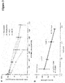
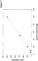
- Fab-mediated residual binding at neutral pH was correlated with the pharmacokinetic properties of a set of therapeutic antibodies, indicating that IgGs with excessive binding to FcRn at pH 7.3 suffer from reduced terminal half-life ([24]).
- Schlothauer et al. [25]
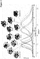
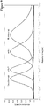
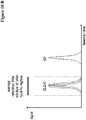
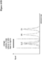
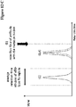
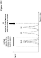
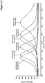
- Schlothauer et al. [25] have described a novel pH-gradient FcRn affinity chromatography method that closely mimics physiological conditions for the dissociation between FcRn and IgGs.
- IgGs with identical Fc-regions differ in their dissociation from FcRn, thereby indicating the influence of the Fab region on FcRn binding.
- the amino acid sequences of the antibody Briakinumab are reported in WO 2013/087911 (SEQ ID NO: 39 and SEQ ID NO: 40), of the antibody Ustekinumab in WO 2013/087911 (SEQ ID NO: 37 and SEQ ID NO: 38) and of the antibody Bevacizumab in Drug Bank entry DB00112.
- One aspect of the invention is a method for selecting a variant antibody having amino acid alterations in the antibody-Fab-region (Fab-variant antibody) with increased or decreased in vivo half-life relative to its parent antibody comprising the following steps:
- the antibody-Fab-FcRn interaction is an interaction between the Fab-region of an antibody with the FcRn. This interaction occurs, if present at all, after the antibody has been bound by the FcRn. Thus, the establishment of this interaction is a two-step process. In the first step an antibody-FcRn complex, to be more precise an antibody-Fc-FcRn complex, is formed. The second step after the antibody-Fc-FcRn complex has been formed is the establishment of the antibody-Fab-FcRn interaction. As can be seen from this, only with a full-length antibody these two interactions, i.e. the antibody-Fc-FcRn interaction and the antibody-Fab-FcRn interaction, can be established.
- a method for determining the presence of Fab-FcRn interaction in an antibody-FcRn complex influencing the in vivo half-life comprising the following steps:
- the antibody of the IgG class is an antibody of the IgG1, IgG2, IgG3 or IgG4 subclass. In one embodiment the antibody of the IgG class is an antibody of the IgG1, IgG3 or IgG4 subclass. In one embodiment the antibody of the IgG class is an antibody of the IgG1 or IgG4 subclass. In one embodiment the antibody of the IgG class is an antibody of the IgG1 subclass. In one embodiment the antibody of the IgG class is an antibody of the IgG4 subclass.
- a method for determining an increase or a decrease in the vivo half-life of a variant antibody relative to its parent antibody comprising the following steps:
- the method according to the invention is based on the general method for selecting an antibody with increased or decreased in the vivo half-life relative to a reference antibody comprising the following steps:
- Disclosed herein is a method for selecting an antibody without antibody-Fab-FcRn interaction influencing the vivo half-life of the antibody:
- a method for determining an increase or a decrease of the vivo half-life of an antibody comprising the following steps:
- the antibody is a full length antibody.
- the salt is sodium chloride.
- the first salt concentration is between 50 mM and 200 mM.
- the first salt concentration is about 140 mM.
- the second salt concentration is between 300 mM and 600 mM.
- the second salt concentration is about 400 mM. Retention times that are substantially different in step a) and step b) differ by at least 5 %.
- step a) and step b) differ by at least 10 %.
- step a) and step b) differ by at least 15 %.
- step b) the retention time in step a) is bigger/longer than in step b).
- step b) the retention time in step b) is smaller/shorter than in step a).
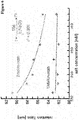
- the retention times are substantially different in step a) and step b) the retention times are proportional to one above the square root of the salt concentration ( ⁇ 1/SQRT(c(salt))).
- the parent or reference antibody is the anti-IL-1R antibody with SEQ ID NO: 01 (heavy chain) and SEQ ID NO: 02 (light chain) for the subclass IgG1 and the anti-IL-1R antibody with SEQ ID NO: 03 (heavy chain) and SEQ ID NO: 04 (light chain) for the subclass IgG4.
- the parent or reference antibody is the anti-HER2 antibody with SEQ ID NO: 36 (heavy chain) and SEQ ID NO: 37 (light chain) for the subclass IgG1 and the anti-HER2 antibody with SEQ ID NO: 38 (heavy chain) and SEQ ID NO: 39 (light chain) for the subclass IgG4.
- the parent or reference antibody is Ustekinumab with light and heavy chain amino acid sequence as depicted in Figure 5 .
- the FcRn affinity chromatography column comprises a non-covalent complex of a neonatal Fc receptor (FcRn) and beta-2-microglobulin (b2m).
- the FcRn affinity chromatography column comprises a covalent complex of a neonatal Fc receptor (FcRn) and beta-2-microglobulin (b2m).
- the complex of the neonatal Fc receptor (FcRn) and beta-2-microglobulin (b2m) is bound to a solid phase.
- the solid phase is a chromatography material.
- the complex of a neonatal Fc receptor (FcRn) and beta-2-microglobulin (b2m) is biotinylated and the solid phase is derivatized with streptavidin.
- beta-2-microglobulin is from the same species as the neonatal Fc receptor (FcRn).
- beta-2-microglobulin is from a different species as the FcRn.
- the FcRn is selected from human FcRn, cynomolgus FcRn, mouse FcRn, rat FcRn, sheep FcRn, dog FcRn, pig FcRn, minipig FcRn, and rabbit FcRn.
- the antibody is a monospecific antibody or antibody fragment of fusion polypeptide, or a bispecific antibody or antibody fragment of fusion polypeptide, or a trispecific antibody or antibody fragment of fusion polypeptide, or a tetraspecific antibody or antibody fragment of fusion polypeptide.
- the antibody is an antibody of the class IgG. In one embodiment the antibody is an antibody of the subclass IgG1, IgG2, IgG3 or IgG4. In one embodiment the antibody is an antibody of the subclass IgG1 or IgG4.
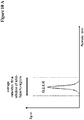
- Antibodies did not show differences in pH 6.0 affinity, therefore the Fab region seems to have no influence on pH 6.0 binding. In contrast, the dissociation between FcRn and the antibodies was influenced by the Fab region.
- the term "about” denotes a range of +/- 20 % of the thereafter following numerical value. In one embodiment the term about denotes a range of +/- 10 % of the thereafter following numerical value. In one embodiment the term about denotes a range of +/-5 % of the thereafter following numerical value.
- alteration denotes the mutation (substitution), insertion (addition), modification (derivatization), or deletion of one or more amino acid residues in a parent antibody or fusion polypeptide, e.g. a fusion polypeptide comprising at least an FcRn binding portion of an Fc-region, to obtain a modified antibody or fusion polypeptide.
- mutation denotes that the specified amino acid residue is substituted for a different amino acid residue.
- the mutation L234A denotes that the amino acid residue lysine at position 234 in an antibody Fc-region (polypeptide) is substituted by the amino acid residue alanine (substitution of lysine with alanine) (numbering according to the EU index).
- the replacing amino acid residue may be a "naturally occurring amino acid residues" and selected from the group consisting of alanine (three letter code: ala, one letter code: A), arginine (arg, R), asparagine (asn, N), aspartic acid (asp, D), cysteine (cys, C), glutamine (gln, Q), glutamic acid (glu, E), glycine (gly, G), histidine (his, H), isoleucine (ile, I), leucine (leu, L), lysine (lys, K), methionine (met, M), phenylalanine (phe, F), proline (pro, P), serine (ser, S), threonine (thr, T), tryptophan (trp, W), tyrosine (tyr, Y
- the replacing amino acid residue may be a "non-naturally occurring amino acid residue". See e.g. US 6,586,207 , WO 98/48032 , WO 03/073238 , US 2004/0214988 , WO 2005/35727 , WO 2005/74524 , Chin, J.W., et al., J. Am. Chem. Soc. 124 (2002) 9026-9027 ; Chin, J.W. and Schultz, P.G., ChemBioChem 11 (2002) 1135-1137 ; Chin, J.W., et al., PICAS United States of America 99 (2002) 11020-11024 ; and, Wang, L. and Schultz, P.G., Chem. (2002) 1-10 .
- amino acid insertion denotes the (additional) incorporation of at least one amino acid residue at a predetermined position in an amino acid sequence. In one embodiment the insertion will be the insertion of one or two amino acid residues.
- the inserted amino acid residue(s) can be any naturally occurring or non-naturally occurring amino acid residue.
- amino acid deletion denotes the removal of at least one amino acid residue at a predetermined position in an amino acid sequence.
- antibody herein is used in a broad sense and encompasses various antibody structures, including but not limited to monoclonal antibodies and multispecific antibodies (e.g. bispecific antibodies, trispecific antibodies) so long as they are full length antibodies and exhibit the desired antigen- and/or FcRn-binding activity.
- binding denotes the binding of an antibody in an in vitro assay. In one embodiment binding is determined in a binding assay in which the antibody is bound to a surface and binding of the antigen to the antibody is measured by Surface Plasmon Resonance (SPR). Binding means e.g. a binding affinity (K D ) of 10 -8 M or less, in some embodiments of 10 -13 to 10 -8 M, in some embodiments of 10 -13 to 10 -9 M.
- K D binding affinity
- Binding can be investigated by a BIAcore assay (GE Healthcare Biosensor AB, Uppsala, Sweden).
- the affinity of the binding is defined by the terms ka (rate constant for the association of the antibody from the antibody/antigen complex), k d (dissociation constant), and K D (k d /k a ).
- buffer substance denotes a substance that when in solution can level changes of the pH value of the solution e.g. due to the addition or release of acidic or basic substances.
- CH2 domain denotes the part of an antibody heavy chain polypeptide that extends approximately from EU position 231 to EU position 340 (EU numbering system according to Kabat).
- a CH2 domain has the amino acid sequence of SEQ ID NO: 05: APELLGG PSVFLFPPKP KDTLMISRTP
- CH3-domain denotes the part of an antibody heavy chain polypeptide that extends approximately from EU position 341 to EU position 446.
- the CH3 domain has the amino acid sequence of SEQ ID NO: 06: GQPREPQ VYTLPPSRDE LTKNQVSLTC LVKGFYPSDI AVEWESNGQP ENNYKTTPPV LDSDGSFFLY SKLTVDKSRW QQGNVFSCSV MHEALHNHYT QKSLSLSPG.
- the "class" of an antibody refers to the type of constant domain or constant region possessed by its heavy chain.
- the heavy chain constant domains that correspond to the different classes of immunoglobulins are called ⁇ , ⁇ , ⁇ , ⁇ , and ⁇ , respectively.
- an "effective amount" of an agent refers to an amount effective, at dosages and for periods of time necessary, to achieve the desired therapeutic or prophylactic result.
- Fc-fusion polypeptide denotes a fusion of a binding domain (e.g. an antigen binding domain such as a single chain antibody, or a polypeptide such as a ligand of a receptor) with an antibody Fc-region that exhibits the desired target- and/or protein A and/or FcRn-binding activity.
- a binding domain e.g. an antigen binding domain such as a single chain antibody, or a polypeptide such as a ligand of a receptor
- Fc-region of human origin denotes the C-terminal region of an immunoglobulin heavy chain of human origin that contains at least a part of the hinge region, the CH2 domain and the CH3 domain.
- a human IgG heavy chain Fc-region extends from Cys226, or from Pro230, to the carboxyl-terminus of the heavy chain.
- the Fc-region has the amino acid sequence of SEQ ID NO: 07.
- the C-terminal lysine (Lys447) of the Fc-region may or may not be present.
- numbering of amino acid residues in the Fc-region or constant region is according to the EU numbering system, also called the EU index, as described in Kabat, E.A., et al., Sequences of Proteins of Immunological Interest, 5th ed., Public Health Service, National Institutes of Health, Bethesda, MD (1991), NIH Publication 91 3242 .
- the Fc-region is composed of two heavy chain Fc-region polypeptides, which can be covalently linked to each other via the hinge region cysteine residues forming interpolypeptide disulfide bonds.
- FcRn denotes the human neonatal Fc-receptor. FcRn functions to salvage IgG from the lysosomal degradation pathway, resulting in reduced clearance and increased half-life.
- the FcRn is a heterodimeric protein consisting of two polypeptides: a 50 kDa class I major histocompatibility complex-like protein ( ⁇ -FcRn) and a 15 kDa ⁇ 2-microglobulin ( ⁇ 2m). FcRn binds with high affinity to the CH2-CH3 portion of the Fc-region of IgG.
- IgG and FcRn The interaction between IgG and FcRn is strictly pH dependent and occurs in a 1:2 stoichiometry, with one IgG binding to two FcRn molecules via its two heavy chains ( Huber, A.H., et al., J. Mol. Biol. 230 (1993) 1077-1083 ).
- FcRn binding occurs in the endosome at acidic pH (pH ⁇ 6.5) and IgG is released at the neutral cell surface (pH of about 7.4).
- the pH-sensitive nature of the interaction facilitates the FcRn-mediated protection of IgGs pinocytosed into cells from intracellular degradation by binding to the receptor within the acidic environment of endosomes.
- FcRn then facilitates the recycling of IgG to the cell surface and subsequent release into the blood stream upon exposure of the FcRn-IgG complex to the neutral pH environment outside the cell.
- FcRn binding portion of an Fc-region denotes the part of an antibody heavy chain polypeptide that extends approximately from EU position 243 to EU position 261 and approximately from EU position 275 to EU position 293 and approximately from EU position 302 to EU position 319 and approximately from EU position 336 to EU position 348 and approximately from EU position 367 to EU position 393 and EU position 408 and approximately from EU position 424 to EU position 440.
- one or more of the following amino acid residues according to the EU numbering of Kabat are altered F243, P244, P245 P, K246, P247, K248, D249, T250, L251, M252, 1253, S254, R255, T256, P257, E258, V259, T260, C261, F275, N276, W277, Y278, V279, D280, V282, E283, V284, H285, N286, A287, K288, T289, K290, P291, R292, E293, V302, V303, S304, V305, L306, T307, V308, L309, H310, Q311, D312, W313, L314, N315, G316, K317, E318, Y319, 1336, S337, K338, A339, K340, G341, Q342, P343, R344, E345, P346, Q347, V348, C367, V369,
- full length antibody denotes an antibody having a structure substantially similar to a native antibody structure.
- a full length antibody comprises two full length antibody light chains comprising a light chain variable domain and a light chain constant domain and two full length antibody heavy chains comprising a heavy chain variable domain, a first constant domain, a hinge region, a second constant domain and a third constant domain.
- a full length antibody may comprise further domains, such as e.g. additional scFv or a scFab conjugated to one or more of the chains of the full length antibody. These conjugates are also encompassed by the term full length antibody.
- hinge region denotes the part of an antibody heavy chain polypeptide that joins the CH1 domain and the CH2 domain, e. g. from about position 216 to position about 230 according to the EU numbering system of Kabat.
- the hinge region is a shortened hinge region comprising residues 221 to 230 according to the EU numbering system of Kabat.
- the hinge region is normally a dimeric molecule consisting of two polypeptides with identical amino acid sequence.
- the hinge region generally comprises about 25 amino acid residues and is flexible allowing the antigen binding regions to move independently.
- the hinge region can be subdivided into three domains: the upper, the middle, and the lower hinge domain ( Roux, et al., J. Immunol. 161 (1998) 4083 ).
- host cell refers to cells into which exogenous nucleic acid has been introduced, including the progeny of such cells.
- Host cells include “transformants” and “transformed cells,” which include the primary transformed cell and progeny derived therefrom without regard to the number of passages. Progeny may not be completely identical in nucleic acid content to a parent cell, but may contain mutations. Mutant progeny that have the same function or biological activity as screened or selected for in the originally transformed cell are included herein.
- derived from denotes that an amino acid sequence is derived from a parent amino acid sequence by introducing alterations at at least one position.
- a derived amino acid sequence differs from the corresponding parent amino acid sequence at at least one corresponding position (numbering according to Kabat EU index for antibody Fc-regions).
- an amino acid sequence derived from a parent amino acid sequence differs by one to fifteen amino acid residues at corresponding positions.
- an amino acid sequence derived from a parent amino acid sequence differs by one to ten amino acid residues at corresponding positions.
- an amino acid sequence derived from a parent amino acid sequence differs by one to six amino acid residues at corresponding positions.
- a derived amino acid sequence has a high amino acid sequence identity to its parent amino acid sequence.
- an amino acid sequence derived from a parent amino acid sequence has 80 % or more amino acid sequence identity.
- an amino acid sequence derived from a parent amino acid sequence has 90 % or more amino acid sequence identity.
- an amino acid sequence derived from a parent amino acid sequence has 95 % or more amino acid sequence identity.
- human Fc-region polypeptide denotes an amino acid sequence which is identical to a "native” or “wild-type” human Fc-region polypeptide.
- variant (human) Fc-region polypeptide denotes an amino acid sequence which derived from a "native” or "wild-type” human Fc-region polypeptide by virtue of at least one "amino acid alteration".
- a "human Fc-region” is consisting of two human Fc-region polypeptides.
- a “variant (human) Fc-region” is consisting of two Fc-region polypeptides, whereby both can be variant (human) Fc-region polypeptides or one is a human Fc-region polypeptide and the other is a variant (human) Fc-region polypeptide.
- the human Fc-region polypeptide has the amino acid sequence of a human IgG1 Fc-region polypeptide of SEQ ID NO: 07, or of a human IgG2 Fc-region polypeptide of SEQ ID NO: 08, or of a human IgG3 Fc-region polypeptide of SEQ ID NO: 09, or of a human IgG4 Fc-region polypeptide of SEQ ID NO: 10.
- the Fc-region polypeptide is derived from an Fc-region polypeptide of SEQ ID NO: 07, or 08, or 09, or 10 and has at least one amino acid mutation compared to the Fc-region polypeptide of SEQ ID NO: 07, or 08, or 09, or 10.
- the Fc-region polypeptide comprises/has from about one to about ten amino acid mutations, and in one embodiment from about one to about five amino acid mutations. In one embodiment the Fc-region polypeptide has at least about 80 % homology with a human Fc-region polypeptide of SEQ ID NO: 07, or 08, or 09, or 10. In one embodiment the Fc-region polypeptide has least about 90 % homology with a human Fc-region polypeptide of SEQ ID NO: 07, or 08, or 09, or 10. In one embodiment the Fc-region polypeptide has at least about 95 % homology with a human Fc-region polypeptide of SEQ ID NO: 07, or 08, or 09, or 10.
- the Fc-region polypeptide derived from a human Fc-region polypeptide of SEQ ID NO: 07, or 08 or 09, or 10 is defined by the amino acid alterations that are contained.
- P329G denotes an Fc-region polypeptide derived human Fc-region polypeptide with the mutation of proline to glycine at amino acid position 329 relative to the human Fc-region polypeptide of SEQ ID NO: 07, or 08, or 09, or 10.
- EU index or EU index as in Kabat or Kabat EU index or EU numbering scheme refers to the numbering of the EU antibody ( Edelman, et al., Proc. Natl. Acad. Sci. USA 63 (1969) 78-85 ).
- the numbering of the light chain residues is according to the Kabat nomenclature ( Kabat, E.A., et al., Sequences of Proteins of Immunological Interest, 5th ed., Public Health Service, National Institutes of Health, Bethesda, MD (1991), NIH Publication 91 3242 ).
- a human IgG1 Fc-region polypeptide has the following amino acid sequence:
- a human IgG1 Fc-region derived Fc-region polypeptide with the mutations L234A, L235A has the following amino acid sequence:
- a human IgG1 Fc-region derived Fc-region polypeptide with Y349C, T366S, L368A and Y407V mutations has the following amino acid sequence:
- a human IgG1 Fc-region derived Fc-region polypeptide with S354C, T366W mutations has the following amino acid sequence:
- a human IgG1 Fc-region derived Fc-region polypeptide with L234A, L235A mutations and Y349C, T366S, L368A, Y407V mutations has the following amino acid sequence:
- a human IgG1 Fc-region derived Fc-region polypeptide with a L234A, L235A and S354C, T366W mutations has the following amino acid sequence:
- a human IgG1 Fc-region derived Fc-region polypeptide with a P329G mutation has the following amino acid sequence:
- a human IgG1 Fc-region derived Fc-region polypeptide with L234A, L235A mutations and P329G mutation has the following amino acid sequence:
- a human IgG1 Fc-region derived Fc-region polypeptide with a P329G mutation and Y349C, T366S, L368A, Y407V mutations has the following amino acid sequence:
- a human IgG1 Fc-region derived Fc-region polypeptide with a P329G mutation and S354C, T366W mutation has the following amino acid sequence:
- a human IgG1 Fc-region derived Fc-region polypeptide with L234A, L235A, P329G and Y349C, T366S, L368A, Y407V mutations has the following amino acid sequence:
- a human IgG1 Fc-region derived Fc-region polypeptide with L234A, L235A, P329G mutations and S354C, T366W mutations has the following amino acid sequence:
- a human IgG4 Fc-region polypeptide has the following amino acid sequence:
- a human IgG4 Fc-region derived Fc-region polypeptide with S228P and L235E mutations has the following amino acid sequence:
- a human IgG4 Fc-region derived Fc-region polypeptide with S228P, L235E mutations and P329G mutation has the following amino acid sequence:
- a human IgG4 Fc-region derived Fc-region polypeptide with S354C, T366W mutations has the following amino acid sequence:
- a human IgG4 Fc-region derived Fc-region polypeptide with Y349C, T366S, L368A, Y407V mutations has the following amino acid sequence:
- a human IgG4 Fc-region derived Fc-region polypeptide with a S228P, L235E and S354C, T366W mutations has the following amino acid sequence:
- a human IgG4 Fc-region derived Fc-region polypeptide with a S228P, L235E and Y349C, T366S, L368A, Y407V mutations has the following amino acid sequence:
- a human IgG4 Fc-region derived Fc-region polypeptide with a P329G mutation has the following amino acid sequence:
- a human IgG4 Fc-region derived Fc-region polypeptide with a P329G and Y349C, T366S, L368A, Y407V mutations has the following amino acid sequence:
- a human IgG4 Fc-region derived Fc-region polypeptide with a P329G and S354C, T366W mutations has the following amino acid sequence:
- a human IgG4 Fc-region derived Fc-region polypeptide with a S228P, L235E, P329G and Y349C, T366S, L368A, Y407V mutations has the following amino acid sequence:
- a human IgG4 Fc-region derived Fc-region polypeptide with a S228P, L235E, P329G and S354C, T366W mutations has the following amino acid sequence:
- a “humanized” antibody refers to a chimeric antibody comprising amino acid residues from non-human HVRs and amino acid residues from human FRs.
- a humanized antibody will comprise substantially all of at least one, and typically two, variable domains, in which all or substantially all of the HVRs (e.g., the CDRs) correspond to those of a non-human antibody, and all or substantially all of the FRs correspond to those of a human antibody.
- a humanized antibody optionally may comprise at least a portion of an antibody constant region derived from a human antibody.
- a "humanized form" of an antibody, e.g., a non-human antibody refers to an antibody that has undergone humanization.
- mammals include, but are not limited to, domesticated animals (e.g. cows, sheep, cats, dogs, and horses), primates (e.g., humans and non-human primates such as monkeys), rabbits, and rodents (e.g., mice and rats).
- domesticated animals e.g. cows, sheep, cats, dogs, and horses
- primates e.g., humans and non-human primates such as monkeys
- rabbits e.g., mice and rats
- rodents e.g., mice and rats.
- the individual or subject is a human.
- an “isolated” antibody is one which has been separated from a component of its natural environment.
- an antibody is purified to greater than 95 % or 99 % purity as determined by, for example, electrophoretic (e.g., SDS-PAGE, isoelectric focusing (IEF), capillary electrophoresis) or chromatographic (e.g., size exclusion chromatography or ion exchange or reverse phase HPLC).
- electrophoretic e.g., SDS-PAGE, isoelectric focusing (IEF), capillary electrophoresis
- chromatographic e.g., size exclusion chromatography or ion exchange or reverse phase HPLC.
- nucleic acid refers to a nucleic acid molecule that has been separated from a component of its natural environment.
- An isolated nucleic acid includes a nucleic acid molecule contained in cells that ordinarily contain the nucleic acid molecule, but the nucleic acid molecule is present extrachromosomally or at a chromosomal location that is different from its natural chromosomal location.
- monoclonal antibody refers to an antibody obtained from a population of substantially homogeneous antibodies, i.e., the individual antibodies comprising the population are identical and/or bind the same epitope, except for possible variant antibodies, e.g., containing naturally occurring mutations or arising during production of a monoclonal antibody preparation, such variants generally being present in minor amounts.
- polyclonal antibody preparations typically include different antibodies directed against different determinants (epitopes)
- each monoclonal antibody of a monoclonal antibody preparation is directed against a single determinant on an antigen.
- the modifier "monoclonal” indicates the character of the antibody as being obtained from a substantially homogeneous population of antibodies, and is not to be construed as requiring production of the antibody by any particular method.
- the monoclonal antibodies to be used in accordance with the present invention may be made by a variety of techniques, including but not limited to the hybridoma method, recombinant DNA methods, phage-display methods, and methods utilizing transgenic animals containing all or part of the human immunoglobulin loci, such methods and other exemplary methods for making monoclonal antibodies being described herein.
- Native antibodies refer to naturally occurring immunoglobulin molecules with varying structures.
- native IgG antibodies are heterotetrameric glycoproteins of about 150,000 daltons, composed of two identical light chains and two identical heavy chains that are disulfide-bonded. From N- to C-terminus, each heavy chain has a variable region (VH), also called a variable heavy domain or a heavy chain variable domain, followed by three constant domains (CHI, CH2, and CH3).
- VH variable region
- VL variable region
- the light chain of an antibody may be assigned to one of two types, called kappa ( ⁇ ) and lambda ( ⁇ ), based on the amino acid sequence of its constant domain.
- negative linear pH gradient denotes a pH gradient starting at a high (i.e. neutral or alkaline) pH value and ending at a lower (i.e. neutral or acidic) pH value.
- the negative linear pH gradient starts at a pH value of about 8.8 and ends at a pH value of about 5.5.
- non-naturally occurring amino acid residue denotes an amino acid residue, other than the naturally occurring amino acid residues as listed above, which can be covalently bound to the adjacent amino acid residues in a polypeptide chain.
- non-naturally occurring amino acid residues are norleucine, ornithine, norvaline, homoserine. Further examples are listed in Ellman, et al., Meth. Enzym. 202 (1991) 301-336 . Exemplary method for the synthesis of non-naturally occurring amino acid residues are reported in, e. g., Noren, et al., Science 244 (1989) 182 and Ellman et al., supra.
- pharmaceutical formulation refers to a preparation which is in such form as to permit the biological activity of an active ingredient contained therein to be effective, and which contains no additional components which are unacceptably toxic to a subject to which the formulation would be administered.
- a “pharmaceutically acceptable carrier” refers to an ingredient in a pharmaceutical formulation, other than an active ingredient, which is nontoxic to a subject.
- a pharmaceutically acceptable carrier includes, but is not limited to, a buffer, excipient, stabilizer, or preservative.
- plasmid refers to a nucleic acid molecule capable of propagating another nucleic acid to which it is linked.
- the term includes the plasmid as a self-replicating nucleic acid structure as well as the plasmid incorporated into the genome of a host cell into which it has been introduced.
- Certain plasmids are capable of directing the expression of nucleic acids to which they are operatively linked. Such plasmids are referred to herein as "expression plasmid”.
- positive linear pH gradient denotes a pH gradient starting at a low (i.e. more acidic) pH value and ending at a higher (i.e. less acidic, neutral or alkaline) pH value.
- the positive linear pH gradient starts at a pH value of about 5.5 and ends at a pH value of about 8.8.
- recombinant antibody denotes all antibodies (chimeric, humanized and human) that are prepared, expressed, created or isolated by recombinant means. This includes antibodies isolated from a host cell such as a NS0 or CHO cell or from an animal (e.g. a mouse) that is transgenic for human immunoglobulin genes or antibodies expressed using a recombinant expression plasmid transfected into a host cell. Such recombinant antibodies have variable and constant regions in a rearranged form. The recombinant antibodies as reported herein can be subjected to in vivo somatic hypermutation. Thus, the amino acid sequences of the VH and VL regions of the recombinant antibodies are sequences that, while derived from and related to human germ line VH and VL sequences, may not naturally exist within the human antibody germ line repertoire in vivo.
- a “solid phase” denotes a non-fluid substance, and includes particles (including microparticles and beads) made from materials such as polymer, metal (paramagnetic, ferromagnetic particles), glass, and ceramic; gel substances such as silica, alumina, and polymer gels; capillaries, which may be made of polymer, metal, glass, and/or ceramic; zeolites and other porous substances; electrodes; microtiter plates; solid strips; and cuvettes, tubes or other spectrometer sample containers.
- a solid phase component of an assay is distinguished from inert solid surfaces in that a "solid support" contains at least one moiety on its surface, which is intended to interact chemically with a molecule.
- a solid phase may be a stationary component, such as a chip, tube, strip, cuvette, or microtiter plate, or may be non-stationary components, such as beads and microparticles.
- Microparticles can also be used as a solid support for homogeneous assay formats.
- Such particles include polymer particles such as polystyrene and poly (methylmethacrylate); gold particles such as gold nanoparticles and gold colloids; and ceramic particles such as silica, glass, and metal oxide particles. See for example Martin, C.R., et al., Analytical Chemistry-News & Features, May 1 (1998) 322A-327A .
- the solid support is sepharose.
- substantially the same denotes that two values, e.g. the retention times on an FcRn affinity chromatography column of two different antibodies, are within 5 % of each other, i.e. they differ by less than 5 %. For example, a first retention time of 80 minutes and a second retention time of 84 minutes are substantially the same, whereas a retention time of 80 minutes and a retention time of 85 minutes are not substantially the same, these retention times are different.
- substantially the same denotes that two values are within 3.5 % of each other, i.e. they differ by 3.5 % or less.
- substantially the same denotes that two values are within 2.5 % of each other, i.e. they differ by 2.5 % or less. The smaller of the two values is taken as basis for this calculation.
- treatment refers to clinical intervention in an attempt to alter the natural course of the individual being treated, and can be performed either for prophylaxis or during the course of clinical pathology. Desirable effects of treatment include, but are not limited to, preventing occurrence or recurrence of disease, alleviation of symptoms, diminishment of any direct or indirect pathological consequences of the disease, preventing metastasis, decreasing the rate of disease progression, amelioration or palliation of the disease state, and remission or improved prognosis.
- bivalent as used within the current application denotes the presence of a specified number of binding sites in a (antibody) molecule.
- bivalent tetravalent
- hexavalent denote the presence of two binding site, four binding sites, and six binding sites, respectively, in a (antibody) molecule.
- the bispecific antibodies as reported herein as reported herein are in one preferred embodiment "bivalent”.
- variable region or “variable domain” refer to the domain of an antibody heavy or light chain that is involved in binding of the antibody to its antigen.
- the variable domains of the heavy chain and light chain (VH and VL, respectively) of an antibody generally have similar structures, with each domain comprising four framework regions (FRs) and three hypervariable regions (HVRs) (see, e.g., Kindt, T.J. et al. Kuby Immunology, 6th ed., W.H. Freeman and Co., N.Y. (2007), page 91 ).
- a single VH or VL domain may be sufficient to confer antigen-binding specificity.
- antibodies that bind a particular antigen may be isolated using a VH or VL domain from an antibody that binds the antigen to screen a library of complementary VL or VH domains, respectively. See, e.g., Portolano, S. et al., J. Immunol. 150 (1993) 880-887 ; Clackson, T. et al., Nature 352 (1991) 624-628 ).
- variant denotes molecules which have an amino acid sequence that differs from the amino acid sequence of a parent molecule. Typically such molecules have one or more alterations, insertions, or deletions.
- the modified antibody or the modified fusion polypeptide comprises an amino acid sequence comprising at least a portion of an Fc-region which is not naturally occurring. Such molecules have less than 100 % sequence identity with the parent antibody or parent fusion polypeptide.
- the variant antibody or the variant fusion polypeptide has an amino acid sequence that has from about 75 % to less than 100 % amino acid sequence identity with the amino acid sequence of the parent antibody or parent fusion polypeptide, especially from about 80 % to less than 100 %, especially from about 85 % to less than 100 %, especially from about 90 % to less than 100 %, and especially from about 95 % to less than 100 %.
- the parent antibody or the parent fusion polypeptide and the variant antibody or the variant fusion polypeptide differ by one (a single), two or three amino acid residue(s).
- the invention is based, at least in part, on the finding that the charge distribution in the Fv domain influences antibody-FcRn binding and results in additional interactions between the antibody and the FcRn. This changes the FcRn binding characteristics, especially with respect to the dissociation of the antibody-FcRn complex at pH 7.4, thereby reducing FcRn-dependent terminal half-life of the antibody.
- the neonatal Fc-receptor (FcRn) is important for the metabolic fate of antibodies of the IgG class in vivo.
- the FcRn functions to salvage wild-type IgG from the lysosomal degradation pathway, resulting in reduced clearance and increased half-life. It is a heterodimeric protein consisting of two polypeptides: a 50 kDa class I major histocompatibility complex-like protein ( ⁇ -FcRn) and a 15 kDa ⁇ 2-microglobulin ( ⁇ 2m).
- ⁇ -FcRn major histocompatibility complex-like protein
- ⁇ 2m microglobulin
- an antibody of the class IgG and the FcRn is pH dependent and occurs in a 1:2 stoichiometry, i.e. one IgG antibody molecule can interact with two FcRn molecules via its two heavy chain Fc-region polypeptides (see e.g. [16]).
- an IgGs in vitro FcRn binding properties/characteristics are indicative of its in vivo pharmacokinetic properties in the blood circulation.
- the amino acid residues interacting with the FcRn are located approximately between EU position 243 and EU position 261, approximately between EU position 275 and EU position 293, approximately between EU position 302 and EU position 319, approximately between EU position 336 and EU position 348, approximately between EU position 367 and EU position 393, at EU position 408, and approximately between EU position 424 and EU position 440.
- antibodies with reduced half-life in the blood circulation are desired.
- drugs for intravitreal application should have a long half-life in the eye and a short half-life in the circulation of the patient.
- Such antibodies also have the advantage of increased exposure to a disease site, e.g. in the eye.
- Fc-region residues critical to the mouse Fc-mouse FcRn interaction have been identified by site-directed mutagenesis (see e.g. Dall'Acqua, W.F., et al. J. Immunol 169 (2002) 5171-5180 ).
- Residues 1253, H310, H433, N434, and H435 are involved in the interaction ( Medesan, C., et al., Eur. J. Immunol. 26 (1996) 2533-2536 ; Firan, M., et al., Int. Immunol.
- Residues 1253, H310, and H435 were found to be critical for the interaction of human Fc with murine FcRn ( Kim, J.K., et al., Eur. J. Immunol. 29 (1999) 2819-2825 ).
- Residues M252Y, S254T, T256E have been described by Dall'Acqua et al. to improve FcRn binding by protein-protein interaction studies ( Dall'Acqua, W.F., et al. J. Biol. Chem.
- Table 1 Collocation of different Fc-region mutations and their influence on FcRn-binding and in vivo half-life. mutation effect on FcRn binding half-life in the circulation reference H285 reduced reduced Kim, J.K., Scand. J. Immunol. 40 (1994) 457-465 H310Q/H433N (murine) (in mouse) (murine IgG1) I253A reduced reduced Ghetie, V. and Ward, E.S., Immunol.
- FcRn human neonatal Fc receptor
- IgG-FcRn interactions can be analyzed using plasmon surface resonance (SPR) assays ( Wang, W., et al., Drug Metab. Disp. 39 (2011) 1469-1477 ; Datta-Mannan, A., et al., Drug Metab. Disp. 40 (2012) 1545-1555 ; Vaughn, D.E. and Bjorkman, P.J., Biochemistry 36 (1997) 9374-9380 ; Raghavan, M., et al., Proc. Natl. Acad. Sci. USA 92 (1995) 11200-11204 ; Martin, W.L. and Bjorkman, P.J., Biochemistry 38 (1999) 12639-12647 ).
- SPR plasmon surface resonance
- SPR analysis of the IgG-FcRn interaction provides a qualitative result indicating expected or aberrant binding properties of a sample but does neither give a hint for the cause of aberrant binding nor a quantitative estimation of the amount of antibody with aberrant binding.
- Briakinumab (OzespaTM) and Ustekinumab (StelaraTM) were used as a model system. Both Briakinumab and Ustekinumab are fully human monoclonal IgG1 antibodies. They bind to the same human p40-subunit of interleukin 12 (IL-12) and interleukin 23 (IL-23) [26] and they are not cross-reactive to the corresponding mouse IL-12 and IL-23 [27,28].
- IL-12 interleukin 12
- IL-23) interleukin 23
- Briakinumab and Ustekinumab are an IgG1 ⁇ antibody with variable heavy and light chain domains of the V H 5 and V ⁇ 1D germline families and an IgG1 ⁇ antibody with variable heavy and light chain domains of the V H 3 and V ⁇ 1 germline families, respectively.
- Briakinumab and Ustekinumab show differences in several allotype-specific amino acids in the constant domains (see Figure 5 ). However, these amino acid residues are outside of the (cognate) FcRn binding regions and can therefore be considered to play no role in FcRn-dependent PK [11].
- Ustekinumab has a (reported) median terminal half-life of 22 days [29]
- Briakinumab has a terminal half-life of only 8-9 days [26,30,31].
- Briakinumab exhibits a non-uniform charge distribution at physiological pH of 7.4 (see e.g. the published crystal structure of Ustekinumab [27] and a homology model of Briakinumab).
- Briakinumab shows a large positively charged region on the Fv domain (see Figure 1a ) which is absent in Ustekinumab (see Figure 1b ).
- FcRn possesses a strong and extended negatively charged region (see Figure 1c ) which is however not involved in cognate Fc-region binding.
- Briakinumab and Ustekinumab have calculated isoelectric points of 9.7 and 9.4, respectively.
- the net charge of Briakinumab is slightly more positive over the entire pH range (see Figure Id).
- FcRn binding affinity of Briakinumab and Ustekinumab at pH 6.0 is comparable, i.e. both values differ at most by one order or magnitude, at most 5-fold, whereas the dissociation from the FcRn is very different.
- Briakinumab and Ustekinumab it could be shown that the interaction is predominantly electrostatic and correlates with the extent of a positively charged region (see below).
- Variant antibodies mAb 5 and mAb 6 bear Ustekinumab CDRs (heavy and light chain parts) on the Briakinumab framework and vice versa. Grafting Ustekinumab CDRs on Briakinumab (mAb 5) shifted the retention time of mAb 5 close to that of Ustekinumab. Grafting Briakinumab CDRs on Ustekinumab (mAb 6) described/presented an elution profile which was still close to Ustekinumab.
- MAb 3 comprising the HCs of Ustekinumab and the LCs of Briakinumab, as well as mAb 2 containing the Fv domain of Briakinumab on the Ustekinumab constant domains both eluted close to Briakinumab.
- the FcRn column retention times were aligned with isoelectric points and net charges of the antibodies. No correlation between the FcRn column retention times and the isoelectric points or the net charges of the Fv domains at lysosomal pH 6.0 or physiological pH 7.4 can be seen (see Table 3). However, the measured FcRn column retention times increased with the extent of positively charged regions, especially around the light chain variable domains (see Figure 2 ).
- Amino acids may be grouped according to common side-chain properties:
- FcRn column retention times were also determined in a different set up with increased ionic strength in the mobile phase, i.e. in the presence of increased salt concentrations. Charge-mediated interactions are known to be weakened under high ionic strength conditions, whereas hydrophobic interactions are typically strengthened by salt. It has been found that the FcRn column retention time of Briakinumab was shortened in the presence of salt and was proportional to the inverse square root of the ionic strength as suggested by the Debye-Hückel law of charge screening [32]. The retention time of Ustekinumab remained essentially unaffected (see Figure 6 ). Thus, a significant part of the excessive FcRn-Briakinumab interaction is charge-mediated.
- FcRn affinity chromatography of the engineered variants showed that antibodies with the same Fv domain (mAb 1 & mAb 2) and the same LC (mAb 3 & mAb 4) elute at nearly identical FcRn column retention times. Furthermore, grafting Ustekinumab CDRs on Briakinumab (mAb 5) shifts the elution pH close to that of Ustekinumab. Thus, the light chain CDRs provide the main influence on Briakinumab's FcRn binding.
- Briakinumab and Ustekinumab together with two variants of Briakinumab (mAb 8 and mAb 9), which had FcRn column retention times between Briakinumab and Ustekinumab, were tested.
- the distribution and elimination processes of the four antibodies are in agreement with other IgG PK studies (see Figure 3a ). Briakinumab showed a faster decrease in the ⁇ -phase than the other antibodies.
- the terminal half-life was examined, which is exclusively calculated in the elimination phase where FcRn recycling dominates [39].
- the terminal half-life of the four antibodies correlate linearly with the in vitro FcRn column elution pH: The higher the FcRn column elution pH the shorter the terminal half-life, thereby demonstrating that the FcRn column is a predictive/sensitive tool for in vitro FcRn dissociation.
- the correlation between terminal half-life and the FcRn column elution pH confirms the importance of the fast FcRn-IgG dissociation at physiological pH.
- the FcRn-IgG complex is built in the endosomes at pH 6.0, therefore, less binding results in less IgG-recycling and faster clearance.
- the FcRn-IgG complex is released to the plasma membrane, where the dissociation of the IgG and the FcRn has to take place at a physiological pH of 7.4 within a short period of time [40]. Consequently, dissociation at physiological pH is also important for a prolonged half-life [22,40].
- a homology model of a human FcRn-Fc complex was generated using the published rat FcRn structure as a template.
- the position of the Fv domains of Briakinumab and Ustekinumab was modeled based on the crystal structure of a complete IgG1 (PDB code 1HZH).
- These homology models contain two copies of FcRn (a-FcRn with ⁇ 2 m) on one complete IgG molecule (see Figure 4 a) .
- the distance between FcRn and the Fv domains is >40 ⁇ in the starting structure and exceeds the Debye length of approx. 8 ⁇ under physiological conditions [32].
- the current invention is based on a method comprising the following two steps:
- the second salt concentration is generally higher/bigger than the first salt concentration, so that these concentrations are not about identical, i.e. they differ by at least 10 %, or by at least 20 %.
- the antibody can be a variant antibody of a parent antibody in which case the reference antibody is the parent antibody.
- the reference antibody is an antibody that has substantially the same retention time as its Fc-region after IdeS cleavage or papain cleavage.
- antibodies with extended half-life are desired.
- drugs with an extended half-life in the circulation of a patient in need of a treatment require decreased dosing or increased dosing intervals.
- Such antibodies also have the advantage of increased exposure to a disease site, e. g. a tumor.
- the retention time is substantially not affected by the change from low to high salt concentration or by the cleavage of the Fc-region then no antibody-Fab-FcRn interaction is present and a higher retention time on the FcRn affinity chromatography column correlates with an increased half-life in vivo. But if the retention time is affected, especially if it is reduced, by a change from low to high salt concentrations or by cleavage of the Fc-region then the in vivo half-life correlates differently to the retention time on the FcRn affinity chromatography column, i.e.
- a longer retention time on the FcRn affinity chromatography column correlates to a shorter in vivo half-life due to reduced antibody-FcRn dissociation at physiological pH and, without being bound by this theory, an increased lysosomal degradation of the antibody.
- the herein used FcRn affinity chromatography column comprises a matrix and matrix bound chromatographical functional groups, wherein the matrix bound chromatographical functional group comprises a non-covalent complex of neonatal Fc receptor (FcRn) and beta-2-microglobulin.
- starting point for the method as reported herein is a parent or reference antibody that is characterized by its binding to the FcRn.
- Variant antibodies show either increased or decreased binding to FcRn when compared to a parent antibody polypeptide or compared to a reference antibody, and, thus, have a modified half-life compared to the parent/reference antibody in serum.
- Fc-region variants with increased affinity for the FcRn are predicted at first to have longer serum half-life compared to those with decreased affinity for the FcRn (i.e. with reduced retention time on an FcRn column compared to a parent antibody or reference antibody).
- Antibody variants with increased in vivo half-life have applications in methods of treating mammals, especially humans, where long half-life of the administered antibody is desired, such as in the treatment of a chronic disease or disorder.
- Antibody variants with decreased affinity for the FcRn have applications in methods of treating mammals, especially humans, where a short half-life of the administered antibody or fusion polypeptide is desired, such as in vivo diagnostic imaging.
- the FcRn is selected from human FcRn, cynomolgus FcRn, mouse FcRn, rat FcRn, sheep FcRn, dog FcRn, pig FcRn, minipig FcRn, and rabbit FcRn.
- beta-2-microglobulin is from the same species as the FcRn.
- beta-2-microglobulin is from a different species as the FcRn.
- parent antibody comprises at least one binding domain and at least one Fc-region. In one embodiment the parent antibody comprises two binding domains and two Fc-regions.
- the parent antibody comprises at least one binding domain that specifically binds to a target which mediates a biological effect (in one embodiment a ligand capable of binding to a cell surface receptor or a cell surface receptor capable of binding a ligand) and mediates transmission of a negative or positive signal to a cell.
- the parent antibody comprises at least one binding domain specific for an antigen targeted for reduction or elimination (in one embodiment a cell surface antigen or a soluble antigen) and at least one Fc-region.
- Antibodies specifically binding to a target can be raised in mammals by multiple subcutaneous or intraperitoneal injections of the relevant antigen (e.g. purified antigen, cells or cellular extracts comprising such antigens, or DNA encoding for such antigen) and optionally an adjuvant.
- the relevant antigen e.g. purified antigen, cells or cellular extracts comprising such antigens, or DNA encoding for such antigen
- an adjuvant e.g. purified antigen, cells or cellular extracts comprising such antigens, or DNA encoding for such antigen
- the antibody is a full length antibody.
- the antibody is a monoclonal antibody.
- the parent antibody is a bispecific antibody.
- the parent antibody is a chimeric antibody.
- the non-covalent complex of neonatal Fc receptor (FcRn) and beta-2-microglobulin is bound to a solid phase.
- the conjugation of the non-covalent complex to the solid phase is performed by chemically binding via N-terminal and/or ⁇ -amino groups (lysine), ⁇ -amino groups of different lysins, carboxy-, sulfhydryl-, hydroxyl-, and/or phenolic functional groups of the amino acid backbone of the antibody, and/or sugar alcohol groups of the carbohydrate structure of the antibody.
- ⁇ -amino groups lysine
- ⁇ -amino groups of different lysins carboxy-, sulfhydryl-, hydroxyl-, and/or phenolic functional groups of the amino acid backbone of the antibody
- sugar alcohol groups of the carbohydrate structure of the antibody.
- non-covalent complex is conjugated to the solid phase via a specific binding pair. In one embodiment the non-covalent complex is conjugated to biotin and immobilization to a solid support is performed via solid support immobilized avidin or streptavidin.
- a specific binding pair is in one embodiment selected from streptavidin or avidin/biotin, antibody/antigen (see, for example, Hermanson, G.T., et al., Bioconjugate Techniques, Academic Press (1996 )), lectin/polysaccharide, steroid/steroid binding protein, hormone/hormone receptor, enzyme/substrate, IgG/Protein A and/or G, etc.
- any buffer substance can be used in the methods as reported herein.
- Fc residues critical to the mouse Fc-mouse FcRn interaction have been identified by site-directed mutagenesis (see e.g. Dall'Acqua, W.F., et al. J. Immunol 169 (2002) 5171-5180 ).
- Residues 1253, H310, H433, N434, and H435 are involved in the interaction ( Medesan, C., et al., Eur. J. Immunol. 26 (1996) 2533 ; Firan, M., et al., Int. Immunol.
- Residues 1253, H310, and H435 were found to be critical for the interaction of human Fc with murine FcRn ( Kim, J.K., et al., Eur. J. Immunol. 29 (1999) 2819 ).
- Residues M252Y, S254T, T256E have been described by Dall'Acqua et al. to improve FcRn binding by protein-protein interaction studies ( Dall'Acqua, W.F., et al. J. Biol. Chem. 281 (2006) 23514-23524 ).
- a pharmaceutically acceptable buffer substance such as e.g. phosphoric acid or salts thereof, acetic acid or salts thereof, citric acid or salts thereof, morpholine or salts thereof, 2-(N-morpholino) ethanesulfonic acid (MES) or salts thereof, histidine or salts thereof, glycine or salts thereof, tris (hydroxymethyl) aminomethane (TRIS) or salts thereof, (4-(2-hydroxyethyl)-1-piperazineethanesulfonic acid (HEPES) or salts thereof.
- phosphoric acid or salts thereof such as e.g. phosphoric acid or salts thereof, acetic acid or salts thereof, citric acid or salts thereof, morpholine or salts thereof, 2-(N-morpholino) ethanesulfonic acid (MES) or salts thereof, histidine or salts thereof, glycine or salts thereof, tris (hydroxymethyl) aminomethane (TRIS) or salts
- the buffer substance is selected from phosphoric acid or salts thereof, or acetic acid or salts thereof, or citric acid or salts thereof, or histidine or salts thereof.
- the buffer substance has a concentration of from 10 mM to 500 mM. In one embodiment the buffer substance has a concentration of from 10 mM to 300 mM. In one embodiment the buffer substance has a concentration of from 10 mM to 250 mM. In one embodiment the buffer substance has a concentration of from 10 mM to 100 mM. In one embodiment the buffer substance has a concentration of from 15 mM to 50 mM. In one embodiment the buffer substance has a concentration of about 20 mM.
- An exemplary starting solution for the positive linear pH gradient comprises in one embodiment 20 mM MES and 140 mM NaCl, adjusted to pH 5.5.
- An exemplary final solution for the positive linear pH gradient comprises in one embodiment 20 mM TRIS and 140 mM NaCl, adjusted to pH 8.8.
- the gradient a mixture of the starting solution and the final solution is applied to the FcRn affinity chromatography column, whereby the positive linear gradient starts with 100 % of the starting solution (i.e. pure starting solution), thereafter the fraction of the starting solution is reduced from 100 % to 0 % and the fraction from the final solution is increased from 0 % to 100 % so that after the positive linear pH gradient 100 % of the final solution is applied to the column.
- the starting solution i.e. pure starting solution
- the starting and the final solution comprises an additional salt.
- the additional salt can be selected from sodium chloride, sodium sulphate, potassium chloride, potassium sulfate, sodium citrate, or potassium citrate.
- the solutions can comprise of from 50 mM to 1000 mM of the additional salt. In one embodiment the solution comprise about 140 mM to about 400 mM of the additional salt.
- the starting and the final solution comprises sodium chloride. In one embodiment the starting and the final solution comprises of about 140 mM to about 400 mM sodium chloride.
- An optimal salt concentration for binding of antibodies to FcRn can be determined (140 mM NaCl). If the salt concentration is higher (400 mM) binding to FcRn is reduced due to interference with the charge interactions by the increase of the ionic strength of the solution and a shorter retention time is obtained.
- the FcRn affinity chromatography in step a) and step b) are performed under identical conditions except for the concentration of the salt, which is different between step a) and step b).
- the second salt concentration is at least twice the first salt concentration.
- the amount of applied antibody shows a linear correlation to the area under the curve of the eluted peak.
- the retention time of antibodies having a wild-type Fc-region (IgG1 or IgG2 or IgG4) varies between 45 and 49 min. (tested with 35 therapeutic antibodies against 36 antigens, data not shown) under the conditions of Example 5. If the conditions of Example 2 are used the retention time is increased to about 85 min as the gradient is longer.
- Table 6 Retention time with respect to amount of immobilized FcRn receptor per gram of column material (chromatography conditions of Example 5).
- elution buffer 20 mM Tris/HCl, with 150 mM NaCl, adjusted to pH 8.8 retention time [min] anti-Ox40L antibody anti-Abeta antibody 1.2 mg FcRn/g solid phase 42.5 42.5 3 mg FcRn/g solid phase 45 45.5 6 mg FcRn/g solid phase 48.5 49 12 mg FcRn/g solid phase 48.5 49
- the anti-IGF-1R antibody mutant YTE shows an increased retention time (see Figure 8 ).
- Table 7 Change of retention time with respect to Fc-region mutations.
- antibody retention time [min] anti-IGF-1R antibody (wild-type) 44.5 anti-IGF-1R antibody (YTE-mutant) 57.5
- the term YTE-mutant denotes the triple mutant M252Y/S254T/T256E.
- a method for determining an increase or a decrease in the vivo half-life of a variant antibody relative to its parent antibody comprising the following steps:
- the longer in vivo half-life corresponded to a longer retention time in the FcRn chromatography.
- An extended half-life of an Fc-engineered trastuzumab variant recently was shown to have enhanced in vitro binding to FcRn as measured by flow cytometry ( Petkova, S.B., et al., Int. Immunol. 18 (2006) 1759-1769 ).
- a variant of the anti-VEGF IgG1 antibody Bevacizumab with 11-fold improved FcRn affinity was shown to have a five-fold extended half-life in human FcRn transgenic mice and a three-fold longer half-life in cynomolgus monkeys ( Zalevsky, J., et al., Nat. Biotechnol. 28 (2010) 157-159 ).
- the complex is mono-biotinylated.
- the chromatography material comprising a non-covalent complex of neonatal Fc receptor (FcRn) and beta-2-microglobulin as ligand has a stability of at least 100 cycles in the methods and uses as reported herein.
- a cycle is a pH gradient from the first pH value to the second pH value of the respective method or use whereby for regeneration of the material no further change of conditions is required than the final conditions of the method or use.
- a cycle is a pH gradient from about pH value pH 5.5 to about pH value pH 8.8.
- the Fc-region e.g. can be obtained by enzymatic cleavage with the enzyme IdeS or papain, or can be produced recombinantly. This is important as antibody-Fab-FcRn interactions influence the in vivo half-life of the antibody.
- the antibody in question is a variant antibody further aspects have to be considered: in the variant antibody the antibody-Fc-FcRn interaction as well as the antibody-Fab-FcRn interaction can be changed due to the introduced modifications with respect to the parent antibody.
- the reference antibody is an antibody that has substantially the same retention time as its Fc-region after IdeS cleavage or papain cleavage.
- the antibody-Fab-FcRn interaction can have an influence on the in vivo half-life of the antibody. Also as outlined above the antibody-Fc-FcRn interaction can have an influence on the in vivo half-life of the antibody. Thus, both interactions have to be accounted for.
- the current invention is based on a method for selecting an antibody comprising the following steps (see Figure 12 ):
- the elution is by a positive linear pH gradient at a constant salt concentration.
- the variant antibody has amino acid alterations in the antibody-Fab and in the antibody-Fc-region.
- the antibody-Fab-FcRn interaction is a secondary interaction that is established, if present at all, after an antibody-Fc-FcRn complex has been formed.
- Both interactions i.e. the antibody-Fc-FcRn and the antibody-Fab-FcRn interaction, are charge mediated non-covalent interactions.
- the first pH value is 5.5.
- the second pH value is 8.8.
- step a) and step b) are identical.
- the salt gradient is a sodium chloride gradient.
- Another molecule without charge patch in the CDRs is chosen to create a positive charge patch in the LC-CDRs to verify the findings reported above that positive charge at this position influences FcRn binding affinity of antibodies in general.
- Bevacizumab was chosen because it had only little charge in the LC-CDRs.
- the three basic amino acid residues that were identified using Briakinumab are the arginine residues R27, R55 and R94.
- Aspartic acid D27, leucine L54 and threonine T93 are exchanged into lysine residues to create a positive charge patch (see Figure 14 ).
- Bevacizumab-wild-type has a retention time of 84.7 minutes, whereas Bevacizumab-mutant elutes after 86.9 minutes.
- the positive charge patch in the Fv of Bevacizumab causes a retention time shift of 2.2 minutes.
- the results indicate that charge in the Fv of an IgG1 influences FcRn binding affinity in general, especially the dissociation from the FcRn is influenced.
- the antibodies used in the experiments were Ustekinumab (CNTO 1275, StelaraTM), CAS Registry Number 815610-63-0, variable domains in SEQ ID NO: 42 and 43), Briakinumab (ABT 874, J 695, OzespaTM, variable domains in SEQ ID NO: 40 and 41) as well as ten variants and mutants of Ustekinumab and Briakinumab, hereafter referred to as mAb 1 to mAb 10, respectively.
- mAb 1 to mAb 10 ten variants and mutants of Ustekinumab and Briakinumab
- Synthetic genes were produced for Ustekinumab, Briakinumab, mAb 5 and mAb 6 at Geneart (Life technologies GmbH, Carlsbad, CA, USA). Site-directed mutagenesis was used to exchange specific amino acids to produce mAb 1, mAb 2, mAb 7, mAb 8 and mAb 9.
- MAb 3 was transfected with plasmids encoding Ustekinumab heavy chains and Briakinumab light chains and mAb 4 vice versa.
- the monoclonal antibodies used herein were transiently expressed in HEK293 cells (see below) and purification was performed by protein A chromatography using standard procedures (see below).
- the biochemical characterization included size exclusion chromatography (Waters BioSuiteTM 250 7.8 x 300 mm, eluent: 200 mM KH 2 PO 4 , 250 mM KCl, pH 7.0) and analysis of the molecular weight distribution using the BioAnalyzer 2100 (Agilent technologies, Santa Clara, CA, USA).
- Fc fragments were obtained by IdeS digestion of antibodies within 30 minutes at 37 °C using the FabRICATOR-Kit (GENOVIS, Lund, Sweden).
- variants of expression plasmids for transient expression e.g. in HEK293-F cells based either on a cDNA organization with or without a CMV-Intron A promoter or on a genomic organization with a CMV promoter were applied.
- the transcription unit of the antibody gene was composed of the following elements:
- the fusion genes comprising the antibody chains were generated by PCR and/or gene synthesis and assembled by known recombinant methods and techniques by connection of the according nucleic acid segments e.g. using unique restriction sites in the respective plasmids.
- the subcloned nucleic acid sequences were verified by DNA sequencing.
- larger quantities of the plasmids were prepared by plasmid preparation from transformed E. coli cultures (Nucleobond AX, Macherey-Nagel).
- the antibodies were generated by transient transfection with the respective plasmids (e.g. encoding the heavy chain, as well as the corresponding light chain) using the HEK293-F system (Invitrogen) according to the manufacturer's instruction. Briefly, HEK293-F cells (Invitrogen) growing in suspension either in a shake flask or in a stirred fermenter in serum-free FreeStyleTM 293 expression medium (Invitrogen) were transfected with a mix of the respective expression plasmids and 293fectinTM or fectin (Invitrogen).
- HEK293-F cells Invitrogen growing in suspension either in a shake flask or in a stirred fermenter in serum-free FreeStyleTM 293 expression medium (Invitrogen) were transfected with a mix of the respective expression plasmids and 293fectinTM or fectin (Invitrogen).
- HEK293-F cells were seeded at a density of 1*10 6 cells/mL in 600 mL and incubated at 120 rpm, 8 % CO 2 .
- the antibodies were purified from cell culture supernatants by affinity chromatography using MabSelectSure-SepharoseTM (GE Healthcare, Sweden), hydrophobic interaction chromatography using butyl-Sepharose (GE Healthcare, Sweden) and Superdex 200 size exclusion (GE Healthcare, Sweden) chromatography.
- sterile filtered cell culture supernatants were captured on a MabSelectSuRe resin equilibrated with PBS buffer (10 mMNa 2 HPO 4 , 1 mMKH 2 PO 4 , 137 mMNaCl and 2.7 mM KCl, pH 7.4), washed with equilibration buffer and eluted with 25 mM sodium citrate at pH 3.0. The eluted antibody fractions were pooled and neutralized with 2 M Tris, pH 9.0.
- the antibody pools were prepared for hydrophobic interaction chromatography by adding 1.6 M ammonium sulfate solution to a final concentration of 0.8 M ammonium sulfate and the pH adjusted to pH 5.0 using acetic acid. After equilibration of the butyl-Sepharose resin with 35 mM sodium acetate, 0.8 M ammonium sulfate, pH 5.0, the antibodies were applied to the resin, washed with equilibration buffer and eluted with a linear gradient to 35 mM sodium acetate pH 5.0.
- the antibody containing fractions were pooled and further purified by size exclusion chromatography using a Superdex 200 26/60 GL (GE Healthcare, Sweden) column equilibrated with 20 mM histidine, 140 mM NaCl, pH 6.0.
- the antibody containing fractions were pooled, concentrated to the required concentration using Vivaspin ultrafiltration devices (Sartorius Stedim Biotech S.A., France) and stored at -80 °C. Table 10: Yield of the antibodies.
- the functional characterization includes analysis of the interaction with the target (human IL-12) to test if Briakinumab and Ustekinumab were produced correctly and the target binding is still functional.
- the mAb variants were modified in the Fab region and it is tested if these modifications alter the target binding.
- the interaction of the antibodies used in the mouse PK study with mouse IL-12/-23 are analyzed to exclude target-mediated clearance effects in the mouse study.
- binding levels to mouse Fc ⁇ receptor I (muFcyRI) are measured because stronger binding to mouse FcyRI could lead to a faster decrease in a PK study due to faster uptake into antigen presenting cells.
- Briakinumab 100%. Binding differences of ⁇ 30% were assessed to show similar binding to IL-12 as Briakinumab, differences of ⁇ 30% indicate reduced binding to IL-12.
- Briakinumab, Ustekinumab and mAbs with exchanged Fv domains show similar IL-12 binding profiles. The binding of Briakinumab, Ustekinumab and mAb 2 ranges in a 20% window, mAb 3 in a 30% window.
- MAbs with exchanged LCs mAb 3and mAb 4
- mAbs with exchanged CDRs mAb 5 and mAb 6 do not bind to IL-12.
- Briakinumab variants with modified charge distribution (mAb 7-9) bind IL-12 in a range of 30% relative to Briakinumab indicating similar IL-12 binding. Only mAb 10 shows reduced IL-12 binding with 63% binding compared to Briakinumab.
- the high affinity of Ustekinumab to IL-12 is also described in literature ([52]).
- Table 12 summarizes the calculated kinetic parameters of the target interaction.
- Monoclonal antibodies with exchanged Fv domains (mAb land mAb 2) and mAbs with modified charge distributions (mAb 7-10) have affinities to IL-12 similar to Briakinumab and Ustekinumab.
- MAb 3 and mAb 5 do not bind to IL-12 and mAb 4 and mAb 6 show very weak binding to IL-12. The data is in agreement with the ELISA results.
- Table 12 SPR parameters of mAbs and IL-12. K D , k a and k d were calculated using steady state affinity.
- the affinity of the mAbs and FcRn was assessed to be similar to the Ustekinumab-FcRn affinity if differences were smaller than one decimal power to the Ustekinumab-FcRn K D .
- K DS were assessed to be different if K D differences were bigger than one decimal power to the Ustekinumab-FcRn K D .
- the FcRn affinities at pH 6.0 fell in a narrow range for all mAbs.
- Briakinumab had a relative K D of 0.2 and the variants ranged between Briakinumab and Ustekinumab except for mAb 10 that had a relative K D of 1.1.
- K D values below 1 ⁇ M were assessed to show moderate affinity, between 1-5 ⁇ M to show weak affinity and above 5 ⁇ M to show no binding to FcRn.
- Briakinumab and Ustekinumab showed similar affinities at pH 6.0.
- Ustekinumab showed very weak affinity at pH 6.6 and no affinity at pH 6.8.
- Briakinumab showed a moderate affinity up to pH 6.8, weak affinity at pH 7.0 and no binding at pH 7.2.
- Table 14 K D of Briakinumab and Ustekinumab to FcRn.
- K DS were calculated using buffers with increasing pH values. KD values higher than 5 ⁇ M were classified as no binding.
- the F(ab') 2 fragment and the Fc-region fragment were prepared by incubation for 30 min. at 37°C using the FabRICATOR-Kit (GENOVIS, Lund, Sweden).
- the resulting cleavage products F(ab') 2 and Fc-region were separated on a size exclusion chromatography (SEC) column (Superdex 200, GE Healthcare, Zurich, Switzerland) using an ⁇ KTA Explorer chromatography system (GE Healthcare, Uppsala, Sweden) and the peak fractions were pooled.
- SEC size exclusion chromatography
- ⁇ KTA Explorer chromatography system GE Healthcare, Uppsala, Sweden
- the binding properties of the antibodies to FcRn were analyzed by surface plasmon resonance (SPR) technology using a BIAcore T100 instrument (BIAcore AB, Uppsala, Sweden).
- SPR surface plasmon resonance
- This system is well established for the study of molecular interactions. It allows a continuous real-time monitoring of ligand/analyte bindings and thus the determination of kinetic parameters in various assay settings.
- SPR-technology is based on the measurement of the refractive index close to the surface of a gold coated biosensor chip. Changes in the refractive index indicate mass changes on the surface caused by the interaction of immobilized ligand with analyte injected in solution.
- the FcRn receptor was immobilized onto a BIAcore CM5-biosensor chip (GE Healthcare Bioscience, Uppsala, Sweden) via amine coupling to a level of 400 Response units (RU).
- the assay was carried out at room temperature with PBS, 0.05 % Tween20 pH 6.0 (GE Healthcare Bioscience) as running and dilution buffer. 200 nM of native or oxidized antibody samples were injected at a flow rate of 50 ⁇ L/min at room temperature. Association time was 180 s, dissociation phase took 360 s.
- Regeneration of the chip surface was reached by a short injection of HBS-P, pH 8.0.
- Evaluation of SPR-data was performed by comparison of the biological response signal height at 180 s after injection and at 300 s after injection.
- the corresponding parameters are the RU max level (180 s after injection) and late stability (300 s after end of injection).
- the steady state binding levels and the equilibrium dissociation constants (K D ) for huFcRn and the IgGs were determined at pH 6.0 using a BIAcore T100 SPR instrument (GE Healthcare, Little Chalfont, United Kingdom). Human FcRn was immobilized on a BIAcore CM5-biosensor chip (GE Healthcare Bioscience) via amine-coupling to a level of 50 response units (RU). For mAb 5 and mAb 6, a CM4-biosensor chip was used. The assay was performed using PBS with 0.05% Tween20 (both from Roche Diagnostics, Mannheim, Germany) adjusted to pH 6.0 as running and dilution buffer at room temperature.
- Tween20 both from Roche Diagnostics, Mannheim, Germany
- a concentration series of the samples was prepared in a range of 1500 nM to 23 nM and each sample was injected at a flow rate of 5 ⁇ L/min. Association and dissociation times of 600 and 360 seconds were used, respectively.
- the chip was regenerated by injection of PBS containing 0.05% Tween20 at pH 7.5.
- the equilibrium dissociation constant K D was calculated as steady state affinity and normalized to the K D of Ustekinumab.
- Mouse husbandry was carried out under specific pathogen free conditions. Mice were obtained from the Jackson Laboratory (Bar Harbor, ME, USA) (female, age 4-10 weeks, weight 17-22 g at time of dosing).
- a single dose of antibody was injected i.v. via the lateral tail vein at a dose level of 10 mg/kg.
- the mice were divided into 3 groups of 6 mice each to cover 9 serum collection time points in total (at 0.08, 2, 8, 24, 48, 168, 336, 504 and 672 hours post dose).
- Each mouse was subjected twice to retro-orbital bleeding, performed under light anesthesia with IsofluraneTM (CP-Pharma GmbH, Burgdorf, Germany); a third blood sample was collected at the time of euthanasia. Blood was collected into serum tubes (Microvette 500Z-Gel, Sarstedt, Nümbrecht, Germany). After 2 h incubation, samples were centrifuged for 3 min at 9.300 g to obtain serum. After centrifugation, serum samples were stored frozen at -20 °C until analysis.
- HRP horseradish peroxidase
- ABTS 2,2'Azino-di[3-ethylbenzthiazoline sulfonate]; Roche Diagnostics, Germany
- HRP substrate was used as HRP substrate to form a colored reaction product.
- Absorbance of the resulting reaction product was read at 405 nm with a reference wavelength at 490 nm using a Tecan sunrise plate reader (Männedorf, Switzerland).
- area under the curve (AUC 0-inf ) values were calculated by logarithmic trapezoidal method due to non-linear decrease of the antibodies and extrapolated to infinity using the apparent terminal rate constant ⁇ z, with extrapolation from the observed concentration at the last time point.
- Plasma clearance was calculated as Dose rate (D) divided by AUC 0-inf .
- Tukey's HSD test was used as statistical test for analysis of statistically significant differences in the terminal half-life.
- pH dependent net charge were calculated with the open-source program EMBOSS iep assuming all cysteines involved in disulfide bridges.
- a homology model for the Briakinumab Fab fragment was generated using modeller 9v7 using PDB structure 1AQK [41] as a template.
- the isopotential surfaces for Briakinumab and Ustekinumab Fabs were calculated from this model (Briakinumab) or the crystal structure of Ustekinumab (PDB ID 3HMX), respectively. Structures were protonated using the "prepare protein" protocol with CHARMm force field in DiscoveryStudio Pro, Version 3.5 (Accelrys Inc., San Diego, USA) at pH 7.4 and an ionic strength of 0.145 M. The electrostatic potential was calculated with the "electrostatic potential" protocol in DiscoveryStudio Pro, which invokes the DelPhi program [42].
- Electrostatic interactions were calculated using PME summation with real-space electrostatic cut-off of 1.0 nm.
- the Lennard-Jones potential was cut off at 1.0 nm.
- LINCS was used to constrain all protein bond lengths, allowing a time-step of 2 fs.
- the temperature was kept constant at 300 K using the V-rescale algorithm. Following energy minimization (target: maximum force ⁇ 1000 kJ/mol/nm), a 30 ps equilibration was performed before a trajectory was simulated over a length of 100 ns.
- the electrostatic contribution to the non-bonded interactions between the FcRn and the Fab domain which approaches the FcRn in the MD trajectory was calculated with DiscoveryStudio Pro.
- the protein was protonated at pH 7.4, an ionic strength of 145 mM and a temperature of 37 °C with same settings as described above. Structures were minimized with a maximum of 1000 steps of the "smart minimizer" protocol before interaction energies were calculated using the "calculate interaction energy” protocol with the CHARMm force field in DiscoveryStudio Pro. Implicit waters and the GBMV electrostatics model were used. This calculation was performed at the beginning of the trajectory (0 ns) and at 96 to 100 ns in 1 ns intervals.
- FcRn was transiently expressed by transfection of HEK293 cells with two plasmids containing the coding sequence of FcRn and of beta-2-microglobulin.
- the transfected cells were cultured in shaker flasks at 36.5 °C, 120 rpm (shaker amplitude 5 cm), 80 % humidity and 7 % CO 2 .
- the cells were diluted every 2 - 3 days to a density of 3 to 4*10 5 cells/ml.
- a 14 1 stainless steel bioreactor was started with a culture volume of 8 1 at 36.5°C, pH 7.0 ⁇ 0.2, pO 2 35 % (gassing with N 2 and air, total gas flow 200 ml min -1 ) and a stirrer speed of 100 - 400 rpm.
- 10 mg plasmid DNA (equimolar amounts of both plasmids) was diluted in 400 ml Opti-MEM (Invitrogen). 20 ml of 293fectin (Invitrogen) was added to this mixture, which was then incubated for 15 minutes at room temperature and subsequently transferred into the fermenter.
- the cells were supplied with nutrients in continuous mode: a feed solution was added at a rate of 500 ml per day and glucose as needed to keep the level above 2 g/l.
- the supernatant was harvested 7 days after transfection using a swing head centrifuge with 1 1 buckets: 4000 rpm for 90 minutes.
- the supernatant (13 L) was cleared by a Sartobran P filter (0.45 ⁇ m + 0.2 ⁇ m, Sartorius) and the FcRn beta-2-microglobulin complex was purified therefrom.
- FcRn 3 mg FcRn were solved/diluted in 5.3 mL 20 mM sodium dihydrogenphosphate buffer containing 150 mM sodium chloride and added to 250 ⁇ l PBS and 1 tablet complete protease inhibitor (complete ULTRA Tablets, Roche Diagnostics GmbH).
- FcRn was biotinylated using the biotinylation kit from Avidity according to the manufacturer instructions (Bulk BIRA, Avidity LLC). The biotinylation reaction was done at room temperature overnight.
- the biotinylated FcRn was dialyzed against 20 mM sodium dihydrogen phosphate buffer comprising 150 mMNaCl, pH 7.5 at 4°C overnight to remove excess of biotin.
- streptavidin sepharose For coupling to streptavidin sepharose, one gram streptavidin sepharose (GE Healthcare, United Kingdom) was added to the biotinylated and dialyzed FcRn and incubated at 4°C overnight.
- the FcRn derivatized sepharose was filled in a 1 ml XK column (GE Healthcare, United Kingdom) and the FcRn column then was equilibrated with 20 mM 2-(N-morpholine)-ethanesulfonic acid (MES) sodium salt buffer containing 140 mM sodium chloride, pH 5.5.
- MES 2-(N-morpholine)-ethanesulfonic acid
- the receptor derivatized sepharose was filled in a 1 ml XK column (GE Healthcare) and the FcRn column then was equilibrated with 20 mM 2-(N-morpholine)-ethanesulfonic acid (MES) buffer containing 140 mM NaCl, pH 5.5.
- MES 2-(N-morpholine)-ethanesulfonic acid
- sample equilibration buffer 20 mM MES, with 140 mM NaCl, adjusted to pH 5.5 elution buffer: 20 mM Tris/HCl, with 140 mM NaCl, adjusted to pH 8.8 elution: 7.5 CV equilibration buffer, in 120 min. to 100 % elution buffer, 10 CV elution buffer
- the samples were prepared in 20 mM 2-(N-morpholine)-ethanesulfonic acid (MES) sodium salt, 140 mM sodium chloride, pH 5.5. Each sample contained 30 ⁇ g mAb per injection. Antibodies were eluted by a linear pH gradient from pH 5.5 to 8.8 within 120 minutes using 20 mM 2-(N-morpholine)-ethanesulfonic acid (MES) sodium salt, 140 mM sodium chloride, pH 5.5 and 20 mM tris(hydroxymethyl)aminomethane TRIS, 140 mM sodium chloride, pH 8.8 as eluents and a flow rate of 0.5 ml/min.
- MES 2-(N-morpholine)-ethanesulfonic acid
- FcRn column chromatography shows binding at acidic pH (pH 5.5- 6.0) and release at higher pH values.
- pH is increased in the gradient up to pH 8.8.
- the chromatograms were integrated manually by using the Chromeleon software (Dionex, Germany). The experiments were performed at room temperature. The elution profile was obtained by continuous measurement of the absorbance at 280 nm. To determine the elution pH at particular retention times, samples were collected every 5 minutes and the pH was measured offline.
- the receptor derivatized sepharose was filled in a 1 ml XK column (GE Healthcare) and the FcRn column then was equilibrated with 20 mM 2-(N-morpholine)-ethanesulfonic acid (MES) buffer containing 400 mM NaCl, pH 5.5.
- MES 2-(N-morpholine)-ethanesulfonic acid
- sample equilibration buffer 20 mM MES, with 400 mM NaCl, adjusted to pH 5.5 elution buffer: 20 mM Tris/HCl, with 400 mM NaCl, adjusted to pH 8.8 elution: 7.5 CV equilibration buffer, in 120 min. to 100 % elution buffer, 10 CV elution buffer
- the samples were prepared in 20 mM 2-(N-morpholine)-ethanesulfonic acid (MES) sodium salt, 400 mM sodium chloride, pH 5.5. Each sample contained 30 ⁇ g mAb per injection. Antibodies were eluted by a linear pH gradient from pH 5.5 to 8.8 within 120 minutes using 20 mM 2-(N-morpholine)-ethanesulfonic acid (MES) sodium salt, 400 mM sodium chloride, pH 5.5 and 20 mM tris(hydroxymethyl)aminomethane TRIS, 400 mM sodium chloride, pH 8.8 as eluents and a flow rate of 0.5 ml/min.
- MES 2-(N-morpholine)-ethanesulfonic acid
- FcRn column chromatography shows binding at acidic pH (pH 5.5- 6.0) and release at higher pH values.
- pH is increased in the gradient up to pH 8.8.
- the chromatograms were integrated manually by using the Chromeleon software (Dionex, Germany). The experiments were performed at room temperature. The elution profile was obtained by continuous measurement of the absorbance at 280 nm. To determine the elution pH at particular retention times, samples were collected every 5 minutes and the pH was measured offline.
- the receptor derivatized sepharose was filled in a 1 ml XK column (GE Healthcare) and the FcRn column then was equilibrated with 10 mM 2-(N-morpholine)-ethanesulfonic acid (MES) buffer, pH 7.8.
- MES 2-(N-morpholine)-ethanesulfonic acid
- sample equilibration buffer 10 mM MES, adjusted to pH 7.8 elution buffer: 10 mMMES, with 250 mM NaCl, adjusted to pH 7.8 elution: 7.5 CV equilibration buffer, in 60 min. to 100 % elution buffer, 10 CV elution buffer
- the samples were prepared in 10 mM 2-(N-morpholine)-ethanesulfonic acid (MES) sodium salt, pH 7.8. Each sample contained 30 ⁇ g mAb per injection. Antibodies were eluted by a linear salt gradient from 0 nM to 250 nM sodium chloride within 60 minutes using 10 mM 2-(N-morpholine)-ethanesulfonic acid (MES) sodium salt, pH 7.8 and 10 mM 2-(N-morpholine)-ethanesulfonic acid (MES) sodium salt, 250 mM sodium chloride, pH 7.8 as eluents and a flow rate of 0.5 ml/min. The experiments were performed at room temperature. The elution profile was obtained by continuous measurement of the absorbance at 280 nm. The chromatograms were integrated manually by using the Chromeleon software (Dionex, Germany).
- the receptor derivatized sepharose was filled in a 1 ml XK column (GE Healthcare) and the FcRn column then was equilibrated with 20 mM 2-(N-morpholine)-ethanesulfonic acid (MES) buffer containing 150 mM NaCl, pH 5.5.
- MES 2-(N-morpholine)-ethanesulfonic acid
- sample equilibration buffer 20 mM MES, with 150 mM NaCl, adjusted to pH 5.5 elution buffer: 20 mM Tris/HCl, with 150 mM NaCl, adjusted to pH 8.8 elution: 7.5 CV equilibration buffer, in 30 CV to 100 % elution buffer, 10 CV elution buffer
- Antibody or fusion protein samples containing 50 to 100 ⁇ g of protein were adjusted to pH 5.5 and applied to the FcRn column using ⁇ KTA explorer 10 XT or Dionex Summit (Dionex, Idstein, Germany). The column with 5 cm bed height was then washed with 5-10 column volumes of equilibration buffer 20 mM MES, 150 mM NaCl, pH 5.5. The affinity-bound Fc-containing proteins were eluted with a pH gradient to 20 mM Tris/HCl, 150 mM NaCl, pH 8.8, in 30 column volumes. For complete elution of modified antibodies, the pH was increased in the gradient up to pH 8.8. The experiments were carried out at room temperature. The elution profile was obtained by continuous measurement of the absorbance at 280 nm. The time taken for an analyte peak, X, to reach the detector after sample injection was called the retention time.
- the clarified supernatants containing hexahis-tagged proteins were loaded on a Ni-NTA affinity chromatography resin (Qiagen, Hanbrechtikon, Switzerland) at 4°C. After wash steps with 20 mM sodium phosphate buffer comprising 500 mM NaCl at pH 7.4 and containing 20 mM respectively 100 mM imidazole, proteins were eluted at a flow rate of 2 ml/min using batch elution with the same buffer containing 300 mM imidazole on an ⁇ KTA Prime chromatography system (Amersham Pharmacia Biotech, Uppsala, Sweden).
- Fractions were pooled and further purified in sodium phosphate buffer containing 500 mM NaCl on size exclusion chromatography (SuperdexTM 200, GE Healthcare, Zurich, Switzerland). Purified proteins were quantified using a Nanodrop spectrophotometer (Nanodrop Technologies, Wilmington, DE) and analyzed by SDS PAGE on NuPAGE 4-12% Bis-Tris gels in MES buffer under denaturing and reducing conditions.
- Cynomolgus FcRn affinity column behaves similar as human FcRn affinity column concerning binding of humanized antibodies. On the other hand binding of humanized antibodies to murine FcRn column is stronger than to human FcRn affinity column as can be seen by later retention.
- the F(ab') 2 fragment and the Fc-region fragment were prepared by cleavage of the full-length antibody 1:1 diluted with 100 mM Tris, pH 8.0, by adding 1 ⁇ g IdeS cysteine protease per 50 ⁇ g antibody and incubation for 2 h at 37°C.
- the resulting cleavage products F(ab') 2 and Fc were separated on a size exclusion chromatography (SEC) column (Superdex 200, GE Healthcare, Zurich, Switzerland) using an ⁇ KTA Explorer chromatography system (GE Healthcare, Uppsala, Sweden) and the peak fractions were pooled.
- SEC size exclusion chromatography
- mice Male and female C57BL/6J mice (background); mouse FcRn deficient, but hemizygous transgenic for human FcRn (huFcRn (276) -/tg (30, 31) were used throughout the pharmacokinetic study.
- the animals weighed between 17 and 25 g.
- the respective antibody was given as a single intravenous bolus injection via the tail vein. Due to limited blood volume of mice, three groups of four male and four female animals each were required to cover nine sampling time points, i.e. three sampling time points per animal. Blood samples were taken in group 1 at 5 min, 24 hours and 336 hours, in group 2 at 2 hours, 168 hours and 504 hours and in group 3 at 8 hours, 48 hours and 672 hours after administration. Blood samples of about 100 ⁇ L were obtained by retrobulbar puncture and stored at room temperature for 60 min. to allow clotting. Serum samples of at least 40 ⁇ L were obtained by centrifugation at 9,300 x g at 4 °C for 3 min and immediately frozen and stored at -20 °C until assayed.
- Serum concentrations of the human therapeutic antibodies in murine serum were determined by an antigen-captured enzyme linked immunosorbent assay (ELISA) specific for the antigen binding region (Fab) of the administered antibody and its variants. All reagents or samples were incubated at room temperature on a shaker at 400 rpm. Each washing step included three cycles. Briefly, streptavidin-coated microtiter plates were coated with biotinylated antibody diluted in assay buffer. After washing with phosphate-buffered saline-polysorbate 20 (Tween20), serum samples in various dilutions were added and incubated for 1 h.
- ELISA antigen-captured enzyme linked immunosorbent assay
- human therapeutic antibodies were detected by subsequent incubation with human Fc ⁇ -specific monoclonal antibody Fab fragments conjugated with digoxigenin that do not cross react with mouse IgG.
- an anti-digoxigenin antibody conjugated with horseradish peroxidase (HRP) was added and incubated for 1 h.
- HRP substrate was added as HRP substrate to form a colored reaction product. Absorbance of the resulting reaction product was read at 405 nm with a reference wavelength at 490 nm. All serum samples and positive or negative control samples were analyzed in replicates and calibrated against reference standard.
- the respective antibody was given as a single intravenous bolus injection (10 mg/kg). Due to limited blood volume of mice, three groups of six animals each were required to cover nine sampling time points. The last sampling point was four weeks after administration.
- PK parameters were calculated and summarized: AUC 0-inf , Cl, Vss and T 1/2 .
- Table 20 PK parameters in FcRn knockout mice. PK parameters were calculated after administration of 10 mg/kg to 6 animals per group. PK data represent the mean ⁇ standard deviation.
- Ustekinumab and mAb 9 are comparable regarding AUC 0-inf , Cl, Vss and T 1/2 .
- Briakinumab has a smaller AUC 0-inf , faster Cl and smaller T 1 / 2 than Ustekinumab and mAb 9.
- the calculation of the T 1/2 might differ from the actual value, because time points after 3 and 4 days would have been needed to calculate the terminal half-lives more precisely.
- the statistical analysis of the terminal half-lives was calculated using the Tukey HSD Test. A statistical significance could be detected between the terminal half-lives of Briakinumab and Ustekinumab and of Briakinumab and mAb 9.
- ADAs were tested by detection of drug/ADA immune complexes.
- ADA-positive samples are illustrated as * and ** describing formation of moderate and severe drug/ADA immune complexes, respectively.
- ADA-positive samples are illustrated as * and ** for formation of moderate and severe drug/ADA immune complexes, respectively.
- Serum concentrations of Ustekinumab in human FcRn transgenic mice Serum concentrations are determined after administration of a 10 mg/kg single dose i.v. injection to 6 animals per group. ADA-positive samples are illustrated as * and ** for formation of moderate and severe drug/ADA immune complexes, respectively.
- Serum concentrations are determined after administration of a 10 mg/kg single dose i.v. injection to 6 animals per group.
- ADA-positive samples are illustrated as * and ** for formation of moderate and severe drug/ADA immune complexes, respectively.
- time [h/d] M1 [ ⁇ g/mL] M2 [ ⁇ g/mL] M3 [ ⁇ g/mL] M4 [ ⁇ g/mL] M5 [ ⁇ g/mL] M6 [ ⁇ g/mL] Mean [ ⁇ g/mL] SD [ ⁇ g/mL] 0.08 241 177 174 203 220 217 206 26 2 146 175 155 120 109 146 142 24 8 90 95 97 106 108 85 97 8.7 24/ 1 76 55 55 59 80 72 66 10.9 48/ 2 65 52 56 71 71 68 64 8.1 168/ 7 24 28 25 28 27 21 ** 25 2.6 336/ 14 7.7 2.9 * 4.4 1.3 * 2.4 ** 6.7 * 4.2 2.5 504/ 21
- Serum concentrations of mAb 9 in human FcRn transgenic mice Serum concentrations are determined after administration of a 10 mg/kg single dose i.v. injection to 6 animals per group. ADA-positive samples are illustrated as * and ** for formation of moderate and severe drug/ADA immune complexes, respectively.
- Serum concentrations are determined after administration of a 10 mg/kg single dose i.v. injection to 6 animals per group.
- ADA-positive samples are illustrated as * and ** for formation of moderate and severe drug/ADA immune complexes, respectively.
- time [h/d] M1 [ ⁇ g/mL] M2 [ ⁇ g/mL] M3 [ ⁇ g/mL] M4 [ ⁇ g/mL] M5 [ ⁇ g/mL] M6 [ ⁇ g/mL] Mean [ ⁇ g/mL] SD [ ⁇ g/mL] 0.08 192 181 200 187 186 178 187 8.0 2 86 79 74 91 89 91 85 7.1 8 31 32 36 31 29 33 32 2.3 24/ 1 6.7 12 7.9 11 6.9 8.0 8.7 2.1 48/ 2 1.8 2.3 2.7 2.6 2.2 2.3 0.3 168/ 7 b.l.q.
- Serum concentrations of Ustekinumab in FcRn knockout mice Serum concentrations are determined after administration of a 10 mg/kg single dose i.v. injection to 6 animals per group.
- Serum concentrations of mAb 9 in FcRn knockout mice Serum concentrations are determined after administration of a 10 mg/kg single dose i.v. injection to 6 animals per group. ADA-positive samples are illustrated as * and ** for formation of moderate and severe drug/ADA immune complexes, respectively.
Description
- The current invention is in the field of recombinant antibody technology, especially in the field of tailor made antibodies. Herein is reported a method for the prediction of the in vivo half-life of an antibody based on the retention time determined on an FcRn affinity chromatography column.
- Human immunoglobulins of the class G (IgGs) contain two antigen binding (Fab) regions that convey specificity for the target antigen and a constant region (Fc-region) that is responsible for interactions with Fc receptors ([1,2]). Human IgGs of
subclasses - The neonatal Fc receptor FcRn is a membrane-associated receptor involved in both IgG and albumin homeostasis, in maternal IgG transport across the placenta and in antigen-IgG immune complex phagocytosis ([6,9]). Human FcRn is a heterodimer consisting of the glycosylated class I major histocompatibility complex-like protein (α-FcRn) and a β2 microglobulin (β2m) subunit ([10]). FcRn binds to a site in the CH2-
C H3 region of the Fc-region ([11-14]) and two FcRn molecules can bind to the Fc-region simultaneously ([15,16]). The affinity between the FcRn and the Fc-region is pH dependent, showing nanomolar affinity at endosomal pH of 5-6 and negligible binding at a physiological pH of 7.4 ([13,17,18]). The underlying mechanism conveying long half-life to IgGs can be explained by three fundamental steps. First, IgGs are subject to unspecific pinocytosis by various cell types ([19,20]). Second, IgGs encounter and bind FcRn in the acidic endosome at a pH of 5-6, thereby protecting IgGs from lysosomal degradation ([11,21]). Finally, IgGs are released in the extracellular space at physiological pH of 7.4 [4]. This strict pH-dependent bind- and-release mechanism is critical for IgG recycling and any deviation of the binding characteristics at different pH values may strongly influence circulation half-life of IgGs ([22]). - The Fab regions have also been suggested to contribute to FcRn binding ([23-25]), in addition to the specific interaction of the Fc-region with FcRn. For example, Fab-mediated residual binding at neutral pH was correlated with the pharmacokinetic properties of a set of therapeutic antibodies, indicating that IgGs with excessive binding to FcRn at pH 7.3 suffer from reduced terminal half-life ([24]). Recently, Schlothauer et al. ([25]) have described a novel pH-gradient FcRn affinity chromatography method that closely mimics physiological conditions for the dissociation between FcRn and IgGs. Furthermore, they showed that IgGs with identical Fc-regions differ in their dissociation from FcRn, thereby indicating the influence of the Fab region on FcRn binding.
- However, the underlying mechanism how the Fab region influences FcRn binding is still not elucidated.
- Analytical FcRn affinity chromatography for functional characterization of monoclonal antibodies is reported by Schlothauer, T., et al. ([25]). Wang, W., et al. ([24]) report monoclonal antibodies with identical Fc sequences can bind to FcRn differentially with pharmacokinetic consequences. Importance of neonatal FcR in regulating the serum half-life of therapeutic proteins containing the Fc domain of human IgG1 is reported by Suzuki, T., et al. ([23]). Igawa, T., et al. ([37]) report reduced elimination of IgG antibodies by engineering the variable region. Engineering the Fc-region of immunoglobulins G to modulate in vivo antibody levels is reported by Vaccaro, C., et al. ([22]). Prabhat, P., et al. ([40]) report elucidation of intracellular recycling pathways leading to exocytosis of the Fc receptor, FcRn, by using multifocal plane microscopy. Pharmacokinetic, pharmacodynamic and immunogenicity comparability assessment strategies for monoclonal antibodies is reported by Putnam, W.S., et al. ([36]). Boswell, C.A., et al. ([38]) report effects of charge on antibody tissue distribution and pharmacokinetics. Pharmacokinetic characteristics and biodistribution of radioiodinated chimeric TNT-1, -2, and -3 monoclonal antibodies after chemical modification with biotin is reported by Khawli, L.A., et al. ([35]).
- In
WO 2013/120929 Fc-receptor based affinity chromatography is reported. InUS 2011/0111406 a method for binding antigen-binding molecules to the antigens multiple times is reported. InUS 2014/0013456 histidine engineered light chain antibodies and genetically modified non-human animals for generating the same are reported. - The influence of the Fab region on FcRn interactions has recently been discussed ([23,24,25]).
- However, antibodies having the same Fc-regions do not simply have to have a similar PK profile. An additional contribution of the Fab region to FcRn binding has been reported, but the underlying mechanism remained unknown ([47], [24], [25]).
- In addition to the specific interaction of the Fc region with FcRn, the Fab regions have also been suggested to contribute to the FcRn-IgG interaction ([37,24,25]).
- Post published Li, B., et al. ([48]) report that framework selection can influence pharmacokinetics of a humanized therapeutic antibody through differences in molecule charge.
- Sampei, Z., et al. ([49]) report identification and multidimensional optimization of an asymmetric bispecific IgG antibody mimicking the function of factor VIII cofactor activity.
- Wang et al. ([24]) reported that IgGs with different target specificities and Fab regions but identical Fc sequences can have different FcRn affinities. Fab-mediated residual binding at near physiological pH was correlated with the pharmacokinetic properties of a set of therapeutic antibodies indicating that IgGs with excessive binding to FcRn at pH 7.3 suffer from reduced terminal half-lives.
- Recently, Schlothauer et al. ([25]; MAbs. 5 (2013) 576-586) have described a novel pH-gradient FcRn affinity chromatography method that closely mimics physiological conditions for the dissociation between FcRn and IgG. Furthermore, they showed that IgGs with identical Fc regions differ in their dissociation from FcRn in vitro, thereby indicating the influence of the Fab region on FcRn-IgG interaction.
- Benson, J.M., et al. ([50]) report the discovery and mechanism of Ustekinumab: A human monoclonal antibody targeting interleukin-12 and interleukin-23 for treatment of immunmediated disorders.
- The amino acid sequences of the antibody Briakinumab are reported in
WO 2013/087911 (SEQ ID NO: 39 and SEQ ID NO: 40), of the antibody Ustekinumab inWO 2013/087911 (SEQ ID NO: 37 and SEQ ID NO: 38) and of the antibody Bevacizumab in Drug Bank entry DB00112. - Wang, W., et al. reported that monoclonal antibodies with identical Fc sequences can bind to FcRn differentially with pharmacokinetic consequences (Drug. Metabol. Disp. 39 (2011) 1469-1477).
- The invention is defined by the claims. Everything falling outside the scope of the claims is presented for information only.
- It has been found that the charge distribution in the Fv domain influences antibody-FcRn binding and results in additional interactions between the antibody and the FcRn. This changes the FcRn binding characteristics, especially with respect to the dissociation of the antibody-FcRn complex at pH 7.4, thereby reducing FcRn-dependent terminal half-life of the antibody.
- One aspect of the invention is a method for selecting a variant antibody having amino acid alterations in the antibody-Fab-region (Fab-variant antibody) with increased or decreased in vivo half-life relative to its parent antibody comprising the following steps:
- a) determining the retention time of the Fab-variant antibody and its parent antibody on an FcRn affinity chromatography column with a positive linear pH gradient elution in the presence of a first salt concentration,
- b) determining the retention time of the Fab-variant antibody and its parent antibody on an FcRn affinity chromatography column with a positive linear pH gradient elution in the presence of a second salt concentration,
- whereby in case of selecting a Fab-variant antibody with increased in vivo half-life relative to its parent antibody a Fab-variant antibody is selected that has i) a retention time determined in step a) that is by at least 5 % longer than the retention time of its parent antibody determined in step a), and ii) a retention time determined in step a) that is within 5 % of the retention time determined in step b),
- whereby in case of selecting a Fab-variant antibody with decreased in vivo half-life relative to its parent antibody a Fab-variant antibody is selected that has i) a retention time determined in step a) that is by at least 5 % shorter than the retention time of its parent antibody determined in step a), and ii) a retention time determined in step a) that is within 5 % of the retention time determined in step b),
- wherein the positive linear pH gradient is from pH 5.5 to pH 8.8,
- wherein the salt is sodium chloride,
- wherein the first salt concentration is between 50 mM and 200 mM, and
- wherein the second salt concentration is between 300 mM and 600 mM, wherein the antibody is a full length antibody.
- The antibody-Fab-FcRn interaction is an interaction between the Fab-region of an antibody with the FcRn. This interaction occurs, if present at all, after the antibody has been bound by the FcRn. Thus, the establishment of this interaction is a two-step process. In the first step an antibody-FcRn complex, to be more precise an antibody-Fc-FcRn complex, is formed. The second step after the antibody-Fc-FcRn complex has been formed is the establishment of the antibody-Fab-FcRn interaction. As can be seen from this, only with a full-length antibody these two interactions, i.e. the antibody-Fc-FcRn interaction and the antibody-Fab-FcRn interaction, can be established.
- Disclosed herein is a method for determining the presence of Fab-FcRn interaction in an antibody-FcRn complex influencing the in vivo half-life comprising the following steps:
- a) determining the retention time of the antibody on an FcRn affinity chromatography column with a positive linear pH gradient elution in the presence of a first salt concentration,
- b) determining the retention time of the antibody on an FcRn affinity chromatography column with a positive linear pH gradient elution in the presence of a second salt concentration,
- Disclosed herein is a method for determining the relative in vivo half-life of an antibody comprising the following steps:
- a) determining the retention time of the antibody on an FcRn affinity chromatography column with a positive linear pH gradient elution in the presence of a first salt concentration,
- b) determining the retention time of the antibody on an FcRn affinity chromatography column with a positive linear pH gradient elution in the presence of a second salt concentration,
- In one embodiment the antibody of the IgG class is an antibody of the IgG1, IgG2, IgG3 or IgG4 subclass. In one embodiment the antibody of the IgG class is an antibody of the IgG1, IgG3 or IgG4 subclass. In one embodiment the antibody of the IgG class is an antibody of the IgG1 or IgG4 subclass. In one embodiment the antibody of the IgG class is an antibody of the IgG1 subclass. In one embodiment the antibody of the IgG class is an antibody of the IgG4 subclass.
- Disclosed herein is a method for determining an increase or a decrease in the vivo half-life of a variant antibody relative to its parent antibody comprising the following steps:
- a) determining the retention time of the variant antibody and its parent antibody on an FcRn affinity chromatography column with a positive linear pH gradient elution in the presence of a first salt concentration,
- b) determining the retention time of the variant antibody and its parent antibody on an FcRn affinity chromatography column with a positive linear pH gradient elution in the presence of a second salt concentration,
- The method according to the invention is based on the general method for selecting an antibody with increased or decreased in the vivo half-life relative to a reference antibody comprising the following steps:
- a) determining the retention time of the antibody and the reference antibody on an FcRn affinity chromatography column with a positive linear pH gradient elution in the presence of a first salt concentration,
- b) determining the retention time of the antibody and the reference antibody on an FcRn affinity chromatography column with a positive linear pH gradient elution in the presence of a second salt concentration,
- whereby in case of selecting an antibody with increased in vivo half-life relative to the reference antibody an antibody is selected that has i) a retention time determined in step a) that is longer than the retention time of the reference antibody determined in step a), and ii) a retention time determined in step a) that is substantially the same as the retention time determined in step b),
- whereby in case of selecting an antibody with decreased in vivo half-life relative to the reference antibody an antibody is selected that has i) a retention time determined in step a) that is shorter than the retention time of the reference antibody determined in step a), and ii) a retention time determined in step a) that is substantially the same as the retention time determined in step b).
- Disclosed herein is a method for selecting an antibody without antibody-Fab-FcRn interaction influencing the vivo half-life of the antibody:
- a) determining the retention time of the antibody on an FcRn affinity chromatography column with a positive linear pH gradient elution in the presence of a first salt concentration,
- b) determining the retention time of the antibody on an FcRn affinity chromatography column with a positive linear pH gradient elution in the presence of a second salt concentration,
- Disclosed herein is a method for producing an antibody comprising the following steps:
- a) providing a cell comprising one or more nucleic acids encoding an antibody with increased or decreased in vivo half-life relative to a reference antibody selected with a method as reported herein, and
- b) cultivating the cell in a cultivation medium and recovering the antibody from the cell or the cultivation medium and thereby producing the antibody.
- Disclosed herein is a method for increasing the in vivo half-life of an antibody comprising the step of:
- changing a charged amino acid residue at the positions 27, 55 and 94 in the light chain of an antibody to a hydrophobic or neutral hydrophilic amino acid residue (numbering according to Kabat) and thereby increasing the in vivo half-life of the antibody.
- Disclosed herein is a method for determining the presence of antibody-Fab-FcRn interaction influencing the in vivo half-life of the antibody comprising the following steps:
- a) determining the retention time of the antibody and of a reference antibody on an FcRn affinity chromatography column at a first pH value with a salt gradient elution,
- b) determining the retention time of the antibody and a reference antibody on an FcRn affinity chromatography column at a second pH value with a salt gradient elution,
- Disclosed herein is a method for determining the presence of antibody-Fab-FcRn interaction influencing the in vivo half-life of the antibody comprising the following steps:
- a) determining for a variant antibody and its parent antibody the KD values at
pH 6 using surface plasmon resonance, - b) determining the retention time of the variant antibody and its parent antibody on an FcRn affinity chromatography column with a positive linear pH gradient elution in the presence of a high salt concentration,
- Disclosed herein is a method for determining the relative in vivo half-life of an antibody comprising the following steps:
- a) determining for a variant antibody and its parent antibody the KD values at
pH 6 using surface plasmon resonance, - b) determining the retention time of the variant antibody and its parent antibody on an FcRn affinity chromatography column with a positive linear pH gradient elution in the presence of a high salt concentration,
- whereby the antibody has a relative in vivo half-life that is reduced compared to its parent antibody if the KD values differ by at most a factor of 10 and the retention time determined in step b) of the variant antibody is shorter/smaller than the retention time of its parent antibody, and
- whereby the antibody has a relative in vivo half-life that is increased compared to its parent antibody if the KD values differ by at most a factor of 10 and the retention time determined in step b) of the variant antibody is longer/bigger than the retention time of its parent antibody.
- Disclosed herein is a method for determining an increase or a decrease of the vivo half-life of an antibody comprising the following steps:
- a) determining for a variant antibody and its parent antibody the KD values at
pH 6 using surface plasmon resonance, - b) determining the retention time of the variant antibody and its parent antibody on an FcRn affinity chromatography column with a positive linear pH gradient elution in the presence of a high salt concentration,
- whereby the antibody has a decrease of the in vivo half-life compared to its parent antibody if the KD values differ by at most a factor of 10 and the retention time determined in step b) of the variant antibody is shorter/smaller than the retention time of its parent antibody, and
- whereby the antibody has an increase of the in vivo half-life compared to its parent antibody if the KD values differ by at most a factor of 10 and the retention time determined in step b) of the variant antibody is longer/bigger than the retention time of its parent antibody.
- The antibody is a full length antibody. The salt is sodium chloride.
- The first salt concentration is between 50 mM and 200 mM.
- In one embodiment of all aspects the first salt concentration is about 140 mM.
- The second salt concentration is between 300 mM and 600 mM.
- In one embodiment of all aspects the second salt concentration is about 400 mM. Retention times that are substantially different in step a) and step b) differ by at least 5 %.
- In one embodiment of all aspects the retention times that are substantially different in step a) and step b) differ by at least 10 %.
- In one embodiment of all aspects the retention times that are substantially different in step a) and step b) differ by at least 15 %.
- In one embodiment of all aspects if the retention times are substantially different in step a) and step b) the retention time in step a) is bigger/longer than in step b).
- In one embodiment of all aspects if the retention times are substantially different in step a) and step b) the retention time in step b) is smaller/shorter than in step a).
- In one embodiment of all aspects if the retention times are substantially different in step a) and step b) the retention times are proportional to one above the square root of the salt concentration (∼ 1/SQRT(c(salt))).
- In one embodiment of all aspects the parent or reference antibody is the anti-IL-1R antibody with SEQ ID NO: 01 (heavy chain) and SEQ ID NO: 02 (light chain) for the subclass IgG1 and the anti-IL-1R antibody with SEQ ID NO: 03 (heavy chain) and SEQ ID NO: 04 (light chain) for the subclass IgG4.
- In one embodiment of all aspects the parent or reference antibody is the anti-HER2 antibody with SEQ ID NO: 36 (heavy chain) and SEQ ID NO: 37 (light chain) for the subclass IgG1 and the anti-HER2 antibody with SEQ ID NO: 38 (heavy chain) and SEQ ID NO: 39 (light chain) for the subclass IgG4.
- In one embodiment of all aspects the parent or reference antibody is Ustekinumab with light and heavy chain amino acid sequence as depicted in
Figure 5 . - In one embodiment of all aspects the FcRn affinity chromatography column comprises a non-covalent complex of a neonatal Fc receptor (FcRn) and beta-2-microglobulin (b2m).
- In one embodiment of all aspects the FcRn affinity chromatography column comprises a covalent complex of a neonatal Fc receptor (FcRn) and beta-2-microglobulin (b2m).
- In one embodiment of all aspects the complex of the neonatal Fc receptor (FcRn) and beta-2-microglobulin (b2m) is bound to a solid phase.
- In one embodiment of all aspects the solid phase is a chromatography material.
- In one embodiment of all aspects the complex of a neonatal Fc receptor (FcRn) and beta-2-microglobulin (b2m) is biotinylated and the solid phase is derivatized with streptavidin.
- In one embodiment of all aspects the beta-2-microglobulin is from the same species as the neonatal Fc receptor (FcRn).
- In one embodiment of all aspects the beta-2-microglobulin is from a different species as the FcRn.
- In one embodiment of all aspects the FcRn is selected from human FcRn, cynomolgus FcRn, mouse FcRn, rat FcRn, sheep FcRn, dog FcRn, pig FcRn, minipig FcRn, and rabbit FcRn.
- In one embodiment of all aspects the antibody is a monospecific antibody or antibody fragment of fusion polypeptide, or a bispecific antibody or antibody fragment of fusion polypeptide, or a trispecific antibody or antibody fragment of fusion polypeptide, or a tetraspecific antibody or antibody fragment of fusion polypeptide.
- In one embodiment the antibody is an antibody of the class IgG. In one embodiment the antibody is an antibody of the subclass IgG1, IgG2, IgG3 or IgG4. In one embodiment the antibody is an antibody of the subclass IgG1 or IgG4.
- Combining the results of the structural analysis of the FcRn-mAb (mAb = monoclonal antibody) interaction leads to the conclusion that the Fv domain and especially the light chain variable domain (VL) provides the main influence on the FcRn-mAb dissociation. This finding was unexpected because the Fv domain is distant from the cognate FcRn-binding site.
- Antibodies did not show differences in pH 6.0 affinity, therefore the Fab region seems to have no influence on pH 6.0 binding. In contrast, the dissociation between FcRn and the antibodies was influenced by the Fab region.
- FcRn-IgG dissociation pHs in vitro correlated linearly with in vivo terminal half-lives. In conclusion, these findings support the assumption that antibodies showing slower dissociation at higher pH values are transported back into the cell and are subsequently degraded instead of being released back to blood circulation.
- It has been found that the charge distribution in the Fv domain influences antibody-FcRn binding and results in additional interactions between the antibody and the FcRn. This changes the FcRn binding characteristics, especially with respect to the dissociation of the antibody-FcRn complex at pH 7.4, thereby reducing FcRn-dependent terminal half-life of the antibody.
- The terms "a" and "an" denote one or two or three or four or five or six and up to 109.
- The term "about" denotes a range of +/- 20 % of the thereafter following numerical value. In one embodiment the term about denotes a range of +/- 10 % of the thereafter following numerical value. In one embodiment the term about denotes a range of +/-5 % of the thereafter following numerical value.
- The term "comprising" also includes the term "consisting of'.
- The term "alteration" denotes the mutation (substitution), insertion (addition), modification (derivatization), or deletion of one or more amino acid residues in a parent antibody or fusion polypeptide, e.g. a fusion polypeptide comprising at least an FcRn binding portion of an Fc-region, to obtain a modified antibody or fusion polypeptide. The term "mutation" denotes that the specified amino acid residue is substituted for a different amino acid residue. For example the mutation L234A denotes that the amino acid residue lysine at position 234 in an antibody Fc-region (polypeptide) is substituted by the amino acid residue alanine (substitution of lysine with alanine) (numbering according to the EU index).
- The term "amino acid mutation" denotes the substitution of at least one existing amino acid residue with another different amino acid residue (= replacing amino acid residue). The replacing amino acid residue may be a "naturally occurring amino acid residues" and selected from the group consisting of alanine (three letter code: ala, one letter code: A), arginine (arg, R), asparagine (asn, N), aspartic acid (asp, D), cysteine (cys, C), glutamine (gln, Q), glutamic acid (glu, E), glycine (gly, G), histidine (his, H), isoleucine (ile, I), leucine (leu, L), lysine (lys, K), methionine (met, M), phenylalanine (phe, F), proline (pro, P), serine (ser, S), threonine (thr, T), tryptophan (trp, W), tyrosine (tyr, Y), and valine (val, V). The replacing amino acid residue may be a "non-naturally occurring amino acid residue". See e.g.
US 6,586,207 ,WO 98/48032 WO 03/073238 US 2004/0214988 ,WO 2005/35727 WO 2005/74524 - The term "amino acid insertion" denotes the (additional) incorporation of at least one amino acid residue at a predetermined position in an amino acid sequence. In one embodiment the insertion will be the insertion of one or two amino acid residues. The inserted amino acid residue(s) can be any naturally occurring or non-naturally occurring amino acid residue.
- The term "amino acid deletion" denotes the removal of at least one amino acid residue at a predetermined position in an amino acid sequence.
- The term "antibody" herein is used in a broad sense and encompasses various antibody structures, including but not limited to monoclonal antibodies and multispecific antibodies (e.g. bispecific antibodies, trispecific antibodies) so long as they are full length antibodies and exhibit the desired antigen- and/or FcRn-binding activity.
- The term "binding (to an antigen)" denotes the binding of an antibody in an in vitro assay. In one embodiment binding is determined in a binding assay in which the antibody is bound to a surface and binding of the antigen to the antibody is measured by Surface Plasmon Resonance (SPR). Binding means e.g. a binding affinity (KD) of 10-8 M or less, in some embodiments of 10-13 to 10-8 M, in some embodiments of 10-13 to 10-9 M.
- Binding can be investigated by a BIAcore assay (GE Healthcare Biosensor AB, Uppsala, Sweden). The affinity of the binding is defined by the terms ka (rate constant for the association of the antibody from the antibody/antigen complex), kd (dissociation constant), and KD (kd/ka).
- The term "buffer substance" denotes a substance that when in solution can level changes of the pH value of the solution e.g. due to the addition or release of acidic or basic substances.
- The term "CH2-domain" denotes the part of an antibody heavy chain polypeptide that extends approximately from EU position 231 to EU position 340 (EU numbering system according to Kabat). In one embodiment a CH2 domain has the amino acid sequence of SEQ ID NO: 05: APELLGG PSVFLFPPKP KDTLMISRTP
- The term "CH3-domain" denotes the part of an antibody heavy chain polypeptide that extends approximately from EU position 341 to EU position 446. In one embodiment the CH3 domain has the amino acid sequence of SEQ ID NO: 06: GQPREPQ VYTLPPSRDE LTKNQVSLTC LVKGFYPSDI AVEWESNGQP ENNYKTTPPV LDSDGSFFLY SKLTVDKSRW QQGNVFSCSV MHEALHNHYT QKSLSLSPG.
- The "class" of an antibody refers to the type of constant domain or constant region possessed by its heavy chain. There are five major classes of antibodies: IgA, IgD, IgE, IgG, and IgM, and several of these may be further divided into subclasses (isotypes), e.g., IgG1, IgG2, IgG3, IgG4, IgA1, and IgA2. The heavy chain constant domains that correspond to the different classes of immunoglobulins are called α, δ, ε, γ, and µ, respectively.
- An "effective amount" of an agent, e.g., a pharmaceutical formulation, refers to an amount effective, at dosages and for periods of time necessary, to achieve the desired therapeutic or prophylactic result.
- The term "Fc-fusion polypeptide" denotes a fusion of a binding domain (e.g. an antigen binding domain such as a single chain antibody, or a polypeptide such as a ligand of a receptor) with an antibody Fc-region that exhibits the desired target- and/or protein A and/or FcRn-binding activity.
- The term "Fc-region of human origin" denotes the C-terminal region of an immunoglobulin heavy chain of human origin that contains at least a part of the hinge region, the CH2 domain and the CH3 domain. In one embodiment, a human IgG heavy chain Fc-region extends from Cys226, or from Pro230, to the carboxyl-terminus of the heavy chain. In one embodiment the Fc-region has the amino acid sequence of SEQ ID NO: 07. However, the C-terminal lysine (Lys447) of the Fc-region may or may not be present. Unless otherwise specified herein, numbering of amino acid residues in the Fc-region or constant region is according to the EU numbering system, also called the EU index, as described in Kabat, E.A., et al., Sequences of Proteins of Immunological Interest, 5th ed., Public Health Service, National Institutes of Health, Bethesda, MD (1991), NIH Publication 91 3242. The Fc-region is composed of two heavy chain Fc-region polypeptides, which can be covalently linked to each other via the hinge region cysteine residues forming interpolypeptide disulfide bonds.
- The term "FcRn" denotes the human neonatal Fc-receptor. FcRn functions to salvage IgG from the lysosomal degradation pathway, resulting in reduced clearance and increased half-life. The FcRn is a heterodimeric protein consisting of two polypeptides: a 50 kDa class I major histocompatibility complex-like protein (α-FcRn) and a 15 kDa β2-microglobulin (β2m). FcRn binds with high affinity to the CH2-CH3 portion of the Fc-region of IgG. The interaction between IgG and FcRn is strictly pH dependent and occurs in a 1:2 stoichiometry, with one IgG binding to two FcRn molecules via its two heavy chains (Huber, A.H., et al., J. Mol. Biol. 230 (1993) 1077-1083). FcRn binding occurs in the endosome at acidic pH (pH < 6.5) and IgG is released at the neutral cell surface (pH of about 7.4). The pH-sensitive nature of the interaction facilitates the FcRn-mediated protection of IgGs pinocytosed into cells from intracellular degradation by binding to the receptor within the acidic environment of endosomes. FcRn then facilitates the recycling of IgG to the cell surface and subsequent release into the blood stream upon exposure of the FcRn-IgG complex to the neutral pH environment outside the cell.
- The term "FcRn binding portion of an Fc-region" denotes the part of an antibody heavy chain polypeptide that extends approximately from EU position 243 to EU position 261 and approximately from EU position 275 to EU position 293 and approximately from EU position 302 to EU position 319 and approximately from EU position 336 to EU position 348 and approximately from EU position 367 to EU position 393 and EU position 408 and approximately from EU position 424 to EU position 440. In one embodiment one or more of the following amino acid residues according to the EU numbering of Kabat are altered F243, P244, P245 P, K246, P247, K248, D249, T250, L251, M252, 1253, S254, R255, T256, P257, E258, V259, T260, C261, F275, N276, W277, Y278, V279, D280, V282, E283, V284, H285, N286, A287, K288, T289, K290, P291, R292, E293, V302, V303, S304, V305, L306, T307, V308, L309, H310, Q311, D312, W313, L314, N315, G316, K317, E318, Y319, 1336, S337, K338, A339, K340, G341, Q342, P343, R344, E345, P346, Q347, V348, C367, V369, F372, Y373, P374, S375, D376, 1377, A378, V379, E380, W381, E382, S383, N384, G385, Q386, P387, E388, N389, Y391, T393, S408, S424, C425, S426, V427, M428, H429, E430, A431, L432, H433, N434, H435, Y436, T437, Q438, K439, and S440 (EU numbering).
- The term "full length antibody" denotes an antibody having a structure substantially similar to a native antibody structure. A full length antibody comprises two full length antibody light chains comprising a light chain variable domain and a light chain constant domain and two full length antibody heavy chains comprising a heavy chain variable domain, a first constant domain, a hinge region, a second constant domain and a third constant domain. A full length antibody may comprise further domains, such as e.g. additional scFv or a scFab conjugated to one or more of the chains of the full length antibody. These conjugates are also encompassed by the term full length antibody.
- The term "hinge region" denotes the part of an antibody heavy chain polypeptide that joins the CH1 domain and the CH2 domain, e. g. from about position 216 to position about 230 according to the EU numbering system of Kabat. In one embodiment the hinge region is a shortened hinge region comprising residues 221 to 230 according to the EU numbering system of Kabat. The hinge region is normally a dimeric molecule consisting of two polypeptides with identical amino acid sequence. The hinge region generally comprises about 25 amino acid residues and is flexible allowing the antigen binding regions to move independently. The hinge region can be subdivided into three domains: the upper, the middle, and the lower hinge domain (Roux, et al., J. Immunol. 161 (1998) 4083).
- The terms "host cell", "host cell line", and "host cell culture" are used interchangeably and refer to cells into which exogenous nucleic acid has been introduced, including the progeny of such cells. Host cells include "transformants" and "transformed cells," which include the primary transformed cell and progeny derived therefrom without regard to the number of passages. Progeny may not be completely identical in nucleic acid content to a parent cell, but may contain mutations. Mutant progeny that have the same function or biological activity as screened or selected for in the originally transformed cell are included herein.
- The term "derived from" denotes that an amino acid sequence is derived from a parent amino acid sequence by introducing alterations at at least one position. Thus a derived amino acid sequence differs from the corresponding parent amino acid sequence at at least one corresponding position (numbering according to Kabat EU index for antibody Fc-regions). In one embodiment an amino acid sequence derived from a parent amino acid sequence differs by one to fifteen amino acid residues at corresponding positions. In one embodiment an amino acid sequence derived from a parent amino acid sequence differs by one to ten amino acid residues at corresponding positions. In one embodiment an amino acid sequence derived from a parent amino acid sequence differs by one to six amino acid residues at corresponding positions. Likewise a derived amino acid sequence has a high amino acid sequence identity to its parent amino acid sequence. In one embodiment an amino acid sequence derived from a parent amino acid sequence has 80 % or more amino acid sequence identity. In one embodiment an amino acid sequence derived from a parent amino acid sequence has 90 % or more amino acid sequence identity. In one embodiment an amino acid sequence derived from a parent amino acid sequence has 95 % or more amino acid sequence identity.
- The term "human Fc-region polypeptide" denotes an amino acid sequence which is identical to a "native" or "wild-type" human Fc-region polypeptide. The term "variant (human) Fc-region polypeptide" denotes an amino acid sequence which derived from a "native" or "wild-type" human Fc-region polypeptide by virtue of at least one "amino acid alteration". A "human Fc-region" is consisting of two human Fc-region polypeptides. A "variant (human) Fc-region" is consisting of two Fc-region polypeptides, whereby both can be variant (human) Fc-region polypeptides or one is a human Fc-region polypeptide and the other is a variant (human) Fc-region polypeptide.
- In one embodiment the human Fc-region polypeptide has the amino acid sequence of a human IgG1 Fc-region polypeptide of SEQ ID NO: 07, or of a human IgG2 Fc-region polypeptide of SEQ ID NO: 08, or of a human IgG3 Fc-region polypeptide of SEQ ID NO: 09, or of a human IgG4 Fc-region polypeptide of SEQ ID NO: 10. In one embodiment the Fc-region polypeptide is derived from an Fc-region polypeptide of SEQ ID NO: 07, or 08, or 09, or 10 and has at least one amino acid mutation compared to the Fc-region polypeptide of SEQ ID NO: 07, or 08, or 09, or 10. In one embodiment the Fc-region polypeptide comprises/has from about one to about ten amino acid mutations, and in one embodiment from about one to about five amino acid mutations. In one embodiment the Fc-region polypeptide has at least about 80 % homology with a human Fc-region polypeptide of SEQ ID NO: 07, or 08, or 09, or 10. In one embodiment the Fc-region polypeptide has least about 90 % homology with a human Fc-region polypeptide of SEQ ID NO: 07, or 08, or 09, or 10. In one embodiment the Fc-region polypeptide has at least about 95 % homology with a human Fc-region polypeptide of SEQ ID NO: 07, or 08, or 09, or 10.
- The Fc-region polypeptide derived from a human Fc-region polypeptide of SEQ ID NO: 07, or 08 or 09, or 10 is defined by the amino acid alterations that are contained. Thus, for example, the term P329G denotes an Fc-region polypeptide derived human Fc-region polypeptide with the mutation of proline to glycine at amino acid position 329 relative to the human Fc-region polypeptide of SEQ ID NO: 07, or 08, or 09, or 10.
- For all heavy chain positions discussed in the present invention, numbering is according to the EU index. The EU index or EU index as in Kabat or Kabat EU index or EU numbering scheme refers to the numbering of the EU antibody (Edelman, et al., Proc. Natl. Acad. Sci. USA 63 (1969) 78-85). The numbering of the light chain residues is according to the Kabat nomenclature (Kabat, E.A., et al., Sequences of Proteins of Immunological Interest, 5th ed., Public Health Service, National Institutes of Health, Bethesda, MD (1991), NIH Publication 91 3242).
- A human IgG1 Fc-region polypeptide has the following amino acid sequence:

- A human IgG1 Fc-region derived Fc-region polypeptide with the mutations L234A, L235A has the following amino acid sequence:

- A human IgG1 Fc-region derived Fc-region polypeptide with Y349C, T366S, L368A and Y407V mutations has the following amino acid sequence:

- A human IgG1 Fc-region derived Fc-region polypeptide with S354C, T366W mutations has the following amino acid sequence:

- A human IgG1 Fc-region derived Fc-region polypeptide with L234A, L235A mutations and Y349C, T366S, L368A, Y407V mutations has the following amino acid sequence:

- A human IgG1 Fc-region derived Fc-region polypeptide with a L234A, L235A and S354C, T366W mutations has the following amino acid sequence:

- A human IgG1 Fc-region derived Fc-region polypeptide with a P329G mutation has the following amino acid sequence:

- A human IgG1 Fc-region derived Fc-region polypeptide with L234A, L235A mutations and P329G mutation has the following amino acid sequence:

- A human IgG1 Fc-region derived Fc-region polypeptide with a P329G mutation and Y349C, T366S, L368A, Y407V mutations has the following amino acid sequence:

- A human IgG1 Fc-region derived Fc-region polypeptide with a P329G mutation and S354C, T366W mutation has the following amino acid sequence:

- A human IgG1 Fc-region derived Fc-region polypeptide with L234A, L235A, P329G and Y349C, T366S, L368A, Y407V mutations has the following amino acid sequence:

- A human IgG1 Fc-region derived Fc-region polypeptide with L234A, L235A, P329G mutations and S354C, T366W mutations has the following amino acid sequence:

- A human IgG4 Fc-region polypeptide has the following amino acid sequence:


- A human IgG4 Fc-region derived Fc-region polypeptide with S228P and L235E mutations has the following amino acid sequence:

- A human IgG4 Fc-region derived Fc-region polypeptide with S228P, L235E mutations and P329G mutation has the following amino acid sequence:

- A human IgG4 Fc-region derived Fc-region polypeptide with S354C, T366W mutations has the following amino acid sequence:

- A human IgG4 Fc-region derived Fc-region polypeptide with Y349C, T366S, L368A, Y407V mutations has the following amino acid sequence:

- A human IgG4 Fc-region derived Fc-region polypeptide with a S228P, L235E and S354C, T366W mutations has the following amino acid sequence:


- A human IgG4 Fc-region derived Fc-region polypeptide with a S228P, L235E and Y349C, T366S, L368A, Y407V mutations has the following amino acid sequence:

- A human IgG4 Fc-region derived Fc-region polypeptide with a P329G mutation has the following amino acid sequence:

- A human IgG4 Fc-region derived Fc-region polypeptide with a P329G and Y349C, T366S, L368A, Y407V mutations has the following amino acid sequence:

- A human IgG4 Fc-region derived Fc-region polypeptide with a P329G and S354C, T366W mutations has the following amino acid sequence:

- A human IgG4 Fc-region derived Fc-region polypeptide with a S228P, L235E, P329G and Y349C, T366S, L368A, Y407V mutations has the following amino acid sequence:

- A human IgG4 Fc-region derived Fc-region polypeptide with a S228P, L235E, P329G and S354C, T366W mutations has the following amino acid sequence:

- An alignment of the different human Fc-regions is shown below (EU numbering):


- A "humanized" antibody refers to a chimeric antibody comprising amino acid residues from non-human HVRs and amino acid residues from human FRs. In certain embodiments, a humanized antibody will comprise substantially all of at least one, and typically two, variable domains, in which all or substantially all of the HVRs (e.g., the CDRs) correspond to those of a non-human antibody, and all or substantially all of the FRs correspond to those of a human antibody. A humanized antibody optionally may comprise at least a portion of an antibody constant region derived from a human antibody. A "humanized form" of an antibody, e.g., a non-human antibody, refers to an antibody that has undergone humanization.
- An "individual" or "subject" is a mammal. Mammals include, but are not limited to, domesticated animals (e.g. cows, sheep, cats, dogs, and horses), primates (e.g., humans and non-human primates such as monkeys), rabbits, and rodents (e.g., mice and rats). In certain embodiments, the individual or subject is a human.
- An "isolated" antibody is one which has been separated from a component of its natural environment. In some embodiments, an antibody is purified to greater than 95 % or 99 % purity as determined by, for example, electrophoretic (e.g., SDS-PAGE, isoelectric focusing (IEF), capillary electrophoresis) or chromatographic (e.g., size exclusion chromatography or ion exchange or reverse phase HPLC). For review of methods for assessment of antibody purity, see, e.g., Flatman, S. et al., J. Chrom. B 848 (2007) 79-87.
- An "isolated" nucleic acid refers to a nucleic acid molecule that has been separated from a component of its natural environment. An isolated nucleic acid includes a nucleic acid molecule contained in cells that ordinarily contain the nucleic acid molecule, but the nucleic acid molecule is present extrachromosomally or at a chromosomal location that is different from its natural chromosomal location.
- The term "monoclonal antibody" as used herein refers to an antibody obtained from a population of substantially homogeneous antibodies, i.e., the individual antibodies comprising the population are identical and/or bind the same epitope, except for possible variant antibodies, e.g., containing naturally occurring mutations or arising during production of a monoclonal antibody preparation, such variants generally being present in minor amounts. In contrast to polyclonal antibody preparations, which typically include different antibodies directed against different determinants (epitopes), each monoclonal antibody of a monoclonal antibody preparation is directed against a single determinant on an antigen. Thus, the modifier "monoclonal" indicates the character of the antibody as being obtained from a substantially homogeneous population of antibodies, and is not to be construed as requiring production of the antibody by any particular method. For example, the monoclonal antibodies to be used in accordance with the present invention may be made by a variety of techniques, including but not limited to the hybridoma method, recombinant DNA methods, phage-display methods, and methods utilizing transgenic animals containing all or part of the human immunoglobulin loci, such methods and other exemplary methods for making monoclonal antibodies being described herein.
- "Native antibodies" refer to naturally occurring immunoglobulin molecules with varying structures. For example, native IgG antibodies are heterotetrameric glycoproteins of about 150,000 daltons, composed of two identical light chains and two identical heavy chains that are disulfide-bonded. From N- to C-terminus, each heavy chain has a variable region (VH), also called a variable heavy domain or a heavy chain variable domain, followed by three constant domains (CHI, CH2, and CH3). Similarly, from N- to C-terminus, each light chain has a variable region (VL), also called a variable light domain or a light chain variable domain, followed by a constant light (CL) domain. The light chain of an antibody may be assigned to one of two types, called kappa (κ) and lambda (λ), based on the amino acid sequence of its constant domain.
- The term "negative linear pH gradient" denotes a pH gradient starting at a high (i.e. neutral or alkaline) pH value and ending at a lower (i.e. neutral or acidic) pH value. The negative linear pH gradient starts at a pH value of about 8.8 and ends at a pH value of about 5.5.
- The term "non-naturally occurring amino acid residue" denotes an amino acid residue, other than the naturally occurring amino acid residues as listed above, which can be covalently bound to the adjacent amino acid residues in a polypeptide chain. Examples of non-naturally occurring amino acid residues are norleucine, ornithine, norvaline, homoserine. Further examples are listed in Ellman, et al., Meth. Enzym. 202 (1991) 301-336. Exemplary method for the synthesis of non-naturally occurring amino acid residues are reported in, e. g., Noren, et al., Science 244 (1989) 182 and Ellman et al., supra.
- The term "pharmaceutical formulation" refers to a preparation which is in such form as to permit the biological activity of an active ingredient contained therein to be effective, and which contains no additional components which are unacceptably toxic to a subject to which the formulation would be administered.
- A "pharmaceutically acceptable carrier" refers to an ingredient in a pharmaceutical formulation, other than an active ingredient, which is nontoxic to a subject., A pharmaceutically acceptable carrier includes, but is not limited to, a buffer, excipient, stabilizer, or preservative.
- The term "plasmid", as used herein, refers to a nucleic acid molecule capable of propagating another nucleic acid to which it is linked. The term includes the plasmid as a self-replicating nucleic acid structure as well as the plasmid incorporated into the genome of a host cell into which it has been introduced. Certain plasmids are capable of directing the expression of nucleic acids to which they are operatively linked. Such plasmids are referred to herein as "expression plasmid".
- The term "positive linear pH gradient" denotes a pH gradient starting at a low (i.e. more acidic) pH value and ending at a higher (i.e. less acidic, neutral or alkaline) pH value. The positive linear pH gradient starts at a pH value of about 5.5 and ends at a pH value of about 8.8.
- The term "recombinant antibody", as used herein, denotes all antibodies (chimeric, humanized and human) that are prepared, expressed, created or isolated by recombinant means. This includes antibodies isolated from a host cell such as a NS0 or CHO cell or from an animal (e.g. a mouse) that is transgenic for human immunoglobulin genes or antibodies expressed using a recombinant expression plasmid transfected into a host cell. Such recombinant antibodies have variable and constant regions in a rearranged form. The recombinant antibodies as reported herein can be subjected to in vivo somatic hypermutation. Thus, the amino acid sequences of the VH and VL regions of the recombinant antibodies are sequences that, while derived from and related to human germ line VH and VL sequences, may not naturally exist within the human antibody germ line repertoire in vivo.
- A "solid phase" denotes a non-fluid substance, and includes particles (including microparticles and beads) made from materials such as polymer, metal (paramagnetic, ferromagnetic particles), glass, and ceramic; gel substances such as silica, alumina, and polymer gels; capillaries, which may be made of polymer, metal, glass, and/or ceramic; zeolites and other porous substances; electrodes; microtiter plates; solid strips; and cuvettes, tubes or other spectrometer sample containers. A solid phase component of an assay is distinguished from inert solid surfaces in that a "solid support" contains at least one moiety on its surface, which is intended to interact chemically with a molecule. A solid phase may be a stationary component, such as a chip, tube, strip, cuvette, or microtiter plate, or may be non-stationary components, such as beads and microparticles. Microparticles can also be used as a solid support for homogeneous assay formats. A variety of microparticles that allow both non-covalent or covalent attachment of proteins and other substances may be used. Such particles include polymer particles such as polystyrene and poly (methylmethacrylate); gold particles such as gold nanoparticles and gold colloids; and ceramic particles such as silica, glass, and metal oxide particles. See for example Martin, C.R., et al., Analytical Chemistry-News & Features, May 1 (1998) 322A-327A. In one embodiment the solid support is sepharose.
- The term "substantially the same" denotes that two values, e.g. the retention times on an FcRn affinity chromatography column of two different antibodies, are within 5 % of each other, i.e. they differ by less than 5 %. For example, a first retention time of 80 minutes and a second retention time of 84 minutes are substantially the same, whereas a retention time of 80 minutes and a retention time of 85 minutes are not substantially the same, these retention times are different. In one embodiment substantially the same denotes that two values are within 3.5 % of each other, i.e. they differ by 3.5 % or less. In one embodiment substantially the same denotes that two values are within 2.5 % of each other, i.e. they differ by 2.5 % or less. The smaller of the two values is taken as basis for this calculation.
- As used herein, "treatment" (and grammatical variations thereof such as "treat" or "treating") refers to clinical intervention in an attempt to alter the natural course of the individual being treated, and can be performed either for prophylaxis or during the course of clinical pathology. Desirable effects of treatment include, but are not limited to, preventing occurrence or recurrence of disease, alleviation of symptoms, diminishment of any direct or indirect pathological consequences of the disease, preventing metastasis, decreasing the rate of disease progression, amelioration or palliation of the disease state, and remission or improved prognosis.
- The term "valent" as used within the current application denotes the presence of a specified number of binding sites in a (antibody) molecule. As such, the terms "bivalent", "tetravalent", and "hexavalent" denote the presence of two binding site, four binding sites, and six binding sites, respectively, in a (antibody) molecule. The bispecific antibodies as reported herein as reported herein are in one preferred embodiment "bivalent".
- The term "variable region" or "variable domain" refer to the domain of an antibody heavy or light chain that is involved in binding of the antibody to its antigen. The variable domains of the heavy chain and light chain (VH and VL, respectively) of an antibody generally have similar structures, with each domain comprising four framework regions (FRs) and three hypervariable regions (HVRs) (see, e.g., Kindt, T.J. et al. Kuby Immunology, 6th ed., W.H. Freeman and Co., N.Y. (2007), page 91). A single VH or VL domain may be sufficient to confer antigen-binding specificity. Furthermore, antibodies that bind a particular antigen may be isolated using a VH or VL domain from an antibody that binds the antigen to screen a library of complementary VL or VH domains, respectively. See, e.g., Portolano, S. et al., J. Immunol. 150 (1993) 880-887; Clackson, T. et al., Nature 352 (1991) 624-628).
- The terms "variant", "modified antibody", and "modified fusion polypeptide" denotes molecules which have an amino acid sequence that differs from the amino acid sequence of a parent molecule. Typically such molecules have one or more alterations, insertions, or deletions. In one embodiment the modified antibody or the modified fusion polypeptide comprises an amino acid sequence comprising at least a portion of an Fc-region which is not naturally occurring. Such molecules have less than 100 % sequence identity with the parent antibody or parent fusion polypeptide. In one embodiment the variant antibody or the variant fusion polypeptide has an amino acid sequence that has from about 75 % to less than 100 % amino acid sequence identity with the amino acid sequence of the parent antibody or parent fusion polypeptide, especially from about 80 % to less than 100 %, especially from about 85 % to less than 100 %, especially from about 90 % to less than 100 %, and especially from about 95 % to less than 100 %. In one embodiment the parent antibody or the parent fusion polypeptide and the variant antibody or the variant fusion polypeptide differ by one (a single), two or three amino acid residue(s).
- The invention is based, at least in part, on the finding that the charge distribution in the Fv domain influences antibody-FcRn binding and results in additional interactions between the antibody and the FcRn. This changes the FcRn binding characteristics, especially with respect to the dissociation of the antibody-FcRn complex at pH 7.4, thereby reducing FcRn-dependent terminal half-life of the antibody.
- The neonatal Fc-receptor (FcRn) is important for the metabolic fate of antibodies of the IgG class in vivo. The FcRn functions to salvage wild-type IgG from the lysosomal degradation pathway, resulting in reduced clearance and increased half-life. It is a heterodimeric protein consisting of two polypeptides: a 50 kDa class I major histocompatibility complex-like protein (α-FcRn) and a 15 kDa β2-microglobulin (β2m). FcRn binds with high affinity to the CH2-CH3 portion of the Fc-region of an antibody of the class IgG. The interaction between an antibody of the class IgG and the FcRn is pH dependent and occurs in a 1:2 stoichiometry, i.e. one IgG antibody molecule can interact with two FcRn molecules via its two heavy chain Fc-region polypeptides (see e.g. [16]).
- Thus, an IgGs in vitro FcRn binding properties/characteristics are indicative of its in vivo pharmacokinetic properties in the blood circulation.
- In the interaction between the FcRn and the Fc-region of an antibody of the IgG class different amino acid residues of the heavy chain CH2- and CH3-domain are participating. The amino acid residues interacting with the FcRn are located approximately between EU position 243 and EU position 261, approximately between EU position 275 and EU position 293, approximately between EU position 302 and EU position 319, approximately between EU position 336 and EU position 348, approximately between EU position 367 and EU position 393, at EU position 408, and approximately between EU position 424 and EU position 440. More specifically the following amino acid residues according to the EU numbering of Kabat are involved in the interaction between the Fc-region and the FcRn: F243, P244, P245 P, K246, P247, K248, D249, T250, L251, M252, 1253, S254, R255, T256, P257, E258, V259, T260, C261, F275, N276, W277, Y278, V279, D280, V282, E283, V284, H285, N286, A287, K288, T289, K290, P291, R292, E293, V302, V303, S304, V305, L306, T307, V308, L309, H310, Q311, D312, W313, L314, N315, G316, K317, E318, Y319, 1336, S337, K338, A339, K340, G341, Q342, P343, R344, E345, P346, Q347, V348, C367, V369, F372, Y373, P374, S375, D376, 1377, A378, V379, E380, W381, E382, S383, N384, G385, Q386, P387, E388, N389, Y391, T393, S408, S424, C425, S426, V427, M428, H429, E430, A431, L432, H433, N434, H435, Y436, T437, Q438, K439, and S440.
- Site-directed mutagenesis studies have proven that the critical binding sites in the Fc-region of IgGs for FcRn are Histidine 310, Histidine 435, and Isoleucine 253 and to a lesser extent Histidine 433 and Tyrosine 436 (see e.g. Kim, J.K., et al., Eur. J. Immunol. 29 (1999) 2819-2825; Raghavan, M., et al., Biochem. 34 (1995) 14649-146579; Medesan, C., et al., J Immunol. 158 (1997) 2211-2217).
- Methods to increase IgG binding to FcRn have been performed by mutating IgG at various amino acid residues:
Threonine 250, Methionine 252, Serine 254, Threonine 256, Threonine 307, Glutamic acid 380, Methionine 428, Histidine 433, and Asparagine 434 (see Kuo, T.T., et al., J. Clin. Immunol. 30 (2010) 777-789). - In some cases antibodies with reduced half-life in the blood circulation are desired. For example, drugs for intravitreal application should have a long half-life in the eye and a short half-life in the circulation of the patient. Such antibodies also have the advantage of increased exposure to a disease site, e.g. in the eye.
- Different mutations that influence the FcRn binding and therewith the half-life in the blood circulation are known. Fc-region residues critical to the mouse Fc-mouse FcRn interaction have been identified by site-directed mutagenesis (see e.g. Dall'Acqua, W.F., et al. J. Immunol 169 (2002) 5171-5180). Residues 1253, H310, H433, N434, and H435 (EU numbering according to Kabat) are involved in the interaction (Medesan, C., et al., Eur. J. Immunol. 26 (1996) 2533-2536; Firan, M., et al., Int. Immunol. 13 (2001) 993-1002; Kim, J.K., et al., Eur. J. Immunol. 24 (1994) 542-548). Residues 1253, H310, and H435 were found to be critical for the interaction of human Fc with murine FcRn (Kim, J.K., et al., Eur. J. Immunol. 29 (1999) 2819-2825). Residues M252Y, S254T, T256E have been described by Dall'Acqua et al. to improve FcRn binding by protein-protein interaction studies (Dall'Acqua, W.F., et al. J. Biol. Chem. 281 (2006) 23514-23524). Studies of the human Fc-human FcRn complex have shown that residues 1253, S254, H435, and Y436 are crucial for the interaction (Firan, M., et al., Int. Immunol. 13 (2001) 993-1002; Shields, R.L., et al., J. Biol. Chem. 276 (2001) 6591-6604). In Yeung, Y.A., et al. (J. Immunol. 182 (2009) 7667-7671) various mutants of residues 248 to 259 and 301 to 317 and 376 to 382 and 424 to 437 have been reported and examined. Exemplary mutations and their effect on FcRn binding are listed in the following Table 1.
Table 1: Collocation of different Fc-region mutations and their influence on FcRn-binding and in vivo half-life. mutation effect on FcRn binding half-life in the circulation reference H285 reduced reduced Kim, J.K., Scand. J. Immunol. 40 (1994) 457-465 H310Q/H433N (murine) (in mouse) (murine IgG1) I253A reduced reduced Ghetie, V. and Ward, E.S., Immunol. Today 18 (1997) 592-598 H310A (murine) (in mouse) H435A H436A (murine IgG1) T252L/T254S/T256F increased increased Ghetie, V. and Ward, E.S., Immunol. Today 18 (1997) 592-598 T252A/T254S/T256A (murine) (in mouse) (murine IgG1) I253A reduced reduced Medesan, C., et al., J. Immunol. 158 (1997) 2211-2217 H310A (murine) (in mouse) H435A H436A H433A/N434Q (murine IgG1) I253A reduced H310A: <0.1 rel. binding to muFcRn (murine) reduced Kim, J.K., Eur. J. Immunol. 29 (1999) 2819-2825 H310A (in mouse) H435A H435R (human IgG1) H433A 1.1 rel. binding to muFcRn, 0.4 rel. binding huFcRn (murine) Kim, J.K., Eur. J. Immunol. 29 (1999) 2819-2825 (human IgG1) I253A reduced <0.1 relative binding to huFcRn reduced Shields, R.L., et al., J. Biol. Chem. 276 (2001) 6591-6604 S254A H435A Y436A (human IgG1) R255A reduced reduced Shields, R.L., et al., J. Biol. Chem. 276 (2001) 6591-6604 K288A (human) L309A S415A H433A (human IgG1) P238A increased increased Shields, R.L., et al., J. Biol. Chem. 276 (2001) 6591-6604 T256A (human) E272A V305A T307A Q311A D312A K317A D376A A378Q E380A E382A S424A N434A K288A/N434A E380A/N434A T307A/E380A/N434A (human IgG1) H435A reduced reduced Firan, M., et (humanized IgG1) <0.1 rel. binding to huFcRn al., Int. Immunol. 13 (2001)993-1002 I253A (no binding) increased reduced Dall'Acqua, J. Immunol. 169 (2002)5171-5180 M252W (murine and human) (in mouse) M252Y M252Y/T256Q M252F/T256D N434F/Y436H M252Y/S254T/T256E G385A/Q386P/N389S H433K/N434F/Y436H H433R/N434Y/Y436H G385R/Q386T/P387R/N389P M252Y/S254T/T256E/H433K /N434F/Y436H M252Y/S254T/T256E/G385R /Q386T/P387R/N389P (human IgG1) M428L increased increased Hinton, P.R., et al., J. Biol. Chem. 279 (2004) 6213-6216 T250Q/M428L (human) (in monkey) (human IgG2) M252Y/S254T/T256E + increased increased Vaccaro, C., et al., Nat. Biotechnol. 23 (2005) 1283-1288 H433K/N434F (human) (in mouse) (human IgG) T307A/E380A/N434A increased increased in Pop, L.M., et al., Int. Immunopharm acol. 5 (2005) 1279-1290 (chimeric IgG1) transgenic mouse T250Q increased increased in Petkova, S.B., et al., Int. Immunol 18 (2006) 1759-1769 E380A (human) transgenic mouse M428L N434A K288A/N434A E380A/N434A T307A/E380A/N434A (human IgG1) I253A reduced reduced in transgenic mouse Petkova, S.B., et al., Int. Immunol 18 (2006) 1759-1769 (human IgG1) (human) S239D/A330L/I332E increased increased in Cynomolgus Dall'Acqua, W.F., et al., J. Biol. Chem. 281 (2006) 23514-23524 M252Y/S254T/T256E (human and Cynomolgus) (humanized) T250Q increased increased in Rhesus Hinton, P.R., et al., J. Immunol. 176 (2006) 346-356 M428L (human) apes T250Q/M428L (human IgG1) T250Q/M428L increased no change in Cynomolgus increased in mouse Datta-Mannan, A., et al., J. Biol. Chem. 282 (2007) 1709-1717 P257I/Q311I (mouse and Cynomolgus) (humanized IgG1) P257I/Q311I increased reduced in mice P257I/N434H reduced in Cynomolgus Datta-Mannan, A., et al., Drug Metab. Dispos. 35 (2007) 86-94 P257I/N434H at pH 6 (human, Cynomolgus, mouse) D376V/N434H (humanized IgG1) abrogate FcRn binding: increased and reducing the binding ability of IgG for FcRn reduces its serum persistence; a higher-affinity FcRn-IgG interaction prolongs the half-lives of IgG and Fc-coupled drugs in the serum Ropeenian, D.C. and Akilesh, S., Nat. Rev. Immunol. 7 (2007) 715-725 1253 reduced H310 H433 H435 reduce FcRn binding: Y436 increased FcRn binding: T250 N252 S254 T256 T307 M428 N434 N434A increased increased in Cynomolgus monkey Yeung, Y.A., et al., Cancer Res. 70 (2010) 3269-3277 T307Q/N434A (Cynomolgus monkey) T307Q/N434S V308P/N434A T307Q/E380A/N434A (human IgG1) 256P increased at neutral pH WO 2011/ 122011 280K 339T 385H 428L 434W/Y/F/A/H (human IgG) - It has been found that the charge distribution in the Fv domain influences antibody-FcRn binding and can result in additional interactions between the antibody and the FcRn. This changes the FcRn binding characteristics, especially with respect to the dissociation of the antibody-FcRn complex at pH 7.4, thereby influencing (reducing) FcRn-dependent terminal half-life of the antibody.
- The human neonatal Fc receptor (FcRn) plays an important role in IgG catabolism. An IgGs in vitro FcRn binding properties/characteristics are indicative of its in vivo pharmacokinetic properties. Such in vitro methods would be of great value during antibody development as repeated in vivo studies can be avoided (reduced animal experiments, time and costs).
- IgG-FcRn interactions can be analyzed using plasmon surface resonance (SPR) assays (Wang, W., et al., Drug Metab. Disp. 39 (2011) 1469-1477; Datta-Mannan, A., et al., Drug Metab. Disp. 40 (2012) 1545-1555; Vaughn, D.E. and Bjorkman, P.J., Biochemistry 36 (1997) 9374-9380; Raghavan, M., et al., Proc. Natl. Acad. Sci. USA 92 (1995) 11200-11204; Martin, W.L. and Bjorkman, P.J., Biochemistry 38 (1999) 12639-12647).
- Calorimetric and asymmetrical flow field flow fractionation methods have also been described for assessing IgG binding affinity to FcRn (Huber, A.H., et al., J. Mol. Biol. 230 (1993) 1077-1083; Pollastrini, J., et al., Anal. Biochem. 414 (2011) 88-98).
- In addition of being complex assays, several studies investigating the correlation between in vitro FcRn binding parameters determined by SPR and the serum half-life of antibodies in vivo failed so far to demonstrate such correlation despite improved binding reaction conditions and appropriate modeling (Gurbaxani, B., et al., Mol. Immunol. 43 (2006) 1462-1473; Gurbaxani, B.M. and Morrison, S.L., Mol. Immunol. 43 (2006) 1379-1389; Gurbaxani, B., Clin. Immunol. 122 (2007) 121-124).
- Engineering of the Fc-region of IgG1 to improve affinity of IgG1 to FcRn at
pH 6 and at neutral pH as measured by SPR technology did not result in improved pharmacokinetics in cynomolgus monkeys (Yeung, Y.A., et al., J. Immunol. 182 (2009) 7663-7671). However, only modest increases inpH 6 FcRn affinity in the N434A IgG1 variant without concomitant significant binding to FcRn at pH 7.4 resulted in improved pharmacokinetics in primates demonstrating the importance of the FcRn release at pH 7.4 (see Yeung, Y.A., above). - For example, SPR analysis of the IgG-FcRn interaction provides a qualitative result indicating expected or aberrant binding properties of a sample but does neither give a hint for the cause of aberrant binding nor a quantitative estimation of the amount of antibody with aberrant binding.
- An FcRn affinity chromatography method using a positive linear gradient elution has been reported in
WO 2013/120929 . - Specific manipulation of the Fc-region is known to affect PK parameters by altering interaction between the Fc-region and FcRn and has been used to design therapeutic antibodies with specific PK properties [33,34].
- Although the influence of the Fab region on FcRn interactions has recently been discussed when antibodies of the same wild-type human Fc-region sequences but different Fab regions showed differences in FcRn affinity and altered PK. The mechanism of this interaction remained unclear [23,24].
- To show the influencing factors of the Fab region to FcRn-mediated IgG homeostasis the antibody pair Briakinumab (Ozespa™) and Ustekinumab (Stelara™) were used as a model system. Both Briakinumab and Ustekinumab are fully human monoclonal IgG1 antibodies. They bind to the same human p40-subunit of interleukin 12 (IL-12) and interleukin 23 (IL-23) [26] and they are not cross-reactive to the corresponding mouse IL-12 and IL-23 [27,28]. Briakinumab and Ustekinumab are an IgG1κ antibody with variable heavy and light chain domains of the
V H5 and Vκ1D germline families and an IgG1λ antibody with variable heavy and light chain domains of theV H3 andV λ1 germline families, respectively. In addition to different variable domains, Briakinumab and Ustekinumab show differences in several allotype-specific amino acids in the constant domains (seeFigure 5 ). However, these amino acid residues are outside of the (cognate) FcRn binding regions and can therefore be considered to play no role in FcRn-dependent PK [11]. Interestingly, Ustekinumab has a (reported) median terminal half-life of 22 days [29], whereas Briakinumab has a terminal half-life of only 8-9 days [26,30,31]. - Briakinumab exhibits a non-uniform charge distribution at physiological pH of 7.4 (see e.g. the published crystal structure of Ustekinumab [27] and a homology model of Briakinumab). Briakinumab shows a large positively charged region on the Fv domain (see
Figure 1a ) which is absent in Ustekinumab (seeFigure 1b ). Furthermore FcRn possesses a strong and extended negatively charged region (seeFigure 1c ) which is however not involved in cognate Fc-region binding. Briakinumab and Ustekinumab have calculated isoelectric points of 9.7 and 9.4, respectively. Moreover, the net charge of Briakinumab is slightly more positive over the entire pH range (see Figure Id). - FcRn binding affinity of Briakinumab and Ustekinumab at pH 6.0 is comparable, i.e. both values differ at most by one order or magnitude, at most 5-fold, whereas the dissociation from the FcRn is very different. Using variants of Briakinumab and Ustekinumab, it could be shown that the interaction is predominantly electrostatic and correlates with the extent of a positively charged region (see below).
- Ten variants of Briakinumab and Ustekinumab have been synthesized and characterized with respect to their FcRn binding properties by FcRn affinity chromatography (see Table 2). In the variants the variable regions have been modified and tested for
FcRn pH 6 binding affinity and FcRn dissociation using surface plasmon resonance (SPR) and FcRn affinity chromatography (see Table 3), respectively.Table 2: Systematically engineered variants of Briakinumab and Ustekinumab. Structural parts like Fv, LC and CDRs were exchanged between Briakinumab (light) and Ustekinumab (dark): mAb 1-6. Three and five basic amino acids in the HC of Briakinumab were exchanged into alanine residues (by site-directed mutagenesis) for mAb 7 andmAb 8, respectively.MAb 9 is Briakinumab with three basic amino acids in the light chain CDRs exchanged into alanine residues andmAb 10 presents mAb 9 with additional exchange of five basic amino acids in the HC. Exchange of single basic amino acids is shown by circles, three amino acids exchanged is pictured as 1 circle and 5 amino acids exchanged as 2 circles.No Name Description FcRn column retention time [min] T1/2 [h] pH gradient pH gradient + high salt salt gradient 1 Briakinumab Briakinumab wild type 
93.7 83.1 59.6 48 2 U stekinumab Ustekinumab wild type 
84.3 80.4 31.3 137 3 mAb 1Ustekinumab Fv + Briakinumab constant domains 
84.3 80.4 33.0 4 mAb 2Briakinumab Fv + Ustekinumab constant domains 
93.0 83.0 58.5 5 mAb 3Ustekinumab HC + Briakinumab LC 
92.4 83.0 53.9 6 mAb 4Briakinumab HC + U stekinumab LC 
84.5 80.9 34.0 7 mAb 5Ustekinumab CDRs on Briakinumab 
85.1 80.9 37.4 8 mAb 6Briakinumab CDRs on Ustekinumab 
86.2 81.4 40.7 9 mAb 7Briakinumab R19HCA, K64HCA, R83HCA* 
90.4 82.7 52.1 10 mAb 8Briakinumab R16HCA, R19HCA, K57HCA, K64HCA R83HCA* 
90.1 82.6 49.5 78 11 mAb 9Briakinumab R27LCA, R55LCA, R94LCA* 
86.2 81.6 38.1 109 12 mAb 10Briakinumab R16HCA, R19HCA, K57HCA, K64HCA R83HCA, R27LCA, R55LCA, R94LCA* 
85.0 81.3 22.4 

- The FcRn binding affinities at
pH 6 fell in a narrow range for all eleven antibodies (see Table 3). The equilibrium dissociation constant (KD) was calculated relative to Ustekinumab (Ustekinumab = 1.0). Briakinumab had a relative KD of 0.2 and the nine variants ranged between Briakinumab and Ustekinumab. Thus, it can be concluded that different terminal in vivo half-life are not caused by different FcRn binding at pH 6.0. - The elution profiles of the twelve antibodies were analyzed using an FcRn affinity column with positive linear pH gradient elution (see
Figure 2 ). Ustekinumab andmAb 1, which bears the Fv domain of Ustekinumab on the constant parts of Briakinumab, showed indistinguishable retention times of around 84 minutes, showing that the Fv domain influences the interaction with the FcRn. Briakinumab, on the other hand, eluted at a retention time of 94 minutes and therefore had a clearly different retention time compared to Ustekinumab. The indistinguishable retention times of the IdeS-cleaved Fc-regions of Briakinumab (85.7 min) and Ustekinumab (85.2 min) indicated the negligible role of the Fc-region.MAb 4 containing Ustekinumab LCs (LC=light chain, HC=heavy chain) and Briakinumab HCs had a retention time close to Ustekinumab, showing the impact of the LC on FcRn binding. -
Variant antibodies mAb 5 andmAb 6 bear Ustekinumab CDRs (heavy and light chain parts) on the Briakinumab framework and vice versa. Grafting Ustekinumab CDRs on Briakinumab (mAb 5) shifted the retention time ofmAb 5 close to that of Ustekinumab. Grafting Briakinumab CDRs on Ustekinumab (mAb 6) described/presented an elution profile which was still close to Ustekinumab. - A strong retention time shift from Briakinumab in the direction of Ustekinumab was observed for
mAb 9 that is a Briakinumab variant in which three positively charged residues in the light chain CDRs were mutated to alanine residues. - Three and five positively charged residues in the heavy chain of Briakinumab were mutated in
mAb 7 andmAb 8, respectively. In these variants the retention time shifted relative to Briakinumab. -
MAb 3, comprising the HCs of Ustekinumab and the LCs of Briakinumab, as well asmAb 2 containing the Fv domain of Briakinumab on the Ustekinumab constant domains both eluted close to Briakinumab. - Taken together, the data shows that the Fv domain influences FcRn dissociation and not FcRn binding (at pH 6.0).
- The FcRn column retention times were aligned with isoelectric points and net charges of the antibodies. No correlation between the FcRn column retention times and the isoelectric points or the net charges of the Fv domains at lysosomal pH 6.0 or physiological pH 7.4 can be seen (see Table 3). However, the measured FcRn column retention times increased with the extent of positively charged regions, especially around the light chain variable domains (see
Figure 2 ). - Amino acids may be grouped according to common side-chain properties:
- (1) hydrophobic: Norleucine, Met, Ala, Val, Leu, Ile;
- (2) neutral hydrophilic: Cys, Ser, Thr, Asn, Gln;
- (3) acidic: Asp, Glu;
- (4) basic: His, Lys, Arg;
- (5) residues that influence chain orientation: Gly, Pro;
- (6) aromatic: Trp, Tyr, Phe.
- Acidic and basic amino acid residues together for the group of charged amino acid residues.
- FcRn column retention times were also determined in a different set up with increased ionic strength in the mobile phase, i.e. in the presence of increased salt concentrations. Charge-mediated interactions are known to be weakened under high ionic strength conditions, whereas hydrophobic interactions are typically strengthened by salt. It has been found that the FcRn column retention time of Briakinumab was shortened in the presence of salt and was proportional to the inverse square root of the ionic strength as suggested by the Debye-Hückel law of charge screening [32]. The retention time of Ustekinumab remained essentially unaffected (see
Figure 6 ). Thus, a significant part of the excessive FcRn-Briakinumab interaction is charge-mediated. - Summarizing the above, FcRn affinity chromatography of the engineered variants showed that antibodies with the same Fv domain (
mAb 1 & mAb 2) and the same LC (mAb 3 & mAb 4) elute at nearly identical FcRn column retention times. Furthermore, grafting Ustekinumab CDRs on Briakinumab (mAb 5) shifts the elution pH close to that of Ustekinumab. Thus, the light chain CDRs provide the main influence on Briakinumab's FcRn binding. - Grafting Briakinumab CDRs on Ustekinumab (mAb 6) presented an elution profile which was still close to Ustekinumab. Thus, without being bound by this theory it could be that the antibody-FcRn interaction is more affected by disrupting the large positively charged region of Briakinumab than by creating a smaller positively charged region. As suggested by the MD simulation (see below), a direct, stabilizing interaction between a positively charged Fv region and a negatively charged region on FcRn is readily feasible. Thus, FcRn-Fv interaction may lead to a slower-than-normal dissociation of the FcRn-IgG complex under physiological conditions.
- Previous studies have discussed net charge to be a driving force for altered pharmacokinetic properties by affecting the electrostatic interaction between the antibody and negatively charged groups on the surface of endothelial cells [35,36]. For example, Igawa et al. [37] observed that IgG4 antibodies with lower isoelectric points (pI) due to engineering in the variable region have a lower rate of fluid-phase pinocytosis and in turn a reduced elimination rate. Furthermore, Boswell et al. [38] proposed the pI differences needed to be at least of one unit to influence PK.
- The pIs of Briakinumab, Ustekinumab,
mAb 8 andmAb 9 vary between 9.3 and 9.7. Therefore, it can be assumed that the influence on fluid-phase pinocytosis is minimal due to pI differences. However, Briakinumab's shorter FcRn column retention times under high ionic strength conditions (see above) as well as the higher electrostatic contribution to the FcRn-Fv interaction compared to Ustekinumab in the MD simulation (see below) show that specifically located charge may be the main influencing factor on the FcRn-IgG interaction. The influence of charge in the Fv domain was analyzed using mutants which have three (mAb 7) and five (mAb 8) positively charged residues in the HC of Briakinumab mutated as well as Briakinumab with three positively charged residues mutated in the light chain CDRs (mAb 9).MAb 7 andmAb 8 show small retention time shifts in the direction of Ustekinumab confirming a charge-mediated interaction, whereasmAb 9 shows a high retention time shift in the direction of Ustekinumab. Thus, specifically located charge(s) in the light chain CDRs strongly influence FcRn dissociation. - To assess whether the effect of mutated charged residues in the variable domains of Briakinumab on FcRn binding translates into modulated in vivo PK properties PK studies in human FcRn-transgenic mice were conducted. Briakinumab and Ustekinumab, together with two variants of Briakinumab (
mAb 8 and mAb 9), which had FcRn column retention times between Briakinumab and Ustekinumab, were tested. The distribution and elimination processes of the four antibodies are in agreement with other IgG PK studies (seeFigure 3a ). Briakinumab showed a faster decrease in the α-phase than the other antibodies. Interestingly, Briakinumab and Ustekinumab showed terminal half-life of 48 hours and 137 hours, respectively (seeFigure 3b ). Thevariants mAb 8 andmAb 9, which have smaller positively charged regions in the Fv domain, had terminal half-life of 78 hours and 109 hours, respectively. A statistically significance could be detected between the terminal half-life of Briakinumab and Ustekinumab, Briakinumab andmAb 9 as well as Ustekinumab andmAb 8. Ustekinumab,mAb 9,mAb 8, and Briakinumab eluted at 84.3, 86.2, 90.1 and 93.7 minutes corresponding to an elution pH of 7.4, 7.5, 7.7 and 7.9, respectively. Thus, it has been found that the terminal half-life of the four IgGs is linearly correlated with the in vitro FcRn column elution pH values (seeFigure 3b ). - In the PK experiments the terminal half-life was examined, which is exclusively calculated in the elimination phase where FcRn recycling dominates [39]. The terminal half-life of the four antibodies correlate linearly with the in vitro FcRn column elution pH: The higher the FcRn column elution pH the shorter the terminal half-life, thereby demonstrating that the FcRn column is a predictive/sensitive tool for in vitro FcRn dissociation. The correlation between terminal half-life and the FcRn column elution pH confirms the importance of the fast FcRn-IgG dissociation at physiological pH.
- Without being bound by this theory the FcRn-IgG complex is built in the endosomes at pH 6.0, therefore, less binding results in less IgG-recycling and faster clearance. By exocytosis the FcRn-IgG complex is released to the plasma membrane, where the dissociation of the IgG and the FcRn has to take place at a physiological pH of 7.4 within a short period of time [40]. Consequently, dissociation at physiological pH is also important for a prolonged half-life [22,40].
- Thus, it has been found that the dissociation at higher pH-values indicates a slower dissociation from the FcRn. This could without being bound by this theory lead to degradation of the antibodies in the lysosome instead of releasing the antibodies back to blood circulation.
- Thus, it has been found that charge in the Fv domain of an IgG affects the terminal half-life by altering the interaction between the IgG and FcRn. The structural parts of the Fab which interact with FcRn have been located and it has been demonstrated that the interaction is charge-mediated. The PK study revealed a linear correlation between the in vitro FcRn-IgG dissociation and the terminal half-life in vivo.
- A homology model of a human FcRn-Fc complex was generated using the published rat FcRn structure as a template. The position of the Fv domains of Briakinumab and Ustekinumab was modeled based on the crystal structure of a complete IgG1 (PDB code 1HZH). These homology models contain two copies of FcRn (a-FcRn with β2m) on one complete IgG molecule (see
Figure 4 a) . The distance between FcRn and the Fv domains is >40 Å in the starting structure and exceeds the Debye length of approx. 8 Å under physiological conditions [32]. The dynamics of the FcRn-IgG complexes were simulated by molecular dynamics simulation over a period of 100 ns in explicit water and physiological ionic strength. During the course of the simulation, one of the two Fab regions approached the tip of FcRn and persisted in this conformation for the rest of the simulation time (seeFigure 4 b, c, d ). The region on FcRn found to interact with the Fv domain had hitherto not been described as being involved in IgG binding. Surprisingly, in the MD simulations not only Briakinumab but also Ustekinumab assumed a conformation with Fv and FcRn interacting with one another (seeFigure 4 b, c ). It has been found that in both complexes, two different pairs of Fv and FcRn domains in the asymmetric starting structure approached each other. The electrostatic contribution to the FcRn-Fv interaction was found to be about twice as high in Briakinumab as in Ustekinumab (seeFigure 4 e) . - In summary, it has been found that the intrinsic flexibility of Fab arms of FcRn-IgG complexes structurally allows a direct, stabilizing interaction of the Fv domain with the tip of FcRn.
- The current invention is based on a method comprising the following two steps:
- a) determining the retention time of the antibody on an FcRn affinity chromatography column with a positive linear pH gradient elution in the presence of a first salt concentration,
- b) determining the retention time of the antibody on an FcRn affinity chromatography column with a positive linear pH gradient elution in the presence of a second salt concentration.
- The second salt concentration is generally higher/bigger than the first salt concentration, so that these concentrations are not about identical, i.e. they differ by at least 10 %, or by at least 20 %.
- With this method it is possible to determine the presence of antibody-Fab-FcRn interaction in an antibody-Fc-FcRn complex in a simple chromatography method by comparing the retention times obtained in the presence of different salt concentrations (see
Figure 13 ) or by comparing the retention time of the full antibody and its Fc-region. This is important as antibody-Fab-FcRn interactions influence the in vivo half-life of the antibody. - For antibody/reference antibody pairs different relations with respect to their retention times on an FcRn affinity chromatography column and therewith with respect to their FcRn interaction exist:
- 1) the antibody and the reference antibody have substantially the same retention time in step a) and step b): in this case the in vivo half-life of both antibodies should be substantially the same, i.e. the in vivo half-life is not influenced by antibody-Fab-FcRn interactions, or
- 2) the antibody and the reference antibody have substantially the same retention time in step a) but a different retention time in step b): in this case the in vivo half-life of the antibody is shorter than the in vivo half-life of the reference antibody, i.e. the in vivo half-life is influenced by antibody-Fab-FcRn interactions.
- The antibody can be a variant antibody of a parent antibody in which case the reference antibody is the parent antibody.
- In one case the reference antibody is an antibody that has substantially the same retention time as its Fc-region after IdeS cleavage or papain cleavage.
- In order to provide therapeutic regimens to treat the diversity of diseases known today and also those that will be revealed in the future a need for tailor made antibodies as well as Fc-region containing polypeptides exists.
- To tailor make the FcRn binding characteristics of an antibody the residues involved in FcRn interaction are modified and the resulting modified antibodies have to be tested. If the required characteristics are not met the same process is performed again.
- Thus, it would be advantageous to provide a method that predicts the changes in the characteristic properties of a modified antibody based on a simple chromatographical method and which does not require in vivo studies to analyze the changes of the characteristics in the modified antibody.
- In some cases antibodies with extended half-life are desired. For example, drugs with an extended half-life in the circulation of a patient in need of a treatment require decreased dosing or increased dosing intervals. Such antibodies also have the advantage of increased exposure to a disease site, e. g. a tumor.
- The in vivo half-life correlates with the retention time on an FcRn affinity chromatography column. This is especially true if the interaction between the antibody and the FcRn is almost solely mediated by the residues in the antibody Fc-region. But if residues outside the antibody Fc-region, e.g. in the antibody-Fab, also interact with the FcRn this correlation has to be further confirmed. This can be done with the method as reported herein exploiting the change in retention time in an FcRn affinity chromatography method with a positive linear pH gradient elution in the presence of low and high salt concentrations or of the intact antibody and the Fv-region cleaved antibody (=Fc-region). If the retention time is substantially not affected by the change from low to high salt concentration or by the cleavage of the Fc-region then no antibody-Fab-FcRn interaction is present and a higher retention time on the FcRn affinity chromatography column correlates with an increased half-life in vivo. But if the retention time is affected, especially if it is reduced, by a change from low to high salt concentrations or by cleavage of the Fc-region then the in vivo half-life correlates differently to the retention time on the FcRn affinity chromatography column, i.e. a longer retention time on the FcRn affinity chromatography column correlates to a shorter in vivo half-life due to reduced antibody-FcRn dissociation at physiological pH and, without being bound by this theory, an increased lysosomal degradation of the antibody.
- The herein used FcRn affinity chromatography column comprises a matrix and matrix bound chromatographical functional groups, wherein the matrix bound chromatographical functional group comprises a non-covalent complex of neonatal Fc receptor (FcRn) and beta-2-microglobulin.
- Generally, starting point for the method as reported herein is a parent or reference antibody that is characterized by its binding to the FcRn.
- Variant antibodies show either increased or decreased binding to FcRn when compared to a parent antibody polypeptide or compared to a reference antibody, and, thus, have a modified half-life compared to the parent/reference antibody in serum.
- Generally, Fc-region variants with increased affinity for the FcRn (i.e. increased retention time on an FcRn column compared to a parent antibody or reference antibody) are predicted at first to have longer serum half-life compared to those with decreased affinity for the FcRn (i.e. with reduced retention time on an FcRn column compared to a parent antibody or reference antibody).
- This predicted in vivo half-life has to be confirmed thereafter. For this confirmation the method as reported herein can be used.
- Antibody variants with increased in vivo half-life have applications in methods of treating mammals, especially humans, where long half-life of the administered antibody is desired, such as in the treatment of a chronic disease or disorder.
- Antibody variants with decreased affinity for the FcRn have applications in methods of treating mammals, especially humans, where a short half-life of the administered antibody or fusion polypeptide is desired, such as in vivo diagnostic imaging.
- It is very likely that antibody variants with decreased FcRn binding affinity will be able to cross the placenta and, thus, can be used in the treatment of diseases or disorders in pregnant women especially of unborn children. In addition, reduced FcRn binding affinity may be desired for those drugs intended for application/transport to the brain, kidney, and/or liver.
- In one embodiment of all aspects as reported herein the FcRn is selected from human FcRn, cynomolgus FcRn, mouse FcRn, rat FcRn, sheep FcRn, dog FcRn, pig FcRn, minipig FcRn, and rabbit FcRn.
- In one embodiment of all aspects as reported herein the beta-2-microglobulin is from the same species as the FcRn.
- In one embodiment of all aspects as reported herein the beta-2-microglobulin is from a different species as the FcRn.
- In one embodiment the parent antibody comprises at least one binding domain and at least one Fc-region. In one embodiment the parent antibody comprises two binding domains and two Fc-regions.
- In one embodiment the parent antibody comprises at least one binding domain that specifically binds to a target which mediates a biological effect (in one embodiment a ligand capable of binding to a cell surface receptor or a cell surface receptor capable of binding a ligand) and mediates transmission of a negative or positive signal to a cell. In one embodiment the parent antibody comprises at least one binding domain specific for an antigen targeted for reduction or elimination (in one embodiment a cell surface antigen or a soluble antigen) and at least one Fc-region.
- Antibodies specifically binding to a target can be raised in mammals by multiple subcutaneous or intraperitoneal injections of the relevant antigen (e.g. purified antigen, cells or cellular extracts comprising such antigens, or DNA encoding for such antigen) and optionally an adjuvant.
- In one embodiment the antibody is a full length antibody.
- In one embodiment the antibody is a monoclonal antibody.
- In one embodiment the parent antibody is a bispecific antibody.
- In one embodiment the parent antibody is a chimeric antibody.
- In general the soluble extracellular domain of FcRn (SEQ ID NO: 33 for human FcRn) with C-terminal His-Avi Tag (SEQ ID NO: 34) was co-expressed with β2-microglobulin (SEQ ID NO: 35 for human beta-2-microglobulin) in mammalian cells. The non-covalent FcRn-microglobulin complex was biotinylated and loaded onto streptavidin derivatized sepharose.
- In one embodiment of all aspects as reported herein the non-covalent complex of neonatal Fc receptor (FcRn) and beta-2-microglobulin is bound to a solid phase.
- In one embodiment the conjugation of the non-covalent complex to the solid phase is performed by chemically binding via N-terminal and/or ε-amino groups (lysine), ε-amino groups of different lysins, carboxy-, sulfhydryl-, hydroxyl-, and/or phenolic functional groups of the amino acid backbone of the antibody, and/or sugar alcohol groups of the carbohydrate structure of the antibody.
- In one embodiment the non-covalent complex is conjugated to the solid phase via a specific binding pair. In one embodiment the non-covalent complex is conjugated to biotin and immobilization to a solid support is performed via solid support immobilized avidin or streptavidin.
- A specific binding pair (first component/second component) is in one embodiment selected from streptavidin or avidin/biotin, antibody/antigen (see, for example, Hermanson, G.T., et al., Bioconjugate Techniques, Academic Press (1996)), lectin/polysaccharide, steroid/steroid binding protein, hormone/hormone receptor, enzyme/substrate, IgG/Protein A and/or G, etc.
- In principle any buffer substance can be used in the methods as reported herein. Fc residues critical to the mouse Fc-mouse FcRn interaction have been identified by site-directed mutagenesis (see e.g. Dall'Acqua, W.F., et al. J. Immunol 169 (2002) 5171-5180). Residues 1253, H310, H433, N434, and H435 (EU numbering according to Kabat) are involved in the interaction (Medesan, C., et al., Eur. J. Immunol. 26 (1996) 2533; Firan, M., et al., Int. Immunol. 13 (2001) 993; Kim, J.K., et al., Eur. J. Immunol. 24 (1994) 542). Residues 1253, H310, and H435 were found to be critical for the interaction of human Fc with murine FcRn (Kim, J.K., et al., Eur. J. Immunol. 29 (1999) 2819). Residues M252Y, S254T, T256E have been described by Dall'Acqua et al. to improve FcRn binding by protein-protein interaction studies (Dall'Acqua, W.F., et al. J. Biol. Chem. 281 (2006) 23514-23524). Studies of the human Fc-human FcRn complex have shown that residues 1253, S254, H435, and Y436 are crucial for the interaction (Firan, M., et al., Int. Immunol. 13 (2001) 993; Shields, R.L., et al., J. Biol. Chem. 276 (2001) 6591-6604). In Yeung, Y.A., et al. (J. Immunol. 182 (2009) 7667-7671) various mutants of residues 248 to 259 and 301 to 317 and 376 to 382 and 424 to 437 have been reported and examined.
Table 4: Retention time of different antibodies obtained with different elution buffers and gradients. elution buffer retention time [min] method based on Briakinumab Ustekinumab anti-Ox40L antibody anti-Abeta antibody anti- HER2 antibody (I253H-mutant) 20 mM Tris/HCl, with 50 mM NaCl, adjusted to pH 8.8 example 5 not determined not determined 43 44 not determined 20 mM Tris/HCl, with 140 mM NaCl, adjusted to pH 8.8 example 2 93.7 84.3 not determined not determined not determined 20 mM Tris/HCl, with 150 mM NaCl, adjusted to pH 8.8 example 5 not determined not determined 45 45.5 no binding 20 mM HEPES, with 150 mM NaCl, adjusted to pH 8.6 example 5 not determined not determined 48 48.5 not determined 20 mM Tris/HCl, with 300 mM NaCl, adjusted to pH 8.8 example 5 not determined not determined 42.5 43 not determined 20 mM Tris/HCl, with 400 mM NaCl, adjusted to pH 8.8 example 3 83.1 80.4 not determined not determined not determined The term YTE-mutant denotes the triple mutant M252Y/S254T/T256E. - In one embodiment a pharmaceutically acceptable buffer substance is used, such as e.g. phosphoric acid or salts thereof, acetic acid or salts thereof, citric acid or salts thereof, morpholine or salts thereof, 2-(N-morpholino) ethanesulfonic acid (MES) or salts thereof, histidine or salts thereof, glycine or salts thereof, tris (hydroxymethyl) aminomethane (TRIS) or salts thereof, (4-(2-hydroxyethyl)-1-piperazineethanesulfonic acid (HEPES) or salts thereof.
- In one embodiment the buffer substance is selected from phosphoric acid or salts thereof, or acetic acid or salts thereof, or citric acid or salts thereof, or histidine or salts thereof.
- In one embodiment the buffer substance has a concentration of from 10 mM to 500 mM. In one embodiment the buffer substance has a concentration of from 10 mM to 300 mM. In one embodiment the buffer substance has a concentration of from 10 mM to 250 mM. In one embodiment the buffer substance has a concentration of from 10 mM to 100 mM. In one embodiment the buffer substance has a concentration of from 15 mM to 50 mM. In one embodiment the buffer substance has a concentration of about 20 mM.
- An exemplary starting solution for the positive linear pH gradient comprises in one
embodiment 20 mM MES and 140 mM NaCl, adjusted to pH 5.5. - An exemplary final solution for the positive linear pH gradient comprises in one
embodiment 20 mM TRIS and 140 mM NaCl, adjusted to pH 8.8. - During the gradient a mixture of the starting solution and the final solution is applied to the FcRn affinity chromatography column, whereby the positive linear gradient starts with 100 % of the starting solution (i.e. pure starting solution), thereafter the fraction of the starting solution is reduced from 100 % to 0 % and the fraction from the final solution is increased from 0 % to 100 % so that after the positive
linear pH gradient 100 % of the final solution is applied to the column. - The starting and the final solution comprises an additional salt. The additional salt can be selected from sodium chloride, sodium sulphate, potassium chloride, potassium sulfate, sodium citrate, or potassium citrate. The solutions can comprise of from 50 mM to 1000 mM of the additional salt. In one embodiment the solution comprise about 140 mM to about 400 mM of the additional salt.
- The starting and the final solution comprises sodium chloride. In one embodiment the starting and the final solution comprises of about 140 mM to about 400 mM sodium chloride.
- It has been found that the kind of salt and buffer substance influences the retention time and the resolution. An optimal salt concentration for binding of antibodies to FcRn can be determined (140 mM NaCl). If the salt concentration is higher (400 mM) binding to FcRn is reduced due to interference with the charge interactions by the increase of the ionic strength of the solution and a shorter retention time is obtained.
- Thus, in the method as reported herein the solutions used in step a) and step b) as well as the gradient applied in step a) and step b) as well as the loading of the column in step a) and step b) as well as the dimension of the column and the amount of FcRn affinity chromatography material in step a) and step b) as well as the FcRn affinity ligand density in the FcRn affinity chromatography material in step a) and step b) as well as the conjugation of the FcRn to the solid phase in step a) and step b) as well as the nature of the b2m and FcRn in step a) and step b) are the same or even identical. Thus, in the method as reported herein the FcRn affinity chromatography in step a) and step b) are performed under identical conditions except for the concentration of the salt, which is different between step a) and step b). In one embodiment the second salt concentration is at least twice the first salt concentration.
- As can be seen from
Figure 7 the amount of applied antibody shows a linear correlation to the area under the curve of the eluted peak. - Eight antibodies were analyzed as complete antibody and after cleavage with the enzyme IdeS. The cleavage was controlled by SDS page and analytical SEC. Fc-region and antibody-Fab were separated by preparative SEC.
Table 5: Comparison of retention times of complete antibody, antibody-Fab and Fc-region. determined according to Example antibody retention time [min] complete antibody Fc-region antibody- Fab 5 anti-IGF-1R antibody 44.5 45 no binding 5 anti-IL13Rα antibody 44.5 45 no binding 5 anti-HER2 antibody 45 45 no binding 5 anti-IL 6R antibody 45 45 no binding 5 anti-Ox40L antibody 45 45 no binding 2 Briakinumab 93.7 85.7 not determined 2 Ustekinumab 84.3 85.2 not determined - In general the retention time of antibodies having a wild-type Fc-region (IgG1 or IgG2 or IgG4) varies between 45 and 49 min. (tested with 35 therapeutic antibodies against 36 antigens, data not shown) under the conditions of Example 5. If the conditions of Example 2 are used the retention time is increased to about 85 min as the gradient is longer.
Table 6: Retention time with respect to amount of immobilized FcRn receptor per gram of column material (chromatography conditions of Example 5). elution buffer: 20 mM Tris/HCl, with 150 mM NaCl, adjusted to pH 8.8 retention time [min] anti-Ox40L antibody anti-Abeta antibody 1.2 mg FcRn/g solid phase 42.5 42.5 3 mg FcRn/g solid phase 45 45.5 6 mg FcRn/g solid phase 48.5 49 12 mg FcRn/g solid phase 48.5 49 - In general the retention time in the methods and uses as reported herein is depending on steepness of the pH gradient and the employed salt concentration. If the wild-type antibody is used as reference and a weaker binding is indicated by a shorter retention time (= earlier elution) whereas a stronger binding is indicated by a longer retention time (= later elution).
- Different mutants in the Fc-region of the IgG behave different on the FcRn column, displaying modified retention times.
- For example the anti-IGF-1R antibody mutant YTE shows an increased retention time (see
Figure 8 ).Table 7: Change of retention time with respect to Fc-region mutations. antibody retention time [min] anti-IGF-1R antibody (wild-type) 44.5 anti-IGF-1R antibody (YTE-mutant) 57.5 The term YTE-mutant denotes the triple mutant M252Y/S254T/T256E. - Disclosed herein is a method for determining the relative in vivo half-life of an antibody comprising the following steps:
- a) determining the retention time of the antibody on an FcRn affinity chromatography column with a positive linear pH gradient elution in the presence of a first salt concentration,
- b) determining the retention time of the antibody on an FcRn affinity chromatography column with a positive linear pH gradient elution in the presence of a second salt concentration,
- Disclosed herein is a method for determining an increase or a decrease in the vivo half-life of a variant antibody relative to its parent antibody comprising the following steps:
- a) determining the retention time of the variant antibody and its parent antibody on an FcRn affinity chromatography column with a positive linear pH gradient elution in the presence of a first salt concentration,
- b) determining the retention time of the variant antibody and its parent antibody on an FcRn affinity chromatography column with a positive linear pH gradient elution in the presence of a second salt concentration,
- whereby the in vivo half-life of the variant antibody relative to its parent antibody is increased if i) the retention time of the variant antibody determined in step a) is bigger/longer than the retention time of its parent antibody determined in step a), and ii) the retention time of the variant antibody determined in step a) and the retention time of the variant antibody determined in step b) are substantially the same,
- whereby the in vivo half-life of the variant antibody relative to its parent antibody is decreased if i) the retention time of the variant antibody determined in step a) is smaller/shorter than the retention time of its parent antibody determined in step a), and ii) the retention time of the variant antibody determined in step a) and the retention time of the variant antibody determined in step b) are substantially the same.
- It has been found that antibodies that showed a late elution from the FcRn column, i.e. that had a longer retention time on the FcRn column and that showed no antibody-Fab-FcRn interaction had a longer half-life in vivo (see Example 6).
Table 8: In vivo data. antibody retention time [min] in vivo half-life [h] anti-Abeta antibody (wild-type) 45.5 (Example 5) 103 +/- 51 anti-IGF-1R antibody (wild-type) 45.5 (Example 5) 97 +/- 9 anti-IGF-1R antibody (YTE-mutant) 58 (Example 5) 211 +/- 41 Briakinumab 93.7 (Example 2) 48 Briakinumab with HC mutations R16A, R19A, K57A, K64A, R83A 90.1 (Example 2) 78 Briakinumab with LC mutations R27A, R55A, R94A 86.2 (Example 2) 109 Ustekinumab 84.3 (Example 2) 137 The term YTE-mutant denotes the triple mutant M252Y/S254T/T256E. - The set of in vitro and in vivo experiments conducted with wild-type IgG and IgG variants with YTE-mutations in the Fc-region allowed to show a semi-quantitative correlation of the findings in the FcRn affinity chromatography with those of the in vivo pharmacokinetic studies with mice transgenic for human FcRn (Spiekerman, G.M., et al. J. Exp. Med. 196 (2002) 303-310; Dall'Acqua, W.F., et al., J. Biol. Chem. 281 (2006) 23514-23524). The YTE-mutation leads to a significantly prolonged half-life and slower plasma clearance. The longer in vivo half-life corresponded to a longer retention time in the FcRn chromatography. An extended half-life of an Fc-engineered trastuzumab variant recently was shown to have enhanced in vitro binding to FcRn as measured by flow cytometry (Petkova, S.B., et al., Int. Immunol. 18 (2006) 1759-1769). A variant of the anti-VEGF IgG1 antibody Bevacizumab with 11-fold improved FcRn affinity was shown to have a five-fold extended half-life in human FcRn transgenic mice and a three-fold longer half-life in cynomolgus monkeys (Zalevsky, J., et al., Nat. Biotechnol. 28 (2010) 157-159).
- It has been shown that the antibody format had no impact on the binding to FcRn column. This was shown for the knob-into-hole format and for several bispecific antibody formats. Thus, the method as reported herein can be used for the evaluation of new antibody formats.
- In one embodiment the complex is mono-biotinylated.
- In one embodiment the chromatography material comprising a non-covalent complex of neonatal Fc receptor (FcRn) and beta-2-microglobulin as ligand has a stability of at least 100 cycles in the methods and uses as reported herein. A cycle is a pH gradient from the first pH value to the second pH value of the respective method or use whereby for regeneration of the material no further change of conditions is required than the final conditions of the method or use. Thus, in one embodiment a cycle is a pH gradient from about pH value pH 5.5 to about pH value pH 8.8.
- Disclosed herein is reported a method comprising the following two steps:
- a) determining the retention time of the antibody on an FcRn affinity chromatography column with a positive linear pH gradient elution in the presence of a first salt concentration,
- b) determining the retention time of the Fc-region of the antibody on an FcRn affinity chromatography column with a positive linear pH gradient elution in the presence of the first salt concentration.
- With this method it is possible to determine the presence of antibody-Fab-FcRn interaction in an antibody-Fc-FcRn complex in a simple chromatography method by comparing the retention times of the antibody and its Fc-region. The Fc-region e.g. can be obtained by enzymatic cleavage with the enzyme IdeS or papain, or can be produced recombinantly. This is important as antibody-Fab-FcRn interactions influence the in vivo half-life of the antibody.
- For antibody/antibody-Fc-region pairs different relations with respect to their retention times on an FcRn affinity chromatography column and likewise with respect to their FcRn interaction exist:
- 1) the antibody and its Fc-region have substantially the same retention time: in this case the in vivo half-life of the antibody is not influenced by an antibody-Fab-FcRn interaction,
- 2) the antibody and its Fc-region have different retention times and the retention time of the Fc-region is shorter than the retention time of the antibody: in this case the in vivo half-life of the antibody is influenced by an antibody-Fab-FcRn interaction.
- In case the antibody in question is a variant antibody further aspects have to be considered: in the variant antibody the antibody-Fc-FcRn interaction as well as the antibody-Fab-FcRn interaction can be changed due to the introduced modifications with respect to the parent antibody.
- Thus, the following possible relations between a parent antibody, a variant antibody and their respective Fc-regions exist (see
Figure 10 ): - 1) the parent antibody (1), the variant antibody (3) and the respective Fc-regions (2,4) have substantially the same retention time: in this case the in vivo half-life of the variant antibody i) is not influenced by an antibody-Fab-FcRn interaction and ii) corresponds to the in-vivo half-life of the parent antibody (see
Figure 10 A) , - 2) the variant antibody (3) and its Fc-region (4) have different retention times, the retention time of the variant antibody's Fc-region is shorter than the retention time of the variant antibody, and the parent antibody (1), the parent antibody's Fc-region (2) and the variant antibody's Fc-region (4) have substantially the same retention time: in this case the in vivo half-life of the variant antibody is i) influenced by an antibody-Fab-FcRn interaction and ii) is shorter than the in-vivo half-life of the parent antibody (see
Figure 10 B) , - 3) the parent antibody (1) and the variant antibody (3) have different retention times, the retention time of the variant antibody's Fc-region (4) is substantially the same as the retention time of the variant antibody (3), and the retention time of the variant antibody is longer than the retention time of the parent antibody (1): in this case the in vivo half-life of the variant antibody is i) not influenced by an antibody-Fab-FcRn interaction and ii) is longer than the in-vivo half-life of the parent antibody (see
Figure 10 C) , - 4) the parent antibody (1) and the variant antibody (3) have different retention times, the retention time of the variant antibody's Fc-region (4) is substantially the same as the retention time of the variant antibody (3), and the retention time of the variant antibody (3) is shorter than the retention time of the parent antibody (1): in this case the in vivo half-life of the variant antibody is i) not influenced by an antibody-Fab-FcRn interaction and ii) is shorter than the in-vivo half-life of the parent antibody,
- 5) the parent antibody (1) and the variant antibody (3) have different retention times, the retention time of the variant antibody's Fc-region (4) is different from the retention time of the variant antibody (3) and also different from the retention time of the parent antibody (1) and its Fc-region (2), and the retention time of the variant antibody's Fc-region (4) is between the retention time of the variant antibody (3) and the parent antibody (4): in this case the in vivo half-life of the variant antibody is i) influenced by an antibody-Fab-FcRn interaction and ii) is different from the in-vivo half-life of the parent antibody (see
Figure 10 D) . - In one case the reference antibody is an antibody that has substantially the same retention time as its Fc-region after IdeS cleavage or papain cleavage.
- As outlined above the antibody-Fab-FcRn interaction can have an influence on the in vivo half-life of the antibody. Also as outlined above the antibody-Fc-FcRn interaction can have an influence on the in vivo half-life of the antibody. Thus, both interactions have to be accounted for.
- For example, Ropeenian and Akilesh (Nat. Rev. Immunol. 7 (2007) 715-725) report that, for example, the humanized IgG1 antibody hu4D5 (Herceptin; Genentech; an ERBB2-specific monoclonal antibody) variant Asn434Ala (N434A) and the triply substituted variant Thr307Ala/Asn434Ala/Glu380Ala (T307A/N434A/Q380A) bind human FcRn with 3-fold and 12-fold higher affinity, respectively, than the wild-type hu4D5 antibody at pH 6.0. Unexpectedly, in FcRn transgenic humanized mice, the half-lives of these two variant antibodies were essentially equivalent. This discrepancy may be explained according to Ropeenian and Akilesh by the increased affinity of the triply substituted variant for FcRn at pH 7.4. Fc-region mutations that improve the binding affinity at pH 7.4, as well as at pH 6.0, may actually accelerate the clearance of the antibody in vivo rather than prolong its half-life.
- Thus, it has further to be considered if the retention time of the Fc-region of the variant antibody is longer than a critical retention time. Without being bound by this theory this results in the effect that the interaction at pH 7.4 is increased so much that the dissociation of the antibody-FcRn complex at pH 7.4 is reduced leading to increased degradation of the antibody (see
Figure 11 ). - The current invention is based on a method for selecting an antibody comprising the following steps (see
Figure 12 ): - a) determining the retention time of i) the antibody (3), ii) the antibody's Fc-region (4), iii) a reference antibody (1), iv) the reference antibody's Fc-region (2), and v) the reference antibody with the mutation N434A in the Fc-region (5) on an FcRn affinity chromatography column using the same elution conditions,
- b) selecting an antibody for which
- b-i) the reference antibody (1), the variant antibody (3) and the respective Fc-regions (2,4) have substantially the same retention time, and the retention time of the antibody is shorter than the retention time of the reference antibody with the mutation N434A in the Fc-region (5) and thereby selecting an antibody whose in vivo half-life i) is not influenced by an antibody-Fab-FcRn interaction and ii) corresponds to the in-vivo half-life of the parent antibody (see
Figure 12 A) , - b-ii) the antibody (3) and its Fc-region (4) have different retention times, the retention time of the antibody's Fc-region (4) is shorter than the retention time of the antibody (3) and the same or longer than the retention time of the reference antibody (1) or its Fc-region (2), the retention time of the reference antibody (1), the reference antibody's Fc-region (2), and the antibody (3) is shorter than the retention time of the reference antibody with the mutation N434A in the Fc-region (5) and thereby selecting an antibody whose in vivo half-life is i) influenced by an antibody-Fab-FcRn interaction and ii) shorter than the in-vivo half-life of the reference antibody (see
Figure 12 B) , - b-iii) the reference antibody (1) and the antibody (3) have different retention times, the retention time of the antibody's Fc-region (4) is substantially the same as the retention time of the antibody (3), the retention time of the antibody (3) is longer than the retention time of the reference antibody (1), and the retention time of the antibody (3) is shorter than the retention time of the reference antibody with the mutation N434A in the Fc-region (5) and thereby selecting an antibody whose in vivo half-life is i) not influenced by an antibody-Fab-FcRn interaction and ii) longer than the in-vivo half-life of the reference antibody (see
Figure 12 C) , - b-iv) the reference antibody (1) and the antibody (3) have different retention times, the retention time of the antibody's Fc-region (4) is substantially the same as the retention time of the antibody (3), the retention time of the antibody (3) is shorter than the retention time of the reference antibody (1), and the retention time of the antibody (3) is shorter than the retention time of the reference antibody with the mutation N434A in the Fc-region (5) and thereby selecting an antibody whose in vivo half-life is i) not influenced by an antibody-Fab-FcRn interaction and ii) shorter than the in-vivo half-life of the reference antibody (see
Figure 12 D) , - b-v) the reference antibody (1) and the antibody (3) have different retention times, the retention time of the antibody's Fc-region (4) is different from the retention time of the antibody (3) and also different from the retention time of the reference antibody (1) and its Fc-region (2), the retention time of the antibody's Fc-region (3) is between the retention time of the antibody (3) and the reference antibody (1), and the retention time of the antibody (3) is shorter than the retention time of the reference antibody with the mutation N434A in the Fc-region (5) and thereby selecting an antibody whose in vivo half-life is i) influenced by an antibody-Fab-FcRn interaction and ii) different from the in-vivo half-life of the reference antibody (see
Figure 12 E) .
- b-i) the reference antibody (1), the variant antibody (3) and the respective Fc-regions (2,4) have substantially the same retention time, and the retention time of the antibody is shorter than the retention time of the reference antibody with the mutation N434A in the Fc-region (5) and thereby selecting an antibody whose in vivo half-life i) is not influenced by an antibody-Fab-FcRn interaction and ii) corresponds to the in-vivo half-life of the parent antibody (see
- In one embodiment the elution is by a positive linear pH gradient at a constant salt concentration.
- The variant antibody has amino acid alterations in the antibody-Fab and in the antibody-Fc-region.
- Disclosed herein is reported a method comprising the following steps:
- a) determining the retention time of an antibody and of a reference antibody on an FcRn affinity chromatography column at a first pH value with a salt gradient elution,
- b) determining the retention time of an antibody and a reference antibody on an FcRn affinity chromatography column at a second pH value with a salt gradient elution.
- It has been found that beside an elution with a pH gradient at a constant salt concentration also the elution with a salt gradient at a constant pH value can be used to determine whether antibody-Fab-FcRn interactions in an antibody-Fc-FcRn complex are present or not.
- As already outlined above the antibody-Fab-FcRn interaction is a secondary interaction that is established, if present at all, after an antibody-Fc-FcRn complex has been formed.
- Both interactions, i.e. the antibody-Fc-FcRn and the antibody-Fab-FcRn interaction, are charge mediated non-covalent interactions.
- Disclosed herein is a method for determining the presence of antibody-Fab-FcRn interaction influencing the in vivo half-life of the antibody comprising the following steps:
- a) determining the retention time of the antibody and of a reference antibody on an FcRn affinity chromatography column at a first pH value with a salt gradient elution,
- b) determining the retention time of the antibody and a reference antibody on an FcRn affinity chromatography column at a second pH value with a salt gradient elution,
- The first pH value is 5.5.
- The second pH value is 8.8.
- In one embodiment the salt gradient in step a) and step b) are identical.
- In one embodiment the salt gradient is a sodium chloride gradient.
- Another molecule without charge patch in the CDRs is chosen to create a positive charge patch in the LC-CDRs to verify the findings reported above that positive charge at this position influences FcRn binding affinity of antibodies in general.
- Bevacizumab was chosen because it had only little charge in the LC-CDRs. The three basic amino acid residues that were identified using Briakinumab are the arginine residues R27, R55 and R94. In the Bevacizumab amino acid sequence aspartic acid D27, leucine L54 and threonine T93 are exchanged into lysine residues to create a positive charge patch (see
Figure 14 ). - The FcRn affinity chromatogram of Bevacizumab-wild-type and the Bevacizumab-mutant are shown in
Figure 9 and the respective retention times are listed in the following Table 9.Table 9: FcRn column retention times of Bevacizumab-wild-type and the Bevacizumab-mutant. sample Retention time [min] Ustekinumab 83.6 Briakinumab 91.6 Bevacizumab-wild-type 84.7 Bevacizumab-mutant 86.9 - Bevacizumab-wild-type has a retention time of 84.7 minutes, whereas Bevacizumab-mutant elutes after 86.9 minutes. Thus, the positive charge patch in the Fv of Bevacizumab causes a retention time shift of 2.2 minutes. The results indicate that charge in the Fv of an IgG1 influences FcRn binding affinity in general, especially the dissociation from the FcRn is influenced.
- Description of the Figures
- Figure 1
- Charge distribution and pH-dependent net charge. Isopotential surfaces of the proteins protonated at pH 7.4 and contoured at 2 kBT/e; black: positive/negative. (a) Briakinumab. The light chain is shown in light gray, the heavy chain is shown in darker grey. Views of the middle and right images are related to the view in the left panel by a rotation about a vertical and a horizontal axis, respectively. (b) Ustekinumab. Light and heavy chains are colored in light and dark gray, respectively. The views are identical to (a). (c) Isopotential surface contoured at 2 kBT/e of a human FcRn homology model in complex with human β2 microglobulin (β2m). The Fc domain is shown for clarity. (d) Sequence-based calculated net charge vs. pH of Briakinumab and Ustekinumab. Protein structures were prepared with DiscoveryStudio Pro.
- Figure 2
- pH-dependent FcRn-IgG interaction. FcRn affinity chromatograms of the eleven IgG variants were intensity-normalized for clarity. A molecular surface representation of the structural models, protonated at pH 7.4, were superimposed with isopotential surfaces contoured at 2 kBT/e. The view is identical to the right panel in
Figure 1a and focuses on the CDR regions. A second horizontal axis indicates the elution pH, interpolated from offline pH measurements. - Figure 3
- Effect of the FcRn elution pH on pharmacokinetics in human FcRn transgenic mice. Antibodies were administered as a single i.v. bolus injection of 10 mg/kg to 6 animals per group. Data points represent the mean ± standard deviation. (a) Blood level curves of Briakinumab (orange), Ustekinumab (green), mAb 8 (purple) and mAb 9 (blue). (b) Correlation between the terminal half-life with the FcRn column elution pH.
- Figure 4
- Molecular dynamics simulation of FcRn-IgG models. (a) Conformation at the start of the simulation. The dashed line indicates the distance between two example amino acids in the Fv region and in the FcRn, which approach during the MD simulation as shown in panel (c). The colors are identical to
Figure 1 . (b) Conformation at the end of the simulation (t=100 ns). The box indicates the part of the molecule shown in (c). (c) Detailed view of the interaction between FcRn and the Fv domains. Note that the interacting framework, CDR and FcRn residues are different in Briakinumab and Ustekinumab. (d) Distance between residues 245 (FcRn) and 100 (Ustekinumab LC) and 29 (Briakinumab LC), respectively during the course of the simulation. (e) Interaction energies at the end of the simulation (average and standard deviations of conformations at 96, 97, 98, 99 and 100 ns). "VDW" and "Electrostatic" denote the van-der-Waals and electrostatic contributions, respectively, to the FcRn-Fab interaction. Protein structures were prepared with PyMol™ (Schrodinger LLC). - Figure 5
- Sequence alignment of Briakinumab and Ustekinumab light and heavy chains. VH and VL regions are shown in italics; CDRs are marked with an asterisk (*); a hash (#) denotes amino acids in close proximity (< 4 Å) to the FcRn in the starting structure. A "¤" symbol marks the residue mutated to Cys to establish a disulfide bridge to the FcRn for MD purposes.
- Figure 6
- Salt-dependence of the FcRn affinity column retention times of Briakinumab and Ustekinumab. Briakinumab and Ustekinumab were subjected to FcRn column chromatography with pH gradient elution in the presence of increasing amounts of NaCl. Data are fitted to an inverse square root function to account for the charge shielding effect by dissolved salt. Briakinumab retention times decrease with

- Figure 7
- Linearity of applied antibody and area under the curve of a chromatography using an FcRn column as reported herein.
- Figure 8
- Chromatogram of anti-IGF-1R antibody wild-type and YTE-mutant on FcRn column as reported herein.
- Figure 9
- FcRn affinity chromatogram of Avastin-wild-type and the Avastinmutant.
- Figure 10
- Scheme of the change of the retention time on an FcRn affinity chromatography column depending on the antibody-FcRn interactions of the Fc-region and the antibody-Fab; 1: parent antibody, 2: parent antibody Fc-region, 3: variant antibody, 4: variant antibody Fc-region; solid-line: complete antibody (antibody-Fab+Fc-region), dotted line: Fc-region only; A: wild-type-like Fc-region, no antibody-Fab-FcRn interaction; B: wild-type-like Fc-region, antibody-Fab-FcRn interaction; C: engineered Fc-region with improved FcRn-binding, no antibody-Fab-FcRn interaction; D:. engineered Fc-region with improved FcRn-binding, antibody-Fab-FcRn interaction.
- Figure 11
- Scheme showing an engineered antibody with improved FcRn binding, antibody-Fab-FcRn interaction but reduced in vivo half-life as the antibody-FcRn interaction results in an improved clearance (retention time above critical retention time).
- Figure 12
- Scheme of the change of the retention time on an FcRn affinity chromatography column depending on the antibody-FcRn interactions of the Fc-region and the antibody-Fab; 1: reference antibody, 2: reference antibody Fc-region, 3: antibody, 4: antibody Fc-region; solid-line: complete antibody (antibody-Fab+Fc-region), dotted line: Fc-region only.
- Figure 13
- Dependence of FcRn affinity chromatography retention time on salt concentration and antibody-Fab-FcRn interaction.
- Figure 14
- Sequence alignment of Bevacizumab and the Bevacizumab variant light chain variable domains. Identical and similar amino acids are shown in grey; CDRs are marked with an asterisk (*).
- Figure 15
- IL-12 interaction of Briakinumab, Ustekinumab and mAb 1-6; 1: Briakinumab, 2: Ustekinumab, 3:
mAb 1, 4:mAb 2, 5:mAb 3, 6:mAb 4, 7:mAb 5, 8:mAb 6. - Figure 16
- IL-12 interaction of Briakinumab, Ustekinumab and mAb 7-10; 1: Briakinumab, 2: Ustekinumab, 3:
mAb 7, 4:mAb 8, 5:mAb 9, 6:mAb 10. -
- 1. Edelman, G.M., Scand. J. Immunol. 34 (1991) 1-22.
- 2. Reff, M.E. and Heard, C., Crit. Rev. Oncol. Hematol. 40 (2001) 25-35.
- 3. Waldmann, T.A. and Strober, W., Prog. Allergy 13 (1969) 1-110.
- 4. Ghetie, V. and Ward, E.S., Annu. Rev. Immunol. 18 (2000) 739-766.
- 5. Chaudhury, C., et al., J. Exp. Med. 197 (2003) 315-322.
- 6. Brambell, F.W., et al., Nature 203 (1964) 1352-1354.
- 7. Brambell, F.W., Proc. Nutr. Soc. 28 (1969) 35-41.
- 8. Simister, N.E. and Mostov, K.E., Nature 337 (1989) 184-187.
- 9. Ropeenian, D.C., et al., J. Immunol. 170 (2003) 3528-3533.
- 10. Kuo, T.T., et al., J. Clin. Immunol. 30 (2010) 777-789.
- 11. Ropeenian, D.C. and Akilesh, S., Nat. Rev. Immunol. 7 (2007) 715-725.
- 12. Martin, W.L., et al., Mol. Cell 7 (2001) 867-877.
- 13. Goebl, N.A., et al., Mol. Biol. Cell 19 (2008) 5490-5505.
- 14. Kim, J.K., et al., Eur. J. Immunol. 24 (1994) 542-548.
- 15. Sanchez, L.M., et al., Biochemistry 38 (1999) 9471-9476.
- 16. Huber, A.H., et al., J. Mol. Biol. 230 (1993) 1077-1083.
- 17. Ober, R.J., et al., Proc. Natl. Acad. Sci. USA 101 (2004) 11076-11081.
- 18. Ober, R.J., et al., J. Immunol. 172 (2004) 2021-2029.
- 19. Akilesh, S., et al., J. Immunol. 179 (2007) 4580-4588.
- 20. Montoyo, H.P., et al., Proc. Natl. Acad. Sci. USA 106 (2009) 2788-2793.
- 21. Rodewald, R., J. Cell Biol. 71 (1976) 666-669.
- 22. Vaccaro, C., et al., Nat. Biotechnol. 23 (2005) 1283-1288.
- 23. Suzuki, T., et al., J. Immunol. 184 (2010) 1968-1976.
- 24. Wang, W., et al., Drug Metab Dispos. 39 (2011) 1469-1477.
- 25. Schlothauer, T., et al., MAbs. 5 (2013) 576-586.
- 26. Gandhi, M., et al., Semin. Cutan. Med. Surg. 29 (2010) 48-52.
- 27. Luo, J., et al., J. Mol. Biol. 402 (2010) 797-812.
- 28. Traczewski, P. and Rudnicka, L., BioDrugs. 26 (2012) 9-20.
- 29. Zhu, Y., et al., J. Clin. Pharmacol. 49 (2009) 162-175.
- 30. Lima, X.T., et al. Expert. Opin. Biol. Ther. 9 (2009) 1107-1113.
- 31. Weger, W., Br. J. Pharmacol. 160 (2010) 810-820.
- 32. Israelachvili, J.N., Intermolecular and Surface Forces, Academic Press Inc. (1985).
- 33. Petkova, S.B., et al., Int. Immunol. 18 (2006) 1759-1769.
- 34. Dall'Acqua, W.F., et al., J. Biol. Chem. 281 (2006) 23514-23524.
- 35. Khawli, L.A., et al., Cancer Biother. Radiopharm. 17 (2002) 359-370.
- 36. Putnam, W.S., et al., Trends Biotechnol. 28 (2010) 509-516.
- 37. Igawa, T., et al., Protein Eng. Des. Sel. 23 (2010) 385-392.
- 38. Boswell, C.A., et al., Bioconjug. Chem. 21 (2010) 2153-2163.
- 39. Ropeenian, D.C., et al., Methods Mol. Biol. 602 (2010) 93-104.
- 40. Prabhat, P., et al., Proc. Natl. Acad. Sci. USA 104 (2007) 5889-5894.
- 41. Faber, C., et al., Immunotechnology. 3 (1998) 253-270.
- 42. Li, L., et al., BMC. Biophys. 5 (2012) 9.
- 43. Shields, R.L., et al., J. Biol. Chem. 276 (2001) 6591-6604.
- 44. Hess, B., et al., J. Chem. Theory Comput. 4 (2008) 435-447.
- 45. Kortkhonjia, E., et al., MAbs. 5 (2013) 306-322.
- 46. Jorgensen, W.L., et al., J. Am. Chem. Soc. 118 (1996) 11225-11236.
- 47. Zalevsky, J., et al., Nat. Biotechnol. 28 (2010) 157-159.
- 48. Li, B., et al. (mAbs 6 (2014) 1255-1264).
- 49. Sampei, Z., et al. (PLoS One 8 (2013) e57479.
- 50. Benson, J.M., et al., mAbs 3 (2011) 535-545.
- 51. Ding, C., et al., Curr. Opin. Investig. Drugs 9 (2008) 515-522.
- 52. Weber, J., and Keam, S. J., BioDrugs. 23 (2009) 53-61.
- The following examples, figures and sequences are provided to aid the understanding of the present invention, the true scope of which is set forth in the appended claims.
- The antibodies used in the experiments were Ustekinumab (CNTO 1275, Stelara™), CAS Registry Number 815610-63-0, variable domains in SEQ ID NO: 42 and 43), Briakinumab (ABT 874, J 695, Ozespa™, variable domains in SEQ ID NO: 40 and 41) as well as ten variants and mutants of Ustekinumab and Briakinumab, hereafter referred to as
mAb 1 tomAb 10, respectively. In total 12 IgGs were investigated (see Table 2). - Synthetic genes were produced for Ustekinumab, Briakinumab,
mAb 5 andmAb 6 at Geneart (Life technologies GmbH, Carlsbad, CA, USA). Site-directed mutagenesis was used to exchange specific amino acids to producemAb 1,mAb 2,mAb 7,mAb 8 andmAb 9.MAb 3 was transfected with plasmids encoding Ustekinumab heavy chains and Briakinumab light chains andmAb 4 vice versa. - The monoclonal antibodies used herein were transiently expressed in HEK293 cells (see below) and purification was performed by protein A chromatography using standard procedures (see below).
- The biochemical characterization included size exclusion chromatography (
Waters BioSuite™ 250 7.8 x 300 mm, eluent: 200 mM KH2PO4, 250 mM KCl, pH 7.0) and analysis of the molecular weight distribution using the BioAnalyzer 2100 (Agilent technologies, Santa Clara, CA, USA). - Fc fragments were obtained by IdeS digestion of antibodies within 30 minutes at 37 °C using the FabRICATOR-Kit (GENOVIS, Lund, Sweden).
- For the expression of the above described antibodies, variants of expression plasmids for transient expression (e.g. in HEK293-F) cells based either on a cDNA organization with or without a CMV-Intron A promoter or on a genomic organization with a CMV promoter were applied.
- Beside the antibody expression cassette the plasmids contained:
- an origin of replication which allows replication of this plasmid in E. coli,
- a β-lactamase gene which confers ampicillin resistance in E. coli., and
- the dihydrofolate reductase gene from Mus musculus as a selectable marker in eukaryotic cells.
- The transcription unit of the antibody gene was composed of the following elements:
- unique restriction site(s) at the 5' end
- the immediate early enhancer and promoter from the human cytomegalovirus,
- followed by the Intron A sequence in the case of the cDNA organization,
- a 5'-untranslated region of a human antibody gene,
- an immunoglobulin heavy chain signal sequence,
- the human antibody chain either as cDNA or as genomic organization with the immunoglobulin exon-intron organization
- a 3' non-translated region with a polyadenylation signal sequence, and
- unique restriction site(s) at the 3' end.
- The fusion genes comprising the antibody chains were generated by PCR and/or gene synthesis and assembled by known recombinant methods and techniques by connection of the according nucleic acid segments e.g. using unique restriction sites in the respective plasmids. The subcloned nucleic acid sequences were verified by DNA sequencing. For transient transfections larger quantities of the plasmids were prepared by plasmid preparation from transformed E. coli cultures (Nucleobond AX, Macherey-Nagel).
- Standard cell culture techniques were used as described in Current Protocols in Cell Biology (2000), Bonifacino, J.S., Dasso, M., Harford, J.B., Lippincott-Schwartz, J. and Yamada, K.M. (eds.), John Wiley & Sons, Inc.
- The antibodies were generated by transient transfection with the respective plasmids (e.g. encoding the heavy chain, as well as the corresponding light chain) using the HEK293-F system (Invitrogen) according to the manufacturer's instruction. Briefly, HEK293-F cells (Invitrogen) growing in suspension either in a shake flask or in a stirred fermenter in serum-free FreeStyle™ 293 expression medium (Invitrogen) were transfected with a mix of the respective expression plasmids and 293fectin™ or fectin (Invitrogen). For 2 L shake flask (Corning) HEK293-F cells were seeded at a density of 1*106 cells/mL in 600 mL and incubated at 120 rpm, 8 % CO2. The day after the cells were transfected at a cell density of ca. 1.5*106 cells/mL with ca. 42 mL mix of A) 20 mL Opti-MEM (Invitrogen) with 600 µg total plasmid DNA (1 µg/mL) encoding the heavy chain, respectively and the corresponding light chain in an equimolar ratio and B) 20 ml Opti-MEM + 1.2 mL 293 fectin or fectin (2 µL/mL). According to the glucose consumption glucose solution was added during the course of the fermentation. The supernatant containing the secreted antibody was harvested after 5-10 days and antibodies were either directly purified from the supernatant or the supernatant was frozen and stored.
- The antibodies were purified from cell culture supernatants by affinity chromatography using MabSelectSure-Sepharose™ (GE Healthcare, Sweden), hydrophobic interaction chromatography using butyl-Sepharose (GE Healthcare, Sweden) and
Superdex 200 size exclusion (GE Healthcare, Sweden) chromatography. - Briefly, sterile filtered cell culture supernatants were captured on a MabSelectSuRe resin equilibrated with PBS buffer (10 mMNa2HPO4, 1 mMKH2PO4, 137 mMNaCl and 2.7 mM KCl, pH 7.4), washed with equilibration buffer and eluted with 25 mM sodium citrate at pH 3.0. The eluted antibody fractions were pooled and neutralized with 2 M Tris, pH 9.0. The antibody pools were prepared for hydrophobic interaction chromatography by adding 1.6 M ammonium sulfate solution to a final concentration of 0.8 M ammonium sulfate and the pH adjusted to pH 5.0 using acetic acid. After equilibration of the butyl-Sepharose resin with 35 mM sodium acetate, 0.8 M ammonium sulfate, pH 5.0, the antibodies were applied to the resin, washed with equilibration buffer and eluted with a linear gradient to 35 mM sodium acetate pH 5.0. The antibody containing fractions were pooled and further purified by size exclusion chromatography using a
Superdex 200 26/60 GL (GE Healthcare, Sweden) column equilibrated with 20 mM histidine, 140 mM NaCl, pH 6.0. The antibody containing fractions were pooled, concentrated to the required concentration using Vivaspin ultrafiltration devices (Sartorius Stedim Biotech S.A., France) and stored at -80 °C.Table 10: Yield of the antibodies. sample final purified product amount [mg] final purified product concentration [mg/mL] Briakinumab 23.50 2.36 Ustekinumab 12.55 2.67 mAb 16.96 2.32 mAb 21.89 1.66 mAb 34.26 2.14 mAb 43.50 2.1 mAb 513.64 3.03 mAb 62.04 0.4 mAb 711.60 2.9 mAb 823.25 3.1 mAb 916.80 3.15 mAb 1033.00 4.08 - Purity and antibody integrity were analyzed after each purification step by CE-SDS using microfluidic Labchip technology (Caliper Life Science, USA). Five µl of protein solution was prepared for CE-SDS analysis using the HT Protein Express Reagent Kit according manufacturer's instructions and analyzed on LabChip GXII system using a HT Protein Express Chip. Data were analyzed using LabChip GX Software.
Table 11: Overview biochemical characterization of all mAbs. The concentration is given as the average of 3 measurements. Monomer contents are determined by integration of the SEC chromatogram. Purity of antibody species is determined by CE-SDS of the intact mAbs and after DTT-reduction. The hydrophobicity is presented relative to low and high hydrophobic RS. sample conc. [mg/mL] SEC [%] CE-SDS [%] hydrophobicity [%] Monomer content Intact mAb HC LC Briakinumab 2.36 99.9 92 67 33 7 Ustekinumab 2.67 99.0 84 64 32 -1 mAb 12.32 99.4 74 66 31 -6 mAb 21.66 98.1 91 64 32 8 mAb 32.14 99.3 80 66 28 8 mAb 42.10 99.8 82 64 32 -4 mAb 53.03 99.5 90 70 30 13 mAb 60.40 97.1 95 66 32 12 mAb 72.90 97.2 85 65 33 13 mAb 83.10 98.9 85 64 34 24 mAb 93.15 99.1 80 70 29 16 mAb 104.08 98.6 88 70 28 34 - The functional characterization includes analysis of the interaction with the target (human IL-12) to test if Briakinumab and Ustekinumab were produced correctly and the target binding is still functional. The mAb variants were modified in the Fab region and it is tested if these modifications alter the target binding. Furthermore the interaction of the antibodies used in the mouse PK study with mouse IL-12/-23 are analyzed to exclude target-mediated clearance effects in the mouse study. In addition binding levels to mouse Fcγ receptor I (muFcyRI) are measured because stronger binding to mouse FcyRI could lead to a faster decrease in a PK study due to faster uptake into antigen presenting cells.
- Briakinumab, Ustekinumab and the variants have structural differences in the Fab region that can influence IL-12 binding, therefore the results of all mAbs are presented in detail.
- The absorbance-concentration curves of the variants with cross-over exchanges (mAb 1-6) and with modified charge distribution (mAb 7-10) are shown in
Figures 15 and16 , respectively. The concentration of each mAb was calculated using the fit of the Briakinumab calibration curve to obtain IL-12 binding relative to Briakinumab - (Briakinumab = 100%). Binding differences of ≤ 30% were assessed to show similar binding to IL-12 as Briakinumab, differences of ≥ 30% indicate reduced binding to IL-12. Briakinumab, Ustekinumab and mAbs with exchanged Fv domains (
mAb 1 and mAb 2) show similar IL-12 binding profiles. The binding of Briakinumab, Ustekinumab andmAb 2 ranges in a 20% window,mAb 3 in a 30% window. MAbs with exchanged LCs (mAb 3and mAb 4) and mAbs with exchanged CDRs (mAb 5 and mAb 6) do not bind to IL-12. - Briakinumab variants with modified charge distribution (mAb 7-9) bind IL-12 in a range of 30% relative to Briakinumab indicating similar IL-12 binding. Only
mAb 10 shows reduced IL-12 binding with 63% binding compared to Briakinumab. - SPR was used to confirm the results of the target specific ELISA. Ustekinumab and Briakinumab have nearly identical association rate constant (ka) (ka (Briakinumab) 8*105 1/Ms vs. ka(Ustekinumab) 9*105 1/Ms). The dissociation of IL-12 and the mAbs is very slow, therefore calculation of the dissociation rate constant (kd) and subsequently of the equilibrium dissociation constant (KD) may differ from the actual values. Despite the limitation of the method in this setting, the calculated values can give a general evaluation and can be used to confirm the ELISA results. Briakinumab and Ustekinumab bind IL-12 with high affinity and the KD is in a low nM-range (KD (Briakinumab) = 0.2 nM vs. KD (Ustekinumab) = 0.07 nM). The measured affinity of Briakinumab with ka, kd and KD values of 8*105 1/Ms, 6*10-5 1/s and 70 pM, respectively, are in agreement with literature data (ka 5*105 1/Ms, kd 5.1*10-5 1/s, KD = 100 pM) ([51]). The high affinity of Ustekinumab to IL-12 is also described in literature ([52]).
- Table 12 summarizes the calculated kinetic parameters of the target interaction. Monoclonal antibodies with exchanged Fv domains (mAb land mAb 2) and mAbs with modified charge distributions (mAb 7-10) have affinities to IL-12 similar to Briakinumab and Ustekinumab.
MAb 3 andmAb 5 do not bind to IL-12 andmAb 4 andmAb 6 show very weak binding to IL-12. The data is in agreement with the ELISA results.Table 12: SPR parameters of mAbs and IL-12. KD, ka and kd were calculated using steady state affinity. sample ka [1/Ms] kd [1/s] KD [nM] Ustekinumab 9*105 1.8*10-4 0.20 Briakinumab 8*105 6*10-5 0.07 mAb 11.7*106 4.1*10-4 0.24 mAb 21.2*106 3*10-4 0.25 mAb 3no binding no binding no binding mAb 41.2*107 2.3 191 mAb 5no binding no binding no binding mAb 62*106 1.9*10-2 9.62 mAb 71*106 7.9*10-5 0.08 mAb 81.2*106 9.1*10-5 0.07 mAb 98.8*106 1.3*10-4 0.01 mAb 101.4*107 1.3*10-4 0.01 - The KD was calculated relative to Ustekinumab (Ustekinumab = 1.0). For evaluation of the KD values, the affinity of the mAbs and FcRn was assessed to be similar to the Ustekinumab-FcRn affinity if differences were smaller than one decimal power to the Ustekinumab-FcRn KD. KDS were assessed to be different if KD differences were bigger than one decimal power to the Ustekinumab-FcRn KD. The FcRn affinities at pH 6.0 fell in a narrow range for all mAbs. Briakinumab had a relative KD of 0.2 and the variants ranged between Briakinumab and Ustekinumab except for
mAb 10 that had a relative KD of 1.1.Table 13: Relative KDs of all mAbs to FcRn. Relative KD values (Ustekinumab = 1) are presented as the mean (n = 3) ± standard deviation. Sample rel. KD Ustekinumab 1 Briakinumab 0.2 ± 0.07 mAb 11.0 ± 0.22 mAb 20.3 ± 0.19 mAb 30.2 ± 0.06 mAb 40.5 ± 0.08 mAb 50.9 ± 0.16 mAb 60.4 ± 0.17 mAb 70.2 ± 0.03 mAb 80.4 ± 0.07 mAb 90.4 ± 0.04 mAb 101.1 ± 0.09 - The dissociation of FcRn and the mAbs was analyzed by SPR and FcRn affinity chromatography.
- For evaluation of the KD values, KD values below 1 µM were assessed to show moderate affinity, between 1-5 µM to show weak affinity and above 5 µM to show no binding to FcRn. Briakinumab and Ustekinumab showed similar affinities at pH 6.0. Ustekinumab showed very weak affinity at pH 6.6 and no affinity at pH 6.8. In contrast, Briakinumab showed a moderate affinity up to pH 6.8, weak affinity at pH 7.0 and no binding at pH 7.2.
Table 14: KD of Briakinumab and Ustekinumab to FcRn. KDS were calculated using buffers with increasing pH values. KD values higher than 5 µM were classified as no binding. Briakinumab KD [µM] Ustekinumab KD [µM] pH 6.0 0.09 0.39 pH 6.4 0.10 1.00 pH 6.6 0.36 3.10 pH 6.8 0.60 no binding pH 7.0 4.20 no binding pH 7.2 no binding no binding - The biochemical characterization of all antibodies showed no striking differences between Briakinumab, Ustekinumab and the variants.
- The F(ab')2 fragment and the Fc-region fragment were prepared by incubation for 30 min. at 37°C using the FabRICATOR-Kit (GENOVIS, Lund, Sweden). The resulting cleavage products F(ab')2 and Fc-region were separated on a size exclusion chromatography (SEC) column (
Superdex 200, GE Healthcare, Zurich, Switzerland) using an ÄKTA Explorer chromatography system (GE Healthcare, Uppsala, Sweden) and the peak fractions were pooled. Molecular weight standards on the same column served to identify the two cleavage products based on their retention times. - The binding properties of the antibodies to FcRn were analyzed by surface plasmon resonance (SPR) technology using a BIAcore T100 instrument (BIAcore AB, Uppsala, Sweden). This system is well established for the study of molecular interactions. It allows a continuous real-time monitoring of ligand/analyte bindings and thus the determination of kinetic parameters in various assay settings. SPR-technology is based on the measurement of the refractive index close to the surface of a gold coated biosensor chip. Changes in the refractive index indicate mass changes on the surface caused by the interaction of immobilized ligand with analyte injected in solution. If molecules bind to an immobilized ligand on the surface the mass increases, in case of dissociation the mass decreases. In the current assay, the FcRn receptor was immobilized onto a BIAcore CM5-biosensor chip (GE Healthcare Bioscience, Uppsala, Sweden) via amine coupling to a level of 400 Response units (RU). The assay was carried out at room temperature with PBS, 0.05 % Tween20 pH 6.0 (GE Healthcare Bioscience) as running and dilution buffer. 200 nM of native or oxidized antibody samples were injected at a flow rate of 50 µL/min at room temperature. Association time was 180 s, dissociation phase took 360 s. Regeneration of the chip surface was reached by a short injection of HBS-P, pH 8.0. Evaluation of SPR-data was performed by comparison of the biological response signal height at 180 s after injection and at 300 s after injection. The corresponding parameters are the RU max level (180 s after injection) and late stability (300 s after end of injection).
- The steady state binding levels and the equilibrium dissociation constants (KD) for huFcRn and the IgGs were determined at pH 6.0 using a BIAcore T100 SPR instrument (GE Healthcare, Little Chalfont, United Kingdom). Human FcRn was immobilized on a BIAcore CM5-biosensor chip (GE Healthcare Bioscience) via amine-coupling to a level of 50 response units (RU). For
mAb 5 andmAb 6, a CM4-biosensor chip was used. The assay was performed using PBS with 0.05% Tween20 (both from Roche Diagnostics, Mannheim, Germany) adjusted to pH 6.0 as running and dilution buffer at room temperature. A concentration series of the samples was prepared in a range of 1500 nM to 23 nM and each sample was injected at a flow rate of 5 µL/min. Association and dissociation times of 600 and 360 seconds were used, respectively. The chip was regenerated by injection of PBS containing 0.05% Tween20 at pH 7.5. The equilibrium dissociation constant KD was calculated as steady state affinity and normalized to the KD of Ustekinumab. - B6.Cg-Fcgrttm1Dcr Tg(FCGRT)276Dcr mice deficient in mouse FcRn α-chain gene, but hemizygous transgenic for a human FcRn α-chain gene (muFcRn-/- huFcRn tg +/-, line 276) were used for the pharmacokinetic studies [39]. Mouse husbandry was carried out under specific pathogen free conditions. Mice were obtained from the Jackson Laboratory (Bar Harbor, ME, USA) (female, age 4-10 weeks, weight 17-22 g at time of dosing). All animal experiments were approved by the Government of Upper Bavaria, Germany (permit number 55.2-1-54-2532.2-28-10) and performed in an AAALAC accredited animal facility according to the European Union Normative for Care and Use of Experimental Animals. The animals were housed in standard cages and had free access to food and water during the whole study period.
- A single dose of antibody was injected i.v. via the lateral tail vein at a dose level of 10 mg/kg. The mice were divided into 3 groups of 6 mice each to cover 9 serum collection time points in total (at 0.08, 2, 8, 24, 48, 168, 336, 504 and 672 hours post dose). Each mouse was subjected twice to retro-orbital bleeding, performed under light anesthesia with Isoflurane™ (CP-Pharma GmbH, Burgdorf, Germany); a third blood sample was collected at the time of euthanasia. Blood was collected into serum tubes (Microvette 500Z-Gel, Sarstedt, Nümbrecht, Germany). After 2 h incubation, samples were centrifuged for 3 min at 9.300 g to obtain serum. After centrifugation, serum samples were stored frozen at -20 °C until analysis.
- Concentrations of Ustekinumab, Briakinumab,
mAb 8 andmAb 9 in murine serum were determined by specific enzyme-linked immunoassays.Biotinylated Interleukin 12 specific to the antibodies and digoxigenin-labeled anti-human-Fc mouse monoclonal antibody (Roche Diagnostics, Penzberg, Germany) were used for capturing and detection, respectively. Streptavidin-coated microtiter plates (Roche Diagnostics, Penzberg, Germany) were coated with biotinylated capture antibody diluted in assay buffer (Roche Diagnostics, Penzberg, Germany) for 1 h. After washing, serum samples were added at various dilutions followed by another incubation step for 1 h. After repeated washings, bound human antibodies were detected by subsequent incubation with detection antibody, followed by an anti-digoxigenin antibody conjugated to horseradish peroxidase (HRP; Roche Diagnostics, Penzberg, Germany). ABTS (2,2'Azino-di[3-ethylbenzthiazoline sulfonate]; Roche Diagnostics, Germany) was used as HRP substrate to form a colored reaction product. Absorbance of the resulting reaction product was read at 405 nm with a reference wavelength at 490 nm using a Tecan sunrise plate reader (Männedorf, Switzerland). - All serum samples, positive and negative control samples were analyzed in duplicates and calibrated against reference standard.
- The pharmacokinetic parameters were calculated by non-compartmental analysis using WinNonlin™ 1.1.1 (Pharsight, CA, USA).
- Briefly, area under the curve (AUC0-inf) values were calculated by logarithmic trapezoidal method due to non-linear decrease of the antibodies and extrapolated to infinity using the apparent terminal rate constant λz, with extrapolation from the observed concentration at the last time point.
- Plasma clearance was calculated as Dose rate (D) divided by AUC0-inf. The apparent terminal half-life (T1/2) was derived from the equation T1/2 = ln2/λz.
- Outlying serum concentrations were detected using the Nalimov outlier test and were excluded from further analysis.
- The Tukey's honest significant test (Tukey's HSD test) was used as statistical test for analysis of statistically significant differences in the terminal half-life.
- pH dependent net charge ("titration curves") were calculated with the open-source program EMBOSS iep assuming all cysteines involved in disulfide bridges.
- A homology model for the Briakinumab Fab fragment was generated using modeller 9v7 using PDB structure 1AQK [41] as a template. The isopotential surfaces for Briakinumab and Ustekinumab Fabs were calculated from this model (Briakinumab) or the crystal structure of Ustekinumab (PDB ID 3HMX), respectively. Structures were protonated using the "prepare protein" protocol with CHARMm force field in DiscoveryStudio Pro, Version 3.5 (Accelrys Inc., San Diego, USA) at pH 7.4 and an ionic strength of 0.145 M. The electrostatic potential was calculated with the "electrostatic potential" protocol in DiscoveryStudio Pro, which invokes the DelPhi program [42].
- Homology models of Briakinumab and Ustekinumab as complete IgGs were built using DiscoveryStudio Pro, Version 3.5 with the crystal structure of a complete IgG1 (PDB ID 1HZH) without glycans as a template. This simplification was considered appropriate because in-vitro, glycosylation does not have a significant effect on FcRn binding [43]. The Fab domains in this template were replaced by the Fab structures described above after alignment of their
C H1 and CL domains. A homology model of human FcRn was built with DiscoveryStudio Pro using the rat FcRn-Fc complex (PDB ID 1I1A) as a template. Missing residues were built with the "prepare protein" script of DiscoveryStudio Pro. The homology model of the human FcRn was modeled to both heavy chains of the Briakinumab and Ustekinumab IgG models by superimposing the C-alpha atoms of the rat Fc domains within 5 Å around the FcRn with their homologous counterparts on the human Fc-region. For the MD simulation, a disulfide bond in the FcRn:Fc interface (between residue 108 in FcRn and residue 255 in Fc, Figure S1) was introduced to prevent dissociation of the complex during the time of simulation. The resulting structures represent a complete IgG bearing two copies of the FcRn/β2mg heterodimer. - Molecular dynamics (MD) simulations of the IgG-FcRn complexes were performed with GROMACS 4.6.2 simulation software package (available at www.gromacs.org) [44], essentially as described by Kortkhonjia et al. [45]. The simulations were performed in parallel on 160 processors of a computer cluster running the Linux operating system. The OPLSAA force field [46] was used and the structures were fully solvated with approx. 128'000 TIP3 water molecules. Chloride or sodium atoms were added to neutralize the overall charge of the system. A truncated octahedron with periodic boundary conditions was used with a 7.5 Angstrom border around the protein. Electrostatic interactions were calculated using PME summation with real-space electrostatic cut-off of 1.0 nm. The Lennard-Jones potential was cut off at 1.0 nm. LINCS was used to constrain all protein bond lengths, allowing a time-step of 2 fs. The temperature was kept constant at 300 K using the V-rescale algorithm. Following energy minimization (target: maximum force < 1000 kJ/mol/nm), a 30 ps equilibration was performed before a trajectory was simulated over a length of 100 ns.
- The electrostatic contribution to the non-bonded interactions between the FcRn and the Fab domain which approaches the FcRn in the MD trajectory was calculated with DiscoveryStudio Pro. For the energy calculation, the protein was protonated at pH 7.4, an ionic strength of 145 mM and a temperature of 37 °C with same settings as described above. Structures were minimized with a maximum of 1000 steps of the "smart minimizer" protocol before interaction energies were calculated using the "calculate interaction energy" protocol with the CHARMm force field in DiscoveryStudio Pro. Implicit waters and the GBMV electrostatics model were used. This calculation was performed at the beginning of the trajectory (0 ns) and at 96 to 100 ns in 1 ns intervals.
- FcRn was transiently expressed by transfection of HEK293 cells with two plasmids containing the coding sequence of FcRn and of beta-2-microglobulin. The transfected cells were cultured in shaker flasks at 36.5 °C, 120 rpm (
shaker amplitude 5 cm), 80 % humidity and 7 % CO2. The cells were diluted every 2 - 3 days to a density of 3 to 4*105 cells/ml. - For transient expression, a 14 1 stainless steel bioreactor was started with a culture volume of 8 1 at 36.5°C, pH 7.0 ± 0.2, pO2 35 % (gassing with N2 and air,
total gas flow 200 ml min-1) and a stirrer speed of 100 - 400 rpm. When the cell density reached 20*105 cells/ml, 10 mg plasmid DNA (equimolar amounts of both plasmids) was diluted in 400 ml Opti-MEM (Invitrogen). 20 ml of 293fectin (Invitrogen) was added to this mixture, which was then incubated for 15 minutes at room temperature and subsequently transferred into the fermenter. From the next day on, the cells were supplied with nutrients in continuous mode: a feed solution was added at a rate of 500 ml per day and glucose as needed to keep the level above 2 g/l. The supernatant was harvested 7 days after transfection using a swing head centrifuge with 1 1 buckets: 4000 rpm for 90 minutes. The supernatant (13 L) was cleared by a Sartobran P filter (0.45 µm + 0.2 µm, Sartorius) and the FcRn beta-2-microglobulin complex was purified therefrom. - 3 mg FcRn were solved/diluted in 5.3
mL 20 mM sodium dihydrogenphosphate buffer containing 150 mM sodium chloride and added to 250 µl PBS and 1 tablet complete protease inhibitor (complete ULTRA Tablets, Roche Diagnostics GmbH). FcRn was biotinylated using the biotinylation kit from Avidity according to the manufacturer instructions (Bulk BIRA, Avidity LLC). The biotinylation reaction was done at room temperature overnight. - The biotinylated FcRn was dialyzed against 20 mM sodium dihydrogen phosphate buffer comprising 150 mMNaCl, pH 7.5 at 4°C overnight to remove excess of biotin.
- For coupling to streptavidin sepharose, one gram streptavidin sepharose (GE Healthcare, United Kingdom) was added to the biotinylated and dialyzed FcRn and incubated at 4°C overnight. The FcRn derivatized sepharose was filled in a 1 ml XK column (GE Healthcare, United Kingdom) and the FcRn column then was equilibrated with 20 mM 2-(N-morpholine)-ethanesulfonic acid (MES) sodium salt buffer containing 140 mM sodium chloride, pH 5.5.
- The receptor derivatized sepharose was filled in a 1 ml XK column (GE Healthcare) and the FcRn column then was equilibrated with 20 mM 2-(N-morpholine)-ethanesulfonic acid (MES) buffer containing 140 mM NaCl, pH 5.5.
-
column dimensions: 50 mm x 5 mm bed height: 5 cm loading: 30 µg sample equilibration buffer: 20 mM MES, with 140 mM NaCl, adjusted to pH 5.5 elution buffer: 20 mM Tris/HCl, with 140 mM NaCl, adjusted to pH 8.8 elution: 7.5 CV equilibration buffer, in 120 min. to 100 % elution buffer, 10 CV elution buffer - The samples were prepared in 20 mM 2-(N-morpholine)-ethanesulfonic acid (MES) sodium salt, 140 mM sodium chloride, pH 5.5. Each sample contained 30 µg mAb per injection. Antibodies were eluted by a linear pH gradient from pH 5.5 to 8.8 within 120 minutes using 20 mM 2-(N-morpholine)-ethanesulfonic acid (MES) sodium salt, 140 mM sodium chloride, pH 5.5 and 20 mM tris(hydroxymethyl)aminomethane TRIS, 140 mM sodium chloride, pH 8.8 as eluents and a flow rate of 0.5 ml/min. FcRn column chromatography shows binding at acidic pH (pH 5.5- 6.0) and release at higher pH values. For complete elution of the antibodies, the pH is increased in the gradient up to pH 8.8. The chromatograms were integrated manually by using the Chromeleon software (Dionex, Germany). The experiments were performed at room temperature. The elution profile was obtained by continuous measurement of the absorbance at 280 nm. To determine the elution pH at particular retention times, samples were collected every 5 minutes and the pH was measured offline.
- The receptor derivatized sepharose was filled in a 1 ml XK column (GE Healthcare) and the FcRn column then was equilibrated with 20 mM 2-(N-morpholine)-ethanesulfonic acid (MES) buffer containing 400 mM NaCl, pH 5.5.
-
column dimensions: 50 mm x 5 mm bed height: 5 cm loading: 30 µg sample equilibration buffer: 20 mM MES, with 400 mM NaCl, adjusted to pH 5.5 elution buffer: 20 mM Tris/HCl, with 400 mM NaCl, adjusted to pH 8.8 elution: 7.5 CV equilibration buffer, in 120 min. to 100 % elution buffer, 10 CV elution buffer - The samples were prepared in 20 mM 2-(N-morpholine)-ethanesulfonic acid (MES) sodium salt, 400 mM sodium chloride, pH 5.5. Each sample contained 30 µg mAb per injection. Antibodies were eluted by a linear pH gradient from pH 5.5 to 8.8 within 120 minutes using 20 mM 2-(N-morpholine)-ethanesulfonic acid (MES) sodium salt, 400 mM sodium chloride, pH 5.5 and 20 mM tris(hydroxymethyl)aminomethane TRIS, 400 mM sodium chloride, pH 8.8 as eluents and a flow rate of 0.5 ml/min. FcRn column chromatography shows binding at acidic pH (pH 5.5- 6.0) and release at higher pH values. For complete elution of the antibodies, the pH is increased in the gradient up to pH 8.8. The chromatograms were integrated manually by using the Chromeleon software (Dionex, Germany). The experiments were performed at room temperature. The elution profile was obtained by continuous measurement of the absorbance at 280 nm. To determine the elution pH at particular retention times, samples were collected every 5 minutes and the pH was measured offline.
- The receptor derivatized sepharose was filled in a 1 ml XK column (GE Healthcare) and the FcRn column then was equilibrated with 10 mM 2-(N-morpholine)-ethanesulfonic acid (MES) buffer, pH 7.8.
-
column dimensions: 50 mm x 5 mm bed height: 5 cm loading: 30 µg sample equilibration buffer: 10 mM MES, adjusted to pH 7.8 elution buffer: 10 mMMES, with 250 mM NaCl, adjusted to pH 7.8 elution: 7.5 CV equilibration buffer, in 60 min. to 100 % elution buffer, 10 CV elution buffer - The samples were prepared in 10 mM 2-(N-morpholine)-ethanesulfonic acid (MES) sodium salt, pH 7.8. Each sample contained 30 µg mAb per injection. Antibodies were eluted by a linear salt gradient from 0 nM to 250 nM sodium chloride within 60 minutes using 10 mM 2-(N-morpholine)-ethanesulfonic acid (MES) sodium salt, pH 7.8 and 10 mM 2-(N-morpholine)-ethanesulfonic acid (MES) sodium salt, 250 mM sodium chloride, pH 7.8 as eluents and a flow rate of 0.5 ml/min. The experiments were performed at room temperature. The elution profile was obtained by continuous measurement of the absorbance at 280 nm. The chromatograms were integrated manually by using the Chromeleon software (Dionex, Germany).
- The receptor derivatized sepharose was filled in a 1 ml XK column (GE Healthcare) and the FcRn column then was equilibrated with 20 mM 2-(N-morpholine)-ethanesulfonic acid (MES) buffer containing 150 mM NaCl, pH 5.5.
-
column dimensions: 50 mm x 5 mm bed height: 5 cm loading: 50 µg sample equilibration buffer: 20 mM MES, with 150 mM NaCl, adjusted to pH 5.5 elution buffer: 20 mM Tris/HCl, with 150 mM NaCl, adjusted to pH 8.8 elution: 7.5 CV equilibration buffer, in 30 CV to 100 % elution buffer, 10 CV elution buffer - Antibody or fusion protein samples containing 50 to 100 µg of protein were adjusted to pH 5.5 and applied to the FcRn column using
ÄKTA explorer 10 XT or Dionex Summit (Dionex, Idstein, Germany). The column with 5 cm bed height was then washed with 5-10 column volumes ofequilibration buffer 20 mM MES, 150 mM NaCl, pH 5.5. The affinity-bound Fc-containing proteins were eluted with a pH gradient to 20 mM Tris/HCl, 150 mM NaCl, pH 8.8, in 30 column volumes. For complete elution of modified antibodies, the pH was increased in the gradient up to pH 8.8. The experiments were carried out at room temperature. The elution profile was obtained by continuous measurement of the absorbance at 280 nm. The time taken for an analyte peak, X, to reach the detector after sample injection was called the retention time. - In vivo half-life was measured in human FcRn transgenic C57BL/6J mice after single i.v. administration of 10 mg/kg (n=8) and compared to the retention time on the FcRn column (see Table 15). It was found that antibodies that showed a late elution from the FcRn column had a longer half-life in FcRn transgenic mice.
Table 15. antibody retention time [min] in vivo half-life [h] anti-Abeta antibody (wild-type) 45.5 103 +/- 51 anti-IGF-1R antibody (wild-type) 45.5 97+/-9 anti-IGF-1R antibody (YTE-mutant) 58 211 +/- 41 - The clarified supernatants containing hexahis-tagged proteins were loaded on a Ni-NTA affinity chromatography resin (Qiagen, Hanbrechtikon, Switzerland) at 4°C. After wash steps with 20 mM sodium phosphate buffer comprising 500 mM NaCl at pH 7.4 and containing 20 mM respectively 100 mM imidazole, proteins were eluted at a flow rate of 2 ml/min using batch elution with the same buffer containing 300 mM imidazole on an ÄKTA Prime chromatography system (Amersham Pharmacia Biotech, Uppsala, Sweden). Fractions were pooled and further purified in sodium phosphate buffer containing 500 mM NaCl on size exclusion chromatography (
Superdex™ 200, GE Healthcare, Zurich, Switzerland). Purified proteins were quantified using a Nanodrop spectrophotometer (Nanodrop Technologies, Wilmington, DE) and analyzed by SDS PAGE on NuPAGE 4-12% Bis-Tris gels in MES buffer under denaturing and reducing conditions. - In the following Table 16 retention times of exemplary human antibodies on affinity columns comprising FcRn from Cynomolgus monkey are given. Data were obtained using the following conditions: Elution buffer: 20 mM TRIS/HCl, 150 mM NaCl, pH 8.5. Further description: see Example 2. The term YTE-mutant denotes the triple mutant M252Y/S254T/T256E.
Table 16. antibody retention time [min] anti-IGF-1R antibody (wild-type) 51.2 anti-IGF-1R antibody (YTE-mutant) 63.0 - In the following Table 17 retention times of exemplary human antibodies on murine FcRn are given. Data were obtained using the following conditions: 1.2 mg receptor coupled on 1 ml Sepharose. Elution buffer: 20 mM TRIS/HCl, 150 mM NaCl, pH 8.5. Further description: see Example 2. The YTE-mutants are not included in this table as they could not have been eluted unless the pH of the elution buffer had been adjusted to 9.5.
Table 17. antibody retention time [min] anti-IGF-1R antibody (wild-type) 48.8 - Cynomolgus FcRn affinity column behaves similar as human FcRn affinity column concerning binding of humanized antibodies. On the other hand binding of humanized antibodies to murine FcRn column is stronger than to human FcRn affinity column as can be seen by later retention.
- The F(ab')2 fragment and the Fc-region fragment were prepared by cleavage of the full-length antibody 1:1 diluted with 100 mM Tris, pH 8.0, by adding 1 µg IdeS cysteine protease per 50 µg antibody and incubation for 2 h at 37°C. The resulting cleavage products F(ab')2 and Fc were separated on a size exclusion chromatography (SEC) column (
Superdex 200, GE Healthcare, Zurich, Switzerland) using an ÄKTA Explorer chromatography system (GE Healthcare, Uppsala, Sweden) and the peak fractions were pooled. Molecular weight standards on the same column served to identify the two cleavage products based on their retention times. - Retention times of full-length antibodies varied notably. In contrast, the retention times of the respective Fc portions of all tested antibodies virtually did not differ from each other (<1%).
- When plasmin was used for cleavage of the full-length antibodies, the same findings were obtained (data not shown).
- All procedures were carried out in accordance with the guidelines of the Association for Assessment and Accreditation of Laboratory Animal Care (www.aaalac.org). The study was authorized by the Regional Council of Oberbayern, Germany.
- Male and female C57BL/6J mice (background); mouse FcRn deficient, but hemizygous transgenic for human FcRn (huFcRn (276) -/tg (30, 31) were used throughout the pharmacokinetic study.
- At the time of administration, the animals weighed between 17 and 25 g. The respective antibody was given as a single intravenous bolus injection via the tail vein. Due to limited blood volume of mice, three groups of four male and four female animals each were required to cover nine sampling time points, i.e. three sampling time points per animal. Blood samples were taken in
group 1 at 5 min, 24 hours and 336 hours, ingroup 2 at 2 hours, 168 hours and 504 hours and ingroup 3 at 8 hours, 48 hours and 672 hours after administration. Blood samples of about 100 µL were obtained by retrobulbar puncture and stored at room temperature for 60 min. to allow clotting. Serum samples of at least 40 µL were obtained by centrifugation at 9,300 x g at 4 °C for 3 min and immediately frozen and stored at -20 °C until assayed. - Serum concentrations of the human therapeutic antibodies in murine serum were determined by an antigen-captured enzyme linked immunosorbent assay (ELISA) specific for the antigen binding region (Fab) of the administered antibody and its variants. All reagents or samples were incubated at room temperature on a shaker at 400 rpm. Each washing step included three cycles. Briefly, streptavidin-coated microtiter plates were coated with biotinylated antibody diluted in assay buffer. After washing with phosphate-buffered saline-polysorbate 20 (Tween20), serum samples in various dilutions were added and incubated for 1 h. After washing, bound human therapeutic antibodies were detected by subsequent incubation with human Fcγ-specific monoclonal antibody Fab fragments conjugated with digoxigenin that do not cross react with mouse IgG. After washing, an anti-digoxigenin antibody conjugated with horseradish peroxidase (HRP) was added and incubated for 1 h. After washing, ABTS (2,2'Azino-di[3-ethylbenzthiazoline sulfonate; Roche Diagnostics, Germany) was added as HRP substrate to form a colored reaction product. Absorbance of the resulting reaction product was read at 405 nm with a reference wavelength at 490 nm. All serum samples and positive or negative control samples were analyzed in replicates and calibrated against reference standard.
- The pharmacokinetic parameters were calculated by non-compartmental analysis, using the pharmacokinetic evaluation program WinNonlin™ (Pharsight, St. Louis, MO, USA), version 5.2.1. Briefly, the area under the concentration/time curve AUC(0-672) was calculated by linear trapezoidal rule (with linear interpolation) from
time 0 to infinity. The apparent terminal half-life (T1/2) was derived from the equation: T1/2 = ln2 / λz. Total body clearance (CL) was calculated as dose/AUC. Statistically significant differences in the pharmacokinetic parameters between the wild-type antibody and its variants were determined by ANOVA analysis. - The pharmacokinetic study in C57BL/6J mice deficient for mouse FcRn, but hemizygous transgenic for human FcRn (huFcRn (276) -/tg) showed that the YTE mutation enhanced pharmacokinetics of the antibody. At a level of statistical significance, the YTE mutant had a 1.74-fold higher AUC(0-672), a 1.95-fold slower clearance and a 2.2-fold longer terminal half-life in comparison with wild-type antibody (Table 14).
Table 18: Pharmacokinetic parameters for wild-type antibody and its triple mutant YTE obtained by non-compartmental analysis of serum concentrations measured by ELISA after a single i.v. bolus injection of 10 mg/kg to human FcRn transgenic mice. Mean ± SD, n=8 per group, ANOVA analysis of significance in comparison with wild-type antibody (+++, p<0.001). AUC(0-672), area under the serum concentration-time curve from time 0 to 672 h.antibody AUC(0-672) [h*µg/ml] clearance [ml/min/kg] terminal half-life [h] wild-type antibody 15.693 ± 1.879 0.0107 ± 0.0013 96.8 ± 8.9 YTE-mutant 27.359 ± 2.731 0.0055 ± 0.0006 211.4 ± 40.6 - All procedures were carried out in accordance with the guidelines of the Association for Assessment and Accreditation of Laboratory Animal Care (www.aaalac.org). The study was authorized by the Regional Council of Oberbayern, Germany. Male and female C57BL/6J mice (background); mouse FcRn deficient, but hemizygous transgenic for human FcRn (huFcRn (276) -/tg (30, 31) were used throughout the pharmacokinetic study.
- Four antibodies were used in the in vivo study: Briakinumab, Ustekinumab,
mAb 8, andmAb 9. - The respective antibody was given as a single intravenous bolus injection (10 mg/kg). Due to limited blood volume of mice, three groups of six animals each were required to cover nine sampling time points. The last sampling point was four weeks after administration.
- The results are shown in
Figure 3 .Table 19: Pharmacokinetic parameters for Briakinumab, Ustekinumab and antibody variants mAb 8 andmAb 9.mAb AUC 0-inf [h*µg/mL] Cl [mL/min/kg] Vss [l/kg] T 1/2 [h]Briakinumab 4228 ± 119 0.0394 ± 0.001 0.162 ± 0.015 48 ± 9 Ustekinumab 12238 ± 864 0.0137 ± 0.001 0.116 ± 0.006 137 ± 48 mAb 811459 ± 843 0.0146 ± 0.001 0.101 ± 0.013 78 ± 22 mAb 916039 ± 936 0.0104 ± 0.001 0.099 ± 0.011 109 ± 13 - To confirm that differences in the terminal half-lives in human FcRn transgenic mice were caused by different FcRn-mAb interactions, a second in vivo study in FcRn knockout mice was conducted. In order to reduce the number of mice used in this study, only three antibodies were used: Briakinumab, Ustekinumab and
mAb 9. - After i.v. administration of 10 mg/kg antibody the clearance of all antibodies is much faster in FcRn knockout mice than in human FcRn transgenic mice due to missing FcRn-mediated IgG-recycling. Division in alpha and beta phase is not clearly definable because antibodies are eliminated very fast. It can be demonstrated that Briakinumab has a different pharmacokinetic behavior with faster distribution in the first hours after administration compared to Ustekinumab and
mAb 9. These findings were also observed in human FcRn transgenic mice indicating that the distribution process in the first hours after administration is not FcRn-mediated. - The following PK parameters were calculated and summarized: AUC0-inf, Cl, Vss and T1/2.
Table 20: PK parameters in FcRn knockout mice. PK parameters were calculated after administration of 10 mg/kg to 6 animals per group. PK data represent the mean ± standard deviation. sample AUC0-inf [h*mg/mL] Cl [mL/min/kg] Vss [L/kg] T½ [h] Briakinumab 1.0 ± 0.1 0.163 ± 0.008 0.113 ± 0.004 10.6 ± 0.6 Ustekinumab 3.3 ± 0.1 0.051 ± 0.002 0.077 ± 0.004 22.8 ± 1.1 mAb 92.9 ± 0.1 0.059 ± 0.003 0.093 ± 0.005 23.2 ± 1.2 - Ustekinumab and
mAb 9 are comparable regarding AUC0-inf, Cl, Vss and T1/2. Briakinumab has a smaller AUC0-inf, faster Cl and smaller T1/2 than Ustekinumab andmAb 9. The calculation of the T1/2 might differ from the actual value, because time points after 3 and 4 days would have been needed to calculate the terminal half-lives more precisely. - The statistical analysis of the terminal half-lives was calculated using the Tukey HSD Test. A statistical significance could be detected between the terminal half-lives of Briakinumab and Ustekinumab and of Briakinumab and
mAb 9. - The formation of ADAs was tested by detection of drug/ADA immune complexes. In FcRn knockout mice administration of 10 mg/kg Briakinumab resulted in formation of Briakinumab/ADA immune complexes after about 168-192 hours (7-8 days).
Table 21: ADA-positive samples after Briakinumab administration in FcRn knockout mice. Serum concentrations of each sampling time point after i.v. administration of 10 mg/kg Briakinumab in FcRn knockout mice. ADA-positive samples are illustrated as * and ** describing formation of moderate and severe drug/ADA immune complexes, respectively. time [h/d] M1 [µg/mL] M2 [µg/mL] M3 [µg/mL] M4 [µg/mL] M5 [µg/mL] M6 [µg/mL] 0.08 192 181 200 187 186 178 2 86 79 74 91 89 91 8 31 32 36 31 29 33 24/ 1 6.7 12 7.9 11 6.9 8.0 48/ 2 1.8 2.3 2.7 2.6 2.4 2.2 168/ 7 b.l.q. b.l.q. b.l.q. ** b.l.q. b.l.q. ** b.l.q. 192/ 8 b.l.q. * b.l.q. * b.l.q. ** b.l.q. * b.l.q. ** b.l.q. ** 216/ 9 b.l.q. * b.l.q. b.l.q. ** b.l.q. ** b.l.q. * b.l.q. ** 336/ 14 b.l.q. b.l.q. ** b.l.q. * b.l.q. ** b.l.q. ** b.l.q. * b.l.q. = below limit of quantification - After administration of
mAb 9, drug/ADA immune complexes were also first detected after about 168 hours (7 days, Table 28). After administration of Ustekinumab, no Ustekinumab/ADA complexes were detected in FcRn knockout mice (Table 27Table 27: ). The concentration-time curves of Briakinumab andmAb 9 show no rapid decrease due to ADA formation. Ustekinumab andmAb 9 have very similar concentration-time curves indicating that ADA formation aftermAb 9 administration does not influence PK.Table 22: Serum concentrations of Briakinumab in human FcRn transgenic mice. Serum concentrations are determined after administration of a 10 mg/kg single dose i.v. injection to 6 animals per group. ADA-positive samples are illustrated as * and ** for formation of moderate and severe drug/ADA immune complexes, respectively. time [h/d] M1 [µg/mL] M2 [µg/mL] M3 [µg/mL] M4 [µg/mL] M5 [µg/mL] M6 [µg/mL] Mean [µg/mL] SD [µg/mL] 0.08 201 174 188 192 214 194 194 13 2 109 105 106 105 110 110 107 2.4 8 50 53 53 54 56 58 54 2.8 24/ 1 37 32 30 34 35 35 34 2.4 48/ 2 27 23 24 27 24 28 25 2.2 168/ 7 6.2 5.4 6.7 6.7 * 6.4 6.8 6.4 0.5 336/ 14 0.4 1.0 * 0.1 ** 0.2 ** 0.7 * 0.1 ** 0.4 0.4 504/ 21 b.l.q. b.l.q.** b.l.q.** b.l.q.** b.l.q.** b.l.q.** b.l.q. - 672/ 28 b.l.q. b.l.q.** b.l.q. b.l.q.** b.l.q.** b.l.q.** b.l.q. - Table 23: Serum concentrations of Ustekinumab in human FcRn transgenic mice. Serum concentrations are determined after administration of a 10 mg/kg single dose i.v. injection to 6 animals per group. ADA-positive samples are illustrated as * and ** for formation of moderate and severe drug/ADA immune complexes, respectively. time [h/d] M1 [µg/mL] M2 [µg/mL] M3 [µg/mL] M4 [µg/mL] M5 [µg/mL] M6 [µg/mL] Mean [µg/mL] SD [µg/mL] 0.08 205 219 212 225 202 226 215 10 2 156 161 191 175 153 215 175 24 8 120 116 110 114 114 115 115 3.2 24/ 1 73 77 72 73* 69 76 73 2.9 48/ 2 58 57 64 57 56 59 59 2.7 168/ 7 18 19 27 22 20 25 22 3.5 336/ 14 7.2 6.8 8.0 6.2 8.3 4.8 6.9 1.3 504/ 21 2.2 2.2 2.5 2.3 2.5 3.0 * 2.5 0.3 672/ 28 0.9 0.8 2.1 1.1 1.0 1.8 1.3 0.5 Table 24: Serum concentrations of mAb 8 in human FcRn transgenic mice. Serum concentrations are determined after administration of a 10 mg/kg single dose i.v. injection to 6 animals per group. ADA-positive samples are illustrated as * and ** for formation of moderate and severe drug/ADA immune complexes, respectively.time [h/d] M1 [µg/mL] M2 [µg/mL] M3 [µg/mL] M4 [µg/mL] M5 [µg/mL] M6 [µg/mL] Mean [µg/mL] SD [µg/mL] 0.08 241 177 174 203 220 217 206 26 2 146 175 155 120 109 146 142 24 8 90 95 97 106 108 85 97 8.7 24/ 1 76 55 55 59 80 72 66 10.9 48/ 2 65 52 56 71 71 68 64 8.1 168/ 7 24 28 25 28 27 21 ** 25 2.6 336/ 14 7.7 2.9 * 4.4 1.3 * 2.4 ** 6.7 * 4.2 2.5 504/ 21 3.1 0.1 ** 2.3 2.1 0.1 ** 0.1 1.3 1.4 672/ 28 b.l.q. ** b.l.q. ** b.l.q. ** b.l.q. ** b.l.q. * b.l.q. ** b.l.q. - Table 25: Serum concentrations of mAb 9 in human FcRn transgenic mice. Serum concentrations are determined after administration of a 10 mg/kg single dose i.v. injection to 6 animals per group. ADA-positive samples are illustrated as * and ** for formation of moderate and severe drug/ADA immune complexes, respectively.time [h/d] M1 [µg/mL] M2 [µg/mL] M3 [µg/mL] M4 [µg/mL] M5 [µg/mL] M6 [µg/mL] Mean [µg/mL] SD [µg/mL] 0.08 194 254 198 270 295 233 241 40 2 151 183 124 143 165 137 150 21 8 126 104 109 89 111 114 109 12 24/ 1 73 80 64 89 94 77 80 11 48/ 2 65 81 66 69 83 60 71 9.2 168/ 7 34 37 31 30 * 38 36 34 3.1 336/ 14 13 15 ** 14 ** 15 ** 13 ** 9.6 ** 13 1.9 504/ 21 4.2 0.3 ** 4.5 * 4.9 ** 0.1 ** 4.8 ** 3.1 2.3 672/ 28 0.1 ** 2.4 * 1.4 * 1.5 * 2.5 0.1 * 1.3 1.1 Table 26: Serum concentrations of Briakinumab in FcRn knockout mice. Serum concentrations are determined after administration of a 10 mg/kg single dose i.v. injection to 6 animals per group. ADA-positive samples are illustrated as * and ** for formation of moderate and severe drug/ADA immune complexes, respectively. time [h/d] M1 [µg/mL] M2 [µg/mL] M3 [µg/mL] M4 [µg/mL] M5 [µg/mL] M6 [µg/mL] Mean [µg/mL] SD [µg/mL] 0.08 192 181 200 187 186 178 187 8.0 2 86 79 74 91 89 91 85 7.1 8 31 32 36 31 29 33 32 2.3 24/ 1 6.7 12 7.9 11 6.9 8.0 8.7 2.1 48/ 2 1.8 2.3 2.7 2.6 2.4 2.2 2.3 0.3 168/ 7 b.l.q. b.l.q. b.l.q. ** b.l.q. b.l.q. ** b.l.q. b.l.q. - 192/ 8 b.l.q. * b.l.q. * b.l.q. ** b.l.q. * b.l.q. ** b.l.q. ** b.l.q. - 216/ 9 b.l.q. * b.l.q. b.l.q. ** b.l.q. ** b.l.q. * b.l.q.** b.l.q. - 336/ 14 b.l.q. b.l.q. ** b.l.q. * b.l.q. ** b.l.q. ** b.l.q. * b.l.q. - Table 27: Serum concentrations of Ustekinumab in FcRn knockout mice. Serum concentrations are determined after administration of a 10 mg/kg single dose i.v. injection to 6 animals per group. time [h/d] M1 [µg/mL] M2 [µg/mL] M3 [µg/mL] M4 [µg/mL] M5 [µg/mL] M6 [µg/mL] Mean [µg/mL] SD [µg/mL] 0.08 209 221 229 228 220 219 221 7.4 2 153 164 158 155 157 149 156 4.8 8 80 95 88 96 104 95 93 8.0 24/ 1 50 47 37 44 38 37 42 5.5 48/ 2 16 16 17 13 11 14 15 2.2 168/ 7 0.4 0.6 0.4 0.3 0.6 0.4 0.5 0.1 192/ 8 0.5 0.2 0.1 0.1 0.2 0.4 0.2 0.2 216/ 9 b.l.q. b.l.q. b.l.q. b.l.q. b.l.q. b.l.q. b.l.q. - 336/ 14 b.l.q. b.l.q. b.l.q. b.l.q. b.l.q. b.l.q. b.l.q. - Table 28: Serum concentrations of mAb 9 in FcRn knockout mice. Serum concentrations are determined after administration of a 10 mg/kg single dose i.v. injection to 6 animals per group. ADA-positive samples are illustrated as * and ** for formation of moderate and severe drug/ADA immune complexes, respectively.time [h/d] M1 [µg/mL] M2 [µg/mL] M3 [µg/mL] M4 [µg/mL] M5 [µg/mL] M6 [µg/mL] Mean [µg/mL] SD [µg/mL] 0.08 249 292 232 214 242 226 242 27 2 119 130 139 136 123 129 129 7.3 8 62 65 74 80 83 87 75 10 24/ 1 31 37 37 32 35 36 35 2.6 48/ 2 16 15 17 12 13 16 15 1.9 168/ 7 0.2 * 0.4 ** 0.5 0.5 0.5 0.3 0.4 0.1 192/ 8 0.2 0.1 ** 0.2 0.1 ** 0.1 ** 0.2 0.2 0.2 216/ 9 b.l.q. b.l.q. ** b.l.q. ** b.l.q. b.l.q. * b.l.q. ** b.l.q. - 336/ 14 b.l.q. * b.l.q. ** b.l.q. b.l.q. b.l.q. * b.l.q. b.l.q. - -
- <110> F. Hoffmann-La Roche
- <120> In vitro prediction of in vivo half-life
- <130> P32047-WO
- <150>
EP14161103.8
<151> 2014-03-21 - <150>
EP14165987.0
<151> 2014-04-25 - <160> 45
- <170> PatentIn version 3.5
- <210> 1
<211> 445
<212> PRT
<213> Artificial Sequence - <220>
<223> anti-IL-1R antibody IgG 1 HC - <400> 1


- <210> 2
<211> 218
<212> PRT
<213> Artificial Sequence - <220>
<223> anti-IL-1R antibody IgG 1 LC - <400> 2


- <210> 3
<211> 442
<212> PRT
<213> Artificial Sequence - <220>
<223> anti-IL-1R antibody IgG 4 HC - <400> 3


- <210> 4
<211> 218
<212> PRT
<213> Artificial Sequence - <220>
<223> anti-IL-1R antibody IgG 4 LC - <400> 4


- <210> 5
<211> 107
<212> PRT
<213> Homo sapiens - <400> 5

- <210> 6
<211> 106
<212> PRT
<213> Homo sapiens - <400> 6


- <210> 7
<211> 227
<212> PRT
<213> Homo sapiens - <400> 7


- <210> 8
<211> 326
<212> PRT
<213> Homo sapiens - <400> 8


- <210> 9
<211> 377
<212> PRT
<213> Homo sapiens - <400> 9


- <210> 10
<211> 229
<212> PRT
<213> Homo sapiens - <400> 10


- <210> 11
<211> 227
<212> PRT
<213> Artificial Sequence - <220>
<223> human IgG1 Fc-region derived Fc-region polypeptide with the mutations L234A, L235A - <400> 11


- <210> 12
<211> 227
<212> PRT
<213> Artificial Sequence - <220>
<223> human IgG1 Fc-region derived Fc-region polypeptide with Y349C, T366S, L368A and Y407V mutations

- <210> 13
<211> 227
<212> PRT
<213> Artificial Sequence - <220>
<223> human IgG1 Fc-region derived Fc-region polypeptide with S354C, T366W mutations - <400> 13

- <210> 14
<211> 227
<212> PRT
<213> Artificial Sequence - <220>
<223> human IgG1 Fc-region derived Fc-region polypeptide with L234A, L235A mutations and Y349C, T366S, L368A, Y407V mutations - <400> 14


- <210> 15
<211> 227
<212> PRT
<213> Artificial Sequence - <220>
<223> human IgG1 Fc-region derived Fc-region polypeptide with a L234A, L235A and S354C, T366W mutations - <400> 15


- <210> 16
<211> 227
<212> PRT
<213> Artificial Sequence - <220>
<223> human IgG1 Fc-region derived Fc-region polypeptide with a P329G mutation - <400> 16


- <210> 17
<211> 227
<212> PRT
<213> Artificial Sequence - <220>
<223> human IgG1 Fc-region derived Fc-region polypeptide with L234A, L235A mutations and P329G mutation - <400> 17


- <210> 18
<211> 227
<212> PRT
<213> Artificial Sequence - <220>
<223> human IgG1 Fc-region derived Fc-region polypeptide with a P239G mutation and Y349C, T366S, L368A, Y407V mutations - <400> 18


- <210> 19
<211> 227
<212> PRT
<213> Artificial Sequence - <220>
<223> human IgG1 Fc-region derived Fc-region polypeptide with a P329G mutation and S354C, T366W mutation - <400> 19


- <210> 20
<211> 227
<212> PRT
<213> Artificial Sequence - <220>
<223> human IgG1 Fc-region derived Fc-region polypeptide with L234A, L235A, P329G and Y349C, T366S, L368A, Y407V mutations - <400> 20


- <210> 21
<211> 227
<212> PRT
<213> Artificial Sequence - <220>
<223> human IgG1 Fc-region derived Fc-region polypeptide with L234A, L235A, P329G mutations and S354C, T366W mutations - <400> 21

- <210> 22
<211> 229
<212> PRT
<213> Artificial Sequence - <220>
<223> human IgG4 Fc-region derived Fc-region polypeptide with S228P and L235E mutations - <400> 22


- <210> 23
<211> 229
<212> PRT
<213> Artificial Sequence - <220>
<223> human IgG4 Fc-region derived Fc-region polypeptide with S228P, L235E mutations and P329G mutation - <400> 23


- <210> 24
<211> 229
<212> PRT
<213> Artificial Sequence - <220>
<223> human IgG4 Fc-region derived Fc-region polypeptide with S354C, T366W mutations - <400> 24


- <210> 25
<211> 229
<212> PRT
<213> Artificial Sequence - <220>
<223> human IgG4 Fc-region derived Fc-region polypeptide with Y349C, T366S, L368A, Y407V mutations - <400> 25


- <210> 26
<211> 229
<212> PRT
<213> Artificial Sequence - <220>
<223> human IgG4 Fc-region derived Fc-region polypeptide with a S228P, L235E and S354C, T366W mutations - <400> 26


- <210> 27
<211> 229
<212> PRT
<213> Artificial Sequence - <220>
<223> human IgG4 Fc-region derived Fc-region polypeptide with a S228P, L235E and Y349C, T366S, L368A, Y407V mutations - <400> 27


- <210> 28
<211> 229
<212> PRT
<213> Artificial Sequence - <220>
<223> human IgG4 Fc-region derived Fc-region polypeptide with a P329G mutation - <400> 28


- <210> 29
<211> 229
<212> PRT
<213> Artificial Sequence - <220>
<223> human IgG4 Fc-region derived Fc-region polypeptide with a P239G and Y349C, T366S, L368A, Y407V mutations - <400> 29

- <210> 30
<211> 229
<212> PRT
<213> Artificial Sequence - <220>
<223> human IgG4 Fc-region derived Fc-region polypeptide with a P329G and S354C, T366W mutations - <400> 30


- <210> 31
<211> 229
<212> PRT
<213> Artificial Sequence - <220>
<223> human IgG4 Fc-region derived Fc-region polypeptide with a S228P, L235E, P329G and Y349C, T366S, L368A, Y407V mutations - <400> 31


- <210> 32
<211> 229
<212> PRT
<213> Artificial Sequence - <220>
<223> human IgG4 Fc-region derived Fc-region polypeptide with a S228P, L235E, P329G and S354C, T366W mutations - <400> 32


- <210> 33
<211> 274
<212> PRT
<213> Homo sapiens - <400> 33


- <210> 34
<211> 21
<212> PRT
<213> Artificial Sequence - <220>
<223> HIS-AVITAG - <400> 34

- <210> 35
<211> 99
<212> PRT
<213> Homo sapiens - <400> 35

- <210> 36
<211> 450
<212> PRT
<213> Artificial Sequence - <220>
<223> anti-HER2 antibody IgG1 HC - <400> 36


- <210> 37
<211> 214
<212> PRT
<213> Artificial Sequence - <220>
<223> anti-HER2 antibody IgG1 LC - <400> 37


- <210> 38
<211> 447
<212> PRT
<213> Artificial Sequence - <220>
<223> anti-HER2 antibody IgG4 HC - <400> 38


- <210> 39
<211> 214
<212> PRT
<213> Artificial Sequence - <220>
<223> anti-HER2 antibody IgG4 LC - <400> 39


- <210> 40
<211> 115
<212> PRT
<213> Artificial Sequence - <220>
<223> Amino acid sequence of the heavy chain variable region of briakinumab (CJ-695, ABT-874). - <400> 40


- <210> 41
<211> 112
<212> PRT
<213> Artificial Sequence - <220>
<223> Amino acid sequence of the light chain variable region of briakinumab (J-695, ABT-874). - <400> 41

- <210> 42
<211> 119
<212> PRT
<213> Artificial Sequence - <220>
<223> Amino acid sequence of the heavy chain variable region of ustekinumab (CTNO-1275). - <400> 42


- <210> 43
<211> 107
<212> PRT
<213> Artificial Sequence - <220>
<223> Amino acid sequence of the light chain variable region of ustekinumab (CTNO-1275). - <400> 43


- <210> 44
<211> 123
<212> PRT
<213> Artificial Sequence - <220>
<223> Bevacizumab heavy chain variable domain (Drug Bank DB00112) - <400> 44

- <210> 45
<211> 104
<212> PRT
<213> Artificial Sequence - <220>
<223> Bevacizumab light chain variable domain (Drug Bank DB00112) - <400> 45


Claims (6)
- A method for selecting a variant antibody having amino acid alterations in the antibody-Fab region (Fab-variant antibody) with increased or decreased in vivo half-life relative to its parent antibody comprising the following steps:a) determining the retention time of the Fab-variant antibody and its parent antibody on an FcRn affinity chromatography column with a positive linear pH gradient elution in the presence of a first salt concentration,b) determining the retention time of the Fab-variant antibody and its parent antibody on an FcRn affinity chromatography column with a positive linear pH gradient elution in the presence of a second salt concentration,whereby in case of selecting a Fab-variant antibody with increased in vivo half-life relative to its parent antibody a Fab-variant antibody is selected that has i) a retention time determined in step a) that is by at least 5 % longer than the retention time of its parent antibody determined in step a), and ii) a retention time determined in step a) that is within 5 % of the retention time determined in step b),whereby in case of selecting a Fab-variant antibody with decreased in vivo half-life relative to its parent antibody a Fab-variant antibody is selected that has i) a retention time determined in step a) that is by at least 5 % shorter than the retention time of its parent antibody determined in step a), and ii) a retention time determined in step a) that is within 5 % of the retention time determined in step b),wherein the positive linear pH gradient is from pH 5.5 to pH 8.8,wherein the salt is sodium chloride,wherein the first salt concentration is between 50 mM and 200 mM, andwherein the second salt concentration is between 300 mM and 600 mM, wherein the antibody is a full length antibody.
- The method according to claim 1 wherein the method is for selecting a Fab-variant antibody that has a relative in vivo half-life that is increased compared to its parent of the IgG1, IgG3 or IgG4 subclass.
- The method according to claim 1 wherein- further the retention time of an IgG Fc-region with the mutation N434A is determined, and- by selecting a Fab-variant antibody that has a first retention time that is at least 5% longer than the first retention time of its parent antibody, that has a first retention time and a second retention time that are within 5% of each other, and that has a first retention time that is at least 5% shorter than the retention time of the Fc-region with the mutation N434A and thereby selecting a Fab-variant antibody with relative increased in vivo half-life.
- The method according to any one of claims 1 to 3 wherein the first salt concentration is 140 mM.
- The method according to any one of claims 1 to 4 wherein the second salt concentration is 400 mM.
- The method according to any one of claims 1 to 5, wherein if the retention times differ by at least 5 % the retention times are proportional to one above the square root of the salt concentration (~ 1/SQRT(c(salt))).
Applications Claiming Priority (3)
| Application Number | Priority Date | Filing Date | Title |
|---|---|---|---|
| EP14161103 | 2014-03-21 | ||
| EP14165987 | 2014-04-25 | ||
| PCT/EP2015/055482 WO2015140126A1 (en) | 2014-03-21 | 2015-03-17 | In vitro prediction of in vivo half-life of antibodies |
Publications (2)
| Publication Number | Publication Date |
|---|---|
| EP3119490A1 EP3119490A1 (en) | 2017-01-25 |
| EP3119490B1 true EP3119490B1 (en) | 2021-09-08 |
Family
ID=52684232
Family Applications (1)
| Application Number | Title | Priority Date | Filing Date |
|---|---|---|---|
| EP15710178.3A Active EP3119490B1 (en) | 2014-03-21 | 2015-03-17 | In vitro prediction of in vivo half-life of antibodies |
Country Status (10)
| Country | Link |
|---|---|
| US (2) | US20170227547A1 (en) |
| EP (1) | EP3119490B1 (en) |
| JP (2) | JP6744855B2 (en) |
| KR (1) | KR20160134687A (en) |
| CN (1) | CN106103478B (en) |
| BR (1) | BR112016023417A2 (en) |
| CA (1) | CA2938722A1 (en) |
| MX (1) | MX2016011982A (en) |
| RU (1) | RU2688349C2 (en) |
| WO (1) | WO2015140126A1 (en) |
Families Citing this family (17)
| Publication number | Priority date | Publication date | Assignee | Title |
|---|---|---|---|---|
| US20150018241A1 (en) | 2012-02-15 | 2015-01-15 | Hoffmann-La Roche Inc. | Fc-receptor based affinity chromatography |
| EP3042205B1 (en) * | 2013-09-06 | 2019-12-25 | F. Hoffmann-La Roche AG | Method for improving antibody stability |
| KR20170078677A (en) | 2014-11-06 | 2017-07-07 | 에프. 호프만-라 로슈 아게 | Fc-region variants with modified fcrn-binding and methods of use |
| CN106957365B (en) * | 2016-01-11 | 2021-03-16 | 上海交通大学 | Monoclonal antibody FnAb8 and application thereof |
| CN106957364B (en) * | 2016-01-11 | 2021-03-16 | 上海交通大学 | Monoclonal antibody FnAb12 and application thereof |
| EP3615678A1 (en) | 2017-04-28 | 2020-03-04 | H. Hoffnabb-La Roche Ag | Antibody selection method |
| EP3608674A1 (en) * | 2018-08-09 | 2020-02-12 | Regeneron Pharmaceuticals, Inc. | Methods for assessing binding affinity of an antibody variant to the neonatal fc receptor |
| KR20210069675A (en) | 2018-10-01 | 2021-06-11 | 에프. 호프만-라 로슈 아게 | Bispecific antigen binding molecule comprising anti-FAP clone 212 |
| CN112654641A (en) | 2018-10-01 | 2021-04-13 | 豪夫迈·罗氏有限公司 | Bispecific antigen binding molecules with trivalent binding to CD40 |
| EP3870331A1 (en) | 2018-10-25 | 2021-09-01 | F. Hoffmann-La Roche AG | Modification of antibody fcrn binding |
| JPWO2020116560A1 (en) * | 2018-12-05 | 2021-10-21 | 株式会社バイカ・セラピュティクス | Fc region variant of antibody |
| EP3903102B1 (en) * | 2018-12-30 | 2023-04-12 | F. Hoffmann-La Roche AG | Ph-gradient spr-based binding assay |
| AR121706A1 (en) | 2020-04-01 | 2022-06-29 | Hoffmann La Roche | OX40 AND FAP-TARGETED BSPECIFIC ANTIGEN-BINDING MOLECULES |
| JP7322857B2 (en) * | 2020-11-02 | 2023-08-08 | 株式会社三洋物産 | game machine |
| JP7322858B2 (en) * | 2020-11-02 | 2023-08-08 | 株式会社三洋物産 | game machine |
| CN113308476B (en) * | 2021-05-12 | 2022-06-28 | 广东药康生物科技有限公司 | Construction method of FcRn gene humanized animal model |
| JPWO2022244838A1 (en) * | 2021-05-19 | 2022-11-24 |
Family Cites Families (3)
| Publication number | Priority date | Publication date | Assignee | Title |
|---|---|---|---|---|
| EP1697415A1 (en) * | 2003-11-12 | 2006-09-06 | Biogen Idec MA Inc. | NEONATAL Fc RECEPTOR (FcRn)-BINDING POLYPEPTIDE VARIANTS, DIMERIC Fc BINDING PROTEINS AND METHODS RELATED THERETO |
| KR101862402B1 (en) * | 2011-12-06 | 2018-05-30 | 삼성전기주식회사 | Conductor Pattern And Coil Parts Having The Same |
| US20150018241A1 (en) * | 2012-02-15 | 2015-01-15 | Hoffmann-La Roche Inc. | Fc-receptor based affinity chromatography |
-
2015
- 2015-03-17 RU RU2016137932A patent/RU2688349C2/en active
- 2015-03-17 EP EP15710178.3A patent/EP3119490B1/en active Active
- 2015-03-17 CN CN201580015172.9A patent/CN106103478B/en active Active
- 2015-03-17 KR KR1020167025760A patent/KR20160134687A/en not_active Application Discontinuation
- 2015-03-17 MX MX2016011982A patent/MX2016011982A/en active IP Right Grant
- 2015-03-17 JP JP2017500408A patent/JP6744855B2/en active Active
- 2015-03-17 WO PCT/EP2015/055482 patent/WO2015140126A1/en active Application Filing
- 2015-03-17 BR BR112016023417-0A patent/BR112016023417A2/en not_active Application Discontinuation
- 2015-03-17 CA CA2938722A patent/CA2938722A1/en not_active Abandoned
-
2016
- 2016-09-20 US US15/271,091 patent/US20170227547A1/en not_active Abandoned
-
2020
- 2020-07-31 JP JP2020129979A patent/JP7032490B2/en active Active
-
2021
- 2021-01-26 US US17/158,431 patent/US20210190795A1/en active Pending
Also Published As
| Publication number | Publication date |
|---|---|
| WO2015140126A1 (en) | 2015-09-24 |
| RU2016137932A (en) | 2018-04-23 |
| MX2016011982A (en) | 2016-12-09 |
| BR112016023417A2 (en) | 2019-04-16 |
| CN106103478A (en) | 2016-11-09 |
| KR20160134687A (en) | 2016-11-23 |
| RU2688349C2 (en) | 2019-05-21 |
| CN106103478B (en) | 2020-04-03 |
| JP6744855B2 (en) | 2020-08-19 |
| JP2017517745A (en) | 2017-06-29 |
| US20170227547A1 (en) | 2017-08-10 |
| JP2020183967A (en) | 2020-11-12 |
| CA2938722A1 (en) | 2015-09-24 |
| JP7032490B2 (en) | 2022-03-08 |
| RU2016137932A3 (en) | 2018-10-29 |
| EP3119490A1 (en) | 2017-01-25 |
| US20210190795A1 (en) | 2021-06-24 |
Similar Documents
| Publication | Publication Date | Title |
|---|---|---|
| US20210190795A1 (en) | Vitro prediction of in vivo half-life | |
| US11814409B2 (en) | Fc-receptor based affinity chromatography | |
| JP6105472B2 (en) | Anti-huTNFR1 antibody | |
| US11668722B2 (en) | Antibody selection method | |
| AU2014230134A1 (en) | Novel medicaments comprising an antibody composition enriched with predominant charge isoform | |
| JP2023027215A (en) | Polypeptide linker for preparing multispecific antibody | |
| KR102578087B1 (en) | Method for purifying glycosylated proteins from host cell galectins and other contaminants | |
| US20220073593A1 (en) | Modification of Antibody FcRn binding | |
| CA3148764A1 (en) | Methods of treating antibody-mediated disorders with fcrn antagonists |
Legal Events
| Date | Code | Title | Description |
|---|---|---|---|
| STAA | Information on the status of an ep patent application or granted ep patent |
Free format text: STATUS: THE INTERNATIONAL PUBLICATION HAS BEEN MADE |
|
| PUAI | Public reference made under article 153(3) epc to a published international application that has entered the european phase |
Free format text: ORIGINAL CODE: 0009012 |
|
| STAA | Information on the status of an ep patent application or granted ep patent |
Free format text: STATUS: REQUEST FOR EXAMINATION WAS MADE |
|
| 17P | Request for examination filed |
Effective date: 20161021 |
|
| AK | Designated contracting states |
Kind code of ref document: A1 Designated state(s): AL AT BE BG CH CY CZ DE DK EE ES FI FR GB GR HR HU IE IS IT LI LT LU LV MC MK MT NL NO PL PT RO RS SE SI SK SM TR |
|
| AX | Request for extension of the european patent |
Extension state: BA ME |
|
| RIN1 | Information on inventor provided before grant (corrected) |
Inventor name: SCHLOTHAUER, TILMAN Inventor name: KETTENBERGER, HUBERT Inventor name: EMRICH, THOMAS Inventor name: SCHOCH, ANGELA |
|
| DAV | Request for validation of the european patent (deleted) | ||
| DAX | Request for extension of the european patent (deleted) | ||
| STAA | Information on the status of an ep patent application or granted ep patent |
Free format text: STATUS: EXAMINATION IS IN PROGRESS |
|
| 17Q | First examination report despatched |
Effective date: 20170710 |
|
| STAA | Information on the status of an ep patent application or granted ep patent |
Free format text: STATUS: EXAMINATION IS IN PROGRESS |
|
| GRAP | Despatch of communication of intention to grant a patent |
Free format text: ORIGINAL CODE: EPIDOSNIGR1 |
|
| STAA | Information on the status of an ep patent application or granted ep patent |
Free format text: STATUS: GRANT OF PATENT IS INTENDED |
|
| INTG | Intention to grant announced |
Effective date: 20210409 |
|
| GRAS | Grant fee paid |
Free format text: ORIGINAL CODE: EPIDOSNIGR3 |
|
| GRAA | (expected) grant |
Free format text: ORIGINAL CODE: 0009210 |
|
| STAA | Information on the status of an ep patent application or granted ep patent |
Free format text: STATUS: THE PATENT HAS BEEN GRANTED |
|
| AK | Designated contracting states |
Kind code of ref document: B1 Designated state(s): AL AT BE BG CH CY CZ DE DK EE ES FI FR GB GR HR HU IE IS IT LI LT LU LV MC MK MT NL NO PL PT RO RS SE SI SK SM TR |
|
| REG | Reference to a national code |
Ref country code: GB Ref legal event code: FG4D |
|
| REG | Reference to a national code |
Ref country code: CH Ref legal event code: EP Ref country code: AT Ref legal event code: REF Ref document number: 1428048 Country of ref document: AT Kind code of ref document: T Effective date: 20210915 |
|
| REG | Reference to a national code |
Ref country code: DE Ref legal event code: R096 Ref document number: 602015073068 Country of ref document: DE |
|
| REG | Reference to a national code |
Ref country code: IE Ref legal event code: FG4D |
|
| REG | Reference to a national code |
Ref country code: LT Ref legal event code: MG9D |
|
| REG | Reference to a national code |
Ref country code: NL Ref legal event code: MP Effective date: 20210908 |
|
| PG25 | Lapsed in a contracting state [announced via postgrant information from national office to epo] |
Ref country code: ES Free format text: LAPSE BECAUSE OF FAILURE TO SUBMIT A TRANSLATION OF THE DESCRIPTION OR TO PAY THE FEE WITHIN THE PRESCRIBED TIME-LIMIT Effective date: 20210908 Ref country code: FI Free format text: LAPSE BECAUSE OF FAILURE TO SUBMIT A TRANSLATION OF THE DESCRIPTION OR TO PAY THE FEE WITHIN THE PRESCRIBED TIME-LIMIT Effective date: 20210908 Ref country code: NO Free format text: LAPSE BECAUSE OF FAILURE TO SUBMIT A TRANSLATION OF THE DESCRIPTION OR TO PAY THE FEE WITHIN THE PRESCRIBED TIME-LIMIT Effective date: 20211208 Ref country code: LT Free format text: LAPSE BECAUSE OF FAILURE TO SUBMIT A TRANSLATION OF THE DESCRIPTION OR TO PAY THE FEE WITHIN THE PRESCRIBED TIME-LIMIT Effective date: 20210908 Ref country code: BG Free format text: LAPSE BECAUSE OF FAILURE TO SUBMIT A TRANSLATION OF THE DESCRIPTION OR TO PAY THE FEE WITHIN THE PRESCRIBED TIME-LIMIT Effective date: 20211208 Ref country code: SE Free format text: LAPSE BECAUSE OF FAILURE TO SUBMIT A TRANSLATION OF THE DESCRIPTION OR TO PAY THE FEE WITHIN THE PRESCRIBED TIME-LIMIT Effective date: 20210908 Ref country code: RS Free format text: LAPSE BECAUSE OF FAILURE TO SUBMIT A TRANSLATION OF THE DESCRIPTION OR TO PAY THE FEE WITHIN THE PRESCRIBED TIME-LIMIT Effective date: 20210908 Ref country code: HR Free format text: LAPSE BECAUSE OF FAILURE TO SUBMIT A TRANSLATION OF THE DESCRIPTION OR TO PAY THE FEE WITHIN THE PRESCRIBED TIME-LIMIT Effective date: 20210908 |
|
| REG | Reference to a national code |
Ref country code: AT Ref legal event code: MK05 Ref document number: 1428048 Country of ref document: AT Kind code of ref document: T Effective date: 20210908 |
|
| PG25 | Lapsed in a contracting state [announced via postgrant information from national office to epo] |
Ref country code: LV Free format text: LAPSE BECAUSE OF FAILURE TO SUBMIT A TRANSLATION OF THE DESCRIPTION OR TO PAY THE FEE WITHIN THE PRESCRIBED TIME-LIMIT Effective date: 20210908 Ref country code: GR Free format text: LAPSE BECAUSE OF FAILURE TO SUBMIT A TRANSLATION OF THE DESCRIPTION OR TO PAY THE FEE WITHIN THE PRESCRIBED TIME-LIMIT Effective date: 20211209 |
|
| PG25 | Lapsed in a contracting state [announced via postgrant information from national office to epo] |
Ref country code: AT Free format text: LAPSE BECAUSE OF FAILURE TO SUBMIT A TRANSLATION OF THE DESCRIPTION OR TO PAY THE FEE WITHIN THE PRESCRIBED TIME-LIMIT Effective date: 20210908 |
|
| PG25 | Lapsed in a contracting state [announced via postgrant information from national office to epo] |
Ref country code: IS Free format text: LAPSE BECAUSE OF FAILURE TO SUBMIT A TRANSLATION OF THE DESCRIPTION OR TO PAY THE FEE WITHIN THE PRESCRIBED TIME-LIMIT Effective date: 20220108 Ref country code: SM Free format text: LAPSE BECAUSE OF FAILURE TO SUBMIT A TRANSLATION OF THE DESCRIPTION OR TO PAY THE FEE WITHIN THE PRESCRIBED TIME-LIMIT Effective date: 20210908 Ref country code: SK Free format text: LAPSE BECAUSE OF FAILURE TO SUBMIT A TRANSLATION OF THE DESCRIPTION OR TO PAY THE FEE WITHIN THE PRESCRIBED TIME-LIMIT Effective date: 20210908 Ref country code: RO Free format text: LAPSE BECAUSE OF FAILURE TO SUBMIT A TRANSLATION OF THE DESCRIPTION OR TO PAY THE FEE WITHIN THE PRESCRIBED TIME-LIMIT Effective date: 20210908 Ref country code: PT Free format text: LAPSE BECAUSE OF FAILURE TO SUBMIT A TRANSLATION OF THE DESCRIPTION OR TO PAY THE FEE WITHIN THE PRESCRIBED TIME-LIMIT Effective date: 20220110 Ref country code: PL Free format text: LAPSE BECAUSE OF FAILURE TO SUBMIT A TRANSLATION OF THE DESCRIPTION OR TO PAY THE FEE WITHIN THE PRESCRIBED TIME-LIMIT Effective date: 20210908 Ref country code: NL Free format text: LAPSE BECAUSE OF FAILURE TO SUBMIT A TRANSLATION OF THE DESCRIPTION OR TO PAY THE FEE WITHIN THE PRESCRIBED TIME-LIMIT Effective date: 20210908 Ref country code: EE Free format text: LAPSE BECAUSE OF FAILURE TO SUBMIT A TRANSLATION OF THE DESCRIPTION OR TO PAY THE FEE WITHIN THE PRESCRIBED TIME-LIMIT Effective date: 20210908 Ref country code: CZ Free format text: LAPSE BECAUSE OF FAILURE TO SUBMIT A TRANSLATION OF THE DESCRIPTION OR TO PAY THE FEE WITHIN THE PRESCRIBED TIME-LIMIT Effective date: 20210908 Ref country code: AL Free format text: LAPSE BECAUSE OF FAILURE TO SUBMIT A TRANSLATION OF THE DESCRIPTION OR TO PAY THE FEE WITHIN THE PRESCRIBED TIME-LIMIT Effective date: 20210908 |
|
| REG | Reference to a national code |
Ref country code: DE Ref legal event code: R097 Ref document number: 602015073068 Country of ref document: DE |
|
| PLBE | No opposition filed within time limit |
Free format text: ORIGINAL CODE: 0009261 |
|
| STAA | Information on the status of an ep patent application or granted ep patent |
Free format text: STATUS: NO OPPOSITION FILED WITHIN TIME LIMIT |
|
| PG25 | Lapsed in a contracting state [announced via postgrant information from national office to epo] |
Ref country code: DK Free format text: LAPSE BECAUSE OF FAILURE TO SUBMIT A TRANSLATION OF THE DESCRIPTION OR TO PAY THE FEE WITHIN THE PRESCRIBED TIME-LIMIT Effective date: 20210908 |
|
| 26N | No opposition filed |
Effective date: 20220609 |
|
| PG25 | Lapsed in a contracting state [announced via postgrant information from national office to epo] |
Ref country code: SI Free format text: LAPSE BECAUSE OF FAILURE TO SUBMIT A TRANSLATION OF THE DESCRIPTION OR TO PAY THE FEE WITHIN THE PRESCRIBED TIME-LIMIT Effective date: 20210908 |
|
| PG25 | Lapsed in a contracting state [announced via postgrant information from national office to epo] |
Ref country code: MC Free format text: LAPSE BECAUSE OF FAILURE TO SUBMIT A TRANSLATION OF THE DESCRIPTION OR TO PAY THE FEE WITHIN THE PRESCRIBED TIME-LIMIT Effective date: 20210908 |
|
| REG | Reference to a national code |
Ref country code: BE Ref legal event code: MM Effective date: 20220331 |
|
| PG25 | Lapsed in a contracting state [announced via postgrant information from national office to epo] |
Ref country code: LU Free format text: LAPSE BECAUSE OF NON-PAYMENT OF DUE FEES Effective date: 20220317 Ref country code: IT Free format text: LAPSE BECAUSE OF FAILURE TO SUBMIT A TRANSLATION OF THE DESCRIPTION OR TO PAY THE FEE WITHIN THE PRESCRIBED TIME-LIMIT Effective date: 20210908 Ref country code: IE Free format text: LAPSE BECAUSE OF NON-PAYMENT OF DUE FEES Effective date: 20220317 |
|
| PG25 | Lapsed in a contracting state [announced via postgrant information from national office to epo] |
Ref country code: BE Free format text: LAPSE BECAUSE OF NON-PAYMENT OF DUE FEES Effective date: 20220331 |
|
| PGFP | Annual fee paid to national office [announced via postgrant information from national office to epo] |
Ref country code: FR Payment date: 20230209 Year of fee payment: 9 |
|
| PGFP | Annual fee paid to national office [announced via postgrant information from national office to epo] |
Ref country code: GB Payment date: 20230208 Year of fee payment: 9 Ref country code: DE Payment date: 20230210 Year of fee payment: 9 |
|
| PGFP | Annual fee paid to national office [announced via postgrant information from national office to epo] |
Ref country code: CH Payment date: 20230401 Year of fee payment: 9 |
|
| REG | Reference to a national code |
Ref country code: DE Ref legal event code: R082 Ref document number: 602015073068 Country of ref document: DE Representative=s name: KRAUS & LEDERER PARTGMBB, DE |
|
| PG25 | Lapsed in a contracting state [announced via postgrant information from national office to epo] |
Ref country code: HU Free format text: LAPSE BECAUSE OF FAILURE TO SUBMIT A TRANSLATION OF THE DESCRIPTION OR TO PAY THE FEE WITHIN THE PRESCRIBED TIME-LIMIT; INVALID AB INITIO Effective date: 20150317 |