KR20160134687A - In vitro prediction of in vivo half-life of antibodies - Google Patents
In vitro prediction of in vivo half-life of antibodies Download PDFInfo
- Publication number
- KR20160134687A KR20160134687A KR1020167025760A KR20167025760A KR20160134687A KR 20160134687 A KR20160134687 A KR 20160134687A KR 1020167025760 A KR1020167025760 A KR 1020167025760A KR 20167025760 A KR20167025760 A KR 20167025760A KR 20160134687 A KR20160134687 A KR 20160134687A
- Authority
- KR
- South Korea
- Prior art keywords
- antibody
- fcrn
- residence time
- ser
- val
- Prior art date
Links
Images


Classifications
-
- G—PHYSICS
- G01—MEASURING; TESTING
- G01N—INVESTIGATING OR ANALYSING MATERIALS BY DETERMINING THEIR CHEMICAL OR PHYSICAL PROPERTIES
- G01N33/00—Investigating or analysing materials by specific methods not covered by groups G01N1/00 - G01N31/00
- G01N33/48—Biological material, e.g. blood, urine; Haemocytometers
- G01N33/50—Chemical analysis of biological material, e.g. blood, urine; Testing involving biospecific ligand binding methods; Immunological testing
- G01N33/68—Chemical analysis of biological material, e.g. blood, urine; Testing involving biospecific ligand binding methods; Immunological testing involving proteins, peptides or amino acids
- G01N33/6854—Immunoglobulins
-
- B—PERFORMING OPERATIONS; TRANSPORTING
- B01—PHYSICAL OR CHEMICAL PROCESSES OR APPARATUS IN GENERAL
- B01D—SEPARATION
- B01D15/00—Separating processes involving the treatment of liquids with solid sorbents; Apparatus therefor
- B01D15/08—Selective adsorption, e.g. chromatography
- B01D15/10—Selective adsorption, e.g. chromatography characterised by constructional or operational features
- B01D15/16—Selective adsorption, e.g. chromatography characterised by constructional or operational features relating to the conditioning of the fluid carrier
- B01D15/166—Fluid composition conditioning, e.g. gradient
- B01D15/168—Fluid composition conditioning, e.g. gradient pH gradient, chromatofocusing, i.e. separation according to the isoelectric point pI
-
- B—PERFORMING OPERATIONS; TRANSPORTING
- B01—PHYSICAL OR CHEMICAL PROCESSES OR APPARATUS IN GENERAL
- B01D—SEPARATION
- B01D15/00—Separating processes involving the treatment of liquids with solid sorbents; Apparatus therefor
- B01D15/08—Selective adsorption, e.g. chromatography
- B01D15/26—Selective adsorption, e.g. chromatography characterised by the separation mechanism
- B01D15/38—Selective adsorption, e.g. chromatography characterised by the separation mechanism involving specific interaction not covered by one or more of groups B01D15/265 - B01D15/36
- B01D15/3804—Affinity chromatography
- B01D15/3809—Affinity chromatography of the antigen-antibody type, e.g. protein A, G, L chromatography
-
- C—CHEMISTRY; METALLURGY
- C07—ORGANIC CHEMISTRY
- C07K—PEPTIDES
- C07K16/00—Immunoglobulins [IGs], e.g. monoclonal or polyclonal antibodies
-
- C—CHEMISTRY; METALLURGY
- C07—ORGANIC CHEMISTRY
- C07K—PEPTIDES
- C07K2317/00—Immunoglobulins specific features
- C07K2317/50—Immunoglobulins specific features characterized by immunoglobulin fragments
- C07K2317/52—Constant or Fc region; Isotype
-
- C—CHEMISTRY; METALLURGY
- C07—ORGANIC CHEMISTRY
- C07K—PEPTIDES
- C07K2317/00—Immunoglobulins specific features
- C07K2317/90—Immunoglobulins specific features characterized by (pharmaco)kinetic aspects or by stability of the immunoglobulin
- C07K2317/94—Stability, e.g. half-life, pH, temperature or enzyme-resistance
Abstract
본원에는, a) 제 1 염화나트륨 농도의 존재하에서 양의 선형 pH 구배 용출하에 FcRn 친화성 크로마토그래피 컬럼 상에서의 항체의 체류 시간을 측정하고, b) 제 2 염화나트륨 농도의 존재하에서 양의 선형 pH 구배 용출하에 FcRn 친화성 크로마토그래피 컬럼 상에서의 항체의 체류 시간을 측정하는 단계를 포함하는, 생체내 반감기에 영향을 미치는 항체-Fc-FcRn 복합체 중 항체-Fab-FcRn 상호작용의 존재를 측정하기 위한 방법으로서, 이때 단계 a)에서 측정된 체류 시간과 단계 b)에서 측정된 체류 시간이 실질적으로 상이한 경우, 생체내 반감기에 영향을 미치는 항체-Fc-FcRn 복합체 중 항체-Fab-FcRn 상호작용의 존재가 측정되는 방법이 보고되어 있다.Comprising the steps of: a) measuring the retention time of the antibody on an FcRn affinity chromatography column in the shipment for positive linear pH gradient in the presence of a sodium chloride concentration, b) measuring a positive linear pH gradient in the presence of sodium chloride concentration Fc-FcRn interaction in an antibody-Fc-FcRn complex affecting the in vivo half-life, comprising measuring the residence time of the antibody on the FcRn affinity chromatography column Wherein the presence of antibody-Fab-FcRn interaction in the antibody-Fc-FcRn complex affecting in vivo half-life, when the retention time measured in step a) is substantially different from the residence time measured in step b) Have been reported.

Description
본 발명은 재조합 항체 기술 분야, 특히 맞춤형(tailor made) 항체 분야이다. 본원에는 FcRn 친화성 크로마토그래피 컬럼 상에서 측정된 체류 시간에 기초한 항체의 생체내 반감기의 예측 방법이 보고되어 있다.The present invention is in the field of recombinant antibodies, particularly in the field of tailor made antibodies. Methods for predicting the in vivo half-life of antibodies based on residence time measured on an FcRn affinity chromatography column have been reported herein.
클래스 G의 인간 면역글로불린(IgG)은 표적 항원에 대한 특이성을 부여하는 2개의 항원 결합(Fab) 영역 및 Fc 수용체와의 상호작용을 담당하는 불변 영역(Fc-영역)을 함유한다([1,2]). 서브클래스(subclass) 1, 2 및 4의 인간 IgG는, 임의의 다른 알려진 혈청 단백질의 반감기보다 긴 21 일의 평균 혈청 반감기를 갖는다([3]). 상기 긴 반감기는 주로 Fc-영역과 신생아 Fc 수용체(FcRn) 사이의 상호작용에 의해 매개된다([4,5]). 이것이 IgG 또는 Fc-함유 융합 단백질이 광범위한 종류의 치료제로 사용되는 이유중 하나이다.The human immunoglobulin (IgG) of Class G contains two antigen-binding (Fab) regions conferring specificity for the target antigen and a constant region (Fc-region) responsible for interaction with Fc receptors ([ 2]). Human IgG in
신생아 Fc 수용체 FcRn은, IgG 및 알부민 둘 다의 항상성에, 태반을 가로지르는 모체 IgG 운반에, 및 항원-IgG 면역 복합체 식균작용에 수반되는 막-결합 수용체이다([6,9]). 인간 FcRn은 글리코실화 클래스 I 주조직적합성 복합체-유사 단백질(α-FcRn) 및 β2 마이크로글로불린(β2m) 소단위(subunit)로 이루어지는 헤테로이량체이다([10]). FcRn은 Fc-영역의 CH2-CH3 영역내 한 부위에 결합하고([11-14]), 2개의 FcRn 분자가 Fc-영역에 동시에 결합할 수 있다([15,16]). FcRn과 Fc-영역 사이의 친화도는 pH 의존성으로, 5 내지 6의 엔도솜 pH에서 나노몰 친화도 및 7.4의 생리적 pH에서 무시할 만한 결합을 나타낸다([13,17,18]). IgG에 긴 반감기를 부여하는 근본 메카니즘은 3개의 기본적인 단계들에 의해 설명될 수 있다. 첫번째로, IgG는 다양한 세포 유형에 의한 비특이적 음세포작용(pinocytosis)에 적용된다([19,20]). 두번째로, IgG는 5 내지 6의 pH의 산성 엔도솜 내에서 FcRn과 만나서 그에 결합함으로써, 리소좀 분해로부터 IgG를 보호한다([11,21]). 최종적으로, IgG는 7.4의 생리적 pH에서 세포외 공간에서 방출된다[4]. 상기 엄격한 pH-의존성 결합-및-방출 메카니즘은 IgG 재순환에 중요하며, 상이한 pH 값에서 결합 특성들의 임의의 편차는 IgG의 순환 반감기에 강하게 영향을 미칠 수 있다([22]).The neonatal Fc receptor FcRn is a membrane-bound receptor involved in the homeostasis of both IgG and albumin, maternal IgG transport across the placenta, and antigen-IgG immunoconjugate action (6,9). Human FcRn is a heterodimer consisting of a glycosylation class I main histocompatibility complex-like protein (α-FcRn) and β 2 microglobulin (β 2 m) subunits ([10]). FcRn is coupled to a region within the C H 2-
Fab 영역은 또한, Fc-영역의 FcRn과의 특이적 상호작용 이외에, FcRn 결합에 기여하는 것으로 제시되었다([23-25]). 예를 들면, 중성 pH에서 Fab-매개된 잔기 결합은 일련의 치료 항체들의 약동학적 성질과 상관되어, pH 7.3에서 FcRn에 대한 과도한 결합을 갖는 IgG는 감소된 최종 반감기를 갖게 됨을 시사하였다([24]). 최근에, 슐로타우어 등(Schlothauer et al.)([25])은 FcRn과 IgG 사이의 해리에 대한 생리적 조건을 엄밀하게 모방하는 새로운 pH-구배 FcRn 친화성 크로마토그래피 방법을 기술하였다. 또한, 이들은 동일한 Fc-영역을 갖는 IgG들이 FcRn으로부터의 그의 해리가 상이하여 FcRn 결합에 대한 Fab 영역의 영향을 시사함을 밝혔다.The Fab region has also been shown to contribute to FcRn binding, in addition to specific interactions with the FcRn of the Fc-region ([23-25]). For example, Fab-mediated residue binding at neutral pH correlated with the pharmacokinetic properties of a series of therapeutic antibodies suggesting that IgG with excess binding to FcRn at pH 7.3 would have a reduced final half-life ([ ]). Recently, Schlothauer et al. ([25]) described a new pH-gradient FcRn affinity chromatography method that closely mimics the physiological conditions for dissociation between FcRn and IgG. In addition, they found that IgGs with the same Fc-region differ in their dissociation from FcRn suggesting the effect of the Fab region on FcRn binding.
그러나, Fab 영역이 FcRn 결합에 영향을 미치는 근본적인 메카니즘은 여전히 밝혀지지 않았다.However, the fundamental mechanism by which the Fab region affects FcRn binding remains unclear.
단클론성 항체의 기능 특성화를 위한 분석적 FcRn 친화성 크로마토그래피가 슐로타우어 등에 의해 보고되었다([25]). 왕 등(Wang, W., et al.)([24])은 동일한 Fc 서열을 갖는 단클론성 항체가 약동학적 결과들과 달리 FcRn에 결합할 수 있음을 보고하였다. 인간 IgG1의 Fc 도메인을 함유하는 치료 단백질의 혈청 반감기를 조절하는데 있어서 신생아 FcR의 중요성을 스즈키 등(Suzuki, T., et al.)이 보고하였다([23]). 이가와 등(Igawa, T., et al.)([37])은 가변 영역 조작에 의한 IgG 항체의 감소된 제거를 보고하였다. 면역글로불린 G의 Fc-영역을 조작하여 생체내 항체 수준을 조절하는 것을 바카로 등(Vaccaro, C., et al.)이 보고하였다([22]). 프라바트 등(Prabhat, P., et al.)([40])은 다초점 평면 현미경을 사용하여 Fc 수용체, FcRn의 세포외배출(exocytosis)을 유도하는 세포내 재순환 경로에 대한 설명을 보고하였다. 단클론성 항체에 대한 약동학적, 약력학적 및 면역원성 비교 평가 방법을 퍼트남 등(Putnam, W.S., et al.)이 보고하였다([36]). 보스웰 등(Boswell, C.A., et al.)([38])은 항체 조직 분포 및 약동학에 대한 전하의 영향을 보고하였다. 비오틴을 사용한 화학적 변형 후에 방사성요오드화 키메라 TNT-1, -2, 및 -3 단클론성 항체의 약동학적 특성 및 생체분포를 콜리 등(Khawli, L.A., et al.)이 보고하였다([35]).Analytical FcRn affinity chromatography for functional characterization of monoclonal antibodies has been reported by Schlotauer et al. [25]. (Wang, W., et al.) ([24]) reported that monoclonal antibodies with the same Fc sequence could bind to FcRn, unlike pharmacokinetic results. Suzuki, T., et al. Reported the importance of neonatal FcR in regulating the serum half-life of therapeutic proteins containing the Fc domain of human IgG1 (23). Igawa, T., et al. ([37]) reported reduced ablation of IgG antibodies by variable region manipulation. Vaccaro, C., et al. Reported that manipulating the Fc-region of immunoglobulin G to regulate antibody levels in vivo [22]. Prabhat, P., et al. ([40]) reported a description of the intracellular recirculation pathway leading to the exocytosis of the Fc receptor, FcRn, using a multifocal plane microscope . Putnam, W.S., et al. Reported a pharmacokinetic, pharmacodynamic, and immunogenicity comparative evaluation method for monoclonal antibodies (36). Boswell et al. (Boswell, C. A., et al.) ([38]) reported the effect of charge on antibody tissue distribution and pharmacokinetics. (Khawli, L.A., et al.) Reported the pharmacokinetic properties and biodistribution of radioactive iodinated chimeric TNT-1, -2, and -3 monoclonal antibodies after chemical modification with biotin (35).
WO 2013/120929 호에는 Fc-수용체 기반 친화성 크로마토그래피가 보고되어 있다. US 2011/0111406 호에는 항원-결합 분자를 항원에 수차례 결합시키는 방법이 보고되어 있다. US 2014/0013456 호에는 히스티딘 조작된 경쇄 항체 및 이를 생성하기 위한 유전자 변형된 비-인간 동물이 보고되어 있다.WO 2013/120929 reports Fc-receptor based affinity chromatography. In US 2011/0111406, a method of binding an antigen-binding molecule several times to an antigen has been reported. US 2014/0013456 reports histidine engineered light chain antibodies and genetically modified non-human animals to generate them.
FcRn에 대한 Fab 영역의 영향은 최근에 고찰되었다([23,24,25]).The influence of the Fab region on FcRn has recently been discussed ([23,24,25]).
그러나, 동일한 Fc-영역들을 갖는 항체들은 단순히 유사한 PK 프로필을 가져야 하는 것은 아니다. FcRn 결합에 대한 Fab 영역의 추가의 기여가 보고되었지만, 근본적인 메카니즘은 미지로 남아있다([47,24,25]).However, antibodies with the same Fc-regions need not simply have a similar PK profile. An additional contribution of the Fab region to FcRn binding has been reported, but the underlying mechanism remains unknown (47,24,25).
Fc 영역의 FcRn과의 특이적 상호작용 이외에, Fab 영역은 또한 FcRn-IgG 상호작용에 기여하는 것으로 제시되었다([37,24,25]). In addition to specific interactions with FcRn in the Fc region, Fab domains have also been shown to contribute to FcRn-IgG interaction ([37,24,25]).
포스트 발표된 리 등(Li, B., et al.)의 문헌([48])은 프레임워크 선택이 분자 전하에서의 차이를 통해 인간화 치료 항체의 약동학에 영향을 미칠 수 있음을 보고하고 있다.Li et al., (Li, B., et al.) (48) reported that framework selection can affect the pharmacokinetics of humanized therapeutic antibodies through differences in molecular charge.
샘페이 등(Sampei, Z., et al.)([49])은 인자 VIII 보조인자 활성의 기능을 모방하는 비대칭 이중특이성 IgG 항체의 동정 및 다차원적 최적화를 보고하였다.Sampei et al. ([49]) reported the identification and multidimensional optimization of asymmetric bispecific IgG antibodies mimicking the function of factor VIII cofactor activity.
왕 등([24])은 상이한 표적 특이성 및 Fab 영역을 갖지만 동일한 Fc 서열을 갖는 IgG가 상이한 FcRn 친화도를 가질 수 있음을 보고하였다. 거의 생리적 pH에서 Fab-매개된 잔기 결합은 일련의 치료 항체들의 약동학적 성질과 상관되어, pH 7.3에서 FcRn에 대한 과도한 결합을 갖는 IgG는 감소된 최종 반감기를 갖게 됨을 시사하였다.King et al. [24] reported that IgGs with different target specificities and Fab regions but with the same Fc sequence could have different FcRn affinities. At nearly physiological pH, Fab-mediated residue binding correlated with the pharmacokinetic properties of a range of therapeutic antibodies suggesting that IgG with an excess binding to FcRn at pH 7.3 would have a reduced final half-life.
최근에, 슐로타우어 등([25])은 FcRn과 IgG 사이의 해리에 대한 생리적 조건을 엄밀하게 모방하는 새로운 pH-구배 FcRn 친화성 크로마토그래피 방법을 기술하였다. 또한, 이들은 동일한 Fc-영역을 갖는 IgG들이 시험관내에서 FcRn으로부터의 그의 해리가 상이하여 FcRn-IgG 상호작용에 대한 Fab 영역의 영향을 시사함을 밝혔다.Recently, Schlotauer et al. [25] described a new pH-gradient FcRn affinity chromatography method that closely mimics the physiological conditions for dissociation between FcRn and IgG. In addition, they have found that IgGs with the same Fc-region differ in their dissociation from FcRn in vitro, suggesting the effect of the Fab region on FcRn-IgG interaction.
벤슨 등(Benson, J.M., et al.)([50])은 우스테키누맙(Ustekinumab): 면역매개 질환의 치료를 위한 인터루킨-12 및 인터루킨-23을 표적화하는 인간 단클론성 항체의 발견 및 메카니즘을 보고하였다.Ustekinumab: Discovery and Mechanism of Human Monoclonal Antibodies Targeting Interleukin-12 and Interleukin-23 for the Treatment of Immune Mediated Diseases (Benson, JM, et al. .
항체 브리아키누맙(Briakinumab)의 아미노산 서열이 WO 2013/087911 호에 보고되어 있으며(서열번호 39 및 서열번호 40), WO 2013/087911 호에는 항체 우스테키누맙의 아미노산 서열이 보고되어 있고(서열번호 37 및 서열번호 38), 드럭 뱅크(Drug Bank) 등록 DB00112에는 항체 베바시주맙(Bevacizumab)의 아미노산 서열이 보고되어 있다.The amino acid sequence of the antibody briakinumab is reported in WO 2013/087911 (SEQ ID NO: 39 and SEQ ID NO: 40), WO 2013/087911 reports the amino acid sequence of the antibody wustekinumab 37 and SEQ ID NO: 38), and Drug Bank registration DB00112 reports the amino acid sequence of the antibody Bevacizumab.
Fv 도메인내 전하 분포가 항체-FcRn 결합에 영향을 미치고 항체와 FcRn 사이에 추가의 상호작용을 야기하는 것으로 밝혀졌다. 이것은, 특히 pH 7.4에서의 항체-FcRn 복합체의 해리와 관련하여 FcRn 결합 특성을 변화시킴으로써, 항체의 FcRn-의존성 최종 반감기를 감소시킨다.It has been found that the charge distribution in the Fv domain affects antibody-FcRn binding and causes additional interactions between the antibody and FcRn. This reduces the FcRn-dependent final half-life of the antibody, in particular by altering the FcRn binding properties in conjunction with dissociation of the antibody-FcRn complex at pH 7.4.
본원에 보고된 바와 같은 한 양태는 하기 단계를 포함하는, 항체의 생체내 반감기에 영향을 미치는 항체-Fab-FcRn 상호작용의 존재를 측정하기 위한 방법이다:One embodiment as reported herein is a method for determining the presence of an antibody-Fab-FcRn interaction that affects the in vivo half-life of an antibody, comprising the steps of:
a) 제 1 염 농도의 존재하에서 양의 선형 pH 구배 용출하에 FcRn 친화성 크로마토그래피 컬럼 상에서의 항체의 체류 시간을 측정하고, a) measuring the retention time of the antibody on the FcRn affinity chromatography column in the shipment for positive linear pH gradient in the presence of the primary salt concentration,
b) 제 2 염 농도의 존재하에서 양의 선형 pH 구배 용출하에 FcRn 친화성 크로마토그래피 컬럼 상에서의 항체의 체류 시간을 측정하고,b) measuring the retention time of the antibody on the FcRn affinity chromatography column in the shipment for a positive linear pH gradient in the presence of the second salt concentration,
이때, 단계 a)에서 측정된 체류 시간과 단계 b)에서 측정된 체류 시간이 실질적으로 상이한 경우, 항체의 생체내 반감기에 영향을 미치는 항체-Fab-FcRn 상호작용의 존재가 측정된다.At this time, if the residence time measured in step a) is substantially different from the residence time measured in step b), the presence of antibody-Fab-FcRn interaction that affects the in vivo half-life of the antibody is measured.
항체-Fab-FcRn 상호작용은 FcRn과 항체의 Fab-영역 사이의 상호작용이다. 상기 상호작용은, 존재한다면, 항체가 FcRn에 의해 결합된 후에 일어난다. 따라서, 상기 상호작용의 수립은 2-단계 과정이다. 1 단계에서, 항체-FcRn 복합체, 보다 정확하기 위해, 항체-Fc-FcRn 복합체가 형성된다. 항체-Fc-FcRn 복합체가 형성된 후 2 단계는 항체-Fab-FcRn 상호작용의 수립이다. 이로부터 알 수 있듯이, 전장 항체를 사용하여서만, 상기 두 상호작용, 즉, 항체-Fc-FcRn 상호작용 및 항체-Fab-FcRn 상호작용이 수립될 수 있다.The antibody-Fab-FcRn interaction is the interaction between the FcRn and the Fab-region of the antibody. The interaction, if present, occurs after the antibody is bound by FcRn. Thus, the establishment of the interaction is a two-step process. In
본원에 보고된 바와 같은 한 양태는 하기 단계를 포함하는, 생체내 반감기에 영향을 미치는 항체-FcRn 복합체중 Fab-FcRn 상호작용의 존재를 측정하기 위한 방법이다:One embodiment as reported herein is a method for determining the presence of Fab-FcRn interaction in an antibody-FcRn complex affecting in vivo half-life, comprising the steps of:
a) 제 1 염 농도의 존재하에서 양의 선형 pH 구배 용출하에 FcRn 친화성 크로마토그래피 컬럼 상에서의 항체의 체류 시간을 측정하고, a) measuring the retention time of the antibody on the FcRn affinity chromatography column in the shipment for positive linear pH gradient in the presence of the primary salt concentration,
b) 제 2 염 농도의 존재하에서 양의 선형 pH 구배 용출하에 FcRn 친화성 크로마토그래피 컬럼 상에서의 항체의 체류 시간을 측정하고,b) measuring the retention time of the antibody on the FcRn affinity chromatography column in the shipment for a positive linear pH gradient in the presence of the second salt concentration,
이때, 단계 a)에서 측정된 체류 시간과 단계 b)에서 측정된 체류 시간이 실질적으로 상이한 경우, 생체내 반감기에 영향을 미치는 항체-FcRn 복합체중 Fab-FcRn 상호작용의 존재가 측정된다.At this time, if the residence time measured in step a) is substantially different from the residence time measured in step b), the presence of the Fab-FcRn interaction in the antibody-FcRn complex affecting the in vivo half-life is measured.
본원에 보고된 바와 같은 또 다른 양태는 하기 단계를 포함하는, 항체의 상대적 생체내 반감기를 측정하기 위한 방법이다:Another aspect as reported herein is a method for measuring the relative in vivo half-life of an antibody, comprising the steps of:
a) 제 1 염 농도의 존재하에서 양의 선형 pH 구배 용출하에 FcRn 친화성 크로마토그래피 컬럼 상에서의 항체의 체류 시간을 측정하고, a) measuring the retention time of the antibody on the FcRn affinity chromatography column in the shipment for positive linear pH gradient in the presence of the primary salt concentration,
b) 제 2 염 농도의 존재하에서 양의 선형 pH 구배 용출하에 FcRn 친화성 크로마토그래피 컬럼 상에서의 항체의 체류 시간을 측정하고,b) measuring the retention time of the antibody on the FcRn affinity chromatography column in the shipment for a positive linear pH gradient in the presence of the second salt concentration,
이때, 단계 a)에서 측정된 체류 시간과 단계 b)에서 측정된 체류 시간이 실질적으로 상이한 경우, 항체는 IgG 클래스의 표준/천연 항체에 비해 감소된 상대적 생체내 반감기를 갖는다.At this time, if the residence time measured in step a) is substantially different from the residence time measured in step b), the antibody has a reduced relative in vivo half life compared to the IgG class of standard / natural antibodies.
한 태양에서, IgG 클래스의 항체는 IgG1, IgG2, IgG3 또는 IgG4 서브클래스의 항체이다. 한 태양에서, IgG 클래스의 항체는 IgG1, IgG3 또는 IgG4 서브클래스의 항체이다. 한 태양에서, IgG 클래스의 항체는 IgG1 또는 IgG4 서브클래스의 항체이다. 한 태양에서, IgG 클래스의 항체는 IgG1 서브클래스의 항체이다. 한 태양에서, IgG 클래스의 항체는 IgG4 서브클래스의 항체이다. In one embodiment, the antibody of the IgG class is an antibody of the IgG1, IgG2, IgG3 or IgG4 subclass. In one embodiment, the antibody of the IgG class is an antibody of the IgG1, IgG3 or IgG4 subclass. In one embodiment, the antibody of the IgG class is an antibody of the IgG1 or IgG4 subclass. In one embodiment, the antibody of the IgG class is an antibody of the IgGl subclass. In one embodiment, the antibody of the IgG class is an antibody of the IgG4 subclass.
본원에 보고된 바와 같은 또 다른 양태는 하기 단계를 포함하는, 그의 모 항체 대비 변이 항체의 생체내 반감기의 증가 또는 감소를 측정하기 위한 방법이다:Another aspect as reported herein is a method for measuring the increase or decrease in half-life in vivo of a variant antibody relative to its parent antibody, comprising the steps of:
a) 제 1 염 농도의 존재하에서 양의 선형 pH 구배 용출하에 FcRn 친화성 크로마토그래피 컬럼 상에서의 변이 항체 및 그의 모 항체의 체류 시간을 측정하고, a) measuring the residence time of the mutated antibody and its parent antibody on the FcRn affinity chromatography column in the presence of a primary linear salt concentration and shipping for a positive linear pH gradient,
b) 제 2 염 농도의 존재하에서 양의 선형 pH 구배 용출하에 FcRn 친화성 크로마토그래피 컬럼 상에서의 변이 항체 및 그의 모 항체의 체류 시간을 측정하고,b) measuring the residence time of the mutated antibody and its parent antibody on the FcRn affinity chromatography column in the shipment for a positive linear pH gradient in the presence of a second salt concentration,
이때, i) 단계 a)에서 측정된 변이 항체의 체류 시간이 단계 a)에서 측정된 그의 모 항체의 체류 시간보다 길고, ii) 단계 a)에서 측정된 변이 항체의 체류 시간과 단계 b)에서 측정된 변이 항체의 체류 시간이 실질적으로 동일한 경우, 그의 모 항체 대비 변이 항체의 생체내 반감기는 증가되고,Wherein i) the residence time of the mutated antibody measured in step a) is longer than the residence time of its parent antibody measured in step a), ii) the residence time of the mutated antibody measured in step a) When the residence time of the mutated antibody is substantially equal, the in vivo half-life of the mutant antibody relative to its parent antibody is increased,
이때, i) 단계 a)에서 측정된 변이 항체의 체류 시간이 단계 a)에서 측정된 그의 모 항체의 체류 시간보다 짧고, ii) 단계 a)에서 측정된 변이 항체의 체류 시간과 단계 b)에서 측정된 변이 항체의 체류 시간이 실질적으로 동일한 경우, 그의 모 항체 대비 변이 항체의 생체내 반감기는 감소된다.Wherein i) the residence time of the mutated antibody measured in step a) is shorter than the residence time of its parent antibody measured in step a), ii) the residence time of the mutated antibody measured in step a) If the residence time of the mutated antibody is substantially equal, the in vivo half-life of the mutant antibody relative to its parent antibody is reduced.
본원에 보고된 바와 같은 또 다른 양태는 하기 단계를 포함하는, 기준 항체에 비해 증가되거나 감소된 생체내 반감기를 갖는 항체를 선택하기 위한 방법이다:Another embodiment as reported herein is a method for selecting an antibody having an increased or decreased in vivo half life compared to a reference antibody, comprising the steps of:
a) 제 1 염 농도의 존재하에서 양의 선형 pH 구배 용출하에 FcRn 친화성 크로마토그래피 컬럼 상에서의 항체 및 기준 항체의 체류 시간을 측정하고, a) measuring the residence time of the antibody and the reference antibody on the FcRn affinity chromatography column in a shipment for a positive linear pH gradient in the presence of the first salt concentration,
b) 제 2 염 농도의 존재하에서 양의 선형 pH 구배 용출하에 FcRn 친화성 크로마토그래피 컬럼 상에서의 항체 및 기준 항체의 체류 시간을 측정하고,b) measuring the residence time of the antibody and the reference antibody on the FcRn affinity chromatography column in the shipment for positive linear pH gradient in the presence of the second salt concentration,
이때, 기준 항체에 비해 증가된 생체내 반감기를 갖는 항체를 선택하는 경우, i) 단계 a)에서 측정된 기준 항체의 체류 시간보다 긴 단계 a)에서 측정된 체류 시간, 및 ii) 단계 b)에서 측정된 체류 시간과 실질적으로 동일한 단계 a)에서 측정된 체류 시간을 갖는 항체가 선택되고,Wherein the antibody has an increased in vivo half life compared to the reference antibody, i) the residence time measured in step a) which is longer than the residence time of the reference antibody measured in step a), and ii) the residence time measured in step b) An antibody having a residence time measured in step a) substantially the same as the measured residence time is selected,
이때, 기준 항체에 비해 감소된 생체내 반감기를 갖는 항체를 선택하는 경우, i) 단계 a)에서 측정된 기준 항체의 체류 시간보다 짧은 단계 a)에서 측정된 체류 시간, 및 ii) 단계 b)에서 측정된 체류 시간과 실질적으로 동일한 단계 a)에서 측정된 체류 시간을 갖는 항체가 선택된다.I) the residence time measured in step a) which is shorter than the residence time of the reference antibody measured in step a), and ii) the residence time measured in step b) An antibody having a retention time measured in step a) that is substantially the same as the measured residence time is selected.
본원에 보고된 바와 같은 또 다른 양태는 하기 단계를 포함하는, 항체의 생체내 반감기에 영향을 미치는 항체-Fab-FcRn 상호작용이 없는 항체를 선택하기 위한 방법이다:Another embodiment as reported herein is a method for selecting an antibody without antibody-Fab-FcRn interaction that affects the in vivo half-life of the antibody, comprising the steps of:
a) 제 1 염 농도의 존재하에서 양의 선형 pH 구배 용출하에 FcRn 친화성 크로마토그래피 컬럼 상에서의 항체의 체류 시간을 측정하고, a) measuring the retention time of the antibody on the FcRn affinity chromatography column in the shipment for positive linear pH gradient in the presence of the primary salt concentration,
b) 제 2 염 농도의 존재하에서 양의 선형 pH 구배 용출하에 FcRn 친화성 크로마토그래피 컬럼 상에서의 항체의 체류 시간을 측정하고,b) measuring the retention time of the antibody on the FcRn affinity chromatography column in the shipment for a positive linear pH gradient in the presence of the second salt concentration,
이때, 단계 b)에서 측정된 체류 시간과 실질적으로 상이하지 않은 단계 a)에서 측정된 체류 시간을 갖는 항체가 선택됨으로써 항체의 생체내 반감기에 영향을 미치는 항체-Fab-FcRn 상호작용이 없는 항체를 선택한다.Wherein an antibody having a retention time measured in step a) which is not substantially different from the retention time measured in step b) is selected to produce an antibody that has no antibody-Fab-FcRn interaction that affects the in vivo half-life of the antibody Select.
본원에 보고된 바와 같은 한 양태는 하기 단계를 포함하는, 항체의 제조 방법이다:One embodiment as reported herein is a method of producing an antibody, comprising the steps of:
a) 본원에 보고된 바와 같은 방법에 의해 선택된 기준 항체에 비해 증가되거나 감소된 생체내 반감기를 갖는 항체를 암호화하는 하나 이상의 핵산을 포함하는 세포를 제공하고,a) providing a cell comprising at least one nucleic acid encoding an antibody having an increased or decreased in vivo half life compared to a reference antibody selected by a method as reported herein,
b) 상기 세포를 배양 배지에서 배양하고, 항체를 세포 또는 배양 배지로부터 회수함으로써 항체를 생성한다.b) culturing the cells in a culture medium, and recovering the antibody from the cell or culture medium to produce an antibody.
본원에 보고된 바와 같은 한 양태는, 항체의 경쇄의 위치 27, 55 및 94에서 하전된 아미노산 잔기를 소수성 또는 중성 친수성 아미노산 잔기(카밧(Kabat)에 따른 번호)로 변화시킴으로써 항체의 생체내 반감기를 증가시키는 단계를 포함하는, 항체의 생체내 반감기를 증가시키는 방법이다.One embodiment as reported herein is a method of modulating the in vivo half-life of an antibody by changing the charged amino acid residue at positions 27, 55 and 94 of the light chain of the antibody to a hydrophobic or neutral hydrophilic amino acid residue (number according to Kabat) In vivo half-life of the antibody.
본원에 보고된 바와 같은 한 양태는 하기 단계를 포함하는, 항체의 생체내 반감기에 영향을 미치는 항체-Fab-FcRn 상호작용의 존재를 측정하기 위한 방법이다:One embodiment as reported herein is a method for determining the presence of an antibody-Fab-FcRn interaction that affects the in vivo half-life of an antibody, comprising the steps of:
a) 염 구배 용출하에 제 1 pH 값에서 FcRn 친화성 크로마토그래피 컬럼 상에서의 항체 및 기준 항체의 체류 시간을 측정하고,a) measuring the retention time of the antibody and the reference antibody on the FcRn affinity chromatography column at the first pH value on shipment for the salt gradient,
b) 염 구배 용출하에 제 2 pH 값에서 FcRn 친화성 크로마토그래피 컬럼 상에서의 항체 및 기준 항체의 체류 시간을 측정하고,b) measuring the retention time of the antibody and the reference antibody on the FcRn affinity chromatography column at the second pH value in the shipment for salting,
이때, 단계 a)에서 측정된 항체 및 기준 항체의 체류 시간의 비가 단계 b)에서 측정된 항체 및 기준 항체의 체류 시간의 비와 실질적으로 상이한 경우, 항체의 생체내 반감기에 영향을 미치는 항체-Fab-FcRn 상호작용의 존재가 측정된다.If the ratio of the residence time of the antibody and the reference antibody measured in step a) is substantially different from the ratio of the residence time of the antibody and the reference antibody measured in step b), the antibody -Fab affects the in vivo half- The presence of the-FcRn interaction is measured.
본원에 보고된 바와 같은 한 양태는 하기 단계를 포함하는, 항체의 생체내 반감기에 영향을 미치는 항체-Fab-FcRn 상호작용의 존재를 측정하기 위한 방법이다:One embodiment as reported herein is a method for determining the presence of an antibody-Fab-FcRn interaction that affects the in vivo half-life of an antibody, comprising the steps of:
a) 표면 플라즈몬 공명을 이용하여 pH 6에서 변이 항체 및 그의 모 항체에 대해 KD 값을 측정하고,a) Measure the K D value for the mutant antibody and its parent antibody at pH 6 using surface plasmon resonance,
b) 높은 염 농도의 존재하에서 양의 선형 pH 구배 용출하에 FcRn 친화성 크로마토그래피 컬럼 상에서 변이 항체 및 그의 모 항체의 체류 시간을 측정하고,b) measuring the residence time of the mutated antibody and its parent antibody on an FcRn affinity chromatography column for delivery for a positive linear pH gradient in the presence of a high salt concentration,
이때, KD 값이 최대 10배만큼 상이하고 변이 항체와 그의 모 항체 사이에 단계 b)에서 측정된 체류 시간이 실질적으로 상이한 경우, 항체의 생체내 반감기에 영향을 미치는 항체-Fab-FcRn 상호작용의 존재가 측정된다.When the K D value is at most 10 times different and the residence time measured in step b) is substantially different between the mutated antibody and its parent antibody, the antibody-Fab-FcRn interaction that affects the in vivo half-life of the antibody Is measured.
본원에 보고된 바와 같은 한 양태는 하기 단계를 포함하는, 항체의 상대적 생체내 반감기를 측정하기 위한 방법이다:One embodiment as reported herein is a method for measuring the relative in vivo half-life of an antibody, comprising the steps of:
a) 표면 플라즈몬 공명을 이용하여 pH 6에서 변이 항체 및 그의 모 항체에 대해 KD 값을 측정하고,a) Measure the K D value for the mutant antibody and its parent antibody at pH 6 using surface plasmon resonance,
b) 높은 염 농도의 존재하에서 양의 선형 pH 구배 용출하에 FcRn 친화성 크로마토그래피 컬럼 상에서 변이 항체 및 그의 모 항체의 체류 시간을 측정하고,b) measuring the residence time of the mutated antibody and its parent antibody on an FcRn affinity chromatography column for delivery for a positive linear pH gradient in the presence of a high salt concentration,
이때, KD 값이 최대 10배만큼 상이하고 변이 항체의 단계 b)에서 측정된 체류 시간이 그의 모 항체의 체류 시간보다 더 짧은/작은 경우, 항체는 그의 모 항체에 비해 감소된 상대적 생체내 반감기를 가지고,At this time, if the K D value is at most 10 times different and the residence time measured in step b) of the mutated antibody is shorter / smaller than the residence time of its parent antibody, the antibody has a reduced relative in vivo half life Lt; / RTI &
이때, KD 값이 최대 10배만큼 상이하고 변이 항체의 단계 b)에서 측정된 체류 시간이 그의 모 항체의 체류 시간보다 더 긴/큰 경우, 항체는 그의 모 항체에 비해 증가된 상대적 생체내 반감기를 갖는다.If the K D value is at most 10 times different and the residence time measured in step b) of the mutated antibody is greater / longer than the retention time of its parent antibody, the antibody has an increased relative in vivo half life .
본원에 보고된 바와 같은 한 양태는 하기 단계를 포함하는, 항체의 생체내 반감기의 증가 또는 감소를 측정하기 위한 방법이다:One embodiment as reported herein is a method for measuring the increase or decrease in half-life of an antibody in vivo, comprising the steps of:
a) 표면 플라즈몬 공명을 이용하여 pH 6에서 변이 항체 및 그의 모 항체에 대해 KD 값을 측정하고,a) Measure the K D value for the mutant antibody and its parent antibody at pH 6 using surface plasmon resonance,
b) 높은 염 농도의 존재하에서 양의 선형 pH 구배 용출하에 FcRn 친화성 크로마토그래피 컬럼 상에서 변이 항체 및 그의 모 항체의 체류 시간을 측정하고,b) measuring the residence time of the mutated antibody and its parent antibody on an FcRn affinity chromatography column for delivery for a positive linear pH gradient in the presence of a high salt concentration,
이때, KD 값이 최대 10배만큼 상이하고 변이 항체의 단계 b)에서 측정된 체류 시간이 그의 모 항체의 체류 시간보다 더 짧은/작은 경우, 항체는 그의 모 항체에 비해 생체내 반감기의 감소를 가지고,At this time, if the K D value is at most 10 times different and the residence time measured in step b) of the mutated antibody is shorter / smaller than the residence time of its parent antibody, the antibody shows a reduction in in vivo half life have,
이때, KD 값이 최대 10배만큼 상이하고 변이 항체의 단계 b)에서 측정된 체류 시간이 그의 모 항체의 체류 시간보다 더 긴/큰 경우, 항체는 그의 모 항체에 비해 생체내 반감기의 증가를 갖는다.At this time, if the K D value is at most 10 times different and the residence time measured in step b) of the mutated antibody is greater / longer than the residence time of its parent antibody, the antibody shows an increase in in vivo half-life .
한 태양에서, 항체는 전장 항체이다.In one embodiment, the antibody is a full-length antibody.
모든 양태들의 한 태양에서, 양의 선형 pH 구배는 약 pH 5.5에서 약 pH 8.8까지이다.In one aspect of all aspects, the positive linear pH gradient is from about pH 5.5 to about pH 8.8.
모든 양태들의 한 태양에서, 염은 염화나트륨, 황산나트륨, 염화칼륨, 황산칼륨, 시트르산 나트륨 또는 시트르산 칼륨으로부터 선택된다.In one aspect of all aspects, the salt is selected from sodium chloride, sodium sulfate, potassium chloride, potassium sulfate, sodium citrate or potassium citrate.
모든 양태들의 한 태양에서, 염은 염화나트륨이다.In one aspect of all embodiments, the salt is sodium chloride.
모든 양태들의 한 태양에서, 제 1 염 농도는 50 내지 200 mM이다.In one embodiment of all aspects, the primary salt concentration is 50 to 200 mM.
모든 양태들의 한 태양에서, 제 1 염 농도는 약 140 mM이다.In one embodiment of all aspects, the primary salt concentration is about 140 mM.
모든 양태들의 한 태양에서, 제 2 염 농도는 300 내지 600 mM이다.In one embodiment of all aspects, the secondary salt concentration is 300 to 600 mM.
모든 양태들의 한 태양에서, 제 2 염 농도는 약 400 mM이다.In one embodiment of all aspects, the secondary salt concentration is about 400 mM.
모든 양태들의 한 태양에서, 단계 a) 및 단계 b)에서 실질적으로 상이한 체류 시간은 5% 이상만큼 상이하다.In one aspect of all aspects, the substantially different residence times in steps a) and b) are different by at least 5%.
모든 양태들의 한 태양에서, 단계 a) 및 단계 b)에서 실질적으로 상이한 체류 시간은 적어도 10%만큼 상이하다.In one aspect of all aspects, the substantially different residence times in steps a) and b) are different by at least 10%.
모든 양태들의 한 태양에서, 단계 a) 및 단계 b)에서 실질적으로 상이한 체류 시간은 적어도 15%만큼 상이하다.In one aspect of all aspects, the substantially different residence times in steps a) and b) are different by at least 15%.
모든 양태들의 한 태양에서, 체류 시간이 단계 a)와 단계 b)에서 실질적으로 상이한 경우, 단계 a)에서의 체류 시간은 단계 b)에서보다 더 크다/길다.In one aspect of all aspects, if the residence time is substantially different in steps a) and b), the residence time in step a) is greater / longer than in step b).
모든 양태들의 한 태양에서, 체류 시간이 단계 a)와 단계 b)에서 실질적으로 상이한 경우, 단계 b)에서의 체류 시간은 단계 a)에서보다 더 작다/짧다.In one aspect of all aspects, if the residence time is substantially different in steps a) and b), the residence time in step b) is smaller / shorter than in step a).
모든 양태들의 한 태양에서, 체류 시간이 단계 a)와 단계 b)에서 실질적으로 상이한 경우, 체류 시간은 염 농도의 제곱근보다 높은 값(약 1/SORT(c(염))에 비례한다.In one aspect of all aspects, if the residence time is substantially different in steps a) and b), the residence time is proportional to a value higher than the square root of the salt concentration (about 1 / SORT (c (salt)).
모든 양태들의 한 태양에서, 모 항체 또는 기준 항체는 서브클래스 IgG1의 경우 서열번호 1(중쇄) 및 서열번호 2(경쇄)를 갖는 항-IL-1R 항체, 및 서브클래스 IgG4의 경우 서열번호 3(중쇄) 및 서열번호 4(경쇄)를 갖는 항-IL-1R 항체이다.In one aspect of all aspects, the parent antibody or reference antibody is an anti-IL-1R antibody having SEQ ID NO: 1 (heavy chain) and SEQ ID NO: 2 (light chain) for subclass IgG1 and SEQ ID NO: Lt; / RTI > antibody having the heavy chain (heavy chain) and SEQ ID NO: 4 (light chain).
모든 양태들의 한 태양에서, 모 항체 또는 기준 항체는 서브클래스 IgG1의 경우 서열번호 36(중쇄) 및 서열번호 37(경쇄)을 갖는 항-HER2 항체, 및 서브클래스 IgG4의 경우 서열번호 38(중쇄) 및 서열번호 39(경쇄)를 갖는 항-HER2 항체이다.In one aspect of all aspects, the parent antibody or reference antibody is an anti-HER2 antibody having SEQ ID NO: 36 (heavy chain) and SEQ ID NO: 37 (light chain) for subclass IgG1 and SEQ ID NO: 38 (heavy chain) And SEQ ID NO: 39 (light chain).
모든 양태들의 한 태양에서, 모 항체 또는 기준 항체는 도 5에 나타낸 바와 같은 경쇄 및 중쇄 아미노산 서열을 갖는 우스테키누맙이다.In one aspect of all aspects, the parent antibody or reference antibody is uestekinum with light and heavy chain amino acid sequences as shown in Fig.
모든 양태들의 한 태양에서, FcRn 친화성 크로마토그래피 컬럼은 신생아 Fc 수용체(FcRn) 및 베타-2-마이크로글로불린(b2m)의 비-공유 복합체를 포함한다.In one aspect of all aspects, the FcRn affinity chromatography column comprises a non-covalent complex of the neonatal Fc receptor (FcRn) and beta-2-microglobulin (b2m).
모든 양태들의 한 태양에서, FcRn 친화성 크로마토그래피 컬럼은 신생아 Fc 수용체(FcRn) 및 베타-2-마이크로글로불린(b2m)의 공유 복합체를 포함한다.In one aspect of all aspects, the FcRn affinity chromatography column comprises a covalent complex of the neonatal Fc receptor (FcRn) and beta-2-microglobulin (b2m).
모든 양태들의 한 태양에서, 신생아 Fc 수용체(FcRn)와 베타-2-마이크로글로불린(b2m)의 복합체는 고체상에 결합된다.In one aspect of all aspects, the complex of neonatal Fc receptor (FcRn) and beta-2-microglobulin (b2m) binds to a solid phase.
모든 양태들의 한 태양에서, 고체상은 크로마토그래피 물질이다.In one aspect of all aspects, the solid phase is a chromatographic material.
모든 양태들의 한 태양에서, 신생아 Fc 수용체(FcRn)와 베타-2-마이크로글로불린(b2m)의 복합체는 비오티닐화되고, 고체상은 스트렙타비딘으로 유도체화된다.In one aspect of all aspects, the complex of neonatal Fc receptor (FcRn) and beta-2-microglobulin (b2m) is biotinylated and the solid phase is derivatized to streptavidin.
모든 양태들의 한 태양에서, 베타-2-마이크로글로불린은 신생아 Fc 수용체(FcRn)와 동일한 종으로부터의 것이다.In one aspect of all aspects, the beta-2-microglobulin is from the same species as the neonatal Fc receptor (FcRn).
모든 양태들의 한 태양에서, 베타-2-마이크로글로불린은 FcRn과 상이한 종으로부터의 것이다.In one aspect of all embodiments, the beta-2-microglobulin is from a species that differs from FcRn.
모든 양태들의 한 태양에서, FcRn은 인간 FcRn, 사이노몰거스(cynomolgus) FcRn, 마우스 FcRn, 래트 FcRn, 양 FcRn, 개 FcRn, 돼지 FcRn, 미니피그 FcRn 및 토끼 FcRn으로부터 선택된다.In one aspect of all aspects, FcRn is selected from human FcRn, cynomolgus FcRn, mouse FcRn, rat FcRn, both FcRn, dog FcRn, pig FcRn, minipig FcRn and rabbit FcRn.
모든 양태들의 한 태양에서, 항체는 융합 폴리펩티드의 단일특이성 항체 또는 항체 단편, 또는 융합 폴리펩티드의 이중특이성 항체 또는 항체 단편, 또는 융합 폴리펩티드의 삼중특이성 항체 또는 항체 단편, 또는 융합 폴리펩티드의 사중특이성 항체 또는 항체 단편이다.In one aspect of all aspects, the antibody comprises a monospecific antibody or antibody fragment of the fusion polypeptide, or a bispecific antibody or antibody fragment of the fusion polypeptide, or a trispecific antibody or antibody fragment of the fusion polypeptide, or a quadruplicate antibody or antibody fragment of the fusion polypeptide It is a fragment.
한 태양에서, 항체는 클래스 IgG의 항체이다. 한 태양에서, 항체는 서브클래스 IgG1, IgG2, IgG3 또는 IgG4의 항체이다. 한 태양에서, 항체는 서브클래스 IgG1 또는 IgG4의 항체이다.In one embodiment, the antibody is an antibody of class IgG. In one embodiment, the antibody is an antibody of subclass IgGl, IgG2, IgG3 or IgG4. In one embodiment, the antibody is an antibody of subclass IgGl or IgG4.
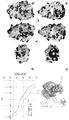
도 1은 전하 분포 및 pH-의존성 순전하를 나타낸 것이다. pH 7.4에서 양성자화되고 2 kBT/e에서 윤곽형성된 단백질의 등전위 표면; 흑색: 양/음. (a) 브리아키누맙. 경쇄는 밝은 회색으로 나타나 있고 중쇄는 더 진한 회색으로 나타나 있다. 중앙 및 오른쪽 그림의 시각은 각각 수직 및 수평 축 주위로 회전시킴으로써 왼쪽 패널의 시각과 연관된다. (b) 우스테키누맙. 경쇄 및 중쇄는 각각 밝은 회색 및 진한 회색으로 나타내었다. 시각은 (a)와 동일하다. (c) 인간 β2 마이크로글로불린(β2m)과의 복합체에서 인간 FcRn 상동성 모델의 2 kBT/e에서 윤곽형성된 등전위 표면. Fc 도메인은 명확성을 위해 나타내었다. (d) 브리아키누맙 및 우스테키누맙의 서열-기반 산출된 순전하 대 pH. 단백질 구조물은 디스커버리스튜디오 프로(DiscoveryStudio Pro)를 사용하여 제조되었다.
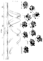
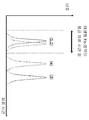
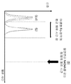
도 2는 pH-의존성 FcRn-IgG 상호작용을 나타낸 것이다. 11개 IgG 변이체의 FcRn 친화성 크로마토그램은 명확성을 위해 강도-표준화하였다. pH 7.4에서 양성자화된 구조 모델들의 분자 표면은 2 kBT/e에서 윤곽형성된 등전위 표면과 겹쳐 나타내었다. 시각은 도 1a에서의 오른쪽 패널과 동일하며 CDR 영역 상에 초점을 맞춘 것이다. 두번째 수평 축은 오프라인 pH 측정치들로부터 내삽된 용출 pH를 나타낸다.
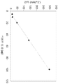
도 3은 인간 FcRn 유전자전이 마우스에서 약동학에 대한 FcRn 용출 pH의 영향을 나타낸 것이다. 항체는 군당 6마리 동물에게 10 mg/kg의 단일 i.v. 볼루스(bolus) 주사로서 투여되었다. 데이터 점들은 평균 ± 표준 편차를 나타낸다. (a) 브리아키누맙(오렌지색), 우스테키누맙(녹색), mAb 8(자주색) 및 mAb 9(청색)의 혈액내 수준 곡선. (b) 최종 반감기와 FcRn 컬럼 용출 pH와의 상관성.
도 4는 FcRn-IgG 모델의 분자 동역학 시뮬레이션을 나타낸 것이다. (a) 시뮬레이션 시작할 때의 형태. 파선은, 패널 (c)에 나타낸 바와 같이 MD 시뮬레이션동안 접근하는, Fv 영역에서 및 FcRn에서 2개의 예시 아미노산들 사이의 거리를 나타낸다. 색은 도 1과 동일하다. (b) 시뮬레이션 종료시 형태(t = 100 ns). 박스는 (c)에 나타낸 분자의 부분을 나타낸다. (c) FcRn과 Fv 도메인 사이의 상호작용의 상세도. 상호작용하는 프레임워크, CDR 및 FcRn 잔기들은 브리아키누맙 및 우스테키누맙에서 상이함을 주지하시오. (d) 시뮬레이션 과정동안 잔기 245(FcRn) 및 100(우스테키누맙 LC) 및 29(브리아키누맙 LC) 각각의 사이의 거리. (e) 시뮬레이션 종료시 상호작용 에너지(96, 97, 98, 99 및 100 ns에서 형태들의 평균 및 표준 편차). "VDW" 및 "정전기"는 각각 FcRn-Fab 상호작용에 대한 반-데르-발스(van-der-Waals) 및 정전기적 기여를 나타낸다. 단백질 구조물들은 파이몰(PyMol: 상표)(슈뢰딩거(Schrodinger) LLC)을 사용하여 제조하였다.
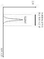
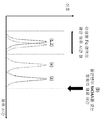
도 5는 브리아키누맙 및 우스테키누맙 경쇄 및 중쇄의 서열 정렬을 나타낸 것이다. VH 및 VL 영역들은 이탤릭체로 나타내었다; CDR은 별표(*)로 표시하였다; 우물정자(#)는 출발 구조물에서 FcRn에 매우 근접한(<4 Å) 아미노산을 의미한다. "¤" 기호는 MD 목적으로 FcRn에 다이설파이드 가교를 세우기 위해 Cys로 돌연변이된 잔기를 표시한 것이다.
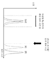
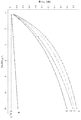
도 6은 브리아키누맙 및 우스테키누맙의 FcRn 친화성 컬럼 체류 시간의 염-의존성을 나타낸 것이다. 브리아키누맙 및 우스테키누맙은 증가하는 양의 NaCl의 존재하에서 pH 구배 용출하에 FcRn 컬럼 크로마토그래피에 적용되었다. 데이터는 용해된 염에 의한 전하 차폐 효과를 고려하기 위해 역제곱근 함수에 적합화시켰다. 브리아키누맙 체류 시간은 ![]()
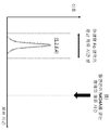
도 7은 적용된 항체의 선형성 및 본원에 보고된 바와 같은 FcRn 컬럼을 사용한 크로마토그래피의 곡선하 면적을 나타낸 것이다.
도 8은 본원에 보고된 바와 같은 FcRn 컬럼 상에서의 항-IGF-1R 항체 야생형 및 YTE-돌연변이체의 크로마토그램이다.
도 9는 아바스틴-야생형 및 아바스틴-돌연변이체의 FcRn 친화성 크로마토그램이다.
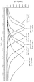
도 10은 Fc-영역과 항체-Fab의 항체-FcRn 상호작용에 의존하는 FcRn 친화성 크로마토그래피 컬럼 상에서의 체류 시간의 변화를 나타낸 도식이다; 1: 모 항체, 2: 모 항체 Fc-영역, 3: 변이 항체, 4: 변이 항체 Fc-영역; 실선: 완전 항체(항체-Fab+Fc-영역), 점선: Fc-영역만;
A: 야생형-유사 Fc-영역, 항체-Fab-FcRn 상호작용 없음;
B: 야생형-유사 Fc-영역, 항체-Fab-FcRn 상호작용;
C: 개선된 FcRn-결합을 갖는 조작된 Fc-영역, 항체-Fab-FcRn 상호작용 없음;
D: 개선된 FcRn-결합을 갖는 조작된 Fc-영역, 항체-Fab-FcRn 상호작용.
도 11은 항체-FcRn 상호작용이 개선된 제거율(임계 체류 시간보다 높은 체류 시간)을 제공하기 때문에 개선된 FcRn 결합, 항체-Fab-FcRn 상호작용을 갖지만 감소된 생체내 반감기를 갖는 조작된 항체를 나타내는 도식이다.
도 12는 Fc-영역과 항체-Fab의 항체-FcRn 상호작용에 의존하는 FcRn 친화성 크로마토그래피 컬럼 상에서의 체류 시간의 변화를 나타낸 도식이다; 1: 기준 항체, 2: 기준 항체 Fc-영역, 3: 항체, 4: 항체 Fc-영역; 실선: 완전 항체(항체-Fab+Fc-영역), 점선: Fc-영역만.
도 13은 염 농도 및 항체-Fab-FcRn 상호작용에 대한 FcRn 친화성 크로마토그래피 체류 시간의 의존성을 나타낸 것이다.
도 14는 베바시주맙 및 베바시주맙 변이체 경쇄 가변 도메인의 서열 정렬을 나타낸 것이다. 동일한 아미노산 및 유사한 아미노산들은 회색으로 나타내었다; CDR은 별표(*)로 표시하였다.
도 15는 브리아키누맙, 우스테키누맙 및 mAb 1 내지 6의 IL-12 상호작용을 나타낸 것이다; 1: 브리아키누맙, 2: 우스테키누맙, 3: mAb 1, 4: mAb 2, 5: mAb 3, 6: mAb 4, 7: mAb 5, 8: mAb 6.
도 16은 브리아키누맙, 우스테키누맙 및 mAb 7 내지 10의 IL-12 상호작용을 나타낸 것이다; 1: 브리아키누맙, 2: 우스테키누맙, 3: mAb 7, 4: mAb 8, 5: mAb 9, 6: mAb 10.Figure 1 shows the charge distribution and pH-dependent net charge. The equipotential surface of the protein are protonated contour formed from 2 k B T / e at pH 7.4; Black: positive / negative. (a) briquetinum. The light chain is light gray and the heavy chain is darker gray. The angles in the center and right figures are associated with the time of the left panel by rotating around the vertical and horizontal axes, respectively. (b) Uesutequinum. The light and heavy chains are shown as light gray and dark gray respectively. The time is the same as in (a). (c) a human β 2 microglobulin (β 2 m) and the equipotential surface in the complex formed from the contour 2 k B T / e of the human FcRn homology models. The Fc domain is shown for clarity. (d) Sequence-based calculated net charge versus pH of briacinum and utestinum. Protein constructs were constructed using DiscoveryStudio Pro.
Figure 2 shows the pH-dependent FcRn-IgG interaction. FcRn affinity chromatograms of 11 IgG variants were intensity-normalized for clarity. the molecular surface of the structural model protonated at pH 7.4 is shown to overlap with the equipotential surface formed in the outline 2 k B T / e. The view is the same as the right panel in FIG. 1A and focused on the CDR area. The second horizontal axis represents the elution pH interpolated from off-line pH measurements.
Figure 3 shows the effect of FcRn elution pH on pharmacokinetics in human FcRn transgenic mice. Antibodies were administered to 6 animals per group as a single bolus bolus injection at 10 mg / kg. Data points represent the mean ± standard deviation. (a) Level curves in blood of briacinumup (orange), uestekinum (green), mAb 8 (purple) and mAb 9 (blue). (b) Correlation between final half-life and pH of FcRn column elution.
Figure 4 shows a molecular dynamics simulation of an FcRn-IgG model. (a) The form at the start of the simulation. The dashed lines represent distances between the two exemplary amino acids in the Fv region and in FcRn, which are accessed during the MD simulation as shown in panel (c). The color is the same as in Fig. (b) At the end of simulation (t = 100 ns). The box represents the part of the molecule shown in (c). (c) Detailed view of interaction between FcRn and Fv domains. The interacting framework, CDR and FcRn residues are different in briacinum and utestinumum. (d) the distance between residues 245 (FcRn) and 100 (Ustekinumab LC) and 29 (briacinumab LC), respectively, during the simulation process. (e) Interaction energies at the end of the simulation (mean and standard deviation of shapes at 96, 97, 98, 99 and 100 ns). "VDW" and "electrostatic" denote van-der-wals and electrostatic contributions to FcRn-Fab interaction, respectively. Protein constructs were prepared using PyMol (trademark) (Schrodinger LLC).
Figure 5 depicts the sequence alignment of briacinum and streptomycin light and heavy chains. VH and VL regions are shown in italics; CDRs are marked with an asterisk (*); Well sperm (#) means amino acids very close to FcRn (<4 Å) in the starting structure. The symbol "¤ " denotes a residue mutated to Cys to establish a disulfide bridging in FcRn for MD purposes.
Figure 6 shows the salt-dependency of the residence time of the FcRn affinity column of briacinum and utestinumum. Briacinum and Ustekinumab were applied to FcRn column chromatography for pH gradient delivery in the presence of increasing amounts of NaCl. Data were fitted to the inverse square root function to account for charge shielding effects by dissolved salts. The residence time of briacinum was ![]()
Figure 7 shows the linearity of the antibody applied and the area under the curve of the chromatography using an FcRn column as reported herein.
Figure 8 is a chromatogram of anti-IGF-1R antibody wild type and YTE-mutants on FcRn columns as reported herein.
Figure 9 is an FcRn affinity chromatogram of Avastin-wild-type and Avastin-mutants.
Figure 10 is a schematic depicting the change in retention time on an FcRn affinity chromatography column depending on the antibody-FcRn interaction of the Fc-region and the antibody-Fab; 1: parent antibody, 2: parent antibody Fc-region, 3: mutant antibody, 4: mutant antibody Fc-region; Solid line: complete antibody (antibody-Fab + Fc- region), dotted line: Fc-region only;
A: wild-type-like Fc-region, no antibody-Fab-FcRn interaction;
B: wild-type-like Fc-region, antibody-Fab-FcRn interaction;
C: engineered Fc-region with improved FcRn-binding, no antibody-Fab-FcRn interaction;
D: engineered Fc-region, antibody-Fab-FcRn interaction with improved FcRn-binding.
Figure 11 shows that a modified antibody with improved FcRn binding, antibody-Fab-FcRn interaction but with reduced in vivo half-life, as antibody-FcRn interaction provides improved clearance (residence time higher than the threshold residence time) .
Figure 12 is a schematic depicting the change in retention time on an FcRn affinity chromatography column depending on the antibody-FcRn interaction of the Fc-region and the antibody-Fab; 1: reference antibody, 2: reference antibody Fc-region, 3: antibody, 4: antibody Fc-region; Solid line: Complete antibody (antibody-Fab + Fc- region), dotted line: Fc-region only.
Figure 13 shows the dependence of FcRn affinity chromatography retention time on salt concentration and antibody-Fab-FcRn interaction.
Figure 14 shows a sequence alignment of the bevacizumab and bevacizumab mutants light chain variable domains. The same amino acids and similar amino acids are grayed; CDRs are marked with an asterisk (*).
Figure 15 depicts IL-12 interactions of briacinum, utestinumab and mAbs 1-6; 1: briquetinum, 2: utestinumup, 3:
Figure 16 shows IL-12 interactions of briacinum, utestinumum and mAbs 7-10; 3: mAb 7, 4:
FcRn-mAb(mAb = 단클론성 항체) 상호작용에 대한 구조적 분석의 결과를 취합하면 Fv 도메인 및 특히 경쇄 가변 도메인(VL)이 FcRn-mAb 해리에 주요한 영향을 제공한다는 결론이 도출된다. 상기 결과는 Fv 도메인이 동족 FcRn-결합 부위로부터 떨어져 있기 때문에 예상치 못한 것이었다.Gathering the results of a structural analysis of the FcRn-mAb (mAb = monoclonal antibody) interaction leads to the conclusion that the Fv domain and in particular the light chain variable domain (VL) provide a major effect on FcRn-mAb dissociation. The results were unexpected because the Fv domain was away from the homologous FcRn-binding site.
항체들은 pH 6.0 친화도에서 차이를 나타내지 않았으므로, Fab 영역은 pH 6.0 결합에는 영향을 미치지 않는 것으로 보인다. 대조적으로, FcRn과 항체 사이의 해리는 Fab 영역에 의해 영향을 받았다.Since the antibodies did not show a difference in pH 6.0 affinity, the Fab domain appears to have no effect on pH 6.0 binding. In contrast, the dissociation between FcRn and antibody was affected by the Fab domain.
FcRn-IgG 해리 pH는 시험관내에서 생체내 최종 반감기와 선형으로 상관되었다. 결론적으로, 이러한 결과들은 더 높은 pH 값에서 더 느린 해리를 나타내는 항체가 세포내로 다시 운반되고 이어서 혈액 순환으로 다시 방출되는 대신에 분해된다는 가정을 뒷받침한다.The dissociation pH of FcRn-IgG was linearly correlated with the final half-life in vivo in vitro. In conclusion, these results support the hypothesis that at higher pH values, antibodies exhibiting slower dissociation are degraded instead of being transported back into the cell and subsequently released back into the blood circulation.
Fv 도메인내 전하 분포가 항체-FcRn 결합에 영향을 미치고 항체와 FcRn 사이에 추가의 상호작용을 야기하는 것으로 밝혀졌다. 이것은, 특히 pH 7.4에서의 항체-FcRn 복합체의 해리와 관련하여 FcRn 결합 특성을 변화시킴으로써, 항체의 FcRn-의존성 최종 반감기를 감소시킨다.It has been found that the charge distribution in the Fv domain affects antibody-FcRn binding and causes additional interactions between the antibody and FcRn. This reduces the FcRn-dependent final half-life of the antibody, in particular by altering the FcRn binding properties in conjunction with dissociation of the antibody-FcRn complex at pH 7.4.
정의Justice
단수형 용어는 1개 또는 2개 또는 3개 또는 4개 또는 5개 또는 6개 및 109 까지를 의미한다.Singular terms mean 1 or 2 or 3 or 4 or 5 or 6 and up to 10 9 .
용어 "약"은 그 다음에 뒤따르는 수치의 +/- 20%의 범위를 의미한다. 한 태양에서, 용어 약은 그 다음에 뒤따르는 수치의 +/- 10%의 범위를 의미한다. 한 태양에서, 용어 약은 그 다음에 뒤따르는 수치의 +/- 5%의 범위를 의미한다.The term " about "means a range of +/- 20% of the value that follows. In one embodiment, the term drug refers to a range of +/- 10% of the following numerical value. In one embodiment, the term drug refers to a range of +/- 5% of the following numerical value.
용어 "포함하는"은 또한 용어 "로 이루어지는"을 포함한다.The term " comprising "also includes the word" comprising ".
용어 "변이"는, 변형된 항체 또는 융합 폴리펩티드를 수득하기 위한, 모 항체 또는 융합 폴리펩티드, 예를 들면, 적어도 Fc-영역의 FcRn 결합 부위를 포함하는 융합 폴리펩티드에서 하나 이상의 아미노산 잔기들의 돌연변이(치환), 삽입(부가), 변형(유도체화), 또는 결실을 의미한다. 용어 "돌연변이"는 명시된 아미노산 잔기가 다른 아미노산 잔기 대신 치환되는 것을 의미한다. 예를 들면, 돌연변이 L234A는 항체 Fc-영역(폴리펩티드)내 위치 234에서 아미노산 잔기 라이신이 아미노산 잔기 알라닌으로 치환됨(알라닌에 의한 라이신의 치환)을 의미한다(EU 인덱스에 따른 번호).The term "mutation" refers to a mutation (substitution) of one or more amino acid residues in a fusion polypeptide comprising an FcRn binding site of a parent antibody or fusion polypeptide, such as at least an Fc-region, to obtain a modified antibody or fusion polypeptide. , Insertion (addition), modification (derivatization), or deletion. The term "mutation" means that the indicated amino acid residue is substituted for another amino acid residue. For example, mutation L234A means substitution of amino acid residue lysine with alanine residue alanine (substitution of lysine by alanine) at position 234 in the antibody Fc-region (polypeptide) (number according to EU index).
용어 "아미노산 돌연변이"는 또 다른 상이한 아미노산 잔기(= 대체 아미노산 잔기)에 의한 하나 이상의 기존 아미노산 잔기의 치환을 의미한다. 대체 아미노산 잔기는 "천연 아미노산 잔기"일 수 있으며, 알라닌(3 문자 코드: ala, 1 문자 코드: A), 아르기닌(arg, R), 아스파라긴(asn, N), 아스파트산(asp, D), 시스테인(cys, C), 글루타민(gln, Q), 글루탐산(glu, E), 글리신(gly, G), 히스티딘(his, H), 이소류신(ile, I), 류신(leu, L), 라이신(lys, K), 메티오닌(met, M), 페닐알라닌(phe, F), 프롤린(pro, P), 세린(ser, S), 트레오닌(thr, T), 트립토판(trp, W), 티로신(tyr, Y) 및 발린(val, V)으로부터 선택될 수 있다. 대체 아미노산 잔기는 "비-천연 아미노산 잔기"일 수 있다. 예를 들면, US 6,586,207, WO 98/48032, WO 03/073238, US 2004/0214988, WO 2005/35727, WO 2005/74524, 문헌 [Chin, J.W., et al., J. Am. Chem. Soc. 124 (2002) 9026-9027; Chin, J.W. and Schultz, P.G., ChemBioChem 11 (2002) 1135-1137; Chin, J.W., et al., PICAS United States of America 99 (2002) 11020-11024; and, Wang, L. and Schultz, P.G., Chem. (2002) 1-10](모두 전체로 본원에 참고로 인용됨)을 참조하시오.The term "amino acid mutation" refers to the substitution of one or more existing amino acid residues by another different amino acid residue (= alternative amino acid residue). Alternative amino acid residues can be "natural amino acid residues" and include alanine (3 letter code: ala, 1 letter code: A), arginine (arg, R), asparagine (asn, N), aspartic acid (asp, , Cysteine (cys, C), glutamine (gln, Q), glutamic acid (glu, E), glycine (gly, G), histidine (his, H), isoleucine (ile, I) (Lys, K), methionine (met, M), phenylalanine (phe, F), proline (pro, P), serine (S), threonine (thr, T), tryptophan (tyr, Y) and valine (val, V). The alternative amino acid residue may be a "non-natural amino acid residue ". See, for example, US 6,586,207, WO 98/48032, WO 03/073238, US 2004/0214988, WO 2005/35727, WO 2005/74524, Chin, J. W., et al., J. Am. Chem. Soc. 124 (2002) 9026-9027; Chin, J.W. and Schultz, P. G., Chem. Biochem 11 (2002) 1135-1137; Chin, J. W., et al., PICAS United States of America 99 (2002) 11020-11024; and Wang, L. and Schultz, P. G., Chem. (2002) 1-10] (all incorporated herein by reference in their entireties).
용어 "아미노산 삽입"은 아미노산 서열중 미리결정된 위치에서 1개 이상의 아미노산 잔기의 (추가) 혼입을 의미한다. 한 태양에서, 상기 삽입은 1개 또는 2개 아미노산 잔기의 삽입일 것이다. 삽입된 아미노산 잔기(들)은 임의의 천연 또는 비천연 아미노산 잔기일 수 있다.The term " amino acid insert "means (further) incorporation of one or more amino acid residues at a predetermined position in the amino acid sequence. In one embodiment, the insertion will be insertion of one or two amino acid residues. The inserted amino acid residue (s) may be any natural or unnatural amino acid residue.
용어 "아미노산 결실"은 아미노산 서열중 미리결정된 위치에서 1개 이상의 아미노산 잔기의 제거를 의미한다.The term " amino acid deletion "means the removal of one or more amino acid residues at a predetermined position in the amino acid sequence.
본원에서 용어 "항체"는 광의로 사용되며, 전장 항체이고 목적하는 항원- 및/또는 FcRn-결합 활성을 나타내는 한 단클론성 항체 및 다중특이성 항체(예를 들면, 이중특이성 항체, 삼중특이성 항체)를 포함하여(이로 한정되지는 않는다), 다양한 항체 구조물을 포함한다.As used herein, the term "antibody" is used broadly to refer to a monoclonal antibody and multispecific antibody (e. G., Bispecific antibody, trispecific antibody) that is a full length antibody and exhibits the desired antigen- and / or FcRn- Including, but not limited to, various antibody constructs.
용어 "(항원에) 결합하는"은 시험관내 분석에서 항체의 결합을 의미한다. 한 태양에서, 결합은, 항체를 표면에 결합시키고 항체에 대한 항원의 결합을 표면 플라즈몬 공명(Surface Plasmon Resonance, SPR)으로 측정하는 결합 분석법에서 측정된다. 결합은, 예를 들면, 10-8 M 이하, 일부 태양에서, 10-13 내지 10-8 M, 일부 태양에서, 10-13 내지 10-9 M의 결합 친화도(KD)를 의미한다.The term " binding to (antigen) "means binding of an antibody in an in vitro assay. In one embodiment, binding is measured in binding assays that bind the antibody to a surface and measure the binding of the antigen to the antibody by Surface Plasmon Resonance (SPR). Binding means binding affinity (K D ) of, for example, 10 -8 M or less, in some embodiments, 10 -13 to 10 -8 M, and in some embodiments, 10 -13 to 10 -9 M.
결합은 비아코어(BIAcore) 분석법(지이 헬쓰케어 바이오센서 에이비(GE Healthcare Biosensor AB), 스웨덴 웁살라 소재)에 의해 조사할 수 있다. 결합 친화도는 용어 ka(항체/항원 복합체로부터 항체의 회합에 대한 속도 상수), kd(해리 상수) 및 KD(kd/ka)로 정의된다.Binding can be investigated by the BIAcore assay (GE Healthcare Biosensor AB, Uppsala, Sweden). Binding affinity is defined as the term k a (rate constant for association of antibody from an antibody / antigen complex), k d (dissociation constant) and K D (k d / k a ).
용어 "완충 물질"은 용액 상태일 때, 예를 들면, 산성 또는 염기성 물질의 첨가 또는 방출로 인해 용액의 pH 값의 변화를 균등화시킬 수 있는 물질을 의미한다.The term "buffering material" refers to a material that, when in solution, can equalize the change in the pH value of the solution due to, for example, the addition or release of acidic or basic materials.
용어 "CH2-도메인"은 대략 EU 위치 231로부터 EU 위치 340(카밧에 따른 EU 번호 체계)까지 이어지는 항체 중쇄 폴리펩티드 부분을 의미한다. 한 태양에서, CH2 도메인은 서열번호 5: APELLGG PSVFLFPPKP KDTLMISRTP EVTCVWDVS HEDPEVKFNW YVDGVEVHNA KTKPREEQ E STYRWSVLT VLHQDWLNGK EYKCKVSNKA LPAPIEKTIS KAK의 아미노산 서열을 갖는다.The term "CH2-domain" means an antibody heavy chain polypeptide portion that extends from approximately EU position 231 to EU position 340 (EU numbering system according to Kabat). In one embodiment, the CH2 domain has the amino acid sequence of SEQ ID NO: 5: APELLGG PSVFLFPPKP KDTLMISRTP EVTCVWDVS HEDPEVKFNW YVDGVEVHNA KTKPREEQ E STYRWSVLT VLHQDWLNGK EYKCKVSNKA LPAPIEKTIS KAK.
용어 "CH3-도메인"은 대략 EU 위치 341로부터 EU 위치 446까지 이어지는 항체 중쇄 폴리펩티드 부분을 의미한다. 한 태양에서, CH3 도메인은 서열번호 6: GQPREPQ VYTLPPSRDE LTKNQVSLTC LVKGFYPSDI AVEWESNGQP ENNYKTTPPV LDSDGSFFLY SKLTVDKSRW QQGNVFSCSV MHEALHNHYT QKSLSLSPG의 아미노산 서열을 갖는다.The term "CH3-domain" refers to the portion of the antibody heavy chain polypeptide that extends from about EU position 341 to EU position 446. [ In one embodiment, the CH3 domain has the amino acid sequence of SEQ ID NO: 6: GQPREPQ VYTLPPSRDE LTKNQVSLTC LVKGFYPSDI AVEWESNGQP ENNYKTTPPV LDSDGSFFLY SKLTVDKSRW QQGNVFSCSV MHEALHNHYT QKSLSLSPG.
항체의 "클래스"는 그 중쇄가 갖는 불변 도메인 또는 불변 영역의 유형을 말한다. 항체의 5개 주요 클래스: IgA, IgD, IgE, IgG 및 IgM이 존재하며, 이들 중 여러개가 서브클래스(이소타입), 예를 들면, IgG1, IgG2, IgG3, IgG4, IgA1 및 IgA2로 더 분류될 수 있다. 면역글로불린의 상이한 클래스들에 상응하는 중쇄 불변 도메인은 각각 α, δ, ε, γ 및 μ로 불린다.The "class" of an antibody refers to the type of constant domain or constant region that the heavy chain has. Five of the antibodies major classes: IgA, IgD, IgE, IgG, and and IgM are present, there are multiple, for subclasses (isotypes), for example, of these, IgG 1, IgG 2, IgG 3,
약제, 예를 들면, 약학 제형의 "효과량"은 목적하는 치료 또는 예방 결과를 달성하기 위해 필요한 투여량으로 및 필요한 시간동안 효과적인 양을 말한다.The "effective amount" of an agent, e. G., A pharmaceutical formulation, refers to an amount that is effective for the amount of time required and the time required to achieve the desired therapeutic or prophylactic result.
용어 "Fc-융합 폴리펩티드"는, 목적하는 표적- 및/또는 단백질 A 및/또는 FcRn-결합 활성을 나타내는 항체 Fc-영역을 갖는 결합 도메인(예를 들면, 단일쇄 항체와 같은 항원 결합 도메인, 또는 수용체의 리간드와 같은 폴리펩티드)의 융합물을 의미한다.The term "Fc-fusion polypeptide" means a binding domain (e. G., An antigen binding domain, such as a single chain antibody, or an antigen binding domain) having an antibody Fc-region exhibiting the desired target- and / or protein A and / or FcRn- A polypeptide such as a ligand of a receptor).
용어 "인간 기원의 Fc-영역"은, 힌지(hinge) 영역, CH2 도메인 및 CH3 도메인의 적어도 일부분을 함유하는, 인간 기원의 면역글로불린 중쇄의 C-말단 영역을 의미한다. 한 태양에서, 인간 IgG 중쇄 Fc-영역은 Cys226으로부터, 또는 Pro230으로부터 중쇄의 카복실-말단까지 이어진다. 한 태양에서, Fc-영역은 서열번호 7의 아미노산 서열을 갖는다. 그러나, Fc-영역의 C-말단 라이신(Lys447)은 존재하거나 존재하지 않을 수 있다. 본원에서 달리 언급되지 않는 한, Fc-영역 또는 불변 영역중 아미노산 잔기의 번호는, 문헌 [Kabat, E.A. et al., Sequences of Proteins of Immunological Interest, 5th Ed. Public Health Service, National Institutes of Health, Bethesda, MD (1991), NIH Publication 91 3242]에 기술된 바와 같이, EU 인덱스로도 불리는 EU 번호 체계에 따른다. Fc-영역은, 폴리펩티드간 다이설파이드 결합을 형성하는 힌지 영역 시스테인 잔기들을 통해 서로에 공유 결합될 수 있는 2개의 중쇄 Fc-영역 폴리펩티드로 이루어진다.The term "Fc-region of human origin" means the C-terminal region of an immunoglobulin heavy chain of human origin, containing at least a portion of a hinge region, a CH2 domain and a CH3 domain. In one embodiment, the human IgG heavy chain Fc-region extends from Cys226 or from Pro230 to the carboxyl-terminal end of the heavy chain. In one embodiment, the Fc-region has the amino acid sequence of SEQ ID NO: 7. However, the C-terminal lysine (Lys447) of the Fc-region may or may not be present. Unless otherwise stated herein, the numbers of amino acid residues in the Fc-region or constant region are given in Kabat, E.A. et al., Sequences of Proteins of Immunological Interest, 5th Ed. According to the EU numbering system, also referred to as the EU index, as described in the Public Health Service, National Institutes of Health, Bethesda, MD (1991), NIH Publication 91 3242. The Fc-region consists of two heavy chain Fc-region polypeptides that can be covalently bound to each other through hinge region cysteine residues that form a disulfide bond between the polypeptides.
용어 "FcRn"은 인간 신생아 Fc-수용체를 의미한다. FcRn은 리소좀 분해 경로로부터 IgG를 구제하는 기능을 하여 감소된 제거율(clearance) 및 증가된 반감기를 제공한다. FcRn은 2개의 폴리펩티드: 50 kDa 클래스 I 주조직적합성 복합체-유사 단백질(α-FcRn) 및 15 kDa β2-마이크로글로불린(β2m)로 이루어지는 헤테로이량체성 단백질이다. FcRn은 IgG의 Fc-영역의 CH2-CH3 부분에 높은 친화도하에 결합한다. IgG와 FcRn 사이의 상호작용은 엄격하게 pH 의존성이며 1:2 화학량론으로 일어나되, 1개의 IgG가 그의 2개의 중쇄를 통해 2개의 FcRn 분자에 결합한다[Huber, A.H., et al., J. Mol. Biol. 230 (1993) 1077-1083]. FcRn 결합은 산성 pH(pH < 6.5)의 엔도솜에서 일어나며, IgG는 중성 세포 표면(약 7.4의 pH)에서 방출된다. 상호작용의 pH-민감 특성은, 엔도솜의 산성 환경 내에서 수용체에 결합함으로써 세포내 분해로부터 세포내에 음세포작용화된 IgG의 FcRn-매개 보호를 촉진한다. FcRn은 이어서 세포 표면으로의 IgG의 재순환 및 세포 외부의 중성 pH 환경에 FcRn-IgG 복합체의 노출시 혈류 내로의 후속 방출을 촉진한다.The term "FcRn" refers to the human neonatal Fc-receptor. FcRn functions to relieve IgG from the lysosomal degradation pathway, thereby providing reduced clearance and increased half-life. FcRn is a heterodimeric protein consisting of two polypeptides: a 50 kDa class I histocompatibility complex-like protein (? -FcRn) and 15 kDa? 2-microglobulin (? 2m). FcRn binds to the CH2-CH3 portion of the Fc-region of IgG with high affinity. The interaction between IgG and FcRn is strictly pH dependent and occurs at 1: 2 stoichiometry, with one IgG binding to two FcRn molecules through its two heavy chains [Huber, AH, et al. Mol. Biol. 230 (1993) 1077-1083]. FcRn binding occurs in endosomes at acidic pH (pH <6.5) and IgG is released at the neutrophil surface (pH of about 7.4). The pH-sensitive nature of the interaction promotes FcRn-mediated protection of intracellularly intracellularly IgGs from intracellular degradation by binding to receptors in the acidic environment of the endosome. FcRn then promotes recirculation of IgG to the cell surface and subsequent release into the bloodstream upon exposure of the FcRn-IgG complex to a neutral pH environment outside the cell.
용어 "Fc-영역의 FcRn 결합 부분"은, 대략 EU 위치 243으로부터 EU 위치 261까지, 및 대략 EU 위치 275로부터 EU 위치 293까지, 및 대략 EU 위치 302로부터 EU 위치 319까지, 및 대략 EU 위치 336으로부터 EU 위치 348까지, 및 대략 EU 위치 367로부터 EU 위치 393 및 EU 위치 408까지, 및 대략 EU 위치 424로부터 EU 위치 440까지 이어지는 항체 중쇄 폴리펩티드의 부분을 의미한다. 한 태양에서, 카밧의 EU 번호에 따른 하기의 아미노산 잔기들 중 하나 이상이 변이된다: F243, P244, P245, K246, P247, K248, D249, T250, L251, M252, I253, S254, R255, T256, P257, E258, V259, T260, C261, F275, N276, W277, Y278, V279, D280, V282, E283, V284, H285, N286, A287, K288, T289, K290, P291, R292, E293, V302, V303, S304, V305, L306, T307, V308, L309, H310, Q311, D312, W313, L314, N315, G316, K317, E318, Y319, I336, S337, K338, A339, K340, G341, Q342, P343, R344, E345, P346, Q347, V348, C367, V369, F372, Y373, P374, S375, D376, I377, A378, V379, E380, W381, E382, S383, N384, G385, Q386, P387, E388, N389, Y391, T393, S408, S424, C425, S426, V427, M428, H429, E430, A431, L432, H433, N434, H435, Y436, T437, Q438, K439, 및 S440(EU 번호체계).The term "FcRn binding portion of the Fc-region" refers to the FcRn binding portion of the FcRn region from about the EU position 243 to the EU position 261, and from about the EU position 275 to the EU position 293 and from about the EU position 302 to the EU position 319, To EU position 348, and from about EU position 367 to EU position 393 and EU position 408, and from about EU position 424 to EU position 440. In one embodiment, one or more of the following amino acid residues according to the EU number of Kabat are mutated: F243, P244, P245, K246, P247, K248, D249, T250, L251, M252, I253, S254, R255, T256, P257, E258, V259, T260, C261, F275, N276, W277, Y278, V279, D280, V282, E283, V284, H285, N286, A287, K288, T289, K290, P291, R292, E293, V302, V303, S304, V305, L306, T307, V308, L309, H310, Q311, D312, W313, L314, N315, G316, K317, E318, Y319, I336, S337, K338, A339, K340, G341, Q342, P343, E345, P346, Q347, V348, C367, V369, F372, Y373, P374, S375, D376, I377, A378, V379, E380, W381, E382, S383, N384, G385, Q386, P387, E388, T393, S408, S424, C425, S426, V427, M428, H429, E430, A431, L432, H433, N434, H435, Y436, T437, Q438, K439, and S440 (EU numbering system).
용어 "전장 항체"는 천연 항체 구조와 실질적으로 유사한 구조를 갖는 항체를 의미한다. 전장 항체는, 경쇄 가변 도메인 및 경쇄 불변 도메인을 포함하는 2개의 전장 항체 경쇄, 및 중쇄 가변 도메인, 제 1 불변 도메인, 힌지 영역, 제 2 불변 도메인 및 제 3 불변 도메인을 포함하는 2개의 전장 항체 중쇄를 포함한다. 전장 항체는, 예를 들면, 전장 항체의 상기 쇄들중 하나 이상에 접합된 추가의 scFv 또는 scFab와 같은 추가의 도메인들을 포함할 수 있다. 상기 접합체들은 또한 용어 전장 항체에 포함된다.The term "full-length antibody" means an antibody having a structure substantially similar to that of a native antibody. The full length antibody comprises two full length antibody light chains comprising a light chain variable domain and a light chain constant domain and two full length antibody heavy chains comprising a heavy chain variable domain, a first constant domain, a hinge region, a second constant domain and a third constant domain . The full-length antibody may comprise additional domains such as, for example, additional scFv or scFab conjugated to one or more of the above chains of the full length antibody. The conjugates are also included in the term full-length antibody.
용어 "힌지 영역"은, 예를 들면, 카밧의 EU 번호 체계에 따른 대략 위치 216으로부터 대략 위치 230까지의, CH1 도메인과 CH2 도메인을 연결시키는 항체 중쇄 폴리펩티드의 부분을 의미한다. 한 태양에서, 힌지 영역은 카밧의 EU 번호 체계에 따른 잔기 221 내지 230을 포함하는 단축된 힌지 영역이다. 힌지 영역은 통상적으로는 동일한 아미노산 서열을 갖는 2개의 폴리펩티드로 이루어진 이량체 분자이다. 힌지 영역은 일반적으로 약 25개 아미노산 잔기를 포함하고, 항원 결합 영역들이 독립적으로 움직이도록 유연하다. 힌지 영역은 3개의 도메인: 상부, 중앙 및 하부 힌지 도메인으로 세분될 수 있다[Roux, et al., J. Immunol. 161 (1998) 4083].The term "hinge region" means a portion of an antibody heavy chain polypeptide linking a CH1 domain and a CH2 domain from, for example, approximately position 216 to approximately position 230, according to Kabat's EU numbering system. In one embodiment, the hinge region is a shortened hinge region comprising residues 221 to 230 according to Kabat's EU numbering scheme. The hinge region is typically a dimer molecule composed of two polypeptides having the same amino acid sequence. The hinge region generally comprises about 25 amino acid residues, and the antigen binding regions are flexible to move independently. The hinge region can be subdivided into three domains: upper, middle and lower hinge domains [Roux, et al., J. Immunol. 161 (1998) 4083].
용어 "숙주 세포", "숙주 세포주" 및 "숙주 세포 배양물"은 상호교환적으로 사용되며, 상기 세포들의 자손을 포함하여, 외인성 핵산이 도입된 세포를 말한다. 숙주 세포는 "형질전환체" 및 "형질전환 세포"를 포함하며, 이들은 1차 형질전환된 세포, 및 계대 수에 관계없이 그로부터 유도된 자손을 포함한다. 자손은 핵산 함량에 있어서 모 세포와 완전히 동일하지 않을 수 있으며, 돌연변이를 함유할 수 있다. 원래 형질전환된 세포에서 선별되거나 선택된 바와 동일한 기능 또는 생물 활성을 갖는 돌연변이 자손이 본 발명에 포함된다.The terms "host cell," " host cell line, "and" host cell culture "are used interchangeably and refer to a cell into which an exogenous nucleic acid has been introduced, including progeny of such cells. Host cells include "transformants" and "transformed cells" which include primary transformed cells and progeny derived therefrom, regardless of number of passages. The offspring may not be completely identical to the parent cells in the nucleic acid content, and may contain mutations. Mutant progeny having the same function or biological activity as selected or selected in the originally transformed cells are included in the present invention.
용어 "로부터 유도된"은 아미노산 서열이 적어도 한 위치에 변이를 도입함으로써 모 아미노산 서열로부터 유도된 것을 의미한다. 따라서, 유도된 아미노산 서열은 적어도 1개의 상응하는 위치에서 상응하는 모 아미노산 서열과 상이하다(항체 Fc-영역에 대해 카밧 EU 인덱스에 따른 번호). 한 태양에서, 모 아미노산 서열로부터 유도된 아미노산 서열은 상응하는 위치에서 1 내지 15개 아미노산 잔기가 상이하다. 한 태양에서, 모 아미노산 서열로부터 유도된 아미노산 서열은 상응하는 위치에서 1 내지 10개 아미노산 잔기가 상이하다. 한 태양에서, 모 아미노산 서열로부터 유도된 아미노산 서열은 상응하는 위치에서 1 내지 6개 아미노산 잔기가 상이하다. 유사하게, 유도된 아미노산 서열은 그의 모 아미노산 서열에 대해 높은 아미노산 서열 동일성을 갖는다. 한 태양에서, 모 아미노산 서열로부터 유도된 아미노산 서열은 80% 이상의 서열 동일성을 갖는다. 한 태양에서, 모 아미노산 서열로부터 유도된 아미노산 서열은 90% 이상의 서열 동일성을 갖는다. 한 태양에서, 모 아미노산 서열로부터 유도된 아미노산 서열은 95% 이상의 서열 동일성을 갖는다.The term "derived from" means that an amino acid sequence is derived from a parent amino acid sequence by introducing a mutation in at least one position. Thus, the derived amino acid sequence differs from the corresponding parent amino acid sequence in at least one corresponding position (number according to Kabat EU index for antibody Fc-region). In one embodiment, the amino acid sequence derived from the parent amino acid sequence differs from 1 to 15 amino acid residues at the corresponding position. In one embodiment, the amino acid sequence derived from the parent amino acid sequence differs from 1 to 10 amino acid residues at the corresponding position. In one embodiment, the amino acid sequence derived from the parent amino acid sequence differs from the corresponding position by one to six amino acid residues. Similarly, the derived amino acid sequence has high amino acid sequence identity to its parent amino acid sequence. In one embodiment, the amino acid sequence derived from a parent amino acid sequence has at least 80% sequence identity. In one embodiment, the amino acid sequence derived from the parent amino acid sequence has at least 90% sequence identity. In one embodiment, the amino acid sequence derived from the parent amino acid sequence has at least 95% sequence identity.
용어 "인간 Fc-영역 폴리펩티드"는 "천연" 또는 "야생형" 인간 Fc-영역 폴리펩티드와 동일한 아미노산 서열을 의미한다. 용어 "변이 (인간) Fc-영역 폴리펩티드"는 1개 이상의 "아미노산 변이"에 의해 "천연" 또는 "야생형" 인간 Fc-영역 폴리펩티드로부터 유도된 아미노산 서열을 의미한다. "인간 Fc-영역"은 2개의 인간 Fc-영역 폴리펩티드로 이루어진다. "변이 (인간) Fc-영역"은 2개의 Fc-영역 폴리펩티드로 이루어지며, 이때 이들은 둘 다 변이 (인간) Fc-영역 폴리펩티드일 수 있거나, 또는 하나는 인간 Fc-영역 폴리펩티드이고 다른 하나는 변이 (인간) Fc-영역 폴리펩티드이다.The term "human Fc-region polypeptide" means the same amino acid sequence as "native" or "wild type" human Fc-region polypeptide. The term "mutant (human) Fc-region polypeptide" means an amino acid sequence derived from a "native" or "wild type" human Fc-region polypeptide by one or more "amino acid mutations". A "human Fc-region" consists of two human Fc-region polypeptides. A "mutant (human) Fc-region" consists of two Fc-region polypeptides, both of which may be mutated (human) Fc-region polypeptides, or one is a human Fc-region polypeptide and the other is a mutation Human) Fc-region polypeptide.
한 태양에서, 인간 Fc-영역 폴리펩티드는 서열번호 7의 인간 IgG1 Fc-영역 폴리펩티드의, 또는 서열번호 8의 인간 IgG2 Fc-영역 폴리펩티드의, 또는 서열번호 9의 인간 IgG3 Fc-영역 폴리펩티드의, 또는 서열번호 10의 인간 IgG4 Fc-영역 폴리펩티드의 아미노산 서열을 갖는다. 한 태양에서, Fc-영역 폴리펩티드는 서열번호 7, 또는 8, 또는 9, 또는 10의 Fc-영역 폴리펩티드로부터 유도되고, 서열번호 7, 또는 8, 또는 9, 또는 10의 Fc-영역 폴리펩티드에 비해 1개 이상의 아미노산 돌연변이를 갖는다. 한 태양에서, Fc-영역 폴리펩티드는 약 1 내지 약 10개의 아미노산 돌연변이, 및 한 태양에서 약 1 내지 약 5개의 아미노산 돌연변이를 포함한다/갖는다. 한 태양에서, Fc-영역 폴리펩티드는 서열번호 7, 또는 8, 또는 9, 또는 10의 인간 Fc-영역 폴리펩티드와 약 80% 이상의 상동성을 갖는다. 한 태양에서, Fc-영역 폴리펩티드는 서열번호 7, 또는 8, 또는 9, 또는 10의 인간 Fc-영역 폴리펩티드와 약 90% 이상의 상동성을 갖는다. 한 태양에서, Fc-영역 폴리펩티드는 서열번호 7, 또는 8, 또는 9, 또는 10의 인간 Fc-영역 폴리펩티드와 약 95% 이상의 상동성을 갖는다. In one embodiment, the human Fc-region polypeptide comprises a human IgG1 Fc-region polypeptide of SEQ ID NO: 7, or of a human IgG2 Fc-region polypeptide of SEQ ID NO: 8, or of a human IgG3 Fc- Lt; RTI ID = 0.0 > 10 < / RTI > human IgG4 Fc-region polypeptide. In one embodiment, the Fc-region polypeptide is derived from an Fc-region polypeptide of SEQ ID NO: 7, or 8, or 9, or 10, and is derived from an Fc- region polypeptide of SEQ ID NO: 7 or 8, or 9, More than one amino acid mutation. In one embodiment, the Fc-region polypeptide comprises about 1 to about 10 amino acid mutations, and in one embodiment about 1 to about 5 amino acid mutations. In one embodiment, the Fc-region polypeptide has about 80% or more homology with a human Fc-region polypeptide of SEQ ID NO: 7, or 8, or 9, In one embodiment, the Fc-region polypeptide has at least about 90% homology with a human Fc-region polypeptide of SEQ ID NO: 7, or 8, or 9, In one embodiment, the Fc-region polypeptide has about 95% or more homology with a human Fc-region polypeptide of SEQ ID NO: 7, or 8, or 9,
서열번호 7, 또는 8, 또는 9, 또는 10의 인간 Fc-영역 폴리펩티드로부터 유도된 Fc-영역 폴리펩티드는 함유된 아미노산 변이에 의해 정의된다. 따라서, 예를 들면, 용어 P329G는, 서열번호 7, 또는 8, 또는 9, 또는 10의 인간 Fc-영역 폴리펩티드와 비교하여 아미노산 위치 329에 프롤린의 글리신으로의 돌연변이를 갖는 Fc-영역 폴리펩티드 유도된 인간 Fc-영역 폴리펩티드를 의미한다.An Fc-region polypeptide derived from a human Fc-region polypeptide of SEQ ID NO: 7, or 8, or 9, or 10 is defined by the amino acid variation contained. Thus, for example, the term P329G is an Fc-region polypeptide derived human having a mutation of proline to glycine at amino acid position 329 compared to a human Fc-region polypeptide of SEQ ID NO: 7, or 8, Lt; RTI ID = 0.0 > Fc-region < / RTI > polypeptide.
본 발명에서 논의된 모든 중쇄 위치에 있어서, 번호는 EU 인덱스에 따른다. EU 인덱스 또는 카밧에서와 같은 EU 인덱스 또는 카밧 EU 인덱스 또는 EU 번호 체계는 EU 항체의 번호를 말한다(본원에 전체로 참고로 인용된 문헌 [Edelman, et al., Proc. Natl. Acad. Sci. USA 63 (1969) 78-85]). 경쇄 잔기의 번호는 카밧 명명법에 따른다[Kabat, E.A., et al., Sequences of Proteins of Immunological Interest, 5th ed., Public Health Service, National Institutes of Health, Bethesda, MD (1991), NIH Publication 91 3242].For all heavy chain positions discussed in the present invention, the numbers are in accordance with the EU index. The EU index, such as in the EU index or Kabat, or the Kabat EU index or EU numbering system refers to the number of the EU antibody (see Edelman, et al., Proc. Natl. Acad. Sci. USA 63 (1969) 78-85). The number of light chain residues follows the Kabat nomenclature [Kabat, EA, et al., Sequences of Proteins of Immunological Interest, 5th ed., National Institutes of Health, Bethesda, MD (1991) .
인간 IgG1 Fc-영역 폴리펩티드는 하기의 아미노산 서열을 갖는다:The human IgG1 Fc-region polypeptide has the following amino acid sequence:
DKTHTCPPCPAPELLGGPSVFLFPPKPKDTLMISRTPEVTCVVVDVSHEDPEVKFNWYVDGVEVHNAKTKPREEQYNSTYRVVSVLTVLHQDWLNGKEYKCKVSNKALPAPIEKTISKAKGQPREPQVYTLPPSRDELTKNQVSLTCLVKGFYPSDIAVEWESNGQPENNYKTTPPVLDSDGSFFLYSKLTVDKSRWQQGNVFSCSVMHEALHNHYTQKSLSLSPGK (서열번호 7).DKTHTCPPCPAPELLGGPSVFLFPPKPKDTLMISRTPEVTCVVVDVSHEDPEVKFNWYVDGVEVHNAKTKPREEQYNSTYRVVSVLTVLHQDWLNGKEYKCKVSNKALPAPIEKTISKAKGQPREPQVYTLPPSRDELTKNQVSLTCLVKGFYPSDIAVEWESNGQPENNYKTTPPVLDSDGSFFLYSKLTVDKSRWQQGNVFSCSVMHEALHNHYTQKSLSLSPGK (SEQ ID NO: 7).
돌연변이 L234A, L235A를 갖는 인간 IgG1 Fc-영역 유도된 Fc-영역 폴리펩티드는 하기의 아미노산 서열을 갖는다:The human IgG1 Fc-region derived Fc-region polypeptides with mutations L234A, L235A have the following amino acid sequences:
DKTHTCPPCPAPEAAGGPSVFLFPPKPKDTLMISRTPEVTCVVVDVSHEDPEVKFNWYVDGVEVHNAKTKPREEQYNSTYRVVSVLTVLHQDWLNGKEYKCKVSNKALPAPIEKTISKAKGQPREPQVYTLPPSRDELTKNQVSLTCLVKGFYPSDIAVEWESNGQPENNYKTTPPVLDSDGSFFLYSKLTVDKSRWQQGNVFSCSVMHEALHNHYTQKSLSLSPGK (서열번호 11).DKTHTCPPCPAPEAAGGPSVFLFPPKPKDTLMISRTPEVTCVVVDVSHEDPEVKFNWYVDGVEVHNAKTKPREEQYNSTYRVVSVLTVLHQDWLNGKEYKCKVSNKALPAPIEKTISKAKGQPREPQVYTLPPSRDELTKNQVSLTCLVKGFYPSDIAVEWESNGQPENNYKTTPPVLDSDGSFFLYSKLTVDKSRWQQGNVFSCSVMHEALHNHYTQKSLSLSPGK (SEQ ID NO: 11).
Y349C, T366S, L368A 및 Y407V 돌연변이를 갖는 인간 IgG1 Fc-영역 유도된 Fc-영역 폴리펩티드는 하기의 아미노산 서열을 갖는다:The human IgGl Fc-region derived Fc-region polypeptides with Y349C, T366S, L368A and Y407V mutations have the following amino acid sequences:
DKTHTCPPCPAPELLGGPSVFLFPPKPKDTLMISRTPEVTCVVVDVSHEDPEVKFNWYVDGVEVHNAKTKPREEQYNSTYRVVSVLTVLHQDWLNGKEYKCKVSNKALPAPIEKTISKAKGQPREPQVCTLPPSRDELTKNQVSLSCAVKGFYPSDIAVEWESNGQPENNYKTTPPVLDSDGSFFLVSKLTVDKSRWQQGNVFSCSVMHEALHNHYTQKSLSLSPGK (서열번호 12).DKTHTCPPCPAPELLGGPSVFLFPPKPKDTLMISRTPEVTCVVVDVSHEDPEVKFNWYVDGVEVHNAKTKPREEQYNSTYRVVSVLTVLHQDWLNGKEYKCKVSNKALPAPIEKTISKAKGQPREPQVCTLPPSRDELTKNQVSLSCAVKGFYPSDIAVEWESNGQPENNYKTTPPVLDSDGSFFLVSKLTVDKSRWQQGNVFSCSVMHEALHNHYTQKSLSLSPGK (SEQ ID NO: 12).
S354C, T366W 돌연변이를 갖는 인간 IgG1 Fc-영역 유도된 Fc-영역 폴리펩티드는 하기의 아미노산 서열을 갖는다:The human IgGl Fc-region derived Fc-region polypeptides with S354C, T366W mutations have the following amino acid sequences:
DKTHTCPPCPAPELLGGPSVFLFPPKPKDTLMISRTPEVTCVVVDVSHEDPEVKFNWYVDGVEVHNAKTKPREEQYNSTYRVVSVLTVLHQDWLNGKEYKCKVSNKALPAPIEKTISKAKGQPREPQVYTLPPCRDELTKNQVSLWCLVKGFYPSDIAVEWESNGQPENNYKTTPPVLDSDGSFFLYSKLTVDKSRWQQGNVFSCSVMHEALHNHYTQKSLSLSPGK (서열번호 13).DKTHTCPPCPAPELLGGPSVFLFPPKPKDTLMISRTPEVTCVVVDVSHEDPEVKFNWYVDGVEVHNAKTKPREEQYNSTYRVVSVLTVLHQDWLNGKEYKCKVSNKALPAPIEKTISKAKGQPREPQVYTLPPCRDELTKNQVSLWCLVKGFYPSDIAVEWESNGQPENNYKTTPPVLDSDGSFFLYSKLTVDKSRWQQGNVFSCSVMHEALHNHYTQKSLSLSPGK (SEQ ID NO: 13).
L234A, L235A 돌연변이 및 Y349C, T366S, L368A, Y407V 돌연변이를 갖는 인간 IgG1 Fc-영역 유도된 Fc-영역 폴리펩티드는 하기의 아미노산 서열을 갖는다:The human IgGl Fc-region derived Fc-region polypeptides with L234A, L235A mutations and Y349C, T366S, L368A, Y407V mutations have the following amino acid sequences:
DKTHTCPPCPAPEAAGGPSVFLFPPKPKDTLMISRTPEVTCVVVDVSHEDPEVKFNWYVDGVEVHNAKTKPREEQYNSTYRVVSVLTVLHQDWLNGKEYKCKVSNKALPAPIEKTISKAKGQPREPQVCTLPPSRDELTKNQVSLSCAVKGFYPSDIAVEWESNGQPENNYKTTPPVLDSDGSFFLVSKLTVDKSRWQQGNVFSCSVMHEALHNHYTQKSLSLSPGK (서열번호 14).DKTHTCPPCPAPEAAGGPSVFLFPPKPKDTLMISRTPEVTCVVVDVSHEDPEVKFNWYVDGVEVHNAKTKPREEQYNSTYRVVSVLTVLHQDWLNGKEYKCKVSNKALPAPIEKTISKAKGQPREPQVCTLPPSRDELTKNQVSLSCAVKGFYPSDIAVEWESNGQPENNYKTTPPVLDSDGSFFLVSKLTVDKSRWQQGNVFSCSVMHEALHNHYTQKSLSLSPGK (SEQ ID NO: 14).
L234A, L235A 및 S354C, T366W 돌연변이를 갖는 인간 IgG1 Fc-영역 유도된 Fc-영역 폴리펩티드는 하기의 아미노산 서열을 갖는다:The human IgGl Fc-region derived Fc-region polypeptides with L234A, L235A and S354C, T366W mutations have the following amino acid sequences:
DKTHTCPPCPAPEAAGGPSVFLFPPKPKDTLMISRTPEVTCVVVDVSHEDPEVKFNWYVDGVEVHNAKTKPREEQYNSTYRVVSVLTVLHQDWLNGKEYKCKVSNKALPAPIEKTISKAKGQPREPQVYTLPPCRDELTKNQVSLWCLVKGFYPSDIAVEWESNGQPENNYKTTPPVLDSDGSFFLYSKLTVDKSRWQQGNVFSCSVMHEALHNHYTQKSLSLSPGK (서열번호 15).DKTHTCPPCPAPEAAGGPSVFLFPPKPKDTLMISRTPEVTCVVVDVSHEDPEVKFNWYVDGVEVHNAKTKPREEQYNSTYRVVSVLTVLHQDWLNGKEYKCKVSNKALPAPIEKTISKAKGQPREPQVYTLPPCRDELTKNQVSLWCLVKGFYPSDIAVEWESNGQPENNYKTTPPVLDSDGSFFLYSKLTVDKSRWQQGNVFSCSVMHEALHNHYTQKSLSLSPGK (SEQ ID NO: 15).
P329G 돌연변이를 갖는 인간 IgG1 Fc-영역 유도된 Fc-영역 폴리펩티드는 하기의 아미노산 서열을 갖는다:The human IgG1 Fc-region derived Fc-region polypeptide with P329G mutation has the following amino acid sequence:
DKTHTCPPCPAPELLGGPSVFLFPPKPKDTLMISRTPEVTCVVVDVSHEDPEVKFNWYVDGVEVHNAKTKPREEQYNSTYRVVSVLTVLHQDWLNGKEYKCKVSNKALGAPIEKTISKAKGQPREPQVYTLPPSRDELTKNQVSLTCLVKGFYPSDIAVEWESNGQPENNYKTTPPVLDSDGSFFLYSKLTVDKSRWQQGNVFSCSVMHEALHNHYTQKSLSLSPGK (서열번호 16).DKTHTCPPCPAPELLGGPSVFLFPPKPKDTLMISRTPEVTCVVVDVSHEDPEVKFNWYVDGVEVHNAKTKPREEQYNSTYRVVSVLTVLHQDWLNGKEYKCKVSNKALGAPIEKTISKAKGQPREPQVYTLPPSRDELTKNQVSLTCLVKGFYPSDIAVEWESNGQPENNYKTTPPVLDSDGSFFLYSKLTVDKSRWQQGNVFSCSVMHEALHNHYTQKSLSLSPGK (SEQ ID NO: 16).
L234A, L235A 돌연변이 및 P329G 돌연변이를 갖는 인간 IgG1 Fc-영역 유도된 Fc-영역 폴리펩티드는 하기의 아미노산 서열을 갖는다:The human IgGl Fc-region derived Fc-region polypeptide with L234A, L235A mutation and P329G mutation has the following amino acid sequence:
DKTHTCPPCPAPEAAGGPSVFLFPPKPKDTLMISRTPEVTCVVVDVSHEDPEVKFNWYVDGVEVHNAKTKPREEQYNSTYRVVSVLTVLHQDWLNGKEYKCKVSNKALGAPIEKTISKAKGQPREPQVYTLPPSRDELTKNQVSLTCLVKGFYPSDIAVEWESNGQPENNYKTTPPVLDSDGSFFLYSKLTVDKSRWQQGNVFSCSVMHEALHNHYTQKSLSLSPGK (서열번호 17).DKTHTCPPCPAPEAAGGPSVFLFPPKPKDTLMISRTPEVTCVVVDVSHEDPEVKFNWYVDGVEVHNAKTKPREEQYNSTYRVVSVLTVLHQDWLNGKEYKCKVSNKALGAPIEKTISKAKGQPREPQVYTLPPSRDELTKNQVSLTCLVKGFYPSDIAVEWESNGQPENNYKTTPPVLDSDGSFFLYSKLTVDKSRWQQGNVFSCSVMHEALHNHYTQKSLSLSPGK (SEQ ID NO: 17).
P329G 돌연변이 및 Y349C, T366S, L368A, Y407V 돌연변이 갖는 인간 IgG1 Fc-영역 유도된 Fc-영역 폴리펩티드는 하기의 아미노산 서열을 갖는다:The human IgG1 Fc-region derived Fc-region polypeptides with the P329G mutation and the Y349C, T366S, L368A, Y407V mutants have the following amino acid sequences:
DKTHTCPPCPAPELLGGPSVFLFPPKPKDTLMISRTPEVTCVVVDVSHEDPEVKFNWYVDGVEVHNAKTKPREEQYNSTYRVVSVLTVLHQDWLNGKEYKCKVSNKALGAPIEKTISKAKGQPREPQVCTLPPSRDELTKNQVSLSCAVKGFYPSDIAVEWESNGQPENNYKTTPPVLDSDGSFFLVSKLTVDKSRWQQGNVFSCSVMHEALHNHYTQKSLSLSPGK (서열번호 18).DKTHTCPPCPAPELLGGPSVFLFPPKPKDTLMISRTPEVTCVVVDVSHEDPEVKFNWYVDGVEVHNAKTKPREEQYNSTYRVVSVLTVLHQDWLNGKEYKCKVSNKALGAPIEKTISKAKGQPREPQVCTLPPSRDELTKNQVSLSCAVKGFYPSDIAVEWESNGQPENNYKTTPPVLDSDGSFFLVSKLTVDKSRWQQGNVFSCSVMHEALHNHYTQKSLSLSPGK (SEQ ID NO: 18).
P329G 돌연변이 및 S354C, T366W 돌연변이를 갖는 인간 IgG1 Fc-영역 유도된 Fc-영역 폴리펩티드는 하기의 아미노산 서열을 갖는다:The human IgG1 Fc-region derived Fc-region polypeptide with the P329G mutation and the S354C, T366W mutation has the following amino acid sequence:
DKTHTCPPCPAPELLGGPSVFLFPPKPKDTLMISRTPEVTCVVVDVSHEDPEVKFNWYVDGVEVHNAKTKPREEQYNSTYRVVSVLTVLHQDWLNGKEYKCKVSNKALGAPIEKTISKAKGQPREPQVYTLPPCRDELTKNQVSLWCLVKGFYPSDIAVEWESNGQPENNYKTTPPVLDSDGSFFLYSKLTVDKSRWQQGNVFSCSVMHEALHNHYTQKSLSLSPGK (서열번호 19).DKTHTCPPCPAPELLGGPSVFLFPPKPKDTLMISRTPEVTCVVVDVSHEDPEVKFNWYVDGVEVHNAKTKPREEQYNSTYRVVSVLTVLHQDWLNGKEYKCKVSNKALGAPIEKTISKAKGQPREPQVYTLPPCRDELTKNQVSLWCLVKGFYPSDIAVEWESNGQPENNYKTTPPVLDSDGSFFLYSKLTVDKSRWQQGNVFSCSVMHEALHNHYTQKSLSLSPGK (SEQ ID NO: 19).
L234A, L235A, P329G 및 Y349C, T366S, L368A, Y407V 돌연변이를 갖는 인간 IgG1 Fc-영역 유도된 Fc-영역 폴리펩티드는 하기의 아미노산 서열을 갖는다:The human IgG1 Fc-region derived Fc-region polypeptides with L234A, L235A, P329G and Y349C, T366S, L368A, Y407V mutations have the following amino acid sequences:
DKTHTCPPCPAPEAAGGPSVFLFPPKPKDTLMISRTPEVTCVVVDVSHEDPEVKFNWYVDGVEVHNAKTKPREEQYNSTYRVVSVLTVLHQDWLNGKEYKCKVSNKALGAPIEKTISKAKGQPREPQVCTLPPSRDELTKNQVSLSCAVKGFYPSDIAVEWESNGQPENNYKTTPPVLDSDGSFFLVSKLTVDKSRWQQGNVFSCSVMHEALHNHYTQKSLSLSPGK (서열번호 20).DKTHTCPPCPAPEAAGGPSVFLFPPKPKDTLMISRTPEVTCVVVDVSHEDPEVKFNWYVDGVEVHNAKTKPREEQYNSTYRVVSVLTVLHQDWLNGKEYKCKVSNKALGAPIEKTISKAKGQPREPQVCTLPPSRDELTKNQVSLSCAVKGFYPSDIAVEWESNGQPENNYKTTPPVLDSDGSFFLVSKLTVDKSRWQQGNVFSCSVMHEALHNHYTQKSLSLSPGK (SEQ ID NO: 20).
L234A, L235A, P329G 돌연변이 및 S354C, T366W 돌연변이를 갖는 인간 IgG1 Fc-영역 유도된 Fc-영역 폴리펩티드는 하기의 아미노산 서열을 갖는다:The human IgGl Fc-region derived Fc-region polypeptides with L234A, L235A, P329G mutations and S354C, T366W mutations have the following amino acid sequences:
DKTHTCPPCPAPEAAGGPSVFLFPPKPKDTLMISRTPEVTCVVVDVSHEDPEVKFNWYVDGVEVHNAKTKPREEQYNSTYRVVSVLTVLHQDWLNGKEYKCKVSNKALGAPIEKTISKAKGQPREPQVYTLPPCRDELTKNQVSLWCLVKGFYPSDIAVEWESNGQPENNYKTTPPVLDSDGSFFLYSKLTVDKSRWQQGNVFSCSVMHEALHNHYTQKSLSLSPGK (서열번호 21).DKTHTCPPCPAPEAAGGPSVFLFPPKPKDTLMISRTPEVTCVVVDVSHEDPEVKFNWYVDGVEVHNAKTKPREEQYNSTYRVVSVLTVLHQDWLNGKEYKCKVSNKALGAPIEKTISKAKGQPREPQVYTLPPCRDELTKNQVSLWCLVKGFYPSDIAVEWESNGQPENNYKTTPPVLDSDGSFFLYSKLTVDKSRWQQGNVFSCSVMHEALHNHYTQKSLSLSPGK (SEQ ID NO: 21).
인간 IgG4 Fc-영역 폴리펩티드는 하기의 아미노산 서열을 갖는다:The human IgG4 Fc-region polypeptide has the following amino acid sequence:
ESKYGPPCPSCPAPEFLGGPSVFLFPPKPKDTLMISRTPEVTCVVVDVSQEDPEVQFNWYVDGVEVHNAKTKPREEQFNSTYRVVSVLTVLHQDWLNGKEYKCKVSNKGLPSSIEKTISKAKGQPREPQVYTLPPSQEEMTKNQVSLTCLVKGFYPSDIAVEWESNGQPENNYKTTPPVLDSDGSFFLYSRLTVDKSRWQEGNVFSCSVMHEALHNHYTQKSLSLSLGK (서열번호 10).ESKYGPPCPSCPAPEFLGGPSVFLFPPKPKDTLMISRTPEVTCVVVDVSQEDPEVQFNWYVDGVEVHNAKTKPREEQFNSTYRVVSVLTVLHQDWLNGKEYKCKVSNKGLPSSIEKTISKAKGQPREPQVYTLPPSQEEMTKNQVSLTCLVKGFYPSDIAVEWESNGQPENNYKTTPPVLDSDGSFFLYSRLTVDKSRWQEGNVFSCSVMHEALHNHYTQKSLSLSLGK (SEQ ID NO: 10).
S228P 및 L235E 돌연변이를 갖는 인간 IgG4 Fc-영역 유도된 Fc-영역 폴리펩티드는 하기의 아미노산 서열을 갖는다:The human IgG4 Fc-region derived Fc-region polypeptides with S228P and L235E mutations have the following amino acid sequences:
ESKYGPPCPPCPAPEFEGGPSVFLFPPKPKDTLMISRTPEVTCVVVDVSQEDPEVQFNWYVDGVEVHNAKTKPREEQFNSTYRVVSVLTVLHQDWLNGKEYKCKVSNKGLPSSIEKTISKAKGQPREPQVYTLPPSQEEMTKNQVSLTCLVKGFYPSDIAVEWESNGQPENNYKTTPPVLDSDGSFFLYSRLTVDKSRWQEGNVFSCSVMHEALHNHYTQKSLSLSLGK (서열번호 22).ESKYGPPCPPCPAPEFEGGPSVFLFPPKPKDTLMISRTPEVTCVVVDVSQEDPEVQFNWYVDGVEVHNAKTKPREEQFNSTYRVVSVLTVLHQDWLNGKEYKCKVSNKGLPSSIEKTISKAKGQPREPQVYTLPPSQEEMTKNQVSLTCLVKGFYPSDIAVEWESNGQPENNYKTTPPVLDSDGSFFLYSRLTVDKSRWQEGNVFSCSVMHEALHNHYTQKSLSLSLGK (SEQ ID NO: 22).
S228P, L235E 돌연변이 및 P329G 돌연변이를 갖는 인간 IgG4 Fc-영역 유도된 Fc-영역 폴리펩티드는 하기의 아미노산 서열을 갖는다:The human IgG4 Fc-region derived Fc-region polypeptide with S228P, L235E mutation and P329G mutation has the following amino acid sequence:
ESKYGPPCPPCPAPEFEGGPSVFLFPPKPKDTLMISRTPEVTCVVVDVSQEDPEVQFNWYVDGVEVHNAKTKPREEQFNSTYRVVSVLTVLHQDWLNGKEYKCKVSNKGLGSSIEKTISKAKGQPREPQVYTLPPSQEEMTKNQVSLTCLVKGFYPSDIAVEWESNGQPENNYKTTPPVLDSDGSFFLYSRLTVDKSRWQEGNVFSCSVMHEALHNHYTQKSLSLSLGK (서열번호 23).ESKYGPPCPPCPAPEFEGGPSVFLFPPKPKDTLMISRTPEVTCVVVDVSQEDPEVQFNWYVDGVEVHNAKTKPREEQFNSTYRVVSVLTVLHQDWLNGKEYKCKVSNKGLGSSIEKTISKAKGQPREPQVYTLPPSQEEMTKNQVSLTCLVKGFYPSDIAVEWESNGQPENNYKTTPPVLDSDGSFFLYSRLTVDKSRWQEGNVFSCSVMHEALHNHYTQKSLSLSLGK (SEQ ID NO: 23).
S354C, T366W 돌연변이를 갖는 인간 IgG4 Fc-영역 유도된 Fc-영역 폴리펩티드는 하기의 아미노산 서열을 갖는다:The human IgG4 Fc-region derived Fc-region polypeptide with S354C, T366W mutation has the following amino acid sequence:
ESKYGPPCPSCPAPEFLGGPSVFLFPPKPKDTLMISRTPEVTCVVVDVSQEDPEVQFNWYVDGVEVHNAKTKPREEQFNSTYRVVSVLTVLHQDWLNGKEYKCKVSNKGLPSSIEKTISKAKGQPREPQVYTLPPCQEEMTKNQVSLWCLVKGFYPSDIAVEWESNGQPENNYKTTPPVLDSDGSFFLYSRLTVDKSRWQEGNVFSCSVMHEALHNHYTQKSLSLSLGK (서열번호 24).ESKYGPPCPSCPAPEFLGGPSVFLFPPKPKDTLMISRTPEVTCVVVDVSQEDPEVQFNWYVDGVEVHNAKTKPREEQFNSTYRVVSVLTVLHQDWLNGKEYKCKVSNKGLPSSIEKTISKAKGQPREPQVYTLPPCQEEMTKNQVSLWCLVKGFYPSDIAVEWESNGQPENNYKTTPPVLDSDGSFFLYSRLTVDKSRWQEGNVFSCSVMHEALHNHYTQKSLSLSLGK (SEQ ID NO: 24).
Y349C, T366S, L368A, Y407V 돌연변이를 갖는 인간 IgG4 Fc-영역 유도된 Fc-영역 폴리펩티드는 하기의 아미노산 서열을 갖는다:The human IgG4 Fc-region derived Fc-region polypeptides with Y349C, T366S, L368A, Y407V mutations have the following amino acid sequences:
ESKYGPPCPSCPAPEFLGGPSVFLFPPKPKDTLMISRTPEVTCVVVDVSQEDPEVQFNWYVDGVEVHNAKTKPREEQFNSTYRVVSVLTVLHQDWLNGKEYKCKVSNKGLPSSIEKTISKAKGQPREPQVCTLPPSQEEMTKNQVSLSCAVKGFYPSDIAVEWESNGQPENNYKTTPPVLDSDGSFFLVSRLTVDKSRWQEGNVFSCSVMHEALHNHYTQKSLSLSLGK (서열번호 25).ESKYGPPCPSCPAPEFLGGPSVFLFPPKPKDTLMISRTPEVTCVVVDVSQEDPEVQFNWYVDGVEVHNAKTKPREEQFNSTYRVVSVLTVLHQDWLNGKEYKCKVSNKGLPSSIEKTISKAKGQPREPQVCTLPPSQEEMTKNQVSLSCAVKGFYPSDIAVEWESNGQPENNYKTTPPVLDSDGSFFLVSRLTVDKSRWQEGNVFSCSVMHEALHNHYTQKSLSLSLGK (SEQ ID NO: 25).
S228P, L235E 및 S354C, T366W 돌연변이를 갖는 인간 IgG4 Fc-영역 유도된 Fc-영역 폴리펩티드는 하기의 아미노산 서열을 갖는다:The human IgG4 Fc-region derived Fc-region polypeptides with S228P, L235E and S354C, T366W mutations have the following amino acid sequences:
ESKYGPPCPPCPAPEFEGGPSVFLFPPKPKDTLMISRTPEVTCVVVDVSQEDPEVQFNWYVDGVEVHNAKTKPREEQFNSTYRVVSVLTVLHQDWLNGKEYKCKVSNKGLPSSIEKTISKAKGQPREPQVYTLPPCQEEMTKNQVSLWCLVKGFYPSDIAVEWESNGQPENNYKTTPPVLDSDGSFFLYSRLTVDKSRWQEGNVFSCSVMHEALHNHYTQKSLSLSLGK (서열번호 26).ESKYGPPCPPCPAPEFEGGPSVFLFPPKPKDTLMISRTPEVTCVVVDVSQEDPEVQFNWYVDGVEVHNAKTKPREEQFNSTYRVVSVLTVLHQDWLNGKEYKCKVSNKGLPSSIEKTISKAKGQPREPQVYTLPPCQEEMTKNQVSLWCLVKGFYPSDIAVEWESNGQPENNYKTTPPVLDSDGSFFLYSRLTVDKSRWQEGNVFSCSVMHEALHNHYTQKSLSLSLGK (SEQ ID NO: 26).
S228P, L235E 및 Y349C, T366S, L368A, Y407V 돌연변이를 갖는 인간 IgG4 Fc-영역 유도된 Fc-영역 폴리펩티드는 하기의 아미노산 서열을 갖는다:The human IgG4 Fc-region derived Fc-region polypeptides with S228P, L235E and Y349C, T366S, L368A, Y407V mutations have the following amino acid sequences:
ESKYGPPCPPCPAPEFEGGPSVFLFPPKPKDTLMISRTPEVTCVVVDVSQEDPEVQFNWYVDGVEVHNAKTKPREEQFNSTYRVVSVLTVLHQDWLNGKEYKCKVSNKGLPSSIEKTISKAKGQPREPQVCTLPPSQEEMTKNQVSLSCAVKGFYPSDIAVEWESNGQPENNYKTTPPVLDSDGSFFLVSRLTVDKSRWQEGNVFSCSVMHEALHNHYTQKSLSLSLGK (서열번호 27).ESKYGPPCPPCPAPEFEGGPSVFLFPPKPKDTLMISRTPEVTCVVVDVSQEDPEVQFNWYVDGVEVHNAKTKPREEQFNSTYRVVSVLTVLHQDWLNGKEYKCKVSNKGLPSSIEKTISKAKGQPREPQVCTLPPSQEEMTKNQVSLSCAVKGFYPSDIAVEWESNGQPENNYKTTPPVLDSDGSFFLVSRLTVDKSRWQEGNVFSCSVMHEALHNHYTQKSLSLSLGK (SEQ ID NO: 27).
P329G 돌연변이를 갖는 인간 IgG4 Fc-영역 유도된 Fc-영역 폴리펩티드는 하기의 아미노산 서열을 갖는다:The human IgG4 Fc-region derived Fc-region polypeptide with P329G mutation has the following amino acid sequence:
ESKYGPPCPSCPAPEFLGGPSVFLFPPKPKDTLMISRTPEVTCVVVDVSQEDPEVQFNWYVDGVEVHNAKTKPREEQFNSTYRVVSVLTVLHQDWLNGKEYKCKVSNKGLGSSIEKTISKAKGQPREPQVYTLPPSQEEMTKNQVSLTCLVKGFYPSDIAVEWESNGQPENNYKTTPPVLDSDGSFFLYSRLTVDKSRWQEGNVFSCSVMHEALHNHYTQKSLSLSLGK (서열번호 28).ESKYGPPCPSCPAPEFLGGPSVFLFPPKPKDTLMISRTPEVTCVVVDVSQEDPEVQFNWYVDGVEVHNAKTKPREEQFNSTYRVVSVLTVLHQDWLNGKEYKCKVSNKGLGSSIEKTISKAKGQPREPQVYTLPPSQEEMTKNQVSLTCLVKGFYPSDIAVEWESNGQPENNYKTTPPVLDSDGSFFLYSRLTVDKSRWQEGNVFSCSVMHEALHNHYTQKSLSLSLGK (SEQ ID NO: 28).
P329G 및 Y349C, T366S, L368A, Y407V 돌연변이를 갖는 인간 IgG4 Fc-영역 유도된 Fc-영역 폴리펩티드는 하기의 아미노산 서열을 갖는다:The human IgG4 Fc-region derived Fc-region polypeptides with P329G and Y349C, T366S, L368A, Y407V mutations have the following amino acid sequences:
ESKYGPPCPSCPAPEFLGGPSVFLFPPKPKDTLMISRTPEVTCVVVDVSQEDPEVQFNWYVDGVEVHNAKTKPREEQFNSTYRVVSVLTVLHQDWLNGKEYKCKVSNKGLGSSIEKTISKAKGQPREPQVCTLPPSQEEMTKNQVSLSCAVKGFYPSDIAVEWESNGQPENNYKTTPPVLDSDGSFFLVSRLTVDKSRWQEGNVFSCSVMHEALHNHYTQKSLSLSLGK (서열번호 29).ESKYGPPCPSCPAPEFLGGPSVFLFPPKPKDTLMISRTPEVTCVVVDVSQEDPEVQFNWYVDGVEVHNAKTKPREEQFNSTYRVVSVLTVLHQDWLNGKEYKCKVSNKGLGSSIEKTISKAKGQPREPQVCTLPPSQEEMTKNQVSLSCAVKGFYPSDIAVEWESNGQPENNYKTTPPVLDSDGSFFLVSRLTVDKSRWQEGNVFSCSVMHEALHNHYTQKSLSLSLGK (SEQ ID NO: 29).
P329G 및 S354C, T366W 돌연변이를 갖는 인간 IgG4 Fc-영역 유도된 Fc-영역 폴리펩티드는 하기의 아미노산 서열을 갖는다:The human IgG4 Fc-region derived Fc-region polypeptides with P329G and S354C, T366W mutations have the following amino acid sequences:
ESKYGPPCPSCPAPEFLGGPSVFLFPPKPKDTLMISRTPEVTCVVVDVSQEDPEVQFNWYVDGVEVHNAKTKPREEQFNSTYRVVSVLTVLHQDWLNGKEYKCKVSNKGLGSSIEKTISKAKGQPREPQVYTLPPCQEEMTKNQVSLWCLVKGFYPSDIAVEWESNGQPENNYKTTPPVLDSDGSFFLYSRLTVDKSRWQEGNVFSCSVMHEALHNHYTQKSLSLSLGK (서열번호 30).ESKYGPPCPSCPAPEFLGGPSVFLFPPKPKDTLMISRTPEVTCVVVDVSQEDPEVQFNWYVDGVEVHNAKTKPREEQFNSTYRVVSVLTVLHQDWLNGKEYKCKVSNKGLGSSIEKTISKAKGQPREPQVYTLPPCQEEMTKNQVSLWCLVKGFYPSDIAVEWESNGQPENNYKTTPPVLDSDGSFFLYSRLTVDKSRWQEGNVFSCSVMHEALHNHYTQKSLSLSLGK (SEQ ID NO: 30).
S228P, L235E, P329G 및 Y349C, T366S, L368A, Y407V 돌연변이를 갖는 인간 IgG4 Fc-영역 유도된 Fc-영역 폴리펩티드는 하기의 아미노산 서열을 갖는다:The human IgG4 Fc-region derived Fc-region polypeptides with S228P, L235E, P329G and Y349C, T366S, L368A, Y407V mutations have the following amino acid sequences:
ESKYGPPCPPCPAPEFEGGPSVFLFPPKPKDTLMISRTPEVTCVVVDVSQEDPEVQFNWYVDGVEVHNAKTKPREEQFNSTYRVVSVLTVLHQDWLNGKEYKCKVSNKGLGSSIEKTISKAKGQPREPQVCTLPPSQEEMTKNQVSLSCAVKGFYPSDIAVEWESNGQPENNYKTTPPVLDSDGSFFLVSRLTVDKSRWQEGNVFSCSVMHEALHNHYTQKSLSLSLGK (서열번호 31).ESKYGPPCPPCPAPEFEGGPSVFLFPPKPKDTLMISRTPEVTCVVVDVSQEDPEVQFNWYVDGVEVHNAKTKPREEQFNSTYRVVSVLTVLHQDWLNGKEYKCKVSNKGLGSSIEKTISKAKGQPREPQVCTLPPSQEEMTKNQVSLSCAVKGFYPSDIAVEWESNGQPENNYKTTPPVLDSDGSFFLVSRLTVDKSRWQEGNVFSCSVMHEALHNHYTQKSLSLSLGK (SEQ ID NO: 31).
S228P, L235E, P329G 및 S354C, T366W 돌연변이를 갖는 인간 IgG4 Fc-영역 유도된 Fc-영역 폴리펩티드는 하기의 아미노산 서열을 갖는다:The human IgG4 Fc-region derived Fc-region polypeptides with S228P, L235E, P329G and S354C, T366W mutations have the following amino acid sequences:
ESKYGPPCPPCPAPEFEGGPSVFLFPPKPKDTLMISRTPEVTCVVVDVSQEDPEVQFNWYVDGVEVHNAKTKPREEQFNSTYRVVSVLTVLHQDWLNGKEYKCKVSNKGLGSSIEKTISKAKGQPREPQVYTLPPCQEEMTKNQVSLWCLVKGFYPSDIAVEWESNGQPENNYKTTPPVLDSDGSFFLYSRLTVDKSRWQEGNVFSCSVMHEALHNHYTQKSLSLSLGK (서열번호 32).ESKYGPPCPPCPAPEFEGGPSVFLFPPKPKDTLMISRTPEVTCVVVDVSQEDPEVQFNWYVDGVEVHNAKTKPREEQFNSTYRVVSVLTVLHQDWLNGKEYKCKVSNKGLGSSIEKTISKAKGQPREPQVYTLPPCQEEMTKNQVSLWCLVKGFYPSDIAVEWESNGQPENNYKTTPPVLDSDGSFFLYSRLTVDKSRWQEGNVFSCSVMHEALHNHYTQKSLSLSLGK (SEQ ID NO: 32).
상이한 인간 Fc-영역들의 정렬을 하기에 나타내었다(EU 번호):The alignment of the different human Fc-regions is shown below (EU number):


"인간화" 항체는 비-인간 HVR로부터의 아미노산 잔기 및 인간 FR로부터의 아미노산 잔기를 포함하는 키메라 항체를 말한다. 특정 태양에서, 인간화 항체는 하나 이상, 및 전형적으로는 2개의 가변 도메인의 실질적으로 전부를 포함할 것이며, 여기서 HVR(예를 들면, CDR)의 전부 또는 거의 전부가 비-인간 항체에 상응하고, FR의 전부 또는 거의 전부가 인간 항체에 상응한다. 인간화 항체는 선택적으로 인간 항체로부터 유도된 항체 불변 영역의 적어도 일부분을 포함할 수 있다. 항체, 예를 들면, 비-인간 항체의 "인간화 형태"는 인간화가 일어난 항체를 말한다.A "humanized" antibody refers to a chimeric antibody comprising an amino acid residue from a non-human HVR and an amino acid residue from a human FR. In certain embodiments, the humanized antibody will comprise substantially all of one or more, and typically two, variable domains, wherein all or substantially all of the HVR (e. G., CDRs) correspond to non-human antibodies, All or almost all of the FRs correspond to human antibodies. Humanized antibodies may optionally comprise at least a portion of an antibody constant region derived from a human antibody. An antibody, e. G., A "humanized form," of a non-human antibody refers to an antibody that has undergone humanization.
"개체" 또는 "대상"은 포유동물이다. 포유동물로는 가축(예를 들면, 소, 양, 고양이, 개 및 말), 영장류(예를 들면, 인간 및 비-인간 영장류, 예를 들면, 원숭이), 토끼 및 설치류(예를 들면, 마우스 및 래트)가 포함되나, 이로 한정되지는 않는다. 특정 태양에서, 개체 또는 대상은 인간이다."Object" or "subject" is a mammal. Mammals include, but are not limited to, domestic animals (e.g., cows, sheep, cats, dogs and horses), primates (e.g., human and non-human primates such as monkeys), rabbits and rodents And rats). ≪ / RTI > In certain embodiments, the subject or subject is a human.
"단리된" 항체는 그 천연 환경의 성분으로부터 분리된 것이다. 일부 태양에서, 항체는, 예를 들면, 전기영동(예를 들면, SDS-PAGE, 등전 초점(IEF), 모세관 전기영동) 또는 크로마토그래피(예를 들면, 크기 배제 크로마토그래피 또는 이온 교환 또는 역상 HPLC)에 의해 측정시, 95% 또는 99%보다 더 큰 순도로 정제된다. 항체 순도의 평가 방법에 대한 검토를 위해서는, 예를 들면, 문헌 [Flatman, S. et al, J. Chromagr. B 848 (2007) 79-87]을 참조하시오.An "isolated" antibody is isolated from its natural environment components. In some embodiments, the antibody may be purified by electrophoresis (e.g., SDS-PAGE, isoelectric focusing (IEF), capillary electrophoresis) or chromatography (e.g., size exclusion chromatography or ion- ) Is purified to a purity greater than 95% or 99%. For a review of methods for evaluating antibody purity, see, for example, Flatman, S. et al, J. Chromagr. B 848 (2007) 79-87.
"단리된" 핵산은 그의 천연 환경의 성분으로부터 분리된 핵산 분자를 말한다. 단리된 핵산은, 통상적으로 핵산 분자를 함유하는 세포에 함유된 핵산 분자를 포함하지만, 상기 핵산 분자는 염색체외에 또는 그의 천연 염색체 위치와 상이한 염색체 위치에 존재한다.An "isolated" nucleic acid refers to a nucleic acid molecule isolated from a component of its natural environment. The isolated nucleic acid typically contains nucleic acid molecules contained in a cell containing the nucleic acid molecule, but the nucleic acid molecule is present at a chromosomal location other than or at a different chromosomal location than the native chromosomal location.
본원에서 사용된 바와 같은 용어 "단클론성 항체"는 실질적으로 동종 항체의 집단으로부터 수득된 항체를 말하는바, 상기 집단을 이루는 개개 항체들은, 예를 들면, 천연 돌연변이를 함유하거나 또는 단클론성 항체 제제의 제조시 발생하는 가능한 변이 항체(상기 변이체는 일반적으로 미량으로 존재한다)를 제외하고, 동일하고/하거나, 동일 에피토프에 결합한다. 전형적으로 상이한 결정인자(에피토프)에 대해 유도된 상이한 항체들을 포함하는 다클론성 항체 제제와 대조적으로, 단클론성 항체 제제의 각각의 단클론성 항체는 항원상의 단일 결정인자에 대해 유도된다. 따라서, 수식어 "단클론성"은 항체의 실질적으로 동종 집단으로부터 수득된 것으로서의 항체의 특성을 나타내며, 임의의 특정 방법에 의한 항체의 제조를 필요로 하는 것으로 이해되지 않아야 한다. 예를 들면, 본 발명에 따라 사용될 단클론성 항체는, 하이브리도마 방법, 재조합 DNA 방법, 파지-디스플레이 방법, 및 인간 면역글로불린 유전자좌 전체 또는 일부를 함유하는 유전자전이 동물을 이용하는 방법을 포함하여(이로 한정되지는 않는다) 다양한 기술에 의해 제조될 수 있으며, 단클론성 항체를 제조하기 위한 상기 방법 및 다른 대표적인 방법들은 본원에 기술되어 있다.As used herein, the term "monoclonal antibody" refers to an antibody obtained from a population of substantially allogeneic antibodies, wherein the individual antibodies that make up the population may, for example, contain a natural mutation or a monoclonal antibody Except for the possible variant antibodies (these variants are generally present in minor amounts) that occur during manufacture, they are identical and / or bind to the same epitope. In contrast to polyclonal antibody preparations which typically comprise different antibodies directed against different determinants (epitopes), each monoclonal antibody of the monoclonal antibody preparation is directed against a single determinant on the antigen. Thus, the modifier "monoclonal" indicates the nature of the antibody as obtained from a substantially homogeneous population of antibodies and should not be understood as requiring the production of antibodies by any particular method. For example, a monoclonal antibody to be used in accordance with the present invention can be prepared by a method comprising a hybridoma method, a recombinant DNA method, a phage-display method, and a method using a transgenic animal containing all or part of a human immunoglobulin locus But are not limited to, those described herein, and the methods and other exemplary methods for producing monoclonal antibodies are described herein.
"천연(native) 항체"는 다양한 구조를 갖는 천연 면역글로불린 분자를 말한다. 예를 들면, 천연 IgG 항체는 다이설파이드-결합된 2개의 동일한 경쇄 및 2개의 동일한 중쇄로 이루어진, 약 150,000 달톤의 헤테로사량체성 당단백질이다. N-말단으로부터 C-말단까지, 각각의 중쇄는 가변 중쇄 도메인 또는 중쇄 가변 도메인으로도 불리는 가변 영역(VH)에 이어, 3개의 불변 도메인(CH1, CH2 및 CH3)을 갖는다. 유사하게, N-말단으로부터 C-말단까지, 각각의 경쇄는 가변 경쇄 도메인 또는 경쇄 가변 도메인으로도 불리는 가변 영역(VL)에 이어, 불변 경쇄(CL) 도메인을 갖는다. 항체의 경쇄는, 그 불변 도메인의 아미노산 서열을 기준으로, 카파(κ) 및 람다(λ)로 불리는 2개 유형 중 하나로 지정될 수 있다."Native antibody" refers to a native immunoglobulin molecule having a variety of structures. For example, a natural IgG antibody is a heterotetrameric glycoprotein of about 150,000 daltons, consisting of two identical light chains and two identical heavy chains disulfide-linked. From the N-terminus to the C-terminus, each heavy chain has three constant domains (CH1, CH2 and CH3), followed by a variable region (VH), also referred to as a variable heavy chain domain or heavy chain variable domain. Similarly, from the N-terminus to the C-terminus, each light chain has a constant light chain (CL) domain, followed by a variable region (VL), also referred to as a variable light chain domain or light chain variable domain. The light chain of an antibody can be designated as one of two types, referred to as kappa (?) And lambda (?), Based on the amino acid sequence of its constant domain.
용어 "음의 선형 pH 구배"는 높은(즉, 중성 또는 알칼리성) pH 값에서 출발하고 더 낮은(즉, 중성 또는 산성) pH 값에서 종료되는 pH 구배를 의미한다. 한 태양에서, 음의 선형 pH 구배는 약 8.8의 pH 값에서 출발하여 약 5.5의 pH 값에서 종료된다.The term "negative linear pH gradient" means a pH gradient that starts at a high (i.e., neutral or alkaline) pH value and ends at a lower (i.e. neutral or acidic) pH value. In one embodiment, the negative linear pH gradient starts at a pH value of about 8.8 and ends at a pH value of about 5.5.
용어 "비-천연 아미노산 잔기"는, 폴리펩티드 쇄 중의 인접 아미노산 잔기에 공유 결합될 수 있는, 상기 열거된 바와 같은 천연 아미노산 잔기가 아닌 아미노산 잔기를 의미한다. 비-천연 아미노산 잔기의 예는 노르류신, 오르니틴, 노르발린, 호모세린이다. 다른 예들은 문헌 [Ellman, et al., Meth. Enzym. 202 (1991) 301-336]에 열거되어 있다. 비-천연 아미노산 잔기의 합성을 위한 대표적인 방법은, 예를 들면, 문헌 [Noren, et al., Science 244 (1989) 182 and Ellman et al., Meth. Enzym. 202 (1991) 301-336]에 보고되어 있다.The term "non-naturally occurring amino acid residue" means an amino acid residue that is not a naturally occurring amino acid residue as listed above, which can be covalently bonded to an adjacent amino acid residue in the polypeptide chain. Examples of non-natural amino acid residues are norleucine, ornithine, norvaline, homoserine. Other examples are described in Ellman, et al., Meth. Enzym. 202 (1991) 301-336. Representative methods for the synthesis of non-natural amino acid residues are described, for example, in Noren, et al., Science 244 (1989) 182 and Ellman et al., Meth. Enzym. 202 (1991) 301-336.
용어 "약학 제형"은 그에 함유된 활성 성분의 생물 활성이 효과적이 되게 하기 위한 형태이면서, 제형이 투여될 대상에게 허용될 수 없을 정도로 독성인 추가 성분을 함유하지 않는 제제를 말한다.The term "pharmaceutical formulation" refers to a formulation that is intended to render the biological activity of the active ingredient contained therein effective, and which does not contain additional ingredients that are toxic to the dosage to which the formulation is administered.
"약학적으로 허용되는 담체"는 대상에게 무독성인, 활성 성분 이외의 다른, 약학 제형중의 성분을 말한다. 약학적으로 허용되는 담체로는 완충제, 부형제, 안정화제 또는 방부제가 포함되나, 이로 한정되지는 않는다."Pharmaceutically acceptable carrier" refers to an ingredient in a pharmaceutical formulation other than the active ingredient that is non-toxic to the subject. Pharmaceutically acceptable carriers include, but are not limited to, buffers, excipients, stabilizers, or preservatives.
용어 "플라스미드"는 본원에서 사용된 바와 같이, 핵산 분자가 연결되는 또 다른 핵산을 증식시킬 수 있는 핵산 분자를 말한다. 상기 용어는 자가-복제 핵산 구조물로서의 플라스미드 뿐 아니라, 상기 플라스미드가 도입된 숙주 세포의 게놈 내에 혼입된 플라스미드도 포함한다. 특정 플라스미드는 이들이 작동가능하게 연결되는 핵산의 발현을 유도할 수 있다. 상기 플라스미드는 본원에서 "발현 플라스미드"로 지칭된다.The term "plasmid" as used herein refers to a nucleic acid molecule capable of propagating another nucleic acid to which the nucleic acid molecule is ligated. The term includes plasmids as self-replicating nucleic acid constructs as well as plasmids incorporated into the genome of host cells into which the plasmids are introduced. Certain plasmids can induce the expression of nucleic acids to which they are operably linked. The plasmid is referred to herein as an " expression plasmid ".
용어 "양의 선형 pH 구배"는 낮은(즉, 보다 산성) pH 값에서 출발하고 더 높은(즉, 덜 산성, 중성 또는 알칼리성) pH 값에서 종료되는 pH 구배를 의미한다. 한 태양에서, 양의 선형 pH 구배는 약 5.5의 pH 값에서 출발하여 약 8.8의 pH 값에서 종료된다.The term "positive linear pH gradient" means a pH gradient starting at a low (i.e., more acidic) pH value and ending at a higher (i.e., less acidic, neutral or alkaline) pH value. In one embodiment, the positive linear pH gradient starts at a pH value of about 5.5 and ends at a pH value of about 8.8.
용어 "재조합 항체"는 본원에서 사용된 바와 같이, 재조합 방법에 의해 제조되거나, 발현되거나, 생성되거나 단리된 모든 항체(키메라, 인간화 및 인간 항체)를 의미한다. 상기 용어는 NS0 또는 CHO 세포와 같은 숙주 세포로부터 단리되거나 또는 인간 면역글로불린 유전자에 대해 유전자전이된 동물(예를 들면, 마우스)로부터 단리된 항체, 또는 숙주 세포내에 형질감염된 재조합 발현 프라스미드를 사용하여 발현된 항체를 포함한다. 상기 재조합 항체는 재배열된 형태의 가변 및 불변 영역들을 갖는다. 본원에 보고된 바와 같은 재조합 항체는 생체내 체세포 초돌연변이에 적용될 수 있다. 따라서, 재조합 항체의 VH 및 VL 영역들의 아미노산 서열은, 인간 생식선 VH 및 VL 서열로부터 유도되고 그와 관련되면서, 생체내에서 인간 항체 생식선 레퍼토리 내에 천연적으로는 존재할 수 없는 서열들이다.The term "recombinant antibody" as used herein refers to all antibodies (chimeric, humanized and human antibodies) produced, expressed, generated or isolated by recombinant methods. The term encompasses the use of recombinant expression plasmids isolated from host cells such as NS0 or CHO cells or isolated from animals (e. G., Mice) transgenic for human immunoglobulin genes, or transfected into host cells Lt; / RTI > antibody. The recombinant antibody has variable and constant regions in a rearranged form. Recombinant antibodies as reported herein can be applied to somatic somatic mutations in vivo. Thus, the amino acid sequences of the VH and VL regions of the recombinant antibody are those sequences derived from and related to the human germline VH and VL sequences, which are not naturally present in vivo in the human antibody germline repertoire.
"고체 상"은 비-유체 물질을 의미하며, 중합체, 금속(상자성, 강자성 입자), 유리 및 세라믹과 같은 물질로부터 제조된 입자(미립자 및 비드 포함); 실리카, 알루미나 및 중합체 겔과 같은 겔 물질; 중합체, 금속, 유리 및/또는 세라믹으로 제조될 수 있는 모세관; 제올라이트 및 기타 다공성 물질; 전극; 미세적정 플레이트; 고체 스트립; 및 큐벳, 튜브 또는 기타 분광계 샘플 용기를 포함한다. 분석의 고체 상 성분은, 화학적으로 분자와 상호작용하기 위한 것인 "고체 지지체"가 그 표면 상에 하나 이상의 잔기를 함유한다는 점에서, 불활성 고체 표면과 구별된다. 고체 상은 정지 성분, 예를 들면, 칩, 튜브, 스트립, 큐벳 또는 미세적정 플레이트일 수 있거나, 또는 비-정지 성분, 예를 들면, 비드 및 미립자일 수 있다. 미립자는 또한 균일 분석 포맷에 고체 지지체로서 사용될 수 있다. 단백질과 다른 물질의 비-공유 또는 공유 결합 둘 다를 허용하는 다양한 미립자가 사용될 수 있다. 상기 입자로는 중합체 입자, 예를 들면, 폴리스티렌 및 폴리(메틸메타크릴레이트); 금 입자, 예를 들면, 금 나노입자 및 금 콜로이드; 및 세라믹 입자, 예를 들면, 실리카, 유리 및 산화금속 입자가 포함된다(예를 들면, 본원에 참고로 인용된 문헌 [Martin, C.R., et al., Analytical Chemistry-News & Features, May 1 (1998) 322A-327A] 참조), 한 태양에서, 고체 지지체는 세파로스이다."Solid phase" means a non-fluid material and includes particles (including particulates and beads) made from materials such as polymers, metals (paramagnetic, ferromagnetic particles), glass and ceramics; Gel materials such as silica, alumina and polymer gels; Capillaries, which may be made of polymers, metals, glasses and / or ceramics; Zeolites and other porous materials; electrode; Fine titration plate; Solid strip; And a cuvette, tube, or other spectrometer sample container. Solid phase components of the assay are distinguished from inert solid surfaces in that a "solid support" for chemically interacting with the molecule contains one or more moieties on its surface. The solid phase may be a stationary component, for example a chip, tube, strip, cuvet or microtiter plate, or it may be a non-stationary component, for example beads and particulates. The microparticles can also be used as a solid support in uniform analysis format. A variety of microparticles may be used that allow both non-covalent or covalent bonding of proteins and other materials. Such particles include polymer particles, such as polystyrene and poly (methyl methacrylate); Gold particles, such as gold nanoparticles and gold colloids; And ceramic particles such as silica, glass and metal oxide particles (see, for example, Martin, CR, et al., Analytical Chemistry-News & ) 322A-327A). In one embodiment, the solid support is Sepharose.
용어 "실질적으로 동일한"은, 2개의 값, 예를 들면, 2개의 상이한 항체들의 FcRn 친화성 크로마토그래피 컬럼 상에서의 체류 시간이 서로의 5% 이내인 것, 즉, 이들이 5% 미만으로 상이한 것을 의미한다. 예를 들면, 80 분의 제 1 체류 시간과 84 분의 제 2 체류 시간은 실질적으로 동일한 것인 반면, 80 분의 체류 시간과 85 분의 체류 시간은 실질적으로 동일하지 않고, 이들 체류 시간은 상이하다. 한 태양에서, 실질적으로 동일한은 2개의 값이 서로의 3.5% 이내인 것, 즉 이들이 3.5% 이하만큼 상이한 것을 의미한다. 한 태양에서, 실질적으로 동일한은 2개의 값이 서로의 2.5% 이내인 것, 즉 이들이 2.5% 이하로 상이한 것을 의미한다. 2개의 값 중 더 작은 값을 상기 계산에 기준으로 취한다.The term "substantially the same" means that two values, e.g., two different antibodies, are retained on the FcRn affinity chromatography column within 5% of each other, i.e., they differ by less than 5% do. For example, the first residence time of 80 minutes and the second residence time of 84 minutes are substantially the same, while the residence time of 80 minutes and residence time of 85 minutes are not substantially the same, Do. In one embodiment, substantially the same means that the two values are within 3.5% of each other, i.e., they differ by 3.5% or less. In one embodiment, substantially the same means that the two values are within 2.5% of each other, i.e., they differ by 2.5% or less. The smaller of the two values is taken as the basis for the calculation.
본원에서 사용된 바와 같이, "치료"(및 "치료하다" 또는 "치료하는"과 같은 그의 문법적 변형)은 치료되는 개체의 자연적 과정을 변화시키기 위한 시도의 임상적 개입을 말하며, 예방을 위해 또는 임상 병리 과정동안 수행될 수 있다. 치료의 바람직한 효과는 질환의 발생 또는 재발 방지, 증상의 완화, 질환의 임의의 직접 또는 간접적 병리학적 결과의 경감, 전이 방지, 질환 진행 속도의 감소, 질환 상태의 개선 또는 완화, 및 관해 또는 개선된 예후를 포함하나, 이로 한정되지는 않는다. 일부 태양에서, 본원에 보고된 바와 같은 항체 또는 Fc-영역 융합 폴리펩티드는 질환의 진전을 지연시키거나 또는 질환의 진행을 늦추기 위해 사용된다.As used herein, "treatment" (and its grammatical variants such as "treating" or "treating") refers to the clinical intervention of an attempt to alter the natural course of the subject being treated, Can be performed during the clinical pathology process. A preferred effect of the treatment is to prevent the occurrence or recurrence of the disease, alleviate the symptoms, relieve any direct or indirect pathological consequences of the disease, prevent metastasis, reduce the rate of disease progression, improve or alleviate the disease state, But are not limited to, prognosis. In some embodiments, an antibody or Fc-region fusion polypeptide as reported herein is used to delay the progression of the disease or to slow the progression of the disease.
본 출원에서 사용되는 바와 같은 용어 "가(valent)"는 (항체) 분자에 특정 수의 결합 부위의 존재를 의미한다. 따라서, 용어 "2가", "4가" 및 "6가"는 (항체) 분자에 2개의 결합 부위, 4개의 결합 부위 및 6개의 결합 부위의 존재를 의미한다. 본원에 보고된 바와 같은 이중특이성 항체는 한 바람직한 태양에서 2가이다.The term " valent "as used in this application means the presence of a certain number of binding sites in a (antibody) molecule. Thus, the terms "2 "," 4 valency "and" 6 valency "refer to the presence of two binding sites, four binding sites and six binding sites in a (antibody) molecule. Bispecific antibodies as reported herein are divalent in one preferred embodiment.
용어 "가변 영역" 또는 "가변 도메인"은 항체를 그 항원에 결합시키는데 수반되는 항체 중쇄 또는 경쇄의 도메인을 말한다. 항체의 중쇄 및 경쇄의 가변 도메인(각각 VH 및 VL)은 일반적으로 유사한 구조를 가지며, 이때 각각의 도메인은 4개의 프레임워크 영역(FR) 및 3개의 초가변 영역(HVR)을 포함한다(예를 들면, 문헌 [Kindt, T.J. et al., Kuby Immunology, 6th ed., W.H. Freeman and Co., N.Y. (2007), page 91] 참조). 단일 VH 또는 VL 도메인이면 항원-결합 특이성을 제공하기에 충분할 수 있다. 또한, 특정 항원에 결합하는 항체는, 상보성 VL 또는 VH 도메인 각각의 라이브리러를 검색하기 위해, 상기 항원에 결합하는 항체로부터의 VH 또는 VL 도메인을 이용하여 단리될 수 있다(예를 들면, 문헌 [Portolano, S. et al., J. Immunol. 150 (1993) 880-887; Clackson, T. et al., Nature 352 (1991) 624-628] 참조).The term "variable region" or "variable domain" refers to the domain of the antibody heavy chain or light chain that is involved in binding the antibody to the antigen. The variable domains (VH and VL, respectively) of the heavy and light chains of the antibody generally have a similar structure, wherein each domain comprises four framework regions (FR) and three hypervariable regions (HVR) See, for example, Kindt, TJ et al., Kuby Immunology, 6th ed., WH Freeman and Co., NY (2007), page 91). A single VH or VL domain may be sufficient to provide antigen-binding specificity. In addition, an antibody that binds to a particular antigen can be isolated using a VH or VL domain from an antibody that binds to the antigen to search for a library of each of the complementary VL or VH domains (see, e.g., Portolano, S. et al., J. Immunol. 150 (1993) 880-887; Clackson, T. et al., Nature 352 (1991) 624-628).
용어 "변이체", "변형된 항체" 및 "변형된 융합 폴리펩티드"는 모 분자의 아미노산 서열과 상이한 아미노산 서열을 갖는 분자를 의미한다. 전형적으로, 상기 분자는 하나 이상의 변이, 삽입 또는 결실을 갖는다. 한 태양에서, 변형된 항체 또는 변형된 융합 폴리펩티드는 천연이 아닌 Fc-영역의 적어도 일부분을 포함하는 아미노산 서열을 포함한다. 상기 분자는 모 항체 또는 모 융합 폴리펩티드와 100% 미만의 서열 동일성을 갖는다. 한 태양에서, 변이 항체 또는 변이 융합 폴리펩티드는, 모 항체 또는 모 융합 폴리펩티드의 아미노산 서열과 약 75% 내지 100% 미만, 특히 약 80% 내지 100% 미만, 특히 약 85% 내지 100% 미만, 특히 약 90% 내지 100% 미만, 및 특히 약 95% 내지 100% 미만의 아미노산 서열 동일성을 갖는 아미노산 서열을 갖는다. 한 태양에서, 모 항체 또는 모 융합 폴리펩티드, 및 변이 항체 또는 변이 융합 폴리펩티드는 1개(단일), 2개 또는 3개의 아미노산 잔기(들)이 상이하다.The terms "variant "," modified antibody ", and "modified fusion polypeptide" refer to a molecule having an amino acid sequence that differs from the amino acid sequence of the parent molecule. Typically, the molecule has one or more mutations, insertions or deletions. In one embodiment, the modified antibody or modified fusion polypeptide comprises an amino acid sequence comprising at least a portion of a non-native Fc-region. The molecule has less than 100% sequence identity with the parent antibody or parent fusion polypeptide. In one embodiment, the variant antibody or variant fusion polypeptide comprises at least about 75% to less than 100%, especially less than about 80% to less than 100%, especially less than about 85% to less than 100% , Less than 90% to less than 100%, and particularly less than about 95% to less than 100% amino acid sequence identity. In one embodiment, the parent antibody or parent fusion polypeptide, and the variant antibody or variant fusion polypeptide differ in one (single), two or three amino acid residue (s).
본원에 보고된 바와 같은 방법Methods as reported herein
본 발명은, 적어도 부분적으로, Fv 도메인 내의 전하 분포가 항체-FcRn 결합에 영향을 미치고 항체와 FcRn 사이에 추가의 상호작용을 야기한다는 발견에 기초한다. 이것은, 특히 pH 7.4에서의 항체-FcRn 복합체의 해리와 관련하여 FcRn 결합 특성을 변화시킴으로써, 항체의 FcRn-의존성 최종 반감기를 감소시킨다.The present invention is based, at least in part, on the discovery that the charge distribution within the Fv domain affects antibody-FcRn binding and causes additional interactions between the antibody and FcRn. This reduces the FcRn-dependent final half-life of the antibody, in particular by altering the FcRn binding properties in conjunction with dissociation of the antibody-FcRn complex at pH 7.4.
a) 신생아 a) newborn FcFc -수용체(- receptor FcRnFcRn ))
신생아 Fc-수용체(FcRn)는 생체내에서 IgG 클래스의 항체의 대사 경로에서 중요하다. FcRn은 야생형 IgG를 리소좀 분해 경로로부터 구제하는 기능을 하여, 감소된 제거율 및 증가된 반감기를 제공한다. FcRn은 2개의 폴리펩티드: 50 kDa 클래스 I 주조직적합성 복합체-유사 단백질(α-FcRn) 및 15 kDa β2-마이크로글로불린(β2m)으로 이루어지는 헤테로이량체성 단백질이다. FcRn은 클래스 IgG 항체의 Fc-영역의 CH2-CH3 부분에 높은 친화도하에 결합한다. 클래스 IgG의 항체와 FcRn 사이의 상호작용은 pH 의존성이며 1:2 화학량론으로 일어나고, 즉, 1개의 IgG 항체 분자가 그의 2개의 중쇄 Fc-영역 폴리펩티드를 통해 2개의 FcRn 분자와 상호작용할 수 있다(예를 들면, [16] 참조).The neonatal Fc receptor (FcRn) is important in the metabolic pathway of antibodies of the IgG class in vivo. FcRn functions to rescue wild-type IgG from the lysosomal degradation pathway, providing a reduced elimination rate and increased half-life. FcRn is a heterodimeric protein consisting of two polypeptides: a 50 kDa class I major histocompatibility complex-like protein (? -FcRn) and 15 kDa? 2-microglobulin (? 2m). FcRn binds with high affinity to the CH2-CH3 portion of the Fc-region of a class IgG antibody. The interaction between an antibody of class IgG and FcRn is pH dependent and occurs at a 1: 2 stoichiometry, i.e., one IgG antibody molecule can interact with two FcRn molecules through its two heavy chain Fc-region polypeptides For example, see [16]).
따라서, IgG의 시험관내 FcRn 결합 성질/특성은 혈액 순환에서 그의 생체내 약동학적 성질을 나타낸다.Thus, the in vitro FcRn binding properties / properties of IgG indicate its in vivo pharmacokinetic properties in blood circulation.
FcRn과 IgG 클래스 항체의 Fc-영역 사이의 상호작용에, 중쇄 CH2- 및 CH3- 도메인의 상이한 아미노산 잔기들이 관여된다. FcRn과 상호작용하는 아미노산 잔기는 대략 EU 위치 243과 EU 위치 261 사이에, 대략 EU 위치 275와 EU 위치 293 사이에, 대략 EU 위치 302와 EU 위치 319 사이에, 대략 EU 위치 336과 EU 위치 348 사이에, 대략 EU 위치 367과 EU 위치 393 사이에, EU 위치 408에, 및 대략 EU 위치 424와 EU 위치 440 사이에 위치한다. 보다 특히, 카밧의 EU 번호에 따른 하기의 아미노산 잔기들이 Fc-영역과 FcRn 사이의 상호작용에 수반된다: F243, P244, P245, K246, P247, K248, D249, T250, L251, M252, I253, S254, R255, T256, P257, E258, V259, T260, C261, F275, N276, W277, Y278, V279, D280, V282, E283, V284, H285, N286, A287, K288, T289, K290, P291, R292, E293, V302, V303, S304, V305, L306, T307, V308, L309, H310, Q311, D312, W313, L314, N315, G316, K317, E318, Y319, I336, S337, K338, A339, K340, G341, Q342, P343, R344, E345, P346, Q347, V348, C367, V369, F372, Y373, P374, S375, D376, I377, A378, V379, E380, W381, E382, S383, N384, G385, Q386, P387, E388, N389, Y391, T393, S408, S424, C425, S426, V427, M428, H429, E430, A431, L432, H433, N434, H435, Y436, T437, Q438, K439, 및 S440.The interaction between FcRn and the Fc-region of an IgG class antibody involves the different amino acid residues of the heavy chain CH2- and CH3- domains. The amino acid residues interacting with FcRn are located between about EU position 243 and EU position 261, between about EU position 275 and EU position 293, between about EU position 302 and EU position 319, between about EU position 336 and EU position 348 , Between approximately EU position 367 and EU position 393, at EU position 408, and between approximately EU position 424 and EU position 440. More specifically, the following amino acid residues according to the EU number of Kabat are involved in the interaction between Fc-region and FcRn: F243, P244, P245, K246, P247, K248, D249, T250, L251, M252, I253, S254 , R255, T256, P257, E258, V259, T260, C261, F275, N276, W277, Y278, V279, D280, V282, E283, V284, H285, N286, A287, K288, T289, K290, P291, , V302, V303, S304, V305, L306, T307, V308, L309, H310, Q311, D312, W313, L314, N315, G316, K317, E318, Y319, I336, S337, K338, A339, K340, G341, , P343, R344, E345, P346, Q347, V348, C367, V369, F372, Y373, P374, S375, D376, I377, A378, V379, E380, W381, E382, S383, N384, G385, Q386, , N389, Y391, T393, S408, S424, C425, S426, V427, M428, H429, E430, A431, L432, H433, N434, H435, Y436, T437, Q438, K439, and S440.
부위-지향 돌연변이유발 연구는, FcRn에 대한 IgG의 Fc-영역내 중요한 결합 부위가 히스티딘 310, 히스티딘 435 및 이소류신 253, 및 덜한 정도로 히스티딘 433 및 티로신 436임을 입증하였다(예를 들면, 문헌 [Kim, J.K., et al., Eur. J. Immunol. 29 (1999) 2819-2825; Raghavan, M., et al., Biochem. 34 (1995) 14649-146579; Medesan, C., et al., J Immunol. 158 (1997) 2211-2217] 참조).Site-directed mutagenesis studies have demonstrated that the critical binding sites in the Fc-region of IgG for FcRn are histidine 310, histidine 435 and isoleucine 253, and to a lesser extent histidine 433 and tyrosine 436 (see, for example, Kim, J. Immunol. 29 (1999) 2819-2825; Raghavan, M., et al., Biochem. 34 (1995) 14649-146579; Medesan, C., et al., J Immunol 158 (1997) 2211-2217).
FcRn에 대한 IgG 결합을 증가시키기 위한 방법은 다음의 다양한 아미노산 잔기에서 IgG를 돌연변이시킴으로써 수행되었다: 트레오닌 250, 메티오닌 252, 세린 254, 트레오닌 256, 트레오닌 307, 글루탐산 380, 메티오닌 428, 히스티딘 433, 및 아스파라긴 434(문헌 [Kuo, T.T., et al., J. Clin. Immunol. 30 (2010) 777-789] 참조).A method for increasing IgG binding to FcRn was performed by mutating IgG at various amino acid residues:
일부 경우에서, 혈액 순환에서 감소된 반감기를 갖는 항체가 바람직하다. 예를 들면, 유리체내 용도의 약물은 눈에서는 긴 반감기 및 환자의 순환에서는 짧은 반감기를 가져야 한다. 상기 항체는 또한, 예를 들면, 눈에서 질환 부위에 증가된 노출의 이점을 갖는다.In some cases, an antibody having a reduced half-life in blood circulation is preferred. For example, drugs for intravitreal use should have a long half-life in the eyes and a short half-life in the circulation of the patient. The antibody also has the advantage of increased exposure to, for example, diseased areas in the eye.
FcRn 결합 및 그와 함께 혈액 순환에서의 반감기에 영향을 미치는 다양한 돌연변이들이 공지되어 있다. 마우스 Fc-마우스 FcRn 상호작용에 중요한 Fc-영역 잔기들은 부위-지향 돌연변이유발에 의해 확인되었다(예를 들면, 문헌 [Dall'Acqua, W.F., et al. J. Immunol 169 (2002) 5171-5180] 참조). 잔기 I253, H310, H433, N434 및 H435(카밧에 따른 EU 번호)가 상호작용에 수반된다[Medesan, C., et al., Eur. J. Immunol. 26 (1996) 2533-2536; Firan, M., et al., Int. Immunol. 13 (2001) 993-1002; Kim, J.K., et al., Eur. J. Immunol. 24 (1994) 542-548]. 잔기 I253, H310 및 H435가 인간 Fc와 뮤린 FcRn의 상호작용에 중요한 것으로 밝혀졌다[Kim, J.K., et al., Eur. J. Immunol. 29 (1999) 2819-2825]. 잔기 M252Y, S254T, T256E는 돌아쿠아(Dall'Acqua) 등에 의해 단백질-단백질 상호작용 연구에 의해 FcRn 결합을 개선시키는 것으로 기술되었다[Dall'Acqua, W.F., et al. J. Biol. Chem. 281 (2006) 23514-23524]. 인간 Fc-인간 FcRn 복합체에 대한 연구는 잔기 I253, S254, H435 및 Y436이 상호작용에 필수적임을 밝혔다[Firan, M., et al., Int. Immunol. 13 (2001) 993-1002; Shields, R.L., et al., J. Biol. Chem. 276 (2001) 6591-6604]. 문헌 [Yeung, Y.A., et al., J. Immunol. 182 (2009) 7667-7671]에서는 잔기 248 내지 259 및 301 내지 317 및 376 내지 382 및 424 내지 437의 다양한 돌연변이체를 보고하고 조사하였다. 대표적인 돌연변이들 및 FcRn 결합에 대한 그의 영향을 하기 표 1에 나열하였다.Various mutations affecting FcRn binding and the half-life in blood circulation therewith are known. Fc-region residues important for mouse Fc-mouse FcRn interaction have been identified by site-directed mutagenesis (e.g. Dall'Acqua, WF, et al. J. Immunol 169 (2002) 5171-5180) Reference). Residues I253, H310, H433, N434 and H435 (EU number according to Kabat) are involved in the interactions [Medesan, C., et al., Eur. J. Immunol. 26 (1996) 2533-2536; Firan, M., et al., Int. Immunol. 13 (2001) 993-1002; Kim, J. K., et al., Eur. J. Immunol. 24 (1994) 542-548). Residues I253, H310 and H435 have been found to be important for the interaction of human Fc with murine FcRn [Kim, J. K., et al., Eur. J. Immunol. 29 (1999) 2819-2825. Residues M252Y, S254T, and T256E have been described by Dall'Acqua et al. To improve FcRn binding by protein-protein interaction studies (Dall'Acqua, W. F., et al. J. Biol. Chem. 281 (2006) 23514-23524). Studies on human Fc-human FcRn complexes revealed that residues I253, S254, H435 and Y436 are essential for the interaction [Firan, M., et al., Int. Immunol. 13 (2001) 993-1002; Shields, R. L., et al., J. Biol. Chem. 276 (2001) 6591-6604). Yeung, Y. A., et al., J. Immunol. 182 (2009) 7667-7671 reported and investigated the various mutants of residues 248 to 259 and 301 to 317 and 376 to 382 and 424 to 437, respectively. Representative mutations and their effect on FcRn binding are listed in Table 1 below.
[표 1][Table 1]
다양한 Fc-영역 돌연변이 및 FcRn-결합 및 생체내 반감기에 대한 그의 영향의 나열List of various Fc-region mutations and their effect on FcRn-binding and in vivo half-life


Fv 도메인내 전하 분포가 항체-FcRn 결합에 영향을 미치고 항체와 FcRn 사이에 추가의 상호작용을 야기할 수 있는 것으로 밝혀졌다. 이것은, 특히 pH 7.4에서의 항체-FcRn 복합체의 해리와 관련하여 FcRn 결합 특성을 변화시킴으로써, 항체의 FcRn-의존성 최종 반감기에 영향을 미친다(감소시킨다).It has been found that the charge distribution in the Fv domain affects antibody-FcRn binding and can cause additional interactions between the antibody and FcRn. This affects (reduces) the FcRn-dependent final half-life of the antibody, in particular by altering the FcRn binding properties in connection with the dissociation of the antibody-FcRn complex at pH 7.4.
인간 신생아 Fc 수용체(FcRn)는 IgG 이화작용에서 중요한 역할을 한다. IgG의 시험관내 FcRn 결합 성질/특성은 그의 생체내 약동학적 성질을 나타낸다. 상기 시험관내 방법들은 반복되는 생체내 연구가 방지될 수 있기 때문에 항체 개발시 큰 가치를 가질 것이다(감소된 동물 실험, 시간 및 비용).The human neonatal Fc receptor (FcRn) plays an important role in IgG catabolism. The in vitro FcRn binding properties / properties of IgG indicate its in vivo pharmacokinetic properties. These in vitro methods will have great value in antibody development (reduced animal testing, time and cost) because repeated in vivo studies can be prevented.
IgG-FcRn 상호작용은 플라즈몬 표면 공명(SPR) 분석법을 이용하여 분석할 수 있다[Wang, W., et al., Drug Metab. Disp. 39 (2011) 1469-1477; Datta-Mannan, A., et al., Drug Metab. Disp. 40 (2012) 1545-1555; Vaughn, D.E. and Bjorkman, P.J., Biochemistry 36 (1997) 9374-9380; Raghavan, M., et al., Proc. Natl. Acad. Sci. USA 92 (1995) 11200-11204; Martin, W.L. and Bjorkman, P.J., Biochemistry 38 (1999) 12639-12647].IgG-FcRn interactions can be analyzed using plasmon surface resonance (SPR) assays [Wang, W., et al., Drug Metab. Disp. 39 (2011) 1469-1477; Datta-Mannan, A., et al., Drug Metab. Disp. 40 (2012) 1545-1555; Vaughn, D.E. and Bjorkman, P. J., Biochemistry 36 (1997) 9374-9380; Raghavan, M., et al., Proc. Natl. Acad. Sci. USA 92 (1995) 11200-11204; Martin, W.L. and Bjorkman, P. J., Biochemistry 38 (1999) 12639-12647].
FcRn에 대한 IgG 결합 친화도를 평가하기 위한 열량측정 및 비대칭 흐름 장 흐름 분획 방법도 기술되었다[Huber, A.H., et al., J. Mol. Biol. 230 (1993) 1077-1083; Pollastrini, J., et al., Anal. Biochem. 414 (2011) 88-98]. Calorimetric and asymmetric flow field flow fractionation methods for evaluating IgG binding affinity for FcRn have also been described [Huber, A. H., et al., J. Mol. Biol. 230 (1993) 1077-1083; Pollastrini, J., et al., Anal. Biochem. 414 (2011) 88-98].
복합체 분석 이외에, SPR에 의해 측정된 시험관내 FcRn 결합 파라미터와 생체내 항체의 혈청 반감기 사이의 상관성을 조사하는 여러 연구들이, 개선된 결합 반응 조건 및 적절한 모델링에도 불구하고 지금까지 상기 상관성을 입증하는데 실패하였다[Gurbaxani, B., et al., Mol. Immunol. 43 (2006) 1462-1473; Gurbaxani, B.M. and Morrison, S.L., Mol. Immunol. 43 (2006) 1379-1389; Gurbaxani, B., Clin. Immunol. 122 (2007) 121-124]. In addition to complex assays, several studies investigating the correlation between in vitro FcRn binding parameters as measured by SPR and serum half-life of in vivo antibodies have failed to demonstrate this correlation so far, despite improved binding reaction conditions and proper modeling [Gurbaxani, B., et al., Mol. Immunol. 43 (2006) 1462-1473; Gurbaxani, B.M. and Morrison, S. L., Mol. Immunol. 43 (2006) 1379-1389; Gurbaxani, B., Clin. Immunol. 122 (2007) 121-124.
SPR 기술로 측정시 pH 6에서 및 중성 pH에서 FcRn에 대한 IgG1의 친화도를 개선하기 위한 IgG1의 Fc-영역의 조작은 사이노몰거스 원숭이에서 개선된 약동학을 제공하지 못했다[Yeung, Y.A., et al., J. Immunol. 182 (2009) 7663-7671]. 그러나, pH 7.4에서 FcRn에 대한 유의적인 동반 결합없이 N434A IgG1 변이체에서 pH 6 FcRn 친화도에서 단지 중간정도의 증가는 영장류에서 개선된 약동학을 제공하여, pH 7.4에서 FcRn 방출의 중요성을 입증하였다(상기 융(Yeung, Y.A.)의 문헌 참조).The manipulation of the Fc-region of IgG1 to improve the affinity of IgGl for FcRn at pH 6 and at neutral pH as measured by the SPR technique did not provide improved pharmacokinetics in cynomolgus monkeys [Yeung, YA, et al , J. Immunol. 182 (2009) 7663-7671. However, only moderate increase in pH 6 FcRn affinity in N434A IgGl variants without significant co-binding to FcRn at pH 7.4 provided improved pharmacokinetics in primates, demonstrating the importance of FcRn release at pH 7.4 See Yeung, YA).
예를 들면, IgG-FcRn 상호작용에 대한 SPR 분석은 샘플의 예상되거나 비정상적인 결합 성질을 시사하는 정량적 결과를 제공하지만, 비정상적 결합의 원인에 대한 단서도, 비정상적 결합을 갖는 항체의 양의 정량적 평가도 제공하지 않는다.For example, SPR analysis of IgG-FcRn interactions provides quantitative results suggesting the expected or abnormal binding properties of the sample, but also provides clues to the cause of abnormal binding, quantitative assessment of the amount of antibodies with abnormal binding Not provided.
양의 선형 구배 용출을 사용한 FcRn 친화성 크로마토그래피 방법은 WO 2013/120929 호에서 보고되었다.An FcRn affinity chromatography method using a positive linear gradient elution was reported in WO 2013/120929.
b) b) FcRnFcRn -- FabFab 전하-매개 상호작용 Charge-mediated interaction
Fc-영역의 특정한 조작은 Fc-영역과 FcRn 사이의 상호작용을 변화시킴으로써 PK 파라미터에 영향을 미치는 것으로 알려져 있으며, 특정 PK 성질을 갖는 치료 항체를 설계하기 위해 사용되었다[33,34].Specific manipulation of the Fc-region is known to affect PK parameters by altering the interaction between the Fc-region and FcRn and has been used to design therapeutic antibodies with specific PK properties [33,34].
FcRn 상호작용에 대한 Fab 영역의 영향은 최근에 논의되었지만, 동일한 야생형 인간 Fc-영역 서열을 갖지만 상이한 Fab 영역을 갖는 항체는 FcRn 친화도에 차이를 나타내었으며 PK를 변화시켰다. 상기 상호작용의 메카니즘은 불명확하게 남아 있다[23,24].Although the effects of the Fab region on FcRn interaction have been discussed recently, antibodies with identical wild-type human Fc-region sequences but different Fab regions showed differences in FcRn affinity and altered PK. The mechanism of this interaction remains unclear [23,24].
FcRn-매개된 IgG 항상성에 대한 Fab 영역의 영향을 미치는 요인들을 밝히기 위애, 항체 쌍 브리아키누맙(오제스파(Ozespa: 상표)) 및 우스테키누맙(스텔라라(Stelara: 상표))을 모델 시스템으로 사용하였다. 브리아키누맙 및 우스테키누맙은 둘 다 완전 인간 단클론성 IgG1 항체이다. 이들은 인터루킨 12(IL-12) 및 인터루킨 23(IL-23)의 동일한 인간 p40-소단위에 결합하고[26], 이들은 상응하는 마우스 IL-12 및 IL-23에는 교차-반응성이 아니다[27,28]. 브리아키누맙 및 우스테키누맙은 각각, VH5 및 Vκ1D 생식선 계열의 가변 중쇄 및 경쇄 도메인을 갖는 IgG1κ 항체, 및 VH3 and Vλ1 생식선 계열의 가변 중쇄 및 경쇄 도메인을 갖는 IgG1λ 항체이다. 상이한 가변 도메인들 이외에, 브리아키누맙 및 우스테키누맙은 불변 도메인 중 여러 알로타입(allotype)-특이적 아미노산에서 차이를 나타낸다(도 5 참조). 그러나, 이들 아미노산 잔기들은 (동족) FcRn 결합 영역 밖에 존재하며, 따라서 FcRn-의존성 PK에서 어떤 역할도 하지 않는 것으로 간주될 수 있다[11]. 흥미롭게, 우스테키누맙은 22 일의 (보고된) 중간 최종 반감기를 갖는[29] 반면에, 브리아키누맙은 단지 8 내지 9 일의 최종 반감기를 갖는다[26,30,31].Antibodies pbeliacinum (Ozespa (R)) and Ustekinumab (Stelara (R)) were used as model systems to elucidate factors affecting the Fab domain on FcRn-mediated IgG homeostasis Respectively. Both briacinum and utestinumum are fully human monoclonal IgG1 antibodies. They bind to the same human p40-subunit of interleukin 12 (IL-12) and interleukin 23 (IL-23) [26], which are not cross-reactive to the corresponding murine IL-12 and IL-23 [27,28 ]. Calabria Kinugawa Thank and right stereo Kinugawa Thank respectively,
c) 전하 분포 및 pH 의존성 c) Charge distribution and pH dependence 순전하Net charge
브리아키누맙은 7.4의 생리적 pH에서 불균일한 전하 분포를 나타낸다(예를 들면, 우스테키누맙의 발표된 결정 구조[27] 및 브리아키누맙의 상동성 모델 참조). 브리아키누맙은 우스테키누맙(도 1b 참조)에는 부재하는 Fv 도메인 상의 큰 양으로 하전된 영역(도 1a 참조)을 나타낸다. 또한, FcRn은 강하고 연장된 음으로 하전된 영역(도 1c 참조)을 갖지만, 상기 영역은 동족 Fc-영역 결합에는 수반되지 않는다. 브리아키누맙 및 우스테키누맙은 각각 9.7 및 9.4의 계산된 등전점을 갖는다. 또한, 브리아키누맙의 순전하는 전체 pH 범위에 걸쳐서 약간 더 양성이다(도 1d 참조).Briacinum shows an uneven charge distribution at physiological pH of 7.4 (see, e. G., The published crystal structure of ustekinumab [27] and the baryquinum homology model). Briacinum shows a large positively charged region (see FIG. 1A) on the absent Fv domain in Ustekinumab (see FIG. 1b). In addition, FcRn has a strong and extended negatively charged region (see FIG. 1C), but the region is not involved in homologous Fc-region binding. Briacinum and utestinum have calculated isoelectric points of 9.7 and 9.4, respectively. In addition, the net charge of bryaquinumab is slightly more positive over the entire pH range (see Figure 1d).
pH 6.0에서 브리아키누맙 및 우스테키누맙의 FcRn 결합 친화도는 필적하고, 즉, 두 값 모두 최대한 한자릿수 만큼, 한 태양에서 최대 5배 차이나는 반면, FcRn으로부터의 해리는 매우 상이하다. 브리아키누맙 및 우스테키누맙의 변이체들을 사용하여, 상호작용이 주로 정전기적이며 양으로 하전된 영역의 정도와 상관됨을 밝힐 수 있었다(하기 참조).At pH 6.0, the FcRn binding affinities of briacinum and utestinumab are comparable, i.e., dissociation from FcRn is very different, while both values differ by at most one order of magnitude, up to five times in one mode. Using variants of briacinum and utestinumup, it was possible to reveal that the interaction is primarily correlated with the extent of electrostatically and positively charged regions (see below).
d) pH-의존성 d) pH-dependence FcRnFcRn -- IgGIgG 상호작용 Interaction
브리아키누맙 및 우스테키누맙의 10개의 변이체를 합성하고 FcRn 친화성 크로마토그래피에 의해 그의 FcRn 결합 성질에 대해 특성화하였다(표 2 참조). 변이체들에서는, 가변 영역을 변형시키고 표면 플라즈몬 공명(SPR) 및 FcRn 친화성 크로마토그래피를 각각 사용하여 FcRn pH 6 결합 친화도 및 FcRn 해리에 대해 시험하였다(표 3 참조).Ten variants of briacinum and utestinumum were synthesized and characterized for their FcRn binding properties by FcRn affinity chromatography (see Table 2). In variants, the variable region was modified and tested for FcRn pH 6 binding affinity and FcRn dissociation using surface plasmon resonance (SPR) and FcRn affinity chromatography, respectively (see Table 3).
[표 2][Table 2]
브리아키누맙 및 우스테키누맙의 조직적으로 조작된 변이체A systemically engineered mutant of briacinum and utestinumum
Fv, LC 및 CDR과 같은 구조적 부분들을 브리아키누맙(밝은부분)과 우스테키누맙(어두운부분) 사이에 교환하였다: mAb 1 내지 6. mAb 7 및 mAb 8의 경우, 브리아키누맙의 HC 중 3개 및 5개 염기성 아미노산을 각각 알라닌 잔기로 교환하였다(부위-지향 돌연변이에 의해). mAb 9는 알라닌 잔기로 교환된 경쇄 CDR내 3개의 염기성 아미노산을 갖는 브리아키누맙이고, mAb 10은 mAb 9에 HC내 5개 염기성 아미노산의 추가 교환을 제공한다. 단일 염기성 아미노산의 교환은 원으로 나타내고, 교환된 3개 아미노산은 1개 원으로 나타내고 교환된 5개 아미노산 2개 원으로 나타내었다.Structural portions such as Fv, LC and CDR were exchanged between briacinumup (bright portion) and uestekinum (dark portion): mAb 1-6 mAb 7 and

[표 3][Table 3]
시험한 모든 항체들의 FcRn 결합 친화도 및 전하 분포FcRn binding affinity and charge distribution of all tested antibodies
항체들은 FcRn 컬럼 체류 시간에 따라 분류된다. 평형 해리 상수 KD를 정상 상태 친화도로서 산출하고 우스테키누맙의 KD로 표준화시켰다. 상대적 KD 값(우스테키누맙 = 1)의 비교는 평균(n=3) ± 표준 편차(SD)로 나타내었다. pH 6.0 및 pH 7.4에서의 Fv 도메인의 등전점 및 순전하를 산출하였다(사위-툴(SaWI-Tools)). FcRn 컬럼 체류 시간은 pH 6.0 또는 pH 7.4에서의 Fv 도메인의 등전점 또는 순전하와 상관되지 않는다.Antibodies are classified according to the FcRn column retention time. The equilibrium dissociation constant K D was calculated as steady state affinity and normalized to the K D of uestekinum. Comparisons of relative K D values (Ustekinumab = 1) were expressed as mean (n = 3) ± standard deviation (SD). The isoelectric point and net charge of the Fv domain at pH 6.0 and pH 7.4 were calculated (SaWI-Tools). The FcRn column retention time is not correlated with the isoelectric point or net charge of the Fv domain at pH 6.0 or pH 7.4.

pH 6에서의 FcRn 결합 친화도는 11개 항체 모두에서 좁은 범위에 들었다(표 3 참조). 평형 해리 상수(KD)는 우스테키누맙(우스테키누맙 = 1.0)에 대해 산출되었다. 브리아키누맙은 2.0의 상대적 KD를 가졌으며, 9개의 변이체가 브리아키누맙과 우스테키누맙 사이의 범위였다. 따라서, 상이한 최종 생체내 반감기는 pH 6.0에서 상이한 FcRn 결합에 의해 야기되지 않는 것으로 결론지을 수 있다.The FcRn binding affinity at pH 6 was within a narrow range for all 11 antibodies (see Table 3). The equilibrium dissociation constant (K D ) was calculated for uestekinum (utestinumum = 1.0). Briacinum had a relative K D of 2.0, with nine variants ranging between briacinum and utestinumab. Thus, it can be concluded that different final in vivo half-lives are not caused by different FcRn binding at pH 6.0.
본원에 보고된 바와 같은 한 양태는 하기 단계를 포함하는, 항체의 생체내 반감기에 영향을 미치는 항체-Fc-FcRn 복합체중 항체-Fab-FcRn 상호작용의 존재를 측정하기 위한 방법이다:One embodiment as reported herein is a method for determining the presence of an antibody-Fab-FcRn interaction in an antibody-Fc-FcRn complex affecting the in vivo half-life of the antibody, comprising the steps of:
a) 표면 플라즈몬 공명을 이용하여 pH 6에서 변이 항체 및 그의 모 항체에 대해 KD 값을 측정하고,a) Measure the K D value for the mutant antibody and its parent antibody at pH 6 using surface plasmon resonance,
b) 높은 염 농도의 존재하에서 양의 선형 pH 구배 용출하에 FcRn 친화성 크로마토그래피 컬럼 상에서의 변이 항체 및 그의 모 항체의 체류 시간을 측정하고,b) determining the residence time of the mutated antibody and its parent antibody on the FcRn affinity chromatography column for shipping for a positive linear pH gradient in the presence of a high salt concentration,
이때, KD 값이 최대 10배만큼 상이하고 변이 항체와 그의 모 항체 사이에 단계 b)에서 측정된 체류 시간이 실질적으로 상이한 경우, 생체내 반감기에 영향을 미치는 항체-Fc-FcRn 복합체중 항체-Fab-FcRn 상호작용의 존재가 측정된다.Fc-FcRn complexes that affect in vivo half-life, when the K D value is at most 10 times different and the residence time measured in step b) is substantially different between the mutated antibody and its parent antibody, The presence of Fab-FcRn interaction is measured.
본원에 보고된 바와 같은 한 양태는 하기 단계를 포함하는, 항체의 상대적 생체내 반감기를 측정하기 위한 방법이다:One embodiment as reported herein is a method for measuring the relative in vivo half-life of an antibody, comprising the steps of:
a) 표면 플라즈몬 공명을 이용하여 pH 6에서 변이 항체 및 그의 모 항체에 대해 KD 값을 측정하고,a) Measure the K D value for the mutant antibody and its parent antibody at pH 6 using surface plasmon resonance,
b) 높은 염 농도의 존재하에서 양의 선형 pH 구배 용출하에 FcRn 친화성 크로마토그래피 컬럼 상에서의 변이 항체 및 그의 모 항체의 체류 시간을 측정하고,b) determining the residence time of the mutated antibody and its parent antibody on the FcRn affinity chromatography column for shipping for a positive linear pH gradient in the presence of a high salt concentration,
이때, KD 값이 최대 10배만큼 상이하고 변이 항체의 단계 b)에서 측정된 체류 시간이 그의 모 항체의 체류 시간보다 더 짧은/작은 경우, 항체는 그의 모 항체에 비해 감소된 상대적 생체내 반감기를 가지고,At this time, if the K D value is at most 10 times different and the residence time measured in step b) of the mutated antibody is shorter / smaller than the residence time of its parent antibody, the antibody has a reduced relative in vivo half life Lt; / RTI &
이때, KD 값이 최대 10배만큼 상이하고 변이 항체의 단계 b)에서 측정된 체류 시간이 그의 모 항체의 체류 시간보다 더 긴/큰 경우, 항체는 그의 모 항체에 비해 증가된 상대적 생체내 반감기를 갖는다.If the K D value is at most 10 times different and the residence time measured in step b) of the mutated antibody is greater / longer than the retention time of its parent antibody, the antibody has an increased relative in vivo half life .
본원에 보고된 바와 같은 한 양태는 하기 단계를 포함하는, 항체의 생체내 반감기의 증가 또는 감소를 측정하기 위한 방법이다:One embodiment as reported herein is a method for measuring the increase or decrease in half-life of an antibody in vivo, comprising the steps of:
a) 표면 플라즈몬 공명을 이용하여 pH 6에서 변이 항체 및 그의 모 항체에 대해 KD 값을 측정하고,a) Measure the K D value for the mutant antibody and its parent antibody at pH 6 using surface plasmon resonance,
b) 높은 염 농도의 존재하에서 양의 선형 pH 구배 용출하에 FcRn 친화성 크로마토그래피 컬럼 상에서의 변이 항체 및 그의 모 항체의 체류 시간을 측정하고,b) determining the residence time of the mutated antibody and its parent antibody on the FcRn affinity chromatography column for shipping for a positive linear pH gradient in the presence of a high salt concentration,
이때, KD 값이 최대 10배만큼 상이하고 변이 항체의 단계 b)에서 측정된 체류 시간이 그의 모 항체의 체류 시간보다 더 짧은/작은 경우, 항체는 그의 모 항체에 비해 생체내 반감기의 감소를 가지고,At this time, if the K D value is at most 10 times different and the residence time measured in step b) of the mutated antibody is shorter / smaller than the residence time of its parent antibody, the antibody shows a reduction in in vivo half life have,
이때, KD 값이 최대 10배만큼 상이하고 변이 항체의 단계 b)에서 측정된 체류 시간이 그의 모 항체의 체류 시간보다 더 긴/큰 경우, 항체는 그의 모 항체에 비해 생체내 반감기의 증가를 갖는다.At this time, if the K D value is at most 10 times different and the residence time measured in step b) of the mutated antibody is greater / longer than the residence time of its parent antibody, the antibody shows an increase in in vivo half-life .
12개 항체의 용출 프로필을 양의 선형 pH 구배 용출을 사용하여 FcRn 친화성 컬럼을 이용하여 분석하였다(도 2 참조). 우스테키누맙, 및 브리아키누맙의 불변 영역상에 우스테키누맙의 Fv 도메인을 갖는 mAb 1은 약 84 분의 구별되지 않는 체류 시간을 나타내어, Fv 도메인이 FcRn과의 상호작용에 영향을 미침을 시사하였다. 다른 한편으로, 브리아키누맙은 94 분의 체류 시간에서 용출되었으며, 따라서 우스테키누맙에 비해 명백히 상이한 체류 시간을 나타내었다. 브리아키누맙(85.7 분) 및 우스테키누맙(85.2 분)의 IdeS-절단된 Fc-영역의 구별되지 않는 체류 시간은 Fc-영역의 미미한 역할을 시사하였다. 우스테키누맙 LC(LC = 경쇄, HC = 중쇄) 및 브리아키누맙 HC를 함유하는 mAb 4는 우스테키누맙에 가까운 체류 시간을 가져, FcRn 결합에 대한 LC의 영향을 시사하였다.The elution profiles of the 12 antibodies were analyzed using an FcRn affinity column using a positive linear pH gradient elution (see Figure 2).
변이 항체 mAb 5 및 mAb 6은 브리아키누맙 프레임워크 상에 우스테키누맙 CDR(중쇄 및 경쇄 부분)을 가지며, 반대도 마찬가지이다. 브리아키누맙 상에 우스테키누맙 CDR을 그라프팅시킨 것(mAb 5)은 mAb 5의 체류 시간을 우스테키누맙의 체류 시간에 가깝게 변화시켰다. 우스테키누맙 상에 브리아키누맙 CDR을 그라프팅시킨 것(mAb 6)은 여전히 우스테키누맙에 가까운 용출 프로필을 나타내었다/보여주었다.
경쇄 CDR중 3개의 양으로 하전된 잔기가 알라닌 잔기로 돌연변이된 브리아키누맙 변이체인 mAb 9의 경우에는 브리아키누맙으로부터 우스테키누맙 쪽으로 강한 체류 시간 변화가 관찰되었다.In the case of
브리아키누맙의 중쇄 중 3개 및 5개의 양으로 하전된 잔기들이 mAb 7 및 mAb 8 각각에서 돌연변이되었다. 이들 변이체에서, 체류 시간은 브리아키누맙에 비해 변화되었다.Three and five positively charged residues in the heavy chain of bryaquinum were mutated in mAb 7 and
우스테키누맙의 HC 및 브리아키누맙의 LC를 포함하는 mAb 3 뿐 아니라, 우스테키누맙 불변 도메인 상에 브리아키누맙의 Fv 도메인을 함유하는 mAb 2는 둘 다 브리아키누맙에 가깝게 용출되었다.
총괄하여, 데이터는 Fv 도메인이 FcRn 해리에는 영향을 미치지만 FcRn 결합(pH 6.0에서)에는 영향을 미치지 않음을 시사한다.Overall, the data suggest that the Fv domain affects FcRn dissociation but does not affect FcRn binding (at pH 6.0).
FcRn 컬럼 체류 시간은 항체의 등전점 및 순전하에 맞춰 조정되었다. 리소좀 pH 6.0 또는 생리적 pH 7.4에서는 FcRn 컬럼 체류 시간과 Fv 도메인의 등전점 또는 순전하 사이의 상관성을 볼 수 없다(표 3 참조). 그러나, 측정된 FcRn 컬럼 체류 시간은 특히 경쇄 가변 도메인들 주위의 양으로 하전된 영역들의 정도에 따라 증가되었다(도 2 참조).The FcRn column residence time was adjusted to the isoelectric point and net charge of the antibody. At lysosomal pH 6.0 or physiological pH 7.4, there is no correlation between the FcRn column retention time and the isoelectric point or net charge of the Fv domain (see Table 3). However, the measured FcRn column residence time was increased, especially with the degree of positively charged regions around the light chain variable domains (see FIG. 2).
본원에 보고된 바와 같은 한 양태는, 항체의 경쇄의 위치 27, 55 및 94에서 하전된 아미노산 잔기를 소수성 또는 중성 친수성 아미노산 잔기(카밧에 따른 번호)로 변화시킴으로써 항체의 생체내 반감기를 증가시키는 단계를 포함하는, 항체의 생체내 반감기를 증가시키는 방법이다.One embodiment as reported herein is a method of increasing the in vivo half-life of an antibody by changing the charged amino acid residue at positions 27, 55 and 94 of the light chain of the antibody to a hydrophobic or neutral hydrophilic amino acid residue (number according to Kabat) In vivo half-life of the antibody.
아미노산은 공통적인 측쇄 성질에 따라 분류될 수 있다:Amino acids can be classified according to their common side chain properties:
(1) 소수성: 노르류신, Met, Ala, Val, Leu, Ile;(1) hydrophobicity: norleucine, Met, Ala, Val, Leu, Ile;
(2) 중성 친수성: Cys, Ser, Thr, Asn, Gln;(2) Neutral hydrophilic: Cys, Ser, Thr, Asn, Gln;
(3) 산성: Asp, Glu;(3) Acid: Asp, Glu;
(4) 염기성: His, Lys, Arg;(4) Basicity: His, Lys, Arg;
(5) 쇄 배향에 영향을 미치는 잔기: Gly, Pro;(5) Residues affecting chain orientation: Gly, Pro;
(6) 방향족: Trp, Tyr, Phe.(6) Aromatic: Trp, Tyr, Phe.
하전된 아미노산 잔기들의 군에는 산성 및 염기성 아미노산 잔기가 함께 있다.There are acidic and basic amino acid residues in the group of charged amino acid residues.
FcRn 컬럼 체류 시간은 또한 이동 상에서 증가된 이온 강도하에, 즉, 증가된 염 농도의 존재하에 상이한 구성에서 측정되었다. 전하-매개 상호작용은 높은 이온 강도 조건하에서 약화되는 것으로 알려진 반면, 소수성 상호작용은 전형적으로 염에 의해 강해진다. 브리아키누맙의 FcRn 컬럼 체류 시간은 염의 존재하에서 단축되고 전하 스크리닝에 대한 디바이-휘켈(Debye-Huckel) 법칙에 의해 제시된 바와 같이 이온 강도의 역제곱근에 비례하였다[32]. 우스테키누맙의 체류 시간은 근본적으로 영향받지 않고 유지되었다(도 6 참조). 따라서, 과도한 FcRn-브리아키누맙 상호작용의 상당 부분이 전하-매개된다.The FcRn column retention time was also measured in different configurations under increased ionic strength, i.e., increased salt concentration, in the mobile phase. While charge-mediated interactions are known to be attenuated under high ionic strength conditions, hydrophobic interactions are typically enhanced by salt. The FcRn column retention time of bryaninum was shortened in the presence of salt and proportional to the inverse square root of the ionic strength as indicated by the Debye-Huckel rule for charge screening [32]. The residence time of Ustekinumab remained essentially unaffected (see FIG. 6). Thus, a substantial portion of the excess FcRn-briquinumab interaction is charge-mediated.
상기를 요약하면, 조작된 변이체들의 FcRn 친화성 크로마토그래피는 동일한 Fv 도메인(mAb 1 및 mAb 2) 및 동일한 LC(mAb 3 및 mAb 4)를 갖는 항체들이 거의 동일한 FcRn 컬럼 체류 시간에서 용출됨을 보여주었다. 또한, 브리아키누맙 상에 우스테키누맙 CDR을 그라프팅시킨 것(mAb 5)은 용출 pH를 우스테키누맙의 용출 pH에 가깝게 변화시킨다. 따라서, 경쇄 CDR은 브리아키누맙의 FcRn 결합에 주된 영향을 제공한다.Summarizing the above, FcRn affinity chromatography of engineered mutants showed that antibodies with the same Fv domains (
우스테키누맙 상에 브리아키누맙 CDR을 그라프팅한 것(mAb 6)은 여전히 우스테키누맙에 가까운 용출 프로필을 제공하였다. 따라서, 상기 이론에 결부되지 않고, 항체-FcRn 상호작용은, 더 작은, 양으로 하전된 영역을 생성하는 것보다 브리아키누맙의 큰, 양으로 하전된 영역을 방해함으로써 더 영향받는 것일 수 있다. MD 시뮬레이션(하기 참조)에 의해 제시되는 바와 같이, 양으로 하전된 Fv 영역과 FcRn 상의 음으로 하전된 영역 사이의 상호작용을 직접적으로 안정화시키는 것이 용이하게 실현가능하다. 따라서, FcRn-Fv 상호작용은 생리적 조건하에서 FcRn-IgG 복합체의 정상적인 것보다 느린 해리를 야기할 수 있다.Grafting of briacinumab CDRs on mAbs (mAb 6) still provided a dissolution profile close to uestekinum. Thus, without being bound by the above theory, antibody-FcRn interactions may be more affected by interfering with the larger, positively charged regions of bryaquineum than by producing smaller, positively charged regions. It is easily feasible to directly stabilize the interaction between the positively charged Fv region and the negatively charged region on FcRn, as shown by the MD simulation (see below). Thus, FcRn-Fv interactions may cause slow dissociation under physiological conditions than normal of FcRn-IgG complexes.
e) 인간 e) Human FcRnFcRn 유전자전이 마우스에서 약동학에 대한 For pharmacokinetics in gene transfer mice FcRnFcRn 용출 pH의 상관성 Correlation of elution pH
이전의 연구들은, 순전하가 항체와 내피 세포 표면상의 음으로 하전된 기 사이의 정전기적 상호작용에 영향을 미침으로써 변화된 약동학적 성질에 구동력인 것으로 토의하였다[35,36]. 예를 들면, 이가와 등(Igawa et al.)[37]은 가변 영역에서의 조작으로 인해 더 낮은 등전점(pI)을 갖는 IgG4 항체가 더 낮은 속도의 유체-상 음세포작용 및 이어서 감소된 제거 속도를 갖는 것을 관찰하였다. 또한, 보스웰 등(Boswell et al.)[38]은 pI 차이가 PK에 영향을 미치기 위해서는 적어도 하나의 단위이어야 했음을 제시하였다.Previous studies have discussed the altered pharmacokinetic properties as a driving force by net charge affecting electrostatic interactions between the antibody and the negatively charged group on the endothelial cell surface [35,36]. For example, Igawa et al. [37] reported that manipulation in the variable region caused IgG4 antibodies with lower isoelectric point (pI) to react at a lower rate of fluid- Respectively. Boswell et al. [38] also suggested that the pI difference should be at least one unit to affect PK.
브리아키누맙, 우스테키누맙, mAb 8 및 mAb 9의 pI는 9.3 내지 9.7로 달라졌다. 그러므로, 유체-상 음세포작용에 대한 영향은 pI 차이로 인해 최소인 것으로 추정할 수 있다. 그러나, MD 시뮬레이션(하기 참조)에서 우스테키누맙에 비해, 높은 이온 강도 조건하에서(하기 참조) 브리아키누맙의 더 짧은 FcRn 컬럼 체류 시간뿐 아니라 FcRn-Fv 상호작용에 더 높은 정전기적 기여는, 특이적으로 위치한 전하가 FcRn-IgG 상호작요에 대한 주된 영향을 미치는 요인일 수 있음을 나타낸다. Fv 도메인내 전하의 영향은, 돌연변이된 브리아키누맙의 HC 중의 3개(mAb 7) 및 5개(mAb 8)의 양으로 하전된 잔기들을 갖는 돌연변이체 및 경쇄 CDR에 돌연변이된 3개의 양으로 하전된 잔기를 갖는 브리아키누맙(mAb 9)을 사용하여 분석하였다. mAb 7 및 mAb 8은 우스테키누맙 쪽으로의 적은 체류 시간 변화를 나타내어 전하-매개 상호작용을 확인시킨 반면, mAb 9는 우스테키누맙 쪽으로 높은 체류 시간 변화를 나타낸다. 따라서, 경쇄 CDR에 특이적으로 위치한 전하(들)은 FcRn 해리에 강하게 영향을 미친다.The pi of briacinum, utestinum,
FcRn 결합에 대한 브리아키누맙의 가변 도메인들중 돌연변이된 하전된 잔기들의 영향이 조절된 생체내 PK 성질로 전환되는지를 평가하기 위해, 인간 FcRn-유전자전이 마우스에서 PK 연구를 수행하였다. 브리아키누맙 및 우스테키누맙을, 브리아키누맙과 우스테키누맙 사이의 FcRn 컬럼 체류 시간을 갖는 브리아키누맙의 2개의 변이체들(mAb 8 및 mAb 9)과 함께 시험하였다. 4개 항체들의 분포 및 제거 과정은 다른 IgG PK 연구와 일치한다(도 3a 참조). 브리아키누맙은 다른 항체들보다 α-상에서 더 빠른 감소를 나타내었다. 흥미롭게, 브리아키누맙 및 우스테키누맙은 각각 48 시간 및 137 시간의 최종 반감기를 나타내었다(도 3b). Fv 도메인에 더 작은, 양으로 하전된 영역을 갖는 변이체 mAb 8 및 mAb 9는 각각 78 시간 및 109 시간의 최종 반감기를 가졌다. 통계적 유의성은 브리아키누맙과 우스테키누맙, 브리아키누맙과 mAb 9 뿐 아니라 우스테키누맙과 mAb 8의 최종 반감기 사이에서 검출할 수 있었다. 우스테키누맙, mAb 9, mAb 8 및 브리아키누맙은 각각 7.4, 7.5, 7.7 및 7.9의 용출 pH에 상응하는 84.3, 86.2, 90.1 및 93.7 분에서 용출되었다. 따라서, 4개 IgG의 최종 반감기는 시험관내 FcRn 컬럼 용출 pH 값과 선형으로 상관되는 것으로 밝혀졌다(도 3b 참조).PK studies were performed on human FcRn-gene transgenic mice to assess whether the effects of mutated charged residues in the variable domains of bryaquinumab on FcRn binding are converted into controlled in vivo PK properties. Briacinum and utestinumup were tested with two variants of bryakinum (
PK 실험에서, 최종 반감기를 조사하였으며, 상기 최종 반감기는 FcRn 재순환이 우세한 제거 단계에서만 산출된다[39]. 4개 항체의 최종 반감기는 시험관내 FcRn 컬럼 용출 pH와 선형으로 상관된다. FcRn 컬럼 용출 pH가 높을 수록 최종 반감기는 더 짧으며, 이로써 FcRn 컬럼이 시험관내 FcRn 해리에 대한 예측/민감성 도구임을 나타낸다. 최종 반감기와 FcRn 컬럼 용출 pH 사이의 상관성은 생리적 pH에서 신속한 FcRn-IgG 해리의 중요성을 뒷받침한다.In the PK experiment, the final half-life was investigated, and the final half-life was calculated only in the elimination step in which the FcRn recirculation predominated [39]. The final half-life of the four antibodies correlates linearly with the in vitro FcRn column elution pH. The higher the FcRn column elution pH, the shorter the final half-life, indicating that the FcRn column is a predictive / sensitive tool for FcRn dissociation in vitro. The correlation between the final half-life and the FcRn column elution pH supports the importance of rapid FcRn-IgG dissociation at physiological pH.
상기 이론에 결부되지 않고, FcRn-IgG 복합체는 pH 6.0의 엔도솜에서 구성되므로, 더 적은 결합은 더 적은 IgG-재순환 및 더 빠른 제거율을 야기한다. 세포외배출에 의해 FcRn-IgG 복합체는 혈장막으로 방출되고, 여기서 IgG와 FcRn의 해리는 단시간내에 7.4의 생리적 pH에서 일어나야 한다[40]. 결과적으로, 생리적 pH에서의 해리 또한 연장된 반감기에 중요하다[22,40].Not tied to this theory, the FcRn-IgG complex is composed in the endosomes of pH 6.0, so less binding results in less IgG-recirculation and faster removal rates. By extracellular release, the FcRn-IgG complex is released into the plasma membrane, where dissociation of IgG and FcRn should occur within a short period of time at a physiological pH of 7.4 [40]. As a result, dissociation at physiological pH is also important for prolonged half-life [22,40].
따라서, 더 높은 pH값에서의 해리는 FcRn으로부터의 더 느린 해리를 나타내는 것으로 밝혀졌다. 이것은 상기 이론에 결부되지 않고 항체를 혈액 순환으로 다시 방출하는 대신 리소좀에서 항체의 분해를 야기할 수 있다.Thus, dissociation at higher pH values was found to indicate slower dissociation from FcRn. This is not tied to the theory above and may cause degradation of the antibody in the lysosome instead of releasing the antibody back into the blood circulation.
따라서, IgG의 Fv 도메인내 전하가 IgG와 FcRn 사이의 상호작용을 변화시킴으로써 최종 반감기에 영향을 미치는 것으로 밝혀졌다. FcRn과 상호작용하는 Fab의 구조적 부분은 위치가 확인되었으며. 상호작용은 전하-매개됨이 입증되었다. PK 연구는 시험관내 FcRn-IgG 해리와 생체내 최종 반감기 사이의 선형 상관성을 밝혔다.Thus, it has been found that the charge in the Fv domain of IgG affects the final half-life by altering the interaction between IgG and FcRn. The structural part of the Fab interacting with FcRn has been identified. The interaction proved charge-mediated. PK studies revealed a linear correlation between in vitro FcRn-IgG dissociation and final in vivo half-life.
f) f) FcRnFcRn -- IgGIgG 모델의 분가 The model's price 동역학Dynamics (MD) 시뮬레이션(MD) simulation
발표된 래트 FcRn 구조물을 주형으로 사용하여 인간 FcRn-Fc 복합체의 상동성 모델을 제작하였다. 브리아키누맙 및 우스테키누맙의 Fv 도메인의 위치는 완전 IgG1(PDB 코드 1HZH)의 결정 구조에 근거하여 모델링하였다. 이들 상동성 모델은 1개의 완전 IgG 분자 상에 FcRn(β2m을 갖는 α-FcRn)의 2개의 복사체를 함유한다. FcRn과 Fv 도메인 사이의 거리는 출발 구조에서는 >40 Å이고 생리적 조건하에서는 약 8 Å의 디바이 거리(Debye length)를 초과한다[32]. FcRn-IgG 복합체의 동역학은 명시적인 물 및 생리적 이온 강도에서 100 ns의 시간에 걸쳐 분자 동역학 시뮬레이션에 의해 시뮬레이션되었다. 시뮬레이션 과정동안, 2개의 Fab 영역중 하나는 FcRn의 첨단에 접근하였으며 나머지 시뮬레이션 시간동안 상기 형태로 지속되었다(도 4b, c, d 참조). Fv 도메인과 상호작용하는 것으로 밝혀진 FcRn 상의 영역은 지금까지 IgG 결합에 수반되는 것으로 기술되지 않았다. 놀랍게도, MD 시뮬레이션에서는 브리아키누맙 뿐 아니라 우스테키누맙도 Fv 및 FcRn가 서로 상호작용하는 형태를 띠었다(도 4b, c 참조). 두 복합체 모두에서, 비대칭 출발 구조에서 Fv 및 FcRn 도메인의 2개의 상이한 쌍이 서로에 접근한 것으로 밝혀졌다. FcRn-Fv 상호작용에 대한 정전기적 기여는 우스테키누맙에서와 같이 브리아키누맙에서 약 2배만큼 높은 것으로 밝혀졌다(도 4e 참조).A homology model of the human FcRn-Fc complex was constructed using the published rat FcRn construct as a template. The positions of the Fv domain of briacinum and utestinumum were modeled based on the crystal structure of complete IgG1 (PDB code 1HZH). These homology models contain two copies of FcRn (? -FcRn with? 2 m) on one complete IgG molecule. The distance between the FcRn and the Fv domain is> 40 Å in the starting structure and over the Debye length of about 8 Å under physiological conditions [32]. The kinetics of the FcRn-IgG complexes were simulated by molecular dynamics simulation over a time of 100 ns with explicit water and physiological ionic strength. During the simulation process, one of the two Fab regions approached the tip of FcRn and lasted in this form for the rest of the simulation time (see Figures 4b, c, and d). The region on FcRn that has been found to interact with the Fv domain has not been described so far to be associated with IgG binding. Surprisingly, in the MD simulation, besides bryaquinum, Ustekinum was also in a form in which Fv and FcRn interacted with each other (see Fig. 4b, c). In both complexes, two different pairs of Fv and FcRn domains were found to approach each other in the asymmetric starter structure. The electrostatic contribution to FcRn-Fv interaction was found to be about twice as high as bryaquinumab as in uosteinumab (see Fig. 4e).
요약하면, FcRn-IgG 복합체의 Fab 팔의 본질적인 유연성은 구조적으로 Fv 도메인과 FcRn의 첨단과의 상호작용을 직접 안정화시키는 것을 가능하게 하는 것으로 밝혀졌다.In summary, the intrinsic flexibility of the Fab arm of the FcRn-IgG complex has been found to be able to directly stabilize the interaction of the Fv domain with the forward end of the FcRn structurally.
g) 본 발명에 따른 방법g) Method according to the invention
g.i) 상이한 염 농도에서 양의 선형 pH g. i) a positive linear pH 구배를The gradient 사용한 용출 Elution used
본원에는 하기 단계를 포함하는 방법이 보고되어 있다:Methods have been described herein that involve the following steps:
a) 제 1 염 농도의 존재하에서 양의 선형 pH 구배 용출하에 FcRn 친화성 크로마토그래피 컬럼 상에서의 항체의 체류 시간을 측정하고, a) measuring the retention time of the antibody on the FcRn affinity chromatography column in the shipment for positive linear pH gradient in the presence of the primary salt concentration,
b) 제 2 염 농도의 존재하에서 양의 선형 pH 구배 용출하에 FcRn 친화성 크로마토그래피 컬럼 상에서의 항체의 체류 시간을 측정한다.b) The retention time of the antibody on the FcRn affinity chromatography column is measured in the shipment for positive linear pH gradient in the presence of the second salt concentration.
제 2 염 농도는 일반적으로 제 1 염 농도보다 더 높으므로/크므로, 상기 농도들은 대략 동일하지 않고, 즉, 이들은 적어도 10%만큼, 한 태양에서 적어도 20%만큼 상이하다.Since the secondary salt concentration is generally higher / higher than the primary salt concentration, the concentrations are not approximately the same, i.e., they differ by at least 10%, in one embodiment by at least 20%.
상기 방법에 의해, 상이한 염 농도의 존재하에서 수득된 체류 시간을 비교함으로써(도 13 참조) 또는 전체 항체 및 그의 Fc-영역의 체류 시간을 비교함으로써, 단순한 크로마토그래피 방법에서 항체-Fc-FcRn 복합체중 항체-Fab-FcRn 상호작용의 존재를 측정하는 것이 가능하다. 이것은 항체-Fab-FcRn 상호작용이 항체의 생체내 반감기에 영향을 미치기 때문에 중요하다.By this method, by comparing the retention times obtained in the presence of different salt concentrations (see FIG. 13) or by comparing the residence times of the whole antibody and its Fc-region, it is possible to determine, in a simple chromatographic method, It is possible to measure the presence of antibody-Fab-FcRn interaction. This is important because the antibody-Fab-FcRn interaction affects the in vivo half-life of the antibody.
항체/기준 항체 쌍의 경우, FcRn 친화성 크로마토그래피 컬럼 상에서의 그의 체류 시간에 대하여 및 그와 함께 그의 FcRn 상호작용에 대하여 상이한 관계가 존재한다:In the case of an antibody / reference antibody pair, there is a different relationship for its residence time on the FcRn affinity chromatography column and for its FcRn interaction with it:
1) 항체 및 기준 항체는 단계 a) 및 단계 b)에서 실질적으로 동일한 체류 시간을 갖는다: 이 경우 두 항체 모두의 생체내 반감기는 실질적으로 동일해야 하는바, 생체내 반감기는 항체-Fab-FcRn 상호작용에 의해 영향받지 않거나,1) The antibody and reference antibody have substantially the same retention time in steps a) and b): In this case, the in vivo half-life of both antibodies should be substantially the same, with the in vivo half life being determined by the antibody-Fab-FcRn mutation Is not affected by the action,
2) 항체 및 기준 항체는 단계 a)에서는 실질적으로 동일한 체류 시간을 갖지만, 단계 b)에서는 상이한 체류 시간을 갖는다: 이 경우 항체의 생체내 반감기는 기준 항체의 생체내 반감기보다 더 짧은바, 생체내 반감기는 항체-Fab-FcRn 상호작용에 의해 영향받는다.2) The antibody and the reference antibody have substantially the same residence time in step a) but have different residence times in step b): in this case the in vivo half life of the antibody is shorter than the in vivo half life of the reference antibody, Half-life is affected by antibody-Fab-FcRn interaction.
항체는 모 항체의 변이 항체일 수 있으며, 이 경우 기준 항체가 모 항체이다.The antibody may be a mutated antibody of the parent antibody, in which case the reference antibody is the parent antibody.
한 경우에서, 기준 항체는 IdeS 절단 또는 파파인 절단 후에 그의 Fc-영역과 실질적으로 동일한 체류 시간을 갖는 항체이다.In one case, the reference antibody is an antibody that has substantially the same residence time as its Fc-region after IdeS cleavage or papain cleavage.
오늘날 알려진 다양한 질환 및 또한 미래에 밝혀질 질환들을 치료하기 위한 치료 방법을 제공하기 위해, 맞춤 항체 뿐 아니라 Fc-영역 함유 폴리펩티드에 대한 필요가 존재한다.There is a need for Fc-region containing polypeptides as well as custom antibodies in order to provide treatment methods for treating various diseases known today, as well as diseases that will be revealed in the future.
항체의 FcRn 결합 특성에 맞추기 위해, FcRn 상호작용에 수반되는 잔기들을 변형시키고, 생성된 변형된 항체들을 시험해야 한다. 필요한 특성들이 충족되지 않으면 동일한 과정을 다시 수행한다.In order to match the FcRn binding characteristics of the antibody, the residues involved in the FcRn interaction must be modified and the resulting modified antibodies tested. If the required characteristics are not satisfied, repeat the same procedure.
따라서, 단순한 크로마토그래피 방법을 기반으로 하고 변형된 항체에서 특성의 변화를 분석하기 위한 생체내 연구가 필요하지 않은, 변형된 항체의 특징적 성질의 변화를 예측하는 방법을 제공하는 것이 유리하다.It would therefore be advantageous to provide a method of predicting changes in the characteristic properties of a modified antibody based on a simple chromatographic method and which does not require in vivo studies to analyze changes in properties in the modified antibody.
일부 경우에서, 연장된 반감기를 갖는 항체가 바람직하다. 예를 들면, 치료가 필요한 환자의 순환에서 연장된 반감기를 갖는 약물은 감소된 투약 또는 증가된 투약 간격을 필요로 한다. 상기 항체들은 또한 질환 부위, 예를 들면, 종양에 증가된 노출의 이점을 갖는다.In some cases, an antibody having an extended half-life is preferred. For example, drugs with an extended half-life in the circulation of a patient in need of treatment require reduced dosing or increased dosage intervals. The antibodies also have the advantage of increased exposure to disease sites, e.g., tumors.
생체내 반감기는 FcRn 친화성 크로마토그래피 컬럼 상에서의 체류 시간과 상관된다. 이것은 항체와 FcRn 사이의 상호작용이 거의 항체 Fc-영역내 잔기에 의해서만 매개되는 경우 특히 그러하다. 그러나, 항체 Fc-영역 밖의, 예를 들면, 항체-Fab 내 잔기들이 또한 FcRn과 상호작용하는 경우, 상기 상관성은 더 확인되어야 한다. 이것은, 낮고 높은 염 농도의 존재하에서 또는 온전한 항체 및 Fv-영역 절단된 항체(= Fc-영역)의 존재하에서 양의 선형 pH 구배 용출을 이용하는 FcRn 친화성 크로마토그래피 방법에서 체류 시간의 변화를 조사하는 본원에 보고된 바와 같은 방법에 의해 수행될 수 있다. 체류 시간이 낮은 염 농도로부터 높은 염 농도까지의 변화에 의해 또는 Fc-영역의 절단에 의해 실질적으로 영향받지 않는 경우, 항체-Fab-FcRn 상호작용은 존재하지 않으며, FcRn 친화성 크로마토그래피 컬럼 상에서의 더 높은 체류 시간은 증가된 생체내 반감기와 상관된다. 그러나, 낮은 염 농도로부터 높은 염 농도까지의 변화에 의해 또는 Fc-영역의 절단에 의해 체류 시간이 영향받는 경우, 특히 체류 시간이 감소되는 경우, 생체내 반감기는 FcRn 친화성 크로마토그래피 컬럼 상에서의 체류 시간에 다르게 상관되고, 즉, FcRn 친화성 크로마토그래피 컬럼 상에서의 더 긴 체류 시간은, 생리적 pH에서 감소된 항체-FcRn 해리 및 상기 이론에 결부되지 않고, 항체의 증가된 리소좀 분해로 인해 더 짧은 생체내 반감기와 상관된다.The in vivo half-life correlates with the residence time on the FcRn affinity chromatography column. This is especially true if the interaction between the antibody and FcRn is mediated only by residues in the antibody Fc-region. However, if the moieties outside the antibody Fc-region, e. G., The antibody-Fab, also interact with FcRn, this correlation should be further confirmed. This is done by examining the change in retention time in the presence of low and high salt concentrations or in a FcRn affinity chromatography method using positive linear gradient elution in the presence of intact antibody and Fv-region truncated antibody (= Fc-region) Can be carried out by methods as reported herein. If retention time is not substantially affected by changes in salt concentration to high salt concentration or by cleavage of the Fc-region, there is no antibody-Fab-FcRn interaction and the presence of antibody-FcRn affinity chromatography on FcRn affinity chromatography columns The higher residence time correlates with the increased in vivo half-life. However, if the residence time is affected by a change from a low salt concentration to a high salt concentration, or by a cleavage of the Fc-region, especially if the residence time is reduced, the in vivo half- The longer residence time on the FcRn affinity chromatography column is correlated differently with time, i.e., the antibody-FcRn dissociation reduced at physiological pH and not the above theory, and due to the increased lysosomal degradation of the antibody, It correlates with my half-life.
본원에서 사용된 FcRn 친화성 크로마토그래피 컬럼은 매트릭스 및 매트릭스 결합된 크로마토그래피 작용기를 포함하고, 이때 상기 매트릭스 결합된 크로마토그래피 작용기는 신생아 Fc 수용체(FcRn) 및 베타-2-마이크로글로불린의 비-공유 복합체를 포함한다.The FcRn affinity chromatography column used herein comprises a matrix and a matrix-coupled chromatography functional group, wherein the matrix-bound chromatography functional group is a non-covalent complex of the neonatal Fc receptor (FcRn) and beta-2-microglobulin .
일반적으로, 본원에 보고된 바와 같은 방법을 위한 출발점은 FcRn에 대한 그의 결합을 특징으로 하는 모 항체 또는 기준 항체이다.In general, the starting point for methods as reported herein are parent or reference antibodies characterized by their binding to FcRn.
본원에 보고된 바와 같은 한 양태는 하기 단계를 포함하는, 생체내 반감기에 영향을 미치는 항체-Fc-FcRn 복합체중 항체-Fab-FcRn 상호작용의 존재를 측정하기 위한 본원에 보고된 바와 같은 방법의 용도이다:One embodiment as reported herein is a method of determining the presence of antibody-Fab-FcRn interaction in an antibody-Fc-FcRn complex affecting in vivo half-life, comprising the steps of: Uses:
a) 제 1 염 농도의 존재하에서 양의 선형 pH 구배 용출하에 FcRn 친화성 크로마토그래피 컬럼 상에서의 항체의 체류 시간을 측정하고, a) measuring the retention time of the antibody on the FcRn affinity chromatography column in the shipment for positive linear pH gradient in the presence of the primary salt concentration,
b) 제 2 염 농도의 존재하에서 양의 선형 pH 구배 용출하에 FcRn 친화성 크로마토그래피 컬럼 상에서의 항체의 체류 시간을 측정하고,b) measuring the retention time of the antibody on the FcRn affinity chromatography column in the shipment for a positive linear pH gradient in the presence of the second salt concentration,
이때, 단계 a)에서 측정된 체류 시간과 단계 b)에서 측정된 체류 시간이 실질적으로 상이한 경우, 생체내 반감기에 영향을 미치는 항체-Fc-FcRn 복합체중 항체-Fab-FcRn 상호작용의 존재가 측정된다.At this time, if the residence time measured in step a) is substantially different from the residence time measured in step b), the presence of the antibody-Fab-FcRn interaction in the antibody-Fc-FcRn complex affecting the in vivo half- do.
본원에 보고된 바와 같은 한 양태는 하기 단계를 포함하는, 생체내 반감기에 영향을 미치는 항체-Fc-FcRn 복합체 중 항체-Fab-FcRn 상호작용의 존재를 측정하기 위한 방법이다:One embodiment as reported herein is a method for determining the presence of an antibody-Fab-FcRn interaction in an antibody-Fc-FcRn complex affecting in vivo half-life, comprising the steps of:
a) 제 1 염 농도의 존재하에서 양의 선형 pH 구배 용출하에 FcRn 친화성 크로마토그래피 컬럼 상에서의 항체의 체류 시간을 측정하고, a) measuring the retention time of the antibody on the FcRn affinity chromatography column in the shipment for positive linear pH gradient in the presence of the primary salt concentration,
b) 제 2 염 농도의 존재하에서 양의 선형 pH 구배 용출하에 FcRn 친화성 크로마토그래피 컬럼 상에서의 항체의 체류 시간을 측정하고,b) measuring the retention time of the antibody on the FcRn affinity chromatography column in the shipment for a positive linear pH gradient in the presence of the second salt concentration,
이때, 단계 a)에서 측정된 체류 시간과 단계 b)에서 측정된 체류 시간이 실질적으로 상이한 경우, 생체내 반감기에 영향을 미치는 항체-Fc-FcRn 복합체중 항체-Fab-FcRn 상호작용의 존재가 측정된다.At this time, if the residence time measured in step a) is substantially different from the residence time measured in step b), the presence of the antibody-Fab-FcRn interaction in the antibody-Fc-FcRn complex affecting the in vivo half- do.
변이 항체는 모 항체 폴리펩티드와 비교할 때 또는 기준 항체와 비교할 때 FcRn에 대해 증가되거나 감소된 결합을 나타내며, 따라서 모/기준 항체에 비해 변형된 혈청내 반감기를 갖는다.Mutated antibodies exhibit increased or decreased binding to FcRn as compared to the parent antibody or relative to the reference antibody and thus have a modified serum half-life relative to the parent / reference antibody.
일반적으로, FcRn에 대한 증가된 친화도(즉, 모 항체 또는 기준 항체에 비해 FcRn 컬럼상에서의 증가된 체류 시간)를 갖는 Fc-영역 변이체는 처음에는 FcRn에 대한 감소된 친화도를 갖는(즉, 모 항체 또는 기준 항체에 비해 FcRn 컬럼상에서의 감소된 체류 시간을 갖는) 것들에 비해 더 긴 혈청 반감기를 갖는 것으로 예측된다.In general, Fc-region variants with increased affinity for FcRn (i. E., Increased residence time on the FcRn column relative to the parent antibody or reference antibody) initially have a reduced affinity for FcRn With a reduced residence time on the FcRn column relative to the parent antibody or reference antibody).
상기 예측된 생체내 반감기는 그 후에 확인되어야 한다. 상기 확인을 위해 본원에 보고된 바와 같은 방법을 이용할 수 있다.The predicted in vivo half-life should then be confirmed. Methods such as those reported herein may be used for this verification.
증가된 생체내 반감기를 갖는 항체 변이체는, 만성 질환 또는 질병의 치료에서와 같이, 투여되는 항체의 긴 반감기가 바람직한, 포유동물, 특히 인간을 치료하는 방법에서 용도를 갖는다.Antibody variants having an increased in vivo half life have utility in methods of treating mammals, particularly humans, wherein a long half-life of the administered antibody is desirable, such as in the treatment of chronic diseases or diseases.
FcRn에 대한 감소된 친화도를 갖는 항체 변이체는, 생체내 진단 영상검사에서와 같이, 투여되는 항체 또는 융합 폴리펩티드의 짧은 반감기가 바람직한, 포유동물, 특히 인간을 치료하는 방법에서 용도를 갖는다.Antibody variants having reduced affinity for FcRn have utility in methods of treating mammals, particularly humans, in which a short half-life of the administered antibody or fusion polypeptide is desirable, such as in in vivo diagnostic imaging studies.
감소된 FcRn 결합 친화도를 갖는 항체 변이체는 태반을 횡단할 수 있을 것이므로 특히 태아를 가진 임신한 여성에서의 질환 또는 질병의 치료에 사용될 수 있을 가능성이 매우 크다. 또한, 감소된 FcRn 결합 친화도는 뇌, 신장 및/또는 간에 적용/운반되기 위한 약물의 경우에 바람직할 수 있다.Antibody variants with reduced FcRn binding affinity will be able to cross the placenta and are therefore very likely to be used in the treatment of diseases or diseases, especially in pregnant women with fetuses. In addition, reduced FcRn binding affinity may be desirable in the case of drugs for application / delivery to the brain, kidney and / or liver.
본원에 보고된 바와 같은 한 양태는 혈관계로부터 신장 사구체의 상피를 횡단하여 감소된 운반을 나타내는 항체를 확인하기 위한 본원에 보고된 바와 같은 방법의 용도이다.One embodiment as reported herein is the use of a method as described herein for identifying an antibody exhibiting decreased transport across the epithelium of the kidney glomeruli from the vascular system.
본원에 보고된 바와 같은 한 양태는 뇌로부터 혈관강 내로의 혈액뇌 장벽을 횡단하여 감소된 운반을 나타내는 항체를 확인하기 위한 본원에 보고된 바와 같은 방법의 용도이다.One aspect as reported herein is the use of a method as described herein for identifying an antibody that exhibits reduced transport across the blood brain barrier from the brain into the blood vessel.
본원에 보고된 바와 같은 모든 양태들의 한 태양에서, FcRn은 인간 FcRn, 사이노몰거스 FcRn, 마우스 FcRn, 래트 FcRn, 양 FcRn, 개 FcRn, 돼지 FcRn, 미니피그 FcRn 및 토끼 FcRn으로부터 선택된다.In one aspect of all aspects as reported herein, the FcRn is selected from human FcRn, cynomolgus FcRn, mouse FcRn, rat FcRn, both FcRn, dog FcRn, pig FcRn, minipig FcRn and rabbit FcRn.
본원에 보고된 바와 같은 모든 양태들의 한 태양에서, 베타-2-마이크로글로불린은 FcRn과 동일한 종으로부터의 것이다.In one aspect of all aspects as reported herein, beta-2-microglobulin is from the same species as FcRn.
본원에 보고된 바와 같은 모든 양태들의 한 태양에서, 베타-2-마이크로글로불린은 FcRn과 상이한 종으로부터의 것이다.In one aspect of all aspects as reported herein, beta-2-microglobulin is from a species that differs from FcRn.
한 태양에서, 모 항체는 하나 이상의 결합 도메인 및 하나 이상의 Fc-영역을 포함한다. 한 태양에서, 모 항체는 2개의 결합 도메인 및 2개의 Fc-영역을 포함한다.In one embodiment, the parent antibody comprises at least one binding domain and at least one Fc-region. In one embodiment, the parent antibody comprises two binding domains and two Fc-regions.
한 태양에서, 모 항체는 생물 효과를 매개하고(한 태양에서, 세포 표면 수용체에 결합할 수 있는 리간드 또는 리간드에 결합할 수 있는 세포 표면 수용체) 세포에 음성 또는 양성 신호의 전달을 매개하는 표적에 특이적으로 결합하는 1개 이상의 결합 도메인을 포함한다. 한 태양에서, 모 항체는 감소 또는 제거를 위해 표적화된 항원(한 태양에서, 세포 표면 항원 또는 가용성 항원)에 특이적인 하나 이상의 결합 도메인 및 하나 이상의 Fc-영역을 포함한다.In one embodiment, the parent antibody mediates biological effects (in one embodiment, a cell surface receptor capable of binding to a ligand or ligand capable of binding to a cell surface receptor), a target that mediates the delivery of a negative or positive signal to the cell And at least one binding domain that specifically binds. In one embodiment, the parent antibody comprises one or more binding domains and one or more Fc-regions specific for the targeted antigen (in one embodiment, a cell surface antigen or soluble antigen) for reduction or elimination.
표적에 특이적으로 결합하는 항체는 관련 항원(예를 들면, 정제된 항원, 상기 항원을 포함하는 세포 또는 세포 추출물, 또는 상기 항원을 암호화하는 DNA) 및 선택적으로 보조제의 다중 피하 또는 복강내 주사에 의해 포유동물에서 증가될 수 있다.An antibody that specifically binds to a target can be obtained by multiple subcutaneous or intraperitoneal injection of an associated antigen (e. G., A purified antigen, a cell or cell extract containing the antigen, or a DNA encoding the antigen) Lt; RTI ID = 0.0 > mammalian < / RTI >
한 태양에서, 항체는 전장 항체이다.In one embodiment, the antibody is a full-length antibody.
한 태양에서, 항체는 단클론성 항체이다.In one embodiment, the antibody is a monoclonal antibody.
한 태양에서, 모 항체는 이중특이성 항체이다.In one embodiment, the parent antibody is a bispecific antibody.
한 태양에서, 모 항체는 키메라 항체이다.In one embodiment, the parent antibody is a chimeric antibody.
모든 상기 양태들의 한 태양에서, pH는 약 pH 5.5로부터 약 pH 8.8까지의 구배이다.In one embodiment of all such embodiments, the pH is from about pH 5.5 to about pH 8.8.
일반적으로, C-말단 His-Avi 태그(서열번호 34)를 갖는 FcRn의 가용성 세포외 도메인(인간 FcRn의 경우 서열번호 33)을 포유동물 세포에서 β2-마이크로글로불린(인간 베타-2-마이클로글로불린의 경우 서열번호 35)과 공-발현시켰다. 비-공유 FcRn-마이크로글로불린 복합체를 비오티닐화시키고 스트렙타비딘 유도체화된 세파로스 상에 부하시켰다.Generally, the soluble extracellular domain of FcRn with the C-terminal His-Avi tag (SEQ ID NO: 34) (SEQ ID NO: 33 for human FcRn) was cloned in mammalian cells into β 2 -microglobulin SEQ ID NO: 35 for globulin). Non-shared FcRn-microglobulin complexes were biotinylated and loaded onto streptavidin derivatized Sepharose.
본원에 보고된 바와 같은 모든 양태들의 한 태양에서, 신생아 Fc 수용체(FcRn)와 베타-2-마이크로글로불린의 비-공유 복합체는 고체 상에 결합된다.In one aspect of all aspects as reported herein, the non-covalent complex of the neonatal Fc receptor (FcRn) and beta-2-microglobulin binds to the solid phase.
한 태양에서, 고체 상에 비-공유 복합체의 접합은 N-말단 및/또는 ε-아미노기(라이신), 다른 라이신들의 ε-아미노기, 항체의 아미노산 주쇄의 카복시-, 설피드릴-, 하이드록실- 및/또는 페놀 작용기, 및/또는 항체의 탄수화물 구조의 당 알콜 기를 통한 화학적 결합에 의해 수행된다.In one embodiment, the conjugation of the non-covalent complex to the solid phase comprises contacting N-terminal and / or ε-amino groups (lysine), ε-amino groups of other lysines, carboxy-, / Or phenolic functional groups, and / or chemical bonds through sugar alcohol groups of the carbohydrate structure of the antibody.
한 태양에서, 비-공유 복합체는 특이적 결합 쌍을 통해 고체 상에 접합된다. 한 태양에서, 비-공유 복합체는 비오틴에 접합되고, 고체 지지체에 고정화는 고체 지지체 고정화된 아비딘 또는 스트렙타비딘을 통해 수행된다.In one embodiment, the non-covalent complex is conjugated to the solid phase via a specific binding pair. In one embodiment, the non-covalent complex is conjugated to biotin and the immobilization on a solid support is carried out via solid support immobilized avidin or streptavidin.
특이적 결합 쌍(제 1 성분/제 2 성분)은 한 태양에서 스트렙타비딘 또는 아비딘/비오틴, 항체/항원(예를 들면, 문헌 [Hermanson, G.T., et al., Bioconjugate Techniques, Academic Press (1996)] 참조), 렉틴/폴리사카라이드, 스테로이드/스테로이드 결합 단백질, 호르몬/호르몬 수용체, 효소/기질, IgG/단백질 A 및/또는 G 등에서 선택된다.The specific binding pair (first component / second component) may in one embodiment be selected from streptavidin or avidin / biotin, an antibody / antigen (e.g., Hermanson, GT, et al., Bioconjugate Techniques, Academic Press ), Lectin / polysaccharide, steroid / steroid binding protein, hormone / hormone receptor, enzyme / substrate, IgG / protein A and /
원칙적으로, 임의의 완충 물질이 본원에 보고된 바와 같은 방법에 사용될 수 있다.In principle, any buffer material may be used in the method as reported herein.
마우스 Fc-마우스 FcRn 상호작용에 중요한 Fc 잔기들은 부위-지향 돌연변이유발에 의해 확인되었다(예를 들면, 문헌 [Dall'Acqua, W.F., et al. J. Immunol 169 (2002) 5171-5180] 참조). 잔기 I253, H310, H433, N434 및 H435(카밧에 따른 EU 번호)가 상호작용에 수반된다[Medesan, C., et al., Eur. J. Immunol. 26 (1996) 2533; Firan, M., et al., Int. Immunol. 13 (2001) 993; Kim, J.K., et al., Eur. J. Immunol. 24 (1994) 542]. 잔기 I253, H310 및 H435가 인간 Fc와 뮤린 FcRn의 상호작용에 중요한 것으로 밝혀졌다[Kim, J.K., et al., Eur. J. Immunol. 29 (1999) 2819]. 잔기 M252Y, S254T, T256E는 돌아쿠아(Dall'Acqua) 등에 의해 단백질-단백질 상호작용 연구에 의해 FcRn 결합을 개선시키는 것으로 기술되었다[Dall'Acqua, W.F., et al. J. Biol. Chem. 281 (2006) 23514-23524]. 인간 Fc-인간 FcRn 복합체에 대한 연구는 잔기 I253, S254, H435 및 Y436이 상호작용에 필수적임을 밝혔다[Firan, M., et al., Int. Immunol. 13 (2001) 993; Shields, R.L., et al., J. Biol. Chem. 276 (2001) 6591-6604]. 문헌 [Yeung, Y.A., et al., J. Immunol. 182 (2009) 7667-7671]에서는 잔기 248 내지 259 및 301 내지 317 및 376 내지 382 및 424 내지 437의 다양한 돌연변이체들을 보고하고 조사하였다.Fc residues important for mouse Fc-mouse FcRn interaction have been identified by site-directed mutagenesis (see, for example, Dall'Acqua, WF, et al. J. Immunol 169 (2002) 5171-5180) . Residues I253, H310, H433, N434 and H435 (EU number according to Kabat) are involved in the interactions [Medesan, C., et al., Eur. J. Immunol. 26 (1996) 2533; Firan, M., et al., Int. Immunol. 13 (2001) 993; Kim, J. K., et al., Eur. J. Immunol. 24 (1994) 542]. Residues I253, H310 and H435 have been found to be important for the interaction of human Fc with murine FcRn [Kim, J. K., et al., Eur. J. Immunol. 29 (1999) 2819). Residues M252Y, S254T, and T256E have been described by Dall'Acqua et al. To improve FcRn binding by protein-protein interaction studies (Dall'Acqua, W. F., et al. J. Biol. Chem. 281 (2006) 23514-23524). Studies on human Fc-human FcRn complexes revealed that residues I253, S254, H435 and Y436 are essential for the interaction [Firan, M., et al., Int. Immunol. 13 (2001) 993; Shields, R. L., et al., J. Biol. Chem. 276 (2001) 6591-6604). Yeung, Y. A., et al., J. Immunol. 182 (2009) 7667-7671 reported and investigated various mutants of residues 248 to 259 and 301 to 317 and 376 to 382 and 424 to 437.
[표 4][Table 4]
상이한 용출 완충제 및 구배를 사용하여 수득된 상이한 항체들의 체류 시간The retention times of the different antibodies obtained using different elution buffer and gradient

용어 YTE-돌연변이체는 삼중 돌연변이체 M252Y/S254T/T256E를 의미한다.The term YTE-mutant refers to the triple mutant M252Y / S254T / T256E.
한 태양에서, 예를 들면, 인산 또는 그의 염, 아세트산 또는 그의 염, 시트르산 또는 그의 염, 모르폴린 또는 그의 염, 2-(N-모르폴리노) 에탄설폰산(MES) 또는 그의 염, 히스티딘 또는 그의 염, 글리신 또는 그의 염, 트리스(하이드록시메틸)아미노메탄(TRIS) 또는 그의 염, (4-(2-하이드록시에틸)-1-피페라진에탄설폰산(HEPES) 또는 그의 염과 같은 약학적으로 허용되는 완충 물질이 사용된다.(MES) or a salt thereof, a histidine or a salt thereof, or a salt thereof, for example, a phosphoric acid or a salt thereof, acetic acid or a salt thereof, citric acid or a salt thereof, morpholine or a salt thereof, 2- (N-morpholino) ethanesulfonic acid (2-hydroxyethyl) -1-piperazinethanesulfonic acid (HEPES) or a salt thereof, or a salt thereof, glycine or a salt thereof, tris (hydroxymethyl) aminomethane (TRIS) An acceptable buffer material is used.
한 태양에서, 완충 물질은 인산 또는 그의 염, 또는 아세트산 또는 그의 염, 또는 시트르산 또는 그의 염, 또는 히스티딘 또는 그의 염으로부터 선택된다.In one embodiment, the buffer material is selected from phosphoric acid or a salt thereof, or acetic acid or a salt thereof, or citric acid or a salt thereof, or histidine or a salt thereof.
한 태양에서, 완충 물질은 10 내지 500 mM의 농도를 갖는다. 한 태양에서, 완충 물질은 10 내지 300 mM의 농도를 갖는다. 한 태양에서, 완충 물질은 10 내지 250 mM의 농도를 갖는다. 한 태양에서, 완충 물질은 10 내지 100 mM의 농도를 갖는다. 한 태양에서, 완충 물질은 15 내지 50 mM의 농도를 갖는다. 한 태양에서, 완충 물질은 약 20 mM의 농도를 갖는다.In one embodiment, the buffer material has a concentration of 10 to 500 mM. In one embodiment, the buffer material has a concentration of 10 to 300 mM. In one embodiment, the buffer material has a concentration of 10 to 250 mM. In one embodiment, the buffer material has a concentration of 10 to 100 mM. In one embodiment, the buffer material has a concentration of 15-50 mM. In one embodiment, the buffer material has a concentration of about 20 mM.
양의 선형 pH 구배에 대표적인 출발 용액은, 한 태양에서, pH 5.5로 조정된 20 mM MES 및 140 mM NaCl을 포함한다.Representative starting solutions for positive linear pH gradients include, in one embodiment, 20 mM MES and 140 mM NaCl adjusted to pH 5.5.
양의 선형 pH 구배에 대표적인 최종 용액은, 한 태양에서, pH 8.8로 조정된 20 mM TRIS 및 140 mM NaCl을 포함한다.A typical final solution for a positive linear pH gradient comprises, in one embodiment, 20 mM TRIS and 140 mM NaCl adjusted to pH 8.8.
구배동안 출발 용액과 최종 용액의 혼합물을 FcRn 친화성 크로마토그래피 컬럼에 적용하며, 이때 양의 선형 구배는 100%의 출발 용액(즉, 순수한 출발 용액)으로부터 시작한 후에 출발 용액의 분획이 100%로부터 0%로 감소되고, 최종 용액으로부터의 분획은 0%로부터 100%로 증가되어 양의 선형 pH 구배 후에 100%의 최종 용액이 컬럼에 적용된다.During the gradient a mixture of the starting solution and the final solution is applied to an FcRn affinity chromatography column wherein the linear gradient of the positive starts from 100% starting solution (i. E., Pure starting solution) %, And the fraction from the final solution is increased from 0% to 100%, after which 100% of the final solution is applied to the column after a positive linear pH gradient.
한 태양에서, 출발 및 최종 용액은 추가의 염을 포함한다. 한 태양에서, 추가의 염은 염화나트륨, 황산나트륨, 염화칼륨, 황산칼륨, 시트르산 나트륨 또는 시트르산 칼륨으로부터 선택된다. 한 태양에서, 용액은 50 내지 1000 mM의 추가 염을 포함한다. 한 태양에서, 용액은 50 내지 750 mM의 추가 염을 포함한다. 한 태양에서, 용액은 50 내지 500 mM의 추가 염을 포함한다. 한 태양에서, 용액은 50 내지 750 mM의 추가 염을 포함한다. 한 태양에서, 용액은 약 140 내지 약 400 mM의 추가 염을 포함한다.In one embodiment, the starting and final solutions include additional salts. In one embodiment, the further salt is selected from sodium chloride, sodium sulfate, potassium chloride, potassium sulfate, sodium citrate or potassium citrate. In one embodiment, the solution comprises 50 to 1000 mM of additional salt. In one embodiment, the solution comprises 50 to 750 mM additional salt. In one embodiment, the solution comprises 50 to 500 mM additional salt. In one embodiment, the solution comprises 50 to 750 mM additional salt. In one embodiment, the solution comprises about 140 to about 400 mM additional salt.
한 태양에서, 출발 및 최종 용액은 염화나트륨을 포함한다. 한 태양에서, 출발 및 최종 용액은 약 140 내지 약 400 mM의 염화나트륨을 포함한다.In one embodiment, the starting and final solution comprises sodium chloride. In one embodiment, the starting and final solutions comprise about 140 to about 400 mM sodium chloride.
염 및 완충 물질의 종류가 체류 시간 및 분해능에 영향을 미치는 것으로 밝혀졌다. FcRn에 대한 항체의 결합을 위한 최적 염 농도는 측정될 수 있다(140 mM NaCl). 염 농도가 더 높은 경우(400 mM), FcRn에 대한 결합은 용액의 이온 강도의 증가에 의해 전하 상호작용을 방해하는 것으로 인해 감소되고 더 짧은 체류 시간이 수득된다.It has been found that the type of salt and buffer material affects residence time and resolution. Optimal salt concentration for binding of antibody to FcRn can be determined (140 mM NaCl). At higher salt concentrations (400 mM), the binding to FcRn is reduced by interfering with charge interactions by an increase in the ionic strength of the solution and a shorter residence time is obtained.
따라서, 본원에 보고된 바와 같은 방법에서, 단계 a) 및 단계 b)에 사용된 용액, 및 단계 a) 및 단계 b)에서 적용된 구배, 및 단계 a) 및 단계 b)에서의 컬럼 부하량, 및 단계 a) 및 단계 b)에서 컬럼의 크기 및 FcRn 친화성 크로마토그래피 물질의 양, 및 단계 a) 및 단계 b)에서 FcRn 친화성 크로마토그래피 물질중 FcRn 친화성 리간드 밀도, 및 단계 a) 및 단계 b)에서 고체상에 대한 FcRn의 접합, 및 단계 a) 및 단계 b)에서 b2m 및 FcRn의 성질은 동일하거나 심지어 똑같다. 따라서, 본원에 보고된 바와 같은 방법에서, 단계 a) 및 단계 b)에서의 FcRn 친화성 크로마토그래피는, 단계 a) 및 단계 b) 사이에 상이한 염 농도를 제외하고 동일한 조건하에서 수행된다. 한 태양에서, 제 2 염 농도는 제 1 염 농도보다 크다. 한 태양에서, 제 2 염 농도는 제 1 염 농도의 적어도 2배이다.Thus, in a method as reported herein, the solution used in step a) and step b), and the gradient applied in step a) and step b), and the column load in steps a) and b) a) and the amount of FcRn affinity chromatography material in step b), and the FcRn affinity ligand density in the FcRn affinity chromatography material in steps a) and b), and a) and b) , And the properties of b2m and FcRn in step a) and step b) are the same or even the same. Thus, in the method as reported herein, the FcRn affinity chromatography in step a) and step b) is carried out under the same conditions except for the different salt concentrations between steps a) and b). In one embodiment, the secondary salt concentration is greater than the primary salt concentration. In one embodiment, the secondary salt concentration is at least twice the primary salt concentration.
도 7에서 볼 수 있듯이, 적용되는 항체의 양은 용출 피크의 곡선하 면적에 대해 선형 상관성을 나타낸다.As can be seen from Fig. 7, the amount of antibody applied shows a linear correlation with the area under the curve of the elution peak.
8개의 항체들을 완전 항체로서 및 효소 IdeS로 절단한 후에 분석하였다. 절단은 SDS page 및 분석용 SEC에 의해 조절하였다. Fc-영역 및 항체-Fab는 예비 SEC에 의해 분리하였다.Eight antibodies were analyzed as whole antibodies and after digestion with Enzyme IdeS. Cleavage was controlled by SDS page and analytical SEC. The Fc-region and the antibody-Fab were separated by preparative SEC.
[표 5][Table 5]
완전 항체, 항체-Fab 및 Fc-영역의 체류 시간의 비교Comparison of retention times of whole antibody, antibody-Fab and Fc-region

일반적으로, 야생형 Fc-영역을 갖는 항체들(IgG1 또는 IgG2 또는 IgG4)의 체류 시간은 실시예 5의 조건하에서 45분 내지 49분 사이에서 달라졌다(36개 항원에 대해 35개 치료 항체를 사용하여 시험함, 데이터 나타내지 않음). 실시예 2의 조건을 사용하는 경우, 체류 시간은 구배가 더 길어짐에 따라 약 85 분으로 증가하였다.In general, the retention times of antibodies with a wild-type Fc-region (IgG1 or IgG2 or IgG4) varied between 45 minutes and 49 minutes under the conditions of Example 5 (using 35 therapeutic antibodies for 36 antigens Tested, data not shown). Using the conditions of Example 2, the residence time increased to about 85 minutes as the gradient became longer.
[표 6][Table 6]
컬럼 물질 g 당 고정화된 FcRn 수용체의 양과 관련한 체류 시간(실시예 5의 크로마토그래피 조건)Retention time with respect to the amount of immobilized FcRn receptor per gram of column material (chromatographic conditions of Example 5)

일반적으로, 본원에 보고된 바와 같은 방법 및 용도에서 체류 시간은 pH 구배의 경사도 및 사용된 염 농도에 따라 달라진다. 야생형 항체가 기준 항체로 사용되는 경우, 더 짧은 체류 시간(= 보다 이른 용출)은 더 약한 결합을 나타내는 반면 더 긴 체류 시간(= 보다 늦은 용출)은 더 강한 결합을 나타낸다.In general, the residence time in methods and uses as reported herein will depend on the slope of the pH gradient and the salt concentration used. When the wild-type antibody is used as the reference antibody, the shorter residence time (= earlier elution) indicates weaker binding, while the longer residence time (= later elution) indicates stronger binding.
IgG의 Fc-영역에서의 상이한 돌연변이체들은 FcRn 컬럼 상에서 상이하게 작용하여, 변형된 체류 시간을 나타낸다.The different mutants in the Fc-region of IgG act differently on the FcRn column, indicating a modified residence time.
예를 들면, 항-IGF-1R 항체 돌연변이체 YTE는 증가된 체류 시간을 나타낸다(도 8).For example, anti-IGF-1R antibody mutant YTE exhibits increased residence time (FIG. 8).
[표 7][Table 7]
Fc-영역 돌연변이와 관련한 체류 시간의 변화Change in residence time with Fc-region mutation

용어 YTE-돌연변이체는 삼중 돌연변이체 M252Y/S254T/T256E를 의미한다.The term YTE-mutant refers to the triple mutant M252Y / S254T / T256E.
본원에 보고된 바와 같은 한 양태는 하기 단계를 포함하는, 항체의 상대적 생체내 반감기를 측정하기 위한 방법이다:One embodiment as reported herein is a method for measuring the relative in vivo half-life of an antibody, comprising the steps of:
a) 제 1 염 농도의 존재하에서 양의 선형 pH 구배 용출하에 FcRn 친화성 크로마토그래피 컬럼 상에서의 항체의 체류 시간을 측정하고, a) measuring the retention time of the antibody on the FcRn affinity chromatography column in the shipment for positive linear pH gradient in the presence of the primary salt concentration,
b) 제 2 염 농도의 존재하에서 양의 선형 pH 구배 용출하에 FcRn 친화성 크로마토그래피 컬럼 상에서의 항체의 체류 시간을 측정하고,b) measuring the retention time of the antibody on the FcRn affinity chromatography column in the shipment for a positive linear pH gradient in the presence of the second salt concentration,
이때, 단계 a)에서 측정된 체류 시간과 단계 b)에서 측정된 체류 시간이 실질적으로 상이한 경우, 항체는 IgG1, IgG3 또는 IgG4 서브클래스의 표준/천연 항체에 비해 감소된 상대적 생체내 반감기를 갖는다.At this time, if the residence time measured in step a) is substantially different from the residence time measured in step b), the antibody has a reduced relative in vivo half life compared to a standard / natural antibody of the IgGl, IgG3 or IgG4 subclass.
본원에 보고된 바와 같은 한 양태는 하기 단계를 포함하는, 항체의 상대적 생체내 반감기를 측정하기 위한 본원에 보고된 바와 같은 방법의 용도이다:One embodiment as reported herein is the use of a method as reported herein for determining the relative in vivo half-life of an antibody, comprising the steps of:
a) 제 1 염 농도의 존재하에서 양의 선형 pH 구배 용출하에 FcRn 친화성 크로마토그래피 컬럼 상에서의 항체의 체류 시간을 측정하고, a) measuring the retention time of the antibody on the FcRn affinity chromatography column in the shipment for positive linear pH gradient in the presence of the primary salt concentration,
b) 제 2 염 농도의 존재하에서 양의 선형 pH 구배 용출하에 FcRn 친화성 크로마토그래피 컬럼 상에서의 항체의 체류 시간을 측정하고,b) measuring the retention time of the antibody on the FcRn affinity chromatography column in the shipment for a positive linear pH gradient in the presence of the second salt concentration,
이때, 단계 a)에서 측정된 체류 시간과 단계 b)에서 측정된 체류 시간이 실질적으로 상이한 경우, 항체는 IgG1, IgG3 또는 IgG4 서브클래스의 표준/천연 항체에 비해 감소된 상대적 생체내 반감기를 갖는다.At this time, if the residence time measured in step a) is substantially different from the residence time measured in step b), the antibody has a reduced relative in vivo half life compared to a standard / natural antibody of the IgGl, IgG3 or IgG4 subclass.
본원에 보고된 바와 같은 한 양태는 하기 단계를 포함하는, 그의 모 항체 대비 변이 항체의 생체내 반감기의 증가 또는 감소를 측정하기 위한 방법이다:One embodiment as reported herein is a method for measuring the increase or decrease in half-life in vivo of a variant antibody relative to its parent antibody, comprising the steps of:
a) 제 1 염 농도의 존재하에서 양의 선형 pH 구배 용출하에 FcRn 친화성 크로마토그래피 컬럼 상에서의 변이 항체 및 그의 모 항체의 체류 시간을 측정하고, a) measuring the residence time of the mutated antibody and its parent antibody on the FcRn affinity chromatography column in the presence of a primary linear salt concentration and shipping for a positive linear pH gradient,
b) 제 2 염 농도의 존재하에서 양의 선형 pH 구배 용출하에 FcRn 친화성 크로마토그래피 컬럼 상에서의 변이 항체 및 그의 모 항체의 체류 시간을 측정하고,b) measuring the residence time of the mutated antibody and its parent antibody on the FcRn affinity chromatography column in the shipment for a positive linear pH gradient in the presence of a second salt concentration,
이때, i) 단계 a)에서 측정된 변이 항체의 체류 시간이 단계 a)에서 측정된 그의 모 항체의 체류 시간보다 크고/길고, ii) 단계 a)에서 측정된 변이 항체의 체류 시간과 단계 b)에서 측정된 변이 항체의 체류 시간이 실질적으로 동일한 경우, 그의 모 항체에 비해 변이 항체의 생체내 반감기는 증가되고,Wherein i) the residence time of the mutated antibody measured in step a) is greater than / longer than the residence time of its parent antibody measured in step a), ii) the residence time of the mutated antibody measured in step a) , The in vivo half-life of the mutant antibody is increased as compared with that of the parent antibody,
이때, i) 단계 a)에서 측정된 변이 항체의 체류 시간이 단계 a)에서 측정된 그의 모 항체의 체류 시간보다 작고/짧고, ii) 단계 a)에서 측정된 변이 항체의 체류 시간과 단계 b)에서 측정된 변이 항체의 체류 시간이 실질적으로 동일한 경우, 그의 모 항체에 비해 변이 항체의 생체내 반감기는 감소된다.I) the residence time of the mutated antibody measured in step a) is smaller / shorter than the residence time of its parent antibody measured in step a), ii) the residence time of the mutated antibody measured in step a) , The in vivo half-life of the mutant antibody is reduced compared to the parent antibody.
본원에 보고된 바와 같은 한 양태는 하기 단계를 포함하는, 그의 모 항체 대비 변이 항체의 생체내 반감기의 증가 또는 감소를 측정하기 위한 본원에 보고된 바와 같은 방법의 용도이다:One embodiment as reported herein is the use of a method as reported herein for measuring an increase or decrease in vivo half-life of a variant antibody relative to its parent antibody, comprising the steps of:
a) 제 1 염 농도의 존재하에서 양의 선형 pH 구배 용출하에 FcRn 친화성 크로마토그래피 컬럼 상에서의 변이 항체 및 그의 모 항체의 체류 시간을 측정하고, a) measuring the residence time of the mutated antibody and its parent antibody on the FcRn affinity chromatography column in the presence of a primary linear salt concentration and shipping for a positive linear pH gradient,
b) 제 2 염 농도의 존재하에서 양의 선형 pH 구배 용출하에 FcRn 친화성 크로마토그래피 컬럼 상에서의 변이 항체 및 그의 모 항체의 체류 시간을 측정하고,b) measuring the residence time of the mutated antibody and its parent antibody on the FcRn affinity chromatography column in the shipment for a positive linear pH gradient in the presence of a second salt concentration,
이때, i) 단계 a)에서 측정된 변이 항체의 체류 시간이 단계 a)에서 측정된 그의 모 항체의 체류 시간보다 크고/길고, ii) 단계 a)에서 측정된 변이 항체의 체류 시간과 단계 b)에서 측정된 변이 항체의 체류 시간이 실질적으로 동일한 경우, 그의 모 항체에 비해 변이 항체의 생체내 반감기는 증가되고,Wherein i) the residence time of the mutated antibody measured in step a) is greater than / longer than the residence time of its parent antibody measured in step a), ii) the residence time of the mutated antibody measured in step a) , The in vivo half-life of the mutant antibody is increased as compared with that of the parent antibody,
이때, i) 단계 a)에서 측정된 변이 항체의 체류 시간이 단계 a)에서 측정된 그의 모 항체의 체류 시간보다 작고/짧고, ii) 단계 a)에서 측정된 변이 항체의 체류 시간과 단계 b)에서 측정된 변이 항체의 체류 시간이 실질적으로 동일한 경우, 그의 모 항체에 비해 변이 항체의 생체내 반감기는 감소된다.I) the residence time of the mutated antibody measured in step a) is smaller / shorter than the residence time of its parent antibody measured in step a), ii) the residence time of the mutated antibody measured in step a) , The in vivo half-life of the mutant antibody is reduced compared to the parent antibody.
FcRn 컬럼으로부터의 늦은 용출을 나타내는 항체, 즉, FcRn 컬럼 상에서 더 긴 체류 시간을 가지고 항체-Fab-FcRn 상호작용을 나타내지 않는 항체가 더 긴 생체내 반감기를 갖는 것으로 밝혀졌다(실시예 6 참조).Antibodies exhibiting a late elution from the FcRn column, i.e., antibodies that do not exhibit antibody-Fab-FcRn interaction with a longer retention time on the FcRn column, have been found to have a longer in vivo half-life (see Example 6).
[표 8][Table 8]
생체내 데이터In vivo data

용어 YTE-돌연변이체는 삼중 돌연변이체 M252Y/S254T/T256E를 의미한다.The term YTE-mutant refers to the triple mutant M252Y / S254T / T256E.
본원에 보고된 바와 같은 한 양태는 항체의 생체내 반감기를 측정하기 위한 본원에 보고된 바와 같은 방법의 용도이다.One embodiment as reported herein is the use of a method as reported herein for determining the in vivo half-life of an antibody.
야생형 IgG, 및 Fc-영역에 YTE-돌연변이를 갖는 IgG 변이체를 사용하여 수행된 시험관내 및 생체내 실험들의 세트에 의해 인간 FcRn에 유전자전이성인 마우스를 사용한 생체내 약동학 연구의 결과와 함께 FcRn 친화성 크로마토그래피에서의 결과의 반-정량적 상관성을 나타내는 것이 가능하였다[Spiekerman, G.M., et al. J. Exp. Med. 196 (2002) 303-310; Dall'Acqua, W.F., et al., J. Biol. Chem. 281 (2006) 23514-23524]. YTE-돌연변이는 상당히 연장된 반감기 및 더 느린 혈장 제거율을 유도한다. 더 긴 생체내 반감기는 FcRn 크로마토그래피에서의 더 긴 체류 시간에 상응한다. Fc-조작된 트라스투주맙 변이체의 연장된 반감기는 최근에 유세포분석에 의해 측정시 FcRn에 대한 시험관내 결합에서 증대된 것으로 나타났다[Petkova, S.B., et al., Int. Immunol. 18 (2006) 1759-1769]. 11배 개선된 FcRn 친화도를 갖는 항-VEGF IgG1 항체 베바시주맙의 변이체는 인간 FcRn 유전자전이 마우스에서 5배 연장된 반감기 및 사이노몰거스 원숭이에서 3배 더 긴 반감기를 갖는 것으로 나타났다[Zalevsky, J., et al., Nat. Biotechnol. 28 (2010) 157-159].The results of in vivo pharmacokinetic studies using mice that are transgenic in human FcRn by wild-type IgG, and a set of in vitro and in vivo experiments performed using IgG mutants with the YTE-mutation in the Fc-region, It was possible to show the semi-quantitative correlation of the results in chromatography [Spiekerman, GM, et al. J. Exp. Med. 196 (2002) 303-310; Dall'Acqua, W. F., et al., J. Biol. Chem. 281 (2006) 23514-23524). YTE-mutations induce significantly longer half-life and slower plasma clearance. A longer in vivo half-life corresponds to a longer residence time in FcRn chromatography. The extended half-life of Fc-engineered trastuzumab variants has recently been shown to be increased in vitro binding to FcRn as measured by flow cytometry [Petkova, SB, et al., Int. Immunol. 18 (2006) 1759-1769. An 11-fold improved anti-VEGF IgG1 antibody anti-VEGF IgG1 affinity has been shown to have a 5-fold prolonged half-life in human FcRn gene transgenic mice and a 3-fold longer half-life in cynomolgus monkeys [Zalevsky, J , et al., Nat. Biotechnol. 28 (2010) 157-159].
항체 포맷은 FcRn 컬럼에 대한 결합에 영향을 미치지 않는 것으로 밝혀졌다. 이것은 놉-인투-홀(knob-into-hole) 포맷의 경우에서 및 여러 이중특이성 항체 포맷의 경우에서 나타났다. 따라서, 본원에 보고된 바와 같은 방법은 새로운 항체 포맷의 평가에 사용될 수 있다.The antibody format was found to have no effect on binding to the FcRn column. This has been shown in the case of the knob-into-hole format and in the case of several bispecific antibody formats. Thus, the method as reported herein can be used to evaluate new antibody formats.
한 태양에서, 복합체는 모노-비오티닐화된다.In one embodiment, the complex is mono-biotinylated.
한 태양에서, 리간드로서 신생아 Fc 수용체(FcRn) 및 베타-2-마이크로글로불린의 비-공유 복합체를 포함하는 크로마토그래피 물질은 본원에 보고된 바와 같은 방법 및 용도에서 적어도 100 주기의 안정성을 갖는다. 주기는 각각의 방법 또는 용도의 제 1 pH 값으로부터 제 2 pH 값까지의 pH 구배이며, 이때 물질의 재생을 위해 상기 방법 또는 용도의 최종 조건보다 추가의 조건 변화는 필요하지 않다. 따라서, 한 태양에서, 주기는 약 pH 5.5로부터 약 pH 8.8까지의 pH 구배이다.In one embodiment, the chromatographic material comprising a non-covalent complex of a neonatal Fc receptor (FcRn) and beta-2-microglobulin as a ligand has at least 100 cycles of stability in methods and uses as reported herein. The cycle is a pH gradient from the first pH value to the second pH value of each method or application, wherein no further conditioning changes are required than the final conditions of the method or use for regeneration of the material. Thus, in one embodiment, the cycle is a pH gradient from about pH 5.5 to about pH 8.8.
g.ii) 항체 및 그의 g. ii) antibody and his FcFc -영역의 동일 염 농도에서의 양의 선형 pH - positive linear pH at the same salt concentration of the region 구배를The gradient 사용한 용출 Elution used
본원에는 하기의 두 단계를 포함하는 방법이 보고되어 있다:Methods have been reported here that involve the following two steps:
a) 제 1 염 농도의 존재하에서 양의 선형 pH 구배 용출하에 FcRn 친화성 크로마토그래피 컬럼 상에서의 항체의 체류 시간을 측정하고, a) measuring the retention time of the antibody on the FcRn affinity chromatography column in the shipment for positive linear pH gradient in the presence of the primary salt concentration,
b) 제 2 염 농도의 존재하에서 양의 선형 pH 구배 용출하에 FcRn 친화성 크로마토그래피 컬럼 상에서의 항체의 Fc-영역의 체류 시간을 측정한다.b) The residence time of the Fc-region of the antibody on the FcRn affinity chromatography column in the presence of a secondary salt concentration and shipping for a positive linear pH gradient is determined.
상기 방법에 의해, 항체와 그의 Fc-영역의 체류 시간을 비교함으로써 단순한 크로마토그래피 방법에서 항체-Fc-FcRn 복합체중 항체-Fab-FcRn 상호작용의 존재를 측정하는 것이 가능하다. Fc-영역은, 예를 들면, 효소 IdeS 또는 파파인에 의한 효소적 절단에 의해 수득될 수 있거나, 또는 재조합적으로 생성될 수 있다. 이것은 항체-Fab-FcRn 상호작용이 항체의 생체내 반감기에 영향을 미치기 때문에 중요하다.By this method it is possible to measure the presence of antibody-Fab-FcRn interaction in the antibody-Fc-FcRn complex in a simple chromatographic method by comparing the residence time of the antibody with its Fc-region. The Fc-region may be obtained, for example, by enzymatic cleavage by the enzyme IdeS or papain, or may be recombinantly produced. This is important because the antibody-Fab-FcRn interaction affects the in vivo half-life of the antibody.
항체/항체-Fc-영역 쌍의 경우, FcRn 친화성 크로마토그래피 컬럼 상에서의 그의 체류 시간에 대하여 및 유사하게 그의 FcRn 상호작용에 대하여 상이한 관계가 존재한다:For the antibody / antibody-Fc-region pair, there is a different relationship for its residence time on the FcRn affinity chromatography column and for its FcRn interaction similarly:
1) 항체 및 그의 Fc-영역은 실질적으로 동일한 체류 시간을 갖고, 이 경우 항체의 생체내 반감기는 항체-Fab-FcRn 상호작용에 의해 영향받지 않는다; 1) antibody and its Fc-region have substantially the same retention time, wherein the in vivo half-life of the antibody is not affected by the antibody-Fab-FcRn interaction;
2) 항체 및 그의 Fc-영역은 상이한 체류 시간을 가지며, Fc-영역의 체류 시간이 항체의 체류 시간보다 짧고, 이 경우 항체의 생체내 반감기는 항체-Fab-FcRn 상호작용에 의해 영향받는다.2) The antibody and its Fc-region have different retention times and the residence time of the Fc-region is shorter than the residence time of the antibody, in which case the in vivo half-life of the antibody is influenced by the antibody-Fab-FcRn interaction.
해당 항체가 변이 항체인 경우, 추가의 측면이 고려되어야 하고, 변이 항체에서 항체-Fc-FcRn 상호작용 뿐 아니라 항체-Fab-FcRn 상호작용은 모 항체에 대해 도입된 변형으로 인해 변화될 수 있다.If the antibody is a variant antibody, additional aspects must be considered and antibody-Fc-FcRn interactions as well as antibody-Fab-FcRn interactions in the mutant antibodies may be altered due to modifications introduced for the parent antibody.
따라서, 모 항체, 변이 항체 및 이들 각각의 Fc-영역 사이에 하기의 가능한 관계가 존재한다(도 10 참조):Thus, there is the following possible relationship between the parent antibody, the variant antibody, and their respective Fc-regions (see Figure 10):
1) 모 항체(1), 변이 항체(3) 및 각각의 Fc-영역들(2, 4)은 실질적으로 동일한 체류 시간을 갖고, 이 경우 변이 항체의 생체내 반감기는 i) 항체-Fab-FcRn 상호작용에 의해 영향받지 않고, ii) 모 항체의 생체내 반감기에 상응한다(도 10a 참조),1) the parent antibody (1), the variant antibody (3) and the respective Fc-regions (2,4) have substantially the same retention time, wherein the in vivo half-life of the mutant antibody is i) the antibody-Fab-FcRn Ii) corresponds to the in vivo half-life of the parent antibody (see Fig. 10A), < RTI ID = 0.0 >
2) 변이 항체(3) 및 그의 Fc-영역(4)은 상이한 체류 시간을 가지고, 변이 항체의 Fc-영역의 체류 시간은 변이 항체의 체류 시간보다 짧고, 모 항체(1), 모 항체의 Fc-영역(2) 및 변이 항체의 Fc-영역(4)은 실질적으로 동일한 체류 시간을 갖고, 이 경우 변이 항체의 생체내 반감기는 i) 항체-Fab-FcRn 상호작용에 의해 영향받고, ii) 모 항의 생체내 반감기보다 짧다(도 10b 참조), (2) Mutant antibody (3) and its Fc-region (4) have different retention times, and the residence time of the Fc-region of the mutant antibody is shorter than the residence time of the mutant antibody. (2) and the Fc-region (4) of the variant antibody have substantially the same residence time, wherein the in vivo half-life of the mutated antibody is influenced by i) antibody-Fab-FcRn interaction, ii) Is shorter than the in vivo half-life of the compound (see Fig. 10B)
3) 모 항체(1) 및 변이 항체(3)는 상이한 체류 시간을 가지고, 변이 항체의 Fc-영역(4)의 체류 시간은 변이 항체(3)의 체류 시간과 실질적으로 동일하고, 변이 항체의 체류 시간은 모 항체(1)의 체류 시간보다 더 길고, 이 경우 변이 항체의 생체내 반감기는 i) 항체-Fab-FcRn 상호작용에 의해 영향받지 않고, ii) 모 항체의 생체내 반감기보다 길다(도 10c 참조), 3) The retention time of the Fc-region (4) of the mutated antibody is substantially the same as the retention time of the mutant antibody (3), and the mutant antibody The residence time is longer than the residence time of the parent antibody (1), in which the in vivo half-life of the transient antibody is not influenced by i) antibody-Fab-FcRn interaction and ii) longer than the in vivo half-life of the parent antibody See Fig. 10c)
4) 모 항체(1) 및 변이 항체(3)는 상이한 체류 시간을 가지고, 변이 항체의 Fc-영역(4)의 체류 시간은 변이 항체(3)의 체류 시간과 실질적으로 동일하고, 변이 항체(3)의 체류 시간은 모 항체(1)의 체류 시간보다 짧고, 이 경우 변이 항체의 생체내 반감기는 i) 항체-Fab-FcRn 상호작용에 의해 영향받지 않고, ii) 모 항체의 생체내 반감기보다 짧다,4) The retention time of the Fc-region (4) of the mutated antibody is substantially the same as the retention time of the mutant antibody (3), and the mutant antibody 3) is shorter than the retention time of the parent antibody (1). In this case, the in vivo half-life of the mutant antibody is not influenced by i) antibody-Fab-FcRn interaction, and ii) short,
5) 모 항체(1) 및 변이 항체(3)는 상이한 체류 시간을 가지고, 변이 항체의 Fc-영역(4)의 체류 시간은 변이 항체(3)의 체류 시간과 상이하고 또한 모 항체(1) 및 그의 Fc-영역(2)의 체류 시간과도 상이하고, 변이 항체의 Fc-영역(4)의 체류 시간은 변이 항체(3)와 모 항체(4)의 체류 시간의 사이이고, 이 경우 변이 항체의 생체내 반감기는 i) 항체-Fab-FcRn 상호작용에 의해 영향받고, ii) 모 항의 생체내 반감기와 상이하다(도 10d 참조).5) The retention time of the Fc-region (4) of the mutated antibody is different from the retention time of the mutant antibody (3) and the retention time of the parent antibody (1) And the retention time of the Fc-
한 경우에서, 기준 항체는 IdeS 절단 또는 파파인 절단 후에 그의 Fc-영역과 실질적으로 동일한 체류 시간을 갖는 항체이다.In one case, the reference antibody is an antibody that has substantially the same residence time as its Fc-region after IdeS cleavage or papain cleavage.
상기에 개략된 바와 같이, 항체-Fab-FcRn 상호작용은 항체의 생체내 반감기에 영향을 미칠 수 있다. 또한, 상기에 개략된 바와 같이, 항체-Fc-FcRn 상호작용은 항체의 생체내 반감기에 영향을 미칠 수 있다. 따라서, 두 상호작용 모두 고려되어야 한다.As outlined above, antibody-Fab-FcRn interactions may affect the in vivo half-life of the antibody. In addition, as outlined above, antibody-Fc-FcRn interactions may affect the in vivo half-life of the antibody. Therefore, both interactions must be considered.
예를 들면, 문헌 [Ropeenian and Akilesh, Nat. Rev. Immunol. 7 (2007) 715-725]은, 예를 들면, 인간화 IgG1 항체 hu4D5(허셉틴(Herceptin); 제넨테크(Genentech); ERBB2-특이적 단클론성 항체) 변이체 Asn434Ala(N434A) 및 삼중 치환된 변이체 Thr307Ala/Asn434Ala/Glu380Ala(T307A/N434A/Q380A)가 pH 6.0에서 야생형 hu4D5 항체보다 각각 3배 및 12배 더 높은 친화도하에 인간 FcRn에 결합함을 보고하고 있다. 예기치 않게, FcRn 유전자전이 인간화 마우스에서, 상기 두 변이 항체들의 반감기는 근본적으로 동등하였다. 상기 모순은 로피니언 및 아킬레쉬(Ropeenian and Akilesh)에 따르면 pH 7.4에서 FcRn에 대한 삼중 치환된 변이체의 증가된 친화도에 의해 설명될 수 있다. pH 7.4에서 뿐 아니라 pH 6.0에서의 결합 친화도를 개선시키는 Fc-영역 돌연변이는 실제로 그의 반감기를 연장하는 것보다 생체내 항체의 제거율을 촉진할 수 있다.See, for example, Ropeenian and Akilesh, Nat. Rev. Immunol. 7 (2007) 715-725, for example, discloses the use of the humanized IgG1 antibody hu4D5 (Herceptin; Genentech; ERBB2-specific monoclonal antibody) variant Asn434Ala (N434A) and the trisubstituted mutant Thr307Ala / Reported that Asn434Ala / Glu380Ala (T307A / N434A / Q380A) binds human FcRn at a pH of 6.0 and affinity three times and 12 times higher than the wild type hu4D5 antibody, respectively. Unexpectedly, in FcRn transgenic humanized mice, the half-lives of the two mutant antibodies were essentially equivalent. This contradiction can be explained by the increased affinity of the triplet-substituted mutants for FcRn at pH 7.4 according to Ropeenian and Akilesh. Fc-region mutations that improve binding affinity at pH 7.4 as well as at pH 6.0 can actually promote the removal rate of the antibody in vivo rather than prolonging its half life.
따라서, 변이 항체의 Fc-영역의 체류 시간이 임계 체류 시간보다 더 긴 경우가 추가로 고려되어야 한다. 상기 이론에 결부되지 않고, 상기 경우는 pH 7.4에서의 상호작용이 매우 많이 증가되어 pH 7.4에서 항체-FcRn 복합체의 해리가 감소되어 항체의 증가된 분해를 유도하는 효과를 야기한다(도 11 참조).Therefore, further consideration should be given to the case where the residence time of the Fc-region of the mutated antibody is longer than the critical residence time. Without being bound by this theory, this case results in a very large increase in interactions at pH 7.4, resulting in a decrease in dissociation of the antibody-FcRn complex at pH 7.4, leading to an increased degradation of the antibody (see Figure 11) .
본 발명의 한 양태는 하기 단계를 포함하는 항체를 선택하기 위한 방법이다(도 12 참조):One aspect of the invention is a method for selecting an antibody comprising the steps of (see Figure 12):
a) 동일한 용출 조건을 이용하여 FcRn 친화성 크로마토그래피 컬럼 상에서, i) 항체(3), ii) 항체의 Fc-영역(4), iii) 기준 항체(1), iv) 기준 항체의 Fc-영역(4), 및 v) Fc-영역에 돌연변이 N434A를 갖는 기준 항체(5)의 체류 시간을 측정하고,(i) the Fc-region of the antibody, (iii) the reference antibody, (iv) the Fc-region of the reference antibody, (4), and v) the retention time of the reference antibody (5) having the mutation N434A in the Fc-
b) b-i) 기준 항체(1), 변이 항체(3) 및 각각의 Fc-영역(2,4)이 실질적으로 동일한 체류 시간을 가지고, 항체의 체류 시간이 Fc-영역에 돌연변이 N434A를 갖는 기준 항체(5)의 체류 시간보다 짧은 항체를 선택함으로써, 그의 생체내 반감기가 i) 항체-Fab-FcRn 상호작용에 의해 영향받지 않고 ii) 모 항체의 생체내 반감기에 상응하는 항체를 선택하고(도 12a 참조),b) the reference antibody (1), the variant antibody (3) and each Fc-region (2,4) having substantially the same residence time and the retention time of the antibody having the mutation N434A in the Fc- (5), the in vivo half-life is not affected by i) antibody-Fab-FcRn interaction, and ii) an antibody corresponding to the in vivo half-life of the parent antibody is selected Reference),
b-ii) 항체(3) 및 그의 Fc-영역(4)이 상이한 체류 시간을 가지고, 항체의 Fc-영역(4)의 체류 시간이 항체(3)의 체류 시간보다 짧고 기준 항체(1) 또는 그의 Fc-영역(2)의 체류 시간과 같거나 더 길고, 기준 항체(1), 기준 항체의 Fc-영역(2) 및 항체(3)의 체류 시간이 Fc-영역에 돌연변이 N434A를 갖는 기준 항체(5)의 체류 시간보다 짧은 항체를 선택함으로써, 그의 생체내 반감기가 i) 항체-Fab-FcRn 상호작용에 의해 영향받고 ii) 기준 항체의 생체내 반감기보다 짧은 항체를 선택하고(도 12b 참조),wherein the antibody (3) and its Fc-region (4) have different retention times and the residence time of the Fc-region (4) of the antibody is shorter than the residence time of the antibody (3) The
b-iii) 기준 항체(1) 및 항체(3)가 상이한 체류 시간을 가지고, 항체의 Fc-영역(4)의 체류 시간이 항체(3)의 체류 시간과 실질적으로 동일하고, 항체(3)의 체류 시간이 기준 항체(1)의 체류 시간보다 길고, 항체(3)의 체류 시간이 Fc-영역에 돌연변이 N434A를 갖는 기준 항체(5)의 체류 시간보다 짧은 항체를 선택으로써, 그의 생체내 반감기가 i) 항체-Fab-FcRn 상호작용에 의해 영향받지 않고 ii) 기준 항체의 생체내 반감기보다 긴 항체를 선택하고(도 12c 참조), wherein the reference antibody (1) and the antibody (3) have different retention times and the residence time of the Fc-region (4) of the antibody is substantially the same as the residence time of the antibody (3) Is shorter than the residence time of the reference antibody (1) and the residence time of the antibody (3) is shorter than the residence time of the reference antibody (5) having the mutation N434A in the Fc- Ii) selecting an antibody that is longer than the in vivo half-life of the reference antibody (see Figure 12c), i)
b-iv) 기준 항체(1) 및 항체(3)가 상이한 체류 시간을 가지고, 항체의 Fc-영역(4)의 체류 시간이 항체(3)의 체류 시간과 실질적으로 동일하고, 항체(3)의 체류 시간이 기준 항체(1)의 체류 시간보다 짧고, 항체(3)의 체류 시간이 Fc-영역에 돌연변이 N434A를 갖는 기준 항체(5)의 체류 시간보다 짧은 항체를 선택함으로써, 그의 생체내 반감기가 i) 항체-Fab-FcRn 상호작용에 의해 영향받지 않고 ii) 기준 항체의 생체내 반감기보다 짧은 항체를 선택하고(도 12d 참조),wherein the
b-v) 기준 항체(1) 및 항체(3)가 상이한 체류 시간을 가지고, 항체의 Fc-영역(4)의 체류 시간이 항체(3)의 체류 시간과 상이하고 또한 기준 항체(1) 및 그의 Fc-영역(2)의 체류 시간과 상이하고, 항체의 Fc-영역(3)의 체류 시간이 항체(3)와 기준 항체(1)의 체류 시간 사이이며, 항체(3)의 체류 시간이 Fc-영역에 돌연변이 N434A를 갖는 기준 항체(5)의 체류 시간보다 짧은 항체를 선택함으로써, 그의 생체내 반감기가 i) 항체-Fab-FcRn 상호작용에 의해 영향받고 ii) 기준 항체의 생체내 반감기와 상이한 항체를 선택한다(도 12e 참조).b) the
한 태양에서, 용출은 일정한 염 농도에서 양의 선형 pH 구배에 의해, 또는 일정한 pH 값에서 선형 염 구배를 사용하여 이루어진다.In one embodiment, elution is accomplished by a linear pH gradient at a constant salt concentration, or by using a linear salt gradient at a constant pH value.
한 태양에서, 항체는 모 항체의 변이 항체이고, 기준 항체는 모 항체이다. 한 태양에서, 변이 항체는 항체-Fab에 및/또는 항체-Fc-영역에 아미노산 변이를 갖는다.In one embodiment, the antibody is a variant antibody of the parent antibody, and the reference antibody is a parent antibody. In one embodiment, the variant antibody has an amino acid variation in the antibody-Fab and / or the antibody-Fc-region.
g.iii) 염 g.iii) salt 구배에On the gradient 의한 용출 Elution by
본원에는 하기 단계를 포함하는 방법이 보고되어 있다:Methods have been described herein that involve the following steps:
a) 염 구배 용출하에 제 1 pH 값에서 FcRn 친화성 크로마토그래피 컬럼 상에서의 항체 및 기준 항체의 체류 시간을 측정하고,a) measuring the retention time of the antibody and the reference antibody on the FcRn affinity chromatography column at the first pH value on shipment for the salt gradient,
b) 염 구배 용출하에 제 2 pH 값에서 FcRn 친화성 크로마토그래피 컬럼 상에서의 항체 및 기준 항체의 체류 시간을 측정한다.b) The residence time of the antibody and reference antibody on the FcRn affinity chromatography column at the second pH value on release for salting is determined.
항체-Fc-FcRn 복합체중 항체-Fab-FcRn 상호작용이 존재하는지 아닌지를 측정하기 위해, 일정한 염 농도에서 pH 구배를 사용한 용출 외에 일정한 pH 값에서 염 구배를 사용한 용출도 또한 이용할 수 있는 것으로 밝혀졌다.In order to determine whether antibody-Fab-FcRn interaction is present or not in the antibody-Fc-FcRn complex, elution with a salt gradient at a constant pH value in addition to elution using a pH gradient at a constant salt concentration can also be used .
상기에서 이미 개략한 바와 같이, 항체-Fab-FcRn 상호작용은, 존재하는 경우 항체-Fc-FcRn 복합체가 형성된 후에 수립되는 2차 상호작용이다.As already outlined above, the antibody-Fab-FcRn interaction is a secondary interaction that is established after the antibody-Fc-FcRn complex is formed, if present.
두 상호작용, 즉, 항체-Fc-FcRn 및 항체-Fab-FcRn 상호작용은 둘 다 전하 매개된 비-공유적 상호작용이다.Both interactions, i.e., antibody-Fc-FcRn and antibody-Fab-FcRn interactions are both charge-mediated non-covalent interactions.
본원에 보고된 바와 같은 한 양태는 하기 단계를 포함하는, 항체의 생체내 반감기에 영향을 미치는 항체-Fab-FcRn 상호작용의 존재를 측정하기 위한 방법이다:One embodiment as reported herein is a method for determining the presence of an antibody-Fab-FcRn interaction that affects the in vivo half-life of an antibody, comprising the steps of:
a) 염 구배 용출하에 제 1 pH 값에서 FcRn 친화성 크로마토그래피 컬럼 상에서의 항체 및 기준 항체의 체류 시간을 측정하고,a) measuring the retention time of the antibody and the reference antibody on the FcRn affinity chromatography column at the first pH value on shipment for the salt gradient,
b) 염 구배 용출하에 제 2 pH 값에서 FcRn 친화성 크로마토그래피 컬럼 상에서의 항체 및 기준 항체의 체류 시간을 측정하고,b) measuring the retention time of the antibody and the reference antibody on the FcRn affinity chromatography column at the second pH value in the shipment for salting,
이때, 단계 a)에서 측정된 항체 및 기준 항체의 체류 시간의 비가 단계 b)에서 측정된 항체 및 기준 항체의 체류 시간의 비와 실질적으로 상이한 경우, 항체의 생체내 반감기에 영향을 미치는 항체-Fab-FcRn 상호작용의 존재가 측정된다.If the ratio of the residence time of the antibody and the reference antibody measured in step a) is substantially different from the ratio of the residence time of the antibody and the reference antibody measured in step b), the antibody -Fab affects the in vivo half- The presence of the-FcRn interaction is measured.
한 태양에서, 제 1 pH 값은 5.5이다. 한 태양에서, 제 2 pH 값은 8.8이다.In one embodiment, the first pH value is 5.5. In one embodiment, the second pH value is 8.8.
한 태양에서, 단계 a) 및 단계 b)에서의 염 구배는 동일하다.In one embodiment, the salt gradient in step a) and step b) is the same.
한 태양에서, 염 구배는 염화나트륨 구배이다.In one embodiment, the salt gradient is a sodium chloride gradient.
한 태양에서, 염 구배는 0 mM로부터 250 mM 염 까지이다.In one embodiment, the salt gradient is from 0 mM to 250 mM salt.
h) h) 베바시주맙Bevacizumab (( BevacizumabBevacizumab ) 및 ) And 베바시주맙Bevacizumab -돌연변이체- Mutant
LC-CDR에 양전하 패치를 생성하여 상기 위치에서의 양전하가 항체의 FcRn 결합 친화도에 일반적으로 영향을 미친다는 상기 보고된 결과들을 입증하기 위해 CDR에 전하 패치가 없는 또 다른 분자를 선택하였다.Another molecule without a charge patch was selected for the CDRs to demonstrate the reported results that positive charge at this position generally affects the FcRn binding affinity of the antibody by generating a positively charged patch on the LC-CDR.
베바시주맙은 LC-CDR에 약간의 전하만을 갖기 때문에 선택되었다. 브리아키누맙을 사용하여 확인된 3개의 염기성 아미노산 잔기는 아르기닌 잔기 R27, R55 및 R94이다. 베바시주맙에서 아미노산 서열 아스파트산 D27, 류신 L54 및 트레오닌 T93을 라이신 잔기로 교환하여 양전하 패치를 생성하였다(도 14 참조).Bevacizumab was chosen because it has only a small charge on the LC-CDR. The three basic amino acid residues identified using briacinum are the arginine residues R27, R55 and R94. The amino acid sequences aspartic acid D27, leucine L54 and threonine T93 were exchanged with lysine residues in bevacizumab to produce a positive charge patch (see Fig. 14).
베바시주맙-야생형 및 베바시주맙-돌연변이체의 FcRn 친화성 크로마토그램을 도 9에 나타내었으며, 각각의 체류 시간을 하기 표 9에 열거하였다.FcRn affinity chromatograms of bevacizumab-wild-type and bevacizumab-mutants are shown in Figure 9, and the respective residence times are listed in Table 9 below.
[표 9][Table 9]
베바시주맙-야생형 및 베바시주맙-돌연변이체의 FcRn 컬럼 체류 시간FcRn column retention time of bevacizumab-wild type and bevacizumab-mutant

베바시주맙-야생형은 84.7 분의 체류 시간을 갖는 반면, 베바시주맙-돌연변이체는 86.9 분 후에 용출된다. 따라서, 베바시주맙의 Fv에서 양전하 패치가 2.2 분의 체류 시간 변화를 야기한다. 상기 결과는, IgG1의 Fv에서의 전하가 일반적으로 FcRn 결합 친화도에 영향을 미치며, 특히 FcRn으로부터의 해리가 영향받음을 시사한다.The bevacizumab-wild type has a residence time of 84.7 min, whereas the bevacizumab-mutant elutes after 86.9 min. Therefore, a positive charge patch in bevacizumab Fv causes a 2.2-minute residence time change. The results suggest that the charge on the Fv of IgG1 generally affects the FcRn binding affinity, particularly the dissociation from FcRn.
특정 태양들Certain aspects
1. 하기 단계를 포함하는, 항체를 선택하기 위한 방법:1. A method for selecting an antibody, comprising the steps of:
i) 제 1 염 농도의 존재하에서 양의 선형 pH 구배 용출하에 FcRn 친화성 크로마토그래피 컬럼 상에서의 항체 및 기준 항체의 1차 체류 시간을 측정하고, 제 2 염 농도의 존재하에서 양의 선형 pH 구배 용출하에 FcRn 친화성 크로마토그래피 컬럼 상에서의 항체 및 기준 항체의 2차 체류 시간을 측정하거나, 또는i) measuring the first residence time of the antibody and the reference antibody on an FcRn affinity chromatography column in the presence of a primary salt concentration and shipping for a positive linear pH gradient, and determining a positive linear pH gradient Measuring the secondary residence time of the antibody and the reference antibody on the FcRn affinity chromatography column for shipping, or
ii) 제 1 pH 값에서 선형 염 구배 용출하에 FcRn 친화성 크로마토그래피 컬럼 상에서의 항체 및 기준 항체의 1차 체류 시간을 측정하고, 제 2 pH 값에서 선형 염 구배 용출하에 FcRn 친화성 크로마토그래피 컬럼 상에서의 항체 및 기준 항체의 2차 체류 시간을 측정하거나, 또는ii) measuring the primary residence time of the antibody and reference antibody on the FcRn affinity chromatography column for shipment for linear saline at the first pH value, and determining the FcRn affinity chromatography Measuring the secondary residence time of the antibody and the reference antibody on the column, or
iii) 표면 플라즈몬 공명을 이용하여 pH 6에서 항체 및 기준 항체에 대해 KD 값을 측정하고, 높은 염 농도의 존재하에서 양의 선형 pH 구배 용출하에 FcRn 친화성 크로마토그래피 컬럼 상에서 항체 및 기준 항체의 체류 시간을 측정하거나, 또는iii) Determination of the K D value for antibody and reference antibody at pH 6 using surface plasmon resonance and determination of the antibody and reference antibody on an FcRn affinity chromatography column in the presence of a high salt concentration and shipping for a positive linear pH gradient Measure the residence time, or
iv) 표면 플라즈몬 공명을 이용하여 pH 6에서 항체 및 기준 항체에 대해 KD 값을 측정하고, 선형 염 구배 용출하에 FcRn 친화성 크로마토그래피 컬럼 상에서의 항체 및 기준 항체의 체류 시간을 측정하거나, 또는iv) measuring the K D value for the antibody and reference antibody at pH 6 using surface plasmon resonance, measuring the residence time of the antibody and reference antibody on the FcRn affinity chromatography column for shipment for linear saline delivery, or
v) 양의 선형 pH 구배 용출하에 FcRn 친화성 크로마토그래피 컬럼 상에서의 항체 및 그의 Fc-영역의 체류 시간을 측정하거나, 또는v) measuring the residence time of the antibody and its Fc-region on the FcRn affinity chromatography column for shipping for a positive linear pH gradient, or
vi) 높은 pH 값에서 선형 염 구배 용출하에 FcRn 친화성 크로마토그래피 컬럼 상에서의 항체 및 그의 Fc-영역의 체류 시간을 측정하거나, 또는vi) measuring the residence time of the antibody and its Fc-region on the FcRn affinity chromatography column for shipping for linear salt gradient at high pH values, or
vii) 표면 플라즈몬 공명을 이용하여 pH 6에서 항체 및 그의 Fc-영역에 대해 KD 값을 측정하고, 높은 염 농도의 존재하에서 양의 선형 pH 구배 용출하에 FcRn 친화성 크로마토그래피 컬럼 상에서의 항체 및 그의 Fc-영역의 체류 시간을 측정하거나, 또는vii) measuring the K D value for the antibody and its Fc-region at pH 6 using surface plasmon resonance and measuring the K D value for the antibody on the FcRn affinity chromatography column in the presence of a high salt concentration and shipping for a positive linear pH gradient Measuring the residence time of its Fc-region, or
viii) 표면 플라즈몬 공명을 이용하여 pH 6에서 항체 및 그의 Fc-영역에 대해 KD 값을 측정하고, 높은 pH 값에서 선형 염 구배 용출하에 FcRn 친화성 크로마토그래피 컬럼 상에서의 항체 및 그의 Fc-영역의 체류 시간을 측정하고,viii) Determining the K D value for the antibody and its Fc-region at pH 6 using surface plasmon resonance and comparing the antibody and its Fc-region on the FcRn affinity chromatography column for shipping for linear salt precipitation at high pH values The residence time of the sample was measured,
다음 항체를 선택한다:Select the following antibodies:
a) 2차 체류 시간과 실질적으로 동일한 1차 체류 시간을 갖는 항체, 또는a) an antibody having a first residence time substantially equal to the second residence time, or
b) 기준 항체의 KD 값과 최대 10배만큼 상이한 KD 값을 가지고 기준 항체의 체류 시간과 실질적으로 동일한 체류 시간을 갖는 항체, 또는b) an antibody having a K D value that is at least 10 times the K D value of the reference antibody and has a retention time that is substantially the same as the residence time of the reference antibody, or
c) 그의 Fc-영역의 체류 시간과 실질적으로 동일한 체류 시간을 갖는 항체, 또는c) an antibody having a retention time substantially equal to the retention time of its Fc-region, or
d) 그의 Fc-영역의 KD 값과 최대 10배만큼 상이한 KD 값을 가지고 그의 Fc-영역의 체류 시간과 실질적으로 동일한 체류 시간을 갖는 항체.d) its K D value of the Fc- region and 10 times as much as with a different value of K D antibodies with the same residence time in the Fc- region of his residence time substantially.
2. 하기 단계를 포함하는, 항체를 선택하기 위한 방법:2. A method for selecting an antibody, comprising the steps of:
- 제 1 염 농도의 존재하에서 양의 선형 pH 구배 용출하에 FcRn 친화성 크로마토그래피 컬럼 상에서의 항체 및 기준 항체의 1차 체류 시간을 측정하고, 제 2 염 농도의 존재하에서 양의 선형 pH 구배 용출하에 FcRn 친화성 크로마토그래피 컬럼 상에서의 항체 및 기준 항체의 2차 체류 시간을 측정하고,The first residence time of the antibody and the reference antibody on the FcRn affinity chromatography column in the presence of the primary salt concentration and the delivery for the positive linear pH gradient is measured and determined for a positive linear pH gradient in the presence of the secondary salt concentration The secondary residence time of the antibody and reference antibody on the FcRn affinity chromatography column at delivery was measured,
- 다음 항체를 선택한다:- Select the following antibodies:
a) 2차 체류 시간과 실질적으로 동일한 1차 체류 시간을 갖는 항체, 또는a) an antibody having a first residence time substantially equal to the second residence time, or
b) 기준 항체의 KD 값과 최대 10배만큼 상이한 KD 값을 가지고 기준 항체의 체류 시간과 실질적으로 동일한 체류 시간을 갖는 항체, 또는b) an antibody having a K D value that is at least 10 times the K D value of the reference antibody and has a retention time that is substantially the same as the residence time of the reference antibody, or
c) 그의 Fc-영역의 체류 시간과 실질적으로 동일한 체류 시간을 갖는 항체, 또는c) an antibody having a retention time substantially equal to the retention time of its Fc-region, or
d) 그의 Fc-영역의 KD 값과 최대 10배만큼 상이한 KD 값을 가지고 그의 Fc-영역의 체류 시간과 실질적으로 동일한 체류 시간을 갖는 항체.d) its K D value of the Fc- region and 10 times as much as with a different value of K D antibodies with the same residence time in the Fc- region of his residence time substantially.
3. 하기 단계를 포함하는, 항체를 선택하기 위한 방법:3. A method for selecting an antibody, comprising the steps of:
- 제 1 pH 값에서 선형 염 구배 용출하에 FcRn 친화성 크로마토그래피 컬럼 상에서의 항체 및 기준 항체의 1차 체류 시간을 측정하고, 제 2 pH 값에서 선형 염 구배 용출하에 FcRn 친화성 크로마토그래피 컬럼 상에서의 항체 및 기준 항체의 2차 체류 시간을 측정하고,- the first residence time of the antibody and reference antibody on the FcRn affinity chromatography column for shipment for linear salt gradient at the first pH value was measured and the FcRn affinity chromatography column And the secondary residence time of the reference antibody was measured,
- 다음 항체를 선택한다:- Select the following antibodies:
a) 2차 체류 시간과 실질적으로 동일한 1차 체류 시간을 갖는 항체, 또는a) an antibody having a first residence time substantially equal to the second residence time, or
b) 기준 항체의 KD 값과 최대 10배만큼 상이한 KD 값을 가지고 기준 항체의 체류 시간과 실질적으로 동일한 체류 시간을 갖는 항체, 또는b) an antibody having a K D value that is at least 10 times the K D value of the reference antibody and has a retention time that is substantially the same as the residence time of the reference antibody, or
c) 그의 Fc-영역의 체류 시간과 실질적으로 동일한 체류 시간을 갖는 항체, 또는c) an antibody having a retention time substantially equal to the retention time of its Fc-region, or
d) 그의 Fc-영역의 KD 값과 최대 10배만큼 상이한 KD 값을 가지고 그의 Fc-영역의 체류 시간과 실질적으로 동일한 체류 시간을 갖는 항체.d) its K D value of the Fc- region and 10 times as much as with a different value of K D antibodies with the same residence time in the Fc- region of his residence time substantially.
4. 하기 단계를 포함하는, 항체를 선택하기 위한 방법:4. A method for selecting an antibody, comprising the steps of:
- 표면 플라즈몬 공명을 이용하여 pH 6에서 항체 및 기준 항체에 대해 KD 값을 측정하고, 높은 염 농도의 존재하에서 양의 선형 pH 구배 용출하에 FcRn 친화성 크로마토그래피 컬럼 상에서의 항체 및 기준 항체의 체류 시간을 측정하고,- Determination of the K D value for antibodies and reference antibodies at pH 6 using surface plasmon resonance and the detection of antibodies and reference antibodies on FcRn affinity chromatography columns in the presence of high salt concentrations and delivery for positive linear pH gradient The retention time was measured,
- 다음 항체를 선택한다:- Select the following antibodies:
a) 2차 체류 시간과 실질적으로 동일한 1차 체류 시간을 갖는 항체, 또는a) an antibody having a first residence time substantially equal to the second residence time, or
b) 기준 항체의 KD 값과 최대 10배만큼 상이한 KD 값을 가지고 기준 항체의 체류 시간과 실질적으로 동일한 체류 시간을 갖는 항체, 또는b) an antibody having a K D value that is at least 10 times the K D value of the reference antibody and has a retention time that is substantially the same as the residence time of the reference antibody, or
c) 그의 Fc-영역의 체류 시간과 실질적으로 동일한 체류 시간을 갖는 항체, 또는c) an antibody having a retention time substantially equal to the retention time of its Fc-region, or
d) 그의 Fc-영역의 KD 값과 최대 10배만큼 상이한 KD 값을 가지고 그의 Fc-영역의 체류 시간과 실질적으로 동일한 체류 시간을 갖는 항체.d) its K D value of the Fc- region and 10 times as much as with a different value of K D antibodies with the same residence time in the Fc- region of his residence time substantially.
5. 하기 단계를 포함하는, 항체를 선택하기 위한 방법:5. A method for selecting an antibody, comprising the steps of:
- 표면 플라즈몬 공명을 이용하여 pH 6에서 항체 및 기준 항체에 대해 KD 값을 측정하고, 선형 염 구배 용출하에 FcRn 친화성 크로마토그래피 컬럼 상에서의 항체 및 기준 항체의 체류 시간을 측정하고,- Determination of K D values for antibodies and reference antibodies at pH 6 using surface plasmon resonance, measuring the residence time of antibody and reference antibody on FcRn affinity chromatography column for shipment for linear salt gradient,
- 다음 항체를 선택한다:- Select the following antibodies:
a) 2차 체류 시간과 실질적으로 동일한 1차 체류 시간을 갖는 항체, 또는a) an antibody having a first residence time substantially equal to the second residence time, or
b) 기준 항체의 KD 값과 최대 10배만큼 상이한 KD 값을 가지고 기준 항체의 체류 시간과 실질적으로 동일한 체류 시간을 갖는 항체, 또는b) an antibody having a K D value that is at least 10 times the K D value of the reference antibody and has a retention time that is substantially the same as the residence time of the reference antibody, or
c) 그의 Fc-영역의 체류 시간과 실질적으로 동일한 체류 시간을 갖는 항체, 또는c) an antibody having a retention time substantially equal to the retention time of its Fc-region, or
d) 그의 Fc-영역의 KD 값과 최대 10배만큼 상이한 KD 값을 가지고 그의 Fc-영역의 체류 시간과 실질적으로 동일한 체류 시간을 갖는 항체.d) its K D value of the Fc- region and 10 times as much as with a different value of K D antibodies with the same residence time in the Fc- region of his residence time substantially.
6. 하기 단계를 포함하는, 항체를 선택하기 위한 방법:6. A method for selecting an antibody, comprising the steps of:
- 양의 선형 pH 구배 용출하에 FcRn 친화성 크로마토그래피 컬럼 상에서의 항체 및 그의 Fc-영역의 체류 시간을 측정하고,Measuring the residence time of the antibody and its Fc-region on an FcRn affinity chromatography column for shipping for a positive linear pH gradient,
- 다음 항체를 선택한다:- Select the following antibodies:
a) 2차 체류 시간과 실질적으로 동일한 1차 체류 시간을 갖는 항체, 또는a) an antibody having a first residence time substantially equal to the second residence time, or
b) 기준 항체의 KD 값과 최대 10배만큼 상이한 KD 값을 가지고 기준 항체의 체류 시간과 실질적으로 동일한 체류 시간을 갖는 항체, 또는b) an antibody having a K D value that is at least 10 times the K D value of the reference antibody and has a retention time that is substantially the same as the residence time of the reference antibody, or
c) 그의 Fc-영역의 체류 시간과 실질적으로 동일한 체류 시간을 갖는 항체, 또는c) an antibody having a retention time substantially equal to the retention time of its Fc-region, or
d) 그의 Fc-영역의 KD 값과 최대 10배만큼 상이한 KD 값을 가지고 그의 Fc-영역의 체류 시간과 실질적으로 동일한 체류 시간을 갖는 항체.d) its K D value of the Fc- region and 10 times as much as with a different value of K D antibodies with the same residence time in the Fc- region of his residence time substantially.
7. 하기 단계를 포함하는, 항체를 선택하기 위한 방법:7. A method for selecting an antibody, comprising the steps of:
- 높은 pH 값에서 선형 염 구배 용출하에 FcRn 친화성 크로마토그래피 컬럼 상에서의 항체 및 그의 Fc-영역의 체류 시간을 측정하고,- measuring the residence time of the antibody and its Fc-region on the FcRn affinity chromatography column for shipment for linear salt gradient at high pH values,
- 다음 항체를 선택한다:- Select the following antibodies:
a) 2차 체류 시간과 실질적으로 동일한 1차 체류 시간을 갖는 항체, 또는a) an antibody having a first residence time substantially equal to the second residence time, or
b) 기준 항체의 KD 값과 최대 10배만큼 상이한 KD 값을 가지고 기준 항체의 체류 시간과 실질적으로 동일한 체류 시간을 갖는 항체, 또는b) an antibody having a K D value that is at least 10 times the K D value of the reference antibody and has a retention time that is substantially the same as the residence time of the reference antibody, or
c) 그의 Fc-영역의 체류 시간과 실질적으로 동일한 체류 시간을 갖는 항체, 또는c) an antibody having a retention time substantially equal to the retention time of its Fc-region, or
d) 그의 Fc-영역의 KD 값과 최대 10배만큼 상이한 KD 값을 가지고 그의 Fc-영역의 체류 시간과 실질적으로 동일한 체류 시간을 갖는 항체.d) its K D value of the Fc- region and 10 times as much as with a different value of K D antibodies with the same residence time in the Fc- region of his residence time substantially.
8. 하기 단계를 포함하는, 항체를 선택하기 위한 방법:8. A method for selecting an antibody, comprising the steps of:
- 표면 플라즈몬 공명을 이용하여 pH 6에서 항체 및 그의 Fc-영역에 대해 KD 값을 측정하고, 높은 염 농도의 존재하에서 양의 선형 pH 구배 용출하에 FcRn 친화성 크로마토그래피 컬럼 상에서의 항체 및 그의 Fc-영역의 체류 시간을 측정하고,- Determination of the K D value for the antibody and its Fc-region at pH 6 using surface plasmon resonance and the detection of the antibody on the FcRn affinity chromatography column in the presence of a high salt concentration and the shipment for positive linear gradient, The retention time of the Fc-region was measured,
- 다음 항체를 선택한다:- Select the following antibodies:
a) 2차 체류 시간과 실질적으로 동일한 1차 체류 시간을 갖는 항체, 또는a) an antibody having a first residence time substantially equal to the second residence time, or
b) 기준 항체의 KD 값과 최대 10배만큼 상이한 KD 값을 가지고 기준 항체의 체류 시간과 실질적으로 동일한 체류 시간을 갖는 항체, 또는b) an antibody having a K D value that is at least 10 times the K D value of the reference antibody and has a retention time that is substantially the same as the residence time of the reference antibody, or
c) 그의 Fc-영역의 체류 시간과 실질적으로 동일한 체류 시간을 갖는 항체, 또는c) an antibody having a retention time substantially equal to the retention time of its Fc-region, or
d) 그의 Fc-영역의 KD 값과 최대 10배만큼 상이한 KD 값을 가지고 그의 Fc-영역의 체류 시간과 실질적으로 동일한 체류 시간을 갖는 항체.d) its K D value of the Fc- region and 10 times as much as with a different value of K D antibodies with the same residence time in the Fc- region of his residence time substantially.
9. 하기 단계를 포함하는, 항체를 선택하기 위한 방법:9. A method for selecting an antibody, comprising the steps of:
- 표면 플라즈몬 공명을 이용하여 pH 6에서 항체 및 그의 Fc-영역에 대해 KD 값을 측정하고, 높은 pH 값에서 선형 염 구배 용출하에 FcRn 친화성 크로마토그래피 컬럼 상에서의 항체 및 그의 Fc-영역의 체류 시간을 측정하고,- Determination of the K D value for the antibody and its Fc-region at pH 6 using surface plasmon resonance and for the detection of the antibody on the FcRn affinity chromatography column and its Fc-region The retention time was measured,
- 다음 항체를 선택한다:- Select the following antibodies:
a) 2차 체류 시간과 실질적으로 동일한 1차 체류 시간을 갖는 항체, 또는a) an antibody having a first residence time substantially equal to the second residence time, or
b) 기준 항체의 KD 값과 최대 10배만큼 상이한 KD 값을 가지고 기준 항체의 체류 시간과 실질적으로 동일한 체류 시간을 갖는 항체, 또는b) an antibody having a K D value that is at least 10 times the K D value of the reference antibody and has a retention time that is substantially the same as the residence time of the reference antibody, or
c) 그의 Fc-영역의 체류 시간과 실질적으로 동일한 체류 시간을 갖는 항체, 또는c) an antibody having a retention time substantially equal to the retention time of its Fc-region, or
d) 그의 Fc-영역의 KD 값과 최대 10배만큼 상이한 KD 값을 가지고 그의 Fc-영역의 체류 시간과 실질적으로 동일한 체류 시간을 갖는 항체.d) its K D value of the Fc- region and 10 times as much as with a different value of K D antibodies with the same residence time in the Fc- region of his residence time substantially.
10. 항체의 생체내 반감기에 영향을 미치는 항체-Fab-FcRn 상호작용이 없는 항체를 선택하기 위한, 태양 1 내지 10 중 어느 하나에 따른 방법.10. A method according to any one of
11. 다음과 같은, 태양 1, 6, 7, 8 및 9 중 어느 하나에 따른 방법:11. A method according to any one of
- 상기 방법은 IgG1, IgG3 또는 IgG4 서브클래스의 항체에 비해 증가된 상대 생체내 반감기를 갖는 항체를 선택하기 위한 것이고,The method is for selecting an antibody having an increased relative in vivo half life compared to an antibody of the IgG1, IgG3 or IgG4 subclass,
- 추가로 기준 항체 또는 기준 Fc-영역의 체류 시간을 측정하고,- further measuring the residence time of the reference antibody or reference Fc-region,
- 다음 항체를 선택한다:- Select the following antibodies:
a) 기준 항체의 1차 체류 시간보다 긴 1차 체류 시간, 및 2차 체류 시간과 실질적으로 동일한 1차 체류 시간을 갖는 항체, 또는a) an antibody having a first residence time longer than the first residence time of the reference antibody and a first residence time substantially equal to the second residence time, or
b) 기준 항체의 KD 값과 최대 10배만큼 상이한 KD 값을 가지고 기준 항체의 체류 시간보다 긴 체류 시간을 갖는 항체, 또는b) an antibody having a K D value that is different by up to 10-fold from the K D value of the reference antibody and is longer than the residence time of the reference antibody, or
c) 그의 Fc-영역의 체류 시간과 실질적으로 동일하고 기준 항체의 체류 시간보다 긴 체류 시간을 갖는 항체, 또는c) an antibody that is substantially identical to the residence time of its Fc-region and has a residence time that is longer than the residence time of the reference antibody, or
d) 그의 Fc-영역의 KD 값과 최대 10배만큼 상이한 KD 값을 가지고, 그의 Fc-영역의 체류 시간과 실질적으로 동일하고 기준 항체의 체류 시간보다 긴 체류 시간을 갖는 항체.d) His Fc- has a K D value and the K D values different by up to 10 times the region, antibodies with substantially the same as the residence time of the Fc- domain and its long residence time than the residence time of the reference antibody.
12. 다음과 같은, 태양 1, 6, 7, 8 및 9 중 어느 하나에 따른 방법:12. A method according to any one of
- 상기 방법은 기준 항체에 대한 항체의 생체내 반감기의 상대적 증가 또는 감소를 측정하기 위한 것이고,The method is for measuring the relative increase or decrease in the in vivo half-life of the antibody against the reference antibody,
- 추가로 기준 항체 또는 기준 Fc-영역의 체류 시간을 측정하고,- further measuring the residence time of the reference antibody or reference Fc-region,
- 추가로 돌연변이 N434A를 갖는 IgG Fc-영역의 체류 시간을 측정하고,The residence time of the IgG Fc-region with the further mutation N434A was measured,
- a) 기준 항체의 1차 체류 시간보다 긴 1차 체류 시간을 가지고, 실질적으로 동일한 1차 체류 시간 및 2차 체류 시간을 가지고, 돌연변이 N434A를 갖는 Fc-영역의 체류 시간보다 짧은 1차 체류 시간을 갖는 항체를 선택함으로써 증가된 상대 생체내 반감기를 갖는 항체를 선택하거나, 또는a) a first retention time shorter than the retention time of the Fc-region with mutation N434A, with a first retention time longer than the first retention time of the reference antibody and having substantially the same first retention time and second retention time, Selecting an antibody having an increased relative in vivo half-life, or < RTI ID = 0.0 >
b) 기준 항체의 KD 값과 최대 10배만큼 상이한 KD 값을 가지고, 기준 항체의 체류 시간보다 긴 체류 시간을 가지고, 돌연변이 N434A를 갖는 Fc-영역의 체류 시간보다 짧은 1차 체류 시간을 갖는 항체를 선택함으로써 증가된 상대 생체내 반감기를 갖는 항체를 선택하거나, 또는b) has a different value as the K D K D value and up to 10 times of the reference antibody, have a longer residence time than the retention time of a reference antibody, having a primary retention times shorter than the residence time of the Fc- region having a mutation N434A Selecting an antibody having an increased relative in vivo half-life by selecting the antibody, or
c) 기준 항체의 1차 체류 시간보다 짧은 1차 체류 시간을 가지고, 실질적으로 동일한 1차 체류 시간 및 2차 체류 시간을 갖는 항체를 선택함으로써 감소된 상대 생체내 반감기를 갖는 항체를 선택하거나, 또는c) selecting an antibody having a reduced relative in vivo half-life by selecting an antibody having a first residence time shorter than the first residence time of the reference antibody and having substantially the same first residence time and second residence time, or
d) 기준 항체의 KD 값과 최대 10배만큼 상이한 KD 값을 가지고, 기준 항체의 체류 시간보다 짧은 체류 시간을 갖는 항체를 선택함으로써 증가된 상대 생체내 반감기를 갖는 항체를 선택한다.d) K D value of the reference antibody and 10 times as much as with the different K D value, and select antibodies having the half-life increased by selecting the relative biological antibodies having shorter retention time than the residence time of the reference antibody.
13. (양의) 선형 pH 구배가 약 pH 5.5로부터 약 pH 8.8까지인, 태양 1, 2, 4, 6, 8, 및 10 내지 12 중 어느 하나에 따른 방법.13. The method according to any one of
14. 염이 염화나트륨, 황산나트륨, 염화칼륨, 황산칼륨, 시트르산 나트륨 또는 시트르산 칼륨으로부터 선택되는, 태양 1 내지 13 중 어느 하나에 따른 방법.14. The process according to any one of
15. 염이 염화나트륨인, 태양 1 내지 14 중 어느 하나에 따른 방법.15. The method according to any one of
16. 제 1 염 농도가 50 내지 200 mM인, 태양 1, 2 및 10 내지 15 중 어느 하나에 따른 방법.16. The method according to any one of
17. 제 1 염 농도가 약 140 mM인, 태양 1, 2 및 10 내지 16 중 어느 하나에 따른 방법.17. The method according to any one of
18. 제 2 염 농도가 300 내지 600 mM인, 태양 1, 2 및 10 내지 17 중 어느 하나에 따른 방법.18. The method according to any one of
19. 제 2 염 농도가 약 400 mM인, 태양 1, 2 및 10 내지 18 중 어느 하나에 따른 방법.19. The method according to any one of
20. 선형 염 구배가 0 mM 염으로부터 500 mM 염까지인, 태양 1, 3, 5, 7 및 9 내지 19 중 어느 하나에 따른 방법.20. The method according to any one of
21. 선형 염 구배가 0 mM 염으로부터 250 mM 염까지인, 태양 1, 3, 5, 7 및 9 내지 20 중 어느 하나에 따른 방법.21. The method according to any one of
22. 제 1 pH 값이 약 5.5인, 태양 1, 3 및 10 내지 21 중 어느 하나에 따른 방법.22. The method according to any one of
23. 제 2 pH 값이 약 7.4인, 태양 1, 3 및 10 내지 22 중 어느 하나에 따른 방법.23. The method according to any one of
24. 높은 염 농도가 250 내지 600 mM인, 태양 1, 4, 8 및 10 내지 23 중 어느 하나에 따른 방법.24. The method according to any one of
25. 높은 염 농도가 약 400 mM인, 태양 1, 4, 8 및 10 내지 24 중 어느 하나에 따른 방법.25. The method according to any one of
26. 높은 pH 값이 pH 6.5 내지 pH 8.8인, 태양 1, 7 및 9 내지 25 중 어느 하나에 따른 방법.26. The method according to any one of
27. 높은 pH 값이 약 pH 7.4인, 태양 1, 7 및 9 내지 26 중 어느 하나에 따른 방법.27. The method according to any one of
28. 실질적으로 상이한 체류 시간이 5% 이상만큼 상이한, 태양 1 내지 27 중 어느 하나에 따른 방법.28. The method according to any one of clauses 1-27, wherein the substantially different residence times are different by at least 5%.
29. 실질적으로 상이한 체류 시간이 적어도 10%만큼 상이한, 태양 1 내지 28 중 어느 하나에 따른 방법.29. The method according to any one of clauses 1-228, wherein the substantially different residence times are different by at least 10%.
30. 실질적으로 상이한 체류 시간이 적어도 15%만큼 상이한, 태양 1 내지 29 중 어느 하나에 따른 방법.30. A method according to any one of
31. 실질적으로 동일한 체류 시간이 5% 미만으로 상이한, 태양 1 내지 30 중 어느 하나에 따른 방법.31. The method according to any one of
32. 실질적으로 동일한 체류 시간이 3.5% 이하만큼 상이한, 태양 1 내지 31 중 어느 하나에 따른 방법.32. The method according to any one of clauses 31 to 31, wherein substantially the same residence time is different by not more than 3.5%.
33. 실질적으로 동일한 체류 시간이 2.5% 이하로 상이한, 태양 1 내지 32 중 어느 하나에 따른 방법.33. The method according to any one of
34. 체류 시간이 실질적으로 상이한 경우, 체류 시간이 염 농도의 제곱근보다 높은 값(약 1/SORT(c(염))에 비례하는, 태양 1 내지 33 중 어느 하나에 따른 방법.34. The method according to any one of clauses 33 to 33, wherein the residence time is proportional to a value higher than the square root of the salt concentration (about 1 / SORT (c (salt)), if the residence time is substantially different.
35. 기준 항체가 서브클래스 IgG1의 경우 서열번호 1(중쇄) 및 서열번호 2(경쇄)를 갖는 항-IL-1R 항체 및 서브클래스 IgG4의 경우 서열번호 3(중쇄) 및 서열번호 4(경쇄)를 갖는 항-IL-1R 항체이거나, 또는 서브클래스 IgG1의 경우 서열번호 36(중쇄) 및 서열번호 37(경쇄)을 갖는 항-HER2 항체 및 서브클래스 IgG4의 경우 서열번호 38(중쇄) 및 서열번호 39(경쇄)를 갖는 항-HER2 항체인, 태양 1 내지 34 중 어느 하나에 따른 방법.(Heavy chain) and SEQ ID NO: 4 (light chain) for the anti-IL-1R antibody and subclass IgG4 having the reference antibody of SEQ ID NO: 1 (heavy chain) (Heavy chain) and SEQ ID NO: 37 (light chain) for subclass IgGl and SEQ ID NO: 38 (heavy chain) for subclass IgG4 and SEQ ID NO: The method according to any one of
36. FcRn 친화성 크로마토그래피 컬럼이 신생아 Fc 수용체(FcRn) 및 베타-2-마이크로글로불린(b2m)의 비-공유 복합체를 포함하는, 태양 1 내지 35 중 어느 하나에 따른 방법.36. The method according to any one of the preceding clauses, wherein the FcRn affinity chromatography column comprises a non-covalent complex of a neonatal Fc receptor (FcRn) and beta-2-microglobulin (b2m).
37. FcRn 친화성 크로마토그래피 컬럼이 신생아 Fc 수용체(FcRn) 및 베타-2-마이크로글로불린(b2m)의 공유 복합체를 포함하는, 태양 1 내지 36 중 어느 하나에 따른 방법.37. The method according to any one of
38. 신생아 Fc 수용체(FcRn)와 베타-2-마이크로글로불린(b2m)의 복합체가 고체상에 결합되는, 태양 36 또는 37 중 어느 하나에 따른 방법.38. The method according to any one of
39. 고체 상이 크로마토그래피 물질인, 태양 38에 따른 방법.39. The method according to 39, wherein the solid phase is a chromatography material.
40. 신생아 Fc 수용체(FcRn)와 베타-2-마이크로글로불린(b2m)의 복합체가 비오티닐화되고, 고체상이 스트렙타비딘으로 유도체화되는, 태양 36 내지 39 중 어느 하나에 따른 방법.40. The method according to any one of
41. 베타-2-마이크로글로불린이 신생아 Fc 수용체(FcRn)와 동일한 종으로부터의 것인, 태양 36 내지 40 중 어느 하나에 따른 방법.41. The method according to any one of clauses 36-40, wherein the beta-2-microglobulin is from the same species as the neonatal Fc receptor (FcRn).
42. 베타-2-마이크로글로불린이 FcRn과 상이한 종으로부터의 것인, 태양 36 내지 40 중 어느 하나에 따른 방법.42. The method according to any one of
43. FcRn이 인간 FcRn, 사이노몰거스 FcRn, 마우스 FcRn, 래트 FcRn, 양 FcRn, 개 FcRn, 돼지 FcRn, 미니피그 FcRn 및 토끼 FcRn으로부터 선택되는, 태양 1 내지 42 중 어느 하나에 따른 방법.43. The method according to any one of
44. 항체가 융합 폴리펩티드의 단일특이성 항체 또는 항체 단편, 또는 융합 폴리펩티드의 이중특이성 항체 또는 항체 단편, 또는 융합 폴리펩티드의 삼중특이성 항체 또는 항체 단편, 또는 융합 폴리펩티드의 사중특이성 항체 또는 항체 단편인, 태양 1 내지 43 중 어느 하나에 따른 방법.44. The method of
45. 항체가 전장 항체인, 태양 1 내지 44 중 어느 하나에 따른 방법.45. The method according to any one of
46. 항체가 단클론성 항체인, 태양 1 내지 45 중 어느 하나에 따른 방법.46. The method according to any one of
47. 하기 단계를 포함하는, 항체의 제조 방법:47. A method for producing an antibody, comprising the steps of:
a) 태양 1 내지 46 중 어느 하나에 따른 방법에 의해 선택된 항체를 암호화하는 하나 이상의 핵산을 포함하는 세포를 제공하고,a) providing a cell comprising at least one nucleic acid encoding an antibody selected by a method according to any one of < RTI ID = 0.0 > 1 < / RTI &
b) 상기 세포를 배양 배지에서 배양하고,b) culturing said cells in a culture medium,
c) 상기 세포 또는 배양 배지로부터 항체를 회수함으로써 항체를 생성한다.c) recovering the antibody from the cell or culture medium to produce an antibody.
참조문헌 목록List of references
1. Edelman, G.M., Scand. J. Immunol. 34 (1991) 1-22.1. Edelman, G. M., Scand. J. Immunol. 34 (1991) 1-22.
2. Reff, M.E. and Heard, C., Crit. Rev. Oncol. Hematol. 40 (2001) 25-35.2. Reff, M.E. and Heard, C., Crit. Rev. Oncol. Hematol. 40 (2001) 25-35.
3. Waldmann, T.A. and Strober, W., Prog. Allergy 13 (1969) 1-110.3. Waldmann, T.A. and Strober, W., Prog. Allergy 13 (1969) 1-110.
4. Ghetie, V. and Ward, E.S., Annu. Rev. Immunol. 18 (2000) 739-766.4. Ghetie, V. and Ward, E. S., Annu. Rev. Immunol. 18 (2000) 739-766.
5. Chaudhury, C., et al., J. Exp. Med. 197 (2003) 315-322.5. Chaudhury, C., et al., J. Exp. Med. 197 (2003) 315-322.
6. Brambell, F.W., et al., Nature 203 (1964) 1352-1354.6. Brambell, F. W., et al., Nature 203 (1964) 1352-1354.
7. Brambell, F.W., Proc. Nutr. Soc. 28 (1969) 35-41.7. Brambell, F. W., Proc. Nutr. Soc. 28 (1969) 35-41.
8. Simister, N.E. and Mostov, K.E., Nature 337 (1989) 184-187.8. Simister, N.E. and Mostov, K. E., Nature 337 (1989) 184-187.
9. Ropeenian, D.C., et al., J. Immunol. 170 (2003) 3528-3533.9. Ropeenian, D. C., et al., J. Immunol. 170 (2003) 3528-3533.
10. Kuo, T.T., et al., J. Clin. Immunol. 30 (2010) 777-789.10. Kuo, T. T., et al., J. Clin. Immunol. 30 (2010) 777-789.
11. Ropeenian, D.C. and Akilesh, S., Nat. Rev. Immunol. 7 (2007) 715-725.11. Ropeenian, D.C. and Akilesh, S., Nat. Rev. Immunol. 7 (2007) 715-725.
12. Martin, W.L., et al., Mol. Cell 7 (2001) 867-877.12. Martin, W. L., et al., Mol. Cell 7 (2001) 867-877.
13. Goebl, N.A., et al., Mol. Biol. Cell 19 (2008) 5490-5505.13. Goebl, N. A., et al., Mol. Biol. Cell 19 (2008) 5490-5505.
14. Kim, J.K., et al., Eur. J. Immunol. 24 (1994) 542-548.14. Kim, J. K., et al., Eur. J. Immunol. 24 (1994) 542-548.
15. Sanchez, L.M., et al., Biochemistry 38 (1999) 9471-9476.15. Sanchez, L. M., et al., Biochemistry 38 (1999) 9471-9476.
16. Huber, A.H., et al., J. Mol. Biol. 230 (1993) 1077-1083.16. Huber, A. H., et al., J. Mol. Biol. 230 (1993) 1077-1083.
17. Ober, R.J., et al., Proc. Natl. Acad. Sci. USA 101 (2004) 11076-11081.17. Ober, R. J., et al., Proc. Natl. Acad. Sci. USA 101 (2004) 11076-11081.
18. Ober, R.J., et al., J. Immunol. 172 (2004) 2021-2029.18. Ober, R. J., et al., J. Immunol. 172 (2004) 2021-2029.
19. Akilesh, S., et al., J. Immunol. 179 (2007) 4580-4588.19. Akilesh, S., et al., J. Immunol. 179 (2007) 4580-4588.
20. Montoyo, H.P., et al., Proc. Natl. Acad. Sci. USA 106 (2009) 2788-2793.20. Montoyo, H. P., et al., Proc. Natl. Acad. Sci. USA 106 (2009) 2788-2793.
21. Rodewald, R., J. Cell Biol. 71 (1976) 666-669.21. Rodewald, R., J. Cell Biol. 71 (1976) 666-669.
22. Vaccaro, C., et al., Nat. Biotechnol. 23 (2005) 1283-1288.22. Vaccaro, C., et al., Nat. Biotechnol. 23 (2005) 1283-1288.
23. Suzuki, T., et al., J. Immunol. 184 (2010) 1968-1976.23. Suzuki, T., et al., J. Immunol. 184 (2010) 1968-1976.
24. Wang, W., et al., Drug Metab Dispos. 39 (2011) 1469-1477.24. Wang, W., et al., Drug Metab Dispos. 39 (2011) 1469-1477.
25. Schlothauer, T., et al., MAbs. 5 (2013) 576-586.25. Schlothauer, T., et al., MAbs. 5 (2013) 576-586.
26. Gandhi, M., et al., Semin. Cutan. Med. Surg. 29 (2010) 48-52.26. Gandhi, M., et al., Semin. Cutan. Med. Surg. 29 (2010) 48-52.
27. Luo, J., et al., J. Mol. Biol. 402 (2010) 797-812.27. Luo, J., et al., J. Mol. Biol. 402 (2010) 797-812.
28. Traczewski, P. and Rudnicka, L., BioDrugs. 26 (2012) 9-20.28. Traczewski, P. and Rudnicka, L., BioDrugs. 26 (2012) 9-20.
29. Zhu, Y., et al., J. Clin. Pharmacol. 49 (2009) 162-175.29. Zhu, Y., et al., J. Clin. Pharmacol. 49 (2009) 162-175.
30. Lima, X.T., et al. Expert. Opin. Biol. Ther. 9 (2009) 1107-1113.30. Lima, X.T., et al. Expert. Opin. Biol. Ther. 9 (2009) 1107-1113.
31. Weger, W., Br. J. Pharmacol. 160 (2010) 810-820.31. Weger, W., Br. J. Pharmacol. 160 (2010) 810-820.
32. Israelachvili, J.N., Intermolecular and Surface Forces, Academic Press Inc. (1985).32. Israelachvili, J. N., Intermolecular and Surface Forces, Academic Press Inc. (1985).
33. Petkova, S.B., et al., Int. Immunol. 18 (2006) 1759-1769.33. Petkova, S.B., et al., Int. Immunol. 18 (2006) 1759-1769.
34. Dall'Acqua, W.F., et al., J. Biol. Chem. 281 (2006) 23514-23524.34. Dall'Acqua, W. F., et al., J. Biol. Chem. 281 (2006) 23514-23524.
35. Khawli, L.A., et al., Cancer Biother. Radiopharm. 17 (2002) 359-370.35. Khawli, L. A., et al., Cancer Biother. Radiopharm. 17 (2002) 359-370.
36. Putnam, W.S., et al., Trends Biotechnol. 28 (2010) 509-516.36. Putnam, W. S., et al., Trends Biotechnol. 28 (2010) 509-516.
37. Igawa, T., et al., Protein Eng. Des. Sel. 23 (2010) 385-392.37. Igawa, T., et al., Protein Eng. Des. Flood. 23 (2010) 385-392.
38. Boswell, C.A., et al., Bioconjug. Chem. 21 (2010) 2153-2163.38. Boswell, C. A., et al., Bioconjug. Chem. 21 (2010) 2153-2163.
39. Ropeenian, D.C., et al., Methods Mol. Biol. 602 (2010) 93-104.39. Ropeenian, D. C., et al., Methods Mol. Biol. 602 (2010) 93-104.
40. Prabhat, P., et al., Proc. Natl. Acad. Sci. USA 104 (2007) 5889-5894.40. Prabhat, P., et al., Proc. Natl. Acad. Sci. USA 104 (2007) 5889-5894.
41. Faber, C., et al., Immunotechnology. 3 (1998) 253-270.41. Faber, C., et al., Immunotechnology. 3 (1998) 253-270.
42. Li, L., et al., BMC. Biophys. 5 (2012) 9.42. Li, L., et al., BMC. Biophys. 5 (2012) 9.
43. Shields, R.L., et al., J. Biol. Chem. 276 (2001) 6591-6604.43. Shields, R. L., et al., J. Biol. Chem. 276 (2001) 6591-6604.
44. Hess, B., et al., J. Chem. Theory Comput. 4 (2008) 435-447.44. Hess, B., et al., J. Chem. Theory Comput. 4 (2008) 435-447.
45. Kortkhonjia, E., et al., MAbs. 5 (2013) 306-322.45. Kortkhonjia, E., et al., MAbs. 5 (2013) 306-322.
46. Jorgensen, W.L., et al., J. Am. Chem. Soc. 118 (1996) 11225-11236.46. Jorgensen, W. L., et al., J. Am. Chem. Soc. 118 (1996) 11225-11236.
47. Zalevsky, J., et al., Nat. Biotechnol. 28 (2010) 157-159.47. Zalevsky, J., et al., Nat. Biotechnol. 28 (2010) 157-159.
48. Li, B., et al. (mAbs 6 (2014) 1255-1264).48. Li, B., et al. (mAbs 6 (2014) 1255-1264).
49. Sampei, Z., et al. (PLoS One 8 (2013) e57479.49. Sampei, Z., et al. (PLoS One 8 (2013) e57479.
50. Benson, J.M., et al., mAbs 3 (2011) 535-545.50. Benson, J. M., et al., MAbs 3 (2011) 535-545.
51. Ding, C., et al., Curr. Opin. Investig. Drugs 9 (2008) 515-522.51. Ding, C., et al., Curr. Opin. Investig. Drugs 9 (2008) 515-522.
52. Weber, J., and Keam, S. J., BioDrugs. 23 (2009) 53-61.52. Weber, J., and Keam, S. J., BioDrugs. 23 (2009) 53-61.
하기의 실시예, 도면 및 서열들은 본 발명의 이해를 돕기 위해 제공되며, 본 발명의 진정한 범위는 첨부된 특허청구범위에 나타나 있다. 본 발명의 진의로부터 벗어나지 않고 나타낸 절차들에 변형이 이루어질 수 있음을 이해할 것이다.The following examples, figures and sequences are provided to aid the understanding of the present invention and the true scope of the invention is set forth in the appended claims. It will be understood that modifications may be made to the procedures set forth without departing from the spirit of the invention.
재료 및 방법Materials and methods
항체Antibody
실험에 사용된 항체는 우스테키누맙(CNTO 1275, 스텔라라(상표), CAS 등록 번호 815610-63-0, 서열번호 42 및 43의 가변 도메인), 브리아키누맙(ABT 874, J 695, 오제스파(상표), 서열번호 40 및 41의 가변 도메인), 및 이하에서 각각 mAb 1 내지 mAb 10으로 지칭되는, 우스테키누맙과 브리아키누맙의 10개의 변이체 및 돌연변이체들이었다. 총 12개의 IgG를 조사하였다(표 2 참조).Antibodies used in the experiments were selected from the group consisting of uestezinum (CNTO 1275, Stellar (R), CAS Registry No. 815610-63-0, variable domains of SEQ ID NOs: 42 and 43), briquetinum (ABT 874, J 695, SPA (trademark), the variable domains of SEQ ID NOS: 40 and 41), and 10 variants and mutants of utestinumab and briacinum, respectively referred to hereinafter as
진아트(Geneart)(라이프 테크놀로지스 게엠베하(Life technologies GmbH), 미국 캘리포니아주 칼즈배드 소재)에서 우스테키누맙, 브리아키누맙, mAb 5 및 mAb 6에 대해 합성 유전자를 제조하였다. 부위-지향 돌연변이유발을 이용하여 특정 아미노산들을 교환시켜 mAb 1, mAb 2, mAb 7, mAb 8 및 mAb 9를 생성하였다. 우스테키누맙 중쇄 및 브리아키누맙 경쇄 및 mAb 4를 암호화하는 플라스미드로 mAb 3을 형질감염시키고, 그 반대도 마찬가지로 행하였다.Synthetic genes were generated for uestekinum, briquinum,
본원에서 사용된 단클론성 항체를 HEK293 세포(하기 참조)에서 일시적으로 발현시키고, 표준 절차(하기 참조)를 이용하여 단백질 A 크로마토그래피에 의해 정제를 수행하였다.The monoclonal antibodies used herein were transiently expressed in HEK293 cells (see below) and purified by protein A chromatography using standard procedures (see below).
생화학적 특성화는 크기 배제 크로마토그래피(워터스 바이오스위트(Waters BioSuite: 상표) 250 7.8 x 300 mm, 용출제: 200 mM KH2PO4, 250 mM KCl, pH 7.0), 및 바이오분석기(BioAnalyzer) 2100(애질런트 테크놀로지스(Agilent technologies), 미국 캘리포니아주 산타클라라 소재)을 이용한 분자량 분포의 분석을 포함하였다. Biochemical characterization was performed using size exclusion chromatography (
Fc 단편들은 37 ℃에서 Fab리카토르-키트(FabRICATOR-Kit)(제노비스(GENOVIS), 스웨덴 룬드 소재)를 사용하여 30 분 이내에 항체의 IdeS 절단에 의해 수득하였다.Fc fragments were obtained by IdeS cleavage of the antibody within 30 minutes using Fab RICATOR-Kit (GENOVIS, Lund, Sweden) at 37 ° C.
발현 플라스미드Expression plasmids
전술한 항체들의 발현을 위해, CMV-인트론 A 프로모터를 사용하거나 사용하지 않은 cDNA 구성을 기반으로 하거나 또는 CMV 프로모터를 사용한 게놈 구성을 기반으로 하는 일시 발현(예를 들면, HEK293-F에서) 세포를 위한 발현 플라스미드의 변이체를 적용하였다.For expression of the aforementioned antibodies, transient expression (e.g., in HEK293-F) cells based on cDNA constructs with or without the CMV-Intron A promoter or based on genomic organization using the CMV promoter Lt; RTI ID = 0.0 > plasmid < / RTI >
항체 발현 카세트 이외에 플라스미드는 다음을 함유하였다:In addition to the antibody expression cassette, the plasmid contained:
- 에스케리키아 콜라이(Escherichia coli)에서 상기 플라스미드의 복제를 허용하는 복제 기점,A replication origin that allows replication of the plasmid in Escherichia coli ,
- 에스케리키아 콜라이에 암피실린 내성을 부여하는 β-락타마제 유전자, 및- a? -Lactamase gene conferring resistance to Escherichia coli on ampicillin, and
- 진핵 세포에서 선택성 마커로서 무스 무스쿨루스(Mus musculus)로부터의 다이하이드로폴레이트 환원효소 유전자.- dihydrofolate reductase gene from Mus musculus as a selectable marker in eukaryotic cells.
항체 유전자의 전사 단위는 하기 요소들로 이루어졌다:Transcription units of the antibody gene consisted of the following elements:
- 5' 말단에 특정 제한효소 부위(들),- at the 5 'end, the specific restriction enzyme site (s),
- 인간 사이토메갈로바이러스로부터의 급속초기발현(immediate early) 인핸서 및 프로모터,- immediate early enhancers and promoters from human cytomegalovirus,
- cDNA 구성의 경우 뒤따르는 인트론 A 서열,- For cDNA constructs, the following intron A sequence,
- 인간 항체 유전자의 5'-미번역 영역,- 5'-untranslated region of the human antibody gene,
- 면역글로불린 중쇄 신호 서열,- immunoglobulin heavy chain signal sequence,
- 면역글로불린 엑손-인트론 구성과 함께 cDNA로서 또는 게놈 구성으로서 인간 항체 쇄,Immunoglobulin exon-intron construct, or as a genomic construct, a human antibody chain,
- 폴리아데닐화 신호 서열을 갖는 3'-미번역 영역, 및A 3'-untranslated region having a polyadenylation signal sequence, and
- 3' 말단에 특정 제한효소 부위(들).- the specific restriction enzyme site (s) at the 3 'end.
항체 쇄들을 포함하는 융합 유전자들을 PCR 및/또는 유전자 합성에 의해 생성하고, 일치되는 핵산 절편들의 연결에 의해, 예를 들면, 각각의 플라스미드에 특정 제한효소 부위를 사용하여, 공지된 재조합 방법 및 기술에 의해 조립하였다. 서브클로닝된 핵산 서열들은 DNA 서열분석에 의해 확인하였다. 일시적 형질감염을 위해, 형질전환된 에스케리키아 콜라이 배양균(뉴클레오본드 에이엑스(Nucleobond AX), 마슈레이-나겔(Macherey-Nagel))으로부터의 플라스미드 제조에 의해 보다 대량의 플라스미드를 제조하였다.The fusion genes comprising antibody chains can be generated by PCR and / or gene synthesis, and can be obtained by linking the corresponding nucleic acid fragments, for example, using specific restriction enzyme sites for each plasmid, using known recombinant methods and techniques Lt; / RTI > Subcloned nucleic acid sequences were confirmed by DNA sequencing. For transient transfection, larger amounts of plasmids were prepared by plasmid production from transformed Escherichia coli cultures (Nucleobond AX, Macherey-Nagel).
세포 배양 기술Cell culture technology
문헌 [Current Protocols in Cell Biology (2000), Bonifacino, J.S., Dasso, M., Harford, J.B., Lippincott-Schwartz, J. and Yamada, K.M. (eds.), John Wiley & Sons, Inc]에 기술된 바와 같은 표준 세포 배양 기술을 이용하였다.Harifford, J. B., Lippincott-Schwartz, J. and Yamada, K.M., " Current Protocols in Cell Biology (2000), Bonifacino, J. S., Dasso, M., Harford, (eds.), John Wiley & Sons, Inc.].
HEK293HEK293 -F 시스템에서의 일시적 형질감염Transient transfection in -F system
HEK293-F 시스템(인비트로겐(Invitrogen))을 제조사의 지시에 따라 사용하여 각각의 플라스미드(예를 들면, 중쇄, 및 상응하는 경쇄를 암호화하는)를 사용한 일시적 형질감염에 의해 항체를 생성하였다. 간략하게, 진탕 플라스크에서 또는 교반 발효조에서 무혈청 프리스타일(FreeStyle: 상표) 293 발현 배지(인비트로겐) 중에서 현탁액 상태로 성장하는 HEK293-F 세포(인비트로겐)를 각각의 발현 플라스미드와 293펙틴(상표) 또는 펙틴(인비트로겐)과의 혼합물로 형질감염시켰다. 2 L 진탕 플라스크(코닝(Corning))에, HEK293-F 세포를 600 mL 중에 1*106 세포/mL의 밀도로 접종하고 120 rpm, 8% CO2에서 배양하였다. 다음 날, A) 동몰비의, 중쇄 각각 및 상응하는 경쇄를 암호화하는 600 ㎍ 전체 플라스미드 DNA(1 ㎍/mL)와 함께 20 mL 옵티-멤(Opti-MEM)(인비트로겐) 및 B) 20 mL 옵티-멤 + 1.2 mL 293 펙틴 또는 펙틴(2 ㎕/mL)의 혼합물 약 42 mL로 약 1.5*106 세포/mL의 밀도에서 세포를 형질감염시켰다. 글루코스 소비에 따라서, 글루코스 용액을 발효 과정동안 첨가하였다. 분비된 항체를 함유하는 상등액을 5 내지 10 일 후에 수거하고, 항체를 상등액으로부터 직접 정제하거나 또는 상등액을 냉동시키고 저장하였다.Antibodies were generated by transient transfection using the HEK293-F system (Invitrogen) according to the manufacturer's instructions, using each plasmid (e. G., Encoding the heavy chain and the corresponding light chain). Briefly, HEK293-F cells (Invitrogen) growing in suspension in a serum free flask or in a serum free style (FreeStyle) 293 expression medium (Invitrogen) in a stirred fermentor were incubated with each expression plasmid and 293 pectin ) Or pectin (Invitrogen). In a 2 L shaking flask (Corning), HEK293-F cells were inoculated at a density of 1 * 10 6 cells / mL in 600 mL and cultured at 120 rpm, 8% CO 2 . On the next day, A) 20 mL Opti-MEM (Invitrogen) and 20 mL (Invitrogen) together with 600 ㎍ total plasmid DNA (1 / / mL) encoding each of the heavy chain and the corresponding light chain Cells were transfected at a density of about 1.5 * 10 6 cells / mL with about 42 mL of a mixture of Opti-MEM + 1.2 mL 293 pectin or pectin (2 μL / mL). Depending on the glucose consumption, the glucose solution was added during the fermentation process. The supernatant containing the secreted antibody was collected after 5-10 days and the antibody purified directly from the supernatant or the supernatant was frozen and stored.
정제refine
Mab셀렉트슈어-세파로스(MabSelectSure-Sepharose: 상표)(지이 헬쓰케어, 스웨덴 소재)를 사용한 친화성 크로마토그래피, 부틸-세파로스(지이 헬쓰케어, 스웨덴 소재)를 사용한 소수성 상호작용 크로마토그래피 및 슈퍼덱스(Superdex) 200 크기 배제(지이 헬쓰케어, 스웨덴 소재) 크로마토그래피에 의해 세포 배양 상등액으로부터 항체를 정제하였다.Affinity chromatography using MabSelectSure-Sepharose (trademark) (GE Healthcare, Sweden), hydrophobic interaction chromatography using butyl-Sepharose (Gly-Healthcare, Sweden) and Superdex Antibodies were purified from cell culture supernatants by
간략하게, 멸균 여과된 세포 배양 상등액을, PBS 완충액(10 mM Na2HPO4, 1 mM KH2PO4, 137 mM NaCl 및 2.7 mM KCl, pH 7.4)으로 평형화된 Mab셀렉트슈어 수지 상에 포획하고, 평형화 완충액으로 세척하고, pH 3.0에서 25 mM 시트르산 나트륨으로 용출시켰다. 용출된 항체 분획을 모으고 2 M 트리스, pH 9.0로 중성화시켰다. 1.6 M 황산암모늄 용액을 0.8 M 황산암모늄의 최종 농도로 첨가하여 소수성 상호작용 크로마토그래피를 위해 항체 풀을 제조하고 아세트산을 사용하여 pH를 5.0으로 조정하였다. 부틸-세파로스 수지를 35 mM 아세트산 나트륨, 0.8 M 황산암모늄, pH 5.0으로 평형화시킨 후에, 항체를 상기 수지에 적용하고, 평형화 완충액으로 세척하고, 35 mM 아세트산 나트륨 pH 5.0까지의 선형 구배로 용출시켰다. 항체 함유 분획을 모으고, 20 mM 히스티딘, 140 mM NaCl, pH 6.0로 평형화된 슈퍼덱스 200 26/60 GL(지이 헬쓰케어, 스웨덴 소재)을 사용하여 크기 배제 크로마토그래피에 의해 더 정제하였다. 항체 함유 분획들을 모으고, 비바스핀(Vivaspin) 한외여과 장치(사토리우스 스테딤 바이오테크 에스에이(Sartorius Stedim Biotech S.A.), 프랑스 소재)를 사용하여 필요한 농도로 농축하고, -80 ℃에서 저장하였다.Briefly, the sterile-filtered cell culture supernatant was captured on a Mab Select Sure resin equilibrated with PBS buffer (10 mM Na 2 HPO 4 , 1 mM KH 2 PO 4 , 137 mM NaCl and 2.7 mM KCl, pH 7.4) , Washed with equilibration buffer and eluted with 25 mM sodium citrate pH 3.0. The eluted antibody fractions were pooled and neutralized with 2 M Tris, pH 9.0. An antibody pool was prepared for hydrophobic interaction chromatography by adding a 1.6 M ammonium sulfate solution to a final concentration of 0.8 M ammonium sulfate and the pH was adjusted to 5.0 using acetic acid. After equilibrating the butyl-Sepharose resin with 35 mM sodium acetate, 0.8 M ammonium sulfate, pH 5.0, the antibody was applied to the resin, washed with equilibration buffer and eluted with a linear gradient to 35 mM sodium acetate pH 5.0 . The antibody containing fractions were pooled and further purified by size exclusion
[표 10][Table 10]
항체 수율Antibody yield

각 정제 단계 후에 순도 및 항체 온전함을 미세유체 랩칩(Rabchip) 기술(캘리퍼 라이프 사이언스(Caliper Life Science), USA)을 이용하여 CE-DSD에 의해 분석하였다. CE-SDS 분석을 위해 HT 단백질 발현 시약 키트(HT Protein Express Reagent Kit)를 제조사의 지시에 따리 사용하여 5 ㎕의 단백질 용액을 제조하고, HT 단백질 발현 칩(HT Protein Express Chip)을 사용하여 랩칩 GXII 시스템 상에서 분석하였다. 데이터는 랩칩 GX 소프트웨어를 사용하여 분석하였다.After each purification step, purity and antibody integrity were analyzed by CE-DSD using a microfluidic Rabchip technique (Caliper Life Science, USA). For the CE-SDS analysis, 5 μl of a protein solution was prepared using HT Protein Express Reagent Kit according to the manufacturer's instructions. The HT protein Expression Chip was used for Lab-Chip GXII System. Data were analyzed using Labchip GX software.
[표 11][Table 11]
모든 mAb들의 개관적 생화학 특성Overview of biochemical characteristics of all mAbs
농도는 3회 측정치의 평균으로 나타내었다. 단량체 함량은 SEC 크로마토그램의 적분에 의해 측정하였다. 항체 종의 순도는 온전한 mAb의 CE-SDS에 의해 DTT-환원 이후에 측정하였다. 소수성은 저 및 고 소수성 RS와 대비하여 제공된다.Concentration was expressed as the average of three measurements. The monomer content was measured by integration of the SEC chromatogram. Purity of antibody species was measured after DTT-reduction by CE-SDS of intact mAb. Hydrophobicity is provided in contrast to low and high hydrophobic RSs.

기능적 특성화Functional characterization
기능적 특성화는 브리아키누맙 및 우스테키누맙이 정확하게 생성되었고 표적 결합이 여전히 기능성인지를 시험하기 위한 표적(인간 IL-12)과의 상호작용에 대한 분석을 포함한다. mAb 변이체들을 Fab 영역에서 변형시키고, 이들 변형이 표적 결합을 변화시키는지를 시험하였다. 또한, 마우스 연구에서 표적-매개된 제거를 배제시키기 위해 마우스 PK 연구에 사용된 항체들과 마우스 IL-12/-23과의 상호작용을 분석하였다. 또한, 마우스 FcγRI에 대한 더 강한 결합은 항원 제공 세포내로의 더 빠른 흡수로 인해 PK 연구에서 더 빠른 감소를 야기할 수 있기 때문에 마우스 Fcγ 수용체 I(muFcγRI)에 대한 결합 수준을 측정하였다.Functional characterization involves an analysis of the interaction with the target (human IL-12) to test whether bryaquinum and utestinumup were correctly generated and whether the target binding is still functional. mAb variants were modified in the Fab region and tested to see whether these modifications altered target binding. We also analyzed the interaction of mouse IL-12 / -23 with antibodies used in mouse PK studies to exclude target-mediated removal in mouse studies. In addition, binding levels to mouse Fc [gamma] receptor I (muFc [gamma] RI) were measured because stronger binding to mouse Fc [gamma] RI could lead to a faster decrease in PK studies due to faster absorption into antigen presenting cells.
- 인간 IL-12와의 상호작용- Interaction with human IL-12
브리아키누맙, 우스테키누맙 및 변이체들은 IL-12 결합에 영향을 미칠 수 있는 Fab 영역에서의 구조적 차이를 가지므로, 모든 mAb의 결과를 상세하게 나타내었다.Briacinum, Ustekinumab and mutants have structural differences in the Fab domain that can affect IL-12 binding, thus detailing the results of all mAbs.
- ELISA- ELISA
크로스오버 교환을 갖는 변이체(mAb 1 내지 6) 및 변형된 전하 분포를 갖는 변이체(mAb 7 내지 10)의 흡광도-농도 곡선을 각각 도 15 및 16에 나타내었다. 각 mAb의 농도는 브리아키누맙(브리아키누맙 = 100%) 대비 IL-12 결합을 수득하기 위해 브리아키누맙 검정 곡선 맞춤을 이용하여 산출하였다. ≤30%의 결합 차이는 브리아키누맙과 유사한 IL-12에 대한 결합을 나타내는 것으로 평가되었으며, ≥30%의 차이는 IL-12에 대한 감소된 결합을 시사한다. 교환된 Fv 도메인을 갖는 브리아키누맙, 우스테키누맙 및 mAb(mAb 1 및 mAb 2)는 유사한 Il-12 결합 프로필을 나타낸다. 브리아키누맙, 우스테키누맙 및 mAb 2의 결합은 20% 창의 범위이고, mAb 3은 30% 창의 범위이다. 교환된 LC를 갖는 mAb(mAb 3 및 mAb 4) 및 교환된 CDR을 갖는 mAb(mAb 5 및 mAb 6)는 IL-12에 결합하지 않는다.The absorbance-concentration curves of the mutants (
변형된 전하 분포를 갖는 브리아키누맙 변이체들(mAb 7 내지 9)은 브리아키누맙 대비 30% 범위로 IL-12에 결합하여, 유사한 IL-12 결합을 시사하였다. mAb 10 만이 브리아키누맙에 비해 63% 결합하에 감소된 IL-12 결합을 나타내었다.Briacinum mutants (mAbs 7-9) with a modified charge distribution bound to IL-12 in a 30% range relative to briacinum, suggesting similar IL-12 binding.
-표면 플라즈몬 공명- surface plasmon resonance
SPR을 이용하여 표적 특이적 ELISA의 결과를 확인하였다. 우스테키맙 및 브리아키누맙은 거의 동일한 결합 속도 상수(ka)를 갖는다(ka(브리아키누맙) 8*105 1/Ms 대 ka(우스테키누맙) 9*105 1/Ms). IL-12 및 mAb의 해리는 매우 느리므로, 해리 속도 상수(kd) 및 이어서 평형 해리 상수(KD)의 계산은 실제 값과 상이할 수 있다. 상기 환경에서 상기 방법의 한계에도 불구하고, 계산된 값은 일반적인 평가를 제공할 수 있으며 ELISA 결과를 확인하기 위해 사용될 수 있다. 브리아키누맙 및 우스테키누맙은 고 친화도하에 IL-12에 결합하며, KD는 낮은 nM-범위이다(KD(브리아키누맙) = 0.2 nM 대 KD(우스테키누맙) = 0.07 nM). 각각 8*105 1/Ms, 6*10-5 1/s 및 70 pM의 ka, kd 및 KD 값을 갖는 브리아키누맙의 측정된 친화도는 문헌의 데이터(ka 5*105 1/Ms, kd 5.1*10-5 1/s, KD = 100 pM)([51])와 일치한다. IL-12에 대한 우스테키누맙의 높은 친화도도 또한 문헌에 기술되어 있다([52]).SPR was used to confirm the results of the target specific ELISA. Ustekimab and briacinum have almost the same binding rate constant (k a ) (k a (briacinum) 8 * 10 5 1 / Ms vs. k a (uestekinum) 9 * 10 5 1 / Ms ). Since dissociation of IL-12 and mAb is very slow, the calculation of the dissociation rate constant (k d ) and then the equilibrium dissociation constant (K D ) may differ from the actual value. Notwithstanding the limitations of the method in such an environment, the calculated values may provide a general assessment and may be used to confirm the ELISA results. Briacinum and Ustecinum are bound to IL-12 at high affinity and K D is in the low nM-range (K D (briacinum) = 0.2 nM versus K D (Ustekinum) = 0.07 nM). Each 8 * 10 5 1 / Ms, 6 * 10 -5 1 / s and 70 pM of k a, k d, and the data in the literature the affinity measurement of Calabria Kinugawa Thank also having a K D value (
표 12는 표적 상호작용의 산출된 동적 파라미터들을 요약한 것이다. 교환된 Fv 도메인을 갖는 단클론성 항체(mAb 1 및 mAb 2) 및 변형된 전하 분포를 갖는 mAb(mAb 7 내지 10)는 브리아키누맙 및 우스테키누맙과 유사한 IL-12에 대한 친화도를 갖는다. mAb 3 및 mAb 5는 IL-12에 결합하지 않고, mAb 4 및 mAb 6은 IL-12에 대한 매우 약한 결합을 나타낸다. 데이터는 ELISA 결과와 일치한다.Table 12 summarizes the calculated dynamic parameters of the target interaction. Monoclonal antibodies (
[표 12][Table 12]
mAb 및 IL-12의 SPR 파라미터SPR parameters of mAb and IL-12
KD, ka 및 kd는 정상 상태 친화도를 이용하여 산출하였다.K D , k a, and k d were calculated using steady-state affinity.

pH 6.0에서 At pH 6.0 FcRnFcRn -- mAbmAb 친화도 Affinity
KD는 우스테키누맙(우스테키누맙 = 1.0)에 대비해 산출하였다. KD 값의 평가를 위해, 차이가 우스테키누맙-FcRn KD에 대해 소수 첫째자리 거듭제곱보다 작은 경우 mAb와 FcRn의 친화도는 우스테키누맙-FcRn 친화도와 유사한 것으로 평가되었다. KD 차이가 우스테키누맙-FcRn KD에 대해 소수 첫째자리 거듭제곱보다 큰 경우 KD는 상이한 것으로 평가되었다. pH 6.0에서의 FcRn 친화도는 모든 mAb에 대해 좁은 범위안에 들었다. 브리아키누맙은 0.2의 상대 KD를 가지고, 변이체들은 1.1의 상대 KD를 갖는 mAb 10을 제외하고 브리아키누맙과 우스테키누맙 사이의 범위였다.K D was calculated in comparison with Ustekinumab (Ustekinumapp = 1.0). For the evaluation of K D values, the affinities of mAb and FcRn were evaluated to be similar to those of Ustekinemach-FcRn when the difference was less than the power of the first decimal place for Ustekinumab-FcRn K D. When the K D difference was greater than the power of the first decimal place for Ustekinumab-FcRn K D , the K D was estimated to be different. The FcRn affinity at pH 6.0 was within a narrow range for all mAbs. Briacinum had a relative K D of 0.2 and the variants ranged between briacinum and utestinumab except for
[표 13][Table 13]
FcRn에 대한 모든 mAb들의 상대 KD The relative K D of all mAbs to FcRn
상대 KD 값(우스테키누맙 = 1)은 평균(n = 3) ± 표준 편차로 나타내었다.Relative K D values (Ustekinumab = 1) were expressed as mean (n = 3) ± standard deviation.

FcRnFcRn -- mAbmAb 해리 Harry
FcRn과 mAb의 해리는 SPR 및 FcRn 친화성 크로마토그래피로 분석하였다.The dissociation of FcRn and mAb was analyzed by SPR and FcRn affinity chromatography.
- SPR을 사용한 FcRn-mAb 해리 - FcRn-mAb dissociation using SPR
KD 값의 평가를 위해, 1 μM 미만의 KD 값은 중간 친화도를 나타내는 것으로 평가되었고, 1 내지 5 μM의 값은 약한 친화도를 나타내고, 5 μM보다 높은 값은 FcRn에 대한 결합이 없는 것을 나타내는 것으로 평가되었다. 브리아키누맙 및 우스테키누맙은 pH 6.0에서 유사한 친화도를 나타내었다. 우스테키누맙은 pH 6.6에서 매우 약한 친화도를 나타내었고 pH 6.8에서는 친화도를 나타내지 않았다. 대조적으로, 브리아키누맙은 pH 6.8까지는 중간 친화도를, pH 7.0에서는 약한 친화도를 나타내었고, pH 7.2에서는 결합을 나타내지 않았다.For the evaluation of the K D value, 1 K D value of less than μM was evaluated as indicating Fig intermediate affinity, values of 1 to 5 μM indicates a low affinity, higher than 5 μM was no binding to FcRn . Briacinum and Ustekinumab showed similar affinities at pH 6.0. Uesutequinum showed very weak affinity at pH 6.6 and no affinity at pH 6.8. In contrast, bryaquinum showed moderate affinity at pH 6.8, weak affinity at pH 7.0, and no binding at pH 7.2.
[표 14][Table 14]
FcRn에 대한 브리아키누맙 및 우스테키누맙의 KD K D of briacinum and streptomycin for FcRn
KD는 증가하는 pH 값을 갖는 완충액을 사용하여 산출하였다. 5 μM보다 높은 KD 값은 결합이 없는 것으로 분류하였다.K D was calculated using a buffer having an increasing pH value. K D values greater than 5 μM were classified as unbound.

모든 항체들의 생화학적 특성은 브리아키누맙, 우스테키누맙 및 변이체들 사이에 엄밀한 차이를 나타내지 않았다.The biochemical properties of all antibodies did not show any significant differences between bryaquinumab, ustestinumab and mutants.
항체 단편의 생성Generation of antibody fragments
F(ab')2 단편 및 Fc-영역 단편은 37 ℃에서 Fab리타토르-키트(제노비스, 스웨덴 룬드 소재)를 사용하여 30 분동안 배양하여 제조하였다. 생성된 절단 생성물 F(ab')2 및 Fc-영역을 악타 익스플로러(AKTA Explorer) 크로마토그래피 시스템(지이 헬쓰케어, 스웨덴 웁살라 소재)을 사용하여 크기 배제 크로마토그래피(SEC) 컬럼(슈퍼덱스 200, 지이 헬쓰케어, 스위스 취리히 소재) 상에서 분리하고 피크 분획들을 모았다. 동일 컬럼상에서의 분자량 표준물을 사용하여 그의 체류 시간을 기준으로 2개의 절단 생성물들을 확인하였다.F (ab ') 2 fragments and Fc-region fragments were prepared by incubating for 30 minutes with Fab ritator-kit (Genovis, Lund, Sweden) at 37 ° C. The resulting cleavage products F (ab ') 2 and Fc- region were purified by size exclusion chromatography (SEC) column (
FcRnFcRn 표면 surface 플라즈몬Plasmon 공명( resonance( SPRSPR ) 분석) analysis
FcRn에 대한 항체의 결합 성질은 비아코어(BIAcore) T100 기기(비아코어 에이비, 스웨덴 웁살라 소재)를 사용하여 표면 플라즈몬 공명(SPR) 기술에 의해 분석하였다. 상기 시스템은 분자 상호작용 연구를 위해 잘 확립되어 있다. 상기 시스템은 리간드/분석물 결합의 연속 실시간 모니터링 및 따라서 다양한 분석 환경에서 동적 파라미터의 측정을 허용한다. SPR-기술은 금 코팅된 바이오센서 칩의 표면에 가까운 굴절률의 측정을 기반으로 한다. 굴절률의 변화는 용액으로 주사된 분석물과 고정화된 리간드의 상호작용에 의해 야기된 표면상에서의 질량 변화를 시사한다. 분자가 표면 상의 고정화된 리간드에 결합하는 경우, 질량이 증가하고, 해리의 경우에는 질량이 감소한다. 본 분석에서는, FcRn 수용체를 아민 커플링을 통해 400 반응 단위(Response unit, RU)의 수준으로 비아코어 CM5-바이오센서 칩(지이 헬쓰케어 바이오사이언스, 스웨덴 웁살라 소재) 상에 고정화시켰다. 분석은 실온에서 런닝 및 희석 완충액으로서 PBS, 0.05% 트윈20 pH 6.0(지이 헬쓰케어 바이오사이언스)을 사용하여 수행하였다. 200 nM의 천연 또는 산화된 항체 샘플을 실온에서 50 ㎕/분의 유량으로 주입하였다. 결합 시간은 180초였고, 해리 단계는 360초가 걸렸다. 칩 표면의 재생은 HBS-P, pH 8.0의 짧은 주입에 의해 달성되었다. SPR-데이터의 평가는 주입 180초 후 및 주입 300초 후에 생물 반응 신호 높이를 비교하여 수행되었다. 상응하는 파라미터는 RU 최대 수준(주입 180초 후) 및 말기 안정성(주입 종료 300초 후)이다.The binding properties of the antibodies to FcRn were analyzed by surface plasmon resonance (SPR) techniques using a BIAcore T100 instrument (Biacore AB, Uppsala, Sweden). The system is well established for molecular interaction studies. The system allows continuous real-time monitoring of the ligand / analyte binding and therefore measurement of dynamic parameters in various assay environments. SPR technology is based on the measurement of refractive index near the surface of a gold-coated biosensor chip. The change in refractive index suggests a mass change on the surface caused by the interaction of the immobilized ligand with the analyte injected into the solution. When the molecule binds to the immobilized ligand on the surface, the mass increases, and in the case of dissociation, the mass decreases. In this assay, the FcRn receptor was immobilized on a Biacore CM5-biosensor chip (GlyHealthCare Biosciences, Uppsala, Sweden) at a level of 400 response units (RU) via amine coupling. Analysis was performed at room temperature using PBS, 0.05
huFcRn 및 IgG에 대한 정상 상태 결합 수준 및 평형 해리 상수(KD)를 pH 6.0에서 비아코어 T100 SPR 기기(지이 헬쓰케어, 영국 리틀 찰폰트 소재)를 사용하여 측정하였다. 인간 FcRn을 아민-커플링에 의해 50 반응 단위(RU)의 수준으로 비아코어CM5-바이오센서 칩(지이 헬쓰케어 바이오사이언스) 상에 고정화시켰다. mAb 5 및 mAb 6의 경우, CM4-바이오센서 칩을 사용하였다. 분석은 실온에서 런닝 및 희석 완충액으로서 pH 6.0으로 조정된 0.05% 트윈20을 함유하는 PBS(둘다 로슈 다이아그노스틱스(Roche Diagnostics), 독일 만하임 소재)를 사용하여 수행하였다. 일련의 농도의 샘플들을 1500 nM 내지 23 nM의 범위로 제조하였으며, 각각의 샘플은 5 ㎕/분의 유량으로 주입하였다. 각각 600초 및 300초의 결합 및 해리 시간을 이용하였다. 칩은 pH 7.5에서 0.05% 트윈20을 함유하는 PBS를 주입하여 재생시켰다. 평형 해리 상수 KD는 정상 상태 친화도로서 산출하고 우스테키누맙의 KD로 표준화시켰다.Steady state binding levels and equilibrium dissociation constants (K D ) for huFcRn and IgG were measured at pH 6.0 using Biacore TlOO SPR instrument (Gly Healthcare, Little Charlotte, England). Human FcRn was immobilized on a Biacore CM5-biosensor chip (GlyHealthCare Bioscience) at a level of 50 reaction units (RU) by amine-coupling. For
마우스mouse
마우스 FcRn α-쇄 유전자가 결핍되어 있지만 인간 FcRn α-쇄 유전자에 대해 반접합성 유전자전이된 B6.Cg-Fcgrt tm1Dcr Tg(FCGRT)276Dcr 마우스(muFcRn-/- huFcRn tg +/-, 라인 276)를 약동학 연구에 이용하였다[39]. 마우스 사육은 특정 병원균이 없는 조건하에서 수행하였다. 마우스는 잭슨 래버러토리(Jackson Laboratory)(미국 메인주 바하버 소재)에서 입수하였다(암컷, 4 내지 10주령, 투약시 17 내지 22 g 체중). 모든 동물 실험은 독일 우퍼 바바리아 정부에 의해 승인되었으며(승인 번호 55.2-1-54-2532.2-28-10), 실험 동물 관리 및 복지에 관한 유럽 연합 규범(European Union Normative for Care and Use of Experimental Animals)에 따라서 AAALAC 공인 동물 시설에서 수행하였다. 동물들은 표준 우리에서 사육되었으며 전체 연구 기간동안 사료와 물에 자유롭게 접근시켰다.B6.Cg- Fcgrt tm1Dcr, which lacks the mouse FcRn alpha -chain gene, but which has a half-splicing gene transfer to the human FcRn alpha -chain gene Tg (FCGRT) 276Dcr mice (muFcRn - / - huFcRn tg +/-, line 276) were used for pharmacokinetic studies [39]. Mouse breeding was performed under conditions free of specific pathogens. Mice were obtained from the Jackson Laboratory (Bar Harbor, Maine, USA) (female, 4 to 10 weeks old, 17 to 22 g body weight at dosing). All animal tests were approved by the German government of Ufa Bavaria (approval number 55.2-1-54-2532.2-28-10), the European Union Norm for the Care and Use of Experimental Animals At an AAALAC accredited animal facility. Animals were raised in standard cows and allowed free access to feed and water during the entire study period.
약동학 연구Pharmacokinetic study
단일 용량의 항체를 10 mg/kg의 용량 수준으로 측면 꼬리 정맥을 통해 i.v. 주사하였다. 마우스를 각각 6 마리씩 3개 군으로 나누어서 총 9개 혈청 채취 시점을 포함시켰다(투약 후 0.08, 2, 8, 24, 48, 168, 336, 504 및 672 시간에). 각각의 마우스는 이소플루레인(Isoflurane: 상표)(씨피-파르마 게엠베하(CP-Pharma GmbH), 독일 부르크도르프 소재)으로 가볍게 마취시켜 수행하였으며; 안락사 때에 세번째 혈액 샘플을 수거하였다. 혈액은 혈청 튜브(마이크로베트(Microvette) 500Z-겔, 사르스테트, 독일 눔브레흐트 소재) 내에 수거하였다. 2 시간동안 배양한 후, 샘플을 9.300 g에서 3 분간 원심분리하여 혈청을 수득하였다. 원심분리 후에, 혈청 샘플을 분석까지 -20 ℃에서 냉동 저장하였다.A single dose of antibody was administered via the side-tail vein at a dose level of 10 mg / kg i.v. Respectively. The mice were divided into three groups of 6 mice each, and a total of 9 serum sampling points were included (at 0.08, 2, 8, 24, 48, 168, 336, 504 and 672 hours after dosing). Each mouse was lightly anesthetized with isoflurane (CP-Pharma GmbH, Burgdorf, Germany); A third blood sample was collected during euthanasia. Blood was collected in serum tubes (Microvette 500Z-gel, Sarstedt, Nymphenburg, Germany). After incubation for 2 hours, the sample was centrifuged at 9.300 g for 3 minutes to obtain serum. After centrifugation, serum samples were stored frozen at -20 < 0 > C until analysis.
인간 항체 혈청 농도의 측정Measurement of Serum Concentration of Human Antibody
뮤린 혈청중 우스테키누맙, 브리아키누맙, mAb 8 및 mAb 9의 농도를 특이적 효소-결합 면역분석법으로 측정하였다. 상기 항체들 및 디곡시게닌-표지된 항-인간-Fc 마우스 단클론성 항체(로슈 다이아그노스틱스, 독일 펜츠베르크 소재)에 특이적인 비오티닐화 인터루킨 12를 포획 및 검출 각각에 사용하였다. 스트렙타비딘-코팅된 미세적정 플레이트(로슈 다이아그노스틱스, 독일 펜츠베르크 소재)를, 분석 완충액(로슈 다이아그노스틱스, 독일 펜츠베르크 소재)에 희석된 비오티닐화 포획 항체로 1 시간동안 코팅하였다. 세척 후에, 혈청 샘플을 다양한 희석률로 첨가한 후 1 시간동안 추가 배양 단계를 수행하였다. 반복 세척 후에, 검출 항체에 이어, 양고추냉이 퍼옥시다제(HRP; 로슈 다이아그노스틱스, 독일 펜츠베르크 소재)에 접합된 항-디곡시게닌 항체와 함께 후속 배양하여 결합된 인간 항체를 검출하였다. ABTS(2,2'-아지노-다이[3-에틸벤즈티아졸린 설포네이트]; 로슈 다이아그노스틱스, 독일 소재)를 HRP 기질로 사용하여 착색된 반응 생성물을 생성하였다. 생성된 반응 생성물의 흡광도를 테칸 선라이즈 플레이트(Tecan sunrise plate) 판독기(만네도르프, 스위스)를 사용하여 490 nm에서의 기준 파장하에 405 nm에서 판독하였다.The concentration of uestekinum, briacinum,
모든 혈청 샘플, 양성 및 음성 대조군 샘플들을 이중으로 분석하고 기준 표준물에 대해 보정하였다.All serum samples, positive and negative control samples were double analyzed and corrected for reference standards.
PK 분석PK analysis
약동학 파라미터들은 윈놀린(WinNonlin: 상표) 1.1.1(파르사이트(Pharsight), 미국 캘리포니아주 소재)을 사용하여 비-구획 분석에 의해 산출하였다.Pharmacokinetic parameters were calculated by non-compartmental analysis using WinNonlin (TM) 1.1.1 (Pharsight, CA, USA).
간략하게, 곡선하 면적(AUC0 - inf) 값은, 항체의 비-선형 감소로 인해 대수 사다리꼴 방법에 의해 산출하고, 마지막 시점에서 관찰된 농도로부터의 외삽하에, 겉보기 최종 속도 상수 λz를 사용하여 무한대로 외삽하였다.Briefly, the area under the curve (AUC 0 - inf ) is calculated by the logarithmic trapezoidal method due to the non-linear decrease of the antibody and is calculated by extrapolating from the concentration observed at the last time using the apparent final rate constant, Extrapolated to infinity.
혈장 제거율은 AUC0 - inf로 나눈 투여속도(D)로서 산출하였다. 겉보기 최종 반감기(T/2)는 식 T1/2 = ln2/λz로부터 유도하였다.The plasma clearance rate was calculated as the administration rate (D) divided by AUC 0 - inf . The apparent final half-life (T / 2) is derived from the formula T1 / 2 = ln2 / lambda z.
통계 분석Statistical analysis
범위밖의 혈청 농도를 날리모프(Nalimov) 이상치 검정을 이용하여 검출하고 추가의 분석에서 배제시켰다.Serum concentrations outside the range were detected using the Nalimov outlier assay and excluded from further analysis.
터키의 정직 유의차 검정(Tukey's honest significant test)(터키 HSD 검정)을 최종 반감기에서 통계적 유의차의 분석을 위한 통계 검정법으로 이용하였다.Turkey's Tukey's honest significant test (Turkish HSD test) was used as a statistical test for the analysis of statistical significance at the final half-life.
pH-의존성 pH-dependence 순전하의Net charge 산출 Calculation
모든 시스테인이 다이설파이드 가교에 포함된 것으로 가정하고 공개-소스 프로그램 EMBOSS iep를 사용하여 pH 의존성 순전하("적정 곡선")를 산출하였다.The pH-dependent net charge ("titration curve") was calculated using the open-source program EMBOSS iep, assuming all cysteines were involved in disulfide bridging.
브리아키누맙Briacinum 상동성Homology 모델의 제작 및 Model creation and 등전위Equipotential 표면의 산출 Surface production
주형으로 PDB 구조 1AQK[41]를 이용하여 모델러(modeller) 9v7을 사용하여 브리아키누맙 Fab 단편에 대한 상동성 모델을 제작하였다. 브리아키누맙 및 우스테키누맙 Fab에 대한 등전위 표면은 상기 모델로부터(브리아키누맙) 또는 우스테키누맙의 결정 구조(PDB ID 3HMX)로부터 각각 산출하였다. 구조물들을 pH 7.4 및 0.145 M의 이온강도에서 디스커버리스튜디오 프로, 버전 3.5(액셀라이스 인코포레이티드(Accelrys Inc.), 미국 샌디에이고 소재)에서 CHARMm 역장(force field) 하에 "단백질 제조" 프로토콜을 이용하여 양성자화시켰다. 정전기 전위는 델파이(Delphi) 프로그램[42]을 호출하는 디스커버리스튜디오 프로에서 "정전기 전위" 프로토콜을 사용하여 산출하였다.A homology model for the briacinum Fab fragment was constructed using the PDB structure 1AQK [41] as a template and a modeller 9v7. The equipotential surfaces for briacinum and uestekinumab Fab were respectively calculated from (briacinum) or the crystal structure (PDB ID 3HMX) of Ustekinumab from the above model. Structures were analyzed using a "protein production" protocol under the CHARMm force field at Discovery Studio Pro, Version 3.5 (Accelrys Inc., San Diego, USA) at an ionic strength of pH 7.4 and 0.145 M Lt; / RTI > The electrostatic potential was calculated using the "electrostatic potential" protocol in Discovery Studio Pro, which calls the Delphi program [42].
브리아키누맙Briacinum 및 And 우스테키누맙Ustekinumab -- FcRnFcRn 복합체의 분자 동역학 시뮬레이션 Molecular dynamics simulation of complex
완전 IgG와 브리아키누맙 및 우스테키누맙의 상동성 모델은 주형으로서 글리칸을 함유하지 않는 완전 IgG1(PDB ID 1HZH)의 결정 구조를 사용하여 디스커버리스튜디오 프로, 버전 3.5를 이용하여 구축하였다. 시험관내 글리코실화가 FcRn 결합에 의미있는 영향을 미치지 않기 때문에[43], 상기 단순화는 적절한 것으로 간주되었다. 상기 주형에서 Fab 도메인은 그의 CH1 및 CL 도메인의 정렬 후에 전술한 Fab 구조에 의해 대체되었다. 인간 FcRn의 상동성 모델은 주형으로 래트 FcRn-Fc 복합체(PDB ID 1I1A)를 사용하여 디스커버리스튜디오 프로를 이용하여 구축하였다. 소실 잔기들은 디스커버리스튜디오 프로의 "단백질 제조" 스트립트를 사용하여 구축하였다. 인간 FcRn의 상동성 모델은, FcRn 주위의 5 Å 이내의 래트 Fc 도메인의 C-알파 원자를 인간 Fc-영역 상의 그의 상동성 상대물과 중첩시킴으로써 브리아키누맙 및 우스테키누맙 IgG 모델의 중쇄 둘 다에 대해 모델링되었다. MD 시뮬레이션의 경우, 시뮬레이션 시간동안 복합체의 해리를 방지하기 위해 FcRn:Fc 계면(FcRn의 잔기 108과 Fc의 잔기 225 사이, 도 S1)에 다이설파이드 결합을 도입하였다. 생성된 구조물은 FcRn/β2mg 헤테로이량체의 2개의 복사체를 갖는 완전 IgG를 나타낸다.The homology model of complete IgG, briacinum and Ustekinumab was constructed using Discovery Studio Pro, version 3.5, using the crystal structure of complete IgG1 (PDB ID 1HZH) without glycans as template. Since in vitro glycosylation did not have a significant effect on FcRn binding [43], the simplification was considered appropriate. The Fab domain in the template was replaced by the Fab structure described above after alignment of its
IgG-FcRn 복합체의 분자 동역학(MD) 시뮬레이션은, 근본적으로 코르트콘지아 등(Kortkhonkia et al.)에 의해 기술된 바와 같이[45], GROMACS 4.6.2 시뮬레이션 소프트웨어 패키지(www.gromacs.org에서 이용가능)를 사용하여 수행하였다. 시뮬레이션은, 리눅스(Lunux) 운영 체제를 실행하는 컴퓨터 클러스터의 160 프로세서 상에서 동시에 수행되었다. OPLSAA 역장[46]을 이용하였으며, 구조물을 약 128,000 TIP3 물 분자로 완전히 용매화시켰다. 클로라이드 또는 나트륨 원자를 첨가하여 시스템의 전체 전하를 중성화시켰다. 주기적 경계 조건하에서 절두된 8면체를 단백질 주위의 7.5 Å 경계하에 사용하였다. 정전기 상호작용은 1.0 nm의 실제-공간 정전기 컷-오프하에 PME 가산법을 이용하여 산출하였다. 레너드-존스(Lennard-Jones) 포텐셜은 1.0 nm에서 컷오프되었다. LINCS를 이용하여 모든 단백질 결합 길이를 억제하여, 2 fs의 시간-단계를 가능하게 하였다. 온도는 V-재설계 알고리즘을 사용하여 300 K에서 일정하게 유지시켰다. 에너지 최소화(목표: 최대 힘 < 1000 kJ/몰/nm) 후에, 30 ps 평형화를 수행한 다음, 100 ns의 길이에 걸쳐 궤적을 시뮬레이션하였다.Molecular kinetics (MD) simulations of IgG-FcRn complexes were essentially performed in the GROMACS 4.6.2 simulation software package (www.gromacs.org) as described by Kortkhonkia et al. [45] Lt; / RTI > available). Simulations were performed concurrently on a 160 processor in a cluster of computers running the Linux (Lunux) operating system. The OPLSAA field [46] was used and the structure was completely solvated with about 128,000 TIP3 water molecules. A chloride or sodium atom was added to neutralize the total charge in the system. Under cyclic boundary conditions, the truncated octahedron was used under the 7.5 A boundary around the protein. The electrostatic interaction was calculated using the PME add method under a 1.0 nm real-space electrostatic cut-off. The Lennard-Jones potential was cut off at 1.0 nm. LINCS was used to inhibit all protein-binding lengths, allowing a time-step of 2 fs. The temperature was kept constant at 300 K using a V-redesign algorithm. After energy minimization (target: maximum force < 1000 kJ / mole / nm), 30 ps equilibration was performed and the trajectory was simulated over a length of 100 ns.
IgGIgG -- FcRnFcRn 상호작용 에너지 산출 Interactive Energy Output
FcRn과, MD 궤적에서 FcRn에 접근하는 Fab 도메인 사이의 비-결합 상호작용에 대한 정전기적 기여를 디스커버리스튜디오 프로를 사용하여 산출하였다. 에너지 산출을 위해, pH 7.4, 145 mM의 이온 강도 및 37 ℃의 온도에서 전술한 바와 동일한 환경하에 단백질을 양성자화시켰다. 구조물들을 "스마트 미니마이저(smart minimizer)" 프로토콜의 최대 1000 단계를 사용하여 최소화한 후에, 디스커버리스튜디오 프로에서 CHARMm 역장하에 "상호작용 에너지 산출" 프로토콜을 이용하여 상호작용 에너지를 산출하였다. 암묵적 물 및 GBMV 정전기 모델을 이용하였다. 상기 산출은 궤적 초기에(0 ns) 및 1 ns 간격으로 96 내지 100 ns에 수행하였다.The electrostatic contribution to non-binding interactions between FcRn and the Fab domain approaching FcRn in the MD locus was calculated using Discovery Studio Pro. For energy calculation, proteins were protonated under the same conditions as described above at pH 7.4, ionic strength of 145 mM and temperature of 37 < 0 > C. After minimizing structures using up to 1000 steps of a "smart minimizer" protocol, Interactive Energy was calculated using Discovery Studio Pro using the "Interactive Energy Output" protocol under the CHARMm force field. Implicit water and GBMV electrostatic models were used. The calculation was performed at the beginning of the trajectory (0 ns) and at intervals of 1 ns from 96 to 100 ns.
실시예Example 1 One
FcRnFcRn 친화성 Affinity 컬럼의Column 제조 Produce
HEK293HEK293 세포에서 In a cell FcRn의FcRn 발현 Expression
FcRn 및 베타-2-마이크로글로불린의 암호화 서열을 함유하는 2개의 플라스미드로 HEP293 세포를 형질감염시켜 FcRn을 일시적으로 발현시켰다. 형질감염된 세포는 36.5 ℃, 120 rpm(진탕기 진폭 5 cm), 80% 습도 및 7% CO2에서 진탕기 플라스크에서 배양하였다. 세포를 2 내지 3 일마다 3 내지 4*105 세포/ml의 밀도로 희석하였다.HEP293 cells were transfected with two plasmids containing the coding sequence of FcRn and beta-2-microglobulin to transiently express FcRn. Transfected cells were cultured in shaker flasks at 36.5 ° C, 120 rpm (
일시적 발현을 위해, 14 L의 스테인리스 스틸 생물반응기를 36.5 ℃, pH 7.0 ± 0.2, pO2 35%(N2 및 공기로 가스처리, 총 가스 유량 200 ml 분-1) 및 100 내지 400 rpm의 교반기 속도에서 8 L의 배양 부피로 출발하였다. 세포 밀도가 20*105 세포/ml에 이르면, 10 mg 플라스미드 DNA(두 플라스미드 모두 동몰량)를 400 ml 옵티-멤(인비트로겐)에 희석하였다. 20 ml의 293펙틴(인비트로겐)을 상기 혼합물에 첨가하고, 이어서 실온에서 15 분동안 배양하고 이어서 발효조로 옮겼다. 다음날부터, 세포에 영양소를 연속 방식으로 공급하였다: 공급 용액은 500 ml/일의 속도로 첨가하였고 글루코스는 2 g/L 이상의 수준을 유지하기 위해 필요한 대로 첨가하였다. 형질감염 7 일후에 1 L 버킷을 사용하여 4000 rpm으로 90 분동안 스윙 헤드 원심분리기를 사용하여 상등액을 수거하였다. 상등액(13 L)을 사토브란(Sartobran) P 필터(0.45 ㎛ + 0.2 ㎛, 사토리우스)로 청정화시키고 FcRn 베타-2-마이크로글로불린 복합체를 그로부터 정제하였다.For the transient expression, a 14 L stainless steel bioreactor was added at 36.5 ° C, pH 7.0 ± 0.2, pO 2 35% (N 2 and gas treatment with air, total
신생아 newborn baby FcFc 수용체의 Receptor 비오티닐화Biotinylation
3 mg FcRn을, 150 mM 염화나트륨을 함유하는 5.3 mL 20 mM 인산이수소 나트륨 완충액에 용해/희석시키고 250 ㎕ PBS 및 1개의 완전 프로테아제 억제제(완전 울트라(ULTRA) 정제, 로슈 다이아그노스틱스 게엠베하)에 첨가하였다. 아비디티(Avidity)로부터의 비오티닐화 키트를 제조사의 지시(벌크 비라(Bulk BIRA), 아비디티 LLC)에 따라 사용하여 FcRn을 비오티닐화시켰다. 비오티닐화 반응은 실온에서 밤새 수행하였다.3 mg FcRn was dissolved / diluted in 5.3
비오티닐화 FcRn을 4 ℃에서 150 mM NaCl, pH 7.5를 포함하는 20 mM 인산이수소나트륨 완충액에 밤새 투석시켜 과량의 비오틴을 제거하였다.Biotinylated FcRn was dialyzed overnight at 4 ° C in 20 mM sodium dihydrogen phosphate buffer containing 150 mM NaCl, pH 7.5 to remove excess biotin.
스트렙타비딘Streptavidin 세파로스에Sepharos 대한 커플링 For coupling
스트렙타비딘 세파로스에 커플링시키기 위해, 1 g 스트렙타비딘 세파로스(지이 헬쓰케어, 영국 소재)를 비오티닐화되고 투석된 FcRn에 첨가하고 4 ℃에서 밤새 배양하였다. FcRn 유도체화된 세파로스를 1 ml XK 컬럼(지이 헬쓰케어, 영국 소재)에 충전한 다음, FcRn 컬럼을, 140 mM 염화나트륨을 함유하는 20 mM 2-(N-모폴린)-에탄설폰산(MES) 나트륨염 완충액(pH 5.5)으로 평형화시켰다.For coupling to streptavidin Sepharose, 1 g streptavidin Sepharose (Gly Healthcare, UK) was added to biotinylated and dialyzed FcRn and incubated overnight at 4 ° C. The FcRn derivatized Sepharose was loaded onto a 1 ml XK column (Gly Healthcare, UK) and the FcRn column was loaded with 20 mM 2- (N-morpholine) -ethanesulfonic acid (MES ) ≪ / RTI > sodium salt buffer (pH 5.5).
실시예Example 2 2
FcRnFcRn 친화성 Affinity 컬럼column 및 pH And pH 구배를The gradient 이용한 크로마토그래피 Chromatography using
수용체 유도체화된 세파로스를 1 ml XK 컬럼(지이 헬쓰케어)에 충전한 다음, FcRn 컬럼을, 140 mM NaCl을 함유하는 20 mM 2-(N-모폴린)-에탄설폰산(MES) 완충액(pH 5.5)으로 평형화시켰다.Receptor derivatized Sepharose was loaded into a 1 ml XK column (Gly Healthcare) and the FcRn column was then eluted with 20 mM 2- (N-morpholine) -ethanesulfonic acid (MES) buffer containing 140 mM NaCl pH 5.5).
조건:Condition:
컬럼 치수: 50 mm x 5 mmColumn dimensions: 50 mm x 5 mm
층 높이: 5 cmFloor Height: 5 cm
부하량: 30 ㎍ 샘플Loading: 30 샘플 sample
평형화 완충액: pH 5.5로 조정된, 140 mM NaCl을 함유하는 20 mM MESEquilibration buffer: 20 mM MES containing 140 mM NaCl, adjusted to pH 5.5
용출 완충액: pH 8.8로 조정된, 140 mM NaCl을 함유하는 20 mM 트리스/HClElution buffer: 20 mM Tris / HCl containing 140 mM NaCl, adjusted to pH 8.8
용출: 120 분내에 7.5 CV 평형화 완충액으로부터 100% 용출 완충액, 10 CV 용출 완충액까지Elution: Within 120 min from 7.5 CV equilibration buffer to 100% elution buffer, 10 CV elution buffer
샘플을 20 mM 2-(N-모폴린)-에탄설폰산(MES) 나트륨 염, 140 mM 염화나트륨, pH 5.5에서 제조하였다. 각 샘플은 주입당 30 ㎍ mAb를 함유하였다. 항체들은 20 mM 2-(N-모폴린)-에탄설폰산(MES) 나트륨 염, 140 mM 염화나트륨, pH 5.5 및 20 mM 트리스(하이드록시메틸)아미노메탄 트리스, 140 mM 염화나트륨, pH 8.8을 용출제로 사용하여 120 분 이내에 및 0.5 ml/분의 유량으로 pH 5.5로부터 8.8까지의 선형 pH 구배에 의해 용출시켰다. FcRn 컬럼 크로마토그래피는 산성 pH(pH 5.5 내지 6.0)에서 결합을 나타내고, 더 높은 pH 값에서는 방출된다. 항체의 완전 용출을 위해, pH는 pH 8.8까지의 구배로 증가시킨다. 크로마토그램은 크로멜레온(Chromeleon) 소프트웨어(디오넥스(Dionex), 독일)를 사용하여 수동으로 적분하였다. 실험은 실온에서 수행하였다. 용출 프로필은 280 nm에서 흡광도의 연속 측정에 의해 수득하였다. 특정 체류 시간에서 용출 pH를 측정하기 위해, 샘플을 5 분마다 수거하고 pH를 오프라인으로 측정하였다.Samples were prepared in 20 mM 2- (N-morpholine) -ethanesulfonic acid (MES) sodium salt, 140 mM sodium chloride, pH 5.5. Each sample contained 30 μg mAb per injection. The antibodies were eluted with 20 mM 2- (N-morpholine) -ethanesulfonic acid (MES) sodium salt, 140 mM sodium chloride, pH 5.5 and 20 mM tris (hydroxymethyl) aminomethane tris, 140 mM sodium chloride, pH 8.8 Using a linear pH gradient from pH 5.5 to 8.8 at a flow rate of 0.5 ml / min and within 120 min. FcRn column chromatography shows binding at acidic pH (pH 5.5 to 6.0) and is released at higher pH values. For complete elution of the antibody, the pH is increased to a gradient of up to pH 8.8. The chromatograms were manually integrated using Chromeleon software (Dionex, Germany). The experiment was carried out at room temperature. Elution profiles were obtained by continuous measurement of absorbance at 280 nm. To measure the elution pH at a specific residence time, samples were collected every 5 minutes and the pH was measured off-line.
실시예Example 3 3
FcRnFcRn 친화성 Affinity 컬럼column , pH , pH 구배gradient 및 높은 염 농도를 사용한 크로마토그래피 And chromatography using high salt concentration
수용체 유도체화된 세파로스를 1 ml XK 컬럼(지이 헬쓰케어)에 충전한 다음, FcRn 컬럼을, 400 mM 염화나트륨을 함유하는 20 mM 2-(N-모폴린)-에탄설폰산(MES) 완충액(pH 5.5)으로 평형화시켰다.Receptor derivatized Sepharose was loaded on a 1 ml XK column (Gly Healthcare) and the FcRn column was eluted with 20 mM 2- (N-morpholine) -ethanesulfonic acid (MES) buffer containing 400 mM sodium chloride pH 5.5).
조건:Condition:
컬럼 치수: 50 mm x 5 mmColumn dimensions: 50 mm x 5 mm
층 높이: 5 cmFloor Height: 5 cm
부하량: 30 ㎍ 샘플Loading: 30 샘플 sample
평형화 완충액: pH 5.5로 조정된, 400 mM NaCl을 함유하는 20 mM MESEquilibration buffer: 20 mM MES containing 400 mM NaCl, adjusted to pH 5.5
용출 완충액: pH 8.8로 조정된, 400 mM NaCl을 함유하는 20 mM 트리스/HClElution buffer: 20 mM Tris / HCl containing 400 mM NaCl, adjusted to pH 8.8
용출: 120 분내에 7.5 CV 평형화 완충액으로부터 100% 용출 완충액, 10 CV 용출 완충액까지 Elution: Within 120 min from 7.5 CV equilibration buffer to 100% elution buffer, 10 CV elution buffer
샘플을 20 mM 2-(N-모폴린)-에탄설폰산(MES) 나트륨 염, 400 mM 염화나트륨, pH 5.5에서 제조하였다. 각 샘플은 주입당 30 ㎍ mAb를 함유하였다. 항체들은 20 mM 2-(N-모폴린)-에탄설폰산(MES) 나트륨 염, 400 mM 염화나트륨, pH 5.5 및 20 mM 트리스(하이드록시메틸)아미노메탄 트리스, 400 mM 염화나트륨, pH 8.8을 용출제로 사용하여 120 분 이내에 및 0.5 ml/분의 유량으로 pH 5.5로부터 8.8까지의 선형 pH 구배에 의해 용출시켰다. FcRn 컬럼 크로마토그래피는 산성 pH(pH 5.5 내지 6.0)에서 결합을 나타내고, 더 높은 pH 값에서는 방출된다. 항체의 완전 용출을 위해, pH는 pH 8.8까지의 구배로 증가시킨다. 크로마토그램은 크로멜레온 소프트웨어(디오넥스, 독일)를 사용하여 수동으로 적분하였다. 실험은 실온에서 수행하였다. 용출 프로필은 280 nm에서 흡광도의 연속 측정에 의해 수득하였다. 특정 체류 시간에서 용출 pH를 측정하기 위해, 샘플을 5 분마다 수거하고 pH를 오프라인으로 측정하였다.Samples were prepared in 20 mM 2- (N-morpholine) -ethanesulfonic acid (MES) sodium salt, 400 mM sodium chloride, pH 5.5. Each sample contained 30 μg mAb per injection. The antibodies were eluted with 20 mM 2- (N-morpholine) -ethanesulfonic acid (MES) sodium salt, 400 mM sodium chloride, pH 5.5 and 20 mM tris (hydroxymethyl) aminomethane tris, 400 mM sodium chloride, Using a linear pH gradient from pH 5.5 to 8.8 at a flow rate of 0.5 ml / min and within 120 min. FcRn column chromatography shows binding at acidic pH (pH 5.5 to 6.0) and is released at higher pH values. For complete elution of the antibody, the pH is increased to a gradient of up to pH 8.8. The chromatograms were manually integrated using Chromel Leon software (Dionex, Germany). The experiment was carried out at room temperature. Elution profiles were obtained by continuous measurement of absorbance at 280 nm. To measure the elution pH at a specific residence time, samples were collected every 5 minutes and the pH was measured off-line.
실시예Example 4 4
FcRnFcRn 친화성 Affinity 컬럼column 및 염 And salts 구배를The gradient 사용한 크로마토그래피 Chromatography
수용체 유도체화된 세파로스를 1 ml XK 컬럼(지이 헬쓰케어)에 충전한 다음, FcRn 컬럼을, 10 mM 2-(N-모폴린)-에탄설폰산(MES) 완충액, pH 7.8로 평형화시켰다.Receptor derivatized Sepharose was loaded onto a 1 ml XK column (Gly Healthcare) and the FcRn column was equilibrated to 10 mM 2- (N-morpholine) -ethanesulfonic acid (MES) buffer, pH 7.8.
조건:Condition:
컬럼 치수: 50 mm x 5 mmColumn dimensions: 50 mm x 5 mm
층 높이: 5 cmFloor Height: 5 cm
부하량: 30 ㎍ 샘플Loading: 30 샘플 sample
평형화 완충액: pH 7.8로 조정된, 10 mM MESEquilibration buffer: 10 mM MES < RTI ID = 0.0 >
용출 완충액: pH 7.8로 조정된, 250 mM NaCl을 함유하는 10 mM MESElution buffer: 10 mM MES containing 250 mM NaCl, adjusted to pH 7.8
용출: 60 분내에 7.5 CV 평형화 완충액으로부터 100% 용출 완충액, 10 CV 용출 완충액까지Elution: from 7.5 CV equilibration buffer to 100% elution buffer, 10 CV elution buffer
샘플을 10 mM 2-(N-모폴린)-에탄설폰산(MES) 나트륨 염, pH 7.8에서 제조하였다. 각 샘플은 주입당 30 ㎍ mAb를 함유하였다. 항체들은 10 mM 2-(N-모폴린)-에탄설폰산(MES) 나트륨 염, pH 7.8 및 10 mM 2-(N-모폴린)-에탄설폰산(MES) 나트륨 염, 250 mM 염화나트륨, pH 7.8을 용출제로 사용하여 60 분 이내에 및 0.5 ml/분의 유량으로 0 nM로부터 250 nM 염화나트륨까지의 선형 염 구배에 의해 용출시켰다. 실험은 실온에서 수행하였다. 용출 프로필은 280 nm에서 흡광도의 연속 측정에 의해 수득하였다. 크로마토그램은 크로멜레온 소프트웨어(디오넥스, 독일)를 사용하여 수동으로 적분하였다.Samples were prepared in 10 mM 2- (N-morpholine) -ethanesulfonic acid (MES) sodium salt, pH 7.8. Each sample contained 30 μg mAb per injection. (MES) sodium salt, 10 mM 2- (N-morpholine) -ethanesulfonic acid (MES) sodium salt, 250 mM sodium chloride, pH 7.8 as eluent by linear salt gradient from 0 nM to 250 nM sodium chloride at a flow rate of 0.5 ml / min and within 60 min. The experiment was carried out at room temperature. Elution profiles were obtained by continuous measurement of absorbance at 280 nm. The chromatograms were manually integrated using Chromel Leon software (Dionex, Germany).
실시예Example 5 5
FcRnFcRn 친화성 Affinity 컬럼을Column 사용한 크로마토그래피 Chromatography
수용체 유도체화된 세파로스를 1 ml XK 컬럼(지이 헬쓰케어)에 충전한 다음, FcRn 컬럼을, 150 mM NaCl을 함유하는 20 mM 2-(N-모폴린)-에탄설폰산(MES) 완충액(pH 5.5)으로 평형화시켰다.Receptor derivatized Sepharose was loaded onto a 1 ml XK column (Gly Healthcare) and the FcRn column was eluted with 20 mM 2- (N-morpholine) -ethanesulfonic acid (MES) buffer containing 150 mM NaCl pH 5.5).
조건:Condition:
컬럼 치수: 50 mm x 5 mmColumn dimensions: 50 mm x 5 mm
층 높이: 5 cmFloor Height: 5 cm
부하량: 50 ㎍ 샘플Loading: 50 샘플 samples
평형화 완충액: pH 5.5로 조정된, 150 mM NaCl을 함유하는 20 mM MESEquilibration buffer: 20 mM MES containing 150 mM NaCl, adjusted to pH 5.5
용출 완충액: pH 8.8로 조정된, 150 mM NaCl을 함유하는 20 mM 트리스/HClElution buffer: 20 mM Tris / HCl containing 150 mM NaCl, adjusted to pH 8.8
용출: 30 CV중 7.5 CV 평형화 완충액으로부터 100% 용출 완충액, 10 CV 용출 완충액까지Elution: From a 7.5 CV equilibration buffer to a 100% elution buffer, 10 CV elution buffer in 30 CV
50 내지 100 ㎍의 단백질을 함유하는 항체 또는 융합 단백질 샘플을 pH 5.5로 조정하고 악타 익스플로러 10 XT 또는 디오넥스 서밋(Dionex Summit)(디오넥스, 독일 이트슈타인 소재)을 사용하여 FcRn 컬럼에 적용하였다. 이어서, 5 cm 층 높이를 갖는 컬럼을 5 내지 10 컬럼 부피의 평형화 완충액 20 mM MES, 150 mM NaCl, pH 5.5로 세척하였다. 친화도-결합 Fc-함유 단백질을 30 컬럼 부피에서 20 mM 트리스/HCl, 150 mM NaCl, pH 8.8까지의 pH 구배로 용출시켰다. 변형된 항체의 완전 용출을 위해, pH를 pH 8.8까지의 구배로 증가시킨다. 실험은 실온에서 수행하였다. 용출 프로필은 280 nm에서 흡광도의 연속 측정에 의해 수득하였다. 샘플 주입후 검출기에 도달하기 위해 분석물 피크, X에 걸리는 시간을 체류 시간으로 지칭하였다.An antibody or fusion protein sample containing 50-100 ug of protein was adjusted to pH 5.5 and applied to an FcRn column using either
실시예Example 6 6
생체내In vivo 반감기에 대한 About half-life FcRnFcRn 컬럼column 상에서의 체류 시간의 상관성 Of residence time on
10 mg/kg(n = 8)의 단일 i.v. 투여후에 인간 FcRn 유전자전이 C57BL/6J 마우스에서 생체내 반감기를 측정하고 FcRn 컬럼 상에서의 체류 시간과 비교하였다(표 15 참조). FcRn 컬럼으로부터 지체된 용출을 나타낸 항체들이 FcRn 유전자전이 마우스에서 더 긴 반감기를 갖는 것으로 밝혀졌다.A single i.v. dose of 10 mg / kg (n = 8). After administration, the in vivo half-life of human FcRn gene transfer C57BL / 6J mice was measured and compared to the residence time on the FcRn column (see Table 15). Antibodies exhibiting retarded elution from the FcRn column have been found to have a longer half-life in FcRn gene transfer mice.
[표 15][Table 15]

실시예Example 7 7
인간 human FcRnFcRn , 마우스 , mouse FcRnFcRn 및 And 사이노몰거스Cynomologus FcRn의FcRn 정제 refine
6his-표지된 단백질을 함유하는 청정화된 상등액을 4 ℃에서 Ni-NTA 친화성 크로마토그래피 수지(키아겐(Qiagen), 한브레히티콘, 스위스) 상에 부하시켰다. pH 7.4에서 500 mM NaCl을 포함하고 각각 100 mM 이미다졸 20 mM을 함유하는 20 mM 인산나트륨 완충액을 사용한 세척 단계 후에, 악타 프라임 크로마토그래피 시스템(아머샴 파마시아 바이오테크(Amersham Pharmacia Biotech), 스웨덴 웁살라 소재) 상에서 300 mM 이미다졸을 함유하는 동일 완충액을 사용한 배치 용출을 이용하여 2 ml/분의 유량으로 단백질을 용출시켰다. 분획들을 모으고, 크기 배제 크로마토그래피(슈퍼덱스(상표) 200, 지이 헬쓰케어, 스위스 취리히 소재) 상에서 500 mM NaCl을 함유하는 인산나트륨 완충액에서 더 정제하였다. 정제된 단백질을 나노드롭(Nanodrop) 분광광도계(나노드롭 테크놀로지스(Nanodrop Technologies), 미국 델라웨어주 윌밍턴 소재)를 사용하여 정량화하고, 변성 및 환원 조건하에 MES 완충액중에서 NuPAGE 4 내지 12% 비스-트리스 겔 상에서의 SDS PAGE에 의해 분석하였다.The purified supernatant containing the 6his-labeled protein was loaded onto a Ni-NTA affinity chromatography resin (Qiagen, Hanbrechticon, Switzerland) at 4 ° C. (Amersham Pharmacia Biotech, Uppsala, Sweden) after washing with 20 mM sodium phosphate buffer containing 500 mM NaCl at pH 7.4 and containing 20 mM each of 100 mM imidazole, ) At a flow rate of 2 ml / min using batch elution using the same buffer containing 300 mM imidazole. The fractions were pooled and further purified on size exclusion chromatography (Superdex (R) 200, Zeiss Healthcare, Zurich, Switzerland) in sodium phosphate buffer containing 500 mM NaCl. The purified protein was quantitated using a Nanodrop spectrophotometer (Nanodrop Technologies, Wilmington, Del., USA) and analyzed with NuPAGE 4-12% Bis-tris ≪ / RTI > gel by SDS PAGE.
실시예Example 8 8
마우스 및 Mouse and 사이노몰거스Cynomologus FcRnFcRn 친화성 Affinity 컬럼column 크로마토그래피 Chromatography
하기 표 16에는 사이노몰거스 원숭이로부터의 FcRn을 포함하는 친화성 컬럼 상에서의 대표적인 인간 항체들의 체류 시간이 제공되어 있다. 데이터는 하기의 조건을 이용하여 수득하였다: 용출 완충액: 20 mM 트리스/HCl, 150 mM NaCl, pH 8.5. 추가 설명: 실시예 2 참조. 용어 YTE-돌연변이체는 3중 돌연변이체 M252Y/S254T/T256E를 의미한다.Table 16 below provides retention times of representative human antibodies on affinity columns containing FcRn from cynomolgus monkeys. Data were obtained using the following conditions: Elution buffer: 20 mM Tris / HCl, 150 mM NaCl, pH 8.5. Further explanation: see Example 2. The term YTE-mutant refers to the triple mutant M252Y / S254T / T256E.
[표 16][Table 16]

하기 표 17에는 뮤린 FcRn 상에서의 대표적인 인간 항체들의 체류 시간이 제공되어 있다. 데이터는 하기의 조건을 이용하여 수득하였다: 1 ml 세파로스 상에 커플링된 1.2 mg 수용체. 용출 완충액: 20 mM 트리스/HCl, 150 mM NaCl, pH 8.5. 추가 설명: 실시예 2 참조. YTE-돌연변이체는 용출 완충액의 pH가 9.5로 조정되지 않은 한 용출될 수 없었기 때문에 이 표에는 포함되지 않는다.Table 17 below provides retention times of representative human antibodies on murine FcRn. Data were obtained using the following conditions: 1.2 mg receptor coupled on 1 ml Sepharose. Elution buffer: 20 mM Tris / HCl, 150 mM NaCl, pH 8.5. Further explanation: see Example 2. The YTE-mutant is not included in this table because the elution buffer could not be eluted unless the pH was adjusted to 9.5.
[표 17][Table 17]
![]()
![]()
사이노몰거스 FcRn 친화성 컬럼은 인간화 항체의 결합에 관해 인간 FcRn 친화성 컬럼과 유사한 양상을 나타낸다. 다른 한편으로, 뮤린 FcRn 컬럼에 대한 인간화 항체의 결합은 더 늦은 체류로 알 수 있듯이 인간 FcRn 친화성 컬럼에 대한 것보다 더 강하다.The Cynomolgus FcRn affinity column shows a similar pattern to the human FcRn affinity column for binding of humanized antibodies. On the other hand, the binding of the humanized antibody to the murine FcRn column is stronger than that for the human FcRn affinity column, as can be seen from a later retention.
실시예Example 9 9
항체 단편의 생성Generation of antibody fragments
50 ㎍ 항체당 1 ㎍ IdeS 시스테인 프로테아제를 첨가하여 100 mM 트리스, pH 8.0로 1:1 희석된 전장 항체를 절단하고 37 ℃에서 2 시간동안 배양하여 F(ab')2 단편 및 Fc-영역 단편을 제조하였다. 생성된 절단 생성물 F(ab')2 및 Fc를 악타 익스플로러 크로마토그래피 시스템(지이 헬쓰케어, 스웨덴 웁살라 소재)을 사용하여 크기 배제 크로마토그래피(SEC) 컬럼(슈퍼덱스 200, 지이 헬쓰케어, 스위스 취리히 소재) 상에서 분리하고 피크 분획들을 모았다. 동일 컬럼상에서의 분자량 표준물을 사용하여 그의 체류 시간을 기준으로 2개의 절단 생성물을 확인하였다.1 [mu] g of IdeS cysteine protease per 50 [mu] g antibody was added and the 1: 1 diluted electric field antibody was diluted with 100 mM Tris, pH 8.0 and incubated at 37 [deg.] C for 2 hours to obtain F (ab ') 2 fragment and Fc- . The resulting cleavage products F (ab ') 2 and Fc were purified by size exclusion chromatography (SEC) column (
전장 항체들의 체류 시간은 현저하게 변하였다. 대조적으로, 모든 시험한 항체들의 각각의 Fc 부분들의 체류 시간은 실질적으로 서로 차이가 없었다(<1%).The residence time of the whole-length antibodies changed significantly. In contrast, the residence times of each Fc portion of all tested antibodies were not substantially different (<1%).
전장 항체의 절단에 플라스민을 사용한 경우, 동일한 결과가 수득되었다(데이터 나타내지 않음).When plasmin was used for cleavage of the full length antibody, the same results were obtained (data not shown).
실시예Example 10 10
인간 human FcRnFcRn 마우스에서의 약동학 연구 Pharmacokinetic Studies in Mice
모든 절차는 국제실험동물 관리평가인증협회(Association for Assessment and Accreditation of Laboratory Animal Care)(www.aaalac.org)의 지침에 따라 수행하였다. 연구는 독일 오버바이에른의 주의회에 의해 공인되었다.All procedures were performed in accordance with the guidelines of the Association for Assessment and Accreditation of Laboratory Animal Care (www.aaalac.org). The study was approved by the Legislature of Oberhausen, Germany.
마우스 FcRn은 결핍되었지만 인간 FcRn에 대해 반접합 유전자전이된 수컷 및 암컷 C57BJ/6J 마우스(배경)(huFcRn (276) -/tg (30, 31))를 약동학 연구 전체에 걸쳐 사용하였다.Male and female C57BJ / 6J mice (background) (huFcRn (276) - / tg (30, 31)), deficient in mouse FcRn but semi-conjugated to human FcRn, were used throughout pharmacokinetic studies.
투여시에, 동물들은 17 내지 25 g으로 계량되었다. 각각의 항체들은 꼬리 정맥을 통해 단일 정맥내 볼루스 주사로서 제공되었다. 마우스의 제한된 혈액 부피로 인해, 4마리 수컷 및 4마리 암컷 동물들의 3개 군은 각각 9회의 샘플링 시점, 즉 동물 당 3회의 샘플링 시점을 포함시켜야 했다. 혈액 샘플은 1군에서는 투여후 5 분, 24 시간 및 336 시간에, 2군에서는 투여후 2 시간, 168 시간 및 504 시간에, 및 3군에서는 투여후 8 시간, 48 시간 및 672 시간에 채취하였다. 안구후부 천공에 의해 약 100 ㎕의 혈액 샘플을 수득하고 실온에서 60 분간 저장하여 응고시켰다. 적어도 40 ㎕의 혈청 샘플을 4 ℃에서 9,300 x g에서 3 분동안 원심분리시켜 수득하고 즉시 냉동하고 분석까지 -20 ℃에서 저장하였다.At the time of administration, the animals were weighed to 17-25 g. Each antibody was provided as a single intravenous bolus injection through the tail vein. Due to the limited blood volume of the mice, the three groups of 4 male and 4 female animals had to include 9 sampling times each, i.e. 3 sampling times per animal. Blood samples were collected at 5, 24 and 336 hours after administration in
뮤린 혈청내 인간 치료 항체의 혈청 농도는 투여된 항체 및 그의 변이체의 항원 결합 영역(Fab)에 특이적인 항원-포획된 효소 결합 면역흡착 분석법(ELISA)에 의해 측정하였다. 모든 시약 및 샘플은 실온에서 진탕기 상에서 400 rpm으로 배양되었다. 각 세척 단계는 3회의 주기를 포함하였다. 간략하게, 스트렙타비딘-코팅된 미세적정 플레이트를 분석 완충액에 희석된 비오티닐화 항체로 코팅하였다. 포스페이트-완충 식염수-폴리솔베이트 20(트윈20)으로 세척한 후, 다양한 희석률의 혈청 샘플들을 첨가하고 1 시간동안 배양하였다. 세척후에, 마우스 IgG와 교차 반응하지 않는 디곡시게닌과 접합된 인간 Fcγ-특이적 단클론성 항체 Fab 단편과 함께 후속 배양하여, 결합된 인간 치료 항체를 검출하였다. 세척 후에, 양고추냉이 퍼옥시다제(HRP)와 접합된 항-디곡시게닌 항체를 첨가하고 1 시간동안 배양하였다. 세척 후에, ABTS(2,2'-아지노-다이[3-에틸벤즈티아졸린 설포네이트; 로슈 다이아그노스틱스, 독일 소재)를 HRP 기질로서 첨가하여 착색된 반응 생성물을 생성시켰다. 생성된 반응 생성물의 흡광도는 490 nm의 기준 파장하에 405 nm에서 판독하였다. 모든 혈청 샘플 및 양성 또는 음성 대조군 샘플은 반복하여 분석하였으며 기준 표준물에 대해 보정하였다.Serum concentrations of human therapeutic antibodies in murine sera were determined by antigen-trapped enzyme-linked immunosorbant assay (ELISA) specific for the antigen binding region (Fab) of the administered antibody and its variants. All reagents and samples were incubated at 400 rpm on a shaker at room temperature. Each washing step included three cycles. Briefly, streptavidin-coated microtiter plates were coated with diluted biotinylated antibodies in assay buffer. After washing with phosphate-buffered saline-polysorbate 20 (Tween 20), various dilution serum samples were added and incubated for 1 hour. After washing, the cells were subsequently incubated with human Fc [gamma] -specific monoclonal antibody Fab fragments conjugated with digoxigenin that did not cross-react with mouse IgG to detect bound human therapeutic antibodies. After washing, anti-digoxigenin antibody conjugated with horseradish peroxidase (HRP) was added and incubated for 1 hour. After washing, ABTS (2,2'-azino-di [3-ethylbenzthiazoline sulfonate; Roche Diagnostics, Germany) was added as a HRP substrate to give a colored reaction product. The absorbance of the resulting reaction product was read at 405 nm under a reference wavelength of 490 nm. All serum samples and positive or negative control samples were repeatedly analyzed and corrected for reference standards.
약동학 파라미터들은 약동학 평가 프로그램 윈놀린(상표)(파르사이트, 미국 미주리주 세인트루이스 소재), 버전 5.2.1을 사용하여 비-구획 분석에 의해 산출하였다. 간략하게, 농도/시간 곡선하 면적 AUC0(0-672)는 0 시간으로부터 무한대까지 선형 사다리꼴 법칙(선형 내삽하에)에 의해 산출하였다. 겉보기 최종 반감기(T1 /2)는 식: T1/2 = ln2/λz로부터 유도하였다. 총 체내 제거율(CL)은 용량/AUC로서 산출하였다. 야생형 항체와 그의 변이체 사이의 약동학 파라미터들에서의 통계적 유의차는 ANOVA 분석에 의해 측정하였다.Pharmacokinetic parameters were calculated by non-compartmental analysis using a pharmacokinetic evaluation program, Winnlin (TM) (Farsight, St. Louis, Mo.), version 5.2.1. Briefly, the area under the concentration / time curve, AUC0 (0-672), was calculated by linear linear trapezoidal rule (under linear interpolation) from 0 h to infinity. The apparent final half-life (T 1/2) is the formula: derived from the T 1/2 = ln2 / λz. The total body clearance rate (CL) was calculated as capacity / AUC. Statistical significance in pharmacokinetic parameters between the wild-type antibody and its variants was determined by ANOVA analysis.
마우스 FcRn이 결핍되었지만 인간 FcRn에 대해 반접합 유전자전이된 C57BL/6J 마우스(huFcRn (276) -/tg)에서의 약동학 연구는 YTE 돌연변이가 항체의 약동학을 개선시켰음을 보여주었다. 통계적 유의성의 수준에서, YTE 돌연변이체는 야생형 항체에 비해 1.74배 더 높은 AUC(0-672), 1.95배 더 느린 제거율 및 2.2배 더 긴 최종 반감기를 가졌다(표 14).Pharmacokinetic studies in C57BL / 6J mice (huFcRn (276) - / tg), in which mouse FcRn was deficient but anti-splice gene transfer to human FcRn, showed that the YTE mutation improved antibody pharmacokinetics. At the level of statistical significance, the YTE mutants had a 1.74-fold higher AUC (0-672), a 1.95-fold slower removal rate and a 2.2-fold longer final half-life compared to wild-type antibodies (Table 14).
[표 18][Table 18]
인간 FcRn 유전자전이 마우스에게 10 mg/kg의 단일 i.v. 볼루스 주사후에 ELISA에 의해 측정된 혈청 농도의 비-구획 분석에 의해 수득된 야생형 항체 및 그의 3중 돌연변이체 YTE에 대한 약동학 파라미터. 평균 ± SD, n = 8/군, 야생형 항체와 비교시 유의성에 대한 ANOVA 분석(+++, p<0.001). AUC(0-672), 0 시간으로부터 672 시간까지 혈청 농도-시간 곡선하 면적.Human FcRn gene transgenic mice were treated with a single i.v. Pharmacokinetic parameters for the wild-type antibody and its triple mutant YTE obtained by non-compartmental analysis of the serum concentration measured by ELISA after bolus injection. ANOVA analysis of significance (+++, p <0.001) compared to mean ± SD, n = 8 / group, wild-type antibody. AUC (0-672), the area under the serum concentration-time curve from 0 hours to 672 hours.

실시예Example 11 11
인간 human FcRnFcRn 마우스에서의 약동학 연구 Pharmacokinetic Studies in Mice
모든 절차는 국제실험동물 관리평가인증협회(www.aaalac.org)의 지침에 따라 수행하였다. 연구는 독일 오버바이에른의 주의회에 의해 공인되었다.All procedures were performed in accordance with the guidelines of the International Laboratory Animal Care Assessment and Certification Institute (www.aaalac.org). The study was approved by the Legislature of Oberhausen, Germany.
마우스 FcRn은 결핍되었지만 인간 FcRn에 대해 반접합 유전자전이된 수컷 및 암컷 C57BJ/6J 마우스(배경)(huFcRn (276) -/tg (30, 31))를 약동학 연구 전체에 걸쳐 사용하였다.Male and female C57BJ / 6J mice (background) (huFcRn (276) - / tg (30, 31)), deficient in mouse FcRn but semi-conjugated to human FcRn, were used throughout pharmacokinetic studies.
4개의 항체를 생체내 연구에 사용하였다: 브리아키누맙, 우스테키누맙, mAb 8 및 mAb 9.Four antibodies were used for in vivo studies: briquetinum, utestinumum,
각각의 항체는 단일 정맥내 볼루스 주사로 제공되었다(10 mg/kg). 마우스의 제한된 혈액 부피로 인해, 6마리 동물들의 3개 군은 각각 9회의 샘플링 시점을 포함시켜야 했다. 마지막 샘플링 시점은 투여 4주후였다.Each antibody was given in a single intravenous bolus injection (10 mg / kg). Due to the limited blood volume of the mice, the three groups of 6 animals had to include 9 sampling times each. The last sampling point was 4 weeks after administration.
결과는 도 3에 나타내었다.The results are shown in FIG.
[표 19][Table 19]
브리아키누맙, 우스테키누맙, 항체 변이체 mAb 8 및 mAb 9에 대한 약동학 파라미터Pharmacokinetic parameters for briacinum, utestinumum,

인간 FcRn 유전자전이 마우스에서 최종 반감기의 차이가 상이한 FcRn-mAb 상호작용에 의해 야기되었음을 확인하기 위해, FcRn 녹아웃(knockout) 마우스에서 2차 생체내 연구를 수행하였다. 이 연구에 사용된 마우스의 수를 줄이기 위해, 3개의 항체만을 사용하였다: 브리아키누맙, 우스테키누맙 및 mAb 9.To confirm that the difference in final half-life in human FcRn gene transgenic mice was caused by different FcRn-mAb interactions, a second in vivo study was performed in FcRn knockout mice. To reduce the number of mice used in this study, only 3 antibodies were used: briquetinum, utestinumum and
10 mg/kg 항체의 i.v. 투여후에, 모든 항체의 제거는, 소실 FcRn-매개 IgG-재순환으로 인해 인간 FcRn 유전자전이 마우스에서보다 FcRn 녹아웃 마우스에서 훨씬 더 빠르다. 알파 및 베타 상으로의 분할은 항체가 매우 빨리 제거되기 때문에 명확히 정의할 수 없다. 브리아키누맙이 우스테키누맙 및 mAb9에 비해 투여후 처음 몇시간 내에 더 빠른 분포하에 상이한 약동학적 양태를 나타냄이 입증될 수 있다. 이러한 결과는 또한 인간 FcRn 유전자전이 마우스에서도 관찰되어, 투여후 처음 몇시간내의 분포 과정이 FcRn-매개되지 않음을 시사하였다.I.v. of 10 mg / kg antibody. After administration, the elimination of all antibodies is much faster in FcRn knockout mice than in human FcRn gene transfer mice due to loss of FcRn-mediated IgG-recycling. Splitting into alpha and beta phases can not be clearly defined because the antibody is removed very quickly. It can be demonstrated that bryaquinum may exhibit a different pharmacokinetic profile under the faster distribution within the first hours after administration compared to uestekinum and mAb9. These results were also observed in human FcRn transgenic mice, suggesting that the distribution process within the first few hours after administration was not FcRn-mediated.
하기의 PK 파라미터들을 산출하고 요약하였다: AUC0 - inf, C1, VSS 및 T1/2.The following PK parameters were calculated and summarized: AUC 0 - inf , C1, V SS and T 1/2 .
[표 20][Table 20]
FcRn 녹아웃 마우스에서의 PK 파라미터PK parameters in FcRn knockout mouse
PK 파라미터는 군당 6마리 동물에게 10 mg/kg의 투여후에 산출하였다. PK 데이터는 평균 ± 표준 편차를 나타낸다.PK parameters were calculated after administration of 10 mg / kg to 6 animals per group. The PK data represents the mean ± standard deviation.

우스테키누맙 및 mAb 9는 AUC0 - inf, C1, VSS 및 T1/2에 관해 필적하였다. 브리아키누맙은 우스테키누맙 및 mAb 9보다 더 작은 AUC0 - inf, 더 빠른 C1 및 더 작은 T1/2을 가졌다. T1 /2의 산출은, 최종 반감기를 보다 정확하게 산출하기 위해서는 3 및 4 일후 시점이 필요하였기 때문에, 실제 값과 다를 수 있다.Ustekinumab and
최종 반감기의 통계적 분석은 터키 HSD 검정을 이용하여 산출하였다. 통계적 유의성은 브리아키누맙 및 우스테키누맙의 최종 반감기와 브리아키누맙 및 mAb 9의 최종 반감기 사이에서 검출할 수 있었다.Statistical analysis of the final half-life was calculated using the Turkish HSD test. Statistical significance was detectable between the final half-life of briacinum and utestinumab and the final half-life of briacinumab and
ADA의 생성은 약물/ADA 면역 복합체의 검출에 의해 시험하였다. FcRn 녹아웃 마우스에서 10 mg/kg 브리아키누맙의 투여는 약 168 내지 192 시간(7 내지 8 일) 후에 브리아키누맙/ADA 면역 복합체의 생성을 야기하였다.Generation of ADA was tested by detection of the drug / ADA immune complex. Administration of 10 mg / kg briacinumab in FcRn knockout mice resulted in the production of briacinumab / ADA immunoconjugate after about 168-192 hours (7-8 days).
[표 21][Table 21]
FcRn 녹아웃 마우스에서 브리아키누맙 투여후 ADA-양성 샘플ADA-positive samples after administration of briacinumab in FcRn knockout mice
FcRn 녹아웃 마우스에서 10 mg/kg 브리아키누맙의 i.v. 투여후 각 샘플링 시점의 혈청 농도. ADA-양성 샘플은, 각각 중간정도 및 심한 약물/ADA 면역 복합체의 생성을 기술하는 * 및 **로서 나타내었다.10 mg / kg i.v. of bryaninum in FcRn knockout mice. Serum concentration at each sampling time after administration. ADA-positive samples are shown as * and **, respectively, which describe the generation of moderate and severe drug / ADA immune complexes.

b.l.q. = 정량화 한계 미만b.l.q. = Less than quantification limit
mAb 9의 투여 후에, 약물/ADA 면역 복합체는 또한 약 168 시간(7 일, 표 28)후에 처음 검출되었다. 우스테키누맙의 투여 후에, 우스테키누맙/ADA 복합체는 FcRn 녹아웃 마우스에서 검출되지 않았다(표 27). 브리아키누맙 및 mAb 9의 농도-시간 곡선은 ADA 생성으로 인한 신속한 감소를 나타내지 않는다. 우스테키누맙 및 mAb 9는 매우 유사한 농도-시간 곡선을 가져, mAb 9 투여후 ADA 생성이 PK에 영향을 미치지 않음을 시사한다.After administration of
[표 22][Table 22]
인간 FcRn 유전자전이 마우스에서 브리아키누맙의 혈청 농도Serum concentrations of bryaquinumab in human FcRn transgenic mice
혈청 농도는 군 당 6마리 동물에게 10 mg/kg 단일 용량 i.v. 주사 투여후에 측정하였다. ADA-양성 샘플은, 각각 중간정도 및 심한 약물/ADA 면역 복합체의 생성에 대해 * 및 **로서 나타내었다.Serum concentrations were 10 mg / kg single dose iv for 6 animals per group. Were measured after injection administration. ADA-positive samples were shown as * and ** for the generation of moderate and severe drug / ADA immune complexes, respectively.

[표 23][Table 23]
인간 FcRn 유전자전이 마우스에서 우스테키누맙의 혈청 농도Serum concentration of Ustekinumab in human FcRn transgenic mice
혈청 농도는 군 당 6마리 동물에게 10 mg/kg 단일 용량 i.v. 주사 투여후에 측정하였다. ADA-양성 샘플은, 각각 중간정도 및 심한 약물/ADA 면역 복합체의 생성에 대해 * 및 **로서 나타내었다.Serum concentrations were 10 mg / kg single dose iv for 6 animals per group. Were measured after injection administration. ADA-positive samples were shown as * and ** for the generation of moderate and severe drug / ADA immune complexes, respectively.

[표 24][Table 24]
인간 FcRn 유전자전이 마우스에서 mAb 8의 혈청 농도Serum concentrations of
혈청 농도는 군 당 6마리 동물에게 10 mg/kg 단일 용량 i.v. 주사 투여후에 측정하였다. ADA-양성 샘플은, 각각 중간정도 및 심한 약물/ADA 면역 복합체의 생성에 대해 * 및 **로서 나타내었다.Serum concentrations were 10 mg / kg single dose iv for 6 animals per group. Were measured after injection administration. ADA-positive samples were shown as * and ** for the generation of moderate and severe drug / ADA immune complexes, respectively.

[표 25][Table 25]
인간 FcRn 유전자전이 마우스에서 mAb 9의 혈청 농도Serum concentration of
혈청 농도는 군 당 6마리 동물에게 10 mg/kg 단일 용량 i.v. 주사 투여후에 측정하였다. ADA-양성 샘플은, 각각 중간정도 및 심한 약물/ADA 면역 복합체의 생성에 대해 * 및 **로서 나타내었다.Serum concentrations were 10 mg / kg single dose iv for 6 animals per group. Were measured after injection administration. ADA-positive samples were shown as * and ** for the generation of moderate and severe drug / ADA immune complexes, respectively.

[표 26][Table 26]
FcRn 녹아웃 마우스에서 브리아키누맙의 혈청 농도Serum concentrations of briacinum in FcRn knockout mice
혈청 농도는 군 당 6마리 동물에게 10 mg/kg 단일 용량 i.v. 주사 투여후에 측정하였다. ADA-양성 샘플은, 각각 중간정도 및 심한 약물/ADA 면역 복합체의 생성에 대해 * 및 **로서 나타내었다.Serum concentrations were 10 mg / kg single dose iv for 6 animals per group. Were measured after injection administration. ADA-positive samples were shown as * and ** for the generation of moderate and severe drug / ADA immune complexes, respectively.

[표 27][Table 27]
FcRn 녹아웃 마우스에서 우스테키누맙의 혈청 농도Serum concentrations of Ustekinumab in FcRn knockout mice
혈청 농도는 군 당 6마리 동물에게 10 mg/kg 단일 용량 i.v. 주사 투여후에 측정하였다.Serum concentrations were 10 mg / kg single dose iv for 6 animals per group. Were measured after injection administration.

[표 28][Table 28]
FcRn 녹아웃 마우스에서 mAb 9의 혈청 농도Serum concentrations of
혈청 농도는 군 당 6마리 동물에게 10 mg/kg 단일 용량 i.v. 주사 투여후에 측정하였다. ADA-양성 샘플은, 각각 중간정도 및 심한 약물/ADA 면역 복합체의 생성에 대해 * 및 **로서 나타내었다.Serum concentrations were 10 mg / kg single dose iv for 6 animals per group. Were measured after injection administration. ADA-positive samples were shown as * and ** for the generation of moderate and severe drug / ADA immune complexes, respectively.

<110> F. Hoffmann-La Roche
<120> In vitro prediction of in vivo half-life of antibodies
<130> P32047-WO
<140> PCT/EP2015/055482
<141> 2015-03-17
<150> EP 14161103.8
<151> 2014-03-21
<150> EP 14165987.0
<151> 2014-04-25
<160> 45
<170> PatentIn version 3.5
<210> 1
<211> 445
<212> PRT
<213> Artificial Sequence
<220>
<223> anti-IL-1R antibody IgG 1 HC
<400> 1
Gln Leu Gln Leu Gln Glu Ser Gly Pro Gly Leu Val Lys Pro Ser Gln
1 5 10 15
Thr Leu Ser Leu Thr Cys Thr Val Ser Gly Leu Ser Leu Thr Ser Asn
20 25 30
Ser Ile Thr Trp Ile Arg Gln Pro Pro Gly Lys Gly Pro Glu Trp Met
35 40 45
Gly Met Ile Trp Ser Asn Gly Asp Thr Asp Tyr Ser Thr Ser Leu Lys
50 55 60
Ser Arg Leu Thr Ile Ser Lys Asp Thr Ser Lys Ser Gln Val Val Leu
65 70 75 80
Thr Met Thr Asn Met Asp Pro Val Asp Thr Ala Thr Tyr Tyr Cys Ala
85 90 95
Arg Tyr Asn Tyr Tyr Phe Asp Tyr Trp Gly Gln Gly Thr Leu Val Thr
100 105 110
Val Ser Ser Ala Ser Thr Lys Gly Pro Ser Val Phe Pro Leu Ala Pro
115 120 125
Ser Ser Lys Ser Thr Ser Gly Gly Thr Ala Ala Leu Gly Cys Leu Val
130 135 140
Lys Asp Tyr Phe Pro Glu Pro Val Thr Val Ser Trp Asn Ser Gly Ala
145 150 155 160
Leu Thr Ser Gly Val His Thr Phe Pro Ala Val Leu Gln Ser Ser Gly
165 170 175
Leu Tyr Ser Leu Ser Ser Val Val Thr Val Pro Ser Ser Ser Leu Gly
180 185 190
Thr Gln Thr Tyr Ile Cys Asn Val Asn His Lys Pro Ser Asn Thr Lys
195 200 205
Val Asp Lys Lys Val Glu Pro Lys Ser Cys Asp Lys Thr His Thr Cys
210 215 220
Pro Pro Cys Pro Ala Pro Glu Leu Leu Gly Gly Pro Ser Val Phe Leu
225 230 235 240
Phe Pro Pro Lys Pro Lys Asp Thr Leu Met Ile Ser Arg Thr Pro Glu
245 250 255
Val Thr Cys Val Val Val Asp Val Ser His Glu Asp Pro Glu Val Lys
260 265 270
Phe Asn Trp Tyr Val Asp Gly Val Glu Val His Asn Ala Lys Thr Lys
275 280 285
Pro Arg Glu Glu Gln Tyr Asn Ser Thr Tyr Arg Val Val Ser Val Leu
290 295 300
Thr Val Leu His Gln Asp Trp Leu Asn Gly Lys Glu Tyr Lys Cys Lys
305 310 315 320
Val Ser Asn Lys Ala Leu Pro Ala Pro Ile Glu Lys Thr Ile Ser Lys
325 330 335
Ala Lys Gly Gln Pro Arg Glu Pro Gln Val Tyr Thr Leu Pro Pro Ser
340 345 350
Arg Asp Glu Leu Thr Lys Asn Gln Val Ser Leu Thr Cys Leu Val Lys
355 360 365
Gly Phe Tyr Pro Ser Asp Ile Ala Val Glu Trp Glu Ser Asn Gly Gln
370 375 380
Pro Glu Asn Asn Tyr Lys Thr Thr Pro Pro Val Leu Asp Ser Asp Gly
385 390 395 400
Ser Phe Phe Leu Tyr Ser Lys Leu Thr Val Asp Lys Ser Arg Trp Gln
405 410 415
Gln Gly Asn Val Phe Ser Cys Ser Val Met His Glu Ala Leu His Asn
420 425 430
His Tyr Thr Gln Lys Ser Leu Ser Leu Ser Pro Gly Lys
435 440 445
<210> 2
<211> 218
<212> PRT
<213> Artificial Sequence
<220>
<223> anti-IL-1R antibody IgG 1 LC
<400> 2
Asp Ile Val Leu Thr Gln Ser Pro Ala Thr Leu Ser Leu Ser Pro Gly
1 5 10 15
Glu Arg Ala Thr Leu Ser Cys Lys Ala Ser Gln Asn Val Asp Asn Arg
20 25 30
Gly Val Ser Tyr Val His Trp Tyr Gln Gln Lys Pro Gly Gln Ala Pro
35 40 45
Lys Leu Leu Ile Tyr Lys Gly Ser Asn Leu Ala Phe Gly Val Pro Ser
50 55 60
Arg Phe Ser Gly Ser Gly Ser Gly Thr Asp Phe Thr Leu Thr Ile Ser
65 70 75 80
Ser Leu Gln Pro Glu Asp Phe Ala Thr Tyr Phe Cys Gln Gln Ser Lys
85 90 95
Gly His Pro Asp Thr Phe Gly Ala Gly Thr Lys Leu Glu Leu Lys Arg
100 105 110
Thr Val Ala Ala Pro Ser Val Phe Ile Phe Pro Pro Ser Asp Glu Gln
115 120 125
Leu Lys Ser Gly Thr Ala Ser Val Val Cys Leu Leu Asn Asn Phe Tyr
130 135 140
Pro Arg Glu Ala Lys Val Gln Trp Lys Val Asp Asn Ala Leu Gln Ser
145 150 155 160
Gly Asn Ser Gln Glu Ser Val Thr Glu Gln Asp Ser Lys Asp Ser Thr
165 170 175
Tyr Ser Leu Ser Ser Thr Leu Thr Leu Ser Lys Ala Asp Tyr Glu Lys
180 185 190
His Lys Val Tyr Ala Cys Glu Val Thr His Gln Gly Leu Ser Ser Pro
195 200 205
Val Thr Lys Ser Phe Asn Arg Gly Glu Cys
210 215
<210> 3
<211> 442
<212> PRT
<213> Artificial Sequence
<220>
<223> anti-IL-1R antibody IgG 4 HC
<400> 3
Gln Leu Gln Leu Gln Glu Ser Gly Pro Gly Leu Val Lys Pro Ser Gln
1 5 10 15
Thr Leu Ser Leu Thr Cys Thr Val Ser Gly Leu Ser Leu Thr Ser Asn
20 25 30
Ser Ile Thr Trp Ile Arg Gln Pro Pro Gly Lys Gly Pro Glu Trp Met
35 40 45
Gly Met Ile Trp Ser Asn Gly Asp Thr Asp Tyr Ser Thr Ser Leu Lys
50 55 60
Ser Arg Leu Thr Ile Ser Lys Asp Thr Ser Lys Ser Gln Val Val Leu
65 70 75 80
Thr Met Thr Asn Met Asp Pro Val Asp Thr Ala Thr Tyr Tyr Cys Ala
85 90 95
Arg Tyr Asn Tyr Tyr Phe Asp Tyr Trp Gly Gln Gly Thr Leu Val Thr
100 105 110
Val Ser Ser Ala Ser Thr Lys Gly Pro Ser Val Phe Pro Leu Ala Pro
115 120 125
Cys Ser Arg Ser Thr Ser Glu Ser Thr Ala Ala Leu Gly Cys Leu Val
130 135 140
Lys Asp Tyr Phe Pro Glu Pro Val Thr Val Ser Trp Asn Ser Gly Ala
145 150 155 160
Leu Thr Ser Gly Val His Thr Phe Pro Ala Val Leu Gln Ser Ser Gly
165 170 175
Leu Tyr Ser Leu Ser Ser Val Val Thr Val Pro Ser Ser Ser Leu Gly
180 185 190
Thr Lys Thr Tyr Thr Cys Asn Val Asp His Lys Pro Ser Asn Thr Lys
195 200 205
Val Asp Lys Arg Val Glu Ser Lys Tyr Gly Pro Pro Cys Pro Ser Cys
210 215 220
Pro Ala Pro Glu Phe Leu Gly Gly Pro Ser Val Phe Leu Phe Pro Pro
225 230 235 240
Lys Pro Lys Asp Thr Leu Met Ile Ser Arg Thr Pro Glu Val Thr Cys
245 250 255
Val Val Val Asp Val Ser Gln Glu Asp Pro Glu Val Gln Phe Asn Trp
260 265 270
Tyr Val Asp Gly Val Glu Val His Asn Ala Lys Thr Lys Pro Arg Glu
275 280 285
Glu Gln Phe Asn Ser Thr Tyr Arg Val Val Ser Val Leu Thr Val Leu
290 295 300
His Gln Asp Trp Leu Asn Gly Lys Glu Tyr Lys Cys Lys Val Ser Asn
305 310 315 320
Lys Gly Leu Pro Ser Ser Ile Glu Lys Thr Ile Ser Lys Ala Lys Gly
325 330 335
Gln Pro Arg Glu Pro Gln Val Tyr Thr Leu Pro Pro Ser Gln Glu Glu
340 345 350
Met Thr Lys Asn Gln Val Ser Leu Thr Cys Leu Val Lys Gly Phe Tyr
355 360 365
Pro Ser Asp Ile Ala Val Glu Trp Glu Ser Asn Gly Gln Pro Glu Asn
370 375 380
Asn Tyr Lys Thr Thr Pro Pro Val Leu Asp Ser Asp Gly Ser Phe Phe
385 390 395 400
Leu Tyr Ser Arg Leu Thr Val Asp Lys Ser Arg Trp Gln Glu Gly Asn
405 410 415
Val Phe Ser Cys Ser Val Met His Glu Ala Leu His Asn His Tyr Thr
420 425 430
Gln Lys Ser Leu Ser Leu Ser Leu Gly Lys
435 440
<210> 4
<211> 218
<212> PRT
<213> Artificial Sequence
<220>
<223> anti-IL-1R antibody IgG 4 LC
<400> 4
Asp Ile Val Leu Thr Gln Ser Pro Ala Thr Leu Ser Leu Ser Pro Gly
1 5 10 15
Glu Arg Ala Thr Leu Ser Cys Lys Ala Ser Gln Asn Val Asp Asn Arg
20 25 30
Gly Val Ser Tyr Val His Trp Tyr Gln Gln Lys Pro Gly Gln Ala Pro
35 40 45
Lys Leu Leu Ile Tyr Lys Gly Ser Asn Leu Ala Phe Gly Val Pro Ser
50 55 60
Arg Phe Ser Gly Ser Gly Ser Gly Thr Asp Phe Thr Leu Thr Ile Ser
65 70 75 80
Ser Leu Gln Pro Glu Asp Phe Ala Thr Tyr Phe Cys Gln Gln Ser Lys
85 90 95
Gly His Pro Asp Thr Phe Gly Ala Gly Thr Lys Leu Glu Leu Lys Arg
100 105 110
Thr Val Ala Ala Pro Ser Val Phe Ile Phe Pro Pro Ser Asp Glu Gln
115 120 125
Leu Lys Ser Gly Thr Ala Ser Val Val Cys Leu Leu Asn Asn Phe Tyr
130 135 140
Pro Arg Glu Ala Lys Val Gln Trp Lys Val Asp Asn Ala Leu Gln Ser
145 150 155 160
Gly Asn Ser Gln Glu Ser Val Thr Glu Gln Asp Ser Lys Asp Ser Thr
165 170 175
Tyr Ser Leu Ser Ser Thr Leu Thr Leu Ser Lys Ala Asp Tyr Glu Lys
180 185 190
His Lys Val Tyr Ala Cys Glu Val Thr His Gln Gly Leu Ser Ser Pro
195 200 205
Val Thr Lys Ser Phe Asn Arg Gly Glu Cys
210 215
<210> 5
<211> 107
<212> PRT
<213> Homo sapiens
<400> 5
Ala Pro Glu Leu Leu Gly Gly Pro Ser Val Phe Leu Phe Pro Pro Lys
1 5 10 15
Pro Lys Asp Thr Leu Met Ile Ser Arg Thr Pro Glu Val Thr Cys Val
20 25 30
Trp Asp Val Ser His Glu Asp Pro Glu Val Lys Phe Asn Trp Tyr Val
35 40 45
Asp Gly Val Glu Val His Asn Ala Lys Thr Lys Pro Arg Glu Glu Gln
50 55 60
Glu Ser Thr Tyr Arg Trp Ser Val Leu Thr Val Leu His Gln Asp Trp
65 70 75 80
Leu Asn Gly Lys Glu Tyr Lys Cys Lys Val Ser Asn Lys Ala Leu Pro
85 90 95
Ala Pro Ile Glu Lys Thr Ile Ser Lys Ala Lys
100 105
<210> 6
<211> 106
<212> PRT
<213> Homo sapiens
<400> 6
Gly Gln Pro Arg Glu Pro Gln Val Tyr Thr Leu Pro Pro Ser Arg Asp
1 5 10 15
Glu Leu Thr Lys Asn Gln Val Ser Leu Thr Cys Leu Val Lys Gly Phe
20 25 30
Tyr Pro Ser Asp Ile Ala Val Glu Trp Glu Ser Asn Gly Gln Pro Glu
35 40 45
Asn Asn Tyr Lys Thr Thr Pro Pro Val Leu Asp Ser Asp Gly Ser Phe
50 55 60
Phe Leu Tyr Ser Lys Leu Thr Val Asp Lys Ser Arg Trp Gln Gln Gly
65 70 75 80
Asn Val Phe Ser Cys Ser Val Met His Glu Ala Leu His Asn His Tyr
85 90 95
Thr Gln Lys Ser Leu Ser Leu Ser Pro Gly
100 105
<210> 7
<211> 227
<212> PRT
<213> Homo sapiens
<400> 7
Asp Lys Thr His Thr Cys Pro Pro Cys Pro Ala Pro Glu Leu Leu Gly
1 5 10 15
Gly Pro Ser Val Phe Leu Phe Pro Pro Lys Pro Lys Asp Thr Leu Met
20 25 30
Ile Ser Arg Thr Pro Glu Val Thr Cys Val Val Val Asp Val Ser His
35 40 45
Glu Asp Pro Glu Val Lys Phe Asn Trp Tyr Val Asp Gly Val Glu Val
50 55 60
His Asn Ala Lys Thr Lys Pro Arg Glu Glu Gln Tyr Asn Ser Thr Tyr
65 70 75 80
Arg Val Val Ser Val Leu Thr Val Leu His Gln Asp Trp Leu Asn Gly
85 90 95
Lys Glu Tyr Lys Cys Lys Val Ser Asn Lys Ala Leu Pro Ala Pro Ile
100 105 110
Glu Lys Thr Ile Ser Lys Ala Lys Gly Gln Pro Arg Glu Pro Gln Val
115 120 125
Tyr Thr Leu Pro Pro Ser Arg Asp Glu Leu Thr Lys Asn Gln Val Ser
130 135 140
Leu Thr Cys Leu Val Lys Gly Phe Tyr Pro Ser Asp Ile Ala Val Glu
145 150 155 160
Trp Glu Ser Asn Gly Gln Pro Glu Asn Asn Tyr Lys Thr Thr Pro Pro
165 170 175
Val Leu Asp Ser Asp Gly Ser Phe Phe Leu Tyr Ser Lys Leu Thr Val
180 185 190
Asp Lys Ser Arg Trp Gln Gln Gly Asn Val Phe Ser Cys Ser Val Met
195 200 205
His Glu Ala Leu His Asn His Tyr Thr Gln Lys Ser Leu Ser Leu Ser
210 215 220
Pro Gly Lys
225
<210> 8
<211> 326
<212> PRT
<213> Homo sapiens
<400> 8
Ala Ser Thr Lys Gly Pro Ser Val Phe Pro Leu Ala Pro Cys Ser Arg
1 5 10 15
Ser Thr Ser Glu Ser Thr Ala Ala Leu Gly Cys Leu Val Lys Asp Tyr
20 25 30
Phe Pro Glu Pro Val Thr Val Ser Trp Asn Ser Gly Ala Leu Thr Ser
35 40 45
Gly Val His Thr Phe Pro Ala Val Leu Gln Ser Ser Gly Leu Tyr Ser
50 55 60
Leu Ser Ser Val Val Thr Val Pro Ser Ser Asn Phe Gly Thr Gln Thr
65 70 75 80
Tyr Thr Cys Asn Val Asp His Lys Pro Ser Asn Thr Lys Val Asp Lys
85 90 95
Thr Val Glu Arg Lys Cys Cys Val Glu Cys Pro Pro Cys Pro Ala Pro
100 105 110
Pro Val Ala Gly Pro Ser Val Phe Leu Phe Pro Pro Lys Pro Lys Asp
115 120 125
Thr Leu Met Ile Ser Arg Thr Pro Glu Val Thr Cys Val Val Val Asp
130 135 140
Val Ser His Glu Asp Pro Glu Val Gln Phe Asn Trp Tyr Val Asp Gly
145 150 155 160
Val Glu Val His Asn Ala Lys Thr Lys Pro Arg Glu Glu Gln Phe Asn
165 170 175
Ser Thr Phe Arg Val Val Ser Val Leu Thr Val Val His Gln Asp Trp
180 185 190
Leu Asn Gly Lys Glu Tyr Lys Cys Lys Val Ser Asn Lys Gly Leu Pro
195 200 205
Ala Pro Ile Glu Lys Thr Ile Ser Lys Thr Lys Gly Gln Pro Arg Glu
210 215 220
Pro Gln Val Tyr Thr Leu Pro Pro Ser Arg Glu Glu Met Thr Lys Asn
225 230 235 240
Gln Val Ser Leu Thr Cys Leu Val Lys Gly Phe Tyr Pro Ser Asp Ile
245 250 255
Ser Val Glu Trp Glu Ser Asn Gly Gln Pro Glu Asn Asn Tyr Lys Thr
260 265 270
Thr Pro Pro Met Leu Asp Ser Asp Gly Ser Phe Phe Leu Tyr Ser Lys
275 280 285
Leu Thr Val Asp Lys Ser Arg Trp Gln Gln Gly Asn Val Phe Ser Cys
290 295 300
Ser Val Met His Glu Ala Leu His Asn His Tyr Thr Gln Lys Ser Leu
305 310 315 320
Ser Leu Ser Pro Gly Lys
325
<210> 9
<211> 377
<212> PRT
<213> Homo sapiens
<400> 9
Ala Ser Thr Lys Gly Pro Ser Val Phe Pro Leu Ala Pro Cys Ser Arg
1 5 10 15
Ser Thr Ser Gly Gly Thr Ala Ala Leu Gly Cys Leu Val Lys Asp Tyr
20 25 30
Phe Pro Glu Pro Val Thr Val Ser Trp Asn Ser Gly Ala Leu Thr Ser
35 40 45
Gly Val His Thr Phe Pro Ala Val Leu Gln Ser Ser Gly Leu Tyr Ser
50 55 60
Leu Ser Ser Val Val Thr Val Pro Ser Ser Ser Leu Gly Thr Gln Thr
65 70 75 80
Tyr Thr Cys Asn Val Asn His Lys Pro Ser Asn Thr Lys Val Asp Lys
85 90 95
Arg Val Glu Leu Lys Thr Pro Leu Gly Asp Thr Thr His Thr Cys Pro
100 105 110
Arg Cys Pro Glu Pro Lys Ser Cys Asp Thr Pro Pro Pro Cys Pro Arg
115 120 125
Cys Pro Glu Pro Lys Ser Cys Asp Thr Pro Pro Pro Cys Pro Arg Cys
130 135 140
Pro Glu Pro Lys Ser Cys Asp Thr Pro Pro Pro Cys Pro Arg Cys Pro
145 150 155 160
Ala Pro Glu Leu Leu Gly Gly Pro Ser Val Phe Leu Phe Pro Pro Lys
165 170 175
Pro Lys Asp Thr Leu Met Ile Ser Arg Thr Pro Glu Val Thr Cys Val
180 185 190
Val Val Asp Val Ser His Glu Asp Pro Glu Val Gln Phe Lys Trp Tyr
195 200 205
Val Asp Gly Val Glu Val His Asn Ala Lys Thr Lys Pro Arg Glu Glu
210 215 220
Gln Tyr Asn Ser Thr Phe Arg Val Val Ser Val Leu Thr Val Leu His
225 230 235 240
Gln Asp Trp Leu Asn Gly Lys Glu Tyr Lys Cys Lys Val Ser Asn Lys
245 250 255
Ala Leu Pro Ala Pro Ile Glu Lys Thr Ile Ser Lys Thr Lys Gly Gln
260 265 270
Pro Arg Glu Pro Gln Val Tyr Thr Leu Pro Pro Ser Arg Glu Glu Met
275 280 285
Thr Lys Asn Gln Val Ser Leu Thr Cys Leu Val Lys Gly Phe Tyr Pro
290 295 300
Ser Asp Ile Ala Val Glu Trp Glu Ser Ser Gly Gln Pro Glu Asn Asn
305 310 315 320
Tyr Asn Thr Thr Pro Pro Met Leu Asp Ser Asp Gly Ser Phe Phe Leu
325 330 335
Tyr Ser Lys Leu Thr Val Asp Lys Ser Arg Trp Gln Gln Gly Asn Ile
340 345 350
Phe Ser Cys Ser Val Met His Glu Ala Leu His Asn Arg Phe Thr Gln
355 360 365
Lys Ser Leu Ser Leu Ser Pro Gly Lys
370 375
<210> 10
<211> 229
<212> PRT
<213> Homo sapiens
<400> 10
Glu Ser Lys Tyr Gly Pro Pro Cys Pro Ser Cys Pro Ala Pro Glu Phe
1 5 10 15
Leu Gly Gly Pro Ser Val Phe Leu Phe Pro Pro Lys Pro Lys Asp Thr
20 25 30
Leu Met Ile Ser Arg Thr Pro Glu Val Thr Cys Val Val Val Asp Val
35 40 45
Ser Gln Glu Asp Pro Glu Val Gln Phe Asn Trp Tyr Val Asp Gly Val
50 55 60
Glu Val His Asn Ala Lys Thr Lys Pro Arg Glu Glu Gln Phe Asn Ser
65 70 75 80
Thr Tyr Arg Val Val Ser Val Leu Thr Val Leu His Gln Asp Trp Leu
85 90 95
Asn Gly Lys Glu Tyr Lys Cys Lys Val Ser Asn Lys Gly Leu Pro Ser
100 105 110
Ser Ile Glu Lys Thr Ile Ser Lys Ala Lys Gly Gln Pro Arg Glu Pro
115 120 125
Gln Val Tyr Thr Leu Pro Pro Ser Gln Glu Glu Met Thr Lys Asn Gln
130 135 140
Val Ser Leu Thr Cys Leu Val Lys Gly Phe Tyr Pro Ser Asp Ile Ala
145 150 155 160
Val Glu Trp Glu Ser Asn Gly Gln Pro Glu Asn Asn Tyr Lys Thr Thr
165 170 175
Pro Pro Val Leu Asp Ser Asp Gly Ser Phe Phe Leu Tyr Ser Arg Leu
180 185 190
Thr Val Asp Lys Ser Arg Trp Gln Glu Gly Asn Val Phe Ser Cys Ser
195 200 205
Val Met His Glu Ala Leu His Asn His Tyr Thr Gln Lys Ser Leu Ser
210 215 220
Leu Ser Leu Gly Lys
225
<210> 11
<211> 227
<212> PRT
<213> Artificial Sequence
<220>
<223> human IgG1 Fc-region derived Fc-region polypeptide with the
mutations L234A, L235A
<400> 11
Asp Lys Thr His Thr Cys Pro Pro Cys Pro Ala Pro Glu Ala Ala Gly
1 5 10 15
Gly Pro Ser Val Phe Leu Phe Pro Pro Lys Pro Lys Asp Thr Leu Met
20 25 30
Ile Ser Arg Thr Pro Glu Val Thr Cys Val Val Val Asp Val Ser His
35 40 45
Glu Asp Pro Glu Val Lys Phe Asn Trp Tyr Val Asp Gly Val Glu Val
50 55 60
His Asn Ala Lys Thr Lys Pro Arg Glu Glu Gln Tyr Asn Ser Thr Tyr
65 70 75 80
Arg Val Val Ser Val Leu Thr Val Leu His Gln Asp Trp Leu Asn Gly
85 90 95
Lys Glu Tyr Lys Cys Lys Val Ser Asn Lys Ala Leu Pro Ala Pro Ile
100 105 110
Glu Lys Thr Ile Ser Lys Ala Lys Gly Gln Pro Arg Glu Pro Gln Val
115 120 125
Tyr Thr Leu Pro Pro Ser Arg Asp Glu Leu Thr Lys Asn Gln Val Ser
130 135 140
Leu Thr Cys Leu Val Lys Gly Phe Tyr Pro Ser Asp Ile Ala Val Glu
145 150 155 160
Trp Glu Ser Asn Gly Gln Pro Glu Asn Asn Tyr Lys Thr Thr Pro Pro
165 170 175
Val Leu Asp Ser Asp Gly Ser Phe Phe Leu Tyr Ser Lys Leu Thr Val
180 185 190
Asp Lys Ser Arg Trp Gln Gln Gly Asn Val Phe Ser Cys Ser Val Met
195 200 205
His Glu Ala Leu His Asn His Tyr Thr Gln Lys Ser Leu Ser Leu Ser
210 215 220
Pro Gly Lys
225
<210> 12
<211> 227
<212> PRT
<213> Artificial Sequence
<220>
<223> human IgG1 Fc-region derived Fc-region polypeptide with Y349C,
T366S, L368A and Y407V mutations
<400> 12
Asp Lys Thr His Thr Cys Pro Pro Cys Pro Ala Pro Glu Leu Leu Gly
1 5 10 15
Gly Pro Ser Val Phe Leu Phe Pro Pro Lys Pro Lys Asp Thr Leu Met
20 25 30
Ile Ser Arg Thr Pro Glu Val Thr Cys Val Val Val Asp Val Ser His
35 40 45
Glu Asp Pro Glu Val Lys Phe Asn Trp Tyr Val Asp Gly Val Glu Val
50 55 60
His Asn Ala Lys Thr Lys Pro Arg Glu Glu Gln Tyr Asn Ser Thr Tyr
65 70 75 80
Arg Val Val Ser Val Leu Thr Val Leu His Gln Asp Trp Leu Asn Gly
85 90 95
Lys Glu Tyr Lys Cys Lys Val Ser Asn Lys Ala Leu Pro Ala Pro Ile
100 105 110
Glu Lys Thr Ile Ser Lys Ala Lys Gly Gln Pro Arg Glu Pro Gln Val
115 120 125
Cys Thr Leu Pro Pro Ser Arg Asp Glu Leu Thr Lys Asn Gln Val Ser
130 135 140
Leu Ser Cys Ala Val Lys Gly Phe Tyr Pro Ser Asp Ile Ala Val Glu
145 150 155 160
Trp Glu Ser Asn Gly Gln Pro Glu Asn Asn Tyr Lys Thr Thr Pro Pro
165 170 175
Val Leu Asp Ser Asp Gly Ser Phe Phe Leu Val Ser Lys Leu Thr Val
180 185 190
Asp Lys Ser Arg Trp Gln Gln Gly Asn Val Phe Ser Cys Ser Val Met
195 200 205
His Glu Ala Leu His Asn His Tyr Thr Gln Lys Ser Leu Ser Leu Ser
210 215 220
Pro Gly Lys
225
<210> 13
<211> 227
<212> PRT
<213> Artificial Sequence
<220>
<223> human IgG1 Fc-region derived Fc-region polypeptide with S354C,
T366W mutations
<400> 13
Asp Lys Thr His Thr Cys Pro Pro Cys Pro Ala Pro Glu Leu Leu Gly
1 5 10 15
Gly Pro Ser Val Phe Leu Phe Pro Pro Lys Pro Lys Asp Thr Leu Met
20 25 30
Ile Ser Arg Thr Pro Glu Val Thr Cys Val Val Val Asp Val Ser His
35 40 45
Glu Asp Pro Glu Val Lys Phe Asn Trp Tyr Val Asp Gly Val Glu Val
50 55 60
His Asn Ala Lys Thr Lys Pro Arg Glu Glu Gln Tyr Asn Ser Thr Tyr
65 70 75 80
Arg Val Val Ser Val Leu Thr Val Leu His Gln Asp Trp Leu Asn Gly
85 90 95
Lys Glu Tyr Lys Cys Lys Val Ser Asn Lys Ala Leu Pro Ala Pro Ile
100 105 110
Glu Lys Thr Ile Ser Lys Ala Lys Gly Gln Pro Arg Glu Pro Gln Val
115 120 125
Tyr Thr Leu Pro Pro Cys Arg Asp Glu Leu Thr Lys Asn Gln Val Ser
130 135 140
Leu Trp Cys Leu Val Lys Gly Phe Tyr Pro Ser Asp Ile Ala Val Glu
145 150 155 160
Trp Glu Ser Asn Gly Gln Pro Glu Asn Asn Tyr Lys Thr Thr Pro Pro
165 170 175
Val Leu Asp Ser Asp Gly Ser Phe Phe Leu Tyr Ser Lys Leu Thr Val
180 185 190
Asp Lys Ser Arg Trp Gln Gln Gly Asn Val Phe Ser Cys Ser Val Met
195 200 205
His Glu Ala Leu His Asn His Tyr Thr Gln Lys Ser Leu Ser Leu Ser
210 215 220
Pro Gly Lys
225
<210> 14
<211> 227
<212> PRT
<213> Artificial Sequence
<220>
<223> human IgG1 Fc-region derived Fc-region polypeptide with L234A,
L235A mutations and Y349C, T366S, L368A, Y407V mutations
<400> 14
Asp Lys Thr His Thr Cys Pro Pro Cys Pro Ala Pro Glu Ala Ala Gly
1 5 10 15
Gly Pro Ser Val Phe Leu Phe Pro Pro Lys Pro Lys Asp Thr Leu Met
20 25 30
Ile Ser Arg Thr Pro Glu Val Thr Cys Val Val Val Asp Val Ser His
35 40 45
Glu Asp Pro Glu Val Lys Phe Asn Trp Tyr Val Asp Gly Val Glu Val
50 55 60
His Asn Ala Lys Thr Lys Pro Arg Glu Glu Gln Tyr Asn Ser Thr Tyr
65 70 75 80
Arg Val Val Ser Val Leu Thr Val Leu His Gln Asp Trp Leu Asn Gly
85 90 95
Lys Glu Tyr Lys Cys Lys Val Ser Asn Lys Ala Leu Pro Ala Pro Ile
100 105 110
Glu Lys Thr Ile Ser Lys Ala Lys Gly Gln Pro Arg Glu Pro Gln Val
115 120 125
Cys Thr Leu Pro Pro Ser Arg Asp Glu Leu Thr Lys Asn Gln Val Ser
130 135 140
Leu Ser Cys Ala Val Lys Gly Phe Tyr Pro Ser Asp Ile Ala Val Glu
145 150 155 160
Trp Glu Ser Asn Gly Gln Pro Glu Asn Asn Tyr Lys Thr Thr Pro Pro
165 170 175
Val Leu Asp Ser Asp Gly Ser Phe Phe Leu Val Ser Lys Leu Thr Val
180 185 190
Asp Lys Ser Arg Trp Gln Gln Gly Asn Val Phe Ser Cys Ser Val Met
195 200 205
His Glu Ala Leu His Asn His Tyr Thr Gln Lys Ser Leu Ser Leu Ser
210 215 220
Pro Gly Lys
225
<210> 15
<211> 227
<212> PRT
<213> Artificial Sequence
<220>
<223> human IgG1 Fc-region derived Fc-region polypeptide with a L234A,
L235A and S354C, T366W mutations
<400> 15
Asp Lys Thr His Thr Cys Pro Pro Cys Pro Ala Pro Glu Ala Ala Gly
1 5 10 15
Gly Pro Ser Val Phe Leu Phe Pro Pro Lys Pro Lys Asp Thr Leu Met
20 25 30
Ile Ser Arg Thr Pro Glu Val Thr Cys Val Val Val Asp Val Ser His
35 40 45
Glu Asp Pro Glu Val Lys Phe Asn Trp Tyr Val Asp Gly Val Glu Val
50 55 60
His Asn Ala Lys Thr Lys Pro Arg Glu Glu Gln Tyr Asn Ser Thr Tyr
65 70 75 80
Arg Val Val Ser Val Leu Thr Val Leu His Gln Asp Trp Leu Asn Gly
85 90 95
Lys Glu Tyr Lys Cys Lys Val Ser Asn Lys Ala Leu Pro Ala Pro Ile
100 105 110
Glu Lys Thr Ile Ser Lys Ala Lys Gly Gln Pro Arg Glu Pro Gln Val
115 120 125
Tyr Thr Leu Pro Pro Cys Arg Asp Glu Leu Thr Lys Asn Gln Val Ser
130 135 140
Leu Trp Cys Leu Val Lys Gly Phe Tyr Pro Ser Asp Ile Ala Val Glu
145 150 155 160
Trp Glu Ser Asn Gly Gln Pro Glu Asn Asn Tyr Lys Thr Thr Pro Pro
165 170 175
Val Leu Asp Ser Asp Gly Ser Phe Phe Leu Tyr Ser Lys Leu Thr Val
180 185 190
Asp Lys Ser Arg Trp Gln Gln Gly Asn Val Phe Ser Cys Ser Val Met
195 200 205
His Glu Ala Leu His Asn His Tyr Thr Gln Lys Ser Leu Ser Leu Ser
210 215 220
Pro Gly Lys
225
<210> 16
<211> 227
<212> PRT
<213> Artificial Sequence
<220>
<223> human IgG1 Fc-region derived Fc-region polypeptide with a P329G
mutation
<400> 16
Asp Lys Thr His Thr Cys Pro Pro Cys Pro Ala Pro Glu Leu Leu Gly
1 5 10 15
Gly Pro Ser Val Phe Leu Phe Pro Pro Lys Pro Lys Asp Thr Leu Met
20 25 30
Ile Ser Arg Thr Pro Glu Val Thr Cys Val Val Val Asp Val Ser His
35 40 45
Glu Asp Pro Glu Val Lys Phe Asn Trp Tyr Val Asp Gly Val Glu Val
50 55 60
His Asn Ala Lys Thr Lys Pro Arg Glu Glu Gln Tyr Asn Ser Thr Tyr
65 70 75 80
Arg Val Val Ser Val Leu Thr Val Leu His Gln Asp Trp Leu Asn Gly
85 90 95
Lys Glu Tyr Lys Cys Lys Val Ser Asn Lys Ala Leu Gly Ala Pro Ile
100 105 110
Glu Lys Thr Ile Ser Lys Ala Lys Gly Gln Pro Arg Glu Pro Gln Val
115 120 125
Tyr Thr Leu Pro Pro Ser Arg Asp Glu Leu Thr Lys Asn Gln Val Ser
130 135 140
Leu Thr Cys Leu Val Lys Gly Phe Tyr Pro Ser Asp Ile Ala Val Glu
145 150 155 160
Trp Glu Ser Asn Gly Gln Pro Glu Asn Asn Tyr Lys Thr Thr Pro Pro
165 170 175
Val Leu Asp Ser Asp Gly Ser Phe Phe Leu Tyr Ser Lys Leu Thr Val
180 185 190
Asp Lys Ser Arg Trp Gln Gln Gly Asn Val Phe Ser Cys Ser Val Met
195 200 205
His Glu Ala Leu His Asn His Tyr Thr Gln Lys Ser Leu Ser Leu Ser
210 215 220
Pro Gly Lys
225
<210> 17
<211> 227
<212> PRT
<213> Artificial Sequence
<220>
<223> human IgG1 Fc-region derived Fc-region polypeptide with L234A,
L235A mutations and P329G mutation
<400> 17
Asp Lys Thr His Thr Cys Pro Pro Cys Pro Ala Pro Glu Ala Ala Gly
1 5 10 15
Gly Pro Ser Val Phe Leu Phe Pro Pro Lys Pro Lys Asp Thr Leu Met
20 25 30
Ile Ser Arg Thr Pro Glu Val Thr Cys Val Val Val Asp Val Ser His
35 40 45
Glu Asp Pro Glu Val Lys Phe Asn Trp Tyr Val Asp Gly Val Glu Val
50 55 60
His Asn Ala Lys Thr Lys Pro Arg Glu Glu Gln Tyr Asn Ser Thr Tyr
65 70 75 80
Arg Val Val Ser Val Leu Thr Val Leu His Gln Asp Trp Leu Asn Gly
85 90 95
Lys Glu Tyr Lys Cys Lys Val Ser Asn Lys Ala Leu Gly Ala Pro Ile
100 105 110
Glu Lys Thr Ile Ser Lys Ala Lys Gly Gln Pro Arg Glu Pro Gln Val
115 120 125
Tyr Thr Leu Pro Pro Ser Arg Asp Glu Leu Thr Lys Asn Gln Val Ser
130 135 140
Leu Thr Cys Leu Val Lys Gly Phe Tyr Pro Ser Asp Ile Ala Val Glu
145 150 155 160
Trp Glu Ser Asn Gly Gln Pro Glu Asn Asn Tyr Lys Thr Thr Pro Pro
165 170 175
Val Leu Asp Ser Asp Gly Ser Phe Phe Leu Tyr Ser Lys Leu Thr Val
180 185 190
Asp Lys Ser Arg Trp Gln Gln Gly Asn Val Phe Ser Cys Ser Val Met
195 200 205
His Glu Ala Leu His Asn His Tyr Thr Gln Lys Ser Leu Ser Leu Ser
210 215 220
Pro Gly Lys
225
<210> 18
<211> 227
<212> PRT
<213> Artificial Sequence
<220>
<223> human IgG1 Fc-region derived Fc-region polypeptide with a P239G
mutation and Y349C, T366S, L368A, Y407V mutations
<400> 18
Asp Lys Thr His Thr Cys Pro Pro Cys Pro Ala Pro Glu Leu Leu Gly
1 5 10 15
Gly Pro Ser Val Phe Leu Phe Pro Pro Lys Pro Lys Asp Thr Leu Met
20 25 30
Ile Ser Arg Thr Pro Glu Val Thr Cys Val Val Val Asp Val Ser His
35 40 45
Glu Asp Pro Glu Val Lys Phe Asn Trp Tyr Val Asp Gly Val Glu Val
50 55 60
His Asn Ala Lys Thr Lys Pro Arg Glu Glu Gln Tyr Asn Ser Thr Tyr
65 70 75 80
Arg Val Val Ser Val Leu Thr Val Leu His Gln Asp Trp Leu Asn Gly
85 90 95
Lys Glu Tyr Lys Cys Lys Val Ser Asn Lys Ala Leu Gly Ala Pro Ile
100 105 110
Glu Lys Thr Ile Ser Lys Ala Lys Gly Gln Pro Arg Glu Pro Gln Val
115 120 125
Cys Thr Leu Pro Pro Ser Arg Asp Glu Leu Thr Lys Asn Gln Val Ser
130 135 140
Leu Ser Cys Ala Val Lys Gly Phe Tyr Pro Ser Asp Ile Ala Val Glu
145 150 155 160
Trp Glu Ser Asn Gly Gln Pro Glu Asn Asn Tyr Lys Thr Thr Pro Pro
165 170 175
Val Leu Asp Ser Asp Gly Ser Phe Phe Leu Val Ser Lys Leu Thr Val
180 185 190
Asp Lys Ser Arg Trp Gln Gln Gly Asn Val Phe Ser Cys Ser Val Met
195 200 205
His Glu Ala Leu His Asn His Tyr Thr Gln Lys Ser Leu Ser Leu Ser
210 215 220
Pro Gly Lys
225
<210> 19
<211> 227
<212> PRT
<213> Artificial Sequence
<220>
<223> human IgG1 Fc-region derived Fc-region polypeptide with a P329G
mutation and S354C, T366W mutation
<400> 19
Asp Lys Thr His Thr Cys Pro Pro Cys Pro Ala Pro Glu Leu Leu Gly
1 5 10 15
Gly Pro Ser Val Phe Leu Phe Pro Pro Lys Pro Lys Asp Thr Leu Met
20 25 30
Ile Ser Arg Thr Pro Glu Val Thr Cys Val Val Val Asp Val Ser His
35 40 45
Glu Asp Pro Glu Val Lys Phe Asn Trp Tyr Val Asp Gly Val Glu Val
50 55 60
His Asn Ala Lys Thr Lys Pro Arg Glu Glu Gln Tyr Asn Ser Thr Tyr
65 70 75 80
Arg Val Val Ser Val Leu Thr Val Leu His Gln Asp Trp Leu Asn Gly
85 90 95
Lys Glu Tyr Lys Cys Lys Val Ser Asn Lys Ala Leu Gly Ala Pro Ile
100 105 110
Glu Lys Thr Ile Ser Lys Ala Lys Gly Gln Pro Arg Glu Pro Gln Val
115 120 125
Tyr Thr Leu Pro Pro Cys Arg Asp Glu Leu Thr Lys Asn Gln Val Ser
130 135 140
Leu Trp Cys Leu Val Lys Gly Phe Tyr Pro Ser Asp Ile Ala Val Glu
145 150 155 160
Trp Glu Ser Asn Gly Gln Pro Glu Asn Asn Tyr Lys Thr Thr Pro Pro
165 170 175
Val Leu Asp Ser Asp Gly Ser Phe Phe Leu Tyr Ser Lys Leu Thr Val
180 185 190
Asp Lys Ser Arg Trp Gln Gln Gly Asn Val Phe Ser Cys Ser Val Met
195 200 205
His Glu Ala Leu His Asn His Tyr Thr Gln Lys Ser Leu Ser Leu Ser
210 215 220
Pro Gly Lys
225
<210> 20
<211> 227
<212> PRT
<213> Artificial Sequence
<220>
<223> human IgG1 Fc-region derived Fc-region polypeptide with L234A,
L235A, P329G and Y349C, T366S, L368A, Y407V mutations
<400> 20
Asp Lys Thr His Thr Cys Pro Pro Cys Pro Ala Pro Glu Ala Ala Gly
1 5 10 15
Gly Pro Ser Val Phe Leu Phe Pro Pro Lys Pro Lys Asp Thr Leu Met
20 25 30
Ile Ser Arg Thr Pro Glu Val Thr Cys Val Val Val Asp Val Ser His
35 40 45
Glu Asp Pro Glu Val Lys Phe Asn Trp Tyr Val Asp Gly Val Glu Val
50 55 60
His Asn Ala Lys Thr Lys Pro Arg Glu Glu Gln Tyr Asn Ser Thr Tyr
65 70 75 80
Arg Val Val Ser Val Leu Thr Val Leu His Gln Asp Trp Leu Asn Gly
85 90 95
Lys Glu Tyr Lys Cys Lys Val Ser Asn Lys Ala Leu Gly Ala Pro Ile
100 105 110
Glu Lys Thr Ile Ser Lys Ala Lys Gly Gln Pro Arg Glu Pro Gln Val
115 120 125
Cys Thr Leu Pro Pro Ser Arg Asp Glu Leu Thr Lys Asn Gln Val Ser
130 135 140
Leu Ser Cys Ala Val Lys Gly Phe Tyr Pro Ser Asp Ile Ala Val Glu
145 150 155 160
Trp Glu Ser Asn Gly Gln Pro Glu Asn Asn Tyr Lys Thr Thr Pro Pro
165 170 175
Val Leu Asp Ser Asp Gly Ser Phe Phe Leu Val Ser Lys Leu Thr Val
180 185 190
Asp Lys Ser Arg Trp Gln Gln Gly Asn Val Phe Ser Cys Ser Val Met
195 200 205
His Glu Ala Leu His Asn His Tyr Thr Gln Lys Ser Leu Ser Leu Ser
210 215 220
Pro Gly Lys
225
<210> 21
<211> 227
<212> PRT
<213> Artificial Sequence
<220>
<223> human IgG1 Fc-region derived Fc-region polypeptide with L234A,
L235A, P329G mutations and S354C, T366W mutations
<400> 21
Asp Lys Thr His Thr Cys Pro Pro Cys Pro Ala Pro Glu Ala Ala Gly
1 5 10 15
Gly Pro Ser Val Phe Leu Phe Pro Pro Lys Pro Lys Asp Thr Leu Met
20 25 30
Ile Ser Arg Thr Pro Glu Val Thr Cys Val Val Val Asp Val Ser His
35 40 45
Glu Asp Pro Glu Val Lys Phe Asn Trp Tyr Val Asp Gly Val Glu Val
50 55 60
His Asn Ala Lys Thr Lys Pro Arg Glu Glu Gln Tyr Asn Ser Thr Tyr
65 70 75 80
Arg Val Val Ser Val Leu Thr Val Leu His Gln Asp Trp Leu Asn Gly
85 90 95
Lys Glu Tyr Lys Cys Lys Val Ser Asn Lys Ala Leu Gly Ala Pro Ile
100 105 110
Glu Lys Thr Ile Ser Lys Ala Lys Gly Gln Pro Arg Glu Pro Gln Val
115 120 125
Tyr Thr Leu Pro Pro Cys Arg Asp Glu Leu Thr Lys Asn Gln Val Ser
130 135 140
Leu Trp Cys Leu Val Lys Gly Phe Tyr Pro Ser Asp Ile Ala Val Glu
145 150 155 160
Trp Glu Ser Asn Gly Gln Pro Glu Asn Asn Tyr Lys Thr Thr Pro Pro
165 170 175
Val Leu Asp Ser Asp Gly Ser Phe Phe Leu Tyr Ser Lys Leu Thr Val
180 185 190
Asp Lys Ser Arg Trp Gln Gln Gly Asn Val Phe Ser Cys Ser Val Met
195 200 205
His Glu Ala Leu His Asn His Tyr Thr Gln Lys Ser Leu Ser Leu Ser
210 215 220
Pro Gly Lys
225
<210> 22
<211> 229
<212> PRT
<213> Artificial Sequence
<220>
<223> human IgG4 Fc-region derived Fc-region polypeptide with S228P and
L235E mutations
<400> 22
Glu Ser Lys Tyr Gly Pro Pro Cys Pro Pro Cys Pro Ala Pro Glu Phe
1 5 10 15
Glu Gly Gly Pro Ser Val Phe Leu Phe Pro Pro Lys Pro Lys Asp Thr
20 25 30
Leu Met Ile Ser Arg Thr Pro Glu Val Thr Cys Val Val Val Asp Val
35 40 45
Ser Gln Glu Asp Pro Glu Val Gln Phe Asn Trp Tyr Val Asp Gly Val
50 55 60
Glu Val His Asn Ala Lys Thr Lys Pro Arg Glu Glu Gln Phe Asn Ser
65 70 75 80
Thr Tyr Arg Val Val Ser Val Leu Thr Val Leu His Gln Asp Trp Leu
85 90 95
Asn Gly Lys Glu Tyr Lys Cys Lys Val Ser Asn Lys Gly Leu Pro Ser
100 105 110
Ser Ile Glu Lys Thr Ile Ser Lys Ala Lys Gly Gln Pro Arg Glu Pro
115 120 125
Gln Val Tyr Thr Leu Pro Pro Ser Gln Glu Glu Met Thr Lys Asn Gln
130 135 140
Val Ser Leu Thr Cys Leu Val Lys Gly Phe Tyr Pro Ser Asp Ile Ala
145 150 155 160
Val Glu Trp Glu Ser Asn Gly Gln Pro Glu Asn Asn Tyr Lys Thr Thr
165 170 175
Pro Pro Val Leu Asp Ser Asp Gly Ser Phe Phe Leu Tyr Ser Arg Leu
180 185 190
Thr Val Asp Lys Ser Arg Trp Gln Glu Gly Asn Val Phe Ser Cys Ser
195 200 205
Val Met His Glu Ala Leu His Asn His Tyr Thr Gln Lys Ser Leu Ser
210 215 220
Leu Ser Leu Gly Lys
225
<210> 23
<211> 229
<212> PRT
<213> Artificial Sequence
<220>
<223> human IgG4 Fc-region derived Fc-region polypeptide with S228P,
L235E mutations and P329G mutation
<400> 23
Glu Ser Lys Tyr Gly Pro Pro Cys Pro Pro Cys Pro Ala Pro Glu Phe
1 5 10 15
Glu Gly Gly Pro Ser Val Phe Leu Phe Pro Pro Lys Pro Lys Asp Thr
20 25 30
Leu Met Ile Ser Arg Thr Pro Glu Val Thr Cys Val Val Val Asp Val
35 40 45
Ser Gln Glu Asp Pro Glu Val Gln Phe Asn Trp Tyr Val Asp Gly Val
50 55 60
Glu Val His Asn Ala Lys Thr Lys Pro Arg Glu Glu Gln Phe Asn Ser
65 70 75 80
Thr Tyr Arg Val Val Ser Val Leu Thr Val Leu His Gln Asp Trp Leu
85 90 95
Asn Gly Lys Glu Tyr Lys Cys Lys Val Ser Asn Lys Gly Leu Gly Ser
100 105 110
Ser Ile Glu Lys Thr Ile Ser Lys Ala Lys Gly Gln Pro Arg Glu Pro
115 120 125
Gln Val Tyr Thr Leu Pro Pro Ser Gln Glu Glu Met Thr Lys Asn Gln
130 135 140
Val Ser Leu Thr Cys Leu Val Lys Gly Phe Tyr Pro Ser Asp Ile Ala
145 150 155 160
Val Glu Trp Glu Ser Asn Gly Gln Pro Glu Asn Asn Tyr Lys Thr Thr
165 170 175
Pro Pro Val Leu Asp Ser Asp Gly Ser Phe Phe Leu Tyr Ser Arg Leu
180 185 190
Thr Val Asp Lys Ser Arg Trp Gln Glu Gly Asn Val Phe Ser Cys Ser
195 200 205
Val Met His Glu Ala Leu His Asn His Tyr Thr Gln Lys Ser Leu Ser
210 215 220
Leu Ser Leu Gly Lys
225
<210> 24
<211> 229
<212> PRT
<213> Artificial Sequence
<220>
<223> human IgG4 Fc-region derived Fc-region polypeptide with S354C,
T366W mutations
<400> 24
Glu Ser Lys Tyr Gly Pro Pro Cys Pro Ser Cys Pro Ala Pro Glu Phe
1 5 10 15
Leu Gly Gly Pro Ser Val Phe Leu Phe Pro Pro Lys Pro Lys Asp Thr
20 25 30
Leu Met Ile Ser Arg Thr Pro Glu Val Thr Cys Val Val Val Asp Val
35 40 45
Ser Gln Glu Asp Pro Glu Val Gln Phe Asn Trp Tyr Val Asp Gly Val
50 55 60
Glu Val His Asn Ala Lys Thr Lys Pro Arg Glu Glu Gln Phe Asn Ser
65 70 75 80
Thr Tyr Arg Val Val Ser Val Leu Thr Val Leu His Gln Asp Trp Leu
85 90 95
Asn Gly Lys Glu Tyr Lys Cys Lys Val Ser Asn Lys Gly Leu Pro Ser
100 105 110
Ser Ile Glu Lys Thr Ile Ser Lys Ala Lys Gly Gln Pro Arg Glu Pro
115 120 125
Gln Val Tyr Thr Leu Pro Pro Cys Gln Glu Glu Met Thr Lys Asn Gln
130 135 140
Val Ser Leu Trp Cys Leu Val Lys Gly Phe Tyr Pro Ser Asp Ile Ala
145 150 155 160
Val Glu Trp Glu Ser Asn Gly Gln Pro Glu Asn Asn Tyr Lys Thr Thr
165 170 175
Pro Pro Val Leu Asp Ser Asp Gly Ser Phe Phe Leu Tyr Ser Arg Leu
180 185 190
Thr Val Asp Lys Ser Arg Trp Gln Glu Gly Asn Val Phe Ser Cys Ser
195 200 205
Val Met His Glu Ala Leu His Asn His Tyr Thr Gln Lys Ser Leu Ser
210 215 220
Leu Ser Leu Gly Lys
225
<210> 25
<211> 229
<212> PRT
<213> Artificial Sequence
<220>
<223> human IgG4 Fc-region derived Fc-region polypeptide with Y349C,
T366S, L368A, Y407V mutations
<400> 25
Glu Ser Lys Tyr Gly Pro Pro Cys Pro Ser Cys Pro Ala Pro Glu Phe
1 5 10 15
Leu Gly Gly Pro Ser Val Phe Leu Phe Pro Pro Lys Pro Lys Asp Thr
20 25 30
Leu Met Ile Ser Arg Thr Pro Glu Val Thr Cys Val Val Val Asp Val
35 40 45
Ser Gln Glu Asp Pro Glu Val Gln Phe Asn Trp Tyr Val Asp Gly Val
50 55 60
Glu Val His Asn Ala Lys Thr Lys Pro Arg Glu Glu Gln Phe Asn Ser
65 70 75 80
Thr Tyr Arg Val Val Ser Val Leu Thr Val Leu His Gln Asp Trp Leu
85 90 95
Asn Gly Lys Glu Tyr Lys Cys Lys Val Ser Asn Lys Gly Leu Pro Ser
100 105 110
Ser Ile Glu Lys Thr Ile Ser Lys Ala Lys Gly Gln Pro Arg Glu Pro
115 120 125
Gln Val Cys Thr Leu Pro Pro Ser Gln Glu Glu Met Thr Lys Asn Gln
130 135 140
Val Ser Leu Ser Cys Ala Val Lys Gly Phe Tyr Pro Ser Asp Ile Ala
145 150 155 160
Val Glu Trp Glu Ser Asn Gly Gln Pro Glu Asn Asn Tyr Lys Thr Thr
165 170 175
Pro Pro Val Leu Asp Ser Asp Gly Ser Phe Phe Leu Val Ser Arg Leu
180 185 190
Thr Val Asp Lys Ser Arg Trp Gln Glu Gly Asn Val Phe Ser Cys Ser
195 200 205
Val Met His Glu Ala Leu His Asn His Tyr Thr Gln Lys Ser Leu Ser
210 215 220
Leu Ser Leu Gly Lys
225
<210> 26
<211> 229
<212> PRT
<213> Artificial Sequence
<220>
<223> human IgG4 Fc-region derived Fc-region polypeptide with a S228P,
L235E and S354C, T366W mutations
<400> 26
Glu Ser Lys Tyr Gly Pro Pro Cys Pro Pro Cys Pro Ala Pro Glu Phe
1 5 10 15
Glu Gly Gly Pro Ser Val Phe Leu Phe Pro Pro Lys Pro Lys Asp Thr
20 25 30
Leu Met Ile Ser Arg Thr Pro Glu Val Thr Cys Val Val Val Asp Val
35 40 45
Ser Gln Glu Asp Pro Glu Val Gln Phe Asn Trp Tyr Val Asp Gly Val
50 55 60
Glu Val His Asn Ala Lys Thr Lys Pro Arg Glu Glu Gln Phe Asn Ser
65 70 75 80
Thr Tyr Arg Val Val Ser Val Leu Thr Val Leu His Gln Asp Trp Leu
85 90 95
Asn Gly Lys Glu Tyr Lys Cys Lys Val Ser Asn Lys Gly Leu Pro Ser
100 105 110
Ser Ile Glu Lys Thr Ile Ser Lys Ala Lys Gly Gln Pro Arg Glu Pro
115 120 125
Gln Val Tyr Thr Leu Pro Pro Cys Gln Glu Glu Met Thr Lys Asn Gln
130 135 140
Val Ser Leu Trp Cys Leu Val Lys Gly Phe Tyr Pro Ser Asp Ile Ala
145 150 155 160
Val Glu Trp Glu Ser Asn Gly Gln Pro Glu Asn Asn Tyr Lys Thr Thr
165 170 175
Pro Pro Val Leu Asp Ser Asp Gly Ser Phe Phe Leu Tyr Ser Arg Leu
180 185 190
Thr Val Asp Lys Ser Arg Trp Gln Glu Gly Asn Val Phe Ser Cys Ser
195 200 205
Val Met His Glu Ala Leu His Asn His Tyr Thr Gln Lys Ser Leu Ser
210 215 220
Leu Ser Leu Gly Lys
225
<210> 27
<211> 229
<212> PRT
<213> Artificial Sequence
<220>
<223> human IgG4 Fc-region derived Fc-region polypeptide with a S228P,
L235E and Y349C, T366S, L368A, Y407V mutations
<400> 27
Glu Ser Lys Tyr Gly Pro Pro Cys Pro Pro Cys Pro Ala Pro Glu Phe
1 5 10 15
Glu Gly Gly Pro Ser Val Phe Leu Phe Pro Pro Lys Pro Lys Asp Thr
20 25 30
Leu Met Ile Ser Arg Thr Pro Glu Val Thr Cys Val Val Val Asp Val
35 40 45
Ser Gln Glu Asp Pro Glu Val Gln Phe Asn Trp Tyr Val Asp Gly Val
50 55 60
Glu Val His Asn Ala Lys Thr Lys Pro Arg Glu Glu Gln Phe Asn Ser
65 70 75 80
Thr Tyr Arg Val Val Ser Val Leu Thr Val Leu His Gln Asp Trp Leu
85 90 95
Asn Gly Lys Glu Tyr Lys Cys Lys Val Ser Asn Lys Gly Leu Pro Ser
100 105 110
Ser Ile Glu Lys Thr Ile Ser Lys Ala Lys Gly Gln Pro Arg Glu Pro
115 120 125
Gln Val Cys Thr Leu Pro Pro Ser Gln Glu Glu Met Thr Lys Asn Gln
130 135 140
Val Ser Leu Ser Cys Ala Val Lys Gly Phe Tyr Pro Ser Asp Ile Ala
145 150 155 160
Val Glu Trp Glu Ser Asn Gly Gln Pro Glu Asn Asn Tyr Lys Thr Thr
165 170 175
Pro Pro Val Leu Asp Ser Asp Gly Ser Phe Phe Leu Val Ser Arg Leu
180 185 190
Thr Val Asp Lys Ser Arg Trp Gln Glu Gly Asn Val Phe Ser Cys Ser
195 200 205
Val Met His Glu Ala Leu His Asn His Tyr Thr Gln Lys Ser Leu Ser
210 215 220
Leu Ser Leu Gly Lys
225
<210> 28
<211> 229
<212> PRT
<213> Artificial Sequence
<220>
<223> human IgG4 Fc-region derived Fc-region polypeptide with a P329G
mutation
<400> 28
Glu Ser Lys Tyr Gly Pro Pro Cys Pro Ser Cys Pro Ala Pro Glu Phe
1 5 10 15
Leu Gly Gly Pro Ser Val Phe Leu Phe Pro Pro Lys Pro Lys Asp Thr
20 25 30
Leu Met Ile Ser Arg Thr Pro Glu Val Thr Cys Val Val Val Asp Val
35 40 45
Ser Gln Glu Asp Pro Glu Val Gln Phe Asn Trp Tyr Val Asp Gly Val
50 55 60
Glu Val His Asn Ala Lys Thr Lys Pro Arg Glu Glu Gln Phe Asn Ser
65 70 75 80
Thr Tyr Arg Val Val Ser Val Leu Thr Val Leu His Gln Asp Trp Leu
85 90 95
Asn Gly Lys Glu Tyr Lys Cys Lys Val Ser Asn Lys Gly Leu Gly Ser
100 105 110
Ser Ile Glu Lys Thr Ile Ser Lys Ala Lys Gly Gln Pro Arg Glu Pro
115 120 125
Gln Val Tyr Thr Leu Pro Pro Ser Gln Glu Glu Met Thr Lys Asn Gln
130 135 140
Val Ser Leu Thr Cys Leu Val Lys Gly Phe Tyr Pro Ser Asp Ile Ala
145 150 155 160
Val Glu Trp Glu Ser Asn Gly Gln Pro Glu Asn Asn Tyr Lys Thr Thr
165 170 175
Pro Pro Val Leu Asp Ser Asp Gly Ser Phe Phe Leu Tyr Ser Arg Leu
180 185 190
Thr Val Asp Lys Ser Arg Trp Gln Glu Gly Asn Val Phe Ser Cys Ser
195 200 205
Val Met His Glu Ala Leu His Asn His Tyr Thr Gln Lys Ser Leu Ser
210 215 220
Leu Ser Leu Gly Lys
225
<210> 29
<211> 229
<212> PRT
<213> Artificial Sequence
<220>
<223> human IgG4 Fc-region derived Fc-region polypeptide with a P239G
and Y349C, T366S, L368A, Y407V mutations
<400> 29
Glu Ser Lys Tyr Gly Pro Pro Cys Pro Ser Cys Pro Ala Pro Glu Phe
1 5 10 15
Leu Gly Gly Pro Ser Val Phe Leu Phe Pro Pro Lys Pro Lys Asp Thr
20 25 30
Leu Met Ile Ser Arg Thr Pro Glu Val Thr Cys Val Val Val Asp Val
35 40 45
Ser Gln Glu Asp Pro Glu Val Gln Phe Asn Trp Tyr Val Asp Gly Val
50 55 60
Glu Val His Asn Ala Lys Thr Lys Pro Arg Glu Glu Gln Phe Asn Ser
65 70 75 80
Thr Tyr Arg Val Val Ser Val Leu Thr Val Leu His Gln Asp Trp Leu
85 90 95
Asn Gly Lys Glu Tyr Lys Cys Lys Val Ser Asn Lys Gly Leu Gly Ser
100 105 110
Ser Ile Glu Lys Thr Ile Ser Lys Ala Lys Gly Gln Pro Arg Glu Pro
115 120 125
Gln Val Cys Thr Leu Pro Pro Ser Gln Glu Glu Met Thr Lys Asn Gln
130 135 140
Val Ser Leu Ser Cys Ala Val Lys Gly Phe Tyr Pro Ser Asp Ile Ala
145 150 155 160
Val Glu Trp Glu Ser Asn Gly Gln Pro Glu Asn Asn Tyr Lys Thr Thr
165 170 175
Pro Pro Val Leu Asp Ser Asp Gly Ser Phe Phe Leu Val Ser Arg Leu
180 185 190
Thr Val Asp Lys Ser Arg Trp Gln Glu Gly Asn Val Phe Ser Cys Ser
195 200 205
Val Met His Glu Ala Leu His Asn His Tyr Thr Gln Lys Ser Leu Ser
210 215 220
Leu Ser Leu Gly Lys
225
<210> 30
<211> 229
<212> PRT
<213> Artificial Sequence
<220>
<223> human IgG4 Fc-region derived Fc-region polypeptide with a P329G
and S354C, T366W mutations
<400> 30
Glu Ser Lys Tyr Gly Pro Pro Cys Pro Ser Cys Pro Ala Pro Glu Phe
1 5 10 15
Leu Gly Gly Pro Ser Val Phe Leu Phe Pro Pro Lys Pro Lys Asp Thr
20 25 30
Leu Met Ile Ser Arg Thr Pro Glu Val Thr Cys Val Val Val Asp Val
35 40 45
Ser Gln Glu Asp Pro Glu Val Gln Phe Asn Trp Tyr Val Asp Gly Val
50 55 60
Glu Val His Asn Ala Lys Thr Lys Pro Arg Glu Glu Gln Phe Asn Ser
65 70 75 80
Thr Tyr Arg Val Val Ser Val Leu Thr Val Leu His Gln Asp Trp Leu
85 90 95
Asn Gly Lys Glu Tyr Lys Cys Lys Val Ser Asn Lys Gly Leu Gly Ser
100 105 110
Ser Ile Glu Lys Thr Ile Ser Lys Ala Lys Gly Gln Pro Arg Glu Pro
115 120 125
Gln Val Tyr Thr Leu Pro Pro Cys Gln Glu Glu Met Thr Lys Asn Gln
130 135 140
Val Ser Leu Trp Cys Leu Val Lys Gly Phe Tyr Pro Ser Asp Ile Ala
145 150 155 160
Val Glu Trp Glu Ser Asn Gly Gln Pro Glu Asn Asn Tyr Lys Thr Thr
165 170 175
Pro Pro Val Leu Asp Ser Asp Gly Ser Phe Phe Leu Tyr Ser Arg Leu
180 185 190
Thr Val Asp Lys Ser Arg Trp Gln Glu Gly Asn Val Phe Ser Cys Ser
195 200 205
Val Met His Glu Ala Leu His Asn His Tyr Thr Gln Lys Ser Leu Ser
210 215 220
Leu Ser Leu Gly Lys
225
<210> 31
<211> 229
<212> PRT
<213> Artificial Sequence
<220>
<223> human IgG4 Fc-region derived Fc-region polypeptide with a S228P,
L235E, P329G and Y349C, T366S, L368A, Y407V mutations
<400> 31
Glu Ser Lys Tyr Gly Pro Pro Cys Pro Pro Cys Pro Ala Pro Glu Phe
1 5 10 15
Glu Gly Gly Pro Ser Val Phe Leu Phe Pro Pro Lys Pro Lys Asp Thr
20 25 30
Leu Met Ile Ser Arg Thr Pro Glu Val Thr Cys Val Val Val Asp Val
35 40 45
Ser Gln Glu Asp Pro Glu Val Gln Phe Asn Trp Tyr Val Asp Gly Val
50 55 60
Glu Val His Asn Ala Lys Thr Lys Pro Arg Glu Glu Gln Phe Asn Ser
65 70 75 80
Thr Tyr Arg Val Val Ser Val Leu Thr Val Leu His Gln Asp Trp Leu
85 90 95
Asn Gly Lys Glu Tyr Lys Cys Lys Val Ser Asn Lys Gly Leu Gly Ser
100 105 110
Ser Ile Glu Lys Thr Ile Ser Lys Ala Lys Gly Gln Pro Arg Glu Pro
115 120 125
Gln Val Cys Thr Leu Pro Pro Ser Gln Glu Glu Met Thr Lys Asn Gln
130 135 140
Val Ser Leu Ser Cys Ala Val Lys Gly Phe Tyr Pro Ser Asp Ile Ala
145 150 155 160
Val Glu Trp Glu Ser Asn Gly Gln Pro Glu Asn Asn Tyr Lys Thr Thr
165 170 175
Pro Pro Val Leu Asp Ser Asp Gly Ser Phe Phe Leu Val Ser Arg Leu
180 185 190
Thr Val Asp Lys Ser Arg Trp Gln Glu Gly Asn Val Phe Ser Cys Ser
195 200 205
Val Met His Glu Ala Leu His Asn His Tyr Thr Gln Lys Ser Leu Ser
210 215 220
Leu Ser Leu Gly Lys
225
<210> 32
<211> 229
<212> PRT
<213> Artificial Sequence
<220>
<223> human IgG4 Fc-region derived Fc-region polypeptide with a S228P,
L235E, P329G and S354C, T366W mutations
<400> 32
Glu Ser Lys Tyr Gly Pro Pro Cys Pro Pro Cys Pro Ala Pro Glu Phe
1 5 10 15
Glu Gly Gly Pro Ser Val Phe Leu Phe Pro Pro Lys Pro Lys Asp Thr
20 25 30
Leu Met Ile Ser Arg Thr Pro Glu Val Thr Cys Val Val Val Asp Val
35 40 45
Ser Gln Glu Asp Pro Glu Val Gln Phe Asn Trp Tyr Val Asp Gly Val
50 55 60
Glu Val His Asn Ala Lys Thr Lys Pro Arg Glu Glu Gln Phe Asn Ser
65 70 75 80
Thr Tyr Arg Val Val Ser Val Leu Thr Val Leu His Gln Asp Trp Leu
85 90 95
Asn Gly Lys Glu Tyr Lys Cys Lys Val Ser Asn Lys Gly Leu Gly Ser
100 105 110
Ser Ile Glu Lys Thr Ile Ser Lys Ala Lys Gly Gln Pro Arg Glu Pro
115 120 125
Gln Val Tyr Thr Leu Pro Pro Cys Gln Glu Glu Met Thr Lys Asn Gln
130 135 140
Val Ser Leu Trp Cys Leu Val Lys Gly Phe Tyr Pro Ser Asp Ile Ala
145 150 155 160
Val Glu Trp Glu Ser Asn Gly Gln Pro Glu Asn Asn Tyr Lys Thr Thr
165 170 175
Pro Pro Val Leu Asp Ser Asp Gly Ser Phe Phe Leu Tyr Ser Arg Leu
180 185 190
Thr Val Asp Lys Ser Arg Trp Gln Glu Gly Asn Val Phe Ser Cys Ser
195 200 205
Val Met His Glu Ala Leu His Asn His Tyr Thr Gln Lys Ser Leu Ser
210 215 220
Leu Ser Leu Gly Lys
225
<210> 33
<211> 274
<212> PRT
<213> Homo sapiens
<400> 33
Ala Glu Ser His Leu Ser Leu Leu Tyr His Leu Thr Ala Val Ser Ser
1 5 10 15
Pro Ala Pro Gly Thr Pro Ala Phe Trp Val Ser Gly Trp Leu Gly Pro
20 25 30
Gln Gln Tyr Leu Ser Tyr Asn Ser Leu Arg Gly Glu Ala Glu Pro Cys
35 40 45
Gly Ala Trp Val Trp Glu Asn Gln Val Ser Trp Tyr Trp Glu Lys Glu
50 55 60
Thr Thr Asp Leu Arg Ile Lys Glu Lys Leu Phe Leu Glu Ala Phe Lys
65 70 75 80
Ala Leu Gly Gly Lys Gly Pro Tyr Thr Leu Gln Gly Leu Leu Gly Cys
85 90 95
Glu Leu Gly Pro Asp Asn Thr Ser Val Pro Thr Ala Lys Phe Ala Leu
100 105 110
Asn Gly Glu Glu Phe Met Asn Phe Asp Leu Lys Gln Gly Thr Trp Gly
115 120 125
Gly Asp Trp Pro Glu Ala Leu Ala Ile Ser Gln Arg Trp Gln Gln Gln
130 135 140
Asp Lys Ala Ala Asn Lys Glu Leu Thr Phe Leu Leu Phe Ser Cys Pro
145 150 155 160
His Arg Leu Arg Glu His Leu Glu Arg Gly Arg Gly Asn Leu Glu Trp
165 170 175
Lys Glu Pro Pro Ser Met Arg Leu Lys Ala Arg Pro Ser Ser Pro Gly
180 185 190
Phe Ser Val Leu Thr Cys Ser Ala Phe Ser Phe Tyr Pro Pro Glu Leu
195 200 205
Gln Leu Arg Phe Leu Arg Asn Gly Leu Ala Ala Gly Thr Gly Gln Gly
210 215 220
Asp Phe Gly Pro Asn Ser Asp Gly Ser Phe His Ala Ser Ser Ser Leu
225 230 235 240
Thr Val Lys Ser Gly Asp Glu His His Tyr Cys Cys Ile Val Gln His
245 250 255
Ala Gly Leu Ala Gln Pro Leu Arg Val Glu Leu Glu Ser Pro Ala Lys
260 265 270
Ser Ser
<210> 34
<211> 21
<212> PRT
<213> Artificial Sequence
<220>
<223> HIS-AVITAG
<400> 34
His His His His His His Gly Leu Asn Asp Ile Phe Glu Ala Gln Lys
1 5 10 15
Ile Glu Trp His Glu
20
<210> 35
<211> 99
<212> PRT
<213> Homo sapiens
<400> 35
Ile Gln Arg Thr Pro Lys Ile Gln Val Tyr Ser Arg His Pro Ala Glu
1 5 10 15
Asn Gly Lys Ser Asn Phe Leu Asn Cys Tyr Val Ser Gly Phe His Pro
20 25 30
Ser Asp Ile Glu Val Asp Leu Leu Lys Asn Gly Glu Arg Ile Glu Lys
35 40 45
Val Glu His Ser Asp Leu Ser Phe Ser Lys Asp Trp Ser Phe Tyr Leu
50 55 60
Leu Tyr Tyr Thr Glu Phe Thr Pro Thr Glu Lys Asp Glu Tyr Ala Cys
65 70 75 80
Arg Val Asn His Val Thr Leu Ser Gln Pro Lys Ile Val Lys Trp Asp
85 90 95
Arg Asp Met
<210> 36
<211> 450
<212> PRT
<213> Artificial Sequence
<220>
<223> anti-HER2 antibody IgG1 HC
<400> 36
Glu Val Gln Leu Val Glu Ser Gly Gly Gly Leu Val Gln Pro Gly Gly
1 5 10 15
Ser Leu Arg Leu Ser Cys Ala Ala Ser Gly Phe Asn Ile Lys Asp Thr
20 25 30
Tyr Ile His Trp Val Arg Gln Ala Pro Gly Lys Gly Leu Glu Trp Val
35 40 45
Ala Arg Ile Tyr Pro Thr Asn Gly Tyr Thr Arg Tyr Ala Asp Ser Val
50 55 60
Lys Gly Arg Phe Thr Ile Ser Ala Asp Thr Ser Lys Asn Thr Ala Tyr
65 70 75 80
Leu Gln Met Asn Ser Leu Arg Ala Glu Asp Thr Ala Val Tyr Tyr Cys
85 90 95
Ser Arg Trp Gly Gly Asp Gly Phe Tyr Ala Met Asp Tyr Trp Gly Gln
100 105 110
Gly Thr Leu Val Thr Val Ser Ser Ala Ser Thr Lys Gly Pro Ser Val
115 120 125
Phe Pro Leu Ala Pro Ser Ser Lys Ser Thr Ser Gly Gly Thr Ala Ala
130 135 140
Leu Gly Cys Leu Val Lys Asp Tyr Phe Pro Glu Pro Val Thr Val Ser
145 150 155 160
Trp Asn Ser Gly Ala Leu Thr Ser Gly Val His Thr Phe Pro Ala Val
165 170 175
Leu Gln Ser Ser Gly Leu Tyr Ser Leu Ser Ser Val Val Thr Val Pro
180 185 190
Ser Ser Ser Leu Gly Thr Gln Thr Tyr Ile Cys Asn Val Asn His Lys
195 200 205
Pro Ser Asn Thr Lys Val Asp Lys Lys Val Glu Pro Lys Ser Cys Asp
210 215 220
Lys Thr His Thr Cys Pro Pro Cys Pro Ala Pro Glu Leu Leu Gly Gly
225 230 235 240
Pro Ser Val Phe Leu Phe Pro Pro Lys Pro Lys Asp Thr Leu Met Ile
245 250 255
Ser Arg Thr Pro Glu Val Thr Cys Val Val Val Asp Val Ser His Glu
260 265 270
Asp Pro Glu Val Lys Phe Asn Trp Tyr Val Asp Gly Val Glu Val His
275 280 285
Asn Ala Lys Thr Lys Pro Arg Glu Glu Gln Tyr Asn Ser Thr Tyr Arg
290 295 300
Val Val Ser Val Leu Thr Val Leu His Gln Asp Trp Leu Asn Gly Lys
305 310 315 320
Glu Tyr Lys Cys Lys Val Ser Asn Lys Ala Leu Pro Ala Pro Ile Glu
325 330 335
Lys Thr Ile Ser Lys Ala Lys Gly Gln Pro Arg Glu Pro Gln Val Tyr
340 345 350
Thr Leu Pro Pro Ser Arg Asp Glu Leu Thr Lys Asn Gln Val Ser Leu
355 360 365
Thr Cys Leu Val Lys Gly Phe Tyr Pro Ser Asp Ile Ala Val Glu Trp
370 375 380
Glu Ser Asn Gly Gln Pro Glu Asn Asn Tyr Lys Thr Thr Pro Pro Val
385 390 395 400
Leu Asp Ser Asp Gly Ser Phe Phe Leu Tyr Ser Lys Leu Thr Val Asp
405 410 415
Lys Ser Arg Trp Gln Gln Gly Asn Val Phe Ser Cys Ser Val Met His
420 425 430
Glu Ala Leu His Asn His Tyr Thr Gln Lys Ser Leu Ser Leu Ser Pro
435 440 445
Gly Lys
450
<210> 37
<211> 214
<212> PRT
<213> Artificial Sequence
<220>
<223> anti-HER2 antibody IgG1 LC
<400> 37
Asp Ile Gln Met Thr Gln Ser Pro Ser Ser Leu Ser Ala Ser Val Gly
1 5 10 15
Asp Arg Val Thr Ile Thr Cys Arg Ala Ser Gln Asp Val Asn Thr Ala
20 25 30
Val Ala Trp Tyr Gln Gln Lys Pro Gly Lys Ala Pro Lys Leu Leu Ile
35 40 45
Tyr Ser Ala Ser Phe Leu Tyr Ser Gly Val Pro Ser Arg Phe Ser Gly
50 55 60
Ser Arg Ser Gly Thr Asp Phe Thr Leu Thr Ile Ser Ser Leu Gln Pro
65 70 75 80
Glu Asp Phe Ala Thr Tyr Tyr Cys Gln Gln His Tyr Thr Thr Pro Pro
85 90 95
Thr Phe Gly Gln Gly Thr Lys Val Glu Ile Lys Arg Thr Val Ala Ala
100 105 110
Pro Ser Val Phe Ile Phe Pro Pro Ser Asp Glu Gln Leu Lys Ser Gly
115 120 125
Thr Ala Ser Val Val Cys Leu Leu Asn Asn Phe Tyr Pro Arg Glu Ala
130 135 140
Lys Val Gln Trp Lys Val Asp Asn Ala Leu Gln Ser Gly Asn Ser Gln
145 150 155 160
Glu Ser Val Thr Glu Gln Asp Ser Lys Asp Ser Thr Tyr Ser Leu Ser
165 170 175
Ser Thr Leu Thr Leu Ser Lys Ala Asp Tyr Glu Lys His Lys Val Tyr
180 185 190
Ala Cys Glu Val Thr His Gln Gly Leu Ser Ser Pro Val Thr Lys Ser
195 200 205
Phe Asn Arg Gly Glu Cys
210
<210> 38
<211> 447
<212> PRT
<213> Artificial Sequence
<220>
<223> anti-HER2 antibody IgG4 HC
<400> 38
Glu Val Gln Leu Val Glu Ser Gly Gly Gly Leu Val Gln Pro Gly Gly
1 5 10 15
Ser Leu Arg Leu Ser Cys Ala Ala Ser Gly Phe Asn Ile Lys Asp Thr
20 25 30
Tyr Ile His Trp Val Arg Gln Ala Pro Gly Lys Gly Leu Glu Trp Val
35 40 45
Ala Arg Ile Tyr Pro Thr Asn Gly Tyr Thr Arg Tyr Ala Asp Ser Val
50 55 60
Lys Gly Arg Phe Thr Ile Ser Ala Asp Thr Ser Lys Asn Thr Ala Tyr
65 70 75 80
Leu Gln Met Asn Ser Leu Arg Ala Glu Asp Thr Ala Val Tyr Tyr Cys
85 90 95
Ser Arg Trp Gly Gly Asp Gly Phe Tyr Ala Met Asp Tyr Trp Gly Gln
100 105 110
Gly Thr Leu Val Thr Val Ser Ser Ala Ser Thr Lys Gly Pro Ser Val
115 120 125
Phe Pro Leu Ala Pro Cys Ser Arg Ser Thr Ser Glu Ser Thr Ala Ala
130 135 140
Leu Gly Cys Leu Val Lys Asp Tyr Phe Pro Glu Pro Val Thr Val Ser
145 150 155 160
Trp Asn Ser Gly Ala Leu Thr Ser Gly Val His Thr Phe Pro Ala Val
165 170 175
Leu Gln Ser Ser Gly Leu Tyr Ser Leu Ser Ser Val Val Thr Val Pro
180 185 190
Ser Ser Ser Leu Gly Thr Lys Thr Tyr Thr Cys Asn Val Asp His Lys
195 200 205
Pro Ser Asn Thr Lys Val Asp Lys Arg Val Glu Ser Lys Tyr Gly Pro
210 215 220
Pro Cys Pro Ser Cys Pro Ala Pro Glu Phe Leu Gly Gly Pro Ser Val
225 230 235 240
Phe Leu Phe Pro Pro Lys Pro Lys Asp Thr Leu Met Ile Ser Arg Thr
245 250 255
Pro Glu Val Thr Cys Val Val Val Asp Val Ser Gln Glu Asp Pro Glu
260 265 270
Val Gln Phe Asn Trp Tyr Val Asp Gly Val Glu Val His Asn Ala Lys
275 280 285
Thr Lys Pro Arg Glu Glu Gln Phe Asn Ser Thr Tyr Arg Val Val Ser
290 295 300
Val Leu Thr Val Leu His Gln Asp Trp Leu Asn Gly Lys Glu Tyr Lys
305 310 315 320
Cys Lys Val Ser Asn Lys Gly Leu Pro Ser Ser Ile Glu Lys Thr Ile
325 330 335
Ser Lys Ala Lys Gly Gln Pro Arg Glu Pro Gln Val Tyr Thr Leu Pro
340 345 350
Pro Ser Gln Glu Glu Met Thr Lys Asn Gln Val Ser Leu Thr Cys Leu
355 360 365
Val Lys Gly Phe Tyr Pro Ser Asp Ile Ala Val Glu Trp Glu Ser Asn
370 375 380
Gly Gln Pro Glu Asn Asn Tyr Lys Thr Thr Pro Pro Val Leu Asp Ser
385 390 395 400
Asp Gly Ser Phe Phe Leu Tyr Ser Arg Leu Thr Val Asp Lys Ser Arg
405 410 415
Trp Gln Glu Gly Asn Val Phe Ser Cys Ser Val Met His Glu Ala Leu
420 425 430
His Asn His Tyr Thr Gln Lys Ser Leu Ser Leu Ser Leu Gly Lys
435 440 445
<210> 39
<211> 214
<212> PRT
<213> Artificial Sequence
<220>
<223> anti-HER2 antibody IgG4 LC
<400> 39
Asp Ile Gln Met Thr Gln Ser Pro Ser Ser Leu Ser Ala Ser Val Gly
1 5 10 15
Asp Arg Val Thr Ile Thr Cys Arg Ala Ser Gln Asp Val Asn Thr Ala
20 25 30
Val Ala Trp Tyr Gln Gln Lys Pro Gly Lys Ala Pro Lys Leu Leu Ile
35 40 45
Tyr Ser Ala Ser Phe Leu Tyr Ser Gly Val Pro Ser Arg Phe Ser Gly
50 55 60
Ser Arg Ser Gly Thr Asp Phe Thr Leu Thr Ile Ser Ser Leu Gln Pro
65 70 75 80
Glu Asp Phe Ala Thr Tyr Tyr Cys Gln Gln His Tyr Thr Thr Pro Pro
85 90 95
Thr Phe Gly Gln Gly Thr Lys Val Glu Ile Lys Arg Thr Val Ala Ala
100 105 110
Pro Ser Val Phe Ile Phe Pro Pro Ser Asp Glu Gln Leu Lys Ser Gly
115 120 125
Thr Ala Ser Val Val Cys Leu Leu Asn Asn Phe Tyr Pro Arg Glu Ala
130 135 140
Lys Val Gln Trp Lys Val Asp Asn Ala Leu Gln Ser Gly Asn Ser Gln
145 150 155 160
Glu Ser Val Thr Glu Gln Asp Ser Lys Asp Ser Thr Tyr Ser Leu Ser
165 170 175
Ser Thr Leu Thr Leu Ser Lys Ala Asp Tyr Glu Lys His Lys Val Tyr
180 185 190
Ala Cys Glu Val Thr His Gln Gly Leu Ser Ser Pro Val Thr Lys Ser
195 200 205
Phe Asn Arg Gly Glu Cys
210
<210> 40
<211> 115
<212> PRT
<213> Artificial Sequence
<220>
<223> Amino acid sequence of the heavy chain variable region of
briakinumab (CJ-695, ABT-874).
<400> 40
Gln Val Gln Leu Val Glu Ser Gly Gly Gly Val Val Gln Pro Gly Arg
1 5 10 15
Ser Leu Arg Leu Ser Cys Ala Ala Ser Gly Phe Thr Phe Ser Ser Tyr
20 25 30
Gly Met His Trp Val Arg Gln Ala Pro Gly Lys Gly Leu Glu Trp Val
35 40 45
Ala Phe Ile Arg Tyr Asp Gly Ser Asn Lys Tyr Tyr Ala Asp Ser Val
50 55 60
Lys Gly Arg Phe Thr Ile Ser Arg Asp Asn Ser Lys Asn Thr Leu Tyr
65 70 75 80
Leu Gln Met Asn Ser Leu Arg Ala Glu Asp Thr Ala Val Tyr Tyr Cys
85 90 95
Lys Thr His Gly Ser His Asp Asn Trp Gly Gln Gly Thr Met Val Thr
100 105 110
Val Ser Ser
115
<210> 41
<211> 112
<212> PRT
<213> Artificial Sequence
<220>
<223> Amino acid sequence of the light chain variable region of
briakinumab (J-695, ABT-874).
<400> 41
Gln Ser Val Leu Thr Gln Pro Pro Ser Val Ser Gly Ala Pro Gly Gln
1 5 10 15
Arg Val Thr Ile Ser Cys Ser Gly Ser Arg Ser Asn Ile Gly Ser Asn
20 25 30
Thr Val Lys Trp Tyr Gln Gln Leu Pro Gly Thr Ala Pro Lys Leu Leu
35 40 45
Ile Tyr Tyr Asn Asp Gln Arg Pro Ser Gly Val Pro Asp Arg Phe Ser
50 55 60
Gly Ser Lys Ser Gly Thr Ser Ala Ser Leu Ala Ile Thr Gly Leu Gln
65 70 75 80
Ala Glu Asp Glu Ala Asp Tyr Tyr Cys Gln Ser Tyr Asp Arg Tyr Thr
85 90 95
His Pro Ala Leu Leu Phe Gly Thr Gly Thr Lys Val Thr Val Leu Gly
100 105 110
<210> 42
<211> 119
<212> PRT
<213> Artificial Sequence
<220>
<223> Amino acid sequence of the heavy chain variable region of
ustekinumab (CTNO-1275).
<400> 42
Glu Val Gln Leu Val Gln Ser Gly Ala Glu Val Lys Lys Pro Gly Glu
1 5 10 15
Ser Leu Lys Ile Ser Cys Lys Gly Ser Gly Tyr Ser Phe Thr Thr Tyr
20 25 30
Trp Leu Gly Trp Val Arg Gln Met Pro Gly Lys Gly Leu Asp Trp Ile
35 40 45
Gly Ile Met Ser Pro Val Asp Ser Asp Ile Arg Tyr Ser Pro Ser Phe
50 55 60
Gln Gly Gln Val Thr Met Ser Val Asp Lys Ser Ile Thr Thr Ala Tyr
65 70 75 80
Leu Gln Trp Asn Ser Leu Lys Ala Ser Asp Thr Ala Met Tyr Tyr Cys
85 90 95
Ala Arg Arg Arg Pro Gly Gln Gly Tyr Phe Asp Phe Trp Gly Gln Gly
100 105 110
Thr Leu Val Thr Val Ser Ser
115
<210> 43
<211> 107
<212> PRT
<213> Artificial Sequence
<220>
<223> Amino acid sequence of the light chain variable region of
ustekinumab (CTNO-1275).
<400> 43
Asp Ile Gln Met Thr Gln Ser Pro Ser Ser Leu Ser Ala Ser Val Gly
1 5 10 15
Asp Arg Val Thr Ile Thr Cys Arg Ala Ser Gln Gly Ile Ser Ser Trp
20 25 30
Leu Ala Trp Tyr Gln Gln Lys Pro Glu Lys Ala Pro Lys Ser Leu Ile
35 40 45
Tyr Ala Ala Ser Ser Leu Gln Ser Gly Val Pro Ser Arg Phe Ser Gly
50 55 60
Ser Gly Ser Gly Thr Asp Phe Thr Leu Thr Ile Ser Ser Leu Gln Pro
65 70 75 80
Glu Asp Phe Ala Thr Tyr Tyr Cys Gln Gln Tyr Asn Ile Tyr Pro Tyr
85 90 95
Thr Phe Gly Gln Gly Thr Lys Leu Glu Ile Lys
100 105
<210> 44
<211> 123
<212> PRT
<213> Artificial Sequence
<220>
<223> Bevacizumab heavy chain variable domain (Drug Bank DB00112)
<400> 44
Glu Val Gln Leu Val Glu Ser Gly Gly Gly Leu Val Gln Pro Gly Gly
1 5 10 15
Ser Leu Arg Leu Ser Cys Ala Ala Ser Gly Tyr Thr Phe Thr Asn Tyr
20 25 30
Gly Met Asn Trp Val Arg Gln Ala Pro Gly Lys Gly Leu Glu Trp Val
35 40 45
Gly Trp Ile Asn Thr Tyr Thr Gly Glu Pro Thr Tyr Ala Ala Asp Phe
50 55 60
Lys Arg Arg Phe Thr Phe Ser Leu Asp Thr Ser Lys Ser Thr Ala Tyr
65 70 75 80
Leu Gln Met Asn Ser Leu Arg Ala Glu Asp Thr Ala Val Tyr Tyr Cys
85 90 95
Ala Lys Tyr Pro His Tyr Tyr Gly Ser Ser His Trp Tyr Phe Asp Val
100 105 110
Trp Gly Gln Gly Thr Leu Val Thr Val Ser Ser
115 120
<210> 45
<211> 104
<212> PRT
<213> Artificial Sequence
<220>
<223> Bevacizumab light chain variable domain (Drug Bank DB00112)
<400> 45
Asp Ile Gln Met Thr Gln Ser Pro Ser Ser Leu Ser Ala Ser Val Gly
1 5 10 15
Asp Arg Val Thr Ile Thr Cys Ser Ala Ser Gln Asp Ile Ser Asn Tyr
20 25 30
Leu Asn Trp Tyr Gln Gln Lys Pro Gly Lys Ala Pro Lys Val Leu Ile
35 40 45
Tyr Phe Thr Ser Ser Leu His Ser Gly Val Pro Ser Arg Phe Ser Gly
50 55 60
Ser Gly Ser Gly Thr Asp Phe Thr Leu Thr Ile Ser Ser Leu Gln Pro
65 70 75 80
Glu Asp Phe Ala Thr Tyr Tyr Cys Gln Gln Tyr Ser Thr Val Pro Trp
85 90 95
Thr Phe Gly Gln Gly Thr Lys Val
100
<110> F. Hoffmann-La Roche
<120> In vitro prediction of in vivo half-life of antibodies
<130> P32047-WO
<140> PCT / EP2015 / 055482
<141> 2015-03-17
≪ 150 > EP 14161103.8
<151> 2014-03-21
≪ 150 > EP 14165987.0
<151> 2014-04-25
<160> 45
<170> PatentIn version 3.5
<210> 1
<211> 445
<212> PRT
<213> Artificial Sequence
<220>
<223> Anti-IL-1R antibody IgG 1 HC
<400> 1
Gln Leu Gln Leu Gln Glu Ser Gly Pro Gly Leu Val Lys Pro Ser Gln
1 5 10 15
Thr Leu Ser Leu Thr Cys Thr Val Ser Gly Leu Ser Leu Thr Ser Asn
20 25 30
Ser Ile Thr Trp Ile Arg Gln Pro Pro Gly Lys Gly Pro Glu Trp Met
35 40 45
Gly Met Ile Trp Ser Asn Gly Asp Thr Asp Tyr Ser Thr Ser Leu Lys
50 55 60
Ser Arg Leu Thr Ile Ser Lys Asp Ser Ser Ser Ser Val Ser
65 70 75 80
Thr Met Thr Asn Met Asp Pro Val Asp Thr Ala Thr Tyr Tyr Cys Ala
85 90 95
Arg Tyr Asn Tyr Tyr Phe Asp Tyr Trp Gly Gln Gly Thr Leu Val Thr
100 105 110
Val Ser Ser Ala Ser Thr Lys Gly Pro Ser Val Phe Pro Leu Ala Pro
115 120 125
Ser Ser Lys Ser Thr Ser Gly Gly Thr Ala Ala Leu Gly Cys Leu Val
130 135 140
Lys Asp Tyr Phe Pro Glu Pro Val Thr Val Ser Trp Asn Ser Gly Ala
145 150 155 160
Leu Thr Ser Gly Val His Thr Phe Pro Ala Val Leu Gln Ser Ser Gly
165 170 175
Leu Tyr Ser Leu Ser Ser Val Val Thr Val Ser Ser Ser Leu Gly
180 185 190
Thr Gln Thr Tyr Ile Cys Asn Val Asn His Lys Pro Ser Asn Thr Lys
195 200 205
Val Asp Lys Lys Val Glu Pro Lys Ser Cys Asp Lys Thr His Thr Cys
210 215 220
Pro Pro Cys Pro Ala Pro Glu Leu Leu Gly Gly Pro Ser Val Phe Leu
225 230 235 240
Phe Pro Pro Lys Pro Lys Asp Thr Leu Met Ile Ser Arg Thr Pro Glu
245 250 255
Val Thr Cys Val Val Val Asp Val Ser His Glu Asp Pro Glu Val Lys
260 265 270
Phe Asn Trp Tyr Val Asp Gly Val Glu Val His Asn Ala Lys Thr Lys
275 280 285
Pro Arg Glu Glu Gln Tyr Asn Ser Thr Tyr Arg Val Val Val Ser Leu
290 295 300
Thr Val Leu His Gln Asp Trp Leu Asn Gly Lys Glu Tyr Lys Cys Lys
305 310 315 320
Val Ser Asn Lys Ala Leu Pro Ala Pro Ile Glu Lys Thr Ile Ser Lys
325 330 335
Ala Lys Gly Gln Pro Arg Glu Pro Gln Val Tyr Thr Leu Pro Pro Ser
340 345 350
Arg Asp Glu Leu Thr Lys Asn Gln Val Ser Leu Thr Cys Leu Val Lys
355 360 365
Gly Phe Tyr Pro Ser Asp Ile Ala Val Glu Trp Glu Ser Asn Gly Gln
370 375 380
Pro Glu Asn Asn Tyr Lys Thr Thr Pro Pro Val Leu Asp Ser Asp Gly
385 390 395 400
Ser Phe Phe Leu Tyr Ser Lys Leu Thr Val Asp Lys Ser Arg Trp Gln
405 410 415
Gln Gly Asn Val Phe Ser Cys Ser Val Met His Glu Ala Leu His Asn
420 425 430
His Tyr Thr Gln Lys Ser Leu Ser Leu Ser Pro Gly Lys
435 440 445
<210> 2
<211> 218
<212> PRT
<213> Artificial Sequence
<220>
<223> Anti-IL-1R antibody IgG 1 LC
<400> 2
Asp Ile Val Leu Thr Gln Ser Pro Ala Thr Leu Ser Leu Ser Pro Gly
1 5 10 15
Glu Arg Ala Thr Leu Ser Cys Lys Ala Ser Gln Asn Val Asp Asn Arg
20 25 30
Gly Val Ser Tyr Val His Trp Tyr Gln Gln Lys Pro Gly Gln Ala Pro
35 40 45
Lys Leu Leu Ile Tyr Lys Gly Ser Asn Leu Ala Phe Gly Val Ser Ser
50 55 60
Arg Phe Ser Gly Ser Gly Ser Gly Thr Asp Phe Thr Leu Thr Ile Ser
65 70 75 80
Ser Leu Gln Pro Glu Asp Phe Ala Thr Tyr Phe Cys Gln Gln Ser Lys
85 90 95
Gly His Pro Asp Thr Phe Gly Ala Gly Thr Lys Leu Glu Leu Lys Arg
100 105 110
Thr Val Ala Ala Pro Ser Val Phe Ile Phe Pro Pro Ser Asp Glu Gln
115 120 125
Leu Lys Ser Gly Thr Ala Ser Val Val Cys Leu Leu Asn Asn Phe Tyr
130 135 140
Pro Arg Glu Ala Lys Val Gln Trp Lys Val Asp Asn Ala Leu Gln Ser
145 150 155 160
Gly Asn Ser Gln Glu Ser Val Thr Glu Gln Asp Ser Lys Asp Ser Thr
165 170 175
Tyr Ser Leu Ser Ser Thr Leu Thr Leu Ser Lys Ala Asp Tyr Glu Lys
180 185 190
His Lys Val Tyr Ala Cys Glu Val Thr His Gln Gly Leu Ser Ser Pro
195 200 205
Val Thr Lys Ser Phe Asn Arg Gly Glu Cys
210 215
<210> 3
<211> 442
<212> PRT
<213> Artificial Sequence
<220>
<223> Anti-IL-1R antibody IgG 4 HC
<400> 3
Gln Leu Gln Leu Gln Glu Ser Gly Pro Gly Leu Val Lys Pro Ser Gln
1 5 10 15
Thr Leu Ser Leu Thr Cys Thr Val Ser Gly Leu Ser Leu Thr Ser Asn
20 25 30
Ser Ile Thr Trp Ile Arg Gln Pro Pro Gly Lys Gly Pro Glu Trp Met
35 40 45
Gly Met Ile Trp Ser Asn Gly Asp Thr Asp Tyr Ser Thr Ser Leu Lys
50 55 60
Ser Arg Leu Thr Ile Ser Lys Asp Ser Ser Ser Ser Val Ser
65 70 75 80
Thr Met Thr Asn Met Asp Pro Val Asp Thr Ala Thr Tyr Tyr Cys Ala
85 90 95
Arg Tyr Asn Tyr Tyr Phe Asp Tyr Trp Gly Gln Gly Thr Leu Val Thr
100 105 110
Val Ser Ser Ala Ser Thr Lys Gly Pro Ser Val Phe Pro Leu Ala Pro
115 120 125
Cys Ser Arg Ser Thr Ser Glu Ser Thr Ala Ala Leu Gly Cys Leu Val
130 135 140
Lys Asp Tyr Phe Pro Glu Pro Val Thr Val Ser Trp Asn Ser Gly Ala
145 150 155 160
Leu Thr Ser Gly Val His Thr Phe Pro Ala Val Leu Gln Ser Ser Gly
165 170 175
Leu Tyr Ser Leu Ser Ser Val Val Thr Val Ser Ser Ser Leu Gly
180 185 190
Thr Lys Thr Tyr Thr Cys Asn Val Asp His Lys Pro Ser Asn Thr Lys
195 200 205
Val Asp Lys Arg Val Glu Ser Lys Tyr Gly Pro Pro Cys Pro Ser Cys
210 215 220
Pro Ala Pro Glu Phe Leu Gly Gly Pro Ser Val Phe Leu Phe Pro Pro
225 230 235 240
Lys Pro Lys Asp Thr Leu Met Ile Ser Arg Thr Pro Glu Val Thr Cys
245 250 255
Val Val Val Asp Val Ser Gln Glu Asp Pro Glu Val Gln Phe Asn Trp
260 265 270
Tyr Val Asp Gly Val Glu Val His Asn Ala Lys Thr Lys Pro Arg Glu
275 280 285
Glu Gln Phe Asn Ser Thr Tyr Arg Val Val Ser Val Leu Thr Val Leu
290 295 300
His Gln Asp Trp Leu Asn Gly Lys Glu Tyr Lys Cys Lys Val Ser Asn
305 310 315 320
Lys Gly Leu Pro Ser Ser Ile Glu Lys Thr Ile Ser Lys Ala Lys Gly
325 330 335
Gln Pro Arg Glu Pro Gln Val Tyr Thr Leu Pro Pro Ser Gln Glu Glu
340 345 350
Met Thr Lys Asn Gln Val Ser Leu Thr Cys Leu Val Lys Gly Phe Tyr
355 360 365
Pro Ser Asp Ile Ala Val Glu Trp Glu Ser Asn Gly Gln Pro Glu Asn
370 375 380
Asn Tyr Lys Thr Thr Pro Pro Val Leu Asp Ser Asp Gly Ser Phe Phe
385 390 395 400
Leu Tyr Ser Arg Leu Thr Val Asp Lys Ser Arg Trp Gln Glu Gly Asn
405 410 415
Val Phe Ser Cys Ser Val Met His Glu Ala Leu His Asn His Tyr Thr
420 425 430
Gln Lys Ser Leu Ser Leu Ser Leu Gly Lys
435 440
<210> 4
<211> 218
<212> PRT
<213> Artificial Sequence
<220>
<223> Anti-IL-1R antibody IgG 4 LC
<400> 4
Asp Ile Val Leu Thr Gln Ser Pro Ala Thr Leu Ser Leu Ser Pro Gly
1 5 10 15
Glu Arg Ala Thr Leu Ser Cys Lys Ala Ser Gln Asn Val Asp Asn Arg
20 25 30
Gly Val Ser Tyr Val His Trp Tyr Gln Gln Lys Pro Gly Gln Ala Pro
35 40 45
Lys Leu Leu Ile Tyr Lys Gly Ser Asn Leu Ala Phe Gly Val Ser Ser
50 55 60
Arg Phe Ser Gly Ser Gly Ser Gly Thr Asp Phe Thr Leu Thr Ile Ser
65 70 75 80
Ser Leu Gln Pro Glu Asp Phe Ala Thr Tyr Phe Cys Gln Gln Ser Lys
85 90 95
Gly His Pro Asp Thr Phe Gly Ala Gly Thr Lys Leu Glu Leu Lys Arg
100 105 110
Thr Val Ala Ala Pro Ser Val Phe Ile Phe Pro Pro Ser Asp Glu Gln
115 120 125
Leu Lys Ser Gly Thr Ala Ser Val Val Cys Leu Leu Asn Asn Phe Tyr
130 135 140
Pro Arg Glu Ala Lys Val Gln Trp Lys Val Asp Asn Ala Leu Gln Ser
145 150 155 160
Gly Asn Ser Gln Glu Ser Val Thr Glu Gln Asp Ser Lys Asp Ser Thr
165 170 175
Tyr Ser Leu Ser Ser Thr Leu Thr Leu Ser Lys Ala Asp Tyr Glu Lys
180 185 190
His Lys Val Tyr Ala Cys Glu Val Thr His Gln Gly Leu Ser Ser Pro
195 200 205
Val Thr Lys Ser Phe Asn Arg Gly Glu Cys
210 215
<210> 5
<211> 107
<212> PRT
<213> Homo sapiens
<400> 5
Ala Pro Glu Leu Leu Gly Gly Pro Ser Val Phe Leu Phe Pro Pro Lys
1 5 10 15
Pro Lys Asp Thr Leu Met Ile Ser Arg Thr Pro Glu Val Thr Cys Val
20 25 30
Trp Asp Val Ser His Glu Asp Pro Glu Val Lys Phe Asn Trp Tyr Val
35 40 45
Asp Gly Val Glu Val His Asn Ala Lys Thr Lys Pro Arg Glu Glu Gln
50 55 60
Glu Ser Thr Tyr Arg Trp Ser Val Leu Thr Val Leu His Gln Asp Trp
65 70 75 80
Leu Asn Gly Lys Glu Tyr Lys Cys Lys Val Ser Asn Lys Ala Leu Pro
85 90 95
Ala Pro Ile Glu Lys Thr Ile Ser Lys Ala Lys
100 105
<210> 6
<211> 106
<212> PRT
<213> Homo sapiens
<400> 6
Gly Gln Pro Arg Glu Pro Gln Val Tyr Thr Leu Pro Pro Ser Arg Asp
1 5 10 15
Glu Leu Thr Lys Asn Gln Val Ser Leu Thr Cys Leu Val Lys Gly Phe
20 25 30
Tyr Pro Ser Asp Ile Ala Val Glu Trp Glu Ser Asn Gly Gln Pro Glu
35 40 45
Asn Asn Tyr Lys Thr Thr Pro Pro Val Leu Asp Ser Asp Gly Ser Phe
50 55 60
Phe Leu Tyr Ser Lys Leu Thr Val Asp Lys Ser Arg Trp Gln Gln Gly
65 70 75 80
Asn Val Phe Ser Cys Ser Val Met His Glu Ala Leu His Asn His Tyr
85 90 95
Thr Gln Lys Ser Leu Ser Leu Ser Pro Gly
100 105
<210> 7
<211> 227
<212> PRT
<213> Homo sapiens
<400> 7
Asp Lys Thr His Thr Cys Pro Pro Cys Pro Ala Pro Glu Leu Leu Gly
1 5 10 15
Gly Pro Ser Val Phe Leu Phe Pro Pro Lys Pro Lys Asp Thr Leu Met
20 25 30
Ile Ser Arg Thr Pro Glu Val Thr Cys Val Val Val Asp Val Ser His
35 40 45
Glu Asp Pro Glu Val Lys Phe Asn Trp Tyr Val Asp Gly Val Glu Val
50 55 60
His Asn Ala Lys Thr Lys Pro Arg Glu Glu Gln Tyr Asn Ser Thr Tyr
65 70 75 80
Arg Val Val Ser Val Leu Thr Val Leu His Gln Asp Trp Leu Asn Gly
85 90 95
Lys Glu Tyr Lys Cys Lys Val Ser Asn Lys Ala Leu Pro Ala Pro Ile
100 105 110
Glu Lys Thr Ile Ser Lys Ala Lys Gly Gln Pro Arg Glu Pro Gln Val
115 120 125
Tyr Thr Leu Pro Pro Ser Arg Asp Glu Leu Thr Lys Asn Gln Val Ser
130 135 140
Leu Thr Cys Leu Val Lys Gly Phe Tyr Pro Ser Asp Ile Ala Val Glu
145 150 155 160
Trp Glu Ser Asn Gly Gln Pro Glu Asn Asn Tyr Lys Thr Thr Pro Pro
165 170 175
Val Leu Asp Ser Asp Gly Ser Phe Phe Leu Tyr Ser Lys Leu Thr Val
180 185 190
Asp Lys Ser Arg Trp Gln Gln Gly Asn Val Phe Ser Cys Ser Val Met
195 200 205
His Glu Ala Leu His Asn His Tyr Thr Gln Lys Ser Leu Ser Leu Ser
210 215 220
Pro Gly Lys
225
<210> 8
<211> 326
<212> PRT
<213> Homo sapiens
<400> 8
Ala Ser Thr Lys Gly Pro Ser Val Phe Pro Leu Ala Pro Cys Ser Arg
1 5 10 15
Ser Thr Ser Glu Ser Thr Ala Ala Leu Gly Cys Leu Val Lys Asp Tyr
20 25 30
Phe Pro Glu Pro Val Thr Val Ser Trp Asn Ser Gly Ala Leu Thr Ser
35 40 45
Gly Val His Thr Phe Pro Ala Val Leu Gln Ser Ser Gly Leu Tyr Ser
50 55 60
Leu Ser Ser Val Val Thr Val Ser Ser Asn Phe Gly Thr Gln Thr
65 70 75 80
Tyr Thr Cys Asn Val Asp His Lys Pro Ser Asn Thr Lys Val Asp Lys
85 90 95
Thr Val Glu Arg Lys Cys Cys Val Glu Cys Pro Pro Cys Pro Ala Pro
100 105 110
Pro Val Ala Gly Pro Ser Val Phe Leu Phe Pro Pro Lys Pro Lys Asp
115 120 125
Thr Leu Met Ile Ser Arg Thr Pro Glu Val Thr Cys Val Val Val Asp
130 135 140
Val Ser His Glu Asp Pro Glu Val Gln Phe Asn Trp Tyr Val Asp Gly
145 150 155 160
Val Glu Val His Asn Ala Lys Thr Lys Pro Arg Glu Glu Gln Phe Asn
165 170 175
Ser Thr Phe Arg Val Val Ser Val Leu Thr Val Val His Gln Asp Trp
180 185 190
Leu Asn Gly Lys Glu Tyr Lys Cys Lys Val Ser Asn Lys Gly Leu Pro
195 200 205
Ala Pro Ile Glu Lys Thr Ile Ser Lys Thr Lys Gly Gln Pro Arg Glu
210 215 220
Pro Gln Val Tyr Thr Leu Pro Pro Ser Arg Glu Glu Met Thr Lys Asn
225 230 235 240
Gln Val Ser Leu Thr Cys Leu Val Lys Gly Phe Tyr Pro Ser Asp Ile
245 250 255
Ser Val Glu Trp Glu Ser Asn Gly Gln Pro Glu Asn Asn Tyr Lys Thr
260 265 270
Thr Pro Pro Met Leu Asp Ser Asp Gly Ser Phe Phe Leu Tyr Ser Lys
275 280 285
Leu Thr Val Asp Lys Ser Arg Trp Gln Gln Gly Asn Val Phe Ser Cys
290 295 300
Ser Val Met His Glu Ala Leu His Asn His Tyr Thr Gln Lys Ser Leu
305 310 315 320
Ser Leu Ser Pro Gly Lys
325
<210> 9
<211> 377
<212> PRT
<213> Homo sapiens
<400> 9
Ala Ser Thr Lys Gly Pro Ser Val Phe Pro Leu Ala Pro Cys Ser Arg
1 5 10 15
Ser Thr Ser Gly Gly Thr Ala Ala Leu Gly Cys Leu Val Lys Asp Tyr
20 25 30
Phe Pro Glu Pro Val Thr Val Ser Trp Asn Ser Gly Ala Leu Thr Ser
35 40 45
Gly Val His Thr Phe Pro Ala Val Leu Gln Ser Ser Gly Leu Tyr Ser
50 55 60
Leu Ser Ser Val Val Thr Val Ser Ser Ser Leu Gly Thr Gln Thr
65 70 75 80
Tyr Thr Cys Asn Val Asn His Lys Pro Ser Asn Thr Lys Val Asp Lys
85 90 95
Arg Val Glu Leu Lys Thr Pro Leu Gly Asp Thr Thr His Thr Cys Pro
100 105 110
Arg Cys Pro Glu Pro Lys Ser Cys Asp Thr Pro Pro Pro Cys Pro Arg
115 120 125
Cys Pro Glu Pro Lys Ser Cys Asp Thr Pro Pro Pro Cys Pro Arg Cys
130 135 140
Pro Glu Pro Lys Ser Cys Asp Thr Pro Pro Pro Cys Pro Arg Cys Pro
145 150 155 160
Ala Pro Glu Leu Leu Gly Gly Pro Ser Val Phe Leu Phe Pro Pro Lys
165 170 175
Pro Lys Asp Thr Leu Met Ile Ser Arg Thr Pro Glu Val Thr Cys Val
180 185 190
Val Val Asp Val Ser His Glu Asp Pro Glu Val Gln Phe Lys Trp Tyr
195 200 205
Val Asp Gly Val Glu Val His Asn Ala Lys Thr Lys Pro Arg Glu Glu
210 215 220
Gln Tyr Asn Ser Thr Phe Arg Val Val Ser Val Leu Thr Val Leu His
225 230 235 240
Gln Asp Trp Leu Asn Gly Lys Glu Tyr Lys Cys Lys Val Ser Asn Lys
245 250 255
Ala Leu Pro Ala Pro Ile Glu Lys Thr Ile Ser Lys Thr Lys Gly Gln
260 265 270
Pro Arg Glu Pro Gln Val Tyr Thr Leu Pro Pro Ser Arg Glu Glu Met
275 280 285
Thr Lys Asn Gln Val Ser Leu Thr Cys Leu Val Lys Gly Phe Tyr Pro
290 295 300
Ser Asp Ile Ala Val Glu Trp Glu Ser Ser Gly Gln Pro Glu Asn Asn
305 310 315 320
Tyr Asn Thr Thr Pro Pro Met Leu Asp Ser Asp Gly Ser Phe Phe Leu
325 330 335
Tyr Ser Lys Leu Thr Val Asp Lys Ser Arg Trp Gln Gln Gly Asn Ile
340 345 350
Phe Ser Cys Ser Val Met His Glu Ala Leu His Asn Arg Phe Thr Gln
355 360 365
Lys Ser Leu Ser Leu Ser Pro Gly Lys
370 375
<210> 10
<211> 229
<212> PRT
<213> Homo sapiens
<400> 10
Glu Ser Lys Tyr Gly Pro Pro Cys Pro Ser Cys Pro Ala Pro Glu Phe
1 5 10 15
Leu Gly Gly Pro Ser Val Phe Leu Phe Pro Pro Lys Pro Lys Asp Thr
20 25 30
Leu Met Ile Ser Arg Thr Pro Glu Val Thr Cys Val Val Val Asp Val
35 40 45
Ser Gln Glu Asp Pro Glu Val Gln Phe Asn Trp Tyr Val Asp Gly Val
50 55 60
Glu Val His Asn Ala Lys Thr Lys Pro Arg Glu Glu Gln Phe Asn Ser
65 70 75 80
Thr Tyr Arg Val Val Ser Val Leu Thr Val Leu His Gln Asp Trp Leu
85 90 95
Asn Gly Lys Glu Tyr Lys Cys Lys Val Ser Asn Lys Gly Leu Pro Ser
100 105 110
Ser Ile Glu Lys Thr Ile Ser Lys Ala Lys Gly Gln Pro Arg Glu Pro
115 120 125
Gln Val Tyr Thr Leu Pro Pro Ser Gln Glu Glu Met Thr Lys Asn Gln
130 135 140
Val Ser Leu Thr Cys Leu Val Lys Gly Phe Tyr Pro Ser Asp Ile Ala
145 150 155 160
Val Glu Trp Glu Ser Asn Gly Gln Pro Glu Asn Asn Tyr Lys Thr Thr
165 170 175
Pro Pro Val Leu Asp Ser Asp Gly Ser Phe Phe Leu Tyr Ser Arg Leu
180 185 190
Thr Val Asp Lys Ser Arg Trp Gln Glu Gly Asn Val Phe Ser Cys Ser
195 200 205
Val Met His Glu Ala Leu His Asn His Tyr Thr Gln Lys Ser Leu Ser
210 215 220
Leu Ser Leu Gly Lys
225
<210> 11
<211> 227
<212> PRT
<213> Artificial Sequence
<220>
<223> human IgG1 Fc-region derived Fc-region polypeptide with the
mutations L234A, L235A
<400> 11
Asp Lys Thr His Thr Cys Pro Pro Cys Pro Ala Pro Glu Ala Ala Gly
1 5 10 15
Gly Pro Ser Val Phe Leu Phe Pro Pro Lys Pro Lys Asp Thr Leu Met
20 25 30
Ile Ser Arg Thr Pro Glu Val Thr Cys Val Val Val Asp Val Ser His
35 40 45
Glu Asp Pro Glu Val Lys Phe Asn Trp Tyr Val Asp Gly Val Glu Val
50 55 60
His Asn Ala Lys Thr Lys Pro Arg Glu Glu Gln Tyr Asn Ser Thr Tyr
65 70 75 80
Arg Val Val Ser Val Leu Thr Val Leu His Gln Asp Trp Leu Asn Gly
85 90 95
Lys Glu Tyr Lys Cys Lys Val Ser Asn Lys Ala Leu Pro Ala Pro Ile
100 105 110
Glu Lys Thr Ile Ser Lys Ala Lys Gly Gln Pro Arg Glu Pro Gln Val
115 120 125
Tyr Thr Leu Pro Pro Ser Arg Asp Glu Leu Thr Lys Asn Gln Val Ser
130 135 140
Leu Thr Cys Leu Val Lys Gly Phe Tyr Pro Ser Asp Ile Ala Val Glu
145 150 155 160
Trp Glu Ser Asn Gly Gln Pro Glu Asn Asn Tyr Lys Thr Thr Pro Pro
165 170 175
Val Leu Asp Ser Asp Gly Ser Phe Phe Leu Tyr Ser Lys Leu Thr Val
180 185 190
Asp Lys Ser Arg Trp Gln Gln Gly Asn Val Phe Ser Cys Ser Val Met
195 200 205
His Glu Ala Leu His Asn His Tyr Thr Gln Lys Ser Leu Ser Leu Ser
210 215 220
Pro Gly Lys
225
<210> 12
<211> 227
<212> PRT
<213> Artificial Sequence
<220>
<223> human IgG1 Fc-region derived Fc-region polypeptide with Y349C,
T366S, L368A and Y407V mutations
<400> 12
Asp Lys Thr His Thr Cys Pro Pro Cys Pro Ala Pro Glu Leu Leu Gly
1 5 10 15
Gly Pro Ser Val Phe Leu Phe Pro Pro Lys Pro Lys Asp Thr Leu Met
20 25 30
Ile Ser Arg Thr Pro Glu Val Thr Cys Val Val Val Asp Val Ser His
35 40 45
Glu Asp Pro Glu Val Lys Phe Asn Trp Tyr Val Asp Gly Val Glu Val
50 55 60
His Asn Ala Lys Thr Lys Pro Arg Glu Glu Gln Tyr Asn Ser Thr Tyr
65 70 75 80
Arg Val Val Ser Val Leu Thr Val Leu His Gln Asp Trp Leu Asn Gly
85 90 95
Lys Glu Tyr Lys Cys Lys Val Ser Asn Lys Ala Leu Pro Ala Pro Ile
100 105 110
Glu Lys Thr Ile Ser Lys Ala Lys Gly Gln Pro Arg Glu Pro Gln Val
115 120 125
Cys Thr Leu Pro Pro Ser Arg Asp Glu Leu Thr Lys Asn Gln Val Ser
130 135 140
Leu Ser Cys Ala Val Lys Gly Phe Tyr Pro Ser Asp Ile Ala Val Glu
145 150 155 160
Trp Glu Ser Asn Gly Gln Pro Glu Asn Asn Tyr Lys Thr Thr Pro Pro
165 170 175
Val Leu Asp Ser Asp Gly Ser Phe Phe Leu Val Ser Lys Leu Thr Val
180 185 190
Asp Lys Ser Arg Trp Gln Gln Gly Asn Val Phe Ser Cys Ser Val Met
195 200 205
His Glu Ala Leu His Asn His Tyr Thr Gln Lys Ser Leu Ser Leu Ser
210 215 220
Pro Gly Lys
225
<210> 13
<211> 227
<212> PRT
<213> Artificial Sequence
<220>
Human IgG1 Fc-region derived Fc-region polypeptide with S354C,
T366W mutations
<400> 13
Asp Lys Thr His Thr Cys Pro Pro Cys Pro Ala Pro Glu Leu Leu Gly
1 5 10 15
Gly Pro Ser Val Phe Leu Phe Pro Pro Lys Pro Lys Asp Thr Leu Met
20 25 30
Ile Ser Arg Thr Pro Glu Val Thr Cys Val Val Val Asp Val Ser His
35 40 45
Glu Asp Pro Glu Val Lys Phe Asn Trp Tyr Val Asp Gly Val Glu Val
50 55 60
His Asn Ala Lys Thr Lys Pro Arg Glu Glu Gln Tyr Asn Ser Thr Tyr
65 70 75 80
Arg Val Val Ser Val Leu Thr Val Leu His Gln Asp Trp Leu Asn Gly
85 90 95
Lys Glu Tyr Lys Cys Lys Val Ser Asn Lys Ala Leu Pro Ala Pro Ile
100 105 110
Glu Lys Thr Ile Ser Lys Ala Lys Gly Gln Pro Arg Glu Pro Gln Val
115 120 125
Tyr Thr Leu Pro Pro Cys Arg Asp Glu Leu Thr Lys Asn Gln Val Ser
130 135 140
Leu Trp Cys Leu Val Lys Gly Phe Tyr Pro Ser Asp Ile Ala Val Glu
145 150 155 160
Trp Glu Ser Asn Gly Gln Pro Glu Asn Asn Tyr Lys Thr Thr Pro Pro
165 170 175
Val Leu Asp Ser Asp Gly Ser Phe Phe Leu Tyr Ser Lys Leu Thr Val
180 185 190
Asp Lys Ser Arg Trp Gln Gln Gly Asn Val Phe Ser Cys Ser Val Met
195 200 205
His Glu Ala Leu His Asn His Tyr Thr Gln Lys Ser Leu Ser Leu Ser
210 215 220
Pro Gly Lys
225
<210> 14
<211> 227
<212> PRT
<213> Artificial Sequence
<220>
Human IgG1 Fc-region derived Fc-region polypeptide with L234A,
L235A mutations and Y349C, T366S, L368A, Y407V mutations
<400> 14
Asp Lys Thr His Thr Cys Pro Pro Cys Pro Ala Pro Glu Ala Ala Gly
1 5 10 15
Gly Pro Ser Val Phe Leu Phe Pro Pro Lys Pro Lys Asp Thr Leu Met
20 25 30
Ile Ser Arg Thr Pro Glu Val Thr Cys Val Val Val Asp Val Ser His
35 40 45
Glu Asp Pro Glu Val Lys Phe Asn Trp Tyr Val Asp Gly Val Glu Val
50 55 60
His Asn Ala Lys Thr Lys Pro Arg Glu Glu Gln Tyr Asn Ser Thr Tyr
65 70 75 80
Arg Val Val Ser Val Leu Thr Val Leu His Gln Asp Trp Leu Asn Gly
85 90 95
Lys Glu Tyr Lys Cys Lys Val Ser Asn Lys Ala Leu Pro Ala Pro Ile
100 105 110
Glu Lys Thr Ile Ser Lys Ala Lys Gly Gln Pro Arg Glu Pro Gln Val
115 120 125
Cys Thr Leu Pro Pro Ser Arg Asp Glu Leu Thr Lys Asn Gln Val Ser
130 135 140
Leu Ser Cys Ala Val Lys Gly Phe Tyr Pro Ser Asp Ile Ala Val Glu
145 150 155 160
Trp Glu Ser Asn Gly Gln Pro Glu Asn Asn Tyr Lys Thr Thr Pro Pro
165 170 175
Val Leu Asp Ser Asp Gly Ser Phe Phe Leu Val Ser Lys Leu Thr Val
180 185 190
Asp Lys Ser Arg Trp Gln Gln Gly Asn Val Phe Ser Cys Ser Val Met
195 200 205
His Glu Ala Leu His Asn His Tyr Thr Gln Lys Ser Leu Ser Leu Ser
210 215 220
Pro Gly Lys
225
<210> 15
<211> 227
<212> PRT
<213> Artificial Sequence
<220>
Human IgG1 Fc-region derived Fc-region polypeptide with a L234A,
L235A and S354C, T366W mutations
<400> 15
Asp Lys Thr His Thr Cys Pro Pro Cys Pro Ala Pro Glu Ala Ala Gly
1 5 10 15
Gly Pro Ser Val Phe Leu Phe Pro Pro Lys Pro Lys Asp Thr Leu Met
20 25 30
Ile Ser Arg Thr Pro Glu Val Thr Cys Val Val Val Asp Val Ser His
35 40 45
Glu Asp Pro Glu Val Lys Phe Asn Trp Tyr Val Asp Gly Val Glu Val
50 55 60
His Asn Ala Lys Thr Lys Pro Arg Glu Glu Gln Tyr Asn Ser Thr Tyr
65 70 75 80
Arg Val Val Ser Val Leu Thr Val Leu His Gln Asp Trp Leu Asn Gly
85 90 95
Lys Glu Tyr Lys Cys Lys Val Ser Asn Lys Ala Leu Pro Ala Pro Ile
100 105 110
Glu Lys Thr Ile Ser Lys Ala Lys Gly Gln Pro Arg Glu Pro Gln Val
115 120 125
Tyr Thr Leu Pro Pro Cys Arg Asp Glu Leu Thr Lys Asn Gln Val Ser
130 135 140
Leu Trp Cys Leu Val Lys Gly Phe Tyr Pro Ser Asp Ile Ala Val Glu
145 150 155 160
Trp Glu Ser Asn Gly Gln Pro Glu Asn Asn Tyr Lys Thr Thr Pro Pro
165 170 175
Val Leu Asp Ser Asp Gly Ser Phe Phe Leu Tyr Ser Lys Leu Thr Val
180 185 190
Asp Lys Ser Arg Trp Gln Gln Gly Asn Val Phe Ser Cys Ser Val Met
195 200 205
His Glu Ala Leu His Asn His Tyr Thr Gln Lys Ser Leu Ser Leu Ser
210 215 220
Pro Gly Lys
225
<210> 16
<211> 227
<212> PRT
<213> Artificial Sequence
<220>
<223> human IgG1 Fc-region derived Fc-region polypeptide with a P329G
mutation
<400> 16
Asp Lys Thr His Thr Cys Pro Pro Cys Pro Ala Pro Glu Leu Leu Gly
1 5 10 15
Gly Pro Ser Val Phe Leu Phe Pro Pro Lys Pro Lys Asp Thr Leu Met
20 25 30
Ile Ser Arg Thr Pro Glu Val Thr Cys Val Val Val Asp Val Ser His
35 40 45
Glu Asp Pro Glu Val Lys Phe Asn Trp Tyr Val Asp Gly Val Glu Val
50 55 60
His Asn Ala Lys Thr Lys Pro Arg Glu Glu Gln Tyr Asn Ser Thr Tyr
65 70 75 80
Arg Val Val Ser Val Leu Thr Val Leu His Gln Asp Trp Leu Asn Gly
85 90 95
Lys Glu Tyr Lys Cys Lys Val Ser Asn Lys Ala Leu Gly Ala Pro Ile
100 105 110
Glu Lys Thr Ile Ser Lys Ala Lys Gly Gln Pro Arg Glu Pro Gln Val
115 120 125
Tyr Thr Leu Pro Pro Ser Arg Asp Glu Leu Thr Lys Asn Gln Val Ser
130 135 140
Leu Thr Cys Leu Val Lys Gly Phe Tyr Pro Ser Asp Ile Ala Val Glu
145 150 155 160
Trp Glu Ser Asn Gly Gln Pro Glu Asn Asn Tyr Lys Thr Thr Pro Pro
165 170 175
Val Leu Asp Ser Asp Gly Ser Phe Phe Leu Tyr Ser Lys Leu Thr Val
180 185 190
Asp Lys Ser Arg Trp Gln Gln Gly Asn Val Phe Ser Cys Ser Val Met
195 200 205
His Glu Ala Leu His Asn His Tyr Thr Gln Lys Ser Leu Ser Leu Ser
210 215 220
Pro Gly Lys
225
<210> 17
<211> 227
<212> PRT
<213> Artificial Sequence
<220>
Human IgG1 Fc-region derived Fc-region polypeptide with L234A,
L235A mutations and P329G mutation
<400> 17
Asp Lys Thr His Thr Cys Pro Pro Cys Pro Ala Pro Glu Ala Ala Gly
1 5 10 15
Gly Pro Ser Val Phe Leu Phe Pro Pro Lys Pro Lys Asp Thr Leu Met
20 25 30
Ile Ser Arg Thr Pro Glu Val Thr Cys Val Val Val Asp Val Ser His
35 40 45
Glu Asp Pro Glu Val Lys Phe Asn Trp Tyr Val Asp Gly Val Glu Val
50 55 60
His Asn Ala Lys Thr Lys Pro Arg Glu Glu Gln Tyr Asn Ser Thr Tyr
65 70 75 80
Arg Val Val Ser Val Leu Thr Val Leu His Gln Asp Trp Leu Asn Gly
85 90 95
Lys Glu Tyr Lys Cys Lys Val Ser Asn Lys Ala Leu Gly Ala Pro Ile
100 105 110
Glu Lys Thr Ile Ser Lys Ala Lys Gly Gln Pro Arg Glu Pro Gln Val
115 120 125
Tyr Thr Leu Pro Pro Ser Arg Asp Glu Leu Thr Lys Asn Gln Val Ser
130 135 140
Leu Thr Cys Leu Val Lys Gly Phe Tyr Pro Ser Asp Ile Ala Val Glu
145 150 155 160
Trp Glu Ser Asn Gly Gln Pro Glu Asn Asn Tyr Lys Thr Thr Pro Pro
165 170 175
Val Leu Asp Ser Asp Gly Ser Phe Phe Leu Tyr Ser Lys Leu Thr Val
180 185 190
Asp Lys Ser Arg Trp Gln Gln Gly Asn Val Phe Ser Cys Ser Val Met
195 200 205
His Glu Ala Leu His Asn His Tyr Thr Gln Lys Ser Leu Ser Leu Ser
210 215 220
Pro Gly Lys
225
<210> 18
<211> 227
<212> PRT
<213> Artificial Sequence
<220>
<223> human IgG1 Fc-region derived Fc-region polypeptide with a P239G
mutation and Y349C, T366S, L368A, Y407V mutations
<400> 18
Asp Lys Thr His Thr Cys Pro Pro Cys Pro Ala Pro Glu Leu Leu Gly
1 5 10 15
Gly Pro Ser Val Phe Leu Phe Pro Pro Lys Pro Lys Asp Thr Leu Met
20 25 30
Ile Ser Arg Thr Pro Glu Val Thr Cys Val Val Val Asp Val Ser His
35 40 45
Glu Asp Pro Glu Val Lys Phe Asn Trp Tyr Val Asp Gly Val Glu Val
50 55 60
His Asn Ala Lys Thr Lys Pro Arg Glu Glu Gln Tyr Asn Ser Thr Tyr
65 70 75 80
Arg Val Val Ser Val Leu Thr Val Leu His Gln Asp Trp Leu Asn Gly
85 90 95
Lys Glu Tyr Lys Cys Lys Val Ser Asn Lys Ala Leu Gly Ala Pro Ile
100 105 110
Glu Lys Thr Ile Ser Lys Ala Lys Gly Gln Pro Arg Glu Pro Gln Val
115 120 125
Cys Thr Leu Pro Pro Ser Arg Asp Glu Leu Thr Lys Asn Gln Val Ser
130 135 140
Leu Ser Cys Ala Val Lys Gly Phe Tyr Pro Ser Asp Ile Ala Val Glu
145 150 155 160
Trp Glu Ser Asn Gly Gln Pro Glu Asn Asn Tyr Lys Thr Thr Pro Pro
165 170 175
Val Leu Asp Ser Asp Gly Ser Phe Phe Leu Val Ser Lys Leu Thr Val
180 185 190
Asp Lys Ser Arg Trp Gln Gln Gly Asn Val Phe Ser Cys Ser Val Met
195 200 205
His Glu Ala Leu His Asn His Tyr Thr Gln Lys Ser Leu Ser Leu Ser
210 215 220
Pro Gly Lys
225
<210> 19
<211> 227
<212> PRT
<213> Artificial Sequence
<220>
<223> human IgG1 Fc-region derived Fc-region polypeptide with a P329G
mutation and S354C, T366W mutation
<400> 19
Asp Lys Thr His Thr Cys Pro Pro Cys Pro Ala Pro Glu Leu Leu Gly
1 5 10 15
Gly Pro Ser Val Phe Leu Phe Pro Pro Lys Pro Lys Asp Thr Leu Met
20 25 30
Ile Ser Arg Thr Pro Glu Val Thr Cys Val Val Val Asp Val Ser His
35 40 45
Glu Asp Pro Glu Val Lys Phe Asn Trp Tyr Val Asp Gly Val Glu Val
50 55 60
His Asn Ala Lys Thr Lys Pro Arg Glu Glu Gln Tyr Asn Ser Thr Tyr
65 70 75 80
Arg Val Val Ser Val Leu Thr Val Leu His Gln Asp Trp Leu Asn Gly
85 90 95
Lys Glu Tyr Lys Cys Lys Val Ser Asn Lys Ala Leu Gly Ala Pro Ile
100 105 110
Glu Lys Thr Ile Ser Lys Ala Lys Gly Gln Pro Arg Glu Pro Gln Val
115 120 125
Tyr Thr Leu Pro Pro Cys Arg Asp Glu Leu Thr Lys Asn Gln Val Ser
130 135 140
Leu Trp Cys Leu Val Lys Gly Phe Tyr Pro Ser Asp Ile Ala Val Glu
145 150 155 160
Trp Glu Ser Asn Gly Gln Pro Glu Asn Asn Tyr Lys Thr Thr Pro Pro
165 170 175
Val Leu Asp Ser Asp Gly Ser Phe Phe Leu Tyr Ser Lys Leu Thr Val
180 185 190
Asp Lys Ser Arg Trp Gln Gln Gly Asn Val Phe Ser Cys Ser Val Met
195 200 205
His Glu Ala Leu His Asn His Tyr Thr Gln Lys Ser Leu Ser Leu Ser
210 215 220
Pro Gly Lys
225
<210> 20
<211> 227
<212> PRT
<213> Artificial Sequence
<220>
Human IgG1 Fc-region derived Fc-region polypeptide with L234A,
L235A, P329G and Y349C, T366S, L368A, Y407V mutations
<400> 20
Asp Lys Thr His Thr Cys Pro Pro Cys Pro Ala Pro Glu Ala Ala Gly
1 5 10 15
Gly Pro Ser Val Phe Leu Phe Pro Pro Lys Pro Lys Asp Thr Leu Met
20 25 30
Ile Ser Arg Thr Pro Glu Val Thr Cys Val Val Val Asp Val Ser His
35 40 45
Glu Asp Pro Glu Val Lys Phe Asn Trp Tyr Val Asp Gly Val Glu Val
50 55 60
His Asn Ala Lys Thr Lys Pro Arg Glu Glu Gln Tyr Asn Ser Thr Tyr
65 70 75 80
Arg Val Val Ser Val Leu Thr Val Leu His Gln Asp Trp Leu Asn Gly
85 90 95
Lys Glu Tyr Lys Cys Lys Val Ser Asn Lys Ala Leu Gly Ala Pro Ile
100 105 110
Glu Lys Thr Ile Ser Lys Ala Lys Gly Gln Pro Arg Glu Pro Gln Val
115 120 125
Cys Thr Leu Pro Pro Ser Arg Asp Glu Leu Thr Lys Asn Gln Val Ser
130 135 140
Leu Ser Cys Ala Val Lys Gly Phe Tyr Pro Ser Asp Ile Ala Val Glu
145 150 155 160
Trp Glu Ser Asn Gly Gln Pro Glu Asn Asn Tyr Lys Thr Thr Pro Pro
165 170 175
Val Leu Asp Ser Asp Gly Ser Phe Phe Leu Val Ser Lys Leu Thr Val
180 185 190
Asp Lys Ser Arg Trp Gln Gln Gly Asn Val Phe Ser Cys Ser Val Met
195 200 205
His Glu Ala Leu His Asn His Tyr Thr Gln Lys Ser Leu Ser Leu Ser
210 215 220
Pro Gly Lys
225
<210> 21
<211> 227
<212> PRT
<213> Artificial Sequence
<220>
Human IgG1 Fc-region derived Fc-region polypeptide with L234A,
L235A, P329G mutations and S354C, T366W mutations
<400> 21
Asp Lys Thr His Thr Cys Pro Pro Cys Pro Ala Pro Glu Ala Ala Gly
1 5 10 15
Gly Pro Ser Val Phe Leu Phe Pro Pro Lys Pro Lys Asp Thr Leu Met
20 25 30
Ile Ser Arg Thr Pro Glu Val Thr Cys Val Val Val Asp Val Ser His
35 40 45
Glu Asp Pro Glu Val Lys Phe Asn Trp Tyr Val Asp Gly Val Glu Val
50 55 60
His Asn Ala Lys Thr Lys Pro Arg Glu Glu Gln Tyr Asn Ser Thr Tyr
65 70 75 80
Arg Val Val Ser Val Leu Thr Val Leu His Gln Asp Trp Leu Asn Gly
85 90 95
Lys Glu Tyr Lys Cys Lys Val Ser Asn Lys Ala Leu Gly Ala Pro Ile
100 105 110
Glu Lys Thr Ile Ser Lys Ala Lys Gly Gln Pro Arg Glu Pro Gln Val
115 120 125
Tyr Thr Leu Pro Pro Cys Arg Asp Glu Leu Thr Lys Asn Gln Val Ser
130 135 140
Leu Trp Cys Leu Val Lys Gly Phe Tyr Pro Ser Asp Ile Ala Val Glu
145 150 155 160
Trp Glu Ser Asn Gly Gln Pro Glu Asn Asn Tyr Lys Thr Thr Pro Pro
165 170 175
Val Leu Asp Ser Asp Gly Ser Phe Phe Leu Tyr Ser Lys Leu Thr Val
180 185 190
Asp Lys Ser Arg Trp Gln Gln Gly Asn Val Phe Ser Cys Ser Val Met
195 200 205
His Glu Ala Leu His Asn His Tyr Thr Gln Lys Ser Leu Ser Leu Ser
210 215 220
Pro Gly Lys
225
<210> 22
<211> 229
<212> PRT
<213> Artificial Sequence
<220>
<223> human IgG4 Fc-region derived Fc-region polypeptide with S228P and
L235E mutations
<400> 22
Glu Ser Lys Tyr Gly Pro Pro Cys Pro Pro Cys Pro Ala Pro Glu Phe
1 5 10 15
Glu Gly Gly Pro Ser Val Phe Leu Phe Pro Pro Lys Pro Lys Asp Thr
20 25 30
Leu Met Ile Ser Arg Thr Pro Glu Val Thr Cys Val Val Val Asp Val
35 40 45
Ser Gln Glu Asp Pro Glu Val Gln Phe Asn Trp Tyr Val Asp Gly Val
50 55 60
Glu Val His Asn Ala Lys Thr Lys Pro Arg Glu Glu Gln Phe Asn Ser
65 70 75 80
Thr Tyr Arg Val Val Ser Val Leu Thr Val Leu His Gln Asp Trp Leu
85 90 95
Asn Gly Lys Glu Tyr Lys Cys Lys Val Ser Asn Lys Gly Leu Pro Ser
100 105 110
Ser Ile Glu Lys Thr Ile Ser Lys Ala Lys Gly Gln Pro Arg Glu Pro
115 120 125
Gln Val Tyr Thr Leu Pro Pro Ser Gln Glu Glu Met Thr Lys Asn Gln
130 135 140
Val Ser Leu Thr Cys Leu Val Lys Gly Phe Tyr Pro Ser Asp Ile Ala
145 150 155 160
Val Glu Trp Glu Ser Asn Gly Gln Pro Glu Asn Asn Tyr Lys Thr Thr
165 170 175
Pro Pro Val Leu Asp Ser Asp Gly Ser Phe Phe Leu Tyr Ser Arg Leu
180 185 190
Thr Val Asp Lys Ser Arg Trp Gln Glu Gly Asn Val Phe Ser Cys Ser
195 200 205
Val Met His Glu Ala Leu His Asn His Tyr Thr Gln Lys Ser Leu Ser
210 215 220
Leu Ser Leu Gly Lys
225
<210> 23
<211> 229
<212> PRT
<213> Artificial Sequence
<220>
<223> human IgG4 Fc-region derived Fc-region polypeptide with S228P,
L235E mutations and P329G mutation
<400> 23
Glu Ser Lys Tyr Gly Pro Pro Cys Pro Pro Cys Pro Ala Pro Glu Phe
1 5 10 15
Glu Gly Gly Pro Ser Val Phe Leu Phe Pro Pro Lys Pro Lys Asp Thr
20 25 30
Leu Met Ile Ser Arg Thr Pro Glu Val Thr Cys Val Val Val Asp Val
35 40 45
Ser Gln Glu Asp Pro Glu Val Gln Phe Asn Trp Tyr Val Asp Gly Val
50 55 60
Glu Val His Asn Ala Lys Thr Lys Pro Arg Glu Glu Gln Phe Asn Ser
65 70 75 80
Thr Tyr Arg Val Val Ser Val Leu Thr Val Leu His Gln Asp Trp Leu
85 90 95
Asn Gly Lys Glu Tyr Lys Cys Lys Val Ser Asn Lys Gly Leu Gly Ser
100 105 110
Ser Ile Glu Lys Thr Ile Ser Lys Ala Lys Gly Gln Pro Arg Glu Pro
115 120 125
Gln Val Tyr Thr Leu Pro Pro Ser Gln Glu Glu Met Thr Lys Asn Gln
130 135 140
Val Ser Leu Thr Cys Leu Val Lys Gly Phe Tyr Pro Ser Asp Ile Ala
145 150 155 160
Val Glu Trp Glu Ser Asn Gly Gln Pro Glu Asn Asn Tyr Lys Thr Thr
165 170 175
Pro Pro Val Leu Asp Ser Asp Gly Ser Phe Phe Leu Tyr Ser Arg Leu
180 185 190
Thr Val Asp Lys Ser Arg Trp Gln Glu Gly Asn Val Phe Ser Cys Ser
195 200 205
Val Met His Glu Ala Leu His Asn His Tyr Thr Gln Lys Ser Leu Ser
210 215 220
Leu Ser Leu Gly Lys
225
<210> 24
<211> 229
<212> PRT
<213> Artificial Sequence
<220>
Human IgG4 Fc-region derived Fc-region polypeptide with S354C,
T366W mutations
<400> 24
Glu Ser Lys Tyr Gly Pro Pro Cys Pro Ser Cys Pro Ala Pro Glu Phe
1 5 10 15
Leu Gly Gly Pro Ser Val Phe Leu Phe Pro Pro Lys Pro Lys Asp Thr
20 25 30
Leu Met Ile Ser Arg Thr Pro Glu Val Thr Cys Val Val Val Asp Val
35 40 45
Ser Gln Glu Asp Pro Glu Val Gln Phe Asn Trp Tyr Val Asp Gly Val
50 55 60
Glu Val His Asn Ala Lys Thr Lys Pro Arg Glu Glu Gln Phe Asn Ser
65 70 75 80
Thr Tyr Arg Val Val Ser Val Leu Thr Val Leu His Gln Asp Trp Leu
85 90 95
Asn Gly Lys Glu Tyr Lys Cys Lys Val Ser Asn Lys Gly Leu Pro Ser
100 105 110
Ser Ile Glu Lys Thr Ile Ser Lys Ala Lys Gly Gln Pro Arg Glu Pro
115 120 125
Gln Val Tyr Thr Leu Pro Pro Cys Gln Glu Glu Met Thr Lys Asn Gln
130 135 140
Val Ser Leu Trp Cys Leu Val Lys Gly Phe Tyr Pro Ser Asp Ile Ala
145 150 155 160
Val Glu Trp Glu Ser Asn Gly Gln Pro Glu Asn Asn Tyr Lys Thr Thr
165 170 175
Pro Pro Val Leu Asp Ser Asp Gly Ser Phe Phe Leu Tyr Ser Arg Leu
180 185 190
Thr Val Asp Lys Ser Arg Trp Gln Glu Gly Asn Val Phe Ser Cys Ser
195 200 205
Val Met His Glu Ala Leu His Asn His Tyr Thr Gln Lys Ser Leu Ser
210 215 220
Leu Ser Leu Gly Lys
225
<210> 25
<211> 229
<212> PRT
<213> Artificial Sequence
<220>
Human IgG4 Fc-region derived Fc-region polypeptide with Y349C,
T366S, L368A, Y407V mutations
<400> 25
Glu Ser Lys Tyr Gly Pro Pro Cys Pro Ser Cys Pro Ala Pro Glu Phe
1 5 10 15
Leu Gly Gly Pro Ser Val Phe Leu Phe Pro Pro Lys Pro Lys Asp Thr
20 25 30
Leu Met Ile Ser Arg Thr Pro Glu Val Thr Cys Val Val Val Asp Val
35 40 45
Ser Gln Glu Asp Pro Glu Val Gln Phe Asn Trp Tyr Val Asp Gly Val
50 55 60
Glu Val His Asn Ala Lys Thr Lys Pro Arg Glu Glu Gln Phe Asn Ser
65 70 75 80
Thr Tyr Arg Val Val Ser Val Leu Thr Val Leu His Gln Asp Trp Leu
85 90 95
Asn Gly Lys Glu Tyr Lys Cys Lys Val Ser Asn Lys Gly Leu Pro Ser
100 105 110
Ser Ile Glu Lys Thr Ile Ser Lys Ala Lys Gly Gln Pro Arg Glu Pro
115 120 125
Gln Val Cys Thr Leu Pro Pro Ser Gln Glu Glu Met Thr Lys Asn Gln
130 135 140
Val Ser Leu Ser Cys Ala Val Lys Gly Phe Tyr Pro Ser Asp Ile Ala
145 150 155 160
Val Glu Trp Glu Ser Asn Gly Gln Pro Glu Asn Asn Tyr Lys Thr Thr
165 170 175
Pro Pro Val Leu Asp Ser Asp Gly Ser Phe Phe Leu Val Ser Arg Leu
180 185 190
Thr Val Asp Lys Ser Arg Trp Gln Glu Gly Asn Val Phe Ser Cys Ser
195 200 205
Val Met His Glu Ala Leu His Asn His Tyr Thr Gln Lys Ser Leu Ser
210 215 220
Leu Ser Leu Gly Lys
225
<210> 26
<211> 229
<212> PRT
<213> Artificial Sequence
<220>
<223> human IgG4 Fc-region derived Fc-region polypeptide with a S228P,
L235E and S354C, T366W mutations
<400> 26
Glu Ser Lys Tyr Gly Pro Pro Cys Pro Pro Cys Pro Ala Pro Glu Phe
1 5 10 15
Glu Gly Gly Pro Ser Val Phe Leu Phe Pro Pro Lys Pro Lys Asp Thr
20 25 30
Leu Met Ile Ser Arg Thr Pro Glu Val Thr Cys Val Val Val Asp Val
35 40 45
Ser Gln Glu Asp Pro Glu Val Gln Phe Asn Trp Tyr Val Asp Gly Val
50 55 60
Glu Val His Asn Ala Lys Thr Lys Pro Arg Glu Glu Gln Phe Asn Ser
65 70 75 80
Thr Tyr Arg Val Val Ser Val Leu Thr Val Leu His Gln Asp Trp Leu
85 90 95
Asn Gly Lys Glu Tyr Lys Cys Lys Val Ser Asn Lys Gly Leu Pro Ser
100 105 110
Ser Ile Glu Lys Thr Ile Ser Lys Ala Lys Gly Gln Pro Arg Glu Pro
115 120 125
Gln Val Tyr Thr Leu Pro Pro Cys Gln Glu Glu Met Thr Lys Asn Gln
130 135 140
Val Ser Leu Trp Cys Leu Val Lys Gly Phe Tyr Pro Ser Asp Ile Ala
145 150 155 160
Val Glu Trp Glu Ser Asn Gly Gln Pro Glu Asn Asn Tyr Lys Thr Thr
165 170 175
Pro Pro Val Leu Asp Ser Asp Gly Ser Phe Phe Leu Tyr Ser Arg Leu
180 185 190
Thr Val Asp Lys Ser Arg Trp Gln Glu Gly Asn Val Phe Ser Cys Ser
195 200 205
Val Met His Glu Ala Leu His Asn His Tyr Thr Gln Lys Ser Leu Ser
210 215 220
Leu Ser Leu Gly Lys
225
<210> 27
<211> 229
<212> PRT
<213> Artificial Sequence
<220>
<223> human IgG4 Fc-region derived Fc-region polypeptide with a S228P,
L235E and Y349C, T366S, L368A, Y407V mutations
<400> 27
Glu Ser Lys Tyr Gly Pro Pro Cys Pro Pro Cys Pro Ala Pro Glu Phe
1 5 10 15
Glu Gly Gly Pro Ser Val Phe Leu Phe Pro Pro Lys Pro Lys Asp Thr
20 25 30
Leu Met Ile Ser Arg Thr Pro Glu Val Thr Cys Val Val Val Asp Val
35 40 45
Ser Gln Glu Asp Pro Glu Val Gln Phe Asn Trp Tyr Val Asp Gly Val
50 55 60
Glu Val His Asn Ala Lys Thr Lys Pro Arg Glu Glu Gln Phe Asn Ser
65 70 75 80
Thr Tyr Arg Val Val Ser Val Leu Thr Val Leu His Gln Asp Trp Leu
85 90 95
Asn Gly Lys Glu Tyr Lys Cys Lys Val Ser Asn Lys Gly Leu Pro Ser
100 105 110
Ser Ile Glu Lys Thr Ile Ser Lys Ala Lys Gly Gln Pro Arg Glu Pro
115 120 125
Gln Val Cys Thr Leu Pro Pro Ser Gln Glu Glu Met Thr Lys Asn Gln
130 135 140
Val Ser Leu Ser Cys Ala Val Lys Gly Phe Tyr Pro Ser Asp Ile Ala
145 150 155 160
Val Glu Trp Glu Ser Asn Gly Gln Pro Glu Asn Asn Tyr Lys Thr Thr
165 170 175
Pro Pro Val Leu Asp Ser Asp Gly Ser Phe Phe Leu Val Ser Arg Leu
180 185 190
Thr Val Asp Lys Ser Arg Trp Gln Glu Gly Asn Val Phe Ser Cys Ser
195 200 205
Val Met His Glu Ala Leu His Asn His Tyr Thr Gln Lys Ser Leu Ser
210 215 220
Leu Ser Leu Gly Lys
225
<210> 28
<211> 229
<212> PRT
<213> Artificial Sequence
<220>
<223> human IgG4 Fc-region derived Fc-region polypeptide with a P329G
mutation
<400> 28
Glu Ser Lys Tyr Gly Pro Pro Cys Pro Ser Cys Pro Ala Pro Glu Phe
1 5 10 15
Leu Gly Gly Pro Ser Val Phe Leu Phe Pro Pro Lys Pro Lys Asp Thr
20 25 30
Leu Met Ile Ser Arg Thr Pro Glu Val Thr Cys Val Val Val Asp Val
35 40 45
Ser Gln Glu Asp Pro Glu Val Gln Phe Asn Trp Tyr Val Asp Gly Val
50 55 60
Glu Val His Asn Ala Lys Thr Lys Pro Arg Glu Glu Gln Phe Asn Ser
65 70 75 80
Thr Tyr Arg Val Val Ser Val Leu Thr Val Leu His Gln Asp Trp Leu
85 90 95
Asn Gly Lys Glu Tyr Lys Cys Lys Val Ser Asn Lys Gly Leu Gly Ser
100 105 110
Ser Ile Glu Lys Thr Ile Ser Lys Ala Lys Gly Gln Pro Arg Glu Pro
115 120 125
Gln Val Tyr Thr Leu Pro Pro Ser Gln Glu Glu Met Thr Lys Asn Gln
130 135 140
Val Ser Leu Thr Cys Leu Val Lys Gly Phe Tyr Pro Ser Asp Ile Ala
145 150 155 160
Val Glu Trp Glu Ser Asn Gly Gln Pro Glu Asn Asn Tyr Lys Thr Thr
165 170 175
Pro Pro Val Leu Asp Ser Asp Gly Ser Phe Phe Leu Tyr Ser Arg Leu
180 185 190
Thr Val Asp Lys Ser Arg Trp Gln Glu Gly Asn Val Phe Ser Cys Ser
195 200 205
Val Met His Glu Ala Leu His Asn His Tyr Thr Gln Lys Ser Leu Ser
210 215 220
Leu Ser Leu Gly Lys
225
<210> 29
<211> 229
<212> PRT
<213> Artificial Sequence
<220>
<223> human IgG4 Fc-region derived Fc-region polypeptide with a P239G
and Y349C, T366S, L368A, Y407V mutations
<400> 29
Glu Ser Lys Tyr Gly Pro Pro Cys Pro Ser Cys Pro Ala Pro Glu Phe
1 5 10 15
Leu Gly Gly Pro Ser Val Phe Leu Phe Pro Pro Lys Pro Lys Asp Thr
20 25 30
Leu Met Ile Ser Arg Thr Pro Glu Val Thr Cys Val Val Val Asp Val
35 40 45
Ser Gln Glu Asp Pro Glu Val Gln Phe Asn Trp Tyr Val Asp Gly Val
50 55 60
Glu Val His Asn Ala Lys Thr Lys Pro Arg Glu Glu Gln Phe Asn Ser
65 70 75 80
Thr Tyr Arg Val Val Ser Val Leu Thr Val Leu His Gln Asp Trp Leu
85 90 95
Asn Gly Lys Glu Tyr Lys Cys Lys Val Ser Asn Lys Gly Leu Gly Ser
100 105 110
Ser Ile Glu Lys Thr Ile Ser Lys Ala Lys Gly Gln Pro Arg Glu Pro
115 120 125
Gln Val Cys Thr Leu Pro Pro Ser Gln Glu Glu Met Thr Lys Asn Gln
130 135 140
Val Ser Leu Ser Cys Ala Val Lys Gly Phe Tyr Pro Ser Asp Ile Ala
145 150 155 160
Val Glu Trp Glu Ser Asn Gly Gln Pro Glu Asn Asn Tyr Lys Thr Thr
165 170 175
Pro Pro Val Leu Asp Ser Asp Gly Ser Phe Phe Leu Val Ser Arg Leu
180 185 190
Thr Val Asp Lys Ser Arg Trp Gln Glu Gly Asn Val Phe Ser Cys Ser
195 200 205
Val Met His Glu Ala Leu His Asn His Tyr Thr Gln Lys Ser Leu Ser
210 215 220
Leu Ser Leu Gly Lys
225
<210> 30
<211> 229
<212> PRT
<213> Artificial Sequence
<220>
<223> human IgG4 Fc-region derived Fc-region polypeptide with a P329G
and S354C, T366W mutations
<400> 30
Glu Ser Lys Tyr Gly Pro Pro Cys Pro Ser Cys Pro Ala Pro Glu Phe
1 5 10 15
Leu Gly Gly Pro Ser Val Phe Leu Phe Pro Pro Lys Pro Lys Asp Thr
20 25 30
Leu Met Ile Ser Arg Thr Pro Glu Val Thr Cys Val Val Val Asp Val
35 40 45
Ser Gln Glu Asp Pro Glu Val Gln Phe Asn Trp Tyr Val Asp Gly Val
50 55 60
Glu Val His Asn Ala Lys Thr Lys Pro Arg Glu Glu Gln Phe Asn Ser
65 70 75 80
Thr Tyr Arg Val Val Ser Val Leu Thr Val Leu His Gln Asp Trp Leu
85 90 95
Asn Gly Lys Glu Tyr Lys Cys Lys Val Ser Asn Lys Gly Leu Gly Ser
100 105 110
Ser Ile Glu Lys Thr Ile Ser Lys Ala Lys Gly Gln Pro Arg Glu Pro
115 120 125
Gln Val Tyr Thr Leu Pro Pro Cys Gln Glu Glu Met Thr Lys Asn Gln
130 135 140
Val Ser Leu Trp Cys Leu Val Lys Gly Phe Tyr Pro Ser Asp Ile Ala
145 150 155 160
Val Glu Trp Glu Ser Asn Gly Gln Pro Glu Asn Asn Tyr Lys Thr Thr
165 170 175
Pro Pro Val Leu Asp Ser Asp Gly Ser Phe Phe Leu Tyr Ser Arg Leu
180 185 190
Thr Val Asp Lys Ser Arg Trp Gln Glu Gly Asn Val Phe Ser Cys Ser
195 200 205
Val Met His Glu Ala Leu His Asn His Tyr Thr Gln Lys Ser Leu Ser
210 215 220
Leu Ser Leu Gly Lys
225
<210> 31
<211> 229
<212> PRT
<213> Artificial Sequence
<220>
<223> human IgG4 Fc-region derived Fc-region polypeptide with a S228P,
L235E, P329G and Y349C, T366S, L368A, Y407V mutations
<400> 31
Glu Ser Lys Tyr Gly Pro Pro Cys Pro Pro Cys Pro Ala Pro Glu Phe
1 5 10 15
Glu Gly Gly Pro Ser Val Phe Leu Phe Pro Pro Lys Pro Lys Asp Thr
20 25 30
Leu Met Ile Ser Arg Thr Pro Glu Val Thr Cys Val Val Val Asp Val
35 40 45
Ser Gln Glu Asp Pro Glu Val Gln Phe Asn Trp Tyr Val Asp Gly Val
50 55 60
Glu Val His Asn Ala Lys Thr Lys Pro Arg Glu Glu Gln Phe Asn Ser
65 70 75 80
Thr Tyr Arg Val Val Ser Val Leu Thr Val Leu His Gln Asp Trp Leu
85 90 95
Asn Gly Lys Glu Tyr Lys Cys Lys Val Ser Asn Lys Gly Leu Gly Ser
100 105 110
Ser Ile Glu Lys Thr Ile Ser Lys Ala Lys Gly Gln Pro Arg Glu Pro
115 120 125
Gln Val Cys Thr Leu Pro Pro Ser Gln Glu Glu Met Thr Lys Asn Gln
130 135 140
Val Ser Leu Ser Cys Ala Val Lys Gly Phe Tyr Pro Ser Asp Ile Ala
145 150 155 160
Val Glu Trp Glu Ser Asn Gly Gln Pro Glu Asn Asn Tyr Lys Thr Thr
165 170 175
Pro Pro Val Leu Asp Ser Asp Gly Ser Phe Phe Leu Val Ser Arg Leu
180 185 190
Thr Val Asp Lys Ser Arg Trp Gln Glu Gly Asn Val Phe Ser Cys Ser
195 200 205
Val Met His Glu Ala Leu His Asn His Tyr Thr Gln Lys Ser Leu Ser
210 215 220
Leu Ser Leu Gly Lys
225
<210> 32
<211> 229
<212> PRT
<213> Artificial Sequence
<220>
<223> human IgG4 Fc-region derived Fc-region polypeptide with a S228P,
L235E, P329G and S354C, T366W mutations
<400> 32
Glu Ser Lys Tyr Gly Pro Pro Cys Pro Pro Cys Pro Ala Pro Glu Phe
1 5 10 15
Glu Gly Gly Pro Ser Val Phe Leu Phe Pro Pro Lys Pro Lys Asp Thr
20 25 30
Leu Met Ile Ser Arg Thr Pro Glu Val Thr Cys Val Val Val Asp Val
35 40 45
Ser Gln Glu Asp Pro Glu Val Gln Phe Asn Trp Tyr Val Asp Gly Val
50 55 60
Glu Val His Asn Ala Lys Thr Lys Pro Arg Glu Glu Gln Phe Asn Ser
65 70 75 80
Thr Tyr Arg Val Val Ser Val Leu Thr Val Leu His Gln Asp Trp Leu
85 90 95
Asn Gly Lys Glu Tyr Lys Cys Lys Val Ser Asn Lys Gly Leu Gly Ser
100 105 110
Ser Ile Glu Lys Thr Ile Ser Lys Ala Lys Gly Gln Pro Arg Glu Pro
115 120 125
Gln Val Tyr Thr Leu Pro Pro Cys Gln Glu Glu Met Thr Lys Asn Gln
130 135 140
Val Ser Leu Trp Cys Leu Val Lys Gly Phe Tyr Pro Ser Asp Ile Ala
145 150 155 160
Val Glu Trp Glu Ser Asn Gly Gln Pro Glu Asn Asn Tyr Lys Thr Thr
165 170 175
Pro Pro Val Leu Asp Ser Asp Gly Ser Phe Phe Leu Tyr Ser Arg Leu
180 185 190
Thr Val Asp Lys Ser Arg Trp Gln Glu Gly Asn Val Phe Ser Cys Ser
195 200 205
Val Met His Glu Ala Leu His Asn His Tyr Thr Gln Lys Ser Leu Ser
210 215 220
Leu Ser Leu Gly Lys
225
<210> 33
<211> 274
<212> PRT
<213> Homo sapiens
<400> 33
Ala Glu Ser His Leu Ser Leu Leu Tyr His Leu Thr Ala Val Ser Ser
1 5 10 15
Pro Ala Pro Gly Thr Pro Ala Phe Trp Val Ser Gly Trp Leu Gly Pro
20 25 30
Gln Gln Tyr Leu Ser Tyr Asn Ser Leu Arg Gly Glu Ala Glu Pro Cys
35 40 45
Gly Ala Trp Val Trp Glu Asn Gln Val Ser Trp Tyr Trp Glu Lys Glu
50 55 60
Thr Thr Asp Leu Arg Ile Lys Glu Lys Leu Phe Leu Glu Ala Phe Lys
65 70 75 80
Ala Leu Gly Gly Lys Gly Pro Tyr Thr Leu Gln Gly Leu Leu Gly Cys
85 90 95
Glu Leu Gly Pro Asp Asn Thr Ser Val Pro Thr Ala Lys Phe Ala Leu
100 105 110
Asn Gly Glu Glu Phe Met Asn Phe Asp Leu Lys Gln Gly Thr Trp Gly
115 120 125
Gly Asp Trp Pro Glu Ala Leu Ala Ile Ser Gln Arg Trp Gln Gln Gln
130 135 140
Asp Lys Ala Ala Asn Lys Glu Leu Thr Phe Leu Leu Phe Ser Cys Pro
145 150 155 160
His Arg Leu Arg Glu His Leu Glu Arg Gly Arg Gly Asn Leu Glu Trp
165 170 175
Lys Glu Pro Pro Ser Met Arg Leu Lys Ala Arg Pro Ser Ser Pro Gly
180 185 190
Phe Ser Val Leu Thr Cys Ser Ala Phe Ser Phe Tyr Pro Pro Glu Leu
195 200 205
Gln Leu Arg Phe Leu Arg Asn Gly Leu Ala Ala Gly Thr Gly Gln Gly
210 215 220
Asp Phe Gly Pro Asn Ser Asp Gly Ser Phe His Ala Ser Ser Ser Leu
225 230 235 240
Thr Val Lys Ser Gly Asp Glu His His Tyr Cys Cys Ile Val Gln His
245 250 255
Ala Gly Leu Ala Gln Pro Leu Arg Val Glu Leu Glu Ser Pro Ala Lys
260 265 270
Ser Ser
<210> 34
<211> 21
<212> PRT
<213> Artificial Sequence
<220>
<223> HIS-AVITAG
<400> 34
His His His His His His Gly Leu Asn Asp Ile Phe Glu Ala Gln Lys
1 5 10 15
Ile Glu Trp His Glu
20
<210> 35
<211> 99
<212> PRT
<213> Homo sapiens
<400> 35
Ile Gln Arg Thr Pro Lys Ile Gln Val Tyr Ser Arg His Pro Ala Glu
1 5 10 15
Asn Gly Lys Ser Asn Phe Leu Asn Cys Tyr Val Ser Gly Phe His Pro
20 25 30
Ser Asp Ile Glu Val Asp Leu Leu Lys Asn Gly Glu Arg Ile Glu Lys
35 40 45
Val Glu His Ser Asp Leu Ser Phe Ser Lys Asp Trp Ser Phe Tyr Leu
50 55 60
Leu Tyr Tyr Thr Glu Phe Thr Pro Thr Glu Lys Asp Glu Tyr Ala Cys
65 70 75 80
Arg Val Asn His Val Thr Leu Ser Gln Pro Lys Ile Val Lys Trp Asp
85 90 95
Arg Asp Met
<210> 36
<211> 450
<212> PRT
<213> Artificial Sequence
<220>
<223> Anti-HER2 antibody IgG1 HC
<400> 36
Glu Val Gln Leu Val Glu Ser Gly Gly Gly Leu Val Gln Pro Gly Gly
1 5 10 15
Ser Leu Arg Leu Ser Cys Ala Ala Ser Gly Phe Asn Ile Lys Asp Thr
20 25 30
Tyr Ile His Trp Val Arg Gln Ala Pro Gly Lys Gly Leu Glu Trp Val
35 40 45
Ala Arg Ile Tyr Pro Thr Asn Gly Tyr Thr Arg Tyr Ala Asp Ser Val
50 55 60
Lys Gly Arg Phe Thr Ile Ser Ala Asp Thr Ser Lys Asn Thr Ala Tyr
65 70 75 80
Leu Gln Met Asn Ser Leu Arg Ala Glu Asp Thr Ala Val Tyr Tyr Cys
85 90 95
Ser Arg Trp Gly Gly Asp Gly Phe Tyr Ala Met Asp Tyr Trp Gly Gln
100 105 110
Gly Thr Leu Val Thr Val Ser Ser Ala Ser Thr Lys Gly Ser Ser Val
115 120 125
Phe Pro Leu Ala Pro Ser Ser Lys Ser Thr Ser Gly Gly Thr Ala Ala
130 135 140
Leu Gly Cys Leu Val Lys Asp Tyr Phe Pro Glu Pro Val Thr Val Ser
145 150 155 160
Trp Asn Ser Gly Ala Leu Thr Ser Gly Val His Thr Phe Pro Ala Val
165 170 175
Leu Gln Ser Ser Gly Leu Tyr Ser Leu Ser Ser Val Val Thr Val Pro
180 185 190
Ser Ser Ser Leu Gly Thr Gln Thr Tyr Ile Cys Asn Val Asn His Lys
195 200 205
Pro Ser Asn Thr Lys Val Asp Lys Lys Val Glu Pro Lys Ser Cys Asp
210 215 220
Lys Thr His Thr Cys Pro Pro Cys Pro Ala Pro Glu Leu Leu Gly Gly
225 230 235 240
Pro Ser Val Phe Leu Phe Pro Pro Lys Pro Lys Asp Thr Leu Met Ile
245 250 255
Ser Arg Thr Pro Glu Val Thr Cys Val Val Val Asp Val Ser His Glu
260 265 270
Asp Pro Glu Val Lys Phe Asn Trp Tyr Val Asp Gly Val Glu Val His
275 280 285
Asn Ala Lys Thr Lys Pro Arg Glu Glu Gln Tyr Asn Ser Thr Tyr Arg
290 295 300
Val Val Ser Val Leu Thr Val Leu His Gln Asp Trp Leu Asn Gly Lys
305 310 315 320
Glu Tyr Lys Cys Lys Val Ser Asn Lys Ala Leu Pro Ala Pro Ile Glu
325 330 335
Lys Thr Ile Ser Lys Ala Lys Gly Gln Pro Arg Glu Pro Gln Val Tyr
340 345 350
Thr Leu Pro Pro Ser Arg Asp Glu Leu Thr Lys Asn Gln Val Ser Leu
355 360 365
Thr Cys Leu Val Lys Gly Phe Tyr Pro Ser Asp Ile Ala Val Glu Trp
370 375 380
Glu Ser Asn Gly Gln Pro Glu Asn Asn Tyr Lys Thr Thr Pro Pro Val
385 390 395 400
Leu Asp Ser Asp Gly Ser Phe Phe Leu Tyr Ser Lys Leu Thr Val Asp
405 410 415
Lys Ser Arg Trp Gln Gln Gly Asn Val Phe Ser Cys Ser Val Met His
420 425 430
Glu Ala Leu His Asn His Tyr Thr Gln Lys Ser Leu Ser Leu Ser Pro
435 440 445
Gly Lys
450
<210> 37
<211> 214
<212> PRT
<213> Artificial Sequence
<220>
<223> Anti-HER2 antibody IgG1 LC
<400> 37
Asp Ile Gln Met Thr Gln Ser Ser Ser Leu Ser Ala Ser Val Gly
1 5 10 15
Asp Arg Val Thr Ile Thr Cys Arg Ala Ser Gln Asp Val Asn Thr Ala
20 25 30
Val Ala Trp Tyr Gln Gln Lys Pro Gly Lys Ala Pro Lys Leu Leu Ile
35 40 45
Tyr Ser Ala Ser Phe Leu Tyr Ser Gly Val Ser Ser Phe Ser Gly
50 55 60
Ser Arg Ser Gly Thr Asp Phe Thr Leu Thr Ile Ser Ser Leu Gln Pro
65 70 75 80
Glu Asp Phe Ala Thr Tyr Tyr Cys Gln Gln His Tyr Thr Thr Pro Pro
85 90 95
Thr Phe Gly Gln Gly Thr Lys Val Glu Ile Lys Arg Thr Val Ala Ala
100 105 110
Pro Ser Val Phe Ile Phe Pro Pro Ser Asp Glu Gln Leu Lys Ser Gly
115 120 125
Thr Ala Ser Val Val Cys Leu Leu Asn Asn Phe Tyr Pro Arg Glu Ala
130 135 140
Lys Val Gln Trp Lys Val Asp Asn Ala Leu Gln Ser Gly Asn Ser Gln
145 150 155 160
Glu Ser Val Thr Glu Gln Asp Ser Lys Asp Ser Thr Tyr Ser Leu Ser
165 170 175
Ser Thr Leu Thr Leu Ser Lys Ala Asp Tyr Glu Lys His Lys Val Tyr
180 185 190
Ala Cys Glu Val Thr His Gln Gly Leu Ser Ser Pro Val Thr Lys Ser
195 200 205
Phe Asn Arg Gly Glu Cys
210
<210> 38
<211> 447
<212> PRT
<213> Artificial Sequence
<220>
<223> Anti-HER2 antibody IgG4 HC
<400> 38
Glu Val Gln Leu Val Glu Ser Gly Gly Gly Leu Val Gln Pro Gly Gly
1 5 10 15
Ser Leu Arg Leu Ser Cys Ala Ala Ser Gly Phe Asn Ile Lys Asp Thr
20 25 30
Tyr Ile His Trp Val Arg Gln Ala Pro Gly Lys Gly Leu Glu Trp Val
35 40 45
Ala Arg Ile Tyr Pro Thr Asn Gly Tyr Thr Arg Tyr Ala Asp Ser Val
50 55 60
Lys Gly Arg Phe Thr Ile Ser Ala Asp Thr Ser Lys Asn Thr Ala Tyr
65 70 75 80
Leu Gln Met Asn Ser Leu Arg Ala Glu Asp Thr Ala Val Tyr Tyr Cys
85 90 95
Ser Arg Trp Gly Gly Asp Gly Phe Tyr Ala Met Asp Tyr Trp Gly Gln
100 105 110
Gly Thr Leu Val Thr Val Ser Ser Ala Ser Thr Lys Gly Ser Ser Val
115 120 125
Phe Pro Leu Ala Pro Cys Ser Arg Ser Thr Ser Glu Ser Thr Ala Ala
130 135 140
Leu Gly Cys Leu Val Lys Asp Tyr Phe Pro Glu Pro Val Thr Val Ser
145 150 155 160
Trp Asn Ser Gly Ala Leu Thr Ser Gly Val His Thr Phe Pro Ala Val
165 170 175
Leu Gln Ser Ser Gly Leu Tyr Ser Leu Ser Ser Val Val Thr Val Pro
180 185 190
Ser Ser Ser Leu Gly Thr Lys Thr Tyr Thr Cys Asn Val Asp His Lys
195 200 205
Pro Ser Asn Thr Lys Val Asp Lys Arg Val Glu Ser Lys Tyr Gly Pro
210 215 220
Pro Cys Pro Ser Cys Pro Ala Pro Glu Phe Leu Gly Gly Pro Ser Val
225 230 235 240
Phe Leu Phe Pro Pro Lys Pro Lys Asp Thr Leu Met Ile Ser Arg Thr
245 250 255
Pro Glu Val Thr Cys Val Val Val Asp Val Ser Gln Glu Asp Pro Glu
260 265 270
Val Gln Phe Asn Trp Tyr Val Asp Gly Val Glu Val His Asn Ala Lys
275 280 285
Thr Lys Pro Arg Glu Glu Gln Phe Asn Ser Thr Tyr Arg Val Val Ser
290 295 300
Val Leu Thr Val Leu His Gln Asp Trp Leu Asn Gly Lys Glu Tyr Lys
305 310 315 320
Cys Lys Val Ser Asn Lys Gly Leu Pro Ser Ser Ile Glu Lys Thr Ile
325 330 335
Ser Lys Ala Lys Gly Gln Pro Arg Glu Pro Gln Val Tyr Thr Leu Pro
340 345 350
Pro Ser Gln Glu Glu Met Thr Lys Asn Gln Val Ser Leu Thr Cys Leu
355 360 365
Val Lys Gly Phe Tyr Pro Ser Asp Ile Ala Val Glu Trp Glu Ser Asn
370 375 380
Gly Gln Pro Glu Asn Asn Tyr Lys Thr Thr Pro Pro Val Leu Asp Ser
385 390 395 400
Asp Gly Ser Phe Phe Leu Tyr Ser Arg Leu Thr Val Asp Lys Ser Arg
405 410 415
Trp Gln Glu Gly Asn Val Phe Ser Cys Ser Val Met His Glu Ala Leu
420 425 430
His Asn His Tyr Thr Gln Lys Ser Leu Ser Leu Ser Leu Gly Lys
435 440 445
<210> 39
<211> 214
<212> PRT
<213> Artificial Sequence
<220>
<223> Anti-HER2 antibody IgG4 LC
<400> 39
Asp Ile Gln Met Thr Gln Ser Ser Ser Leu Ser Ala Ser Val Gly
1 5 10 15
Asp Arg Val Thr Ile Thr Cys Arg Ala Ser Gln Asp Val Asn Thr Ala
20 25 30
Val Ala Trp Tyr Gln Gln Lys Pro Gly Lys Ala Pro Lys Leu Leu Ile
35 40 45
Tyr Ser Ala Ser Phe Leu Tyr Ser Gly Val Ser Ser Phe Ser Gly
50 55 60
Ser Arg Ser Gly Thr Asp Phe Thr Leu Thr Ile Ser Ser Leu Gln Pro
65 70 75 80
Glu Asp Phe Ala Thr Tyr Tyr Cys Gln Gln His Tyr Thr Thr Pro Pro
85 90 95
Thr Phe Gly Gln Gly Thr Lys Val Glu Ile Lys Arg Thr Val Ala Ala
100 105 110
Pro Ser Val Phe Ile Phe Pro Pro Ser Asp Glu Gln Leu Lys Ser Gly
115 120 125
Thr Ala Ser Val Val Cys Leu Leu Asn Asn Phe Tyr Pro Arg Glu Ala
130 135 140
Lys Val Gln Trp Lys Val Asp Asn Ala Leu Gln Ser Gly Asn Ser Gln
145 150 155 160
Glu Ser Val Thr Glu Gln Asp Ser Lys Asp Ser Thr Tyr Ser Leu Ser
165 170 175
Ser Thr Leu Thr Leu Ser Lys Ala Asp Tyr Glu Lys His Lys Val Tyr
180 185 190
Ala Cys Glu Val Thr His Gln Gly Leu Ser Ser Pro Val Thr Lys Ser
195 200 205
Phe Asn Arg Gly Glu Cys
210
<210> 40
<211> 115
<212> PRT
<213> Artificial Sequence
<220>
<223> Amino acid sequence of the heavy chain variable region of
briacinumab (CJ-695, ABT-874).
<400> 40
Gln Val Gln Leu Val Glu Ser Gly Gly Gly Val Val Gln
Claims (17)
(ii) 제 1 pH 값에서 선형 염 구배 용출하에 FcRn 친화성 크로마토그래피 컬럼 상에서의 항체 및 기준 항체의 1차 체류 시간을 측정하고, 제 2 pH 값에서 선형 염 구배 용출하에 FcRn 친화성 크로마토그래피 컬럼 상에서의 항체 및 기준 항체의 2차 체류 시간을 측정하는 단계,
(iii) 표면 플라즈몬 공명을 이용하여 pH 6에서 항체 및 기준 항체에 대해 KD 값을 측정하고, 높은 염 농도의 존재하에서 양의 선형 pH 구배 용출하에 FcRn 친화성 크로마토그래피 컬럼 상에서의 항체 및 기준 항체의 체류 시간을 측정하는 단계,
(iv) 표면 플라즈몬 공명을 이용하여 pH 6에서 항체 및 기준 항체에 대해 KD 값을 측정하고, 선형 염 구배 용출하에 FcRn 친화성 크로마토그래피 컬럼 상에서의 항체 및 기준 항체의 체류 시간을 측정하는 단계,
(v) 양의 선형 pH 구배 용출하에 FcRn 친화성 크로마토그래피 컬럼 상에서의 항체 및 그의 Fc-영역의 체류 시간을 측정하는 단계,
(vi) 높은 pH 값에서 선형 염 구배 용출하에 FcRn 친화성 크로마토그래피 컬럼 상에서의 항체 및 그의 Fc-영역의 체류 시간을 측정하는 단계,
(vii) 표면 플라즈몬 공명을 이용하여 pH 6에서 항체 및 그의 Fc-영역에 대해 KD 값을 측정하고, 높은 염 농도의 존재하에서 양의 선형 pH 구배 용출하에 FcRn 친화성 크로마토그래피 컬럼 상에서의 항체 및 그의 Fc-영역의 체류 시간을 측정하는 단계, 또는
(viii) 표면 플라즈몬 공명을 이용하여 pH 6에서 항체 및 그의 Fc-영역에 대해 KD 값을 측정하고, 높은 pH 값에서 선형 염 구배 용출하에 FcRn 친화성 크로마토그래피 컬럼 상에서의 항체 및 그의 Fc-영역의 체류 시간을 측정하는 단계
를 포함하고, 다음 항체 (a) 내지 (d)를 선택하는, 항체를 선택하는 방법:
(a) 2차 체류 시간과 실질적으로 동일한 1차 체류 시간을 갖는 항체,
(b) 기준 항체의 KD 값과 최대 10배만큼 상이한 KD 값을 가지며 기준 항체의 체류 시간과 실질적으로 동일한 체류 시간을 갖는 항체,
(c) 그의 Fc-영역의 체류 시간과 실질적으로 동일한 체류 시간을 갖는 항체, 또는
(d) 그의 Fc-영역의 KD 값과 최대 10배만큼 상이한 KD 값을 가지며 그의 Fc-영역의 체류 시간과 실질적으로 동일한 체류 시간을 갖는 항체.(i) measuring the primary residence time of the antibody and reference antibody on an FcRn affinity chromatography column in the presence of a primary salt concentration and shipping for a positive linear pH gradient, and determining a positive linear pH Measuring the secondary residence time of the antibody and reference antibody on the FcRn affinity chromatography column for delivery to the gradient,
(ii) measuring the primary residence time of the antibody and reference antibody on the FcRn affinity chromatography column for shipment for linear salt gradient at the first pH value, and determining the FcRn affinity chromatography Measuring the second residence time of the antibody and the reference antibody on the gel column,
(iii) measuring K D values for antibody and reference antibody at pH 6 using surface plasmon resonance, and determining the antibody and reference on an FcRn affinity chromatography column for shipping for positive linear pH gradient in the presence of high salt concentration Measuring the residence time of the antibody,
(iv) measuring the K D value for the antibody and reference antibody at pH 6 using surface plasmon resonance, measuring the residence time of the antibody and reference antibody on the FcRn affinity chromatography column for shipment for linear saline delivery ,
(v) measuring the residence time of the antibody and its Fc-region on the FcRn affinity chromatography column for shipping for a positive linear pH gradient,
(vi) measuring the residence time of the antibody and its Fc-region on the FcRn affinity chromatography column for shipment for linear salting at a high pH value,
(vii) measuring the K D value for the antibody and its Fc-region at pH 6 using surface plasmon resonance and measuring the K D value for the antibody on the FcRn affinity chromatography column for shipping for positive linear gradient in the presence of high salt concentration And measuring the residence time of the Fc-region thereof, or
(viii) measuring the K D value for the antibody and its Fc-region at pH 6 using surface plasmon resonance, and comparing the antibody on the FcRn affinity chromatography column with its Fc- The step of measuring the residence time of the region
, And selecting antibodies (a) to (d) for the following:
(a) an antibody having a first residence time that is substantially the same as the second residence time,
(b) an antibody having a K D value that is different by up to 10-fold from the K D value of the reference antibody and has a retention time that is substantially the same as the residence time of the reference antibody,
(c) an antibody having a retention time substantially equal to the retention time of its Fc-region, or
(d) an antibody having a K D value of at most 10-fold different from the K D value of its Fc-region and having a retention time that is substantially the same as the residence time of its Fc-region.
항체의 생체내 반감기에 영향을 미치는 항체-Fab-FcRn 상호작용이 없는 항체를 선택하기 위한 방법.The method according to claim 1,
A method for selecting an antibody that has no antibody-Fab-FcRn interaction that affects the in vivo half-life of the antibody.
- IgG1, IgG3 또는 IgG4 서브클래스의 항체에 비해 증가된 상대적 생체내 반감기를 갖는 항체를 선택하기 위한 것이고,
- 단계 (v), (vi), (vii) 및 (viii)에서 추가로 기준 항체 또는 기준 Fc-영역의 체류 시간을 측정하고,
- 다음 항체 (a) 내지 (d)를 선택하는 방법:
(a) 기준 항체의 1차 체류 시간보다 긴 1차 체류 시간, 및 2차 체류 시간과 실질적으로 동일한 1차 체류 시간을 갖는 항체,
(b) 기준 항체의 KD 값과 최대 10배만큼 상이한 KD 값을 가지며 기준 항체의 체류 시간보다 긴 체류 시간을 갖는 항체,
(c) 그의 Fc-영역의 체류 시간과 실질적으로 동일하고 기준 항체의 체류 시간보다 긴 체류 시간을 갖는 항체, 또는
(d) 그의 Fc-영역의 KD 값과 최대 10배만큼 상이한 KD 값을 가지며 그의 Fc-영역의 체류 시간과 실질적으로 동일하고 기준 항체의 체류 시간보다 긴 체류 시간을 갖는 항체.The method according to claim 1,
- an antibody with an increased relative in vivo half life compared to an antibody of the IgG1, IgG3 or IgG4 subclass,
Measuring the residence time of the reference antibody or reference Fc-region further in steps (v), (vi), (vii) and (viii)
- Method of selecting the following antibodies (a) - (d):
(a) an antibody having a first residence time longer than the first residence time of the reference antibody and a first residence time substantially equal to the second residence time,
(b) an antibody having a K D value that is different by up to 10-fold from the K D value of the reference antibody and having a retention time longer than the retention time of the reference antibody,
(c) an antibody that is substantially identical to the residence time of its Fc-region and has a residence time that is longer than the residence time of the reference antibody, or
(d) His Fc- has a K D value and the K D values different by up to 10 times the area of his Fc- region of an antibody with retention times and substantially equal and longer residence time than the residence time of the reference antibody.
- 기준 항체에 대한 항체의 생체내 반감기의 상대적 증가 또는 감소를 측정하기 위한 것이고,
- 단계 (v), (vi), (vii) 및 (viii)에서 추가로 기준 항체 또는 기준 Fc-영역의 체류 시간을 측정하고,
- 단계 (i) 내지 (viii)에서 추가로 돌연변이 N434A를 갖는 IgG Fc-영역의 체류 시간을 측정하고,
- (a) 기준 항체의 1차 체류 시간보다 긴 1차 체류 시간을 가지고, 실질적으로 동일한 1차 체류 시간 및 2차 체류 시간을 가지고, 돌연변이 N434A를 갖는 Fc-영역의 체류 시간보다 짧은 1차 체류 시간을 갖는 항체를 선택함으로써 증가된 상대 생체내 반감기를 갖는 항체를 선택하거나,
(b) 기준 항체의 KD 값과 최대 10배만큼 상이한 KD 값을 가지고, 기준 항체의 체류 시간보다 긴 체류 시간을 가지고, 돌연변이 N434A를 갖는 Fc-영역의 체류 시간보다 짧은 1차 체류 시간을 갖는 항체를 선택함으로써 증가된 상대 생체내 반감기를 갖는 항체를 선택하거나,
(c) 기준 항체의 1차 체류 시간보다 짧은 1차 체류 시간을 가지고, 실질적으로 동일한 1차 체류 시간 및 2차 체류 시간을 갖는 항체를 선택함으로써 감소된 상대 생체내 반감기를 갖는 항체를 선택하거나,
(d) 기준 항체의 KD 값과 최대 10배만큼 상이한 KD 값을 가지고, 기준 항체의 체류 시간보다 짧은 체류 시간을 갖는 항체를 선택함으로써 증가된 상대 생체내 반감기를 갖는 항체를 선택하는, 방법.The method according to claim 1,
- the relative increase or decrease in the in vivo half-life of the antibody against the reference antibody,
Measuring the residence time of the reference antibody or reference Fc-region further in steps (v), (vi), (vii) and (viii)
- the residence time of the IgG Fc-region further having the mutation N434A in steps (i) to (viii)
- (a) having a first residence time longer than the first residence time of the reference antibody and having substantially the same first residence time and second residence time and having a first residence time shorter than the residence time of the Fc-region with the mutation N434A Selecting an antibody having an increased relative in vivo half-life,
(b) have a different K D value by K D value and up to 10 times of the reference antibody, have a longer residence time than the residence time of the reference antibody, the primary retention times shorter than the residence time of the Fc- region having a mutation N434A Selecting an antibody having an increased relative in vivo half-life,
(c) selecting an antibody having a reduced relative in vivo half-life by selecting an antibody having a first residence time shorter than the first residence time of the reference antibody and having substantially the same first residence time and second residence time,
method (d) has a different K D value as the K D value of 10 times of the reference antibody, to select antibodies having the half-life increased relative vivo by selecting antibodies with a shorter residence time than the retention time of a reference antibody, .
양의 선형 pH 구배가 약 pH 5.5로부터 약 pH 8.8까지인 방법.5. The method according to any one of claims 1 to 4,
Wherein the positive linear pH gradient is from about pH 5.5 to about pH 8.8.
염이 염화나트륨인 방법.6. The method according to any one of claims 1 to 5,
Wherein the salt is sodium chloride.
제 1 염 농도가 약 140 mM인 방법.7. The method according to any one of claims 1 to 6,
Wherein the primary salt concentration is about 140 mM.
제 2 염 농도가 약 400 mM인 방법.8. The method according to any one of claims 1 to 7,
Wherein the secondary salt concentration is about 400 mM.
선형 염 구배가 0 mM 염으로부터 250 mM 염까지인 방법.9. The method according to any one of claims 1 to 8,
Wherein the linear salt gradient is from 0 mM salt to 250 mM salt.
제 1 pH 값이 약 5.5인 방법.10. The method according to any one of claims 1 to 9,
Wherein the first pH value is about 5.5.
제 2 pH 값이 약 7.4인 방법.11. The method according to any one of claims 1 to 10,
Wherein the second pH value is about 7.4.
높은 염 농도가 약 400 mM인 방법. 12. The method according to any one of claims 1 to 11,
Wherein the high salt concentration is about 400 mM.
높은 pH 값이 약 pH 7.4인 방법.13. The method according to any one of claims 1 to 12,
Wherein the high pH value is about pH 7.4.
실질적으로 상이한 체류 시간이 5% 이상만큼 상이한 방법.14. The method according to any one of claims 1 to 13,
Wherein the substantially different residence times differ by at least 5%.
실질적으로 동일한 체류 시간이 3.5% 이하만큼 상이한 방법.15. The method according to any one of claims 1 to 14,
Wherein substantially the same residence time is different by 3.5% or less.
체류 시간이 실질적으로 상이한 경우, 체류 시간이 염 농도의 제곱근(약 1/SQRT(c(염)))보다 높은 값에 비례하는 방법.16. The method according to any one of claims 1 to 15,
Wherein the residence time is proportional to a value higher than the square root of the salt concentration (about 1 / SQRT (c (salt)), if the residence time is substantially different.
항체가 전장 항체인 방법.17. The method according to any one of claims 1 to 16,
Wherein the antibody is a full length antibody.
Applications Claiming Priority (5)
| Application Number | Priority Date | Filing Date | Title |
|---|---|---|---|
| EP14161103 | 2014-03-21 | ||
| EP14161103.8 | 2014-03-21 | ||
| EP14165987 | 2014-04-25 | ||
| EP14165987.0 | 2014-04-25 | ||
| PCT/EP2015/055482 WO2015140126A1 (en) | 2014-03-21 | 2015-03-17 | In vitro prediction of in vivo half-life of antibodies |
Publications (1)
| Publication Number | Publication Date |
|---|---|
| KR20160134687A true KR20160134687A (en) | 2016-11-23 |
Family
ID=52684232
Family Applications (1)
| Application Number | Title | Priority Date | Filing Date |
|---|---|---|---|
| KR1020167025760A KR20160134687A (en) | 2014-03-21 | 2015-03-17 | In vitro prediction of in vivo half-life of antibodies |
Country Status (10)
| Country | Link |
|---|---|
| US (2) | US20170227547A1 (en) |
| EP (1) | EP3119490B1 (en) |
| JP (2) | JP6744855B2 (en) |
| KR (1) | KR20160134687A (en) |
| CN (1) | CN106103478B (en) |
| BR (1) | BR112016023417A2 (en) |
| CA (1) | CA2938722A1 (en) |
| MX (1) | MX2016011982A (en) |
| RU (1) | RU2688349C2 (en) |
| WO (1) | WO2015140126A1 (en) |
Families Citing this family (17)
| Publication number | Priority date | Publication date | Assignee | Title |
|---|---|---|---|---|
| US20150018241A1 (en) | 2012-02-15 | 2015-01-15 | Hoffmann-La Roche Inc. | Fc-receptor based affinity chromatography |
| EP3042205B1 (en) * | 2013-09-06 | 2019-12-25 | F. Hoffmann-La Roche AG | Method for improving antibody stability |
| KR20170078677A (en) | 2014-11-06 | 2017-07-07 | 에프. 호프만-라 로슈 아게 | Fc-region variants with modified fcrn-binding and methods of use |
| CN106957365B (en) * | 2016-01-11 | 2021-03-16 | 上海交通大学 | Monoclonal antibody FnAb8 and application thereof |
| CN106957364B (en) * | 2016-01-11 | 2021-03-16 | 上海交通大学 | Monoclonal antibody FnAb12 and application thereof |
| EP3615678A1 (en) | 2017-04-28 | 2020-03-04 | H. Hoffnabb-La Roche Ag | Antibody selection method |
| EP3608674A1 (en) * | 2018-08-09 | 2020-02-12 | Regeneron Pharmaceuticals, Inc. | Methods for assessing binding affinity of an antibody variant to the neonatal fc receptor |
| KR20210069675A (en) | 2018-10-01 | 2021-06-11 | 에프. 호프만-라 로슈 아게 | Bispecific antigen binding molecule comprising anti-FAP clone 212 |
| CN112654641A (en) | 2018-10-01 | 2021-04-13 | 豪夫迈·罗氏有限公司 | Bispecific antigen binding molecules with trivalent binding to CD40 |
| EP3870331A1 (en) | 2018-10-25 | 2021-09-01 | F. Hoffmann-La Roche AG | Modification of antibody fcrn binding |
| JPWO2020116560A1 (en) * | 2018-12-05 | 2021-10-21 | 株式会社バイカ・セラピュティクス | Fc region variant of antibody |
| EP3903102B1 (en) * | 2018-12-30 | 2023-04-12 | F. Hoffmann-La Roche AG | Ph-gradient spr-based binding assay |
| AR121706A1 (en) | 2020-04-01 | 2022-06-29 | Hoffmann La Roche | OX40 AND FAP-TARGETED BSPECIFIC ANTIGEN-BINDING MOLECULES |
| JP7322857B2 (en) * | 2020-11-02 | 2023-08-08 | 株式会社三洋物産 | game machine |
| JP7322858B2 (en) * | 2020-11-02 | 2023-08-08 | 株式会社三洋物産 | game machine |
| CN113308476B (en) * | 2021-05-12 | 2022-06-28 | 广东药康生物科技有限公司 | Construction method of FcRn gene humanized animal model |
| JPWO2022244838A1 (en) * | 2021-05-19 | 2022-11-24 |
Family Cites Families (3)
| Publication number | Priority date | Publication date | Assignee | Title |
|---|---|---|---|---|
| EP1697415A1 (en) * | 2003-11-12 | 2006-09-06 | Biogen Idec MA Inc. | NEONATAL Fc RECEPTOR (FcRn)-BINDING POLYPEPTIDE VARIANTS, DIMERIC Fc BINDING PROTEINS AND METHODS RELATED THERETO |
| KR101862402B1 (en) * | 2011-12-06 | 2018-05-30 | 삼성전기주식회사 | Conductor Pattern And Coil Parts Having The Same |
| US20150018241A1 (en) * | 2012-02-15 | 2015-01-15 | Hoffmann-La Roche Inc. | Fc-receptor based affinity chromatography |
-
2015
- 2015-03-17 RU RU2016137932A patent/RU2688349C2/en active
- 2015-03-17 EP EP15710178.3A patent/EP3119490B1/en active Active
- 2015-03-17 CN CN201580015172.9A patent/CN106103478B/en active Active
- 2015-03-17 KR KR1020167025760A patent/KR20160134687A/en not_active Application Discontinuation
- 2015-03-17 MX MX2016011982A patent/MX2016011982A/en active IP Right Grant
- 2015-03-17 JP JP2017500408A patent/JP6744855B2/en active Active
- 2015-03-17 WO PCT/EP2015/055482 patent/WO2015140126A1/en active Application Filing
- 2015-03-17 BR BR112016023417-0A patent/BR112016023417A2/en not_active Application Discontinuation
- 2015-03-17 CA CA2938722A patent/CA2938722A1/en not_active Abandoned
-
2016
- 2016-09-20 US US15/271,091 patent/US20170227547A1/en not_active Abandoned
-
2020
- 2020-07-31 JP JP2020129979A patent/JP7032490B2/en active Active
-
2021
- 2021-01-26 US US17/158,431 patent/US20210190795A1/en active Pending
Also Published As
| Publication number | Publication date |
|---|---|
| WO2015140126A1 (en) | 2015-09-24 |
| RU2016137932A (en) | 2018-04-23 |
| MX2016011982A (en) | 2016-12-09 |
| BR112016023417A2 (en) | 2019-04-16 |
| CN106103478A (en) | 2016-11-09 |
| RU2688349C2 (en) | 2019-05-21 |
| CN106103478B (en) | 2020-04-03 |
| JP6744855B2 (en) | 2020-08-19 |
| JP2017517745A (en) | 2017-06-29 |
| US20170227547A1 (en) | 2017-08-10 |
| JP2020183967A (en) | 2020-11-12 |
| EP3119490B1 (en) | 2021-09-08 |
| CA2938722A1 (en) | 2015-09-24 |
| JP7032490B2 (en) | 2022-03-08 |
| RU2016137932A3 (en) | 2018-10-29 |
| EP3119490A1 (en) | 2017-01-25 |
| US20210190795A1 (en) | 2021-06-24 |
Similar Documents
| Publication | Publication Date | Title |
|---|---|---|
| US20210190795A1 (en) | Vitro prediction of in vivo half-life | |
| ES2676031T3 (en) | Affinity chromatography based on the Fc receptor | |
| RU2698969C2 (en) | VARIANTS OF Fc-REGION WITH IMPROVED ABILITY TO BIND TO PROTEIN A | |
| RU2730592C2 (en) | Versions of the fc-domain with modified abilities to bind to fcrn | |
| RU2687043C2 (en) | Fc-RECEPTOR BINDING MODIFIED ASYMMETRIC ANTIBODIES AND METHODS OF USE | |
| CA2810465C (en) | Anti-hutnfr1 antibody | |
| KR102636726B1 (en) | Improved il-6 antibodies | |
| KR20150076172A (en) | Purification of Hetero-dimeric Immunoglobulins | |
| US11668722B2 (en) | Antibody selection method | |
| AU2014230134A1 (en) | Novel medicaments comprising an antibody composition enriched with predominant charge isoform | |
| JP2023027215A (en) | Polypeptide linker for preparing multispecific antibody | |
| CN110709417A (en) | Human IgG having mutations4Polypeptide of (4) | |
| KR20220038432A (en) | Methods of treating antibody-mediated disorders with FcRn antagonists | |
| US20220073593A1 (en) | Modification of Antibody FcRn binding | |
| CA3231335A1 (en) | Bispecific antibody comprising a heterodimer based on mhc proteins | |
| KR20240052854A (en) | Bispecific antibodies containing MHC protein-based heterodimers |
Legal Events
| Date | Code | Title | Description |
|---|---|---|---|
| A201 | Request for examination | ||
| E902 | Notification of reason for refusal | ||
| E601 | Decision to refuse application |