CN106103478B - In vitro prediction of in vivo half-life of antibodies - Google Patents
In vitro prediction of in vivo half-life of antibodies Download PDFInfo
- Publication number
- CN106103478B CN106103478B CN201580015172.9A CN201580015172A CN106103478B CN 106103478 B CN106103478 B CN 106103478B CN 201580015172 A CN201580015172 A CN 201580015172A CN 106103478 B CN106103478 B CN 106103478B
- Authority
- CN
- China
- Prior art keywords
- antibody
- fcrn
- retention time
- region
- life
- Prior art date
- Legal status (The legal status is an assumption and is not a legal conclusion. Google has not performed a legal analysis and makes no representation as to the accuracy of the status listed.)
- Active
Links
Images


Classifications
-
- G—PHYSICS
- G01—MEASURING; TESTING
- G01N—INVESTIGATING OR ANALYSING MATERIALS BY DETERMINING THEIR CHEMICAL OR PHYSICAL PROPERTIES
- G01N33/00—Investigating or analysing materials by specific methods not covered by groups G01N1/00 - G01N31/00
- G01N33/48—Biological material, e.g. blood, urine; Haemocytometers
- G01N33/50—Chemical analysis of biological material, e.g. blood, urine; Testing involving biospecific ligand binding methods; Immunological testing
- G01N33/68—Chemical analysis of biological material, e.g. blood, urine; Testing involving biospecific ligand binding methods; Immunological testing involving proteins, peptides or amino acids
- G01N33/6854—Immunoglobulins
-
- B—PERFORMING OPERATIONS; TRANSPORTING
- B01—PHYSICAL OR CHEMICAL PROCESSES OR APPARATUS IN GENERAL
- B01D—SEPARATION
- B01D15/00—Separating processes involving the treatment of liquids with solid sorbents; Apparatus therefor
- B01D15/08—Selective adsorption, e.g. chromatography
- B01D15/10—Selective adsorption, e.g. chromatography characterised by constructional or operational features
- B01D15/16—Selective adsorption, e.g. chromatography characterised by constructional or operational features relating to the conditioning of the fluid carrier
- B01D15/166—Fluid composition conditioning, e.g. gradient
- B01D15/168—Fluid composition conditioning, e.g. gradient pH gradient, chromatofocusing, i.e. separation according to the isoelectric point pI
-
- B—PERFORMING OPERATIONS; TRANSPORTING
- B01—PHYSICAL OR CHEMICAL PROCESSES OR APPARATUS IN GENERAL
- B01D—SEPARATION
- B01D15/00—Separating processes involving the treatment of liquids with solid sorbents; Apparatus therefor
- B01D15/08—Selective adsorption, e.g. chromatography
- B01D15/26—Selective adsorption, e.g. chromatography characterised by the separation mechanism
- B01D15/38—Selective adsorption, e.g. chromatography characterised by the separation mechanism involving specific interaction not covered by one or more of groups B01D15/265 - B01D15/36
- B01D15/3804—Affinity chromatography
- B01D15/3809—Affinity chromatography of the antigen-antibody type, e.g. protein A, G, L chromatography
-
- C—CHEMISTRY; METALLURGY
- C07—ORGANIC CHEMISTRY
- C07K—PEPTIDES
- C07K16/00—Immunoglobulins [IGs], e.g. monoclonal or polyclonal antibodies
-
- C—CHEMISTRY; METALLURGY
- C07—ORGANIC CHEMISTRY
- C07K—PEPTIDES
- C07K2317/00—Immunoglobulins specific features
- C07K2317/50—Immunoglobulins specific features characterized by immunoglobulin fragments
- C07K2317/52—Constant or Fc region; Isotype
-
- C—CHEMISTRY; METALLURGY
- C07—ORGANIC CHEMISTRY
- C07K—PEPTIDES
- C07K2317/00—Immunoglobulins specific features
- C07K2317/90—Immunoglobulins specific features characterized by (pharmaco)kinetic aspects or by stability of the immunoglobulin
- C07K2317/94—Stability, e.g. half-life, pH, temperature or enzyme-resistance
Abstract
Herein is reported a method for determining the presence of an antibody-Fab-FcRn interaction in an antibody-Fc-FcRn complex affecting the in vivo half-life, said method comprising the steps of a) determining the retention time of the antibody on an FcRn affinity chromatography column eluted with a positive linear pH gradient in the presence of a first sodium chloride concentration, and b) determining the retention time of the antibody on an FcRn affinity chromatography column eluted with a positive linear pH gradient in the presence of a second sodium chloride concentration, whereby the presence of an antibody-Fab-FcRn interaction in an antibody-Fc-FcRn complex affecting the in vivo half-life is determined if the retention time determined in step a) and the retention time determined in step b) are substantially different.
Description
The present invention is in the field of recombinant antibody technology, in particular in the field of custom antibodies. Herein is reported a method for predicting the in vivo half-life of an antibody based on the determined retention time on an FcRn affinity chromatography column.
Background
Human immunoglobulin of class G (IgG) contains two antigen binding (Fab) regions conferring specificity to a target antigen and a constant region (Fc region) responsible for interaction with Fc receptors ([1,2 ]). Human IgG subclasses 1,2 and 4 had a mean serum half-life of 21 days, which was longer than the mean serum half-life of any other known serum protein ([3 ]). This long half-life is mainly mediated by the interaction between the Fc region and the neonatal Fc receptor (FcRn) [4,5 ]. This is one of the reasons why IgG or Fc-containing fusion proteins are used as a broad class of therapeutic agents.
The neonatal Fc receptor FcRn is a membrane-bound receptor involved in IgG and albumin homeostasis, transplacental transport of maternal IgG and phagocytosis of antigen-IgG immune complexes ([6, 9)]) Human FcRn is a protein consisting of glycosylated class I major histocompatibility complex-like protein (α -FcRn) and β2Microglobulin (β)2m) heterodimers of subunits ([10 ]]). FcRn and C of Fc regionH2-CHSite binding in region 3 ([11-14 ]]) And two FcRn molecules can bind to the Fc region simultaneously ([15, 16)]). The affinity between FcRn and Fc region is pH dependent, showing nanomolar affinity at in vivo pH 5-6 and negligible binding at physiological pH 7.4 ([13,17, 18)]). The basic mechanism that confers IgG a long half-life can be explained by three basic steps. First, IgG is subject to nonspecific pinocytosis in a variety of cell types ([19, 20)]). Second, IgG encounters and binds FcRn in acidic endosomes at pH 5-6, thus protecting IgG from lysosomal degradation ([11, 21)]). Finally, the IgG is released in the extracellular space at physiological pH 7.44]. This strictly pH-dependent binding and release mechanism is crucial for IgG regeneration and any deviation in binding characteristics at different pH values may strongly influence the circulating half-life of IgG ([ 22)])。
In addition to the specific interaction of the Fc region with FcRn, it has also been proposed that the Fab region contributes to FcRn binding ([23-25 ]). For example, Fab-mediated residual binding at neutral pH correlates with the pharmacokinetic profile of a panel of therapeutic antibodies, suggesting that IgG that binds too much to FcRn at pH 7.3 suffers from a reduction in terminal half-life ([24 ]). Recently, Schlothauer et al ([25]) have described a new pH gradient FcRn affinity chromatography method that closely mimics the physiological state of dissociation between FcRn and IgG. In addition, they showed that IgG with the same Fc region differed in its dissociation from FcRn, thus suggesting that the Fab region affected FcRn binding.
However, the underlying mechanism of how the Fab region affects FcRn binding remains unexplained.
Analytical FcRn affinity chromatography for the functional characterization of monoclonal antibodies ([25]) was reported by Schlothauer, t. Wang, w. et al ([24]) reported that monoclonal antibodies with the same Fc sequence could differentially bind to FcRn, along with pharmacokinetic results. Suzuki, t. et al reported the importance of neonatal FcR in modulating the serum half-life of therapeutic proteins containing the human IgG1Fc domain ([23 ]). Igawa, T. et al ([37]) reported a reduction in IgG antibody elimination by engineering the variable regions. Vaccarao, c. et al reported engineering the Fc region of immunoglobulin G to modulate antibody levels in vivo ([22 ]). Prabhat, p. et al ([40]) reported that the intracellular regenerative pathway leading to exocytic Fc receptors (FcRn) was elucidated by using multi-focal plane microscopy. Putnam, W.S. et al reported a pharmacokinetic, pharmacodynamic and immunogenicity comparability evaluation strategy for monoclonal antibodies ([36 ]). Boswell, C.A. et al ([38]) reported the effect of charge on antibody tissue distribution and pharmacokinetics. Khawli, L.A. et al reported the pharmacokinetic profile and biodistribution ([35]) of chimeric TNT-1, -2, and-3 monoclonal antibodies that were radioiodinated after chemical modification with biotin.
In WO 2013/120929 Fc receptor based affinity chromatography is reported. A method of multiple binding of antigen binding molecules to antigens is reported in US 2011/0111406. Histidine engineered light chain antibodies and genetically modified non-human animals for the production of the same are reported in US 2014/0013456.
The influence of the Fab region on FcRn interaction has been discussed recently ([23,24,25 ]).
However, antibodies with the same Fc region simply do not necessarily have similar PK profiles. Additional contributions of the Fab region to the FcRn binding have been reported, but the underlying mechanisms remain unknown ([47], [24], [25 ]).
In addition to the specific interaction of the Fc region with FcRn, it is proposed that the Fab region contributes to FcRn-IgG interaction ([37,24,25 ]).
Postponed published Li, b. et al ([48]) reports that framework selection can influence the pharmacokinetics of humanized therapeutic antibodies by differences in molecular charge.
Sampei, z et al ([49]) reported the identification and multidimensional optimization of asymmetric bispecific IgG antibodies that mimic the function of factor VIII cofactor activity.
Wang et al ([24]) reported that IgGs with different target specificity and Fab region but the same Fc sequence could have different FcRn affinities. The Fab-mediated residual binding at near physiological pH correlated with the pharmacokinetic profile of a panel of therapeutic antibodies, suggesting that IgG binding to FcRn too much at pH 7.3 suffered from a decrease in terminal half-life.
Recently, Schlothauer et al ([25]) have described a new pH gradient FcRn affinity chromatography method that closely mimics the physiological state of dissociation between FcRn and IgG. In addition, they showed that IgG with the same Fc region differed in dissociation from FcRn in vitro, thus suggesting that the Fab region affected FcRn-IgG binding.
Benson, j.m. et al ([50]) reported the discovery and mechanism of Ustekinumab (human monoclonal antibody targeting interleukin-12 and interleukin-23 for the treatment of immune-mediated diseases).
The amino acid sequences of the antibody brerunumab (briakiumab) (SEQ ID NO:39 and SEQ ID NO:40) are reported in WO 2013/087911, the amino acid sequence of the antibody eutekumab (SEQ ID NO:37 and SEQ ID NO:38) is reported in WO 2013/087911 and the amino acid sequence of the antibody bevacizumab is reported in the drug library entry DB 00112.
Brief description of the invention
It has been found that charge distribution in the Fv domain affects antibody-FcRn binding and leads to additional interactions between the antibody and FcRn. This alters the FcRn binding profile, particularly with respect to dissociation of the antibody-FcRn complex at pH 7.4, thus reducing the FcRn-dependent terminal half-life of the antibody.
One aspect as reported herein is a method for determining the presence of an antibody-Fab-FcRn interaction affecting the in vivo half-life of an antibody, said method comprising the steps of:
a) determining the retention time of the antibody on an FcRn affinity chromatography column eluted with a positive linear pH gradient in the presence of a first salt concentration,
b) determining the retention time of the antibody on an FcRn affinity chromatography column eluted with a positive linear pH gradient in the presence of a second salt concentration,
thus, if the retention time determined in step a) and the retention time determined in step b) are substantially different, it is determined that there is an antibody-Fab-FcRn interaction that affects the in vivo half-life of the antibody.
An antibody-Fab-FcRn interaction is an interaction between the Fab-region of an antibody and FcRn. This interaction occurs after the antibody has been bound by FcRn, if it does exist. Thus, the establishment of this interaction is a two-step process. In a first step, an antibody-FcRn complex, more precisely, an antibody-Fc-FcRn complex, is formed. The second step after the antibody-Fc-FcRn complex has been formed is the establishment of the antibody-Fab-FcRn interaction. As can be seen here, these two interactions, the antibody-Fc-FcRn interaction and the antibody-Fab-FcRn interaction, can only be established when full-length antibodies are used.
One aspect as reported herein is a method for determining the presence of Fab-FcRn interactions in an antibody-FcRn complex affecting half-life in vivo, said method comprising the steps of:
a) determining the retention time of the antibody on an FcRn affinity chromatography column eluted with a positive linear pH gradient in the presence of a first salt concentration,
b) determining the retention time of the antibody on an FcRn affinity chromatography column eluted with a positive linear pH gradient in the presence of a second salt concentration,
thus, if the retention time determined in step a) and the retention time determined in step b) are substantially different, it is determined that a Fab-FcRn interaction affecting the in vivo half-life is present in the antibody-FcRn complex.
Another aspect as reported herein is a method for determining the relative in vivo half-life (the relative in vivo half-life) of an antibody, said method comprising the steps of:
a) determining the retention time of the antibody on an FcRn affinity chromatography column eluted with a positive linear pH gradient in the presence of a first salt concentration,
b) determining the retention time of the antibody on an FcRn affinity chromatography column eluted with a positive linear pH gradient in the presence of a second salt concentration,
thus, if the retention time determined in step a) and the retention time determined in step b) are substantially different, the antibody has a reduced relative in vivo half-life compared to a standard/native antibody of the IgG class.
In one embodiment, the antibody of the IgG class is an antibody of the subclass IgG1, IgG2, IgG3, or IgG 4. In one embodiment, the antibody of the IgG class is an antibody of the subclass IgG1, IgG3, or IgG 4. In one embodiment, the antibody of the IgG class is an antibody of the subclass IgG1 or IgG 4. In one embodiment, the antibody of the IgG class is an antibody of the subclass IgG 1. In one embodiment, the antibody of the IgG class is an antibody of the subclass IgG 4.
Yet another aspect as reported herein is a method for determining an increase or decrease of the in vivo half-life of a variant antibody relative to its parent antibody, said method comprising the steps of:
a) determining the retention time of the variant and its parent antibody on an FcRn affinity chromatography column eluted with a positive linear pH gradient in the presence of a first salt concentration,
b) determining the retention time of the variant and its parent antibodies on an FcRn affinity chromatography column eluted with a positive linear pH gradient in the presence of a second salt concentration,
thus, the in vivo half-life of the variant antibody is increased relative to its parent antibody if i) the retention time of the variant antibody determined in step a) is longer than the retention time of its parent antibody determined in step a), and ii) the retention time of the variant antibody determined in step a) is substantially the same as the retention time of the variant antibody determined in step b), and thus the in vivo half-life of the variant antibody is decreased relative to its parent antibody if i) the retention time of the variant antibody determined in step a) is shorter than the retention time of its parent antibody determined in step a), and ii) the retention time of the variant antibody determined in step a) is substantially the same as the retention time of the variant antibody determined in step b).
Another aspect as reported herein is a method for determining an antibody with an increased or decreased in vivo half-life relative to a reference antibody, said method comprising the steps of:
a) determining the retention time of the antibody and the reference antibody on an FcRn affinity chromatography column eluted with a positive linear pH gradient in the presence of a first salt concentration,
b) determining the retention time of the antibody and the reference antibody on an FcRn affinity chromatography column eluted with a positive linear pH gradient in the presence of a second salt concentration,
thus in case an antibody is selected for which the in vivo half-life is increased relative to the reference antibody, an antibody is selected which has i) a retention time determined in step a) which is longer than the retention time of the reference antibody determined in step a), and ii) a retention time determined in step a) which is substantially the same as the retention time determined in step b),
thus in case an antibody is selected having a reduced in vivo half-life relative to the reference antibody, an antibody is selected having i) a retention time determined in step a) that is shorter than the retention time of the reference antibody determined in step a), and ii) a retention time determined in step a) that is substantially the same as the retention time determined in step b).
Another aspect as reported herein is a method for selecting an antibody that does not affect the antibody-Fab-FcRn interaction with the antibody in vivo half-life:
a) determining the retention time of the antibody on an FcRn affinity chromatography column eluted with a positive linear pH gradient in the presence of a first salt concentration,
b) determining the retention time of the antibody on an FcRn affinity chromatography column eluted with a positive linear pH gradient in the presence of a second salt concentration,
selecting antibodies having a retention time determined in step a) that is not substantially different from the retention time determined in step b), and thus selecting antibodies that do not have antibody-Fab-FcRn interactions that affect the in vivo half-life of the antibody.
One aspect as reported herein is a method for the production of an antibody, said method comprising the following steps
a) Providing a cell comprising one or more nucleic acids encoding an antibody with an increased or decreased half-life relative to a reference antibody selected with a method as reported herein, and
b) culturing the cell in a culture medium and recovering the antibody from the cell or the culture medium, and thereby producing the antibody.
One aspect as reported herein is a method for increasing the in vivo half-life of an antibody, said method comprising the steps of:
-changing the charged amino acid residues at positions 27, 55 and 94 in the light chain of the antibody to hydrophobic or neutral hydrophilic amino acid residues (numbering according to Kabat) and thereby increasing the in vivo half-life of the antibody.
One aspect as reported herein is a method for determining the presence of an antibody-Fab-FcRn interaction affecting the in vivo half-life of an antibody, said method comprising the steps of:
a) determining the retention time of the antibody and the reference antibody on the FcRn affinity chromatography column by elution with a salt gradient at a first pH value,
b) determining the retention time of the antibody and the reference antibody on the FcRn affinity chromatography column by eluting with a salt gradient at a second pH,
thus, if the ratio of the antibody retention time determined in step a) to the reference antibody retention time is substantially different from the ratio of the antibody retention time determined in step b) to the reference antibody retention time, it is determined that there is an antibody-Fab-FcRn interaction that affects the in vivo half-life of the antibody.
One aspect as reported herein is a method for determining the presence of an antibody-Fab-FcRn interaction affecting the in vivo half-life of an antibody, said method comprising the steps of:
a) determination of K of variant and parent antibodies at pH6 using surface plasmon resonanceDThe value of the one or more of,
b) determining the retention time of the variant and its parent antibodies on an FcRn affinity chromatography column eluted with a positive linear pH gradient in the presence of high salt concentration,
thus if K is between the variant antibody and its parent antibodyDValues differing by a factor of up to 10 and the retention time determined in step b) are substantially different, it is determined that there is an antibody-Fab-FcRn interaction which affects the in vivo half-life of the antibody.
One aspect as reported herein is a method for determining the relative in vivo half-life of an antibody, said method comprising the steps of:
a) determination of K of variant and parent antibodies at pH6 using surface plasmon resonanceDThe value of the one or more of,
b) determining the retention time of the variant and its parent antibodies on an FcRn affinity chromatography column eluted with a positive linear pH gradient in the presence of high salt concentration,
thus if K isDValues differing by at most 10-fold and the retention time of the variant antibody determined in step b) being shorter/smaller than the retention time of its parent antibody, the antibody has a reduced relative in vivo half-life compared to its parent antibody, and
thus if K isDValues differing by up to 10-fold and the retention time of the variant antibody determined in step b) being longer/greater than that of its parent antibody, the antibody has an increased relative in vivo half-life compared to its parent antibodyAnd (4) period.
One aspect as reported herein is a method for determining an increase or decrease in the in vivo half-life of an antibody, said method comprising the steps of:
a) determination of K of variant and parent antibodies at pH6 using surface plasmon resonanceDThe value of the one or more of,
b) determining the retention time of the variant and its parent antibodies on an FcRn affinity chromatography column eluted with a positive linear pH gradient in the presence of high salt concentration,
thus if K isDValues differing by at most 10-fold and the retention time of the variant antibody determined in step b) being shorter/smaller than the retention time of its parent antibody, the antibody has a reduced in vivo half-life as compared to its parent antibody, and
thus if K isDValues differ by up to 10-fold and the retention time of the variant antibody determined in step b) is longer/greater than that of its parent antibody, the antibody has an increased in vivo half-life compared to its parent antibody.
In one embodiment, the antibody is a full length antibody.
In one embodiment of all aspects, the positive linear pH gradient is from about pH5.5 to about pH 8.8.
In one embodiment of all aspects, the salt is selected from sodium chloride, sodium sulfate, potassium chloride, potassium sulfate, sodium citrate, or potassium citrate.
In one embodiment of all aspects, the salt is sodium chloride.
In one embodiment of all aspects, the first salt concentration is between 50mM and 200 mM.
In one embodiment of all aspects, the first salt concentration is about 140 mM.
In one embodiment of all aspects, the second salt concentration is between 300mM and 600 mM.
In one embodiment of all aspects, the second salt concentration is about 400 mM.
In one embodiment of all aspects, the substantially different retention times in step a) and step b) differ by at least 5%.
In one embodiment of all aspects, the substantially different retention times in step a) and step b) differ by at least 10%.
In one embodiment of all aspects, the substantially different retention times in step a) and step b) differ by at least 15%.
In one embodiment of all aspects, the retention time in step a) is greater/longer than in step b) if the retention time is substantially different in step a) and step b).
In one embodiment of all aspects, the retention time in step b) is smaller/shorter than in step a) if the retention time is substantially different in step a) and step b).
In one embodiment of all aspects, the retention time is proportional to one-half the square root of the salt concentration (about 1/SQRT (c (salt))) if the retention time is substantially different in step a) and step b).
In one embodiment of all aspects, the parent or reference antibody is an anti-IL-1R antibody having SEQ ID NO:01 (heavy chain) and SEQ ID NO:02 (light chain) of the IgG1 subclass and an anti-IL-1R antibody having SEQ ID NO:03 (heavy chain) and SEQ ID NO:04 (light chain) of the IgG4 subclass.
In one embodiment of all aspects, the parent or reference antibody is an anti-HER 2 antibody having SEQ ID NO:36 (heavy chain) and SEQ ID NO:37 (light chain) of the IgG1 subclass and an anti-HER 2 antibody having SEQ ID NO:38 (heavy chain) and SEQ ID NO:39 (light chain) of the IgG4 subclass.
In one embodiment of all aspects, the parent or reference antibody is eculizumab having light and heavy chain amino acid sequences as shown in figure 5.
In one embodiment of all aspects, the FcRn affinity chromatography column comprises a non-covalent complex of neonatal Fc receptor (FcRn) and β -2-microglobulin (b2 m).
In one embodiment of all aspects, the FcRn affinity chromatography column comprises a covalent complex of a neonatal Fc receptor (FcRn) and β -2-microglobulin (b2 m).
In one embodiment of all aspects, the complex of a neonatal Fc receptor (FcRn) and β -2-microglobulin (b2m) is bound to a solid phase.
In one embodiment of all aspects, the solid phase is a chromatographic material.
In one embodiment of all aspects, the complex of neonatal Fc receptor (FcRn) and β -2-microglobulin (b2m) is biotinylated and the solid phase is derivatized with streptavidin.
In one embodiment of all aspects, β -2-microglobulin is from the same species as the neonatal Fc receptor (FcRn).
In one embodiment of all aspects, β -2-microglobulin is from a different species than FcRn.
In one embodiment of all aspects, the FcRn is selected from human FcRn, cynomolgus FcRn, mouse FcRn, rat FcRn, ovine FcRn, canine FcRn, porcine FcRn, mini-porcine FcRn, and rabbit FcRn.
In one embodiment of all aspects, the antibody is a monospecific antibody or antibody fragment of a fusion polypeptide, or a bispecific antibody or antibody fragment of a fusion polypeptide, or a trispecific antibody or antibody fragment of a fusion polypeptide, or a tetraspecific antibody or antibody fragment of a fusion polypeptide.
In one embodiment, the antibody is an IgG class antibody. In one embodiment, the antibody is an antibody of the subclass IgG1, IgG2, IgG3, or IgG 4. In one embodiment, the antibody is an antibody of the subclass IgG1 or IgG 4.
Detailed description of the preferred embodiments
The results of the structural analysis of the combined FcRn-mAb (mAb ═ monoclonal antibody) interactions led to the following conclusions: fv domains and in particular the light chain variable domain (VL) have a major impact on FcRn-mAb dissociation. This result is unexpected because the Fv domain is remote from the FcRn-binding site of the same family.
The antibody did not show an affinity difference at pH 6.0, so the Fab region did not appear to affect binding at pH 6.0. In contrast, dissociation between FcRn and antibody is affected by the Fab region.
FcRn-IgG dissociation pH correlates linearly with end-half-life in vitro. Taken together, these results support the following assumptions: antibodies that show slower dissociation at higher pH values are transported back to the cells and subsequently degraded, rather than released back into the blood circulation.
It has been found that charge distribution in the Fv domain affects antibody-FcRn binding and leads to additional interactions between the antibody and FcRn. This alters the FcRn binding profile, particularly with respect to dissociation of the antibody-FcRn complex at pH 7.4, thus reducing the FcRn-dependent terminal half-life of the antibody.
I. Definition of
The terms "a" and "an" mean one or two or three or four or five or six and up to 109。
The term "about" refers to a range of +/-20% of the numerical value below. In one embodiment, the term "about" refers to a range of +/-10% of the numerical value below. In one embodiment, the term "about" refers to a range of +/-5% of the numerical value below.
The term "comprising" also includes the term "consisting of … …".
The term "alteration" refers to a mutation (substitution), insertion (addition), modification (derivatization) or deletion of one or more amino acid residues in a parent antibody or fusion polypeptide (e.g., a fusion polypeptide comprising at least an FcRn binding portion of an Fc region) to obtain a modified antibody or fusion polypeptide. The term "mutation" refers to a substitution of the indicated amino acid residue with a different amino acid residue. For example, the mutation L234A refers to the substitution of the amino acid residue lysine to the amino acid residue alanine at position 234 in the Fc region (polypeptide) of the antibody (lysine to alanine) (numbering according to EU index).
The term "amino acid mutation" refers to a substitution of at least one existing amino acid residue with another, different amino acid residue (a replacement amino acid residue). Alternative amino acid residues may be "naturally occurring amino acid residues" and are selected from alanine (three letter code: ala, one letter code: a), arginine (arg, R), asparagine (asn, N), aspartic acid (asp, D), cysteine (cys, C), glutamine (gln, Q), glutamic acid (glu, E), glycine (gly, G), histidine (his, H), isoleucine (ile, I), leucine (leu, L), lysine (lys, K), methionine (met, M), phenylalanine (phe, F), proline (pro, P), serine (ser, S), threonine (thr, T), tryptophan (trp, W), tyrosine (tyr, Y) and valine (val, V). Alternative amino acid residues may be "non-naturally occurring amino acid residues". See, e.g., US 6,586,207, WO 98/48032, WO 03/073238, US 2004/0214988, WO 2005/35727, WO 2005/74524, Chin, J.W. et al, J.Am.chem.Soc.124(2002) 9026-; chi, j.w. and Schultz, p.g., chem biochem11(2002) 1135-1137; chin, J.W. et al, PICAS United States of America 99(2002) 11020-11024; and Wang, l, and Schultz, p.g., chem. (2002)1-10 (all incorporated herein by reference in their entirety).
The term "amino acid insertion" refers to the (additional) incorporation of at least one amino acid residue at a predetermined position in an amino acid sequence. In one embodiment, the insertion will be an insertion of one or two amino acid residues. The inserted amino acid residue can be any naturally occurring or non-naturally occurring amino acid residue.
The term "amino acid deletion" refers to the removal of at least one amino acid residue at a predetermined position in an amino acid sequence.
The term "antibody" is used broadly herein and encompasses a variety of antibody structures, including but not limited to monoclonal antibodies and multispecific antibodies (e.g., bispecific antibodies, trispecific antibodies) so long as they are full-length antibodies and exhibit the desired antigen-binding activity and/or FcRn binding activity.
The term "binding to an antigen" refers to the binding of an antibody in an in vitro assay. In one embodiment, binding is determined in a binding assay in which an antibody binds to a surface and the binding of antigen to the antibody is measured by Surface Plasmon Resonance (SPR). Bonded means, for example, 10-8M or less, in some embodiments 10-13To 10-8M, in some embodiments 10-13To 10-9Binding affinity (K) of MD)。
Binding can be studied by BIAcore assay (GE Healthcare Biosensor AB, uppsala, sweden). Affinity of the binding action is determined byThe term ka(association rate constant of antibody from antibody/antigen Complex), kd(dissociation constant) and KD(kd/ka) And (4) defining.
The term "buffering substance" refers to a substance that, when in solution, can change the pH of the solution to a level, for example, as a result of the addition or release of an acidic or basic substance.
The term "CH 2 domain" refers to the portion of an antibody heavy chain polypeptide that extends approximately from EU position 231 to EU position 340 (according to the EU numbering system of Kabat). In one embodiment, the CH2 domain has the amino acid sequence of SEQ ID NO: 05: the term "
CH 3 domain" refers to the portion of an antibody heavy chain polypeptide that extends approximately from EU position 341 to EU position 446. In one embodiment, the CH3 domain has the amino acid sequence of SEQ ID NO: 06:

the "class" of an antibody refers to the type of constant domain or constant region that is possessed by its heavy chain. There are five main classes of antibodies: IgA, IgD, IgE, IgG and IgM, and several of these classes may be further divided into subclasses (isotypes), e.g., IgG1、IgG2、IgG3、IgG4、IgA1And IgA2The heavy chain constant domains corresponding to the different immunoglobulin classes are called α, δ, ε, γ, and μ, respectively.
An "effective amount" of an agent (e.g., a pharmaceutical formulation) is an amount effective, at dosages and for periods of time necessary, to achieve the desired therapeutic or prophylactic result.
The term "Fc-fusion polypeptide" refers to a fusion of a binding domain (e.g., an antigen binding domain such as a single chain antibody, or a polypeptide such as a ligand for a receptor) to an Fc region of an antibody that exhibits the desired target binding and/or protein a binding and/or FcRn-binding activity.
The term "human Fc region" refers to the C-terminal region of a human immunoglobulin heavy chain that contains at least a portion of the hinge region, CH2 domain, and CH3 domain. In one embodiment, the human IgG heavy chain Fc region extends from Cys226 or from Pro230 to the carboxy-terminus of the heavy chain. In one embodiment, the Fc region has the amino acid sequence of SEQ ID NO: 07. However, the C-terminal lysine (Lys447) of the Fc region may or may not be present. Unless otherwise indicated herein, the numbering of amino acid residues in the Fc region or constant region is according to the EU numbering system as described in Kabat, E.A. et al, Sequences of Proteins of immunological Interces, 5 th edition, Public Health Service, National Institutes of Health, Bethesda, MD (1991), NIH Publication 913242, also known as EU index. The Fc region consists of two heavy chain Fc region polypeptides that can be covalently linked to each other by hinge region cysteine residues that form inter-polypeptide disulfide bonds.
FcRn is a heterodimeric protein consisting of two polypeptides, the 50kDa class I major histocompatibility complex-like protein (α -FcRn) and the 15kDa β 2-microglobulin (β 2 m). FcRn binds with high affinity to the CH2-CH3 portion of the Fc region of IgG, the interaction between IgG and FcRn occurs strictly at pH and 1: 2 stoichiometry, i.e., one IgG binds to two FcRn molecules through its two heavy chains (Huber, a.h. et al, j.mol.biol.230(1993)1077 and 1083). FcRn binding occurs with acidic pH in endosomes (pH < 6.5) and IgG is released at the surface of neutrophils (pH about 7.4). this interactive pH sensitive property promotes the re-uptake of IgG into the cell environment via its internal pH-mediated degradation of the cell-surface.
The term "FcRn-binding portion of an Fc-region" refers to the portion of an antibody heavy chain polypeptide that extends from about EU position 243 to EU position 261, and from about EU position 275 to EU position 293, and from about EU position 302 to EU position 319, and from about EU position 336 to EU position 348, and from about EU position 367 to EU positions 393 and 408, and from about EU position 424 to EU position 440. In one embodiment, one or more of the following amino acid residues are changed by EU numbering according to Kabat: f243, P244, P245P, K246, P247, K248, D249, T250, L251, M252, I253, S254, R255, T256, P257, E258, V259, T260, C261, F275, N276, W277, Y278, V279, D280, V282, E283, V284, H285, N286, a287, K288, T289, K290, P291, R292, E293, V302, V303, 5304, V305, L306, T307, V308, L309, H310, Q311, D312, W313, L314, N315, G316, K317, E318, Y319, I336, S337, K338, a339, K340, G341, Q342, P343, R344, E427, P345, Q346, Q347, V385, C367, V376, F372, Y375, P338, a339, K340, N340, G341, Q342, P343, R344, N427, N121, N142, N373, N440.
The term "full-length antibody" refers to an antibody having a structure substantially similar to a native antibody structure. A full-length antibody comprises two full-length antibody light chains comprising a light chain variable domain and a light chain constant domain and two full-length antibody heavy chains comprising a heavy chain variable domain, a first constant domain, a hinge region, a second constant domain, and a third constant domain. The full-length antibody may comprise other domains, such as additional scfvs or scfabs conjugated to one or more chains of the full-length antibody. These conjugates are also encompassed by the term full length antibody.
The term "hinge region" refers to the portion of an antibody heavy chain polypeptide that connects the CH1 domain and the CH2 domain, e.g., from about position 216 to about position 230 according to the EU numbering system of Kabat. In one embodiment, the hinge region is a shortened hinge region comprising residues 221 to 230 according to the EU numbering system of Kabat. The hinge region is normally a dimeric molecule consisting of two polypeptides having the same amino acid sequence. The hinge region typically comprises about 25 amino acid residues and is flexible, allowing the antigen binding regions to move independently. The hinge region can be subdivided into three domains: upper, middle and lower hinge domains (Roux et al, j. immunol.161(1998) 4083).
The terms "host cell," "host cell line," and "host cell culture" are used interchangeably and refer to a cell into which an exogenous nucleic acid has been introduced, including the progeny of such a cell. Host cells include "transformants" and "transformed cells," which include the primary transformed cell and progeny derived therefrom, regardless of the number of passages. Progeny may not be identical in nucleic acid content to the parent cell, but may instead contain mutations. Included herein are mutant progeny that have the same function or biological activity as screened or selected in the originally transformed cell.
The term "derived from" refers to an amino acid sequence that is derived from a parent amino acid sequence by introducing an alteration at least one position. Thus, the derived amino acid sequence differs from the corresponding parent amino acid sequence at least one corresponding position (Kabat EU index according to the Fc region of the antibody). In one embodiment, the amino acid sequences derived from the parent amino acid sequence differ in the corresponding position by 1 to 15 amino acid residues. In one embodiment, the amino acid sequences derived from the parent amino acid sequence differ in the corresponding position by 1 to 10 amino acid residues. In one embodiment, the amino acid sequences derived from the parent amino acid sequence differ in the corresponding position by 1 to 6 amino acid residues. Likewise, the derived amino acid sequence has high amino acid sequence identity to its parent amino acid sequence. In one embodiment, the amino acid sequences derived from a parent amino acid sequence have 80% or greater amino acid sequence identity. In one embodiment, the amino acid sequences derived from a parent amino acid sequence have 90% or greater amino acid sequence identity. In one embodiment, the amino acid sequences derived from a parent amino acid sequence have 95% or greater amino acid sequence identity.
The term "human Fc region polypeptide" refers to the same amino acid sequence as a "native" or "wild-type" human Fc region polypeptide. The term "variant (human) Fc region polypeptide" refers to an amino acid sequence derived from a "native" or "wild-type" Fc region polypeptide by at least one "amino acid change". The "human Fc region" consists of two human Fc region polypeptides. A "variant (human) Fc region" consists of two Fc region polypeptides, thus both may be variant (human) Fc region polypeptides or one is a human Fc region polypeptide and the other is a variant (human) Fc region polypeptide.
In one embodiment, the human Fc region polypeptide has the polypeptide amino acid sequence of human IgG1Fc region of SEQ ID NO. 07, or the polypeptide amino acid sequence of human IgG2Fc region of SEQ ID NO. 08, or the polypeptide amino acid sequence of human IgG3Fc region of SEQ ID NO. 09, or the polypeptide amino acid sequence of human IgG4Fc region of SEQ ID NO. 10. In one embodiment, the Fc region polypeptide is derived from the Fc region polypeptide of SEQ ID NO:07, or 08, or 09, or 10 and has at least one amino acid mutation compared to the Fc region polypeptide of SEQ ID NO:07, or 08, or 09, or 10. In one embodiment, the Fc region polypeptide comprises/has from about 1 to about 10 amino acid mutations, and in one embodiment about 1 to about 5 amino acid mutations. In one embodiment, the Fc region polypeptide has at least about 80% homology to the human Fc region polypeptide of SEQ ID NO:07, or 08, or 09, or 10. In one embodiment, the Fc region polypeptide has at least about 90% homology to human Fc region polypeptide SEQ ID NOs 07, or 08, or 09, or 10. In one embodiment, the Fc region polypeptide has at least about 95% homology to human Fc region polypeptide SEQ ID NOs 07, or 08, or 09, or 10.
The Fc region polypeptides derived from the human Fc region polypeptides of SEQ ID NOs 07, or 08, or 09, or 10 are defined by the amino acid changes contained. Thus, for example, the term P329G refers to an Fc region polypeptide derived from a human Fc region polypeptide having a proline to glycine mutation at amino acid position 329 relative to the human Fc region polypeptide of SEQ ID NO:07, or 08, or 09, or 10.
For all heavy chain positions discussed in the present invention, numbering was performed according to EU index. EU index or EU numbering scheme as in Kabat or Kabat EU index refers to the numbering of EU antibodies (Edelman et al, proc. natl. acad. sci. usa 63(1969)78-85, thus fully incorporated by reference). Numbering of light chain residues is performed according to Kabat nomenclature (Kabat, E.A. et al, Sequences of Proteins of immunological interest, 5 th edition, Public Health Service, National Institutes of Health, Bethesda, MD (1991), NIH Publication 913242).
The human IgG1Fc region polypeptide has the following amino acid sequence:
DKTHTCPPCPAPELLGGPSVFLFPPKPKDTLMISRTPEVTCVVVDVSHEDPEVKFNWYVDGVEVHNAKTKPREEQYNSTYRVVSVLTVLHQDWLNGKEYKCKVSNKALPAPIEKTISKAKGQPREPQVYTLPPSRDELTKNQVSLTCLVKGFYPSDIAVEWESNGQPENNYKTTPPVLDSDGSFFLYSKLTVDKSRWQQGNVFSCSVMHEALHNHYTQKSLSLSPGK(SEQ ID NO:07)。
the Fc region polypeptide derived from the Fc region of human IgG1 with mutations L234A, L235A has the following amino acid sequence:
DKTHTCPPCPAPEAAGGPSVFLFPPKPKDTLMISRTPEVTCVVVDVSHEDPEVKFNWYVDGVEVHNAKTKPREEQYNSTYRVVSVLTVLHQDWLNGKEYKCKVSNKALPAPIEKTISKAKGQPREPQVYTLPPSRDELTKNQVSLTCLVKGFYPSDIAVEWESNGQPENNYKTTPPVLDSDGSFFLYSKLTVDKSRWQQGNVFSCSVMHEALHNHYTQKSLSLSPGK(SEQ ID NO:11)。
the human IgG1Fc region-derived Fc region polypeptide having Y349C, T366S, L368A and Y407V mutations has the following amino acid sequence:
DKTHTCPPCPAPELLGGPSVFLFPPKPKDTLMISRTPEVTCVVVDVSHEDPEVKFNWYVDGVEVHNAKTKPREEQYNSTYRVVSVLTVLHQDWLNGKEYKCKVSNKALPAPIEKTISKAKGQPREPQVCTLPPSRDELTKNQVSLSCAVKGFYPSDIAVEWESNGQPENNYKTTPPVLDSDGSFFLVSKLTVDKSRWQQGNVFSCSVMHEALHNHYTQKSLSLSPGK(SEQ ID NO:12)。
the human IgG1Fc region-derived Fc region polypeptide having the S354C, T366W mutations has the following amino acid sequence:
DKTHTCPPCPAPELLGGPSVFLFPPKPKDTLMISRTPEVTCVVVDVSHEDPEVKFNWYVDGVEVHNAKTKPREEQYNSTYRVVSVLTVLHQDWLNGKEYKCKVSNKALPAPIEKTISKAKGQPREPQVYTLPPCRDELTKNQVSLWCLVKGFYPSDIAVEWESNGQPENNYKTTPPVLDSDGSFFLYSKLTVDKSRWQQGNVFSCSVMHEALHNHYTQKSLSLSPGK(SEQ ID NO:13)。
the Fc region polypeptide derived from the Fc region of human IgG1 with mutations L234A, L235A and Y349C, T366S, L368A, Y407V has the following amino acid sequence:
DKTHTCPPCPAPEAAGGPSVFLFPPKPKDTLMISRTPEVTCVVVDVSHEDPEVKFNWYVDGVEVHNAKTKPREEQYNSTYRVVSVLTVLHQDWLNGKEYKCKVSNKALPAPIEKTISKAKGQPREPQVCTLPPSRDELTKNQVSLSCAVKGFYPSDIAVEWESNGQPENNYKTTPPVLDSDGSFFLVSKLTVDKSRWQQGNVFSCSVMHEALHNHYTQKSLSLSPGK(SEQ ID NO:14)。
the human IgG1Fc region-derived Fc region polypeptide having L234A, L235A and S354C, T366W mutations has the following amino acid sequence:
DKTHTCPPCPAPEAAGGPSVFLFPPKPKDTLMISRTPEVTCVVVDVSHEDPEVKFNWYVDGVEVHNAKTKPREEQYNSTYRVVSVLTVLHQDWLNGKEYKCKVSNKALPAPIEKTISKAKGQPREPQVYTLPPCRDELTKNQVSLWCLVKGFYPSDIAVEWESNGQPENNYKTTPPVLDSDGSFFLYSKLTVDKSRWQQGNVFSCSVMHEALHNHYTQKSLSLSPGK(SEQ ID NO:15)。
the human IgG1Fc region-derived Fc region polypeptide having the P329G mutation has the following amino acid sequence:
DKTHTCPPCPAPELLGGPSVFLFPPKPKDTLMISRTPEVTCVVVDVSHEDPEVKFNWYVDGVEVHNAKTKPREEQYNSTYRVVSVLTVLHQDWLNGKEYKCKVSNKALGAPIEKTISKAKGQPREPQVYTLPPSRDELTKNQVSLTCLVKGFYPSDIAVEWESNGQPENNYKTTPPVLDSDGSFFLYSKLTVDKSRWQQGNVFSCSVMHEALHNHYTQKSLSLSPGK(SEQ ID NO:16)。
the Fc region polypeptide derived from the Fc region of human IgG1 with the L234A, L235A and P329G mutations has the following amino acid sequence:
DKTHTCPPCPAPEAAGGPSVFLFPPKPKDTLMISRTPEVTCVVVDVSHEDPEVKFNWYVDGVEVHNAKTKPREEQYNSTYRVVSVLTVLHQDWLNGKEYKCKVSNKALGAPIEKTISKAKGQPREPQVYTLPPSRDELTKNQVSLTCLVKGFYPSDIAVEWESNGQPEMNYKTTPPVLDSDGSFFLYSKLTVDKSRWQQGNVFSCSVMHEALHNHYTQKSLSLSPGK(SEQ ID NO:17)。
the Fc region polypeptide derived from the Fc region of human IgG1 with the P329G mutation and the Y349C, T366S, L368A, Y407V mutations has the following amino acid sequence:
DKTHTCPPCPAPELLGGPSVFLFPPKPKDTLMISRTPEVTCVVVDVSHEDPEVKFNWYVDGVEVHNAKTKPREEQYNSTYRVVSVLTVLHQDWLNGKEYKCKVSNKALGAPIEKTISKAKGQPREPQVCTLPPSRDELTKNQVSLSCAVKGFYPSDIAVEWESNGQPENNYKTTPPVLDSDGSFFLVSKLTVDKSRWQQGNVFSCSVMHEALHNHYTQKSLSLSPGK(SEQ ID NO:18)。
the Fc region polypeptide derived from the Fc region of human IgG1 having the P329G mutation and the S354C and T366W mutations has the following amino acid sequence:
DKTHTCPPCPAPELLGGPSVFLFPPKPKDTLMISRTPEVTCVVVDVSHEDPEVKFNWYVDGVEVHNAKTKPREEQYNSTYRVVSVLTVLHQDWLNGKEYKCKVSNKALGAPIEKTISKAKGQPREPQVYTLPPCRDELTKNQVSLWCLVKGFYPSDIAVEWESNGQPENNYKTTPPVLDSDGSFFLYSKLTVDKSRWQQGNVFSCSVMHEALHNHYTQKSLSLSPGK(SEQ ID NO:19)。
human IgG1Fc region-derived Fc region polypeptides having L234A, L235A, P329G and Y349C, T366S, L368A, Y407V mutations have the following amino acid sequences:
DKTHTCPPCPAPEAAGGPSVFLFPPKPKDTLMISRTPEVTCVVVDVSHEDPEVKFNWYVDGVEVHNAKTKPREEQYNSTYRVVSVLTVLHQDWLNGKEYKCKVSNKALGAPIEKTISKAKGQPREPQVCTLPPSRDELTKNQVSLSCAVKGFYPSDIAVEWESNGQPENNYKTTPPVLDSDGSFFLVSKLTVDKSRWQQGNVFSCSVMHEALHNHYTQKSLSLSPGK(SEQ ID NO:20)。
the human IgG1Fc region-derived Fc region polypeptide having L234A, L235A, P329G mutations and S354C, T366W mutations has the following amino acid sequence:
DKTHTCPPCPAPEAAGGPSVFLFPPKPKDTLMISRTPEVTCVVVDVSHEDPEVKFNWYVDGVEVHNAKTKPREEQYNSTYRVVSVLTVLHQDWLNGKEYKCKVSNKALGAPIEKTISKAKGQPREPQVYTLPPCRDELTKNQVSLWCLVKGFYPSDIAVEWESNGQPENNYKTTPPVLDSDGSFFLYSKLTVDKSRWQQGNVFSCSVMHEALHNHYTQKSLSLSPGK(SEQ ID NO:21)。
the human IgG4Fc region polypeptide has the following amino acid sequence:
ESKYGPPCPSCPAPEFLGGPSVFLFPPKPKDTLMISRTPEVTCVVVDVSQEDPEVQFNWYVDGVEVHNAKTKPREEQFNSTYRVVSVLTVLHQDWLNGKEYKCKVSNKGLPSSIEKTISKAKGQPREPQVYTLPPSQEEMTKNQVSLTCLVKGFYPSDIAVEWESNGQPENNYKTTPPVLDSDGSFFLYSRLTVDKSRWQEGNVFSCSVMHEALHNHYTQKSLSLSLGK(SEQ ID NO:10)。
the human IgG4Fc region-derived Fc region polypeptide having S228P and L235E mutations has the following amino acid sequence:
ESKYGPPCPPCPAPEFEGGPSVFLFPPKPKDTLMISRTPEVTCVVVDVSQEDPEVQFNWYVDGVEVHNAKTKPREEQFNSTYRVVSVLTVLHQDWLNGKEYKCKVSNKGLPSSIEKTISKAKGQPREPQVYTLPPSQEEMTKNQVSLTCLVKGFYPSDIAVEWESNGQPENNYKTTPPVLDSDGSFFLYSRLTVDKSRWQEGNVFSCSVMHEALHNHYTQKSLSLSLGK(SEQ ID NO:22)。
the Fc region polypeptide derived from the Fc region of human IgG4 with the S228P, L235E and P329G mutations has the following amino acid sequence:
ESKYGPPCPPCPAPEFEGGPSVFLFPPKPKDTLMISRTPEVTCVVVDVSQEDPEVQFNWYVDGVEVHNAKTKPREEQFNSTYRVVSVLTVLHQDWLNGKEYKCKVSNKGLGSSIEKTISKAKGQPREPQVYTLPPSQEEMTKNQVSLTCLVKGFYPSDIAVEWESNGQPENNYKTTPPVLDSDGSFFLYSRLTVDKSRWQEGNVFSCSVMHEALHNHYTQKSLSLSLGK(SEQ ID NO:23)。
the human IgG4Fc region-derived Fc region polypeptide having the S354C, T366W mutations has the following amino acid sequence:
ESKYGPPCPSCPAPEFLGGPSVFLFPPKPKDTLMISRTPEVTCVVVDVSQEDPEVQFNWYVDGVEVHNAKTKPREEQFNSTYRVVSVLTVLHQDWLNGKEYKCKVSNKGLPSSIEKTISKAKGQPREPQVYTLPPCQEEMTKNQVSLWCLVKGFYPSDIAVEWESNGQPENNYKTTPPVLDSDGSFFLYSRLTVDKSRWQEGNVFSCSVMHEALHNHYTQKSLSLSLGK(SEQ ID NO:24)。
the human IgG4Fc region-derived Fc region polypeptide having Y349C, T366S, L368A, Y407V mutations has the following amino acid sequence:
ESKYGPPCPSCPAPEFLGGPSVFLFPPKPKDTLMISRTPEVTCVVVDVSQEDPEVQFNWYVDGVEVHNAKTKPREEQFNSTYRVVSVLTVLHQDWLNGKEYKCKVSNKGLPSSIEKTISKAKGQPREPQVCTLPPSQEEMTKNQVSLSCAVKGFYPSDIAVEWESNGQPENNYKTTPPVLDSDGSFFLVSRLTVDKSRWQEGNVFSCSVMHEALHNHYTQKSLSLSLGK(SEQ ID NO:25)。
the human IgG4Fc region-derived Fc region polypeptide having S228P, L235E and S354C, T366W mutations has the following amino acid sequence:
ESKYGPPCPPCPAPEFEGGPSVFLFPPKPKDTLMISRTPEVTCVVVDVSQEDPEVQFNWYVDGVEVHNAKTKPREEQFNSTYRVVSVLTVLHQDWLNGKEYKCKVSNKGLPSSIEKTISKAKGQPREPQVYTLPPCQEEMTKNQVSLWCLVKGFYPSDIAVEWESNGQPENNYKTTPPVLDSDGSFFLYSRLTVDKSRWQEGNVFSCSVMHEALHNHYTQKSLSLSLGK(SEQ ID NO:26)。
the Fc region polypeptides derived from the Fc region of human IgG4 with the mutations S228P, L235E and Y349C, T366S, L368A, Y407V have the following amino acid sequences:
ESKYGPPCPPCPAPEFEGGPSVFLFPPKPKDTLMISRTPEVTCVVVDVSQEDPEVQFNWYVDGVEVHNAKTKPREEQFNSTYRVVSVLTVLHQDWLNGKEYKCKVSNKGLPSSIEKTISKAKGQPREPQVCTLPPSQEEMTKNQVSLSCAVKGFYPSDIAVEWESNGQPENNYKTTPPVLDSDGSFFLVSRLTVDKSRWQEGNVFSCSVMHEALHNHYTQKSLSLSLGK(SEQ ID NO:27)。
the human IgG4Fc region-derived Fc region polypeptide having the P329G mutation has the following amino acid sequence:
ESKYGPPCPSCPAPEFLGGPSVFLFPPKPKDTLMISRTPEVTCVVVDVSQEDPEVQFNWYVDGVEVHNAKTKPREEQFNSTYRVVSVLTVLHQDWLNGKEYKCKVSNKGLGSSIEKTISKAKGQPREPQVYTLPPSQEEMTKNQVSLTCLVKGFYPSDIAVEWESNGQPENNYKTTPPVLDSDGSFFLYSRLTVDKSRWQEGNVFSCSVMHEALHNHYTQKSLSLSLGK(SEQ ID NO:28)。
the Fc region polypeptide derived from the Fc region of human IgG4 with mutations P329G and Y349C, T366S, L368A, Y407V has the following amino acid sequence:
ESKYGPPCPSCPAPEFLGGPSVFLFPPKPKDTLMISRTPEVTCVVVDVSQEDPEVQFNWYVDGVEVHNAKTKPREEQFNSTYRVVSVLTVLHQDWLNGKEYKCKVSNKGLGSSIEKTISKAKGQPREPQVCTLPPSQEEMTKNQVSLSCAVKGFYPSDIAVEWESNGQPENNYKTTPPVLDSDGSFFLVSRLTVDKSRWQEGNVFSCSVMHEALHNHYTQKSLSLSLGK(SEQ ID NO:29)。
the Fc region polypeptide derived from the Fc region of human IgG4 with mutations P329G and S354C, T366W has the following amino acid sequence:
ESKYGPPCPSCPAPEFLGGPSVFLFPPKPKDTLMISRTPEVTCVVVDVSQEDPEVQFNWYVDGVEVHNAKTKPREEQFNSTYRVVSVLTVLHQDWLNGKEYKCKVSNKGLGSSIEKTISKAKGQPREPQVYTLPPCQEEMTKNQVSLWCLVKGFYPSDIAVEWESNGQPENKYKTTPPVLDSDGSFFLYSRLTVDKSRWQEGNVFSCSVMHEALHNHYTQKSLSLSLGK(SEQ ID NO:30)。
the human IgG4Fc region-derived Fc region polypeptides with S228P, L235E, P329G and Y349C, T366S, L368A, Y407V mutations have the following amino acid sequences:
ESKYGPPCPPCPAPEFEGGPSVFLFPPKPKDTLMISRTPEVTCVVVDVSQEDPEVQFNWYVDGVEVHNAKTKPREEQFNSTYRVVSVLTVLHQDWLNGKEYKCKVSNKGLGSSIEKTISKAKGQPREPQVCTLPPSQEEMTKNQVSLSCAVKGFYPSDIAVEWESNGQPENNYKTTPPVLDSDGSFFLVSRLTVDKSRWQEGNVFSCSVMHEALHNHYTQKSLSLSLGK(SEQ ID NO:31)。
the human IgG4Fc region-derived Fc region polypeptide having S228P, L235E, P329G and S354C, T366W mutations has the following amino acid sequence:
ESKYGPPCPPCPAPEFEGGPSVFLFPPKPKDTLMISRTPEVTCVVVDVSQEDPEVQFNWYVDGVEVHNAKTKPREEQFNSTYRVVSVLTVLHQDWLNGKEYKCKVSNKGLGSSIEKTISKAKGQPREPQVYTLPPCQEEMTKNQVSLWCLVKGFYPSDIAVEWESNGQPENNYKTTPPVLDSDGSFFLYSRLTVDKSRWQEGNVFSCSVMHEALHNHYTQKSLSLSLGK(SEQ ID NO:32)。
the alignment results (EU numbering) for the different human Fc regions are shown below:


a "humanized" antibody is a chimeric antibody comprising amino acid residues from non-human HVRs and amino acid residues from human FRs. In certain embodiments, a humanized antibody will comprise substantially all of at least 1, and typically 2, variable domains, in which all or substantially all of the HVRs (e.g., CDRs) correspond to those of a non-human antibody and all or substantially all of the FR regions correspond to those of a human antibody. The humanized antibody optionally may comprise at least a portion of an antibody constant region derived from a human antibody. "humanized forms" of an antibody, e.g., a non-human antibody, refer to an antibody that has undergone humanization.
An "individual" or "subject" is a mammal. Mammals include, but are not limited to, domesticated animals (e.g., cows, sheep, cats, dogs, and horses), primates (e.g., human and non-human primates such as monkeys), rabbits, and rodents (e.g., mice and rats). In certain embodiments, the individual or subject is a human.
An "isolated" antibody is one that has been separated from components of its natural environment. In some embodiments, the antibody is purified to greater than 95% or 99% purity as determined by, for example, electrophoresis (e.g., SDS-PAGE, isoelectric focusing (IEF), capillary electrophoresis), or chromatography (e.g., size exclusion chromatography or ion exchange or reverse phase HPLC). For a review of methods for assessing antibody purity, see, e.g., Flatman, s. et al, j.chrom.b 848(2007) 79-87.
An "isolated" nucleic acid refers to a nucleic acid molecule that has been separated from components of its natural environment. An isolated nucleic acid includes a nucleic acid molecule contained within a cell that normally contains the nucleic acid molecule, but which is present extrachromosomally or at a chromosomal location other than its natural chromosomal location.
The term "monoclonal antibody" as used herein refers to an antibody obtained from a substantially homogeneous population of antibodies, i.e., the individual antibodies comprising the population are identical and/or bind the same epitope, except for possible variant antibodies (e.g., containing naturally occurring mutations or occurring during the production of a monoclonal antibody preparation), which are typically present in minute amounts. In contrast to polyclonal antibody preparations, which typically include different antibodies directed against different determinants (epitopes), each monoclonal antibody of a monoclonal antibody preparation is directed against a single determinant on the antigen. Thus, the modifier "monoclonal" indicates that the antibody is characterized as being obtained from a substantially homogeneous population of antibodies, and is not to be construed as requiring production of the antibody by any particular method. For example, monoclonal antibodies to be used in accordance with the present invention can be produced by a variety of techniques including, but not limited to, hybridoma methods, recombinant DNA methods, phage display methods, and methods that utilize transgenic animals containing all or part of a human immunoglobulin locus, such methods and other exemplary methods for producing monoclonal antibodies being described herein.
"native antibody" refers to a naturally occurring immunoglobulin molecule having an indeterminate structure. For example, a natural IgG antibody is a heterotetrameric glycoprotein of about 150,000 daltons consisting of two identical light chains and two identical heavy chains that are disulfide-bonded. From N-terminus to C-terminus, each heavy chain has one variable region (VH), also known as variable heavy domain or heavy chain variable domain, followed by three constant domains (CH1, CH2 and CH 3). Similarly, from N-terminus to C-terminus, each light chain has a variable region (VL), also known as a variable light domain or light chain variable domain, followed by a constant light Chain (CL) domain. The light chains of antibodies can be divided into one of two types, called kappa (κ) and lambda (λ), based on the amino acid sequences of their constant domains.
The term "negative linear pH gradient" refers to a pH gradient that begins at a high (i.e., neutral or basic) pH value and ends at a lower (i.e., neutral or acidic) pH value. In one embodiment, the negative linear pH gradient starts at a pH of about 8.8 and ends at a pH of about 5.5.
The term "non-naturally occurring amino acid residue" refers to an amino acid residue other than a naturally occurring amino acid residue as listed above that can be covalently bound to a contiguous amino acid residue in a polypeptide chain. Examples of non-naturally occurring amino acid residues are norleucine, ornithine, norvaline, homoserine. Other examples are listed in Ellman et al, meth.enzym.202(1991) 301-. Exemplary methods for synthesizing non-naturally occurring amino acid residues are reported, for example, in Noren et al, Science 244(1989)182 and Ellman et al, supra.
The term "pharmaceutical formulation" refers to a preparation in such a form as to allow the biological activity of the active ingredient contained therein to be effective, and which does not contain additional components that are unacceptably toxic to the subject to which the formulation will be administered.
"pharmaceutically acceptable carrier" refers to an ingredient of a pharmaceutical formulation that is not toxic to the subject, other than the active ingredient. Pharmaceutically acceptable carriers include, but are not limited to, buffers, excipients, stabilizers, or preservatives.
As used herein, the term "plasmid" refers to a nucleic acid molecule capable of propagating another nucleic acid to which it is linked. The term includes plasmids which are self-replicating nucleic acid structures as well as plasmids which are incorporated into the genome of a host cell into which the vector has been introduced. Certain plasmids are capable of directing the expression of nucleic acids to which they are operably linked. Such plasmids are referred to herein as "expression plasmids".
The term "positive linear pH gradient" refers to a pH gradient that begins at a low (i.e., more acidic) pH value and ends at a higher (i.e., less acidic, neutral, or basic) pH value. In one embodiment, a positive linear pH gradient begins at a pH of about 5.5 and ends at a pH of about 8.8.
The term "recombinant antibody" as used herein refers to all antibodies (chimeric, humanized and human) prepared, expressed, produced or isolated by recombinant means. This includes antibodies isolated from host cells such as NS0 or CHO cells or from animals transgenic for human immunoglobulin genes (e.g., mice) or expressed using recombinant expression plasmids transfected into host cells. Such recombinant antibodies have rearranged forms of variable and constant regions. Recombinant antibodies as reported herein can undergo somatic hypermutation in vivo. Thus, the amino acid sequences of the VH and VL regions of a recombinant antibody are sequences that, although derived from and related to human germline VH and VL sequences, may not naturally exist within the human antibody germline repertoire in vivo.
"solid phase" refers to a non-fluid substance and includes particles (including microparticles and beads) made from a variety of materials such as polymers, metals (paramagnetic, ferromagnetic particles), glasses, and ceramics; gel substances such as silica, alumina and polymer gels; capillaries that may be constructed of polymers, metals, glass, and/or ceramics; zeolites and other porous materials; an electrode; a microtiter plate; a solid state strip; and cuvettes, tubes or other spectrometer sample containers. The solid phase component of the assay is distinguished from an inert solid surface in that the "solid support" contains on its surface at least one moiety intended to chemically interact with a molecule. The solid phase may be a fixed component, such as a chip, tube, strip, cuvette or microtiter plate, or may be a non-fixed component, such as beads and microparticles. Microparticles may also be used as solid supports in homogeneous assay formats. A variety of microparticles may be used that allow non-covalent or covalent attachment to proteins and other substances. Such particles include polymeric particles such as polystyrene and poly (methyl methacrylate); gold particles such as gold nanoparticles and gold colloids; and ceramic particles such as quartz glass, and metal oxide particles. See, e.g., Martin, C.R. et al, Analytical Chemistry-News & Features, May 1(1998)322A-327A, which is incorporated herein by reference. In one embodiment, the solid support is an agarose gel.
The term "substantially identical" means that two values, e.g. the retention times of two different antibodies on an FcRn affinity chromatography column, are in the range of 5% of each other, i.e. they differ by less than 5%. For example, the first retention time 80 minutes and the second retention time 84 minutes are substantially the same, while the retention times 80 minutes and 85 minutes are not substantially the same, and these retention times are different. In one embodiment, substantially identical means that the two values are within 3.5% of each other, i.e., they differ by 3.5% or less. In one embodiment, substantially the same means that the two values are within 2.5% of each other, i.e., they differ by 2.5% or less. The smaller of the two values is taken as the basis for this calculation.
As used herein, the term "treatment" (and grammatical variations thereof, such as the verb "treat" or the word "treat") refers to a clinical intervention intended to alter the natural process of the individual being treated, and may be intended to be prophylactic or to be performed during the clinical pathological process. Desirable therapeutic effects include, but are not limited to, preventing the occurrence or recurrence of a disease, alleviating symptoms, reducing any direct or indirect pathological consequences of a disease, preventing metastasis, reducing the rate of disease progression, ameliorating or palliating a disease state, and ameliorating or improving prognosis. In some embodiments, an antibody or Fc region fusion polypeptide as reported herein is used to delay the formation of a disease or to slow the progression of a disease.
As used herein, the term "valency" refers to the presence of a specified number of binding sites in an (antibody) molecule. As such, the terms "bivalent", "tetravalent" and "hexavalent" refer to the presence of 2 binding sites, 4 binding sites and 6 binding sites, respectively, in the (antibody) molecule. In a preferred embodiment, the bispecific antibody as reported herein is "bivalent".
The term "variable region" or "variable domain" refers to a domain of an antibody heavy or light chain that is involved in binding the antibody to its antigen. The variable domains of antibody heavy and light chains (VH and VL, respectively) typically have similar structures, each domain comprising four Framework Regions (FRs) and three hypervariable regions (HVRs) (see, e.g., Kindt, t.j. et al, Kuby Immunology, 6 th edition, w.h.freeman and co., n.y. (2007), page 91). A single VH domain or VL domain may be sufficient to confer antigen binding specificity. Alternatively, antibodies that bind a particular antigen can be isolated by screening libraries of complementary VL or VH domains, respectively, using VH or VL domains from antibodies that bind the antigen. See, e.g., Portolano, S. et al, J.Immunol.150(1993)880- "887; clackson, T.et al, Nature 352(1991) 624-.
The terms "variant", "modified antibody" and "modified fusion polypeptide" refer to a molecule having an amino acid sequence that differs from the amino acid sequence of a parent molecule. Typically, such molecules have one or more alterations, insertions, or deletions. In one embodiment, the modified antibody or modified fusion polypeptide comprises an amino acid sequence comprising at least a portion of a non-naturally occurring Fc region. Such molecules have less than 100% identity to the parent antibody or parent fusion polypeptide sequence. In one embodiment, the variant antibody or variant fusion polypeptide has an amino acid sequence having from about 75% to less than 100%, particularly from about 80% to less than 100%, particularly from about 85% to less than 100%, particularly from about 90% to less than 100%, and particularly from about 95% to less than 100% amino acid sequence identity to the amino acid sequence of the parent antibody or parent fusion polypeptide. In one embodiment, the parent antibody or parent fusion polypeptide differs from the variant antibody or variant fusion polypeptide by one (single), two or three amino acid residues.
A method as reported herein
The present invention is based, at least in part, on the following findings: the charge distribution in the Fv domain affects antibody-FcRn binding and leads to additional interactions between the antibody and FcRn. This alters the FcRn binding profile, in particular reducing the FcRn-dependent terminal half-life of the antibody relative to dissociation of the antibody-FcRn complex at pH 7.4.
a) Neonatal Fc-receptor (FcRn)
The nascent Fc-receptor (FcRn) is important for metabolic fate in IgG class antibodies, FcRn functions to reuse wild-type IgG from the lysosomal degradation pathway, leading to decreased clearance and increased half-life, it is a heterodimeric protein consisting of two polypeptides, 50kDa class I major histocompatibility complex-like protein (α -FcRn) and 15kDa β 2-microglobulin (β 2m), FcRn binds with high affinity to the CH2-CH3 portion of the Fc region of IgG class antibodies, the interaction between IgG class antibody and FcRn occurs strictly at pH and 1: 2 stoichiometry, i.e., one IgG antibody molecule can interact with two FcRn molecules through its two heavy chain Fc region polypeptides (see, e.g., [16 ]).
Thus, the IgG FcRn binding properties/characteristics in vitro are indicative for its pharmacokinetic properties in vivo in the blood circulation.
The different amino acid residues of the heavy chain CH2 domain and CH3 domain are involved in the interaction between FcRn and the Fc region of class IgG antibodies. Amino acid residues that interact with FcRn are located between about EU positions 243 and 261, between about EU positions 275 and 293, between about EU positions 302 and 319, between about EU positions 336 and 348, between about EU positions 367 and 393, at EU position 408 and between about EU positions 424 and 440. More specifically, the following amino acid residues, numbered according to the EU of Kabat, are involved in the interaction between the Fc region and FcRn:
FcRn: f243, P244, P245P, K246, P247, K248, D249, T250, L251, M252, I253, S254, R255, T256, P257, E258, V259, T260, C261, F275, N276, W277, Y278, V279, D280, V282, E283, V284, H285, N286, a287, K288, T289, K290, P291, R292, E293, V302, V303, S304, V305, L306, T307, V308, L309, H310, Q311, D312, W313, L314, N315, G316, K317, E318, Y319, I336, 5337, K338, a339, K340, G341, Q342, P343, R344, E427, P345, Q346, Q347, V347, C367, V376, F372, Y375, P429, N121, N187, N121, G341, N121, N376, N121, N142, N341, N343, N336, N344, N440, N375, N440, N column 94, N column 166, P343, N column 166, N column 94, P343, N.
Site-directed mutagenesis studies have demonstrated that the key binding sites for FcRn in the Fc region of IgG are histidine 310, histidine 435 and isoleucine 253 and to a lesser extent histidine 433 and tyrosine 436 (see, e.g., Kim, j.k. et al, eur.j.immunol.29(1999) 2819-22125; Raghavan, m. et al, biochem.34(1995) 14649-146579; Medesan, c. et al, j.immunol.158(1997) 2211-2217).
Methods to increase binding of IgG to FcRn have been performed by mutating IgG at various amino acid residues: threonine 250, methionine 252, serine 254, threonine 256, threonine 307, glutamic acid 380, methionine 428, histidine 433 and asparagine 434 (see Kuo, t.t. et al, j.clin.immunol.30(2010) 777-.
In some cases antibodies with reduced half-life in the blood circulation are required. For example, drugs for intravitreal application should have a long half-life in the eye and a short half-life in the patient's circulation. Such antibodies also have the advantage of increased exposure to disease sites such as the eye.
Different mutations are known which affect FcRn binding and affect half-life in the blood circulation. Fc region residues critical for mouse Fc-mouse FcRn interaction have been identified by site-directed mutagenesis (see, e.g., Dall' Acqua, W.F. et al, J.Immunol 169(2002) 5171-5180). Residues I253, H310, H433, N434, and H435 (numbering according to EU of Kabat) are involved in this interaction (Medesan, C. et al, Eur. J. Immunol.26(1996) 2533-. Residues I253, H310 and H435 were found to be critical for the interaction of human Fc with murine FcRn (Kim, J.K. et al, Eur.J.Immunol.29(1999) 2819-2825). Through protein-protein interaction studies, Dall 'Acqua et al have described residues M252Y, S254T, T256E as improving FcRn binding (Dall' Acqua, W.F. et al, J.biol.chem.281(2006) 23514-23524). Studies of the human Fc-human FcRn complex have shown that residues I253, S254, H435 and Y436 are critical for this interaction (Firan, M. et al, int. Immunol.13(2001) 993-661002; Shields, R.L. et al, J.biol. chem.276(2001) 6591-6604). In Yeung, y.a. et al (j.immunol.182(2009)7667-7671), various mutants of residues 248 to 259 and 301 to 317 and 376 to 382 and 424 to 437 have been reported and studied. Exemplary mutations and their effect on FcRn binding are listed in table 1 below.
Table 1: the collocation of the different Fc region mutations and their effect on FcRn binding and half-life in vivo.


It has been found that charge distribution in the Fv domain affects antibody-FcRn binding and can lead to additional interactions between the antibody and FcRn. This alters the FcRn binding profile, particularly with respect to dissociation of the antibody-FcRn complex at pH 7.4, thus affecting (reducing) the FcRn-dependent terminal half-life of the antibody.
Human neonatal Fc receptor (FcRn) plays an important role in IgG catabolism. The in vitro FcRn binding properties/characteristics of IgG are indicative of its pharmacokinetic properties in vivo. Such in vitro methods would be of great value during antibody development, as repeated in vivo studies (reduction in animal experiments, time and cost) can be avoided.
IgG-FcRn interactions can be analyzed using plasmon surface resonance (SPR) assays (Wang, W. et al, Drug Metab.Disp.39(2011) 1469-1477; Datta-Mannan, A. et al, Drug Metab.Disp.40(2012) 1545-1555; Vaughn, D.E. and Bjorkman, P.J., Biochemistry 36(1997) 9374-9380; Raghavan, M. et al, Proc. Natl.Acad.Sci.USA 92(1995) 11200-11204; Martin, W.L. and Bjorkman, P.J., Biochemistry 38(1999) 12639-12647).
Calorimetry and asymmetric flow field fractionation methods to assess binding affinity of IgG to FcRn have also been described (Huber, a.h. et al, j.mol.biol.230(1993) 1077-1083; pollastini, j. et al, anal.biochem.414(2011) 88-98).
In addition to being a complex assay, several studies investigating the correlation between in vitro FcRn binding parameters as determined by SPR and the in vivo serum half-life of the antibody have so far failed to demonstrate this correlation even with improved binding reaction conditions and proper modeling (Gurbaxani, b. et al, mol. immunol.43(2006) 1462-.
Engineering the Fc region of IgG1 to improve the affinity of IgG1 for FcRn at pH6 and at neutral pH (as measured by SPR techniques) did not result in improved pharmacokinetics in cynomolgus monkeys (Yeung, y.a. et al, j.immunol.182(2009) 7663-. However, only a modest increase in pH6FcRn affinity without simultaneously significant binding to FcRn at pH 7.4 in the N434A IgG1 variant resulted in improved pharmacokinetics in primates, showing the importance of FcRn release at pH 7.4 (see Yeung, y.a., supra).
For example, SPR analysis of IgG-FcRn interactions provides qualitative results suggesting the expected or abnormal binding properties of the sample, but gives neither clues to cause abnormal binding nor quantitative estimates of the amount of antibody with abnormal binding.
FcRn affinity chromatography using positive linear gradient elution has been reported in WO 2013/120929.
b) FcRn-Fab charge-mediated interactions
Specific manipulations of the Fc region are known to affect PK parameters by altering the interaction between the Fc region and FcRn and such manipulations have been used to design therapeutic antibodies with specific PK properties [33,34 ].
The effect of the Fab region on FcRn interaction has been recently discussed when antibodies with the same wild-type human Fc region sequence, but different Fab regions, show differences in FcRn affinity and altered PK. The mechanism of this interaction is still unclear [23,24 ].
To show that the Fab region is mediated by FcRnEffect factors on IgG Steady State antibody-to-Brainumab (Ozespa)TM) And Ultecumab (Stelara)TM) As a model system. Both brainumab and ustrocumab are fully human monoclonal IgG1 antibodies. They bind to the same human p 40-subunit of interleukin 12(IL-12) and interleukin 23(IL-23) [26]And they do not cross-react with the corresponding mouse IL-12 and IL-23 [27,28 ]]. The brainumab and the ustekinumab respectively have VH5And VκIgG1 kappa antibody of variable heavy and light chain domains of 1D germline family and IgG1 kappa antibody with VH3And V λ1 germline family of variable heavy and light chain domains of IgG1 lambda antibodies. In addition to different variable domains, brainumab and ustlizumab also show differences in several allotype-specific amino acids in the constant domain (see fig. 5). However, these amino acid residues are located outside the (cognate) FcRn binding region and may therefore be considered to be non-functional in FcRn-dependent PK [11]. Interestingly, Ultecumab had a (reported) median terminal half-life of 22 days [29]Whereas brainumab has a terminal half-life of only 8-9 days [26,30,31]。
c) Charge distribution and pH dependent net charge
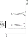
Brauzumab shows a non-uniform charge distribution at physiological pH 7.4 (see, e.g., published ustlizumab crystal structure [27] and homology models for brauzumab). Brazzumab shows a large positively charged region on the Fv domain (see fig. 1a) that is not present in ustromumab (see fig. 1 b). In addition, FcRn possesses a strong and prolonged negatively charged region (see fig. 1c), which however is not involved in the binding of the cognate Fc region. The isoelectric points of brainumab and ustlizumab have been calculated to be 9.7 and 9.4, respectively. In addition, the net charge of braunizumab was slightly more positive over the entire pH range (see fig. 1 d).
The FcRn binding affinities of brazinumab and ustlizumab are comparable at pH 6.0, i.e., the two values differ by at most one order or magnitude, in one embodiment by at most 5-fold, while the dissociation process from FcRn is very different. When using variants of brenuzumab and ustrocumab, it can be shown that the interaction is predominantly electrostatic and correlates with the extent of positively charged regions (see below).
d) pH-dependent FcRn-IgG interaction
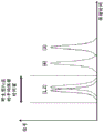
Ten variants of brainumab and ustekinumab have been synthesized and characterized for their FcRn binding properties by FcRn affinity chromatography (see table 2). In variants, the variable region has been modified and tested for FcRn pH6 binding affinity and FcRn dissociation using Surface Plasmon Resonance (SPR) and FcRn affinity chromatography, respectively (see table 3).
Table 2: fully engineered variants of brerunumab and ustrocumab
Exchanging structural parts such as Fv, LC and CDR between braunumab (light) and ustlizumab (dark): mAb 1-6. For mAb7 and mAb 8, 3 and 5 basic amino acids (by site-directed mutagenesis) in the HC of brainumab were exchanged for alanine residues, respectively. MAb9 is brazinumab in which the three basic amino acids of the light chain CDRs are exchanged for alanine residues and MAb 10 represents MAb9 additionally exchanged for 5 basic amino acids in HC. Exchanges of single basic amino acids are shown by circles, with three amino acid exchanges plotted as 1 circle and five amino acid exchanges plotted as 2 circles.


TABLE 3 FcRn binding affinity and charge distribution for all tested antibodies. Antibodies were sorted according to FcRn column retention time. Will balance the dissociation constant KDK calculated as Steady State affinity and against UltecumabDAnd (6) normalizing. Will be opposite to KDComparison of values (ustekinumab ═ 1) is shown as mean (n ═ 3) ± -Standard Deviation (SD). The isoelectric point and net charge (SaWI-Tools) of the Fv domain at pH 6.0 and pH 7.4 were calculated. The FcRn column retention time is not related to the isoelectric point or net charge of the Fv domain at pH 6.0 or pH 7.4.

The FcRn binding affinity at pH6 falls within a narrow range for all 11 antibodies (see table 3). The equilibrium dissociation constant (K) was calculated relative to ustekinumab (ustekinumab ═ 1.0)D). Braunizumab has a relative K of 0.2DAnd K of 9 variantsDBetween brerunumab and ustlizumab. Thus, it can be concluded that different terminal in vivo half-lives are not caused by different binding of FcRn at pH 6.0.
One aspect as reported herein is a method for determining the presence of an antibody-Fab-FcRn interaction in an antibody-Fc-FcRn complex affecting the half-life of the antibody in vivo, said method comprising the steps of:
a) determination of K of variant and parent antibodies at pH6 using surface plasmon resonanceDThe value of the one or more of,
b) determining the retention time of the variant and its parent antibodies on an FcRn affinity chromatography column eluted with a positive linear pH gradient in the presence of high salt concentration,
thus if K is between the variant antibody and its parent antibodyDValues differing by a factor of up to 10 and the retention time determined in step b) are substantially different, it is determined that there is an antibody-Fab-FcRn interaction in the antibody-Fc-FcRn complex which affects the in vivo half-life of the antibody.
One aspect as reported herein is a method for determining the relative in vivo half-life of an antibody, said method comprising the steps of:
a) determination of K of variant and parent antibodies at pH6 using surface plasmon resonanceDThe value of the one or more of,
b) determining the retention time of the variant and its parent antibodies on an FcRn affinity chromatography column eluted with a positive linear pH gradient in the presence of high salt concentration,
thus if K isDThe values differ by a factor of at most 10 and the retention time of the variant antibody determined in step b) is greater than the retention time of its parent antibodyShort/smaller, the antibody has a reduced relative in vivo half-life as compared to its parent antibody, an
Thus if K isDValues differ by up to 10-fold and the retention time of the variant antibody determined in step b) is longer/greater than that of its parent antibody, the antibody has an increased relative in vivo half-life compared to its parent antibody.
One aspect as reported herein is a method for determining an increase or decrease in the in vivo half-life of an antibody, said method comprising the steps of:
a) determination of K of variant and parent antibodies at pH6 using surface plasmon resonanceDThe value of the one or more of,
b) determining the retention time of the variant and its parent antibodies on an FcRn affinity chromatography column eluted with a positive linear pH gradient in the presence of high salt concentration,
thus if K isDValues differing by at most 10-fold and the retention time of the variant antibody determined in step b) being shorter/smaller than the retention time of its parent antibody, the antibody has a reduced in vivo half-life as compared to its parent antibody, and
thus if K isDValues differ by up to 10-fold and the retention time of the variant antibody determined in step b) is longer/greater than that of its parent antibody, the antibody has an increased in vivo half-life compared to its parent antibody.
Elution profiles of 12 antibodies were analyzed using FcRn affinity columns with positive linear pH gradient elution (see figure 2). Eutlizumab and mAb 1 carrying the eutlizumab Fv domain on the brekinumab constant region portion showed an indistinguishable retention time of about 84 minutes, suggesting that the Fv domain influences the interaction with FcRn. On the other hand, brauzumab elutes at a retention time of 94 minutes and therefore has a significantly different retention time compared to ustlizumab. The indistinguishable retention times of IdeS-cleaved brazzumab Fc region (85.7 min) and ustlizumab (85.2 min) indicate negligible effect of the Fc region. MAb 4, containing eutekmab LC (LC ═ light chain, HC ═ heavy chain) and brazinumab HC, had retention times close to that of eutekmab, showing that LC affects FcRn binding.
Variant antibodies mAb5 and mAb 6 carry the ustrocumab CDRs (heavy and light chain portions) on the brainumab framework and vice versa. Grafting of the ustrocumab CDR (mAb5) on brekinumab shifted the retention time of mAb5 close to that of ustrocumab. Grafting brekinumab CDR (mAb 6) on ustmab describes/demonstrates an elution profile that is still close to that of ustmab.
For mAb9, which is a brainumab variant in which three positively charged residues of the light chain CDRs are mutated to alanine residues, a strong shift in retention time from brainumab in the direction of ustlizumab was observed.
In mAb7 and mAb 8, 3 and 5 positively charged residues in the brainumab heavy chain were mutated, respectively. In these variants, the retention time is offset relative to brazinumab.
Taken together, the data show that the Fv domain affects FcRn dissociation and does not affect FcRn binding (at pH 6.0).
The FcRn column retention time coincides with the isoelectric point and net charge of the antibody. No correlation between FcRn column retention time and isoelectric point or net charge of the Fv domain could be observed at lysosomal pH 6.0 or physiological pH 7.4 (see table 3). However, the FcRn column retention time measured increases with the extent of the positively charged region, particularly around the light chain variable domain (see figure 2).
One aspect as reported herein is a method for increasing the in vivo half-life of an antibody, said method comprising the steps of:
-changing the charged amino acid residues at positions 27, 55 and 94 in the light chain of the antibody to hydrophobic or neutral hydrophilic amino acid residues (numbering according to Kabat) and thereby increasing the in vivo half-life of the antibody.
Amino acids can be grouped according to common side chain properties:
(1) hydrophobicity: norleucine, Met, Ala, Val, Leu; ile;
(2) neutral hydrophilicity: cys, Ser, Thr, Asn; gln;
(3) acidity: asp and Glu;
(4) alkalinity: his, Lys, Arg;
(5) chain orientation affecting residues: gly, Pro;
(6) aromatic: trp, Tyr, Phe.
The acidic and basic amino acid residues together belong to the group of charged amino acid residues.
FcRn column retention time for brazinumab has been found to be shortened in the presence of salt and is proportional to the inverse square root of ionic strength, as suggested by Debye-H ü ckel charge screening law [32 ]. ustekinumab retention time remains substantially unaffected (see fig. 6). thus, a significant portion of excess FcRn-brazinumab interaction is charge-mediated.
Taken together, FcRn affinity chromatography of the engineered variants showed that antibodies with the same Fv domain (mAb 1 and mAb 2) and the same LC (mAb 3 and mAb 4) eluted at nearly the same FcRn column retention time. Furthermore, grafting the ustrocumab CDRs on brazzumab (mAb5) shifted the elution pH close to that of ustrocumab. Thus, the light chain CDRs have a major impact on the FcRn binding of brainumab.
Grafting brainumab CDRs on ustrombin (mAb 6) showed an elution profile that was still close to that of ustrombin. Thus, without being bound by this theory, disrupting the bulky positively charged regions of brazzumab may more affect antibody-FcRn interaction than creating smaller positively charged regions. As suggested by MD simulations (see below), direct, stable interactions between positively charged Fv regions and negatively charged regions on FcRn are readily feasible. Thus, FcRn-Fv interactions can lead to slower dissociation of the FcRn-IgG complex under physiological conditions than normal.
e) Correlation of FcRn elution pH with pharmacokinetics in human FcRn transgenic mice
Previous studies have discussed that by affecting the electrostatic interaction between antibodies and negatively charged groups on the endothelial cell surface, the net charge is the driving force for changes in pharmacokinetic properties [35,36 ]. For example, Igawa et al [37] observed that IgG4 antibodies with lower isoelectric points (pI) due to engineering in the variable region have lower mobile phase pinocytosis rates and in turn have reduced elimination rates. In addition, Boswell et al [38] suggested that the pI difference needs to be at least one unit to affect PK.
The pI of brainumab, ustekinumab, mAb 8 and mAb9 varied between 9.3 and 9.7. Thus, it can be assumed that the effect on mobile phase pinocytosis is minimal due to pI differences. However, the shorter FcRn column retention time of brarenitumumab under high ionic strength conditions (see above) and the greater contribution of static to FcRn-Fv interactions in MD simulations compared to eculizumab (see below) indicate that the specific charge present may be a major contributor to FcRn-IgG interactions. The effect of charge in the Fv domain was analyzed using a mutant with 3 (mAb 7) and 5 (mAb 8) positively charged residues mutated in brainumab HC and brainumab (mAb 9) with three positively charged residues mutated in the light chain CDRs. MAb7 and MAb 8 showed a small shift in retention time in the direction of eculizumab, confirming charge-mediated interactions, while MAb9 showed a significant shift in retention time in the direction of eculizumab. Thus, the specific presence of charge in the light chain CDRs strongly influences FcRn dissociation.
To assess whether the effect of mutated charged residues in braun mAb variable domains shifts to a modulated in vivo PK profile, PK studies were performed in human FcRn-transgenic mice, braun mAb and ustlizumab were tested, along with the distribution and elimination process of two braun mAb variants (mAb 8 and mAb 9) with FcRn column retention time between braun mAb and ustlizumab, 4 antibodies, consistent with other IgG PK studies (see fig. 3a), braun mAb showed a α -faster reduction compared to other antibodies, interestingly, braun mAb and ustlizumab showed a terminal half-life of 48 hours and 137 hours, respectively (see fig. 3b), variants mAb 8 and mAb9 with smaller positively charged regions in the Fv domain had a terminal half-life of 78 hours and 109 hours, respectively, hi braun mAb and utlizumab, braun mAb and braun mAb9, and especially mAb 8, and mAb9, respectively, and 4, 7.7.7, 7.7, 7.84, 7.7, 7.8, 7.7.7, 7.8, 7.7.7.7.7, 7.8, 7.7.7, 7.7, 7.7.7, 7.7, 7, 7.7, respectively, 7, 7.7.7, 7, 7.7, 7, and 7, respectively, 7.
In PK experiments, the terminal half-life was examined and calculated completely at the elimination phase where FcRn regeneration predominates [39 ]. The terminal half-life of the 4 antibodies was linearly related to the in vitro FcRn column elution pH: the higher the FcRn column elution pH, the shorter the terminal half-life, thus showing that the FcRn column is a predictive/sensitive tool for FcRn dissociation in vitro. The correlation between terminal half-life and FcRn column elution pH confirms the importance of rapid dissociation of FcRn-IgG at physiological pH.
Without being bound by this theory, FcRn-IgG complexes accumulate in the endosomes at pH 6.0, and thus less binding results in less regeneration and faster clearance of IgG. By exocytosis, the FcRn-IgG complex is released to the plasma membrane, where dissociation of IgG and FcRn has to occur within a short time at physiological pH 7.4 [40 ]. Therefore, dissociation at physiological pH is also important for long-term half-life [22,40 ].
Thus, it has been found that dissociation at higher pH values suggests slower dissociation from FcRn. Without being bound by this theory, this may result in degradation of the antibody in the lysosome rather than releasing the antibody back into the blood circulation.
Thus, it has been found that the charge in the IgG Fv domain influences the terminal half-life by altering the interaction between IgG and FcRn. The portion of the Fab that interacts with FcRn has been located and this interaction has been demonstrated to be charge mediated. The PK study revealed a linear correlation between FcRn-IgG dissociation in vitro and terminal half-life in vivo.
f) Molecular Dynamics (MD) simulation of the FcRn-IgG model
Modeling the location of the Fv domains of brazinumab and ustekumab based on the crystal structure of intact IgG1(PDB code 1HZH), these homology models contain FcRn on one intact IgG molecule (α -FcRn along with β)2m) (see fig. 4 a). Distance between FcRn and Fv domains in the starting Structure> And is greater than or equal to under physiological conditions
 Debye length of [32]]. The kinetics of the FcRn-IgG complex were simulated by molecular dynamics simulation at well-defined water and physiological ionic strengths over a 100 nanosecond time frame. During the simulation, one of the two Fab regions was close to the top of FcRn and maintained in this conformation for the remaining simulation time (see fig. 4b, c, d). The region on FcRn that interacts with the Fv domain has not been described to date as being involved in IgG binding. Surprisingly, both brazinumab and ustekinumab adopt a conformation in which Fv and FcRn interact with each other in the MD simulation (see fig. 4b, c). It has been found that in both complexes, two different pairs of Fv and FcRn domains are close to each other in the asymmetric starting structure. The contribution of static in brainumab to FcRn-Fv interactions was found to be about 2-fold greater than in motlizumab (see figure 4 e).
In summary, it has been found that the inherent flexibility of the Fab arm of the FcRn-IgG complex structurally allows for direct, stable interaction of the Fv domain with the FcRn tip.
g) The method of the invention
g.i) elution with a linear positive pH gradient of different salt concentrations
Herein is reported a method comprising the following two steps:
a) determining the retention time of the antibody on an FcRn affinity chromatography column eluted with a positive linear pH gradient in the presence of a first salt concentration,
b) the retention time of the antibody on the FcRn affinity chromatography column eluted with a positive linear pH gradient was determined in the presence of the second salt concentration.
The second salt concentration is typically higher/greater than the first salt concentration, such that these concentrations are substantially different, i.e., they differ by at least 10%, and in one embodiment by at least 20%.
With this method, the presence of antibody-Fab-FcRn interactions in the antibody-Fc-FcRn complex can be determined in a simple chromatographic method by comparing the retention times obtained in the presence of different salt concentrations (see fig. 13) or by comparing the retention times of the intact antibody and its Fc region. This is important because antibody-Fab-FcRn interactions affect the in vivo half-life of the antibody.
For antibody/reference antibody pairs, there is a different relationship with respect to their retention time on the FcRn affinity chromatography column and with respect to their FcRn interaction:
1) the antibody and the reference antibody have substantially the same retention time in step a) and step b): in this case the in vivo half-life of both antibodies should be essentially the same, i.e. the in vivo half-life is not affected by antibody-Fab-FcRn interactions, or
2) The antibody and the reference antibody have substantially the same retention time in step a) but different retention times in step b): in this case the in vivo half-life of the antibody is shorter than the in vivo half-life of the reference antibody, i.e. the in vivo half-life is influenced by the antibody-Fab-FcRn interaction.
The antibody may be a variant antibody of a parent antibody, in which case the reference antibody is the parent antibody.
In one instance, the reference antibody is an antibody having substantially the same retention time as its Fc region following IdeS cleavage or papain cleavage.
In order to provide a therapeutic regimen to treat the diverse diseases known today and also to treat those diseases that will be revealed in the future, it is necessary to tailor the antibodies as well as the Fc region containing polypeptides.
To customize the FcRn binding characteristics, the residues of the antibody involved in the FcRn interaction are modified and the resulting modified antibody has to be tested. If the desired features are not met, the same process is performed again.
It would therefore be advantageous to provide a method for predicting characteristic property changes of modified antibodies based on simple chromatographic methods and which does not require in vivo studies to analyze the change in characteristics in the modified antibody.
In some cases, antibodies with extended half-life are desired. For example, a drug with an extended half-life in the circulation of a patient in need of treatment requires a reduced dose or an increased time between administrations. Such antibodies also have the advantage of increased exposure at disease sites, such as tumors.
In vivo half-life correlates with retention time on the FcRn affinity chromatography column. This is particularly true if the interaction between the antibody and FcRn is mediated almost exclusively by residues in the Fc region of the antibody. However, this correlation has to be further confirmed if residues outside the Fc region of the antibody (e.g. in antibody-Fab) also interact with FcRn. This can be done using the change in retention time in the FcRn affinity chromatography method with positive linear pH gradient elution in the presence of low and high salt concentrations or intact antibody and Fv region cleaved antibody (═ Fc region) in the method as reported herein. If the retention time is not substantially affected by changes from low to high salt concentrations or by cleavage of the Fc region, there is no antibody-Fab-FcRn interaction and a higher retention time on the FcRn affinity chromatography column correlates with an increased in vivo half-life. However, if the retention time is affected by a change from low to high salt concentration or by cleavage of the Fc region, especially if it is reduced thereby, the in vivo half-life is correlated with a difference in retention time on the FcRn affinity chromatography column, i.e. a longer retention time on the FcRn affinity chromatography column is correlated with a shorter in vivo half-life due to a reduced dissociation of the antibody-FcRn at physiological pH and (without being bound by this theory) an increased lysosomal degradation of the antibody.
An FcRn affinity chromatography column as used herein comprises a matrix and a matrix-bound chromatography functionality, wherein the matrix-bound chromatography functionality comprises a non-covalent complex of neonatal Fc receptor (FcRn) and β -2-microglobulin.
Typically, the starting point of the method as reported herein is a parent or reference antibody which is characterized by its binding to FcRn.
One aspect as reported herein is the use of a method as reported herein for determining the presence of an antibody-Fab-FcRn interaction in an antibody-Fc-FcRn complex affecting the half-life in vivo, said method comprising the steps of:
a) determining the retention time of the antibody on an FcRn affinity chromatography column eluted with a positive linear pH gradient in the presence of a first salt concentration,
b) determining the retention time of the antibody on an FcRn affinity chromatography column eluted with a positive linear pH gradient in the presence of a second salt concentration,
thus, if the retention time determined in step a) and the retention time determined in step b) are substantially different, it is determined that an antibody-Fab-FcRn interaction affecting the in vivo half-life is present in the antibody-Fc-FcRn complex.
One aspect as reported herein is a method for determining the presence of an antibody-Fab-FcRn interaction in an antibody-Fc-FcRn complex affecting half-life in vivo, said method comprising the steps of:
a) determining the retention time of the antibody on an FcRn affinity chromatography column eluted with a positive linear pH gradient in the presence of a first salt concentration,
b) determining the retention time of the antibody on an FcRn affinity chromatography column eluted with a positive linear pH gradient in the presence of a second salt concentration,
thus, if the retention time determined in step a) and the retention time determined in step b) are substantially different, it is determined that an antibody-Fab-FcRn interaction affecting the in vivo half-life is present in the antibody-Fc-FcRn complex.
Variant antibodies show increased or decreased binding to FcRn when compared to the parent antibody polypeptide or when compared to a reference antibody and, therefore, have a modulated half-life in serum when compared to the parent/reference antibody.
Typically, Fc region variants that are first predicted to have increased affinity for FcRn (i.e., increased retention time on the FcRn column compared to the parent or reference antibody) have a longer serum half-life than those Fc region variants that have reduced affinity for FcRn (i.e., reduced retention time on the FcRn column compared to the parent or reference antibody).
This predicted in vivo half-life has to be confirmed afterwards. For this confirmation, the methods as reported herein can be used.
Antibody variants with increased in vivo half-life are useful in methods of treating mammals, particularly humans, where a long half-life of the administered antibody is desired, such as for treating chronic diseases or disorders.
Antibody variants with reduced affinity for FcRn are useful in methods of treating mammals, particularly humans, where short half-lives of the administered antibody or fusion polypeptide are desired, such as in vivo diagnostic imaging.
It is likely that antibody variants with reduced FcRn binding affinity will be able to cross the placenta and may therefore be used in the treatment of diseases or disorders in pregnant women, especially in unborn children. Furthermore, reduced FcRn binding affinity may be required for those drugs intended to be administered/transported to the brain, kidney, and/or liver.
One aspect as reported herein is the use of a method as reported herein for the determination of an antibody showing a reduced transport across the epithelium of a glomerulus from the vascular system.
One aspect as reported herein is the use of a method as reported herein for the determination of an antibody showing a reduced transport from the brain across the blood-brain barrier into the vascular space.
In one embodiment of all aspects as reported herein, the FcRn is selected from the group consisting of human FcRn, cynomolgus monkey FcRn, mouse FcRn, rat FcRn, sheep FcRn, canine FcRn, porcine FcRn, mini-porcine FcRn and rabbit FcRn.
In one embodiment of all aspects as reported herein, β -2-microglobulin is from the same species as FcRn.
In one embodiment of all aspects as reported herein, β -2-microglobulin is from a different species than FcRn.
In one embodiment, the parent antibody comprises at least one binding domain and at least one Fc region. In one embodiment, the parent antibody comprises two binding domains and two Fc regions.
In one embodiment, the parent antibody comprises at least one binding domain that specifically binds to a target that mediates a biological effect (in one embodiment, a ligand capable of binding to a cell surface receptor or a cell surface receptor capable of binding a ligand) and mediates the propagation of a negative or positive signal to a cell. In one embodiment, the parent antibody comprises at least one binding domain specific for an antigen (in one embodiment, a cell surface antigen or a soluble antigen) that is targeted for reduction or elimination and at least one Fc region.
Antibodies that specifically bind to a target can be produced in a mammal by multiple subcutaneous or intraperitoneal injections of the antigen of interest (e.g., a purified antigen, a cell or cell extract comprising such an antigen, or DNA encoding such an antigen), and optionally an adjuvant.
In one embodiment, the antibody is a full length antibody.
In one embodiment, the antibody is a monoclonal antibody.
In one embodiment, the parent antibody is a bispecific antibody.
In one embodiment, the parent antibody is a chimeric antibody.
In one embodiment of all of the previous aspects, the pH gradient is a gradient from about pH5.5 to about pH 8.8.
Typically, the soluble extracellular domain of FcRn (SEQ ID NO:33 for human FcRn) with a C-terminal His-Avi Tag (SEQ ID NO:34) is co-expressed with β 2-microglobulin (SEQ ID NO:35 for human β -2-microglobulin) in mammalian cells the non-covalent FcRn-microglobulin complex is biotinylated and loaded onto streptavidin-derivatized agarose gel.
In one embodiment of all aspects as reported herein, a non-covalent complex of a neonatal Fc receptor (FcRn) and β -2-microglobulin is bound to a solid phase.
In one embodiment, conjugation of the non-covalent complex to the solid phase is performed by chemical conjugation via the N-terminus and/or epsilon-amino group (lysine), epsilon-amino groups of different lysines, carboxyl, sulfhydryl, hydroxyl and/or phenol functions of the amino acid backbone of the antibody and/or sugar alcohol groups of the carbohydrate structure of the antibody.
In one embodiment, the non-covalent complex is conjugated to the solid phase via a specific binding pair. In one embodiment, the non-covalent complex is conjugated to biotin and immobilized to a solid support by means of avidin or streptavidin immobilized to the solid support.
In one embodiment, the specific binding pair (first component/second component) is selected from streptavidin or avidin/biotin, antibodies/antigens (see, e.g., Hermanson, g.t. et al, bioconjugate techniques, Academic Press (1996)), lectins/polysaccharides, steroid/steroid binding proteins, hormone/hormone receptors, enzymes/substrates, IgG/protein a and/or protein G, and the like.
In principle, any buffer substance may be used in the method as reported herein.
Fc residues critical for mouse Fc-mouse FcRn interaction have been identified by site-directed mutagenesis (see, e.g., Dall' Acqua, w.f. et al, j.immunol 169(2002) 5171-5180). Residues I253, H310, H433, N434, and H435 (numbering according to EU of Kabat) are involved in this interaction (Medesan, C. et al, Eur.J. Immunol.26(1996) 2533; Firan, M. et al, int.Immunol.13(2001) 993; Kim, J.K. et al, Eur.J. Immunol.24(1994) 542). Residues I253, H310 and H435 were found to be critical for the interaction of human Fc with murine FcRn (Kim, j.k. et al, eur.j.immunol.29(1999) 2819). Through protein-protein interaction studies, Dall 'Acqua et al have described residues M252Y, S254T, T256E as improving FcRn binding (Dall' Acqua, W.F. et al, J.biol.chem.281(2006) 23514-23524). Studies of the human Fc-human FcRn complex have shown that residues I253, S254, H435 and Y436 are critical for this interaction (Firan, M. et al, int. Immunol.13(2001) 993; Shields, R.L. et al, J.biol. chem.276(2001) 6591-6604). In Yeung, y.a. et al (j.immunol.182(2009)7667-7671), various mutants of residues 248 to 259 and 301 to 317 and 376 to 382 and 424 to 437 have been reported and studied.
Table 4: retention times of different antibodies obtained with different elution buffers and gradients.

The term YTE-mutant refers to the triple mutation M252Y/S254T/T256E.
In one embodiment, pharmaceutically acceptable buffer substances are used, such as, for example, phosphoric acid or a salt thereof, acetic acid or a salt thereof, citric acid or a salt thereof, morpholine or a salt thereof, 2- (N-morpholino) ethanesulfonic acid (MES) or a salt thereof, histidine or a salt thereof, glycine or a salt thereof, TRIS (hydroxymethyl) aminomethane (TRIS) or a salt thereof, (4- (2-hydroxyethyl) -1-piperazineethanesulfonic acid (HEPES) or a salt thereof.
In one embodiment, the buffering substance is selected from phosphoric acid or a salt thereof, or acetic acid or a salt thereof, or citric acid or a salt thereof, or histidine or a salt thereof.
In one embodiment, the buffer substance has a concentration of 10mM to 500 mM. In one embodiment, the buffer substance has a concentration of 10mM to 300 mM. In one embodiment, the buffer substance has a concentration of 10mM to 250 mM. In one embodiment, the buffer substance has a concentration of 10mM to 100 mM. In one embodiment, the buffer substance has a concentration of 15mM to 50 mM. In one embodiment, the buffer substance has a concentration of about 20 mM.
In one embodiment, an exemplary starting solution for a positive linear pH gradient comprises 20mM MES and 140mM nacl, adjusted to pH 5.5.
In one embodiment, an exemplary final solution of a positive linear pH gradient comprises 20mM TRIS and 140mM NaCl, adjusted to pH 8.8.
During the gradient, a mixture of starting and final solutions is applied to the FcRn affinity chromatography column, whereby a positive linear gradient starts with 100% of the starting solution (i.e. pure starting solution), after which the proportion of the starting solution decreases from 100% to 0% and the proportion of the final solution increases from 0% to 100%, such that after a positive linear pH gradient, 100% of the final solution is applied to the column.
In one embodiment, the starting solution and the final solution comprise additional salts. In one embodiment, the additional salt is selected from sodium chloride, sodium sulfate, potassium chloride, potassium sulfate, sodium citrate, or potassium citrate. In one embodiment, the solution comprises 50mM to 1000mM of the additional salt. In one embodiment, the solution comprises from 50mM to 750mM of an additional salt. In one embodiment, the solution comprises 50mM to 500mM of the additional salt. In one embodiment, the solution comprises from 50mM to 750mM of an additional salt. In one embodiment, the solution comprises from about 140mM to about 400mM of the additional salt.
In one embodiment, the starting solution and the final solution comprise sodium chloride. In one embodiment, the starting solution and the final solution comprise about 140mM to about 400mM sodium chloride.
It has been found that the type of salt and buffer material affects the retention time and the degree of separation. The optimal salt concentration (140mM NaCl) for antibody binding to FcRn can be determined. If the salt concentration is higher (400mM), the binding to FcRn is reduced by interfering with the charge interaction due to the increase in ionic strength of the solution and a shorter retention time is obtained.
Thus, in the method as reported herein, the solutions used in step a) and step b) and the gradients applied in step a) and step b) and the loading of the cartridge in step a) and step b) and the dimensions of the cartridge and the amount of FcRn affinity chromatography material in step a) and step b) and the FcRn affinity ligand density of the FcRn affinity chromatography material in step a) and step b) and the conjugation of FcRn to the solid phase in step a) and step b) and the properties of b2m and FcRn in step a) and step b) are identical or even identical. Thus, in the method as reported herein, the FcRn affinity chromatography in step a) and step b) is performed under the same conditions except that the salt concentration is different between step a) and step b). In one embodiment, the second salt concentration is greater than the first salt concentration. In one embodiment, the second salt concentration is at least 2 times the first salt concentration.
As can be seen from fig. 7, the amount of antibody applied showed a linear correlation with the area under the curve of the elution peak.
The 8 antibodies were analyzed as intact antibodies and after cleavage with the enzyme IdeS. Cleavage was controlled by SDS PAGE and analytical SEC. The Fc region and antibody-Fab were isolated by preparative SEC.
Table 5: comparison of retention times of intact antibody, antibody-Fab and Fc regions.

Typically, the retention time of antibodies with wild-type Fc regions (IgG1 or IgG2 or IgG4) under the conditions of example 5 varied between 45 minutes and 49 minutes (tested with 35 therapeutic antibodies against 36 antigens, data not shown). If the conditions of example 2 are used, the retention time increases with longer gradients to about 85 minutes.
Table 6: retention time relative to the amount of immobilized FcRn receptor/gram column material (chromatographic conditions of example 5).

In general, the retention time in the methods and uses as reported herein depends on the pH gradient and the steepness of the salt concentration used. If a wild-type antibody is used as a reference, weaker binding is indicated by a shorter retention time (earlier elution) and stronger binding is indicated by a longer retention time (later elution).
Different IgG Fc region mutants differed on the FcRn column, showing modulated retention times.
For example, the anti-IGF-1R antibody mutant YTE showed increased retention time (see figure 8).
Table 7: change in retention time relative to the Fc region mutation.
| Antibodies | Retention time [ min ]] |
| anti-IGF-1R antibody (wild type) | 44.5 |
| Anti IGF-1R antibody (YTE-mutant) | 57.5 |
The term YTE-mutant refers to the triple mutation M252Y/S254T/T256E.
One aspect as reported herein is a method for determining the relative in vivo half-life of an antibody, said method comprising the steps of:
a) determining the retention time of the antibody on an FcRn affinity chromatography column eluted with a positive linear pH gradient in the presence of a first salt concentration,
b) determining the retention time of the antibody on an FcRn affinity chromatography column eluted with a positive linear pH gradient in the presence of a second salt concentration,
thus, if the retention time determined in step a) and the retention time determined in step b) are substantially different, the antibody has a reduced relative in vivo half-life compared to a standard/native antibody of the subclass IgG1, IgG3 or IgG 4.
One aspect as reported herein is the use of a method as reported herein for determining the relative in vivo half-life of an antibody, wherein said method comprises the steps of:
a) determining the retention time of the antibody on an FcRn affinity chromatography column eluted with a positive linear pH gradient in the presence of a first salt concentration,
b) determining the retention time of the antibody on an FcRn affinity chromatography column eluted with a positive linear pH gradient in the presence of a second salt concentration,
thus, if the retention time determined in step a) and the retention time determined in step b) are substantially different, the antibody has a reduced relative in vivo half-life compared to a standard/native antibody of the subclass IgG1, IgG3 or IgG 4.
One aspect as reported herein is a method for determining an increase or decrease in the in vivo half-life of a variant antibody relative to its parent antibody, said method comprising the steps of:
a) determining the retention time of the variant and its parent antibody on an FcRn affinity chromatography column eluted with a positive linear pH gradient in the presence of a first salt concentration,
b) determining the retention time of the variant and its parent antibodies on an FcRn affinity chromatography column eluted with a positive linear pH gradient in the presence of a second salt concentration,
thus, if i) the retention time of the variant antibody determined in step a) is greater/longer than the retention time of its parent antibody determined in step a), and ii) the retention time of the variant antibody determined in step a) and the retention time of the variant antibody determined in step b) are substantially the same, the in vivo half-life of the variant antibody is increased relative to its parent antibody,
thus, the in vivo half-life of the variant antibody is reduced relative to its parent antibody if i) the retention time of the variant antibody determined in step a) is smaller/shorter than the retention time of its parent antibody determined in step a), and ii) the retention time of the variant antibody determined in step a) and the retention time of the variant antibody determined in step b) are substantially the same.
One aspect as reported herein is the use of a method as reported herein for determining an increase or decrease of the in vivo half-life of a variant antibody relative to its parent antibody, wherein said method comprises the steps of:
a) determining the retention time of the variant and its parent antibody on an FcRn affinity chromatography column eluted with a positive linear pH gradient in the presence of a first salt concentration,
b) determining the retention time of the variant and its parent antibodies on an FcRn affinity chromatography column eluted with a positive linear pH gradient in the presence of a second salt concentration,
thus, if i) the retention time of the variant antibody determined in step a) is greater/longer than the retention time of its parent antibody determined in step a), and ii) the retention time of the variant antibody determined in step a) and the retention time of the variant antibody determined in step b) are substantially the same, the in vivo half-life of the variant antibody is increased relative to its parent antibody,
thus, the in vivo half-life of the variant antibody is reduced relative to its parent antibody if i) the retention time of the variant antibody determined in step a) is smaller/shorter than the retention time of its parent antibody determined in step a), and ii) the retention time of the variant antibody determined in step a) and the retention time of the variant antibody determined in step b) are substantially the same.
It has been found that antibodies showing a delayed elution from the FcRn column (i.e. a longer retention time on the FcRn column) and showing no antibody-Fab-FcRn interaction have a longer in vivo half-life (see example 6).
Table 8: in vivo data.


The term YTE-mutant refers to the triple mutation M252Y/S254T/T256E.
One aspect as reported herein is the use of a method as reported herein for determining the in vivo half-life of an antibody.
A set of in vitro and in vivo experiments carried out with wild-type IgG and IgG variants carrying YTE-mutations in the Fc region allowed to show a semi-quantitative correlation of the results of FcRn affinity chromatography with those of in vivo pharmacokinetic studies using human FcRn transgenic mice (Spiekerman, G.M. et al, J.Exp.Med.196(2002) 303-235310; Dall' Acqua, W.F. et al, J.biol.chem.281(2006) 23514-23524). YTE-mutations result in a significantly prolonged half-life and slower plasma clearance. Longer in vivo half-life corresponds to longer retention time in the FcRn chromatography. It has recently been shown that the extended half-life of Fc-engineered trastuzumab variants has enhanced FcRn binding in vitro as measured by flow cytometry (Petkova, s.b. et al, int.immunol.18(2006) 1759-. Variants of the anti-veggg 1 antibody bevacizumab with 11-fold improvement in FcRn affinity were shown to have a 5-fold extended half-life in human FcRn transgenic mice and a three-fold longer half-life in cynomolgus monkeys (Zalevsky, j. et al, nat. biotechnol.28(2010) 157-159).
The antibody pattern has been shown to have no effect on binding to FcRn columns. This was confirmed for the knob-and-hole (knob-and-hole) format and several bispecific antibody formats. Thus, the methods as reported herein can be used to evaluate new antibody patterns.
In one embodiment, the complex is monobiotinylated.
In one embodiment, a chromatographic material comprising a non-covalent complex of a neonatal Fc receptor (FcRn) and β -2-microglobulin as ligand has a stability of at least 100 cycles in the methods and uses as reported herein.
g.ii) elution of the antibody and its Fc region with a linear positive pH gradient at the same salt concentration
Herein is reported a method comprising the following two steps:
a) determining the retention time of the antibody on an FcRn affinity chromatography column eluted with a positive linear pH gradient in the presence of a first salt concentration,
b) the retention time of the Fc region of the antibody on an FcRn affinity chromatography column eluted with a positive linear pH gradient was determined in the presence of the first salt concentration.
In this way, the presence of the antibody-Fab-FcRn interaction in the antibody-Fc-FcRn complex can be determined in a simple chromatographic method by comparing the retention times of the antibody and its Fc region. The Fc region may be obtained, for example, by enzymatic cleavage with the enzyme IdeS or papain, or may be produced recombinantly. This is important because antibody-Fab-FcRn interactions affect the in vivo half-life of the antibody.
For antibody/antibody-Fc region pairs, there is a different relationship with respect to their retention time on the FcRn affinity chromatography column and likewise with respect to their FcRn interaction:
1) the antibody and its Fc region have substantially the same retention time: in this case the in vivo half-life of the antibody is not affected by antibody-Fab-FcRn interactions,
2) the antibodies and their Fc regions have different retention times and the retention time of the Fc region is shorter than that of the antibody: in this case, the in vivo half-life of the antibody is affected by antibody-Fab-FcRn interactions.
In case the antibody in question is a variant antibody, other aspects have to be considered: in variant antibodies, antibody-Fc-FcRn interactions as well as antibody-Fab-FcRn interactions may be altered by modifications introduced relative to the parent antibody.
Thus, there are the following possible relationships between parent, variant and their corresponding Fc regions (see fig. 10):
1) the parent antibody (1), variant antibody (3) and corresponding Fc regions (2,4) have substantially the same retention time: in this case the in vivo half-life of the variant antibody i) is not affected by the antibody-Fab-FcRn interaction and ii) corresponds to the in vivo half-life of the parent antibody (see FIG. 10A),
2) the variant antibody (3) and its Fc region (4) have different retention times, the retention time of the Fc region of the variant antibody is shorter than the retention time of the variant antibody, and the parent antibody (1) \\ the Fc region (2) of the parent antibody and the Fc region (4) of the variant antibody have substantially the same retention times: in this case, the in vivo half-life of the variant antibody is i) affected by antibody-Fab-FcRn interaction and ii) shorter than the in vivo half-life of the parent antibody (see FIG. 10B),
3) the parent antibody (1) and the variant antibody (3) have different retention times, the retention time of the Fc region (4) of the variant antibody is substantially the same as the retention time of the variant antibody (3), and the retention time of the variant antibody is longer than the retention time of the parent antibody (1): in this case the in vivo half-life of the variant antibody i) is not affected by antibody-Fab-FcRn interaction and ii) is longer than the in vivo half-life of the parent antibody (see FIG. 10C),
4) the parent antibody (1) and the variant antibody (3) have different retention times, the retention time of the Fc region (4) of the variant antibody is substantially the same as the retention time of the variant antibody (3), and the retention time of the variant antibody (3) is shorter than the retention time of the parent antibody (1): in this case the in vivo half-life of the variant antibody is i) not affected by the antibody-Fab-FcRn interaction and ii) shorter than the in vivo half-life of the parent antibody,
5) the parent antibody (1) and the variant antibody (3) have different retention times, the retention time of the Fc region (4) of the variant antibody is different from the retention time of the variant antibody (3) and also different from the retention times of the parent antibody (1) and its Fc region (2), and the retention time of the Fc region (4) of the variant antibody is between the retention times of the variant antibody (3) and the parent antibody (4): in this case the in vivo half-life of the variant antibody i) was affected by antibody-Fab-FcRn interaction and ii) and was different from that of the parent antibody (see figure 10D).
In one instance, the reference antibody is an antibody having substantially the same retention time as its Fc region following IdeS cleavage or papain cleavage.
As outlined above, antibody-Fab-FcRn interactions may have an effect on the in vivo half-life of the antibody. As also outlined above, antibody-Fc-FcRn interactions may have an effect on the in vivo half-life of the antibody. Therefore, two interactions have to be explained.
For example, Ropeenian and Akilesh (Nat. Rev. Immunol.7(2007)715-725) report: the humanized IgG1 antibody hu4D5 (Herceptin; Genentech; ERBB 2-specific monoclonal antibody) variant Asn434Ala (N434A) and the triple-substituted variant Thr307Ala/Asn434Ala/Glu380Ala (T307A/N434A/Q380A) bound human FcRn at pH 6.0 with 3-fold and 12-fold higher affinity than the wild-type hu4D5 antibody, respectively. Unexpectedly, the half-lives of the two variant antibodies were substantially equivalent in FcRn transgenic humanized mice. According to Ropeenian and Akilesh, this inconsistency can be explained by the increased affinity of the triple-substitution variant for FcRn at pH 7.4. Mutations in the Fc region that improve binding affinity at pH 7.4 and at pH 6.0 may actually accelerate antibody clearance in vivo rather than extend its half-life.
Therefore, it has been further considered whether the retention time of the Fc region of the variant antibody is longer than the critical retention time. Without being bound by this theory, this leads to the following effects; the interaction at pH 7.4 is so increased that dissociation of the antibody-FcRn complex at pH 7.4 is reduced, resulting in increased degradation of the antibody (see figure 11).
One aspect of the present invention is a method for selecting an antibody, the method comprising the steps of (see fig. 12):
a) determining the retention time on an FcRn affinity chromatography column of i) the antibody (3), ii) the Fc region of the antibody (4), iii) the reference antibody (1), iv) the Fc region of the reference antibody (2) and v) the reference antibody having the mutation N434A in the Fc region (5) using the same elution conditions,
b) selecting antibodies for which
b-i) the reference antibody (1), the variant antibody (3) and the respective Fc region (2,4) have essentially the same retention time and the retention time of the antibody is shorter than that of the reference antibody (5) having the mutation N434A in the Fc region and is thus selected for an antibody whose in vivo half-life i) is not affected by the antibody-Fab-FcRn interaction and ii) corresponds to that of the parent antibody (see FIG. 12A),
b-ii) the antibody (3) and its Fc region (4) have different retention times, the retention time of the antibody Fc region (4) being shorter than the retention time of the antibody (3) and equal to or longer than the retention time of the reference antibody (1) or its Fc region (2), the retention times of the reference antibody (1), the Fc region (2) of the reference antibody and the antibody (3) being shorter than the retention time of the reference antibody (5) having the mutation N434A in the Fc region and thus selecting an antibody whose in vivo half-life i) is influenced by the antibody-Fab-FcRn interaction and ii) is shorter than the in vivo half-life of the reference antibody (see FIG. 12B),
b-iii) the reference antibody (1) and the antibody (3) have different retention times, the retention time of the Fc region (4) of the antibodies is essentially the same as the retention time of the antibody (3), the retention time of the antibody (3) is longer than the retention time of the reference antibody (1), and the retention time of the antibody (3) is shorter than the retention time of the reference antibody (5) having the mutation N434A in the Fc region and is thus selected for an antibody whose in vivo half-life i) is not affected by antibody-Fab-FcRn interactions and ii) is longer than the in vivo half-life of the reference antibody (see FIG. 12C),
b-iv) the reference antibody (1) and the antibody (3) have different retention times, the retention time of the Fc region (4) of the antibodies is essentially the same as the retention time of the antibody (3), the retention time of the antibody (3) is shorter than the retention time of the reference antibody (1), and the retention time of the antibody (3) is shorter than the retention time of the reference antibody (5) having the mutation N434A in the Fc region and is thus selected for an antibody whose in vivo half-life i) is not affected by antibody-Fab-FcRn interactions and ii) is shorter than the in vivo half-life of the reference antibody (see FIG. 12D),
b-v) the reference antibody (1) and the antibody (3) have different retention times, the retention time of the Fc region (4) of the antibody is different from the retention time of the antibody (3) and also different from the retention times of the reference antibody (1) and its Fc region (2), the retention time of the Fc region (3) of the antibody is between the retention times of the antibody (3) and the reference antibody (1), and the retention time of the antibody (3) is shorter than the retention time of the reference antibody (5) having the mutation N434A in the Fc region and thus selecting an antibody whose in vivo half-life i) is affected by the antibody-Fab-FcRn interaction and ii) is different from the in vivo half-life of the reference antibody (see fig. 12E).
In one embodiment, elution is at constant salt concentration by a positive linear pH gradient or at constant pH by using a linear salt gradient.
In one embodiment, the antibody is a variant antibody of a parent antibody and the reference antibody is the parent antibody. In one embodiment, the variant antibody has an amino acid change in the antibody-Fab or/and in the antibody-Fc region.
g.iii) elution with a salt gradient
Herein is reported a method comprising the steps of:
a) determining the retention time of the antibody and the reference antibody on the FcRn affinity chromatography column by elution with a salt gradient at a first pH value,
b) determining the retention time of the antibody and the reference antibody on the FcRn affinity chromatography column by eluting with a salt gradient at a second pH,
it has been found that in addition to elution with a pH gradient at constant salt concentration, elution with a salt gradient at constant pH values can also be used to determine whether antibody-Fab-FcRn interactions are present in the antibody-Fc-FcRn complex.
As already outlined above, the antibody-Fab-FcRn interaction is (if indeed present) a secondary interaction established after the antibody-Fc-FcRn complex has formed.
Two interactions, antibody-Fc-FcRn and antibody-Fab-FcRn, are charge-mediated non-covalent interactions.
One aspect as reported herein is a method for determining the presence of an antibody-Fab-FcRn interaction affecting the in vivo half-life of an antibody, said method comprising the steps of:
a) determining the retention time of the antibody and the reference antibody on the FcRn affinity chromatography column by elution with a salt gradient at a first pH value,
b) determining the retention time of the antibody and the reference antibody on the FcRn affinity chromatography column by eluting with a salt gradient at a second pH,
thus, if the ratio of the antibody retention time determined in step a) to the reference antibody retention time is substantially different from the ratio of the antibody retention time determined in step b) to the reference antibody retention time, it is determined that there is an antibody-Fab-FcRn interaction that affects the in vivo half-life of the antibody.
In one embodiment, the first pH is 5.5. In one embodiment, the second pH is 8.8.
In one embodiment, the salt gradient in step a) and step b) is the same.
In one embodiment, the salt gradient is a sodium chloride gradient.
In one embodiment, the salt gradient is from 0mM to 250mM salt.
h) Bevacizumab and bevacizumab-mutants
Another molecule without a charge cluster in the CDR was selected to generate a positive charge cluster in the LC-CDR to validate the results reported above: the positive charge at this position generally affects the FcRn binding affinity of the antibody.
Bevacizumab was chosen because it has only a small charge in the LC-CDR. The three basic amino acid residues identified using brainumab are arginine residues R27, R55, and R94. In the bevacizumab amino acid sequence, aspartate D27, leucine L54 and threonine T93 were exchanged for lysine residues to generate positively charged clusters (see fig. 14).
FcRn affinity chromatograms of bevacizumab-wild-type and bevacizumab-mutant are shown in fig. 9 and the corresponding retention times are listed in table 9 below.
Table 9: FcRn column retention time for bevacizumab-wild type and bevacizumab-mutants.
| Sample (I) | Retention time [ min ]] |
| Ultecal monoclonal antibody | 83.6 |
| Brainumab | 91.6 |
| Bevacizumab-wild type | 84.7 |
| Bevacizumab-mutants | 86.9 |
Bevacizumab-wild type has a retention time of 84.7 minutes, while bevacizumab-mutant elutes after 86.9 minutes. Thus, the positive charge cluster in the Fv of bevacizumab causes a retention time shift of 2.2 minutes. The results indicate that the charge in IgG1Fv generally affects FcRn binding affinity, and in particular, dissociation from FcRn.
Brief Description of Drawings
Fig. 1 charge distribution and pH dependent net charge. Protonation at pH 7.4 and at 2kBT/e plotted protein equipotential surface, black: positive/negative (a) braunuzumab light chain shown in light gray and heavy chain shown in dark gray the views of the middle and right images are related to the views in the left panel by rotation about the vertical and horizontal axes, respectively, (b) ustekumab light chain and heavy chain are shown in light gray and dark gray, respectively, each view is the same as (a), (c) human β2Microglobulin (β)2m) 2k of a complex human FcRn homology modelBAnd (4) drawing an equipotential surface at T/e. For clarity, the Fc domain is shown. (d) Calculated net charge based on sequence vs. pH of brainumab and ustekinumab. Protein constructs were compiled with DiscoveryStudio.
FIG. 2 pH dependent FcRn-IgG interaction. For clarity, FcRn affinity chromatograms of 11 IgG variants were intensity-normalized (intensity-normalized). The molecular surface representation of the structural model protonated at pH 7.4 was superimposed with an equipotential surface plotted at 2 kBT/e. The view is the same as the right panel in fig. 1a and is focused on the CDR region. The second horizontal axis shows the elution pH interpolated from the offline pH values.
Figure 3 effect of FcRn elution pH on pharmacokinetics in human FcRn transgenic mice. The antibody was administered as a single intravenous bolus injection of 10mg/kg to 6 animals/group. Data points represent mean ± standard deviation. (a) Blood level curves for brainumab (orange), ustekinumab (green), mAb 8 (violet), and mAb9 (blue). (b) Correlation between terminal half-life and FcRn column elution pH.
Figure 4 molecular dynamics simulation of the FcRn-IgG model.
(a) Simulating the initial conformation. Dashed lines indicate the distance between two exemplary amino acids in the Fv region and in FcRn,the amino acids are close during MD simulation as shown in plane (c). The color is the same as in fig. 1. (b) The end-of-run constellation (t 100 ns) was simulated. The box lines indicate the components of the molecules shown in (c). (c) Detailed view of the interaction between FcRn and Fv domains. Note that the interacting framework residues, CDR residues, and FcRn residues differ in brainumab and ustekinumab. (d) Distance between residue 245(FcRn) and residue 100 (ustekumab LC) and residue 29 (brazinumab LC) respectively during the simulation. (e) The interaction energies at the end of the simulation (mean and standard deviation of the conformations at 96, 97, 98, 99 and 100 nanoseconds). "VDW" and "electrostatic" refer to the van der Waals and electrostatic contributions to FcRn-Fab interactions, respectively. With PyMolTM(Schrodinger LLC) compiles protein structures.
FIG. 5 sequence alignment of brazzitumumab and Ultecumab light and heavy chains VH and VL regions are shown in italics, CDRs are marked with an asterisk (*); and # indicates the immediate vicinity in the starting structure Amino acids of FcRn. The identity marks the residue that was mutated to Cys for MD purposes to establish a disulfide bond with FcRn.
Figure 6 salt dependence of FcRn affinity column retention time for brazinumab and ustlizumab. Brainumab and ustekinumab were subjected to FcRn column chromatography with a pH gradient elution in the presence of increasing amounts of NaCl. The data were fit to the reciprocal square root function to account for charge shielding effects due to dissolved salts. Brainumab retention time Decrease (r2 ═ 0.898), while retention time of ustekumab remained essentially unaffected.
Figure 7 linearity of area under curve of applied antibody and chromatography using FcRn column as reported herein.
Figure 8 chromatograms of anti-IGF-1R antibody wild-type and YTE-mutant on FcRn column as reported herein.
FIG. 9 FcRn affinity chromatograms of Avastin-wild type and Avastin-mutant.
Figure 10 schematic representation of the change in retention time of antibody-FcRn interaction on an FcRn affinity chromatography column depending on Fc region and antibody-Fab; 1: parent antibody, 2: parent antibody Fc region, 3: variant antibody, 4: a variant antibody Fc region; solid line: intact antibody (antibody-Fab + Fc-region), dotted line: an Fc region only;
a: a wild-type-like Fc region, without antibody-Fab-FcRn interaction;
b: a wild-type-like Fc region, antibody-Fab-FcRn interaction;
c: an engineered Fc region with improved FcRn binding, no antibody-Fab-FcRn interaction;
d: an engineered Fc region with improved FcRn binding, antibody-Fab-FcRn interaction;
figure 11 shows a scheme of engineered antibodies with improved FcRn binding, antibody-Fab-FcRn interaction, but reduced in vivo half-life, as antibody-FcRn interaction results in improved clearance (retention time above critical retention time).
Figure 12 schematic representation of the change in retention time of antibody-FcRn interaction on an FcRn affinity chromatography column depending on Fc region and antibody-Fab; 1: reference antibody, 2: reference antibody Fc region, 3: antibody, 4: an antibody Fc region; solid line: intact antibody (antibody-Fab + Fc-region), dotted line: only the Fc region.
Figure 13 FcRn affinity chromatography retention time dependence on salt concentration and antibody-Fab-FcRn interaction.
FIG. 14 sequence alignment of bevacizumab and bevacizumab variant light chain variable domains identical and similar amino acids are shown in grey, CDRs are labeled with an asterisk (*).
FIG. 15 IL-12 interaction of brazinumab, ustekumab and mAbs 1-6; 1: brenuzumab, 2: ultecumab, 3: mAb 1, 4: mAb2, 5: mAb 3, 6: mAb 4, 7: mAb5, 8: and (3) mAb 6.
FIG. 16 IL-12 interaction of ranibizumab, ustrombin and mAb 7-10; 1: brenuzumab, 2: ultecumab, 3: mAb7, 4: mAb 8, 5: mAb9, 6: mAb 10.
Detailed description of the preferred embodiments
1. A method for selecting antibodies comprising the steps of
i) Determining a first retention time of the antibody and the reference antibody on an FcRn affinity chromatography column eluting with a positive linear pH gradient in the presence of a first salt concentration and determining a second retention time of the antibody and the reference antibody on an FcRn affinity chromatography column eluting with a linear pH gradient in the presence of a second salt concentration, or
ii) determining at a first pH a first retention time of the antibody and the reference antibody on an FcRn affinity chromatography column eluted with a linear salt gradient and determining at a second pH a second retention time of the antibody and the reference antibody on an FcRn affinity chromatography column eluted with a positive linear salt gradient, or
iii) determining the K of the antibody and the reference antibody at pH6 using surface plasmon resonanceDAnd determining the retention time of the antibody and the reference antibody on an FcRn affinity chromatography column eluting with a positive linear pH gradient in the presence of a high salt concentration, or
iv) determining the K of the antibody and the reference antibody at pH6 using surface plasmon resonanceDAnd determining the retention time of the antibody and the reference antibody on an FcRn affinity chromatography column eluted with a linear salt gradient, or
v) determining the retention time of the antibody and its Fc region on an FcRn affinity chromatography column eluted with a positive linear pH gradient, or
vi) determination of the retention time of the antibody and its Fc region on an FcRn affinity chromatography column eluted with a linear salt gradient at high pH values, or
vii) determination of K of antibody and its Fc region at pH6 Using surface plasmon resonanceDAnd determining the retention time of the antibody and its Fc region on an FcRn affinity chromatography column eluting with a positive linear pH gradient in the presence of high salt concentration, or
viii) determination of K of the antibody and its Fc region at pH6 using surface plasmon resonanceDAnd determining at high pH the retention time of the antibody and its Fc region on an FcRn affinity chromatography column eluted with a linear salt gradient,
and by selecting
a) An antibody having a first retention time substantially the same as the second retention time, or
b) Antibodies having a K with a reference antibodyDK with a value which differs by a factor of at most 10DA value and a retention time substantially the same as that of the reference antibody, or
a) An antibody having a retention time substantially the same as that of its Fc region, or
d) An antibody having K with its Fc regionDK with a value which differs by a factor of at most 10DAnd has a retention time substantially the same as that of its Fc region.
2. A method for selecting antibodies comprising the steps of
-determining a first retention time of the antibody and the reference antibody on the FcRn affinity chromatography column eluting with a positive linear pH gradient in the presence of a first salt concentration and determining a second retention time of the antibody and the reference antibody on the FcRn affinity chromatography column eluting with a positive linear pH gradient in the presence of a second salt concentration, and
-selecting
a) An antibody having a first retention time substantially the same as the second retention time, or
b) Antibodies having a K with a reference antibodyDK with a value which differs by a factor of at most 10DA value and a retention time substantially the same as that of the reference antibody, or
c) An antibody having a retention time substantially the same as that of its Fc region, or
d) An antibody having K with its Fc regionDK with a value which differs by a factor of at most 10DAnd has a retention time substantially the same as that of the Fc region.
3. A method for selecting antibodies comprising the steps of
-determining at a first pH a first retention time of the antibody and the reference antibody on an FcRn affinity chromatography column eluted with a linear salt gradient and determining at a second pH a second retention time of the antibody and the reference antibody on an FcRn affinity chromatography column eluted with a linear salt gradient, and
-selecting
a) An antibody having a first retention time substantially the same as the second retention time, or
b) Antibodies having a K with a reference antibodyDK with a value which differs by a factor of at most 10DA value and a retention time substantially the same as that of the reference antibody, or
c) An antibody having a retention time substantially the same as that of its Fc region, or
d) An antibody having K with its Fc regionDK with a value which differs by a factor of at most 10DAnd has a retention time substantially the same as that of the Fc region.
4. A method for selecting antibodies comprising the steps of
Determination of the K of the antibody and of the reference antibody at pH6 using surface plasmon resonanceDAnd determining the retention time of the antibody and the reference antibody on an FcRn affinity chromatography column eluting with a positive linear pH gradient in the presence of a high salt concentration, and
-selecting
a) An antibody having a first retention time substantially the same as the second retention time, or
b) Antibodies having a K with a reference antibodyDK with a value which differs by a factor of at most 10DA value and a retention time substantially the same as that of the reference antibody, or
c) An antibody having a retention time substantially the same as that of its Fc region, or
d) An antibody having K with its Fc regionDK with a value which differs by a factor of at most 10DAnd has a retention time substantially the same as that of the Fc region.
5. A method for selecting antibodies comprising the steps of
-determining the KD values of the antibody and the reference antibody at pH6 using surface plasmon resonance, and determining the retention times of the antibody and the reference antibody on an FcRn affinity chromatography column eluted with a linear salt gradient, and
-selecting
a) An antibody having a first retention time substantially the same as the second retention time, or
b) Antibodies having a K with a reference antibodyDK with a value which differs by a factor of at most 10DA value and a retention time substantially the same as that of the reference antibody, or
c) An antibody having a retention time substantially the same as that of its Fc region, or
d) An antibody having K with its Fc regionDK with a value which differs by a factor of at most 10DAnd has a retention time substantially the same as that of the Fc region.
6. A method for selecting antibodies comprising the steps of
-determining the retention time of the antibody and its Fc region on an FcRn affinity chromatography column eluted with a positive linear pH gradient, and
-selecting
a) An antibody having a first retention time substantially the same as the second retention time, or
b) Antibodies having a K with a reference antibodyDK with a value which differs by a factor of at most 10DA value and a retention time substantially the same as that of the reference antibody, or
c) An antibody having a retention time substantially the same as that of its Fc region, or
d) An antibody having K with its Fc regionDK with a value which differs by a factor of at most 10DAnd has a retention time substantially the same as that of the Fc region.
7. A method for selecting antibodies comprising the steps of
-determining the retention time of the antibody and its Fc region on an FcRn affinity chromatography column eluted at high pH with a linear salt gradient, and
-selecting
a) An antibody having a first retention time substantially the same as the second retention time, or
b) Antibodies having a K with a reference antibodyDK with a value which differs by a factor of at most 10DA value and a retention time substantially the same as that of the reference antibody, or
c) An antibody having a retention time substantially the same as that of its Fc region, or
d) An antibody having K with its Fc regionDK with a value which differs by a factor of at most 10DAnd has a retention time substantially the same as that of the Fc region.
8. A method for selecting antibodies comprising the steps of
Determination of the K of the antibody and its Fc region at pH6 using surface plasmon resonanceDAnd determining the retention time of the antibody and its Fc region on an FcRn affinity chromatography column eluting with a positive linear pH gradient in the presence of a high salt concentration, and
-selecting
a) An antibody having a first retention time substantially the same as the second retention time, or
b) Antibodies having a K with a reference antibodyDK with a value which differs by a factor of at most 10DA value and a retention time substantially the same as that of the reference antibody, or
c) An antibody having a retention time substantially the same as that of its Fc region, or
d) An antibody having K with its Fc regionDK with a value which differs by a factor of at most 10DAnd has a retention time substantially the same as that of the Fc region.
9.A method for selecting antibodies comprising the steps of
Determination of K at pH6 of antibodies and their Fc regions using surface plasmon resonanceDValue, and determination of the resistance at high pHThe retention time of the body and its Fc region on an FcRn affinity chromatography column eluted with a linear salt gradient, and
-selecting
a) An antibody having a first retention time substantially the same as the second retention time, or
b) Antibodies having a K with a reference antibodyDK with a value which differs by a factor of at most 10DA value and a retention time substantially the same as that of the reference antibody, or
c) An antibody having a retention time substantially the same as that of its Fc region, or
d) An antibody having K with its Fc regionDK with a value which differs by a factor of at most 10DAnd has a retention time substantially the same as that of the Fc region.
10. A method according to any one of embodiments 1 to 10, wherein the method is for selecting an antibody which does not comprise an antibody-Fab-FcRn interaction which affects the in vivo half-life of the antibody.
11. The method according to any one of embodiments 1, 6, 7, 8 and 9, wherein
-a method for selecting antibodies having an increased relative in vivo half-life compared to antibodies of the subclass IgG1, IgG3 or IgG4, and
-further determining the retention time of the reference antibody or the reference Fc region, and
by selection
a) An antibody having a first retention time that is longer than the first retention time of the reference antibody and substantially the same first retention time as the second retention time, or
b) Antibodies having a K with a reference antibodyDK with a value which differs by a factor of at most 10DA value and a retention time longer than that of the reference antibody, or
c) An antibody having a retention time substantially the same as the retention time of its Fc region and longer than the retention time of a reference antibody, or
d) An antibody having K with its Fc regionDK with a value which differs by a factor of at most 10DAnd has a retention time that is substantially the same as the retention time of its Fc region and longer than the retention time of the reference antibody.
12. The method according to any one of embodiments 1, 6, 7, 8 and 9, wherein
-a method for determining the increase or decrease of the in vivo half-life of an antibody relative to a reference antibody, and
-further determining the retention time of the reference antibody or the reference Fc region, and
the retention time of the IgG Fc region containing the mutation N434A was further determined, and
by selection
a) An antibody having a first retention time that is longer than the first retention time of a reference antibody, having substantially the same first and second retention times and having a first retention time that is shorter than the retention time of the Fc region comprising mutation N434A, and thereby selecting an antibody having a relatively increased in vivo half-life, or
b) Antibodies having a K with a reference antibodyDK with a value which differs by a factor of at most 10DValues, had a retention time longer than that of the reference antibody and had a first retention time shorter than that of the Fc region containing the mutation N434A, and thus antibodies with a relatively increased in vivo half-life were selected, or
c) An antibody having a first retention time that is shorter than the first retention time of the reference antibody and having substantially the same first retention time and second retention time, and thereby selecting an antibody having a relatively reduced in vivo half-life, or
d) Antibodies having a K with a reference antibodyDK with a value which differs by a factor of at most 10DAnd has a retention time that is shorter than that of the reference antibody, and thus antibodies with relatively increased in vivo half-life are selected.
13. The method according to any one of embodiments 1,2, 4, 6, 8 and 10 to12, wherein the positive linear pH gradient is from about pH5.5 to about pH 8.8.
14. The method according to any one of embodiments 1 to 13, wherein the salt is selected from sodium chloride, sodium sulfate, potassium chloride, potassium sulfate, sodium citrate, or potassium citrate.
15. The method according to any one of embodiments 1 to 14, wherein the salt is sodium chloride.
16. The method according to any one of embodiments 1,2 and 10 to 15, wherein the first salt concentration is between 50mM and 200 mM.
17. The method according to any one of embodiments 1,2, and 10 to 16, wherein the first salt concentration is about 140 mM.
18. The method according to any one of embodiments 1,2, and 10 to 17, wherein the second salt concentration is between 300mM and 600 mM.
19. The method according to any one of embodiments 1,2, and 10 to 18, wherein the second salt concentration is about 400 mM.
20. The method according to any one of embodiments 1, 3, 5, 7, and 9 to 19, wherein the linear salt gradient is from 0mM salt to 500mM salt.
21. The method according to any one of embodiments 1, 3, 5, 7 and 9 to 20, wherein the linear salt gradient is from 0mM salt to 250mM salt.
22. The method according to any one of embodiments 1, 3, and 10 to 21, wherein the first pH is about 5.5.
23. The method according to any one of embodiments 1, 3, and 10 to 22, wherein the second pH is about 7.4.
24. The method according to any one of embodiments 1, 4, 8 and 10 to 23, wherein the high salt concentration is between 250mM and 600 mM.
25. The method according to any one of embodiments 1, 4, 8 and 10 to 24, wherein the high salt concentration is about 400 mM.
26. The method according to any one of embodiments 1, 7 and 9 to 25, wherein the high pH is between pH 6.5 and pH 8.8.
27. The method according to any one of embodiments 1, 7, and 9 to 26, wherein the high pH is about pH 7.4.
28. The method according to any one of embodiments 1 to 27, wherein the substantially different retention times differ by at least 5%.
29. The method according to any one of embodiments 1 to 28, wherein the substantially different retention times differ by at least 10%.
30. The method according to any one of embodiments 1 to 29, wherein the substantially different retention times differ by at least 15%.
31. The method according to any one of embodiments 1 to 30, wherein substantially the same retention times differ by less than 5%.
32. The method according to any one of embodiments 1 to 31, wherein substantially the same retention times differ by 3.5% or less.
33. The method according to any one of embodiments 1 to 32, wherein the substantially identical retention times differ by 2.5% or less.
34. The method according to any one of embodiments 1 to 33, wherein the retention time is proportional to one-half the square root of the salt concentration (about 1/SQRT (c (salt))) if the retention times are substantially different.
35. The method according to any one of embodiments 1 to 34, wherein the reference antibody is an anti-IL-1R antibody of SEQ ID NO:01 (heavy chain) and SEQ ID NO:02 (light chain) of the subclass IgG1 and an anti-IL-1R antibody of SEQ ID NO:03 (heavy chain) and SEQ ID NO:04 (light chain) of the subclass IgG4, or the reference antibody is an anti-HER 2 antibody of the subclass IgG1 and an anti-HER 2 antibody of the subclass IgG4 and SEQ ID NO:38 (heavy chain) and SEQ ID NO:39 (light chain).
36. The method according to any one of embodiments 1 to 35, wherein the FcRn affinity chromatography column comprises a non-covalent complex of neonatal Fc receptor (FcRn) and β -2-microglobulin (b2 m).
37. The method according to any one of embodiments 1 to 36, wherein the FcRn affinity chromatography column comprises a covalent complex of a neonatal Fc receptor (FcRn) and β -2-microglobulin (b2 m).
38. A method according to any one of embodiments 36 to 37, wherein the complex of neonatal Fc receptor (FcRn) and β -2-microglobulin (b2m) is bound to a solid phase.
39. The method according to embodiment 38, wherein the solid phase is a chromatographic material.
40. A method according to any one of embodiments 36 to 39, wherein the complex of neonatal Fc receptor (FcRn) and β -2-microglobulin (b2m) is biotinylated and the solid phase is derivatized with streptavidin.
41. A method according to any one of embodiments 36 to 40, wherein β -2-microglobulin is from the same species as a neonatal Fc receptor (FcRn).
42. A method according to any one of embodiments 36 to 40, wherein β -2-microglobulin is from a different species than FcRn.
43. The method according to any one of embodiments 1 to 42, wherein the FcRn is selected from the group consisting of human FcRn, cynomolgus monkey FcRn, mouse FcRn, rat FcRn, sheep FcRn, canine FcRn, porcine FcRn, mini-porcine FcRn and rabbit FcRn.
44. The method according to any one of embodiments 1 to 43, wherein the antibody is a monospecific antibody or antibody fragment of a fusion polypeptide, or a bispecific antibody or antibody fragment of a fusion polypeptide, or a trispecific antibody or antibody fragment of a fusion polypeptide, or a tetraspecific antibody or antibody fragment of a fusion polypeptide.
45. The method according to any one of embodiments 1 to 44, wherein the antibody is a full length antibody.
46. The antibody according to any one of embodiments 1 to 45, wherein the antibody is a monoclonal antibody.
47. A method for producing an antibody, the method comprising the steps of:
a) providing a cell comprising one or more nucleic acids encoding an antibody selected with a method according to any one of embodiments 1 to 46,
b) culturing the cells in a culture medium, and
c) recovering the antibody from the cell or the culture medium, and thereby producing the antibody.
List of references
1.Edelman,G.M.,Scand.J.Immunol.34(1991)1-22。
Reff, m.e. and board, c., crit.rev.oncol.hematosol.40 (2001) 25-35.
Waldmann, t.a. and Strober, w., prog.allergy 13(1969) 1-110.
Ghetie, v. and Ward, e.s., annu.rev.immunol.18(2000) 739-.
Chaudhury, C, et al, J.Exp.Med.197(2003) 315-.
Brambell, F.W. et al, Nature 203(1964) 1352-1354.
7.Brambell,F.W.,Proc.Nutr.Soc.28(1969)35-41。
Simister, N.E. and Mostov, K.E., Nature 337(1989) 184-187.
Ropeenian, D.C. et al, J.Immunol.170(2003) 3528-3533.
Kuo, T.T. et al, J.Clin.Immunol.30(2010) 777-789.
Ropeenian, D.C. and Akilesh, S., Nat. Rev. Immunol.7(2007) 715-Across 725.
Martin, W.L. et al, mol.cell 7(2001) 867-877.
Goebl, N.A. et al, mol.biol.cell 19(2008) 5490-5505.
Kim, J.K. et al, Eur.J.Immunol.24(1994) 542-548.
Sanchez, L.M. et al, Biochemistry 38(1999) 9471-9476.
Huber, A.H. et al, J.mol.biol.230(1993) 1077-1083.
Ober, R.J. et al, Proc. Natl. Acad. Sci. USA 101(2004) 11076-11081.
18.Ober, R.J. et al, J.Immunol.172(2004) 2021-2029.
Akilesh, S. et al, J.Immunol.179(2007) 4580-.
20 Montoyo, H.P. et al, Proc. Natl. Acad. Sci. USA 106(2009) 2788-.
21.Rodewald,R.,J.Cell Biol.71(1976)666-669。
Vaccaro, C. et al, nat. Biotechnol.23(2005) 1283-1288.
Suzuki, T. et al, J.Immunol.184(2010) 1968-.
24, Wang, W.et al, Drug Metab Dispos.39(2011) 1469-1477.
Schlothauer, T. et al, MAbs.5(2013) 576. 586.
Gandhi, m. et al, semin.
27 Luo, J.et al, J.mol.biol.402(2010) 797-812.
Traczewski, P. and Rudnicka, L., BioDrugs.26(2012) 9-20.
Zhu, Y, et al, J.Clin.Pharmacol.49(2009) 162-.
Lima, X.T. et al, expert, Opin, biol, ther.9(2009) 1107-.
31.Weger,W.,Br.J.Pharmacol.160(2010)810-820。
Israelachvili, J.N., molecular and Surface Forces, Academic Press Inc. (1985).
Petkova, S.B. et al, int.Immunol.18(2006) 1759-.
Dall' Acqua, W.F. et al, J.biol.chem.281(2006) 23514-23524.
Khawli, L.A. et al, Cancer Biother, radiopharm.17(2002) 359-.
Putnam, W.S. et al, Trends Biotechnol.28(2010) 509-516.
Igawa, T, et al, Protein Eng.Des.Sel.23(2010) 385-392.
Boswell, C.A. et al, bioconjugate. chem.21(2010) 2153-.
Ropeenian, d.c. et al, Methods mol.biol.602(2010) 93-104.
Prabhat, P. et al, Proc. Natl. Acad. Sci. USA 104(2007) 5889-5894.
Faber, C. et al, immunology.3 (1998) 253-270.
Li, l. et al, bmc.biophysis.5 (2012) 9.
43.Shield, R.L. et al, J.biol.chem.276(2001) 6591-6604.
Hess, B. et al, J.chem.the theory company.4 (2008) 435-447.
45.Kortkhonjia, E.et al MAbs.5(2013) 306-322.
46 Jorgensen, W.L. et al, J.Am.chem.Soc.118(1996) 11225-11236.
Zalevsky, J.et al, nat. Biotechnol.28(2010) 157-.
Li, B, et al (mAbs 6(2014) 1255-1264).
Sampei, z. et al (PLoS One 8(2013) e 57479.
50 Benson, J.M. et al, mAbs 3(2011)535- & 545.
51, Ding, C, et al, curr, Opin, investig, drugs 9(2008)515- "522.
Weber, j, and Keam, s.j., biodrugs.23(2009) 53-61.
The following examples, figures and sequences are provided to aid the understanding of the present invention, the true scope of which is set forth in the appended claims. It will be appreciated that modifications may be made to the method described without departing from the spirit of the invention.
Materials and methods
Antibodies
The antibody used in the experiment was Ultecumab (CNTO1275, Stelara)TM) CAS accession number 815610-63-0, variable domains in SEQ ID NOS: 42 and 43), brerunumab (ABT874, J695, OzespaTMVariable domains are in SEQ ID NOs 40 and 41) and 10 variants and mutants of utlizumab and brekinumab, hereinafter referred to as mAb 1 to mAb 10, respectively. A total of 12 IgG were studied (see table 2).
Synthetic genes for ustekumab, brekinumab, mAb5 and mAb 6 were generated at Geneart (Life technologies GmbH, Carlsbad, CA, usa). Site-directed mutagenesis was used to exchange specific amino acids to generate mAb 1, mAb2, mAb7, mAb 8, and mAb 9. MAb 3 was generated by transfection with plasmids encoding the ustunless and brekinumab light chains and MAb 4 was generated by transfection with plasmids encoding the ustunless and brekinumab light chains.
The monoclonal antibodies used herein were transiently expressed in HEK293 cells (see below) and purified by protein a chromatography using standard procedures (see below).
Biochemical characterization included size exclusion chromatography (Waters Biosuite)TM2507.8 × 300mm, elution: 200mM KH2PO4250mM KCl, pH 7.0) and molecular weight distribution using BioAnalyzer 2100(Agilent technologies, Santa Clara, Calif., USA).
The Fc fragment was obtained by digesting the antibody with IdeS at 37 ℃ for 30 minutes using FabRICATOR-kit (GENOVIS, Lund, Sweden).
Expression plasmid
For the expression of the antibodies mentioned above, expression plasmid variants based on cDNA constructs (organization) with or without the CMV-intron A promoter or on genomic constructs with the CMV promoter are used for transient expression (e.g.in HEK293-F cells).
In addition to the antibody expression cassette, the plasmid also contains:
an origin of replication allowing the plasmid to replicate in E.coli,
β -lactamase gene conferring ampicillin resistance in E.coli, and
the dihydrofolate reductase gene from mice (Mus musculus) as a selection marker in eukaryotic cells.
The transcription unit of the antibody gene consists of the following elements:
a single restriction site at the 5' end
Immediate early enhancer and promoter from human cytomegalovirus,
a subsequent intron A sequence in the case of cDNA organization,
-the 5' untranslated region of a human antibody gene,
an immunoglobulin heavy chain signal sequence,
human antibody chains as cDNA or as genomic structure with an immunoglobulin exon-intron organisation
-a 3' untranslated region having a polyadenylation signal sequence, and
a single restriction site at the 3' end
Fusion genes comprising antibody chains are generated by PCR and/or genetic synthesis and assembled by linking the corresponding nucleic acid segments, for example using single restriction sites in the corresponding plasmids, by known recombinant methods and techniques. The subcloned nucleic acid sequences were verified by DNA sequencing. For transient transfection, plasmids were prepared from transformed E.coli cultures (Nucleobond AX, Macherey-Nagel) and larger quantities of plasmid were made.
Cell culture techniques
Standard Cell culture techniques as described in Current Protocols in Cell Biology (2000), Bonifacino, j.s., Dasso, m., Harford, j.b., Lippincott-Schwartz, j. and Yamada, K.M (eds.), John Wiley & Sons, inc.
Transient transfection in the HEK293-F System
Antibodies were generated by transient transfection with the corresponding plasmids (e.g., the plasmids encoding the heavy chain and the corresponding light chain) using the HEK293-F system (Invitrogen) according to the manufacturer's instructions. Briefly, serum-free FreeStyle in shake flasks or stirred fermentorsTMHEK293-F cells (Invitrogen) grown in suspension in 293 expression Medium (Invitrogen) with the corresponding expression plasmids and 293fectinTMTransfection of mixtures of fectin (Invitrogen) for 2L Shake flasks (Corning), HEK293-F cells were transfected at a density of 1 * 106cells/mL were seeded in 600mL and 120 rpm, 8% CO2After this day, about 1.5 * 10 will be6Cells at individual cells/mL cell density were transfected with approximately 42mL of the following mixture: A)20mL of Opti-MEM (Invitrogen) and 600. mu.g of total plasmid DNA encoding the heavy and corresponding light chains, respectively, in equimolar ratio (1. mu.g/mL) and B)20mL of Opti-MEM +1.2mL of 293fectin or a mixture of fectins (2. mu.L/mL). The glucose solution is added during the fermentation process according to the glucose consumption. The supernatant containing the secreted antibody is harvested after 5-10 days and the antibody is purified directly from the supernatant or the supernatant is frozen and stored.
Purification of
By using MabSelectSure-SepharoseTM(GE Healthcare, Sweden) affinity chromatography using butyl-sepharose (GE He)althecare, sweden) and Superde x200 size exclusion (GEHealthcare, sweden) chromatography, the antibodies were purified from the cell culture supernatant.
Briefly, in PBS buffer (10mM Na)2HPO4,1mM KH2PO4137mM NaCl and 2.7mM KCl, pH 7.4) sterile filtered cell culture supernatant was captured on MabSelectSuRe resin equilibrated, washed with equilibration buffer and eluted with 25mM sodium citrate pH 3.0. Eluted antibody fractions were pooled and neutralized with 2M Tris, pH 9.0. The antibody pool was prepared for hydrophobic interaction chromatography by adding 1.6M ammonium sulfate solution to a final concentration of 0.8M ammonium sulfate and adjusting the pH to pH 5.0 using acetic acid. After equilibrating the butyl-sepharose resin with 35mM sodium acetate, 0.8M ammonium sulfate, pH 5.0, the antibody was applied to the resin, washed with equilibration buffer and eluted with a linear gradient to 35mM sodium acetate pH 5.0. The antibody-containing fractions were pooled and further purified by size exclusion chromatography using a Superde x 20026/60GL (GE Healthcare, Sweden) column equilibrated with 20mM histidine, 140mM NaCl, pH 6.0. Antibody-containing fractions were pooled, concentrated to the desired concentration using a Vivaspin ultrafiltration device (Sartorius Stedim biotech.a., france) and stored at-80 ℃.
Table 10: antibody yield
| Sample (I) | Amount of purified product [ mg ]] | Concentration of purified final product [ mg/mL] |
| Brainumab | 23.50 | 2.36 |
| Ultecal monoclonal antibody | 12.55 | 2.67 |
| |
6.96 | 2.32 |
| |
1.89 | 1.66 |
| |
4.26 | 2.14 |
| |
3.50 | 2.1 |
| |
13.64 | 3.03 |
| |
2.04 | 0.4 |
| |
11.60 | 2.9 |
| |
23.25 | 3.1 |
| |
16.80 | 3.15 |
| |
33.00 | 4.08 |
Purity and antibody integrity were analyzed by CE-SDS after each purification step using the microfluidic Labchip technique (Caliper Life Science, usa). Using the HT protein expression kit, 5. mu.l of protein solution for CE-SDS analysis was prepared according to the manufacturer's instructions and analyzed on a LabChip GXII system using the HT protein expression chip. Data were analyzed using LabChip GX software.
Table 11: a summary of biochemical characterization of all mabs. The concentration is given as the average of 3 measurements. The monomer content was determined by integration of the SEC chromatogram. The purity of the antibody species was determined by intact mAb and CE-SDS after DTT reduction. The hydrophobicity is shown relative to the low and high hydrophobicity RSs.

Functional characterization
Functional characterization involves analysis of the interaction with the target (human IL-12) to examine whether brazzitumumab and ustekinumab were produced correctly and target binding was still functional. mAb variants are modified in the Fab region and the variants are tested for whether these modifications alter target binding. In addition, the antibodies used in the mouse PK study were analyzed to interact with mouse IL-12/-23 to rule out target-mediated clearance effects in the mouse study. Furthermore, the level of binding to mouse Fc γ receptor I (muFc γ RI) was measured, as stronger binding to mouse FcyRI may lead to a faster decline in PK studies due to faster uptake into antigen presenting cells.
Interaction with human IL-12
Brainumab, ustlizumab and variants with structural differences in the Fab region that could affect IL-12 binding, thus showing the results of all mabs in detail.
-ELISA
The absorbance-concentration curves for the variant with cross-exchange (mAb 1-6) and the variant with adjusted charge distribution (mAb 7-10) are shown in fig. 15 and fig. 16, respectively. The concentration of each mAb was calculated using the fitting results of the brainumab calibration curve to obtain IL-12 binding effect relative to brainumab (100%). Assessment of less than or equal to 30% of the binding difference shows similar IL-12 binding to brazzumab, more than or equal to 30% of the binding difference shows reduced IL-12 binding. Brainumab, ustrocumab and mabs with exchanged Fv domains (mAb 1 and mAb 2) showed similar IL-12 binding characteristics. Binding ranges for brainumab, ustekinumab and mAb2 were in the 20% window and mAb 3 was in the 30% window. MAbs with exchanged LC (mAb 3 and mAb 4) and mAbs with exchanged CDRs (mAb5 and mAb 6) did not bind to IL-12.
Brainumab variants with modulated charge distribution (mAb 7-9) bound IL-12 in the 30% range relative to brainumab, showing similar IL-12 binding. mAb 10 alone showed reduced IL-12 binding, 63% binding compared to brazinumab.
Surface plasmon resonance method
SPR was used to confirm the results of the target-specific ELISA. Ultecumab and braunizumab have nearly identical binding rate constants (k)a)(ka(brazinumab) 8 * 1051/Ms vs.ka(Ultecumab) 9 * 1051/Ms). Dissociation of IL-12 and mAb is very slow, so the dissociation rate constant (k)d) Calculated value of (D) and subsequent equilibrium dissociation constant (K)D) The calculated value of (c) may be different from the actual value. Although this method has limitations in this context, calculations can yield an overall assessment and can be used to confirm ELISA results. Brerunumab and ustrocumab bind IL-12 with high affinity and KDIn the low nM range (K)D(brainumab) ═ 0.2nM vsD(Ulteclmimab) ═ 0.07 nM.) with a respective score of 8 * 1051/Ms、6*10-51/s and 7K of 0pMa,kdAnd KDThe measured affinities of brainumab of values are in agreement with literature data ((k)a5*1051/Ms,kd5.1*10-51/s,KD=100pM)([51]). The high affinity of Ultezumab for IL-12 has also been described in the literature ([ 52)])。
Table 12: summary of computational kinetic parameters of target interaction. Monoclonal antibodies with exchanged Fv domains (mAb 1 and mAb 2) and mabs with modulated charge distribution (mAb 7-10) have similar affinity for IL-12 as brerunumab and ustrocumab. MAb 3 and mAb5 did not bind to IL-12 and mAb 4 and mAb 6 showed very weak binding to IL-12. The data were consistent with ELISA results.
Table 12: SPR parameters for mAb and IL-12. Calculation of K Using Steady State affinityD、kaAnd kd。
| Sample (I) | ka[1/Ms] | kd[1/s] | KD[nM] |
| Ultecal |
9*105 | 1.8*10-4 | 0.20 |
| Brainumab | 8*105 | 6*10-5 | 0.07 |
| |
1.7*106 | 4.1*10-4 | 0.24 |
| |
1.2*106 | 3*10-4 | 0.25 |
| |
Is not combined with | Is not combined with | Is not combined with |
| |
1.2*107 | 2.3 | 191 |
| |
Is not combined with | Is not combined with | Is not combined with |
| |
2*106 | 1.9*10-2 | 9.62 |
| |
1*106 | 7.9*10-5 | 0.08 |
| |
1.2*106 | 9.1*10-5 | 0.07 |
| |
8.8*106 | 1.3*10-4 | 0.01 |
| |
1.4*107 | 1.3*10-4 | 0.01 |
FcRn-mAb affinity at pH 6.0
K was calculated relative to ustekinumab (ustekinumab ═ 1.0)D. To evaluate KDValue, if relative to Ultecumab-FcRn KDAnd the difference is less than a power of ten (decimal power), then the affinity of the mAb and FcRn is assessed to be similar to the affinity of Ultramadol-FcRn. If compared to Ultecumab-FcRn KD,KDIf the difference is greater than a power of ten (decimal power), K is assessedDIs different. For all mabs, FcRn affinity at pH 6.0 falls within a narrow range. Braunizumab has a relative K of 0.2DAnd K of each variantDBetween brarenitumumab and ustekab, except with relative KDmAb 10 at 1.1.
Table 13: relative K of all mAbs to FcRnD. Will be opposite to KDThe value (ustekinumab ═ 1) is shown as mean (n ═ 3) ± -standard deviation.


FcRn-mAb dissociation
Dissociation of FcRn and mAb was analyzed by SPR and FcRn affinity chromatography.
FcRn-mAb dissociation using SPR
To evaluate KDValue, rating K below 1. mu.MDValues representing moderate affinities, K between 1-5. mu.MDValues representing weak affinity and K above 5. mu.MDValues do not indicate binding to FcRn. Brerunumab and ustlizumab showed similar affinities at pH 6.0. Ultezumab shows very weak affinity at pH 6.6 and no affinity at pH 6.8. In contrast, brainumab showed moderate affinity up to pH 6.8, weak affinity at pH 7.0 and no binding at pH 7.2.
Table 14: k of brerunumab and ustekumab to FcRnD. K calculation Using increasing pH bufferD. K higher than 5. mu.MDValues were scored as no binding.
| Braitumumab KD[μM] | Ulteck monoclonal antibody KD[μM] | |
| pH 6.0 | 0.09 | 0.39 |
| pH 6.4 | 0.10 | 1.00 |
| pH 6.6 | 0.36 | 3.10 |
| pH 6.8 | 0.60 | Is not combined with |
| pH 7.0 | 4.20 | Is not combined with |
| pH 7.2 | Is not combined with | Is not combined with |
Biochemical characterization of all antibodies showed no significant differences between brazzumab, ustekumab and variants.
Production of antibody fragments
F (ab')2Fragments and Fc region fragments. Use of The resulting cleavage product F (ab') was separated on a Size Exclusion Chromatography (SEC) column (Superdex200, GE Healthcare, Zurich, Switzerland) using an Explorer chromatography system (GE Healthcare, Uppsala, Sweden)2And Fc region and peak fractions were pooled. Molecular weight standards on the same column serve to identify both cleavage products based on retention time.
FcRn Surface Plasmon Resonance (SPR) analysis
The binding properties of the antibodies to FcRn were analyzed by Surface Plasmon Resonance (SPR) techniques using a BIAcore t100 instrument (BIAcore AB, uppsala, sweden). This system is well established for studying molecular interactions. It allows continuous real-time monitoring of ligand/analyte binding and thus determination of kinetic parameters in a variety of analytical environments. SPR-technology is based on measuring the refractive index close to the gold plated biosensor chip surface. The change in refractive index represents a mass change on the surface caused by interaction of the immobilized ligand with the analyte injected in solution. The mass increases if the molecule binds to the immobilized ligand on the surface and decreases in the case of dissociation. In the current assay, the FcRn receptor is immobilized by means of amine coupling on biacore cm 5-biosensor chips (GE healthcare bioscience, uppsala, sweden) to the level of 400 Response Units (RU). The assay was performed at room temperature with PBS, 0.05% Tween20 pH6, 0(GE healthcare bioscience) as running buffer and dilution buffer. 200nM natural or oxidized antibody sample was injected at room temperature at a flow rate of 50. mu.L/min. The binding time was 180 seconds and the dissociation phase took 360 seconds. Chip surface regeneration was achieved by brief injection of HBS-P, pH 8.0. SPR-data was evaluated by comparing the heights of the biological reaction signals 180 seconds after injection and 300 seconds after injection. The corresponding parameters are RU maximum level (180 seconds after injection) and late stability (300 seconds after injection).
Steady-state binding levels and equilibrium dissociation constants (K) for huFcRn and IgG were determined at pH 6.0 using a BIAcore T100SPR instrument (GE Healthcare, Little Chalfot, UK)D). Human FcRn was immobilized by amine-coupling to a level of 50 Response Units (RU) on a biacore cm 5-biosensor chip (GE Healthcare Bioscience). For mAb5 and mAb 6, CM 4-biosensor chips were used. The assay was performed at room temperature using 0.05% Tween20 in PBS (both from Roche Diagnostics, mannheim, germany) adjusted to pH 6.0 as running buffer and dilution buffer. A series of sample concentrations in the range of 1500nM to 23nM was prepared and each sample was injected at a flow rate of 5 μ L/min. Binding and dissociation times of 600 and 360 seconds were used, respectively. By injection of PBS containing.05% Tween20 at pH 7.5And (5) regenerating the chip. Will balance the dissociation constant KDK calculated as Steady State affinity and against UltecumabDAnd (6) normalizing.
Mouse
B6.Cg-Fcgrt transgenic for mouse FcRn α -chain gene defective but hemizygous for human FcRn α -chain genetm1DcrTg (FCGRT)276Dcr mice (muFcRn-/-huFcRn tg +/-, line 276) for pharmacokinetic studies [39]. Mouse breeding was performed under specific pathogen-free conditions. Mice were obtained from the Jackson laboratory (Bar Harbor, ME, USA) (female, age 4-10 weeks, body weight 17-22g at dosing). All animal experiments were approved by the Bavaria government in Germany (permit No. 55.2-1-54-2532.2-28-10) and conducted in an AAALAC certified animal facility according to European Union laboratory animal Care and use codes. Animals were housed in standard cages and were free to ingest and drink water throughout the study.
Pharmacokinetic Studies
A single dose of antibody was injected intravenously via the lateral tail vein at a dose level of 10 mg/kg. Mice were divided into 3 groups of 6 mice each to cover a total of 9 serum collection time points (0.08, 2, 8, 24, 48, 168, 336, 504 and 672 hours post-dose). Each mouse received 2 retro-orbital bleeds at isofluraneTM(CP-Pharma GmbH, Burgdorf, Germany) under light anesthesia, a third blood sample was taken at euthanasia, blood was taken to a serum tube (Microvette500Z-Gel, Sarstedt, N ü mbrecht, Germany) after 2 hours incubation, the sample was centrifuged at 9,300g for 3 minutes to obtain serum after centrifugation, the serum samples were frozen at-20 ℃ until analysis.
Determination of human antibody serum concentration
The concentrations of ustekumab, brekinumab, mAb 8 and mAb9 in the mouse serum were determined by specific enzyme-linked immunoassay. Biotinylated interleukin 12 specific for the antibody and a digitonin-labeled anti-human-Fc mouse monoclonal antibody (Roche Diagnostics, Pentgegberg, Germany) were used for capture and detection, respectively. Streptavidin-coated microtiter plates (Roche Diagnostics, Pentgibe, Germany) were fractionatedDiluted biotinylated capture antibody in assay buffer (Roche Diagnostics, pentgibberg, germany) was coated for 1 hour. After washing, serum samples were added at various dilutions, followed by another incubation step for 1 hour. After repeated washing, bound human antibodies were detected by subsequent incubation with detection antibodies followed by incubation with anti-digitonin antibody conjugated to horseradish peroxidase (HRP; Roche Diagnostics, Pentgere, Germany). Using ABTS (2,2' -biazo-bis [ 3-ethylbenzothiazoline-sulfonic acid)](ii) a Roche Diagnostics, germany) as HRP substrate to form colored reaction products. Using a Tecan sunrise plate reader ( Switzerland), the absorbance of the resulting reaction product was read at 405nm using a reference wavelength at 490 nm.
All serum samples, positive control samples and negative control samples were analyzed in duplicate and calibrated against the reference standard provided.
PK analysis
Using WinNonlinTM1.1.1(Pharsight, CA, USA), pharmacokinetic parameters were calculated by non-compartmental analysis.
Briefly, the area under the curve (AUC) was calculated by the logarithmic trapezoidal method due to the nonlinear reduction of the antibody0-inf) Values and using the apparent terminal rate constant λ z, the values were extrapolated to infinity, extrapolated from the observed concentration at the last time point.
Plasma clearance was calculated as dose rate (D) divided by AUC0-inf. Apparent terminal half-life (T)1/2) From equation T1/2Estimated under ln2/λ z.
Statistical analysis
Outlier serum concentrations were detected using the Nalimov outlier test and excluded from further analysis.
The Tukey loyalty significance test (Tukey's HSD test) was used as a statistical test to analyze statistically significant differences in end half-life.
Calculation of pH-dependent Net Charge
The pH-dependent net charge ("titration curve") was calculated using the open source program EMBOSS, assuming that all cysteines are involved in disulfide bonds.
Generation of brainumab homology model and calculation of equipotential surface
A homology model for brainumab Fab fragments was generated using modeller 9v7, using PDB structure 1AQK [41] as template. Equipotential surfaces of brazinumab Fab and ustlizumab Fab were calculated from this model (brazinumab) or crystal structure of ustlizumab (PDB ID 3HMX), respectively. Each structure was protonated using the "protein preparation" protocol in discovery studio Pro version 3.5 (Accelrys inc., san diego, usa) in conjunction with CHARMm force field at pH 7.4 and ionic strength 0.145M. The electrostatic potential was calculated using the "electrostatic potential" protocol in discovery studio Pro activating DelPhi program [42 ].
Molecular dynamics simulation of brazinumab-FcRn complexes and ustekumab-FcRn complexes
Homology models of brazzitumumab and ustekinumab as intact IgG were established using the crystal structure of glycan-free intact IgG1(PDB ID1HZH) as template and using discovery studio pro version 3.5. This simplification is thought to be appropriate because glycosylation has no significant effect on FcRn binding in vitro [43]Using the rat FcRn-Fc complex (PDB ID1I1A) as a template, a homology model for human FcRn was established using Discovery studio Pro, the "make protein" script for Discovery studio Pro was used to establish missing residues, the Fab-1 domain was replaced with the Fab structure described above after alignment of its CH1 domain and CL domain Overlapping the range with its homologous counterpart on the human Fc region, the homology model for human FcRn was modeled for both heavy chains of the brainumab IgG model and the ustekumab IgG model. For MD simulation, a disulfide bond was introduced in the FcRn: Fc interface (between residue 108 in FcRn and residue 255 in Fc, fig. S1) to prevent complex dissociation during the simulation time. The resulting knotConstructs represent intact IgG carrying two copies of FcRn/β 2mg heterodimer.
Molecular Dynamics (MD) simulation of the IgG-FcRn complex was performed using the GROMACS 4.6.2 simulation software package (available at www.gromacs.org) [44], essentially as described by Kortkhonjia et al [45 ]. The simulation was performed in parallel on 160 processors of a computer cluster running the Linux operating system. The OPLSAA force field [46] was used and the structure was fully solvated with approximately 128,000 TIP3 water molecules. Chlorine or sodium atoms are added to neutralize the overall charge of the system. Truncated octahedrons with periodic boundary conditions were used, with 7.5 angstrom boundaries around the protein. The electrostatic interaction was calculated using PME addition with a real space electrostatic threshold of 1.0 nm. The threshold for the Lennard-Jones potential was 1.0 nm. LINCS was used to constrain the overall protein bond length, allowing 2 femtosecond time steps. Using the V-recalibration algorithm, the temperature was kept constant at 300K. After energy minimization (target: maximum force <1000kJ/mol/nm), 30 picosecond equilibration was performed before simulating the trajectory over a period of 100 nanoseconds.
IgG-FcRnInteraction ofCalculation of energy
The contribution of electrostatics to the unconstrained interaction between the FcRn domain and the Fab domain close to FcRn in the MD trajectory was calculated using the DiscoveryStudio Pro. For energy calculations, the protein was protonated at pH 7.4, ionic strength of 145mM and temperature of 37 ℃ using the same environment as described above. The structure was minimized with a "smart minimization" protocol of up to 1000 steps before using the "compute interaction energy" protocol in discovery studio Pro in conjunction with CHARMm force field to compute the interaction energy. Explicit water and GBMV electrostatic models were used. This calculation is performed at the start of the trajectory (0 ns) and at 1 ns intervals between 96 ns and 100 ns.
Example 1
Preparation of FcRn affinity column
Expression of FcRn in HEK293 cells
Transient expression of FcRn transfected cells in shake flasks at 36.5 ℃ at 120 rpm (oscillator amplitude 5 cm) by transfection of HEK293 cells with two plasmids containing the encoding sequences for FcRn and β -2-microglobulin) 80% humidity and 7% CO2Culture cells were diluted to a density of 3 to 4 * 10 every 2-3 days5Individual cells/ml.
For transient expression, pO at 36.5 ℃ pH 7.0. + -. 0.2, in a culture volume of 8L235% (in N)2Aerating with air at a total gas flow rate of 200ml min-1) And starting the 14L stainless steel bioreactor at the stirrer speed of 100-5At each cell/ml, 10mg of plasmid DNA (equimolar amounts of both plasmids) was diluted in 400ml Opti-MEM (Invitrogen) 20ml 293fectin (Invitrogen) was added to this mixture, which was then incubated at room temperature for 15 minutes and then transferred to a fermentor from the next day the cells were supplied with nutrients in continuous mode feeding solution was added at a rate of 500 ml/day and glucose was added as required to maintain a level above 2g/L the supernatant was harvested 7 days post transfection using a basket centrifuge with 1L basket (swing head centrifuge) at 4000 rpm for 90 minutes the supernatant was clarified through a Sartobran P filter (0.45 μm +0.2 μm, Sartorius) and the n β -2-microglobulin complex was purified from it.
Biotinylation of neonatal Fc receptors
3mg FcRn was dissolved/diluted in 5.3mL 20mM sodium dihydrogen phosphate buffer containing 150mM sodium chloride and added to 250. mu.l PBS and 1 intact protease inhibitor (intact ULTRA tablets, Roche Diagnostics GmbH). FcRn was biotinylated using the biotinylation kit from Avidity according to the manufacturer's instructions (Bulk BIRA, Avidity LLC). The biotinylation reaction was carried out at room temperature overnight.
Biotinylated FcRn was dialyzed overnight at 4 ℃ against pH 7.520 mM monobasic sodium phosphate buffer containing 150mM NaCl to remove excess biotin.
Coupled to streptavidin Sepharose
For coupling to streptavidin sepharose, 1 gram of streptavidin sepharose (GEHealthcare, uk) was added to biotinylated and dialyzed FcRn and incubated overnight at 4 ℃. The FcRn-derivatized sepharose was packed in a 1mL XK column (GE Healthcare, uk) and the FcRn column was subsequently equilibrated with 20mM 2- (N-morpholine) -ethanesulfonic acid (MES) sodium salt buffer containing 140mM sodium chloride, pH 5.5.
Example 2
Chromatography using FcRn affinity column and pH gradient
The receptor-derivatized sepharose was packed in a 1mL XK column (GE Healthcare) and the FcRn column was then equilibrated with 20mM 2- (N-morpholine) -ethanesulfonic acid (MES) buffer containing 140mM NaCl, pH 5.5.
Condition:


Samples were prepared in 20mM 2- (N-morpholine) -ethanesulfonic acid (MES) sodium salt, 140mM sodium chloride, pH 5.5. Each sample contained 30 μ g mAb per injection. Using 20mM 2- (- (N-morpholine) -ethanesulfonic acid (MES) sodium salt, 140mM sodium chloride, pH5.5 and 20mM TRIS (hydroxymethyl) aminomethane TRIS, 140mM sodium chloride, pH8.8 as eluent and 0.5 ml/min flow rate, the antibody was eluted via a linear pH gradient from pH5.5 to 8.8 within 120 minutes FcRn column chromatography showed binding at acidic pH (pH 5.5-6.0) and released at higher pH values.
Example 3
Chromatography using FcRn affinity column, pH gradient and high salt conditions
The receptor-derivatized sepharose was packed in a 1mL XK column (GE Healthcare) and the FcRn column was then equilibrated with 20mM 2- (N-morpholine) -ethanesulfonic acid (MES) buffer containing 400mM NaCl, pH 5.5.
Condition:

Samples were prepared in 20mM 2- (N-morpholine) -ethanesulfonic acid (MES) sodium salt, 400mM sodium chloride, pH 5.5. Each sample contained 30 μ g mAb per injection. Using 20mM 2- (- (N-morpholine) -ethanesulfonic acid (MES) sodium salt, 400mM sodium chloride, pH5.5 and 20mM TRIS (hydroxymethyl) aminomethane TRIS, 400mM sodium chloride, pH8.8 as eluent and a 0.5 ml/min flow rate, the antibodies were eluted by means of a linear pH gradient from pH5.5 to 8.8 over 120 minutes FcRn column chromatography showed binding at acidic pH (pH 5.5-6.0) and release at higher pH values.
Example 4
Chromatography using FcRn affinity column and salt gradient
The receptor-derivatized sepharose was packed in a 1mL XK column (GE Healthcare) and the FcRn column was then equilibrated with 10mM 2- (N-morpholine) -ethanesulfonic acid (MES) buffer, pH 7.8.
Condition:

Samples were prepared in 10mM 2- (N-morpholine) -ethanesulfonic acid (MES) sodium salt, pH 7.8. Each sample contained 30 μ g mAb per injection. The antibody was eluted by means of a linear salt gradient from 0nM to 250nM sodium chloride over 60 minutes using 10mM 2- (- (N-morpholine) -ethanesulfonic acid (MES) sodium salt, pH 7.8 and 10mM 2- (- (N-morpholine) -ethanesulfonic acid (MES) sodium salt, 250mM sodium chloride, pH 7.8 as eluent and a flow rate of 0.5 ml/min.
Example 5
Chromatography using an FcRn affinity column
The receptor-derivatized sepharose was packed in a 1mL XK column (GE Healthcare) and the FcRn column was then equilibrated with 20mM 2- (N-morpholine) -ethanesulfonic acid (MES) buffer containing 150mM NaCl, pH 5.5.
Condition:

The antibody or fusion protein sample containing 50 to 100. mu.g of protein was adjusted to pH5.5 and used Seeker 10XT or Dionex Summit (Dionex, Idstein, Germany) was applied to the FcRn column. The column was then washed 5cm of bed height with 5-10 column volumes of equilibration buffer 20mM MES, 150mM NaCl, pH 5.5. The affinity bound Fc containing proteins were eluted in 30 column volumes with a pH gradient up to 20mM Tris/HCl, 150mM NaCl, pH 8.8. To completely elute the modified antibody, the pH was increased in a gradient up to pH 8.8. The experiment was performed at room temperature. The elution profile was obtained by continuously measuring the absorbance at 280 nm. The time it takes for the analyte peak X to reach the detector after sample introduction is referred to as the retention time.
Example 6
Correlation of retention time on FcRn column with in vivo half-life
In vivo half-life was measured after a single intravenous administration of 10mg/kg (n-8) in human FcRn transgenic C57BL/6J mice and compared to retention time on the FcRn column (see table 15). Antibodies that show late elution from the FcRn column were found to have longer half-lives in FcRn transgenic mice.
Table 15.

Example 7
Purification of human, mouse and cynomolgus FcRn
The clarified supernatant containing the hexahistidine-tagged protein was loaded onto a Ni-NTA affinity chromatography resin (Qiagen, Hanbrechtikon, Switzerland) at 4 ℃. After washing step with 20mM sodium phosphate buffer containing 500mM NaCl pH 7.4 and 20mM, 100mM imidazole, respectively, batch elution with the same buffer containing 300mM imidazole was performed Proteins were eluted on a Prime chromatography system (Amersham Pharmacia Biotech, Uppsala, Sweden) at a flow rate of 2 ml/min. Fractions were pooled and chromatographed in sodium phosphate buffer containing 500mM NaCl in size exclusion chromatography (Superdex)TM200, GE Healthcare, zurich, switzerland). Purified proteins were quantified using a Nanodrop spectrophotometer (Nanodrop Technologies, Wilmington, DE) and analyzed by SDS PAGE on NuPAGE 4-12% Bis-Tris gels under denaturing and reducing conditions in MES buffer.
Example 8
FcRn affinity column chromatography for mouse and cynomolgus monkey
In table 16 below, the retention time of exemplary human antibodies on an affinity column comprising FcRn from cynomolgus monkeys is given. Data were obtained using the following conditions: elution buffer: 20mM TRIS/HCl, 150mM NaCl, pH 8.5. Further described is: see example 2. The term YTE-mutant refers to the triple mutation M252Y/S254T/T256E.
Table 16:


in table 17 below, the retention time of exemplary human antibodies on murine FcRn is given. Data were obtained using the following conditions: 1.2mg of receptor coupled to 1ml agarose gel. Elution buffer: 20mM TRIS/HCl, 150mM NaCl, pH 8.5. Further described is: see example 2. YTE-mutants are not included in the table because they cannot be eluted unless the elution buffer has been adjusted to 9.5.
Table 17.
| Antibodies | Retention time [ min ]] |
| anti-IGF-1R antibody (wild type) | 48.8 |
Regarding the binding effect of the humanized antibodies, the cynomolgus monkey FcRn affinity column behaves similarly to the human FcRn affinity column. In another aspect, the humanized antibody binds to a murine FcRn column more strongly than a human FcRn affinity column, as can be seen by retention later.
Example 9
Production of antibody fragments
Preparation of F (ab')2Fragments and Fc region fragments. Use of The resulting cleavage product F (ab') was separated on a Size Exclusion Chromatography (SEC) column (Superdex200, GE Healthcare, Zurich, Switzerland) using an Explorer chromatography system (GE Healthcare, Uppsala, Sweden)2And Fc and peak fractions were pooled. Molecular weight standards on the same column serve to identify both cleavage products based on retention time.
The retention time of the full length antibody varied significantly. In contrast, the retention times of the corresponding Fc portions of all test antibodies were virtually no different (< 1%) from each other.
The same results were obtained when plasmin was used to cleave the full length antibody (data not shown).
Example 10
Pharmacokinetic studies in human FcRn mice
All procedures were performed according to the guidelines of the experimental animal assessment and approval committee (www.aaalac.org). The study was approved by the regional council of Oberbayern, germany.
Male and female C57BL/6J mice (background) were used throughout the pharmacokinetic study; FcRn-deficient, but human FcRn hemizygous transgenic mice (huFcRn (276) -/tg (30, 31).
At the time of administration, the animals weighed between 17g and 25 g. The corresponding antibodies were administered via the tail vein as a single intravenous bolus injection. Due to the limited blood volume of the mice, three groups of four male and four female animals each are required to cover 9 sampling time points, i.e. three sampling time points per animal. Blood samples were taken 5 minutes, 24 hours and 336 hours after administration in group 1,2 hours, 168 hours and 504 hours after administration in group 2 and 8 hours, 48 hours and 672 hours after administration in group 3. Approximately 100 μ L of blood sample was obtained by retrobulbar puncture and stored at room temperature for 60 minutes to allow clotting. At least 40 μ L of serum samples were obtained by centrifugation at 9300x g for 3 minutes at 4 ℃ and immediately frozen and stored at-20 ℃ until analysis.
The serum concentration of human therapeutic antibodies in the mouse serum is determined by an antigen capture enzyme linked immunosorbent assay (ELISA) specific for the antigen binding region (Fab) of the administered antibody and its variants. All reagents or samples were incubated at room temperature on a shaker at 400 rpm. Each washing step comprises three cycles. Briefly, streptavidin-coated microtiter plates were coated with biotinylated antibody diluted in assay buffer. After washing with phosphate buffered saline-polysorbate-20 (Tween20), serum samples of each dilution were added and incubated for 1 hour. After washing, bound human therapeutic antibody was detected by subsequent incubation with human Fc γ -specific monoclonal antibody Fab fragment conjugated to digitonin, which is not cross-reactive with mouse IgG. After washing, an anti-digitonin antibody conjugated to horseradish peroxidase (HRP) was added and incubated for 1 hour. After washing, ABTS (2,2' diaza-bis [ 3-ethylbenzothiazolinesulfonic acid ]; Roche Diagnostics, Germany) was added as HRP substrate to form a colored reaction product. The absorbance of the resulting reaction product was read at 405nm using a reference wavelength at 490 nm. All serum samples, positive control samples or negative control samples were analyzed in duplicate and calibrated against the provided reference standard.
Evaluation program WinNonlin Using pharmacokineticsTM(Pharsight, st. louis, MO, usa) version 5.2.1, pharmacokinetic parameters were calculated by non-compartmental analysis. Briefly, the area under the concentration/time curve AUC was calculated from time 0 to infinity by a linear trapezoidal method (along with linear interpolation)(0-672). Apparent terminal half-life (T)1/2) From the equation: t is1/2Estimated under ln2/λ z. Total body Clearance (CL) was calculated as dose/AUC. Statistically significant differences in pharmacokinetic parameters between wild-type antibodies and variants thereof were determined by ANOVA analysis.
Pharmacokinetic studies in C57BL/6J mice deficient in mouse FcRn, but hemizygous for human FcRn (huFcRn (276) -/tg) showed that YTE mutations enhance the pharmacokinetics of the antibodies. At a statistically significant level, YTE mutants had a 1.74-fold higher AUC compared to wild-type antibody(0-672)1.95 times slower clearance and 2.2 times longer end half-life (Table 14).
Table 18: pharmacokinetic parameters of wild type antibodies and their triple mutant YTE obtained by serum concentration measured by ELISA after a single intravenous bolus injection of 10mg/kg into human FcRn transgenic mice by non-compartmental analysis. Mean ± SD, n-8/group, ANOVA analysis of significance compared to wild-type antibody (+++, p)<0,001)。AUC(0-672)Area under the serum concentration-time curve from time 0 to 672 hours.

Example 11
Pharmacokinetic studies in human FcRn mice
All procedures were performed according to the guidelines of the experimental animal assessment and approval committee (www.aaalac.org). The study was approved by the regional council of Oberbayern, germany.
Male and female C57BL/6J mice (background) were used throughout the pharmacokinetic study; FcRn-deficient, but human FcRn hemizygous transgenic mice (huFcRn (276) -/tg (30, 31).
4 antibodies were used in vivo studies: brenuzumab, ustrocumab, mAb 8, and mAb 9.
The corresponding antibody was administered as a single intravenous bolus injection (10 mg/kg). Due to the limited blood volume of the mice, three groups of six animals each were required to cover 9 sampling time points. The final sampling point was 4 weeks after administration.
The results are shown in fig. 3.
Table 19: pharmacokinetic parameters of brainumab, ustlizumab and antibody variants mAb 8 and mAb 9.

To confirm that the terminal half-life difference in human FcRn transgenic mice is caused by different FcRn-mAb interactions, a second in vivo study was performed in FcRn knockout mice. To reduce the number of mice used in this study, only three antibodies were used: brenuzumab, ustrocumab, and mAb 9.
The clearance of all antibodies in FcRn knockout mice was much faster than in human FcRn transgenic mice following intravenous administration of 10mg/kg antibody due to loss of FcRn-mediated IgG regeneration the division between phases α and β was not clearly defined as the antibodies eliminated very rapidly it could be shown that brazinumab has a different pharmacokinetic behavior and distributed more rapidly within the first hour after administration compared to altekumab and mAb 9.
The following PK parameters were calculated and summarized: AUC0-inf、Cl、VSSAnd T1/2。
Table 20: FcRn knock out PK parameters in mice. PK parameters were calculated after administration of 10mg/kg to 6 animals/group. PK data represent mean ± standard deviation.

In terms of AUC0-inf、Cl、VSSAnd T1/2For the best, the antibody was comparable to mAb 9. Brerunumab had a lower AUC than eculizumab and mAb90-infFaster Cl and smaller T1/2。T1/2The calculated value of (c) may differ from the actual value, as time points 3 days and 4 days later would be needed to calculate the terminal half-life more accurately.
Statistical analysis of the terminal half-life was calculated using the Tukey HSD test. Statistical significance can be detected between the terminal half-lives of braunumab and ustumab, braunumab and mAb 9.
ADA formation was examined by detecting drug/ADA immune complexes. In FcRn knockout mice, administration of 10mg/kg brenomizumab resulted in the formation of brenomizumab/ADA immune complexes after about 168-192 hours (7-8 days).
TABLE 21 ADA-positive samples after brerunumab administration in FcRn knockout mice serum concentrations at each sampling time point after intravenous administration of 10mg/kg brerunumab in FcRn knockout mice ADA-positive samples are shown as * and **, respectively, which describe the formation of moderate and severe drug/ADA immune complexes.

b.l.q ═ below the limit of quantitation.
drug/ADA immune complexes were also first detected after about 168 hours (7 days, table 28) after mAb9 administration. No eculizumab/ADA complex was detected in FcRn knockout mice after the administration of eculizumab (Table 27, Table 27:). The concentration-time curves for brainumab and mAb9 did not show a rapid decrease due to ADA formation. Eculizumab and mAb9 had very similar concentration-time curves, indicating that ADA formation did not affect PK after mAb9 administration.
Table 22 serum concentration of brazzumab in human FcRn transgenic mice serum concentrations were determined after administration of a 10mg/kg single dose intravenous injection to 6 animals per group ADA-positive samples forming moderate and severe drug/ADA immune complexes are shown as * and **, respectively.

TABLE 23 serum concentration of Ultecumab in human FcRn transgenic mice serum concentration was determined after administration of a 10mg/kg single dose intravenous injection to 6 animals per group ADA-positive samples for moderate and severe drug/ADA immune complex formation are shown as * and **, respectively.

Table 24 serum concentration of mAb 8 in human FcRn transgenic mice serum concentrations were determined after administration of a 10mg/kg single dose intravenous injection to 6 animals per group ADA-positive samples for moderate and severe drug/ADA immune complexes are shown as * and **, respectively.

Table 25 serum concentration of mAb9 in human FcRn transgenic mice serum concentrations were determined after administration of 10mg/kg single dose intravenous injections to 6 animals per group ADA-positive samples for moderate and severe drug/ADA immune complexes are shown as * and **, respectively.

Table 26 serum concentration of brazzitumumab in FcRn knockout mice serum concentrations were determined after administration of a 10mg/kg single dose intravenous injection to 6 animals per group ADA-positive samples for moderate and severe drug/ADA immune complexes are shown as * and **, respectively.


Table 27: serum concentration of ustekumab in FcRn knockout mice. Serum concentrations were determined after administration of a 10mg/kg single dose intravenous injection to 6 animals per group.

TABLE 28 serum concentration of mAb9 in FcRn knockout mice serum concentrations were determined after administration of a 10mg/kg single dose intravenous injection to 6 animals per group ADA-positive samples for moderate and severe drug/ADA immune complexes formed are shown as * and **, respectively.


Claims (5)
1. A method of selecting a Fab variant antibody having increased or decreased in vivo half-life relative to a parent antibody, comprising the steps of:
a) determining the retention time of the Fab variant antibody and the parent antibody on an FcRn affinity chromatography column eluted with a positive linear pH gradient in the presence of a first salt concentration,
b) determining the retention time of the Fab variant antibody and the parent antibody on an FcRn affinity chromatography column eluting with a positive linear pH gradient in the presence of a second salt concentration,
thus, in case a Fab variant antibody is selected which has an increased in vivo half-life relative to the parent antibody, a Fab variant antibody is selected which has i) a retention time determined in step a) which is at least 5% longer than the retention time of the parent antibody determined in step a), and ii) a retention time determined in step a) which is within 5% of the retention time determined in step b),
thus, in case a Fab variant antibody is selected which has a reduced in vivo half-life relative to the parent antibody, a Fab variant antibody is selected which has i) a retention time determined in step a) which is at least 5% shorter than the retention time of the parent antibody determined in step a), and ii) a retention time determined in step a) which is within 5% of the retention time determined in step b),
wherein the positive linear pH gradient is from pH5.5 to pH8.8,
wherein the salt is a sodium chloride salt, wherein,
wherein the first salt concentration is between 50mM and 200mM, and
wherein the second salt concentration is between 300mM and 600 mM.
2. The method of claim 1, wherein the method is for selecting a Fab variant antibody having an increased relative in vivo half-life compared to its parent antibody of the IgG1, IgG3 or IgG4 subclasses.
3. The method of claim 1 or 2, wherein the first salt concentration is about 140 mM.
4. The method of claim 1 or 2, wherein the second salt concentration is about 400 mM.
5. The method of claim 1 or 2, wherein the retention time is proportional to the square root of the salt concentration (1/SQRT (c (salt))).
Applications Claiming Priority (5)
| Application Number | Priority Date | Filing Date | Title |
|---|---|---|---|
| EP14161103 | 2014-03-21 | ||
| EP14161103.8 | 2014-03-21 | ||
| EP14165987 | 2014-04-25 | ||
| EP14165987.0 | 2014-04-25 | ||
| PCT/EP2015/055482 WO2015140126A1 (en) | 2014-03-21 | 2015-03-17 | In vitro prediction of in vivo half-life of antibodies |
Publications (2)
| Publication Number | Publication Date |
|---|---|
| CN106103478A CN106103478A (en) | 2016-11-09 |
| CN106103478B true CN106103478B (en) | 2020-04-03 |
Family
ID=52684232
Family Applications (1)
| Application Number | Title | Priority Date | Filing Date |
|---|---|---|---|
| CN201580015172.9A Active CN106103478B (en) | 2014-03-21 | 2015-03-17 | In vitro prediction of in vivo half-life of antibodies |
Country Status (10)
| Country | Link |
|---|---|
| US (2) | US20170227547A1 (en) |
| EP (1) | EP3119490B1 (en) |
| JP (2) | JP6744855B2 (en) |
| KR (1) | KR20160134687A (en) |
| CN (1) | CN106103478B (en) |
| BR (1) | BR112016023417A2 (en) |
| CA (1) | CA2938722A1 (en) |
| MX (1) | MX2016011982A (en) |
| RU (1) | RU2688349C2 (en) |
| WO (1) | WO2015140126A1 (en) |
Families Citing this family (17)
| Publication number | Priority date | Publication date | Assignee | Title |
|---|---|---|---|---|
| US20150018241A1 (en) | 2012-02-15 | 2015-01-15 | Hoffmann-La Roche Inc. | Fc-receptor based affinity chromatography |
| EP3042205B1 (en) * | 2013-09-06 | 2019-12-25 | F. Hoffmann-La Roche AG | Method for improving antibody stability |
| KR20170078677A (en) | 2014-11-06 | 2017-07-07 | 에프. 호프만-라 로슈 아게 | Fc-region variants with modified fcrn-binding and methods of use |
| CN106957365B (en) * | 2016-01-11 | 2021-03-16 | 上海交通大学 | Monoclonal antibody FnAb8 and application thereof |
| CN106957364B (en) * | 2016-01-11 | 2021-03-16 | 上海交通大学 | Monoclonal antibody FnAb12 and application thereof |
| EP3615678A1 (en) | 2017-04-28 | 2020-03-04 | H. Hoffnabb-La Roche Ag | Antibody selection method |
| EP3608674A1 (en) * | 2018-08-09 | 2020-02-12 | Regeneron Pharmaceuticals, Inc. | Methods for assessing binding affinity of an antibody variant to the neonatal fc receptor |
| KR20210069675A (en) | 2018-10-01 | 2021-06-11 | 에프. 호프만-라 로슈 아게 | Bispecific antigen binding molecule comprising anti-FAP clone 212 |
| CN112654641A (en) | 2018-10-01 | 2021-04-13 | 豪夫迈·罗氏有限公司 | Bispecific antigen binding molecules with trivalent binding to CD40 |
| EP3870331A1 (en) | 2018-10-25 | 2021-09-01 | F. Hoffmann-La Roche AG | Modification of antibody fcrn binding |
| JPWO2020116560A1 (en) * | 2018-12-05 | 2021-10-21 | 株式会社バイカ・セラピュティクス | Fc region variant of antibody |
| EP3903102B1 (en) * | 2018-12-30 | 2023-04-12 | F. Hoffmann-La Roche AG | Ph-gradient spr-based binding assay |
| AR121706A1 (en) | 2020-04-01 | 2022-06-29 | Hoffmann La Roche | OX40 AND FAP-TARGETED BSPECIFIC ANTIGEN-BINDING MOLECULES |
| JP7322857B2 (en) * | 2020-11-02 | 2023-08-08 | 株式会社三洋物産 | game machine |
| JP7322858B2 (en) * | 2020-11-02 | 2023-08-08 | 株式会社三洋物産 | game machine |
| CN113308476B (en) * | 2021-05-12 | 2022-06-28 | 广东药康生物科技有限公司 | Construction method of FcRn gene humanized animal model |
| JPWO2022244838A1 (en) * | 2021-05-19 | 2022-11-24 |
Citations (1)
| Publication number | Priority date | Publication date | Assignee | Title |
|---|---|---|---|---|
| WO2013120929A1 (en) * | 2012-02-15 | 2013-08-22 | F. Hoffmann-La Roche Ag | Fc-receptor based affinity chromatography |
Family Cites Families (2)
| Publication number | Priority date | Publication date | Assignee | Title |
|---|---|---|---|---|
| EP1697415A1 (en) * | 2003-11-12 | 2006-09-06 | Biogen Idec MA Inc. | NEONATAL Fc RECEPTOR (FcRn)-BINDING POLYPEPTIDE VARIANTS, DIMERIC Fc BINDING PROTEINS AND METHODS RELATED THERETO |
| KR101862402B1 (en) * | 2011-12-06 | 2018-05-30 | 삼성전기주식회사 | Conductor Pattern And Coil Parts Having The Same |
-
2015
- 2015-03-17 RU RU2016137932A patent/RU2688349C2/en active
- 2015-03-17 EP EP15710178.3A patent/EP3119490B1/en active Active
- 2015-03-17 CN CN201580015172.9A patent/CN106103478B/en active Active
- 2015-03-17 KR KR1020167025760A patent/KR20160134687A/en not_active Application Discontinuation
- 2015-03-17 MX MX2016011982A patent/MX2016011982A/en active IP Right Grant
- 2015-03-17 JP JP2017500408A patent/JP6744855B2/en active Active
- 2015-03-17 WO PCT/EP2015/055482 patent/WO2015140126A1/en active Application Filing
- 2015-03-17 BR BR112016023417-0A patent/BR112016023417A2/en not_active Application Discontinuation
- 2015-03-17 CA CA2938722A patent/CA2938722A1/en not_active Abandoned
-
2016
- 2016-09-20 US US15/271,091 patent/US20170227547A1/en not_active Abandoned
-
2020
- 2020-07-31 JP JP2020129979A patent/JP7032490B2/en active Active
-
2021
- 2021-01-26 US US17/158,431 patent/US20210190795A1/en active Pending
Patent Citations (1)
| Publication number | Priority date | Publication date | Assignee | Title |
|---|---|---|---|---|
| WO2013120929A1 (en) * | 2012-02-15 | 2013-08-22 | F. Hoffmann-La Roche Ag | Fc-receptor based affinity chromatography |
Non-Patent Citations (5)
| Title |
|---|
| Analytical FcRn affinity chromatography for functional characterization of monoclonal antibodies;Schlothauer T et al;《MABs.》;20130831;第5卷(第4期);第576-586页 * |
| Discovery and mechanism of ustekinumab:A human monoclonal antibody targeting interleukin-12 and interleukin-23 for treatment of immune-mediated disorders;JACQUELINE M.BENSON et al;《MABS》;20111101;第3卷(第6期);第535-545页 * |
| Fc engineering:serum half-life modulation through FcRn binding;OLAFSEN TOVE;《Methods In Molecular Biology》;20120101;第907卷;第537-556页 * |
| FcRn:the neonatal Fc receptor comes of age;ROOPENIAN DERRY C et al;《The Journal of Immunology》;20070901;第7卷(第9期);第715-725页 * |
| Monoclonal Antibodies with Identical Fc Sequences Can Bind to FcRn Differentially with Pharmacokinetic Consequences;W.WANG et al;《DRUG METABOLISM AND DISPOSITION》;20110901;第39卷(第9期);第1474-1476页 * |
Also Published As
| Publication number | Publication date |
|---|---|
| WO2015140126A1 (en) | 2015-09-24 |
| RU2016137932A (en) | 2018-04-23 |
| MX2016011982A (en) | 2016-12-09 |
| BR112016023417A2 (en) | 2019-04-16 |
| CN106103478A (en) | 2016-11-09 |
| KR20160134687A (en) | 2016-11-23 |
| RU2688349C2 (en) | 2019-05-21 |
| JP6744855B2 (en) | 2020-08-19 |
| JP2017517745A (en) | 2017-06-29 |
| US20170227547A1 (en) | 2017-08-10 |
| JP2020183967A (en) | 2020-11-12 |
| EP3119490B1 (en) | 2021-09-08 |
| CA2938722A1 (en) | 2015-09-24 |
| JP7032490B2 (en) | 2022-03-08 |
| RU2016137932A3 (en) | 2018-10-29 |
| EP3119490A1 (en) | 2017-01-25 |
| US20210190795A1 (en) | 2021-06-24 |
Similar Documents
| Publication | Publication Date | Title |
|---|---|---|
| CN106103478B (en) | In vitro prediction of in vivo half-life of antibodies | |
| US11814409B2 (en) | Fc-receptor based affinity chromatography | |
| US11668722B2 (en) | Antibody selection method | |
| US20220073593A1 (en) | Modification of Antibody FcRn binding |
Legal Events
| Date | Code | Title | Description |
|---|---|---|---|
| C06 | Publication | ||
| PB01 | Publication | ||
| SE01 | Entry into force of request for substantive examination | ||
| SE01 | Entry into force of request for substantive examination | ||
| REG | Reference to a national code |
Ref country code: HK Ref legal event code: DE Ref document number: 1230632 Country of ref document: HK |
|
| GR01 | Patent grant | ||
| GR01 | Patent grant |