EP1482309A1 - Moyens pour la détection de la conformation de protéines et leurs applications - Google Patents
Moyens pour la détection de la conformation de protéines et leurs applications Download PDFInfo
- Publication number
- EP1482309A1 EP1482309A1 EP03291096A EP03291096A EP1482309A1 EP 1482309 A1 EP1482309 A1 EP 1482309A1 EP 03291096 A EP03291096 A EP 03291096A EP 03291096 A EP03291096 A EP 03291096A EP 1482309 A1 EP1482309 A1 EP 1482309A1
- Authority
- EP
- European Patent Office
- Prior art keywords
- rab6
- rab6a
- golgi
- gtp
- conformation
- Prior art date
- Legal status (The legal status is an assumption and is not a legal conclusion. Google has not performed a legal analysis and makes no representation as to the accuracy of the status listed.)
- Withdrawn
Links
Images


Classifications
-
- C—CHEMISTRY; METALLURGY
- C07—ORGANIC CHEMISTRY
- C07K—PEPTIDES
- C07K16/00—Immunoglobulins [IGs], e.g. monoclonal or polyclonal antibodies
- C07K16/40—Immunoglobulins [IGs], e.g. monoclonal or polyclonal antibodies against enzymes
-
- G—PHYSICS
- G01—MEASURING; TESTING
- G01N—INVESTIGATING OR ANALYSING MATERIALS BY DETERMINING THEIR CHEMICAL OR PHYSICAL PROPERTIES
- G01N33/00—Investigating or analysing materials by specific methods not covered by groups G01N1/00 - G01N31/00
- G01N33/48—Biological material, e.g. blood, urine; Haemocytometers
- G01N33/50—Chemical analysis of biological material, e.g. blood, urine; Testing involving biospecific ligand binding methods; Immunological testing
- G01N33/68—Chemical analysis of biological material, e.g. blood, urine; Testing involving biospecific ligand binding methods; Immunological testing involving proteins, peptides or amino acids
- G01N33/6854—Immunoglobulins
- G01N33/6857—Antibody fragments
-
- C—CHEMISTRY; METALLURGY
- C07—ORGANIC CHEMISTRY
- C07K—PEPTIDES
- C07K2317/00—Immunoglobulins specific features
- C07K2317/30—Immunoglobulins specific features characterized by aspects of specificity or valency
- C07K2317/32—Immunoglobulins specific features characterized by aspects of specificity or valency specific for a neo-epitope on a complex, e.g. antibody-antigen or ligand-receptor
-
- C—CHEMISTRY; METALLURGY
- C07—ORGANIC CHEMISTRY
- C07K—PEPTIDES
- C07K2317/00—Immunoglobulins specific features
- C07K2317/60—Immunoglobulins specific features characterized by non-natural combinations of immunoglobulin fragments
- C07K2317/62—Immunoglobulins specific features characterized by non-natural combinations of immunoglobulin fragments comprising only variable region components
- C07K2317/622—Single chain antibody (scFv)
-
- C—CHEMISTRY; METALLURGY
- C07—ORGANIC CHEMISTRY
- C07K—PEPTIDES
- C07K2317/00—Immunoglobulins specific features
- C07K2317/80—Immunoglobulins specific features remaining in the (producing) cell, i.e. intracellular antibodies or intrabodies
- C07K2317/81—Immunoglobulins specific features remaining in the (producing) cell, i.e. intracellular antibodies or intrabodies functional in the endoplasmatic reticulum [ER] or the Golgi apparatus
Definitions
- the invention relates to a method comprises the use of recombinant antibodies as sensors for specifically detecting a particular protein conformation and their applications in normal and pathological applications.
- the inventors decided to develop a method to design protein conformation sensors that would not require the knowledge of an existing effector, and that would not necessarily lead to inhibition of effector binding to the activated protein and used the antibody phage display.
- Antibodies have been extensively used to identify protein both qualitatively with respect to their nature and localization, and quantitatively.
- the antibody phage display was shown to be both a fast and powerful approach to select for Fab or Fv fragments of immunoglobulins that are presented at the surface of filamentous phages.
- an object of the invention is to provide means, particularly tools and methods, for specifically detecting a given protein conformation in various conditions, in vitro and in vivo by using such recombinant antibodies.
- Another object of the invention is to provide a method using said recombinant antibodies as sensors for detecting the localization of the proteins and follow in living cells the behaviour of endogeneous proteins.
- the invention thus relates to the use of the scFv fragments as conformation-specific antibodies for specifically detecting a conformational protein state.
- said scFv are selected from a combinatorial library of scFv presented at the surface of phages.
- the method of the invention comprises the use of said scFv in immunofluorescence to determine the subcellular localization of the protein in a specific conformation.
- the invention also relates to the use of said scFv fragments for detecting the expression of mutated proteins locked in a particular conformation.
- the invention thus provides means for detecting the presence of mutated proteins in certain pathologies where no overexpression could be detected when looking at the mRNA level.
- mutated proteins for example, dominant active Ras, found in many cancers, may be detectable, while no difference would be visible by using a cDNA chip.
- the invention also relates to the use of said scFv fragments for detecting by immunoprecipitation the presence of proteins in particular conformation.
- the inventions thus provides means for setting up diagnostic protocols based on the dosage of particularly conformed proteins.
- the invention also relates to the use of said scFv as sensors for following in living cells, upon intracellular expression, the behavior of endogeneous proteins.
- the dynamics of proteins is usually followed in vivo upon overexpression of that protein in fusion with fluorescent markers.
- the invention alows to follow endogeneous protein dynamics, hence avoiding the potential artefact due to the overexpresion of proteins tagged with fluorescent markers.
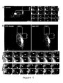
- AA2 only detects overexpressed Rab6A and A' and not various other Rabs.
- HeLa cells were transfected with expression plasmids encoding GFP or YFP-fusions of various Rabs (left column, the transfected Rab is indicated on the left), fixed with 3% paraformaldehyde and stained with AA2 (middle column) and DAPI to stain nuclei (right column).
- AA2 detects overexpressed Rab6A and Rab6A', but not the other Rabs tested, including those that are at least partially localized to the Golgi.
- AA2 does not interfere with Rab6 function in intracellular traffic.
- A. AA2 co-precipitates Rab6•GTP and at least three of its effectors. Rab6•GTP was immunoprecipitated with AA2 from detergent extracts of HeLa cells expressing myc-tagged Rab6AQ72L (and Rab6IP2-GFP). Bound and unbound fractions were analyzed by western blotting (as well as an aliquot of the PNS as positive control). Rab6, Rab6IP2-GFP, endogenous p150 glued and endogenous BICD1 (but not endogenous ⁇ tubulin, negative control) are co-precipitated.
- HeLa cells expressing AA2-YFP were continuously incubated with 5 ⁇ g/mL Cy5-coupled Shiga toxin B subunit, fixed with 3% paraformaldehyde in PBS after 10 min, 45 min or 120 min and stained for GalT.
- StxB is normally transported from the plasma membrane to endosomes (10 min), the Golgi (reached at 30-45 min, and continuously refilled from the surface thereafter) and then the ER (barely visible at 45 min, clearly visible at 120 min at the nuclear envelope) in all cells, regardless of AA2-YFP expression.
- Results in B and C indicate that AA2-YFP expression does not interfere with Rab6 A and A' functions in intracellular transport.
- Movie S1 (and movie S3 as a less compressed version): Corresponding to Figure 4A
- Movie S2 (and movie S4 as a less compressed version):Corresponding to Figure 4B
- CFP-Rab6A labels the Golgi and the endoplasmic reticulum (nuclear envelope), and 5-20 ⁇ mlong tubules (arrows) moving towards the periphery at putative ER entry points, and sometimes back to the Golgi.
- CFP-Rab6A dynamics match those already reported for GFP-Rab6A (S11).
- AA2-YFP labels Rab6 in its GTP-bound form on the Golgi and also on all tubules all along their length. Note however that no signal is detected on the ER.
- Anti- ⁇ GDI ( S1 ), anti-Rab6IP1 and anti-Rab6 were raised in rabbits.
- Anti-GalT was kindly provided by E.G. Berger (University of Zurich, Switzerland, CH), but Golgi apparatus can be stained by using other available, appropriate antibodies; anti-BICD1 was provided by C. Hoogenraad (Erasmus University, Rotterdam, NL).
- the anti-GFP antibody was from ROCHE, the anti- ⁇ tubulin from SIGMA, the anti-p150 glued from BD-Transduction Laboratories. Fluorescent secondary antibodies were from Jackson(?) (Cy3 and Cy5-labelled) and
- HRP-coupled secondary antibodies were from Jackson Immunochemicals. Purified Shiga toxin was obtained from L. Johannes (Institut Curie, Paris, F).
- Vectors used to express fluorescent Rab6A and A' wt and mutant proteins were obtained from J.
- Rab6-GST pull-down experiments were carried out as described ( S1 ). Briefly, Rab6-GST purified from E. coli was coupled to Gluthation-sepharose beads (12.5 ⁇ g per condition), loaded with GDP or GTP ⁇ S in 10 mM EDTA buffer for 1hr at 37°C and then incubated with 0.2 ⁇ g AA2 and either 10 ⁇ g mouse post nuclear supernatant (PNS), 1.5 ⁇ L cytosol of Rab6IP1-expressing SF9 insect cells, 0.1% casein, 1% BSA or no blocking agent for 90 min at room temperature in the presence of 10 mM MgCl 2 . Beads were washed 5 times and processed for SDS-PAGE.
- PPS mouse post nuclear supernatant
- HeLa cells were cultured as described in Mallard et al. (S5) and transfected using CaPO 4 (S6).
- S5 Mallard et al.
- S6 CaPO 4
- siRNA siRNA .
- Rab6 knock down experiments were performed in HeLa cells essentially as described by Elbashir et al. (S7) using double stranded RNA oligos (Dharmacon) and will be described elsewhere. Rab6 was depleted in nearly 100% of cells incubated with the siRNA duplex for three days. siRNA-treated and wt, non-treated , HeLa cells were thus mixed on the same cover slips 24 hr after the siRNA transfection,. This ensured that control, non-Rab6-depleted cells, would be visualized simultaneously as Rab6-depleted cells (as shown in Figure 1D). Cells were fixed with 3% paraformaldehyde 48 hr later (i.e.
- siRNA depletion of Rab6A/A' abolished AA2 Golgi labeling while specific siRNA-depletion of either Rab6 A or A' only reduced, but did not abolish, AA2 staining.
- the NotI site downstream of thJe fluorescent protein cDNA in pECFP-N3, pEGFP-N3, pEYFP-N3 were mutated by filling-in.
- the mutated vectors were then modified by inserting a synthetic adaptor containing a His 6 -tag and a myc-tag, downstream of a NotI site, identical to the tags found in pHEN2.
- the synthetic adaptor also inserted an upstream BbsI/NcoI site so that cutting the modified vector with BbsI opens the vector inside the NcoI site exactly as if using NcoI itself.
- Time-lapse images were acquired on a Leica SP2 confocal microscope every 1.5 s or 3 s for 1 or 2 channel-recordings respectively. Both channels were acquired simultaneously using the 457 nm and 514 nm laser wavelengths for CFP and YFP respectively. Excitation intensities and spectral detection windows were systematically independently adjusted (using the AOTF and the spectral set-up respectively) so as to optimize signals and avoid crosstalk between channels.
- Rab6AQ72L (a GTP-locked mutant of Rab6A) cloned in pET15b vector was expressed and purified by Ni-NTA (Qiagen) affinity chromatography essentially as described for Sar1 and Rab1 (S2, 3).
- the cystein-specific coupling agent PEO-Maleimide activated biotin (PEO-biotin, Pierce) was used to biotinylate Rab6AQ72L.
- Bacterially expressed Rab6AQ72L was modified in PBS supplemented with 0.5 % Tween20 in the presence of a 50x molar excess of PEO-biotin overnight at 4°C. 50% of the proteins were modified as estimated by recovery on streptavidin-coated beads and western blot analysis of bound and unbound fractions.
- biotinylated Rab6AQ72L was separated from free biotin using a Sephadex G25 column.
- the screen consisted of three rounds of selection, each of which took 2 days and used 250 ng biotinylated Rab6 (hence 10 nM of biotinylated Rab6 during the selection).
- the selection process was modified as follows. Phages (10 14 ) were first incubated for 90 min with 50 ⁇ L washed streptavidin M280 dynabeads (DYNAL), beads were separated on a magnet and the non-pre-adsorbed phages were recovered. They were then slowly agitated for 30 min in the presence of 10 nM biotinylated Rab6AQ72L (in 1 mL PBS, 0.1% Tween20, 2% Marvel fat free milk), and the interaction was then left standing for 90 min.
- 10 nM biotinylated Rab6AQ72L in 1 mL PBS, 0.1% Tween20, 2% Marvel fat free milk
- scFvs from the Griffin. 1 library are cloned in pHEN2 which allows the production of scFv-M13 pIII fusion proteins in amber suppressor E. coli strains (e.g. SupE) and the production of soluble scFvs in non-suppressor strains. Selected plasmids were thus introduced in the non-suppressor HB2151 strain before production.
- scFvs secreted in 1 mL 2xTY culture produced in 96-deep well plates
- 1 mM IPTG at 30°C were used undiluted without purification.
- scFvs secreted in the culture medium were concentrated on 2 mL 50% slurry Ni-NTA agarose columns (Qiagen).
- scFvs were used at 10 ⁇ g/mL (or undiluted when culture medium was directly used) co-incubated with 9E10 anti-myc and/or anti-His 6 (SIGMA) monoclonals in the presence of 0.2% BSA, followed by anti-mouse fluorescent secondary antibodies.
- SIGMA anti-myc and/or anti-His 6
- AA2 stains the Golgi complex positive for the Galactosyl Transferase (GalT). Extensive colocalization was observed when co-staining with AA2 and an anti-Rab6 polyclonal (Figure 1B d-f), including at peripheral sites, likely to be ER entry points ( Figure 1B d-f, arrows).
- GST-Rab6A bound to glutathion-Sepharose® was loaded with GDP or with GTP ⁇ S, incubated with AA2 and recovered by pull-down.
- the protein ⁇ GDI ( 15 ) present in the post-nuclear supernatant (PNS) used as a control, bound more strongly to the GDP-bound form of Rab6A ( 16 ).
- PPS post-nuclear supernatant
- AA2 interacted preferentially with the activated GTP ⁇ S-bound form of Rab6 ( Figure 2A).
- GTP ⁇ S-loaded Rab11 did not bind to AA2, although Rab6IP1, which is an effector of both Rab6 and Rab11 ( Figure 2B), bound to both Rab6A and Rab11 in these conditions.
- AA2 is an anti-Rab6 antibody directed against the GTP-bound conformation.
- AA2 still detected endogenous Rab6 that remained localized at the Golgi.
- AA2 detected Rab6•GTP, but not Rab6•GDP by immunofluorescence. This allowed the observation of the behavior of endogenous Rab6 upon overexpression of the GDP-locked mutant.
- AA2 was used as a fluorescent intrabody to investigate Rab6•GTP dynamics in vivo.
- Recombinant antibodies can be GFP-tagged and expressed in the cytoplasm of cultured cells in order to follow the behavior of endogenous proteins in living cells, without tagging and overexpressing the target protein ( 11 , 19 ).
- AA2 was fused to EYFP and expressed in HeLa cells.
- AA2-YFP co-localizes with Golgi markers in fixed cells ( Figure S2) and living cells ( Figure 4B), allowing the endogenous Rab6•GTP t obe followed in vivo. No effect on Rab6 function in intracellular traffic was observed upon AA2-YFP overexpression (Figure S2).
- AA2-YFP stained the Golgi complex and short tubulo-vesicular structures emanating from the Golgi, moving towards the cell periphery and sometimes back to the Golgi ( Figure 4A, arrows and arrowhead respectively). They converge into regions ( Figure 4A, brackets) that likely represent ER entry sites (22).
- Rab6 was present on the Golgi and on transport intermediates as well.
- AA2-YFP The size and shape of structures stained by AA2-YFP were in perfect accordance with those observed after localization of endogenous Rab6 by immunofluorescence (see Figure 1A d-f). However, they were relatively small compared to the 5-20 ⁇ m-long tubular transport intermediates observed in cells expressing GFP-Rab6 (22). Three reasons may explain such a difference. (i) AA2 may perturb the formation of tubules. (ii) Rab6 may be concentrated in a GTP-bound conformation at the tip of tubules. (iii) These relatively small structures represent endogenous transport intermediates, and overexpression, even slight, of Rab6 may cause an extension and stabilization of tubular structures.
Priority Applications (2)
| Application Number | Priority Date | Filing Date | Title |
|---|---|---|---|
| EP03291096A EP1482309A1 (fr) | 2003-05-07 | 2003-05-07 | Moyens pour la détection de la conformation de protéines et leurs applications |
| PCT/EP2004/005733 WO2004099775A1 (fr) | 2003-05-07 | 2004-05-07 | Moyen de detection de conformation proteique et ses applications |
Applications Claiming Priority (1)
| Application Number | Priority Date | Filing Date | Title |
|---|---|---|---|
| EP03291096A EP1482309A1 (fr) | 2003-05-07 | 2003-05-07 | Moyens pour la détection de la conformation de protéines et leurs applications |
Publications (1)
| Publication Number | Publication Date |
|---|---|
| EP1482309A1 true EP1482309A1 (fr) | 2004-12-01 |
Family
ID=33104201
Family Applications (1)
| Application Number | Title | Priority Date | Filing Date |
|---|---|---|---|
| EP03291096A Withdrawn EP1482309A1 (fr) | 2003-05-07 | 2003-05-07 | Moyens pour la détection de la conformation de protéines et leurs applications |
Country Status (2)
| Country | Link |
|---|---|
| EP (1) | EP1482309A1 (fr) |
| WO (1) | WO2004099775A1 (fr) |
Cited By (1)
| Publication number | Priority date | Publication date | Assignee | Title |
|---|---|---|---|---|
| IT202000009232A1 (it) * | 2020-04-28 | 2020-07-28 | Univ Degli Studi Di Messina | Metodo per l'identificazione degli stati conformazionali delle proteine mediante spettroscopia uv-visibile e microscopia in fluorescenza |
Families Citing this family (6)
| Publication number | Priority date | Publication date | Assignee | Title |
|---|---|---|---|---|
| EP3816625A1 (fr) | 2013-05-06 | 2021-05-05 | Scholar Rock, Inc. | Compositions et procédés de modulation de facteur de croissance |
| EP4183806A3 (fr) | 2014-11-12 | 2023-08-02 | Seagen Inc. | Composés interagissant avec le glycane et procédés d'utilisation |
| US9879087B2 (en) | 2014-11-12 | 2018-01-30 | Siamab Therapeutics, Inc. | Glycan-interacting compounds and methods of use |
| IL302822A (en) | 2015-11-12 | 2023-07-01 | Seagen Inc | Compounds interacting with glycans and methods of use |
| EP3541847A4 (fr) | 2016-11-17 | 2020-07-08 | Seattle Genetics, Inc. | Composés interagissant avec le glycane et méthodes d'utilisation |
| KR102653141B1 (ko) | 2017-03-03 | 2024-04-01 | 씨젠 인크. | 글리칸-상호작용 화합물 및 사용 방법 |
Citations (1)
| Publication number | Priority date | Publication date | Assignee | Title |
|---|---|---|---|---|
| US6251393B1 (en) * | 1998-10-23 | 2001-06-26 | The Brigham And Women's Hospital, Inc. | Conformation-specific anti-von Willebrand Factor antibodies |
-
2003
- 2003-05-07 EP EP03291096A patent/EP1482309A1/fr not_active Withdrawn
-
2004
- 2004-05-07 WO PCT/EP2004/005733 patent/WO2004099775A1/fr active Application Filing
Patent Citations (1)
| Publication number | Priority date | Publication date | Assignee | Title |
|---|---|---|---|---|
| US6251393B1 (en) * | 1998-10-23 | 2001-06-26 | The Brigham And Women's Hospital, Inc. | Conformation-specific anti-von Willebrand Factor antibodies |
Non-Patent Citations (7)
| Title |
|---|
| GAO C ET AL: "De novo identification of tumor-specific internalizing human antibody-receptor pairs by phage-display methods", JOURNAL OF IMMUNOLOGICAL METHODS, ELSEVIER SCIENCE PUBLISHERS B.V.,AMSTERDAM, NL, vol. 274, no. 1-2, 1 March 2003 (2003-03-01), pages 185 - 197, XP004411948, ISSN: 0022-1759 * |
| KIKUCHI MASAKAZU ET AL: "A single-chain Fv fragment 2A3 specific for native lysozyme: Isolation from a human synthetic phage display antibody library and characterization", JOURNAL OF BIOCHEMISTRY (TOKYO), vol. 129, no. 2, February 2001 (2001-02-01), pages 237 - 242, XP009019649, ISSN: 0021-924X * |
| MORINO K ET AL: "Antibody fusions with fluorescent proteins: a versatile reagent for profiling protein expression", JOURNAL OF IMMUNOLOGICAL METHODS, ELSEVIER SCIENCE PUBLISHERS B.V.,AMSTERDAM, NL, vol. 257, no. 1-2, 1 November 2001 (2001-11-01), pages 175 - 184, XP004311949, ISSN: 0022-1759 * |
| NIZAK CLEMENT ET AL: "Recombinant antibodies to the small GTPase Rab6 as conformation sensors.", SCIENCE (WASHINGTON D C), vol. 300, no. 5621, 9 May 2003 (2003-05-09), pages 984 - 987, XP002259250, ISSN: 0036-8075 (ISSN print) * |
| PINI A ET AL: "PHAGE DISPLAY OF ANTIBODY FRAGMENTS", CURRENT PROTEIN AND PEPTIDE SCIENCE, BENTHAM SCIENCE PULBISHERS, NL, vol. 1, no. 2, September 2000 (2000-09-01), pages 155 - 169, XP001084580, ISSN: 1389-2037 * |
| SCHWALBACH G ET AL: "Production of Fluorescent Single-Chain Antibody Fragments in Escherichia coli", PROTEIN EXPRESSION AND PURIFICATION, ACADEMIC PRESS, SAN DIEGO, CA, US, vol. 18, no. 2, March 2000 (2000-03-01), pages 121 - 132, XP004435559, ISSN: 1046-5928 * |
| WINTER G ET AL: "MAKING ANTIBODIES BY PHAGE DISPLAY TECHNOLOGY", ANNUAL REVIEW OF IMMUNOLOGY, ANNUAL REVIEWS INC, US, vol. 12, 1994, pages 433 - 455, XP000564245, ISSN: 0732-0582 * |
Cited By (1)
| Publication number | Priority date | Publication date | Assignee | Title |
|---|---|---|---|---|
| IT202000009232A1 (it) * | 2020-04-28 | 2020-07-28 | Univ Degli Studi Di Messina | Metodo per l'identificazione degli stati conformazionali delle proteine mediante spettroscopia uv-visibile e microscopia in fluorescenza |
Also Published As
| Publication number | Publication date |
|---|---|
| WO2004099775A1 (fr) | 2004-11-18 |
Similar Documents
| Publication | Publication Date | Title |
|---|---|---|
| Campa et al. | Rab11 activity and PtdIns (3) P turnover removes recycling cargo from endosomes | |
| Colwill et al. | A roadmap to generate renewable protein binders to the human proteome | |
| Nizak et al. | Recombinant antibodies against subcellular fractions used to track endogenous Golgi protein dynamics in vivo | |
| JP6053672B2 (ja) | アフィニティに基づく用途のためのエピトープタグ | |
| Marsman et al. | Dynein-mediated vesicle transport controls intracellular Salmonella replication | |
| EP1945673B1 (fr) | Ciblage et suivi d'antigenes dans des cellules vivantes | |
| Modi et al. | Recombinant antibody mediated delivery of organelle-specific DNA pH sensors along endocytic pathways | |
| EP3194976B1 (fr) | Procédés permettant de sélectionner des agents qui stabilisent des complexes protéiques | |
| Sugai et al. | PTH/PTH-related protein receptor interacts directly with Tctex-1 through its COOH terminus | |
| Elkind et al. | The role of the COOH terminus of Sec2p in the transport of post-Golgi vesicles | |
| Allemand et al. | A conserved Drosophila transportin-serine/arginine-rich (SR) protein permits nuclear import of Drosophila SR protein splicing factors and their antagonist repressor splicing factor 1 | |
| EP1482309A1 (fr) | Moyens pour la détection de la conformation de protéines et leurs applications | |
| Zeytun et al. | Fluorobodies combine GFP fluorescence with the binding characteristics of antibodies | |
| JP6654779B2 (ja) | ターゲットを認識するタンパク質の発現スクリーニング法 | |
| JPWO2006080396A1 (ja) | 組換え蛋白質の定量法 | |
| Freund et al. | Generation of an intrabody‐based reagent suitable for imaging endogenous proliferating cell nuclear antigen in living cancer cells | |
| US9404123B2 (en) | Nucleic acid assembly, vector, cell, methods and kit thereof | |
| US20140243507A1 (en) | Method of screening antibodies with high antigen selectivity | |
| Houghton et al. | Interacting partners of Golgi‐localized small G protein Arl5b identified by a combination of in vivo proximity labelling and GFP‐Trap pull down | |
| Baker et al. | In vivo proximity biotin ligation identifies the interactome of Egalitarian, a Dynein cargo adaptor | |
| Gu et al. | Conformation of 4.5 S RNA in the signal recognition particle and on the 30S ribosomal subunit | |
| Pinson et al. | Mutations in the yeast Myb-like protein Bas1p resulting in discrimination between promoters in vivo but not in vitro | |
| WO2006095654A1 (fr) | Procede d’essai effectue avec un gene rapporteur | |
| Nizak et al. | Selection and application of recombinant antibodies as sensors of rab protein conformation | |
| Görlich et al. | Nucleoporin-binding nanobodies that either track or inhibit nuclear pore complex assembly |
Legal Events
| Date | Code | Title | Description |
|---|---|---|---|
| PUAI | Public reference made under article 153(3) epc to a published international application that has entered the european phase |
Free format text: ORIGINAL CODE: 0009012 |
|
| AK | Designated contracting states |
Kind code of ref document: A1 Designated state(s): AT BE BG CH CY CZ DE DK EE ES FI FR GB GR HU IE IT LI LU MC NL PT RO SE SI SK TR |
|
| AX | Request for extension of the european patent |
Extension state: AL LT LV MK |
|
| 17P | Request for examination filed |
Effective date: 20050131 |
|
| AKX | Designation fees paid |
Designated state(s): FR |
|
| REG | Reference to a national code |
Ref country code: DE Ref legal event code: 8566 |
|
| STAA | Information on the status of an ep patent application or granted ep patent |
Free format text: STATUS: THE APPLICATION IS DEEMED TO BE WITHDRAWN |
|
| 18D | Application deemed to be withdrawn |
Effective date: 20060210 |