EP1467757B1 - Utilisation d'anticorps diriges contre l'antigene muc18 - Google Patents
Utilisation d'anticorps diriges contre l'antigene muc18 Download PDFInfo
- Publication number
- EP1467757B1 EP1467757B1 EP02797517A EP02797517A EP1467757B1 EP 1467757 B1 EP1467757 B1 EP 1467757B1 EP 02797517 A EP02797517 A EP 02797517A EP 02797517 A EP02797517 A EP 02797517A EP 1467757 B1 EP1467757 B1 EP 1467757B1
- Authority
- EP
- European Patent Office
- Prior art keywords
- seq
- muc18
- antibody
- nos
- antibodies
- Prior art date
- Legal status (The legal status is an assumption and is not a legal conclusion. Google has not performed a legal analysis and makes no representation as to the accuracy of the status listed.)
- Expired - Lifetime
Links
- 102000036639 antigens Human genes 0.000 title description 53
- 108091007433 antigens Proteins 0.000 title description 53
- 239000000427 antigen Substances 0.000 title description 52
- 206010028980 Neoplasm Diseases 0.000 claims abstract description 123
- 102100023126 Cell surface glycoprotein MUC18 Human genes 0.000 claims abstract description 111
- 101000623903 Homo sapiens Cell surface glycoprotein MUC18 Proteins 0.000 claims abstract description 106
- 201000001441 melanoma Diseases 0.000 claims abstract description 58
- 241000282414 Homo sapiens Species 0.000 claims description 93
- 206010027476 Metastases Diseases 0.000 claims description 49
- 241001465754 Metazoa Species 0.000 claims description 45
- 230000009401 metastasis Effects 0.000 claims description 41
- 230000012010 growth Effects 0.000 claims description 26
- 230000002401 inhibitory effect Effects 0.000 claims description 25
- 206010061289 metastatic neoplasm Diseases 0.000 claims description 21
- 230000004083 survival effect Effects 0.000 claims description 21
- 108010039491 Ricin Proteins 0.000 claims description 20
- 230000004614 tumor growth Effects 0.000 claims description 20
- 230000001225 therapeutic effect Effects 0.000 claims description 15
- 239000003814 drug Substances 0.000 claims description 14
- 229940127089 cytotoxic agent Drugs 0.000 claims description 10
- 239000002254 cytotoxic agent Substances 0.000 claims description 8
- 231100000599 cytotoxic agent Toxicity 0.000 claims description 8
- 238000002360 preparation method Methods 0.000 claims description 8
- 229940124597 therapeutic agent Drugs 0.000 claims description 8
- 208000020816 lung neoplasm Diseases 0.000 claims description 5
- 208000037841 lung tumor Diseases 0.000 claims description 3
- 125000003275 alpha amino acid group Chemical group 0.000 claims 6
- 238000011282 treatment Methods 0.000 abstract description 40
- 208000037265 diseases, disorders, signs and symptoms Diseases 0.000 abstract description 24
- 230000000694 effects Effects 0.000 abstract description 20
- 208000035475 disorder Diseases 0.000 abstract description 17
- 238000012512 characterization method Methods 0.000 abstract description 3
- 238000003745 diagnosis Methods 0.000 abstract 1
- 210000004027 cell Anatomy 0.000 description 146
- 150000001413 amino acids Chemical class 0.000 description 72
- 108090000623 proteins and genes Proteins 0.000 description 51
- 201000011510 cancer Diseases 0.000 description 42
- 238000000034 method Methods 0.000 description 38
- 230000014509 gene expression Effects 0.000 description 30
- 210000004881 tumor cell Anatomy 0.000 description 30
- 108020004414 DNA Proteins 0.000 description 29
- 241000699670 Mus sp. Species 0.000 description 27
- 230000027455 binding Effects 0.000 description 27
- 210000004602 germ cell Anatomy 0.000 description 27
- 108091035707 Consensus sequence Proteins 0.000 description 20
- 239000002773 nucleotide Substances 0.000 description 19
- 125000003729 nucleotide group Chemical group 0.000 description 19
- 108060003951 Immunoglobulin Proteins 0.000 description 17
- 102000018358 immunoglobulin Human genes 0.000 description 17
- 235000018102 proteins Nutrition 0.000 description 17
- 102000004169 proteins and genes Human genes 0.000 description 17
- 241000699666 Mus <mouse, genus> Species 0.000 description 14
- 229940024606 amino acid Drugs 0.000 description 14
- 235000001014 amino acid Nutrition 0.000 description 14
- 210000004408 hybridoma Anatomy 0.000 description 14
- 238000001727 in vivo Methods 0.000 description 14
- 230000001394 metastastic effect Effects 0.000 description 14
- 108010047041 Complementarity Determining Regions Proteins 0.000 description 13
- 206010035226 Plasma cell myeloma Diseases 0.000 description 13
- 201000000050 myeloid neoplasm Diseases 0.000 description 12
- 230000004071 biological effect Effects 0.000 description 11
- 229940127121 immunoconjugate Drugs 0.000 description 11
- 206010027480 Metastatic malignant melanoma Diseases 0.000 description 10
- 239000012528 membrane Substances 0.000 description 10
- 208000021039 metastatic melanoma Diseases 0.000 description 10
- 108020004707 nucleic acids Proteins 0.000 description 10
- 102000039446 nucleic acids Human genes 0.000 description 10
- 150000007523 nucleic acids Chemical class 0.000 description 10
- 210000001519 tissue Anatomy 0.000 description 10
- 238000002474 experimental method Methods 0.000 description 9
- 230000006870 function Effects 0.000 description 9
- 239000001963 growth medium Substances 0.000 description 9
- 230000005764 inhibitory process Effects 0.000 description 9
- 210000004379 membrane Anatomy 0.000 description 9
- 239000013615 primer Substances 0.000 description 9
- 238000002965 ELISA Methods 0.000 description 8
- 206010061309 Neoplasm progression Diseases 0.000 description 8
- 239000000872 buffer Substances 0.000 description 8
- 239000007924 injection Substances 0.000 description 8
- 238000002347 injection Methods 0.000 description 8
- 239000002609 medium Substances 0.000 description 8
- 239000000203 mixture Substances 0.000 description 8
- 239000003053 toxin Substances 0.000 description 8
- 231100000765 toxin Toxicity 0.000 description 8
- 108700012359 toxins Proteins 0.000 description 8
- 230000005751 tumor progression Effects 0.000 description 8
- 241000124008 Mammalia Species 0.000 description 7
- 241000699660 Mus musculus Species 0.000 description 7
- 238000012452 Xenomouse strains Methods 0.000 description 7
- 210000003719 b-lymphocyte Anatomy 0.000 description 7
- 230000015572 biosynthetic process Effects 0.000 description 7
- 201000010099 disease Diseases 0.000 description 7
- 230000004927 fusion Effects 0.000 description 7
- 229940072221 immunoglobulins Drugs 0.000 description 7
- 238000011580 nude mouse model Methods 0.000 description 7
- 210000003462 vein Anatomy 0.000 description 7
- 239000012981 Hank's balanced salt solution Substances 0.000 description 6
- 101100458666 Homo sapiens MCAM gene Proteins 0.000 description 6
- 208000008839 Kidney Neoplasms Diseases 0.000 description 6
- 206010027458 Metastases to lung Diseases 0.000 description 6
- 206010038389 Renal cancer Diseases 0.000 description 6
- 210000001744 T-lymphocyte Anatomy 0.000 description 6
- IQFYYKKMVGJFEH-XLPZGREQSA-N Thymidine Chemical compound O=C1NC(=O)C(C)=CN1[C@@H]1O[C@H](CO)[C@@H](O)C1 IQFYYKKMVGJFEH-XLPZGREQSA-N 0.000 description 6
- 239000002671 adjuvant Substances 0.000 description 6
- 238000003556 assay Methods 0.000 description 6
- 230000003053 immunization Effects 0.000 description 6
- 230000002637 immunotoxin Effects 0.000 description 6
- 229940051026 immunotoxin Drugs 0.000 description 6
- 239000002596 immunotoxin Substances 0.000 description 6
- 231100000608 immunotoxin Toxicity 0.000 description 6
- 238000000338 in vitro Methods 0.000 description 6
- 201000010982 kidney cancer Diseases 0.000 description 6
- 210000004072 lung Anatomy 0.000 description 6
- 210000004698 lymphocyte Anatomy 0.000 description 6
- 238000004519 manufacturing process Methods 0.000 description 6
- 108090000765 processed proteins & peptides Proteins 0.000 description 6
- 239000000243 solution Substances 0.000 description 6
- 238000007920 subcutaneous administration Methods 0.000 description 6
- 230000003442 weekly effect Effects 0.000 description 6
- 102100026802 72 kDa type IV collagenase Human genes 0.000 description 5
- 101710151806 72 kDa type IV collagenase Proteins 0.000 description 5
- 108010025714 CD146 Antigen Proteins 0.000 description 5
- 206010009944 Colon cancer Diseases 0.000 description 5
- 108010037362 Extracellular Matrix Proteins Proteins 0.000 description 5
- 102000010834 Extracellular Matrix Proteins Human genes 0.000 description 5
- 108700020796 Oncogene Proteins 0.000 description 5
- 206010060862 Prostate cancer Diseases 0.000 description 5
- 208000000236 Prostatic Neoplasms Diseases 0.000 description 5
- 230000004913 activation Effects 0.000 description 5
- 238000004458 analytical method Methods 0.000 description 5
- 238000004113 cell culture Methods 0.000 description 5
- 230000004709 cell invasion Effects 0.000 description 5
- 230000004663 cell proliferation Effects 0.000 description 5
- 238000006243 chemical reaction Methods 0.000 description 5
- 238000005516 engineering process Methods 0.000 description 5
- 210000002744 extracellular matrix Anatomy 0.000 description 5
- 238000002649 immunization Methods 0.000 description 5
- 238000011534 incubation Methods 0.000 description 5
- 230000036210 malignancy Effects 0.000 description 5
- 108010082117 matrigel Proteins 0.000 description 5
- 229920001184 polypeptide Polymers 0.000 description 5
- 102000004196 processed proteins & peptides Human genes 0.000 description 5
- 239000006228 supernatant Substances 0.000 description 5
- 231100000331 toxic Toxicity 0.000 description 5
- 230000002588 toxic effect Effects 0.000 description 5
- 239000013598 vector Substances 0.000 description 5
- 238000005406 washing Methods 0.000 description 5
- KAQXCFRYVVJPEV-UHFFFAOYSA-N 2,4-dimethyl-1,3-thiazole;1,5-diphenyl-1h-tetrazol-1-ium;bromide Chemical compound [Br-].CC1=CSC(C)=N1.C1=CC=CC=C1[NH+]1C(C=2C=CC=CC=2)=NN=N1 KAQXCFRYVVJPEV-UHFFFAOYSA-N 0.000 description 4
- CIWBSHSKHKDKBQ-JLAZNSOCSA-N Ascorbic acid Chemical compound OC[C@H](O)[C@H]1OC(=O)C(O)=C1O CIWBSHSKHKDKBQ-JLAZNSOCSA-N 0.000 description 4
- 238000011729 BALB/c nude mouse Methods 0.000 description 4
- 208000001333 Colorectal Neoplasms Diseases 0.000 description 4
- 108091006020 Fc-tagged proteins Proteins 0.000 description 4
- 102000018251 Hypoxanthine Phosphoribosyltransferase Human genes 0.000 description 4
- 108010091358 Hypoxanthine Phosphoribosyltransferase Proteins 0.000 description 4
- 108010021625 Immunoglobulin Fragments Proteins 0.000 description 4
- 102000008394 Immunoglobulin Fragments Human genes 0.000 description 4
- 108010000684 Matrix Metalloproteinases Proteins 0.000 description 4
- 102000002274 Matrix Metalloproteinases Human genes 0.000 description 4
- 108091028043 Nucleic acid sequence Proteins 0.000 description 4
- 208000006265 Renal cell carcinoma Diseases 0.000 description 4
- 125000000539 amino acid group Chemical group 0.000 description 4
- 230000010261 cell growth Effects 0.000 description 4
- 230000012292 cell migration Effects 0.000 description 4
- 239000003795 chemical substances by application Substances 0.000 description 4
- 238000002512 chemotherapy Methods 0.000 description 4
- 239000002299 complementary DNA Substances 0.000 description 4
- 230000008878 coupling Effects 0.000 description 4
- 238000010168 coupling process Methods 0.000 description 4
- 238000005859 coupling reaction Methods 0.000 description 4
- 238000011161 development Methods 0.000 description 4
- 230000018109 developmental process Effects 0.000 description 4
- 231100000673 dose–response relationship Toxicity 0.000 description 4
- 238000001943 fluorescence-activated cell sorting Methods 0.000 description 4
- 239000012634 fragment Substances 0.000 description 4
- FDGQSTZJBFJUBT-UHFFFAOYSA-N hypoxanthine Chemical compound O=C1NC=NC2=C1NC=N2 FDGQSTZJBFJUBT-UHFFFAOYSA-N 0.000 description 4
- 229960001001 ibritumomab tiuxetan Drugs 0.000 description 4
- 230000016784 immunoglobulin production Effects 0.000 description 4
- APFVFJFRJDLVQX-AHCXROLUSA-N indium-111 Chemical compound [111In] APFVFJFRJDLVQX-AHCXROLUSA-N 0.000 description 4
- 230000009545 invasion Effects 0.000 description 4
- 238000001638 lipofection Methods 0.000 description 4
- 238000005259 measurement Methods 0.000 description 4
- 230000000683 nonmetastatic effect Effects 0.000 description 4
- 108020003175 receptors Proteins 0.000 description 4
- 102000005962 receptors Human genes 0.000 description 4
- 230000008685 targeting Effects 0.000 description 4
- 108091032973 (ribonucleotides)n+m Proteins 0.000 description 3
- RTQWWZBSTRGEAV-PKHIMPSTSA-N 2-[[(2s)-2-[bis(carboxymethyl)amino]-3-[4-(methylcarbamoylamino)phenyl]propyl]-[2-[bis(carboxymethyl)amino]propyl]amino]acetic acid Chemical compound CNC(=O)NC1=CC=C(C[C@@H](CN(CC(C)N(CC(O)=O)CC(O)=O)CC(O)=O)N(CC(O)=O)CC(O)=O)C=C1 RTQWWZBSTRGEAV-PKHIMPSTSA-N 0.000 description 3
- FWMNVWWHGCHHJJ-SKKKGAJSSA-N 4-amino-1-[(2r)-6-amino-2-[[(2r)-2-[[(2r)-2-[[(2r)-2-amino-3-phenylpropanoyl]amino]-3-phenylpropanoyl]amino]-4-methylpentanoyl]amino]hexanoyl]piperidine-4-carboxylic acid Chemical compound C([C@H](C(=O)N[C@H](CC(C)C)C(=O)N[C@H](CCCCN)C(=O)N1CCC(N)(CC1)C(O)=O)NC(=O)[C@H](N)CC=1C=CC=CC=1)C1=CC=CC=C1 FWMNVWWHGCHHJJ-SKKKGAJSSA-N 0.000 description 3
- DWRXFEITVBNRMK-UHFFFAOYSA-N Beta-D-1-Arabinofuranosylthymine Natural products O=C1NC(=O)C(C)=CN1C1C(O)C(O)C(CO)O1 DWRXFEITVBNRMK-UHFFFAOYSA-N 0.000 description 3
- 201000009030 Carcinoma Diseases 0.000 description 3
- 108010067225 Cell Adhesion Molecules Proteins 0.000 description 3
- 102000016289 Cell Adhesion Molecules Human genes 0.000 description 3
- 102000005636 Cyclic AMP Response Element-Binding Protein Human genes 0.000 description 3
- 108010045171 Cyclic AMP Response Element-Binding Protein Proteins 0.000 description 3
- 241000282412 Homo Species 0.000 description 3
- 229930126263 Maytansine Natural products 0.000 description 3
- 108091007491 NSP3 Papain-like protease domains Proteins 0.000 description 3
- 102000043276 Oncogene Human genes 0.000 description 3
- 239000002202 Polyethylene glycol Substances 0.000 description 3
- BQCADISMDOOEFD-UHFFFAOYSA-N Silver Chemical compound [Ag] BQCADISMDOOEFD-UHFFFAOYSA-N 0.000 description 3
- 208000005718 Stomach Neoplasms Diseases 0.000 description 3
- 206010064390 Tumour invasion Diseases 0.000 description 3
- VWQVUPCCIRVNHF-OUBTZVSYSA-N Yttrium-90 Chemical compound [90Y] VWQVUPCCIRVNHF-OUBTZVSYSA-N 0.000 description 3
- 239000002246 antineoplastic agent Substances 0.000 description 3
- 230000008901 benefit Effects 0.000 description 3
- IQFYYKKMVGJFEH-UHFFFAOYSA-N beta-L-thymidine Natural products O=C1NC(=O)C(C)=CN1C1OC(CO)C(O)C1 IQFYYKKMVGJFEH-UHFFFAOYSA-N 0.000 description 3
- 230000009400 cancer invasion Effects 0.000 description 3
- 230000001413 cellular effect Effects 0.000 description 3
- 238000005119 centrifugation Methods 0.000 description 3
- 239000003431 cross linking reagent Substances 0.000 description 3
- 230000006378 damage Effects 0.000 description 3
- 230000007423 decrease Effects 0.000 description 3
- 238000012217 deletion Methods 0.000 description 3
- 230000037430 deletion Effects 0.000 description 3
- 230000001419 dependent effect Effects 0.000 description 3
- 239000012636 effector Substances 0.000 description 3
- 210000002889 endothelial cell Anatomy 0.000 description 3
- 230000001747 exhibiting effect Effects 0.000 description 3
- 238000000684 flow cytometry Methods 0.000 description 3
- 238000009472 formulation Methods 0.000 description 3
- 206010017758 gastric cancer Diseases 0.000 description 3
- 230000002068 genetic effect Effects 0.000 description 3
- 238000003119 immunoblot Methods 0.000 description 3
- 230000003902 lesion Effects 0.000 description 3
- 150000002632 lipids Chemical class 0.000 description 3
- 210000001165 lymph node Anatomy 0.000 description 3
- 239000011159 matrix material Substances 0.000 description 3
- WKPWGQKGSOKKOO-RSFHAFMBSA-N maytansine Chemical compound CO[C@@H]([C@@]1(O)C[C@](OC(=O)N1)([C@H]([C@@H]1O[C@@]1(C)[C@@H](OC(=O)[C@H](C)N(C)C(C)=O)CC(=O)N1C)C)[H])\C=C\C=C(C)\CC2=CC(OC)=C(Cl)C1=C2 WKPWGQKGSOKKOO-RSFHAFMBSA-N 0.000 description 3
- 238000013508 migration Methods 0.000 description 3
- 230000009826 neoplastic cell growth Effects 0.000 description 3
- 229920001223 polyethylene glycol Polymers 0.000 description 3
- 210000002307 prostate Anatomy 0.000 description 3
- 238000012216 screening Methods 0.000 description 3
- 229910052709 silver Inorganic materials 0.000 description 3
- 239000004332 silver Substances 0.000 description 3
- 241000894007 species Species 0.000 description 3
- 206010041823 squamous cell carcinoma Diseases 0.000 description 3
- 238000010186 staining Methods 0.000 description 3
- 201000011549 stomach cancer Diseases 0.000 description 3
- 239000000126 substance Substances 0.000 description 3
- 238000013268 sustained release Methods 0.000 description 3
- 239000012730 sustained-release form Substances 0.000 description 3
- 229940104230 thymidine Drugs 0.000 description 3
- 238000001890 transfection Methods 0.000 description 3
- 238000012546 transfer Methods 0.000 description 3
- 231100000588 tumorigenic Toxicity 0.000 description 3
- 230000000381 tumorigenic effect Effects 0.000 description 3
- OZFAFGSSMRRTDW-UHFFFAOYSA-N (2,4-dichlorophenyl) benzenesulfonate Chemical compound ClC1=CC(Cl)=CC=C1OS(=O)(=O)C1=CC=CC=C1 OZFAFGSSMRRTDW-UHFFFAOYSA-N 0.000 description 2
- TVZGACDUOSZQKY-LBPRGKRZSA-N 4-aminofolic acid Chemical compound C1=NC2=NC(N)=NC(N)=C2N=C1CNC1=CC=C(C(=O)N[C@@H](CCC(O)=O)C(O)=O)C=C1 TVZGACDUOSZQKY-LBPRGKRZSA-N 0.000 description 2
- 206010003445 Ascites Diseases 0.000 description 2
- 206010005003 Bladder cancer Diseases 0.000 description 2
- 239000011547 Bouin solution Substances 0.000 description 2
- 206010006187 Breast cancer Diseases 0.000 description 2
- 208000026310 Breast neoplasm Diseases 0.000 description 2
- 241000283707 Capra Species 0.000 description 2
- 108091026890 Coding region Proteins 0.000 description 2
- 239000012591 Dulbecco’s Phosphate Buffered Saline Substances 0.000 description 2
- 239000006144 Dulbecco’s modified Eagle's medium Substances 0.000 description 2
- 206010014733 Endometrial cancer Diseases 0.000 description 2
- 206010014759 Endometrial neoplasm Diseases 0.000 description 2
- 102000004190 Enzymes Human genes 0.000 description 2
- 108090000790 Enzymes Proteins 0.000 description 2
- JRZJKWGQFNTSRN-UHFFFAOYSA-N Geldanamycin Natural products C1C(C)CC(OC)C(O)C(C)C=C(C)C(OC(N)=O)C(OC)CCC=C(C)C(=O)NC2=CC(=O)C(OC)=C1C2=O JRZJKWGQFNTSRN-UHFFFAOYSA-N 0.000 description 2
- DHMQDGOQFOQNFH-UHFFFAOYSA-N Glycine Chemical compound NCC(O)=O DHMQDGOQFOQNFH-UHFFFAOYSA-N 0.000 description 2
- 102000003886 Glycoproteins Human genes 0.000 description 2
- 108090000288 Glycoproteins Proteins 0.000 description 2
- 108010001336 Horseradish Peroxidase Proteins 0.000 description 2
- 241000701044 Human gammaherpesvirus 4 Species 0.000 description 2
- UGQMRVRMYYASKQ-UHFFFAOYSA-N Hypoxanthine nucleoside Natural products OC1C(O)C(CO)OC1N1C(NC=NC2=O)=C2N=C1 UGQMRVRMYYASKQ-UHFFFAOYSA-N 0.000 description 2
- 108010054738 Immunologic Receptors Proteins 0.000 description 2
- 102000001749 Immunologic Receptors Human genes 0.000 description 2
- WHUUTDBJXJRKMK-VKHMYHEASA-N L-glutamic acid Chemical compound OC(=O)[C@@H](N)CCC(O)=O WHUUTDBJXJRKMK-VKHMYHEASA-N 0.000 description 2
- ZDXPYRJPNDTMRX-VKHMYHEASA-N L-glutamine Chemical compound OC(=O)[C@@H](N)CCC(N)=O ZDXPYRJPNDTMRX-VKHMYHEASA-N 0.000 description 2
- 206010058467 Lung neoplasm malignant Diseases 0.000 description 2
- 102000012750 Membrane Glycoproteins Human genes 0.000 description 2
- 108010090054 Membrane Glycoproteins Proteins 0.000 description 2
- 102000005741 Metalloproteases Human genes 0.000 description 2
- 108010006035 Metalloproteases Proteins 0.000 description 2
- 241001529936 Murinae Species 0.000 description 2
- 101100458667 Mus musculus Mcam gene Proteins 0.000 description 2
- 108010013731 Myelin-Associated Glycoprotein Proteins 0.000 description 2
- 102100021831 Myelin-associated glycoprotein Human genes 0.000 description 2
- 108010069196 Neural Cell Adhesion Molecules Proteins 0.000 description 2
- 102100023616 Neural cell adhesion molecule L1-like protein Human genes 0.000 description 2
- 206010033128 Ovarian cancer Diseases 0.000 description 2
- 206010061535 Ovarian neoplasm Diseases 0.000 description 2
- 206010061902 Pancreatic neoplasm Diseases 0.000 description 2
- 108010004729 Phycoerythrin Proteins 0.000 description 2
- 206010056342 Pulmonary mass Diseases 0.000 description 2
- 108020004511 Recombinant DNA Proteins 0.000 description 2
- 206010061934 Salivary gland cancer Diseases 0.000 description 2
- PXIPVTKHYLBLMZ-UHFFFAOYSA-N Sodium azide Chemical compound [Na+].[N-]=[N+]=[N-] PXIPVTKHYLBLMZ-UHFFFAOYSA-N 0.000 description 2
- 208000024770 Thyroid neoplasm Diseases 0.000 description 2
- 208000007097 Urinary Bladder Neoplasms Diseases 0.000 description 2
- 230000009471 action Effects 0.000 description 2
- 229960003896 aminopterin Drugs 0.000 description 2
- 230000003698 anagen phase Effects 0.000 description 2
- 238000010171 animal model Methods 0.000 description 2
- 210000000628 antibody-producing cell Anatomy 0.000 description 2
- 229960005070 ascorbic acid Drugs 0.000 description 2
- 235000010323 ascorbic acid Nutrition 0.000 description 2
- 239000011668 ascorbic acid Substances 0.000 description 2
- 238000001815 biotherapy Methods 0.000 description 2
- 239000000969 carrier Substances 0.000 description 2
- 230000008619 cell matrix interaction Effects 0.000 description 2
- 230000003833 cell viability Effects 0.000 description 2
- 208000019065 cervical carcinoma Diseases 0.000 description 2
- 239000002738 chelating agent Substances 0.000 description 2
- 239000002975 chemoattractant Substances 0.000 description 2
- HVYWMOMLDIMFJA-DPAQBDIFSA-N cholesterol Chemical compound C1C=C2C[C@@H](O)CC[C@]2(C)[C@@H]2[C@@H]1[C@@H]1CC[C@H]([C@H](C)CCCC(C)C)[C@@]1(C)CC2 HVYWMOMLDIMFJA-DPAQBDIFSA-N 0.000 description 2
- 238000004587 chromatography analysis Methods 0.000 description 2
- 230000001684 chronic effect Effects 0.000 description 2
- 208000029742 colonic neoplasm Diseases 0.000 description 2
- 208000030381 cutaneous melanoma Diseases 0.000 description 2
- 238000010586 diagram Methods 0.000 description 2
- 238000010790 dilution Methods 0.000 description 2
- 239000012895 dilution Substances 0.000 description 2
- 239000000539 dimer Substances 0.000 description 2
- 229940079593 drug Drugs 0.000 description 2
- 230000003511 endothelial effect Effects 0.000 description 2
- 239000013604 expression vector Substances 0.000 description 2
- 108020001507 fusion proteins Proteins 0.000 description 2
- 102000037865 fusion proteins Human genes 0.000 description 2
- QTQAWLPCGQOSGP-GBTDJJJQSA-N geldanamycin Chemical compound N1C(=O)\C(C)=C/C=C\[C@@H](OC)[C@H](OC(N)=O)\C(C)=C/[C@@H](C)[C@@H](O)[C@H](OC)C[C@@H](C)CC2=C(OC)C(=O)C=C1C2=O QTQAWLPCGQOSGP-GBTDJJJQSA-N 0.000 description 2
- 230000000762 glandular Effects 0.000 description 2
- 206010073071 hepatocellular carcinoma Diseases 0.000 description 2
- 230000001900 immune effect Effects 0.000 description 2
- 238000002513 implantation Methods 0.000 description 2
- 208000015181 infectious disease Diseases 0.000 description 2
- 238000001802 infusion Methods 0.000 description 2
- 230000008611 intercellular interaction Effects 0.000 description 2
- 238000001990 intravenous administration Methods 0.000 description 2
- 238000005304 joining Methods 0.000 description 2
- 210000003734 kidney Anatomy 0.000 description 2
- 208000032839 leukemia Diseases 0.000 description 2
- 239000003446 ligand Substances 0.000 description 2
- 239000002502 liposome Substances 0.000 description 2
- 239000012669 liquid formulation Substances 0.000 description 2
- 208000014018 liver neoplasm Diseases 0.000 description 2
- 201000005202 lung cancer Diseases 0.000 description 2
- 208000015486 malignant pancreatic neoplasm Diseases 0.000 description 2
- 239000000463 material Substances 0.000 description 2
- 238000002156 mixing Methods 0.000 description 2
- VMGAPWLDMVPYIA-HIDZBRGKSA-N n'-amino-n-iminomethanimidamide Chemical compound N\N=C\N=N VMGAPWLDMVPYIA-HIDZBRGKSA-N 0.000 description 2
- 230000003472 neutralizing effect Effects 0.000 description 2
- 201000002528 pancreatic cancer Diseases 0.000 description 2
- 208000008443 pancreatic carcinoma Diseases 0.000 description 2
- 230000000737 periodic effect Effects 0.000 description 2
- 239000000546 pharmaceutical excipient Substances 0.000 description 2
- 239000013612 plasmid Substances 0.000 description 2
- 230000003389 potentiating effect Effects 0.000 description 2
- 230000002265 prevention Effects 0.000 description 2
- 230000008569 process Effects 0.000 description 2
- 230000035755 proliferation Effects 0.000 description 2
- 238000000746 purification Methods 0.000 description 2
- 238000003127 radioimmunoassay Methods 0.000 description 2
- 238000011363 radioimmunotherapy Methods 0.000 description 2
- 230000009257 reactivity Effects 0.000 description 2
- 230000008707 rearrangement Effects 0.000 description 2
- BOLDJAUMGUJJKM-LSDHHAIUSA-N renifolin D Natural products CC(=C)[C@@H]1Cc2c(O)c(O)ccc2[C@H]1CC(=O)c3ccc(O)cc3O BOLDJAUMGUJJKM-LSDHHAIUSA-N 0.000 description 2
- 238000012340 reverse transcriptase PCR Methods 0.000 description 2
- 230000002441 reversible effect Effects 0.000 description 2
- 238000007790 scraping Methods 0.000 description 2
- 210000002966 serum Anatomy 0.000 description 2
- 239000012679 serum free medium Substances 0.000 description 2
- 201000003708 skin melanoma Diseases 0.000 description 2
- 238000002415 sodium dodecyl sulfate polyacrylamide gel electrophoresis Methods 0.000 description 2
- 208000017572 squamous cell neoplasm Diseases 0.000 description 2
- 239000003381 stabilizer Substances 0.000 description 2
- 238000002560 therapeutic procedure Methods 0.000 description 2
- 201000002510 thyroid cancer Diseases 0.000 description 2
- 201000005112 urinary bladder cancer Diseases 0.000 description 2
- 239000013603 viral vector Substances 0.000 description 2
- JWDFQMWEFLOOED-UHFFFAOYSA-N (2,5-dioxopyrrolidin-1-yl) 3-(pyridin-2-yldisulfanyl)propanoate Chemical compound O=C1CCC(=O)N1OC(=O)CCSSC1=CC=CC=N1 JWDFQMWEFLOOED-UHFFFAOYSA-N 0.000 description 1
- XMQUEQJCYRFIQS-YFKPBYRVSA-N (2s)-2-amino-5-ethoxy-5-oxopentanoic acid Chemical compound CCOC(=O)CC[C@H](N)C(O)=O XMQUEQJCYRFIQS-YFKPBYRVSA-N 0.000 description 1
- NVKAWKQGWWIWPM-ABEVXSGRSA-N 17-β-hydroxy-5-α-Androstan-3-one Chemical compound C1C(=O)CC[C@]2(C)[C@H]3CC[C@](C)([C@H](CC4)O)[C@@H]4[C@@H]3CC[C@H]21 NVKAWKQGWWIWPM-ABEVXSGRSA-N 0.000 description 1
- JKMHFZQWWAIEOD-UHFFFAOYSA-N 2-[4-(2-hydroxyethyl)piperazin-1-yl]ethanesulfonic acid Chemical compound OCC[NH+]1CCN(CCS([O-])(=O)=O)CC1 JKMHFZQWWAIEOD-UHFFFAOYSA-N 0.000 description 1
- -1 22: 547-556 (1983) ) Polymers 0.000 description 1
- QFVHZQCOUORWEI-UHFFFAOYSA-N 4-[(4-anilino-5-sulfonaphthalen-1-yl)diazenyl]-5-hydroxynaphthalene-2,7-disulfonic acid Chemical compound C=12C(O)=CC(S(O)(=O)=O)=CC2=CC(S(O)(=O)=O)=CC=1N=NC(C1=CC=CC(=C11)S(O)(=O)=O)=CC=C1NC1=CC=CC=C1 QFVHZQCOUORWEI-UHFFFAOYSA-N 0.000 description 1
- 229940127124 90Y-ibritumomab tiuxetan Drugs 0.000 description 1
- 108700028369 Alleles Proteins 0.000 description 1
- 206010061424 Anal cancer Diseases 0.000 description 1
- 239000004475 Arginine Substances 0.000 description 1
- DCXYFEDJOCDNAF-UHFFFAOYSA-N Asparagine Natural products OC(=O)C(N)CC(N)=O DCXYFEDJOCDNAF-UHFFFAOYSA-N 0.000 description 1
- 102100024222 B-lymphocyte antigen CD19 Human genes 0.000 description 1
- 241000283690 Bos taurus Species 0.000 description 1
- 101001011741 Bos taurus Insulin Proteins 0.000 description 1
- XDTMQSROBMDMFD-UHFFFAOYSA-N C1CCCCC1 Chemical compound C1CCCCC1 XDTMQSROBMDMFD-UHFFFAOYSA-N 0.000 description 1
- 241000282472 Canis lupus familiaris Species 0.000 description 1
- 208000017897 Carcinoma of esophagus Diseases 0.000 description 1
- 206010008342 Cervix carcinoma Diseases 0.000 description 1
- KRKNYBCHXYNGOX-UHFFFAOYSA-K Citrate Chemical compound [O-]C(=O)CC(O)(CC([O-])=O)C([O-])=O KRKNYBCHXYNGOX-UHFFFAOYSA-K 0.000 description 1
- 241000699800 Cricetinae Species 0.000 description 1
- 241000699802 Cricetulus griseus Species 0.000 description 1
- FBPFZTCFMRRESA-FSIIMWSLSA-N D-Glucitol Natural products OC[C@H](O)[C@H](O)[C@@H](O)[C@H](O)CO FBPFZTCFMRRESA-FSIIMWSLSA-N 0.000 description 1
- FBPFZTCFMRRESA-KVTDHHQDSA-N D-Mannitol Chemical compound OC[C@@H](O)[C@@H](O)[C@H](O)[C@H](O)CO FBPFZTCFMRRESA-KVTDHHQDSA-N 0.000 description 1
- 150000008574 D-amino acids Chemical class 0.000 description 1
- FBPFZTCFMRRESA-JGWLITMVSA-N D-glucitol Chemical compound OC[C@H](O)[C@@H](O)[C@H](O)[C@H](O)CO FBPFZTCFMRRESA-JGWLITMVSA-N 0.000 description 1
- WQZGKKKJIJFFOK-QTVWNMPRSA-N D-mannopyranose Chemical compound OC[C@H]1OC(O)[C@@H](O)[C@@H](O)[C@@H]1O WQZGKKKJIJFFOK-QTVWNMPRSA-N 0.000 description 1
- 239000003155 DNA primer Substances 0.000 description 1
- 239000004375 Dextrin Substances 0.000 description 1
- 229920001353 Dextrin Polymers 0.000 description 1
- BWGNESOTFCXPMA-UHFFFAOYSA-N Dihydrogen disulfide Chemical compound SS BWGNESOTFCXPMA-UHFFFAOYSA-N 0.000 description 1
- 206010061818 Disease progression Diseases 0.000 description 1
- KCXVZYZYPLLWCC-UHFFFAOYSA-N EDTA Chemical compound OC(=O)CN(CC(O)=O)CCN(CC(O)=O)CC(O)=O KCXVZYZYPLLWCC-UHFFFAOYSA-N 0.000 description 1
- 241000196324 Embryophyta Species 0.000 description 1
- 241000283086 Equidae Species 0.000 description 1
- 241000588724 Escherichia coli Species 0.000 description 1
- 241000282326 Felis catus Species 0.000 description 1
- 206010017993 Gastrointestinal neoplasms Diseases 0.000 description 1
- 108010010803 Gelatin Proteins 0.000 description 1
- WQZGKKKJIJFFOK-GASJEMHNSA-N Glucose Natural products OC[C@H]1OC(O)[C@H](O)[C@@H](O)[C@@H]1O WQZGKKKJIJFFOK-GASJEMHNSA-N 0.000 description 1
- 239000004471 Glycine Substances 0.000 description 1
- 208000001258 Hemangiosarcoma Diseases 0.000 description 1
- 101000980825 Homo sapiens B-lymphocyte antigen CD19 Proteins 0.000 description 1
- 101000800116 Homo sapiens Thy-1 membrane glycoprotein Proteins 0.000 description 1
- DGAQECJNVWCQMB-PUAWFVPOSA-M Ilexoside XXIX Chemical compound C[C@@H]1CC[C@@]2(CC[C@@]3(C(=CC[C@H]4[C@]3(CC[C@@H]5[C@@]4(CC[C@@H](C5(C)C)OS(=O)(=O)[O-])C)C)[C@@H]2[C@]1(C)O)C)C(=O)O[C@H]6[C@@H]([C@H]([C@@H]([C@H](O6)CO)O)O)O.[Na+] DGAQECJNVWCQMB-PUAWFVPOSA-M 0.000 description 1
- 108700005091 Immunoglobulin Genes Proteins 0.000 description 1
- 102000016844 Immunoglobulin-like domains Human genes 0.000 description 1
- 108050006430 Immunoglobulin-like domains Proteins 0.000 description 1
- 206010062016 Immunosuppression Diseases 0.000 description 1
- 108090001005 Interleukin-6 Proteins 0.000 description 1
- 208000007766 Kaposi sarcoma Diseases 0.000 description 1
- 150000008575 L-amino acids Chemical class 0.000 description 1
- ODKSFYDXXFIFQN-BYPYZUCNSA-P L-argininium(2+) Chemical compound NC(=[NH2+])NCCC[C@H]([NH3+])C(O)=O ODKSFYDXXFIFQN-BYPYZUCNSA-P 0.000 description 1
- DCXYFEDJOCDNAF-REOHCLBHSA-N L-asparagine Chemical compound OC(=O)[C@@H](N)CC(N)=O DCXYFEDJOCDNAF-REOHCLBHSA-N 0.000 description 1
- 229930182816 L-glutamine Natural products 0.000 description 1
- KDXKERNSBIXSRK-YFKPBYRVSA-N L-lysine Chemical compound NCCCC[C@H](N)C(O)=O KDXKERNSBIXSRK-YFKPBYRVSA-N 0.000 description 1
- 108090001090 Lectins Proteins 0.000 description 1
- 102000004856 Lectins Human genes 0.000 description 1
- 208000018142 Leiomyosarcoma Diseases 0.000 description 1
- 206010025323 Lymphomas Diseases 0.000 description 1
- KDXKERNSBIXSRK-UHFFFAOYSA-N Lysine Natural products NCCCCC(N)C(O)=O KDXKERNSBIXSRK-UHFFFAOYSA-N 0.000 description 1
- 239000004472 Lysine Substances 0.000 description 1
- 241000282553 Macaca Species 0.000 description 1
- 206010025598 Malignant hydatidiform mole Diseases 0.000 description 1
- 229930195725 Mannitol Natural products 0.000 description 1
- 208000007256 Nevus Diseases 0.000 description 1
- 239000000020 Nitrocellulose Substances 0.000 description 1
- 206010030155 Oesophageal carcinoma Diseases 0.000 description 1
- 108020005187 Oligonucleotide Probes Proteins 0.000 description 1
- 241000283973 Oryctolagus cuniculus Species 0.000 description 1
- 238000009004 PCR Kit Methods 0.000 description 1
- 229910019142 PO4 Inorganic materials 0.000 description 1
- 108091005804 Peptidases Proteins 0.000 description 1
- 102000035195 Peptidases Human genes 0.000 description 1
- 201000007131 Placental site trophoblastic tumor Diseases 0.000 description 1
- 229920001213 Polysorbate 20 Polymers 0.000 description 1
- 108010059712 Pronase Proteins 0.000 description 1
- 239000004365 Protease Substances 0.000 description 1
- 108010076504 Protein Sorting Signals Proteins 0.000 description 1
- LCTONWCANYUPML-UHFFFAOYSA-M Pyruvate Chemical compound CC(=O)C([O-])=O LCTONWCANYUPML-UHFFFAOYSA-M 0.000 description 1
- 239000012980 RPMI-1640 medium Substances 0.000 description 1
- 208000015634 Rectal Neoplasms Diseases 0.000 description 1
- 208000004337 Salivary Gland Neoplasms Diseases 0.000 description 1
- 206010039491 Sarcoma Diseases 0.000 description 1
- 229920002684 Sepharose Polymers 0.000 description 1
- 108010071390 Serum Albumin Proteins 0.000 description 1
- 102000007562 Serum Albumin Human genes 0.000 description 1
- 206010041067 Small cell lung cancer Diseases 0.000 description 1
- 101100289792 Squirrel monkey polyomavirus large T gene Proteins 0.000 description 1
- 229930006000 Sucrose Natural products 0.000 description 1
- CZMRCDWAGMRECN-UGDNZRGBSA-N Sucrose Chemical compound O[C@H]1[C@H](O)[C@@H](CO)O[C@@]1(CO)O[C@@H]1[C@H](O)[C@@H](O)[C@H](O)[C@@H](CO)O1 CZMRCDWAGMRECN-UGDNZRGBSA-N 0.000 description 1
- NINIDFKCEFEMDL-UHFFFAOYSA-N Sulfur Chemical compound [S] NINIDFKCEFEMDL-UHFFFAOYSA-N 0.000 description 1
- 108010017842 Telomerase Proteins 0.000 description 1
- 102100032938 Telomerase reverse transcriptase Human genes 0.000 description 1
- 102100033523 Thy-1 membrane glycoprotein Human genes 0.000 description 1
- 108091023040 Transcription factor Proteins 0.000 description 1
- 102000040945 Transcription factor Human genes 0.000 description 1
- 208000006105 Uterine Cervical Neoplasms Diseases 0.000 description 1
- 241000251539 Vertebrata <Metazoa> Species 0.000 description 1
- 206010047741 Vulval cancer Diseases 0.000 description 1
- 230000002378 acidificating effect Effects 0.000 description 1
- 230000001154 acute effect Effects 0.000 description 1
- 238000001042 affinity chromatography Methods 0.000 description 1
- 230000004931 aggregating effect Effects 0.000 description 1
- 229940037003 alum Drugs 0.000 description 1
- 150000001412 amines Chemical class 0.000 description 1
- 230000003321 amplification Effects 0.000 description 1
- 201000007538 anal carcinoma Diseases 0.000 description 1
- 230000033115 angiogenesis Effects 0.000 description 1
- 238000009175 antibody therapy Methods 0.000 description 1
- 230000000890 antigenic effect Effects 0.000 description 1
- 239000003963 antioxidant agent Substances 0.000 description 1
- 235000006708 antioxidants Nutrition 0.000 description 1
- 239000007864 aqueous solution Substances 0.000 description 1
- ODKSFYDXXFIFQN-UHFFFAOYSA-N arginine Natural products OC(=O)C(N)CCCNC(N)=N ODKSFYDXXFIFQN-UHFFFAOYSA-N 0.000 description 1
- 229960001230 asparagine Drugs 0.000 description 1
- 235000009582 asparagine Nutrition 0.000 description 1
- 230000001363 autoimmune Effects 0.000 description 1
- 238000011888 autopsy Methods 0.000 description 1
- 230000009286 beneficial effect Effects 0.000 description 1
- WQZGKKKJIJFFOK-VFUOTHLCSA-N beta-D-glucose Chemical compound OC[C@H]1O[C@@H](O)[C@H](O)[C@@H](O)[C@@H]1O WQZGKKKJIJFFOK-VFUOTHLCSA-N 0.000 description 1
- 230000008827 biological function Effects 0.000 description 1
- 229920001222 biopolymer Polymers 0.000 description 1
- JCXGWMGPZLAOME-RNFDNDRNSA-N bismuth-213 Chemical compound [213Bi] JCXGWMGPZLAOME-RNFDNDRNSA-N 0.000 description 1
- 201000000053 blastoma Diseases 0.000 description 1
- 230000000903 blocking effect Effects 0.000 description 1
- 230000037396 body weight Effects 0.000 description 1
- IXIBAKNTJSCKJM-BUBXBXGNSA-N bovine insulin Chemical compound C([C@@H](C(=O)N[C@@H](CC(C)C)C(=O)N[C@H]1CSSC[C@H]2C(=O)N[C@@H](C)C(=O)N[C@@H](CO)C(=O)N[C@H](C(=O)N[C@H](C(N[C@@H](CO)C(=O)N[C@@H](CC(C)C)C(=O)N[C@@H](CC=3C=CC(O)=CC=3)C(=O)N[C@@H](CCC(N)=O)C(=O)N[C@@H](CC(C)C)C(=O)N[C@@H](CCC(O)=O)C(=O)N[C@@H](CC(N)=O)C(=O)N[C@@H](CC=3C=CC(O)=CC=3)C(=O)N[C@@H](CSSC[C@H](NC(=O)[C@H](C(C)C)NC(=O)[C@H](CC(C)C)NC(=O)[C@H](CC=3C=CC(O)=CC=3)NC(=O)[C@H](CC(C)C)NC(=O)[C@H](C)NC(=O)[C@H](CCC(O)=O)NC(=O)[C@H](C(C)C)NC(=O)[C@H](CC(C)C)NC(=O)[C@H](CC=3NC=NC=3)NC(=O)[C@H](CO)NC(=O)CNC1=O)C(=O)NCC(=O)N[C@@H](CCC(O)=O)C(=O)N[C@@H](CCCNC(N)=N)C(=O)NCC(=O)N[C@@H](CC=1C=CC=CC=1)C(=O)N[C@@H](CC=1C=CC=CC=1)C(=O)N[C@@H](CC=1C=CC(O)=CC=1)C(=O)N[C@@H]([C@@H](C)O)C(=O)N1[C@@H](CCC1)C(=O)N[C@@H](CCCCN)C(=O)N[C@@H](C)C(O)=O)C(=O)N[C@@H](CC(N)=O)C(O)=O)=O)CSSC[C@@H](C(N2)=O)NC(=O)[C@H](CCC(N)=O)NC(=O)[C@H](CCC(O)=O)NC(=O)[C@H](C(C)C)NC(=O)[C@@H](NC(=O)CN)[C@@H](C)CC)C(C)C)NC(=O)[C@H](CCC(N)=O)NC(=O)[C@H](CC(N)=O)NC(=O)[C@@H](NC(=O)[C@@H](N)CC=1C=CC=CC=1)C(C)C)C1=CN=CN1 IXIBAKNTJSCKJM-BUBXBXGNSA-N 0.000 description 1
- 210000004556 brain Anatomy 0.000 description 1
- 239000007975 buffered saline Substances 0.000 description 1
- DQXBYHZEEUGOBF-UHFFFAOYSA-N but-3-enoic acid;ethene Chemical compound C=C.OC(=O)CC=C DQXBYHZEEUGOBF-UHFFFAOYSA-N 0.000 description 1
- VSGNNIFQASZAOI-UHFFFAOYSA-L calcium acetate Chemical compound [Ca+2].CC([O-])=O.CC([O-])=O VSGNNIFQASZAOI-UHFFFAOYSA-L 0.000 description 1
- 239000001639 calcium acetate Substances 0.000 description 1
- 235000011092 calcium acetate Nutrition 0.000 description 1
- 229960005147 calcium acetate Drugs 0.000 description 1
- 150000001720 carbohydrates Chemical class 0.000 description 1
- 235000014633 carbohydrates Nutrition 0.000 description 1
- 230000021523 carboxylation Effects 0.000 description 1
- 238000006473 carboxylation reaction Methods 0.000 description 1
- 230000015556 catabolic process Effects 0.000 description 1
- 230000007910 cell fusion Effects 0.000 description 1
- 210000000170 cell membrane Anatomy 0.000 description 1
- 108091092328 cellular RNA Proteins 0.000 description 1
- 230000007541 cellular toxicity Effects 0.000 description 1
- 201000010881 cervical cancer Diseases 0.000 description 1
- 230000008859 change Effects 0.000 description 1
- 239000003153 chemical reaction reagent Substances 0.000 description 1
- 229940044683 chemotherapy drug Drugs 0.000 description 1
- 235000012000 cholesterol Nutrition 0.000 description 1
- 238000010367 cloning Methods 0.000 description 1
- 210000001072 colon Anatomy 0.000 description 1
- 239000003636 conditioned culture medium Substances 0.000 description 1
- 238000010276 construction Methods 0.000 description 1
- 239000000356 contaminant Substances 0.000 description 1
- 229920001577 copolymer Polymers 0.000 description 1
- 238000012258 culturing Methods 0.000 description 1
- 230000001186 cumulative effect Effects 0.000 description 1
- 210000000805 cytoplasm Anatomy 0.000 description 1
- 210000005220 cytoplasmic tail Anatomy 0.000 description 1
- 230000002354 daily effect Effects 0.000 description 1
- 230000034994 death Effects 0.000 description 1
- 230000003247 decreasing effect Effects 0.000 description 1
- 230000002950 deficient Effects 0.000 description 1
- 238000006731 degradation reaction Methods 0.000 description 1
- 230000000593 degrading effect Effects 0.000 description 1
- 239000003405 delayed action preparation Substances 0.000 description 1
- 235000019425 dextrin Nutrition 0.000 description 1
- 238000000502 dialysis Methods 0.000 description 1
- 229940042399 direct acting antivirals protease inhibitors Drugs 0.000 description 1
- 150000002016 disaccharides Chemical class 0.000 description 1
- 230000005750 disease progression Effects 0.000 description 1
- 238000010494 dissociation reaction Methods 0.000 description 1
- 230000005593 dissociations Effects 0.000 description 1
- 125000002228 disulfide group Chemical group 0.000 description 1
- 230000003828 downregulation Effects 0.000 description 1
- 230000000385 effect on melanoma Effects 0.000 description 1
- 238000004520 electroporation Methods 0.000 description 1
- 230000008030 elimination Effects 0.000 description 1
- 238000003379 elimination reaction Methods 0.000 description 1
- 201000008184 embryoma Diseases 0.000 description 1
- 230000002357 endometrial effect Effects 0.000 description 1
- 201000005619 esophageal carcinoma Diseases 0.000 description 1
- 239000005038 ethylene vinyl acetate Substances 0.000 description 1
- 230000003203 everyday effect Effects 0.000 description 1
- 210000002950 fibroblast Anatomy 0.000 description 1
- 238000001914 filtration Methods 0.000 description 1
- 239000012530 fluid Substances 0.000 description 1
- NBVXSUQYWXRMNV-UHFFFAOYSA-N fluoromethane Chemical compound FC NBVXSUQYWXRMNV-UHFFFAOYSA-N 0.000 description 1
- 238000004108 freeze drying Methods 0.000 description 1
- 238000007499 fusion processing Methods 0.000 description 1
- 230000002496 gastric effect Effects 0.000 description 1
- 238000001502 gel electrophoresis Methods 0.000 description 1
- 239000008273 gelatin Substances 0.000 description 1
- 229920000159 gelatin Polymers 0.000 description 1
- 235000019322 gelatine Nutrition 0.000 description 1
- 235000011852 gelatine desserts Nutrition 0.000 description 1
- 238000003500 gene array Methods 0.000 description 1
- 208000005017 glioblastoma Diseases 0.000 description 1
- 239000008103 glucose Substances 0.000 description 1
- 229960002989 glutamic acid Drugs 0.000 description 1
- ZDXPYRJPNDTMRX-UHFFFAOYSA-N glutamine Natural products OC(=O)C(N)CCC(N)=O ZDXPYRJPNDTMRX-UHFFFAOYSA-N 0.000 description 1
- 239000003102 growth factor Substances 0.000 description 1
- 229940093915 gynecological organic acid Drugs 0.000 description 1
- 238000003306 harvesting Methods 0.000 description 1
- 201000010536 head and neck cancer Diseases 0.000 description 1
- 208000014829 head and neck neoplasm Diseases 0.000 description 1
- 230000036541 health Effects 0.000 description 1
- 238000011134 hematopoietic stem cell transplantation Methods 0.000 description 1
- 230000002440 hepatic effect Effects 0.000 description 1
- 229940088597 hormone Drugs 0.000 description 1
- 239000005556 hormone Substances 0.000 description 1
- 239000000017 hydrogel Substances 0.000 description 1
- 229920001477 hydrophilic polymer Polymers 0.000 description 1
- 238000012872 hydroxylapatite chromatography Methods 0.000 description 1
- 230000003100 immobilizing effect Effects 0.000 description 1
- 230000028993 immune response Effects 0.000 description 1
- 230000002163 immunogen Effects 0.000 description 1
- 238000001114 immunoprecipitation Methods 0.000 description 1
- 230000001506 immunosuppresive effect Effects 0.000 description 1
- 238000011065 in-situ storage Methods 0.000 description 1
- 230000006698 induction Effects 0.000 description 1
- 230000010354 integration Effects 0.000 description 1
- 230000003993 interaction Effects 0.000 description 1
- 210000003962 intermediate trophoblast Anatomy 0.000 description 1
- 238000001361 intraarterial administration Methods 0.000 description 1
- 238000007918 intramuscular administration Methods 0.000 description 1
- 238000007912 intraperitoneal administration Methods 0.000 description 1
- 239000007928 intraperitoneal injection Substances 0.000 description 1
- 239000002523 lectin Substances 0.000 description 1
- 201000010260 leiomyoma Diseases 0.000 description 1
- 230000000670 limiting effect Effects 0.000 description 1
- 201000007270 liver cancer Diseases 0.000 description 1
- 201000005249 lung adenocarcinoma Diseases 0.000 description 1
- 210000005265 lung cell Anatomy 0.000 description 1
- 210000002751 lymph Anatomy 0.000 description 1
- 239000008176 lyophilized powder Substances 0.000 description 1
- 239000012139 lysis buffer Substances 0.000 description 1
- UEGPKNKPLBYCNK-UHFFFAOYSA-L magnesium acetate Chemical compound [Mg+2].CC([O-])=O.CC([O-])=O UEGPKNKPLBYCNK-UHFFFAOYSA-L 0.000 description 1
- 235000011285 magnesium acetate Nutrition 0.000 description 1
- 239000011654 magnesium acetate Substances 0.000 description 1
- 229940069446 magnesium acetate Drugs 0.000 description 1
- 238000012423 maintenance Methods 0.000 description 1
- 230000003211 malignant effect Effects 0.000 description 1
- 239000000594 mannitol Substances 0.000 description 1
- 235000010355 mannitol Nutrition 0.000 description 1
- 239000003550 marker Substances 0.000 description 1
- 230000007246 mechanism Effects 0.000 description 1
- 210000002752 melanocyte Anatomy 0.000 description 1
- 229940071648 metered dose inhaler Drugs 0.000 description 1
- DFTAZNAEBRBBKP-UHFFFAOYSA-N methyl 4-sulfanylbutanimidate Chemical compound COC(=N)CCCS DFTAZNAEBRBBKP-UHFFFAOYSA-N 0.000 description 1
- 239000003094 microcapsule Substances 0.000 description 1
- 230000005012 migration Effects 0.000 description 1
- 239000003607 modifier Substances 0.000 description 1
- 238000010369 molecular cloning Methods 0.000 description 1
- 150000002772 monosaccharides Chemical class 0.000 description 1
- 230000035772 mutation Effects 0.000 description 1
- 208000025402 neoplasm of esophagus Diseases 0.000 description 1
- 210000002569 neuron Anatomy 0.000 description 1
- 229920001220 nitrocellulos Polymers 0.000 description 1
- 208000002154 non-small cell lung carcinoma Diseases 0.000 description 1
- 239000002736 nonionic surfactant Substances 0.000 description 1
- 230000009871 nonspecific binding Effects 0.000 description 1
- 231100000252 nontoxic Toxicity 0.000 description 1
- 230000003000 nontoxic effect Effects 0.000 description 1
- 231100001221 nontumorigenic Toxicity 0.000 description 1
- 238000003199 nucleic acid amplification method Methods 0.000 description 1
- 238000011330 nucleic acid test Methods 0.000 description 1
- 239000002751 oligonucleotide probe Substances 0.000 description 1
- 231100000590 oncogenic Toxicity 0.000 description 1
- 230000002246 oncogenic effect Effects 0.000 description 1
- 108091008819 oncoproteins Proteins 0.000 description 1
- 210000000056 organ Anatomy 0.000 description 1
- 150000007524 organic acids Chemical class 0.000 description 1
- 235000005985 organic acids Nutrition 0.000 description 1
- 230000002611 ovarian Effects 0.000 description 1
- 210000001672 ovary Anatomy 0.000 description 1
- 230000002018 overexpression Effects 0.000 description 1
- KHPXUQMNIQBQEV-UHFFFAOYSA-N oxaloacetic acid Chemical compound OC(=O)CC(=O)C(O)=O KHPXUQMNIQBQEV-UHFFFAOYSA-N 0.000 description 1
- 230000036961 partial effect Effects 0.000 description 1
- 230000001575 pathological effect Effects 0.000 description 1
- 208000030940 penile carcinoma Diseases 0.000 description 1
- 201000008174 penis carcinoma Diseases 0.000 description 1
- 239000000137 peptide hydrolase inhibitor Substances 0.000 description 1
- 201000002628 peritoneum cancer Diseases 0.000 description 1
- NBIIXXVUZAFLBC-UHFFFAOYSA-K phosphate Chemical compound [O-]P([O-])([O-])=O NBIIXXVUZAFLBC-UHFFFAOYSA-K 0.000 description 1
- 239000010452 phosphate Substances 0.000 description 1
- 238000000053 physical method Methods 0.000 description 1
- 230000004962 physiological condition Effects 0.000 description 1
- 230000035790 physiological processes and functions Effects 0.000 description 1
- 201000008824 placental choriocarcinoma Diseases 0.000 description 1
- 238000007747 plating Methods 0.000 description 1
- 229920001983 poloxamer Polymers 0.000 description 1
- 229920001200 poly(ethylene-vinyl acetate) Polymers 0.000 description 1
- 229920000747 poly(lactic acid) Polymers 0.000 description 1
- 229920000728 polyester Polymers 0.000 description 1
- 229920002338 polyhydroxyethylmethacrylate Polymers 0.000 description 1
- 229920000642 polymer Polymers 0.000 description 1
- 238000003752 polymerase chain reaction Methods 0.000 description 1
- 239000000256 polyoxyethylene sorbitan monolaurate Substances 0.000 description 1
- 235000010486 polyoxyethylene sorbitan monolaurate Nutrition 0.000 description 1
- 229920000136 polysorbate Polymers 0.000 description 1
- 229940068977 polysorbate 20 Drugs 0.000 description 1
- 229920000036 polyvinylpyrrolidone Polymers 0.000 description 1
- 239000001267 polyvinylpyrrolidone Substances 0.000 description 1
- 235000013855 polyvinylpyrrolidone Nutrition 0.000 description 1
- 239000000843 powder Substances 0.000 description 1
- 238000001556 precipitation Methods 0.000 description 1
- 239000000047 product Substances 0.000 description 1
- 230000000069 prophylactic effect Effects 0.000 description 1
- 238000000159 protein binding assay Methods 0.000 description 1
- 230000012743 protein tagging Effects 0.000 description 1
- 230000005180 public health Effects 0.000 description 1
- 239000012217 radiopharmaceutical Substances 0.000 description 1
- 229940121896 radiopharmaceutical Drugs 0.000 description 1
- 230000002799 radiopharmaceutical effect Effects 0.000 description 1
- 230000006798 recombination Effects 0.000 description 1
- 238000005215 recombination Methods 0.000 description 1
- 206010038038 rectal cancer Diseases 0.000 description 1
- 201000001275 rectum cancer Diseases 0.000 description 1
- 230000024155 regulation of cell adhesion Effects 0.000 description 1
- 230000012760 regulation of cell migration Effects 0.000 description 1
- 230000001105 regulatory effect Effects 0.000 description 1
- AHTFMWCHTGEJHA-UHFFFAOYSA-N s-(2,5-dioxooxolan-3-yl) ethanethioate Chemical compound CC(=O)SC1CC(=O)OC1=O AHTFMWCHTGEJHA-UHFFFAOYSA-N 0.000 description 1
- 210000003079 salivary gland Anatomy 0.000 description 1
- 201000003804 salivary gland carcinoma Diseases 0.000 description 1
- 239000000523 sample Substances 0.000 description 1
- 238000013391 scatchard analysis Methods 0.000 description 1
- 239000004017 serum-free culture medium Substances 0.000 description 1
- 208000000587 small cell lung carcinoma Diseases 0.000 description 1
- 210000000329 smooth muscle myocyte Anatomy 0.000 description 1
- 208000010485 smooth muscle tumor Diseases 0.000 description 1
- 239000011734 sodium Substances 0.000 description 1
- 229910052708 sodium Inorganic materials 0.000 description 1
- 239000000600 sorbitol Substances 0.000 description 1
- 238000001228 spectrum Methods 0.000 description 1
- 238000009987 spinning Methods 0.000 description 1
- 210000004989 spleen cell Anatomy 0.000 description 1
- 238000010561 standard procedure Methods 0.000 description 1
- 238000011146 sterile filtration Methods 0.000 description 1
- 230000004936 stimulating effect Effects 0.000 description 1
- 210000002784 stomach Anatomy 0.000 description 1
- 238000003860 storage Methods 0.000 description 1
- 239000007929 subcutaneous injection Substances 0.000 description 1
- 238000010254 subcutaneous injection Methods 0.000 description 1
- 239000005720 sucrose Substances 0.000 description 1
- 150000005846 sugar alcohols Chemical class 0.000 description 1
- 239000011593 sulfur Substances 0.000 description 1
- 229910052717 sulfur Inorganic materials 0.000 description 1
- 239000000725 suspension Substances 0.000 description 1
- 208000024891 symptom Diseases 0.000 description 1
- 238000003786 synthesis reaction Methods 0.000 description 1
- 238000012360 testing method Methods 0.000 description 1
- 210000001685 thyroid gland Anatomy 0.000 description 1
- 231100000721 toxic potential Toxicity 0.000 description 1
- 230000009261 transgenic effect Effects 0.000 description 1
- 238000011830 transgenic mouse model Methods 0.000 description 1
- 230000001052 transient effect Effects 0.000 description 1
- GPRLSGONYQIRFK-MNYXATJNSA-N triton Chemical compound [3H+] GPRLSGONYQIRFK-MNYXATJNSA-N 0.000 description 1
- 230000005747 tumor angiogenesis Effects 0.000 description 1
- 230000005740 tumor formation Effects 0.000 description 1
- 208000029729 tumor suppressor gene on chromosome 11 Diseases 0.000 description 1
- 241001515965 unidentified phage Species 0.000 description 1
- 206010046766 uterine cancer Diseases 0.000 description 1
- 208000012991 uterine carcinoma Diseases 0.000 description 1
- 230000002792 vascular Effects 0.000 description 1
- 230000003612 virological effect Effects 0.000 description 1
- 201000005102 vulva cancer Diseases 0.000 description 1
Images


Classifications
-
- C—CHEMISTRY; METALLURGY
- C07—ORGANIC CHEMISTRY
- C07K—PEPTIDES
- C07K16/00—Immunoglobulins [IGs], e.g. monoclonal or polyclonal antibodies
- C07K16/18—Immunoglobulins [IGs], e.g. monoclonal or polyclonal antibodies against material from animals or humans
- C07K16/28—Immunoglobulins [IGs], e.g. monoclonal or polyclonal antibodies against material from animals or humans against receptors, cell surface antigens or cell surface determinants
- C07K16/30—Immunoglobulins [IGs], e.g. monoclonal or polyclonal antibodies against material from animals or humans against receptors, cell surface antigens or cell surface determinants from tumour cells
- C07K16/3076—Immunoglobulins [IGs], e.g. monoclonal or polyclonal antibodies against material from animals or humans against receptors, cell surface antigens or cell surface determinants from tumour cells against structure-related tumour-associated moieties
- C07K16/3092—Immunoglobulins [IGs], e.g. monoclonal or polyclonal antibodies against material from animals or humans against receptors, cell surface antigens or cell surface determinants from tumour cells against structure-related tumour-associated moieties against tumour-associated mucins
-
- A—HUMAN NECESSITIES
- A61—MEDICAL OR VETERINARY SCIENCE; HYGIENE
- A61K—PREPARATIONS FOR MEDICAL, DENTAL OR TOILETRY PURPOSES
- A61K47/00—Medicinal preparations characterised by the non-active ingredients used, e.g. carriers or inert additives; Targeting or modifying agents chemically bound to the active ingredient
- A61K47/50—Medicinal preparations characterised by the non-active ingredients used, e.g. carriers or inert additives; Targeting or modifying agents chemically bound to the active ingredient the non-active ingredient being chemically bound to the active ingredient, e.g. polymer-drug conjugates
- A61K47/51—Medicinal preparations characterised by the non-active ingredients used, e.g. carriers or inert additives; Targeting or modifying agents chemically bound to the active ingredient the non-active ingredient being chemically bound to the active ingredient, e.g. polymer-drug conjugates the non-active ingredient being a modifying agent
- A61K47/68—Medicinal preparations characterised by the non-active ingredients used, e.g. carriers or inert additives; Targeting or modifying agents chemically bound to the active ingredient the non-active ingredient being chemically bound to the active ingredient, e.g. polymer-drug conjugates the non-active ingredient being a modifying agent the modifying agent being an antibody, an immunoglobulin or a fragment thereof, e.g. an Fc-fragment
- A61K47/6835—Medicinal preparations characterised by the non-active ingredients used, e.g. carriers or inert additives; Targeting or modifying agents chemically bound to the active ingredient the non-active ingredient being chemically bound to the active ingredient, e.g. polymer-drug conjugates the non-active ingredient being a modifying agent the modifying agent being an antibody, an immunoglobulin or a fragment thereof, e.g. an Fc-fragment the modifying agent being an antibody or an immunoglobulin bearing at least one antigen-binding site
- A61K47/6851—Medicinal preparations characterised by the non-active ingredients used, e.g. carriers or inert additives; Targeting or modifying agents chemically bound to the active ingredient the non-active ingredient being chemically bound to the active ingredient, e.g. polymer-drug conjugates the non-active ingredient being a modifying agent the modifying agent being an antibody, an immunoglobulin or a fragment thereof, e.g. an Fc-fragment the modifying agent being an antibody or an immunoglobulin bearing at least one antigen-binding site the antibody targeting a determinant of a tumour cell
- A61K47/6865—Medicinal preparations characterised by the non-active ingredients used, e.g. carriers or inert additives; Targeting or modifying agents chemically bound to the active ingredient the non-active ingredient being chemically bound to the active ingredient, e.g. polymer-drug conjugates the non-active ingredient being a modifying agent the modifying agent being an antibody, an immunoglobulin or a fragment thereof, e.g. an Fc-fragment the modifying agent being an antibody or an immunoglobulin bearing at least one antigen-binding site the antibody targeting a determinant of a tumour cell the tumour determinant being from skin, nerves or brain cancer cell
-
- A—HUMAN NECESSITIES
- A61—MEDICAL OR VETERINARY SCIENCE; HYGIENE
- A61K—PREPARATIONS FOR MEDICAL, DENTAL OR TOILETRY PURPOSES
- A61K51/00—Preparations containing radioactive substances for use in therapy or testing in vivo
- A61K51/02—Preparations containing radioactive substances for use in therapy or testing in vivo characterised by the carrier, i.e. characterised by the agent or material covalently linked or complexing the radioactive nucleus
- A61K51/04—Organic compounds
- A61K51/08—Peptides, e.g. proteins, carriers being peptides, polyamino acids, proteins
- A61K51/10—Antibodies or immunoglobulins; Fragments thereof, the carrier being an antibody, an immunoglobulin or a fragment thereof, e.g. a camelised human single domain antibody or the Fc fragment of an antibody
- A61K51/1045—Antibodies or immunoglobulins; Fragments thereof, the carrier being an antibody, an immunoglobulin or a fragment thereof, e.g. a camelised human single domain antibody or the Fc fragment of an antibody against animal or human tumor cells or tumor cell determinants
- A61K51/1066—Antibodies or immunoglobulins; Fragments thereof, the carrier being an antibody, an immunoglobulin or a fragment thereof, e.g. a camelised human single domain antibody or the Fc fragment of an antibody against animal or human tumor cells or tumor cell determinants the tumor cell being from skin
-
- A—HUMAN NECESSITIES
- A61—MEDICAL OR VETERINARY SCIENCE; HYGIENE
- A61P—SPECIFIC THERAPEUTIC ACTIVITY OF CHEMICAL COMPOUNDS OR MEDICINAL PREPARATIONS
- A61P35/00—Antineoplastic agents
-
- A—HUMAN NECESSITIES
- A61—MEDICAL OR VETERINARY SCIENCE; HYGIENE
- A61P—SPECIFIC THERAPEUTIC ACTIVITY OF CHEMICAL COMPOUNDS OR MEDICINAL PREPARATIONS
- A61P35/00—Antineoplastic agents
- A61P35/04—Antineoplastic agents specific for metastasis
-
- A—HUMAN NECESSITIES
- A61—MEDICAL OR VETERINARY SCIENCE; HYGIENE
- A61P—SPECIFIC THERAPEUTIC ACTIVITY OF CHEMICAL COMPOUNDS OR MEDICINAL PREPARATIONS
- A61P43/00—Drugs for specific purposes, not provided for in groups A61P1/00-A61P41/00
-
- A—HUMAN NECESSITIES
- A61—MEDICAL OR VETERINARY SCIENCE; HYGIENE
- A61K—PREPARATIONS FOR MEDICAL, DENTAL OR TOILETRY PURPOSES
- A61K39/00—Medicinal preparations containing antigens or antibodies
- A61K2039/505—Medicinal preparations containing antigens or antibodies comprising antibodies
-
- C—CHEMISTRY; METALLURGY
- C07—ORGANIC CHEMISTRY
- C07K—PEPTIDES
- C07K2317/00—Immunoglobulins specific features
- C07K2317/20—Immunoglobulins specific features characterized by taxonomic origin
- C07K2317/21—Immunoglobulins specific features characterized by taxonomic origin from primates, e.g. man
-
- C—CHEMISTRY; METALLURGY
- C07—ORGANIC CHEMISTRY
- C07K—PEPTIDES
- C07K2317/00—Immunoglobulins specific features
- C07K2317/30—Immunoglobulins specific features characterized by aspects of specificity or valency
- C07K2317/33—Crossreactivity, e.g. for species or epitope, or lack of said crossreactivity
-
- C—CHEMISTRY; METALLURGY
- C07—ORGANIC CHEMISTRY
- C07K—PEPTIDES
- C07K2317/00—Immunoglobulins specific features
- C07K2317/50—Immunoglobulins specific features characterized by immunoglobulin fragments
- C07K2317/56—Immunoglobulins specific features characterized by immunoglobulin fragments variable (Fv) region, i.e. VH and/or VL
-
- C—CHEMISTRY; METALLURGY
- C07—ORGANIC CHEMISTRY
- C07K—PEPTIDES
- C07K2317/00—Immunoglobulins specific features
- C07K2317/70—Immunoglobulins specific features characterized by effect upon binding to a cell or to an antigen
- C07K2317/73—Inducing cell death, e.g. apoptosis, necrosis or inhibition of cell proliferation
-
- C—CHEMISTRY; METALLURGY
- C07—ORGANIC CHEMISTRY
- C07K—PEPTIDES
- C07K2317/00—Immunoglobulins specific features
- C07K2317/70—Immunoglobulins specific features characterized by effect upon binding to a cell or to an antigen
- C07K2317/76—Antagonist effect on antigen, e.g. neutralization or inhibition of binding
-
- C—CHEMISTRY; METALLURGY
- C07—ORGANIC CHEMISTRY
- C07K—PEPTIDES
- C07K2317/00—Immunoglobulins specific features
- C07K2317/90—Immunoglobulins specific features characterized by (pharmaco)kinetic aspects or by stability of the immunoglobulin
- C07K2317/92—Affinity (KD), association rate (Ka), dissociation rate (Kd) or EC50 value
-
- C—CHEMISTRY; METALLURGY
- C07—ORGANIC CHEMISTRY
- C07K—PEPTIDES
- C07K2319/00—Fusion polypeptide
- C07K2319/30—Non-immunoglobulin-derived peptide or protein having an immunoglobulin constant or Fc region, or a fragment thereof, attached thereto
Definitions
- Embodiments of the present invention concern antibodies binding MUC18 antigen as well as methods and means for making and using such antibodies.
- MUC18 is a cell-surface glycoprotein originally identified as a melanoma antigen, melanoma cell adhesion molecule (MCAM), whose expression is associated with tumor progression and the development of metastatic potential.
- MCAM melanoma cell adhesion molecule
- MUC18 is a 113 kDA cell surface integral membrane glycoprotein composed of a signal peptide, five immunoglobulin-like domains, a transmembrane region, and a short cytoplasmic tail ( Lehmann et al., Proc Natl Acad Sci USA, 86(24):9891-5 (1989) ).
- MUC18 is a member of the immunoglobulin superfamily and has significant sequence homology to a number of cell adhesion molecules of the 1g superfamily ( Lehmann et al., Proc. Natl. Acad. Sci. USA, 86:9891-9895 (1989) ), including BEN ( Pourquie et al., Proc. Natl. Acad. Sci. USA, 89:5261-5265 (1992) ), neural-cell adhesion molecule (N-CAM) ( Owens et al., Proc. Natl., Acad. Sci. USA, 84:294-298 (1987 )), myelin-associated glycoprotein (MAG) ( Lai et al., Proc. Natl.
- MUC18 has been detected in relatively limited spectrum of normal human tissues and in a variety of malignant neoplasms. In normal adult tissues, MUC 18 is expressed on endothelial cells, smooth muscle cells ( Shih et al., Lab.
- MUC18 is also expressed on a variety of malignant neoplasms including smooth muscle neoplasms (Leiomyomas and leiomyosarcomas), tumors of vascular origin (angiosarcomas and Kaposi's sarcomas), placental site trophoblastic tumors, choriocarcinomas and melanomas ( Shih ct al., Clinical Cancer Res., 2:569-575 (1996) ; Holzmann et al., Int. J Cancer, 39:466-471 (1987) ).
- the expression of MUC18 correlates directly with the metastatic potential of human melanoma cells ( Bar-Eli, M., Cancer Metastasis, 18(3):377-85 (1999) ).
- MUC18 is a marker of tumor progression and metastasis in melanomas.
- the expression of MUC18 is absent in normal melanocytes and benign nevi but prominent on many primary melanomas and in most metastatic lesions ( Lehmann et al., Proc. Natl. Acad. Sci. USA, 86:9891-9895 (1989) ; Lehmann et al., Cancer Res., 47:841-845 (1987) ; Shih et al., Cancer Res., 54:2514-2520 (1994) ).
- MUC18 expression correlates well with tumor vertical thickness and metastasis formation, and greater than 80% of metastatic lesions express MUC18 ( Lehmann et al., Proc. Natl. Acad. Sci. USA, 86:9891-9895 (1989) ; Xie et al., Cancer Res., 57:2295-2303 (1997) ; Sers et al., Proc. Natl. Acad. Sci. USA, 90:85 14-8518 (1993 ); Lehmann et al., Cancer Res., 47:841-845 (1987) ; Shih et al., Cancer Res., 54:2514-2520 (1994) .
- a diagram depicting the expression of MUC18 with respect to other known molecular lesions in human melanoma is presented in Figure 1 .
- CREB/ATF-1 may play an essential role in invasion by regulating the CRE-dependent expression of the adhesion molecule MUC18 and metalloproteinase MMP-2 ( Jean et al., Mol. Cell Biochem., 212(1-2):19-28 (2000 )) which belongs to the MMP family known to contribute to cancers and to have a role in tumor invasion, angiogenesis, and metastasis.
- Tumor cells are believed to utilize the matrix degrading capability of MMPs to spread to distant sites, and once the tumor cells have metastasized, MMPs are thought to promote the growth of these tumor cells.
- the role of MUC18 in melanoma tumor progression is not completely understood, but may include a role in one or more steps in the metastatic process possibly by affecting MMP-2 activation or cell migration.
- MUC18 on MUC18-negative human melanoma cell lines increased their tumorigenicity and enhanced their metastatic capability in experimental tumor models ( Xie et al., Cancer Res., 57:2295-2303 (1997) ; Bani et al., Cancer Res. 56:3075-3086 (1996) ). Finally, inhibition of MUC18 expression in metastases using genetic suppressor elements of MUC18 cDNA led to a decrease of the tumorigenic phenotype in nude mice ( Styamoorthy et al., Oncogene, 20:4676 (2001 )).
- MUC18 Although the function of MUC18 is not fully understood, several studies have demonstrated a role for this protein in mediating cell-cell and cell-matrix interactions by binding to an unidentified ligand ( Shih et al., Cancer Res., 57:3835-3840 (1997) ; Johnson et al., Int. J. cancer, 73:769-774 (1997) ).
- the expression of cell adhesion molecules which mediate cell-to-cell or cell-to-matrix interactions is a tumor cell property that is essential for metastases.
- MUC18-transfected melanoma cells showed increased homotypic adhesion, increased attachment to human endothelial cells, and increased invasion through Matrigel-coated filters suggesting a role in tumor invasion and trans-endothelial migration ( Xie et al., Cancer Res., 57:2295-2303 (1997) ).
- anti-MUC18 antibodies were able to inhibit these functions in the MUC18-transfected cells ( Xie et al., Cancer Res., 57:2295-2303 (1997) ).
- anti-MUC18 antibodies that are able to inhibit the biological function of MUC18, most importantly cell proliferation and growth which may be essential to tumor progression and metastasis.
- Such antibodies would likely interfere with the inherent ability of MUC18 to mediate cell-cell and cell-matrix interactions.
- the inhibition of such activity may be possible with a monoclonal antibody targeted to MUC18.
- the ability to affect the progression of tumor cells expressing MUC18 on the cell surface may prove to be a treatment for patients with tumors or of use for prevention of metastatic disease in patients with such tumors.
- the present invention is based on the development of monoclonal antibodies that were found to bind MUC18 and affect MUC18 function.
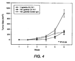
- This application describes human anti-MUC18 antibodies and anti-MUC18 antibody preparations with desirable properties from a therapeutic perspective, including strong binding affinity for MUC18, the ability to inhibit tumor growth and metastasis in vivo , the ability to promote cell survival, and the ability to inhibit tumor invasion in vitro .
- One embodiment of the invention is a use of a monoclonal antibody comprising a heavy chain and a light chain amino acid, wherein said antibody has a heavy chain and light amino acid sequence selected from the group consisting of:
- the antibody is a fully human antibody.
- said antibody is conjugated to a therapeutic and/or cytotoxic agent.
- said cytotoxic agent is ricin.
- said therapeutic agent is a radioisotope.
- said tumor is melanoma. According to another embodiment of the invention, where the antibody is used for inhibiting tumor growth, said tumor is a lung tumor. According to another embodiment of the invention, where said antibody is used for inhibiting tumor growth, said tumor is a tumor metastasis.
- the inhibiting of growth results in inhibited metastasis of said tumor resulting in increased survival of said animal.
- An anti-human MUC18 monoclonal antibody may bind to and neutralize a biological activity of at least human MUC18 or stimulate the internalization and down-regulation of the protein.
- the antibody can significantly reduce or eliminate a biological activity of the human MUC18 in question.
- Inhibiting tumor growth in an animal may include: selecting an animal in need of treatment for a tumor; providing a monoclonal antibody comprising a heavy chain amino acid, wherein the antibody has an amino acid sequence selected from the group consisting of SEQ ID NOs: 1,5 9, 13, 17, 21, 25, 29, 33 and 37, and wherein the monoclonal antibody binds MUC18; and contacting the tumor with an effective amount of said antibody, wherein the contacting results in inhibited proliferation of said cells.
- Inhibiting cell invasion associated with melanoma may proceed by: selecting an animal in need of treatment for melanoma; providing a monoclonal antibody having a heavy chain amino acid, wherein the antibody has an amino acid sequence selected from the group consisting of SEQ ID NOs: 1,5 9, 13, 17, 21, 25, 29, 33 and 37, and wherein the monoclonal antibody binds MUC18; and contacting the melanoma with an effective amount of the antibody, wherein the contacting results in inhibited cell invasion.
- Increasing survival of an animal having a metastatic tumor may include: selecting an animal in need of treatment for a metastatic tumor; providing a monoclonal antibody comprising a heavy chain amino acid, wherein the antibody has an amino acid sequence selected from the group consisting of SEQ ID NOs: 1,5 9, 13, 17, 21, 25, 29, 33 and 37, and wherein the monoclonal antibody binds MUC18; and contacting said animal with an effective amount of the antibody, wherein the contacting results in inhibited tumour growth, resulting in inhibited metastasis of the tumor resulting in increased survival of the animal.
- the biological activity of the subject human MUC18 may be cell proliferation. Further, the biological activity may include angiogenesis and cell proliferation important for primary tumor growth and metastasis, cell invasion and/or migration, and activation of metalloproteinase MMP-2. Even further, the biological activity may include growth and metastasis of tumor cells in patients with tumors, for example, melanoma.
- a vector may comprise the isolated nucleic acid molecule, a host cell transformed with the nucleic acid molecule, and a method of producing the antibody comprising culturing the host cell under conditions wherein the nucleic acid molecule is expressed to produce the antibody and optionally recovering the antibody from the host cell.
- the antibody may be of the IgG class.
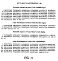
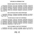
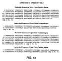
- Isolated nucleic acid molecule may comprise a nucleotide sequence encoding a heavy chain variable domain of a monoclonal antibody, wherein said nucleotide sequence is selected from the group consisting of the nucleotide sequence of the heavy chain variable domain of c3.19.1 (SEQ ID NO: 3), c6.11.3 (SEQ ID NO: 7), C3.10 (SEQ ID NO: 11), C3.22 (SEQ ID NO: 15), C3.27 (SEQ ID NO: 19), C3.45 (SEQ ID NO: 23), C3.65 (SEQ ID NO: 27), C6.
- nucleotide sequence encoding a light chain variable domain of a monoclonal antibody, wherein said nucleotide sequence is selected from the group consisting of the nucleotide sequence of the light chain variable domain of 3.19.1 (SEQ ID NO: 4), 6.11.3 (SEQ ID NO: 8), C3.10 (SEQ ID NO:12), C3.22 (SEQ ID NO: 16), C3.27 (SEQ ID NO: 20), C3.45 (SEQ ID NO: 24), C3.65 (SEQ ID NO: 28), C6.1 (SEQ ID NO: 32), C6.9 (SEQ ID NO: 36), or C6.2 (SEQ ID NO: 40).
- Inhibition of tumour growth associated with the expression of MUC18 in a patient may comprise administering to the patient an effective amount of an anti-MUC18 antibody.
- the patient is a mammalian patient, preferably a human patient.
- the disease is a tumor, such as melanoma.
- MUC18 refers to the cell surface polypeptide that is a member of the immunoglobulin superfamily with sequence similarity to a number of cell adhesion molecules. MUC18 is also known in the art as “MCAM”, “Mel-CAM”, or “CD146". For purposes of this invention, from here on, “MUC18” is used to represent “MCAM”, “Mel-CAM”, and “CD146”.
- C3.19.1 refers to a fully human IgG 2 monoclonal antibody directed against the MUC18 antigen.
- the antibody was generated using XenoMouse® technology (Abgenix, Inc. Fremont, CA) and consists of human gamma 2 heavy and kappa light chains with a molecular weight of approximately 150 kDa.
- biological activity and “biologically active” with regard to MUC18 refer to the ability of a molecule to specifically affect tumor progression. Preferred biological activities include the ability to induce growth and metastasis of tumor cells. The effect of MUC 18 on metastasis of tumor cells may include the ability to induce MMP-2 activation and/or cell migration. A further preferred biological activity is the ability to induce animal death due to tumor burden.
- biological activity and “biologically active” with regard to anti-MUC18 antibodies refer to the ability of a molecule to inhibit the growth and metastasis of tumor cells often associated with MUC18 expression. Further, another metahcnism of action or activity for anti-MUC18 antibodies include the ability to stimulate MUC18 internalization and a consequent loss of cell surface expression. Specifically, the tumor cells include tumor cells in patients with tumors.
- PCR Polymerase chain reaction
- sequence information from the ends of the region of interest or beyond needs to be available, such that oligonucleotide primers can be designed; these primers will be identical or similar in sequence to opposite strands of the template to be amplified.
- the 5' terminal nucleotides of the two primers can coincide with the ends of the amplified material.
- PCR can be used to amplify specific RNA sequences, specific DNA sequences from total genomic DNA, and cDNA transcribed from total cellular RNA, bacteriophage or plasmid sequences, etc. See generally Mullis et al., Cold Spring Harbor Symp. Quant. Biol. 51:263 (1987 ); Erlich, ed., PCR Technology (Stockton Pres, NY, 1989 ).
- PCR is considered to be one, but not the only, example of a nucleic acid polymerase reaction method for amplifying a nucleic acid test sample comprising the use of a known nucleic acid as a primer and a nucleic acid polymerase to amplify or generate a specific piece of nucleic acid.
- Tumor refers to all neoplastic cell growth and proliferation, whether malignant or benign, and all pre-cancerous and cancerous cells and tissues.
- cancer and “cancerous” refer to or describe the physiological condition in mammals that is typically characterized by unregulated cell growth.
- examples of cancer include, but are not limited to, melanoma, carcinoma, lymphoma, blastoma, sarcoma, and leukemia or lymphoid malignancies. More particular examples of such cancers include squamous cell cancer (e.g.
- lung cancer including small-cell lung cancer, non-small cell lung cancer, adenocarcinoma of the lung and squamous carcinoma of the lung, cancer of the peritoneum, hepatocellular cancer, gastric or stomach cancer including gastrointestinal cancer, pancreatic cancer, glioblastoma, cervical cancer, ovarian cancer, liver cancer, bladder cancer, hepatoma, breast cancer, colon cancer, rectal cancer, colorectal cancer, endometrial cancer or uterine carcinoma, salivary gland carcinoma, kidney or renal cancer, prostate cancer, vulval cancer, thyroid cancer, hepatic carcinoma, anal carcinoma, penile carcinoma, as well as head and neck cancer.
- lung cancer including small-cell lung cancer, non-small cell lung cancer, adenocarcinoma of the lung and squamous carcinoma of the lung, cancer of the peritoneum, hepatocellular cancer, gastric or stomach cancer including gastrointestinal cancer, pancreatic cancer, glioblastoma, cervical cancer, ovarian cancer, liver cancer
- Antibodies are glycoproteins having the same structural characteristics. While antibodies exhibit binding specificity to a specific antigen, immunoglobulins include both antibodies and other antibody-like molecules which lack antigen specificity. Polypeptides of the latter kind are, for example, produced at low levels by the lymph system and at increased levels by myelomas.
- “Native antibodies and immunoglobulins” are usually heterotetrameric glycoproteins of about 150,000 daltons, composed of two identical light (L) chains and two identical heavy (H) chains. Each light chain is linked to a heavy chain by one covalent disulfide bond, while the number of disulfide linkages varies between the heavy chains of different immunoglobulin isotypes. Each heavy and light chain also has regularly spaced intrachain disulfide bridges. Each heavy chain has at one end a variable domain (VH) followed by a number of constant domains.
- VH variable domain
- Each light chain has a variable domain at one end (VL) and a constant domain at its other end; the constant domain of the light chain is aligned with the first constant domain of the heavy chain, and the light chain variable domain is aligned with the variable domain of the heavy chain.
- Particular amino acid residues are believed to form an interface between the light- and heavy-chain variable domains ( Chothia et al. J. Mol. Biol. 186:651 (1985 ; Novotny and Haber, Proc. Natl. Acad Sci. U.S.A. 82:4592 (1985 ); Chothia et al., Nature 342:877-883 (1989) ).
- antibody herein is used in the broadest sense and specifically covers intact monoclonal antibodies, polyclonal antibodies, multispecific antibodies (e.g. bispecific antibodies) formed from at least two intact antibodies, and antibody fragments, so long as they exhibit the desired biological activity.
- the "light chains" of antibodies (immunoglobulins) from any vertebrate species can be assigned to one of two clearly distinct types, called ⁇ and ⁇ , based on the amino acid sequences of their constant domains.
- intact antibodies can be assigned to different "classes". There are five major classes of intact antibodies: IgA, IgD, IgE, IgG, and IgM, and several of these may be further divided into “subclasses” (isotypes), e.g. , IgG1, IgG2, IgG3, IgG4, IgA, and IgA2.
- the heavy-chain constant domains that correspond to the different classes of antibodies are called ⁇ , ⁇ , ⁇ , ⁇ , and ⁇ , respectively.
- the subunit structures and three-dimensional configurations of different classes of immunoglobulins are well known.
- antibody includes all classes and subclasses of intact immunoglobulins.
- antibody also covers antibody fragments.
- antibody specifically covers monoclonal antibodies, including antibody fragment clones.
- the term "monoclonal antibody” as used herein refers to an antibody obtained from a population of substantially homogeneous antibodies, i.e., the individual antibodies comprising the population are identical except for possible naturally occurring mutations that may be present in minor amounts. Monoclonal antibodies are highly specific, being directed against a single antigenic site. Furthermore, in contrast to polyclonal antibody preparations which include different antibodies directed against different determinants (epitopes), each monoclonal antibody id directed against a single determinant on the antigen. In addition to their specificity, the monoclonal antibodies are advantageous in that they may be synthesized uncontaminated by other antibodies.
- the modifier "monoclonal” indicates the character of the antibody as being obtained from a substantially homogeneous population of antibodies, and is not to be construed as requiring production of the antibody by any particular method.
- the monoclonal antibodies to be used in accordance with the present invention may be made by the hybridoma method first described by Kohler et al., Nature, 256:495 (1975 ), or may be made by recombinant DNA methods (see, e.g., U.S. Patent No. 4,816,567 ).
- the “monoclonal antibodies” may also be isolated from phage antibody libraries using the techniques described in Clackson et al, Nature, 352:624-628 (1991) and Marks et al., J. Mol. Biol., 222:581-597 (1991) , for example.
- an “isolated” antibody is one which has been identified and separated and/or recovered from a component of its natural environment. Contaminant components of its natural environment are materials which would interfere with diagnostic or therapeutic uses for the antibody, and may include enzymes, hormones, and other proteinaceous or nonproteinaceous solutes.
- the antibody will be purified (1) to greater than 95% by weight of antibody as determined by the Lowry method, and terminal or internal amino acid sequence by use of a spinning cup sequenator, or (3) to homogeneity by SDS-PAGE under reducing or nonreducing conditions using Coomassie blue or, preferably, silver stain.
- Isolated antibody includes the antibody in situ within recombinant cells since at least one component of the antibody's natural environment will not be present. Ordinarily, however, isolated antibody will be prepared by at least one purification step.
- neutralizing antibody an antibody molecule which is able to eliminate or significantly reduce an effector function of a target antigen to which is binds. Accordingly, a “neutralizing" anti-MUC18 antibody is capable of eliminating or significantly reducing an effector function which may include MUC18 dependent regulation of cell adhesion, migration or MMP activation. The antibody can affect the function of MUC18 by stimulating the internalization and degradation of the molecule thus effectively removing cell surface expression of the antigen.
- variable refers to the fact that certain portions of the variable domains differ extensively in sequence among antibodies and are used in the binding and specificity of each particular antibody for its particular antigen. However, the variability is not evenly distributed throughout the variable domains of antibodies. It is concentrated in three segments called complementarity-determining regions (CDRs) or hypervariable regions both in the light-chain and heavy-chain variable domains. The more highly conserved portions of variable domains are called the framework (FR).
- CDRs complementarity-determining regions
- FR framework
- the variable domains of native heavy and light chains each comprise four FR regions, largely adopting a ⁇ -sheet configuration, connected by three CDRs, which form loops connecting, and in some cases forming part of, the ⁇ -sheet structure.
- the CDRs in each chain are held together in close proximity by the FR regions and, with the CDRs from the other chain, contribute to the formation of the antigen-binding site of antibodies (see Kabat et al. (1991).
- the constant domains are not involved directly in binding an antibody to an antigen, but exhibit various effector functions, such as participation of the antibody in antibody-dependent cellular toxicity.
- Fv is the minimum antibody fragment which contains a complete antigen-recogution and binding site.
- this region consists of a dimer of one heavy- and one light-chain variable domain in tight, non-covalent association.
- one heavy- and one light-chain variable domain can be covalently linked by a flexible peptide linker such that the light and heavy chains can associate in a "dimeric" structure analogous to that in a two-chain Fv species. It is in this configuration that the three CDRs of each variable domain interact to define an antigen-binding site on the surface of the VH-VL dimer.
- the six CDRs confer antigen-binding specificity to the antibody.
- hypervariable region when used herein refers to the amino acid residues of an antibody which are responsible for antigen-binding.
- the hypervariable region generally comprises amino acid residues from a "complementarity determining region” or "CDR" (e.g. residues 24-34 (L1), 50-62 (L2), and 89-97 (L3) in the light chain variable domain and 31-55 (H1), 50-65 (H2) and 95-102 (H3) in the heavy chain variable domain; Kabat et al., Sequences of Proteins of Immunological Interest, 5th Ed. Public Health Service, National Institutes of Health, Bethesda, MD.
- CDR complementarity determining region
- residues from a "hypervariable loop” e.g. residues 26-32 (L1), 50-52 (L2) and 91-96 (L3) in the light chain variable domain and 26-32 ((H1), 53-55 (H2) and 96-101 (H3) in the heavy chain variable domain; Chothia and Lesk J. Mol. Biol 196:901-917 (1987) ).
- "Framework Region” or "FR" residues are those variable domain residues other than the hypervariable region residues as herein defined.
- CDRs complementarity determining regions
- the CDRs of immunological receptors are the most variable part of the receptor protein, giving receptors their diversity, and are carried on six loops at the distal end of the receptor's variable domains, three loops coming from each of the two variable domains of the receptor.
- epitopope is used to refer to binding sites for (monoclonal or polyclonal) antibodies on protein antigens.
- amino acid or amino acid residue refers to naturally occurring L amino acids or to D amino acids as described further below with respect to variants.
- the commonly used on- and three-letter abbreviations for amino acids are used herein ( Bruce Alberts et al., Molecular Biology of the Cell, Garland Publishing, Inc., New York (3d ed. 1994 )).
- disease state refers to a physiological state of a cell or of a whole mammal in which an interruption, cessation, or disorder of cellular or body functions, systems, or organs has occurred.
- beneficial or desired clinical results include, but are not limited to, alleviation of symptoms, diminishment of extent of disease, stabilized (i.e., not worsening) state of disease, delay or slowing of disease progression, amelioration or palliation of the disease state, and remission (whether partial or total), whether detectable or undetectable.
- Treatment can also mean prolonging survival as compared to expected survival if not receiving treatment.
- Those in need of treatment include those already with the condition or disorder as well as those prone to have the condition or disorder or those in which the condition or disorder is to be prevented.
- a “disorder” is any condition that would benefit from treatment of the present invention. This includes chronic and acute disorders or disease including those pathological conditions which predispose the mammal to the disorder in question.
- disorders to be treated herein include benign and malignant tumors, leukemias and lymphoid malignancies, in particular prostate, renal, ovarian, stomach, endometrial, salivary gland, kidney, colon, thyroid, pancreatic, prostate or bladder cancer, and malignant tumors, such as cervical carcinomas and cervical intraepithelial squamous and glandular neoplasia, renal cell carcinoma (RCC), esophageal tumors, and carcinoma-derived cell lines.
- a preferred disorder to be treated in accordance with the present invention is renal and prostate cancer.
- An even further preferred disorder to be treated is melanoma.
- mammal for purposes of treatment refers to any animal classified as a mammal, including humans, domestic and farm animals, and zoo, sports, or pet animals, such as dogs, horses, cats, cows, etc. Preferably, the mammal is human.
- Lipofection refers to a practical nonviral method for introduction of genetic information into target tissues.
- Nonviral methods include chemical or physical methods.
- Lipofection uses an electrostatically bonded complex of positively charged lipids and negatively charged DNA as a vector which fuses with the cell membrane and delivers DNA into the cytoplasm. Lipofection differs from viral methods in that the efficiency of transfer of genetic information by lipofection is lower than by viral vectors and that the expression of the gene is transient. Alternatively, the complex of lipid and DNA is more stable and easier to handle when compared to viral vectors.
- Monoclonal Antibodies may be made using the hybridoma method first described by Kohler et al., Nature, 256:495 (1975 ), or may be made by recombinant DNA methods ( U.S. Patent No. 4,816,567 ).
- a mouse or other appropriate host animal such as a hamster or macaque monkey
- a host animal such as a hamster or macaque monkey
- lymphocytes that produce or are capable of producing antibodies that will specifically bind to the protein used for immunization.
- cells expressing the antigen of interest may be used for immunization.
- lymphocytes may be immunized in vitro. Animals are immunized against the immunogenic conjugates or derivatives by combing 1 mg or 1 ⁇ g of conjugate (for rabbits or mice, respectively) with 3 volumes of Freud's complete adjuvant and injecting the solution intradermally at multiple sites.
- the animals are boosted with 1/5 to 1/0 the original amount of conjugate in Freud's complete adjuvant by subcutaneous injection at multiple sites. 7 to 14 days later the animals are bled and the serum is assayed for anti-MUC18 antibody titer. Antibodies are boosted until the titer plateaus. Preferably, the animal is boosted with the conjugate of the same MUC18 antigen, but conjugated to a different protein and/or through a different cross-linking agent. Conjugates also can be made in recombinant cell culture as protein fusions. Also, aggregating agents such as alum are used to enhance the immune response.
- Lymphocytes or more preferably, lymphocytes enriched for B cells isolated from such immunized animals are then fused with myeloma cells by an electrocell fusion process or by using a suitable fusing agent, such as polyethylene glycol, to form a hybridoma cell ( Goding, Monoclonal Antibodies: Principles and Practice, pp. 59-109, [Academic Press, 1996 ]).
- a suitable fusing agent such as polyethylene glycol
- the hybridoma cells thus prepared are seeded and grown in a suitable culture medium that preferably contains one or more substances that inhibit the growth or survival of the unfused, parental myeloma cells.
- a suitable culture medium that preferably contains one or more substances that inhibit the growth or survival of the unfused, parental myeloma cells.
- the culture medium for the hybridomas typically will include hypoxanthinc, aminopterin, and thymidine (HAT medium), which substances prevent the growth of HGPRT-deficient cells.
- Preferred myeloma cells are those that fuse efficiently, support stable high-level production of antibody by the selected antibody-producing cells, and are sensitive to a medium such as HAT medium.
- preferred myeloma cell lines are murine myeloma lines, such as those derived from MOP-21 and MC.-11 mouse tumors available from the Salk Institute Cell Distribution Center, San Diego, California USA, and SP-2 or X63-Ag8-653 cells available from the American Type Culture Collection, Rockville, Maryland USA.
- Human myeloma and mouse-human heteromyeloma cell lines also have been described for the production of human monoclonal antibodies ( Kozbor, J. Immunol. 133: 3001 (1984 ); Brodeur et al., Monoclonal Antibody Production Techniques and Applications, pp. 51-63, Marcel Dekker, Inc., New York, [1987 ]).
- Culture medium in which hybridoma cells are growing is assayed for production of monoclonal antibodies directed against the antigen.
- the binding specificity of monoclonal antibodies produced by hybridoma cells is determined by immunoprecipitation or by an in vitro binding assay, such as radioimmunoassay (RIA) or enzyme-linked immunosorbent assay (ELISA).
- RIA radioimmunoassay
- ELISA enzyme-linked immunosorbent assay
- the binding affinity of the monoclonal antibody can, for example, be determined by the Scatchard analysis of Munson et al., Anal. Biochem. 107: 220 (1980 ).
- hybridoma cells After hybridoma cells are identified that produce antibodies of the desired specificity, affinity, and/or activity, the cells may be subcloned by limiting dilution procedures and grown by standard methods ( Goding, Monoclonal Antibodies: Principles and Practice, pp.59-103, Academic Press, 1996 ). Suitable culture media for this purpose include, for example, DMEM or RPMI-1640 medium. In addition, the hybridoma cells may be grown in vivo as ascites tumors in an animal.
- the monoclonal antibodies secreted by the subclones are suitably separated from the culture medium, ascites fluid, or serum by conventional immunoglobulin purification procedures such as, for example, protein A-Sepharose, hydroxylapatite chromatography, gel electrophoresis, dialysis, or affinity chromatography.
- DNA encoding the monoclonal antibodies is readily isolated and sequenced using conventional procedures (e.g. , by using oligonucleotide probes that are capable of binding specifically to genes encoding the heavy and light chains of the monoclonal antibodies).
- the hybridoma cells serve as a preferred source of such DNA.
- the DNA may be placed into expression vectors, which are then transfected into host cells such as E. coli cells, simian COS cells, Chinese hamster ovary (CHO) cells, or myeloma cells that do not otherwise produce immunoglobulin protein, to obtain the synthesis of monoclonal antibodies in the recombinant host cells.
- the DNA also may be modified, for example, by covalently joining to the immunoglobulin coding sequence all or part of the coding sequence for a non-immunoglobulin polypeptide.
- “chimeric” or “hybrid” antibodies are prepared that have the binding specificity of an anti-MUC18 monoclonal antibody herein.
- non-immunoglobulin polypeptides are substituted for the constant domains of an antibody of the invention, or they are substituted for the variable domains of one antigen-combining site of an antibody of the invention to create a chimeric bivalent antibody comprising one antigen-combining site having specificity for an MUC18 and another antigen-combining site having specificity for a different antigen.
- Chimeric or hybrid antibodies also may be prepared in vitro using known methods in synthetic protein chemistry, including those involving crosslinking agents.
- immunotoxins may be constructed using a disulfide exchange reaction or by forming a thioether bond.
- suitable reagents for this purpose include iminothiolate and methyl-4-mercaptobutyrimidate.
- human antibody-secreting cells can be immortalized by infection with the Epstein-Barr virus (EBV).
- EBV Epstein-Barr virus
- EBV-infected cells are difficult to clone and usually produce only relatively low yields of immunoglobulin ( James and Bell, J. Immunol. Methods 100: 5-40 [1987] ).
- the immortalization of human B cells might possibly be achieved by introducing a defined combination of transforming genes.
- Such a possibility is highlighted by a recent demonstration that the expression of the telomerase catalytic subunit together with the SV40 large T oncoprotein and an oncogenic allele of H -ras resulted in the tumorigenic conversion of normal human epithelial and fibroblast cells ( Hahn et al., Nature 400: 464-468 [1999] ).
- transgenic animals e.g. mice
- transgenic animals e.g. mice
- Lonberg and Huszar Int. Rev. Immunol. 13: 65-93 [1995]
- Mendez et al. (Nature Genetics 15: 146-156 [1997] ) have generated a line of transgenic mice designated as "XenoMouse ® II" that, when challenged with an antigen, generates high affinity fully human antibodies. This was achieved by germ-line integration of megabase human heavy chain and light chain loci into mice with deletion into endogenous J H segment as described above.
- the XenoMouse ® II harbors 1,020 kb of human heavy chain locus containing approximately 66 V H genes, complete D H and J H regions and three different constant regions ( ⁇ , ⁇ and ⁇ ), and also harbors 800 kb of human ⁇ locus containing 32 V ⁇ genes, J ⁇ segments and C ⁇ genes.
- the antibodies produced in these mice closely resemble that seen in humans in all respects, including gene rearrangement, assembly, and repertoire.
- the human antibodies are preferentially expressed over endogenous antibodies due to deletion in endogenous J H segment that prevents gene rearrangement in the murine locus.
- Techniques for generating antibodies using Abgenix's XenoMouse® technology include injection of a particular antigen of interest into such mice. Sera from such immunized animals may be screened for antibody-reactivity against the initial antigen. Lymphocytes may be isolated from lymph nodes or spleen cells and may further be selected for B cells by selecting for CD138-negative and CD19+ cells. The B cell cultures (BCCs) may be either fused to myeloma cells to generate hybridomas as detailed above or screened further for reactivity against the initial antigen. Such screening includes ELISA.
- Transfection refers to the taking up of an expression vector by a host cell whether or not any coding sequences are in fact expressed. Numerous methods of transfection are known to the ordinarily skilled artisan, for example, CaPO 4 precipitation and electroporation. Successful transfection is generally recognized when any indication of the operation of this vector occurs within the host cell.
- the antibodies of the present invention comprise an anti-human MUC18 monoclonal antibody heavy chain or a fragment thereof, comprising the following CDR's (as defined by Kabat et al., Sequences of Proteins of Immunological Interest, Fifth Edition, NIH Publication 91-3242, Bethesda MD (1991), vols 1-3 ): (a) CDR1, (b) CDR2 and (c) CDR3.
- CDR's as defined by Kabat et al., Sequences of Proteins of Immunological Interest, Fifth Edition, NIH Publication 91-3242, Bethesda MD (1991), vols 1-3 ): (a) CDR1, (b) CDR2 and (c) CDR3.
- the heavy chain of the antibodies in one embodiment of the present invention comprise of the following sequences: SEQ ID NO: 1, SEQ ID NO: 5, SEQ ID NO: 9, SEQ ID NO: 13, SEQ ID NO: 17, SEQ ID NO: 21, SEQ ID NO: 25, SEQ ID NO: 29, SEQ ID NO: 33 or SEQ ID NO: 37.
- the invention provides an anti-human MUC18 monoclonal antibody light chain or a fragment thereof, comprising the following CDRs: (a) CDR1, (b) CDR2 and (c) CDR3.
- the light chain of the antibodies in one embodiment of the present invention comprise of the following sequences: SEQ ID NO: 2, SEQ ID NO: 6, SEQ ID NO: 10; SEQ ID NO: 14, SEQ ID NO: 18; SEQ ID NO: 22; SEQ ID NO: 26; SEQ ID NO: 30; SEQ ID NO: 34; or SEQ ID NO: 38.
- the present invention includes anti-MUC18 antibodies such as c3.19.1 and c6.11.3.
- the heavy chain amino acid and nucleotide sequences of c3.19.1 are encoded by SEQ ID NO: 1 and 3, respectively, and the heavy chain amino acid and nucleotide sequence of c6.11.3 are encoded by 5 and 7, respectively.
- the light chain amino acid and nucleotide sequences of c3.1.9.1. are encoded by SEQ ID NO: 2 and 4, respectively, and the light chain amino acid and nucleotide sequences of c6.11.3 are encoded by 6 and 8, respectively.
- melanoma cells were used for analysis of anti-MUC18 antibody function.
- the melanoma cells were derived from a number of sources and possessed a variety of characteristics that could potentially contribute to the metastatic phenotype. Their phenotypes are presented in Table 1.
- Table 1. Properties of Melanoma Cells Used for Studies Cell Line Origin Metastatic Capacity MUC18 Expression A375-SM Selection fromanimal model of metastasis High High SB-2 Cutaneous melanoma Non-metastatic Negative TXM-13 Brain metastasis High High WM-2664 Metastasis from cutaneous melanoma High Medium
- kinetic measurements and binding affinity of the anti-MUC18 antibodies were obtained from Biacore experiments.
- the Biacore experiments measured the affinity of MUC18 antibodies captured on a protein A surface for labeled MUC18 antigen and are further described in the examples below.
- Anti-MUC18 antibodies with a Kd of 6 x 10 -10 M were considered high affinity anti-MUC18 antibodies.
- anti-MUC18 antibodies of the present invention were able to recognize denatured MUC18 in human melanoma cells
- the antibodies were used for immunoblots of metastatic melanoma cells and non-metastatic melanoma cells (control). Those antibodies which were able to detect MUC18 in metastatic melanoma cells were selected as anti-MUC18 antibodies of interest.
- tumor growth inhibitory anti-MUC18 antibodies were screened for their ability to inhibit tumor growth in animal models.
- the growth inhibitory antibody of choice was able to inhibit the growth of WM-2664 tumor cells when grown subcutaneously.
- exponentially growing WM-2664 cells were harvested, resuspended, and injected into the flanks of male BALB/c nude mice. Beginning on Day 3 after implantation, animals were treated with either 0.1 mg or 1 mg anti-MUC18 antibody or 0.1 mg control human antibody once a week. Tumor growth was monitored weekly. Those antibodies which inhibited the growth of WM-2264 tumor cells in vivo were selected as tumor growth inhibitory antibodies.
- tumor growth inhibitory anti-MUC18 antibodies were screened for the ability to inhibit the proliferation of tumor cells in culture.
- cells expressing various levels of MUC18 were incubated in the presence of anti-MUC18 for a 4-5 day period.
- MTT dimethylthiazole diphenyl tetrazolium bromide
- MUC18 inhibition is important for the three-dimensional growth of melanoma tumor cells in vivo, but not in cell culture ( Satyamoorthy et al., Oncogene, 20:4676 (2001 )).
- the anti-MUC18 antibodies of the present invention were tested to determine whether they had an effect on melanoma cell proliferation in vitro.
- Cells expressing various levels of MUC18 were incubated in the presence of anti-MUC18 antibody for 1 4-5 day period. On each day of the experiment, the number of cells in triplicate wells was determined using a dimethylthiazole diphenyl tetrazolium bromide (MTT) assay.
- MTT dimethylthiazole diphenyl tetrazolium bromide
- metastasis inhibitory anti-MUC18 antibodies were screened for the ability to inhibit metastasis in animal models.
- the metastasis inhibitory antibody of choice was able to inhibit the formation of lung metastases of melanoma A375-SM and WM-2664 cells in vivo.
- To evaluate the effect of anti-MUC18 antibody treatment on the metastasis of melanoma cells that were injected into nude mice exponentially growing A375-SM and WM-2664 melanoma cells were harvested, resuspended, and injected into the lateral tail vein of mice. Mice were treated with anti-MUC18 antibody beginning on Day 3 and once weekly thereafter. The mice were sacrificed 6-8 weeks later. After fixing and staining with Bouin's solution, lung nodules were counted with the aid of a dissecting microscope. Those antibodies which inhibited lung metastases in animals injected with tumor cells were selected as metastasis inhibitory antibodies.
- A375SM cells were harvested on Day 0, resuspended and injected into the lateral tail veins of female nude mice. The animals were treated one day prior to tumor cell injection and once a week thereafter with a specific dose of anti-MUC18 antibody. All animals were sacrificed after 6 weeks at which time the lungs were removed and the tumor nodules were counted with the aid of a dissecting microscope. Those antibodies which inhibited melanoma metastasis into the lung in a dose-dependent manner were selected as metastasis inhibitory antibodies.
- mice bearing tumors were screened for the ability to increase the survival of mice bearing tumors.
- WM-2664 human melanoma tumor cells in their exponential phase were harvested, resuspended in suitable buffer, and injected into the lateral tail vein of male BALB/c nude mice.
- Melanoma metastasis is closely associated with MUC18 expression.
- One of the phenotypic changes often associated with metastasis is the ability of cells to migrate and invade through the extracellular matrix.
- antibodies were screened for their ability to inhibit the ability of cells to migrate and invade through Matrigel coated membranes.
- cells expressing MUC18 were seeded into plates and allowed to attach. The growth medium were then removed and replaced with fresh growth medium containing either control antibody or anti-MUC18 antibody. After a period of 5 days, the cells were detached from the plates and placed in the upper chamber of a Matrigel coated membrane in serum-free medium containing either control or anti-MUC18 antibody. Following an incubation period, the cells remaining in the upper chamber were removed by scraping and the bottom filter subjected to staining.
- the matrices were then mounted on slides and the cells that had migrated across the membrane were counted. Those antibodies which demonstrated an inhibitory effect on the ability of melanoma cells to digest the extracellular matrix and migrate toward the chemoattractant on the opposite side were selected as invasion inhibitory antibodies.
- Therapeutic formulations of the anti-MUC18 antibodies of the invention are prepared for storage by mixing antibody having the desired degree of purity with optional physiologically acceptable carriers, excipients, or stabilizers ( Remington: The Science and Practice of Pharmacy, 19th Edition, Alfonso, R., ed, Mack Publishing Co. (Easton, PA: 1995 )), in the form of lyophilized cake or aqueous solutions.
- Acceptable carriers, excipients or stabilizers are nontoxic to recipients at the dosages and concentrations employed, and include buffers such as phosphate, citrate, and other organic acids; antioxidants including ascorbic acid; low molecular weight (less than about 10 residues) polypeptides; proteins, such as serum albumin, gelatin, or immunoglobulins; hydrophilic polymers such as polyvinylpyrrolidone; amino acids such as glycine, glutamine, asparagine, arginine or lysine; monosaccharides, disaccharides, and other carbohydrates including glucose, mannose, or dextrins; chelating agents such as EDTA; sugar alcohols such as mannitol or sorbitol; salt-forming counterions such as sodium; and/or nonionic surfactants such as Tween, Pluronics or polyethylene glycol (PEG).
- buffers such as phosphate, citrate, and other organic acids
- antioxidants including ascorbic acid
- the anti-MUC18 antibody to be used for in vivo administration must be sterile. This is readily accomplished by filtration through sterile filtration membranes, prior to or following lyophilization and reconstitution.
- the anti-MUC18 antibody ordinarily will be stored in lyophilized form or in solution.
- Therapeutic anti-MUC18 antibody compositions generally are placed into a container having a sterile access port, for example, an intravenous solution bag or vial having a stopper pierceable by a hypodermic injection needle.
- the route of anti-MUC18 antibody administration is in accord with known methods, e.g. injection or infusion by intravenous, intraperitoneal, intracerebral, subcutaneous, intramuscular, intraocular, intraarterial, intracerebrospinal, or intralesional routes, or by sustained release systems as noted below.
- the antibody is given systemically.
- sustained-release preparations include semipermeable polymer matrices in the form of shaped articles, e.g. films, or microcapsules.
- Sustained release matrices include polyesters, hydrogels, polylactides ( U.S. 3,773,919 , EP 58,481 ), copolymers of L-glutamic acid and gamma ethyl-L-glutamate ( Sidman et al., Biopolymers, 22: 547-556 (1983) ), poly (2-hydroxyethylmethacrylate) ( Langer et al., J. Biomed. Mater. Res., 15: 167-277 (1981) and Langer, Chem.
- Sustained-release anti-MUC18 antibody compositions may also include liposomally entrapped antibody. Liposomes containing antibody are prepared by methods known per se: DE 3,218,121 ; Epstein et al., Proc. Natl. Acad Sci. USA, 82: 3688-3692 (1985) ; Hwang et al., Proc. Natl. Acad. Sci.
- the liposomes are of the small (about 200-800 Angstroms) unilamelar type in which the lipid content is greater than about 30 mol. % cholesterol, the selected proportion being adjusted for the optimal antibody therapy.
- Anti-MUC18 antibody can also be administered by inhalation.
- Commercially available nebulizers for liquid formulations including jet nebulizers and ultrasonic nebulizers are useful for administration.
- Liquid formulations can be directly nebulized and lyophilized powder can be nebulized after reconstitution.
- anti-MUC18 antibody can be aerosolized using a fluorocarbon formulation and a metered dose inhaler, or inhaled as a lyophilized and milled powder.
- an "effective amount" of anti-MUC18 antibody to be employed therapeutically will depend, for example, upon the therapeutic objectives, the route of administration, the type of anti-MUC18 antibody employed, and the condition of the patient. Accordingly, it will be necessary for the therapist to titer the dosage and modify the route of administration as required to obtain the optimal therapeutic effect. Typically, the clinician will administer the anti-MUC18 antibody until a dosage is reached that achieves the desired effect. The progress of this therapy is easily monitored by conventional assays.
- Antibodies specific to tumor antigens such as anti-MUC18 are useful in targeting of tumor cells for destruction.
- ricin a cellular toxin derived from plants, is finding unique applications, especially in the fight against tumors and cancer. Implications are being discovered as to the use of ricin in the treatment of tumors. Ricin has been suggested to have a greater affinity for cancerous cells than normal cells (Montfort et al. 1987) and has been often termed as a "magic bullet" for targeting malignant tumors. Toxins such as ricin remain active even if the B chain of the toxin is removed.
- the toxin has a specific affinity for cancerous cells over normal cells (Taylorson 1996).
- ricin immunotoxin has been developed to target the CD5 T-cell antigen often found in T-cell and B-cell malignancies (Kreitman et al. 1998).
- the linking of such anti-MUC18 antibodies to radioisotopes provides advantages to tumor treatments. Unlike chemotherapy and other forms of cancer treatment, radioimmunotherapy or the administration of a radioisotope-antibody combination directly targets the cancer cells with minimal damage to surrounding normal, healthy tissue.
- antibodies such as MUC18 antibodies
- modified antibodies can also be readily prepared utilizing techniques that are well known in the art.
- the patients to be treated with the anti-MUC18 antibody of the invention include patients with tumors, preferably melanoma and/or prostate or renal cancer.
- Other tumors include esophageal, pancreatic, colorectal tumors, carcinomas, such as renal cell carcinoma (RCC), cervical carcinomas and cervical intraepithelial squamous and glandular neoplasia, and cancers, such as colorectal cancer, breast cancer, lung cancer, and other malignancies.
- Patients arc candidates for therapy in accord with this invention until such point as no healthy tissue remains to be protected from tumor progression. It is desirable to administer an anti-MUC18 antibody as early as possible in the development of the tumor, and to continue treatment for as long as is necessary.
- the antibody composition will be formulated, dosed, and administered in a fashion consistent with good medical practice.
- Factors for consideration in this context include the particular disorder being treated, the particular mammal being treated, the clinical condition of the individual patient, the cause of the disorder, the site of delivery of the antibody, the particular type of antibody, the method of administration, the scheduling of administration, and other factors known to medical practitioners.
- the "therapeutically effective amount" of antibody to be administered will be governed by such considerations, and is the minimum amount necessary to prevent, ameliorate, or treat the disorder, including treating chronic autoimmune conditions and immunosuppression maintenance in transplant recipients. Such amount is preferably below the amount that is toxic to the host or renders the host significantly more susceptible to infections.
- the initial pharmaceutically effective amount of the antibody administered parenterally will be in the range of about 0.1 to 50 mg/kg of patient body weight per day, with the typical initial range of antibody used being 0.3 to 20 mg/kg/day, more preferably 0.3 to 15 mg/kg/day.
- the desired dosage can be delivered by a single bolus administration, by multiple bolus administrations, or by continuous infusion administration of antibody, depending on the pattern of pharmacokinetic decay that the practitioner wishes to achieve.
- the antibody may be optionally formulated with one or more agents currently used to prevent or treat tumors such as standard- or high-dose chemotherapy and hematopoietic stem-cell transplantation.
- the effective amount of such other agents depends on the amount of anti-MUC18 antibody present in the formulation, the type of disorder or treatment, and other factors discussed above. These are generally used in the same dosages and with administration routes as used hereinbefore or about from 1 to 99% of the heretofore employed dosages.
- recombinant MUC18 proteins were prepared.
- the extracellular domain (ECD) (aa#1-559) of human MUC18 was cloned from SK-MEL-28 cells (ATCC HTB-72) by Reverse Transcriptase-PCR (RT-PCR) with primers that incorporate an EcoRJ site in the forward primer and an NheI site in the reverse primer based on the published NCBI sequence (Accession # NM_006500).
- the primers used for the amplification of the ECD of MUC18 were as follows:
- MUC18 cDNA was amplified (Gene Amp XL PCR kit, Perkin Elmer) from RNA (RNAzol, Tel Test, INC) prepared from SK-MEL-28 cells (ATCC HTB-72).
- RNA RNAzol, Tel Test, INC
- HuIgG2 fusion protein the 1700 bp PCR product encoding amino acids 1-559 was digested with EcoRI and NheI and ligated into CD147HuIgG2DHFR vector (ABGX) digested with EcoRI and NheI or pcDNA3.1V5HISB vector (Invitrogen) digested with EcoRI and Xbal.
- the resulting plasmids were transfected in 293 cells by CaPO 4 method, and then, the fusion protein was purified from harvested conditioned media via ProteinA chromatography (MUC18-HulgG2) or Ni-NTA chromatography (MUC18-V5HIS).
- the MUC18 ECD contained 4 amino acid differences from the published NCBI sequence: #383 D>G,#390 P>L,#424 K>N, and #425 L>V.
- Monoclonal antibody against MUC18 was developed by sequentially immunizing XenoMouse mice (XenoMouse G2, Abgenix, Inc. Fremont, CA). The initial immunization was with 5 x 10 6 SK-MEL-28 cells admixed 1:1 v/v with Complete Freund's Adjuvant (CFA).
- CFA Complete Freund's Adjuvant
- mice were initially injected with MUC18 antigen at a concentration of 1-5 ⁇ g/mouse. Each mouse was further immunized into each hind footpad 6 additional times (at 3-4 day intervals) with soluble antigen, specifically 5 ⁇ g of soluble MUC18-human IgG2 Fc fusion protein in DPBS admixed 1:1 v/v with IFA then a final boost of 10 ⁇ g soluble MUC18-human IgG2 Fc fusion protein in DPBS without adjuvant. The animals were immunized on days 0, 4, 7, 10, 14, 17 and 20 and four days later on day 4, fusions were performed. For the fusions, the mice were euthanized, and inguinal and popliteal lymph nodes were recovered.
- Lymphocytes from the immunized XenoMouse mice were released by mechanical disruption of the lymph nodes using a tissue grinder and then depleted of T cells by CD90 negative selection.
- the fusion was performed by mixing washed enriched B cells and non-secretory myeloma P2X63Ag8.653 cells purchased from ATCC (Cat. #CRL 1580) ( Kearney et al., J. Immunol., 123:1548-1550 (1979) ) at a ratio of 1:1.
- the cell mixture was gently subjected to centrifugation at 800 g. After complete removal of the supernatant, the cells were treated with 2-4 mL of Pronase solution (CalBiochem, Cat.
- Electro-cell fusion was performed using a fusion generator, model ECM2001, Genetronic, Inc., San Diego, CA.
- the cells were resuspended in DMEM (JRH Biosciences), 15 %FCS (Hyclone), containing HAT, and supplemented with L-glutamine, pen/strep, OPI (oxaloacetate, pyruvate, bovine insulin) (all from Sigma) and IL-6 (Boehringcr Mannheim) for culture at 37°C and 10% CO 2 in air.
- DMEM JRH Biosciences
- 15 %FCS Hyclone
- OPI oxaloacetate, pyruvate, bovine insulin
- IL-6 Boehringcr Mannheim
- HAT hypoxanthine, aminopterin and thymidine
- HT hyperxanthine and thymidine
- Hybridomas were selected for by survival in HAT medium and supernatants from those wells containing hybridomas were screened for antigen reactivity by ELISA.
- the ELISA format entailed incubating supernatants on antigen coated plates and detecting human anti-MUC18 binding using horseradish peroxidase (HRP) labeled mouse anti-human IgG2.
- HR horseradish peroxidase
- Cloning was performed on selected antigen-positive wells using limited dilution plating. Plates were visually inspected for the presence of single colony growth and supernatants from single colony wells then screened by antigen-specific ELISA as described above. Highly reactive clones were assayed to verify purity of human gamma and kappa chain by multiplex ELISA using a Luminex instrument.
- c6.9 and c6.12 were identical individually identified clones.
- the antibodies of the present invention were analyzed for sequence similarity to germline V H and V K genes. Such analysis is summarized in Table 2 and Figure 36 .
- the amino acid sequences of the heavy and light chain variable regions of the MUC18 antibodies of the present invention were further aligned with germline V H and V K sequences, respectively.
- melanoma cell lines A375SM, SB2, TXM-13, WM-2664 and nude mouse endothelial cells (NME) were seeded (1 x 10 6 ) in 100 mm tissue culture plates (Falcon) in 10 mL complete growth medium. After overnight incubation, the plates were washed two times in PBS, and scraped in 400 ⁇ l Triton lysis buffer containing a cocktail of protease inhibitors plus DTT. Following centrifugation, the protein concentration was determined using a kit from BioRad.
- Anti-MUC18 antibodies detected high levels of MUC18 in the metastatic A375SM, TXM-13 and WM-2664 cells and no signal in the nonmetastatic cell line SB-2 and normal mouse endothelial (NME2) cells were MUC18 ( Figure 2 ). The reason for the lack of a signal in the NME2 in this experiment was due most likely to the failure of anti-MUC18 antibody to cross-react with mouse MUC18 protein.
- A375-SM and WM-2664 cells (4 X 10 5 ) were detached with PBS-EDTA and incubated in FACS buffer (PBS, 2% FBS and 0.02% sodium azide) with either an isotype-matched control human IgG2 antibody or anti-MUC18 antibody for 20 minutes at 4°C. After washing with FACS buffer, all samples were incubated with phycoerythrin-conjugated F(ab') 2 fragments of Goat Anti-Human IgG (H+L) (Jackson) for 20 minutes at 4°C in the dark. After several washings, the cells were resuspended in FACS buffer and analyzed by cytofluorometry.
- a Biacore 3000 instrument was used for all kinetic measurements with HBS-P (Hepes-buffered saline, 0.005% polysorbate 20) buffer. The measurements were made utilizing three B1 sensor chips (carboxymethyldextran matrix with a low amount of carboxylation). The experiments were performed by covalently immobilizing protein A by standard amine coupling at a level of 1500-3000 RU (resonance units) on the surface of the four flow cells of a B1 chip. MAb 3.19.1 was captured by flowing a 1 ⁇ g/ml solution of 3.19.1 at a flow rate of 60 ⁇ L/min. for 20-30 sec. across the protein A surface, giving a captured level of 110-250 RU.
- control protein A surface did not have any MAb captured on it.
- concentrations of MUC18-VS-His antigen ranging from 0.5 nM-100 nM, were flowed across the surface in triplicate for 2.5 minutes at 100 ⁇ L/min., and the dissociation phase was followed for 10 mins.
- the data were processed by "Scrubber”, version 1.10, and the processed sensorgrams were non-linearly fit by "Clamp”, version 3.40, employing a simple bimolecular 1:1 kinetic model (Table 3).
- HBSS Hank's Balanced Salt solution
- Metastases Range of Metastases Incidence of Metastases A375SM IgG Control 18 5-34 5/5 A375SM c3.19.1 1 0-6 3/5 (100 ⁇ g/mL) A375SM c3.19.1 0 0-1 2/5 (1 mg/mL) WM-2664 IgG 11 4-21 5/5 WM-2664 c3.19.1 1 0-4 3/5 (100 ⁇ g/mL) WM-2664 c3.19.1 2 0-8 4/5 (1 mg/mL)
- anti-MUC18 antibody (c3.19.1) treatment resulted in a significant decrease in the number and incidence of lung metastases in animals injected with A375-SM tumor cells. Inhibition of lung metastases was observed at both the high and low dose treatment groups. A trend towards a decrease in the number of metastases was also observed in the WM-2664 cells.
- mice receiving the 1.0 mg per mouse dose ofanti-MUC18 (c3.19.1), the tumor burden was very low and no animals had more than 50 nodules in their lungs.
- Table 5 A375-SM Melanoma Metastasis Formation in Mouse Lungs Treatment Incidence of Tumors Animals with ⁇ 10 Tumors Animals with 11-50 Tumors Animals with >50 Tumors Median No. Tumors/Anima I Control Antibody 13/14 7/14 3/14 4/14 10 c3.19.1 10/14 6/14 5/14 3/14 12 0.1 mg/dose c3.19.1 14/14 12/14 2/14 0/14 5 1.0 mg/dose
- mice bearing metastatic melanoma tumors were treated with anti-MUC18 antibody (c3.19.1) to increase the survival of mice bearing metastatic melanoma tumors.
- the WM-2664 human melanoma tumor cells in their exponential growth phase were harvested and resuspended in PBS.
- Viable tumor cells (10 6 in 0.2 mL PBS) were injected into the lateral tail vein of male BALB/c nude mice on Day 0.
- the mice were monitored everyday for survival. Autopsies were performed on dead mice from the different groups to confirm the presence of tumor metastases. The data were expressed as percent survival calculated as follows: 100 - [100 x (Number of dead mice/Total number of mice)].
- Figure 5 demonstrated that treatment with anti-MUC18 antibody (c3.19.1) can prolong the survival of mice bearing metastatic melanoma tumors. A dose dependent increase in survival was observed and to date no animals in the group receiving the high dose of anti-MUC18 antibody have died due to tumor burden.
- Melanoma metastasis is closely associated with MUC18 expression.
- One of the phenotypic changes often associated with metastasis is the ability of cells to migrate and invade through the extracellular matrix.
- anti-MUC18 antibodies were tested for their ability to influence the ability of MUC18 expressing cells to invade and migrate through Matrigel coated membranes. 5 x 10 3 cells were seeded into six well plates and allowed to attach for 24 hours. At this time the medium was removed and replaced with fresh growth medium containing either a nonspecific IgG, anti-MUC18 antibody or no addition. After five days the cells were detached from the plates using Trypsin-EDTA and counted.
- Table 6 The data presented in Table 6 demonstrates that exposure of metastatic melanoma cells to anti-MUC18 antibody (c3.19.1) inhibited their ability to digest the extracellular matrix and migrate toward the chemoattractant placed on the opposite side.
- Table 6 C3.19.1 Inhibits Invasion of Melanoma Cells through Matrigel-Coated Membrane Cell Line Treatment Average No.
- anti-MUC18 antibody does not inhibit the growth of melanoma tumor cells in cell culture, it does inhibit the growth of subcutaneous and metastatic tumor cells in vivo.
- the cumulative evidence indicates that MUC18 plays a role in one or more steps in the metastatic process possibly by affecting MMP-2 activation or cell migration.
- Antibodies specific to antigens such as anti-MUC18 are useful in targeting of tumor cells expressing such antigens for elimination.
- Ricin a cellular toxin
- Ricin has been suggested to have a greater affinity for cancerous cells than normal cells (Montfort et al. 1987) and has been often termed as a "magic bullet" for targeting malignant tumors.
- Toxins such as ricin remain active even if the B chain which is responsible for because of toxin nonspecific lectin activity leads to toxic side effects is removed. Accordingly, if the solitary A chain is coupled to a tumor-specific antibody, the toxin has a specific affinity for cancerous cells over normal cells (Taylorson 1996).
- ricin immunotoxin has been developed to target the CD5 T-cell antigen often found in T-cell and B-cell malignancies (Kreitman et al. 1998).
- radioisotopes include yttrium 90 (90Y), indium 111 (111In), 131 I, 99 mTc, radiosilver-111, radiosilver-199, and Bismuth 213 .
- Linkage of radioisotopes to antibodies may be performed with conventional bifunction chelates. Since silver is monovalent, for radiosilver-111 and radiosilver-199 linkage, sulfur-based linkers may be used ( Hazra et al., Cell Biophys, 24-25:1-7 (1994 )). Linkage of silver radioisotopes may involved reducing the immunoglobulin with ascorbic acid.
- tiuxetan is an MX-DTPA linker chelator attached to ibritumomab to fonn ibritumomab tiuxetan (Zevalin) ( Witzig, T.E, Cancer Chemother Pharmacol, 48 Suppl 1:S91-5 (2001) .
- Ibritumomab tiuxetan can react with radioisotypes such as indium 111 (111In) or 90Y to form 111In-ibritumomab tiuxetan and 90Y-ibritumomab tiuxetan, respectively.
- antibodies to treat cancer are being conjugated with toxic chemotherapeutic drugs such as maytansine, geldanamycin or calichaemycin.
- toxic chemotherapeutic drugs such as maytansine, geldanamycin or calichaemycin.
- Different linkers that release the drugs under acidic or reducing conditions or upon exposure to specific proteases are employed with this technology.
- a tumor patient treated with anti-MUC18 antibodies have a lower level of tumor growth and metastasis compared to the level of tumor growth and metastasis of tumors in tumor patients treated with control antibodies.
- Control antibodies that may be used include antibodies of the same isotype as the anti-MUC18 antibodies tested and further, may not have the ability to bind to MUC18 tumor antigen.
- human patients or animals exhibiting tumors are injected over a certain amount of time with an effective amount of anti-MUC18 antibody conjugate.
- the anti-MUC18 antibody conjugate administered is maytansine-anti-MUC18 antibody conjugate or radioisotope-anti-MUC18 antibody conjugate.
- the human patients or animals are monitored to determine whether their tumors progress, in particular, whether the tumors grow and metastasize.
- a human patient or animal exhibiting tumors and undergoing treatment with either maytansine-anti-MUC18 antibody or radioisotope-anti-MUC18 antibody conjugates have a lower level of tumor growth and metastasis when compared to a control patient or animal exhibiting tumors and undergoing treatment with control antibody conjugates, such as control maytansine-antibody or control radioisotope-antibody.
- Control maytansine-antibodies that may be used include conjugates comprising maytansine linked to antibodies of the same isotype of the anti-MUC18 antibodies, but more specifically, not having the ability to bind to MUC18 tumor antigen.
- Control radioisotope-antibodies that may be used include conjugates comprising radioisotope linked to antibodies of the same isotype of the anti-MUC18 antibodies, but more specifically, not having the ability to bind to MUC18 tumor antigen.
Claims (19)
- Utilisation d'un anticorps monoclonal comprenant une séquence d'acides aminés de chaîne lourde et de chaîne légère, dans laquelle ledit anticorps a une séquence d'acides aminés de chaîne lourde et de chaîne légère choisie dans le groupe constitué par :SEQ ID NO : 1 & 2,SEQ ID NO : 5 & 6,SEQ ID NO : 9 & 10,SEQ ID NO : 13 & 14,SEQ ID NO : 17 & 18,SEQ ID NO : 21 & 22,SEQ ID NO : 25 & 26,SEQ ID NO : 39 & 30,SEQ ID NO : 33 & 34 etSEQ ID NO : 37 & 38,et dans laquelle ledit anticorps monoclonal se lie à MUC18, pour la préparation d'un médicament destiné à inhiber la croissance tumorale.
- Utilisation selon la revendication 1, dans laquelle ledit anticorps est un anticorps totalement humain.
- Utilisation selon l'une quelconque des revendications 1 à 2, dans laquelle ledit anticorps est conjugué à un agent thérapeutique et/ou cytotoxique.
- Utilisation selon la revendication 3, dans laquelle l'agent cytotoxique est la ricine.
- Utilisation selon les revendications 3 ou 4, dans laquelle l'agent thérapeutique est un radioisotope.
- Utilisation de l'une quelconque des revendications 1 à 5, dans laquelle ladite tumeur est un mélanome.
- Utilisation selon l'une quelconque des revendications 1 à 5, dans laquelle ladite tumeur est une tumeur pulmonaire.
- Utilisation selon l'une quelconque des revendications 1 à 8, dans laquelle ladite tumeur est une métastase tumorale.
- Utilisation d'un anticorps monoclonal comprenant une séquence d'acides aminés de chaîne lourde et de chaîne légère, dans laquelle ledit anticorps a une séquence d'acides aminés de chaîne lourde et de chaîne légère choisie dans le groupe constitué par :SEQ ID NO : 1 & 2,SEQ ID NO : 5 & 6,SEQ ID NO : 9 & 10,SEQ ID NO : 13 & 14,SEQ ID NO : 17 & 18,SEQ ID NO : 21 & 22,SEQ ID NO : 25 & 26,SEQ ID NO : 39 & 30,SEQ ID NO : 33 & 34 etSEQ ID NO : 37 & 38, et dans laquelle ledit anticorps monoclonal se lie à MUC18, pour la préparation d'un médicament destiné à inhiber la croissance d'un mélanome.
- Utilisation selon la revendication 9, dans laquelle ledit anticorps est un anticorps totalement humain.
- Utilisation selon les revendications 9 ou 10, dans laquelle ledit anticorps est conjugué à un agent thérapeutique et/ou cytotoxique.
- Utilisation selon la revendication 11, dans laquelle l'agent cytotoxique est la ricine.
- Utilisation selon les revendications 11 ou 12, dans laquelle l'agent thérapeutique est un radioisotope.
- Utilisation d'un anticorps monoclonal comprenant une séquence d'acides aminés de chaîne lourde et de chaîne légère, dans laquelle ledit anticorps a une séquence d'acides aminés de chaîne lourde et de chaîne légère choisie dans le groupe constitué par :SEQ ID NO : 1 & 2,SEQ ID NO : 5 & 6,SEQ ID NO : 9 & 10,SEQ ID NO : 13 & 14,SEQ ID NO : 17 & 18,SEQ ID NO : 21 & 22,SEQ ID NO : 25 & 26,SEQ ID NO : 39 & 30,SEQ ID NO : 33 & 34 etSEQ ID NO : 37 & 38, et dans laquelle ledit anticorps monoclonal se lie à MUC18, pour la préparation d'un médicament destiné à inhiber la croissance d'une tumeur métastatique.
- Utilisation selon la revendication 14, dans laquelle ladite inhibition de la croissance résulte en l'inhibition de la métastase de ladite tumeur résultant en une survie accrue dudit animal.
- Utilisation selon les revendications 14 ou 15, dans laquelle ledit anticorps est un anticorps totalement humain.
- Utilisation selon l'une quelconque des revendications 14 à 16, dans laquelle ledit anticorps est conjugué à un agent thérapeutique et/ou cytotoxique.
- Utilisation selon la revendication 17, dans laquelle l'agent cytotoxique est la ricine.
- Utilisation selon les revendications 17 ou 18, dans laquelle l'agent thérapeutique est un radioisotope.
Applications Claiming Priority (3)
| Application Number | Priority Date | Filing Date | Title |
|---|---|---|---|
| US34646001P | 2001-12-28 | 2001-12-28 | |
| US346460P | 2001-12-28 | ||
| PCT/US2002/041582 WO2003057006A2 (fr) | 2001-12-28 | 2002-12-26 | Utilisation d'anticorps diriges contre l'antigene muc18 |
Publications (3)
| Publication Number | Publication Date |
|---|---|
| EP1467757A2 EP1467757A2 (fr) | 2004-10-20 |
| EP1467757A4 EP1467757A4 (fr) | 2005-08-17 |
| EP1467757B1 true EP1467757B1 (fr) | 2008-05-07 |
Family
ID=23359486
Family Applications (1)
| Application Number | Title | Priority Date | Filing Date |
|---|---|---|---|
| EP02797517A Expired - Lifetime EP1467757B1 (fr) | 2001-12-28 | 2002-12-26 | Utilisation d'anticorps diriges contre l'antigene muc18 |
Country Status (9)
| Country | Link |
|---|---|
| US (2) | US7090844B2 (fr) |
| EP (1) | EP1467757B1 (fr) |
| JP (1) | JP2005514409A (fr) |
| AT (1) | ATE394121T1 (fr) |
| AU (1) | AU2002361887A1 (fr) |
| CA (1) | CA2472154A1 (fr) |
| DE (1) | DE60226499D1 (fr) |
| ES (1) | ES2307824T3 (fr) |
| WO (1) | WO2003057006A2 (fr) |
Families Citing this family (38)
| Publication number | Priority date | Publication date | Assignee | Title |
|---|---|---|---|---|
| EP1379274A4 (fr) * | 2001-03-23 | 2006-02-15 | Univ Texas | Methodes servant a inhiber l'angiogenese, la croissance tumorale et la metastase faisant appel a un anti-il8 et a un anti-muc18 entierement humains dans divers types de tumeurs |
| US7521053B2 (en) * | 2001-10-11 | 2009-04-21 | Amgen Inc. | Angiopoietin-2 specific binding agents |
| US7658924B2 (en) * | 2001-10-11 | 2010-02-09 | Amgen Inc. | Angiopoietin-2 specific binding agents |
| WO2003057838A2 (fr) * | 2001-12-28 | 2003-07-17 | Abgenix, Inc. | Anticorps contre l'antigene muc18 |
| ES2307824T3 (es) | 2001-12-28 | 2008-12-01 | Amgen Fremont Inc. | Uso de anticuerpos contra el antigeno muc18. |
| EP1467756A4 (fr) | 2001-12-28 | 2007-03-21 | Abgenix Inc | Procedes d'utilisation d'anticorps anti-muc18 |
| US20060034952A1 (en) * | 2004-08-13 | 2006-02-16 | Kondhalkar Mrinmayee B | Agent for inhibiting the growth of mammalian hair |
| JO2913B1 (en) * | 2008-02-20 | 2015-09-15 | امجين إنك, | Antibodies directed towards angiopoietin-1 and angiopoietin-2 proteins and their uses |
| EP2313504B1 (fr) * | 2008-06-30 | 2015-01-07 | Oncotherapy Science, Inc. | Anticorps anti-cdh3 marqués par un marqueur radioisotopique, et leurs utilisations |
| US8293468B2 (en) * | 2009-06-10 | 2012-10-23 | Centre Hospitalier De L'université De Montréal | MCAM modulation and uses thereof |
| ES2656168T3 (es) * | 2010-02-10 | 2018-02-23 | Fujifilm Ri Pharma Co., Ltd. | Anticuerpo anti cadherina marcado con un metal radioactivo |
| CA2836373A1 (fr) * | 2011-06-06 | 2012-12-13 | Neotope Biosciences Limited | Antagonistes de mcam et methodes de traitement |
| TWI595007B (zh) | 2012-09-10 | 2017-08-11 | Neotope Biosciences Ltd | 抗mcam抗體及相關使用方法 |
| UY35148A (es) | 2012-11-21 | 2014-05-30 | Amgen Inc | Immunoglobulinas heterodiméricas |
| US9708375B2 (en) | 2013-03-15 | 2017-07-18 | Amgen Inc. | Inhibitory polypeptides specific to WNT inhibitors |
| EP3712166A1 (fr) | 2013-09-05 | 2020-09-23 | Amgen Inc. | Molécules contenant des fc et présentant des profils de glycoforme prévisibles, uniformes et reproductibles |
| WO2015136469A1 (fr) | 2014-03-12 | 2015-09-17 | Prothena Biosciences Limited | Anticorps anti-mcam et procédés d'utilisation associés |
| TW201623331A (zh) | 2014-03-12 | 2016-07-01 | 普羅帝納生物科學公司 | 抗黑色素瘤細胞黏著分子(mcam)抗體類及使用彼等之相關方法 |
| EP3116907A1 (fr) | 2014-03-12 | 2017-01-18 | Prothena Biosciences Limited | Anticorps anti-laminine4 spécifiques contre lg4 -5 |
| WO2015175861A1 (fr) | 2014-05-16 | 2015-11-19 | Amgen Inc. | Dosage pour la détection de populations de cellules th1 et th2 |
| JP2018530540A (ja) | 2015-09-16 | 2018-10-18 | プロセナ バイオサイエンシーズ リミテッド | 巨細胞動脈炎、リウマチ性多発筋痛症又は高安動脈炎の処置又は予防のための抗mcam抗体の使用 |
| WO2017046774A2 (fr) | 2015-09-16 | 2017-03-23 | Prothena Biosciences Limited | Utilisation d'anticorps anti-mcam pour le traitement ou la prophylaxie de l'artérite à cellules géantes, la pseudo-polyarthrite rhizomélique ou l'artérite de takayasu |
| US10894834B2 (en) | 2015-11-27 | 2021-01-19 | Csl Limited | CD131 binding proteins |
| WO2017149513A1 (fr) | 2016-03-03 | 2017-09-08 | Prothena Biosciences Limited | Anticorps anti-mcam et méthodes d'utilisation associées |
| WO2017153955A1 (fr) | 2016-03-09 | 2017-09-14 | Prothena Biosciences Limited | Utilisation d'anticorps anti-mcam pour le traitement ou la prophylaxie de maladies pulmonaires granulomateuses |
| WO2017153953A1 (fr) | 2016-03-09 | 2017-09-14 | Prothena Biosciences Limited | Utilisation d'anticorps anti-mcam pour le traitement ou la prophylaxie de maladies pulmonaires granulomateuses |
| WO2017208210A1 (fr) | 2016-06-03 | 2017-12-07 | Prothena Biosciences Limited | Anticorps anti-mcam et methodes d'utilisation associées |
| AU2018235928B2 (en) | 2017-03-14 | 2023-09-21 | Amgen Inc. | Control of total afucosylated glycoforms of antibodies produced in cell culture |
| US11427648B2 (en) | 2017-10-06 | 2022-08-30 | Universite D'aix-Marseille | Anti-CD146 antibodies and uses thereof |
| MA52186A (fr) | 2018-03-26 | 2021-02-17 | Amgen Inc | Glycoformes afucosylées totales d'anticorps produits en culture cellulaire |
| WO2021062372A1 (fr) | 2019-09-26 | 2021-04-01 | Amgen Inc. | Procédés de production de compositions d'anticorps |
| EP4162257A1 (fr) | 2020-06-04 | 2023-04-12 | Amgen Inc. | Évaluation de procédures de nettoyage d'un procédé de fabrication biothérapeutique |
| EP4229080A1 (fr) | 2020-10-15 | 2023-08-23 | Amgen Inc. | Glycanes relatifs non appariés dans des procédés de production d'anticorps |
| EP4352094A1 (fr) | 2021-06-07 | 2024-04-17 | Amgen Inc. | Utilisation de fucosidase pour contrôler le taux d'afucosylation de protéines glycosylées |
| WO2023059607A1 (fr) | 2021-10-05 | 2023-04-13 | Amgen Inc. | Liaison de récepteur fc-gamma et teneur en glycane |
| WO2023133595A2 (fr) | 2022-01-10 | 2023-07-13 | Sana Biotechnology, Inc. | Méthodes de dosage et d'administration ex vivo de particules lipidiques ou de vecteurs viraux ainsi que systèmes et utilisations associés |
| WO2023215725A1 (fr) | 2022-05-02 | 2023-11-09 | Fred Hutchinson Cancer Center | Compositions et méthodes pour l'immunothérapie cellulaire |
| WO2024081820A1 (fr) | 2022-10-13 | 2024-04-18 | Sana Biotechnology, Inc. | Particules virales ciblant des cellules souches hématopoïétiques |
Family Cites Families (19)
| Publication number | Priority date | Publication date | Assignee | Title |
|---|---|---|---|---|
| US68319A (en) * | 1867-08-27 | Charles f | ||
| US5101827A (en) | 1985-07-05 | 1992-04-07 | Immunomedics, Inc. | Lymphographic and organ imaging method and kit |
| US4735210A (en) | 1985-07-05 | 1988-04-05 | Immunomedics, Inc. | Lymphographic and organ imaging method and kit |
| US5648471A (en) | 1987-12-03 | 1997-07-15 | Centocor, Inc. | One vial method for labeling antibodies with Technetium-99m |
| US5800820A (en) | 1989-01-10 | 1998-09-01 | Repligen Corporation | Methods and compositions for treatment of angiogenic diseases |
| ES2125854T3 (es) | 1989-08-09 | 1999-03-16 | Rhomed Inc | Radiomarcado directo de anticuerpos y otras proteinas con tecnetio o renio. |
| US5697901A (en) | 1989-12-14 | 1997-12-16 | Elof Eriksson | Gene delivery by microneedle injection |
| US6150584A (en) | 1990-01-12 | 2000-11-21 | Abgenix, Inc. | Human antibodies derived from immunized xenomice |
| DE69120146T2 (de) * | 1990-01-12 | 1996-12-12 | Cell Genesys Inc | Erzeugung xenogener antikörper |
| US5466468A (en) | 1990-04-03 | 1995-11-14 | Ciba-Geigy Corporation | Parenterally administrable liposome formulation comprising synthetic lipids |
| US6048972A (en) | 1994-07-13 | 2000-04-11 | Chugai Pharmaceutical Co., Ltd. | Recombinant materials for producing humanized anti-IL-8 antibodies |
| DK1500329T3 (da) | 1996-12-03 | 2012-07-09 | Amgen Fremont Inc | Humane antistoffer, der specifikt binder TNF-alfa |
| US6133426A (en) | 1997-02-21 | 2000-10-17 | Genentech, Inc. | Humanized anti-IL-8 monoclonal antibodies |
| IL121440A0 (en) | 1997-07-31 | 1998-01-04 | Yeda Res & Dev | The use of 1,3,4,6-tetrahydroxy-helianthrone and its derivatives in photodynamic therapy and certain such novel derivatives |
| US6342220B1 (en) * | 1997-08-25 | 2002-01-29 | Genentech, Inc. | Agonist antibodies |
| WO2000064946A2 (fr) | 1999-04-28 | 2000-11-02 | Board Of Regents, The University Of Texas System | Compositions et procedes de traitement du cancer par inhibition selective de vegf |
| US6833268B1 (en) | 1999-06-10 | 2004-12-21 | Abgenix, Inc. | Transgenic animals for producing specific isotypes of human antibodies via non-cognate switch regions |
| EP1379274A4 (fr) | 2001-03-23 | 2006-02-15 | Univ Texas | Methodes servant a inhiber l'angiogenese, la croissance tumorale et la metastase faisant appel a un anti-il8 et a un anti-muc18 entierement humains dans divers types de tumeurs |
| ES2307824T3 (es) | 2001-12-28 | 2008-12-01 | Amgen Fremont Inc. | Uso de anticuerpos contra el antigeno muc18. |
-
2002
- 2002-12-26 ES ES02797517T patent/ES2307824T3/es not_active Expired - Lifetime
- 2002-12-26 CA CA002472154A patent/CA2472154A1/fr not_active Abandoned
- 2002-12-26 JP JP2003557374A patent/JP2005514409A/ja active Pending
- 2002-12-26 DE DE60226499T patent/DE60226499D1/de not_active Expired - Fee Related
- 2002-12-26 EP EP02797517A patent/EP1467757B1/fr not_active Expired - Lifetime
- 2002-12-26 AT AT02797517T patent/ATE394121T1/de not_active IP Right Cessation
- 2002-12-26 AU AU2002361887A patent/AU2002361887A1/en not_active Abandoned
- 2002-12-26 WO PCT/US2002/041582 patent/WO2003057006A2/fr active Application Filing
-
2003
- 2003-09-10 US US10/660,357 patent/US7090844B2/en not_active Expired - Fee Related
-
2006
- 2006-06-30 US US11/479,339 patent/US20060246077A1/en not_active Abandoned
Non-Patent Citations (2)
| Title |
|---|
| SCHLAGBAUER-WADL-H ET AL.: "Influence of MUC18/MCAM/CD146 expression on human melanoma growth and metastasis in SCID mice", INT J CANCER, vol. 81, 1999, pages 951-955 * |
| YANG-H ET AL.: "Isolation and characterization of mouse MUC18 cDNA gene, and correlation of MUC18 expression in mouse jmelanoma cell lines with metastatic ability", GENE, vol. 265, 7 March 2001 (2001-03-07) * |
Also Published As
| Publication number | Publication date |
|---|---|
| US7090844B2 (en) | 2006-08-15 |
| JP2005514409A (ja) | 2005-05-19 |
| US20040115205A1 (en) | 2004-06-17 |
| DE60226499D1 (de) | 2008-06-19 |
| WO2003057006A2 (fr) | 2003-07-17 |
| EP1467757A4 (fr) | 2005-08-17 |
| US20060246077A1 (en) | 2006-11-02 |
| CA2472154A1 (fr) | 2003-07-17 |
| AU2002361887A1 (en) | 2003-07-24 |
| WO2003057006A3 (fr) | 2003-09-04 |
| ATE394121T1 (de) | 2008-05-15 |
| EP1467757A2 (fr) | 2004-10-20 |
| ES2307824T3 (es) | 2008-12-01 |
Similar Documents
| Publication | Publication Date | Title |
|---|---|---|
| EP1467757B1 (fr) | Utilisation d'anticorps diriges contre l'antigene muc18 | |
| EP1470146B1 (fr) | Anticorps contre l'antigene muc18 | |
| US7067131B2 (en) | Methods for using anti-MUC18 antibodies | |
| JP6629837B2 (ja) | 抗ptk7抗体−薬物コンジュゲート | |
| US7378091B2 (en) | Antibodies against carbonic anhydrase IX (CA IX) tumor antigen | |
| KR102072183B1 (ko) | 클라우딘 6을 발현하는 암 치료용 항체 | |
| TWI483736B (zh) | 抗-gcc抗體分子及其相關之組合物及方法 | |
| US20180296691A1 (en) | Anti-efna4 antibody-drug conjugates | |
| AU2002361885B2 (en) | Methods for using anti-MUC18 antibodies | |
| EP3619237B1 (fr) | Anticorps dirigés contre l'antigène carcino-embryonnaire pour le diagnostic et la thérapie du cancer |
Legal Events
| Date | Code | Title | Description |
|---|---|---|---|
| PUAI | Public reference made under article 153(3) epc to a published international application that has entered the european phase |
Free format text: ORIGINAL CODE: 0009012 |
|
| 17P | Request for examination filed |
Effective date: 20040723 |
|
| AK | Designated contracting states |
Kind code of ref document: A2 Designated state(s): AT BE BG CH CY CZ DE DK EE ES FI FR GB GR IE IT LI LU MC NL PT SE SI SK TR |
|
| AX | Request for extension of the european patent |
Extension state: AL LT LV MK RO |
|
| RIN1 | Information on inventor provided before grant (corrected) |
Inventor name: GUDAS, JEAN Inventor name: GREEN, LARRY L. Inventor name: BAR-ELI, MENASHE |
|
| RIC1 | Information provided on ipc code assigned before grant |
Ipc: 7A 61K 39/395 A Ipc: 7C 07K 16/00 B Ipc: 7C 07K 16/18 B Ipc: 7C 07K 16/28 B Ipc: 7C 07K 16/30 B |
|
| A4 | Supplementary search report drawn up and despatched |
Effective date: 20050701 |
|
| GRAP | Despatch of communication of intention to grant a patent |
Free format text: ORIGINAL CODE: EPIDOSNIGR1 |
|
| RAP1 | Party data changed (applicant data changed or rights of an application transferred) |
Owner name: AMGEN FREMONT INC. |
|
| RIN1 | Information on inventor provided before grant (corrected) |
Inventor name: GREEN, LARRY L. Inventor name: BAR-ELI, MENASHE |
|
| GRAS | Grant fee paid |
Free format text: ORIGINAL CODE: EPIDOSNIGR3 |
|
| GRAA | (expected) grant |
Free format text: ORIGINAL CODE: 0009210 |
|
| AK | Designated contracting states |
Kind code of ref document: B1 Designated state(s): AT BE BG CH CY CZ DE DK EE ES FI FR GB GR IE IT LI LU MC NL PT SE SI SK TR |
|
| RAP1 | Party data changed (applicant data changed or rights of an application transferred) |
Owner name: AMGEN FREMONT INC. Owner name: BOARD OF REGENTS, THE UNIVERSITY OF TEXAS SYSTEM |
|
| REG | Reference to a national code |
Ref country code: GB Ref legal event code: FG4D |
|
| REG | Reference to a national code |
Ref country code: CH Ref legal event code: EP |
|
| REG | Reference to a national code |
Ref country code: IE Ref legal event code: FG4D Free format text: LANGUAGE OF EP DOCUMENT: FRENCH |
|
| REF | Corresponds to: |
Ref document number: 60226499 Country of ref document: DE Date of ref document: 20080619 Kind code of ref document: P |
|
| REG | Reference to a national code |
Ref country code: CH Ref legal event code: NV Representative=s name: ING. MARCO ZARDI C/O M. ZARDI & CO. S.A. |
|
| PG25 | Lapsed in a contracting state [announced via postgrant information from national office to epo] |
Ref country code: SI Free format text: LAPSE BECAUSE OF FAILURE TO SUBMIT A TRANSLATION OF THE DESCRIPTION OR TO PAY THE FEE WITHIN THE PRESCRIBED TIME-LIMIT Effective date: 20080507 |
|
| PG25 | Lapsed in a contracting state [announced via postgrant information from national office to epo] |
Ref country code: FI Free format text: LAPSE BECAUSE OF FAILURE TO SUBMIT A TRANSLATION OF THE DESCRIPTION OR TO PAY THE FEE WITHIN THE PRESCRIBED TIME-LIMIT Effective date: 20080507 Ref country code: NL Free format text: LAPSE BECAUSE OF FAILURE TO SUBMIT A TRANSLATION OF THE DESCRIPTION OR TO PAY THE FEE WITHIN THE PRESCRIBED TIME-LIMIT Effective date: 20080507 |
|
| NLV1 | Nl: lapsed or annulled due to failure to fulfill the requirements of art. 29p and 29m of the patents act | ||
| PG25 | Lapsed in a contracting state [announced via postgrant information from national office to epo] |
Ref country code: AT Free format text: LAPSE BECAUSE OF FAILURE TO SUBMIT A TRANSLATION OF THE DESCRIPTION OR TO PAY THE FEE WITHIN THE PRESCRIBED TIME-LIMIT Effective date: 20080507 |
|
| REG | Reference to a national code |
Ref country code: ES Ref legal event code: FG2A Ref document number: 2307824 Country of ref document: ES Kind code of ref document: T3 |
|
| PG25 | Lapsed in a contracting state [announced via postgrant information from national office to epo] |
Ref country code: SE Free format text: LAPSE BECAUSE OF FAILURE TO SUBMIT A TRANSLATION OF THE DESCRIPTION OR TO PAY THE FEE WITHIN THE PRESCRIBED TIME-LIMIT Effective date: 20080807 Ref country code: CZ Free format text: LAPSE BECAUSE OF FAILURE TO SUBMIT A TRANSLATION OF THE DESCRIPTION OR TO PAY THE FEE WITHIN THE PRESCRIBED TIME-LIMIT Effective date: 20080507 Ref country code: PT Free format text: LAPSE BECAUSE OF FAILURE TO SUBMIT A TRANSLATION OF THE DESCRIPTION OR TO PAY THE FEE WITHIN THE PRESCRIBED TIME-LIMIT Effective date: 20081007 Ref country code: DK Free format text: LAPSE BECAUSE OF FAILURE TO SUBMIT A TRANSLATION OF THE DESCRIPTION OR TO PAY THE FEE WITHIN THE PRESCRIBED TIME-LIMIT Effective date: 20080507 |
|
| PGFP | Annual fee paid to national office [announced via postgrant information from national office to epo] |
Ref country code: CH Payment date: 20081230 Year of fee payment: 7 |
|
| PG25 | Lapsed in a contracting state [announced via postgrant information from national office to epo] |
Ref country code: BE Free format text: LAPSE BECAUSE OF FAILURE TO SUBMIT A TRANSLATION OF THE DESCRIPTION OR TO PAY THE FEE WITHIN THE PRESCRIBED TIME-LIMIT Effective date: 20080507 Ref country code: SK Free format text: LAPSE BECAUSE OF FAILURE TO SUBMIT A TRANSLATION OF THE DESCRIPTION OR TO PAY THE FEE WITHIN THE PRESCRIBED TIME-LIMIT Effective date: 20080507 |
|
| PLBE | No opposition filed within time limit |
Free format text: ORIGINAL CODE: 0009261 |
|
| STAA | Information on the status of an ep patent application or granted ep patent |
Free format text: STATUS: NO OPPOSITION FILED WITHIN TIME LIMIT |
|
| PGFP | Annual fee paid to national office [announced via postgrant information from national office to epo] |
Ref country code: IT Payment date: 20081222 Year of fee payment: 7 |
|
| 26N | No opposition filed |
Effective date: 20090210 |
|
| PG25 | Lapsed in a contracting state [announced via postgrant information from national office to epo] |
Ref country code: EE Free format text: LAPSE BECAUSE OF FAILURE TO SUBMIT A TRANSLATION OF THE DESCRIPTION OR TO PAY THE FEE WITHIN THE PRESCRIBED TIME-LIMIT Effective date: 20080507 Ref country code: BG Free format text: LAPSE BECAUSE OF FAILURE TO SUBMIT A TRANSLATION OF THE DESCRIPTION OR TO PAY THE FEE WITHIN THE PRESCRIBED TIME-LIMIT Effective date: 20080807 |
|
| PGFP | Annual fee paid to national office [announced via postgrant information from national office to epo] |
Ref country code: ES Payment date: 20090120 Year of fee payment: 7 Ref country code: FR Payment date: 20081212 Year of fee payment: 7 |
|
| PGFP | Annual fee paid to national office [announced via postgrant information from national office to epo] |
Ref country code: DE Payment date: 20081219 Year of fee payment: 7 |
|
| PGFP | Annual fee paid to national office [announced via postgrant information from national office to epo] |
Ref country code: GB Payment date: 20081224 Year of fee payment: 7 |
|
| PG25 | Lapsed in a contracting state [announced via postgrant information from national office to epo] |
Ref country code: MC Free format text: LAPSE BECAUSE OF NON-PAYMENT OF DUE FEES Effective date: 20081231 |
|
| REG | Reference to a national code |
Ref country code: IE Ref legal event code: MM4A |
|
| PG25 | Lapsed in a contracting state [announced via postgrant information from national office to epo] |
Ref country code: IE Free format text: LAPSE BECAUSE OF NON-PAYMENT OF DUE FEES Effective date: 20081226 |
|
| PG25 | Lapsed in a contracting state [announced via postgrant information from national office to epo] |
Ref country code: CY Free format text: LAPSE BECAUSE OF FAILURE TO SUBMIT A TRANSLATION OF THE DESCRIPTION OR TO PAY THE FEE WITHIN THE PRESCRIBED TIME-LIMIT Effective date: 20080507 Ref country code: LU Free format text: LAPSE BECAUSE OF NON-PAYMENT OF DUE FEES Effective date: 20081226 |
|
| REG | Reference to a national code |
Ref country code: CH Ref legal event code: PL |
|
| GBPC | Gb: european patent ceased through non-payment of renewal fee |
Effective date: 20091226 |
|
| PG25 | Lapsed in a contracting state [announced via postgrant information from national office to epo] |
Ref country code: TR Free format text: LAPSE BECAUSE OF FAILURE TO SUBMIT A TRANSLATION OF THE DESCRIPTION OR TO PAY THE FEE WITHIN THE PRESCRIBED TIME-LIMIT Effective date: 20080507 |
|
| REG | Reference to a national code |
Ref country code: FR Ref legal event code: ST Effective date: 20100831 |
|
| PG25 | Lapsed in a contracting state [announced via postgrant information from national office to epo] |
Ref country code: GR Free format text: LAPSE BECAUSE OF FAILURE TO SUBMIT A TRANSLATION OF THE DESCRIPTION OR TO PAY THE FEE WITHIN THE PRESCRIBED TIME-LIMIT Effective date: 20080808 Ref country code: CH Free format text: LAPSE BECAUSE OF NON-PAYMENT OF DUE FEES Effective date: 20091231 Ref country code: LI Free format text: LAPSE BECAUSE OF NON-PAYMENT OF DUE FEES Effective date: 20091231 Ref country code: FR Free format text: LAPSE BECAUSE OF NON-PAYMENT OF DUE FEES Effective date: 20091231 |
|
| PG25 | Lapsed in a contracting state [announced via postgrant information from national office to epo] |
Ref country code: DE Free format text: LAPSE BECAUSE OF NON-PAYMENT OF DUE FEES Effective date: 20100701 |
|
| PG25 | Lapsed in a contracting state [announced via postgrant information from national office to epo] |
Ref country code: GB Free format text: LAPSE BECAUSE OF NON-PAYMENT OF DUE FEES Effective date: 20091226 |
|
| REG | Reference to a national code |
Ref country code: ES Ref legal event code: FD2A Effective date: 20110303 |
|
| PG25 | Lapsed in a contracting state [announced via postgrant information from national office to epo] |
Ref country code: IT Free format text: LAPSE BECAUSE OF NON-PAYMENT OF DUE FEES Effective date: 20091226 |
|
| PG25 | Lapsed in a contracting state [announced via postgrant information from national office to epo] |
Ref country code: ES Free format text: LAPSE BECAUSE OF NON-PAYMENT OF DUE FEES Effective date: 20110302 |
|
| PG25 | Lapsed in a contracting state [announced via postgrant information from national office to epo] |
Ref country code: ES Free format text: LAPSE BECAUSE OF NON-PAYMENT OF DUE FEES Effective date: 20091227 |