EP1459073B1 - Method for determining molecule-molecule interaction in proteomics - Google Patents
Method for determining molecule-molecule interaction in proteomics Download PDFInfo
- Publication number
- EP1459073B1 EP1459073B1 EP02791831A EP02791831A EP1459073B1 EP 1459073 B1 EP1459073 B1 EP 1459073B1 EP 02791831 A EP02791831 A EP 02791831A EP 02791831 A EP02791831 A EP 02791831A EP 1459073 B1 EP1459073 B1 EP 1459073B1
- Authority
- EP
- European Patent Office
- Prior art keywords
- biomolecule
- interest
- target
- linker molecule
- cell
- Prior art date
- Legal status (The legal status is an assumption and is not a legal conclusion. Google has not performed a legal analysis and makes no representation as to the accuracy of the status listed.)
- Expired - Lifetime
Links
- 238000000034 method Methods 0.000 title claims abstract description 81
- 230000003993 interaction Effects 0.000 title abstract description 23
- 239000003550 marker Substances 0.000 claims abstract description 40
- 102000004169 proteins and genes Human genes 0.000 claims description 58
- 108090000623 proteins and genes Proteins 0.000 claims description 58
- 108090000765 processed proteins & peptides Proteins 0.000 claims description 36
- 239000000126 substance Substances 0.000 claims description 21
- 238000004458 analytical method Methods 0.000 claims description 17
- 239000000284 extract Substances 0.000 claims description 8
- 238000002372 labelling Methods 0.000 claims description 8
- 238000006303 photolysis reaction Methods 0.000 claims description 8
- 230000015843 photosynthesis, light reaction Effects 0.000 claims description 8
- ATDGTVJJHBUTRL-UHFFFAOYSA-N cyanogen bromide Chemical compound BrC#N ATDGTVJJHBUTRL-UHFFFAOYSA-N 0.000 claims description 7
- 239000012634 fragment Substances 0.000 claims description 7
- 108090000631 Trypsin Proteins 0.000 claims description 5
- 102000004142 Trypsin Human genes 0.000 claims description 5
- 230000029087 digestion Effects 0.000 claims description 5
- 239000012588 trypsin Substances 0.000 claims description 5
- MEZJQXVOMGUAMP-UHFFFAOYSA-N 1-(2-methylnaphthalen-1-yl)pyrrole-2,5-dione Chemical compound CC1=CC=C2C=CC=CC2=C1N1C(=O)C=CC1=O MEZJQXVOMGUAMP-UHFFFAOYSA-N 0.000 claims description 4
- DXBJXKJWSYXGLZ-UHFFFAOYSA-N 1-pyridin-2-ylethanethiol Chemical group CC(S)C1=CC=CC=N1 DXBJXKJWSYXGLZ-UHFFFAOYSA-N 0.000 claims description 4
- RWCCWEUUXYIKHB-UHFFFAOYSA-N benzophenone Chemical compound C=1C=CC=CC=1C(=O)C1=CC=CC=C1 RWCCWEUUXYIKHB-UHFFFAOYSA-N 0.000 claims description 4
- 239000012965 benzophenone Substances 0.000 claims description 4
- 239000003795 chemical substances by application Substances 0.000 claims description 4
- NQTADLQHYWFPDB-UHFFFAOYSA-N N-Hydroxysuccinimide Chemical group ON1C(=O)CCC1=O NQTADLQHYWFPDB-UHFFFAOYSA-N 0.000 claims description 3
- 150000001540 azides Chemical group 0.000 claims description 3
- 150000002148 esters Chemical class 0.000 claims description 3
- 150000002271 geminal diols Chemical group 0.000 claims description 3
- 238000004012 multidimensional HPLC Methods 0.000 claims description 3
- 239000007800 oxidant agent Substances 0.000 claims description 3
- 238000013519 translation Methods 0.000 claims description 3
- 125000000539 amino acid group Chemical group 0.000 claims description 2
- 230000001419 dependent effect Effects 0.000 claims description 2
- 102000034287 fluorescent proteins Human genes 0.000 claims description 2
- 108091006047 fluorescent proteins Proteins 0.000 claims description 2
- 238000001514 detection method Methods 0.000 abstract description 5
- 238000012544 monitoring process Methods 0.000 abstract 1
- 235000018102 proteins Nutrition 0.000 description 51
- 210000004027 cell Anatomy 0.000 description 29
- 150000002500 ions Chemical class 0.000 description 23
- 102000004196 processed proteins & peptides Human genes 0.000 description 22
- 238000013459 approach Methods 0.000 description 13
- 238000004949 mass spectrometry Methods 0.000 description 11
- 239000004971 Cross linker Substances 0.000 description 10
- 238000004132 cross linking Methods 0.000 description 9
- 238000003776 cleavage reaction Methods 0.000 description 8
- 230000007017 scission Effects 0.000 description 8
- 239000003153 chemical reaction reagent Substances 0.000 description 7
- 238000000926 separation method Methods 0.000 description 7
- 102000000584 Calmodulin Human genes 0.000 description 6
- 108010041952 Calmodulin Proteins 0.000 description 6
- 230000008901 benefit Effects 0.000 description 6
- 235000014633 carbohydrates Nutrition 0.000 description 6
- 150000001720 carbohydrates Chemical class 0.000 description 6
- 239000003446 ligand Substances 0.000 description 6
- 239000000203 mixture Substances 0.000 description 6
- 239000002773 nucleotide Substances 0.000 description 6
- 125000003729 nucleotide group Chemical group 0.000 description 6
- 238000012546 transfer Methods 0.000 description 6
- 150000001413 amino acids Chemical group 0.000 description 5
- 239000012491 analyte Substances 0.000 description 5
- 230000004850 protein–protein interaction Effects 0.000 description 5
- 239000000523 sample Substances 0.000 description 5
- 150000003384 small molecules Chemical class 0.000 description 5
- MTCFGRXMJLQNBG-REOHCLBHSA-N (2S)-2-Amino-3-hydroxypropansäure Chemical compound OC[C@H](N)C(O)=O MTCFGRXMJLQNBG-REOHCLBHSA-N 0.000 description 4
- AYFVYJQAPQTCCC-GBXIJSLDSA-N L-threonine Chemical compound C[C@@H](O)[C@H](N)C(O)=O AYFVYJQAPQTCCC-GBXIJSLDSA-N 0.000 description 4
- 108700026244 Open Reading Frames Proteins 0.000 description 4
- 230000004913 activation Effects 0.000 description 4
- 238000001994 activation Methods 0.000 description 4
- 238000002474 experimental method Methods 0.000 description 4
- 238000004128 high performance liquid chromatography Methods 0.000 description 4
- 238000013507 mapping Methods 0.000 description 4
- 238000000746 purification Methods 0.000 description 4
- 238000003786 synthesis reaction Methods 0.000 description 4
- OYPRJOBELJOOCE-UHFFFAOYSA-N Calcium Chemical compound [Ca] OYPRJOBELJOOCE-UHFFFAOYSA-N 0.000 description 3
- 108010052285 Membrane Proteins Proteins 0.000 description 3
- MTCFGRXMJLQNBG-UHFFFAOYSA-N Serine Natural products OCC(N)C(O)=O MTCFGRXMJLQNBG-UHFFFAOYSA-N 0.000 description 3
- AYFVYJQAPQTCCC-UHFFFAOYSA-N Threonine Natural products CC(O)C(N)C(O)=O AYFVYJQAPQTCCC-UHFFFAOYSA-N 0.000 description 3
- 239000004473 Threonine Substances 0.000 description 3
- 235000001014 amino acid Nutrition 0.000 description 3
- 230000015572 biosynthetic process Effects 0.000 description 3
- 229910052791 calcium Inorganic materials 0.000 description 3
- 239000011575 calcium Substances 0.000 description 3
- 150000001875 compounds Chemical class 0.000 description 3
- 235000018417 cysteine Nutrition 0.000 description 3
- XUJNEKJLAYXESH-UHFFFAOYSA-N cysteine Natural products SCC(N)C(O)=O XUJNEKJLAYXESH-UHFFFAOYSA-N 0.000 description 3
- 238000013467 fragmentation Methods 0.000 description 3
- 238000006062 fragmentation reaction Methods 0.000 description 3
- 238000001502 gel electrophoresis Methods 0.000 description 3
- 125000003588 lysine group Chemical group [H]N([H])C([H])([H])C([H])([H])C([H])([H])C([H])([H])C([H])(N([H])[H])C(*)=O 0.000 description 3
- 238000001228 spectrum Methods 0.000 description 3
- 230000001052 transient effect Effects 0.000 description 3
- 108020004705 Codon Proteins 0.000 description 2
- QRLVDLBMBULFAL-UHFFFAOYSA-N Digitonin Natural products CC1CCC2(OC1)OC3C(O)C4C5CCC6CC(OC7OC(CO)C(OC8OC(CO)C(O)C(OC9OCC(O)C(O)C9OC%10OC(CO)C(O)C(OC%11OC(CO)C(O)C(O)C%11O)C%10O)C8O)C(O)C7O)C(O)CC6(C)C5CCC4(C)C3C2C QRLVDLBMBULFAL-UHFFFAOYSA-N 0.000 description 2
- 102000002068 Glycopeptides Human genes 0.000 description 2
- 108010015899 Glycopeptides Proteins 0.000 description 2
- 102000007474 Multiprotein Complexes Human genes 0.000 description 2
- 108010085220 Multiprotein Complexes Proteins 0.000 description 2
- DTQVDTLACAAQTR-UHFFFAOYSA-N Trifluoroacetic acid Chemical compound OC(=O)C(F)(F)F DTQVDTLACAAQTR-UHFFFAOYSA-N 0.000 description 2
- 239000002253 acid Substances 0.000 description 2
- 230000003213 activating effect Effects 0.000 description 2
- 210000004671 cell-free system Anatomy 0.000 description 2
- 230000008859 change Effects 0.000 description 2
- 238000007796 conventional method Methods 0.000 description 2
- 239000003599 detergent Substances 0.000 description 2
- 238000011161 development Methods 0.000 description 2
- 235000014113 dietary fatty acids Nutrition 0.000 description 2
- UVYVLBIGDKGWPX-KUAJCENISA-N digitonin Chemical compound O([C@@H]1[C@@H]([C@]2(CC[C@@H]3[C@@]4(C)C[C@@H](O)[C@H](O[C@H]5[C@@H]([C@@H](O)[C@@H](O[C@H]6[C@@H]([C@@H](O[C@H]7[C@@H]([C@@H](O)[C@H](O)CO7)O)[C@H](O)[C@@H](CO)O6)O[C@H]6[C@@H]([C@@H](O[C@H]7[C@@H]([C@@H](O)[C@H](O)[C@@H](CO)O7)O)[C@@H](O)[C@@H](CO)O6)O)[C@@H](CO)O5)O)C[C@@H]4CC[C@H]3[C@@H]2[C@@H]1O)C)[C@@H]1C)[C@]11CC[C@@H](C)CO1 UVYVLBIGDKGWPX-KUAJCENISA-N 0.000 description 2
- UVYVLBIGDKGWPX-UHFFFAOYSA-N digitonine Natural products CC1C(C2(CCC3C4(C)CC(O)C(OC5C(C(O)C(OC6C(C(OC7C(C(O)C(O)CO7)O)C(O)C(CO)O6)OC6C(C(OC7C(C(O)C(O)C(CO)O7)O)C(O)C(CO)O6)O)C(CO)O5)O)CC4CCC3C2C2O)C)C2OC11CCC(C)CO1 UVYVLBIGDKGWPX-UHFFFAOYSA-N 0.000 description 2
- 201000010099 disease Diseases 0.000 description 2
- 208000037265 diseases, disorders, signs and symptoms Diseases 0.000 description 2
- 239000003814 drug Substances 0.000 description 2
- 229940079593 drug Drugs 0.000 description 2
- 238000000132 electrospray ionisation Methods 0.000 description 2
- 230000006862 enzymatic digestion Effects 0.000 description 2
- 229930195729 fatty acid Natural products 0.000 description 2
- 239000000194 fatty acid Substances 0.000 description 2
- 150000004665 fatty acids Chemical class 0.000 description 2
- 239000007789 gas Substances 0.000 description 2
- 238000002013 hydrophilic interaction chromatography Methods 0.000 description 2
- 230000002209 hydrophobic effect Effects 0.000 description 2
- 238000002955 isolation Methods 0.000 description 2
- 239000012528 membrane Substances 0.000 description 2
- 238000012986 modification Methods 0.000 description 2
- 230000004048 modification Effects 0.000 description 2
- 238000000148 multi-dimensional chromatography Methods 0.000 description 2
- 230000001935 permeabilising effect Effects 0.000 description 2
- 239000012071 phase Substances 0.000 description 2
- 230000002186 photoactivation Effects 0.000 description 2
- 229920001184 polypeptide Polymers 0.000 description 2
- 230000002285 radioactive effect Effects 0.000 description 2
- 230000019491 signal transduction Effects 0.000 description 2
- 238000002415 sodium dodecyl sulfate polyacrylamide gel electrophoresis Methods 0.000 description 2
- 239000000243 solution Substances 0.000 description 2
- 125000003396 thiol group Chemical group [H]S* 0.000 description 2
- YEDDVXZFXSHDIB-UHFFFAOYSA-N 1,1,2,2,3,3-hexafluoropropan-1-ol Chemical compound OC(F)(F)C(F)(F)C(F)F YEDDVXZFXSHDIB-UHFFFAOYSA-N 0.000 description 1
- OSWFIVFLDKOXQC-UHFFFAOYSA-N 4-(3-methoxyphenyl)aniline Chemical compound COC1=CC=CC(C=2C=CC(N)=CC=2)=C1 OSWFIVFLDKOXQC-UHFFFAOYSA-N 0.000 description 1
- 102000005701 Calcium-Binding Proteins Human genes 0.000 description 1
- 108010045403 Calcium-Binding Proteins Proteins 0.000 description 1
- 102000004190 Enzymes Human genes 0.000 description 1
- 108090000790 Enzymes Proteins 0.000 description 1
- 240000005702 Galium aparine Species 0.000 description 1
- 235000014820 Galium aparine Nutrition 0.000 description 1
- 108010051815 Glutamyl endopeptidase Proteins 0.000 description 1
- 102000005744 Glycoside Hydrolases Human genes 0.000 description 1
- 108010031186 Glycoside Hydrolases Proteins 0.000 description 1
- 241000764238 Isis Species 0.000 description 1
- 102000018697 Membrane Proteins Human genes 0.000 description 1
- 108091005804 Peptidases Proteins 0.000 description 1
- 108010030544 Peptidyl-Lys metalloendopeptidase Proteins 0.000 description 1
- 239000004365 Protease Substances 0.000 description 1
- 102000016611 Proteoglycans Human genes 0.000 description 1
- 108010067787 Proteoglycans Proteins 0.000 description 1
- 102100037486 Reverse transcriptase/ribonuclease H Human genes 0.000 description 1
- 240000004808 Saccharomyces cerevisiae Species 0.000 description 1
- 101710106714 Shutoff protein Proteins 0.000 description 1
- 101000829189 Staphylococcus aureus Glutamyl endopeptidase Proteins 0.000 description 1
- 238000010521 absorption reaction Methods 0.000 description 1
- 238000001261 affinity purification Methods 0.000 description 1
- 238000004220 aggregation Methods 0.000 description 1
- 230000009830 antibody antigen interaction Effects 0.000 description 1
- CKLJMWTZIZZHCS-REOHCLBHSA-N aspartic acid group Chemical group N[C@@H](CC(=O)O)C(=O)O CKLJMWTZIZZHCS-REOHCLBHSA-N 0.000 description 1
- QVGXLLKOCUKJST-UHFFFAOYSA-N atomic oxygen Chemical compound [O] QVGXLLKOCUKJST-UHFFFAOYSA-N 0.000 description 1
- 230000001580 bacterial effect Effects 0.000 description 1
- 230000033228 biological regulation Effects 0.000 description 1
- 210000004899 c-terminal region Anatomy 0.000 description 1
- 238000004850 capillary HPLC Methods 0.000 description 1
- 238000005119 centrifugation Methods 0.000 description 1
- 238000004182 chemical digestion Methods 0.000 description 1
- 239000002299 complementary DNA Substances 0.000 description 1
- ARUVKPQLZAKDPS-UHFFFAOYSA-L copper(II) sulfate Chemical compound [Cu+2].[O-][S+2]([O-])([O-])[O-] ARUVKPQLZAKDPS-UHFFFAOYSA-L 0.000 description 1
- 230000002596 correlated effect Effects 0.000 description 1
- 230000008878 coupling Effects 0.000 description 1
- 238000010168 coupling process Methods 0.000 description 1
- 238000005859 coupling reaction Methods 0.000 description 1
- 239000003431 cross linking reagent Substances 0.000 description 1
- 230000003436 cytoskeletal effect Effects 0.000 description 1
- 238000003745 diagnosis Methods 0.000 description 1
- 230000001079 digestive effect Effects 0.000 description 1
- 238000010790 dilution Methods 0.000 description 1
- 239000012895 dilution Substances 0.000 description 1
- 229940000406 drug candidate Drugs 0.000 description 1
- 230000000694 effects Effects 0.000 description 1
- 230000002255 enzymatic effect Effects 0.000 description 1
- 238000001976 enzyme digestion Methods 0.000 description 1
- 238000001914 filtration Methods 0.000 description 1
- BDAGIHXWWSANSR-UHFFFAOYSA-N formic acid Substances OC=O BDAGIHXWWSANSR-UHFFFAOYSA-N 0.000 description 1
- 238000007429 general method Methods 0.000 description 1
- 238000000589 high-performance liquid chromatography-mass spectrometry Methods 0.000 description 1
- 239000001257 hydrogen Substances 0.000 description 1
- 229910052739 hydrogen Inorganic materials 0.000 description 1
- 238000011065 in-situ storage Methods 0.000 description 1
- 238000010348 incorporation Methods 0.000 description 1
- 238000012482 interaction analysis Methods 0.000 description 1
- 238000005342 ion exchange Methods 0.000 description 1
- 238000004969 ion scattering spectroscopy Methods 0.000 description 1
- 238000005040 ion trap Methods 0.000 description 1
- 238000004895 liquid chromatography mass spectrometry Methods 0.000 description 1
- 238000001294 liquid chromatography-tandem mass spectrometry Methods 0.000 description 1
- 108020004999 messenger RNA Proteins 0.000 description 1
- 239000002207 metabolite Substances 0.000 description 1
- 229910021645 metal ion Inorganic materials 0.000 description 1
- 239000002547 new drug Substances 0.000 description 1
- HEGSGKPQLMEBJL-RKQHYHRCSA-N octyl beta-D-glucopyranoside Chemical compound CCCCCCCCO[C@@H]1O[C@H](CO)[C@@H](O)[C@H](O)[C@H]1O HEGSGKPQLMEBJL-RKQHYHRCSA-N 0.000 description 1
- 230000003647 oxidation Effects 0.000 description 1
- 238000007254 oxidation reaction Methods 0.000 description 1
- 239000001301 oxygen Substances 0.000 description 1
- 229910052760 oxygen Inorganic materials 0.000 description 1
- 238000002542 parent ion scan Methods 0.000 description 1
- YPJUNDFVDDCYIH-UHFFFAOYSA-N perfluorobutyric acid Chemical compound OC(=O)C(F)(F)C(F)(F)C(F)(F)F YPJUNDFVDDCYIH-UHFFFAOYSA-N 0.000 description 1
- KHIWWQKSHDUIBK-UHFFFAOYSA-N periodic acid Chemical compound OI(=O)(=O)=O KHIWWQKSHDUIBK-UHFFFAOYSA-N 0.000 description 1
- 102000005681 phospholamban Human genes 0.000 description 1
- 108010059929 phospholamban Proteins 0.000 description 1
- 230000026731 phosphorylation Effects 0.000 description 1
- 238000006366 phosphorylation reaction Methods 0.000 description 1
- 238000011197 physicochemical method Methods 0.000 description 1
- 230000008884 pinocytosis Effects 0.000 description 1
- 239000013612 plasmid Substances 0.000 description 1
- 230000004481 post-translational protein modification Effects 0.000 description 1
- 108020003175 receptors Proteins 0.000 description 1
- 125000006853 reporter group Chemical group 0.000 description 1
- 239000011347 resin Substances 0.000 description 1
- 229920005989 resin Polymers 0.000 description 1
- 238000004007 reversed phase HPLC Methods 0.000 description 1
- VGGUKFAVHPGNBF-UHFFFAOYSA-N s-ethyl 2,2,2-trifluoroethanethioate Chemical compound CCSC(=O)C(F)(F)F VGGUKFAVHPGNBF-UHFFFAOYSA-N 0.000 description 1
- 235000002020 sage Nutrition 0.000 description 1
- 210000001908 sarcoplasmic reticulum Anatomy 0.000 description 1
- 238000012216 screening Methods 0.000 description 1
- 230000035945 sensitivity Effects 0.000 description 1
- 238000012163 sequencing technique Methods 0.000 description 1
- 230000011664 signaling Effects 0.000 description 1
- 238000010532 solid phase synthesis reaction Methods 0.000 description 1
- 239000002904 solvent Substances 0.000 description 1
- 238000001179 sorption measurement Methods 0.000 description 1
- 125000006850 spacer group Chemical group 0.000 description 1
- 241000894007 species Species 0.000 description 1
- 238000010183 spectrum analysis Methods 0.000 description 1
- 230000009897 systematic effect Effects 0.000 description 1
- 238000004885 tandem mass spectrometry Methods 0.000 description 1
- 125000001302 tertiary amino group Chemical group 0.000 description 1
- CNHYKKNIIGEXAY-UHFFFAOYSA-N thiolan-2-imine Chemical compound N=C1CCCS1 CNHYKKNIIGEXAY-UHFFFAOYSA-N 0.000 description 1
- 238000010399 three-hybrid screening Methods 0.000 description 1
- UONOETXJSWQNOL-UHFFFAOYSA-N tungsten carbide Chemical compound [W+]#[C-] UONOETXJSWQNOL-UHFFFAOYSA-N 0.000 description 1
- 238000010396 two-hybrid screening Methods 0.000 description 1
- XLYOFNOQVPJJNP-UHFFFAOYSA-N water Substances O XLYOFNOQVPJJNP-UHFFFAOYSA-N 0.000 description 1
- 238000002424 x-ray crystallography Methods 0.000 description 1
Images


Classifications
-
- G—PHYSICS
- G01—MEASURING; TESTING
- G01N—INVESTIGATING OR ANALYSING MATERIALS BY DETERMINING THEIR CHEMICAL OR PHYSICAL PROPERTIES
- G01N33/00—Investigating or analysing materials by specific methods not covered by groups G01N1/00 - G01N31/00
- G01N33/48—Biological material, e.g. blood, urine; Haemocytometers
- G01N33/50—Chemical analysis of biological material, e.g. blood, urine; Testing involving biospecific ligand binding methods; Immunological testing
- G01N33/68—Chemical analysis of biological material, e.g. blood, urine; Testing involving biospecific ligand binding methods; Immunological testing involving proteins, peptides or amino acids
- G01N33/6803—General methods of protein analysis not limited to specific proteins or families of proteins
- G01N33/6845—Methods of identifying protein-protein interactions in protein mixtures
-
- G—PHYSICS
- G01—MEASURING; TESTING
- G01N—INVESTIGATING OR ANALYSING MATERIALS BY DETERMINING THEIR CHEMICAL OR PHYSICAL PROPERTIES
- G01N33/00—Investigating or analysing materials by specific methods not covered by groups G01N1/00 - G01N31/00
- G01N33/48—Biological material, e.g. blood, urine; Haemocytometers
- G01N33/50—Chemical analysis of biological material, e.g. blood, urine; Testing involving biospecific ligand binding methods; Immunological testing
- G01N33/68—Chemical analysis of biological material, e.g. blood, urine; Testing involving biospecific ligand binding methods; Immunological testing involving proteins, peptides or amino acids
- G01N33/6803—General methods of protein analysis not limited to specific proteins or families of proteins
- G01N33/6848—Methods of protein analysis involving mass spectrometry
-
- Y—GENERAL TAGGING OF NEW TECHNOLOGICAL DEVELOPMENTS; GENERAL TAGGING OF CROSS-SECTIONAL TECHNOLOGIES SPANNING OVER SEVERAL SECTIONS OF THE IPC; TECHNICAL SUBJECTS COVERED BY FORMER USPC CROSS-REFERENCE ART COLLECTIONS [XRACs] AND DIGESTS
- Y10—TECHNICAL SUBJECTS COVERED BY FORMER USPC
- Y10S—TECHNICAL SUBJECTS COVERED BY FORMER USPC CROSS-REFERENCE ART COLLECTIONS [XRACs] AND DIGESTS
- Y10S436/00—Chemistry: analytical and immunological testing
- Y10S436/824—Immunological separation techniques
-
- Y—GENERAL TAGGING OF NEW TECHNOLOGICAL DEVELOPMENTS; GENERAL TAGGING OF CROSS-SECTIONAL TECHNOLOGIES SPANNING OVER SEVERAL SECTIONS OF THE IPC; TECHNICAL SUBJECTS COVERED BY FORMER USPC CROSS-REFERENCE ART COLLECTIONS [XRACs] AND DIGESTS
- Y10—TECHNICAL SUBJECTS COVERED BY FORMER USPC
- Y10S—TECHNICAL SUBJECTS COVERED BY FORMER USPC CROSS-REFERENCE ART COLLECTIONS [XRACs] AND DIGESTS
- Y10S436/00—Chemistry: analytical and immunological testing
- Y10S436/905—Photochemical activation of reactions
-
- Y—GENERAL TAGGING OF NEW TECHNOLOGICAL DEVELOPMENTS; GENERAL TAGGING OF CROSS-SECTIONAL TECHNOLOGIES SPANNING OVER SEVERAL SECTIONS OF THE IPC; TECHNICAL SUBJECTS COVERED BY FORMER USPC CROSS-REFERENCE ART COLLECTIONS [XRACs] AND DIGESTS
- Y10—TECHNICAL SUBJECTS COVERED BY FORMER USPC
- Y10T—TECHNICAL SUBJECTS COVERED BY FORMER US CLASSIFICATION
- Y10T436/00—Chemistry: analytical and immunological testing
- Y10T436/14—Heterocyclic carbon compound [i.e., O, S, N, Se, Te, as only ring hetero atom]
- Y10T436/142222—Hetero-O [e.g., ascorbic acid, etc.]
- Y10T436/143333—Saccharide [e.g., DNA, etc.]
-
- Y—GENERAL TAGGING OF NEW TECHNOLOGICAL DEVELOPMENTS; GENERAL TAGGING OF CROSS-SECTIONAL TECHNOLOGIES SPANNING OVER SEVERAL SECTIONS OF THE IPC; TECHNICAL SUBJECTS COVERED BY FORMER USPC CROSS-REFERENCE ART COLLECTIONS [XRACs] AND DIGESTS
- Y10—TECHNICAL SUBJECTS COVERED BY FORMER USPC
- Y10T—TECHNICAL SUBJECTS COVERED BY FORMER US CLASSIFICATION
- Y10T436/00—Chemistry: analytical and immunological testing
- Y10T436/24—Nuclear magnetic resonance, electron spin resonance or other spin effects or mass spectrometry
Definitions
- the invention relates to a method for labelling and analysing molecule-molecule interactions, preferably by mass spectroscopy. Furthermore, the invention relates to a kit for use in the method.
- transcriptomics analyses the expression levels of the various mRNA species being transcribed using either a 'gene-chip' or a SAGE approach. This is useful for identifying under which conditions a particular protein is being expressed but provides little direct information as to function.
- proteomics the direct quantitation and analysis of expressed proteins, provides a more direct approach to function definition.
- Proteomics can be divided into two areas: Expression Proteomics attempts to define all the proteins being expressed in a cell and their post-translational modifications and how these change under various conditions. Cell-Map Proteomics attempts to define the subcellular location of a protein and with which other proteins it is interacting. It is this field which the inventors wish to address in this grant application.
- EP 0 778 280 (Isis Innovation Limited) relates to a reagent for use in biological and chemical analyses. More specifically, the reagent is comprised of at least two analyte groups linked to a tag comprising one or more reporter groups adapted for detection by mass spectrometry (MS). More specifically, the group for MS detection is a tertiary amino group, which increases sensitivity and which does not allow generation of a specific ion for parent ion scanning. Hence, the disclosed reagent cannot be used in parent ion scattering.
- WO 00/02893 (Brax Group Limited) relates to a method of characterising an analyte, which method comprises to provide a compound in which the analyte is attached by a cleavable linker to a mass marker relatable to the analyte; to cleave the mass marker from the analyte; and to identify the mass marker.
- the marker is a metal ion-binding moiety.
- the labels disclosed are pre-ionised.
- One object of the invention is to provide a method meeting the demands on this point. This and other objects are accomplished by a method for labelling a target biomolecule, interacting with a specific biomolecule of interest, also denoted bait, as disclosed in the first claim of the application.
- the target biomolecule is labelled with a unique mass marker, which preferably is detected by mass spectroscopy, using a parent ion scanning mode.
- the method has a wide applicability, allowing the study of interactions between several types of biomolecules.
- a specific object of the invention is to provide means for the study of interactions between proteins and small molecules or ligands.
- a further object of the invention is to provide a kit for use in the method of the invention.
- An advantage with the present invention is that it enables focus on transient and low affinity protein-protein (ligand) interactions that the conventional methods described in the introduction do not allow.
- the inventor discloses herein a mass spectrometric approach that will enable one to pick out the 'target protein' to which the labelled bait protein has been crosslinked.
- the protein of interest, the bait is modified with a chemical or photoactivatable linker either in situ or externally and then introduced into the cells.
- the bait can then be cross-linked by photolysis to the target under defined conditions.
- the cell is then lysed and the crosslinker cleaved to leave a unique chemical label (the mass marker) on the target, which allows it to be rapidly identified by 'parent ion' scanning in a mass spectrometer.
- the method 'mass marker transfer' is applicable not only to protein-protein interaction mapping but also to determining the targets of small organic (or inorganic) molecules (ligands) in the cell.
- This approach has the advantage in that it not only enables one to identify the interacting partner but also to determine the domain on the protein responsible for the interaction.
- the method is ideally suited to the analysis of rapid transient interactions such as those that occur in signalling cascades as well as for the identification of receptors for small molecules such as drugs or signalling metabolites.
- a target biomolecule is in this context meant the molecule which is desired to find and analyse.
- interacting biomolecule is meant a biomolecule that can attach to a target by chemical binding, ionic interaction, hydrogen bonding, affinity adsorption or any other principle that couple one biomolecule to another. The interaction may well be based on more than one of the above mentioned principles.
- a biomolecule of interest or “bait” is meant a molecule potentially having specificity for the target biomolecule. It is the interaction between the biomolecule of interest and the target biomolecule that is desired to monitor by the invention.
- linker molecule is meant a molecule, which is used for cross-linking the biomolecule of interest and the target biomolecule.
- the linker molecule comprises "an attachment part” for binding to the biomolecule of interest, "a photoactivatable part”, which has the ability to be activated and thereby be able to bind to the target biomolecule, "a cleavable part”, which may be cleaved during the analysis step of the invention, and "a mass marker part” which provides a unique mass marker for the subsequent analysis.
- a first aspect of the invention is a method for identifying an interacting target biomolecule to a biomolecule of interest comprising the steps of:
- the target biomolecule is a polypeptide, a protein, a nucleotide, a small molecule, or any other biomolecule, such as a fatty acid or a carbohydrate.
- the target biomolecule is a protein.
- the biomolecule of interest is provided.
- the biomolecule of interest is provided in a concentration as close as possible to that as is naturally found in the cell type being analysed.
- the method for isolating the biomolecule of interest depends on the nature of the molecule. For instance, drug molecules may be provided by chemical synthesis, and proteins by expression and purification.
- the biomolecule of interest is any biomolecule, small molecule or ligand, which potential interaction with another biomolecule is desired to study. Accordingly, if its interaction with a specific target is already known, the present method can be used to determine whether such a target is present in a specific cell. This can for example be used in diagnosis of disease, where the target then is a known marker of a disease condition.
- biomolecule of interest is selected from the group that consists of a polypeptide, a protein, a nucleotide, a small organic or inorganic molecule, a fatty acid and a carbohydrate.
- the biomolecule of interest is a peptide, or a small molecule.
- the term "small” is used for molecules sufficiently small for the herein-described use. The minimum size of the biomolecule of interest is what is required for efficient binding.
- the affinity of the biomolecule of interest for the target biomolecule should be sufficiently strong for the binding to last long enough for the herein disclosed purpose, i.e. to enable an identification of a target, and may for example be in the mM to pM range, for example 10 mM to 0.1 pM.
- the biomolecule of interest is bound, preferably by photoactivation under conditions as close to native as possible, to a linker molecule.
- the linker molecule comprises an attachment part for binding to the biomolecule of interest.
- the attachment part of the linker molecule is designed to bind a specific amino acid residue of the biomolecule of interest.
- the attachment part is a N-hydroxysuccinimide moiety or a N-maleimide.
- the linker molecule comprises a photoactivatable part, for subsequent binding to a target biomolecule, a mass-marker part, for allowing analysis of the target, and a cleavable part, for separation of the target and the agent.
- step c the biomolecule of interest is introduced in a cell either by active uptake, such as by pinocytosis, or by permeabilising the cells temporarily, e.g. by digitonin.
- a cell free system may be used, especially if one or more of the present biomolecules are carbohydrates.
- the cell may be perforated or in the form of a cell extract. The mixture obtained of biomolecule of interest and cell or cell extract is the allowed a sufficient period of time for the desired binding to occur.
- the method also either provides entrance thereof into a nucleus of a cell or alternatively the interacting biomolecule is contacted with a disrupted nucleus.
- the experiment may be carried out in cell extracts.
- the cell or the cell-free system is exposed to photolysis.
- the system is preferably kept at constant temperature, and any standard UV lamp is useable.
- a tungsten carbide lamp is favoured after filtering to remove far UV, which is done by passing the light through a 1M copper sulphate solution (path length 1 cm).
- the photoactivatable part of the linker molecule is activated, thereby allowing it to bind to the target biomolecule.
- the photoactivatable part is an azide or a benzophenone. Benzophenone may need repeated photoactivations in order to bind to the target. However, if repeated activations are performed, the probability for the benzophenone-part to bind to the target may be as high as 80 %.
- the activatable part is a compound that can be activated by chemical means
- the activation is provided by adding such a suitable chemical.
- Chemical activation is well known in the field of biochemistry, and the skilled person can easily choose a suitable combination of chemically activatable part/chemical degrador or cleaver.
- the above discussed part may have been activated to be able to bind a target before being contacted with the cell, as long as the binding to the target is sufficiently specific for the method to be functional.
- target linker molecules having varying lengths may be used in the invention in order to secure that at least some of the linkers bind to the target.
- the linker molecule if the linker molecule is very long, it may tend to bind water, thereby limiting its activity for the target.
- the reason for the linker molecule to be used in varying lengths is that due to the nature of the method, the site where the linker molecule may bind to the target as well as the parts of the biomolecule of interest and the target biomolecule that interact to one another, are unknown at forehand.
- linker molecule which has the ability to bind to the target biomolecule, even though there might be some distance between the binding site to the biomolecule of interest and to the target biomolecule.
- linker molecules having varying lengths information about the naturally occurring interaction between the bait and the target may be provided; i.e. by studying what length of the linker molecule is optimal for binding to the target, information about distances between interacting parts of the target and the bait may be revealed.
- the present biomolecules are nucleotides, in which case the above described linker is tailored to link a nucleotide to another nucleotide, while keeping the feature of being cleavable as described above.
- the biomolecules are carbohydrates, and the linker can link a carbohydrate to another carbohydrate.
- the linker is capable of providing e.g. nucleotide-protein crosslinking, protein-carbohydrate etc.
- the linker molecule is cleaved, thereby releasing the biomolecule of interest from the target biomolecule, and leaving the part of the linker molecule comprising the mass marker bound to the target.
- the cleaving of the cleavable part of the linker molecule may be performed by chemical means.
- the cleavable part is cleaved by an oxidising agent or by a base agent. More specifically, the cleavable part may be a geminal diol or an ester linkage, which can be cleaved by mild oxidation with 10 mM periodate, e.g. for 30 minutes at room temperature, or basic conditions, e.g. at a pH > 9 for two hours at room temperature, respectively.
- the product of step (e) of the invention may be cleaved by enzymatic means either separately or combined with the above-discussed chemical means.
- the digestion may be performed by using cyanogen bromide and/or trypsin.
- the cleaving can comprise an enzymatic digestion, such as with an enzyme, such as a protease (e.g. trypsin, V8 protease, such as Staphylococcus aureus V8 protease, LysC, AspN etc) or a glycosidase, or a chemical digestion, such as with cyanogen bromide.
- an enzyme such as a protease (e.g. trypsin, V8 protease, such as Staphylococcus aureus V8 protease, LysC, AspN etc) or a glycosidase, or a chemical digestion, such as with cyanogen bromide.
- a protease e.g. try
- the cleaving in step (b) is an enzymatic digestion preceded by addition of a digestive chemical, such as cyanogen bromide.
- a digestive chemical such as cyanogen bromide.
- the present inventors have used a scheme wherein the proteins are first digested with cyanogen bromide in a powerful solvent, such as 70% formic or trifluoroacetic acid, with or without hexafluoropropanol. This generates medium sized fragments which can be readily solubilised by a conventional method, e.g.
- the cleaving in step (b) is a serine/threonine cleavage with a fluorinated acid.
- site specific cleavage at serine and threonine is carried out in peptides and proteins with S-ethyltrifluorothioacetate vapour as well as at aspartic acid residues by exposure to 0.2% heptafluorobutyric acid vapour at 90°C.
- Such a serine/threonine cleavage method is advantageous, since Ser and especially Thr are found often in transmembrane segments.
- the skilled in this field can select the most appropriate method to cleave the proteins in the sample depending on factors such as the source of the sample, the purpose of the labelling etc.
- the digested proteins obtained according to the present invention are much easier to handle since physicochemically they are much simpler.
- an essential advantage with the present invention is that the separation of peptides obtained according to the invention can be selected to pick out virtually any one or ones of those present in the original sample as proteins, since the present digestion will be essentially total. Accordingly, in the step of separation and the subsequent labelling, any one of all possible peptides, i.e. fragments of proteins, can be treated, even cysteine-containing peptides, as will be discussed in more detail below. This should be compared to the prior art methods, wherein proteins can be hidden or concealed due e.g. to self-aggregation. Prior methods required the separation of intact proteins and could not deal with peptide digests without losing the quantation aspect.
- the present method of cleavage provides homogenous peptides, which can be separated without the problems associated with proteins have multiple domains (hydrophobic and hydrophilic) which cause them to run at multiple positions.
- the present digestion method also allows the analysis of proteins that are otherwise completely insoluble or are parts of large complexes, which cannot be easily separated, especially cytoskeletal aggregates or proteoglycans.
- step (f) The purpose of the next step of the method, step (f), is to separate and analyse the target biomolecule with its bound mass marker part. Normally, this is performed by coupling an initial multidimensional HPLC to a mass spectrometer (MS). In one embodiment of the present method, the separation is by multi-dimensional chromatography. In another embodiment of the present method, standard reverse phase HPLC is used to separate the majority of the peptides. In a specific embodiment which is efficient if it is desired to get the most hydrophobic peptides, a hydrophilic interaction chromatography (HILIC) approach is used.
- HILIC hydrophilic interaction chromatography
- a first dimension separation can be carried out by ion exchange in the presence of a detergent such as octylglucoside as demonstrated previously ( James P, Inui M, Tada M, Chiesi M, Carafoli E. The nature and site of phospholamban regulation of the Ca2+ pump of sarcoplasmic reticulum. Nature. 1989 Nov 2;342 (6245):90-2 ; James, P., Quadroni, M, Carafoli, E., and Gonnet, G. (1993) Biochem. Biophys. Res. Comm. 195, 58-64 . Protein identification by mass profile fingerprinting).
- the detergent is then easily removed prior to RP-HPLC-MS analysis by using a normal phase pre-column.
- the mass marker part has the ability to fragment during the analysis step.
- the mass marker part is thioethyl-pyridine.
- the MS is in a parent ion scanning mode ( Anal Chem 1996 Feb 1;68(3):527-33 . Parent ion scans of unseparated peptide mixtures. Wilm M, Neubauer G, Mann M. and Carr et al.
- the present method comprises the method described above, which further comprises the step of identifying the amino acid sequence of at least one of the labelled peptides.
- amino acid sequence identification step is by mass spectral analysis using an ion trap spectrometer or a quadrupole time of flight (TOF) instrument.
- TOF time of flight
- any MS instrument capable of carrying out and measuring peptide fragmentation spectra can be used to this end.
- amino acid identification may be followed by a data base search, in order to find homologues, or other relevant information, to the identified sequence. This may be done in order to assign a probable function for the identified sequence.
- the linker molecule may comprise a fluorescent protein tag, in order to make it possible to determine its location in the cell, to which it is introduced.
- the linker molecule may comprise a signal tag, directing it to a specific location or compartment of a cell.
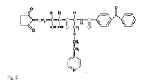
- FIG. 1 shows the principal functional parts of such a molecule. Each position can be tailored to meet a variety of needs; the chemical attachment group could be an N-hydroxysuccinimide moiety for modification of lysine residues or an N-maleimide for cysteine labelling.
- the cleavable group could be a geminal diol for cleavage by oxidising agents or an ester linkage for base cleavage.
- the photoactivatable group could be an azide for rapid labelling or a benzophenone for high efficiency crosslinking.
- the mass transfer marker that will be used in all cases however will preferably be thioethylpyridine.
- the thioether bond is chemically extremely stable however it fragments readily under standard low energy MS/MS conditions in a triple quadrupole mass spectrometer.
- the group leaves as a positively charged ion with m/z of 106. This mass does not correspond to any standard fragment found during low energy fragmentation of peptides and thus provides a unique marker or tag for the peptide to which it is attached.
- the linker molecule is 2-benzophenon-4-yl-carbonylamino-4, 5-dihydroxy-6-(N-succinimidyl)-1-(4-pyridylethylthio)-3-n-hexanone ( figure 2 ).
- this linker should comprise the attachment part, the cleavable part, the label part and the photoactivatable part, but they should mostly be seen as functional parts, and must not necessarily be structurally distinguished from each other.
- the core part of the linker molecule is its ability to render a unique mass marker in the gas phase, making it suitable for parent ion scanning, which feature is a result of the thioether bridge.
- kits for use in the method of identifying an interacting target biomolecule to a biomolecule of interest according to the present invention, which comprises, in separate compartments, at least one linker molecule, and optionally the biomolecule of interest.
- the kit may comprise necessary reagents for the different steps of the method of the invention, as discussed above or in the example section.
- the kit comprises amounts of the reagents, which is sufficient for performing the method of the invention.
- the kit can also comprise written instructions for its use.
- the kit can comprise a biomolecule of interest in a suitable buffer, a linker molecule as described above, and a cleaving agent that can cleave a part of the linker under appropriate conditions, each component being present in a separate compartment.
- the linker comprises a part that can be photo-activated to enable it to bind to a suitable target, as described above.
- said part of the linker that can be activated by chemical means in which case the kit also comprises a suitable substance for providing such activation.
- the kit comprises an interacting biomolecule bound to a linker, and optionally a chemical compound capable of activating a part of the linker to enable binding thereof to a target biomolecule.
- Still another aspect of the invention is the use of a linker molecule as described above for labelling the target biomolecule in the method of the invention.
- Example 1 The principle of the method of the invention
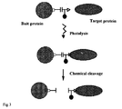
- the principle of the method is shown in Figure 3 .
- the linker is attached to lysine residues on the bait protein.
- the protein is then introduced to a perforated cell or a cell extract and allowed to equilibrate before photolysis. After crosslinking the proteins are cleaved by oxidisation and the mixture is first digested with cyanogen bromide and then by trypsin.
- the complex peptide mixture is then analysed by multi-dimensional HPLC inter faced by electrospray ionisation to a mass spectrometer operating in parent ion scanning mode as shown diagrammatically in Figure 4 .
- a mass spectrometer operating in parent ion scanning mode, only those peptides that give rise to an intense ion at 106 m/z will be detected.
- the mass spectrometer can be programmed to operate in a data-dependant mode, switching from parent ion to daughter ion scanning mode once a peptide containing the marker is detected.
- Figure 5 shows a preliminary experiment that has been carried out to validate the general principle of the approach.
- a synthetic calmodulin-binding peptide was modified with the crosslinker and photolysed in the presence of calcium and calmodulin. The mixture was then digested with cyanogen bromide followed by trypsin and the peptides separated by HPLC. The UV trace in Figure 5 shows that the expected number of peptides are generated whereas the parent ion total ion current trace shows that only one peptide has been labelled by the mass marker transfer method.
- Calmodulin is an 18 kDa calcium binding protein involved in calcium signal transduction in many cells. Upon binding calcium it undergoes a conformational change allowing it to bind a specific domain on its target proteins.
- the present method In order for the present method to be of general use, one should be able to locate the crosslinker on the bait protein at specific locations, especially if a protein interacts with more that one target.
- the inventor uses a cell-free translation system that is commercially available, namely the Roche rapid translation system (RTS).
- RTS rapid translation system
- the N-maleimide derivative of Figure 2 is synthesised and attached to cysteine. This in turn can be coupled to a tRNA with a defined codon specificity as described by Josef Brunner's group and more recently that of Peter Schultz. Site-specific incorporation of the crosslinker is rapidly achieved and commonly 100 ⁇ g quantities can be prepared overnight.
- the protein carries two affinity tags and can be rapidly purified.
- the protein must then be introduced into the cells of interest by either active uptake or by permeabilising the cells temporarily by digitonin. Alternatively the experiment may be carried out in cell extracts. Once the cDNA encoding the protein is correctly engineered into the carrier plasmid, one can rapidly produce mutants by PCR with the codon for the modified tRNA. Thus, tens of proteins modified at different positions can according to the invention be produced within a week once the system is set-up.
- crosslinkers are introduced in a random fashion by chemical means.
- a very gentle method of modification for crosslinking was introduced by the group of Traut to map protein-protein interactions in the ribosome.
- Iminothiolane a reagent that reacts with lysine residues to give a free sulphydryl group in their place, was used to modify the intact complex under mild conditions. Oxygen was then bubbled into the solution causing neighbouring sulphydryl groups to crosslink to form disulphide bridges.
- the complex was then separated by diagonal gel electrophoresis, in which the first dimension is non-reducing SDS PAGE and the second is done under reducing conditions.
- Non-crosslinked proteins appear along the diagonal axis at a position proportional to their mass whereas crosslinked proteins appear as off diagonal vertically separated pairs as shown in Figure 6 .
- This method can be modified slightly to incorporate a mass marker introduction step between the first and second dimension.
- all interacting pairs appearing off-diagonal can be rapidly identified by protein fragment fingerprinting ( James, P., Quadroni, M, Carafoli, E., and Gonnet, G. (1993) Biochem.Biophys.Res.Comm. 195(1), 58-64 . Protein identification by mass profile fingerprinting.) and their sites of interaction can be analysed simultaneously by the parent ion scanning method outlined in Figures 4 and 5 .
- mapping domain interactions between proteins can be taken which is similar to that used for mapping epitopes ( Houghten, R. A. (1985). "General method for the rapid solid-phase synthesis of large numbers of peptides: specificity of antigen-antibody interaction at the level of individual amino acids.” Proc Natl Acad Sci U S A 82(15): 5131-5 .). The inventor synthesises a series of 20mer peptides which cover the entire sequence of a protein with 10 amino acid overlaps (i.e. 100 peptides are needed for a 100 kDa protein).
- Each of the peptides will have a crosslinker at its N-terminal and a long cleavable spacer arm at its C-terminal separating it from the supporting resin.
- the synthesis is carried out by a standard multi-parallel robotic system (e.g. that of Advanced ChemTech amongst many others) in a 96 well plate format.
- the known target protein which has been labelled with a fluorescent group, is then added in the appropriate buffer to the wells and allowed to equilibrate.
- the wells are then washed under progressively more stringent conditions and the fluorescence in each well determined after each round. Finally, the better binding peptides are then re-equilibrated with target protein and photolysed.
- each peptide on the target is then determined and a domain interaction map is constructed.
- the procedure is then reversed and the bait protein is fluorescently labelled and used to screen the peptides from the target and the binding results are correlated with the first map to exclude non-specific interactions.
Landscapes
- Life Sciences & Earth Sciences (AREA)
- Health & Medical Sciences (AREA)
- Engineering & Computer Science (AREA)
- Molecular Biology (AREA)
- Hematology (AREA)
- Physics & Mathematics (AREA)
- Chemical & Material Sciences (AREA)
- Urology & Nephrology (AREA)
- Immunology (AREA)
- Biomedical Technology (AREA)
- Proteomics, Peptides & Aminoacids (AREA)
- General Health & Medical Sciences (AREA)
- Biotechnology (AREA)
- Pathology (AREA)
- Microbiology (AREA)
- Bioinformatics & Computational Biology (AREA)
- Bioinformatics & Cheminformatics (AREA)
- Food Science & Technology (AREA)
- Medicinal Chemistry (AREA)
- Analytical Chemistry (AREA)
- Biochemistry (AREA)
- Cell Biology (AREA)
- General Physics & Mathematics (AREA)
- Biophysics (AREA)
- Spectroscopy & Molecular Physics (AREA)
- Investigating Or Analysing Biological Materials (AREA)
- Peptides Or Proteins (AREA)
- Other Investigation Or Analysis Of Materials By Electrical Means (AREA)
- Saccharide Compounds (AREA)
- Analysing Materials By The Use Of Radiation (AREA)
- Investigating Or Analysing Materials By The Use Of Chemical Reactions (AREA)
- Measuring Or Testing Involving Enzymes Or Micro-Organisms (AREA)
Applications Claiming Priority (3)
| Application Number | Priority Date | Filing Date | Title |
|---|---|---|---|
| GBGB0131014.3A GB0131014D0 (en) | 2001-12-28 | 2001-12-28 | Method for molecule-mlecule analysis |
| GB0131014 | 2001-12-28 | ||
| PCT/EP2002/014315 WO2003056342A2 (en) | 2001-12-28 | 2002-12-16 | Method for determining molecule-molecule interaction in proteomics |
Publications (2)
| Publication Number | Publication Date |
|---|---|
| EP1459073A2 EP1459073A2 (en) | 2004-09-22 |
| EP1459073B1 true EP1459073B1 (en) | 2008-02-13 |
Family
ID=9928438
Family Applications (1)
| Application Number | Title | Priority Date | Filing Date |
|---|---|---|---|
| EP02791831A Expired - Lifetime EP1459073B1 (en) | 2001-12-28 | 2002-12-16 | Method for determining molecule-molecule interaction in proteomics |
Country Status (9)
| Country | Link |
|---|---|
| US (1) | US7022491B2 (https=) |
| EP (1) | EP1459073B1 (https=) |
| JP (1) | JP2005513506A (https=) |
| AT (1) | ATE386270T1 (https=) |
| AU (1) | AU2002358143A1 (https=) |
| CA (1) | CA2465300A1 (https=) |
| DE (1) | DE60225059D1 (https=) |
| GB (1) | GB0131014D0 (https=) |
| WO (1) | WO2003056342A2 (https=) |
Families Citing this family (5)
| Publication number | Priority date | Publication date | Assignee | Title |
|---|---|---|---|---|
| JP2005140755A (ja) * | 2003-11-10 | 2005-06-02 | Japan Science & Technology Agency | 質量分析用プローブ及びそれを用いた質量分析方法 |
| GB0515323D0 (en) | 2005-07-26 | 2005-08-31 | Electrophoretics Ltd | Mass labels |
| JP6145742B2 (ja) * | 2013-01-17 | 2017-06-14 | 国立大学法人富山大学 | 蛍光性質量標識プローブ |
| TW202311746A (zh) * | 2018-02-02 | 2023-03-16 | 美商再生元醫藥公司 | 用於表徵蛋白質二聚合之系統及方法 |
| CN110853712B (zh) * | 2018-08-01 | 2022-06-07 | 清华大学 | 鉴定多对生物分子间相互作用调控因子的方法 |
Family Cites Families (3)
| Publication number | Priority date | Publication date | Assignee | Title |
|---|---|---|---|---|
| US4487838A (en) | 1982-01-12 | 1984-12-11 | Massachusetts Institute Of Technology | Radio-labelled cross-linking reagents |
| GB9315847D0 (en) | 1993-07-30 | 1993-09-15 | Isis Innovation | Tag reagent and assay method |
| GB9815166D0 (en) * | 1998-07-13 | 1998-09-09 | Brax Genomics Ltd | Compounds for mass spectrometry |
-
2001
- 2001-12-28 GB GBGB0131014.3A patent/GB0131014D0/en not_active Ceased
-
2002
- 2002-12-16 DE DE60225059T patent/DE60225059D1/de not_active Expired - Lifetime
- 2002-12-16 EP EP02791831A patent/EP1459073B1/en not_active Expired - Lifetime
- 2002-12-16 AT AT02791831T patent/ATE386270T1/de not_active IP Right Cessation
- 2002-12-16 JP JP2003556814A patent/JP2005513506A/ja active Pending
- 2002-12-16 CA CA002465300A patent/CA2465300A1/en not_active Abandoned
- 2002-12-16 WO PCT/EP2002/014315 patent/WO2003056342A2/en not_active Ceased
- 2002-12-16 US US10/500,431 patent/US7022491B2/en not_active Expired - Fee Related
- 2002-12-16 AU AU2002358143A patent/AU2002358143A1/en not_active Abandoned
Also Published As
| Publication number | Publication date |
|---|---|
| ATE386270T1 (de) | 2008-03-15 |
| CA2465300A1 (en) | 2003-07-10 |
| GB0131014D0 (en) | 2002-02-13 |
| EP1459073A2 (en) | 2004-09-22 |
| US7022491B2 (en) | 2006-04-04 |
| US20050095654A1 (en) | 2005-05-05 |
| JP2005513506A (ja) | 2005-05-12 |
| WO2003056342A3 (en) | 2003-11-06 |
| AU2002358143A1 (en) | 2003-07-15 |
| AU2002358143A8 (en) | 2003-07-15 |
| WO2003056342A2 (en) | 2003-07-10 |
| DE60225059D1 (de) | 2008-03-27 |
Similar Documents
| Publication | Publication Date | Title |
|---|---|---|
| Hamdan et al. | Modern strategies for protein quantification in proteome analysis: advantages and limitations | |
| Kuyama et al. | An approach to quantitative proteome analysis by labeling tryptophan residues | |
| Müller et al. | Isotope-tagged cross-linking reagents. A new tool in mass spectrometric protein interaction analysis | |
| US20100068819A1 (en) | Compounds and methods for double labelling of polypeptides to allow multiplexing in mass spectrometric analysis | |
| EP1397686B1 (en) | Method for characterizing polypeptides | |
| AU2008213716A1 (en) | Affinity selected signature peptides for protein identification and quantification | |
| US7635573B2 (en) | Mass spectroscopic method for comparing protein levels in two or more samples | |
| US20030119069A1 (en) | Labeling of protein samples | |
| Soderblom et al. | Tandem mass spectrometry acquisition approaches to enhance identification of protein‐protein interactions using low‐energy collision‐induced dissociative chemical crosslinking reagents | |
| US7244411B2 (en) | Method of selective peptide isolation for the identification and quantitative analysis of proteins in complex mixtures | |
| EP1459073B1 (en) | Method for determining molecule-molecule interaction in proteomics | |
| US20050164336A1 (en) | Method for protein expression analysis | |
| US7371514B2 (en) | Serial derivatization of peptides for de novo sequencing using tandem mass spectrometry | |
| Czeszak et al. | Identification of substituted sites on MUC5AC mucin motif peptides after enzymatic O‐glycosylation combining β‐elimination and fixed‐charge derivatization | |
| US20050042676A1 (en) | Characterising polypeptides | |
| Yan et al. | Nonprotein based enrichment method to analyze peptide cross-linking in protein complexes | |
| Nokihara et al. | High throughput sequencing of cyclic peptide immobilized on a gel-type single bead | |
| Delahunty et al. | Protein–Protein Interactions | |
| Adamczyk et al. | Sequencing of anti‐thyroxine monoclonal antibody Fab fragment by ion trap mass spectrometry | |
| AU2002310611B2 (en) | Method for characterizing polypeptides | |
| WO2003031982A1 (en) | Method for analysing protein/peptide expression | |
| AU2002310611A1 (en) | Method for characterizing polypeptides |
Legal Events
| Date | Code | Title | Description |
|---|---|---|---|
| PUAI | Public reference made under article 153(3) epc to a published international application that has entered the european phase |
Free format text: ORIGINAL CODE: 0009012 |
|
| 17P | Request for examination filed |
Effective date: 20040421 |
|
| AK | Designated contracting states |
Kind code of ref document: A2 Designated state(s): AT BE BG CH CY CZ DE DK EE ES FI FR GB GR IE IT LI LU MC NL PT SE SI SK TR |
|
| AX | Request for extension of the european patent |
Extension state: AL LT LV MK RO |
|
| GRAP | Despatch of communication of intention to grant a patent |
Free format text: ORIGINAL CODE: EPIDOSNIGR1 |
|
| GRAS | Grant fee paid |
Free format text: ORIGINAL CODE: EPIDOSNIGR3 |
|
| GRAA | (expected) grant |
Free format text: ORIGINAL CODE: 0009210 |
|
| AK | Designated contracting states |
Kind code of ref document: B1 Designated state(s): AT BE BG CH CY CZ DE DK EE ES FI FR GB GR IE IT LI LU MC NL PT SE SI SK TR |
|
| REG | Reference to a national code |
Ref country code: GB Ref legal event code: FG4D |
|
| REG | Reference to a national code |
Ref country code: CH Ref legal event code: EP |
|
| REG | Reference to a national code |
Ref country code: IE Ref legal event code: FG4D |
|
| REF | Corresponds to: |
Ref document number: 60225059 Country of ref document: DE Date of ref document: 20080327 Kind code of ref document: P |
|
| PG25 | Lapsed in a contracting state [announced via postgrant information from national office to epo] |
Ref country code: ES Free format text: LAPSE BECAUSE OF FAILURE TO SUBMIT A TRANSLATION OF THE DESCRIPTION OR TO PAY THE FEE WITHIN THE PRESCRIBED TIME-LIMIT Effective date: 20080524 Ref country code: FI Free format text: LAPSE BECAUSE OF FAILURE TO SUBMIT A TRANSLATION OF THE DESCRIPTION OR TO PAY THE FEE WITHIN THE PRESCRIBED TIME-LIMIT Effective date: 20080213 |
|
| NLV1 | Nl: lapsed or annulled due to failure to fulfill the requirements of art. 29p and 29m of the patents act | ||
| PG25 | Lapsed in a contracting state [announced via postgrant information from national office to epo] |
Ref country code: AT Free format text: LAPSE BECAUSE OF FAILURE TO SUBMIT A TRANSLATION OF THE DESCRIPTION OR TO PAY THE FEE WITHIN THE PRESCRIBED TIME-LIMIT Effective date: 20080213 |
|
| PG25 | Lapsed in a contracting state [announced via postgrant information from national office to epo] |
Ref country code: SI Free format text: LAPSE BECAUSE OF FAILURE TO SUBMIT A TRANSLATION OF THE DESCRIPTION OR TO PAY THE FEE WITHIN THE PRESCRIBED TIME-LIMIT Effective date: 20080213 Ref country code: BE Free format text: LAPSE BECAUSE OF FAILURE TO SUBMIT A TRANSLATION OF THE DESCRIPTION OR TO PAY THE FEE WITHIN THE PRESCRIBED TIME-LIMIT Effective date: 20080213 |
|
| PG25 | Lapsed in a contracting state [announced via postgrant information from national office to epo] |
Ref country code: SK Free format text: LAPSE BECAUSE OF FAILURE TO SUBMIT A TRANSLATION OF THE DESCRIPTION OR TO PAY THE FEE WITHIN THE PRESCRIBED TIME-LIMIT Effective date: 20080213 Ref country code: SE Free format text: LAPSE BECAUSE OF FAILURE TO SUBMIT A TRANSLATION OF THE DESCRIPTION OR TO PAY THE FEE WITHIN THE PRESCRIBED TIME-LIMIT Effective date: 20080513 Ref country code: CZ Free format text: LAPSE BECAUSE OF FAILURE TO SUBMIT A TRANSLATION OF THE DESCRIPTION OR TO PAY THE FEE WITHIN THE PRESCRIBED TIME-LIMIT Effective date: 20080213 Ref country code: DK Free format text: LAPSE BECAUSE OF FAILURE TO SUBMIT A TRANSLATION OF THE DESCRIPTION OR TO PAY THE FEE WITHIN THE PRESCRIBED TIME-LIMIT Effective date: 20080213 Ref country code: PT Free format text: LAPSE BECAUSE OF FAILURE TO SUBMIT A TRANSLATION OF THE DESCRIPTION OR TO PAY THE FEE WITHIN THE PRESCRIBED TIME-LIMIT Effective date: 20080714 Ref country code: NL Free format text: LAPSE BECAUSE OF FAILURE TO SUBMIT A TRANSLATION OF THE DESCRIPTION OR TO PAY THE FEE WITHIN THE PRESCRIBED TIME-LIMIT Effective date: 20080213 |
|
| EN | Fr: translation not filed | ||
| PLBE | No opposition filed within time limit |
Free format text: ORIGINAL CODE: 0009261 |
|
| STAA | Information on the status of an ep patent application or granted ep patent |
Free format text: STATUS: NO OPPOSITION FILED WITHIN TIME LIMIT |
|
| 26N | No opposition filed |
Effective date: 20081114 |
|
| PG25 | Lapsed in a contracting state [announced via postgrant information from national office to epo] |
Ref country code: DE Free format text: LAPSE BECAUSE OF FAILURE TO SUBMIT A TRANSLATION OF THE DESCRIPTION OR TO PAY THE FEE WITHIN THE PRESCRIBED TIME-LIMIT Effective date: 20080514 |
|
| PG25 | Lapsed in a contracting state [announced via postgrant information from national office to epo] |
Ref country code: FR Free format text: LAPSE BECAUSE OF FAILURE TO SUBMIT A TRANSLATION OF THE DESCRIPTION OR TO PAY THE FEE WITHIN THE PRESCRIBED TIME-LIMIT Effective date: 20081205 Ref country code: BG Free format text: LAPSE BECAUSE OF FAILURE TO SUBMIT A TRANSLATION OF THE DESCRIPTION OR TO PAY THE FEE WITHIN THE PRESCRIBED TIME-LIMIT Effective date: 20080513 Ref country code: EE Free format text: LAPSE BECAUSE OF FAILURE TO SUBMIT A TRANSLATION OF THE DESCRIPTION OR TO PAY THE FEE WITHIN THE PRESCRIBED TIME-LIMIT Effective date: 20080213 |
|
| PG25 | Lapsed in a contracting state [announced via postgrant information from national office to epo] |
Ref country code: MC Free format text: LAPSE BECAUSE OF NON-PAYMENT OF DUE FEES Effective date: 20081231 Ref country code: CY Free format text: LAPSE BECAUSE OF FAILURE TO SUBMIT A TRANSLATION OF THE DESCRIPTION OR TO PAY THE FEE WITHIN THE PRESCRIBED TIME-LIMIT Effective date: 20080213 |
|
| REG | Reference to a national code |
Ref country code: CH Ref legal event code: PL |
|
| PG25 | Lapsed in a contracting state [announced via postgrant information from national office to epo] |
Ref country code: IT Free format text: LAPSE BECAUSE OF FAILURE TO SUBMIT A TRANSLATION OF THE DESCRIPTION OR TO PAY THE FEE WITHIN THE PRESCRIBED TIME-LIMIT Effective date: 20080213 |
|
| PG25 | Lapsed in a contracting state [announced via postgrant information from national office to epo] |
Ref country code: LI Free format text: LAPSE BECAUSE OF NON-PAYMENT OF DUE FEES Effective date: 20081231 Ref country code: IE Free format text: LAPSE BECAUSE OF NON-PAYMENT OF DUE FEES Effective date: 20081216 Ref country code: CH Free format text: LAPSE BECAUSE OF NON-PAYMENT OF DUE FEES Effective date: 20081231 |
|
| PG25 | Lapsed in a contracting state [announced via postgrant information from national office to epo] |
Ref country code: LU Free format text: LAPSE BECAUSE OF NON-PAYMENT OF DUE FEES Effective date: 20081216 |
|
| PG25 | Lapsed in a contracting state [announced via postgrant information from national office to epo] |
Ref country code: TR Free format text: LAPSE BECAUSE OF FAILURE TO SUBMIT A TRANSLATION OF THE DESCRIPTION OR TO PAY THE FEE WITHIN THE PRESCRIBED TIME-LIMIT Effective date: 20080213 |
|
| PG25 | Lapsed in a contracting state [announced via postgrant information from national office to epo] |
Ref country code: GR Free format text: LAPSE BECAUSE OF FAILURE TO SUBMIT A TRANSLATION OF THE DESCRIPTION OR TO PAY THE FEE WITHIN THE PRESCRIBED TIME-LIMIT Effective date: 20080514 |
|
| PGFP | Annual fee paid to national office [announced via postgrant information from national office to epo] |
Ref country code: GB Payment date: 20101229 Year of fee payment: 9 |
|
| GBPC | Gb: european patent ceased through non-payment of renewal fee |
Effective date: 20111216 |
|
| PG25 | Lapsed in a contracting state [announced via postgrant information from national office to epo] |
Ref country code: GB Free format text: LAPSE BECAUSE OF NON-PAYMENT OF DUE FEES Effective date: 20111216 |