EP1439870B1 - Dispositif d'administration d'une substance - Google Patents
Dispositif d'administration d'une substance Download PDFInfo
- Publication number
- EP1439870B1 EP1439870B1 EP02803296A EP02803296A EP1439870B1 EP 1439870 B1 EP1439870 B1 EP 1439870B1 EP 02803296 A EP02803296 A EP 02803296A EP 02803296 A EP02803296 A EP 02803296A EP 1439870 B1 EP1439870 B1 EP 1439870B1
- Authority
- EP
- European Patent Office
- Prior art keywords
- skin
- microprotrusions
- microabrader
- abrading
- delivery
- Prior art date
- Legal status (The legal status is an assumption and is not a legal conclusion. Google has not performed a legal analysis and makes no representation as to the accuracy of the status listed.)
- Expired - Lifetime
Links
Images


Classifications
-
- A—HUMAN NECESSITIES
- A61—MEDICAL OR VETERINARY SCIENCE; HYGIENE
- A61M—DEVICES FOR INTRODUCING MEDIA INTO, OR ONTO, THE BODY; DEVICES FOR TRANSDUCING BODY MEDIA OR FOR TAKING MEDIA FROM THE BODY; DEVICES FOR PRODUCING OR ENDING SLEEP OR STUPOR
- A61M37/00—Other apparatus for introducing media into the body; Percutany, i.e. introducing medicines into the body by diffusion through the skin
- A61M37/0015—Other apparatus for introducing media into the body; Percutany, i.e. introducing medicines into the body by diffusion through the skin by using microneedles
-
- A—HUMAN NECESSITIES
- A61—MEDICAL OR VETERINARY SCIENCE; HYGIENE
- A61B—DIAGNOSIS; SURGERY; IDENTIFICATION
- A61B17/00—Surgical instruments, devices or methods, e.g. tourniquets
- A61B17/54—Chiropodists' instruments, e.g. pedicure
-
- A—HUMAN NECESSITIES
- A61—MEDICAL OR VETERINARY SCIENCE; HYGIENE
- A61M—DEVICES FOR INTRODUCING MEDIA INTO, OR ONTO, THE BODY; DEVICES FOR TRANSDUCING BODY MEDIA OR FOR TAKING MEDIA FROM THE BODY; DEVICES FOR PRODUCING OR ENDING SLEEP OR STUPOR
- A61M31/00—Devices for introducing or retaining media, e.g. remedies, in cavities of the body
- A61M31/002—Devices for releasing a drug at a continuous and controlled rate for a prolonged period of time
-
- A—HUMAN NECESSITIES
- A61—MEDICAL OR VETERINARY SCIENCE; HYGIENE
- A61B—DIAGNOSIS; SURGERY; IDENTIFICATION
- A61B17/00—Surgical instruments, devices or methods, e.g. tourniquets
- A61B17/32—Surgical cutting instruments
- A61B2017/320004—Surgical cutting instruments abrasive
-
- A—HUMAN NECESSITIES
- A61—MEDICAL OR VETERINARY SCIENCE; HYGIENE
- A61M—DEVICES FOR INTRODUCING MEDIA INTO, OR ONTO, THE BODY; DEVICES FOR TRANSDUCING BODY MEDIA OR FOR TAKING MEDIA FROM THE BODY; DEVICES FOR PRODUCING OR ENDING SLEEP OR STUPOR
- A61M37/00—Other apparatus for introducing media into the body; Percutany, i.e. introducing medicines into the body by diffusion through the skin
- A61M2037/0007—Other apparatus for introducing media into the body; Percutany, i.e. introducing medicines into the body by diffusion through the skin having means for enhancing the permeation of substances through the epidermis, e.g. using suction or depression, electric or magnetic fields, sound waves or chemical agents
-
- A—HUMAN NECESSITIES
- A61—MEDICAL OR VETERINARY SCIENCE; HYGIENE
- A61M—DEVICES FOR INTRODUCING MEDIA INTO, OR ONTO, THE BODY; DEVICES FOR TRANSDUCING BODY MEDIA OR FOR TAKING MEDIA FROM THE BODY; DEVICES FOR PRODUCING OR ENDING SLEEP OR STUPOR
- A61M37/00—Other apparatus for introducing media into the body; Percutany, i.e. introducing medicines into the body by diffusion through the skin
- A61M37/0015—Other apparatus for introducing media into the body; Percutany, i.e. introducing medicines into the body by diffusion through the skin by using microneedles
- A61M2037/0023—Drug applicators using microneedles
-
- A—HUMAN NECESSITIES
- A61—MEDICAL OR VETERINARY SCIENCE; HYGIENE
- A61M—DEVICES FOR INTRODUCING MEDIA INTO, OR ONTO, THE BODY; DEVICES FOR TRANSDUCING BODY MEDIA OR FOR TAKING MEDIA FROM THE BODY; DEVICES FOR PRODUCING OR ENDING SLEEP OR STUPOR
- A61M37/00—Other apparatus for introducing media into the body; Percutany, i.e. introducing medicines into the body by diffusion through the skin
- A61M37/0015—Other apparatus for introducing media into the body; Percutany, i.e. introducing medicines into the body by diffusion through the skin by using microneedles
- A61M2037/0046—Solid microneedles
-
- A—HUMAN NECESSITIES
- A61—MEDICAL OR VETERINARY SCIENCE; HYGIENE
- A61M—DEVICES FOR INTRODUCING MEDIA INTO, OR ONTO, THE BODY; DEVICES FOR TRANSDUCING BODY MEDIA OR FOR TAKING MEDIA FROM THE BODY; DEVICES FOR PRODUCING OR ENDING SLEEP OR STUPOR
- A61M37/00—Other apparatus for introducing media into the body; Percutany, i.e. introducing medicines into the body by diffusion through the skin
- A61M37/0015—Other apparatus for introducing media into the body; Percutany, i.e. introducing medicines into the body by diffusion through the skin by using microneedles
- A61M2037/0061—Methods for using microneedles
Definitions
- the present invention relates to a device for abrading the skin.
- This technique avoids the nerve net and places the bioactive substance in close proximity to blood vessels and lymphatics for absorption and delivery of the substance throughout the body.
- the epidermis itself is a particularly desirable target as it is rich in antigen presenting cells.
- the dermal layer below the epidermis contains fewer antigen presenting cells.
- the stratum corneum and epidermis do not contain nerves or blood vessels, so this method has the advantage of being essentially painless and blood-free while giving access to the skin layers capable of responding to the antigen.
- the prior art reports a variety of devices and methods for disrupting the stratum corneum for the purpose of delivering substances to the body.
- breach of the stratum corneum may be achieved by puncturing as taught in US Patent 5,679,647 to Carson, et al .
- This patent teaches that narrow diameter tines, such as those found on devices used for tuberculin skin tests and allergy tests, can be coated with polynucleotides or oligonucleotides and used for delivery of such materials into the skin.
- the method of using such devices involves puncturing the skin with the tines resulting in intracutaneous infection of the coated substance.
- US Patent 5,003,987 ; US Patent 5,879,326 ; and US Patent 3,964,482 teach breaching the stratum corneum by cutting.
- EP 1 086 719 discloses a method and device for abrading skin prior to drug delivery.
- US 6,334,856 discloses a microncedle device for injecting fluid through the stratum corneum.
- WO02/08544 discloses a microprojection array for piercing or cutting the stratum corneum.
- WO02/3233 discloses a microstructure for exfoliating the skin.
- WO03/026732 discloses a switchable microneedle array.
- the present invention is directed to a device for abrading the skin, and particularly, the stratum corneum of the skin.
- Substances to be delivered particularly include bioactive substances, including pharmaceutical agents, medicaments, vaccines and the like.
- Substances may be in solid or liquid form, depending on formulation and delivery method. They can be delivered inter alia , in the form of dry powders, gels, solutions, suspensions, and creams. Suitable formulations are familiar to those of skill in the art.
- Particularly preferred medicaments for delivery by the methods of the invention include vaccines, allergens and gene therapeutic agents.
- One aspect of the invention is directed to a device for preparing a delivery site on the skin to enhance the delivery of a pharmaceutical agent through the stratum corneum of the skin to a sufficient depth where the pharmaceutical agent can be absorbed and utilized by the body.
- Dermal tissue represents an attractive target site for delivery of vaccines and gene therapeutic agents.
- the skin is an attractive delivery site due to the high concentration of antigen presenting cells (APC) and APC precursors found within this tissue, especially the epidermal Langerhan's cells (LC).
- APC antigen presenting cells
- LC epidermal Langerhan's cells
- gene therapeutic agents are designed for the treatment of skin disorders, skin diseases and skin cancer. In such cases, direct delivery of the therapeutic agent to the affected skin tissue is desirable.
- skin cells are an attractive target for gene therapeutic agents, of which the encoded protein or proteins are active at sites distant from the skin.
- skin cells e.g., keratinocytes
- bioreactors producing a therapeutic protein which can be rapidly absorbed into the systemic circulation via the papillary dermis.
- direct access of the vaccine or therapeutic agent to the systemic circulation is desirable for the treatment of disorders distant from the skin.
- systemic distribution can be accomplished through the papillary dermis.
- the present invention provides a microabrader device to abrade the skin in conjunction with the delivery of a bioactive substance, including but not limited to nucleic acids, amino acids, amino acid derivatives, peptides or polypeptides. It has been discovered that nucleic acids exhibit enhanced gene expression and produce an enhanced immune response to the expressed protein when they are delivered simultaneously with abrasion of the stratum corneum. Similarly, allergens delivered simultaneously with abrasion produce a more vigorous immune response than conventional allergen testing methods.
- a bioactive substance including but not limited to nucleic acids, amino acids, amino acid derivatives, peptides or polypeptides.
- the present invention comprises a microabrader for delivering a substance into the skin having a base with an abrading facet, to which an abrading surface having an arrangement of microprotrusions that have at least one scraping edge is attached, mounted or integral with, and a handle attachment facet, to which a handle or other grasping device is attached, mounted, or integral with.
- abrading surface is meant the surface that is presented to the skin during the process of abrasion, including microprotrusions, surface area between them and surrounding surface.
- the microabrader device of the present invention is suitable for delivering a substance to the skin by movement of the microabrader device across an area of the skin to produce furrows of sufficient depth to allow the substance, which is administered prior to, simultaneously with, or following the abrasion of the skin, to be taken up by the predetermined skin layer.
- multiple passes of the device across the skin can result in progressively deeper furrows in the skin, thereby allowing delivery of a substance to a desired depth with in the skin.
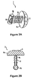
- Figure 1A is an elevated view of the handle end of a preferred embodiment
- Figure 1B is a side view of a preferred embodiment of a microabrader.
- Figure 2A is a transparent perspective view of the microabrader device of Figures 1A and 1B .
- Figure 2B is a cross sectional view of the microabrader device of Figure 1B .
- Figure 3 is a side view of the abrading surface the microabrader device of Figures 1A; 1B , 2A, and 2B on the skin of a subject.
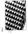
- Figure 4 is a perspective view of the abrading surface in the embodiment of Figure 3 .
- Figure 4A is a cross sectional side view of the abrader surface.
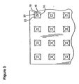
- Figure 5 is a bottom view of the abrader surface of the embodiment of Figure 3 .
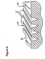
- Figure 6 is a perspective view in partial cross section of abraded furrows of skin.
- Figure 7 illustrates levels of reporter gene activity in skin obtained with the various nucleic acid delivery protocols tested in Example 1.
- Figure 8 illustrates reporter gene activity in skin obtained by varying the number of abrasions as described in Example 2.
- Figure 9 illustrates reporter gene activity in skin obtained by varying the formulation of the nucleic acid and the delivery protocol as described in Example 3.
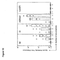
- Figure 10 illustrates the serum antibody response following delivery of plasmid DNA encoding Hepatitis B Surface Antigen (HBsAg) as described in Example 4.
- HBsAg Hepatitis B Surface Antigen
- Figure 11 illustrates mean luciferase activity ( ⁇ SEM) in skin samples from rats treated with a reporter gene using Mantoux-style injection technique (Group 1A), plastic microneedle array of the invention (Group 2A), a tine device pressed against skin and scratched across an area of approximately 0.06 cm 2 (Group 3A), a tine device pressed against skin and moved across an area of approximately 1cm 2 (Group 4A), a tine device pressed against skin and removed (Group 5A), plasmid DNA directly applied in droplet form to the shaved skin (Group 6A).
- ⁇ SEM mean luciferase activity
- Figure 12 shows skin reactions after application of histamine and abrading the skin of weaning pigs using a plastic microneedle array of the invention (Group 1 B), a tine device scratched once across an area of approximately 0.06 cm 2 (Group 2B), a tine device scratched multiple times to produce a scratched area of approximately 1 cm 2 (Group 3B), a time device pressed against the skin and removed (Group 4B).
- numbers 1-5 are replicates and "C" is a control to which histamine was applied without abrasion.
- Figure 13 shows displays the relative area of tissue swelling for each group shown in Figure 12 after subtracting the swelling measurements observed from use of the device only without histamine.
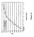
- FIGS 14 and 15 compare Trans Epidermal Water Loss (TEWL) from skin following treatment with plastic and silicon microabraders.
- TEWL Trans Epidermal Water Loss
- Figure 16 illustrates reporter gene activity in skin following delivery of plasmid DNA encoding a reporter gene using plastic and silicon microabraders.
- Figure 17 compares the serum antibody response following delivery of DNA plasmid encoding HBsAg using plastic and silicon microabraders.
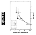
- Figure 18 illustrates the serum antibody response following administration of DNA plasmid encoding influenza hemagglutinin (HA), naked plasmid.
- HA hemagglutinin
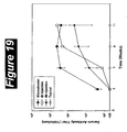
- Figure 19 illustrates the serum antibody response following administration of DNA plasmid encoding influenza hemagglutinin (HA), plasmid plus adjuvant.
- HA hemagglutinin
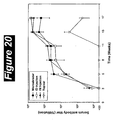
- Figure 20 illustrates the serum antibody response following priming with naked plasmid DNA encoding influenza HA, followed by boosting with whole inactivated influenza virus.
- Figure 21 illustrates the serum antibody response following priming with plasmid DNA encoding influenza HA plus adjuvant, followed by boosting with whole inactivated influenza virus.
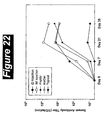
- Figure 22 illustrates the serum antibody response following administration of inactivated virus vaccine for rabies virus.
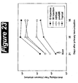
- Figure 23 illustrates the serum antibody response following administration of HBsAg via the delivery protocols as described in Example 11a.
- FIG. 24 illustrates the T-cell proliferative response following administration of HbsAg.
- Figure 25 illustrates the cellular immune response to a melanoma vaccine encoded by an adenoviral vector.
- the present invention is directed to a device for abrading the stratum corneum to enhance the administering of a substance through the stratum corneum of the skin of a patient.
- the term "abrade” refers to removing at least a portion of the stratum corneum to increase the permeability of the skin without causing excessive skin irritation or compromising the skin's barrier to infectious agents. This is in contrast to "puncturing" which produces discrete holes through the stratum corneum with areas of undisrupted stratum corneum between the holes.
- the microabrader of the invention is a device capable of abrading the skin to attain this result.
- the device is capable of abrading the skin thereby penetrating the stratum corneum without piercing the stratum corneum.
- the microabrader also includes an effective amount of a substance to be delivered. This may be included, for example, in a reservoir that is an integral or detachable part of the microabrader, or may be coated on the delivery surface of the microabrader.
- an “effective amount” of a substance is intended to mean an amount that will elicit a desired response in a subject, including, but not limited to, an immunostimulatory or immunomodulatory response in the case of an allergen or vaccine, or another therapeutic or diagnostic response.
- penetrating refers to entering the stratum corneum without passing completely through the stratum corneum and entering into the adjacent layers. This is not to say that that the stratum corneum can not be completely penetrated to reveal the interface of the underlying layer of the skin. Piercing, on the other hand, refers to passing through the stratum corneum completely and entering into the adjacent layers below the stratum corneum.
- the microabrader device of the invention is believed to have a unique immunological advantage in the delivery of vaccines with the potential of increasing the vaccine's clinical value.
- the penetration of the multiple microprotrusions into the stratum corneum is suggested as having an adjuvant-like stimulatory effect.
- the "penetration" response from the multiple microprotrusion is believed more than a simple acute inflammatory response.
- These "penetration" effects can cause damage to a variety of cells and cellular architecture, causing the appearance of polymorphonuclear neutrophils (PMN) and macrophages as well as the release of IL1, tumor necrosis factor (TNF) and other agents, which can lead to a number of other immunological responses.
- PMN polymorphonuclear neutrophils
- TNF tumor necrosis factor
- the soluble stimulatory factors influence the proliferation of lymphocytes and are central to the immune response to vaccines. In addition, these factors influence the migration and activation of resident antigen presenting cells including Langerhan's cells and dendritic cells.
- the microabrader of the present invention is valuable in promoting significant immune response to a vaccine in the abraded area.
- the small grooves and furrows created by the microprotrusion array over the abraded area are believed to increase the availability of the vaccine antigen for interaction with antigen-presenting cells compared to a vaccine applied topically in the absence of abrasion or administered using standard needles.
- the microprotrusion array of the invention is believed to magnify several-fold the trivial or inconsequential immune stimulatory impact of a single needlestick.
- the microabrader facilitates and enhances vaccine immunogenicity by an adjuvant-like immune stimulation.
- the primary barrier properties of the skin including the resistance to delivery of drugs, vaccines and gene therapeutic agents reside in the outermost layer of the epidermis, referred to as the stratum corneum.
- the inner layers of the epidermis generally include three layers, commonly identified as the stratum granulosum, the stratum malpighii, and the stratum germinativum. Once a drug or other substance appears below the stratum corneum, there is essentially no resistance to diffusion into subsequent layers of the skin and eventual uptake by cells or absorption by the body through the bloodstream or lymphatic drainage.
- the present invention is primarily directed to a device and method for facilitating delivery of a substance, and particularly a bioactive substance or pharmaceutical agent, into or through the stratum corneum for more rapid absorption of larger quantities of the bioactive substance or pharmaceutical agent by the patient.
- the device of the invention penetrates, but does not pierce, the stratum corneum.
- the substance to be administered using the methods of this invention may be applied to the skin prior to abrading, simultaneous with abrading, or post-abrading.
- certain or specific bioactive substances including nucleic acids , allergens and live viral vaccines are applied to the skin prior to or simultaneously with abrasion rather than being applied to previously abraded skin. That is, delivery of certain substances, such as nucleic acids, allergens and live viral vaccines are improved when such substances are abraded into the skin rather than being passively applied to skin which has been previously abraded.
- certain or specific bioactive substances including virus-like particles and subunit proteins, are improved when such substances are applied to pre-abraded skin.
- certain or specific bioactive substances including whole inactivated or killed viruses, display similar efficacy whether applied to skin following abrasion or simultaneously with abrasion.
- the substance may be delivered into the skin in any pharmaceutically acceptable form.
- the substance is applied to the skin and an abrading device is then moved or rubbed reciprocally over the skin and the substance. It is preferred that the minimum amount of abrasion to produce the desired result be used. Determination of the appropriate amount of abrasion for a selected substance is within the ordinary skill in the art.
- the substance may be applied in dry form to the abrading surface of the delivery device prior to application.
- a reconstituting liquid is applied to the skin at the delivery site and the substance-coated abrading device is applied to the skin at the site of the reconstituting liquid.
- a reconstituting liquid may be contained in the abrading device and released to dissolve the substance as the device is applied to the skin for abrasion. It has been found that certain substances, such as nucleic acid preparations, may also be coated on the abrading device in the form of a gel.
- a method for delivering a substance into the skin of a patient includes the steps of coating a patient's outer skin layer or a microabrader 2 , see Figure 1B with a medicament or other substance and moving microabrader 2 across the patient's skin to provide abrasions leaving furrows sufficient to permit entry of the substance into the patient's viable epidermis. Due to the structural design of microabrader 2, the leading edge of microabrader 2 first stretches the patient's skin and then the top surface of microabrader 2 abrades the outer protective skin layer opening the stratum corneum thereby permitting medicament or other substance to enter the patient. After the initial abrasion of the outer protective skin layer, the trailing and leading edges of microabrader 2 can rub the surface of the abraded area working the medicament or substance into the abraded skin area thereby improving its medicinal effect.
- microabrader 2 includes base 4 onto which an abrading surface 5 can be mounted.
- the abrading surface may be integral with the base and fabricated as a single two-component part.
- base 4 is a solid molded piece.
- base 4 is configured with a mushroom-like crown 4b that curves upward and is truncated at the top.
- the top of base 4 is generally flat with abrading surface 5 being mounted thereon or integral therewith.
- the truncated top may have a recess for receiving abrading surface 5.
- abrading surface 5 includes a platform with an array of microprotrusions that extends above the truncated top.
- the handle, base and abrading surface may be integral with one another and fabricated as a single three-component device.
- Microabrader 2 is applied to a subject by moving microabrader 2 across the subject's skin with enough pressure to enable abrading surface 5 to open the outer protective skin or stratum corneum of the subject.
- the inward pressure applied to the base causes microabrader 2 to be pressed into the subject's skin. Accordingly, it is preferable that the height of the sloping mushroom-like crown 4b be sufficient to prevent the applied substance from flowing over and onto the facet 4c when microabrader 2 is being used.
- abrading surface 5 comprises an array of microprotrusions.
- a handle 6 is attached to base 4 or may be integral with base 4. As shown in Fig. 2A , an upper end 6a of the handle may be either snap fit or friction fit between the inner circumferential sidewall 4a of base 4. Alternatively, as shown in Figures 1A and 2A , handle 6 may be glued (e.g., with epoxy) to the underside 4c of base 4. Alternatively, the handle and base may be fabricated (e.g., injection-molded) together as a single two-component part. The handle may be of a diameter that is less than the diameter of the base or may be of a similar diameter as the base. Underside 4c of base 4 may be flush with mushroom-like crown 4b or extend beyond the mushroom-like crown.
- the lower end 6b of handle 6 may be wider than the shaft 6c of handle 6 or may be of a similar diameter as shaft.
- Lower end 6b may include an impression 6d that serves as a thumb rest for a person administering the substance and moving microabrader 2.
- protrusions 8 are formed on the outside of handle 6 to assist a user in firmly gripping handle 6 when moving the same against or across a patient's skin.
- lower end 6b may be cylindrical.
- Microabrader 2 may be made of a transparent material, as shown in Figure 2A . Impressions 6d are disposed on both sides of the cylindrical lower end 6b to assist a person using microabrader 2 to grip the same. That is, the movement of microabrader 2 can be provided by hand or fingers.
- the handle 6, as well as the base 4, of the microabrader is preferably molded out of plastic or the like material.
- the microabrader 2 is preferably inexpensively manufactured so that the entire microabrader and abrading surface can be disposed after its use on one patient.
- Abrading surface 5 is designed so that when microabrader 2 is moved across a patient's skin, the resultant abrasions penetrate the stratum corneum.
- Abrading surface 5 may be coated with a medicament or substance desired to be delivered to the patient's viable epidermis.
- the microabrader 2 should be moved across a patient's skin at least once.
- the patient's skin may be abraded in alternating directions.
- the structural design of the microabrader according to the invention enables the medicament or substance to be absorbed more effectively thereby allowing less of the medicament or substance to be applied to a patient's skin or coating abrading surface 5.
- Abrading surface 5 may be coated with a substance desired to be delivered to the patient.
- the substance may be a powder disposed on abrading surface 5.
- the substance to be delivered may be applied directly to the patient's skin prior to the application and movement of microabrader 2 on the patient's skin.
- the microabrader device 10 of the invention includes a substantially planar body or abrading surface support 12 having a plurality of microprotrusions 14 extending from the bottom surface of the support.
- the support generally has a thickness sufficient to allow attachment of the surface to the base of the microabrader device thereby allowing the device to be handled easily as shown in Figure 1B , 2A and 2B .
- a differing handle or gripping device can be attached to or be integral with the top surface of the abrading surface support 12.
- the dimensions of the abrading surface support 12 can vary depending on the length of the microprotrusions, the number of microprotrusions in a given area and the amount of the substance to be administered to the patient.
- the abrading surface support 12 has a surface area of about 1 to 4 cm 2 . In preferred embodiments, the abrading surface support 12 has a surface area of about 1 cm 2 .
- the microprotrusions 14 project from the surface of the abrading surface support 12 and are substantially perpendicular to the plane of the abrading surface support 12.
- the microprotrusions in the illustrated embodiment are arranged in a plurality of rows and columns and are preferably spaced apart a uniform distance.
- the microprotrusions 14 have a generally pyramid shape with sides 16 extending to a tip 18.
- the sides 16 as shown have a generally concave profile when viewed in cross-section and form a curved surface extending from the abrading surface support 12 to the tip 18.
- the microprotrusions are formed by four sides 16 of substantially equal shape and dimension.
- each of the sides 16 of the microprotrusions 14 have opposite side edges contiguous with an adjacent side and form a scraping edge 22 extending outward from the abrading surface support 12.
- the scraping edges 22 define a generally triangular or trapezoidal scraping surface corresponding to the shape of the side 16.
- the microprotrusions 14 can be formed with fewer or more sides.
- the microprotrusions 14 preferably terminate at blunt tips 18.
- the tip 18 is substantially flat and parallel to the support 14. When the tips are flat, the total length of the microprotrusions do not penetrate the skin; thus, the length of the microprotrusions is greater than the total depth to which said microprotrusions penetrate said skin.
- the tip 18 preferably forms a well defined, sharp edge 20 where it meets the sides 16.
- the edge 20 extends substantially parallel to the abrading surface support 12 and defines a further scraping edge.
- the edge 20 can be slightly rounded to form a smooth transition from the sides 16 to the tip 18.
- the microprotrusions are frustoconical or frustopyramidal in shape.
- the microabrader device 10 and the microprotrusions can be made from a plastic material that is non-reactive with the substance being administered.
- suitable plastic materials include, for example, polyethylene, polypropylene, polyamides, polystyrenes, polyesters, and polycarbonates as known in the art.
- the microprotrusions can be made from a metal such as stainless steel, tungsten steel, alloys of nickel, molybdenum, chromium, cobalt, titanium, and alloys thereof, or other materials such as silicon, ceramics and glass polymers.
- Metal microprotrusions can be manufactured using various techniques similar to photolithographic etching of a silicon wafer or micromachining using a diamond tipped mill as known in the art.
- microprotrusions can also be manufactured by photolithographic etching of a silicon wafer using standard techniques as are known in the art. They can also be manufactured in plastic via an injection molding process, as described for example in U.S. application no. 10/193,317, filed July 12, 2002 , which is hereby incorporated by reference.
- the length and thickness of the microprotrusions are selected based on the particular substance being administered and the thickness of the stratum corneum in the location where the device is to be applied.
- the microprotrusions penetrate the stratum corneum substantially without piercing or passing through the stratum corneum.
- the microprotrusions can have a length up to about 500 microns. Suitable microprotrusions have a length of about 50 to 500 microns.
- the microprotrusions have a length of about 50 to about 300 microns, and more preferably in the range of about 150 to 250 microns, with 180 to 220 microns most preferred.
- microprotrusions in the illustrated embodiment have a generally pyramidal shape and are perpendicular to the plane of the device. These shapes have particular advantages in insuring that abrasion occurs to the desired depth.
- the microprotrusions are solid members. In alternative embodiments, the microprotrusions can be hollow.
- the microprotrusions are preferably spaced apart uniformly in rows and columns to form an array for contacting the skin and penetrating the stratum corneum during abrasion.
- the spacing between the microprotrusions can be varied depending on the substance being administered either on the surface of the skin or within the tissue of the skin.
- the rows of microprotrusions are spaced to provide a density of about 2 to about 10 per millimeter (mm).
- the rows or columns are spaced apart a distance substantially equal to the spacing of the microprotrusions in the array to provide a microprotrusion density of about 4 to about 100 microprotrusions per mm 2 .
- the microprotrusions may be arranged in a circular pattern. In yet another embodiment, the microprotrusions may be arranged in a random pattern.
- the distance between the centers of the microprotrusions is preferably at least twice the length of the microprotrusions. In one preferred embodiment, the distance between the centers of the microprotrusions is twice the length of the microprotrusions ⁇ 10 microns. Wider spacings are also included, up to 3, 4, 5 and greater multiples of the length of the micoprotrusions.
- the configuration of the microprotrusions can be such, that the height to the microprotrusions can be greater than the depth into the skin those protrusions will penetrate.
- the flat upper surface of the frustoconical or frustopyramidal microprotrusions is generally 10 to 100, preferably 30-70, and most preferably 35-50 microns in width.
- the method of preparing a delivery site on the skin places the microabrader against the skin 28 of the patient in the desired location.
- the microabrader is gently pressed against the skin and then moved over or across the skin.
- the length of the stroke of the microabrader can vary depending on the desired size of the delivery site, defined by the delivery area desired.
- the dimensions of the delivery site are selected to accomplish the intended result and can vary depending on the substance, and the form of the substance, being delivered. For example, the delivery site can cover a large area for treating a rash or a skin disease.
- the microabrader is moved about 2 to 15 centimeters (cm). In some embodiments of the invention, the microabrader is moved to produce an abraded site having a surface area of about 4 cm 2 to about 300 cm 2 .
- microabrader is then lifted from the skin to expose the abraded area and a suitable delivery device, patch or topical formulation may be applied to the abraded area.
- a suitable delivery device, patch or topical formulation may be applied to the abraded area.
- the substance to be administered may be applied to the surface of the skin either before, or simultaneously with abrasion.
- the extent of the abrasion of the stratum corneum is dependent on the pressure applied during movement and the number of repetitions with the microabrader.
- the microabrader is lifted from the skin after making the first pass and placed back onto the starting position in substantially the same place and position. The microabrader is then moved a second time in the same direction and for the same distance.
- the microabrader is moved repetitively across the same site in alternating direction without being lifted from the skin after making the first pass. Generally, two or more passes are made with the microabrader.
- the microabrader can be swiped back and forth, in the same direction only, in a grid-like pattern, a circular pattern, or in some other pattern for a time sufficient to abrade the stratum corneum a suitable depth to enhance the delivery of the desired substance.
- the linear movement of the microabrader across the skin 28 in one direction removes some of the tissue to form grooves 26, separated by peaks 27 in the skin 28 corresponding to substantially each row of microprotrusions as shown in Figure 6 .
- the edges 20, 22 and the blunt tip 18 of the microprotrusions provide a scraping or abrading action to remove a portion of the stratum corneum to form a groove or furrow in the skin rather than a simple cutting action.
- the edges 20 of the blunt tips 18 of the microprotrusions 14 scrape and remove some of the tissue at the bottom of the grooves 26 and allows them to remain open, thereby allowing the substance to enter the grooves for absorption by the body.
- the microprotrusions 14 are of sufficient length to penetrate the stratum corneum and to form grooves 26 having sufficient depth to allow absorption of the substance applied to the abraded area without inducing pain or unnecessary discomfort to the patient.
- the grooves 26 do not pierce but can extend through the stratum corneum.
- the edges 22 of the pyramid shaped microprotrusions 14 form scraping edges that extend from the abrading surface support 12 to the tip 18.
- the edges 22 adjacent the abrading surface support 12 form scraping surfaces between the microprotrusions which scrape and abrade the peaks 27 formed by the skin between the grooves 26.
- the peaks 27 formed between the grooves generally are abraded slightly.
- the microabrader can include a dried or lyophilized pharmaceutical agent on the support or on or between the microprotrusions.
- the dried pharmaceutical agent can be applied as a coating on the microprotrusions or in the valleys between the microprotrusions.
- the pharmaceutical agent is transferred to the abraded area of the skin.
- the microabrader can remain in place on the abraded delivery site for a sufficient time to allow the pharmaceutical agent to pass through the abraded delivery site into the epidermis.
- the microabrader can be attached to the skin by an adhesive tape or patch covering the microabrader.
- the microabrader is attached to the abraded delivery site as prepared by the above method where the pharmaceutical agent is passively delivered without the use of a diluent or solvent.
- a suitable solvent or diluent such as distilled water or saline solution can be injected through an opening in the support to solubilize and reconstitute the pharmaceutical agent while the microabrader is attached to the delivery site.
- the solvent or diluent can be injected from a syringe or other container, or be contained in a reservoir that is an integral part of the microabrader device.
- the microprotrusions are uniformly spaced apart to form an array and have a substantially uniform length and width.
- the microprotrusions have varying lengths to penetrate the skin at different depths. Varying the length of the microprotrusions allows the substance to be deposited at different depths in the skin and can increase the effectiveness of the delivery.
- the abrading device does not include a reservoir for containment and discharge of fluids from the device, the substance-containing liquid or the reconstituting liquid must be separately applied to the skin prior to or after abrading, for example from a separate dispenser or pump.
- reservoirs may be an integral part of the abrading device.
- the reservoir is in fluid communication with the abrading surface of the device or skin, for example via channels through the needles or protrusions, or via channels which exit the reservoir between such needles or protrusions, or via porous materials, or adjacent to the abrading surface.
- the substance or reconstituting liquid is contained in the reservoir of the abrading device and is dispensed to the skin surface prior to abrasion, simultaneously with abrasion, or after abrasion.
- the abrading device may also include means for controlling the rate of delivery of the substance or reconstituting liquid, or for controlling the amount of substance or reconstituting liquid delivered.
- a patch either dry or pre-moistened, may be applied to the site subsequent to abrasion to facilitate reconstitution, or enhance introduction or uptake of substances into the skin.
- the patch may contain the medicament and may be applied to skin that was pre-treated with a microabrader device.
- Nucleic acids for use in the methods of the invention may be RNA or DNA.
- a nucleic acid may be in any physical form suitable for topical administration and for uptake and expression by cells. It may be contained in a viral vector, liposome, particle, microparticle, nanoparticle, or other suitable formulation as is known in the art, or it may be delivered as a free polynucleotide such as a plasmid as is known in the art.
- the nucleic acid will typically be formulated in a pharmaceutically acceptable formulation such as a fluid or gel which is compatible with the nucleic acid.
- Pharmaceutically acceptable formulations for use in the invention including formulations for vaccines and allergen compositions, are also well known in the art.
- nucleic acid delivery is more efficient than IM delivery even without response enhancers, as evidenced by levels of gene expression and stimulation of an immune response.
- Amino acids, amino acid derivatives, peptides and polypeptides, particularly allergens may also be delivered topically according to the device and methods of the invention.
- Allergens are conventionally delivered into the skin by intracutaneous puncture using devices similar to the tuberculin tine test.
- an enhanced allergenic response can be obtained by simultaneous abrasion and delivery. This produces a more sensitive test and has the advantage that a minor or imperceptible response to the conventional allergen test may be more easily detected using the methods of the invention.
- the devices and methods of the invention result in better performance & less skin irritation and erthyma than methods using tine-based devices previously known in the art.
- abraders for delivery of vaccines as well as other medicaments include those disclosed in U.S. application no. 09/405,488, filed September 24, 1999 . It will be appreciated that the size and shape of the surface area of the abrader, and the shape and pattern of the needles or protrusions can vary according to the particular vaccine or other agent to be delivered and other factors such as ease of application and efficacy, as will be appreciated by those of skill in the art.
- a practitioner will remove the appropriate volume from a vial sealed with a septa using a syringe, and apply the vaccine or medicament to the skin either before or following abrasion using the microabrader.
- This procedure will at a minimum result in the use of both a syringe needle and a microabrader for each administration procedure, and require time and attention for dosage measurement.
- a kit including the microabrader device either in combination with or adapted to integrate therewith, the substance to be delivered.
- Kits and the like comprising the instrument of administration and the therapeutic composition are well known in the art.
- the application of minimally invasive, microabrader devices for the delivery of drugs and vaccines clearly present an immediate need for coupling the device with the formulation to provide safe, efficacious, economic and consistent means for administering formulations for enabling immunogenic or other therapeutic responses.
- the kit provided by the invention comprises at least one microabrader delivery device having an abrading surface, wherein said abrading surface comprises microprotrusions projecting from and arranged in patterns and wherein said microprotrusions comprise at least one scraping edge.
- the microabrader delivery device contained in the kit may be fully integrated, i.e. include a facet adapted to receive or integral with said abrading surface, a handle attachment facet, and a handle that is integral with or detachable from said base.
- a reservoir containing a vaccine or other medicament, and means to effect delivery may also be integrated into the delivery device.
- the kit may contain only parts of the microabrader that may be considered disposable (for example, the abrading surface and medicament doses), with reusable items such as the handle and facet being separately supplied.
- Such kits may, for example, comprise multiple attachable abrading surfaces and multiple vaccine dosages suitable for mass inoculations, with handles and facets being supplied separately (optionally in smaller numbers).
- the kit may contain one or more complete "one use” microabrader devices that include the abrading surface, facet, handle in "use and dispose” form.
- the kit also contains means for containing, measuring, and/or delivering a dosage of a vaccine or other medicament.
- the kit also contains an effective dosage of a vaccine or other medicament, optionally contained in a reservoir that is an integral part of, or is capable of being functionally attached to, the delivery device.
- the vaccine or other medicament may be supplied in a patch that is packaged in a kit also comprising an abrasion device.
- the abrasion device is first used to treat the skin, after which the patch is applied to the treated skin site.
- the kit of the invention comprises a microabrader coated with an effective amount of the medicament or vaccine to be administered.
- an effective amount or “effective dosage” of a substance is intended to mean an amount that will elicit a desired response in a subject, including, but not limited to, an immunostimulatory response in the case of an allergen or vaccine, or other therapeutic or diagnostic response.
- the practitioner would break a hermetic seal to provide access to the microabrader device and optionally, the vaccine or immunogenic or therapeutic composition.
- the composition may be preloaded into a reservoir contained in the microabrader device or a separate application device in any suitable form, including but not limited to gel, paste, oil, emulsion, particle, nanoparticle, microparticle, suspension or liquid, or coated on the microabrader device in a suitable dosage.
- the composition may be separately packaged within the kit package, for example, in a reservoir, vial, tube, blister, pouch, patch or the like.
- One or more of the constituents of the formulation may be lyophilized, freeze-dried, spray freeze-dried, or in any other reconstitutable form.
- reconstitution media cleansing or disinfective agents, or topical steriliants (alcohol wipes, iodine) can further be provided if desired.
- the practitioner would then apply the formulation to the skin of the patient either before or following the abrasion step, or in the case of a preloaded or precoated microabrader device, carry out the abrasion step without separate application of the medicament.
- Plasmid DNA (35 ⁇ g) encoding firefly luciferase was administered to anesthetized BALB/c mice by IM injection or ID injection with a standard 30 g needle and 1 cc syringe, or was administered topically using a microabrader device comprising an abrading surface consisting of 200mm length silicon frustoconical microprojections, as shown in Figure 4 .
- the abrading surface was fitted onto a microabrader device, as depicted in Figures 1 and 2 .
- RLU Relative Light Units
- Luciferase plasmid DNA 35 ⁇ g was administered by ABRdel as described in Example 1, but the number of lateral passes of the microabrader device across the skin surface was varied (12, 10, 6, 4 and 2 times).
- the abrading surface of the microabrader device was repetitively pressed against the skin (six times) to simulate puncture-mediated delivery.
- Topical application of the DNA solution in the absence of abrasion (noABR) was included as a control for possible DNA delivery through hair follicles or nicks.
- Skin biopsies (1 cm 2 ) were collected 24 hr. after application and were assayed for luciferase activity as described in Example 1.
- Luciferase plasmid (35 ⁇ g) was administered as a liquid formulation by ID injection or by simultaneous abrasion and delivery ("ABRdel liquid”) with six passes of the microabrader device across the skin surface as described in Example 1.
- the DNA was lyophilized to a powder and coated onto the surface of the abrading surface of the microabrader device and administered by simultaneous abrasion and delivery either directly as a powder (“ABRdel powder”) or upon reconstitution in PBS buffer at the time of application (“ABRdel powder/recon”). Reconstitution was accomplished by placing the powder-coated abrading surface in direct contact with a droplet of PBS on the surface of . the skin, followed by simultaneous abrasion and delivery.
- Abrading surfaces of microabrader devices were also coated with DNA dissolved in 0.5% agarose gel and administered by simultaneous abrasion and delivery as described above ("ABRdel gel").
- Topical application of the liquid formulation in the absence of abrasion (noABR) was included as a control.
- Plasmid DNA encoding the Hepatitis B surface antigen (HBsAg) was administered to anaesthetized BALB/c mice by IM or ID injection with a standard 30 g needle and 1 cc syringe, or was administered using a microabrader device as described in Example 1 according to the ABRdel protocol of Example 1. Mice were given a total of three immunizations of 100 ⁇ g per dose. Serum samples were analyzed by ELISA for antibodies to HBsAg (total Ig) 2-3 weeks following each immunization. DNA was applied topically to shaven but unabraded (noABR) skin as control for possible delivery through nicks or hair follicles. Data represent an anti-HBsAg titer, defined as the highest dilution of a serum sample yielding absorbance values at least three times above background (serum obtained from naive, unimmunized mice).
- mice per group were analyzed. Mean titers are represented as bars in Figure 10 , with the responses of individual mice indicated as open symbols.
- the results indicate that administration of DNA vaccines using the microabrader device according to the ABRdel protocol induces strong serum antibody responses in vivo. The magnitude of such responses were significantly greater (p ⁇ 0.05 after immunizations 2 and 3) than those induced via either IM (the current standard for DNA-based vaccine delivery) and ID injections. In addition, the responses in the ABRdel group were considerably less variable than those observed following either standard needle-based injection route. Mean titers after three immunizations were 12,160 for the ABRdel group, compared to 820 following IM injection and 4800 via ID injection.
- the ABRdel approach was the most effective delivery route following two immunizations; 100% (10/10) of animals treated via ABRdel seroconverted after two immunizations, compared to 40% (4/10) via the IM route and 50% (5/10) via ID injection. None of the animals administered plasmid DNA topically in the absence of abrasion mounted a detectable antibody response. Further characterization of the antibody isotypes revealed that administration of DNA vaccines using the microabrader device according to the ABRdel protocol induces a similar mixed response as standard needle-based IM and ID injections, consisting of both IgG1 and IgG2a.
- Histamine dihydrochloride (2.5 mg) was administered to the skin of anaesthetized swine by simultaneous abrasion and delivery using a microabrader device, as described in Example 1 (ABRdel; 4 passes of the device across the skin surface).
- the histamine was formulated either as a liquid or as a lyophilized powder, which was coated onto the surface of the abrading surface and reconstituted in water directly on the skin at the time of application.
- histamine solution was placed as a droplet onto the surface of the skin, immediately after which a tine-like device was placed in contact with this solution and used to puncture the skin.
- This tine-like device consisted of seven metal 34 g needles of 1 mm length, similar to commercially available devices used in allergen testing. Adjacent skin sites were treated with the microabrader device or the tine-like puncturing device in the absence of histamine in order to monitor skin reactions due to the devices rather than the effects of histamine. Additional controls included skin sites treated with histamine topically in the absence of abrasion or puncture. Skin sites were monitored for immediate inflammatory reactions including redness, swelling and the appearance of a wheal-and-flare.
- Vigorous inflammatory reactions were observed at skin sites treated with histamine via the microabrader device. Severe erythema and swelling (up to 2 mm of raised tissue) were observed across the entire area of histamine treated skin, whereas sites treated with the device in the absence of histamine displayed only mild redness along the path of abrasion in the complete absence of swelling. Similarly intense reactions were observed with both liquid and reconstituted powder histamine formulations. Skin sites treated with the histamine solution using the tine-like puncturing device also displayed severe erythema and swelling, although the response was localized to the points of contact of the tines and the immediate surrounding area. Skin sites treated topically with histamine solution in the absence of abrasion or puncture were not inflamed and appeared indistinguishable from normal, untreated skin.
- Histamine dihydrochloride is used in the art as a model system for evaluation of peptide and polypeptide allergens. These results indicate that the described protocol of simultaneous abrasion and delivery can be effectively used for the topical administration of allergens which are amino acids or amino acid derivatives, and predict similar results for delivery of peptide or polypeptide allergens.
- Benefits of allergen delivery by microabrasion compared to skin puncture include distribution of the substance to a wider surface area of the skin, thus increasing the reactogenic site compared to the localized distribution accomplished using puncture with tine-like devices.
- the increased area of distribution, combined with better targeting of the highly immune-stimulatory epidermal tissue may increase the sensitivity of allergen testing compared to current tine-based skin puncture testing methods.
- delivery according to the current invention is likely to be less invasive and safer than current testing methods.
- a microabrader device as described in Example 1, except substituting a plastic abrading surface for the silicon abrading surface, was used according to the ABRdel protocol.
- the DNA solution was first applied in droplet form to the shaved skin of the rats.
- the microabrader device was then positioned onto the DNA solution and the skin. Thereafter, the microabrader device was used to simultaneously abrade the skin and deliver the DNA into the skin.
- the microabrader device was laterally moved over the skin 4 times (2 times each in alternating directions) across an area of approximately 1-1.5 cm 2 . The center 1 cm 2 of the treatment site was collected for analysis.
- a tine device (Greer Laboratories, Lenoir, NC, catalog number GP-1) consisting of a cluster of 6 substantially identical pointed elements arranged in a circle with a diameter of approximately 0.19 cm was used in accordance with the teachings of U.S. Patent No. 3,289,670 .
- the device was loaded with the plasmid DNA solution by dipping the tines into a 10 ⁇ l droplet of the solution that essentially suspended all of the solution between the cluster of pointed elements (tines) by capillary action.
- the tine device was pressed against skin and scratched across an area of approximately 0.06 cm 2 being careful not to insert the tines so deep as to draw blood.
- the device was moved along a length of 1/8 inch (0.3175 cm) to provide a scratch with an area of approximately 0.06 cm 2 .
- the tine device was pressed against skin and moved across an area of approximately 1cm 2 being careful not to insert the tines so as to draw blood. The device was moved along a length of 1 cm. Then, the device was removed from the skin and pressed against the skin adjacent to the original treatment site. The device was again moved along a length of 1 cm. This process was repeated until a full 1 cm 2 area of skin was treated.
- the tine device was pressed against skin being careful not to insert the tines so deep as to draw blood.
- the device was not used to scratch the skin; rather, the device was removed from the skin immediately after pressing against skin once.
- plasmid DNA was directly applied in droplet form to the shaved skin of the rats. Using a pipette tip, the droplet was then spread evenly across a 1 cm 2 area taking care not to scratch or abrade the skin.
- the area of skin comprising each of the delivery sites was excised 24 hours post delivery, homogenized, and processed for luciferase activity using the Luciferase Assay System (Promega, Madison, WI). To account for differences in the total amount of tissue collected between groups, luciferase activity was normalized for total protein content in tissue specimens as determined by BCA assay (Pierce, Rockford, IL) and is expressed as Relative Light Units (RLU) per mg of total protein.
- RLU Relative Light Units
- Luciferase activity was 237 RLU/mg when the tine device was used to scratch an area of approximately 0.06cm 2 (Group 2A) compared to 122 RLU/mg when used to scratch an area of approximately 1cm 2 (Group 3A), and 61 RLU/mg when pressed against skin without lateral movement (Group 5A).
- Topical application of the DNA plasmid in the absence of a delivery device (Group 6A) also failed to induce significant luciferase activity in skin (43 RLU/mg).
- administration of plasmid DNA using a microabrader device and method of delivery as described in the Application results in reporter gene activity at levels up to 32 times greater than those observed following delivery using a tine device and method of delivery as described in U.S. Patent No. 3,289,670 .
- delivery using the microabrader device of the present invention results in reporter gene activity at levels up to 90 times greater than those observed following delivery using a tine device as described in U.S. Patent No. 3,289,670 and pressed against the skin or following unassisted topical application.
- a microabrader device as described in connection with the Group A experiment was used.
- the histamine solution was first applied in droplet form to the shaved skin of the pigs.
- the microabrader device was then positioned onto the histamine solution and the skin and used to simultaneously abrade the skin and deliver the histamine into the skin.
- the microabrader device was laterally moved over the skin 6 times (3 times each in alternating directions) across an area of approximately 1-1.5 cm 2 .
- a tine device as described in connection with the Group 3A-5A experiments was used.
- the device was loaded with the histamine solution by dipping the tines into a 10 ⁇ L droplet of the solution that essentially suspended all of the solution between the cluster of tines by capillary action. Using slight pressure, the loaded device was then pressed against the shaved skin, being careful not to insert the tines so deep as to draw blood. The device was then moved along a length of 1/8 inch (0.3175 cm) to provide a scratch with an area of approximately 0.06cm 2 .
- a tine device as described in connection with the Group 3A - 5A experiments was used.
- the tine device was pressed against the shaved skin and moved across an area of approximately 1 cm 2 being careful not to insert the tines so deep as to draw blood.
- the device was moved along a length of 1 cm.
- the device was removed from the skin and pressed against the skin adjacent to the original treatment site.
- the device was again moved along a length of 1 cm. This process was repeated until a full 1 cm 2 area of skin was treated.
- FIG. 13 displays the relative area of tissue swelling for each group obtained after subtracting the swelling measurements observed from use of the device only without histamine. The results indicate that the mean area of histamine-induced swelling is up to 4 times greater when administered using the microabrader device of the present invention (Group 1 B) as compared to the tine device of U.S. Patent No. 3,289,670 (Groups 2B-4B).
- Micro-Electro Mechanical Systems MEMS-based methods to fabricate structurally precise abrading surfaces from silicon.
- Microabrader devices comprising plastic abrading surfaces have several advantages over microabrader devices comprising silicon abrading surfaces including ease of manufacture, low cost and high reproducibility. Although such plastic abrading surfaces appear to have similar features as the silicon originals it was not known whether they would perform to the same capacity in vivo.
- the following example shows the utility of microabrader devices comprising plastic abrading surface.
- An intact stratum corneum prevents passive topical absorption of vaccines and other drug substances into and across the skin.
- trans-epidermal water loss was measured on rat skin following treatment with the microabrader devices, as described in Example 1.
- the treatment process consisted of laterally passing the microabrader device a variable number of times across a shaved section of the caudal dorsum of anaesthetized animals.
- Figure 14 presents mean TEWL measurements and standard errors.
- microabrader devices were also tested in a swine model.
- Histological analyses of stratum corneum disruption and penetration of fluorescent beads in pigs revealed similar results when comparing silicon and plastic abrading surfaces.
- a solution of fluorescent beads was applied to a skin site that was pre-treated by 2 lateral passes of the microabrader device. After topical application of the bead solution, the device was cleaned in alcohol, dried then placed in contact with the bead solution on the skin surface and rubbed across the skin an additional 2 times. Histologic analysis of recovered application sites revealed a similar pattern and extent of stratum corneum disruption and bead distribution following delivery via the silicon and plastic abrading surfaces. Beads were present across the surface of the treated skin sites and showed evidence of epidermal penetration.
- Plasmid DNA encoding the reporter gene, firefly luciferase, was administered to mice using microabrader devices comprising plastic or silicon abrading surfaces ( Figure 16 ).
- the administration protocol was according to the ABRdel protocol as described in Example 1.
- a total of 37.5 ⁇ g of naked plasmid DNA was administered in 25 ⁇ l volume.
- microabraders comprising the plastic abrading surfaces are very effective in the delivery of plasmid DNA resulting in significant levels of localized gene expression in skin ( Figure 16 ).
- Mean luciferase activity in the group receiving plasmid DNA via the microabrader comprising a plastic abrading surface was 140-times greater than controls administered DNA topically without aid of a microabrader device.
- Administration via the microabraders comprising a silicon abrading surface resulted in similar high expression with mean activity approximately 100-times that of controls.
- microabraders comprising plastic abrading surfaces are at least as effective as microabraders comprising silicon abrading surfaces in the delivery and expression of plasmid DNA.
- microabrader devices are more effective than the standard needle in delivering plasmid DNA to skin, resulting in greater levels of gene expression.
- microabraders comprising plastic abrading surfaces are as effective as those comprising silicon abrading surfaces in inducing antigen-specific immune responses ( Figure 17 ).
- Serum antibody titers induced via both microabrader devices were stronger than those induced by standard needle-based ID and IM injections. No significant responses were observed following topical application in the absence of an microabrader device, demonstrating that the device and method of the present invention enables topical immunization.
- Vaccine was administered using a microabrader device comprising a plastic abrading surface as described in Example 6, and according to the ABRdel protocol, as described in Example 1. Alternatively, the vaccine was injected ID or IM using needles.
- DNA was applied topically to shaved, but otherwise untreated skin.
- Sera were collected at weeks 3, 5, 8 and 11 and analyzed for the presence of influenza-specific antibodies by ELISA. Briefly, microtiter wells (Nalge Nunc, Rochester, NY) were coated with 0.1 ⁇ g of whole inactivated influenza virus (A/PR/8/34; Charles River SPAFAS, North Franklin, CT) overnight at 4°C. After blocking for 1hr at 37 °C in PBS plus 5% skim milk, plates were incubated with serial dilutions of test sera for 1 hr at 37 °C.
- rats from Example 7 were boosted at week 11 with whole inactivated influenza virus ( Figure 20 ). (A/PR/8/34) 100 ⁇ g in 50 ⁇ l volume of PBS). (Virus obtained from Charles River SPAFAS, North Franklin, CT.) The results indicate a similar booster effect in all groups.
- Hepatitis B surface antigen (HBsAg) protein subunit vaccine was administered to BALB/c mice by the following delivery routes:
- HBsAg represents a subunit vaccine consisting of protein monomers that self-assemble into virus-like particles.
- the results depicted in Example 11 demonstrate that this class of vaccine is best administered by pre-treatment with the microabrader, although significant responses could also still be induced via the "simultaneous abrasion and delivery" method.
- HBsAg protein subunit vaccine was administered to BALB/c mice by the following delivery routes:
- All animals received 1 immunization with 10 ⁇ g HBsAg plus 10 ⁇ g CpG-containing oligonucleotides.
- Ten days post-immunization single cell suspensions were collected from draining lymph nodes (DLN) and re-stimulated in culture with the indicated doses of HBsAg. T cell proliferation was measured after 5 days of culture using a commercial MTS-based assay.
- adenovirus delivering DNA encoding gp100 (a melanoma tumor antigen) was tested using microabraders, topical, and ID delivery, inter alia, in a mouse melanoma model.
- the most appropriate method of delivery using microabrader devices depends, at least in part, on the type of substance to be delivered.
- the Ad2 vector represents a live virus.
- the results depicted in Example 13 demonstrate that this class of vaccine is best administered by simultaneous abrasion and delivery, although detectable immune responses could also be induced by the "preABR" method.
- adenoviral vectors for use in vaccines can be found, inter alia, in U.S. Pat. No. 5,882,877 .
- mice were immunized with the recombinant protective antigen (rPA) of Bacillus anthracis.
- the rPA was provided by Dr. Robert Ulrich at the United States Army Medical Research in Infectious Diseases (USAMRIID).
- mice were immunized on d0, d21 and d42.
- Sera were collected and analyzed for rPA-specific antibodies by ELISA at d21, d42 and d56. Results are summarized in Tables 1-3 below.
- rPA represents a subunit vaccine consisting of recombinant protein.
- Tables 1-3 demonstrate that this class off vaccine is best administered by pre-treatment with the microabrader, although significant responses could also ultimately be induced via the "simultaneous abrasion and delivery" method.
- the results demonstrate that the device and methods of the invention are compatible with multiple types of vaccine adjuvants including, for example, alum and CpG-containing oligonucleotides.
- microabraders and techniques of the present invention enable topical delivery of a wide variety of classes of vaccines and improve delivery in many cases as compared to conventional delivery methods using a standard needle and syringe.
Claims (37)
- Dispositif (2) pour administrer une substance dans la peau comprenant une surface abrasive (5) revêtue de la substance et un réservoir contenant un liquide reconstituant en communication fluidique avec la surface abrasive (5).
- Dispositif (2) selon la revendication 1, dans lequel le réservoir communique avec la surface abrasive (5) par le biais de canaux à travers des microprotubérances (14) sur la surface abrasive (5).
- Dispositif (2) selon la revendication 1, dans lequel le réservoir communique avec la surface abrasive (5) par le biais de canaux entre des microprotubérances (14) sur la surface abrasive (5).
- Dispositif (2) selon la revendication 1, dans lequel le réservoir communique avec la surface abrasive (5) au moyen d'un matériau poreux entre le réservoir et la surface abrasive (5).
- Dispositif (2) selon la revendication 1, comprenant en outre une base (4) comprenant une facette abrasive adaptée pour recevoir ou solidaire avec ladite surface abrasive (5).
- Dispositif (2) selon la revendication 5, dans lequel ladite surface abrasive (5) fait saillie de ladite facette abrasive.
- Dispositif (2) selon la revendication 5, comprenant en outre une facette de raccord de poignée (4c) appropriée pour raccorder une poignée (6) à ladite base (4).
- Dispositif (2) selon la revendication 5, comprenant en outre une poignée (6) qui est solidaire de ou détachable de ladite base (4).
- Dispositif (2) selon la revendication 8, dans lequel ladite poignée (6) est amovible.
- Dispositif (2) selon la revendication 7, dans lequel ladite facette de raccord de poignée (4c) et ladite facette abrasive sont disposées sensiblement parallèlement l'une à l'autre sur des côtés opposés de ladite base (4).
- Dispositif (2) selon la revendication 7, dans lequel ladite base (4) comprend un bord lisse raccordant ladite facette abrasive à ladite facette de raccord de poignée (4c).
- Dispositif (2) selon la revendication 11, dans lequel ledit bord lisse forme un arc raccordant ladite facette abrasive à ladite facette de raccord de poignée (4c).
- Dispositif (2) selon la revendication 11, dans lequel ladite facette abrasive est sensiblement circulaire d'un premier diamètre, et ladite facette de raccord de poignée (4c) est sensiblement circulaire d'un second diamètre, et dans lequel ledit second diamètre est supérieur audit premier diamètre.
- Dispositif (2) selon la revendication 1, dans lequel la surface abrasive (5) est un ensemble comprenant une pluralité de microprotubérances (14).
- Dispositif (2) selon la revendication 14, dans lequel la substance est revêtue sur les microprotubérances (14).
- Dispositif (2) selon la revendication 14, dans lequel lesdites microprotubérances (14) sont des microprotubérances tronconiques ou pyramidales coniques (14) comprenant au moins un bord de raclage (20, 22) et faisant saillie de ladite surface abrasive (5).
- Dispositif (2) selon la revendication 14, dans lequel lesdites microprotubérances (14) sont au moins partiellement revêtues de ladite substance à administrer.
- Dispositif (2) selon la revendication 14, dans lequel lesdites microprotubérances (14) sont d'une longueur suffisante pour pénétrer dans ou à travers la couche de stratum corneum de ladite peau.
- Dispositif (2) selon la revendication 14, dans lequel lesdites microprotubérances (14) comprennent au moins deux bords de raclage (20, 22).
- Dispositif (2) selon la revendication 14, dans lequel lesdites microprotubérances (14) comprennent au moins trois facettes (16) et dans lequel l'intersection de deux quelconques desdites facettes forme un bord de raclage (20, 22).
- Dispositif (2) selon la revendication 14, dans lequel lesdites microprotubérances (14) comprennent une extrémité plate (18).
- Dispositif (2) selon la revendication 14, dans lequel lesdites microprotubérances (14) ont des bases de microprotubérances et sont construites et agencées dans un motif de sorte que la distance entre les centres desdites bases de microprotubérances représente au moins deux fois la longueur de ladite microprotubérance.
- Dispositif (2) selon la revendication 14, dans lequel lesdites microprotubérances (14) ont des bases de microprotubérances et sont construites et agencées dans un motif de sorte que la distance entre les centres desdites bases de microprotubérances représente au moins cinq fois la longueur de ladite microprotubérance.
- Dispositif (2) selon la revendication 22 ou 23, dans lequel lesdites bases desdites microprotubérances (14) sont espacées pour former des creux entre lesdites microprotubérances (14).
- Dispositif (2) selon la revendication 14, dans lequel lesdites microprotubérances (14) sont agencées dans un motif.
- Dispositif (2) selon la revendication 25, dans lequel ledit motif est constitué de rangées et de colonnes ou dans lequel ledit motif est un motif uniforme, circulaire ou aléatoire.
- Dispositif (2) selon la revendication 14, dans lequel la longueur desdites microprotubérances (14) est supérieure à la profondeur à laquelle elles pénètrent dans ladite peau.
- Dispositif (2) selon la revendication 14, dans lequel la longueur desdites microprotubérances (14) est(i) de 5 à 500 microns,(ii) de 30 à 300 microns,(iii) de 75 à 250 microns, ou(iv) de 180 à 220 microns.
- Dispositif (2) selon la revendication 1, dans lequel ladite surface abrasive (5) est une matière plastique ou du silicium.
- Dispositif (2) selon la revendication 1, dans lequel ladite substance est une substance bioactive.
- Dispositif (2) selon la revendication 30, dans lequel ladite substance bioactive est(i) un médicament,(ii) un vaccin,(iii) un allergène, ou(iv) un agent thérapeutique génique.
- Dispositif (2) selon la revendication 31 (ii), dans lequel ledit vaccin comprend(i) un virus ou vecteur viral atténué, vivant,(ii) un virus inactivé ou tué,(iii) une bactérie inactivée ou tuée,(iv) un acide nucléique,(v) un acide nucléique et une protéine ou un peptide codé par ledit acide nucléique,(vi) un virus ou une bactérie non atténué vivant,(vii) un polysaccharide ou un conjugué de polysaccharide, ou(viii) une protéine ou un peptide.
- Dispositif (2) selon la revendication 31 (ii) ou 32 (viii), dans lequel ledit vaccin comprend en outre un adjuvant.
- Dispositif (2) selon l'une quelconque des revendications précédentes, dans lequel, en utilisation, le liquide reconstituant peut être libéré pour dissoudre la substance lorsque le dispositif est appliqué sur la peau pour abrasion.
- Dispositif (2) selon la revendication 5 ou 6, dans lequel la base (4) est configurée avec une couronne de type champignon (4b) qui se courbe vers le haut et est tronquée au niveau du sommet.
- Dispositif (2) selon l'une quelconque des revendications précédentes, dans lequel le dispositif comprend en outre un moyen pour réguler la vitesse d'administration de la substance ou du liquide reconstituant.
- Nécessaire comprenant au moins un dispositif (2) selon la revendication 1.
Applications Claiming Priority (5)
| Application Number | Priority Date | Filing Date | Title |
|---|---|---|---|
| US33071301P | 2001-10-29 | 2001-10-29 | |
| US330713P | 2001-10-29 | ||
| US33316201P | 2001-11-27 | 2001-11-27 | |
| US333162P | 2001-11-27 | ||
| PCT/US2002/034504 WO2003051284A2 (fr) | 2001-10-29 | 2002-10-29 | Procede et dispositif d'administration d'une substance |
Publications (3)
| Publication Number | Publication Date |
|---|---|
| EP1439870A2 EP1439870A2 (fr) | 2004-07-28 |
| EP1439870A4 EP1439870A4 (fr) | 2006-03-01 |
| EP1439870B1 true EP1439870B1 (fr) | 2009-01-14 |
Family
ID=26987410
Family Applications (1)
| Application Number | Title | Priority Date | Filing Date |
|---|---|---|---|
| EP02803296A Expired - Lifetime EP1439870B1 (fr) | 2001-10-29 | 2002-10-29 | Dispositif d'administration d'une substance |
Country Status (12)
| Country | Link |
|---|---|
| US (1) | US20030093040A1 (fr) |
| EP (1) | EP1439870B1 (fr) |
| JP (1) | JP2005511248A (fr) |
| CN (1) | CN1662266A (fr) |
| AT (1) | ATE420676T1 (fr) |
| AU (1) | AU2002365144A1 (fr) |
| BR (1) | BR0213686A (fr) |
| CA (1) | CA2464994A1 (fr) |
| DE (1) | DE60230894D1 (fr) |
| ES (1) | ES2319625T3 (fr) |
| MX (1) | MXPA04003953A (fr) |
| WO (1) | WO2003051284A2 (fr) |
Families Citing this family (45)
| Publication number | Priority date | Publication date | Assignee | Title |
|---|---|---|---|---|
| US6641591B1 (en) | 1999-08-26 | 2003-11-04 | John H. Shadduck | Instruments and techniques for controlled removal of epidermal layers |
| BR0210628A (pt) * | 2001-06-29 | 2004-08-10 | Becton Dickinson Co | Liberação intradérmica de vacinas e agentes terapêuticos genéticos via microcânula |
| US20060018877A1 (en) * | 2001-06-29 | 2006-01-26 | Mikszta John A | Intradermal delivery of vacccines and therapeutic agents |
| US6908453B2 (en) * | 2002-01-15 | 2005-06-21 | 3M Innovative Properties Company | Microneedle devices and methods of manufacture |
| EP2111885B1 (fr) * | 2002-02-04 | 2011-09-21 | Becton, Dickinson and Company | Dispositif et procédé d'administration ou de suppression d'une substance sur la peau |
| NZ537546A (en) | 2002-07-19 | 2007-04-27 | 3M Innovative Properties Co | Microneedle devices and microneedle delivery apparatus |
| ES2657432T3 (es) * | 2002-08-29 | 2018-03-05 | Becton Dickinson And Company | Matrices de microprotuberancias y métodos para utilizarlas para administrar sustancias a tejido |
| KR20060038407A (ko) * | 2003-06-30 | 2006-05-03 | 알자 코포레이션 | 비휘발성 반대이온을 포함하는 코팅된 미세돌출부용 제제 |
| CA2536249A1 (fr) * | 2003-08-25 | 2005-03-10 | 3M Innovative Properties Company | Administration de composes modificateurs de la reponse immunitaire |
| KR20070010115A (ko) * | 2003-11-13 | 2007-01-22 | 알자 코포레이션 | 경피전달용 조성물 및 장치 |
| EP1718452A1 (fr) * | 2004-02-23 | 2006-11-08 | 3M Innovative Properties Company | Procede de moulage de reseau de micro-aiguilles |
| SE0402100D0 (sv) * | 2004-08-30 | 2004-08-30 | Bonsens Ab | Molded micro-needles |
| ES2701090T3 (es) * | 2004-09-10 | 2019-02-20 | Becton Dickinson Co | Dispositivo de reconstitución de infusión y método de reconstitución de medicamento |
| US20070053965A1 (en) * | 2005-02-25 | 2007-03-08 | Gruenenthal Gmbh | Kit comprising a plaster, optionally containing an active substance, and an agent that at least partially reduces skin irritation |
| EP1928537B1 (fr) * | 2005-09-02 | 2015-01-21 | Intercell USA, Inc. | Dispositifs d'administration transcutanée de vaccins et d'administration transdermique de médicaments |
| DE102005044737A1 (de) * | 2005-09-19 | 2007-03-29 | Braun Gmbh | Verfahren zum Entfernen von Haaren, Haarentfernungsgerät und Aufsatz hierfür |
| WO2007061781A1 (fr) | 2005-11-18 | 2007-05-31 | 3M Innovative Properties Company | Compositions pouvant être revêtues, revêtements dérivés de celles-ci et micro-réseaux comprenant de tels revêtements |
| US8554317B2 (en) * | 2005-11-30 | 2013-10-08 | 3M Innovative Properties Company | Microneedle arrays and methods of use thereof |
| US8048089B2 (en) | 2005-12-30 | 2011-11-01 | Edge Systems Corporation | Apparatus and methods for treating the skin |
| EP1971278A4 (fr) * | 2006-01-12 | 2009-12-23 | Nano Pass Technologies Ltd | Dispositif de traitement abrasif superficiel de la peau |
| US9566088B2 (en) | 2006-03-29 | 2017-02-14 | Edge Systems Llc | Devices, systems and methods for treating the skin |
| US10172644B2 (en) | 2006-03-29 | 2019-01-08 | Edge Systems Llc | Devices, systems and methods for treating the skin |
| CA2651962A1 (fr) * | 2006-05-12 | 2007-12-21 | Oklahoma Medical Research Foundation | Compositions contre l'anthrax et procedes d'utilisation et de production de celles-ci |
| EP2119469B1 (fr) * | 2007-02-06 | 2014-05-14 | Hisamitsu Pharmaceutical Co., Inc. | Dispositif à micro-aiguilles destiné au diagnostic d'allergies |
| WO2009088884A1 (fr) | 2008-01-04 | 2009-07-16 | Edge Systems Corporation | Appareil et procédé pour le traitement de la peau |
| WO2009097451A1 (fr) | 2008-01-29 | 2009-08-06 | Edge Systems Corporation | Appareil et procédé de traitement de la peau |
| EP2305292A4 (fr) * | 2008-08-01 | 2013-02-20 | So Pharmaceutical Corp | Agent d'immunisation transdermique de type s/o |
| WO2011005894A1 (fr) * | 2009-07-07 | 2011-01-13 | Naya Touch, Inc. | Rouleau dermique avec microstructures thérapeutiques |
| EP2451367B1 (fr) | 2009-07-08 | 2020-01-22 | Edge Systems Corporation | Dispositifs de traitement cutané au moyen de substances à libération prolongée |
| GB201007207D0 (en) * | 2010-04-29 | 2010-06-16 | Univ Cork | Method |
| JP5788019B2 (ja) * | 2011-11-18 | 2015-09-30 | 南部化成株式会社 | 経皮投薬装置 |
| US9044582B2 (en) * | 2012-06-26 | 2015-06-02 | Franklin J. Chang | Apparatus and method for transdermal fluid delivery |
| US10238812B2 (en) | 2013-03-15 | 2019-03-26 | Edge Systems Llc | Skin treatment systems and methods using needles |
| ES2936988T3 (es) | 2013-03-15 | 2023-03-23 | Hydrafacial Llc | Dispositivos y sistemas para el tratamiento de la piel |
| US10194935B2 (en) * | 2013-12-31 | 2019-02-05 | L'oreal | Shear-induced dermal infusion |
| WO2015148688A1 (fr) * | 2014-03-26 | 2015-10-01 | Nanomed Skincare, Inc. | Dispositif de transport de masse de type piston pour l'administration par voie dermique et transdermique d'une composition liquide ou solide |
| CN107708583B (zh) * | 2014-12-18 | 2020-06-26 | 皇家飞利浦有限公司 | 微晶磨皮装置 |
| EP3795204B1 (fr) | 2014-12-23 | 2023-10-25 | HydraFacial LLC | Dispositif de traitement de la peau à l'aide d'une bille |
| US10179229B2 (en) | 2014-12-23 | 2019-01-15 | Edge Systems Llc | Devices and methods for treating the skin using a porous member |
| KR20240014104A (ko) | 2015-07-08 | 2024-01-31 | 하이드라페이셜 엘엘씨 | 모발 성장을 촉진하기 위한 디바이스, 시스템 및 방법 |
| CN108056845A (zh) * | 2017-12-15 | 2018-05-22 | 深圳清华大学研究院 | 一种侧路腰椎椎间融合器 |
| CA3112627A1 (fr) | 2018-09-24 | 2020-04-02 | Krystal Biotech, Inc. | Compositions et methodes de traitement du syndrome de netherton |
| WO2022178120A1 (fr) * | 2021-02-18 | 2022-08-25 | Agonex Biopharmaceuticals, Inc. | Systèmes, dispositifs et formulations pour l'administration d'histamine fractionnée |
| USD1016615S1 (en) | 2021-09-10 | 2024-03-05 | Hydrafacial Llc | Container for a skin treatment device |
| GB2622416A (en) * | 2022-09-15 | 2024-03-20 | Ndm Tech Ltd | Skin preparation device |
Family Cites Families (58)
| Publication number | Priority date | Publication date | Assignee | Title |
|---|---|---|---|---|
| US3289670A (en) * | 1964-03-09 | 1966-12-06 | Milo Bundy Corp | Device for abrading and applying biologicals to the skin of a patient |
| US3556080A (en) * | 1968-04-08 | 1971-01-19 | Lincoln Lab Inc | Skin allergy testing device |
| US3964482A (en) * | 1971-05-17 | 1976-06-22 | Alza Corporation | Drug delivery device |
| OA05448A (fr) * | 1975-10-16 | 1981-03-31 | Manufrance Manufacture Francai | Dispositif vaccinateur multipénétrant. |
| US4270548A (en) * | 1979-09-05 | 1981-06-02 | Aller-Screen, Inc. | Allergy testing method |
| FR2474856A1 (fr) * | 1980-01-31 | 1981-08-07 | Merieux Inst | Dispositif scarificateur |
| FR2498443B2 (fr) * | 1981-01-26 | 1986-03-28 | Merieux Inst | Dispositif scarificateur |
| US4483348A (en) * | 1982-03-25 | 1984-11-20 | Nathan Sher | Skin testing device |
| US4597764A (en) * | 1983-09-28 | 1986-07-01 | Bruce Black | Ossicular replacement prosthesis |
| US4568343A (en) * | 1984-10-09 | 1986-02-04 | Alza Corporation | Skin permeation enhancer compositions |
| US4746515A (en) * | 1987-02-26 | 1988-05-24 | Alza Corporation | Skin permeation enhancer compositions using glycerol monolaurate |
| AU614092B2 (en) * | 1987-09-11 | 1991-08-22 | Paul Max Grinwald | Improved method and apparatus for enhanced drug permeation of skin |
| TW279133B (fr) * | 1990-12-13 | 1996-06-21 | Elan Med Tech | |
| US5273528A (en) * | 1992-07-10 | 1993-12-28 | Skeen Mikell L | Livestock inoculator |
| DE4307869C2 (de) * | 1993-03-12 | 1996-04-04 | Microparts Gmbh | Mikrostrukturkörper und Verfahren zu deren Herstellung |
| US5679647A (en) * | 1993-08-26 | 1997-10-21 | The Regents Of The University Of California | Methods and devices for immunizing a host against tumor-associated antigens through administration of naked polynucleotides which encode tumor-associated antigenic peptides |
| US5885211A (en) * | 1993-11-15 | 1999-03-23 | Spectrix, Inc. | Microporation of human skin for monitoring the concentration of an analyte |
| US5380337A (en) * | 1994-01-24 | 1995-01-10 | Romaine; Richard A. | Mesh-type skin biopsy appliance |
| US5660680A (en) * | 1994-03-07 | 1997-08-26 | The Regents Of The University Of California | Method for fabrication of high vertical aspect ratio thin film structures |
| CA2149943C (fr) * | 1994-05-23 | 1999-07-13 | Kwang Kyun Jang | Dispositif de perforation de la peau pour medicaments transdermiques |
| US5591139A (en) * | 1994-06-06 | 1997-01-07 | The Regents Of The University Of California | IC-processed microneedles |
| US6368333B2 (en) * | 1994-06-29 | 2002-04-09 | Mattioli Engineering Ltd. | V-shaped handpiece for dermabrasion |
| AU5740496A (en) * | 1995-05-22 | 1996-12-11 | General Hospital Corporation, The | Micromechanical device and method for enhancing delivery of compounds through the skin |
| AU5869796A (en) * | 1995-05-22 | 1996-12-11 | Ned A. Godshall | Micromechanical patch for enhancing the delivery of compound s through the skin |
| US5958589A (en) * | 1995-07-07 | 1999-09-28 | The United States Of America As Represented By The Secretary Of Agriculture | Starch-based microcellular foams |
| DE19525607A1 (de) * | 1995-07-14 | 1997-01-16 | Boehringer Ingelheim Kg | Transcorneales Arzneimittelfreigabesystem |
| US5658515A (en) * | 1995-09-25 | 1997-08-19 | Lee; Abraham P. | Polymer micromold and fabrication process |
| US6032060A (en) * | 1996-01-25 | 2000-02-29 | 3M Innovative Properties Company | Method for conditioning skin and an electrode by passing electrical energy |
| AUPN814496A0 (en) * | 1996-02-19 | 1996-03-14 | Monash University | Dermal penetration enhancer |
| US5919364A (en) * | 1996-06-24 | 1999-07-06 | Regents Of The University Of California | Microfabricated filter and shell constructed with a permeable membrane |
| EP0921840B1 (fr) * | 1996-07-03 | 2003-05-28 | Altea Therapeutics Corporation | Microporation mecanique multiple de la peau ou de la muqueuse |
| ATE231015T1 (de) * | 1996-09-17 | 2003-02-15 | Deka Products Lp | System zur medikamentenabgabe durch transport |
| US5980898A (en) * | 1996-11-14 | 1999-11-09 | The United States Of America As Represented By The U.S. Army Medical Research & Material Command | Adjuvant for transcutaneous immunization |
| US5910306A (en) * | 1996-11-14 | 1999-06-08 | The United States Of America As Represented By The Secretary Of The Army | Transdermal delivery system for antigen |
| AU6036998A (en) * | 1997-01-24 | 1998-08-18 | Regents Of The University Of California, The | Apparatus and method for planar laminar mixing |
| US5928207A (en) * | 1997-06-30 | 1999-07-27 | The Regents Of The University Of California | Microneedle with isotropically etched tip, and method of fabricating such a device |
| US6077229A (en) * | 1997-10-21 | 2000-06-20 | Pitesky; Isadore | Allergy testing apparatus |
| US5931794A (en) * | 1997-10-21 | 1999-08-03 | Pitesky; Isadore | Allergy testing apparatus |
| US5999847A (en) * | 1997-10-21 | 1999-12-07 | Elstrom; John A. | Apparatus and method for delivery of surgical and therapeutic agents |
| KR100572539B1 (ko) * | 1997-12-11 | 2006-04-24 | 알자 코포레이션 | 경피성 작용제 유동률을 증진시키기 위한 장치 |
| US5970998A (en) * | 1998-02-27 | 1999-10-26 | The Regents Of The University Of California | Microfabricated cantilever ratchet valve, and method for using same |
| US6173202B1 (en) * | 1998-03-06 | 2001-01-09 | Spectrx, Inc. | Method and apparatus for enhancing flux rates of a fluid in a microporated biological tissue |
| US6106751A (en) * | 1998-03-18 | 2000-08-22 | The Regents Of The University Of California | Method for fabricating needles via conformal deposition in two-piece molds |
| US7004933B2 (en) * | 1998-05-29 | 2006-02-28 | Light Bioscience L.L.C. | Ultrasound enhancement of percutaneous drug absorption |
| US6503231B1 (en) * | 1998-06-10 | 2003-01-07 | Georgia Tech Research Corporation | Microneedle device for transport of molecules across tissue |
| US6162232A (en) * | 1999-03-18 | 2000-12-19 | Shadduck; John H. | Instruments and techniques for high-velocity fluid abrasion of epidermal layers with skin cooling |
| US6582442B2 (en) * | 2000-02-28 | 2003-06-24 | Dynatronics Corporation | Method and system for performing microabrasion |
| US6256533B1 (en) * | 1999-06-09 | 2001-07-03 | The Procter & Gamble Company | Apparatus and method for using an intracutaneous microneedle array |
| US6312612B1 (en) * | 1999-06-09 | 2001-11-06 | The Procter & Gamble Company | Apparatus and method for manufacturing an intracutaneous microneedle array |
| US6835184B1 (en) * | 1999-09-24 | 2004-12-28 | Becton, Dickinson And Company | Method and device for abrading skin |
| US6331266B1 (en) * | 1999-09-29 | 2001-12-18 | Becton Dickinson And Company | Process of making a molded device |
| JP4312407B2 (ja) * | 1999-12-10 | 2009-08-12 | アルザ・コーポレーション | 持続した経皮による薬物送達のための皮膚処理装置 |
| KR100750841B1 (ko) * | 1999-12-16 | 2007-08-22 | 알자 코포레이션 | 채취되는 약물의 경피 유동을 강화하기 위한 장치 및 방법 |
| US6706032B2 (en) * | 2000-06-08 | 2004-03-16 | Massachusetts Institute Of Technology | Localized molecular and ionic transport to and from tissues |
| CZ20031302A3 (cs) * | 2000-10-13 | 2003-10-15 | Alza Corporation | Prvek zadržující mikrovýčnělkový prvek pro nárazový aplikátor |
| US6821281B2 (en) * | 2000-10-16 | 2004-11-23 | The Procter & Gamble Company | Microstructures for treating and conditioning skin |
| US20020193729A1 (en) * | 2001-04-20 | 2002-12-19 | Cormier Michel J.N. | Microprojection array immunization patch and method |
| AU2002343457A1 (en) * | 2001-09-28 | 2003-04-07 | Biovalve Technologies, Inc. | Switchable microneedle arrays and systems and methods relating to same |
-
2002
- 2002-10-29 AU AU2002365144A patent/AU2002365144A1/en not_active Abandoned
- 2002-10-29 MX MXPA04003953A patent/MXPA04003953A/es active IP Right Grant
- 2002-10-29 CA CA002464994A patent/CA2464994A1/fr not_active Abandoned
- 2002-10-29 CN CN028214471A patent/CN1662266A/zh active Pending
- 2002-10-29 ES ES02803296T patent/ES2319625T3/es not_active Expired - Lifetime
- 2002-10-29 DE DE60230894T patent/DE60230894D1/de not_active Expired - Lifetime
- 2002-10-29 JP JP2003552217A patent/JP2005511248A/ja not_active Withdrawn
- 2002-10-29 EP EP02803296A patent/EP1439870B1/fr not_active Expired - Lifetime
- 2002-10-29 BR BRPI0213686-4A patent/BR0213686A/pt not_active IP Right Cessation
- 2002-10-29 WO PCT/US2002/034504 patent/WO2003051284A2/fr active Application Filing
- 2002-10-29 US US10/282,231 patent/US20030093040A1/en not_active Abandoned
- 2002-10-29 AT AT02803296T patent/ATE420676T1/de not_active IP Right Cessation
Also Published As
| Publication number | Publication date |
|---|---|
| BR0213686A (pt) | 2006-05-23 |
| JP2005511248A (ja) | 2005-04-28 |
| WO2003051284A3 (fr) | 2004-01-29 |
| EP1439870A2 (fr) | 2004-07-28 |
| ATE420676T1 (de) | 2009-01-15 |
| MXPA04003953A (es) | 2004-06-18 |
| AU2002365144A1 (en) | 2003-06-30 |
| DE60230894D1 (fr) | 2009-03-05 |
| ES2319625T3 (es) | 2009-05-11 |
| CN1662266A (zh) | 2005-08-31 |
| CA2464994A1 (fr) | 2003-06-26 |
| WO2003051284A2 (fr) | 2003-06-26 |
| EP1439870A4 (fr) | 2006-03-01 |
| US20030093040A1 (en) | 2003-05-15 |
Similar Documents
| Publication | Publication Date | Title |
|---|---|---|
| EP1439870B1 (fr) | Dispositif d'administration d'une substance | |
| AU2001261757B2 (en) | Applicator having abrading surface coated with substance to be applied to skin | |
| JP4917540B2 (ja) | 被膜を含む物質の供給方法および装置 | |
| EP1392389B1 (fr) | Reseau de micro-projections avec revetement contenant un principe benefique | |
| JP2004531578A (ja) | ワクチンおよび遺伝子治療剤のマイクロカニューレによる皮内送達 | |
| AU2001261757A1 (en) | Applicator having abrading surface coated with substance to be applied to skin | |
| CA2619452A1 (fr) | Dispositif d'administration transdermique enduisable a microprojections | |
| EP3251722B1 (fr) | Réseau de microprojections ayant un revêtement contenant un agent bénéfique et procédé de revêtement | |
| AU2002303441B2 (en) | Microprojection array having a beneficial agent containing coating | |
| AU2002327186A1 (en) | Intradermal delivery of vaccines and gene therapeutic agents via microcannula | |
| AU2002303441A1 (en) | Microprojection array having a beneficial agent containing coating | |
| NZ538043A (en) | Composition having a beneficial agent for forming a solid coating on microprojections array |
Legal Events
| Date | Code | Title | Description |
|---|---|---|---|
| PUAI | Public reference made under article 153(3) epc to a published international application that has entered the european phase |
Free format text: ORIGINAL CODE: 0009012 |
|
| 17P | Request for examination filed |
Effective date: 20040429 |
|
| AK | Designated contracting states |
Kind code of ref document: A2 Designated state(s): AT BE BG CH CY CZ DE DK EE ES FI FR GB GR IE IT LI LU MC NL PT SE SK TR |
|
| AX | Request for extension of the european patent |
Extension state: AL LT LV MK RO SI |
|
| A4 | Supplementary search report drawn up and despatched |
Effective date: 20060117 |
|
| RTI1 | Title (correction) |
Free format text: DEVICE FOR THE DELIVERY OF A SUBSTANCE |
|
| GRAP | Despatch of communication of intention to grant a patent |
Free format text: ORIGINAL CODE: EPIDOSNIGR1 |
|
| GRAS | Grant fee paid |
Free format text: ORIGINAL CODE: EPIDOSNIGR3 |
|
| GRAA | (expected) grant |
Free format text: ORIGINAL CODE: 0009210 |
|
| AK | Designated contracting states |
Kind code of ref document: B1 Designated state(s): AT BE BG CH CY CZ DE DK EE ES FI FR GB GR IE IT LI LU MC NL PT SE SK TR |
|
| REG | Reference to a national code |
Ref country code: GB Ref legal event code: FG4D |
|
| REG | Reference to a national code |
Ref country code: CH Ref legal event code: EP |
|
| REG | Reference to a national code |
Ref country code: IE Ref legal event code: FG4D |
|
| REF | Corresponds to: |
Ref document number: 60230894 Country of ref document: DE Date of ref document: 20090305 Kind code of ref document: P |
|
| REG | Reference to a national code |
Ref country code: ES Ref legal event code: FG2A Ref document number: 2319625 Country of ref document: ES Kind code of ref document: T3 |
|
| PG25 | Lapsed in a contracting state [announced via postgrant information from national office to epo] |
Ref country code: NL Free format text: LAPSE BECAUSE OF FAILURE TO SUBMIT A TRANSLATION OF THE DESCRIPTION OR TO PAY THE FEE WITHIN THE PRESCRIBED TIME-LIMIT Effective date: 20090114 |
|
| NLV1 | Nl: lapsed or annulled due to failure to fulfill the requirements of art. 29p and 29m of the patents act | ||
| PG25 | Lapsed in a contracting state [announced via postgrant information from national office to epo] |
Ref country code: FI Free format text: LAPSE BECAUSE OF FAILURE TO SUBMIT A TRANSLATION OF THE DESCRIPTION OR TO PAY THE FEE WITHIN THE PRESCRIBED TIME-LIMIT Effective date: 20090114 |
|
| PG25 | Lapsed in a contracting state [announced via postgrant information from national office to epo] |
Ref country code: SE Free format text: LAPSE BECAUSE OF FAILURE TO SUBMIT A TRANSLATION OF THE DESCRIPTION OR TO PAY THE FEE WITHIN THE PRESCRIBED TIME-LIMIT Effective date: 20090414 Ref country code: PT Free format text: LAPSE BECAUSE OF FAILURE TO SUBMIT A TRANSLATION OF THE DESCRIPTION OR TO PAY THE FEE WITHIN THE PRESCRIBED TIME-LIMIT Effective date: 20090615 Ref country code: AT Free format text: LAPSE BECAUSE OF FAILURE TO SUBMIT A TRANSLATION OF THE DESCRIPTION OR TO PAY THE FEE WITHIN THE PRESCRIBED TIME-LIMIT Effective date: 20090114 |
|
| PG25 | Lapsed in a contracting state [announced via postgrant information from national office to epo] |
Ref country code: BE Free format text: LAPSE BECAUSE OF FAILURE TO SUBMIT A TRANSLATION OF THE DESCRIPTION OR TO PAY THE FEE WITHIN THE PRESCRIBED TIME-LIMIT Effective date: 20090114 |
|
| PG25 | Lapsed in a contracting state [announced via postgrant information from national office to epo] |
Ref country code: EE Free format text: LAPSE BECAUSE OF FAILURE TO SUBMIT A TRANSLATION OF THE DESCRIPTION OR TO PAY THE FEE WITHIN THE PRESCRIBED TIME-LIMIT Effective date: 20090114 Ref country code: CZ Free format text: LAPSE BECAUSE OF FAILURE TO SUBMIT A TRANSLATION OF THE DESCRIPTION OR TO PAY THE FEE WITHIN THE PRESCRIBED TIME-LIMIT Effective date: 20090114 Ref country code: DK Free format text: LAPSE BECAUSE OF FAILURE TO SUBMIT A TRANSLATION OF THE DESCRIPTION OR TO PAY THE FEE WITHIN THE PRESCRIBED TIME-LIMIT Effective date: 20090114 |
|
| PLBE | No opposition filed within time limit |
Free format text: ORIGINAL CODE: 0009261 |
|
| STAA | Information on the status of an ep patent application or granted ep patent |
Free format text: STATUS: NO OPPOSITION FILED WITHIN TIME LIMIT |
|
| PG25 | Lapsed in a contracting state [announced via postgrant information from national office to epo] |
Ref country code: SK Free format text: LAPSE BECAUSE OF FAILURE TO SUBMIT A TRANSLATION OF THE DESCRIPTION OR TO PAY THE FEE WITHIN THE PRESCRIBED TIME-LIMIT Effective date: 20090114 |
|
| 26N | No opposition filed |
Effective date: 20091015 |
|
| PG25 | Lapsed in a contracting state [announced via postgrant information from national office to epo] |
Ref country code: BG Free format text: LAPSE BECAUSE OF FAILURE TO SUBMIT A TRANSLATION OF THE DESCRIPTION OR TO PAY THE FEE WITHIN THE PRESCRIBED TIME-LIMIT Effective date: 20090414 |
|
| PG25 | Lapsed in a contracting state [announced via postgrant information from national office to epo] |
Ref country code: MC Free format text: LAPSE BECAUSE OF NON-PAYMENT OF DUE FEES Effective date: 20091031 |
|
| REG | Reference to a national code |
Ref country code: CH Ref legal event code: PL |
|
| PG25 | Lapsed in a contracting state [announced via postgrant information from national office to epo] |
Ref country code: CH Free format text: LAPSE BECAUSE OF NON-PAYMENT OF DUE FEES Effective date: 20091031 Ref country code: IE Free format text: LAPSE BECAUSE OF NON-PAYMENT OF DUE FEES Effective date: 20091029 Ref country code: GR Free format text: LAPSE BECAUSE OF FAILURE TO SUBMIT A TRANSLATION OF THE DESCRIPTION OR TO PAY THE FEE WITHIN THE PRESCRIBED TIME-LIMIT Effective date: 20090415 Ref country code: LI Free format text: LAPSE BECAUSE OF NON-PAYMENT OF DUE FEES Effective date: 20091031 |
|
| PG25 | Lapsed in a contracting state [announced via postgrant information from national office to epo] |
Ref country code: LU Free format text: LAPSE BECAUSE OF NON-PAYMENT OF DUE FEES Effective date: 20091029 |
|
| PG25 | Lapsed in a contracting state [announced via postgrant information from national office to epo] |
Ref country code: TR Free format text: LAPSE BECAUSE OF FAILURE TO SUBMIT A TRANSLATION OF THE DESCRIPTION OR TO PAY THE FEE WITHIN THE PRESCRIBED TIME-LIMIT Effective date: 20090114 |
|
| PG25 | Lapsed in a contracting state [announced via postgrant information from national office to epo] |
Ref country code: CY Free format text: LAPSE BECAUSE OF FAILURE TO SUBMIT A TRANSLATION OF THE DESCRIPTION OR TO PAY THE FEE WITHIN THE PRESCRIBED TIME-LIMIT Effective date: 20090114 |
|
| REG | Reference to a national code |
Ref country code: DE Ref legal event code: R082 Ref document number: 60230894 Country of ref document: DE Representative=s name: ELBPATENT-MARSCHALL & PARTNER MBB, DE |
|
| REG | Reference to a national code |
Ref country code: FR Ref legal event code: PLFP Year of fee payment: 15 |
|
| REG | Reference to a national code |
Ref country code: FR Ref legal event code: PLFP Year of fee payment: 16 |
|
| REG | Reference to a national code |
Ref country code: FR Ref legal event code: PLFP Year of fee payment: 17 |
|
| PGFP | Annual fee paid to national office [announced via postgrant information from national office to epo] |
Ref country code: IT Payment date: 20210922 Year of fee payment: 20 Ref country code: FR Payment date: 20210922 Year of fee payment: 20 |
|
| PGFP | Annual fee paid to national office [announced via postgrant information from national office to epo] |
Ref country code: GB Payment date: 20210922 Year of fee payment: 20 |
|
| PGFP | Annual fee paid to national office [announced via postgrant information from national office to epo] |
Ref country code: ES Payment date: 20211102 Year of fee payment: 20 Ref country code: DE Payment date: 20210921 Year of fee payment: 20 |
|
| REG | Reference to a national code |
Ref country code: DE Ref legal event code: R071 Ref document number: 60230894 Country of ref document: DE |
|
| REG | Reference to a national code |
Ref country code: GB Ref legal event code: PE20 Expiry date: 20221028 |
|
| REG | Reference to a national code |
Ref country code: ES Ref legal event code: FD2A Effective date: 20221125 |
|
| PG25 | Lapsed in a contracting state [announced via postgrant information from national office to epo] |
Ref country code: GB Free format text: LAPSE BECAUSE OF EXPIRATION OF PROTECTION Effective date: 20221028 Ref country code: ES Free format text: LAPSE BECAUSE OF EXPIRATION OF PROTECTION Effective date: 20221030 |