EP1420253B1 - Verfahren zum Nachweis und zur Isolierung von T-Lymphozyten, die ein definiertes Antigen erkennen - Google Patents
Verfahren zum Nachweis und zur Isolierung von T-Lymphozyten, die ein definiertes Antigen erkennen Download PDFInfo
- Publication number
- EP1420253B1 EP1420253B1 EP02090300A EP02090300A EP1420253B1 EP 1420253 B1 EP1420253 B1 EP 1420253B1 EP 02090300 A EP02090300 A EP 02090300A EP 02090300 A EP02090300 A EP 02090300A EP 1420253 B1 EP1420253 B1 EP 1420253B1
- Authority
- EP
- European Patent Office
- Prior art keywords
- lymphocytes
- antigen
- cells
- specific
- detection
- Prior art date
- Legal status (The legal status is an assumption and is not a legal conclusion. Google has not performed a legal analysis and makes no representation as to the accuracy of the status listed.)
- Expired - Lifetime
Links
- 210000001744 T-lymphocyte Anatomy 0.000 title claims abstract description 96
- 239000000427 antigen Substances 0.000 title claims abstract description 60
- 108091007433 antigens Proteins 0.000 title claims abstract description 60
- 102000036639 antigens Human genes 0.000 title claims abstract description 60
- 238000000034 method Methods 0.000 title claims abstract description 30
- 210000004027 cell Anatomy 0.000 claims abstract description 64
- 101150013553 CD40 gene Proteins 0.000 claims abstract description 32
- 102100040245 Tumor necrosis factor receptor superfamily member 5 Human genes 0.000 claims abstract description 32
- 238000001514 detection method Methods 0.000 claims abstract description 29
- 230000004913 activation Effects 0.000 claims abstract description 26
- 239000003112 inhibitor Substances 0.000 claims abstract description 21
- 238000002955 isolation Methods 0.000 claims abstract description 16
- 239000000725 suspension Substances 0.000 claims abstract description 9
- 102000004127 Cytokines Human genes 0.000 claims description 22
- 108090000695 Cytokines Proteins 0.000 claims description 22
- 230000003993 interaction Effects 0.000 claims description 15
- 230000028327 secretion Effects 0.000 claims description 13
- 238000000338 in vitro Methods 0.000 claims description 11
- 230000001105 regulatory effect Effects 0.000 claims description 8
- KQNZDYYTLMIZCT-KQPMLPITSA-N brefeldin A Chemical compound O[C@@H]1\C=C\C(=O)O[C@@H](C)CCC\C=C\[C@@H]2C[C@H](O)C[C@H]21 KQNZDYYTLMIZCT-KQPMLPITSA-N 0.000 claims description 7
- JUMGSHROWPPKFX-UHFFFAOYSA-N brefeldin-A Natural products CC1CCCC=CC2(C)CC(O)CC2(C)C(O)C=CC(=O)O1 JUMGSHROWPPKFX-UHFFFAOYSA-N 0.000 claims description 7
- 230000012202 endocytosis Effects 0.000 claims description 5
- 230000003110 anti-inflammatory effect Effects 0.000 claims description 4
- 230000003834 intracellular effect Effects 0.000 claims description 4
- 230000002757 inflammatory effect Effects 0.000 claims 1
- 210000004698 lymphocyte Anatomy 0.000 description 18
- 210000004443 dendritic cell Anatomy 0.000 description 10
- 210000004369 blood Anatomy 0.000 description 9
- 239000008280 blood Substances 0.000 description 9
- 229960000814 tetanus toxoid Drugs 0.000 description 8
- 108090000765 processed proteins & peptides Proteins 0.000 description 7
- 230000000638 stimulation Effects 0.000 description 7
- 210000000612 antigen-presenting cell Anatomy 0.000 description 6
- 238000004519 manufacturing process Methods 0.000 description 5
- 239000003550 marker Substances 0.000 description 5
- 210000005087 mononuclear cell Anatomy 0.000 description 5
- 102000004196 processed proteins & peptides Human genes 0.000 description 5
- 239000000523 sample Substances 0.000 description 5
- 230000001225 therapeutic effect Effects 0.000 description 5
- 206010028980 Neoplasm Diseases 0.000 description 4
- 108091008874 T cell receptors Proteins 0.000 description 4
- 102000016266 T-Cell Antigen Receptors Human genes 0.000 description 4
- 210000003719 b-lymphocyte Anatomy 0.000 description 4
- 230000015556 catabolic process Effects 0.000 description 4
- 239000003795 chemical substances by application Substances 0.000 description 4
- 238000006731 degradation reaction Methods 0.000 description 4
- 238000011161 development Methods 0.000 description 4
- 230000018109 developmental process Effects 0.000 description 4
- 201000010099 disease Diseases 0.000 description 4
- 208000037265 diseases, disorders, signs and symptoms Diseases 0.000 description 4
- 210000000987 immune system Anatomy 0.000 description 4
- 230000004957 immunoregulator effect Effects 0.000 description 4
- 239000003446 ligand Substances 0.000 description 4
- 229920001184 polypeptide Polymers 0.000 description 4
- 239000000126 substance Substances 0.000 description 4
- 102100025137 Early activation antigen CD69 Human genes 0.000 description 3
- 101000934374 Homo sapiens Early activation antigen CD69 Proteins 0.000 description 3
- 230000003213 activating effect Effects 0.000 description 3
- 239000013566 allergen Substances 0.000 description 3
- 230000000890 antigenic effect Effects 0.000 description 3
- 210000001124 body fluid Anatomy 0.000 description 3
- 239000010839 body fluid Substances 0.000 description 3
- 239000006285 cell suspension Substances 0.000 description 3
- 238000006243 chemical reaction Methods 0.000 description 3
- 230000000694 effects Effects 0.000 description 3
- 230000028993 immune response Effects 0.000 description 3
- 210000000265 leukocyte Anatomy 0.000 description 3
- 108020004999 messenger RNA Proteins 0.000 description 3
- 210000001616 monocyte Anatomy 0.000 description 3
- 230000002093 peripheral effect Effects 0.000 description 3
- 238000002360 preparation method Methods 0.000 description 3
- 238000000926 separation method Methods 0.000 description 3
- 230000032258 transport Effects 0.000 description 3
- 208000023275 Autoimmune disease Diseases 0.000 description 2
- 201000009030 Carcinoma Diseases 0.000 description 2
- 206010020751 Hypersensitivity Diseases 0.000 description 2
- 108010021625 Immunoglobulin Fragments Proteins 0.000 description 2
- 230000024932 T cell mediated immunity Effects 0.000 description 2
- 101150033527 TNF gene Proteins 0.000 description 2
- 108060008683 Tumor Necrosis Factor Receptor Proteins 0.000 description 2
- 208000036142 Viral infection Diseases 0.000 description 2
- 230000007815 allergy Effects 0.000 description 2
- 239000012472 biological sample Substances 0.000 description 2
- 230000015572 biosynthetic process Effects 0.000 description 2
- 201000011510 cancer Diseases 0.000 description 2
- 230000000139 costimulatory effect Effects 0.000 description 2
- 230000007123 defense Effects 0.000 description 2
- 230000001419 dependent effect Effects 0.000 description 2
- 230000004069 differentiation Effects 0.000 description 2
- 210000001163 endosome Anatomy 0.000 description 2
- 210000002889 endothelial cell Anatomy 0.000 description 2
- 210000002950 fibroblast Anatomy 0.000 description 2
- 238000000684 flow cytometry Methods 0.000 description 2
- 239000012634 fragment Substances 0.000 description 2
- 230000006870 function Effects 0.000 description 2
- 210000002443 helper t lymphocyte Anatomy 0.000 description 2
- 210000003958 hematopoietic stem cell Anatomy 0.000 description 2
- 210000003630 histaminocyte Anatomy 0.000 description 2
- 238000001727 in vivo Methods 0.000 description 2
- 238000007898 magnetic cell sorting Methods 0.000 description 2
- 210000003819 peripheral blood mononuclear cell Anatomy 0.000 description 2
- 230000035755 proliferation Effects 0.000 description 2
- 108020003175 receptors Proteins 0.000 description 2
- 102000005962 receptors Human genes 0.000 description 2
- 210000002023 somite Anatomy 0.000 description 2
- 239000007858 starting material Substances 0.000 description 2
- 238000002560 therapeutic procedure Methods 0.000 description 2
- 238000012546 transfer Methods 0.000 description 2
- 108700028369 Alleles Proteins 0.000 description 1
- 108010032595 Antibody Binding Sites Proteins 0.000 description 1
- 102000006306 Antigen Receptors Human genes 0.000 description 1
- 108010083359 Antigen Receptors Proteins 0.000 description 1
- 239000000592 Artificial Cell Substances 0.000 description 1
- 201000001320 Atherosclerosis Diseases 0.000 description 1
- 241000894006 Bacteria Species 0.000 description 1
- 208000035143 Bacterial infection Diseases 0.000 description 1
- 201000007155 CD40 ligand deficiency Diseases 0.000 description 1
- 208000035473 Communicable disease Diseases 0.000 description 1
- 101000867232 Escherichia coli Heat-stable enterotoxin II Proteins 0.000 description 1
- 102000010834 Extracellular Matrix Proteins Human genes 0.000 description 1
- 108010037362 Extracellular Matrix Proteins Proteins 0.000 description 1
- 241000233866 Fungi Species 0.000 description 1
- 208000034826 Genetic Predisposition to Disease Diseases 0.000 description 1
- 102000018713 Histocompatibility Antigens Class II Human genes 0.000 description 1
- 102000008394 Immunoglobulin Fragments Human genes 0.000 description 1
- 108700005091 Immunoglobulin Genes Proteins 0.000 description 1
- 206010061218 Inflammation Diseases 0.000 description 1
- 108090001090 Lectins Proteins 0.000 description 1
- 102000004856 Lectins Human genes 0.000 description 1
- HBBGRARXTFLTSG-UHFFFAOYSA-N Lithium ion Chemical compound [Li+] HBBGRARXTFLTSG-UHFFFAOYSA-N 0.000 description 1
- 108091054438 MHC class II family Proteins 0.000 description 1
- 101710141452 Major surface glycoprotein G Proteins 0.000 description 1
- 101000686985 Mouse mammary tumor virus (strain C3H) Protein PR73 Proteins 0.000 description 1
- 108091028043 Nucleic acid sequence Proteins 0.000 description 1
- 241000640185 Penicillium brefeldianum Species 0.000 description 1
- 108010033276 Peptide Fragments Proteins 0.000 description 1
- 102000007079 Peptide Fragments Human genes 0.000 description 1
- 208000025747 Rheumatic disease Diseases 0.000 description 1
- 230000006044 T cell activation Effects 0.000 description 1
- 206010052779 Transplant rejections Diseases 0.000 description 1
- 208000006391 Type 1 Hyper-IgM Immunodeficiency Syndrome Diseases 0.000 description 1
- 241000700605 Viruses Species 0.000 description 1
- 201000001696 X-linked hyper IgM syndrome Diseases 0.000 description 1
- 230000002411 adverse Effects 0.000 description 1
- 150000001413 amino acids Chemical class 0.000 description 1
- 238000004458 analytical method Methods 0.000 description 1
- 230000005875 antibody response Effects 0.000 description 1
- 230000006907 apoptotic process Effects 0.000 description 1
- 238000013459 approach Methods 0.000 description 1
- 230000001363 autoimmune Effects 0.000 description 1
- 208000022362 bacterial infectious disease Diseases 0.000 description 1
- 230000008901 benefit Effects 0.000 description 1
- 230000033228 biological regulation Effects 0.000 description 1
- 229910052792 caesium Inorganic materials 0.000 description 1
- TVFDJXOCXUVLDH-UHFFFAOYSA-N caesium atom Chemical compound [Cs] TVFDJXOCXUVLDH-UHFFFAOYSA-N 0.000 description 1
- 239000000969 carrier Substances 0.000 description 1
- 238000004113 cell culture Methods 0.000 description 1
- 230000011748 cell maturation Effects 0.000 description 1
- 210000000170 cell membrane Anatomy 0.000 description 1
- 238000002659 cell therapy Methods 0.000 description 1
- 238000012512 characterization method Methods 0.000 description 1
- 230000001684 chronic effect Effects 0.000 description 1
- 230000000295 complement effect Effects 0.000 description 1
- 230000001276 controlling effect Effects 0.000 description 1
- 238000012258 culturing Methods 0.000 description 1
- 230000001461 cytolytic effect Effects 0.000 description 1
- 210000001151 cytotoxic T lymphocyte Anatomy 0.000 description 1
- 230000003247 decreasing effect Effects 0.000 description 1
- 239000000032 diagnostic agent Substances 0.000 description 1
- 229940039227 diagnostic agent Drugs 0.000 description 1
- 238000002405 diagnostic procedure Methods 0.000 description 1
- 230000009266 disease activity Effects 0.000 description 1
- 229940079593 drug Drugs 0.000 description 1
- 239000003814 drug Substances 0.000 description 1
- 210000002472 endoplasmic reticulum Anatomy 0.000 description 1
- 231100000655 enterotoxin Toxicity 0.000 description 1
- 210000000222 eosinocyte Anatomy 0.000 description 1
- 210000002919 epithelial cell Anatomy 0.000 description 1
- 210000003386 epithelial cell of thymus gland Anatomy 0.000 description 1
- 210000002744 extracellular matrix Anatomy 0.000 description 1
- 210000000943 fetal thymocyte Anatomy 0.000 description 1
- 238000001943 fluorescence-activated cell sorting Methods 0.000 description 1
- 125000003712 glycosamine group Chemical group 0.000 description 1
- 210000002288 golgi apparatus Anatomy 0.000 description 1
- 239000003102 growth factor Substances 0.000 description 1
- 230000013632 homeostatic process Effects 0.000 description 1
- 208000026095 hyper-IgM syndrome type 1 Diseases 0.000 description 1
- 230000008105 immune reaction Effects 0.000 description 1
- 208000026278 immune system disease Diseases 0.000 description 1
- 230000000899 immune system response Effects 0.000 description 1
- 230000036039 immunity Effects 0.000 description 1
- 230000006698 induction Effects 0.000 description 1
- 208000015181 infectious disease Diseases 0.000 description 1
- 230000004054 inflammatory process Effects 0.000 description 1
- 230000002401 inhibitory effect Effects 0.000 description 1
- 239000002555 ionophore Substances 0.000 description 1
- 230000000236 ionophoric effect Effects 0.000 description 1
- 238000002372 labelling Methods 0.000 description 1
- 239000002523 lectin Substances 0.000 description 1
- 208000032839 leukemia Diseases 0.000 description 1
- 230000000670 limiting effect Effects 0.000 description 1
- 229910001416 lithium ion Inorganic materials 0.000 description 1
- 230000001926 lymphatic effect Effects 0.000 description 1
- 210000002540 macrophage Anatomy 0.000 description 1
- 239000011159 matrix material Substances 0.000 description 1
- 230000007246 mechanism Effects 0.000 description 1
- 239000012528 membrane Substances 0.000 description 1
- 239000002207 metabolite Substances 0.000 description 1
- 239000011325 microbead Substances 0.000 description 1
- 239000000203 mixture Substances 0.000 description 1
- 210000003097 mucus Anatomy 0.000 description 1
- 210000000663 muscle cell Anatomy 0.000 description 1
- 210000000440 neutrophil Anatomy 0.000 description 1
- 150000007523 nucleic acids Chemical group 0.000 description 1
- 210000000056 organ Anatomy 0.000 description 1
- 244000045947 parasite Species 0.000 description 1
- 239000002243 precursor Substances 0.000 description 1
- 230000000644 propagated effect Effects 0.000 description 1
- XJMOSONTPMZWPB-UHFFFAOYSA-M propidium iodide Chemical compound [I-].[I-].C12=CC(N)=CC=C2C2=CC=C(N)C=C2[N+](CCC[N+](C)(CC)CC)=C1C1=CC=CC=C1 XJMOSONTPMZWPB-UHFFFAOYSA-M 0.000 description 1
- 102000004169 proteins and genes Human genes 0.000 description 1
- 108090000623 proteins and genes Proteins 0.000 description 1
- 230000009257 reactivity Effects 0.000 description 1
- 230000002829 reductive effect Effects 0.000 description 1
- 210000003289 regulatory T cell Anatomy 0.000 description 1
- 238000011160 research Methods 0.000 description 1
- 230000000284 resting effect Effects 0.000 description 1
- 206010039073 rheumatoid arthritis Diseases 0.000 description 1
- 230000019491 signal transduction Effects 0.000 description 1
- 230000007781 signaling event Effects 0.000 description 1
- 210000000130 stem cell Anatomy 0.000 description 1
- 231100000617 superantigen Toxicity 0.000 description 1
- 238000011282 treatment Methods 0.000 description 1
- 210000004881 tumor cell Anatomy 0.000 description 1
- 238000002255 vaccination Methods 0.000 description 1
- 230000009385 viral infection Effects 0.000 description 1
- 230000003612 virological effect Effects 0.000 description 1
Images

Classifications
-
- G—PHYSICS
- G01—MEASURING; TESTING
- G01N—INVESTIGATING OR ANALYSING MATERIALS BY DETERMINING THEIR CHEMICAL OR PHYSICAL PROPERTIES
- G01N33/00—Investigating or analysing materials by specific methods not covered by groups G01N1/00 - G01N31/00
- G01N33/48—Biological material, e.g. blood, urine; Haemocytometers
- G01N33/50—Chemical analysis of biological material, e.g. blood, urine; Testing involving biospecific ligand binding methods; Immunological testing
- G01N33/53—Immunoassay; Biospecific binding assay; Materials therefor
- G01N33/569—Immunoassay; Biospecific binding assay; Materials therefor for microorganisms, e.g. protozoa, bacteria, viruses
- G01N33/56966—Animal cells
- G01N33/56972—White blood cells
-
- G—PHYSICS
- G01—MEASURING; TESTING
- G01N—INVESTIGATING OR ANALYSING MATERIALS BY DETERMINING THEIR CHEMICAL OR PHYSICAL PROPERTIES
- G01N33/00—Investigating or analysing materials by specific methods not covered by groups G01N1/00 - G01N31/00
- G01N33/48—Biological material, e.g. blood, urine; Haemocytometers
- G01N33/50—Chemical analysis of biological material, e.g. blood, urine; Testing involving biospecific ligand binding methods; Immunological testing
- G01N33/5005—Chemical analysis of biological material, e.g. blood, urine; Testing involving biospecific ligand binding methods; Immunological testing involving human or animal cells
- G01N33/5008—Chemical analysis of biological material, e.g. blood, urine; Testing involving biospecific ligand binding methods; Immunological testing involving human or animal cells for testing or evaluating the effect of chemical or biological compounds, e.g. drugs, cosmetics
- G01N33/5044—Chemical analysis of biological material, e.g. blood, urine; Testing involving biospecific ligand binding methods; Immunological testing involving human or animal cells for testing or evaluating the effect of chemical or biological compounds, e.g. drugs, cosmetics involving specific cell types
- G01N33/5047—Cells of the immune system
- G01N33/505—Cells of the immune system involving T-cells
-
- G—PHYSICS
- G01—MEASURING; TESTING
- G01N—INVESTIGATING OR ANALYSING MATERIALS BY DETERMINING THEIR CHEMICAL OR PHYSICAL PROPERTIES
- G01N2333/00—Assays involving biological materials from specific organisms or of a specific nature
- G01N2333/435—Assays involving biological materials from specific organisms or of a specific nature from animals; from humans
- G01N2333/705—Assays involving receptors, cell surface antigens or cell surface determinants
- G01N2333/70575—NGF/TNF-superfamily, e.g. CD70, CD95L, CD153 or CD154
Definitions
- the invention relates to the use of CD154 for the in vitro detection and isolation of T lymphocytes which recognize a defined antigen, and to a method for the detection and isolation of T lymphocytes in cell suspensions or body fluids which recognize a defined antigen.
- the immune system consists of cells and molecules that circulate in the body and that coordinate their homeostasis and cooperation with cytokines and growth factors. Unnecessary cells are eliminated by apoptosis. These mechanisms are influenced by external influences or genetic predispositions of various kinds.
- the immune system plays a central role in the development of many diseases, especially allergies, inflammation and autoimmune diseases. It is responsible for increased immunity after vaccination, but fails in the onset of cancer.
- DCs Dendritic cells
- They are the guardians of the immune system and localized in the body where foreign and potentially dangerous substances can enter. They are professional antigen-presenting cells that pick up the antigen, digest it, present it to peptide fragments of the antigen specific T lymphocytes activate.
- the immature progenitors of the DC migrate continuously through the blood into the lymphatic organs where, under certain circumstances, they can differentiate into mature DC.
- Activation signals are ligands for receptors of T lymphocytes. These ligands are located on the surface of the APC, they are bound to the extracellular matrix, or they are secreted by cells, say the cytokines. However, in addition to antigen-specific activation by signals via the antigen receptor of the T lymphocytes and costimulatory ligands, non-specific activation of T lymphocytes has also been described; z. By cytokines or by lectins.
- DE 100 21 834 A1 discloses mRNA molecules for use as indicators of the functional state of T lymphocytes, wherein the mRNA molecules in activated T lymphocytes are increased or decreased in comparison to the normal or to the resting state.
- determining the interaction between the complementary, hybridized nucleic acid sequences it is possible to determine differences in the expression level of the mRNA molecules, which allows conclusions about the activation state of the T lymphocytes.
- this document discloses the use of polypeptides encoded by the nucleotide sequences and antibodies directed against the polypeptides and the use of these antibodies for detecting the activation and functional state of T lymphocytes.
- DE 37 37 703 A1 discloses antibodies which are directed against a neutrophil activating polypeptide and can be used as a diagnostic agent to activated To detect and characterize structures of the cellular immune response.
- DE 42 26 974 C2 discloses a method and an apparatus for the preparation and separation of cell suspensions.
- the described separation device and the method for the continuous preparation of a cell suspension can be used to separate different components, for example of the human blood, wherein cells that are associated with the cellular immune response can be specifically purified.
- U.S. Patent No. 5,874,302 discloses a method of culturing T lymphocytes and using the T lymphocytes so obtained for clinical treatments of tumors. According to US Pat. No. 5,874,302, blood is used as starting material for the preparation of the T-cell culture suspension, whereby the corresponding starting material is cocultured with certain tumor cells.
- CA 1,296,622 discloses a method and apparatus for determining the immunoregulatory status of leukocytes.
- the leukocytes are stimulated with standard substances to determine the immunoregulatory status, for example with the aid of labeled antibodies.
- a sample of peripheral mononuclear cells of the patient who has received an immune-influencing agent is propagated, and then in an aliquot of the increased sample, the percentage of mononuclear cells expressing an activating antigen in vitro is determined. Subsequently, the percentage with a predetermined value of Percentage as an indication of the in vitro effect, whereby the immunoregulatory state of the patient or the effect of the immune-influencing therapy on the immunoregulatory state can be determined.
- the subclasses of the mononuclear cells to be determined include, for example, T lymphocytes, cytotoxic T lymphocytes and helper T lymphocytes.
- T lymphocytes are separated here by means of the production of cytokines by means of flow cytometry or magnetic cell sorting.
- cytokines by means of flow cytometry or magnetic cell sorting.
- activated T-lymphocytes that express a particular or several specific cytokines after stimulation with antigen.
- T lymphocytes A disadvantage of the known methods of isolating living T lymphocytes based on the production of cytokines is that the cells are removed from the culture at the time of their maximum secretion performance in order to attach to them a specific for the respective cytokines "capture matrix". Thereafter, the cells must be cultured again for a short time. This considerably increases the technical complexity for obtaining specific T-lymphocytes.
- Regulatory T lymphocytes Tr / Treg
- the cells can not be assigned any defined cytokine patterns, have repeatedly been described in recent publications in various publications, ie the cells can not be detected or isolated by cytokine secretion.
- the expression of certain cytokines eg IL-10 is also critically dependent on the type of in vitro activation chosen.
- the previous detection and isolation of specific T-lymphocytes is also limited so far because, although a number of biomolecules are known that can be used as indicators or markers for certain activated T-lymphocytes, but it is currently due to the limited number and quality of the available activation markers, it is not possible to determine by determining one of these markers whether the T lymphocytes are in the desired state.
- the activation marker CD69 is expressed by all T lymphocytes after activation. However, any stress effects lead to the expression of CD69, so that by determining this marker no statement about the antigen-specific T lymphocytes is possible.
- the present invention solves this technical problem by using CD154 for detection and / or detection Isolation of T lymphocytes that recognize a specific antigen.
- CD154 is a member of the TNF gene family and is expressed, inter alia, by different cells, in particular T lymphocytes. CD154 is down-regulated one to three hours after activation by the stimulated T-lymphocytes.
- Lymphocytes are the specific carriers of the immune response, with two large populations known: B and T lymphocytes.
- the T lymphocytes have numerous surface molecules, of which the CD4 and the CD8 protein are of particular importance.
- the CD4 T lymphocytes are also referred to as helper T cells (Th cells) because they help in the production of soluble messengers in the activation of other cells.
- CD154 can be used to detect T lymphocytes regardless of their functional potential.
- the surprising advantage of using CD154 for the detection and separation of T lymphocytes is that the T lymphocytes can be safely detected or isolated regardless of their function, i. All antigen-specific T lymphocytes in a sample can be determined and separated.
- the present invention advantageously provides a new viable candidate for the determination of antigen-specific T lymphocytes.
- the T lymphocytes are Th lymphocytes, in particular CD4 + and / or CD8 + Th lymphocytes.
- T cell lymphocyte populations use a T cell receptor (TCR), which is a heterodimeric molecule composed of different chain combinations. The vast majority of all T lymphocytes carry an alpha / beta T cell receptor. These T lymphocytes have other surface molecules, in particular the CD4 and the CD8 protein.
- CD8 lymphocytes are stimulated by antigens presented by class I molecules of the MHC and, as cytolytic T lymphocytes, are crucial for virus defense.
- CD4 T lymphocytes recognize antigens presented by MHC class II molecules and play a significant role in the defense against bacteria, fungi, protozones and other parasites.
- inflamatory, anti-inflammatory, regulatory and / or suppressive T lymphocytes detected and / or recovered.
- the invention also relates to a method for the detection and / or isolation of antigen-specific T lymphocytes in a suspension after activation with an antigen, wherein the suspension is brought into contact with a CD40 / CD154 system Inibotor, CD154 intra- and / or determined extracellularly and the CD154-containing cells are detected and / or isolated.
- CD154 can be determined intra- and / or extracellularly and thus the cells which have CD154 can be detected and isolated or separated, wherein these cells are in particular the entirety of the antigen-specific CD4 + Th lymphocytes.
- CD40 is a 45-50 kDa membrane-bound glycoprotein from the TNF receptor gene family (tumor necrosis factor receptor gene family). It is characterized by various hematopoietic cell types but also epithelial and endothelial cells, carcinomas, fibroblasts and muscle cells. It can be bound by CD154, which accordingly is a member of the TNF gene family and can also be expressed by many cells with very different functions.
- CD40 is expressed on various cells, such as B lymphocytes, dendritic cells, monocytes / macrophages, mast cells, hematopoietic stem / progenitor cells (human), thymic epithelial cells (mouse), endothelial cells, fibroblasts (Mouse), mucus cells (human) and / or carcinomas (human).
- CD154 is used for example on T lymphocytes, activated dendritic cells (human), monocytes, mast cells (human), basophilic / eosinophilic granulocytes (human), NK lymphocytes (mouse), fetal thymocytes (mouse) and / or B cells (human ) expressed.
- the CD40 / CD154 system is an example of various systems based on receptor-ligand interactions that, as such, are of great importance in immune system responses.
- the CD40 / CD154 interactions between T lymphocytes and dendritic cells (DC) include i.a. Of importance in the induction and regulation of the immune response of importance. A special position is occupied by CD40 / CD154 interactions in the development of humoral antibody reactions. A special clinical significance was found in the so-called X-linked hyper IgM syndrome; an immune disease in which the expression of a non-functional CD154 leads to a greatly reduced antibody formation and pyogenic infections. In other cells, signal transduction via CD40 / CD154 also influences, for example, the formation of thrombi in vivo and increases the development of atherosclerosis.
- CD154 which is expressed by T-lymphocytes
- T lymphocytes marked CD154 also interact with CD40 expressed by different antigen-presenting cells (APC).
- APC antigen-presenting cells
- CD154 is down-regulated very rapidly on activated T-lymphocytes during in vitro stimulation with specific antigens.
- CD154 is down-regulated very rapidly on activated T-lymphocytes during in vitro stimulation with specific antigens.
- CD154 molecules which are specifically expressed by activated T lymphocytes are then degraded in the cells. It is then no longer available in a system not influenced by the method according to the invention as a marker for specific CD4 + Th lymphocytes.
- CD40 / CD154 system inhibitor interferes with or inhibits the interaction, signaling and interaction of CD40 and CD154.
- CD40 / CD154 system inhibitors may be any molecules or also physical agents which are able to block or inhibit the interaction between CD40 and CD154.
- the inhibiting agent may be an antibody directed against, for example, CD40, a molecule, a cesium or a lithium ion which affects the interaction between CD40 and CD154 with each other. It may of course also be a substance that inhibits the secretion or endocytosis in the cell, such as Brefeldin-A (Bref-A).
- Bref-A is an inhibitor of the Goldi apparatus and secretion of a variety of cytokines.
- CD154 is stabilized inside or outside the cell as an external receptor and can be detected by detection methods known in the art and isolated following. The skilled worker is aware of various methods and devices with which detected cells of the immune system can be separated, purified, enriched or isolated, for example flow cytometry or magnetic cell sorting.
- the addition of the CD40 / CD154 system inhibitor inhibits the degradation or degradation of CD154 within and / or outside the cell so that the cells having CD154 can be characterized and subsequently isolated by this characterization.
- the novel method thus provides, in particular, the novel candidate, CD154, for the determination of antigen-specific T lymphocytes.
- Th lymphocytes are detected and / or separated as T lymphocytes, in particular CD4 + and / or CD8 + Th lymphocytes.
- the CD40 / CD154 system inhibitor is an anti-CD40 antibody, an anti-CD154 antibody, a secretion inhibitor and / or an endocytosis inhibitor.
- the skilled person are various secretion inhibitors and Edocytosis known.
- the antibody may in this case be a polyclonal, monoclonal or a specifically modified antibody or an antibody fragment, for example an antibody which is associated with a phage or a phage fragment. It may be directed against a single epitope on CD40 or against several structures of CD40.
- An antibody according to the invention is a molecule which influences CD40 so that an interaction with CD154 is advantageously no longer possible.
- Antibody therefore in the context of the invention means a polypeptide which is substantially encoded by immunoglobulin genes or fragments thereof which specifically binds CD40.
- epitope as used in the present invention means any antigenic determinant on an antigen to which the paratope of an antibody binds.
- epitope determinants consist of chemically active surface groupings of molecules, such as amino acids or sugar side chains, and usually have both specific features of the three-dimensional structure and specific charge characteristics.
- Brefeldin A and / or Monsensin are used as the secretion inhibitor and / or the endocytosis inhibitor.
- Brefeldin A is a metabolite of the fungus Penicillium brefeldianum and as a carboxylated ionophore blocks the transport of newly synthesized proteins from the endoplasmic reticulum into the Golgi apparatus and impedes the exchange between endosomes and lysomes, while the circulation between cell membrane and endosomes advantageously remains undisturbed.
- CD154 is detected intracellularly in fixed cells. Since CD154 is rapidly down-regulated or degraded extracellularly as soon as antigen-specific activation of, in particular, Th lymphocytes has taken place, CD154 can advantageously be detected intracellularly with high accuracy in vitro. In particular, the addition of brefeldin A blocks the transport of the protein CD154 to the cell surface.
- the analysis of the entire CD154, which is located intracellularly is thus possible in a very specific manner, which enables almost complete detection of, in particular, all reactive CD4 + Th lymphocytes. By intracellular determination of CD154 a very good signal-to-noise ratio is achieved, which allows a very safe and efficient determination of the antigen-specific Th lymphocytes.
- CD154 is detected extracellularly on vital cells.
- antigen-specific CD4 + and / or CD8 + Th lymphocytes in the therapeutic or clinical field, it is necessary to detect the cells so that they are not adversely affected in their functionality, that is, advantageously designed to detect is that the cells can be detected live and vigorous in order to use them for the manufacture of a drug.
- the antigen-specific T lymphocytes especially CD4 + and / or CD8 + from fresh blood, frozen cells, peripheral blood mononuclear cells (PBMC) and / or from other body fluids.
- PBMC peripheral blood mononuclear cells
- T-lymphocyte cells that produce no cytokines or have no defined cytokine pattern.
- all antigen-specific T lymphocytes d. H. regardless of whether these T lymphocytes produce cytokines or not or whether the T lymphocytes can not be assigned to a defined cytokine pattern, the whole of this cell population, which is directed against a particular antigen, to be detected and / or isolated.
- the method according to the invention for the detection and isolation of antigen-specific T-lymphocytes makes it possible in particular to use antigen-specific Th-lymphocytes for cell therapy against various diseases, such as, for example, cancer or viral infections.
- Th lymphocytes have been used in the form of cell lines or cell clones for adoptive th-lymphocyte therapy.
- the production of these cell lines or cell clones is very complicated and takes a long time.
- the inventive method allows for the first time, antigen-specific Th-lymphocytes by means of the detection of CD154 in a short time in vivo as in vitro to detect and isolate.
- CD154 as a marker for antigen-specific T lymphocytes, it is possible to obtain, for example, body fluids or artificial cell suspensions against a selected antigen or selected antigen-specific T lymphocytes, in particular for cell therapeutic applications.
- the kinetics of intracellular CD154 expression in antigen-reactive Th cells after in vitro stimulation of human whole blood with the superantigen staphylococcal enterotoxin B (SEB) was determined.
- SEB superantigen staphylococcal enterotoxin B
- 1 ml whole blood of a normal healthy donor was cultured at 37 ° C. for the indicated times with or without SEB (1 ⁇ g / ml).
- the secretion inhibitor BrefA was added. Limited analyzes are shown on CD4 + Th cells.
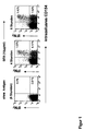
- Fig. 1 the kinetics of intracellular CD154 expression is shown.
- TT tetanus toxoid
- allergen-specific Th cells in human whole blood, regardless of the type of reactively produced cytokines after in vitro stimulation with TT and allergen.
- TT tetanus toxoid
- allergen-specific Th cells in human whole blood, regardless of the type of reactively produced cytokines after in vitro stimulation with TT and allergen.
- TT 10 .mu.g / ml / allergen: 20 .mu.g / ml
- the secretion inhibitor BrefA has been added. Limited analyzes are shown on CD4 + Th cells.
- FIG. 3A shows analyzes of CD154 expression relative to the expression of the activation marker CD69 before isolation. After magnetic enrichment with PE-specific MicroBeads ( Figure 3B), live CD4 + , CD154 + Th cells were further purified with the FACS ( Figure 3C). In Figures 3A and 3B, the analyzes are limited to CD4 + Th cells.
Landscapes
- Health & Medical Sciences (AREA)
- Life Sciences & Earth Sciences (AREA)
- Immunology (AREA)
- Engineering & Computer Science (AREA)
- Cell Biology (AREA)
- Hematology (AREA)
- Biomedical Technology (AREA)
- Chemical & Material Sciences (AREA)
- Urology & Nephrology (AREA)
- Molecular Biology (AREA)
- General Physics & Mathematics (AREA)
- Biochemistry (AREA)
- Biotechnology (AREA)
- Tropical Medicine & Parasitology (AREA)
- Pathology (AREA)
- Food Science & Technology (AREA)
- Medicinal Chemistry (AREA)
- Physics & Mathematics (AREA)
- Analytical Chemistry (AREA)
- Microbiology (AREA)
- General Health & Medical Sciences (AREA)
- Bioinformatics & Cheminformatics (AREA)
- Toxicology (AREA)
- Zoology (AREA)
- Virology (AREA)
- Measuring Or Testing Involving Enzymes Or Micro-Organisms (AREA)
- Micro-Organisms Or Cultivation Processes Thereof (AREA)
- Investigating Or Analysing Biological Materials (AREA)
- Medicines Containing Material From Animals Or Micro-Organisms (AREA)
Description
- Die Erfindung betrifft die Verwendung von CD154 zum in vitro Nachweis und zur Isolierung von T-Lymphozyten, die ein definiertes Antigen erkennen, und ein Verfahren zum Nachweis und zur Isolierung von T-Lymphozyten in Zellsuspensionen oder Körperflüssigkeiten, die ein definiertes Antigen erkennen.
- Das Immunsystem besteht aus Zellen und Molekülen, die im Körper zirkulieren und deren Homeostase und Kooperation durch Zytokine und Wachstumsfaktoren aufeinander abgestimmt werden. Nicht benötigte Zellen werden durch Apoptose eliminiert. Diese Mechanismen werden durch äußere Einflüsse oder genetische Prädispositionen verschiedenster Art beeinflusst. Das Immunsystem spielt eine zentrale Rolle bei der Entstehung vieler Erkrankungen, insbesondere Allergien, Entzündungen und Autoimmunerkrankungen. Es ist verantwortlich für eine verstärkte Immunität nach Impfungen, versagt jedoch bei der Entstehung von Tumorerkrankungen.
- Dendritische Zellen (DC) sind die Wächter des Immunsystems und im Körper dort lokalisiert, wo fremde und möglicherweise gefährliche Stoffe eindringen können. Sie sind professionelle Antigen-präsentierende Zellen, die das Antigen aufnehmen, verdauen, Peptidfragmente des Antigens spezifischen T-Lymphozyten präsentieren und sie dabei aktivieren. Die unreifen Vorläufer der DC wandern kontinuierlich über das Blut in die Lymphorgane, wo sie unter bestimmen Umständen zu reifen DC differenzieren können. Es gibt verschiedene Vorläuferzellen, die zu unterschiedlichen Typen von DC ausreifen können. Diese unterschiedlichen Typen von DC reagieren auch unterschiedlich auf Antigene durch die Expression bestimmter Membranmoleküle und Zytokine. Auf diese Weise kontrollieren sie die Aktivierung der T-Lymphozyten, die dann pro- (Th1) oder antiinflammatorisch (Th2), regulatorisch (Tr) oder tolerant (Ta) werden können. Die Art der Aktivierung der T-Lymphozyten wird durch kostimulatorische Aktivierungssignale von Antigenpräsentierenden Zellen bestimmt. Aktivierungssignale sind Liganden für Rezeptoren der T-Lymphozyten. Diese Liganden befinden sich auf der Oberfläche der APC, sie sind an die extrazelluläre Matrix gebunden oder sie werden von Zellen sezerniert, so die Zytokine. Neben der Antigen-spezifischen Aktivierung durch Signale über den Antigenrezeptor der T-Lymphozyten und kostimulierende Liganden ist jedoch auch eine unspezifische Aktivierung von T-Lymphozyten beschrieben; z. B. durch Zytokine oder durch Lektine.
- Die Gesamtheit der molekularen Veränderungen bei der Aktivierungsreaktion und funktionellen Differenzierung von T-Lymphozyten ist jedoch derzeit nur unvollständig aufgeklärt. So ist auch die Erfassung des Aktivierungs- und Funktionzustandes von T-Lymphozyten, die ein bestimmtes Antigen erkennen, beim gegenwärtigen Stand der Technik nur ungenau möglich.
- Für klinische Anwendungen bei der Dignostik von Krankheiten und in der Forschung ist es essentiell, insbesondere solche spezifischen T-Lymphozyten erfassen zu können, die durch ein bestimmtes Antigen aktiviert werden können oder aktivierbar sind. Weiterhin ist es wünschenswert, solche Antigen-spezifischen T-Lymphozyten aus Zellgemischen insbesondere für zelltherapeutische Zwecke isolieren zu können.
- Im Stand der Technik sind mehrere Verfahren offenbart, mit denen der Aktivierungs- und Funktionszustand von T-Lymphozyten bestimmt werden kann. So offenbart die DE 100 21 834 A1 mRNA-Moleküle zur Verwendung als Indikatoren für den Funktionszustand von T-Lymphozyten, wobei die mRNA-Moleküle bei aktivierten T-Lymphozyten im Vergleich zum Normal- bzw. zum Ruhezustand vermehrt oder vermindert exprimiert sind. Durch Bestimmung der Interaktion zwischen den komplementären, hybridisierten Nucleinsäuresequenzen ist es möglich, Unterschiede in der Expressionsstärke der mRNA-Moleküle zu bestimmen, was Rückschlüsse auf den Aktivierungszustand der T-Lymphozyten zulässt. Weiterhin offenbart diese Druckschrift die Verwendung von Polypeptiden, die durch die Nucleotidsequenzen codiert werden und gegen die Polypeptide gerichtete Antikörper sowie die Verwendung dieser Antikörper zum Nachweis des Aktivierungs- und Funktionszustandes von T-Lymphozyten.
- Die DE 37 37 703 A1 offenbart Antikörper, die gegen ein neutrophil aktivierendes Polypeptid gerichtet sind und als Diagnostikum eingesetzt werden können, um aktivierte Strukturen der zellulären Immunantwort zu detektieren und zu charakterisieren.
- Die DE 42 26 974 C2 offenbart ein Verfahren und eine Vorrichtung zur Aufbereitung und Separation von Zellsuspensionen. Die beschriebene Separationsvorrichtung und das Verfahren zur kontinuierlichen Aufbereitung einer Zellsuspension kann verwendet werden, um unterschiedliche Bestandteile, beispielsweise des menschlichen Blutes zu separieren, wobei Zellen, die im Zusammenhang mit der zellulären Immunantwort stehen, spezifisch aufgereinigt werden können.
- In der US-PS 5,874,302 wird ein Verfahren offenbart, mit dem T-Lymphozyten kultiviert werden und die so gewonnenen T-Lymphozyten zu klinischen Behandlungen von Tumoren eingesetzt werden. Gemäß der US-PS 5,874,302 wird Blut als Ausgangsmaterial für die Herstellung der T-Zellkultur-Suspension verwendet, wobei das entsprechende Startmaterial mit bestimmten Tumorzellen cokultiviert wird.
- Die CA 1,296,622 offenbart ein Verfahren und eine Vorrichtung zum Bestimmen des immunregulatorischen Status von Leukozyten. Die Leukozyten werden mit Standardsubstanzen stimuliert, um den immunregulatorischen Status, beispielsweise mit Hilfe von markierten Antikörpern zu bestimmen. Zunächst wird hierzu eine Probe aus peripheren mononukleären Zellen des Patienten, der ein Immun-beeinflussendes Mittel erhalten hat, vermehrt und anschließend wird in einem Aliquot der vermehrten Probe der Prozentsatz von mononukleären Zellen bestimmt, welche ein Aktivierungs-Antigen in vitro exprimieren. Anschließend wird der Prozentsatz mit einem vorgegebenen Wert des Prozentsatzes als Anzeige des in vitro Effektes verglichen, wodurch der immunregulatorische Zustand des Patienten bzw. des Effektes der Immun-beeinflussenden Therapie auf den immunregulatorischen Zustand bestimmt werden kann. Zu den Unterklassen der zu bestimmenden mononukleären Zellen gehören beispielsweise T-Lymphozyten, cytotoxische T-Lymphozyten und Helfer-T-Lymphozyten.
- In der WO 99/58977 wird ein Verfahren zur direkten Selektion von Antigen-spezifischen T-Lymphozyten beschrieben. Die T-Lymphozyten werden hier anhand der Produktion von Zytokinen mittels Durchfluss-Zytometrie oder der magnetischen Zellsortierung separiert. Es werden hier solche aktivierte T-Lymphozyten selektiert, die nach Stimulation mit Antigen ein bestimmtes oder mehrere bestimmte Zytokine exprimieren.
- In Berner et al, Annals of the Rheumatic Diseases, Bd. 59, 2000, S. 190-195, wird ein Verfahren zum Nachweis von Antigen-aktivierten T-Zellen mittels Bestimmung der CD154 Expression bei Patienten, die an rheumatoider Arthritis leiden, beschrieben. Die erhöhte Expression von CD154 in CD4 T-Lymphozyten wird mit erhöhter CD4 T-Zell-Aktivierung und erhöhter Krankheitsaktivität in Verbindung gebracht.
- Weiterhin ist es mit den bekannten diagnostischen Verfahren nicht möglich, die Aktivierung durch eine virale oder bakterielle Infektion von einer Aktivierung durch eine Autoimmunreaktion oder durch eine Transplantatabstoßung zu unterscheiden, obwohl eine solche Unterscheidung aus diagnostischen und aus klinischen Gesichtspunkten heraus wünschenswert ist. Durch die unzureichende Detektion von aktivierten T-Lymphozyten in einer biologischen Probe ist es auch nicht möglich, diese entsprechenden Zellen gezielt zu separieren, da sie hierfür zunächst eindeutig bestimmt werden müssen.
- Bisher sind keine immundiagnostischen Verfahren beschrieben, die einen schnellen und einfachen Nachweis der Gesamtheit aller für ein Antigen spezifischen T-Lymphozyten und darüber hinaus deren Isolierung ermöglichen. Bei den verschiedensten Erkrankungen ist es von größtem diagnostischem und therapeutischen Interesse, die die Immunantworten zentral und somit auch verschiedenste Immunpathogenesen steuernden T-Lymphozyten direkt Antigenspezifisch nachzuweisen oder sie für zelltherapeutische Ansätze, wie den adoptiven T-Zelltransfer, zu isolieren. Dies betrifft Erkrankungen wie Allergien und Autoimmunerkrankungen, denen fehlgesteuerte Immunreaktionen zu Grunde liegen wie auch chronische Infektionskrankheiten, Tumore oder Leukämien.
- Bisherigen Verfahren liegen zwei unterschiedliche Strategien zugrunde:
- (i) Der "strukturelle" Nachweis Antigen-spezifischer T-Lymphozyten basiert auf der direkten Markierung der von den T-Lymphozyten ausgeprägten T-Zellrezeptoren mittels MHC-Multimeren, die zusammen mit den jeweiligen antigenen Peptiden komplexiert sind (US 5,635,363 / FR 9911133). Die Nachteile solcher Verfahren bestehen darin, dass sie zum einen abhängig von dem der zu untersuchenden Individuen ausgeprägten MHC-Genotyp sind und zum anderen dass die MHC-Moleküle nur mit einem immundominanten Peptidepitop des jeweiligen Antigens komplexiert werden. Jedes Agens muss somit individuell entsprechend des Genotyps und zu verwendenden immundominanten Peptidepitops, das zudem nur bei wenigen Antigenen und auch nur für wenige MHC-Allele bekannt ist, des Antigens hergestellt werden. Zudem konnten bisher die für den Nachweis und die Isolierung MHC-II restringierter Antigen-spezifischer CD4+ Th-Lymphozyten essentiellen MHC-II Multimere aufgrund besonderer struktureller Merkmale des MHC-II Moleküls nicht in für eine Anwendung ausreichender Qualität hergestellt werden.
- (ii) Funktionale Nachweise Antigen-spezifischer T-Lymphozyten beruhen entweder auf der Antigen-reaktiven Proliferation nach Stimulation mit Antigen, die jedoch erst nach 3-5 Tagen detektierbar ist, oder auf Antigen-reaktiver Expression von Aktivierungsmolekülen oder Sekretion von Zytokinen. Während bisher beschriebene Aktivierungsmarker zum Teil auch von speziellen nicht aktivierten T-Lymphozyten ausgeprägt werden und daher nicht spezifisch genug sind, kann anhand der "reaktiven" Expression von Zytokinen nicht die Gesamtheit der spezifischen T-Lymphozyten nachgewiesen werden. Durch die unzureichende Detektion von spezifisch aktivierten T-Lymphozyten in einer biologischen Probe ist somit auch nicht möglich, diese entsprechenden Zellen gezielt zu separieren, da sie hierfür zunächst eindeutig bestimmt werden müssten.
- Die bekannten Verfahren, bei denen spezifische T-Lymphozyten aufgrund ihrer Zytokin Sekretion detektiert und isoliert werden, beispielsweise nach der WO 99/58977, kann die Gesamtheit aller spezifischen T-Lymphozyten nicht bestimmt und isoliert werden, da sich oft jede aktivierte T-Zelle durch individuelle Zytokinprogramme auszeichnet.
- Nachteilig bei den bekannten Verfahren der Isolierung von lebenden T-Lymphozyten anhand der Produktion von Zytokinen ist weiterhin, dass die Zellen zum Zeitpunkt ihrer maximalen Sekretionsleistung aus der Kultur herausgenommen werden, um an ihnen eine für die jeweiligen Zytokine spezifische "Fangmatrix" anzbringen. Danach müssen die Zellen für eine kurze Zeit wieder kultiviert werden. Dies erhöht den technischen Aufwand zur Gewinnung spezifischer T-Lymphozyten beträchtlich. Gerade in den letzten Jahren wurden in verschiedenen Veröffentlichungen immer wieder regulatorische T-Lymphozyten (Tr/Treg) beschrieben, denen sich keine definierten Zytokinmuster zuordnen lassen, d.h. die Zellen sind anhand einer Zytokinsekretion nicht detektierbar oder isolierbar. Zudem ist die Expression bestimmter Zytokine (z. B. IL-10) auch entscheidend von der Art der gewählten in vitro Aktivierung abhängig.
- Der bisherige Nachweis und die Isolierung von spezifischen T-Lymphozyten ist bisher auch deshalb limitiert, da zwar eine Reihe von Biomolekülen bekannt sind, die als Indikatoren bzw. Marker für bestimmte aktivierte T-Lymphozyten herangezogen werden können, jedoch ist es derzeit aufgrund der begrenzten Zahl und Qualität der verfügbaren Aktivierungsmarker nicht möglich, durch Bestimmung eines dieser Marker zu entscheiden, ob die T-Lymphozyten sich in dem gewünschten Zustand befinden. Beispielsweise wird der Aktivierungsmarker CD69 von allen T-Lymphozyten nach Aktivierung exprimiert. Jedoch führen auch jegliche Stresseinwirkungen zur Expression von CD69, so dass durch Bestimmung dieses Markers keine Aussage über die Antigen-spezifischen T-Lymphozyten möglich ist.
- Aufgabe der vorliegenden Erfindung ist es daher Verfahren bereitzustellen, mit denen alle Antigen-spezifischen T-Lymphozyten unabhängig von ihrem funktionalen Potential einfach, kostengünstig und sicher detektiert bzw. isoliert werden können.
- Die vorliegende Erfindung löst dieses technische Problem durch eine Verwendung von CD154 zum Nachweis und/oder zur Isolierung von T-Lymphozyten, die ein bestimmtes Antigen erkennen.
- CD154 ist ein Mitglied der TNF-Gen-Familie und wird unter anderem von verschiedenen Zellen, insbesondere T-Lymphozyten exprimiert. CD154 wird ein bis drei Stunden nach der Aktivierung von den stimulierten T-Lymphozyten herunterreguliert.
- Lymphozyten stellen die spezifischen Träger der Immunantwort dar, wobei zwei große Populationen bekannt sind: Die B- und die T-Lymphozyten. Die T-Lymphozyten besitzen zahlreiche Oberflächenmoleküle, von denen das CD4- und das CD8-Protein von besonderer Bedeutung sind. Die CD4 T-Lymphozyten werden auch als Helfer-T-Zellen (Th-Zellen) bezeichnet, da sie über die Produktion löslicher Botenstoffe bei der Aktivierung anderer Zellen mithelfen.
- Lymphozyten und andere Leukozyten exprimieren viele verschiedene Moleküle auf ihrer Zelloberfläche. Einige erscheinen nur kurzzeitig während bestimmter Phasen der Differenzierung oder nach Aktivierung des Zellen. Andere Moleküle sind konstant und typisch für die jeweilige Zellreihe. Solche konstant exprimierenden Antigene können als Marker für bestimmte Zellpopulationen genutzt werden. Es gibt eine einheitliche Nomenklatur für Zelloberflächenmarker, das CD-System (CD = cluster of differentiation), in welchem die Marker fortlaufend nummeriert sind. So kommen beispielsweise auf NK- und T-Lymphozyten CD 154 wie auch CD 165 oder andere Moleküle vor.
- Überraschend war, dass CD154 verwendet werden kann, um T-Lymphozyten unabhängig von ihrem funktionalen Potential nachzuweisen. Der überraschende Vorteil der Verwendung von CD154 zum Nachweis und zur Separation von T-Lymphozyten liegt darin, dass die T-Lymphozyten unabhängig von ihrer Funktion sicher detektiert bzw. isoliert werden können, d.h. alle antigenspezifischen T-Lymphozyten in einer Probe können bestimmt und separiert werden. Somit wird durch die erfindungsgemäße Verwendung vorteilhafterweise ein neuer praktikabler Kandidat für die Bestimmung von antigenspezifischen T-Lymphozyten bereitgestellt.
- Nach einer besonderen Ausführungsform der Erfindung sind die T-Lymphozyten Th-Lymphozyten, insbesondere CD4+ und/oder CD8+ Th-Lymphozyten. Für die Antigenerkennung benutzen T-Zell-Lymphozytenpopulationen eine T-Zellrezeptor (TZR), der ein heterodimeres Molekül darstellt, das aus verschiedenen Kettenkombinationen besteht. Der weitaus größte Teil aller T-Lymphozyten trägt einen alpha-/beta-T-Zellrezeptor. Diese T-Lymphozyten besitzen weitere Oberflächenmoleküle, insbesondere das CD4- und das CD8-Protein. CD8-Lymphozyten werden von Antigenen stimuliert, die von Klasse-I-Molekülen des MHC präsentiert werden und sind als zytolytische T-Lymphozyten für die Virusabwehr von entscheidender Bedeutung. CD4-T-Lymphozyten erkennen Antigene, welche von MHC-Klasse-II-Molekülen präsentiert werden und haben einen wesentlichen Anteil an der Abwehr von Bakterien, Pilzen, Protozonen und anderen Parasiten.
- In einer weiteren bevorzugten Ausführungsform der Erfindung werden inflamatorische, antiinflamatorische, regulatorische und/oder suppressive T-Lymphozyten nachgewiesen und/oder gewonnen.
- Die Erfindung betrifft auch ein Verfahren zum Nachweis und/oder Isolierung von antigenspezifischen T-Lymphozyten in einer Suspension nach Aktivierung mit einem Antigen, wobei die Suspension mit einem CD40/CD154-System-Inibotor in Kontakt gebracht wird, CD154 intra- oder und/oder extrazellulär bestimmt und die CD154 aufweisenden Zellen nachgewiesen und/oder isoliert werden.
- Es ist überraschend, dass durch das "Inkontakt" bringen der zu untersuchenden Suspension oder Probe mit einem CD40/CD154 System-Inhibitor CD154 intra- und/oder extrazellulär bestimmbar und somit die Zellen, die CD154 aufweisen, nachgewiesen und isoliert oder separiert werden können, wobei es sich bei diesen Zellen insbesondere um die Gesamtheit der Antigen-spezifischen CD4+ Th-Lymphozyten handelt.
- CD40 ist ein 45-50 kDa großes membranständiges Glycoprotein aus der TNF-Rezeptor-Genfamilie (Tumor-Nekrose-Faktor-Rezeptor-Genfamilie). Es wird von verschiedenen hämatopoetischen Zelltypen aber auch von Eptithel- und Endothelzellen, Karzinomen, Fibroblasten und Muskelzellen ausgeprägt. Es kann von CD154 gebunden werden, das dementsprechend Mitglied der TNF-Gen-Familie ist und ebenfalls von vielen Zellen mit sehr unterschiedlichen Funktionen exprimiert werden kann. CD40 wird auf verschiedenen Zellen exprimiert, wie zum Beispiel B-Lymphozyten, dendritische Zellen, Monozyten/Makrophagen, Mastzellen, hämatopoetische Stamm-/Vorläuferzellen (human), Thymusepithelzellen (Maus), Endothelzellen, Fibroblasten (Maus), Mukelzellen (human) und/oder Karzinome (human). CD154 wird beispielsweise auf T-Lymphozyten, aktivierte dendritischen Zellen (human), Monozyten, Mastzellen (human), basophilen/eosinophilen Granulozyten (human), NK-Lymphozyten (Maus), fetale Thymozyten (Maus) und/oder B-Zellen (human) exprimiert.
- Entsprechend der sehr heterogenen Expressionsmuster auf verschiedenen Zelltypen sind zahlreiche CD40/CD154 Wechselwirkungen beschrieben. Das CD40/CD154 System ist ein Beispiel für verschiedene Systeme, die auf Rezeptor-Liganden-Interaktionen basieren und als solche eine große Bedeutung bei Reaktionen des Immunsystems haben. Die CD40/CD154-Interaktionen zwischen T-Lymphozyten und dendritischen Zellen (DC) sind u.a. von Bedeutung bei der Induktion und Regulation der Immunantwort von Bedeutung. Eine spezielle Stellung nehmen CD40/CD154 Interaktionen bei Entwicklung von humoralen Antikörperreaktionen ein. Eine besondere klinische Bedeutung zeigte sich bei dem sogenannten X-chromosomalen Hyper-IgM-Syndrom; einer Immunerkrankung, bei der die Expression eines nichtfunktionellen CD154 zu einer stark verminderten Antikörperbildung und pyogenen Infektionen führt. In anderen Zellen beeinflusst die Signalweiterleitung über CD40/CD154 aber auch beispielsweise die Entstehung von Thromben in vivo und verstärkt die Entwicklung von Ateriosklerose.
- Die Bedeutung des von T-Lymphozyten ausgeprägten CD154 ist somit vor allem während der Interaktion mit B-Lymphozyten für die Entwicklung von humoralen Antikörperantworten essentiell. Anscheinend kann das von T-Lymphozyten ausgeprägte CD154 auch mit von verschiedenen Antigenpräsentierenden Zellen (APC) exprimiertem CD40 in Interaktion treten. Dies wird deutlich, da während der in vitro Stimulation mit spezifischen Antigenen CD154 auf aktivierte T-Lymphozyten sehr schnell herunter reguliert wird. Als für dieses Phänomen verantwortlich angesehen wird die Interaktion von CD154 auf den aktivierten T-Lymphozyten mit von in solchen Systemen auch von APC wie Monozyten stark exprimierten CD40. Die spezifisch von aktivierten T-Lymphozyten ausgeprägten CD154 Moleküle werden danach in den Zellen degradiert. Es steht dann in einem nicht durch das erfindungsgemäße Verfahren beeinflussten System als Marker für spezifische CD4+ Th-Lymphozyten nicht mehr zur Verfügung.
- Durch die Zugabe eines CD40/CD154 System-Inhibitors wird die Wechsel- und Signalwirkung sowie Interaktion von CD40 und CD154 gestört bzw. inhibiert. CD40/CD154 System-Inhibitoren können im Sinne der Erfindung alle Moleküle oder auch physikalische Einwirkungen sein, die in der Lage sind, die Interaktion zwischen CD40 und CD154 zu blockieren oder zu inhibieren. Demgemäß kann das inhibierende Mittel ein Antikörper, der beispielsweise gegen CD40 gerichtet ist, sein, ein Molekül, ein Cäsium- oder ein Lithiumion, welches die Wechselwirkung zwischen CD40 und CD154 untereinander beeinflusst. Es kann selbstverständlich aber auch eine Substanz sein, die die Sekretion oder Endozytose in der Zelle inhibiert, wie z.B. Brefeldin-A (Bref-A). Bref-A ist ein Inhibitor des Goldi-Apparates und der Sekretion einer Vielzahl an Zytokinen. Durch diese Stoffe wird gewährleistet, dass entweder CD40, CD154, die Wechselwirkung zwischen den beiden bzw. das CD40/CD154 System so modifiziert wird, dass CD154 entweder auf der Zelloberfläche nicht mehr herunter reguliert bzw. abgebaut wird, oder, sofern es sich noch innerhalb der Zelle befindet, nicht mehr innerhalb dieser transportiert wird. Durch die Unterbrechung des Transportes innerhalb der Zelle wird der Abbau von CD154 verhindert. Somit wird CD154 innerhalb oder außerhalb der Zelle als außenstehender Rezeptor stabilisiert und kann mit dem Fachmann bekannten Nachweisverfahren detektiert und folgend isoliert werden. Dem Fachmann sind verschiedene Verfahren und Vorrichtungen bekannt, mit denen detektierte Zellen des Immunsystems separiert, aufgereinigt, angereichert bzw. isoliert werden können, z.B. die Durchflusszytometrie oder die magnetische Zellsortierung.
- Durch die Zugabe des CD40/CD154 System-Inhibitors wird der Abbau oder die Degradation von CD154 innerhalb und/oder außerhalb der Zelle unterbunden, so dass die Zellen, die CD154 aufweisen, charakterisiert und folgend anhand dieser Charakterisierung isoliert werden können. Durch das erfindungsgemäße Verfahren wird somit insbesondereder neue erfindungsgemäße Kandidat, CD154, für die Bestimmung von Antigen-spezifischen T-Lymphozyten bereitgestellt.
- In einer bevorzugten Ausführungsform der Erfindung werden als T-Lymphozyten Th-Lymphozyten detektiert und/oder separiert, insbesondere CD4+ und/oder CD8+ Th-Lymphozyten.
- In einer bevorzugten Ausführungsform der Erfindung ist der CD40/CD154 System-Inhibitor ein gegen CD40 gerichteter Antikörper, ein gegen CD154 gerichteter Antikörper, ein Sekretionsinhibitor und/oder ein Endozytoseinhibitor. Dem Fachmann sind verschiedene Sekretionsinhibitoren und Edozytoseinhibitoren bekannt. Der Antikörper kann hierbei ein polyklonaler, monoklonaler bzw. ein spezifisch modifizierter Antikörper oder ein Antikörperfragment, beispielsweise ein Antikörper sein, der mit einem Phagen oder einem Phagenfragment assoziiert ist. Er kann sowohl gegen ein einzelnes Epitop auf CD40 bzw. gegen mehrere Strukturen von CD40 gerichtet sein. Ein Antikörper im Sinne der Erfindung ist ein Molekül, welches CD40 so beeinflusst, dass eine Wechselwirkung mit CD154 vorteilhafterweise nicht mehr möglich ist. Antikörper bedeutet daher im Zusammenhang mit der Erfindung ein Polypeptid, das im Wesentlichen von Immunoglobulin-Genen oder Fragmenten davon codiert wird, das CD40 spezifisch bindet bzw. erkennt. Der in der vorliegenden Erfindung verwendete Begriff Epitop bedeutet eine beliebige Antigen-Determinante auf einem Antigen, an das das Paratop eines Antikörpers bindet. Epitop-Determinaten bestehen insbesondere aus chemisch aktiven Oberflächengruppierungen von Molekülen, wie Aminosäuren oder Zuckerseitenketten und besitzen normalerweise sowohl spezifische Merkmale der dreidimensionalen Struktur als auch spezifische Ladungsmerkmale.
- In einer bevorzugten Ausführungsform der Erfindung werden als der Sekretionsinhibitor und/oder der Endozytosehibotor Brefeldin-A und/oder Monsensin verwendet. Brefeldin A ist ein Metabolit des Pilzes Penicillium brefeldianum und blockiert als carboxyliertes Ionophor den Transport neu synthetisierter Proteine vom endoplasmatischen Retikulum in den Golgi-Apparat und behindert den Austausch zwischen Endosomen und Lysomen während der Kreislauf zwischen Zellmembran und Endosomen vorteilhafterweise ungestört bleibt.
- In einer weiteren, besonders bevorzugten Ausführungsform der Erfindung wird CD154 intrazellulär in fixierten Zellen detektiert. Da CD154 extrazellulär schnell herunterreguliert bzw. abgebaut wird, sobald eine antigen-spezifische Aktivierung von insbesondere Th-Lymphozyten stattgefunden hat, kann CD154 mit Vorteil intrazellulär mit hoher Genauigkeit in-vitro detektiert werden. Insbesondere durch die Zugabe von Brefeldin A wird der Transport des Proteins CD154 an die Zelloberfläche blockiert. Vorteilhafterweise ist hierdurch die Analyse des gesamten CD154, welches sich intrazellulär befindet, auf sehr spezifische Weise möglich, was eine nahezu vollständige Detektion von insbesondere allen reaktiven CD4+ Th-Lymphozyten ermöglicht. Durch die intrazelluläre Bestimmung von CD154 wird ein sehr gutes Signal-Rausch-Verhältnis erreicht, was eine sehr sichere und effiziente Bestimmung der Antigen-spezifischen Th-Lymphozyten ermöglicht.
- In einer weiteren bevorzugten Ausführungsform der Erfindung wird CD154 extrazellulär auf vitalen Zellen detektiert. Um insbesondere Antigen-spezifische CD4+ und/oder CD8+ Th-Lymphozyten im therapeutischen oder klinischen Bereich einzusetzen, ist es erforderlich, die Zellen so zu detektieren, dass sie in ihrer Funktionalität nicht negativ beeinflusst werden, d.h., dass ein Nachweis vorteilhafterweise so gestaltet ist, dass die Zellen lebend und vital detektiert werden können, um sie folgend zur Herstellung eines Arzneimittels verwenden zu können. Es ist beispielsweise möglich, mittels anti-CD40-Antikörper CD40 zu neutralisieren, um den Abbau von CD154 auf der Zelloberfläche zu verhindern, was eine Detektion und Isolierung von Antigen-spezifischen CD4+ und/oder CD8+ Th-Lymphozyten erlaubt.
- Es ist bevorzugt, die Antigen-spezifischen T-Lymphozyten, insbesondere CD4+ und/oder CD8+ aus Frischblut, gefrorenen Zellen, periphere mononukleäre Blutzellen (PBMC) und/oder aus anderen Körperflüssigkeiten zu gewinnen.
- In einer bevorzugten Ausführungsform der Erfindung sind die isolierten und/oder separierten T-Lymphozytenzellen, die keine Cytokine produzieren bzw. die kein definiertes Zytokinmuster aufweisen. Mit Vorteil ist es also möglich, alle antigenspezifischen T-Lymphozyten, d. h. die Gesamtheit dieser Zellpopulation, die gegen ein bestimmtes Antigen gerichtet ist, zu detektieren und/oder zu isolieren, und zwar unabhängig davon, ob diese T-Lymphozyten Cytokine produzieren oder nicht oder ob die T-Lymphozyten sich keinen definiertem Zytokinmuster zuordnen lassen.
- Das erfindungsgemäße Verfahren zur Detektion und zur Isolierung von Antigen-spezifischen T-Lymphozyten, beispielsweise CD4+ Th-Lymphozyten, erlaubt es, insbesondere Antigen-spezifische Th-Lymphozyten für die Zelltherapie gegen verschiedene Krankheiten, wie beispielsweise Krebs oder virale Infektionen einzusetzen. Bisher wurden z.B. Th-Lymphozyten in Form von Zelllinien oder Zellklonen für die adoptive Th-Lymphozytentherapie verwandt. Die Herstellung dieser Zelllinien bzw. Zellklonen ist jedoch sehr aufwendig und benötigt einen langen Zeitraum. Das erfindungsgemäße Verfahren erlaubt es erstmalig, Antigen-spezifische Th-Lymphozyten mittels der Detektion von CD154 in kurzer Zeit in vivo wie in vitro zu detektieren und zu isolieren. Somit ist es erstmals möglich, Th-Lymphozyten aufgrund ihrer Antigenreaktivität unabhängig von ihren Phänotyp zu selektieren.
- Mit Vorteil ist es durch die Verwendung von CD154 als Marker für antigenspezifische T-Lymphozyten möglich, beispielsweise aus Körperflüssigkeiten oder künstlichen Zellsuspensionen gegen ein gewähltes Antigen oder gewählte Antigene spezifische T-Lymphozyten, insbesondere für zelltherapeutische Anwendungen, zu gewinnen. Vorteilhafterweise ist es möglich, spezifische T-Lymphozyten zur Expansion oder mit nachfolgendem oder direktem adoptiven Transfer zu isolieren, wobei diese spezifischen T-Lymphozyten inflamatorische oder antiinflamatorische und insbesondere regulatorische oder supressive T-Lymphozyten darstellen.
- Im folgenden soll die Erfindung anhand von Beispielen veranschaulicht werden, ohne die Erfindung darauf zu beschränken.
- Die Kinetik der intrazellulären CD154 Expression in Antigen-reaktiven Th-Zellen nach in vitro Stimulation von humanen Vollblut mit dem Superantigen Staphylocokkus Enterotoxin B (SEB) wurde bestimmt. Jeweils 1ml Vollblut eines normalen gesunden Spenders wurde für die angegebenen Zeiten mit oder ohne SEB (1µg/ml) bei 37°C kultiviert. Für die letzten 2 Stunden der Kultur wurde der Sekretionsinhibitor BrefA zugegeben. Es sind auf CD4+ Th-Zellen begrenzte Analysen gezeigt. In Fig. 1 ist die Kinetik der intrazellulären CD154 Expression gezeigt.
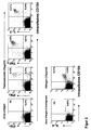
- Fig. 2 zeigt den Nachweis Tetanus-Toxoid-(TT)-spezischer Th-Zellen und Allergen-spezischer Th-Zellen in humanem Vollblut unabhängig von der Art der reaktiv produzierten Zytokine nach in vitro Stimulation mit TT und Allergen. Jeweils 1ml Vollblut gesunder Spender wurde mit oder ohne die angegebenen Antigene (TT: 10µg/ml / Allergen: 20µg/ml) für 6 Stunden bei bei 37°C kultiviert. Für die letzten 4 Stunden wurde der Sekretionsinhibitor BrefA zugegeben. Es sind auf CD4+ Th-Zellen begrenzte Analysen gezeigt.
- Die Isolierung von lebenden Tetanus-Toxoid-(TT)-spezifischen Th-Zellen anhand der reaktiven CD154 Expression aus humanen peripheren mononukleären Zellen nach in vitro Stimulation mit TT ist in Fig. 3 dargestellt. Aus ca. 20ml Volllblut wurden periphere mononukleäre Zellen gewonnen. Diese wurden für 6 Stunden mit 10µg/ml TT in Gegenwart eines monoklonalen anti-CD40 Antikörpers bei 37°C kultiviert. Alle Proben wurden mit Antikörpern gegen CD4-FITC, CD69-APC und CD154-PE markiert. Zum Auschluss von toten Zellen wurden Propidiumjodid verwendet. In 3A sind Analysen der CD154 Expression im Verhältnis zur Expression des Aktivierungsmarkers CD69 vor Isolierung gezeigt. Nach einer magnetischen Anreicherung mit PE-spezifische MicroBeads (3B) wurden lebende CD4+, CD154+ Th-Zellen weiter mit dem FACS aufgereinigt (Figure 3C). In den Figuren 3A und 3B sind die Analysen auf CD4+ Th-Zellen begrenzt.
Claims (10)
- Verwendung von CD154 und eines CD40/CD154 Systeminhibitors, der die Interaktion wischen CD40 und CD154 blockiert oder inhibiert, zum in vitro Nachweis und/oder zur Isolierung von Antigen-spezifischen T-Lymphozyten.
- Verwendung nach Anspruch 1,
dadurch gekennzeichnet, dass
als T-Lymphozyten CD4+ und/oder CD8+ T-Lymphozyten nachgewiesen und/oder isoliert werden. - Verwendung nach Anspruch 1 oder 2,
dadurch gekennzeichnet, dass
inflammatorische, antiinflammatorische, regulatorische und/oder suppressive T-Lymphozyten nachgewiesen und/oder isoliert werden. - Verfahren zum Nachweis und/oder Isolierung von Antigen-spezifischen T-Lymphozyten in einer Suspension nach Aktivierung mit einem Antigen, wobei die Suspension mit einem CD40/CD154 Systeminhibitor, der die Interaktion zwischen CD40 und CD154 blockiert oder inhibiert, in Kontakt gebracht wird, CD154 intra- oder extrazellulär bestimmt und die CD154 aufweisenden Zellen detektiert und/oder separiert werden.
- Verfahren nach Anspruch 4,
dadurch gekennzeichnet, dass
als T-Lymphozyten CD4+ und/oder CD8+ T-Lymphozyten nachgewiesen und/oder isoliert werden. - Verfahren nach einem der Ansprüche 4 oder 5,
dadurch gekennzeichnet, dass
als CD40/CD154 Systeminhibitor ein anti-CD40-Antikörper, ein anti-CD154-Antikörper, ein Sekretionsinhibitor und/oder Endozytoseinhibitor verwendet werden. - Verfahren nach einem der Ansprüche 4 bis 6,
dadurch gekennzeichnet, dass
als Sekretionsinhibitor und/oder Endozytoseinhibitor Brefeldin A und/oder Monsensin verwendet werden. - Verfahren nach einem der Ansprüche 4 bis 7,
dadurch gekennzeichnet, dass
CD154 intrazellulär in fixierten Zellen detektiert wird. - Verfahren nach einem der Ansprüche 4 bis 8,
dadurch gekennzeichnet, dass
CD154 extrazellulär auf vitalen Zellen detektiert wird. - Verfahren nach einem der Ansprüche 4 bis 9,
dadurch gekennzeichnet, dass
die T-Lymphozyten isoliert und/oder separiert werden, die kein definiertes Zytokinmuster aufweisen.
Priority Applications (7)
| Application Number | Priority Date | Filing Date | Title |
|---|---|---|---|
| DE50207025T DE50207025D1 (de) | 2002-08-23 | 2002-08-23 | Verfahren zum Nachweis und zur Isolierung von T-Lymphozyten, die ein definiertes Antigen erkennen |
| ES02090300T ES2267937T3 (es) | 2002-08-23 | 2002-08-23 | Procedimiento de deteccion y aislamiento de linfocitos t que reconocen un antigeno definido. |
| EP02090300A EP1420253B1 (de) | 2002-08-23 | 2002-08-23 | Verfahren zum Nachweis und zur Isolierung von T-Lymphozyten, die ein definiertes Antigen erkennen |
| AT02090300T ATE328279T1 (de) | 2002-08-23 | 2002-08-23 | Verfahren zum nachweis und zur isolierung von t- lymphozyten, die ein definiertes antigen erkennen |
| US10/525,238 US7659084B2 (en) | 2002-08-23 | 2003-08-22 | Methods for detecting and isolating antigen-specific T lymphocytes with CD40/C154 inhibitors |
| PCT/EP2003/009354 WO2004027428A1 (de) | 2002-08-23 | 2003-08-22 | Verfahren zum nachweis und zur isolierung von t-lymphozyten, die ein definiertes antigen erkennen |
| AU2003270096A AU2003270096A1 (en) | 2002-08-23 | 2003-08-22 | Method for detecting and isolating t lymphocytes that recognize a defined antigen |
Applications Claiming Priority (1)
| Application Number | Priority Date | Filing Date | Title |
|---|---|---|---|
| EP02090300A EP1420253B1 (de) | 2002-08-23 | 2002-08-23 | Verfahren zum Nachweis und zur Isolierung von T-Lymphozyten, die ein definiertes Antigen erkennen |
Publications (2)
| Publication Number | Publication Date |
|---|---|
| EP1420253A1 EP1420253A1 (de) | 2004-05-19 |
| EP1420253B1 true EP1420253B1 (de) | 2006-05-31 |
Family
ID=32011028
Family Applications (1)
| Application Number | Title | Priority Date | Filing Date |
|---|---|---|---|
| EP02090300A Expired - Lifetime EP1420253B1 (de) | 2002-08-23 | 2002-08-23 | Verfahren zum Nachweis und zur Isolierung von T-Lymphozyten, die ein definiertes Antigen erkennen |
Country Status (7)
| Country | Link |
|---|---|
| US (1) | US7659084B2 (de) |
| EP (1) | EP1420253B1 (de) |
| AT (1) | ATE328279T1 (de) |
| AU (1) | AU2003270096A1 (de) |
| DE (1) | DE50207025D1 (de) |
| ES (1) | ES2267937T3 (de) |
| WO (1) | WO2004027428A1 (de) |
Cited By (1)
| Publication number | Priority date | Publication date | Assignee | Title |
|---|---|---|---|---|
| EP2597463A1 (de) | 2011-11-22 | 2013-05-29 | Medizinische Hochschule Hannover | Verfahren zur Anreicherung und Isolierung von regulierenden T-Zellen und Verwendung davon |
Families Citing this family (11)
| Publication number | Priority date | Publication date | Assignee | Title |
|---|---|---|---|---|
| DE10112851C1 (de) | 2001-03-16 | 2002-10-10 | Gsf Forschungszentrum Umwelt | Semi-allogene Antitumor-Vakzine mit HLA-haplo-identischen Antigen-präsentierenden Zellen |
| WO2005045009A1 (en) * | 2003-07-23 | 2005-05-19 | President And Fellows Of Harvard College | Methods for isolating t cells and uses thereof |
| CN1291231C (zh) * | 2003-12-08 | 2006-12-20 | 胡军 | 活化淋巴细胞特异性的检测方法 |
| DK1910521T3 (da) * | 2005-08-05 | 2011-02-07 | Helmholtz Zentrum Muenchen | Generering af allo-begrænsede, antigenspecifikke T-celler |
| CN102272153B (zh) | 2008-11-24 | 2015-04-15 | 德国慕尼黑亥姆霍兹研究中心健康和环境有限公司 | 高亲和力t细胞受体及其应用 |
| US8790916B2 (en) | 2009-05-14 | 2014-07-29 | Genestream, Inc. | Microfluidic method and system for isolating particles from biological fluid |
| DE102009040716B4 (de) | 2009-09-10 | 2011-07-14 | Miltenyi Biotec GmbH, 51429 | Verwendung von CD154 zur Identifizierung und Abtrennung von nicht-regulatorischen T-Zellen aus einem Gemisch mit regulatorischen T-Zellen |
| EP2518504A1 (de) | 2011-04-29 | 2012-10-31 | Miltenyi Biotec GmbH | Verfahren zur quantitativen und qualitativen Charakterisierung von antigenspezifischen T-Zellen, die ein spezifischen Antigen erkennen |
| EP2985342A1 (de) | 2014-08-15 | 2016-02-17 | Miltenyi Biotec GmbH | Immunogene Antigene aus Aspergillus fumigatus |
| WO2019028028A1 (en) * | 2017-08-01 | 2019-02-07 | Benaroya Research Institute At Virgina Mason | METHODS OF IDENTIFICATION AND SEPARATION OF PRO-ALLERGIC SPECIFIC T LYMPHOCYTES |
| WO2022195096A1 (en) | 2021-03-19 | 2022-09-22 | Charité - Universitätsmedizin Berlin | Peptide and method for direct analysis of sars-cov-2 immune responses |
Family Cites Families (2)
| Publication number | Priority date | Publication date | Assignee | Title |
|---|---|---|---|---|
| JP4601166B2 (ja) * | 1998-05-11 | 2010-12-22 | ミルテニィ バイオテック ゲーエムベーハー | 抗原特異的t細胞の直接的選択方法 |
| WO2001094586A2 (en) * | 2000-06-06 | 2001-12-13 | Idec Pharmaceuticals Corporation | Non-agonistic antibodies to human gp39, compositions containing, and therapeutic use thereof |
-
2002
- 2002-08-23 DE DE50207025T patent/DE50207025D1/de not_active Expired - Lifetime
- 2002-08-23 AT AT02090300T patent/ATE328279T1/de not_active IP Right Cessation
- 2002-08-23 ES ES02090300T patent/ES2267937T3/es not_active Expired - Lifetime
- 2002-08-23 EP EP02090300A patent/EP1420253B1/de not_active Expired - Lifetime
-
2003
- 2003-08-22 US US10/525,238 patent/US7659084B2/en not_active Expired - Lifetime
- 2003-08-22 WO PCT/EP2003/009354 patent/WO2004027428A1/de not_active Ceased
- 2003-08-22 AU AU2003270096A patent/AU2003270096A1/en not_active Abandoned
Cited By (2)
| Publication number | Priority date | Publication date | Assignee | Title |
|---|---|---|---|---|
| EP2597463A1 (de) | 2011-11-22 | 2013-05-29 | Medizinische Hochschule Hannover | Verfahren zur Anreicherung und Isolierung von regulierenden T-Zellen und Verwendung davon |
| WO2013076181A1 (en) | 2011-11-22 | 2013-05-30 | Medizinische Hochschule Hannover | Methods of enrichment and isolation of regulatory t-cells and use of the same |
Also Published As
| Publication number | Publication date |
|---|---|
| DE50207025D1 (de) | 2006-07-06 |
| EP1420253A1 (de) | 2004-05-19 |
| ES2267937T3 (es) | 2007-03-16 |
| AU2003270096A1 (en) | 2004-04-08 |
| WO2004027428A8 (de) | 2004-08-19 |
| WO2004027428A1 (de) | 2004-04-01 |
| US20060121027A1 (en) | 2006-06-08 |
| US7659084B2 (en) | 2010-02-09 |
| ATE328279T1 (de) | 2006-06-15 |
Similar Documents
| Publication | Publication Date | Title |
|---|---|---|
| Haak et al. | IL-17A and IL-17F do not contribute vitally to autoimmune neuro-inflammation in mice | |
| Beaurain et al. | In vivo T cell preactivation in chronic uremic hemodialyzed and non-hemodialyzed patients | |
| DE68927277T2 (de) | CD40 kodierende DNA | |
| EP2121741B1 (de) | Nachweis von individuellen t-zell-reaktionsmustern gegen tumor-assoziierte antigene (taa) in tumorpatienten als basis für die individuelle therapeutische vakzinierung von patienten | |
| EP1257290B2 (de) | Verfahren zur antigen-spezifischen stimulation von t-lymphozyten mit synthetischen peptidbibliotheken | |
| DE102004026135A1 (de) | An MHC-Moleküle bindende Tumor-assoziierte Peptide | |
| DE10225144A1 (de) | An MHC-Moleküle bindende Tumor-assoziierte Peptide | |
| Souto et al. | Chemokine production and leukocyte recruitment to the lungs of Paracoccidioides brasiliensis-infected mice is modulated by interferon-γ | |
| US20230357689A1 (en) | Intracellular delivery using microfluidics-assisted cell screening (macs) | |
| EP1420253A1 (de) | Verfahren zum Nachweis und zur Isolierung von T-Lymphozyten, die ein definiertes Antigen erkennen | |
| DE4244715C2 (de) | Verfahren zur Herstellung individuum-spezifischer, gegen Tumorantigene gerichtete monoklonale Antikörper | |
| DE60114018T2 (de) | Von zellen präsentierte peptide | |
| Killestein et al. | Intracellular cytokine profile in T-cell subsets of multiple sclerosis patients: different features in primary progressive disease | |
| KR101732532B1 (ko) | 단구 또는 nk 세포를 입수하는 방법 | |
| Daltro et al. | CD4+ T cell profile and activation response in sickle cell disease patients with osteonecrosis | |
| DE69505376T2 (de) | Alpha b crystallin zur verwendung in diagnose und therapie von autoimmunkrankheiten, besonders multipler skerose | |
| US20110177036A1 (en) | Whole blood cultures comprising stimulated immune cells, and use thereof as medicaments | |
| Friedel et al. | Mast cell-derived interleukin-4 mediates activation of dendritic cell during toll-like receptor 2-mediated inflammation | |
| JP2012518407A (ja) | 単離された単球集団及び関連した治療応用 | |
| Kubota et al. | Innate IFN-γ-producing cells in the spleen of mice early after Listeria monocytogenes infection: importance of microenvironment of the cells involved in the production of innate IFN-γ | |
| DE102015106731A1 (de) | Peptide für die Krebsimmuntherapie | |
| DE10160921A1 (de) | Verfahren zum Nachweis von Allergien und Verwendung eines Antikörpers zum Nachweis von Allergien | |
| DE10392933B4 (de) | Verwendung von CD152+ T Zellen zur Behandlung von Krankheiten des rheumatischen Formenkreises | |
| DE10238922A1 (de) | Immunmarker zur Diagnostik und Therapie im Zusammenhang mit Transplantat-Reaktionen | |
| WO2024164927A1 (en) | Use of fetal sheep brain extracts for managing neurodegenerative diseases |
Legal Events
| Date | Code | Title | Description |
|---|---|---|---|
| PUAI | Public reference made under article 153(3) epc to a published international application that has entered the european phase |
Free format text: ORIGINAL CODE: 0009012 |
|
| AK | Designated contracting states |
Kind code of ref document: A1 Designated state(s): AT BE BG CH CY CZ DE DK EE ES FI FR GB GR IE IT LI LU MC NL PT SE SK TR |
|
| AX | Request for extension of the european patent |
Extension state: AL LT LV MK RO SI |
|
| 17P | Request for examination filed |
Effective date: 20041126 |
|
| AKX | Designation fees paid |
Designated state(s): AT BE BG CH CY CZ DE DK EE ES FI FR GB GR IE IT LI LU MC NL PT SE SK TR |
|
| 17Q | First examination report despatched |
Effective date: 20050222 |
|
| GRAP | Despatch of communication of intention to grant a patent |
Free format text: ORIGINAL CODE: EPIDOSNIGR1 |
|
| GRAS | Grant fee paid |
Free format text: ORIGINAL CODE: EPIDOSNIGR3 |
|
| GRAA | (expected) grant |
Free format text: ORIGINAL CODE: 0009210 |
|
| AK | Designated contracting states |
Kind code of ref document: B1 Designated state(s): AT BE BG CH CY CZ DE DK EE ES FI FR GB GR IE IT LI LU MC NL PT SE SK TR |
|
| PG25 | Lapsed in a contracting state [announced via postgrant information from national office to epo] |
Ref country code: FI Free format text: LAPSE BECAUSE OF FAILURE TO SUBMIT A TRANSLATION OF THE DESCRIPTION OR TO PAY THE FEE WITHIN THE PRESCRIBED TIME-LIMIT Effective date: 20060531 Ref country code: SK Free format text: LAPSE BECAUSE OF FAILURE TO SUBMIT A TRANSLATION OF THE DESCRIPTION OR TO PAY THE FEE WITHIN THE PRESCRIBED TIME-LIMIT Effective date: 20060531 Ref country code: CZ Free format text: LAPSE BECAUSE OF FAILURE TO SUBMIT A TRANSLATION OF THE DESCRIPTION OR TO PAY THE FEE WITHIN THE PRESCRIBED TIME-LIMIT Effective date: 20060531 |
|
| REG | Reference to a national code |
Ref country code: GB Ref legal event code: FG4D Free format text: NOT ENGLISH Ref country code: CH Ref legal event code: EP |
|
| REG | Reference to a national code |
Ref country code: IE Ref legal event code: FG4D Free format text: LANGUAGE OF EP DOCUMENT: GERMAN |
|
| REF | Corresponds to: |
Ref document number: 50207025 Country of ref document: DE Date of ref document: 20060706 Kind code of ref document: P |
|
| PG25 | Lapsed in a contracting state [announced via postgrant information from national office to epo] |
Ref country code: SE Free format text: LAPSE BECAUSE OF FAILURE TO SUBMIT A TRANSLATION OF THE DESCRIPTION OR TO PAY THE FEE WITHIN THE PRESCRIBED TIME-LIMIT Effective date: 20060831 Ref country code: DK Free format text: LAPSE BECAUSE OF FAILURE TO SUBMIT A TRANSLATION OF THE DESCRIPTION OR TO PAY THE FEE WITHIN THE PRESCRIBED TIME-LIMIT Effective date: 20060831 Ref country code: MC Free format text: LAPSE BECAUSE OF NON-PAYMENT OF DUE FEES Effective date: 20060831 Ref country code: BE Free format text: LAPSE BECAUSE OF NON-PAYMENT OF DUE FEES Effective date: 20060831 |
|
| GBT | Gb: translation of ep patent filed (gb section 77(6)(a)/1977) |
Effective date: 20060925 |
|
| PG25 | Lapsed in a contracting state [announced via postgrant information from national office to epo] |
Ref country code: PT Free format text: LAPSE BECAUSE OF FAILURE TO SUBMIT A TRANSLATION OF THE DESCRIPTION OR TO PAY THE FEE WITHIN THE PRESCRIBED TIME-LIMIT Effective date: 20061031 |
|
| ET | Fr: translation filed | ||
| REG | Reference to a national code |
Ref country code: ES Ref legal event code: FG2A Ref document number: 2267937 Country of ref document: ES Kind code of ref document: T3 |
|
| PLBE | No opposition filed within time limit |
Free format text: ORIGINAL CODE: 0009261 |
|
| STAA | Information on the status of an ep patent application or granted ep patent |
Free format text: STATUS: NO OPPOSITION FILED WITHIN TIME LIMIT |
|
| 26N | No opposition filed |
Effective date: 20070301 |
|
| PG25 | Lapsed in a contracting state [announced via postgrant information from national office to epo] |
Ref country code: AT Free format text: LAPSE BECAUSE OF NON-PAYMENT OF DUE FEES Effective date: 20060823 |
|
| BERE | Be: lapsed |
Owner name: DEUTSCHES RHEUMA-FORSCHUNGSZENTRUM BERLIN Effective date: 20060831 |
|
| PG25 | Lapsed in a contracting state [announced via postgrant information from national office to epo] |
Ref country code: GR Free format text: LAPSE BECAUSE OF FAILURE TO SUBMIT A TRANSLATION OF THE DESCRIPTION OR TO PAY THE FEE WITHIN THE PRESCRIBED TIME-LIMIT Effective date: 20060901 |
|
| PG25 | Lapsed in a contracting state [announced via postgrant information from national office to epo] |
Ref country code: BG Free format text: LAPSE BECAUSE OF FAILURE TO SUBMIT A TRANSLATION OF THE DESCRIPTION OR TO PAY THE FEE WITHIN THE PRESCRIBED TIME-LIMIT Effective date: 20060831 Ref country code: EE Free format text: LAPSE BECAUSE OF FAILURE TO SUBMIT A TRANSLATION OF THE DESCRIPTION OR TO PAY THE FEE WITHIN THE PRESCRIBED TIME-LIMIT Effective date: 20060531 |
|
| PG25 | Lapsed in a contracting state [announced via postgrant information from national office to epo] |
Ref country code: LU Free format text: LAPSE BECAUSE OF NON-PAYMENT OF DUE FEES Effective date: 20060823 Ref country code: TR Free format text: LAPSE BECAUSE OF FAILURE TO SUBMIT A TRANSLATION OF THE DESCRIPTION OR TO PAY THE FEE WITHIN THE PRESCRIBED TIME-LIMIT Effective date: 20060531 |
|
| PG25 | Lapsed in a contracting state [announced via postgrant information from national office to epo] |
Ref country code: CY Free format text: LAPSE BECAUSE OF FAILURE TO SUBMIT A TRANSLATION OF THE DESCRIPTION OR TO PAY THE FEE WITHIN THE PRESCRIBED TIME-LIMIT Effective date: 20060531 |
|
| PGFP | Annual fee paid to national office [announced via postgrant information from national office to epo] |
Ref country code: IE Payment date: 20100812 Year of fee payment: 9 |
|
| REG | Reference to a national code |
Ref country code: IE Ref legal event code: MM4A |
|
| PG25 | Lapsed in a contracting state [announced via postgrant information from national office to epo] |
Ref country code: IE Free format text: LAPSE BECAUSE OF NON-PAYMENT OF DUE FEES Effective date: 20110823 |
|
| REG | Reference to a national code |
Ref country code: FR Ref legal event code: PLFP Year of fee payment: 15 |
|
| REG | Reference to a national code |
Ref country code: FR Ref legal event code: PLFP Year of fee payment: 16 |
|
| REG | Reference to a national code |
Ref country code: FR Ref legal event code: PLFP Year of fee payment: 17 |
|
| PGFP | Annual fee paid to national office [announced via postgrant information from national office to epo] |
Ref country code: NL Payment date: 20190814 Year of fee payment: 18 |
|
| PGFP | Annual fee paid to national office [announced via postgrant information from national office to epo] |
Ref country code: ES Payment date: 20190902 Year of fee payment: 18 Ref country code: IT Payment date: 20190821 Year of fee payment: 18 |
|
| PGFP | Annual fee paid to national office [announced via postgrant information from national office to epo] |
Ref country code: CH Payment date: 20190819 Year of fee payment: 18 |
|
| REG | Reference to a national code |
Ref country code: CH Ref legal event code: PL |
|
| REG | Reference to a national code |
Ref country code: NL Ref legal event code: MM Effective date: 20200901 |
|
| PG25 | Lapsed in a contracting state [announced via postgrant information from national office to epo] |
Ref country code: CH Free format text: LAPSE BECAUSE OF NON-PAYMENT OF DUE FEES Effective date: 20200831 Ref country code: LI Free format text: LAPSE BECAUSE OF NON-PAYMENT OF DUE FEES Effective date: 20200831 |
|
| PG25 | Lapsed in a contracting state [announced via postgrant information from national office to epo] |
Ref country code: NL Free format text: LAPSE BECAUSE OF NON-PAYMENT OF DUE FEES Effective date: 20200901 |
|
| PGFP | Annual fee paid to national office [announced via postgrant information from national office to epo] |
Ref country code: FR Payment date: 20210714 Year of fee payment: 20 |
|
| PGFP | Annual fee paid to national office [announced via postgrant information from national office to epo] |
Ref country code: DE Payment date: 20210713 Year of fee payment: 20 Ref country code: GB Payment date: 20210714 Year of fee payment: 20 |
|
| REG | Reference to a national code |
Ref country code: ES Ref legal event code: FD2A Effective date: 20211228 |
|
| PG25 | Lapsed in a contracting state [announced via postgrant information from national office to epo] |
Ref country code: ES Free format text: LAPSE BECAUSE OF NON-PAYMENT OF DUE FEES Effective date: 20200824 |
|
| PG25 | Lapsed in a contracting state [announced via postgrant information from national office to epo] |
Ref country code: IT Free format text: LAPSE BECAUSE OF NON-PAYMENT OF DUE FEES Effective date: 20200823 |
|
| REG | Reference to a national code |
Ref country code: DE Ref legal event code: R071 Ref document number: 50207025 Country of ref document: DE |
|
| REG | Reference to a national code |
Ref country code: GB Ref legal event code: PE20 Expiry date: 20220822 |
|
| PG25 | Lapsed in a contracting state [announced via postgrant information from national office to epo] |
Ref country code: GB Free format text: LAPSE BECAUSE OF EXPIRATION OF PROTECTION Effective date: 20220822 |