EP1389121B1 - Method and compositions for treating mammalian nerve tissue injuries - Google Patents
Method and compositions for treating mammalian nerve tissue injuries Download PDFInfo
- Publication number
- EP1389121B1 EP1389121B1 EP02741682A EP02741682A EP1389121B1 EP 1389121 B1 EP1389121 B1 EP 1389121B1 EP 02741682 A EP02741682 A EP 02741682A EP 02741682 A EP02741682 A EP 02741682A EP 1389121 B1 EP1389121 B1 EP 1389121B1
- Authority
- EP
- European Patent Office
- Prior art keywords
- glycol
- injury
- spinal cord
- peg
- nerve
- Prior art date
- Legal status (The legal status is an assumption and is not a legal conclusion. Google has not performed a legal analysis and makes no representation as to the accuracy of the status listed.)
- Expired - Lifetime
Links
Images


Classifications
-
- A—HUMAN NECESSITIES
- A61—MEDICAL OR VETERINARY SCIENCE; HYGIENE
- A61K—PREPARATIONS FOR MEDICAL, DENTAL OR TOILETRY PURPOSES
- A61K31/00—Medicinal preparations containing organic active ingredients
- A61K31/74—Synthetic polymeric materials
- A61K31/765—Polymers containing oxygen
-
- A—HUMAN NECESSITIES
- A61—MEDICAL OR VETERINARY SCIENCE; HYGIENE
- A61K—PREPARATIONS FOR MEDICAL, DENTAL OR TOILETRY PURPOSES
- A61K31/00—Medicinal preparations containing organic active ingredients
-
- A—HUMAN NECESSITIES
- A61—MEDICAL OR VETERINARY SCIENCE; HYGIENE
- A61K—PREPARATIONS FOR MEDICAL, DENTAL OR TOILETRY PURPOSES
- A61K31/00—Medicinal preparations containing organic active ingredients
- A61K31/74—Synthetic polymeric materials
-
- A—HUMAN NECESSITIES
- A61—MEDICAL OR VETERINARY SCIENCE; HYGIENE
- A61K—PREPARATIONS FOR MEDICAL, DENTAL OR TOILETRY PURPOSES
- A61K31/00—Medicinal preparations containing organic active ingredients
- A61K31/74—Synthetic polymeric materials
- A61K31/765—Polymers containing oxygen
- A61K31/77—Polymers containing oxygen of oxiranes
-
- A—HUMAN NECESSITIES
- A61—MEDICAL OR VETERINARY SCIENCE; HYGIENE
- A61P—SPECIFIC THERAPEUTIC ACTIVITY OF CHEMICAL COMPOUNDS OR MEDICINAL PREPARATIONS
- A61P25/00—Drugs for disorders of the nervous system
-
- A—HUMAN NECESSITIES
- A61—MEDICAL OR VETERINARY SCIENCE; HYGIENE
- A61P—SPECIFIC THERAPEUTIC ACTIVITY OF CHEMICAL COMPOUNDS OR MEDICINAL PREPARATIONS
- A61P25/00—Drugs for disorders of the nervous system
- A61P25/02—Drugs for disorders of the nervous system for peripheral neuropathies
-
- A—HUMAN NECESSITIES
- A61—MEDICAL OR VETERINARY SCIENCE; HYGIENE
- A61P—SPECIFIC THERAPEUTIC ACTIVITY OF CHEMICAL COMPOUNDS OR MEDICINAL PREPARATIONS
- A61P35/00—Antineoplastic agents
-
- A—HUMAN NECESSITIES
- A61—MEDICAL OR VETERINARY SCIENCE; HYGIENE
- A61P—SPECIFIC THERAPEUTIC ACTIVITY OF CHEMICAL COMPOUNDS OR MEDICINAL PREPARATIONS
- A61P43/00—Drugs for specific purposes, not provided for in groups A61P1/00-A61P41/00
Definitions
- the present invention relates generally to methods for treating injured mammalian nerve tissue including but not limited to a spinal cord. Specifically, the invention relates to methods for treating injured nerve tissue through an in vivo application of a biomembrane fusion agent. Pharmaceutical compositions for treating an injured spinal cord are also described.
- Traumatic injury to the spinal axons (Waxman, S.G., Kocsis, J.D., Stys, P.K., Eds.): The Axon, New York: Oxford UP, pp 480-503 ; Maxwell, W.L. (1996): Histopathological changes at central nodes of ravier after stretch-injury, Microscopy Research and Technique, 34:522-535 ; Maxwell, W.L., Watt, C., Graham, D.I., Gennarelli, T.A.
- Traumatic injury to the spinal axons (Waxman, S.G., Kocsis, J.D., Stys, P.K., Eds.): The Axon, New York: Oxford UP pp 480-503 ; Young, W. (1993): Secondary injury mechanisms in acute spinal cord injury, J. Emerg. Med, 11:13-22 .] subsequently enlarge the lesion leading to the typical clinical picture of a cavitated contused spinal cord, and intractable behavioral loss.
- transection of the axon leads to the irreversible loss of the distal nerve process segment by Wallerian degeneration, while the proximal segment may survive.
- function may be restored by the endogenous regeneration of proximal segments down fasciculation pathways provided by both connective tissue and Schwann cell "tubes" which may persist for variable amounts of time post injury ( Bisby, M.A. (1995): Regeneration of peripheral nervous system axons (Waxman, S.G., Kocsis, J.D., Stys, P.K., Eds.): The Axon Book, New York, The Oxford University Press, pp 553-578 ].
- the level of the injury is critical to clinical fascicular repair however, as the rate of regeneration (about 1 mm/day) may not be sufficient to avoid loss of target tissues dependent on its innervation (such as motor units in striated muscle).
- target tissues dependent on its innervation (such as motor units in striated muscle).
- distal segments of nerve fibers do not regenerate, and their loss produces nonfunctional "target” cells, which often require innervation to maintain their integrity.
- One ultimate strategy to enhance recovery from CNS injury is to induce or facilitate regeneration of white matter by various means.
- polyethylene glycol PEG
- PEG polyethylene glycol
- This hydrophilic macromolecule has been exploited to form multicellular conjugates for the purpose of exchanging genetic material, hybridoma formation, or as a model for endogeneous vesicle fusion
- the present invention is directed to pharmaceutical compositions for use in the treating of injured mammalian nerve tissue of the spinal cord or central nervous system, wherein the conduction of action potentials through the injured nerve tissue is restored or increased.
- the invention is more particularly directed to a pharmaceutical composition comprising an effective amount of a biomembrane fusion agent as defined below, wherein said composition is administered to the patient so that the biomembrane fusion agent is delivered via the patient's vascular system to the site of the injured nerve tissue, particularly nerve tissue of the spinal cord.
- the biomembrane fusion agent is a polyalkylene glycol from the group consisting of polymethylene glycol, polyethylene glycol, polypropylene glycol, polybutylene glycol, polypentylene glycol, polyhexylene glycol, polyheptylene glycol, polyoctylene glycol, polynonylene glycol, and polydecylene glycol.
- the present invention is directed to a pharmaceutical composition for use in treating an injury to nerve tissue of the spinal cord or central nervous system of a mammalian patient, wherein the conduction of action potentials through the injured nerve tissue is restored or increased, the composition comprising an effective amount of a polyalkylene glycol selected from the group consisting of polymethylene glycol, polyethylene glycol, polypropylene glycol, polybutylene glycol, polypentylene glycol, polyhexylene glycol, polyheptylene glycol, polyoctylene glycol, polynonylene glycol, and polydecylene glycol, wherein said composition is administered to the patient so that the polyalkylene glycol is delivered via the patient's vascular system to the site of the injured nerve tissue; and optionally a pharmaceutical carrier.
- a polyalkylene glycol selected from the group consisting of polymethylene glycol, polyethylene glycol, polypropylene glycol, polybutylene glycol, polypentylene glycol, polyhexylene glyco
- the biomembrane fusion agent may be administered to the patient parenterally.
- the biomembrane fusion agent is of such an amount that its delivery to the site of the injury through the blood supply after injection of the biomembrane fusion agent into the patient is effective to repair injured nerve fibers.
- the injection may be an intravascular, intramuscular, subcutaneous, or intraperitoneal injection of an effective quantity of the biomembrane fusion agent so that an effective amount is delivered to the site of the nerve tissue injury.
- biomembrane fusion agents taking the form of a hydrophilic polymer such as a polyethylene glycol/polypropylene glycol block copolymer such as ethylene oxide-propylene oxide-ethylene oxide (EPAN), or another hydrophilic biocompatible surfactant such as dextrans.
- a hydrophilic polymer such as a polyethylene glycol/polypropylene glycol block copolymer such as ethylene oxide-propylene oxide-ethylene oxide (EPAN), or another hydrophilic biocompatible surfactant such as dextrans.
- Nonionic surfactants are disclosed and may take the form of an amphipathic polymer such as a poloxamine.
- the biomembrane fusion agent is polyethylene glycol (PEG) (H(OCH 2 CH 2 ) n OH), where n preferably ranges from 4 to about 570 or more, more preferably about 30 to about 100.
- PEG is used as a solvent for many compounds used in medicine.
- PEG is used as a carrier for contrast media used in radiology, and a solvent for hemopoetic factors infused into hemophilic patients.
- poloxamers are also disclosed.
- Some of these triblock polymers consist of PEG polymers with a propylene glycol core.
- the sizes of the individual polymeric chains are not critical to the action of the poloxamer, and the poloxamer can also be injected into the blood stream or applied topically in the same manner as PEG.
- Polyxamers are also amphipathic polymers to a greater or lesser extent depending on the relative numbers of ethylene glycol and propylene glycol groups.
- a biomembrane fusion agent more particularly, PEG
- PEG specifically targets the hemorrhagic injury in spinal cord following any means of introducing it to the blood supply (for example, parenterally such as intravenous, subcutaneous, or intraperitoneal injection, transdermally, orally, through buccal administration or via another route of administration).
- parenterally such as intravenous, subcutaneous, or intraperitoneal injection, transdermally, orally, through buccal administration or via another route of administration.
- PEG appears to more uniformly bathe the injury site when delivered by the blood supply than when it is applied to the injury directly.
- a single dose of a biomembrane fusion agent such as PEG in aqueous solution administered beneath the back skin (subcutaneous injection) will reverse many functional deficits in severe or traumatic spinal cord injuries in guinea pigs when the dose is administered up to six (6) to eight (8) hours post injury.
- the PEG migrates to and selectively attaches to the site of a mammalian nerve tissue injury and functions there as a biomembrane fusion agent.
- tests show that the application or administration of a biomembrane fusion agent such as PEG to severe spinal cord crush/contusion injuries in situ produces functional recovery of an identified spinal cord mediated behavior in test mammals as well as a rapid recovery of recorded nerve impulses ascending the spinal cord through the original lesion. These physiological and behavioral recoveries following severe spinal cord injury in the test mammals are not temporary but rather stable, even improving with the passage of time. Moreover, the application of a biomembrane fusion agent such as PEG can be delayed for at least 8 hours after spinal cord injury without a loss in its effectiveness.
- the present invention contemplates a composition for use in treating injured mammalian, preferably human, nerve tissue of the spinal cord or central nervous system, wherein the conduction of action potentials through the injured nerve tissue is restored or increased, said composition comprising an effective amount of a polyalkylene glycol (or oxide) selected from the group consisting of polymethylene glycol, polyethylene glycol, polypropylene glycol, polybutylene glycol, polypentylene glycol, polyhexylene glycol, polylieptylene glycol, polyoctylene glycol, polynonylene glycol, and polydecylene glycol and mixtures thereof, wherein said composition is administered to the patient so that the polyalkylene alcohol is delivered via the patient's vascular system to the site of the injured nerve tissue,
- the treatment includes an injection of the biomembrane fusion agent into a patient parenterally, including intravascularly, intramuscularly, subcutaneously, intraperitoneally, or through any other path which results in a
- the biomembrane fusion agent is a polyalkylene glycol selected from the group consisting of C 1 to C 10 polyalkylene glycol such as polymethylene glycol, polyethylene glycol, polypropylene glycol, polyburylene glycol, polypentylene glycol, polyhexylene glycol, polyheptylene glycol, polyoctylene glycol, polynonylene glycol, and polydecylene glycol.
- the biomembrane fusion agent may more generally take the form of any mixture of acceptable individual agents, such as mixtures of two or more polyalkylene glycols, , mixtures of polyalkylene glycols with block copolymers of polyalkylene glycols.
- polyethylene glycol and polypropylene glycol are particularly preferred for use in the present invention, with polyethylene glycol being most preferred.
- administration is facilitated by using a biomembrane fusion agent having a reduced viscosity, e.g., reduced relative to room-temperature viscosity by healing.
- a suitably low viscosity may be attained by selecting a low-molecular-weight molecule as the biomembrane fusion agent and injecting the agent after heating the agent to a permissibly elevated temperature.
- treating an injured mammalian spinal cord also involves directly or indirectly (by any route of administration including through the vascular system) administering an effective amount of a potassium channel blocker to the site of nerve tissue damage, together with an effective amount of a selected biomembrane fusion agents.
- the potassium channel blocker can be, for example, an amino-substituted pyridine, such as 4-aminopyridine.
- the invention provides a composition for use as defined above, further comprising an effective amount of a potassium channel blocker as described above. It has been unexpectedly found that such compositions synergistically treat a damaged spinal cord.
- the biomembrane fusion agent takes the form of polyethylene glycol
- it is administered in an effective amount and preferably within the dosage range of about 15 to 50 mg of PEG per body weight of the patient in kilograms where the PEG has a weight of about 1500 to 4000 Daltons.
- the fusion agent is preferably administered in combination with a pharmaceutically acceptable carrier, additive or excipient, more preferably in a sterile injectable saline such as lactated Ringer's solution or any other IV "fluids" commonly administered after trauma as a treatment for shock and/or blood loss.
- a pharmaceutically acceptable carrier, additive or excipient more preferably in a sterile injectable saline such as lactated Ringer's solution or any other IV "fluids" commonly administered after trauma as a treatment for shock and/or blood loss.
- Any polyalkylene copolymer having a safe clinical use as an injectable treatment in other contexts is suitable for use in a method for treating injured nerve tissue in accordance with the
- a fusion agent which is a poloxamer, a polyethylene-polypropylene-polyethylene block copolymer, or a poloxamine, which is administered preferably in an isotonic sterile saline such as a lactated Ringer's solution, USP sterile isotonic saline solution, Kreb's solutions, or other IV "fluids" solution at fusion agent dosages of 50 - 150 mg/kg of the patient's body weight, for instance, about 100 mg/kg of body weight.
- the aqueous solution is prepared in such a way as the injection is approximately I cc. Poloxamers are preferably accompanied by a potent antioxidant.
- 0.4 g of a natural antioxidant, Vitamin C may be added to the stock solution of 350 mg/Kg P188.
- Any nonionic surfactant or amphipathic polymer having a safe clinical use as an injectable treatment in other contexts is disclosed for use in a method for treating injured nerve tissue.
- the methodology of the present invention will permit a physician or medical practitioner (e.g., neurosurgeon) to physically and functionally reconnect transected nerve cell processes (axons), as well as immediately rescue crushed nerve processes that would otherwise progress on to axotomy and the irreversible loss of the distal axonal segment. This result is surprising.
- the methodology of the present invention is unexpected and dramatic for at least four more significant reasons:
- biomembrane fusion agent pursuant to the present invention should be made as soon as possible after a severe injury to the central nervous system. Since the biomembrane fusion agent is delivered via the blood stream, this methodology can be used to treat any form of traumatic damage to the peripheral nervous system (crush or injury where nerve fibers are not completely severed), any form of damage to the spinal cord where the cord itself is not severed into two pieces, any type of traumatic damage to the brain such as blunt force trauma or concussion, and stroke or cerebral aneurysms.
- nerve tissue refers to any vertebrate nerve tissue, particularly including cells of the central nervous system (CNS) and peripheral nervous system. More particularly, nerve tissue includes spinal cord neuronal structures, peripheral nervous system nerves, and nerve cells of the brain.
- CNS central nervous system
- nerve tissue includes spinal cord neuronal structures, peripheral nervous system nerves, and nerve cells of the brain.
- the word "injury” is used herein to generally denote a breakdown of the membrane of a nerve cell, such that there is a collapse in the ability of the nerve membrane to separate the salty gel on their insides (cytoplasm) from the salty fluid bathing them (extracellular fluid).
- the types of salts in these two fluid compartments is very different and the exchange of ions and water caused by injury leads to the inability of the nerve to produce and propagate nerve impulses - and further to the death of the cell.
- the injury is generally a structural, physical or mechanical impairment and may be caused by physical impact, as in the case of a crushing, compression, or stretching of nerve fibers.
- the cell membrane may be destroyed by or degraded by a chemical imbalance or physiological malfunction such as anoxia (e.g., stroke), aneurysm or reperfusion.
- an "injury” as that term is used herein more specifically contemplates a nerve membrane defect, interruption, breach, or rupture (in the phospholipid bilayer) which can be treated and sealed by the administration of a biomembrane fusion agent as described herein.
- biomembrane fusion agent is used herein to designate any and all molecules which are not only compatible with vertebrate, and more specifically mammalian, nerve cells but also have an affinity for nerve cell membranes so as to attach to injured nerve cells at the site of an injury.
- a biomembrane fusion agent thus serves in part as a kind of biological cement or filling material which bridges over ruptures in neuronal structures. This sealing is extremely rapid (minutes) and facilitates the repair of the damaged neuronal structures by natural physiological processes which are complete at much later times (1-7 hours). The sealing of neuronal membranes as described herein naturally arrests or inhibits the progressive destruction of nervous tissue after an injury to the nerve cell.
- the biomembrane fusion agents of the present invention are polyalkylene glycols selected from the group consisting of polymethylene glycol, polyethylene glycol, polypropylene glycol, polybutylene glycol, polypentylene glycol, polyhexylene glycol, polyheptylene glycol, polyoctylene glycol, polynonylene glycol, and polydecylene glycol.
- biomembrane fusion agents discussed herein include other hydrophilic polymers such as polyalkylene glycol block copolymers such as polyethylene glycol/polypropylene glycol block copolymers (e.g., poloxamer 188) and ethylene oxide-propylene oxide-ethylene oxide (EPAN), and further include biocompatible surfactants, particularly nonionic surfactants and more particularly amphipathic polymers such as poloxamines. Poloxamers may also be considered to be amphipathic polymers. Poloxamers are hydrophilic to the extent that there is a greater number or greater weight percentage of ethylene glycol groups as opposed to propylene glycol groups.
- a biomembrane fusion agent at that term is used herein may comprise a collection, mixture, or combination of individual biomembrane fusion agents each of which is effective in its own right to seal ruptures in nerve membranes.
- an effective amount when used herein with reference to a biomembrane fusion agent denotes a quantity of the agent which, when administered to a patient or subject, is sufficient to result in a measurable improvement in electrical and/or behavioral function of a nerve which has been so damaged or injured that normal functioning is not possible. As discussed below, the efficacy of the treatment may be determined in a variety of ways, including methods which detect restoration of nerve function. With respect to the use of the term "effective amount” with other agents, for example, potassium channel blockers, that term is used to describe an amount of an agent effective within the context of that agent's use in the present invention.
- hydrophilic polymer means any macromolecule (molecular weights of 200 daltons and greater) which exhibits an affinity for or attraction to water molecules and which comprises multiple instances of an identical subunit ("monomer") connected to each other in chained and/or branched structures.
- a “surfactant” is a molecule exhibiting both an affinity for or attraction to polar molecules such as water and an affinity for or attraction to non-polar molecules such as lipids, fats, oils, and greases.
- a “nonionic surfactant” is electrically neutral, i.e., carries no positive or negative charge.
- a nonionic surfactant may have localized quantum variations in charge leading, for example, to a polar substructure evidencing an affinity for other polar molecular structures such as water molecules.
- surfactants include amphipathic polymers.
- amphipathic polymer as that term is used herein relates to polymers which have localized quantum variations in charge giving rise to polar substructures and non-polar substructures.
- the polar substructures evidence an affinity for or attraction to other polar molecular structures such as water molecules (hydrophilic), while the nonpolar substructures exhibit an affinity or attraction for nonpolar molecules such as lipids, oils, greases, fats, etc. (lipophilic).
- Poloxamers also called non-ionic detergents, and/or triblock polymers, comprise a polyethylene glycol chain(s) (block 1), then a polypropylene glycol chain (block 2), followed by a polyethylene glycol chain(s) (block 3). These compounds can be synthesized in numerous conformations and molecular weights. The weights of the various "blocks" can even vary between themselves - leading to a complicated nomenclature. What all of the poloxamers have in common is a hydrophobic head group (block 2), surrounded by hydrophilic (PEG) chains.
- hydrophobic "head” is believed to insert itself into the “hole” in a membrane (where the hydrophobic interior of the bilamminer membrane is exposed) while the hydrophilic PEG arms interdigitate and link with or attach to the nearby, more normal, membrane.
- polyxamine denotes polyalkoxylated symmetrical block polymers of ethylene diamine conforming to the general type [(PEG) X -(PPG) Y ] 2 -NCH 2 CH 2 N-[(PPG) Y -(PEG) X .
- biocompatible means that a substance can be placed into intimate contact with biological structures, including cells and cellular membranes, without detriment to the continued physiological functioning of the contacted cells and membranes.
- polyalkylene glycol refers to a molecule having the chemical formula H(O[CH 2 ] m ) n OH where m and n are nonzero integers.
- polyalkylene glycols have a molecular weight between about 200 and about 25,000 daltons, and preferably between about 400 daltons and about 3500 daltons.
- carrier is used herein to denote a liquid matrix, medium or solvent in which molecules of a biomembrane fusion agent are dispersed or distributed.
- a pharmaceutically acceptable carrier is one which is biocompatible to vertebrate and more particularly mammalian tissues.
- Generally acceptable carriers include water, saline solutions, among numerous others.
- K + channel blocker is any agent that specifically and sterically inserts itself into (or otherwise deactivates) any of the several and growing classes of K + channels. This includes both fast and slowly activating channels and both "voltage gated or non-gated” channels. Almost all channels for K + are “gated” by the voltage across the cell membrane. When these channels are open, K + tends to move from the cytoplasm into the extracellular fluid because it is about 100 times more concentrated inside than outside the cell. This K + exodus (which among other things helps extinguish the nerve impulse, bringing the membrane potential back to a resting state) can thus be “blocked”.
- K + channel blockade can both increase excitability, as well a extend the distance along a nerve fiber in which a nerve impulse can travel before it is extinguished. In spinal cord injury, this may only be a few millimeters of nerve fiber damage, with absolutely normal membrane on either side.
- K + channel blockers including reversible blockers (TEA) and some proteins (synthesized from snake venoms) that irreversibly block these channels.
- Potassium channel blockers include substituted pyridines and, more particularly, amino-substituted pyridines.
- K + channel blockers to spinal cord repair as described herein involves the fast potassium channel, type I, blocker 4-AP (4-aminopyridine) and its analog 3, 4 di-aminopyridine. Too high a dosage, or the use of the other blockers (more non specific and poorly reversible) may lead to convulsions and even death.
- the delivery of a biomembrane fusion agent via a vascular system of a patient entails the administration of a biomembrane fusion agent via a pathway including one or more veins and/or arteries of the patient.
- the vascular-system-mediated delivery of a biomembrane fusion agent contemplates an administration and subsequent conveyance of the agent to the site of an injured nerve via the vascular system of the patient.
- the administration of the biomembrane fusion agent is preferably by injection, for example, via a hypodermic needle or catheterization, either directly into a vein or artery or indirectly by subcutaneous injection into muscle tissue or intraperitoneally.
- Other methods may also be effective, for example, by ingestion, transmembrane delivery (including transdermal delivery), by suppository, through inhalants, buccally, or by implantation.
- the present invention provides methods and compositions for treating injured nerve tissue of a vertebrate.
- the methods and compositions are designed to at least partially restore nerve function in the vertebrate.
- methods are provided for treating an injured or damaged vertebrate spinal cord that include contacting the spinal cord with an effective amount of a biomembrane fusion agent.
- the compositions include a biomembrane fusion agent, which is a polyalkylene glycol selected from the group consisting of polymethylene glycol, polyethylene glycol, polypropylene glycol, polybutylene glycol, polypentylene glycol, polyhexylene glycol, polyheptylene glycol, polyoctylene glycol, polynonylene glycol, and polydecylene glycol.
- the method may include treating the nervous system with a potassium channel blocker, preferably a substituted pyridine, such as an amino-substituted pyridine, either before, during or after contacting the spinal cord with the biomembrane fusion agent.
- a potassium channel blocker preferably a substituted pyridine, such as an amino-substituted pyridine
- Other aspects of the invention provide compositions for treating an injured nervous system of a vertebrate.
- the preferred compositions include a biomembrane fusion agent and a potassium channel blocker.
- the biomembrane fusion agent is a polyalkylene glycol.
- a wide variety of polyalkylene glycols may be used, including those, for example, where the alkylene group is methylene, ethylene, propylene, butylene, pentylene, hexylene, heptylene, octylene, nonylene, and decylene,
- the polyalkylene glycol will be water-soluble and is selected from the group consisting of polyethylene glycol and polypropylene glycol.
- a more preferred polyalkylene glycol is polyethylene glycol.
- polyalkylene glycols may be used (between about 200 daltons and about 25.000 daltons) depending on the ability of the polyalkylene glycol to pass through various biological barriers such as the digestive tract, polyalkylene glycols of molecular weight of about 400 to about 3500 daltons are preferred.
- biomembrane fusion agents may be synthesized by methods known to the art or may be purchased commercially.
- the biomembrane fusion agent may also be a polyalkylene glycol/protein conjugate as known in the art, wherein the protein preferably aids in scavenging free radicals.
- the biomembrane fusion agent such as polyethylene glycol or other alkylene oxide, may be conjugated to catalase to form PEG-catalase, or to superoxide dismutase to form PEG-SOD.
- Such conjugates are available commercially from Sigma. St. Louis, Mo.
- the biomembrane fusion agent may also be conjugated to a biodegradable surgical glue, such as a commercial fibrin glue, to facilitate and stabilize reattachment and fusion of severed nervous tissue.
- biocompatible surfactants preferably nonionic surfactants and more preferably amphipathic polymers such as poloxamers or poloxamines.
- the biomembrane fusion agent may be provided in a pharmaceutically acceptable carrier.
- suitable carriers include, for example, water, preferably sterile and including distilled water, and any other pharmaceutically acceptable carrier known to the art that will not have an adverse effect on the treatment. Sterile distilled water is a preferred carrier in work to date.
- the biomembrane fusion agent is administered to the patient as soon after injury as possible and prior to irreversible dissolution of axonal membranes and the myelin sheath. Although this time period may vary depending on the nature and extent of the injury, the fusion agent is typically administered immediately after the injury occurs, and preferably not later than about 24 hours post-injury, but is typically administered between about 1 hour to about 8 hours post-injury. Though early treatment is preferred, administration of the biomembrane fusion agent may still be beneficial for up to 2 weeks after the initial nerve injury (called the "primary injury”). This is because nerve injury is a continuous, slow, progressive event, especially in spinal cord where it is called “secondary injury” ( Tator and Albertings 1991, J. Neurosurgery 75:15-26 ).
- the biomembrane fusion agent is administered through the vascular system of the subject or patient.
- the fusion agent may be injected directly into the vascular system or indirectly by injection intramuscularly, subcutaneously or intraperitoneally.
- a biomembrane fusion agent such as polyethylene glycol
- an indirect administration of a biomembrane fusion agent such as polyethylene glycol via the vascular system of the patient unexpectedly results in a selective adherence of the fusion agent (e.g.. PEG or other agent) to the injured nerve tissue. There is little or no adherence to undamaged nerve tissue. Without being limited by way of theory, it is believed that by adhering to damaged nerve tissue, the biomembrane fusion agent promotes the natural healing processes of the damaged nerve cells.
- the fusion solution comprises fusion agent in an amount of typically about 15 to about 50% by weight and preferably is administered in doses of about 15 - 50 mg PEG per body weight in kilograms of the patient where the PEG has a weight of 1500 - 4000 Daltons.
- biomembrane fusion agents which are amphipathic polymers such as a poloxamer or a poloxamine and fusion solutions typically contain such fusion agents in an amount of about 15 to about 50% by weight and being administered in dosages of about 15 - 150 mg poloxamer or poloxamine per body weight in kilograms of the patient.
- the agent can be applied directly to damaged nerve tissue which has been exposed, for example, via surgical procedures, and the agent may be applied with any suitable liquid dispensing device.
- the percentage by weight of the fusion agent in the direct-application composition may vary, the composition typically includes fusion agent in an amount of at least about 40% by weight, more preferably about 40% to about 50% by weight, and most preferably about 50% to about 55% by weight.
- the injured site is exposed to the fusion agent for a time period effective for treating the injury. This time may vary depending on the size of the lesion, the extent and nature of the injury, the biomembrane fusion agent used, and the concentration of the biomembrane fusion agent.
- the lesion is typically exposed to the agent for at least about one minute and more preferably at least about 2 minutes.
- the fusion agent may be removed from the injured tissue being treated prior to the occurrence of deleterious tissue changes.
- the injured tissue is exposed to the fusion agent for no more than about 5 minutes.
- the injured region of the nervous system is treated with the fusion agent, it may be removed by aspiration and the treated site washed with a biowashing solution, such as isotonic Kreb's solution as described in the examples. Excess fusion agent and/or Kreb's solution can then be removed by aspiration.
- a biowashing solution such as isotonic Kreb's solution as described in the examples. Excess fusion agent and/or Kreb's solution can then be removed by aspiration.
- the method may further include administering to the patient or subject an effective amount of a potassium channel blocker.
- a potassium channel blocker in addition to the biomembrane fusion agent.
- a variety of potassium channel blockers may be used, including substituted pyridines.
- Preferred potassium channel blockers include those that improve action potential conduction in injured tissue, including 3,4-diaminopyridine, 4-methylaminopyridine and ampidine.
- the pyridine is substituted with an amino group, more preferably at the 4-position of the ring.
- a potassium channel blocker such as 4-aminopyridine
- a fusion agent such as polyethylene glycol
- the injured nervous system may be contacted with the potassium channel blocker prior to or at the same time as treating with the fusion agent, the system is preferably contacted with the blocker after the treatment with the fusion agent.
- the potassium channel blocker may be applied in a fashion similar to the fusion agent.
- the amount of the potassium channel blocker effective in treating or repairing the injured nervous system, such as injured mammalian spinal cord, will also similarly depend on the factors mentioned above.
- the potassium channel blocker is 4-aminopyridine, it is typically applied at a concentration of about 10-100 ng/ml cerebrospinal fluid and further preferably about 50-100 ng/ml cerebrospinal fluid. After treatment with 4-aminopyridine, it can similarly be removed by aspiration and the lesion site washed with the biowashing agent.
- the method may include treating the injury with a polyalkylene glycol, as well as with other conventional management compounds and/or compositions.
- the injury may be treated with a steroid, such as methylprednisolone.
- the injury may arise from a compression or other contusion of the spinal cord, crushing of the spinal cord or severing of the spinal cord, or anoxia (e.g., stroke), aneurysm or reperfusion.
- anoxia e.g., stroke
- aneurysm e.g., reperfusion
- the efficacy of the treatment may be determined in a variety of ways, including methods which detect restoration of nerve function.
- Restoration or increase in conduction of action potentials, such as CAPs, through the injured site is used as an indicator that nerve function has at least partially been restored as described in the examples.
- Nerve function is considered to have been at least partially restored if there is an increase in the conduction of action potentials after treatment.
- the treatment will be conducted sufficiently to achieve at least about 10% increase in conduction of CAPs.
- restoration of anatomical continuity may also be observed by examination with high-resolution light microscopy and/or by diffusion of intracellular fluorescent dyes through the repaired nervous tissue, such as repaired axons, or by direct observation of repaired axonal membranes.
- the efficacy of preferred treatments may be observed by the restoration of more than one spinal root level as determined by the American Spinal Injury Association (ASIA) motor score and/or the National Animal Spinal Cord Injury Study (NASCIS) score as know in the art and as described in Wagih et al., (1996) Spine 21:614-619 .
- ASIA American Spinal Injury Association
- NASCIS National Animal Spinal Cord Injury Study
- nerve function is considered to have been at least partially restored if there is an increased reflex behavior after treatment, but treatments are desirably preferred so as to achieve at least about a 10% increase in the area of CTM behavioral recovery.
- compositions for use in treating an injury to nerve tissue of the spinal cord or central nervous system of a vertebrate are provided.
- the compositions are designed to at least partially restore nerve function as described below.
- a composition includes a biomembrane fusion agent and a potassium channel blocker as defined above.
- the biomembrane fusion agent is a polyalkylene glycol and a preferred potassium channel blocker is a substituted pyridine.
- the polyalkylene glycol is polyethylene glycol and the potassium channel blocker is an amino-substituted pyridine, such as 4-aminopyridine.
- the composition may be in a pharmaceutically acceptable carrier as described above.
- the methods and compositions of the invention are useful in treating a wide variety of vertebrates, they may be advantageously used to treat mammals and preferably humans. Moreover, although the methods and compositions are advantageously and surprisingly useful in treating the spinal cord, they may also be used in treating the central nervous system, or other areas in which damaged axons are present.
- the spinal cord was divided into four longitudinal strips, first by midline sagittal division, then by separating the dorsal and ventral halves with a scalpel blade against a plastic block. Only the ventral white matter was used for this study. These 35-38 mm long strips of spinal cord white matter will usually be referred to below as “cords” or “spinal cords” for ease of description. Spinal cords were maintained in continuously oxygenated Krebs' solution for an hour before mounting them within the recording chamber. This was to ensure their recovery from dissection before experiments were begun.
- the double sucrose gap recording chamber is shown in FIGS. 1A and 1B and has already been described in previous publications [ Shi, R. and Blight, A.R. (1996) J. of Neurophysiology, 76(3):1572-1579 ; Shi, R. and Blight, A.R. (1997) Neuroscience 77(2):553-562 ]. Briefly, the strip of isolated spinal cord white matter was supported in the three-compartment chamber. The central compartment was continuously superfused with oxygenated Krebs' solution (about 2 ml/min) with a peristaltic pump. The compartments at both ends were filled with isotonic (1120 mM) potassium chloride, and the gap channels with 230 mM sucrose.
- the white matter strip was sealed on either side of the sucrose gap channels with shaped fragments of glass coverslips that also blocked the flow of fluid in the narrow gap between the coverslip and the tissue surface. Note that the central chamber is at ground potential for recording.
- the sucrose solution was run continuously through the gap at a rate of 1 ml/min. Axons within the spinal cord strip were stimulated and compound action potentials (CAPs) were recorded at the opposite end of the white matter strip by silver-silver chloride electrodes positioned within the side chambers and the central bath as shown in FIG. 1B .
- CAPs compound action potentials
- action potentials were stimulated at the left side of the spinal cord strip as shown in the figure, conducted through the spinal cord in the central compartment (also including the injury site), and recorded at the right side of the spinal cord strip as shown.
- Stimuli were delivered through stimulus isolation units in the form of 0. 1 msec constant current unipolar pulses.
- a conventional bridge amplifier with capacity compensation was used to amplify the signal. This data was digitized and stored on video tape with a Neurodata Instruments Neurocorder for subsequent analysis.
- the oxygenated Krebs' solution continuously perfused the isolated spinal cord tract, while temperature was maintained at 37°C.
- a standardized compression injury was produced with a stepper-motor controlled rod which compressed the spinal cord while suspended inside the recording chamber ( FIG. 1 B) .
- the isolated white matter strip was compressed against a flat, raised plastic, plexiglass stage at the center of the recording chamber with the flattened tip of a plexiglass rod.
- the tip was advanced downward to contact the tissue at a standardized rate of about 25 pm/s.
- the downward movement of the rod was controlled with a stepper motor to produce a finely graded crush just sufficient to eliminate all CAP propagation (which was monitored continuously during the procedure).
- the end of the rod with the flattened tip provided a compression surface of 2.5 mm along the length of the tissue, and a transverse width of 7 mm, such that it was always wider than the spinal cord strip, even under full compression.
- Positioning of the compression rod was accomplished with a micromanipulator. CAPs were simultaneously recorded during the injury process. Compression was stopped when CAPs were completely eliminated. The state of complete CAP failure was maintained for an additional 15 seconds before the rod was rapidly withdrawn from the cord's surface to relieve pressure. The recovery of the CAP was then documented. The basic recovery profile following such standardized compression in normal Krebs' solution has been previously characterized and published [ Shi, R. and Blight, A.R. (1996) J. of Neurophysiology, 76(3):1572-1579 ].
- the PEG repair procedure included the following steps:
- Tests were made of the response of "recovering" axons to the additional application of the fast potassium channel blocker, 4 aminopyridine (4-AP).
- 5 separate cords were treated with an application of PEG as described above and compared to 5 control cords.
- 100 pM 4-AP in Krebs' solution was applied for 15 minutes and then washed free with normal Krebs' solution as described above.
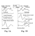
- FIG. 2A shows the enhancement of the CAP in crushed (but untreated with PEG) spinal cord by 4-AP.
- the initial recovered CAP at 1 hour post injury is shown, and the enhanced CAP following 100 pM 4-AP treatment is superimposed upon it.
- the blocker was washed out, and the media in the central compartment was replaced with normal Krebs' solution.
- the CAP fell to pretreatment levels by 15 minutes and was indistinguishable from the original record.
- This final waveform is superimposed on the other two CAPs in FIG. 2A but cannot be discriminated from the pretreatment electrical record.
- 4AP reversibly enhanced the recovered CAP by about 40%.
- FIG. 2B shows an identical test performed on a PEG-treated spinal cord, in which 4-AP was administered at 1 hour post PEG application.
- the second CAP was reversibly enhanced by about 70%.
- FIG. 2C shows the group data, including 5 spinal cords in each group.
- the percent enhancement of the PEG-mediated recovery for the group data mirrors that discussed above for the individual experiments (about 70% enhancement in the experimental group; about 40% in the control group). This experimental enhancement was statistically significantly greater than that observed in the controls. (p ⁇ 0.05, unpaired Student's t test)
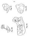
- FIG. 3 depicts a proposed mechanism of the synergistic effect of PEG and 4-AP.
- a severe mechanical compression of a myelinated axon is diagrammed at the top.
- the myelin sheath envelops high densities of fast W channels clustered at the paranodal region.
- Severe crush leads to an exposure of the potassium channels of the paranodal region by a withdrawal or collapse of the myelin lamella at this site [ Shi, R. and Blight, A.R. (1996): Neuroscience, 77:553-562 ].
- This procedure may advantageously applied to treat severe, acute neurotrauma.
- repair of crushed axons in peripheral nerves leading to a rescue of their distal segments would provide the added benefit of reducing atrophy or degeneration of target cells or so called "end organs.”
- PEG-mediated fusion of even transected axons could become a component of microsurgical grafting techniques since the conventional resection of peripheral nerve trunks prior to fasicular grafting exposes the severed tips of proximal and distal axonal segments, making them available for fusion.
- a biomembrane fusion agent specifically the hydrophilic polymer PEG
- the administered PEG specifically targets a hemorrhagic contusion of an adult guinea pig spinal cord.
- Drawing Fig. 4 Behavioral Model and Physiological Evaluation
- This drawing shows the neural circuit of the Cutaneus Trunchi Muscle (CTM) reflex, and its interruption by spinal injury.
- Nociceptive sensory receptors in the skin project their axons into the spinal cord at each vertebral segment bilaterally via the Dorsal Cutaneus Nerves. These synapse within the spinal cord and project 2nd order ascending sensory nerves in the ventral funiculus of the white matter to the cervical region where these synapse on bilaterally organized constellations of CTM motor neurons.
- CTM motor neurons project their axons out of the cord on right and left sides via the brachial plexus, where these innervate the cutaneous muscle of the skin via the lateral thoracic branch of the plexus.
- Histological cross sections were 5 ⁇ m thick, and observed on an Olympus Van Ox Fluorescent microscope using excitation wavelengths of 495 and 545 nm and barrier filters of 475 and 590 nm, respectively. Digital images were captured to the computer with an Optronics DEI 750 camera.
- the FITC decorated PEG (about 1400 Daltons; prepared by Molecular Probes, Chatsworth, Ca) was used to trace the distribution of PEG following different routes of administration.
- F1-PEG 50% weight by weight in SLR was applied directly to exposed spinal cord injury site (with the dura removed) using a Pasteur pipette in two animals.
- PEG was removed by aspiration and the region irrigated with SLR two minutes later.
- Subcutaneous injection of 1 cc F1-PEG (30% w/w in SLR) was made beneath the skin of the neck in two spinal injured guinea pigs using a 22-gauge needle.
- 1 cc of FL-PEG was injected using a 26-gauge needle.
- PEG, 30% in lactated Ringer's was also administered by intraperitoneal injection in one case.
- Tibial nerve of the hind limb produced ascending volleys of nerve impulses recorded at the contralateral sensory cortex of the brain. These were eliminated between the site of stimulation and recording by the spinal lesion - immediately abolishing the recording of these peaks (postcrush records).
- Each electrical record was comprised of a stimulus train of 200 stimulations ( ⁇ 2mA square wave, 200 ⁇ s duration at 3 HZ). Three sets of these recordings were made at each measurement period and averaged to produce the single waveform presented in the following data. The appearance of original records prior to computer averaging can be found in prior reports [ Borgens, R. and Shi, R. (2000) Immediate recovery from spinal cord injury through molecular repair of nerve membranes with polyethylene glycol, FASEB 14, 27-35 ].
- tibial nerve evoked SSEPs Prior to the crush injury of the spinal cord, tibial nerve evoked SSEPs usually segregate into an early and late arriving peaks of CAPs recorded from the sensory cortex (P1 and P2) [ Borgens, R. and Shi, R. (2000): Immediate recovery from spinal cord injury through molecular repair of nerve membranes with polyethylene glycol, FASEB, 14, 27-35 ]. As in prior experiments these peaks are completely eliminated following a severe constant displacement crush to the midthoracic spinal cord ( FIG. 4 ).
- a decrease in the amplitude and extended duration of the CAP is typical of recovering nerve impulses.
- the area under the early arriving peak (P1) was measured in pixels in only PEG-treated animals (since there were no recoveries of SSEPs in Control animals). If 100% of all single nerve fibers contributing to the CAP were once again recruited into conduction subsequent to the injury - but with a decreased amplitude and extended latency period - the normalized mean area under the curve (CAP above baseline) divided by the same pre-injury data should approach unity (1.0).
- the proportion of recovered and unrecovered animals, as well as the unit area of the recovered CTM receptive fields between controls and PEG- injected animals was quantitatively compared.
- P 0.81; Mann Whitney, two tailed test; Table 1).
- the spinal injury produced a similar level of CTM behavioral loss in all animals.
- the area of recovering backskin of these ten animals continued to increase in size to week four when the experiment was ended.
- the mean area of recovered CTM receptive fields was approximately 33%.
- PEG is well known to be able to fuse numerous single cells in vitro into one giant cell, as well as join the membranes of neurons and giant invertebrate axons
- HRP horseradish peroxidase
- Urology 159, 2136-2139 ; Lee, R., River, L.P., Pan, F.S., Wollmann, L. Jr. and R.L. (1992): Surfactant-induced sealing of electropermeabilized skeletal muscle membranes in vivo, Proc. Natl. Acad. Sci. U.S.A., 89, 4524-4528 ] as novel treatments for severe CNS and PNS injury, as well as head injury and stroke.
- IV intravenous
- Intravenous Hydrophilic Polymer Induces Rapid Recovery from Clinical Paraplegia in Dogs
- This example demonstrates a swift, striking, and statistically significant recovery of multiple functions in clinical cases of severe, acute, naturally occurring paraplegia in dogs. Recovery of function occurred in response to a combination of topically applied, and intravenously administered, polyethylene glycol (PEG). Recoveries of sensory and motor functions occurred rapidly and at all time points studied between 3 days and 6-8 weeks post-injury.
- PEG polyethylene glycol
- CT computerized x-ray tomography
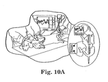
- Figs. 9A-9D Each dog received a radiological examination ( FIGs. 9A-9D ), and a thorough, videotaped, neurological examination ( FIG. 10A ) that included: 1) tests for deep pain in hind limbs and digits, 2) superficial pain appreciation below the level of the injury in flank, lower limbs and digits, 3) proprioceptive evaluation of the hind limbs (i.e. conscious proprioception), 4) evaluation of hind limb load-bearing and voluntary locomotion, and 5) spinal reflex testing (patellar, tibialis, cranialis, flexor withdrawal, and sciatic reflexes). Tests 1-4 were also used as functional measures of outcome and were quantitatively scored using previously reported techniques and methods ( R.B.
- TNS total neurological score
- the dura was removed during decompressive surgery, exposing the spinal cord lesion, and about 1cc of the PEG solution (about 2000daltons, 50% W/W in sterile saline; 150mg/kg body weight) was layered onto the injury site.
- the polymer was aspirated from the surface of the exposed cord within 2 min of application, next the region lavaged with sterile Ringer's solution, and these fluids aspirated as well.
- a fat pad graft was placed superficially, the incision closed, and the animals taken to the Intensive Care Unit (ICU) for recovery.
- ICU Intensive Care Unit
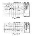
- FIGS. 10A-10C Evoked potential testing [Somatosensory Evoked Potential or SSEP( R.B. Borgens et al., J. Restorative Neurology and Neurosci. 5, 305 (1993 ); R.B. Borgens et al., J Neurotrauma 16, 639 (1999 )), FIGS. 10A-10C ] was performed on 11 of the 12 PEG-treated dogs recruited to the Purdue Center to determine if nerve conduction through the lesion had been restored ( FIGS. 10A-10C ). Somatosensory Evoked Potential recordings could not usually be obtained prior to the first PEG injection and surgery due to the need to move these animals through the battery of evaluations and on to surgery as soon as possible after admission.
- restored nerve impulse traffic through the lesion is not required for voluntary ambulation in animals, walking behavior by itself does not represent a valid behavioral recovery with which to infer restored conduction through the lesion. This requires use of kinestheseological methods confirming fore limb and hind limb coordination during voluntary locomotion.
- a variable level of recovery of the CTM reflex occurred in > 90% of PEG-treated guinea pigs, compared to a range of 0-17% in sham-treated control populations in three separate studies ( R.B. Borgens, R. Shi, FASEB 14, 27 (2000 ); R.B. Borgens, D.M. Bohnert, J. Neurosci. Res. 66, 1179 (2001 ); R.B. Borgens, R. Shi, D.M. Bohnert, J. Exp. Bio. 205, 1 (2002 )).
- Triblock polymers such as poloxamers are comprised largely of PEG - yet they also posses a hydrophobic component (polypropylene oxide) which may actually target breaches in membranes - inserting into the breach where the hydrophobic core of the membrane is exposed ( J.M. Marks, C-Y. Pan, T. Bushell, W. Cromie, R.C. Lee FASEB J15,1107 (2001 )).
- the long PEG side chains likely contribute to sealing in the fashion described above.
- any large molecular polymer like PEG or poloxamers introduced to the blood supply, will target only regions of tissue trauma where there is a loss of vascular integrity.
Landscapes
- Health & Medical Sciences (AREA)
- General Health & Medical Sciences (AREA)
- Life Sciences & Earth Sciences (AREA)
- Chemical & Material Sciences (AREA)
- Public Health (AREA)
- Medicinal Chemistry (AREA)
- Veterinary Medicine (AREA)
- Animal Behavior & Ethology (AREA)
- Pharmacology & Pharmacy (AREA)
- Epidemiology (AREA)
- Nuclear Medicine, Radiotherapy & Molecular Imaging (AREA)
- General Chemical & Material Sciences (AREA)
- Organic Chemistry (AREA)
- Chemical Kinetics & Catalysis (AREA)
- Engineering & Computer Science (AREA)
- Bioinformatics & Cheminformatics (AREA)
- Biomedical Technology (AREA)
- Neurology (AREA)
- Neurosurgery (AREA)
- Medicinal Preparation (AREA)
- Medicines That Contain Protein Lipid Enzymes And Other Medicines (AREA)
- Pharmaceuticals Containing Other Organic And Inorganic Compounds (AREA)
- Medicines Containing Material From Animals Or Micro-Organisms (AREA)
- Materials For Medical Uses (AREA)
- Acyclic And Carbocyclic Compounds In Medicinal Compositions (AREA)
Priority Applications (1)
| Application Number | Priority Date | Filing Date | Title |
|---|---|---|---|
| AU2006200866A AU2006200866B2 (en) | 2001-04-24 | 2006-03-01 | Methods and compositions for treating mammalian nerve tissue injuries |
Applications Claiming Priority (3)
| Application Number | Priority Date | Filing Date | Title |
|---|---|---|---|
| US28620001P | 2001-04-24 | 2001-04-24 | |
| US286200P | 2001-04-24 | ||
| PCT/US2002/013375 WO2002092107A1 (en) | 2001-04-24 | 2002-04-24 | Method and compositions for treating mammalian nerve tissue injuries |
Publications (3)
| Publication Number | Publication Date |
|---|---|
| EP1389121A1 EP1389121A1 (en) | 2004-02-18 |
| EP1389121A4 EP1389121A4 (en) | 2006-11-02 |
| EP1389121B1 true EP1389121B1 (en) | 2012-02-22 |
Family
ID=23097525
Family Applications (1)
| Application Number | Title | Priority Date | Filing Date |
|---|---|---|---|
| EP02741682A Expired - Lifetime EP1389121B1 (en) | 2001-04-24 | 2002-04-24 | Method and compositions for treating mammalian nerve tissue injuries |
Country Status (9)
| Country | Link |
|---|---|
| US (4) | US7837987B2 (enExample) |
| EP (1) | EP1389121B1 (enExample) |
| JP (2) | JP2004527573A (enExample) |
| AT (1) | ATE546146T1 (enExample) |
| AU (2) | AU2002314758B2 (enExample) |
| CA (2) | CA2740056A1 (enExample) |
| ES (1) | ES2382899T3 (enExample) |
| NZ (2) | NZ543953A (enExample) |
| WO (1) | WO2002092107A1 (enExample) |
Families Citing this family (64)
| Publication number | Priority date | Publication date | Assignee | Title |
|---|---|---|---|---|
| US7582680B1 (en) | 1998-11-12 | 2009-09-01 | Purdue Research Foundation | Methods and compositions for treating mammalian spinal cord injuries |
| US7442370B2 (en) | 2001-02-01 | 2008-10-28 | Biogen Idec Ma Inc. | Polymer conjugates of mutated neublastin |
| US7276580B2 (en) * | 2001-03-12 | 2007-10-02 | Biogen Idec Ma Inc. | Neurotrophic factors |
| NZ543953A (en) * | 2001-04-24 | 2007-04-27 | Univ Chicago | Method and compositions for treating mammalian nerve tissue injuries |
| US7147647B2 (en) | 2002-04-26 | 2006-12-12 | Medtronic, Inc. | Sintered titanium tube for the management of spinal cord injury |
| JP4571776B2 (ja) * | 2002-11-05 | 2010-10-27 | Jx日鉱日石エネルギー株式会社 | 潤滑油組成物 |
| NZ543365A (en) | 2003-04-18 | 2009-02-28 | Biogen Idec Inc | Polymer-conjugated glycosylated neublastin |
| EP1786454B1 (en) * | 2004-08-19 | 2010-07-21 | Biogen Idec MA Inc. | Neublastin variants |
| DE602005022082D1 (de) * | 2004-08-19 | 2010-08-12 | Biogen Idec Inc | Rückfaltung von proteinen der transforming-growth-factor-beta-familie |
| US20060121016A1 (en) * | 2004-10-18 | 2006-06-08 | Lee Raphael C | Methods and compositions for treatment of free radical injury |
| CN101084272B (zh) * | 2004-12-21 | 2011-07-13 | 路博润有限公司 | 组合物 |
| US20070048731A1 (en) * | 2005-05-20 | 2007-03-01 | Neurosilicon | High throughput use-dependent assay based on stimulation of cells on a silicon surface |
| WO2006130599A2 (en) * | 2005-05-31 | 2006-12-07 | Warsaw Orthopedic, Inc. | Compositions and methods for treating a damaged cardiovascular element |
| US20070017530A1 (en) * | 2005-06-10 | 2007-01-25 | Syed Naweed I | Detecting electrical activity and assessing agents for the ability to influence electrical activity |
| US20060287660A1 (en) * | 2005-06-15 | 2006-12-21 | Syed Naweed I | Electrically Stimulating Nerve Regeneration |
| US20060292549A1 (en) * | 2005-06-15 | 2006-12-28 | Neurosilicon | Psychotropic drug screening device based on long-term photoconductive stimulation of neurons |
| JP4907908B2 (ja) * | 2005-06-29 | 2012-04-04 | ルネサスエレクトロニクス株式会社 | 駆動回路及び表示装置 |
| US20070092958A1 (en) * | 2005-07-15 | 2007-04-26 | Syed Naweed I | Method and apparatus for guiding growth of neurons |
| US20070213718A1 (en) * | 2006-02-14 | 2007-09-13 | Sdgi Holdings, Inc. | Treatment of the vertebral column |
| US7520888B2 (en) * | 2006-02-14 | 2009-04-21 | Warsaw Orthopedic, Inc. | Treatment of the vertebral column |
| US20070213717A1 (en) * | 2006-02-14 | 2007-09-13 | Sdgi Holdings, Inc. | Biological fusion in the vertebral column |
| US20070227547A1 (en) * | 2006-02-14 | 2007-10-04 | Sdgi Holdings, Inc. | Treatment of the vertebral column |
| TWI501774B (zh) * | 2006-02-27 | 2015-10-01 | Biogen Idec Inc | 神經性病症之治療 |
| ES2450065T3 (es) * | 2006-03-01 | 2014-03-21 | Biogen Idec Ma Inc. | Composiciones y métodos para la administración de proteínas de la familia de ligandos del GDNF |
| US8945623B2 (en) * | 2006-05-03 | 2015-02-03 | Warsaw Orthopedic, Inc. | Compositions comprising biomembrane sealing agent for treatment of neuronal injury, and methods of use |
| US8840933B2 (en) | 2006-05-03 | 2014-09-23 | Warsaw Orthopedic, Inc. | Method of treating neuronal injury by administering magnesium chloride and PEG |
| US9675696B2 (en) | 2006-11-14 | 2017-06-13 | Warsaw Orthopedic, Inc. | Method and use for increasing efficacy of anti-adhesive compositions in controlling inflammation and pain |
| US8329655B2 (en) | 2007-05-01 | 2012-12-11 | Biogen Idec Ma Inc. | Methods for increasing vascularization |
| US20110135648A1 (en) * | 2007-08-08 | 2011-06-09 | Biogen Idec Ma Inc. | Anti-neublastin antibodies and uses thereof |
| US20090263507A1 (en) * | 2008-04-18 | 2009-10-22 | Warsaw Orthopedic, Inc. | Biological markers and response to treatment for pain, inflammation, neuronal or vascular injury and methods of use |
| CA2744100C (en) * | 2008-10-21 | 2020-06-30 | The General Hospital Corporation | Cell transplantation |
| US9244060B2 (en) * | 2009-03-26 | 2016-01-26 | Warsaw Orthopedic, Inc. | Site localization and methods for monitoring treatment of disturbed blood vessels |
| US8858924B2 (en) * | 2009-03-26 | 2014-10-14 | Warsaw Orthopedic, Inc. | Compositions and methods for treatment of hemorrhage |
| US9078808B2 (en) * | 2009-03-26 | 2015-07-14 | Warsaw Orthopedic, Inc. | Device to deliver magnesium in PEG formulation |
| US8852566B2 (en) | 2009-03-26 | 2014-10-07 | Warsaw Orthopedic, Inc. | Compositions and methods for preferential distribution of active agents to injury sites |
| WO2010111212A1 (en) | 2009-03-26 | 2010-09-30 | Warsaw Orthopedic, Inc. | Methods of identifying potential components for targeted drug delivery compositions |
| CA2758118C (en) | 2009-04-07 | 2019-05-21 | Catholic Healthcare West | Uterine electrical stimulation system and method |
| US20110230785A1 (en) * | 2010-03-16 | 2011-09-22 | ProNerve, LLC | Somatosensory Evoked Potential (SSEP) Automated Alert System |
| BR112013002859B1 (pt) | 2010-08-06 | 2020-10-06 | The General Hospital Corporation D/B/A Massachusetts General Hospital | Método e aparelho para tratamento de células |
| CA3063888C (en) | 2010-10-27 | 2020-12-22 | Dignity Health | Uterine electrical stimulation system and method |
| WO2013173833A1 (en) * | 2012-05-18 | 2013-11-21 | Dignity Health | Electrical stimulation of the cervix |
| US9872983B2 (en) | 2010-10-27 | 2018-01-23 | Dignity Health | Uterine electrical stimulation system and method |
| WO2012161823A1 (en) * | 2011-02-25 | 2012-11-29 | The General Hospital Corporation D/B/A Massachusetts General Hospital | Nerve coaptation apparatus |
| EP2953640B1 (en) * | 2013-02-07 | 2020-04-08 | The Cleveland Clinic Foundation | Nanoencapsulated superoxide dismutase and catalase for treating spinal cord injury |
| PL3138031T3 (pl) | 2014-04-28 | 2023-04-11 | Yeda Research And Development Co., Ltd. | Sposób i urządzenie do przewidywania odpowiedzi na pokarm |
| GB2536650A (en) | 2015-03-24 | 2016-09-28 | Augmedics Ltd | Method and system for combining video-based and optic-based augmented reality in a near eye display |
| EP3658072B1 (en) | 2017-07-26 | 2024-03-13 | Neuraptive Therapeutics, Inc. | Device for administering therapeutics to a nerve |
| US12458411B2 (en) | 2017-12-07 | 2025-11-04 | Augmedics Ltd. | Spinous process clamp |
| EP3787543A4 (en) | 2018-05-02 | 2022-01-19 | Augmedics Ltd. | REGISTRATION OF A FIDUCIAL MARKER FOR AN AUGMENTED REALITY SYSTEM |
| US11766296B2 (en) | 2018-11-26 | 2023-09-26 | Augmedics Ltd. | Tracking system for image-guided surgery |
| US11980506B2 (en) | 2019-07-29 | 2024-05-14 | Augmedics Ltd. | Fiducial marker |
| US12178666B2 (en) | 2019-07-29 | 2024-12-31 | Augmedics Ltd. | Fiducial marker |
| JP7653164B2 (ja) * | 2019-09-27 | 2025-03-28 | バージニア コモンウェルス ユニバーシティー | 組織を修復または増加させるための組成物および方法 |
| US11382712B2 (en) | 2019-12-22 | 2022-07-12 | Augmedics Ltd. | Mirroring in image guided surgery |
| WO2021150773A1 (en) * | 2020-01-21 | 2021-07-29 | The Penn State Research Foundation | Methods and materials for treating nerve injury and/or promoting wound healing |
| US11389252B2 (en) | 2020-06-15 | 2022-07-19 | Augmedics Ltd. | Rotating marker for image guided surgery |
| US20230190676A1 (en) * | 2020-07-09 | 2023-06-22 | Musc Foundation For Research Development | Methods of protecting cells from insults |
| US12239385B2 (en) | 2020-09-09 | 2025-03-04 | Augmedics Ltd. | Universal tool adapter |
| US11896445B2 (en) | 2021-07-07 | 2024-02-13 | Augmedics Ltd. | Iliac pin and adapter |
| US12150821B2 (en) | 2021-07-29 | 2024-11-26 | Augmedics Ltd. | Rotating marker and adapter for image-guided surgery |
| EP4387552A4 (en) | 2021-08-18 | 2025-04-30 | Augmedics Ltd. | AUGMENTED REALITY SURGICAL SYSTEM USING DEPTH SENSING |
| WO2023203521A1 (en) | 2022-04-21 | 2023-10-26 | Augmedics Ltd. | Systems and methods for medical image visualization |
| US12268677B2 (en) | 2022-04-27 | 2025-04-08 | The Penn State Research Foundation | 4-aminopyridine (4-AP) and bone morphogenetic protein 2 (BMP-2) |
| IL319523A (en) | 2022-09-13 | 2025-05-01 | Augmedics Ltd | Augmented reality glasses for image-guided medical intervention |
Family Cites Families (23)
| Publication number | Priority date | Publication date | Assignee | Title |
|---|---|---|---|---|
| US4369769A (en) | 1980-06-13 | 1983-01-25 | Edwards Charles C | Spinal fixation device and method |
| US4599354A (en) | 1985-03-11 | 1986-07-08 | Morton Shulman | Composition and method for producing prolonged pain relief |
| US6858409B1 (en) * | 1988-05-27 | 2005-02-22 | Amgen Inc. | Nucleic acids encoding interleukin-1 inhibitors and processes for preparing interleukin-1 inhibitors |
| US4919140A (en) | 1988-10-14 | 1990-04-24 | Purdue Research Foundation | Method and apparatus for regenerating nerves |
| IE82916B1 (en) | 1990-11-02 | 2003-06-11 | Elan Corp Plc | Formulations and their use in the treatment of neurological diseases |
| NL9000634A (nl) | 1990-03-20 | 1991-10-16 | Catharina Ziekenhuis Stichting | Suspensie-injectiepreparaat op waterbasis, werkwijze voor de bereiding daarvan en toepassing van dit preparaat voor pijnbestrijding. |
| CA2075928A1 (en) * | 1991-01-11 | 1992-07-12 | Roger W. Hackett | Method of detecting circulating antibody types using dried or lyophilized cells or cell-like material |
| CN1069741A (zh) | 1991-03-19 | 1993-03-10 | 西特克斯公司 | 具有改进的生物活性的聚氧丙烯/聚氧乙烯共聚物 |
| US5470568A (en) | 1992-02-13 | 1995-11-28 | Arch Development Corporation | Methods and compositions of a polymer (poloxamer) for cell repair |
| US5324322A (en) * | 1992-04-20 | 1994-06-28 | Case Western Reserve University | Thin film implantable electrode and method of manufacture |
| US5605687A (en) * | 1992-05-15 | 1997-02-25 | Arch Development Corporation | Methods and compositions of a polymer (poloxamer) for repair of electrical injury |
| DE69322707T2 (de) | 1992-07-31 | 1999-08-19 | Bristol-Myers Squibb Company | Diphenyl Oxazole-, Thiazole- und Imidazole-Derivate als Inhibitoren der Wiederaufnahme des Adenosins |
| CA2085785C (en) | 1992-12-18 | 2005-03-15 | Robert R. Hansebout | The use of 4-aminopyridine in the treatment of a neurological condition |
| CN1103698C (zh) * | 1995-02-14 | 2003-03-26 | 东丽株式会社 | 平版印刷版及平板印刷版原版 |
| JPH09315972A (ja) | 1996-03-22 | 1997-12-09 | Chugai Pharmaceut Co Ltd | 脊髄損傷治療剤 |
| US6495532B1 (en) * | 1997-03-19 | 2002-12-17 | Sky High, Llc | Compositions containing lysophosphotidic acids which inhibit apoptosis and uses thereof |
| EP1024812B1 (en) | 1997-03-19 | 2007-01-24 | Sky High, LLC | Compositions containing lysophosphatidic acids which inhibit apoptosis and uses thereof |
| US6440455B1 (en) | 1997-09-02 | 2002-08-27 | Children's Medical Center Corporation | Methods for modulating the axonal outgrowth of central nervous system neurons |
| US7582680B1 (en) * | 1998-11-12 | 2009-09-01 | Purdue Research Foundation | Methods and compositions for treating mammalian spinal cord injuries |
| US6232297B1 (en) * | 1999-02-01 | 2001-05-15 | University Of Virginia Patent Foundation | Methods and compositions for treating inflammatory response |
| AU4363600A (en) * | 1999-04-22 | 2000-11-10 | Department Of The Army, U.S. Government | Treatment of and/or prophylaxis against brain and spinal cord injury |
| NZ543953A (en) | 2001-04-24 | 2007-04-27 | Univ Chicago | Method and compositions for treating mammalian nerve tissue injuries |
| US7199110B2 (en) | 2002-12-30 | 2007-04-03 | Purdue Research Foundation | Method of treatment for spinal cord injury |
-
2002
- 2002-04-24 NZ NZ543953A patent/NZ543953A/en not_active IP Right Cessation
- 2002-04-24 JP JP2002589024A patent/JP2004527573A/ja active Pending
- 2002-04-24 WO PCT/US2002/013375 patent/WO2002092107A1/en not_active Ceased
- 2002-04-24 ES ES02741682T patent/ES2382899T3/es not_active Expired - Lifetime
- 2002-04-24 NZ NZ529526A patent/NZ529526A/en not_active IP Right Cessation
- 2002-04-24 US US10/132,542 patent/US7837987B2/en not_active Expired - Fee Related
- 2002-04-24 AT AT02741682T patent/ATE546146T1/de active
- 2002-04-24 EP EP02741682A patent/EP1389121B1/en not_active Expired - Lifetime
- 2002-04-24 AU AU2002314758A patent/AU2002314758B2/en not_active Ceased
- 2002-04-24 CA CA2740056A patent/CA2740056A1/en not_active Abandoned
- 2002-04-24 CA CA2445612A patent/CA2445612C/en not_active Expired - Fee Related
-
2004
- 2004-07-28 US US10/901,481 patent/US9687502B2/en not_active Expired - Fee Related
-
2006
- 2006-03-01 AU AU2006200866A patent/AU2006200866B2/en not_active Ceased
-
2009
- 2009-02-23 JP JP2009039488A patent/JP2009112835A/ja active Pending
- 2009-07-23 US US12/508,184 patent/US8460646B2/en not_active Expired - Fee Related
-
2013
- 2013-06-07 US US13/912,499 patent/US9101655B2/en not_active Expired - Fee Related
Non-Patent Citations (1)
| Title |
|---|
| NAKAJIMA NAOKI; IKADA YOSHITO: "Fusogenic activity of various water-soluble polymers", JOURNAL OF BIOMATERIALS SCIENCE POLYMER EDITION, vol. 6, no. 8, 1994, pages 751 - 759 * |
Also Published As
| Publication number | Publication date |
|---|---|
| EP1389121A4 (en) | 2006-11-02 |
| US9687502B2 (en) | 2017-06-27 |
| CA2445612C (en) | 2011-10-25 |
| AU2006200866B2 (en) | 2007-11-08 |
| AU2006200866A1 (en) | 2006-03-23 |
| ATE546146T1 (de) | 2012-03-15 |
| EP1389121A1 (en) | 2004-02-18 |
| US20100016444A1 (en) | 2010-01-21 |
| NZ529526A (en) | 2006-06-30 |
| ES2382899T3 (es) | 2012-06-14 |
| WO2002092107A1 (en) | 2002-11-21 |
| US20030118545A1 (en) | 2003-06-26 |
| CA2445612A1 (en) | 2002-11-21 |
| US8460646B2 (en) | 2013-06-11 |
| US9101655B2 (en) | 2015-08-11 |
| JP2009112835A (ja) | 2009-05-28 |
| US20140100289A1 (en) | 2014-04-10 |
| AU2002314758B2 (en) | 2006-06-01 |
| US20050069520A1 (en) | 2005-03-31 |
| CA2740056A1 (en) | 2002-11-21 |
| US7837987B2 (en) | 2010-11-23 |
| NZ543953A (en) | 2007-04-27 |
| JP2004527573A (ja) | 2004-09-09 |
Similar Documents
| Publication | Publication Date | Title |
|---|---|---|
| EP1389121B1 (en) | Method and compositions for treating mammalian nerve tissue injuries | |
| AU2002314758A1 (en) | Method and composition for treating mammalian nerve tissue injuries | |
| Laverty et al. | A preliminary study of intravenous surfactants in paraplegic dogs: polymer therapy in canine clinical SCI | |
| Somjen | Mechanisms of spreading depression and hypoxic spreading depression-like depolarization | |
| Awadzi et al. | The chemotherapy of onchocerciasis X. An assessment of four single dose treatment regimes of MK-933 (ivermectin) in human onchocerciasis | |
| Hansebout et al. | 4-Aminopyridine in chronic spinal cord injury: a controlled, double-blind, crossover study in eight patients | |
| Borgens et al. | An imposed oscillating electrical field improves the recovery of function in neurologically complete paraplegic dogs | |
| Shi et al. | Functional reconnection of severed mammalian spinal cord axons with polyethylene glycol | |
| JP5795537B2 (ja) | 中枢神経系への対流増進送達のためのリポソーム組成物 | |
| Collins et al. | Structural and functional recovery of electropermeabilized skeletal muscle in-vivo after treatment with surfactant poloxamer 188 | |
| Borgens | Cellular engineering: molecular repair of membranes to rescue cells of the damaged nervous system | |
| EP0626848B1 (en) | The use of 4-aminopyridine in the treatment of pain and spasticity resulting from spinal cord injury | |
| US7582680B1 (en) | Methods and compositions for treating mammalian spinal cord injuries | |
| Bitar Alatorre et al. | Critical ischemia time in a model of spinal cord section. A study performed on dogs | |
| Añor | Monoparesis | |
| Weiss et al. | Stem Cell Ophthalmology Treatment Study (SCOTS): Autologous bone-marrow derived stem cells in the treatment of hereditary macular degeneration | |
| Singer et al. | Role of electrical activity in axotomy-induced increased glucose use | |
| Fox et al. | The effect of angiography on the electrophysiological state of the spinal cord: a study in control and traumatized cats | |
| KR20200105814A (ko) | 보툴리눔 요법의 부작용 치료 | |
| Francis et al. | Benzalkonium Chloride–Induced Denervation of Orbicularis Oculi Muscle in Rabbits | |
| Borgens | Recovery of Behavioral and Physiological Function In Vivo | |
| Soto | Critical ischemia time in a model of spinal cord section. A study performed on dogs |
Legal Events
| Date | Code | Title | Description |
|---|---|---|---|
| PUAI | Public reference made under article 153(3) epc to a published international application that has entered the european phase |
Free format text: ORIGINAL CODE: 0009012 |
|
| 17P | Request for examination filed |
Effective date: 20031103 |
|
| AK | Designated contracting states |
Kind code of ref document: A1 Designated state(s): AT BE CH CY DE DK ES FI FR GB GR IE IT LI LU MC NL PT SE TR |
|
| AX | Request for extension of the european patent |
Extension state: AL LT LV MK RO SI |
|
| RAP1 | Party data changed (applicant data changed or rights of an application transferred) |
Owner name: THE UNIVERSITY OF CHICAGO Owner name: PURDUE RESEARCH FOUNDATION |
|
| RIC1 | Information provided on ipc code assigned before grant |
Ipc: A61K 31/765 20060101ALI20060906BHEP Ipc: A61P 25/00 20060101ALI20060906BHEP Ipc: A61K 31/00 20060101ALI20060906BHEP Ipc: A61K 31/77 20060101ALI20060906BHEP Ipc: A61K 31/74 20060101AFI20021122BHEP |
|
| A4 | Supplementary search report drawn up and despatched |
Effective date: 20061002 |
|
| 17Q | First examination report despatched |
Effective date: 20070727 |
|
| RIN1 | Information on inventor provided before grant (corrected) |
Inventor name: SHI, RIYI Inventor name: BORGENS, RICHARD, B. |
|
| RAP1 | Party data changed (applicant data changed or rights of an application transferred) |
Owner name: PURDUE RESEARCH FOUNDATION |
|
| GRAP | Despatch of communication of intention to grant a patent |
Free format text: ORIGINAL CODE: EPIDOSNIGR1 |
|
| GRAS | Grant fee paid |
Free format text: ORIGINAL CODE: EPIDOSNIGR3 |
|
| GRAA | (expected) grant |
Free format text: ORIGINAL CODE: 0009210 |
|
| AK | Designated contracting states |
Kind code of ref document: B1 Designated state(s): AT BE CH CY DE DK ES FI FR GB GR IE IT LI LU MC NL PT SE TR |
|
| REG | Reference to a national code |
Ref country code: GB Ref legal event code: FG4D |
|
| REG | Reference to a national code |
Ref country code: CH Ref legal event code: EP |
|
| REG | Reference to a national code |
Ref country code: AT Ref legal event code: REF Ref document number: 546146 Country of ref document: AT Kind code of ref document: T Effective date: 20120315 |
|
| REG | Reference to a national code |
Ref country code: IE Ref legal event code: FG4D |
|
| REG | Reference to a national code |
Ref country code: DE Ref legal event code: R096 Ref document number: 60242258 Country of ref document: DE Effective date: 20120419 |
|
| REG | Reference to a national code |
Ref country code: ES Ref legal event code: FG2A Ref document number: 2382899 Country of ref document: ES Kind code of ref document: T3 Effective date: 20120614 |
|
| REG | Reference to a national code |
Ref country code: NL Ref legal event code: VDEP Effective date: 20120222 |
|
| PG25 | Lapsed in a contracting state [announced via postgrant information from national office to epo] |
Ref country code: NL Free format text: LAPSE BECAUSE OF FAILURE TO SUBMIT A TRANSLATION OF THE DESCRIPTION OR TO PAY THE FEE WITHIN THE PRESCRIBED TIME-LIMIT Effective date: 20120222 |
|
| PG25 | Lapsed in a contracting state [announced via postgrant information from national office to epo] |
Ref country code: PT Free format text: LAPSE BECAUSE OF FAILURE TO SUBMIT A TRANSLATION OF THE DESCRIPTION OR TO PAY THE FEE WITHIN THE PRESCRIBED TIME-LIMIT Effective date: 20120622 Ref country code: FI Free format text: LAPSE BECAUSE OF FAILURE TO SUBMIT A TRANSLATION OF THE DESCRIPTION OR TO PAY THE FEE WITHIN THE PRESCRIBED TIME-LIMIT Effective date: 20120222 Ref country code: GR Free format text: LAPSE BECAUSE OF FAILURE TO SUBMIT A TRANSLATION OF THE DESCRIPTION OR TO PAY THE FEE WITHIN THE PRESCRIBED TIME-LIMIT Effective date: 20120523 Ref country code: BE Free format text: LAPSE BECAUSE OF FAILURE TO SUBMIT A TRANSLATION OF THE DESCRIPTION OR TO PAY THE FEE WITHIN THE PRESCRIBED TIME-LIMIT Effective date: 20120222 |
|
| REG | Reference to a national code |
Ref country code: AT Ref legal event code: MK05 Ref document number: 546146 Country of ref document: AT Kind code of ref document: T Effective date: 20120222 |
|
| PG25 | Lapsed in a contracting state [announced via postgrant information from national office to epo] |
Ref country code: CY Free format text: LAPSE BECAUSE OF FAILURE TO SUBMIT A TRANSLATION OF THE DESCRIPTION OR TO PAY THE FEE WITHIN THE PRESCRIBED TIME-LIMIT Effective date: 20120222 |
|
| PG25 | Lapsed in a contracting state [announced via postgrant information from national office to epo] |
Ref country code: DK Free format text: LAPSE BECAUSE OF FAILURE TO SUBMIT A TRANSLATION OF THE DESCRIPTION OR TO PAY THE FEE WITHIN THE PRESCRIBED TIME-LIMIT Effective date: 20120222 Ref country code: SE Free format text: LAPSE BECAUSE OF FAILURE TO SUBMIT A TRANSLATION OF THE DESCRIPTION OR TO PAY THE FEE WITHIN THE PRESCRIBED TIME-LIMIT Effective date: 20120222 |
|
| PG25 | Lapsed in a contracting state [announced via postgrant information from national office to epo] |
Ref country code: MC Free format text: LAPSE BECAUSE OF NON-PAYMENT OF DUE FEES Effective date: 20120430 |
|
| REG | Reference to a national code |
Ref country code: CH Ref legal event code: PL |
|
| PLBE | No opposition filed within time limit |
Free format text: ORIGINAL CODE: 0009261 |
|
| STAA | Information on the status of an ep patent application or granted ep patent |
Free format text: STATUS: NO OPPOSITION FILED WITHIN TIME LIMIT |
|
| REG | Reference to a national code |
Ref country code: IE Ref legal event code: MM4A |
|
| 26N | No opposition filed |
Effective date: 20121123 |
|
| PG25 | Lapsed in a contracting state [announced via postgrant information from national office to epo] |
Ref country code: IE Free format text: LAPSE BECAUSE OF NON-PAYMENT OF DUE FEES Effective date: 20120424 Ref country code: AT Free format text: LAPSE BECAUSE OF FAILURE TO SUBMIT A TRANSLATION OF THE DESCRIPTION OR TO PAY THE FEE WITHIN THE PRESCRIBED TIME-LIMIT Effective date: 20120222 Ref country code: LI Free format text: LAPSE BECAUSE OF NON-PAYMENT OF DUE FEES Effective date: 20120430 Ref country code: CH Free format text: LAPSE BECAUSE OF NON-PAYMENT OF DUE FEES Effective date: 20120430 |
|
| REG | Reference to a national code |
Ref country code: DE Ref legal event code: R097 Ref document number: 60242258 Country of ref document: DE Effective date: 20121123 |
|
| PG25 | Lapsed in a contracting state [announced via postgrant information from national office to epo] |
Ref country code: TR Free format text: LAPSE BECAUSE OF FAILURE TO SUBMIT A TRANSLATION OF THE DESCRIPTION OR TO PAY THE FEE WITHIN THE PRESCRIBED TIME-LIMIT Effective date: 20120222 |
|
| PG25 | Lapsed in a contracting state [announced via postgrant information from national office to epo] |
Ref country code: LU Free format text: LAPSE BECAUSE OF NON-PAYMENT OF DUE FEES Effective date: 20120424 |
|
| PGFP | Annual fee paid to national office [announced via postgrant information from national office to epo] |
Ref country code: GB Payment date: 20140428 Year of fee payment: 13 |
|
| PGFP | Annual fee paid to national office [announced via postgrant information from national office to epo] |
Ref country code: DE Payment date: 20140429 Year of fee payment: 13 Ref country code: ES Payment date: 20140428 Year of fee payment: 13 Ref country code: FR Payment date: 20140417 Year of fee payment: 13 Ref country code: IT Payment date: 20140428 Year of fee payment: 13 |
|
| REG | Reference to a national code |
Ref country code: DE Ref legal event code: R119 Ref document number: 60242258 Country of ref document: DE |
|
| GBPC | Gb: european patent ceased through non-payment of renewal fee |
Effective date: 20150424 |
|
| PG25 | Lapsed in a contracting state [announced via postgrant information from national office to epo] |
Ref country code: DE Free format text: LAPSE BECAUSE OF NON-PAYMENT OF DUE FEES Effective date: 20151103 Ref country code: IT Free format text: LAPSE BECAUSE OF NON-PAYMENT OF DUE FEES Effective date: 20150424 Ref country code: GB Free format text: LAPSE BECAUSE OF NON-PAYMENT OF DUE FEES Effective date: 20150424 |
|
| REG | Reference to a national code |
Ref country code: FR Ref legal event code: ST Effective date: 20151231 |
|
| PG25 | Lapsed in a contracting state [announced via postgrant information from national office to epo] |
Ref country code: FR Free format text: LAPSE BECAUSE OF NON-PAYMENT OF DUE FEES Effective date: 20150430 |
|
| REG | Reference to a national code |
Ref country code: ES Ref legal event code: FD2A Effective date: 20160527 |
|
| PG25 | Lapsed in a contracting state [announced via postgrant information from national office to epo] |
Ref country code: ES Free format text: LAPSE BECAUSE OF NON-PAYMENT OF DUE FEES Effective date: 20150425 |