EP1259626B1 - Material und verfahren die hybridvaskularendothelwachstumfaktoren dns und proteine enthalten und screeningverfahren für modulatoren - Google Patents
Material und verfahren die hybridvaskularendothelwachstumfaktoren dns und proteine enthalten und screeningverfahren für modulatoren Download PDFInfo
- Publication number
- EP1259626B1 EP1259626B1 EP01914501A EP01914501A EP1259626B1 EP 1259626 B1 EP1259626 B1 EP 1259626B1 EP 01914501 A EP01914501 A EP 01914501A EP 01914501 A EP01914501 A EP 01914501A EP 1259626 B1 EP1259626 B1 EP 1259626B1
- Authority
- EP
- European Patent Office
- Prior art keywords
- vegf
- none
- yes
- seq
- none none
- Prior art date
- Legal status (The legal status is an assumption and is not a legal conclusion. Google has not performed a legal analysis and makes no representation as to the accuracy of the status listed.)
- Expired - Lifetime
Links
Images


Classifications
-
- A—HUMAN NECESSITIES
- A61—MEDICAL OR VETERINARY SCIENCE; HYGIENE
- A61K—PREPARATIONS FOR MEDICAL, DENTAL OR TOILETRY PURPOSES
- A61K31/00—Medicinal preparations containing organic active ingredients
- A61K31/70—Carbohydrates; Sugars; Derivatives thereof
- A61K31/7042—Compounds having saccharide radicals and heterocyclic rings
- A61K31/7052—Compounds having saccharide radicals and heterocyclic rings having nitrogen as a ring hetero atom, e.g. nucleosides, nucleotides
-
- A—HUMAN NECESSITIES
- A61—MEDICAL OR VETERINARY SCIENCE; HYGIENE
- A61K—PREPARATIONS FOR MEDICAL, DENTAL OR TOILETRY PURPOSES
- A61K38/00—Medicinal preparations containing peptides
- A61K38/16—Peptides having more than 20 amino acids; Gastrins; Somatostatins; Melanotropins; Derivatives thereof
-
- A—HUMAN NECESSITIES
- A61—MEDICAL OR VETERINARY SCIENCE; HYGIENE
- A61P—SPECIFIC THERAPEUTIC ACTIVITY OF CHEMICAL COMPOUNDS OR MEDICINAL PREPARATIONS
- A61P17/00—Drugs for dermatological disorders
- A61P17/06—Antipsoriatics
-
- A—HUMAN NECESSITIES
- A61—MEDICAL OR VETERINARY SCIENCE; HYGIENE
- A61P—SPECIFIC THERAPEUTIC ACTIVITY OF CHEMICAL COMPOUNDS OR MEDICINAL PREPARATIONS
- A61P19/00—Drugs for skeletal disorders
- A61P19/02—Drugs for skeletal disorders for joint disorders, e.g. arthritis, arthrosis
-
- A—HUMAN NECESSITIES
- A61—MEDICAL OR VETERINARY SCIENCE; HYGIENE
- A61P—SPECIFIC THERAPEUTIC ACTIVITY OF CHEMICAL COMPOUNDS OR MEDICINAL PREPARATIONS
- A61P29/00—Non-central analgesic, antipyretic or antiinflammatory agents, e.g. antirheumatic agents; Non-steroidal antiinflammatory drugs [NSAID]
-
- A—HUMAN NECESSITIES
- A61—MEDICAL OR VETERINARY SCIENCE; HYGIENE
- A61P—SPECIFIC THERAPEUTIC ACTIVITY OF CHEMICAL COMPOUNDS OR MEDICINAL PREPARATIONS
- A61P35/00—Antineoplastic agents
-
- A—HUMAN NECESSITIES
- A61—MEDICAL OR VETERINARY SCIENCE; HYGIENE
- A61P—SPECIFIC THERAPEUTIC ACTIVITY OF CHEMICAL COMPOUNDS OR MEDICINAL PREPARATIONS
- A61P43/00—Drugs for specific purposes, not provided for in groups A61P1/00-A61P41/00
-
- A—HUMAN NECESSITIES
- A61—MEDICAL OR VETERINARY SCIENCE; HYGIENE
- A61P—SPECIFIC THERAPEUTIC ACTIVITY OF CHEMICAL COMPOUNDS OR MEDICINAL PREPARATIONS
- A61P9/00—Drugs for disorders of the cardiovascular system
- A61P9/10—Drugs for disorders of the cardiovascular system for treating ischaemic or atherosclerotic diseases, e.g. antianginal drugs, coronary vasodilators, drugs for myocardial infarction, retinopathy, cerebrovascula insufficiency, renal arteriosclerosis
-
- C—CHEMISTRY; METALLURGY
- C07—ORGANIC CHEMISTRY
- C07K—PEPTIDES
- C07K14/00—Peptides having more than 20 amino acids; Gastrins; Somatostatins; Melanotropins; Derivatives thereof
- C07K14/435—Peptides having more than 20 amino acids; Gastrins; Somatostatins; Melanotropins; Derivatives thereof from animals; from humans
- C07K14/52—Cytokines; Lymphokines; Interferons
-
- C—CHEMISTRY; METALLURGY
- C12—BIOCHEMISTRY; BEER; SPIRITS; WINE; VINEGAR; MICROBIOLOGY; ENZYMOLOGY; MUTATION OR GENETIC ENGINEERING
- C12N—MICROORGANISMS OR ENZYMES; COMPOSITIONS THEREOF; PROPAGATING, PRESERVING, OR MAINTAINING MICROORGANISMS; MUTATION OR GENETIC ENGINEERING; CULTURE MEDIA
- C12N5/00—Undifferentiated human, animal or plant cells, e.g. cell lines; Tissues; Cultivation or maintenance thereof; Culture media therefor
- C12N5/06—Animal cells or tissues; Human cells or tissues
- C12N5/0602—Vertebrate cells
- C12N5/0634—Cells from the blood or the immune system
- C12N5/0647—Haematopoietic stem cells; Uncommitted or multipotent progenitors
-
- C—CHEMISTRY; METALLURGY
- C12—BIOCHEMISTRY; BEER; SPIRITS; WINE; VINEGAR; MICROBIOLOGY; ENZYMOLOGY; MUTATION OR GENETIC ENGINEERING
- C12N—MICROORGANISMS OR ENZYMES; COMPOSITIONS THEREOF; PROPAGATING, PRESERVING, OR MAINTAINING MICROORGANISMS; MUTATION OR GENETIC ENGINEERING; CULTURE MEDIA
- C12N5/00—Undifferentiated human, animal or plant cells, e.g. cell lines; Tissues; Cultivation or maintenance thereof; Culture media therefor
- C12N5/06—Animal cells or tissues; Human cells or tissues
- C12N5/0602—Vertebrate cells
- C12N5/069—Vascular Endothelial cells
- C12N5/0692—Stem cells; Progenitor cells; Precursor cells
-
- C—CHEMISTRY; METALLURGY
- C07—ORGANIC CHEMISTRY
- C07K—PEPTIDES
- C07K2319/00—Fusion polypeptide
-
- C—CHEMISTRY; METALLURGY
- C12—BIOCHEMISTRY; BEER; SPIRITS; WINE; VINEGAR; MICROBIOLOGY; ENZYMOLOGY; MUTATION OR GENETIC ENGINEERING
- C12N—MICROORGANISMS OR ENZYMES; COMPOSITIONS THEREOF; PROPAGATING, PRESERVING, OR MAINTAINING MICROORGANISMS; MUTATION OR GENETIC ENGINEERING; CULTURE MEDIA
- C12N2501/00—Active agents used in cell culture processes, e.g. differentation
- C12N2501/10—Growth factors
- C12N2501/165—Vascular endothelial growth factor [VEGF]
Definitions
- the PDGF proteins and their receptors are involved in regulation of cell proliferation, survival and migration of several cell types.
- the VEGF proteins and their receptors play important roles in both vasculogenesis, the development of the embryonic vasculature from early differentiating endothelial cells, and angiogenesis, the process of forming new blood vessels from pre-existing ones [ Risau et al., Dev Biol 125 :441-450 (1988) ; Zachary, Intl J Biochem Cell Bio 30:1169-1174 (1998 ), Neufeld et al., FASE J 13:9-22 (1999 ); Ferrara, J Mol Med 77:527-543 (1999 )].
- the PDGF/VEGF family of growth factors includes at least the following members : PDGF-A (see e.g., GenBank Acc. No. X06374), PDGF-B (see e.g. , GenBank Acc. No- M12783), VEGF (see e.g., GenBank Acc. No. Q16989 referred to herein for clarity as VEGF-A or by particular isoform), PIGF (see e.g., GenBank Acc. No. X54936 placental growth factor), VEGF-B (see e.g., GenBank Acc. No.
- VEGF-related factor VRF
- VEGF-C VEGF-see e.g , GeriBank Acc. No. X94216; also known as VEGF related protein (VRP)
- VRP VEGF related protein
- VEGF-D also known as c-fos-induced growth factor (FIGF); see e.g ., Genbank Acc. No. AJ000185
- VEGF-E also known as N27 VEGF or OV NZ7; see e.g ., GenBank Acc. No. S67522
- NZ2 VEGF also known as OV NZ2; see e.g ., GenBank Acc. No.
- VEGF-like protein see e.g., GenBank Acc. No. AF106020; Meyer et al., EMBO J 18:363-374 ), and NZ10 VEGF-like protein (described in International Patent Application PCT/US99/25869 ) [ Stacker and Achen, Growth Factors 17:1-11 (1999 ); Neufeld et al., FASEB J 13:9-22 (1999 ); Ferrara, J Mol Med 77:527-543 (1999 )].
- PDGF/VEGF Members of the PDGF/VEGF family are characterized by a number of structural motifs including a conserved PDGF motif defined by the sequence: P-[PS]-C-V-X(3)-R-C-[GSTA]-G-C-C (SEQ ID NO: 1200).
- the brackets indicate that this position Within the polypeptide can be any one of the amino acids contained within the brackets.
- the number contained within the parentheses indicates the number of amino acids that separate the "V" and "R” residues.
- This conserved motif falls within a large domain of 70-150 amino acids defined in part by eight highly conserved cysteine residues that form inter- and intramolecular disulfide bonds.
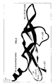
- This domain forms a cysteine knot motif composed of two disulfide bonds which form a covalently linked ring structure between two adjacent ⁇ strands, and a third disulfide bond that penetrates the ring [see for example, Fig I in Muller et al., Structure 5:1325-1338 (1997 )], similar to that found in other cysteine knot growth factors, e.g., transforming growth factor- ⁇ (TGF- ⁇ ).
- TGF- ⁇ transforming growth factor- ⁇
- the PDGF/VEGF family proteins are predominantly secreted glycoproteins that form either disulfide-linked or non-covalently bound homo- or heterodimers whose subunits are arranged in an antiparallel manner [ Stacker and Achen, Growth Factors 17:1-11 (1999 ); Muller et al., Structure 5:1325-1338 (1997 )].
- the PDGFs regulate cell proliferation, cell survival and chemotaxis of many cell types in vitro (reviewed in [ Strukturn et al., Biochimica et Biophysica Acta 1378:F79-113 (1998 )].
- the two chains that make up PDGF, PDGF-A and PDGF-B, can homo- or heterodimerize producing three different isoforms: PDGF-AA, PDGF-AB, or PDGF-BB.
- PDGF-A is only able to bind the PDGF ⁇ -receptor (PDGFR- ⁇ ), whereas PDGF-B can bind both the PDGF- ⁇ and a second PDGF receptor (PDGF- ⁇ )-
- the PDGF proteins exert their effects in a paracrine manner since they often are expressed in epithelial (PDGF-A) or endothelial (PDGF-B) cells in close apposition to the PDGF receptor-expressing mesenchyme (reviewed in Ataliotis et al., Int Rev Cytology 172:95-127 (1997 )].
- PDGFs bas been observed in several pathological conditions, including malignancies, atherosclerosis, and fibroproliferative diseases.
- co-expression of the PDGFs and PDGF receptors generates autocrine loops, which are important for cellular transformation [ Betsholtz et al., Cell 39-447-57 (1984 ); Keating et al., Science 239:914-6 (1988 )].
- mice Homozygous null mutations for either PDGF-A or PDGF-B are lethal in mice. Approximately 50 % of the homozygous PDGF-A deficient mice have an early lethal phenotype, while the surviving animals have a complex postnatal phenotype with lung emphysema due to improper alveolar septum formation, and a dermal phenotype characterized by thin dermis, misshapen hair follicles, and thin hair. PDGF-A is also required for normal development of oligodendrocytes and subsequent myelination of the central nervous system.
- the PDGF-B deficient mice develop renal, hematological and cardiovascular abnormalities; where the renal and cardiovascular defects, at least in part, are due to the lack of proper recruitment of mural cells (vascular smooth muscle cells, pericytes or mesangial cells) to blood vessels.
- mural cells vascular smooth muscle cells, pericytes or mesangial cells
- the VEGF subfamily is composed of PDGF/VEGF members which share a VEGF homology domain (VHD) characterized by the sequence: C-X(22-24)-P-[PSR]-C-V-X(3)-R-C-[GSTA]-G-C-C-X(6)-C-X(32-41)-C(SEQ ID NO: 1201).
- VHD domain determined through analysis of the VEGF subfamily members, comprises the PDGF motif but is more specific.
- VEGF-A was originally purified from several sources on the basis of its mitogenic activity toward endothelial cells, and also by its ability to induce microvascular permeability, hence it is also called vascular permeability factor (VPF).
- VEGF-A has subsequently been shown to induce a number of biological processes including the mobilization of intracellular calcium, the induction of plasminogen activator and plasminogen activator inhibitor-1 synthesis, promotion of monocyte migration in vitro, induction of antiapoptotic protein expression in human endothelial cells, induction of fenestrations in endothelial cells, promotion of cell adhesion molecule expression in endothelial cells and induction of nitric oxide mediated vasodilation and hypotension [ Ferrara, J Mol Med 77: 527-543 (1999 ); Neufeld et al., FASEB J 13 : 9-22 (1999) ; Zachary, Intl J Biochem Cell Bio 30: 1169-1174 (1998) ].
- VEGF-A is a secreted, disulfide-linked homodimeric glycoprotein composed of 23 kD subunits.
- each isoform differs in biological activity, receptor specificity, and affinity for cell surface- and extracellular matrix-associated heparan-sulfate proteoglycans, which behave as low affinity receptors for VEGF-A.
- VEGF 121 does not bind to either heparin or heparan-sulfate; VEGF 145 and VEGF 165 (GenBank Acc. No. M32977) are both capable of binding to heparin; and VEGF 189 and VEGF 206 show the strongest affinity for heparin and heparan-sulfates.
- VEGF 121 , VEGF 145 , and VEGF 165 are secreted in a soluble form although most of VEGF 165 is confined to cell surface and extracellular matrix proteoglycans, whereas VEGF 189 and VEGF 206 remain associated with extracellular matrix.
- Both VEGF 189 and VEGF 206 can be released by treatment with heparin or heparinase, indicating that these isoforms are bound to extracellular matrix via proteoglycans.
- Cell-bound VEGF 189 can also be cleaved by proteases such as plasmin, resulting in release of an active soluble VEGF 110 -
- proteases such as plasmin
- Most tissues that express VEGF are observed to express several VEGF isoforms simultaneously, although VEGF 121 and VEGF 165 are the predominant forms, whereas VEGF 206 is rarely detected [ Ferrara, J Mol Med 77:527-543 (1999 )].
- VEGF 145 differs in that it is primarily expressed in cells derived from reproductive organs [ Neufeld et al., FASEB J 13:9-22 (1999 )].
- VEGF-A The pattern of VEGF-A expression suggests its involvement in the development and maintenance of the normal vascular system, and in angiogenesis associated with tumor growth and other pathological conditions such as rheumatoid arthritis.
- VEGF-A is expressed in embryonic tissues associated with the developing vascular system, and is secreted by numerous tumor cell lines. Analysis of mice in which VEGF-A was knocked out by targeted gene disruption indicate that VEGF-A is critical for survival, and that the development of the cardiovascular system is highly sensitive to VEGF-A concentration gradients. Mice lacking a single copy of VEGF-A die between day 11 and 12 of gestation. These embryos show impaired growth and several developmental abnormalities including defects in the developing cardiovasculature.
- VEGF-A is also required post-natally for growth, organ development, regulation of growth plate morphogenesis and endochondral bone formation. The requirement for VEGF-A decreases with age, especially after the fourth postnatal week. In mature animals, VEGF-A is required primarily for active angiogenesis in processes such as wound healing and the development of the corpus luteum. [ Neufeld et al., FASEB J 13:9-32 (1999 ); Ferrara, J Mol Med 77:527-543 (1999 )]. VEGF-A expression is influenced primarily by hypoxia and a number of hormones and cytokines including epidermal growth factor (EGF), TGF- ⁇ , and various interleukins. Regulation occurs transcriptionally and also post-transcriptionally such as by increased mRNA stability [ Ferrara, J Mol Med 77:527-543 (1999 )].
- EGF epidermal growth factor
- PIGF a second member of the VEGF subfamily, is generally a poor stimulator of angiogenesis and endothelial cell proliferation in comparison to VEGF-A, and the in vivo role of PIGF is not well understood.
- Three isoforms of PIGF produced by alternative mRNA splicing have been described [ Hauser et al., Growth Factors 9:259-268 (1993 ); Maglione et al., Oncogene 8:925-931 (1993 )]- PIGF forms both disulfide-liked homodimers and heterodimers with VEGF-A.
- the P1GF-VEGF-A heterodimers are more effective at inducing endothelial cell proliferation and angiogenesis than PIGF homodimers.
- PIGF is primarily expressed in the placenta, and is also co-expressed with VEGF-A during early embryogenesis in the trophoblastic giant cells of the parietal yolk sac [ Stacker and Achen, Growth Factors 17:1-11 (1999 )].
- VEGF-B shares approximately 44% amino acid identity with VEGF-A.
- VEGF-B has been shown to have angiogenic properties, and may also be involved in cell adhesion and migration, and in regulating the degradation of extracellular matrix. It is expressed as two isoforms of 167 and 186 amino acid residues generated by alternative splicing.
- VEGF-B 167 is associated with the cell surface or extracellular matrix via a heparin-binding domain, whereas VEGF-B 186 is secreted.
- Both VEGF-B 167 and VEGF-B 186 can form disulfids-linked homodimers or heterodimers with VEGF-A.
- the association to the cell surface of VEGF 165 -VEGF-B 167 heterodimers appears to be determined by the VEGF-B component, suggesting that heterodimerization may be important for sequestering VEGF-A.
- VEGF-B is expressed primarily in embryonic and adult cardiac and skeletal muscle tissues [ Joukov et al., J Cell Physiol 173:211-215 (1997 ); Stacker and Achen, Growth Factors 17:1-11 (1999 )]. Mice lacking VEGF-B survive but have smaller hearts, dysfunctional coronary vasculature, and exhibit impaired recovery from cardiac ischemia [ Bellomo et al., Circ Res 2000;E29-E35 ].
- a fourth member of the VEGF subfamily, VEGF-C comprises a VHD that is approximately 30% identical at the amino acid level to VEGF-A VEGF-C is originally expressed as a larger precursor protein, prepro-VEGF-C, having extensive amino- and carboxy-terminal peptide sequences flanking the VHD, with the C-terminal peptide containing tandemly repeated cysteine residues in a motif typical of Balbiani ring 3 protein Prepro-VEGF-C undergoes extensive proteolytic maturation involving the successive cleavage of a signal peptide, the C-terminal pro-peptide, and the N-terminal pro-peptide.
- VEGF-C protein consists of a non-covalently-linked homodimer, in which each monomer contains the VHD.
- the intermediate forms of VEGF-C produced by partial proteolytic processing show increasing affinity for the VEGFR-3 receptor, and the mature protein is also able to bind to the VEGFR-2 receptor.
- Joikov et al., EMBOJ. 16:(13):3898-3911 (1997 ). It has also been demonstrated that a mutant VEGF-C, in which a single cysteine at position 156 is either substituted by another amino acid or deleted, loses the ability to bind VEGFR-2 but remains capable of binding and activating VEGFR-3 [ International Patent Publication No. WO 98/33917 ].
- VEGF-C vascular endothelial growth factor-C mRNA is expressed primarily in the allantois, jugular area, and the metanephros. [ Joukov et al., J Cell Physiol 173:211-215 (1997 )]. VEGF-C is involved in the regulation of lymphatic angiogenesis: when VEGF-C was overexpressed in the skin of transgenic mice, a hyperplastic lymphatic vessel network was observed, suggesting that VEGF-C induces lymphatic growth [ Jeltsch et al., Science, 276:1423-1425 (1997 )].
- VEGF-C also shows angiogenic properties: it can stimulate migration of bovine capillary endothelial (BCE) cells in collagen and promote growth of human endothelial cells [see, e.g., International Patent Publication No. WO 98/33917 , incorporated herein by reference].
- BCE bovine capillary endothelial
- VEGF-D is structurally and functionally most closely related to VEGF-C [see International Patent Publ. No. WO 98/07832 , incorporated herein by reference]. Like VEGF-C, VEGF-D is initially expressed as a prepro-peptide that undergoes N-terminal and C-terminal proteolytic processing, and forms non-covalently linked dimers. VEGF-D stimulates mitogenic responses in endothelial cells in vitro. During embryogenesis, VEGF-D is expressed in a complex temporal and spatial pattern, and its expression persists in the heart, lung, and skeletal muscles in adults.
- VEGF-D ⁇ N ⁇ C Isolation of a biologically active fragment of VEGF-D designated VEGF-D ⁇ N ⁇ C, is described in International Patent Publication No. WO 98/07832 , incorporated herein by reference.
- VFGF-D ⁇ N ⁇ C consists of amino acid residues 93 to 201 of VEGF-D linked to the affinity tag peptide FLAG ® .
- VEGF-E and NZ2 VEGF are potent mitogens and permeability enhancing factors. Both show approximately 25% amino acid identity to mammalian VEGF-A, and are expressed as disulfide-liked homodimers. Infection by these viruses is characterized by pustular dermititis which may involve endothelial cell proliferation and vascular permeability induced by these viral VEGF proteins.
- VEGF-like proteins have also been identified from two additional strains of the orf virus, D1701 [GenBank Acc. No. AF106020; described in Meyer et al., EMBO J 18:363-374 (1999 )] and NZ10 [described in International Patent Application PCT/US99/25869 , incorporated herein by reference]. These viral VEGF-like proteins have been shown to bind VEGFR-2 present on host endothelium, and this binding is important for development of infection and viral induction of angiogenesis [ Meyer et al., EMBO J 18:363-374 (1999 ); International Patent Application PCT/US99/25869 ].
- PDGFR- ⁇ see e.g., GenBank Acc. No. NM006206
- PDGFR- ⁇ see e.g., GenBank Acc. No NM002609
- VEGFR-1/Flt-1 fms -like tyrosine kinase-1; GenBank Acc. No. X51602; De Vries et al., Science 255:989-991 (1992 )
- VEGFR-2/KDR/Flk-1 kinase insert domain containing receptor/fetal liver kinase-1; GenBank Acc- Nos.
- VEGF 121 , VEGF 165 , VEGF-B, P1GF-1 and P1GF-2 bind VEGF-R1; VEGF 121 , VEGF 145 , VEGF 165 , VEGF-C, VEGF-D, VEGF-E, and NZ2 VEGF bind VEGF-R2; VEGF-C and VEGF-D bind VEGFR-3; VEGF 165 , PIGF-2, and NZ2 VEGF bind neuropilin-1; and VEGF 165 binds neuropilin-2.[ Neufeld et al., FASEB J 13:9-22 (1999 ); Stacker and Achen, Growth Factors 17:1-11 (1999 ); Ortega et al., Fron Biosci 4:141-152 (1999 ); Zachary, IntI J Biochem Cell Bio 30
- the PDGF receptors are protein tyrosine kinase receptors (PTKs) that contain five immunoglobulin-like loops in their extracellular domains.
- PTKs protein tyrosine kinase receptors
- VEGFR-1, VEGFR-2, and VEGFR-3 comprise a subgroup of the PDGF subfamily of PTKs, distinguished by the presence of seven Ig domains in their extracellular domain and a split kinase domain in the cytoplasmic region.
- Both neuropilin- and neuropilin-2 are non-PTK VEGF receptors.
- NP-1 has an extracellular portion includes a MAM domain; regions of homology to coagulation factors V and VIII, MFGPs and the DDR tyrosine kinase, and two CUB-like domains.
- VEGFR-1 A soluble isoform of VEGFR-1 lacking the seventh Ig-like loop, transmembrane domain, and the cytoplasmic region is expressed in human umbilical vein endothelial cells.
- This VEGFR-1 isoform binds VEGF-A with high affinity and is capable of preventing VEGF-A-induced mitogenic responses [ Ferrara, J Mol Med 77-527-543 (1999 ); Zachary, Intl J Biochem Cell Bio 30;1169-1174 (1998 )].
- a C-terminal truncated from of VEGFR-2 has also been reported [ Zachary, IntI J Biochem Cell Bio 30:1169-1174 (1998 )].
- there are two isoforms of the VEGFR-3 protein which differ in the length of their C-terminal ends. Studies suggest that the longer isoform is responsible for most of the biological properties of VEGFR-3.
- the receptors for the PDGFs, PDGF ⁇ -receptor (PDGFR- ⁇ ) and the ⁇ -receptor (PDGFR- ⁇ ), are expressed by many in vitro grown cell lines, and they are mainly expressed by mesenchymal cells in vivo (reviewed in [ Raines et al., Peptide growth factors and their receptors, Heidelberg, Springer-Verlag (1990 )]. As mentioned above, PDGF-B binds both PDGFRs, while PDGF-A selectively binds PDGFR- ⁇ .
- PDGFR- ⁇ deficient mice die during embryogenesis at e10, and show incomplete cephalic closure, impaired neural crest development, cardiovascular defects, skeletal defects, and odemas.
- the PDGFR- ⁇ deficient mice develop similar phenotypes to animals deficient in PDGF-B, that are characterized by renal, hematological and cardiovascular abnormalities ; where the renal and cardiovascular defects, at least in part, are due to the lack of proper recruitment of mural cells (vascular smooth muscle cells, pericytes or mesangial cells) to blood vessels.
- mural cells vascular smooth muscle cells, pericytes or mesangial cells
- VLGFR-1 occurs mainly in vascular endothelial cells, although some may be present on monocytes, trophoblast cells, and renal mesangial cells [ Neufeld et al., FASEB J 13:9-22 (1999 )]. High levels of VEGFR-1 mRNA are also detected in adult organs, suggesting that VEGFR-1 has a function in quiescent endothelium of mature vessels not related to cell growth. VEGFR-1-/-mice die in utero between day 8.5 and 9.5. Although endothelial cells developed in these animals, the formation of functional blood vessels was severely impaired, suggesting that VEGFR-1 may be involved in cell-cell or cell-matrix interactions associated with cell migration.
- mice expressing a mutated VEGFR-1 in which only the tyrosine kinase domain was missing show normal angiogenesis and survival, suggesting that the signaling capability of VEGFR-1 is not essential.
- VEGFR-2 expression is similar to that of VEGFR-1 in that it is broadly expressed in the vascular endothelium, but it is also present in hematopoietic stem cells, megakaryocytes, and retinal progenitor cells [ Neufeld et al., FASEB J 13:9-22 (1999 )]. Although the expression pattern of VEGFR-1 and VEGFR-2 overlap extensively, evidence suggests that, in most cell types, VEGFR-2 is the major receptor through which most of the VEGFs exert their biological activities.
- VEGFR-3 is expressed broadly in endothelial cells during early embryogenesis. During later stages of development, the expression of VEGFR-3 becomes restricted to developing lymphatic vessels [ Kaipainen, A., et al., Proc. Natl. Acad. Sci. USA, 92: 3566-3570 (1995 )]. In adults, the lymphatic endothelia and some high endothelial venules express VEGFR-3, and increased expression occurs in lymphatic sinuses in metastatic lymph nodes and in lymphangioma. VEGFR-3 is also expressed in a subset of CD34 + hematopoietic cells which may mediate the myelopoietic activity of VEGF-C demonstrated by overexpression studies [ WO 98/33917 ].
- VEGFR-3 Targeted disruption of the VEGFR-3 gene in mouse embryos leads to failure of the remodeling of the primary vascular network, and death after embryonic day 9.5 [ Durnont et al, Science, 282: 946-949 (1998) ]. These studies suggest an essential role for VEGFR-3 in the development of the embryonic vasculature, and also during lymphangiogenesis.
- VEGF receptors Structural analyses of the VEGF receptors indicate that the VEGF-A binding site on VEGFR-1 and VEGFR-2 is located in the second and third Ig-like loops. Similarly, the VEGF-C and VEGF-D binding sites on VEGFR-2 and VEGFR-3 are also contained within the second Ig-loop [ Taipale et al., Curr Top Microbiol Immunol 237:85-96 (1999 )]. The second Ig-like loop also confers ligand specificity as shown by domain swapping experiments [ Ferrara, J Mol Med 77:527-543 (1999 )].
- VEGFR-1 and VEGFR-2 are structurally similar, share common ligands (VEGF 121 and VEGF 165 ), and exhibit similar expression patterns during development.
- the signals mediated through VEGFR-1 and VEGFR-2 by the same ligand appear to be slightly different.
- VEGFR-2 has been shown to undergo autophosphorylation in response to VEGF-A, but phosphorylation of VEGFR-1 under identical conditions was barely detectable.
- VEGFR-2 mediated signals cause striking changes in the morphology, actin reorganization, and membrane ruffling of porcine aortic endothelial cells recombinantly overexpressing this receptor.
- VFGFR-2 also mediated ligand-induced chemotaxis and mitogenicity; whereas VEGFR-1-transfected cells lacked mitogenic responses to VEGF-A- Mutations in VEGF-A that disrupt binding to VEGFR-2 fail to induce proliferation of endothelial cells, whereas VEGF-A mutants that are deficient in binding VEGFR-1 are still capable of promoting endothelial proliferation.
- VEGF stimulation of cells expressing only VEGFR-2 leads to a mitogenic response whereas comparable stimulation of cells expressing only VEGFR-1 also results in cell migration, but does not induce cell proliferation.
- phosphoproteins co-precipitating with VEGFR-1 and VEGFR-2 arc distinct, suggesting that different signaling molecules interact with receptor-specific intracellular sequences.
- VEGFR-1 in angiogenesis may be to negatively regulate the activity of VEGF-A by binding it and thus preventing its interaction with VEGFR-2, whereas VEGFR-2 is thought to be the main transducer of VEGF-A signals in endothelial cells.
- mice deficient in VEGF1Z-1 die as embryos while mice expressing a VEGPR-1 receptor capable of binding VEGF-A but lacking the tyrosine kinase domain survive and do not exhibit abnormal embryonic development or angiogenesis.
- analyses of VEGF-A mutants that bind only VEGFR-2 show that they retain the ability to induce mitogenic responses in endothelial cells.
- VEGFR-1 VEGFR-1 signaling
- Ferrara J Mol Med 77:527-543 (1999 ); Zachary, Intl J Biochem Cell Bio 30:1169-1174 (1998 )].
- Neuropilin-1 was originally cloned as a receptor for the collapsin/semaphorin family of proteins involved in axon guidance [ Stacker and Achen, Growth Factors 17:1-11 (1999 )]. It is expressed in both endothelia and specific subsets of neurons during embryogenesis, and it thought to be involved in coordinating the developing neuronal and vascular system. Although activation of neuropilin-1 does not appear to elicit biological responses in the absence of the VEGF family tyrosine-kinase receptors, their presence on cells leads to more efficient binding of VEGF 165 and VEGFR-2 mediated responses. [ Neufeld et al., FASEB J 13:9-22 (1999 )] Mice lacking neuropilin-1 show abnormalities in the developing embryonic cardiovascular system. [ Neufeld et al., FASEB J 13:9-22 (1999 )]
- Neuropilin-2 was identified by expression cloning and is a collapsin/semaphorin receptor closely related to neuropilin-1. Neuropilin-2 is an isoform-specific VEGF receptor in that it only binds VEGF 165 . Like neuropilin-1, neuropilin-2 is expressed in both endothelia and specific neurons, and is not predicted to function independently due to its relatively short intracellular domain. The function of neuropilin-2 in vascular development is unknown [ Neufeld et al., FASEB J 13:9-22 (1999 ); WO 99/30157 ].
- VEGF-A as a key regulator of vascular development has spurred active research using VEGF-based therapeutic angiogenesis in cardiovascular medicine, as well as for treating diseases characterized by pathological angiogenesis with VEGF antagonists. Subsequent identification of additional VEGF family proteins and their roles in vascularization have also led to the development of therapies based on these growth factors [ Ferrara and Alitalo, Nature Med 5:1359-1364 (1999 )]. Animal studies of hindlimb ischemia, and myocardial ischemia using VEGF-A or VEGF-C, delivered by administration of recombinant protein or gene transfer using naked DNA or adenoviral vectors, implicate these molecules in promoting vascularization and increasing coronary blood flow.
- VEGF growth factors include using VEGF-C to promote lymphangiogenesis in patients whose axillary lymph nodes were removed during breast carcinoma surgery- Therapies using combinations of growth factors to promote vascularization in tissues may also prove to be preferable in treating certain diseases [ Ferrara and Alitalo, Nature Med 5: 1359-1364 (1999 )].
- VEGF expression is upregulated in most human tumors including primary breast cancer and gastric carcinoma.
- Studies in mice indicate that tumor-associated angiogenesis and growth of the tumor cells can be inhibited by treating the animals with monoclonal antibodies against VEGF-A.
- Further animal studies showed that expression of a dominant negative VBGFR-2 mutant that prevents signaling through this receptor, or administration of recombinant VEGFR- 1 or VEGFR-2 mutants, which only contain the extracellular portion of these receptors, suppresses growth of several tumor cell lines.
- rhuMAb VEGF humanized high affinity monoclonal antibodies against VEGF-A
- Phase II studies using rhuMAb VEGF to treat non-small cell lung carcinoma, colorectal carcinoma, breast, and renal cell carcinoma are currently ongoing.
- Compounds targeting inhibition of VEGF-C activity are also being tested for therapeutic uses in cancer patients: small molecule inhibitors of VEGF-C are in Phase II trials, and monoclonal antibodies against VEGF-C are entering clinical trials.
- VEGF-A Retinopathy associated with diabetes mellitus, occlusion of central retinal vein or prematurity has been correlated with increased levels of VEGF-A.
- VEGF-A is also detected in age-related macular degeneration (AMD), and its expression is thought to be the cause of neovascularization in this disease.
- AMD age-related macular degeneration
- Intravitreal delivery of recombinant humanized anti-VEGF-A Fab antibody fragment or injection of 2'-fluoropyrimidine RNA oligonucleotide ligands (aptamers) to treat AMD are currently in clinical trials.
- Compounds that inhibit the activity of VEGF growth factors may also be used to treat other disease states involving abnormal angiogenesis. These include ischemic-reperfusion related brain edema and injury, conditions associated with ovarian hyperplasia and hypervascularity such as the polycystic ovary syndrome, endometriosis, and ovarian hyperstimulation syndrome [ Ferrara and Alitalo, Nature Med 5:1359-1364 (1999 )].
- VEGF family of growth factors, and inhibitors thereof have tremendous potential as therapeutics.
- growth factors and inhibitors are useful to promote or inhibit angiogenesis where needed, such as in the treatment of ischemic disorders, the promotion of wound healing, or the inhibition or elimination of neoplastic disorders that are angiogenesis-dependent.
- the various naturally-occurring members of this growth factor family often bind multiple receptors, and the various known receptors are expressed on multiple cell types and have expression patterns that may vary depending on stage of development and the presence or absence of pathological conditions.
- the biological effects of any particular growth factor may be receptor-dependent, isoform dependent, and cell-type dependent.
- a desirable therapeutic effect mediated through one receptor may be accompanied by undesirable side-effects mediated through another receptor.
- a desirable therapeutic effect might be enhanced through stimulation of multiple receptors that cannot be stimulated with any single known growth factor that occurs in nature. Therefore, a need exists for novel peptide growth factors with their own unique profile of receptor binding and receptor-stimulating or receptor-inhibiting activities.
- the present invention satisfies needs identified above by providing novel polypeptide binding molecules for naturally occurring vascular endothelial growth factor receptors, and polynucleotides that encode the novel polypeptides and are useful for recombinant expression of the polypeptides.
- vascular endothelial growth factor and the abbreviation "VEGF” (without modifier) are used herein in a generic sense, to describe any of a family of growth factor polypeptides including but not limited to Vascular Endothelial Growth Factor-A (VEGF-A), Vascular Endothelial Growth Factor-13 (VEGF-B), Vascular Endothelial Growth Factor-C (VEGF-C), Vascular Endothelial Growth Factor-D (VEGF-D), Platelet Derived Growth Factor-A (PDGF-A), Platelet Derived Growth Factor-B (PDGF-B), Placenta Growth Factor (PIGF), and virally encoded VEGF-like molecules.
- VEGF-A Vascular Endothelial Growth Factor-A
- VEGF-B Vascular Endothelial Growth Factor-13
- VEGF-C Vascular Endothelial Growth Factor-C
- VEGF-D Vascular Endothelial Growth Factor-D
- VEGF-A is commonly referred to in the art as ''Vascular Endothelia) Growth Factor” or as "VEGF,” but for clarity shall be referred to herein as VEGF-A or referred to as specific isoforms (e.g., VEGF 165 ) of VEGF-A.
- a polypeptide comprising an amino acid sequence at least 95% identical to amino acids 1-102 as set forth in SEQ ID NO: 51, SEQ ID NO: 59, SEQ ID NO: 63, or SEQ ID NO: 163, wherein the polypeptide binds to at least one receptor selected from the group consisting of human VEGFR-1, human VEGFR-2 and human VEGFR-3, is provided.
- a second aspect of the invention provides a polypeptide comprising an amino acid sequence at least 95% identical to amino acids 1-104 as set forth in SEQ ID NO: 67, SEQ ID NO: 71, SEQ ID NO. 75, SEQ ID NO: 77, SEQ ID NO: 153, SEQ ID NO: 165, SEQ ID NO: 167, SEQ ID NO: 169, SEQ ID NO: 173 or SEQ ID NO: 175, wherein the polypeptide binds to at least one receptor selected from the group consisting of human VEGFR-1, human VEGFR-2 and human VEGFR-3.
- a third aspect of the invention provides a polypeptide comprising an amino acid sequence at least 95% identical to amino acids 1-105 as set forth in SEQ ID NO: 155, SEQ ID NO: 157 or SEQ ID NO: 159, wherein the polypeptide binds to exactly one receptor selected from the group consisting of human VEGFR-1, human VEGFR-2 and human VEGFR-3.
- a fourth aspect of the invention provides a polypeptide comprising an amino acid sequence at least 95% identical to amino acids 1-103 as set forth in SEQ ID NO: 161, wherein the polypeptide binds to human VEGFR-1, human VBGFR-2 and human VEGFR-3.
- a fifth aspect of the invention provides a hybrid polypeptide comprising an amino acid sequence at least 95% identical to the expression product of clone number 11.9 (SEQ ID NO: 433), 11.13 (SEQ ID NO: 945), 12. 16 (SEQ ID NO: 1185), 13.9 (SEQ ID NO: 465), 13.11 (SEQ ID NO: 529), 13.13 (SEQ ID NO: 977), 13.15 (SEQ ID NO: 1041), 14.1 (SEQ ID NO: 193), 14.5 (SEQ ID NO: 705), 14.7 (SEQ ID NO: 769), 22.3 (SEQ ID NO: 303), 22.4 (SEQ ID NO: 431), 23.12 (SEQ ID NO: 671), 23.14 (SEQ ID NO: 1119), 32.9 (SEQ ID NO: 485), 32.11 (SEQ ID NO: 549), 32.15 (SEQ ID NO: 1061), 32.16 (SEQ ID NO: 1189), 33.1 (SEQ ID NO: 213), 33.3 (SEQ ID
- a polynucleotide, vector or host cell comprising a nucleotide sequence encoding a polynucleotide of any one of the sequences listed in relation to the first to fifth aspects of the invention is also contemplated.
- naturally-occurring vertebrate vascular endothelial growth factor polypeptides means polypeptides having the following characteristics:
- naturally-occurring vertebrate VEGF polypeptides means polypeptides that have certain specified structural and functional properties. The term is not intended in this context to imply a source of origin. Thus, recombinantly produced polypeptides that satisfy the above criteria because they have an amino acid sequences and receptor binding properties of VEGF polypeptides that exist in nature are considered “naturally-occurring”.
- vascular endothelial growth factor polypeptides are already known in the art, including but not limited to human Vascular Endothelial Growth Factor-A (VEGF-A), Vascular Endothelial Growth Factor-B (VEGF-B), Vascular Endothelial Growth Factor-C (VEGF-C), Vascular Endothelial Growth Factor-D (VEGF-D), Platelet Derived Growth Factor-A (PDGF-A), Platelet Derived Growth Factor-B (PDGF-B), Placenta Growth Factor (PIGF); mammalian and avian orthologs thereof (where the term "ortholog” means species homolog); and Vascular Endothelial Growth Factor E (VEGF-E), NZ2 VEGF, and the two VEGF-like proteins identified in strains D1701 and NZ10, which have been identified in poxviruses.
- VEGF-A Vascular Endothelial Growth Factor-A
- VEGF-B Vascular End
- VEGF vascular endothelial growth factor
- Chimeric polypeptides may be derived from any pair, or three, or four, or more of the VEGFs described herein or their species orthologs.
- chimeric requires that the amino acid sequence of the chimeric molecule include at least one stretch of one or more amino acids (preferably stretches of 1, 2, 3, 4, 5, 6, 7, 8, 9, 10, 11, 12, 13, 14, 15, 16, 17, 18, 19, 20, or more amino acids) from each of the naturally-occurring VEGF's from which it was derived.
- the chimeric polypeptide is a "hybrid” or “mosaic” of two or more polypeptides.
- chimeric is meant that the polypeptide of the invention is not identical to any naturally occurring VEGF sequence (or fragment of a natural VEGF sequence).
- derived from means that, when the amino acid sequences of the chimeric polypeptide and the two or more naturally occurring VHGF's are aligned using a standard algorithm, substantially all of the amino acids in the chimeric polypeptide arc aligned with an identical residue in one or more of the naturally occurring VEGF's from which the chimeric was derived.
- Standard protein alignment algorithms for example, the clustral method [ Nucl Acids Res 22:4673-80 (1994 )], the Jotun Hein method [ Methds Enzymol 183:626-645 (1990 )], or the Feng-Doolittle method [ J Mol Evol 25:351-360 (1987 )], can be used to align naturally-occurring vertebrate vascular endothelial growth factor polypeptides, such alignments being greatly facilitated by the presence of the eight highly conserved cysteines dispersed through the V/PHD.
- a chimeric polypeptide is "derived from" two or more naturally occurring VEGFs by performing an alignment using any generally accepted protein alignment algorithm.
- substantially all reflects the fact that techniques described herein for making chimeric polypeptides will sometimes introduce mutations such as insertions, deletions, or substitutions:, preventing 100% correlation to parent sequences. In such cases, at least about 90%, 92%, 94%, 95%. 96%, 97%, 98%, or 99% of the residues of the chimeric polypeptide will align with identical residues from at least one of the natural VEGF's.
- the term "plurality of peptide subunits” means two or more peptide subunits.
- Exemplified herein are chimeric polypeptides obtained by fragmenting two naturally occurring VEGF cDNA's (human VEGF-A and human VEGF-C) into nine subunits of about 8-16 codons each, recombining these fragments into all 512 permutations of the nine subunits (maintaining subunit order), and expressing the resultant chimeric cDNAs. The number and the size of fragments is not intended as a critical feature.
- the "subunits" are joined by peptide bonds to form a polypeptide chain.
- vascular endothelial growth factor receptor binding profile means the determination of the receptors to which a polypeptide will bind and the receptors to which it will not.
- VEGF receptors including VEGFR-1, VEGFR-2, and VEGFR-3, are described in greater detail elsewhere herein.
- Known PDGF receptors are also described in greater detail elsewhere herein.
- a chimeric polypeptide has been derived in part from a naturally occurring PDGF sequence
- screening the chimeric polypeptide for binding to PDGF receptors is contemplated as part of the receptor binding profile determination.
- the chimeric polypeptide has a different receptor binding profile than either of its parent molecules if it binds to only one of the three receptors, or if it binds to all three receptors, or if it binds to VEGFR-1 and VEGFR-3 but not VEGFR-2.
- NP-1 binding is mediated by amino acid residues 142 to 185 of SEQ ID NO: 2 for VEGF-A, and amino acid residues 138 to 182 for VEGF-B [ Soker et al., J Biol Chem 271:5761-7 (1996 ); Makinen et al., J Biol Chem 274:21217-22 (1999 )].
- addition of upstream or downstream sequences to chimeric polypeptides of the invention is contemplated, and some added sequences are contemplated to result in NP-1 or NP-2 binding.
- the present invention is believed to provide the first disclosure of a polypeptide that is capable of binding to all of VEGFR-1, VEGFR-2, and VEGFR-3. All polypeptides having this receptor binding profile are intended as within the scope of the invention.
- Naturally occurring VEGF polypeptides generally bind their respective receptors with high affinity, which is generally understood in this context to mean binding with a sub-nanomolar dissociation constant.
- VEGF-A binds VEGFR-1 and VEGFR-2 with Kd of approximately 16 pM and 760 pM, respectively; and
- VEGF-C binds VEGFR-2 and VEGFR-3 with Kd of approximately 410 pM and 135 pM, respectively.
- chimeric polypeptides having less receptor affinity i.e., higher dissociation constants
- a 50 nanomolar dissociation constant cutoff is selected.
- Chimeric polypeptides of the invention include chimeric (hybrid) receptor binding domains as explained in the preceding paragraphs, and optionally may include additional upstream or downstream sequences from naturally occurring VEGF's, including upstream and downstream sequences that are present in mature isoforms of naturally occurring circulating VEGF's, and/or upstream or downstream pro-peptide sequences that are removed during normal intracellular or extracellular processing.
- Chimeric polypeptides described in Example 1 were prepared using residues 34-135 (SEQ ID NO: 2) of VEGF-A and using 112- 216 of human prepro-VEGF-C (SEQ ID NO: 22).
- Chimeric polypeptides of the invention include the peptides actually exemplified, and also include such peptides modified by the addition of upstream or downstream VEGF-A or VEGF-C sequences from SEQ ID NOs: 2 or 22.
- VEGF-A/VEGF-C chimeric polypeptides as exemplified herein, the addition of upstream and downstream sequences that correspond with amino- and/or carboxyl- terminal sequences characteristic of natural VEGF-A or VEGF-C isoforms is particularly contemplated.
- Chimeric polypeptides according to the invention may comprise any such modifications and additions to the amino acid sequence derived from two or more naturally-occurring vertebrate vascular endothelial growth factor polypeptides.
- K d dissociation constants
- EC 50 or the half effective concentration, is the concentration that produces 50% of a maximal effect.
- An exemplary assay for determining the EC 50 of a putative ligand for a specific receptor is set forth in Example 6, below.
- the fourth fragment (X 4 ) appears to include residues that are important for conferring VEGFR-3 binding affinity.
- Fragments 5 and 8 of VEGF-C also appear to contribute to VEGFR-3 binding.
- the invention provides hybrid polypeptides as described above, wherein the polypeptide further includes one or more amino acid sequences selected from the group consisting of a prepro-VEGF-C signal peptide, a prepro-VEGF-C amino-terminal propeptide, and a prepro-VEGF-C carboxy-terminal pro-peptide.
- hybrid polypeptides of the invention is not restricted to expression only with naturally-occurring flanking VEGF-A or VEGF-C sequences, however.
- Expression of polypeptides of the invention in bacteria may be accomplished by including an initiator methionine or methionine-lysine upstream of the hybrid VEGF sequences, whereas expression and secretion in mammalian, cells is most conveniently accomplished by including at least a signal peptide.
- polypeptides of the invention as fusions with other heterologous sequences, such as tag sequences to facilitate purification or expression as part of larger fusion peptides also is contemplated.
- An exemplary tag of this type is a polyhistidine sequence, generally around six histidine residues, that permits isolation of a compound so labeled using nickel chelation.
- Other labels and tags such as the FLAG ® tag (Eastman Kodak, Rochester, NY), well known and routinely used in the art, are embraced by the invention.
- Exemplary fusions include use of commercially available vectors that express a desired polypeptide as part of glutathione-S-transferase (GST) fusion product. After cleavage of the GST component from the desired polypeptide, an additional glycine residue at position -1 may remain. Variants which result from expression in other vector systems are also contemplated.
- the present application also provides variants (analogs) of the hybrid polypeptides of the invention, wherein one or more amino acids of the hybrid peptide amino acid sequence has been added, deleted, or substituted by another amino acid, and wherein the hybrid retains the receptor binding and/or a biological activity characteristic of the hybrid polypeptide.
- substitution variants wherein merely conservative substitutions have been introduced (e.g., by modification of polynucleotides encoding polypeptides of the invention) are intended as equivalents of hybrid polypeptides of the invention.
- Amino acids can be classified according to physical properties and contribution to secondary and tertiary protein structure.
- a conservative substitution is recognized in the art as a substitution of one amino acid for another amino acid that has similar properties.
- Exemplary conservative substitutions based on amino acid side chain properties are set out in the table immediately below, using standard one letter abbreviations.
- the following table provides still an another alternative, exemplary set of conservative amino acid substitutions.
- hybrid polypeptides of the invention share at least about 70%, 80%, 90%, 95%, 96%, 97%, 98%, or 99% amino acid identity with hybrids that consist entirely of amino acid sequences derived from naturally occurring VEGF's.
- Preferred methods to determine identity and/or similarity are designed to give the largest match between the sequences tested. Methods to determine identity and similarity are described in publicly available computer programs. Preferred computer program methods to determine identity and similarity between two sequences include, but are not limited to, the GCG program package, including GAP (Devereux et al., Nucl. Acid. Res., 12:387 (1984); Genetics Computer Group, University of Wiconsin, Madison, WI), BLASTP, BLASTN, and FASTA ( Altschul et al., J. Mol. Biol., 215:403-410 (1990) )- The BLASTX program is publicly available from the National Center for Biotechnology Information (NCBI) and other sources (BLAST Manual, Altschul ct al. NCB/NLM/NIH Bethesda, MD 20894; Altschul et al., supra). The well known Smith Waterman algorithm may also be used to determine identity.
- NCBI National Center for Biotechnology Information
- Preferred parameters for a polypeptide sequence comparison include the following:
- Preferred parameters for nucleic acid molecule sequence comparisons include the following.
- non-naturally occurring vascular endothelial growth factor amino acid sequence is meant a sequence that is not identical to any known, naturally occurring amino acid sequence, such as, in this case, receptor binding domains from known VEGF-A or VEGF-C sequences.
- the invention provides polynucleotides (e.g., cDNA, cDNA with introns introduced to facilitate expression in eukaryotic systems, synthetic DNA, RNA, or combinations thereof, single or double stranded) that comprise a nucleotide sequence encoding the amino acid sequence of the polypeptides of the invention.
- Purified and isolated polynucleotides are preferred. Due to the well-known degeneracy of the genetic code, several polynucleotides sequences exist that encode each polypeptide amino acid sequence of the invention. Such polynucleotides are useful for recombinantly expressing the polypeptides of the invention.
- Exemplary highly stringent hybridization conditions are as follows: hybridization at 65°C for at least 12 hours in a hybridization solution comprising 5X SSPE, 5X Denhardt's, 0.5% SDS, and 2 mg sonicated non-homologous DNA per 100 ml of hybridization solution; washing twice for 10 minutes at room temperature in a wash solution comprising 2X SSPE and 0-1% SDS, followed by washing once for 15 minutes at 65°C: with 2X SSPE and 0.1% SDS; followed by a final wash for 10 minutes at 65°C with 0.1X SSPE and 0.1% SDS.
- Moderate stringency washes can be achieved by washing with 0.5X SSPE instead of 0.1X SSPE in the final 10 minute wash at 65°C.
- Low stringency washes can be achieved by using 1X SSPE for the 15 minute wash at 65°C, and omitting the final 10 minute wash. It is understood in the art that conditions of equivalent stringency can be achieved through variation of temperature and buffer, or salt concentration as described Ausubel, et al. (Eds.), Protocols in Molecular Biology, John Wiley & Sons (1994), pp. 6.0.3 to 6.4.10 . Modifications in hybridization conditions can be empirically determined or precisely calculated based on the length and the percentage of guanosine/cytosine (GC) base pairing of the probe. The hybridization conditions can be calculated as described in Sambrook et al., (Eds.), Molecular Cloning: A Laboratory Manual, Cold Spring Harbor Laboratory Press: Cold Spring Harbor, New York (1989), pp. 9.47 to 9.51 .
- the invention provides vectors comprising a polynucleotide of the invention- Such vectors are useful, e.g., for amplifying the polynucleotides in host cells to create useful quantities thereof.
- the vector may be an expression vector wherein the polynucleotide of the invention is operatively linked to a polynucleotide comprising an expression control sequence.
- Autonomously replicating recombinant expression constructs such as plasmid and viral DNA vectors incorporating polynucleotides of the invention are specifically contemplated.
- Expression control DNA sequences include promoters, enhancers, and operators, and are generally selected based on the expression systems in which the expression construct is to be utilized. Preferred promoter and enhancer sequences are generally selected for the ability to increase gene expression, while operator sequences are generally selected for the ability to regulate gene expression.
- Expression vectors are useful for recombinant production of polypeptides of the invention.
- Expression constructs of the invention may also include sequences encoding one or more selectable markers that permit identification of host cells bearing the construct. Expression constructs may also include sequences that facilitate, and preferably promote, homologous recombination in a host cell. Preferred constructs of the invention also include sequences necessary for replication in a host cell.
- Vectors also are useful for "gene therapy" treatment regimens, wherein a polynucleotide that encodes a polypeptide of the invention is introduced into a subject in need of treatment involving the modulation (stimulation or blockage) of vascular endothelial growth factor receptors, in a form that causes cells in the subject to express the polypeptide of the invention in vivo.
- the invention provides host cells, including prokaryotic and eukaryotic cells, that are transformed or transfected (stably or transiently) with polynucleotides of the invention or vectors of the invention.
- Polynucleotides of the invention may be introduced into the host cell as part of a circular plasmid, or as linear DNA comprising an isolated protein coding region or a viral vector.
- Methods for introducing DNA into the host cell well known and routinely practiced in the art include transformation, transfection, electroporation, nuclear injection, or fusion with carriers such as liposomes, micelles, ghost cells, and protoplasts.
- host cells are useful for amplifying the polynucleotides and also for expressing the polypeptides of the invention encoded by the polynucleotide.
- Such host cells are useful in assays as described herein.
- any host cell is acceptable, including but not limited to bacterial, yeast, plant, invertebrate ( e.g. , insect), vertebrate, and mammalian host cells.
- invertebrate e.g. , insect
- vertebrate e.g. , and mammalian host cells.
- mammalian host cells Use of mammalian host cells is expected to provide for such post-translational modifications (e.g., glycosylation, truncation, lipidation, and phosphorylation) as may be desirable to confer optimal biological activity on recombinant expression products of the invention.
- Glycosylated and non-glycosylated forms of polypeptides are embraced by the present invention.
- the invention further embraces polypeptides described above that have been covalently modified to include one or more water soluble polymer attachments such as polyethylene glycol, polyoxyethylene glycol, or polypropylene glycol.
- Polypeptides of the invention also may be chemically synthesized.
- Isolation of the polypeptide from the cells or from the medium in which the cells are grown is accomplished by purification methods known in the art, e.g ., conventional chromatographic methods including immunoaffinity chromatography, receptor affinity chromatography, hydrophobic interaction chromatography, lectin affinity chromatography, size exclusion filtration, cation or anion exchange chromatography, high pressure liquid chromatography (IIPLC), reverse phase HPLC, and the like.
- Other methods of purification include those wherein the desired protein is expressed and purified as a fusion protein having a specific tag, label, or chelating moiety that is recognized by a specific binding partner or agent.
- the purified protein can be cleaved to yield the desired protein, or be left as an intact fusion protein. Cleavage of the fusion component may produce a form of the desired protein having additional amino acid residues as a result of the cleavage process.
- compositions comprising polypeptides or polynucleotides of the invention.
- Such compositions may comprise one or more polynucleotides or polypeptides of the invention that have been formulated with a pharmaceutically acceptable (e.g., sterile and nontoxic) diluent or carrier.
- a pharmaceutically acceptable (e.g., sterile and nontoxic) diluent or carrier e.g., sterile and nontoxic) diluent or carrier.
- Liquid, semisolid, or solid diluents that serve as pharmaceutical vehicles, excipients, or media are preferred. Any diluent known in the art may be used.
- Exemplary diluents include, but are not limited to, water, saline solutions, polyoxyethylene sorbitan monolaurate, magnesium stearate, methyl- and propylhydroxybenzoate, talc, alginates, starches, lactose, sucrose, dextrose, sorbitol, mannitol, glycerol, calcium phosphate, mineral oil, and cocoa butter.
- Such formulations are useful, e.g., for administration of polypeptides or polynucleotides of the invention to mammalian (including human) subjects in therapeutic regimens.
- polypeptides or polynucleotides of the invention may be used in the manufacture of a medicament for the treatment of disorders described herein, including but not limited to disorders characterized by undesirable endothelial cell proliferation and/or disorders characterized by ischemia and/or vessel occlusion, wherein neovascularization is desirable.
- a kit comprising a polynucleotide, polypeptide, or composition of the invention packaged in a container, such as a vial or bottle, and further comprising a label attached to or packaged with the container, the label describing the contents of the container and providing indications and/or instructions regarding use of the contents of the container to treat one or more disease states as described herein, is also envisaged.
- Polypeptides of the invention conjugated to cytotoxic agents or other agents that modulate cell growth are contemplated.
- Single-stranded oligonucleotides may be prepared based on knowledge of mammalian VEGF polypeptide sequences and the universal genetic code and using conventional chemical synthesis techniques.
- Example 1 demonstrates such a technique, wherein synthetic oligonucleotide pairs were prepared and annealed to prepare double-stranded polynucleotides having single-stranded cohesive ends that encoded fragments of human VEGF-A and human VEGF-C cDNAs or genomic DNAs (preferably cDNAs) encoding natural VEGF's may be fragmented using one or more restriction endonucleases, using DNaseI, or using Exonuclease III.
- a cDNA (coding or non-coding strand) may be used as a template to synthesize complementary fragments, using DNA polymerase and chain termination reagents.
- DNA polymerase and chain termination reagents See, e.g., Lehtovaara et al., Protein Engineering, 2: 63-68 (1988) ,]
- Polynucleotides may be prepared with complementary cohesive single-stranded ends, to facilitate annealing of fragments in a desired order under conventional annealing and ligation conditions for polynucleotides.
- Example I provides a demonstration of this technique to generate 510 human VEGF-A/VEGF-C hybrids.
- Such a technique also may be suitable for annealing fragment mixtures of two or more VEGF cDNAs that have been digested with restriction endonucleases.
- polynucleotides may be mixed and subjected to a self-priming PCR reaction that involves successive steps of denaturation, annealing, and extension.
- PCR may be performed under conditions that introduce errors (mutations) in the PCR products. Such mutations introduce additional molecular variation, and are expected to reduce the overall percentage of biologically active molecules, but also may produce molecules with unexpectedly superior activities.
- Hybrid DNA molecules are expressed by any means known in the art.
- the molecules are cloned into expression vectors, which are in turn used to transform or transfect cells to express the polypeptides.
- the polynucleotides are cloned into a phage display vector system for screening.
- Screening assays may entail a direct receptor binding assay as described below in Example 3.
- receptor binding may be assayed indirectly by assaying for a biological activity induced by receptor binding. Screening may comprise contacting the hybrid polypeptide to a cell that expresses the receptor, wherein changes in cell growth or cell survival induced by the hybrid polypeptide is indicative of binding between the hybrid polypeptide and the receptor.
- Antibodies that may be generated against polypeptides of the invention, and that bind polypeptides of the invention with an affinity greater than for any natural occurring VEGF, also are contemplated Polypeptides comprising the antigen-binding fragments of such antibodies also are contemplated.
- Antibodies that bind to the polypeptides of the invention but not to vertebrate VEGF's are contemplated.
- polypeptides of the invention are useful for modulating (stimulating or inhibiting) cellular processes that are mediated through any of the PDGF/VEGF family of receptors, such as PDGFR- ⁇ , PDGFR- ⁇ . VEGER-1, VEGFR-2, and/or VEGFR-3. These receptors may be involved singularly in certain processes and in combination, to varying extents, in other processes.
- Polypeptides of the invention possess many different receptor binding profiles, and one of the advantages of the invention is the ability to select a polypeptide with a receptor binding profile that matches the receptor expression profile of the biological process to be modulated.
- the peptides of the present invention may be synthesized using a variety of methods, including those described in the summary of invention and the examples.
- the peptides of the present invention can be synthesized in solution or on a solid support in accordance with conventional techniques.
- Various automatic synthesizers are commercially available and can be used in accordance with known protocols. See, for example, Stewart and Young, Solid Phase Peptide Synthesis, 2d. ed-, Pierce Chemical Co., (1984 ); Tarn et al. J. Am. Chem.
- Solid phase peptide synthesis methods use a copoly(styrene-divinylbenzene) containing 0.1-1.0 mMol amines/g polymer- These methods for peptide synthesis use butyloxycarbonyl (t-BOC) or 9-fluorenylmethyloxy-carbonyl(FMOC) protection of alpha-amino groups.
- Both methods involve stepwise syntheses whereby a single amino acid is added at each step starting from the C-terminus of the peptide (Sec, Coligan, et al., Current Protocols in Immunology, Wiley Interscience, 1991, Unit 9 )
- the peptides can be deprotected to remove the t-t-BOC or FMOC amino acid blocking groups and cleaved from the polymer by treatment with acid at reduced temperature (e.g., liquid HF-10% anisole for about 0.25 to about 1 hours at 0°C).
- acid at reduced temperature e.g., liquid HF-10% anisole for about 0.25 to about 1 hours at 0°C.
- the peptides are extracted from the polymer with 1% acetic acid solution which is then lyophilized to yield the crude material.
- This can normally be purified by such techniques as gel filtration on Sephadex G-15 using 5% acetic acid as a solvent. Lyophilization of appropriate fractions of the column will yield the homogeneous peptide or peptide derivatives, which can then be characterized by such standard techniques as amino acid analysis, thin layer chromatography, high performance liquid chromatography, ultraviolet absorption spectroscopy, molar rotation, solubility, and quantitated by the solid phase Edman degradation.
- Phage display can be particularly effective in identifying binding peptides useful according to the invention. Briefly, one prepares a phage library (using e.g. ml 13, fd, or lambda phage), displaying inserts from 4 to about 80 amino acid residues. The inserts may represent, for example, a completely degenerate or biased array. One then can select phage-bearing inserts which bind to the target VEGF receptor(s). This process can be repeated through several cycles of reselection of phage that bind to the target receptor(s).
- DNA sequence analysis can be conducted to identify the sequences of the expressed polypeptides.
- the minimal linear portion of the sequence that binds to the target receptor(s) can be determined.
- These techniques may identify peptides of the invention with still greater receptor binding affinity than peptides already identified herein. Screening resultant peptide against multiple receptors will identify peptides with multiple receptor binding affinities.
- Yeast two-hybrid screening methods also may be used to identify polypeptides that bind to the target receptor(s).
- expression vector/host systems may be utilized to contain and express the chimeric peptides of the present invention.
- microorganisms such as bacteria transformed with recombinant bacteriophage, plasmid or cosmid DNA expression vectors; yeast transformed with yeast expression vectors; insect cell systems infected with virus expression vectors ( e.g ., baculovirus); plant cell systems transfected with virus expression vectors ( e.g. , cauliflower mosaic virus, CaMV; tobacco mosaic virus, TMV) or transformed with bacterial expression vectors (e.g., Ti or pBR322 plasmid); or animal cell systems.
- microorganisms such as bacteria transformed with recombinant bacteriophage, plasmid or cosmid DNA expression vectors; yeast transformed with yeast expression vectors; insect cell systems infected with virus expression vectors (e.g ., baculovirus); plant cell systems transfected with virus expression vectors (e.g. , cauliflower mosaic virus, CaMV; tobacco mosaic virus
- Mammalian cells that are useful in recombinant protein productions include but are not limited to VERO cells, HeLa cells, Chinese hamster ovary (CHO) cell lines, COS cells (such as COS-7), W138, BHK, HepG2, 3T3, RIN, MDCK, A549, PC12, K562 and 293 cells. Exemplary protocols for the recombinant expression of the protein are described herein below.
- the chimeric peptide may be recombinantly expressed in yeast using a commercially available expression system, e.g ., the Pichia Expression System (Invitrogen, San Diego, CA.), following the manufacturer's instructions.
- This system also relies on the pre-pro-alpha sequence to direct secretion, but transcription of the insert is driven by the alcohol oxidase (AOX1) promoter upon induction by methanol.
- AOX1 alcohol oxidase
- the secreted peptide is purified from the yeast growth medium by, e.g ., the methods used to purify the chimeric peptide from bacterial and mammalian cell supernatants.
- the cDNA encoding the peptide may be cloned into the baculovirus expression vector pVL1393 (PharMingen, San Diego, CA). This vector is then used according to the manufacturer's directions (PharMingen) to infect Spodoptera frugiperda cells in sF9 protein-free media and to produce recombinant protein.
- the protein is purified and concentrated from the media using a heparin-Sepharose column (Pharmacia, Piscataway, NJ) and sequential molecular sizing columns (Amicon, Beverly, MA), and resuspended in PBS. SDS-PAGE analysis shows a single band and confirms the size of the protein, and Edman sequencing on a Porton 2090 Peptide Sequencer confirms its N-terminal sequence.
- the peptide may be expressed in an insect system.
- Insect systems for protein expression are well known to those of skill in the art.
- Autographa californica nuclear polyhedrosis virus (AcNPV) is used as a vector to - express foreign genes in Spodoptera frugiperda cells or in Trichoplusia larvae.
- the peptide coding sequence is cloned into a nonessential region of the virus, such as the polyhedrin gene, and placed under control of the polyhedrin promoter. Successful insertion of the peptide will render the polyhedrin gene inactive and produce recombinant virus lacking coat protein coat. The recombinant viruses are then used to infect S.
- the DNA sequence encoding the peptide is amplified by PCR and cloned into an appropriate vector for example, pGEX-3X (Pharmacia, Piscataway, NJ).
- the pGEX vector is designed to produce a fusion protein comprising glutathione-S-transferase (GST), encoded by the vector, and a protein encoded by a DNA fragment inserted into the vector's cloning site.
- GST glutathione-S-transferase
- the primers for the PCR may be generated to include for example, an appropriate cleavage site.
- the recombinant fusion protein may then be cleaved from the GST portion of the fusion protein.
- the pGEX-3X/chimeric peptide construct is transformed into E . coli XL-1 Blue cells (Stratagene, La Jolla CA), and individual transformants were isolated and grown. Plasmid DNA from individual transformants is purified and partially sequenced using an automated sequencer to confirm the presence of the desired chimeric peptide encoding nucleic acid insert in the proper orientation.
- Particularly preferred peptide compositions of the present invention are those which are conjugated to any anti-tumor peptide such as a tumor necrosis factor (TNF).
- TNF-peptides chimeras are generated as recombinant fusions with peptide-encoding sequences fused in frame to TNF (Novagen) encoding sequences.
- Peptide-TNF cDNA is cloned into pET-11b vector (Novagen) and the expression of TNF-peptides in BL21 E. coli is induced according to the pET11b manufacturer's instruction.
- Soluble TNF-peptides are purified from bacterial lysates by ammonium sulfate preparation, hydrophobic interaction chromatography on Phenyl-Sepharose 6 Fast Flow, ion exchange chromatography on DEAE-Sepharose Fast Flow and gel filtration chromatography on Sephacryl-S-300 HR.
- recombinant protein production also may be used to produce the chimeric peptide compositions.
- induction of the GST/chimeric peptide is achieved by growing the transformed XL-1 Blue culture at 37°C in LB medium (supplemented with carbenicillin) to an optical density at wavelength 600 nm of 0.4, followed by further incubation for 4 hours in the presence of 0.5 mM isopropyl ⁇ -D-Thiogalactopyranoside (Sigma Chemical Co., St. Louis MO).
- the fusion protein expected to be produced as an insoluble inclusion body in the bacteria, may be purified as follows. Cells are harvested by centrifugation; washed in 0.15 M NaCl, 10 mM Tris, pH 8, 1 mM EDTA; and treated with 0.1 mg/ml lysozyme (Sigma Chemical Co.) for 15 minutes at room temperature. The lysate is cleared by sonication, and cell debris is pelleted by centrifugation for 10 minutes at 12,000 X g. The fusion protein-containing pellet is resuspended in 50 mM Tris, pH 8, and 10 mM EDTA, layered over 50% glycerol, and centrifuged for 30 min. at 6000 X g.
- the pellet is resuspended in standard phosphate buffered saline solution (PBS) free of Mg 1+ and Ca ++ .
- PBS phosphate buffered saline solution
- the fusion protein is further purified by fractionating the resuspended pellet in a denaturing SDS polyacrylamide gel (Sambrook et al., supra ) .
- the gel is soaked in 0.4 M KCl to visualize the protein, which is excised and electroeluted in gel-running buffer lacking SDS.
- the GST/chimeric peptide fusion protein is produced in bacteria as a soluble protein, it may be purified using the GST Purification Module (Pharmacia Biotech).
- the fusion protein may be subjected to digestion to cleave the GST from the chimeric peptide of the invention.
- the digestion reaction (20-40 ⁇ g fusion protein, 20-30 units human thrombin (4000 U/mg (Sigma) in 0.5 ml PBS) is incubated 16-48 hrs at room temperature and loaded on a denaturing SDS-PAGE gel to fractionate the reaction products. The gel is soaked in 0.4 M KCl to visualize the protein bands.
- the identity of the protein band corresponding to the expected molecular weight of chimeric peptide may be confirmed by amino acid sequence analysis using an automated sequencer (Applied Biosystems Model 473A, Foster City, CA). Alternatively, the identity may be confirmed by performing HPLC and/or mass spectometry of the peptides.
- the DNA, sequence encoding the chimeric peptide may be cloned into a plasmid containing a desired promoter and, optionally, a leader sequence ( see, e.g. , Better et al., Science, 240:1041-43, 1988 ). The sequence of this construct may be confirmed by automated sequencing.
- the plasmid is then transformed into E. coli strain MC1061 using standard procedures employing CaCl 2 incubation and heat shock treatment of the bacteria (Sambrook et al, supra ) .
- the transformed bacteria are grown in LB medium supplemented with carbenicillin, and production of the expressed protein is induced by growth in a suitable medium. If present, the leader sequence will effect secretion of the chimeric peptide and be cleaved during secretion.
- the secreted recombinant protein is purified from the bacterial culture media by the method described herein below.
- Mammalian host systems for the expression of the recombinant protein also are well known to those of skill in the art.
- Host cell strains may be chosen for a particular ability to process the expressed protein or produce certain post-translation modifications that will be useful in providing protein activity.
- modifications of the polypeptide include, but are not limited to, acetylation, carboxylation, glycosylation, phosphorylation, lipidation and acylation.
- Different host cells such as CHO, HeLa, MDCK, 293, WI38, and the like have specific cellular machinery and characteristic mechanisms for such post-translational activities and may be chosen to ensure the correct modification and processing of the introduced, foreign protein.
- the transformed cells are used for long-term, high-yield protein production and as such stable expression is desirable.
- the cells may be allowed to grow for 1-2 days in an enriched media before they are switched to selective media.
- the selectable marker is designed to confer resistance to selection and its presence allows growth and recovery of cells that successfully express the introduced sequences. Resistant clumps of stably transformed cells can be proliferated using tissue culture techniques appropriate to the cell.
- selection systems may be used to recover the cells that have been transformed for recombinant protein production.
- selection systems include, but are not limited to, HSV thymidine Kinase, hypoxanthine-guanine phosphoribosyltransferase: and adenine phosphoribosyltransferase genes, in tk-, hgprt-or aprt- cells, respectively.
- anti-metabolite resistance can be used as the basis of selection for dhfr, that confers resistance to methotrexate, gpt, that confers resistance to mycophenolic acid; neo, that confers resistance to the aminoglycoside G418; also that confers resistance to chlorsulfuron; and hygro, that confers resistance to hygromycin.
- Additional selectable genes that may be useful include trpB, which allows cells to utilize indole in place of tryptophan, or hisD, which allows cells to utilize histinol in place of histidine.
- Markers that give a visual indication for identification of transformants include anthocyanins, beta-glucuronidase and its substrate, GUS, and luciferase and its substrate, luciferin.
- peptides or polypeptides of the present invention which are resistant to proteolytic digestion.
- Such peptides may include non-hydrolyzable peptide bonds, and peptides having end modifications such as an amide (e.g., CONH 2 ) at the C-terminus or a acetyl group at the N-terminus. It is contemplated that the peptides of the invention are modified such that their in vivo half Life is increased, their physical stability is increased, rate of in vivo release and rale of in vivo clearance also may be affected.
- non-hydrolyzable peptides To prepare non-hydrolyzable peptides, one may select peptides from a library non-hydrolyzable peptides, or introduce modifications to select peptides, such as one or more D-amino acids or one or more non-hydrolyzable peptide bonds linking amino acids. For example, one can select peptides having a desired receptor binding profile and then modify such peptides as necessary to reduce the potential for hydrolysis by proteases.
- peptides may be labeled and incubated with cell extracts or purified proteases and then isolated to determine which peptide bonds are susceptible to proteolysis, e.g., by sequencing peptides and proteolytic fragments.
- potentially susceptible peptide bonds can be identified by comparing the amino acid sequence of the peptides of the present invention with the known cleavage site specificity of a panel of proteases. Based on the results of such assays, individual peptide bonds which are susceptible to proteolysis can be replaced with non-hydrolyzable peptide bonds by in vitro synthesis of the peptide.
- Non-hydrolyzable bonds include -[CH 2 NH]- reduced amide peptide bonds, -[COCH 2 ]- ketomethylene peptide bonds, -[CH(CN)NH]- (cyanomethylene)amino peptide bonds, -[CH 2 CH(OH)]- hydroxyethylene peptide bonds, -[CH 2 O]- peptide bonds, and -[CH 2 S]-- thiomethylene peptide bonds (see e . g ., U-S. Patent 6,172,043 ).
- Peptides useful in the invention can be linear, or maybe circular or cyclized by natural or synthetic means. For example, disulfide bonds between cysteine residues may cyclize a peptide sequence. Bifunctional reagents can be used to provide a linkage between two or more amino acids of a peptide. Other methods for cyclization of peptides, such as those described by Anwer et al. (Int. J Pep. Protein Res. 36:392-399, 1990 ) and Rivera-Baeza et al. (Neuropeptides 30:327-333,1996 ) are also known in the art.
- nonpeptide analogs of peptides which provide a stabilized structure or lessened biodegradation, are also contemplated.
- Peptide mimetic analogs can be prepared based on a selected peptide by replacement of one or more residues by nonpeptide moieties.
- the nonpeptide moieties permit the peptide to retain its natural confirmation, or stabilize a preferred, e.g., bioactive, confirmation.
- One example of methods for preparation of nonpeptide mimetic analogs from peptides is described in Nachman et al., Regul. Pept. 57:359-370 (1995 ).
- Peptide as used herein embraces all of the foregoing.
- polypeptides of the invention include polypeptides that are modified, for instance, by glycosylation, amidation, carboxylation, or phosphorylation, or by the creation of acid addition salts, amides, esters, in particular C-terminal esters, and N-acyl derivatives.
- the invention embraces polypeptides modified by forming covalent or noncovalent complexes with other moieties.
- Covalently-bound complexes can be prepared by linking the chemical moieties to functional groups on the side chains of amino acids comprising the peptides, or at the N- or C-terminus.
- the aforementioned peptides can be conjugated to a reporter group, including, but not limited to a radiolabel, a fluorescent label, an enzyme (e.g., that catalyzes a colorimetric or fluorometric reaction), a substrate, a solid matrix, or a carrier (e.g., biotin or avidin).
- a reporter group including, but not limited to a radiolabel, a fluorescent label, an enzyme (e.g., that catalyzes a colorimetric or fluorometric reaction), a substrate, a solid matrix, or a carrier (e.g., biotin or avidin).
- the invention accordingly provides a molecule comprising a chimeric polypeptide comprising a plurality of peptide subunits derived from two or more vascular endothelial growth factor - polypeptides, wherein the chimeric polypepitde preferably further comprises a reporter group selected from the group consisting of a radiolabel, a fluorescent label, an enzyme, a substrate, a solid matrix, and a carrier.
- a reporter group selected from the group consisting of a radiolabel, a fluorescent label, an enzyme, a substrate, a solid matrix, and a carrier.
- labels that will be useful include but are not limited to radioactive labels, fluorescent labels and chemiluminescent labels.
- U.S. Patents concerning use of such labels include for example U.S. Patent No. 3.817,837 ; U.S. Patent No. 3,850,752 ; U-S. Patent No. 3,939,350 and U.S. Patent No. 3,996,345 . Any of the peptides of the present invention may comprise one, two, or more of any of these labels.
- the many biological activities mediated through the PDGF/VEGF receptor family (including but not limited to affecting growth and migration of vascular endothelial cells and blood vessels; promoting growth of lymphatic endothelial cells and lymphatic vessels; increasing vascular permeability; and affecting myelopoiesis) support numerous diagnostic and in vitro and in vivo clinical utilities for polypeptides of the invention that are capable of binding one of more members of the VEGF receptor family, for modulating (stimulating or inhibiting) these biological activities.
- polypeptides of the invention will act as growth factors (i.e., agonists or receptor stimulants).
- growth factors i.e., agonists or receptor stimulants.
- polypeptides of the invention that form homodimers that bind and activate one or more members of the VEGF receptor family will be useful as vascular endothelial growth factors.
- polypeptides of the invention that form heterodimers with endogenous growth factor polypeptides VEGF-A or VEGF-C or other family members
- VEGF-A or VEGF-C or other family members will also be effective agonists, provided that the heterodimers so formed are capable of binding and activating receptors to induce signal transduction.
- polypeptides of the invention will act as inhibitors (antagonists) of growth factors of the VEGF family.
- Polypeptides of the invention that bind but fail to stimulate one or more receptors will inhibit stimulation of the receptor by endogenous growth factor, thereby acting as an inhibitor of endogenous growth factor.
- Such failure to stimulate may be due, in whole or in part, to an inability to dimerize the receptor, perhaps due to an inability of the hybrid polypeptide of the invention to form growth factor homodimers.
- Polypeptides of the invention that form heterodimers with endogenous growth factor polypeptides will inhibit stimulation of VEGF receptors if the heterodimer fails to bind receptors, or if the heterodimer binds only to an individual receptor or a heterologous receptor pair in a manner that prevents receptor activation and signal transduction.
- polypeptides of the invention that form activity-destroying heterodimers with endogenous VEGF polypeptides (and that do not form active homodimers) are useful as antagonists of natural endogenous VEGF activity.
- any polypeptide that binds a receptor can be conjugated to a cytotoxic or cytostotic agent in order to deliver such agents to target cells.
- VEGF polypeptides exhibit a mitogenic response.
- exemplary toxins include chemotherapeutic, radionuclide, ricin A chain, cholera toxin, pertussis toxin, etc.
- hybrid polypeptides of the invention can be mixed, and that heterodimers so formed will be useful as modulators depending upon their receptor binding and stimulating properties.
- polypeptides of the invention are hybrids derived from naturally-occurring vascular endothelial growth factors that may have different receptor binding profiles it is contemplated that some of the hybrids will act as activators of one or more receptors, and some will act as inhibitors of one or more receptors. Procedures described herein and other procedures known in the art can be used to determine receptor binding, receptor activation, and receptor inhibition properties of polypeptides of the invention.
- polypeptides of the invention that bind and activate one or more VEGF receptors may be useful for promoting angiogenesis and/or lymphangiogenesis, for example, to promote wound healing, to facilitate tissue transplantation, and to promote the formation of collateral vessels around arterial stenoses, and into injured tissues after infarction, to treat ischemia.
- polypeptides of the invention that behave as antagonists of endogenous VEGF proteins can be used in therapeutic applications to treat diseases such as neoplasias, retinopathy, rheumatoid arthritis, and psoriasis, in which suppression of angiogenesis is desirable.
- Polypeptides of the present invention differ from natural VEGF receptor ligands in that some of them selectively bind one of the VEGF receptors and can thus be used to specifically induce signaling through one particular VEGF receptor.
- polypeptides that solely induce VEGFR-3 signaling can be used therapeutically to target the lymphatic endothelia of individuals affected with lymphatic disorders, to improve the structure and function of the lymphatic vasculature of such individuals.
- Such polypeptides also can be used to target neoplasia characterized by cells expressing VEGFR-3 on their surfaces.
- VEGFR-1 Chemotaxis of monocytes/macrophages [ Barleon et al., Blood 87:3336-3343 (1996 )] due to VEGFs is mediated by VEGFR-1.
- molecules that specifically target the VEGFR-1 receptor can be used to direct therapeutic effects on this particular VEGF receptor.
- inhibitors of VEGFR-1 may be used to prevent virally induced angiogenesis, and molecules that specifically activate VEGFR-1 can be used to enhance monocyte/macrophage migration.
- VEGFR-2 is essential for angiogenesis and sufficient for virally-induced angiogenesis.
- inhibitors of VEGFR-2 may be used for inhibiting angiogenesis, including that induced by viral VEGFs., whereas molecules that stimulate VEGFR-2 can be useful for promoting angiogenesis.
- a subset of the polypeptides of the present invention can bind combinations of VEGF receptors not demonstrated for known natural VEGF ligands, or are able to bind all three known VEGF receptors VEGFR-1, R-2, and R-3. These polypeptides may be useful for therapies in which the activation or inhibition of different combinations of VEGF receptors is desired.
- Polypeptides of the invention that can activate VEGFR-3 can be used to promote the endothelial functions of lymphatic vessels and tissues such as to treat loss of lymphatic vessels, occlusions of lymphatic vessels, lymphangiomas, and primary idiopathic lymphedemas, including Milroy's disease and lymphedema praecox, as well as secondary lymphedemas, including those resulting from removal of lymph nodes and vessels, radiotherapy and surgery in treatment of cancer, trauma and infection.
- Polynucleotides or polypeptides of the invention could be administered purely as a prophylactic treatment to prevent lymphedema in subjects at risk for developing lymphedema, or as a therapeutic treatment to subjects afflicted with lymphedema, for the purpose of ameliorating its symptoms (e.g., swelling due to the accumulation of lymph).
- polynucleotides and polypeptides of the invention that activate VBGFR-3 can also be used to promote re-growth or permeability of lymphatic vessels in patients whose axillary lymphatic vessels were removed during surgical interventions in the treatment of cancer (e.g., breast cancer).
- Polynucleotides and polypeptides of the invention can be used to treat vascularization in, for example, organ transplant patients.
- a composition containing the polypeptide(s) of the invention may be directly applied to the isolated vessel segment prior to its being grafted in vivo to minimize rejection of the transplanted material and to stimulate vascularization of the transplanted materials.
- Polypeptides of the invention that activate VEGF receptor activity may be used to treat wounds, surgical incisions, sores, and other indications where healing is reasonably expected to be promoted if the process of neovascularization can be induced and/or accelerated.
- polynucleotides or polypeptides according to the invention may be used to treat or prevent inflammation, infection, or immune disorders by modulating the proliferation, differentiation and maturation, or migration of immune cells or hematopoictic cells.
- Polynucleotides or polypeptides according to the invention may also be useful to promote or inhibit trafficking of leukocytes between tissues and lymphatic vessels and migration in and out of the thymus.
- Polynucleotides and polypeptides of the invention can be used for stimulating myelopoiesis (especially growth of neutrophilic granuloctyes) or inhibiting it.
- Polynucleotides and polypeptides of the invention may also be used in the treatment of lung disorders to improve blood circulation in the lung and/or gaseous exchange between the lungs and the blood stream; to improve blood circulation to the heart and O 2 gas permeability in cases of cardiac insufficiency; to improve blood flow and gaseous exchange in chronic obstructive airway disease; and to treat conditions such as congestive heart failure, involving accumulations of fluid in, for example, the lung resulting from increases in vascular permeability, by exerting an offsetting effect on vascular permeability in order to counteract the fluid accumulation.
- Polynucleotides and polypeptides of the invention could be used to treat malabsorptive syndromes in the intestinal tract as a result of its blood circulation increasing and vascular permeability increasing activities.
- Polypeptides that bind but do not stimulate signaling through one or more of the VEGF receptors may be used to treat chronic inflammation caused by increased vascular permeability, retinopathy associated with diabetes, rheumatoid arthritis and psoriasis.
- Polynucelotides or polypeptides that are able to inhibit the function of one or more VEGF receptors can also be used to treat edema, peripheral arterial disease, Kaposi's sarcoma, or abnormal retinal development in premature newborns.
- polypeptides of the invention may be useful to treat neoplasias including sarcomas, melanomas, carcinomas, and gliomas by inhibiting tumor angiogenesis.
- cancers may be treated using the peptides of the present invention including cancers of the brain (glioblastoma, astrocytoma, oligodendroglioma, ependymomas), lung, liver, spleen, kidney, lymph node, pancreas, small intestine, blood cells, colon, stomach, breast, endometrium, prostate, testicle, ovary, skin, head and neck, esophagus, bone marrow, blood or other tissue.
- the brain glioblastoma, astrocytoma, oligodendroglioma, ependymomas
- lung liver, spleen, kidney, lymph node, pancreas, small intestine, blood cells, colon, stomach, breast, endometrium, prostate, testicle, ovary, skin, head and neck, esophagus, bone marrow, blood or other tissue.
- the tumor cell be killed or induced to undergo normal cell death or "apoptosis.”
- Rattier to accomplish a meaningful "treatment, all that is required is that the tumor growth be slowed to some degree or localized to a specific area and inhibited from spread to disparate sites. It may be that the tumor growth is completely blocked, however, or that some tumor regression is achieved. Clinical terminology such as “remission” and “reduction of tumor” burden also are contemplated given their normal usage.
- the therapeutic effect may result from an inhibition of angiogenesis and/or an inhibition of lymphangiogenesis.
- VEGFR-3 Targeting VEGFR-3 in tumor imaging and anti-tumor therapy is described in PCT/US99/23525 ( WO 00/21560), published 20 April 2000 .
- Other VEGF receptors e.g., VEGFR-1
- VEGFR-1 also have been implicated in tumor angiogenesis or metastasis.
- VEGF-C and VEGF-D of the VEGF family of growth factors have utility for preventing stenosis or restenosis of blood vessels. See International Patent Application No. PCT/US99/24054 ( WO 00/24412 ), "Use of VEGF-C or VEGF-D Gene or Protein to Prevent Restenosis,” filed October 26, 1999. Polypeptides and polynucleotides of the invention also will have utility for these indications.
- Polynucleotides or polypeptides of the invention could be administered purely as a prophylactic treatment to prevent stenosis, or shortly before, and/or concurrently with, and/or shortly after a percutaneous transluminal coronary angioplasty procedure, for the purpose of preventing restenosis of the subject vessel.
- prevention of stenosis or restenosis is meant prophylactic treatment to reduce the amount/severity of, and/or substantially eliminate, the stenosis or restenosis that frequently occurs in such surgical procedures.
- the polynucleotide or polypeptide is included in the composition in an amount and in a form effective to promote stimulation of VEGF receptors in a blood vessel of the mammalian subject, thereby preventing stenosis or restenosis of the blood vessel.
- Polypeptides according to the invention may be administered in any suitable manner using an appropriate pharmaceutically-acceptable vehicle, e.g.. a pharmaceutically-acceptable diluent, adjuvant, excipient or carrier.
- a pharmaceutically-acceptable carrier solution such as water, saline, phosphate-buffered saline, glucose, or other carriers conventionally used to deliver therapeutics intravascularly.
- Multi-gene therapy is also contemplated, in which case the composition optionally comprises both the polynucleotide of the invention/vector and another polynucleotide/vector selected to prevent restenosis.
- Exemplary candidate genes/vectors for co-transfection with transgenes encoding polypeptides of the invention are described in the literature cited above, including genes encoding cytotoxic factors, cytostatic factors, endothelial growth factors, and smooth muscle cell growth/migration inhibitors.
- the "administering" that is performed may be performed using any medically-accepted means for introducing a therapeutic directly or indirectly into the vasculature of a mammalian subject, including but not limited to injections (e.g ., intravenous, intramuscular, subcutaneous, or catheter); oral ingestion; intranasal or topical administration; and the like.
- injections e.g ., intravenous, intramuscular, subcutaneous, or catheter
- oral ingestion e.g., intranasal or topical administration; and the like.
- the therapeutic composition may be delivered to the patient at multiple sites.
- the multiple administrations may be rendered simultaneously or may be administered over a period of several hours. In certain cases it may be beneficial to provide a continuous flow of the therapeutic composition. Additional therapy may be administered on a period basis, for example, daily, weekly or monthly.
- peroral dosage forms for the therapeutic delivery of peptides is ineffective because in order for such a formulation to the efficacious, the peptide must be protected from the enzymatic environment of the gastrointestinal tract. Additionally, the peptide must be formulated such that it is readily absorbed by the epithelial cell barrier in sufficient concentrations to effect a therapeutic outcome.
- the peptides of the present invention may be formulated with uptake or absorption enhancers to increase their efficacy.
- enhancer include for example, salicylate, glycocholate/linoleate, glycholate, aprotinin, bacitracin, SDS caprate and the like.
- the amounts of peptides in a given dosage will vary according to the size of the individual to whom the therapy is being administered as well as the characteristics of the disorder being treated. In exemplary treatments, it may be necessary to administer about 50mg/day, 75 mg/day, 100mg/day, 150mg/day, 200mg/day, 250 mg/day. These concentrations may be administered as a single dosage form or as multiple doses.
- polypeptides may also be employed by expression of such polypeptide in vivo, which is often referred to as gene therapy.
- a recombinant DNA vector containing a heterologous segment encoding a polypeptide of the invention that is capable of being inserted into a microorganism or eukaryotic cell and that is capable of expressing the encoded protein is also envisaged.
- administration directly to the site of angioplasty or bypass is preferred.
- administration may be by catheter-mediated transfer of the transgene-containing composition into a blood vessel of the mammalian subject, especially into a coronary artery of the mammalian subject
- Exemplary materials and methods for local delivery are reviewed in Lincoff et al., Circulation, 90: 2070-2084 (1994) ; and Wilensky et al., Trends Cardiovasc. Med., 3:163-170 (1993)
- the composition may be administered using infusion-perfusion balloon catheters (preferably microporous balloon catheters) such as those that have been described in the literature for intracoronary drug infusions.
- a transgene encoding a polypeptide of the invention is administered in an adenovirus vector
- the vector is preferably administered in a pharmaceutically acceptable carrier at a titer of 10 7 -10 13 viral particles, and more preferably at a titer of 10 9 -10 11 viral particles.
- the adenoviral vector composition preferably is infused over a period of 15 seconds to 30 minutes, more preferably 1 to 10 minutes.
- an exemplary protocol involves performing PTCA through a 7F guiding catheter according to standard clinical practice using the femoral approach.
- an endovascular stent also is implanted (A nonoptimal result is defined as residual stenosis of> 30 % of the luminal diameter according to a visual estimate, and B or C type dissection.)
- Arterial gene transfer at the site of balloon dilatation is performed with a replication-deficient adenoviral vector expressing a polypeptide of the invention immediately after the angioplasty, but before stent implantation, using an infusion-perfusion balloon catheter.
- the size of the catheter will be selected to match the diameter of the artery as measured from the angiogram, varying, e.g., from 3.0 to 3.5F in diameter.
- the balloon is inflated to the optimal pressure and gene transfer is performed during a 10 minute infusion at the rate of 0.5 ml/min with virus titer of 1.15 X 10 10 .
- Intravascular administration with a gel-coated catheter is contemplated, as has been described in the literature to introduce other transgenes. See, e.g., U.S. Patent No. 5,674,192 (Catheter coated with tenaciously-adhered swellable hydrogel polymer); Riessen et al., Human Gene Therapy, 4 : 749-758 (1993) ; and Steg et al., Circulation, 96: 408-411 (1997) and 90: 1648-1656 (1994) .
- DNA in solution e.g., a polynucleotide of the invention
- a hydrogel polymer e.g., Slider with Hydroplus, Mansfield Boston Scientific Corp., Watertown, MA.
- the Hydroplus coating is a hydrophilic polyacrylic acid polymer that is cross-linked to the balloon to form a high molecular weight hydrogel tightly adhered to the balloon.
- the DNA covered hydrogel is permitted to dry before deflating the balloon Re-inflation of the balloon intravascularly, during an angioplasty procedure, causes the transfer of the DNA to the vessel wall.
- An expandable elastic membrane or similar structure mounted to or integral with a balloon angioplasty catheter or stent may be employed to deliver the transgene encoding a polypeptide of the invention. See, e.g., U.S. Patent Nos. 5,707,385 , 5,697,967 , 5,700,286 , 5,800,507 , and 5,776, 184 .
- composition containing the transgene encoding a polypeptide of the invention is administered extravascularly, e.g., using a device to surround or encapsulate a portion of vessel.
- a device to surround or encapsulate a portion of vessel. See, e.g., International Patent Publication WO 98/20027 , describing a collar that is placed around the outside of an artery (e.g. during a bypass procedure) to deliver a transgene to the arterial wall via a plasmid or liposome vector.
- endothelial cells or endothelial progenitor cells may be transfected ex vivo with the transgene encoding a polypeptide of the invention, and the transfected cells may be administered to a mammalian subject.
- Exemplary procedures for seeding a vascular graft with genetically modified endothelial cells are described in U.S. Patent No. 5,785,965 .
- Polynucleotides of the invention may further comprise additional sequences to facilitate gene therapy.
- a "naked" transgene encoding a polypeptide of the invention i.e., a transgene without a viral, liposomal, or other vector to facilitate transfection
- the polynucleotide of the invention preferably comprises a suitable promoter and/or enhancer sequence (e.g., cytomegalovirus promoter/enhancer [ Lehner et al., J. Clin. Microbiol., 29:2494-2502 (1991) ; Boshart et al., Cell, 41-521-530 (1985 )]; Rous sarcoma virus promoter [ Davis et al., Hum.
- cytomegalovirus promoter/enhancer e.g., cytomegalovirus promoter/enhancer [ Lehner et al., J. Clin. Microbiol., 29:2494-2502 (1991) ; Boshart et al., Cell, 41-521-530 (1985 )]
- Rous sarcoma virus promoter e.g., Rous sarcoma virus promoter [ Davis et al., Hum.
- polypeptide-coding sequence preferably further includes a suitable polyadenylation sequence (e.g., the SV40 or human growth hormone gene polyadenylation sequence) operably linked downstream (i.e., 3') of the polypeptide-coding sequence.
- a suitable polyadenylation sequence e.g., the SV40 or human growth hormone gene polyadenylation sequence
- the polynucleotides of the invention also preferably comprise a nucleotide sequence encoding a secretory signal peptide fused in-frame with the polypeptide sequence.
- the secretory signal peptide directs secretion of the polypeptide of the invention by the cells that express the polynucleotide, and is cleaved by the cell from the secreted polypeptide.
- the signal peptide sequence can be that of another secreted protein, or can be a completely synthetic signal sequence effective to direct secretion in cells of the mammalian subject.
- the polynucleotide may further optionally comprise sequences whose only intended function is to facilitate large-scale production of the vector, e.g., in bacteria, such as a bacterial origin of replication and a sequence encoding a selectable marker.
- extraneous sequences are at least partially cleaved off prior to administration to humans.
- Any suitable vector may be used to introduce the transgene encoding one of the polypeptides of the invention, into the host.
- Exemplary vectors that have been described in the literature include replication-deficient retroviral vectors, including but not limited to lentivirus vectors [ Kim et al., J. Virol., 72(1): 811-816 (1998) ; Kingsman & Johnson, Scrip Magazine, October, 1998, pp. 43-46 .]; adeno-associated viral vectors [ U.S. Patent No. 5,474,935 ; U.S. Patent No. 5,139,941 ; U.S. Patent No. 5,622,856 ; U.S. Patent No. 5,658,776 , U.S. Patent No.
- Non-viral delivery mechanisms contemplated include calcium phosphate precipitation ( Graham and Van Der Eb, Virology, 52:456-467, 1973 ; Chen and Okayama, Mol. Cell Biol., 7:2745-2752, 1987 ; Rippe et al., Mol. Cell Biol., 10:689-695, 1990 ) DEAE-dextran ( Gopal, Mol. Cell Biol., 5:1188-1190, 1985 ), electroporation ( Tur-Kaspa et al., Mol. Cell Biol., 6:716-718, 1986 ; Potter et al., Proc. Nat. Acad. Sci. USA, 81:7161-7165, 1984 ), direct microinjection ( Harland and Weintraub, J.
- Liposomes are vesicular structures characterized by a phospholipid bilayer membrane and :an inner aqueous medium. Multilamellar liposomes have multiple lipid layers separated by aqueous medium. They form spontaneously when phospholipids are suspended in an excess of aqueous solution. The lipid components undergo self-rearrangement before the formation of closed structures and entrap water and dissolved solutes between the lipid bilayers ( Ghosh and Bachhawat, In: Liver diseases, targeted diagnosis and therapy using specific receptors and ligands, Wu G, Wu C ed., New York: Marcel Dekker, pp.
- DNA-lipid complexes are potential non-viral vectors for use in gene therapy and delivery.
- Liposome-mediated nucleic acid delivery and expression of foreign DNA in vitro has been very successful. Also contemplated in the present invention are various commercial approaches involving "lipofection" technology.
- a liposome may be complexed with a hemagglutinating virus (HVJ). This has been shown to facilitate fusion with the cell membrane and promote cell entry of liposome-encapsulated DNA ( Kaneda et al., Science. 243:375-378, 1989 ).
- a liposome may be complexed or employed in conjunction with nuclear nonhistone chromosomal proteins (HMG-1) ( Kato et al., J. Biol. Chem., 266:3361-3364, 1991 ) .
- HMG-1 nuclear nonhistone chromosomal proteins
- a liposome may be complexed or employed in conjunction with both HVJ and HMG-1. In that such expression constructs have been successfully employed in transfer and expression of nucleic acid in vitro and in vivo, then they are applicable for the present invention.
- receptor-mediated delivery vehicles that can be employed to deliver a nucleic acid encoding a therapeutic gene into cells. These take advantage of the selective uptake of macromolecules by receptor-mediated endocytosis in almost all eukaryotic cells. Because of the cell type-specific distribution of various receptors, the delivery can be highly specific (Wu and Wu, 1993 , supra ) .
- Receptor-mediated gene targeting vehicles generally consist of two components: a cell receptor-specific ligand and a DNA-binding agent.
- ligands have been used for receptor-mediated gene transfer. The most extensively characterized ligands are asialoorosomucoid (ASOR) (Wu and Wu, 1987, supra ) and transferrin ( Wagner et al., Proc. Nat'l. Acad Sci. USA, 87(9):3410-3414, 1990 ).
- ASOR asialoorosomucoid
- transferrin Wagner et al., Proc. Nat'l. Acad Sci. USA, 87(9):3410-3414, 1990 .
- neoglycoprotein which recognizes the same receptor as ASOR, has been used as a gene delivery vehicle ( Ferkol et al., FASEB J., 7:1081-1091, 1993 ; Perales et al., Proc. Natl. Acad Sci., USA 91
- a delivery vehicle may comprise a ligand and a liposome.
- a ligand for example, Nicolau et al. (Methods Enzymol., 149:157-176, 1987 ) employed lactosy-ceramide, a galactose-terminal asialganglioside, incorporated into liposomes and observed an increase in the uptake of the insulin gene by hepatocytes.
- a nucleic acid encoding a therapeutic gene also may be specifically delivered into a particular cell type by any number of receptor-ligand systems with or without liposomes.
- An expression construct may simply consist of naked recombinant DNA or plasmids. Transfer of the construct may be performed by any of the methods mentioned above that physically or chemically permeabilize the cell membrane. This is applicable particularly for transfer in vitro, however, it may be applied for in vivo use as well.
- Dubensky et al. Proc. Nat. Acad. Sci. USA, 81:7529-7533, 1984 ) successfully injected polyomavirus DNA in the form of CaPO 4 precipitates into liver and spleen of adult and newborn mice demonstrating active viral replication and acute infection. Benvenisty and Neshif (Proc. Nat. Acad. Sci.
- polynucleotides When employing a viral vector, preferred polynucleotides still include a suitable promoter and polyadenylation sequence as described above. Moreover, it will be readily apparent that, the polynucleotide further includes vector polynucleotide sequences (e.g., adenoviral polynucleotide sequences) operably connected to the sequence encoding a polypeptide of the invention.
- vector polynucleotide sequences e.g., adenoviral polynucleotide sequences
- a kit includes a compound or composition described herein as useful for practice of the invention (e.g., polynucleotides or polypeptides of the invention), packaged in a container such as a sealed bottle or vessel, with a label affixed to the container or included in the package that describes use of the compound or composition to practice the method of the invention
- the compound or composition is packaged in a unit dosage form.
- a kit may include a composition of both a polynucleotide or polypeptide packaged together with a physical device useful for implementing methods of the invention, such as a stent, a catheter, an extravascular collar, a polymer film, or the like.
- a kit may include compositions of both a polynucleotide or polypeptide of the invention packaged together with a hydrogel polymer, or microparticle polymers, or other carriers described herein as useful for delivery of the polynucleotides or polypeptides to the patient.
- polypeptides of the present invention are useful in diagnostic or prognostic assays for detecting VEGF receptor protein expression.
- Polypeptides of the invention that bind to one or more VEGF receptors may be used for detecting and measuring the presence of specific receptor proteins in samples for purposes such as e.g., medical imaging, detection, screening, or targeted therapy.
- Detectable labels such as radioactive or non-radioactive labels, including enzyme labels or labels of the biotin/avidin system, may be used to tag the polypeptide of the invention.
- the polypeptide may also be covalently or non-covalently coupled to a suitable supermagnetic, paramagnetic, electron dense, ecogenic or radioactive agent for imaging.
- a diagnostic assay for detecting altered levels of VEGF receptor proteins in various tissues is also envisaged, since over-expression of the proteins compared to normal control tissue samples may detect the presence of a disease or susceptibility to a disease, for example, abnormal cell growth or differentiation.
- Polypeptides of the invention can be used to quantify future metastatic risk by assaying biopsy material for the presence of active receptors or ligands in a binding assay or kit using detectably-labeled polypeptides of the invention.
- Hybrid polypeptides of the invention that bind VEGF receptors may be used as specific markers for particular tissues and cell types. For example, those polypeptides of the invention that specifically bind VEGFR-3 can serve as markers for lymphatic endothelial cells.
- polypeptides of the invention may be screened for an ability to modulate the growth of isolated cells or cell lines.
- certain neoplastic disease states are characterized by the appearance of VEGF receptors on cell surfaces [ Valtola et al., Am J Path 154:1381-90 (1999 )].
- Polypeptides of the invention may be screened to determine the ability of the polypeptide to modulate the growth of the neoplastic cells.
- Other disease states are likely characterized by mutations in VEGF receptors [ Ferrell et al., Hum Mol Genetics 7:2073-78 (1998 )].
- Polypeptides of the invention that modulate the activity of the mutant forms of the VEGF receptor in a manner different than naturally-occurring vascular endothelial growth factors will be useful at modulating the symptoms and severity of the such disease states.
- In vivo imaging or tissue biopsy may reveal that certain neoplastic cells are expressing a particular combination of receptors, thereby providing an indication for polypeptides of the invention that bind the expressed set of receptors and inhibit ligand mediated growth.
- diagnostic imaging is particularly suitable in obtaining an image of, for example, a tumor mass or the neovascularizarion near a tumor mass. It is contemplated that the peptides of the present invention may be employed for imaging in a manner analogous to the antibody-based methods disclosed in U.S. Patent No. 6,107,046 .
- Suitable imaging agents are known in the art, as are methods of attaching the labeling agents to the peptides of the invention (see, e.g ., U.S.Patent No. 4,965,392 , U.S. Patent No. 4,472,509 , U.S. Patent No. 5,021,236 and U.S. Patent No. 5,037,630 .
- the labeled peptides are administered to a subject in a pharmaceutically acceptable carrier, and allowed to accumulate at a target site having the VEGFR-3 receptor.
- This peptide imaging agent serves as a contrast reagent for X-ray, magnetic resonance, sonographic or scintigraphic imaging of the target site.
- the peptides of the present invention are a convenient and important addition to the available arsenal of medical imaging tools for the diagnostic investigation of cancer and other VEGFR-3 related disorders.
- Paramagnetic ions useful in the imaging agents of the present invention include for example chromium (III), manganese (II), iron (III), iron (II), cobalt (II), nickel (II) copper (II), neodymium (III), samarium (III), ytterbium(III), gadolinium (III), vanadium (II), terbium (III), dysprosium (III), holmium (III) and erbium (III).
- Ions useful for X-ray imaging include but are not limited to lantanum (III), gold (III), lead (II) and particularly bismuth (III).
- Radioisotopes for diagnostic applications include for example, 211 astatine, 14 carbon, 51 chromium, 36 chlorine, 57 cobalt, 67 copper, 152 Eu, 67 gallium, 3 hydrogen, 123 iodine, 125 iodine, 111 indium, 59 iron, 32 phosphorus, 186 rhenium, 75 selenuim, 35 sulphur, 99m technicium and 90 yttrium.
- the peptides of the present invention may be labeled according to techniques well known to those of skill in the art.
- the peptides can be iodinated by contacting the peptide with sodium or potassium iodide and a chemical oxidizing agent such as sodium hypochlorite or an enzymatic oxidant such as lactoperoxidase.
- Peptides may be labeled with technetium-99m by ligand exchange, for example, by reducing pertechnate with stannous solution, chelating the reduced technetium onto a Sephadex column and applying the peptide to the column.
- chemo- and radiotherapy Tumor cell resistance to DNA damaging agents represents a major problem in clinical oncology.
- One goal of current cancer research is to find ways to improve the efficacy of chemo- and radiotherapy.
- the peptides of the present invention may be administered in conjunction with chemo- or radiotherapeutic intervention, immunotherapy, or with other anti-angiogenic/anti-lymphangiogenic therapy.
- a "target" cell or tissue e.g ., a tumor and/or its vasculature
- the therapeutic peptides of the present invention either as a peptide composition or as an expression construct that will express the peptide
- at least one other agent which optionally is conjugated to the peptide of the invention.
- compositions would be provided in a combined amount effective to kill or inhibit proliferation of the cancer by killing and/or inhibiting the proliferation of the cancer cells and/or the endothelia of blood and lymphatic vessels supplying and serving the cancer cells.
- This process may involve contacting the cells with the peptide or expression construct and the agent(s) or factor(s) at the same time. This may be achieved by contacting the cell with a single composition or pharmacological formulation that includes both agents, or by contacting the cell with two distinct compositions or formulations, at the same time, wherein one composition includes the peptide or expression construct and the other includes the second agent.
- the therapeutic treatment employing the peptides of the present invention may precede or follow the other agent treatment by intervals ranging from minutes to weeks.
- the other agent and expression construct are administered separately, one would generally ensure that a significant period of time did not expire between the time of each delivery, such that the agent and the peptide-based therapeutic would still be able to exert an advantageously combined effect.
- an anti-cancer therapy may be delivered which directly attacks the cancer cells in a manner to kill, inhibit or necrotize the cancer cell, in addition a therapeutic composition based on the peptides of the present invention also is administered to the individual in amount effective to have an antiangiogenic and/or anti-lymphangiogenic effect.
- the peptide compositions may be administered following the other anti-cancer agent, before the other anti-cancer agent or indeed at the same time as the other anti-cancer agent, optionally conjugated to the other agent.
- Agents or factors suitable for use in a combined therapy are any chemical compound or treatment method that induces DNA damage when applied to a cell.
- Such agents and factors include radiation and waves that induce DNA damage such as, ⁇ -irradiation, X-rays, UV-irradiation, microwaves, electronic emissions, and the like.
- Chemotherapeutic agents contemplated to be of use include, e.g., adriamycin, 5-fluorouracil (5FU), etoposide (VP-16), camptothecin, actinomycin-D, mitomycin C, cisplatin (CDDP) and even hydrogen peroxide.
- adriamycin 5-fluorouracil (5FU)
- VP-16 etoposide
- camptothecin actinomycin-D
- mitomycin C cisplatin
- CDDP cisplatin
- DNA damaging agents whether radiation-based or actual compounds, such as the use of X-rays with cisplatin or the use of cisplatin with etoposide is also envisaged.
- the tumor cells and/or the endothelia of the tumor vessels are contact the tumor cells and/or the endothelia of the tumor vessels with an agent in addition to the therapeutic agent comprising one or more peptides of the present invention.
- an agent in addition to the therapeutic agent comprising one or more peptides of the present invention.
- This may be achieved by irradiating the localized tumor site with radiation such as X-rays, UV-light, gamma-rays or even microwaves.
- the tumor cells may be contacted with the agent by administering to the subject a therapeutically effective amount of a pharmaceutical composition comprising a compound such as, adriamycin, 5-fluorouracil, etoposide, camptothecin, actinomycin-D, mitomycin C, or cisplatin.
- Kinase inhibitors also contemplated to be useful in combination therapies with the peptides of the present invention.
- the agent may be prepared and used as a combined therapeutic composition, or kit, by combining it with a chimeric peptide of the invention, as described above.
- Agents that directly cross-link nucleic acids, specifically DNA, are envisaged to facilitate DNA damage leading to a synergistic, antineoplastic combination with chimeric peptide -based therapy.
- Agents such as cisplatin, and other DNA alkylating agents may be used.
- Cisplatin has been widely used to treat cancer, with efficacious doses used in clinical applications of 20 mg/m 2 for 5 days every three weeks for a total of three courses. Cisplatin is not absorbed orally and must therefore be delivered via injection intravenously, subcutaneously, intratumorally or intraperitoneally.
- Agents that damage DNA also include compounds that interfere with DNA replication, mitosis and chromosomal segregation.
- chemotherapeutic compounds include adriamycin, also known as doxorubicin, etoposide, verapamil, podophyllotoxin, and the like. Widely used in a clinical setting for the treatment of neoplasms, these compounds are administered through bolus injections intravenously at doses ranging from 25-75 mg/m 2 at 21 day intervals for adriamycin, to 35-50 mg/m 2 for etoposide intravenously or double the intravenous dose orally.
- nucleic acid precursors and subunits also lead to DNA damage.
- nucleic acid precursors have been developed.
- agents that have undergone extensive testing and are readily available are particularly useful.
- agents such as 5-fluorouracil (5-FU) are preferentially used by neoplastic tissue, making this agent particularly useful for targeting to neoplastic cells.
- 5-FU is applicable in a wide range of carriers, including topical, however intravenous administration with doses ranging from 3 to 15 mg/kg/day being commonly used.
- chemotherapeutic agents and the cancers which have been shown to be managed by administration of such agents. Combinations of these chemotherapeutics with the peptides of the present invention may prove to be useful in amelioration of various neoplastic disorders.
- adriamycin also known as doxorubicin
- VP-16 also known as etoposide
- daunorubicin intercalates into DNA, blocks DNA-directed RNA polymerase and inhibits DNA synthesis
- mitomycin also known as mutamycin and/or mitomycin-C
- Actinomycin D also may be a useful drug to employ in combination with the peptides of the present invention because tumors which fail to respond to systemic treatment sometimes respond to local perfusion with dactinomycin which also is known to potentiate radiotherapy.
- Bleomycin is a mixture of cytotoxic glycopeptide antibiotics isolated from a strain of Streptomyces verticillus, is effective in the management of the following neoplasms either as a single agent or in proven combinations with other approved chemotherapeutic agents in squamous cell carcinoma such as head and neck (including mouth, tongue, tonsil, nasopharynx, oropharynx, sinus, palate, lip, buccal mucosa, gingiva, epiglottis, larynx), skin, penis, cervix, and vulva. It has also been used in the treatment of lymphomas and testicular carcinoma.
- Cisplatin has been widely used to treat cancels such as metastatic testicular or ovarian carcinoma, advanced bladder cancer, head or neck cancer, cervical cancer, lung cancer or other tumors and may be a useful combination with the peptides of the present invention.
- VP16 etoposide
- VP16 etoposide
- bleomycin and cisplatin etoposide
- cisplatin small-cell carcinoma of the lung. It is also active against non-Hodgkin's lymphomas, acute nonlymphocytic leukemia, carcinoma of the breast, and Kaposi's sarcoma associated with acquired immunodeficiency syndrome (AIDS).
- AIDS acquired immunodeficiency syndrome
- TNF can be quite toxic when used alone in effective doses, so that the optimal regimens probably will use it in lower doses in combination with other drugs. Its immunosuppressive actions are potentiated by ⁇ -interferon, so that the combination potentially is dangerous.
- a hybrid of TNF and interferon- ⁇ also has been found to possess anti-cancer activity.
- Taxol an antimitotic agent original isolated from the bark of the ash tree, Taxus brevifolia, and its derivative paclitaxol have proven useful against breats cancer and may be used in the combination therapies, Beneficial responses to vincristine have been reported in patients with a variety of other neoplasms, particularly Wilms' tumor, neuroblastoma, brain tumors, rhabdomyosarcoma, and carcinomas of the breast, bladder and the male and female reproductive systems. Vinblastine also is indicated as a useful therapeutic in the same cancers as vincristine. The most frequent clinical use of vinblastine is with bleomycin and cisplatin in the curative therapy of metastatic testicular tumors. It is also active in Kaposi's sarcoma, neuroblastoma, and Letterer-Siwe disease (histiocytosis X), as well as in carcinoma of the breast and choriocarcinoma in women.
- Melphalan also known as alkeran, L-phenylalanine mustard, phenylalanine mustard, L-PAM, or L-sarcolysin, is a phenylalanine derivative of nitrogen mustard.
- Melphalan is a bifunctional alkylating agent which is active against selective human neoplastic diseases.
- Melphalan is the active L-isomer of the D-isomer, known as medphalan, which is less active against certain animal tumors, and the dose needed to produce effects on chromosomes is larger than that required with the L-isomer.
- Melphalan is available in form suitable for oral administration and has been used to treat multiple myeloma. Available evidence suggests that about one third to one half of the patients with multiple myeloma show a favorable response to oral administration of the drug.
- Melphalan has been used in the treatment of epithelial ovarian carcinoma.
- Cyclophosphamide is stable in the gastrointestinal tract, tolerated well and effective by the oral and parental routes and does not cause local vesication, necrosis, phlebitis or even pain.
- Chlorambucil a bifunctional alkylating agent of the nitrogen mustard type that has been found active against selected human neoplastic diseases. Chlorambucil is indicated in the treatment of chronic lymphatic (lymphocytic) leukemia, malignant lymphomas including lymphosarcoma, giant follicular lymphoma and Hodgkin's disease. It is not curative in any of these disorders but may produce clinically useful palliation.
- DNA damaging factors include what are commonly known as gamma-rays, X-rays, and/or the directed delivery of radioisotopes to tumor cells.
- Other forms of DNA damaging factors are also contemplated such as microwaves and UV-irradiation. It is most likely that all of these factors effect a broad range of damage DNA, on the precursors of DNA, the replication and repair of DNA, and the assembly and maintenance of chromosomes.
- Dosage ranges for X-rays range from daily doses of 50 to 200 roentgens for prolonged periods of time (3 to 4 weeks), to single doses of 2000 to 6000 roentgens.
- Dosage ranges for radioisotopes vary widely, and depend on the half-life of the isotope, the strength and type of radiation emitted, and the uptake by the neoplastic cells. (See, e.g., Remington's Pharmaceutical Sciences” 15th Edition, chapter 33, in particular pages 624-652 .) Some variation in dosage will necessarily occur depending on the condition of the subject being treated. The person responsible for administration will, in any event, determine the appropriate dose for the individual subject, Moreover, for human administration, preparations should meet sterility, pyrogenicity, general safety and purity standards as required by FDA Office of Biologics standards.
- chimeric peptide -based therapies with chemo- and radiotherapies
- combination with gene therapies will be advantageous-
- targeting of chimeric peptide -based therapies and p53 or p16 mutations at the same time may produce an improved anti-cancer treatment.
- tumor-related gene conceivably can be targeted in this manner, for example, p21, Rb, APC, DCC, NF-1, NF-2, BCRA2, p16, FHIT, WT-I, MEN-I, MEN-II, BRCA1, VHL, FCC, MCC, ras, myc, neu, raf, erb, src, fms, jun, trk, ret, gsp, list, bcl and abl .
- the peptides of the invention may be combined with other angiogenesis inhibitors.
- the peptides of the present invention are expected to have both anti-lymphangiogenic and anti-angiogenic properties.
- Many anti-angiogenic drugs also may have anti-lymphangiogenic properties- http//cancertrials.nci.nih.gov/news/angio is a website maintained by the National Institutes of Health which provides current information on the trials presently being conducted with anti-angiogenic agents.
- Marimastat Bosh Biotech, Annapolis MD; indicated for non-small cell lung, small cell lung and breast cancers
- AG3340 Agouron, LaJolla, CA; for glioblastoma multiforme
- COL-3 Collagenex, Newtown PA; for brain tumors
- Neovastat Acterna, Quebec, Canada; for kidney and non-small cell lung cancer
- BMS-275291 Bristol-Myers Squibb, Wallingford CT; for metastatic non-small cell ling cancer
- Thalidormide (Celgen; for melanoma, head and neck cancer, ovarian, metastatic prostate, and Kaposi's sarcoma, recurrent or metastatic colorectal cancer (with adjuvants); gynecologic sarcomas, liver cancer, multiple myeloma ; CLL, recurrent or progressive brain cancer, multiple myeloma, non-small cell hung, non
- VEGF-A/VEGF-C hybrid molecules Construction of VEGF-A/VEGF-C hybrid molecules :
- the nucleotide sequences of VEGF-A and mature VEGF-C were analyzed to determine localized regions of nucleotide identity which would be suitable for designing short DNA fragments which could be synthesized and readily recombined. Eight corresponding regions of identity were selected in each molecule as sites for fragmentization (into nine fragments) and recombination into chimeric (hybrid) molecules. These fragmentation sites were chosen based on nucleotide sequence and also because the resultant fragments would correspond to structural elements (e.g ., alpha helix, loop, etc.) based on the crystal structure of VEGF-A (see Fig. 1).
- Each oligonucleotide pair comprised a forward primer containing coding sequence and a reverse primer with nucleotide sequence complementary to a portion of the forward primer, to permit annealing of the primers to each other into a double-stranded DNA fragment
- Either the forward or reverse primer of each pair also included a short 5' and 3' nucleotide sequence that was not complementary to any sequence of its paired primer. These short additional sequences correspond to the localized regions of nucleotide identity set forth above. Following annealing of primer pairs, this additional sequence formed single-stranded overhangs compatible with annealing with other double-stranded annealed primer pairs, as described in greater detail below.
- the nucleotide sequences from the VEGF-A forward and reverse primers are set forth below in Tables 1A and 1B, respectively.
- the nucleotide sequences of forward primers A1-F to A9-F are set forth in SEQ ID NOs: 3-11, respectively.
- the top strand shows the DNA sequence and the bottom strand indicates the amino acids encoded by that particular primer, SEQ ID NOs: 128-136, respectively.
- nucleotides of a given codon are contained in one primer, and the remaining nucleotide of the codon is contained in the preceding or following primer.
- amino acid is listed under the primer that contains 2 out of the 3 nucleotides of that particular codon.
- Boldface type indicate nucleotides coding for amino acids that constitute a protein linker region and are not part of the parent VEGF-A or VEGF-C molecule, underlined nucleotides are those that are removed during assembly of the fragments into hybrid constructs; and the lowercase letters are those nucleotides that produce an overhang when the oligonucleotide pairs are annealed to each other to produce the 9 fragments.
- fragment A1 was created by annealing primer A1-F with primer Al-R
- fragment A2 was created by annealing A2-F with A2-R
- Annealing was accomplished by incubating 2 pmol/ ⁇ l of each appropriate primer, 20 mM Tris/HCl, 2mM MgCl 2 , and 50 mM NaCl, pH 7.4 at 95°C for 5 minutes, followed by cooling of the solution to 37°C at a rate of 1°C/minute.
- fragment A1 encodes amino acid residues 34 to 42, and part of amino acid 43 of SEQ ID NO: 2;
- fragment A2 encodes part of amino acid 43, and amino acids 44-53 of SEQ ID NO: 2;
- fragment A3 encodes amino acids 54 to 63, and part of amino acid 64 of SEQ ID NO: 2;
- fragment A4 encodes part of amino acid 64, amino acids 65 to 71, and part of amino acid 72 of SEQ ID NO: 2;
- fragment A5 encodes part of amino acid 72, amino acids 73 to 83, and part of amino acid 84 of SEQ ID NO: 2;
- fragment A6 encodes part of amino acid 84, amino acids 85 to 92, and part of amino acid 93 of SEQ ID NO: 2;
- fragment A 7 encodes part of amino acid 93, amino acids 94 to 107, and part of amino acid 108 of SEQ ID NO: 2;
- fragment A8 encodes part of amino acid 108, and amino acids 109 to 122 of SEQ ID NO: 2
- oligonucleotides were designed and synthesized based upon the amino acid sequence of the receptor binding domain of VEGF-C (corresponding to nucleotides 658 to 999 of SEQ ID NO: 21, which encode amino acid residues 112 to 216 of SEQ ID NO: 22).
- the nucleotide sequences of the nine forward primers and nine reverse primers are set forth in Table 2A (SEQ ID NOs: 23-31) and Table 2B (SEQ ID NOs: 32-40), respectively.
- the amino acid sequences encoded by the nine forward primers are set forth in SEQ ID. NOS: 137-145, respectively.
- Primer pairs were annealed to form nine double-stranded DNA fragments which together encoded the receptor binding domain of VEGF-C, and which possessed appropriate single stranded overhangs for annealing to other fragments, as described above for VPGF-A.
- Fragment Cl encodes amino acid residues 112 to 120, and part of amino acid 121 of SEQ ID NO: 22; Fragment C2 encodes part of amino acid 121 and amino acids 122 to 132 of SEQ ID NO: 22; fragment C3 encodes amino acids 133 to 142, and part of amino acid 143 of SEQ ID NO: 22; fragment C4 encodes part of amino acid 143, amino acids 144 to 150, and part of amino acid 151 of SEQ ID NO: 22; fragment C5 encodes part of amino acid 151, amino acids 152 to 162, and part of amino acid 163 of SEQ ID NO: 22; fragment C6 encodes part of amino acid 163, and amino acids 164 to 171, and part of amino acid 172 of SEQ ID NO: 22; fragment C7 encodes part of amino acid 172, amino acids 173 to 186, and part of amino acid 187 of SEQ ID NO: 22; fragment C8 encodes part of amino acid 187, amino acid 188 to 203 of SEQ ID NO: 22; and fragment C
- Figure 2 is a schematic diagram illustrating the construction of the 9 VEGF-A and 9 VEGF-C DNA fragments.
- the oligonucleotides were designed to produce double-stranded DNA fragments containing unique cohesive ends upon annealing.
- Ligation of the 9 VEGF-A DNA fragments produces a single linear double-stranded DNA encoding amino acids 34-135 of VEGF-A (SEQ ID NO: 2), and ligation of the 9 VEGF-C DNA fragments results in a single DNA encoding amino acids 112-216 of VEGF-C (SEQ ID NO: 22).
- ligation of fragments can also be accomplished without cohesive ends.
- Blunt-end fragments also can be synthesized and annealed to generate hybrid proteins using the method described above. With a blunt-end strategy, the nucleotide sequences of the parent molecules do not need to be examined for the presence of nucleotide identity to enable the creation of cohesive ends. However, additional post-ligation screening may be required to identify hybrids that contain fragments in the desired order and orientation.
- VEGF-A fragment A1 and VEGF-C fragment C1 the first two encoded amino acids, Asp and Pro, constitute a protein linker (encoded by an engineered BamHI recognition site) and do not correspond to either VEGF-A or VEGF-C sequences.
- fragments were designed to correspond to discrete structural elements within the receptor binding domain of VEGF family proteins.
- Fragment 2 corresponds to the N-terminal helix;
- fragment 4 corresponds to ⁇ 2;
- fragment 6 corresponds to the ⁇ 3- ⁇ 4 loop,
- fragment 7 corresponds to ⁇ 5;
- fragment 8 corresponds to the ⁇ 5- ⁇ 6 loop; and
- fragment 9 corresponds to ⁇ 7.
- the thirty-six oligonucleotides that were designed do not correspond exactly with native human VEGF-A or VEGF-C cDNA sequences ( i.e., DNA counterparts of naturally-occurring human mRNA sequences), notwithstanding the fact that the oligonucleotides were designed to retain encoded amino acid sequences of the human VEGF-A and VEGF-C polypeptides.
- the oligonucleotides were designed such that the native (endogenous) human nucleotide sequence encoding the receptor binding domain for both VEGF-A and VEGF-C were modified to generate new restriction sites, to provide longer stretches of nucleotide identity where overlaps were desired between the "A” and “C” fragments, or to improve codon usage for expression in human cell culture. All nucleotide mutations (relevant to the native sequences) were silent.
- the amino acid sequences of the receptor binding domain of VEGF-A resulting from annealing fragments AI-A9
- VEGF-C from annealing fragments C1-C9 are identical to that of the respective parent molecule.
- each of the nine VEGF-A fragments aligns with the corresponding VEGF-C fragment, and has a compatible cohesive end to anneal to adjacent fragments from the other molecule.
- fragments A1 and C1 correspond to the same relative portions of VEGF-A and VEGF-C, respectively, and have identical top strand cohesive ends. These cohesive ends are exactly complementary to bottom strand cohesive ends of both fragments A2 and C2, such that A1 could anneal to either A2 or C2, and C1 also could anneal to A2 or C2.
- Fragments A2 and C2 correspond to the same relative portions of VEGF-A and VEGF-C, and each possesses another bottom strand cohesive end that is exactly complementary to top strand cohesive ends of fragments A3 and C3, and so on.
- each set of nine fragments was designed not only to anneal to adjacent fragments of its parent VEGF-A/VEGF-C molecule, but also to anneal to adjacent fragments of the other molecule.
- the nine VEGF-A fragments and the nine VEGF-C fragments can be recombined and annealed into 512 distinct hybrids, two of which represent naturally-occuring sequences (A1-A,2 A3-A4-A5-A5-A7-A8-A9 and C1-C2-C3-C4-C5-C6-C7-C8-C9) and 510 of which represent novel hybrids. All 512 sequences were reconstructed using the following three step process.
- N123 consists of the first 3 DNA fragments encoding the receptor binding domain of both VEGF-A and VEGF-C N45
- C67 and C89 each consists of 2 DNA fragments where N45 includes fragments 4 and 5, C67 consists of fragments 6 and 7, and C39 includes fragments 8 and 9.
- Continuous DNA's corresponding to the N123 region were constructed by ligating fragments 1, 2, and 3 from either VEGF-A or VEGF-C, thus producing a total of eight possible different N123 DNA segments shown schematically in Figure 3.
- continuous DNAs corresponding to the N45, C67, and C89 regions were constructed by ligating the two appropriate DNA fragments from VEGF-A or VEGF-C. In these cases, all four possible different molecules were produced for each of the regions.
- Figure 4 is a schematic diagram illustrating all four possible N45 DNA segments
- Figure 5 depicts all four possible C67 DNA segments
- Figure 6 shows all four possible C89 DNA segments.
- Figures 7A-7D depict the amino acid sequences encoded by each of fragments A1-A9 and C1-C9; and schematically depict all the permutations of encoded peptides that result from recombinations that form the eight N123 constructs (Fig 7A), four N45 constructs (Fig. 7B), four C67 constructs (Fig. 7C), and four C89 constructs (Fig. 7D).
- the N123 fragments were joined with N45 fragments, and the C67 fragments were joined with C89 fragments.
- the N123 and N45 fragments were removed from their pKO-Scrambler-V912 host vector by digestion with restriction enzymes that allowed ligation of N123 to N45, and which also achieved removal of the non-protein coding regions of fragments 3 and 4 (see Tables 1A, 1B, 2A and 2B).
- restriction enzymes that allowed ligation of N123 to N45, and which also achieved removal of the non-protein coding regions of fragments 3 and 4 (see Tables 1A, 1B, 2A and 2B).
- By ligating each of the eight different N123 regions to all four possible N45 regions 32 distinct N-terminal portions of the receptor binding domains were obtained.
- These clones were further inserted into the pSccTagI vector (SEQ ID NO: 41).
- the pSecTagI vector is a combined E .
- coli /mammalian expression vector which was constructed by modifying the pSecTagA vector (Invitrogen).
- pSccTagA was modified to eliminate specific restriction sites using site-directed mutagenesis and synthetic linkers, and the EM7 promoters from pICZ ⁇ -A (Invitrogen) and pTRACER-CMV were added downstream to the CMV promoter of pSecTagA.
- Both pSecTagI and it's parent vector, pSecTagA allow high level of expression in mammalian cell culture using suitable cell lines e.g.
- the pSecTagI vector differs from the pSpecTagA vector in that expression in E . coli is constitutive and modification of the restriction sites facilitated cloning of the hybrid constructs.
- the C67 and C89 fragments were removed from their pKO-Scrambler-V912 host vector by digestion with appropriate restriction enzymes, which also achieved removal of the non-protein coding regions of fragments 6 and 9 (see Tables 1A, 1B, 2A and 2B). Ligation of the four different C67 molecules to the four different C89 molecules produced 16 distinct C-terminal halves of the receptor binding domain. The C67-C89 fragments were cloned into the pKO-Scrambler vector during these ligations.
- VEGF-A/VEGF-C* Predicted DNA Sequence Predicted Protein Sequence Chimera Seq ID NO: Seq ID NO: AAAAAAAAA 176 177 CAAAAAAAA 178 179 ACAAAAAAA 180 181 CCAAAAAAA 182 183 AACAAAAAA 184 185 CACAAAAAA 186 187 ACCAAAAAA 188 189 CCCAAAAAA 190 191 AAACAAAAA 192 193 CAACAAAAA 194 195 ACACAAAAA 196 197 CCACAAAAA 198 199 AACCAAAAA 200 201 CACCAAAAA 202 203 ACCCAAAAA 204 205 CCCCAAAAA 206 207 AAAACAAAA 208 209 CAAACAAAA 210 211 ACAACAAAA 212 213 CCAACAAAA 214 215 AACACAAAA 216 217 CACACAAAA 218 219 ACCACAAAA 220 221 CCCACAAAA 222 223 AAACCAAAA 224 225
- ACCACACCC 1116 1117 CCCACACCC 1118 1119 AAACCACCC 1120 1121 CAACCACCC 1122 1123 ACACCACCC 1124 1125 CCACCACCC 1126 1127 AACCCACCC 1128 1129 CACCCACCC 1130 1131 ACCCCACCC 1132 1133 CCCCCACCC 1134 1135 AAAAACCCC 1136 1137 CAAAACCCC 1138 1139 ACAAACCCC 1140 1141 CCAAACCCC 1142 1143 AACAACCCC 1144 1145 CACAACCCC 1146 1147 ACCAACCCC 1148 1149 CCCAACCCC 1150 1151 AAACACCCC 1152 1153 CAACACCCC 1154 1155 ACACACCCC 1156 1157 CCACACCCC 1158 1159 AACCACCCC 1160 1161 CACCACCCC 1162 1163 ACCCACCCC 1164 1165 CCCCACCCC 1166 1167 AAAACCCCC 1168 1169 CAAACCCCC 1170 1171 ACAACCCCC 1172 1173 CCAACCCCC 1174 11
- Assembly of the hybrid molecules can also be accomplished in fewer ligation steps than outlined above. For example, ligation of N123, N45, C67 and C89 can be completed in a single ligation reaction. By designing fragments with cohesive ends that are perfect complements only with cohesive ends of adjacent fragments, it is possible to ligate multiple fragments in a correct order in a single ligation reaction.
- Each of the 512 constructs were separately transfected transiently into 293T cells to express the different hybrid constructs
- 293T cells were grown according to standard protocols in medium consisting of Dulbecco's modified Eagle's medium (D-MEM), and 10% fetal bovine serum (FBS). Twenty-four hours prior to transfection, confluent dishes were diluted 1:10 with fresh media into 6 wells. Four hours prior to transfection, the medium was changed. For each construct, 3 ug of DNA was transfected using standard protocols for calcium phosphate-mediated transfection [Sambrook et al, Molecular Cloning: A Laboratory Manual pp. 16.33-16.36 (1989)]. Twenty hours post-transfection, cells were washed twice with warm PBS and 2 ml of medium was added to each well.
- D-MEM Dulbecco's modified Eagle's medium
- FBS fetal bovine serum
- the supernatant was immunoprecipitated with anti-pentahistidine antibody (Qiagen) by mixing 175 ⁇ l of sample supernatant with 100 ⁇ l IP mix (PBS with 1.5% BSA, 0.05% Tween 20, and 12 ⁇ l/ml anti-pentahistidine antibody) at 4°C overnight; with agitation.
- the pSecTag I expression vector was engineered to express each of the hybrid VEGF proteins with a polyhistidine tag.
- To collect immunoprecipitated protein 50 ⁇ l of a 30% protein A sepharose (PAS, Pharmacia) slurry in PBS was added and incubated under agitation for at least 1.5 hr at 4° C.
- the glycosylation sites within VEGF-A are located at a distance from the receptor binding domain, whereas the glycosylation sites within VEGF-C are positioned closer towards the region of the molecule formed by fragment 3 that is predicted to form contacts with the third domain of the receptor.
- the carbohydrate residues may also be involved in the interaction between ligand and receptor.
- Binding assays of hybrid molecules to soluble VEGF receptor-Fc fusion proteins Binding assays of hybrid molecules to soluble VEGF receptor-Fc fusion proteins.
- hybrid proteins that were expressed in 293T cells were tested for the ability to bind soluble VEGF receptor-Fc fusion proteins. Binding of the hybrid proteins to all three VEGF receptors, VEGFR-1, VEGFR-2, and VEGFR-3, was analyzed in this manner. Exemplary binding assays have been described in Achen et al., Proc Nacl Acad Sci USA 95:548-53 (1998 ), incorporated by reference in its entirely.
- binding assays can be performed with any form of naturally occurring VEGF receptors that retain the ability to bind their respective ligands, including but not limited to whole cells that naturally express a receptor or that have been recombinantly modified to express the receptor, truncated, solublized extracellular ligand binding domains of receptors; fusions comprising receptor extracellular domains fused to other proteins such as alkaline phosphatase (e.g., VEGFR-2-AP described in Cao et al., J. Biol. Chem.
- alkaline phosphatase e.g., VEGFR-2-AP described in Cao et al., J. Biol. Chem.
- immunoglobulin sequence and fusions comprising, receptor extracellular domains fused to tag sequences (e.g., a polyhistidine tag) useful for capturing the protein with an antibody or with a solid support; and receptor extracellular domains chemically attached to solid supports such as CNBr-activated Sepharose beads.
- tag sequences e.g., a polyhistidine tag
- VEGFR-1 the extracellular domain of VEGFR-1, VEGFR-2, or VEGFR-3 fused to immunoglobulin constant region chains.
- the first three Ig domains of VEGFR-1 were fused to the Fc fragment from the Signal-pIgPlus vector (Ingenius/Novagen/R&D Systems).
- This construct (VEGFR-1-Fc) was stably expressed in Drosophila Schneider 2 (S2) cells, and purified using Protein A sepharose. Purity was analyzed by silver staining of a PAGE gel and the functionality of the fusion protein was tested by its ability to bind 35 S-labeled VEGF protein.
- the VEGFR-2-Fc receptor composes the first 3 Ig domains of VEGFR-2 (encoded by nucleotides 64-972 of GenBank Acc. No. X61656) fused to the Fe fragment in the pIg vector.
- the VBGFR-3-Fc receptor similarly consists if the first three Ig domains of VEGFR-3 (encoded by nucleotides 20-1005 of CenBank Acc. No. X68203) fused to the Fc fragment of the pIg vector.
- VEGFR-2-Fc and VEGFR-3-Fc proteins were expressed in 293T cells and purified as described above for VEGFR-1-Fc.
- the binding assay procedure was identical to the immunoprecipitation using pentahistidine antibody described in Example 2, apart from the composition of the immunoprecipitation (IP) mixes.
- the IP mixes used for the receptor binding analysis were as follows: For VEGFR-1 binding assays, the IP mix was phosphate buffered saline (PBS) containing 1.5% BSA, 0.06% Tween 20,3 ⁇ g/ml heparin and 400 ng/ml VEGFR-1-Fc fusion protein (100 ⁇ l of this IP mix was added to 200 ⁇ l of sample supernatant); for VEGFR-2 binding assays, the IP mix was 82% conditioned cell supernatant from 293T cells transiently expressing VEGFR-2-Fc fusion protein in mixture with 18% of a PBS solution that contained 5% BSA, 0.2% Tween 20, and 10 ⁇ g/ml heparin (250 ⁇ l of IP mix was added to 200 ⁇ l of sample supernatant); and for V
- a few selected constructs (clones 12-1, 12-5, 12-7, 12-9, 12-11, 12-13, 12-14, 14-9, 23-1, 32-14, 52-15, 53-3, 82-7, 82-9, 82-11, 82-13, 83-15, 84-9, and 84-11) were examined more than one time.
- results from the binding assays using 35 S labeled hybrid proteins are summarized in Table 3 below.
- the apparent molecular weights of the detected proteins were between 18 and 27 kD. Usually two bands were visible with different band intensities. Sometimes, the second band was only detectable after long exposures. The presence of two bands correlates with the origin of fragment 7 and 9 of the hybrid protein being examined Fragment 7 contains a potential N-glycosylation site irrespective of whether it was derived from VEGF-A or VEGF-C, whereas fragment 9 only contains an N- glycosylation site if it originated from VEGF-C. Thus, the multiple bands are likely due to differential glycosylation of the hybrid protein being analyzed. The following are predicted bands for different combinations of glycosylation sites:
- the binding assay data indicate that several of the hybrid molecules exhibit novel binding properties. Although the analysis was not quantitative, some of the hybrid molecules show different relative signal strengths. For example, clone 72-10 appears to have lost much of its affinity for VEGFR-3 while retaining most of its affinity for VEGFR-2. These results suggest that among the hybrid proteins that retained the receptor specificities of either parent protein (VEGF-A or VEGF-C), some may have undergone differential changes in their binding affinities towards the corresponding receptors.
- parent protein VEGF-A or VEGF-C
- column 1 lists the names of the constructs examined.
- Column 3 labeled “EXP” lists the results from the experiments to express the constructs in 293T cells, as described in Example 2. In this column, “none” indicates that no detectable protein was expressed; “weak” indicates that weak expression was detectable; and ''yes” indicates that the expressed protein was readily detectable.. The final three columns list results from the receptor binding assays described in Example 3, where binding to VEGFR-1-Fc; VEGFR-2-Fc; and VEGFR-3-Fc were examined.
- Receptor binding properties were analyzed only for constructs that were expressed If a clone was weakly expressed, its receptor binding properties were analyzed only if its size allowed distinction from endogenous VEGF-A Expression, or if its amino acid composition allowed removal of endogenous VEGF-A using monoclonal anti-VEGF-A antibodies (R&D Systems) prior to assaying receptor binding. Although the epitope recognized by this anti-VEGF-A antibody has not been characterized, our preliminary results indicate that the epitope is located within one or more of fragments 2, 3, 4, 7, or 9 of VEGF-A. Thus, antibody precipitation of endogenous VEGF-A was performed for all constructs in which fragments 2, 3, 4, 7, and 9 were derived from VEGF-C.
- Further mapping of the epitope of this antibody may allow similar examination of additional constructs. For example, if subsequent analysis indicates that the epitope does not reside in fragment 2, constructs in which fragment 2 was derived from VEGF-A can also be analyzed by this method. This procedure was performed for binding to VEGFR-1 or VEGFR-2, to assess how many low affinity binding hybrid molecules were not detected due to interference with endogenous VEGF. Failure to detect a signal or detection of a weak signal in the receptor binding assays does not conclusively demonstrate lack of or low receptor binding affinity. The intrinsic set-up of the experiment does not allow detection of low affinity binders of VEGFR-1 and VEGFR-2 that are weakly expressed. Thus, the binding assays may have failed to detect low affinity binders of VEGFR-1 and VEGFR-2 for some of the hybrid proteins that were weakly expressed.
- a hybrid protein has no receptor-affinity itself, but is able to dimerize with endogenous VEGF-A, such a heterodimer may be capable of binding one of more receptor(s) with low affinity.
- Purification of chimeric polypeptides of the invention using immunoaffinity chromatography with an antibody that recognizes either the myc or HA tag sequences) and using the purified polypeptide in receptor binding assays will resolve any ambiguities caused by endogenous VEGF-A in conditioned media.
- the hybrid proteins will be expressed in insect cells, e.g., S9 cells, to avoid contamination with endogenous VEGF-A.
- Lack of expression or low level expression of a particular construct may be due to properties of the hybrid protein itself, variations in DNA quality, or may reflect mutations in the DNA acquired during construction of the hybrid clone.
- all constructs were sequenced after the first ligation step, and selected clones were sequenced after the second ligation step. Analysis of these sequences indicated that no mutations occurred during the first step, and none of the sequences examined after the second step of construction contained mutations. Thus, any mutations present in the final clone most probably occurred during the final ligation step.
- Clone 12-16 had undergone a loss of two base pairs at the ligation junction between N45 and C67, resulting in a frameshift mutation after the RCG triplet of fragment C5 and a stop codon only a few codons thereafter.
- Clone 12-13 had acquired a point mutation which results in the substitution of Asp by Asn at the last C-terminal amino acid of this hybrid protein.
- constructs 12-13, 12-11, 12-9, and 12-7 indicate that they show novel binding patterns that are not exhibited by known VEGF receptor ligands.
- 12-9 and 12-13 show binding to VEGFR-1 and VEGFR-3 but not VEGFR-2, whereas 12-7 and 12-11 exhibit binding to all three VEGF receptors.
- the VEGF-A/VEGF-C hybrid proteins can be used to examine interactions between VEGF-A or VFGF-C, and their receptors. Analysis of the results from the receptor binding assays, such as those described in Table 3 and Example 3, enable careful investigation of the receptor-binding epitopes of these two VEGF growth factors.
- the ability of particular hybrid proteins to bind one of the VEGF receptors may be correlated with the presence of one or more particular fragments derived from one of the parent molecules. Such data can help define the amino acid residues important for binding to a specific VEGF receptor. Knowledge of the precise receptor binding epitopes of a particular VEGF protein can facilitate the design of inhibitory molecules useful for therapeutic purposes.
- FIG. 8 is a three-dimensional model of the interaction of a VEGF-A dimer with two VEGFR-1 molecules.
- the two VEGF-A monomers are colored in green and blue, respectively.
- Domain 2 of the two VEGFR-1 receptors are depicted in gray. Red represents the location of residues within the VEGF-A monomers important for VEGFR-1 interfacing. These residues are clustered at the two ends of the VEGF dimer and include the N-terminal helix and part of the ⁇ 5 strand.
- VEGF-C-derived fragment 4 (containing the ⁇ 2 strand of the molecule) is absolutely required for VEGFR-3 binding specificity.
- Fragments 5 which includes the ⁇ 3 strand
- 8 appear to represent two other important VEGF-C fragments for VEGFR-3 binding.
- the amino acid sequence of VEGF-C fragments 4 and 5 in EFGVA TNTF FKP P CVSVYRCG (SEQ ID NO: 1205).
- FIG. 1204 The TNTFxxxP quintet of residues (SEQ ID NO: 1204) is particularly noteworthy because these residues are conserved in human, quail, and bovine VEGF-C and human VEGF-D, all af which bind VEGFR-3.
- Figure 10 is a three-dimensional model of the interaction between portions of a VEGF-C dimer and a single VEGFR-3 molecule, extrapolated from the VEGF-A/VEGFR-1 model. Blue and green represent the two VEGF-C monomers and grey represents VEGFR-3. Fragment 5 of the green VEGF-C monomer is shown in orange and fragment 4 of the same monomer is shown in white. Residues in red are those located within fragment 4 or 5 that are probably in contact with the receptor.
- Figure 9 is a three-dimensional model that depicts the groove formed by the fragments that appear to be important for VEGFR-3 specificity. This groove is speculated to accommodate the linker region between domain 2 and 3 of the VEGFR-3 receptor. The entry and the sides of this groove are formed by the fragments that appear to be important for conferring VEGFR-3 specificity.
- the green and blue indicate the two VEGF-C monomers and the gray indicates the VEGFR-3 receptor molecule.
- the VEGF-C residues that are believed to participate in binding VEGFR-3 are indicated in yellow.
- fragments 6 and 9 are involved in interaction with the VEGF receptors, these fragments do not appear to be involved in determining receptor specificity.
- VEGF-A/VEGF-C hybrid proteins may be used for therapeutic applications where either activation of inhibition of one or more VEGF receptors is desired
- a candidate hybrid protein can be added to stable cell lines expressing a particular VEGF receptor whose activation is necessary for cell survival. Survival of the cell line indicates that the candidate hybrid protein is able to bind and activate that particular VEGF receptor. On the other hand, death of the cell line indicates that the candidate hybrid protein fails to activate the receptor.
- Exemplary examples of such cell-survival assays have been described in International Patent Publication No. WO 98/07832 and in Achen et al., Proc Natl Acad Sci USA 95:548-553 (1998 ),
- This assay employs Ba/F3-NYK-EpoR cells, which are Ba/F3 pre-B cells that have been transfected with a plasmid encoding a chimeric receptor consisting of the extracellular domain of VEGFR-2 and the cytoplasmic domain of the erythropoietin receptor (EpoR). These cells are routinely passaged in interleukin-3 (IL-3) and will die in the absence of IL-3. However, if signaling is induced from the cytoplasmic domain of the chimeric receptor, these cells survive and proliferate in the absence of IL-3. Such signaling is induced by ligands which bind to the VEGFR-2 extrarellular domain of the chimeric receptor.
- IL-3 interleukin-3
- binding of VEGF-A or VEGF-D to the VEGFR-2 extracellular domain causes the cells to survive and proliferate in the absence of IL-3.
- Parental Ba/F3 cells which lack the chimeric receptor are not induced by either VEGF-A or VEGF-D to proliferate in the absence of IL-3, indicating that the responses of the Ba/F3-NYK-EpoR cells to these ligands are totally dependent on the chimeric receptor.
- Candidate hybrid proteins can be tested for binding to the VEGFR-2 extracellular domain and subsequent activation of the chimeric receptor by assaying cell survival in the absence of IL-3.
- hybrid proteins that interfere with the binding of VEGFR-2 ligands, such as VEGF-A or VEGF-D, to the extracellular domain, or with the activation of the cytoplasmic domain will cause cell death in the absence of IL-3.
- IL-3 Cells are cultured in the presence of IL-3 until required, then washed three times in phosphate buffered saline (PBS), resuspended in IL-3-frcc cell culture medium (Dulbecco's Modified Eagle's Medium (DMEM) supplemented with fetal calf serum (10%), L-glutamine (1%), geneticin (1mg/ml), streptomycin (100 ⁇ g/ml) and penicillin (60 ⁇ g/ml)), and replated in 72-well culture plates (Nunc, Denmark) at a density of approximately 1000 cells/well.
- PBS phosphate buffered saline
- DMEM Dulbecco's Modified Eagle's Medium
- candidate hybrid proteins are added to culture wells at final concentrations of 10 -10 to 10 -5 M and incubated for 1 hour at 37°C in 10% CO 2 .
- recombinant VEGF-A or VEGF-D is added to the hybrid protein-containing wells at a concentration to produce near-maximal survival of the Ba/F3-NYK-EpoR cells (typically 500nglml).
- Positive control cultures contain either VEGF-A or VEGF-D supernatant alone and negative control cultures contain neither hybrid protein nor growth factor.
- ANOVA analysis of variance
- Bonferroni multiple comparisons test carried out post-hoc to test for differences between individual cultures of hybrid protein alone (to assay binding and activation of the receptor), or hybrid protein + VFGF-A, or VEGF-D (to assay inhibition of receptor activation), with VEGF-A or VEGF-D alone (positive control).
- Repetition of the same assay using cells transfected with different chimeric receptors allows screening for activation of different VEGFRs.
- VEGFR-2 KDR
- VEGFR-3 Flt4
- the ability of a hybrid protein to stimulate autophosphorylation of a particular VEGF receptor can also be examined.
- a candidate hybrid protein is added to cells expressing a particular VEGF receptor. The cells are then lysed and immuoprecipitated with anti-VEGF receptor antiserum and analyzed by Western blotting using anti-phosphotyrosine antibodies to determine hybrid protein-induced phosphorylation of the VEGF receptor.
- An expression vector comprising a polynucleotide encoding a hybrid VEGF molecule of the invention is transfected into an appropriate host cell (e.g., 293-EBNA cells using a calcium phosphate transfection method. About 48 hours after transfection, the growth medium of the transfected cells is changed (e.g., to DMEM medium lacking fetal calf serum) and the cells are incubated (e.g, for 36 more hours) to provide a conditioned medium. The conditioned medium is collected and centrifuged at 5000 x g for 20 minutes, and the supernatant is concentrated.
- an appropriate host cell e.g., 293-EBNA cells using a calcium phosphate transfection method.
- the growth medium of the transfected cells is changed (e.g., to DMEM medium lacking fetal calf serum) and the cells are incubated (e.g, for 36 more hours) to provide a conditioned medium.
- the conditioned medium is collected and centrifuged
- the concentrated conditioned media is used to stimulate cells expressing a VEGF receptor.
- PAE-KDR cells Pajusola et al., Oncogene, 9:3545-55 (1994 ); Waltenberger et al., J. Biol. Chem. 269:26988-26995 (1994 )
- FCS fetal calf serum
- confluent NIH 3T3 cells expressing VEGFR-3 are grown in DMEM medium.
- the cells are starved overnight in DMEM medium or Ham's F12 supplemented with 0.2% bovine scrum albumin (BSA), and then incubated for 5 minutes with the unconcentrated, 2-fold, 5-fold, and/or 10-fold concentrated conditioned media.
- Recombinant human VEGF-A or VEGP-C and conditioned media from mock-transfected cells are exemplary controls.
- purified hybrid polypeptide can be employed in this or other assays described herein.
- the cells After stimulation with conditioned media, the cells are washed twice with ice-cold Tris-Buffered Saline (TBS) containing 100 mM sodium orthovanadate and lysed in RIPA buffer containing 1 mM phenylmethylsulfonyl fluoride (PMSF), 0.1 U/ml aprotinin and 1 mM sodium orthovanadate.
- TBS Tris-Buffered Saline
- RIPA buffer containing 1 mM phenylmethylsulfonyl fluoride (PMSF), 0.1 U/ml aprotinin and 1 mM sodium orthovanadate.
- the lysates are sonicated, clarified by centrifugation at 16,000 x g for 20 minutes and incubated for 3-6 hours on ice with 3-5 ⁇ l of antisera specific for VEGER-3 or VEGER.-2.
- Immunoprecipitates are bound to protein A-Sepharose, washed three times with RIPA buffer containing 1mM PMSF, 1mM sodium orthovanadate, washed twice with 10 mM Tris-HCl (pH 7.4), and subjected to SDS-PAGE using a 7% gel.
- Polypeptides are transferred to nitrocellulose by Western blotting and analyzed using PY20 phosphotyrosine-specific monoclonal antibodies (Transduction Laboratories) or receptor-specific antiserum) and the ECL detection method (Amersham Corp.).
- the ability of a hybrid polypeptide to stimulate autophosphorylation is scored as stimulating the receptor.
- the level of stimulation observed for various concentrations of hybrid polypeptide, relative to known concentrations of VEGF-A or VEGF-C, provide an indication of the potency of receptor stimulation.
- Polypeptides that have been shown to bind the receptor, but are incapable of stimulating receptor phosphorylation are scored as inhibitors.
- Inhibitory activity can be further assayed by mixing a known receptor agonist such as recombinant VEGF-A or VEGF-C with either media alone or with concentrated conditioned media, to determine if the concentrated conditioned media inhibits VEGF-A-mediated or VEGF-C-mediated receptor phosphorylation.
- hybrid protein is expressed in an insect cell system, e.g., S9 cells, to eliminate contamination with endogenous VEGF-A found in mammalian cells.
- an ELISA-type approach is used.
- binding affinity for VEGFR-1
- serial dilutions of competing VEGFR-1-IgG fusion proteins and a subsaturating concentration of the candidate hybrid protein tagged with the myc epitope is added to microtitre plates coated with VEGFR-1, and incubated until equilibrium is established. The plates arc then washed to remove unbound proteins.
- Hybrid molecules that remain bound to the VEGFR-1 coated plates arc detected using an anti- myc antibody conjugated to a readily detectable label e.g. , horseradish peroxidase.
- Binding affinities (EC50) can be calculated as the concentration of competing VEGFR-IgG fusion protein that results in half-maximal binding.
- binding to VEGFR-2 is accomplished by using a VEGFR-2-IgG fusion protein
- binding to VEGFR-3 is determined using a VEGFR-3-IgG fusion protein.
- Both VEGF-A and VEGF-C stimulate endothelial cell migration in collagen gel.
- the hybrid proteins are examined to determine if they are also capable of stimulating endothelial cell migration in collagen gel, thus providing another indicia of biological activity. Exemplary examples of such cell migration assays have been described in International Patent Publication No. WO 98/33917 .
- bovine capillary endothelial cells are seeded on top of a collagen layer in tissue culture plates.
- Conditioned media from cells transfected with an expression vector producing the candidate hybrid protein is placed in wells made in collagen gel approximately 4mm away from the location of the attached BCE cells.
- the number of BCE cells that have migrated from the original area of attachment in the collagen gel towards the wells containing the hybrid protein is then counted to assess the ability of the hybrid protein to induce cell migration.
- BCE cells ( Folkman et al., Proc. Natl. Acad. Sci. (USA), 76:5217-5221 (1979 )) are cultured as described in Pertovaara et al., J. Biol. Chem., 269: 6271-74 (1994 ).
- Collagen gels are prepared by mixing type I collagen stock solution (5 mg/ml in 1 mM HCl) with an equal volume of 2x MEM and 2 volumes of MEM containing 10% newborn calf serum to give a final collagen concentration of 125 mg/ml.
- Tissue culture plates (5 cm diameter) are coated with about I mm thick layer of the solution, which is allowed to polymerize at 37°C. BCE cells are seeded atop this layer.
- the cells are allowed to attach inside a plastic ring (1 cm diameter) placed on top of the first collagen layer. After 30 minutes, the ring is removed and unattached cells are rinsed away. A second layer of collagen and a layer of growth medium (5% newborn calf serum (NCS)), solidified by 0.75% low melting point agar (FMC BioProducts, Rockland, ME), are added. A well (3 mm diameter) is punched through all the layers on both sides of the cell spot at a distance of 4 mm, and media containing a hybrid VEGF polypeptide (or media alone or media containing VEGF-A or VEGF-C to serve as controls) is pipetted daily into the wells.
- NCS newborn calf serum
- FMC BioProducts 0.75% low melting point agar
- Photomicrographs of the cells migrating out from the spot edge are taken, e.g., after six days, through an Olympus CK 2 inverted microscope equipped with phase-contrast optics.
- the migrating cells are counted after nuclear staining with the fluorescent dye bisbenzimide (1 mg/ml, Hoechst 33258, Sigma).
- the number of cells migrating at different distances from the original area of attachment towards wells containing media conditioned by the non-transfected (control) or transfected (mock; hybrid; VEGF-C; or VEGF-A) cells are determined 6 days after addition of the media.
- the number of cells migrating out from the original ring of attachment are counted in five adjacent 0-5 mm x 0.5 mm squares using a microscope ocular lens grid and 10x magnification with a fluorescence microscope. Cells migrating further than 0-5 mm are counted in a similar way by moving the grid in 0.5 mm steps.
- the ability of a hybrid polypeptide to induce migration of BCE cells in indicative of receptor agonist activity is indicative of receptor agonist activity.
- the number of migrating cells in the presence of a hybrid protein versus a similar concentration of VEGF-A or VEGF-C provides an indication of the potency of agonist activity.
- Polypeptides that have been shown to bind the receptors expressed on BCE cells, but are incapable of stimulating migration, are scored as potential inhibitors. Inhibitory activity can be further assayed by mixing a known receptor agonist such as recombinant VEGF-A or VEGF-C with either media alone or with concentrated conditioned media, to determine if the concentrated conditioned media inhibits VEGF-A-mediated or VEGF-C-mediated BCE migration.
- Both VECF-A and VEGF-C are capable of increasing the permeability of blood vessels.
- the hybrid proteins are assayed to determine which of these proteins possess this biological activity and which inhibit it.
- vascular permeability assays according to Miles and Miles, J. Physiol 118:228-257 (1952 ), are used to analyze the hybrid proteins. Briefly, following intravenous injection of a vital dye, such as pontamine sky blue, animals such as guinea pigs are injected intradermally with a composition containing the candidate hybrid protein being examined. For controls, media alone or media containing VEGF-A or VEGF-C is injected in the same manner. After a period of time, the accumulation of dye at the injection site on the skin is measured. Those hybrid proteins that increase permeability will result in greater accumulation of dye at the injection site as compared to those hybrid proteins that fail to induce vascular permeability.
- hybrid polypeptides that are suspected of being inhibitors of VEGF-A or VEGF-C are first mixed with VEGF-A or with VEGF-C at varying ratios (e.g., 50:1, 10:1, 5:1, 2:1, 1:1, 1:2, 1:5, 1:10) and the mixtures are injected intradermally into the animals. In this manner, the ability of the hybrid polypeptide to inhibit VEGF-A-mediated or VEGF-C-mediated vascular permeability is assayed.
- the mitogenic activity of hybrid proteins can be examined using endothelial cell proliferation assays such as that described in Breier et al., Dev 114:521-332 (1992 ), incorporated herein in its entirety.
- the hybrid proteins are expressed in a mammalian cell line e.g ., COS cells.
- Culture supernatants are then collected and assayed for mitogenic activity on bovine aortic endothelial (BAE) cells by adding the supernatants to the BAE cells. After three days, the cells are dissociated with trypsin and counted using a cytometer to determine any effects of the hybrid protein on the proliferative activity of the BAE cells.
- BAE bovine aortic endothelial
- DMEM fetal calf serum
- FCS fetal calf serum
- conditioned media from untransfected COS cells or from COS cells transfected with vector alone
- supernatants from cells transfected with constructs expressing proteins that have been shown to induce proliferation of BAE cells e.g., VEGF-A
- BAE cells e.g., VEGF-A
- mice Experiments are conducted in transgenic mice to analyze the specific effects of overexpression of hybrid proteins in tissues.
- the physiological effects in vivo provide an indication of receptor activation/inhibition profile and an indication of the potential therapeutic action of a hybrid protein.
- the human K14 keratin promoter which is active in the basal cells of stratified squamous epithelia [ Vassar et al., Proc. Natl. Acad Sci. (USA), 86:1563-1567 (1989 )], is used as the expression control element in the recombinant hybrid protein transgene.
- the vector containing the K14 keratin promoter is described in Vassar et al., Genes Dev., 5:714-727 (1991 ) and Nelson et al., J. Cell Biol. 97:244-251 (1983 ).
- a DNA fragment containing the K14 promoter, hybrid protein cDNA, and K14 polyadenylation signal is synthesized, isolated, and injected into fertilized oocytes of the FVB-NIH mouse strain.
- the injected zygote s are transplanted to oviducts of pseudopregnant C57BL/6 x DBA/2J hybrid mice.
- the resulting founder mice are then analyzed for the presence of the transgene by polymerase chain reaction of tail DNA using appropriate primers or by Southern analysis.
- transgenic mice are then examined for evidence of angiogenesis or lymphangiogenesis in the skin, such as the lymphangiogenesis seen in transgenic mice that overexpress VEGF-C [see International Publication WO98/33917 ].
- Histological examination of K14-VEGF-C transgenic mice showed that in comparison to the skin of wildtype littermates, the dorsal dermis was atrophic and connective tissue was replaced by large lacunae devoid of red cells, but lined with a thin endothelial layer. These distended vessel-like structures resembled those seen in human lymphangiomas. The number of skin adnexal organs and hair follicles were reduced. In the snout region, an increased number of vessels was also seen.
- VEGF-C Overexpression of VEGF-C in the skin of K14-VEGF-C transgenic mice correlates with a distinct alteration in leukocyte populations [see International Publication WO98/33917 ]. Notably, the measured populations of neutrophils were markedly increased in the transgenic mice.
- the effects of the hybrid proteins on hematopoiesis can be analyzed using fluorescence-activated cell sorting analysis using antibodies that recognize proteins expressed on specific leukocyte cell populations. Leukocytes populations are analyzed in blood samples taken from the F1 transgenic mice described in Example 13, and from their non-transgenic littermates.
- VEGF-C Addition of VEGF-C to cultures of cord blood CD34+ cells induces cell proliferation Co-culture of GM-CSF, IL-3, GM-CSF + IL-3, or GM-CSF+ SCF with VEGF-C leads to an enhancement of proportions of mycloid cells [see International Publication WO98/33917 ].
- Hybrid proteins of the invention can also be examined for their ability to induce growth of CD34+ progenitor cells in vitro.
- Human CD34+ progenitor cells (IIPC, 10 x 10 3 ) are isolated from bone marrow or cord blood mononuclear cells using the MACS CD34 Progenitor cell Isolation Kit (Mittenyi Biotec, Bergish Gladbach, Germany), according to the instructions of the manufacturer and cultured in RPMI 1640 medium supplemented with L-glutamine (2.5 mM), penicillin (125 IE/ml), streptomycin (125 ⁇ g/ml) and pooled 10 % umbilical cord blood (CB) plasma at 37 °C in a humidified atmosphere in the presence of 5% CO 2 for seven days, with or without hybrid protein at concentrations ranging from 10 ng/ml to I ⁇ g/ml. After seven days, total cell number is evaluated in each culture.
- hybrid proteins in cultures either supplemented with recombinant human stem cell factor (rhSCF, 20 ng/ml Prepro Tech, Rocky Hill, NY) alone or a combination of granulocyte macrophage colony stimulating factor (rhGM-CSF, 100 ng/ml, Sandoz, Basel, Switzerland) plus SCF can also be examined.
- rhSCF human stem cell factor
- rhGM-CSF granulocyte macrophage colony stimulating factor
- SCF granulocyte macrophage colony stimulating factor
- Experiments can also be conducted to analyze the co-stimulatory effects of hybrid protein on total cell yields of serum-free cultures of CB CD34+ HPC cells supplemented with either GM-CSF alone, IL-3 (rhIL-3, 100 U/ml, Behring AG, Marburg, Germany) alone; or a combination of GM-CSF plus IL-3.
- LZ early granulomonocytic marker molecules lysozyme
- MPO myeloperoxidase
- LPS lipopolysaccharide
- CD34+ cells are cultured in medium supplemented with 50 ng/ml M-CSF, with or without 100 ng/ml hybrid protein, for seven days. After seven days, the cultures were analyzed to determine the percentages of CD14+ cells and mean fluorescence intensity.
- CAM choroallantoic membrane
- a window is cut o Lit into the eggshell of chick or quail eggs.
- the embryos are checked for normal development, the window in the eggshell is sealed with cellolape, and the eggs are incubated until day 13 of development.
- Approximately 3.3 ⁇ g of hybrid protein dissolved in 5 ⁇ l of distilled water is added to Thermanox coverslips (Nunc, Naperville, IL), which have been cut into disks with diameters of approximately 5 mm, and air dried. Disks without added protein are used as controls. The dried disks are then applied on the chorioallantoic membrane (CAM) of the eggs.
- CAM chorioallantoic membrane
- the disks are removed and fixed in 3% glutaraldehyde and 2% formaldehyde and rinsed in 0.12 M sodium cacodylate buffer.
- the fixed specimens are photographed and embedded in Epon resin (Serva, Germany) for semi- (0.75 ⁇ m) and ultrathin (70 nm) sectioning. Both semi- and ultrathin sections are cut using an Ultracut S (Leika, Germany). Ultrathins sections are analyzed by an EM 10 (Zeiss, Germany). Specimens are then analyzed for evidence of growth of new capillaries, which would indicate that the hybrid protein being examined is capable of stimulating angiogenesis.
- tyrosine receptors Activation of tyrosine receptors is commonly mediated by ligand-induced receptor dimerization.
- Investigation of interactions between VEGF and VEGFR-2 indicate that receptor dimerization is accomplished via ligand dimerization in which both receptors bind parts of each of the two ligand proteins that constitute the homo- or heterodimer
- VEGF-A and VEGF-C. fail to heterodimerize with each other.
- VEGF-A/VEGF-C hybrid proteins may dimerize with each other or with one or both of the parent molecules.
- the hybrid proteins may also be capable of homodimerization.
- the following protocols are designed to identify dimerization capabilities of the hybrid proteins
- a candidate hybrid protein is co-expressed with a different hybrid protein or one of the parent molecules in a cell line e.g., 293T or S9 cells. Extracts from these cells are prepared and used for immunoprecipitation using an antibody that recognizes only one of the two proteins being examined. The immunoprecipitated proteins are then subjected to SDS-PAGE and analyzed. If both proteins are detected on the gel, heterodimerization occurred between the two proteins being examined.
- dimerization failed to occur between the two proteins. Since dimerization appears to be critical for receptor activation, hybrid proteins that bind receptor but February 16, 2000fail to dimerize with self or with natural VEGF growth factors endogenously expressed by cells are expected to be inhibitors of endogenous vascular endothelial growth factor activity.
- Heterodimers comprising a polypeptide of the invention with other polypeptides of the invention or with naturally occurring members of the VEGF family of growth factors may be generated essentially as described in Cao et al., J. Biol. Chem., 271:3154-62 (1996 ). Briefly, a recombinantly ptuduced hybrid polypeptide is mixed at an equimolar ratio with another recombinantly produced polypeptide of interest, such as a VEGF-A, VEGF-B, VEGF-C, VEGF-D, PIGF, PDGF ⁇ , PDGF ⁇ , or c-fos induced growth factor polypeptide.
- a VEGF-A, VEGF-B, VEGF-C, VEGF-D, PIGF, PDGF ⁇ , PDGF ⁇ , or c-fos induced growth factor polypeptide such as a VEGF-A, VEGF-B, VEGF-
- VEGF-B GenBank Acc. No. U48801
- Maglione et al. Proc, Natl. Acad. Sci. (USA), 88(20):9267-9271 (1996 )
- PIGF GenBank Acc. No. X54936
- Heldin et al. Growth Factors, 8:245-252 (1993 ); Folkman, Nature Med., 1:27-31 (1995 ); Friesel et al., FASEB J, 9:919-25 (1995 ); Mustonen et al., J. Cell. Biol., 129:895-98 (1995 ); Orlandini, S., Proc. Natl.
- the mixed polypeptides are incubated in the presence of guanidine-HCl and DTT.
- the thiol groups are then protected with S-sulfonation, and the protein is dialyzed overnight, initially against urea/glutathione-SH, glutathione-S-S-glutathione, and subsequently against 20 mM Tris-HCl.
- the heterodimers are screened to determine their binding affinity with respect to receptors of the VEGF/PDGF family (especially VEGFR-1, VEGFR-2 and VEGFR-3), and their ability to stimulate the receptors (e.g., assaying for dimer-stimulated receptor phosphorylation in cells expressing the receptor of interest on their surface).
- the binding assays may be competitive binding assays such as those described herein and in the art.
- recombinantly produced proteins comprising the extracellular domains of receptors are employable, as described in preceding examples for VEGFR-2 and VEGFR-3.
- Heterodimers that bind and stimulate receptors arc useful as recombinant growth factor polypeptides.
- Heterodimers that bind but do not stimulate receptors are useful as growth factor antagonists. Heterodimers that display agonistic or antagonistic activities in the screening assays are further screened using, e.g., endothelial cell migration assays, vascular permeability assays, and in vivo assays. It will also be apparent from the preceding examples that dimers comprising two VEGF-C polypeptides (i.e., dimers of identical VEGF-C polypeptides as well as dimers of different VEGF-C polypeptides) are advantageously screened for agonistic and antagonistic activities using the same assays.
- the iodinated protein is purified using PD-10 Sephadex (Pharmacia) according to the manufacturer's instructions 12-16 week old mice (weighing 20-25 g) are anesthetized with sodium pentobarbital (I mg/20 g body weight mouse) during the course of the experiment 5-10 pmol of the radiolabeled protein diluted in 1.00 ⁇ l sterile saline are is injected into the tail vein over 30 seconds. At specific time points (1 min, 2 min, 4 min, 8 min, 15 min, 30 min, 60 min, and 120 min), 40-50 ⁇ l of blood is collected by periorbital bleeding or form the tail artery.
- Example 1 The procedure described in Example 1 can be extended to create hybrid molecules using any of the PDGF/VEGF growth factors.
- Members of the PDGF/VEGF fumily which comprises at least VEGF-A (SEQ ID NOS: 1 and 2), PIGF (SEQ JOD NOS: 114 and 115), VEGF-B (SEQ ID NOS: 116 and 117), VEGF-C (SEQ ID NOS: 21 and 22), VEGF-D (SEQ ID NOS: 118 and 119), VEGF-E (SEQ ID NOS: 120 and 121), and NZ2 VEGF (SEQ ID NOS: 122 and 123), D1701 VEGF (SEQ ID NOS: 150 and 151; NZ 10 VEGF [described in SEQ ID NO: 11 of International Patent Application PCT/US99/25869 , PDGF-A (SEQ ID NO: 124 and 125), PDGF-B (SEQ ID NO: 126 and 127), and fallotein
- oligonucleotides designed to provide double-stranded fragments having cohesive ends as short as 3-6 bases in length are sufficient to permit successful recombination into novel hybrid molecules (with very few unintended mutations).
- ligation of fragments can also be accomplished without cohesive ends.
- Blunt-end fragments also can be synthesized and annealed to generate hybrid proteins using the method described above. With a blunt-end strategy, the nucleotide sequences of the parent molecules do not need to be examined for the presence of nucleotide identity to enable the creation of cohesive ends. However, additional post-ligation screening may be required to identify hybrids that contain fragments in the desired order and orientation.
- oligonucleotide pairs are designed and annealed as described in Example 1 to provide DNA fragments of the receptor for binding domain of two or more VEGF proteins. Combinatorial ligation of the various DNA fragments produces novel hybrid polypeptides that are screened for receptor binding and for biological properties such as ability to stimulate or inhibit endothelial cell growth and migration and modulate vascular permeability.
- DNA shuffling methodology for generating hybrid vascular endothelial growth factor-encoding polynucleotides and polypeptides.
- DNA shuffling procedures have been described in the literature for enzymes such as antibiotic-resistance-conferring proteins, and a few other protein families. [See, e.g., Chang et al., Nature Biotechnology, 17: 793-797 (1999) ; Kikuchi et al., Gene, 236: 159-167 (1999) ; Harayama et al., TIBTECH, 16: 76-82 (1998 ); Crameri et al., Nature, 391: 288-291 (1998 ), Patten et al., Curr.
- Two or more cDNAs encoding vascular endothelial growth factor polypeptides are first cloned and amplified. In a preferred embodiment, only those portions of the cDNAs that encode minimum VEGF receptor-binding domains, and optionally amino 5' and 3' additional sequences from the cDNAs, are amplified.
- the purified and isolated cDNAs are digested into fragments of about 10-75 base pairs using restriction endonucleases and/or DNaseI, and the fragments of this desired size range are purified and isolated (e.g., by agarose gel electrophoresis, and ethanol precipitation).
- the purified and isolated fragments from the two or more VEGFs are mixed and subjected to a self-priming polymerase chain reaction to shuffle the fragments in order to form new hybrid molecules.
- exemplary PCR protocols are set forth in Kikuchi et al. (1999) and Stemmer (1994).
- the annealing temperature in the PCR reactions is adjusted based on the level of sequence identity between the original cDNAs, to assure that annealing of heterologous sequences containing imperfect matches is possible.
- an aliquot from the PCR. reaction is selected and used as template for a second round of PCR with primers based on 5' and 3' sequences of the original cDNAs.
- the primers also include restriction endonuclease recognition sequences to facilitate cloning the resultant second-round PCR products into an expression vector.
- the resultant clones are ligated into an expression vector and transformed or transected into host cells to express the novel hybrid VEGF polypeptides (if any) encoded thereby.
- the proteins are screened using receptor binding and/or activity assays as set forth in the preceding examples, to select those clones which encode polypeptides having desirable receptor agonist/antagonist profiles.
Claims (6)
- Polypeptid, umfassend eine Aminosäuresequenz, die mindestens zu 95 % identisch mit den Aminosäuren 1 - 102 - wie in SEQ ID NO:51, SEQ ID NO:59, SEQ ID NO:63 oder SEQ ID NO:163 dargelegt - ist, worin das Polypeptid an mindestens einen Rezeptor bindet, der aus der Gruppe ausgewählt ist, bestehend aus humanem VEGFR-1, humanem VEGFR-2 und humanem VEGFR-3.
- Polypeptid, umfassend eine Aminosäuresequenz, die mindestens zu 95 % identisch mit den Aminosäuren 1 - 104 - wie in SEQ ID NO:67, SEQ ID NO:71, SEQ ID NO:75, SEQ ID NO:77, SEQ ID NO:153, SEQ ID NO:165, SEQ ID NO:167, SEQ ID NO:169, SEQ ID NO:173 oder SEQ ID NO:175 dargelegt - ist, worin das Polypeptid an mindestens einen Rezeptor bindet, der aus der Gruppe ausgewählt ist, bestehend aus humanem VEGFR-1, humanem VEGFR-2 und humanem VEGFR-3.
- Polypeptid, umfassend eine Aminosäuresequenz, die mindestens zu 95 % identisch mit den Aminosäuren 1 - 105 - wie in SEQ ID NO:155, SEQ ID NO:157 oder SEQ ID NO:159 dargelegt - ist, worin das Polypeptid an genau einen Rezeptor bindet, der aus der Gruppe ausgewählt ist, bestehend aus humanem VEGFR-1, humanem VEGFR-2 und humanem VEGFR-3.
- Polypeptid, umfassend eine Aminosäuresequenz, die mindestens zu 95 % identisch mit den Aminosäuren 1 - 103 - wie in SEQ ID NO:161 dargelegt - ist, worin das Polypeptid an den humanen VEGFR-1, humanen VEGFR-2 und humanen VEGFR-3 bindet.
- Hybrid-Polypeptid, umfassend eine Aminosäuresequenz, die mindestens zu 95 % identisch mit dem Expressionsprodukt der Klonnummer 11,9 (SEQ ID NO:433), 11,13 (SEQ ID NO:945), 12,16 (SEQ ID NO:1185), 13,9 (SEQ ID NO:465), 13,11 (SEQ ID NO:529), 13,13 (SEQ ID NO:977), 13,15 (SEQ ID NO:1041), 14,1 (SEQ ID NO:193), 14,5 (SEQ ID NO:705), 14,7 (SEQ ID NO:769), 22,3 (SEQ ID NO:303), 22,4 (SEQ ID NO:431), 23,12 (SEQ ID NO:671), 23,14 (SEQ ID NO:1119), 32,9 (SEQ ID NO:485), 32,11 (SEQ ID NO:549), 32,15 (SEQ ID NO:1061), 32,16 (SEQ ID NO:1189), 33,1 (SEQ ID NO:213), 33,3 (SEQ ID NO:277), 33,9 (SEQ ID NO:469), 43,1 (SEQ ID NO:219), 52,9 (SEQ ID NO:487), 52,11 (SEQ ID NO:551), 52,14 (SEQ ID NO: 1127), 53,1 (SEQ ID NO: 215), 53,7 (SEQ ID NO:791), 61,3 (SEQ ID NO:249), 62,1(SEQ ID NO: 233), 62,8 (SEQ ID NO:937), 62,10 (SEQ ID NO:617), 62,13 (SEQ ID NO:1001), 63,3 (SEQ ID NO:281), 63,6 (SEQ ID NO:857), 73,7 (SEQ ID NO:797), 73,15 (SEQ ID NO:1053), 74,8 (SEQ ID NO:909), 74,10 (SEQ ID NO:589), 74,12 (SEQ ID NO:653), 81,9 (SEQ ID NO:435), 81,13 (SEQ ID NO:947), 82,5 (SEQ ID NO:739), 82,14 (SEQ ID NO:1123), 82,16 (SEQ ID NO:1187), 83,9 (SEQ ID NO:467), 83,13 (SEQ ID NO:979), 84,1 (SEQ ID NO:195) oder 84,5 (SEQ ID NO:707) ist, worin das Polypeptid an genau einen Rezeptor bindet, der aus der Gruppe ausgewählt ist, bestehend aus humanem VEGFR-1, humanem VEGFR-2 und humanem VEGFR-3.
- Polynukleotid, Vektor oder Wirtszelle, umfassend eine Nukleotidsequenz, codierend für ein Polypeptid nach einem der Ansprüche 1-5.
Applications Claiming Priority (5)
| Application Number | Priority Date | Filing Date | Title |
|---|---|---|---|
| US18520500P | 2000-02-25 | 2000-02-25 | |
| US185205P | 2000-02-25 | ||
| US20533100P | 2000-05-18 | 2000-05-18 | |
| US205331P | 2000-05-18 | ||
| PCT/US2001/006113 WO2001062942A2 (en) | 2000-02-25 | 2001-02-26 | MATERIALS AND METHODS INVOLVING HYBRID VASCULAR ENDOTHELIAL GROWTH FACTOR DNAs AND PROTEINS AND SCREENING METHODS FOR MODULATORS |
Publications (2)
| Publication Number | Publication Date |
|---|---|
| EP1259626A2 EP1259626A2 (de) | 2002-11-27 |
| EP1259626B1 true EP1259626B1 (de) | 2007-10-31 |
Family
ID=26880920
Family Applications (1)
| Application Number | Title | Priority Date | Filing Date |
|---|---|---|---|
| EP01914501A Expired - Lifetime EP1259626B1 (de) | 2000-02-25 | 2001-02-26 | Material und verfahren die hybridvaskularendothelwachstumfaktoren dns und proteine enthalten und screeningverfahren für modulatoren |
Country Status (8)
| Country | Link |
|---|---|
| US (5) | US6965010B2 (de) |
| EP (1) | EP1259626B1 (de) |
| JP (1) | JP4981229B2 (de) |
| AT (1) | ATE377081T1 (de) |
| AU (3) | AU2001239884B2 (de) |
| CA (1) | CA2400948A1 (de) |
| DE (1) | DE60131146T2 (de) |
| WO (1) | WO2001062942A2 (de) |
Families Citing this family (44)
| Publication number | Priority date | Publication date | Assignee | Title |
|---|---|---|---|---|
| AU5026100A (en) * | 1999-05-20 | 2000-12-12 | Scios Inc. | Vascular endothelial growth factor dimers |
| US7167615B1 (en) | 1999-11-05 | 2007-01-23 | Board Of Regents, The University Of Texas System | Resonant waveguide-grating filters and sensors and methods for making and using same |
| DE60131146T2 (de) * | 2000-02-25 | 2008-03-06 | Ludwig Institute For Cancer Research | Material und verfahren die hybridvaskularendothelwachstumfaktoren dns und proteine enthalten und screeningverfahren für modulatoren |
| EP1289565B1 (de) | 2000-06-02 | 2015-04-22 | Bracco Suisse SA | Arzneimitteln zur zielgerichtung von endothelzellen |
| US8263739B2 (en) | 2000-06-02 | 2012-09-11 | Bracco Suisse Sa | Compounds for targeting endothelial cells, compositions containing the same and methods for their use |
| WO2002014367A1 (fr) * | 2000-08-10 | 2002-02-21 | Center For Advanced Science And Technology Incubation,Ltd. | Facteur de croissance chimere de cellules endotheliales vasculaires de type humain |
| JP4669984B2 (ja) * | 2001-01-19 | 2011-04-13 | ベジェニクス リミテッド | 腫瘍画像化のターゲットとしてのF1t4(VEGFR−3)および抗腫瘍療法 |
| US20040132675A1 (en) * | 2002-02-08 | 2004-07-08 | Calvin Kuo | Method for treating cancer and increasing hematocrit levels |
| AU2002319402B2 (en) * | 2001-06-28 | 2008-09-11 | Domantis Limited | Dual-specific ligand and its use |
| US7794693B2 (en) | 2002-03-01 | 2010-09-14 | Bracco International B.V. | Targeting vector-phospholipid conjugates |
| CA2513044A1 (en) | 2002-03-01 | 2004-08-05 | Dyax Corp. | Kdr and vegf/kdr binding peptides and their use in diagnosis and therapy |
| US7261876B2 (en) | 2002-03-01 | 2007-08-28 | Bracco International Bv | Multivalent constructs for therapeutic and diagnostic applications |
| US8623822B2 (en) | 2002-03-01 | 2014-01-07 | Bracco Suisse Sa | KDR and VEGF/KDR binding peptides and their use in diagnosis and therapy |
| WO2003093419A2 (en) * | 2002-05-03 | 2003-11-13 | Ludwig Institute For Cancer Research | Preventing secondary lymphedema with vegf-d dna |
| US7696320B2 (en) | 2004-08-24 | 2010-04-13 | Domantis Limited | Ligands that have binding specificity for VEGF and/or EGFR and methods of use therefor |
| JP2006512895A (ja) * | 2002-06-28 | 2006-04-20 | ドマンティス リミテッド | リガンド |
| US9321832B2 (en) * | 2002-06-28 | 2016-04-26 | Domantis Limited | Ligand |
| US9453251B2 (en) | 2002-10-08 | 2016-09-27 | Pfenex Inc. | Expression of mammalian proteins in Pseudomonas fluorescens |
| AU2003290330A1 (en) * | 2002-12-27 | 2004-07-22 | Domantis Limited | Dual specific single domain antibodies specific for a ligand and for the receptor of the ligand |
| CA2517939C (en) | 2003-03-03 | 2015-11-24 | Dyax Corp. | Peptides that specifically bind hgf receptor (cmet) and uses thereof |
| WO2005016963A2 (en) * | 2003-06-12 | 2005-02-24 | Ludwig Institute For Cancer Research | Heparin binding veger-3 ligands |
| US20050043235A1 (en) * | 2003-06-12 | 2005-02-24 | Kari Alitalo | Use of VEGF-C or VEGF-D in reconstructive surgery |
| ATE507240T1 (de) | 2004-03-05 | 2011-05-15 | Vegenics Pty Ltd | Materialien und verfahren für wachstumsfaktorbindende konstrukte |
| WO2005086684A2 (en) * | 2004-03-08 | 2005-09-22 | Ludwig Institute For Cancer Research | Method for modulating, regulating and/or stabilizing angiogenesis |
| BRPI0513826A2 (pt) | 2004-07-26 | 2010-06-22 | Dow Global Technologies Inc | processo para expressão de proteìna melhorada através de engenharia de cepa |
| US7553496B2 (en) * | 2004-12-21 | 2009-06-30 | University Of Kentucky Research Foundation | VEGF-A as an inhibitor of angiogenesis and methods of using same |
| BRPI0610499A2 (pt) * | 2005-04-12 | 2010-06-22 | Intradigm Corp | moléculas de ácido nucléico , composições e usos das referidas moléculas |
| US7893244B2 (en) * | 2005-04-12 | 2011-02-22 | Intradigm Corporation | Composition and methods of RNAi therapeutics for treatment of cancer and other neovascularization diseases |
| ES2363758T3 (es) * | 2005-08-15 | 2011-08-16 | Vegenics Pty Ltd | Vegf y pdgf modificados con propiedades angiogénicas mejoradas. |
| AU2006321364B2 (en) * | 2005-12-01 | 2011-11-10 | Domantis Limited | Noncompetitive domain antibody formats that bind Interleukin 1 Receptor type 1 |
| US20090214496A1 (en) * | 2006-01-30 | 2009-08-27 | Licentia Ltd. | Bmx/etk tyrosine kinase gene therapy materials and methods |
| CN1903880B (zh) * | 2006-08-02 | 2010-05-12 | 中国人民解放军军事医学科学院基础医学研究所 | 抗肿瘤血管内皮生长因子vegf-e抗原及其编码基因与应用 |
| US20080267924A1 (en) * | 2007-02-07 | 2008-10-30 | Vegenics Limited | Autologous lymph node transfer in combination with vegf-c or vegf-d growth factor therapy to treat secondary lymphedema and to improve reconstructive surgery |
| US9580719B2 (en) | 2007-04-27 | 2017-02-28 | Pfenex, Inc. | Method for rapidly screening microbial hosts to identify certain strains with improved yield and/or quality in the expression of heterologous proteins |
| CN101688213A (zh) | 2007-04-27 | 2010-03-31 | 陶氏环球技术公司 | 用于快速筛选微生物宿主以鉴定某些在表达异源蛋白质方面具有改善的产量和/或质量的菌株的方法 |
| WO2008149147A2 (en) | 2007-06-06 | 2008-12-11 | Domantis Limited | Polypeptides, antibody variable domains and antagonists |
| WO2010065995A1 (en) * | 2008-12-08 | 2010-06-17 | Vegenics Limited | Isolated vegf-c and vegf-d peptides and uses thereof |
| WO2011154308A1 (en) | 2010-06-08 | 2011-12-15 | Proyecto De Biomedicina Cima, S.L. | New compositions and cell therapy methods for the treatment of cirrhosis |
| WO2012088563A1 (en) * | 2010-11-24 | 2012-07-05 | Vegenics Pty Limited | Vegfr-2-specific forms of vegf-d and vegf-c and uses thereof |
| US9511097B2 (en) | 2012-11-20 | 2016-12-06 | Albert Einstein College Of Medicine, Inc. | Regeneration of coronary artery by coronary endothelial specific progenitor cells |
| US9745558B2 (en) | 2013-02-18 | 2017-08-29 | Vegenics Pty Limited | VEGFR-3 ligand binding molecules and uses thereof |
| TW201506036A (zh) * | 2013-08-14 | 2015-02-16 | Laurantis Pharma Oy | Vegf-c及ccbe1之治療用途 |
| WO2016115506A1 (en) * | 2015-01-16 | 2016-07-21 | The Board Of Trustees Of The Leland Stanford Junior University | Hybrid chimera polypeptides as dual inhibitors of vascular endothelial growth factor receptor and platelet-derived growth factor receptor |
| US11180535B1 (en) | 2016-12-07 | 2021-11-23 | David Gordon Bermudes | Saccharide binding, tumor penetration, and cytotoxic antitumor chimeric peptides from therapeutic bacteria |
Family Cites Families (81)
| Publication number | Priority date | Publication date | Assignee | Title |
|---|---|---|---|---|
| NL154598B (nl) | 1970-11-10 | 1977-09-15 | Organon Nv | Werkwijze voor het aantonen en bepalen van laagmoleculire verbindingen en van eiwitten die deze verbindingen specifiek kunnen binden, alsmede testverpakking. |
| US3817837A (en) | 1971-05-14 | 1974-06-18 | Syva Corp | Enzyme amplification assay |
| US3939350A (en) | 1974-04-29 | 1976-02-17 | Board Of Trustees Of The Leland Stanford Junior University | Fluorescent immunoassay employing total reflection for activation |
| US3996345A (en) | 1974-08-12 | 1976-12-07 | Syva Company | Fluorescence quenching with immunological pairs in immunoassays |
| US4227427A (en) | 1977-09-08 | 1980-10-14 | Dana Corporation | Drive unit assembly |
| US4957939A (en) | 1981-07-24 | 1990-09-18 | Schering Aktiengesellschaft | Sterile pharmaceutical compositions of gadolinium chelates useful enhancing NMR imaging |
| US4472509A (en) | 1982-06-07 | 1984-09-18 | Gansow Otto A | Metal chelate conjugated monoclonal antibodies |
| US5498600A (en) * | 1984-10-12 | 1996-03-12 | Zymogenetics, Inc. | Biologically active mosaic proteins |
| US5037630A (en) | 1985-01-14 | 1991-08-06 | Neorx Corporation | Metal radionuclide labeled proteins for diagnosis and therapy |
| US5139941A (en) | 1985-10-31 | 1992-08-18 | University Of Florida Research Foundation, Inc. | AAV transduction vectors |
| US4965392A (en) | 1987-03-26 | 1990-10-23 | Neorx Corporation | Chelating compounds for metal-radionuclide labeled proteins |
| US5087244A (en) | 1989-01-31 | 1992-02-11 | C. R. Bard, Inc. | Catheter and method for locally applying medication to the wall of a blood vessel or other body lumen |
| US5194596A (en) * | 1989-07-27 | 1993-03-16 | California Biotechnology Inc. | Production of vascular endothelial cell growth factor |
| US5585362A (en) | 1989-08-22 | 1996-12-17 | The Regents Of The University Of Michigan | Adenovirus vectors for gene therapy |
| US5674192A (en) | 1990-12-28 | 1997-10-07 | Boston Scientific Corporation | Drug delivery |
| US5670488A (en) | 1992-12-03 | 1997-09-23 | Genzyme Corporation | Adenovirus vector for gene therapy |
| WO1991018088A1 (en) | 1990-05-23 | 1991-11-28 | The United States Of America, Represented By The Secretary, United States Department Of Commerce | Adeno-associated virus (aav)-based eucaryotic vectors |
| US5328688A (en) | 1990-09-10 | 1994-07-12 | Arch Development Corporation | Recombinant herpes simplex viruses vaccines and methods |
| US5849571A (en) | 1990-10-10 | 1998-12-15 | University Of Pittsburgh Of The Commonwealth System Of Higher Education | Latency active herpes virus promoters and their use |
| US5252479A (en) | 1991-11-08 | 1993-10-12 | Research Corporation Technologies, Inc. | Safe vector for gene therapy |
| US5955291A (en) | 1992-01-09 | 1999-09-21 | Alitalo; Kari | Antibodies recognizing tie receptor tyrosine kinase and uses thereof |
| DE69332197T2 (de) | 1992-03-13 | 2003-04-17 | Organon Teknika Bv | Epstein-Barr-Virus verwandte Peptide und Nukleinsäuresegmenten |
| US5599352A (en) | 1992-03-19 | 1997-02-04 | Medtronic, Inc. | Method of making a drug eluting stent |
| DE69326631T2 (de) | 1992-03-19 | 2000-06-08 | Medtronic Inc | Intraluminales Erweiterungsgerät |
| US5879934A (en) | 1992-07-31 | 1999-03-09 | University Of Pittsburgh Of The Commonwealth System Of Higher Education | Herpes simplex virus strains for gene transfer |
| US5776755A (en) | 1992-10-09 | 1998-07-07 | Helsinki University Licensing, Ltd. | FLT4, a receptor tyrosine kinase |
| US6107046A (en) | 1992-10-09 | 2000-08-22 | Orion Corporation | Antibodies to Flt4, a receptor tyrosine kinase and uses thereof |
| US5336178A (en) | 1992-11-02 | 1994-08-09 | Localmed, Inc. | Intravascular catheter with infusion array |
| US5661033A (en) | 1992-11-25 | 1997-08-26 | The Board Of Trustees Of The Leland Stanford Junior University | Gene transfer using herpes virus vectors as a tool for neuroprotection |
| US5631237A (en) | 1992-12-22 | 1997-05-20 | Dzau; Victor J. | Method for producing in vivo delivery of therapeutic agents via liposomes |
| DE4311651A1 (de) | 1993-04-08 | 1994-10-13 | Boehringer Ingelheim Int | Virus für den Transport von Fremd-DNA in höhere eukaryotische Zellen |
| US5464650A (en) | 1993-04-26 | 1995-11-07 | Medtronic, Inc. | Intravascular stent and method |
| US5834441A (en) | 1993-09-13 | 1998-11-10 | Rhone-Poulenc Rorer Pharmaceuticals Inc. | Adeno-associated viral (AAV) liposomes and methods related thereto |
| DE69433592T2 (de) | 1993-11-09 | 2005-02-10 | Targeted Genetics Corp., Seattle | Die erzielung hoher titer des rekombinanten aav-vektors |
| FR2716682B1 (fr) | 1994-01-28 | 1996-04-26 | Centre Nat Rech Scient | Procédé de préparation de virus adéno-associés (AAV) recombinants et utilisations. |
| WO1995024473A1 (en) * | 1994-03-08 | 1995-09-14 | Human Genome Sciences, Inc. | Vascular endothelial growth factor 2 |
| US6040157A (en) * | 1994-03-08 | 2000-03-21 | Human Genome Sciences, Inc. | Vascular endothelial growth factor 2 |
| US5932540A (en) * | 1994-03-08 | 1999-08-03 | Human Genome Sciences, Inc. | Vascular endothelial growth factor 2 |
| US6734285B2 (en) * | 1994-03-08 | 2004-05-11 | Human Genome Sciences, Inc. | Vascular endothelial growth factor 2 proteins and compositions |
| US6608182B1 (en) * | 1994-03-08 | 2003-08-19 | Human Genome Sciences, Inc. | Human vascular endothelial growth factor 2 |
| US6204059B1 (en) | 1994-06-30 | 2001-03-20 | University Of Pittsburgh | AAV capsid vehicles for molecular transfer |
| US5856152A (en) | 1994-10-28 | 1999-01-05 | The Trustees Of The University Of Pennsylvania | Hybrid adenovirus-AAV vector and methods of use therefor |
| WO1998033917A1 (en) * | 1994-11-14 | 1998-08-06 | The Ludwig Institute For Cancer Research | Vascular endothelial growth factor c (vegf-c) protein and gene, mutants thereof, and uses thereof |
| US6403088B1 (en) | 1995-08-01 | 2002-06-11 | Helsinki University Licensing, Ltd. | Antibodies reactive with VEGF-C, a ligand for the Flt4 receptor tyrosine kinase (VEGFR-3) |
| US6245530B1 (en) | 1995-08-01 | 2001-06-12 | Ludwig Institute For Cancer Research | Receptor ligand |
| US6645933B1 (en) * | 1995-08-01 | 2003-11-11 | Helsinki University Licensing Ltd. Oy | Receptor ligand VEGF-C |
| US6221839B1 (en) | 1994-11-14 | 2001-04-24 | Helsinki University Licensing Ltd. Oy | FIt4 ligand and methods of use |
| US6130071A (en) | 1997-02-05 | 2000-10-10 | Helsinki University Licensing, Ltd. | Vascular endothelial growth factor C (VEGF-C) ΔCys156 protein and gene, and uses thereof |
| US5707385A (en) | 1994-11-16 | 1998-01-13 | Advanced Cardiovascular Systems, Inc. | Drug loaded elastic membrane and method for delivery |
| US5637113A (en) | 1994-12-13 | 1997-06-10 | Advanced Cardiovascular Systems, Inc. | Polymer film for wrapping a stent structure |
| US5792453A (en) | 1995-02-28 | 1998-08-11 | The Regents Of The University Of California | Gene transfer-mediated angiogenesis therapy |
| US5607918A (en) | 1995-03-01 | 1997-03-04 | Ludwig Institute For Cancer Research | Vascular endothelial growth factor-B and DNA coding therefor |
| US5928939A (en) | 1995-03-01 | 1999-07-27 | Ludwig Institute For Cancer Research | Vascular endothelial growth factor-b and dna coding therefor |
| US5707618A (en) | 1995-03-24 | 1998-01-13 | Genzyme Corporation | Adenovirus vectors for gene therapy |
| FR2732357B1 (fr) | 1995-03-31 | 1997-04-30 | Rhone Poulenc Rorer Sa | Vecteurs viraux et utilisation pour le traitement des desordres hyperproliferatifs, notamment de la restenose |
| US5773289A (en) | 1995-06-06 | 1998-06-30 | University Of Pittsburgh | AAV directed targeted integration |
| US6361946B1 (en) | 1997-02-05 | 2002-03-26 | Licentia Ltd | Vascular endothelial growth factor C (VEGF-C) protein and gene, mutants thereof, and uses thereof |
| US5622856A (en) | 1995-08-03 | 1997-04-22 | Avigen | High efficiency helper system for AAV vector production |
| EP1382679A3 (de) * | 1995-09-08 | 2004-11-10 | Genentech, Inc. | Antagonisten gegen Vaskuläres Endothelzellen Wachstumsfaktor-verwandtes Protein (VRP) |
| ES2239338T5 (es) * | 1995-09-29 | 2013-05-31 | Vegenics Pty Ltd | Genes regulados y sus usos |
| US5653689A (en) | 1995-09-30 | 1997-08-05 | Abacus Design & Development, Inc. | Infusion catheter |
| US5830727A (en) | 1995-11-18 | 1998-11-03 | Human Gene Therapy Research Institute | Herpes simplex virus amplicon mini-vector gene transfer system |
| US6100071A (en) | 1996-05-07 | 2000-08-08 | Genentech, Inc. | Receptors as novel inhibitors of vascular endothelial growth factor activity and processes for their production |
| US5785965A (en) | 1996-05-15 | 1998-07-28 | The Board Of Trustees Of The Leland Stanford Junior Univ. | VEGF gene transfer into endothelial cells for vascular prosthesis |
| ES2242227T5 (es) | 1996-07-15 | 2011-12-09 | Chugai Seiyaku Kabushiki Kaisha | Nuevo factor de tipo vegf. |
| CA2263890C (en) * | 1996-08-23 | 2013-05-14 | Ludwig Institute For Cancer Research | Recombinant vascular endothelial cell growth factor d (vegf-d) |
| EP1488761A1 (de) | 1996-11-01 | 2004-12-22 | Ark Therapeutics Limited | Gerät zur Verabreichung eines Arzneimittels an einem Blutgefäss |
| CA2277519A1 (en) | 1997-01-10 | 1998-07-16 | Massachusetts Institute Of Technology | Treatments for neurotoxicity in alzheimer's disease by .beta.-amyloid peptides |
| WO1999029861A1 (en) | 1997-12-09 | 1999-06-17 | Children's Medical Center Corporation | Peptide antagonists of vascular endothelial growth factor |
| NZ505011A (en) * | 1997-12-24 | 2004-12-24 | Ludwig Inst Cancer Res | Expression vectors and cell lines expressing VEGF-D and method of treating melanomas |
| CN100340291C (zh) | 1998-10-09 | 2007-10-03 | 路德维格癌症研究院 | F1t4(VEGFR-3)作为肿瘤显像和抗肿瘤治疗的靶 |
| WO2000024412A2 (en) | 1998-10-26 | 2000-05-04 | Ludwig Institute For Cancer Research | Use of vegf-c or vegf-d gene or protein to prevent restenosis |
| NZ511119A (en) * | 1998-11-02 | 2004-04-30 | Ludwig Inst Cancer Res | Vascular endothelial growth factor-like protein from ORF virus NZ2 binds and activates mammalian VEGF receptor-2 |
| WO2000037025A2 (en) * | 1998-12-21 | 2000-06-29 | Ludwig Institute For Cancer Research | Antibodies to truncated vegf-d and uses thereof |
| DE60039416D1 (de) | 1999-02-08 | 2008-08-21 | Human Genome Sciences Inc | Verwendung vom vaskulären endothelialen wachstumsfaktor 2 (vegf-2) |
| EP1206490A1 (de) * | 1999-08-16 | 2002-05-22 | Universita Degli Studi Di Siena | Vegf-d und dessen verwendung in der foerderung von angiogenese |
| DE60131146T2 (de) | 2000-02-25 | 2008-03-06 | Ludwig Institute For Cancer Research | Material und verfahren die hybridvaskularendothelwachstumfaktoren dns und proteine enthalten und screeningverfahren für modulatoren |
| WO2001082870A2 (en) * | 2000-05-03 | 2001-11-08 | Ludwig Institute For Cancer Research | A method for activating only the vascular endothelial growth factor receptor-3 and uses thereof |
| US20030211988A1 (en) * | 2001-01-09 | 2003-11-13 | Epstein Stephen E | Enhancing lymph channel development and treatment of lymphatic obstructive disease |
| WO2005016963A2 (en) * | 2003-06-12 | 2005-02-24 | Ludwig Institute For Cancer Research | Heparin binding veger-3 ligands |
| ES2363758T3 (es) * | 2005-08-15 | 2011-08-16 | Vegenics Pty Ltd | Vegf y pdgf modificados con propiedades angiogénicas mejoradas. |
-
2001
- 2001-02-26 DE DE60131146T patent/DE60131146T2/de not_active Expired - Lifetime
- 2001-02-26 AT AT01914501T patent/ATE377081T1/de not_active IP Right Cessation
- 2001-02-26 EP EP01914501A patent/EP1259626B1/de not_active Expired - Lifetime
- 2001-02-26 AU AU2001239884A patent/AU2001239884B2/en not_active Ceased
- 2001-02-26 US US09/795,006 patent/US6965010B2/en not_active Expired - Fee Related
- 2001-02-26 WO PCT/US2001/006113 patent/WO2001062942A2/en active IP Right Grant
- 2001-02-26 AU AU3988401A patent/AU3988401A/xx active Pending
- 2001-02-26 JP JP2001561752A patent/JP4981229B2/ja not_active Expired - Fee Related
- 2001-02-26 CA CA002400948A patent/CA2400948A1/en not_active Abandoned
-
2005
- 2005-02-24 US US11/064,774 patent/US7309604B2/en not_active Expired - Fee Related
-
2006
- 2006-11-10 AU AU2006235995A patent/AU2006235995A1/en not_active Abandoned
-
2007
- 2007-08-28 US US11/846,337 patent/US7566566B2/en not_active Expired - Fee Related
-
2009
- 2009-04-10 US US12/422,048 patent/US7902149B2/en not_active Expired - Fee Related
-
2011
- 2011-01-28 US US13/016,728 patent/US8278098B2/en not_active Expired - Fee Related
Non-Patent Citations (1)
| Title |
|---|
| CHANG C.J. ET AL: "Evolution of a cytokine using DNA family shuffling", NATURE BIOTECHNOLOGY, vol. 17, 1999, pages 793 - 797, XP002165178, DOI: doi:10.1038/70147 * |
Also Published As
| Publication number | Publication date |
|---|---|
| US6965010B2 (en) | 2005-11-15 |
| AU2001239884C1 (en) | 2001-09-03 |
| ATE377081T1 (de) | 2007-11-15 |
| AU2001239884B2 (en) | 2006-08-10 |
| JP4981229B2 (ja) | 2012-07-18 |
| WO2001062942A2 (en) | 2001-08-30 |
| US20090318352A1 (en) | 2009-12-24 |
| DE60131146D1 (de) | 2007-12-13 |
| US20050267024A1 (en) | 2005-12-01 |
| US20020151680A1 (en) | 2002-10-17 |
| AU3988401A (en) | 2001-09-03 |
| EP1259626A2 (de) | 2002-11-27 |
| AU2006235995A1 (en) | 2006-12-07 |
| DE60131146T2 (de) | 2008-03-06 |
| US20080058263A1 (en) | 2008-03-06 |
| US8278098B2 (en) | 2012-10-02 |
| US7902149B2 (en) | 2011-03-08 |
| US7566566B2 (en) | 2009-07-28 |
| WO2001062942A3 (en) | 2002-03-21 |
| JP2004507208A (ja) | 2004-03-11 |
| US7309604B2 (en) | 2007-12-18 |
| CA2400948A1 (en) | 2001-08-30 |
| US20110207664A1 (en) | 2011-08-25 |
Similar Documents
| Publication | Publication Date | Title |
|---|---|---|
| EP1259626B1 (de) | Material und verfahren die hybridvaskularendothelwachstumfaktoren dns und proteine enthalten und screeningverfahren für modulatoren | |
| AU2001239884A1 (en) | Materials and methods involving hybrid vascular endothelial growth factor DNAs and proteins and screening methods for modulators | |
| US7611711B2 (en) | VEGFR-3 inhibitor materials and methods | |
| JP4524340B2 (ja) | 血管内皮増殖因子c(vegf−c)タンパク質およびその遺伝子、変異体、ならびにその使用 | |
| AU2002225265A1 (en) | VEGFR-3 Binding peptides and their use for inhibiting angiogenesis | |
| US20040214766A1 (en) | VEGF-C or VEGF-D materials and methods for treatment of neuropathologies | |
| EP1126863B1 (de) | Ein dem vaskularen endothelen wachstumsfaktor verwandtes protein aus orf virus nz10 bindet und aktiviert den säuger vegf rezeptor-2 | |
| ES2336731T3 (es) | Factor de crecimiento d derivado de plaquetas, adn codificante del mismo y sus usos. | |
| JP2004507208A5 (de) | ||
| US20080317723A1 (en) | Lymphatic endothelial cells materials and methods | |
| Ortéga et al. | Control of vascular endothelial growth factor angiogenic activity by the extracellular matrix | |
| CA2539918A1 (en) | Vege-cor vege-d materials and methods for stimulation of neural stem cells | |
| Avraham | Regulation of megakaryocytopoiesis | |
| AU2002322461A1 (en) | Lymphatic endothelial cells materials and methods | |
| JP2002525086A (ja) | 血小板由来増殖因子c、それをコードするdna、及びその利用法 | |
| JP2002535006A (ja) | 血小板由来増殖因子/血管内皮増殖因子様増殖因子hとその利用法 | |
| AU2008200559A8 (en) | VEGFR-3 Binding peptides and their use for inhibiting angiogenesis | |
| ALITALO et al. | Patent 2539918 Summary |
Legal Events
| Date | Code | Title | Description |
|---|---|---|---|
| PUAI | Public reference made under article 153(3) epc to a published international application that has entered the european phase |
Free format text: ORIGINAL CODE: 0009012 |
|
| 17P | Request for examination filed |
Effective date: 20020925 |
|
| AK | Designated contracting states |
Kind code of ref document: A2 Designated state(s): AT BE CH CY DE DK ES FI FR GB GR IE IT LI LU MC NL PT SE TR |
|
| AX | Request for extension of the european patent |
Free format text: AL;LT;LV;MK;RO;SI |
|
| 17Q | First examination report despatched |
Effective date: 20030918 |
|
| GRAP | Despatch of communication of intention to grant a patent |
Free format text: ORIGINAL CODE: EPIDOSNIGR1 |
|
| RAP1 | Party data changed (applicant data changed or rights of an application transferred) |
Owner name: LICENTIA OY Owner name: LUDWIG INSTITUTE FOR CANCER RESEARCH |
|
| GRAS | Grant fee paid |
Free format text: ORIGINAL CODE: EPIDOSNIGR3 |
|
| GRAA | (expected) grant |
Free format text: ORIGINAL CODE: 0009210 |
|
| AK | Designated contracting states |
Kind code of ref document: B1 Designated state(s): AT BE CH CY DE DK ES FI FR GB GR IE IT LI LU MC NL PT SE TR |
|
| REG | Reference to a national code |
Ref country code: GB Ref legal event code: FG4D |
|
| REG | Reference to a national code |
Ref country code: IE Ref legal event code: FG4D |
|
| REG | Reference to a national code |
Ref country code: CH Ref legal event code: EP |
|
| REF | Corresponds to: |
Ref document number: 60131146 Country of ref document: DE Date of ref document: 20071213 Kind code of ref document: P |
|
| REG | Reference to a national code |
Ref country code: CH Ref legal event code: NV Representative=s name: MOINAS & SAVOYE SA |
|
| NLV1 | Nl: lapsed or annulled due to failure to fulfill the requirements of art. 29p and 29m of the patents act | ||
| ET | Fr: translation filed | ||
| PG25 | Lapsed in a contracting state [announced via postgrant information from national office to epo] |
Ref country code: ES Free format text: LAPSE BECAUSE OF FAILURE TO SUBMIT A TRANSLATION OF THE DESCRIPTION OR TO PAY THE FEE WITHIN THE PRESCRIBED TIME-LIMIT Effective date: 20080211 Ref country code: SE Free format text: LAPSE BECAUSE OF FAILURE TO SUBMIT A TRANSLATION OF THE DESCRIPTION OR TO PAY THE FEE WITHIN THE PRESCRIBED TIME-LIMIT Effective date: 20080131 Ref country code: NL Free format text: LAPSE BECAUSE OF FAILURE TO SUBMIT A TRANSLATION OF THE DESCRIPTION OR TO PAY THE FEE WITHIN THE PRESCRIBED TIME-LIMIT Effective date: 20071031 |
|
| PG25 | Lapsed in a contracting state [announced via postgrant information from national office to epo] |
Ref country code: PT Free format text: LAPSE BECAUSE OF FAILURE TO SUBMIT A TRANSLATION OF THE DESCRIPTION OR TO PAY THE FEE WITHIN THE PRESCRIBED TIME-LIMIT Effective date: 20080331 |
|
| PG25 | Lapsed in a contracting state [announced via postgrant information from national office to epo] |
Ref country code: AT Free format text: LAPSE BECAUSE OF FAILURE TO SUBMIT A TRANSLATION OF THE DESCRIPTION OR TO PAY THE FEE WITHIN THE PRESCRIBED TIME-LIMIT Effective date: 20071031 |
|
| PG25 | Lapsed in a contracting state [announced via postgrant information from national office to epo] |
Ref country code: DK Free format text: LAPSE BECAUSE OF FAILURE TO SUBMIT A TRANSLATION OF THE DESCRIPTION OR TO PAY THE FEE WITHIN THE PRESCRIBED TIME-LIMIT Effective date: 20071031 |
|
| PG25 | Lapsed in a contracting state [announced via postgrant information from national office to epo] |
Ref country code: BE Free format text: LAPSE BECAUSE OF FAILURE TO SUBMIT A TRANSLATION OF THE DESCRIPTION OR TO PAY THE FEE WITHIN THE PRESCRIBED TIME-LIMIT Effective date: 20071031 |
|
| PLBE | No opposition filed within time limit |
Free format text: ORIGINAL CODE: 0009261 |
|
| STAA | Information on the status of an ep patent application or granted ep patent |
Free format text: STATUS: NO OPPOSITION FILED WITHIN TIME LIMIT |
|
| 26N | No opposition filed |
Effective date: 20080801 |
|
| PG25 | Lapsed in a contracting state [announced via postgrant information from national office to epo] |
Ref country code: MC Free format text: LAPSE BECAUSE OF NON-PAYMENT OF DUE FEES Effective date: 20080228 |
|
| REG | Reference to a national code |
Ref country code: GB Ref legal event code: 732E |
|
| PG25 | Lapsed in a contracting state [announced via postgrant information from national office to epo] |
Ref country code: GR Free format text: LAPSE BECAUSE OF FAILURE TO SUBMIT A TRANSLATION OF THE DESCRIPTION OR TO PAY THE FEE WITHIN THE PRESCRIBED TIME-LIMIT Effective date: 20080201 Ref country code: IE Free format text: LAPSE BECAUSE OF NON-PAYMENT OF DUE FEES Effective date: 20080226 |
|
| PG25 | Lapsed in a contracting state [announced via postgrant information from national office to epo] |
Ref country code: FI Free format text: LAPSE BECAUSE OF FAILURE TO SUBMIT A TRANSLATION OF THE DESCRIPTION OR TO PAY THE FEE WITHIN THE PRESCRIBED TIME-LIMIT Effective date: 20071031 |
|
| REG | Reference to a national code |
Ref country code: FR Ref legal event code: TP |
|
| PG25 | Lapsed in a contracting state [announced via postgrant information from national office to epo] |
Ref country code: CY Free format text: LAPSE BECAUSE OF FAILURE TO SUBMIT A TRANSLATION OF THE DESCRIPTION OR TO PAY THE FEE WITHIN THE PRESCRIBED TIME-LIMIT Effective date: 20071031 |
|
| PG25 | Lapsed in a contracting state [announced via postgrant information from national office to epo] |
Ref country code: LU Free format text: LAPSE BECAUSE OF NON-PAYMENT OF DUE FEES Effective date: 20080226 |
|
| PG25 | Lapsed in a contracting state [announced via postgrant information from national office to epo] |
Ref country code: TR Free format text: LAPSE BECAUSE OF FAILURE TO SUBMIT A TRANSLATION OF THE DESCRIPTION OR TO PAY THE FEE WITHIN THE PRESCRIBED TIME-LIMIT Effective date: 20071031 |
|
| PGFP | Annual fee paid to national office [announced via postgrant information from national office to epo] |
Ref country code: CH Payment date: 20140220 Year of fee payment: 14 Ref country code: DE Payment date: 20140220 Year of fee payment: 14 |
|
| PGFP | Annual fee paid to national office [announced via postgrant information from national office to epo] |
Ref country code: FR Payment date: 20140218 Year of fee payment: 14 Ref country code: IT Payment date: 20140228 Year of fee payment: 14 |
|
| PGFP | Annual fee paid to national office [announced via postgrant information from national office to epo] |
Ref country code: GB Payment date: 20140220 Year of fee payment: 14 |
|
| REG | Reference to a national code |
Ref country code: DE Ref legal event code: R119 Ref document number: 60131146 Country of ref document: DE |
|
| REG | Reference to a national code |
Ref country code: CH Ref legal event code: PL |
|
| GBPC | Gb: european patent ceased through non-payment of renewal fee |
Effective date: 20150226 |
|
| PG25 | Lapsed in a contracting state [announced via postgrant information from national office to epo] |
Ref country code: CH Free format text: LAPSE BECAUSE OF NON-PAYMENT OF DUE FEES Effective date: 20150228 Ref country code: LI Free format text: LAPSE BECAUSE OF NON-PAYMENT OF DUE FEES Effective date: 20150228 |
|
| REG | Reference to a national code |
Ref country code: DE Ref legal event code: R082 Ref document number: 60131146 Country of ref document: DE Representative=s name: ELBPATENT-MARSCHALL & PARTNER MBB, DE |
|
| REG | Reference to a national code |
Ref country code: FR Ref legal event code: ST Effective date: 20151030 |
|
| PG25 | Lapsed in a contracting state [announced via postgrant information from national office to epo] |
Ref country code: IT Free format text: LAPSE BECAUSE OF NON-PAYMENT OF DUE FEES Effective date: 20150226 |
|
| PG25 | Lapsed in a contracting state [announced via postgrant information from national office to epo] |
Ref country code: DE Free format text: LAPSE BECAUSE OF NON-PAYMENT OF DUE FEES Effective date: 20150901 Ref country code: GB Free format text: LAPSE BECAUSE OF NON-PAYMENT OF DUE FEES Effective date: 20150226 |
|
| PG25 | Lapsed in a contracting state [announced via postgrant information from national office to epo] |
Ref country code: FR Free format text: LAPSE BECAUSE OF NON-PAYMENT OF DUE FEES Effective date: 20150302 |