EP1218743B1 - Layered device with capture regions for cellular analysis - Google Patents
Layered device with capture regions for cellular analysis Download PDFInfo
- Publication number
- EP1218743B1 EP1218743B1 EP00953685A EP00953685A EP1218743B1 EP 1218743 B1 EP1218743 B1 EP 1218743B1 EP 00953685 A EP00953685 A EP 00953685A EP 00953685 A EP00953685 A EP 00953685A EP 1218743 B1 EP1218743 B1 EP 1218743B1
- Authority
- EP
- European Patent Office
- Prior art keywords
- layers
- substrate
- components
- cellular
- specimen
- Prior art date
- Legal status (The legal status is an assumption and is not a legal conclusion. Google has not performed a legal analysis and makes no representation as to the accuracy of the status listed.)
- Expired - Lifetime
Links
- 230000001413 cellular effect Effects 0.000 title claims description 96
- 238000004458 analytical method Methods 0.000 title description 27
- 239000012528 membrane Substances 0.000 claims abstract description 104
- 238000000034 method Methods 0.000 claims abstract description 100
- 238000006243 chemical reaction Methods 0.000 claims abstract description 4
- 239000000758 substrate Substances 0.000 claims description 122
- 239000000499 gel Substances 0.000 claims description 72
- 210000004027 cell Anatomy 0.000 claims description 71
- 108090000623 proteins and genes Proteins 0.000 claims description 68
- 102000004169 proteins and genes Human genes 0.000 claims description 61
- 238000012546 transfer Methods 0.000 claims description 47
- 206010028980 Neoplasm Diseases 0.000 claims description 24
- 238000001962 electrophoresis Methods 0.000 claims description 17
- 239000013592 cell lysate Substances 0.000 claims description 16
- 239000011159 matrix material Substances 0.000 claims description 16
- 239000011543 agarose gel Substances 0.000 claims description 15
- 102000039446 nucleic acids Human genes 0.000 claims description 13
- 108020004707 nucleic acids Proteins 0.000 claims description 13
- 150000007523 nucleic acids Chemical class 0.000 claims description 13
- 229920002401 polyacrylamide Polymers 0.000 claims description 12
- 238000001514 detection method Methods 0.000 claims description 10
- 230000009471 action Effects 0.000 claims description 9
- 230000003993 interaction Effects 0.000 claims description 9
- 206010061309 Neoplasm progression Diseases 0.000 claims description 7
- 230000005751 tumor progression Effects 0.000 claims description 7
- 108090000765 processed proteins & peptides Proteins 0.000 claims description 6
- 102000004196 processed proteins & peptides Human genes 0.000 claims description 6
- 238000000370 laser capture micro-dissection Methods 0.000 claims description 5
- 238000004949 mass spectrometry Methods 0.000 claims description 5
- 102000005962 receptors Human genes 0.000 claims description 5
- 108020003175 receptors Proteins 0.000 claims description 5
- 238000012163 sequencing technique Methods 0.000 claims description 5
- 239000003446 ligand Substances 0.000 claims description 4
- 210000004748 cultured cell Anatomy 0.000 claims description 3
- 230000001293 nucleolytic effect Effects 0.000 claims description 2
- 230000002797 proteolythic effect Effects 0.000 claims description 2
- 230000000717 retained effect Effects 0.000 claims description 2
- SGPGESCZOCHFCL-UHFFFAOYSA-N Tilisolol hydrochloride Chemical compound [Cl-].C1=CC=C2C(=O)N(C)C=C(OCC(O)C[NH2+]C(C)(C)C)C2=C1 SGPGESCZOCHFCL-UHFFFAOYSA-N 0.000 claims 1
- 239000000523 sample Substances 0.000 abstract description 87
- 239000003153 chemical reaction reagent Substances 0.000 abstract description 2
- OKTJSMMVPCPJKN-UHFFFAOYSA-N Carbon Chemical compound [C] OKTJSMMVPCPJKN-UHFFFAOYSA-N 0.000 abstract 1
- 239000003298 DNA probe Substances 0.000 abstract 1
- 229910052799 carbon Inorganic materials 0.000 abstract 1
- 239000010410 layer Substances 0.000 description 222
- 210000001519 tissue Anatomy 0.000 description 83
- 235000018102 proteins Nutrition 0.000 description 51
- 102000007066 Prostate-Specific Antigen Human genes 0.000 description 45
- 108010072866 Prostate-Specific Antigen Proteins 0.000 description 45
- 230000014509 gene expression Effects 0.000 description 38
- 239000000020 Nitrocellulose Substances 0.000 description 31
- 229920001220 nitrocellulos Polymers 0.000 description 31
- 210000002307 prostate Anatomy 0.000 description 22
- 238000000926 separation method Methods 0.000 description 20
- 229920000936 Agarose Polymers 0.000 description 17
- 230000033001 locomotion Effects 0.000 description 17
- 238000002474 experimental method Methods 0.000 description 16
- 239000007788 liquid Substances 0.000 description 12
- 238000013508 migration Methods 0.000 description 12
- 230000005012 migration Effects 0.000 description 12
- 230000027455 binding Effects 0.000 description 11
- 108020004999 messenger RNA Proteins 0.000 description 11
- 230000008569 process Effects 0.000 description 11
- 108020004414 DNA Proteins 0.000 description 10
- 239000002981 blocking agent Substances 0.000 description 10
- 230000002596 correlated effect Effects 0.000 description 10
- 210000000981 epithelium Anatomy 0.000 description 10
- 238000010186 staining Methods 0.000 description 10
- WZUVPPKBWHMQCE-UHFFFAOYSA-N Haematoxylin Chemical compound C12=CC(O)=C(O)C=C2CC2(O)C1C1=CC=C(O)C(O)=C1OC2 WZUVPPKBWHMQCE-UHFFFAOYSA-N 0.000 description 8
- 239000000872 buffer Substances 0.000 description 8
- 229920000728 polyester Polymers 0.000 description 8
- 108010076876 Keratins Proteins 0.000 description 7
- 102000011782 Keratins Human genes 0.000 description 7
- 239000002299 complementary DNA Substances 0.000 description 7
- 230000014759 maintenance of location Effects 0.000 description 7
- 239000011148 porous material Substances 0.000 description 7
- 101000605028 Homo sapiens Large neutral amino acids transporter small subunit 3 Proteins 0.000 description 6
- 239000002131 composite material Substances 0.000 description 6
- 230000000875 corresponding effect Effects 0.000 description 6
- 239000013578 denaturing buffer Substances 0.000 description 6
- 239000012530 fluid Substances 0.000 description 6
- 238000003119 immunoblot Methods 0.000 description 6
- 239000006166 lysate Substances 0.000 description 6
- 102000007469 Actins Human genes 0.000 description 5
- 108010085238 Actins Proteins 0.000 description 5
- 102100038269 Large neutral amino acids transporter small subunit 3 Human genes 0.000 description 5
- 102000000424 Matrix Metalloproteinase 2 Human genes 0.000 description 5
- 108010016165 Matrix Metalloproteinase 2 Proteins 0.000 description 5
- 239000012472 biological sample Substances 0.000 description 5
- 230000006870 function Effects 0.000 description 5
- 238000003364 immunohistochemistry Methods 0.000 description 5
- 238000007901 in situ hybridization Methods 0.000 description 5
- 238000001155 isoelectric focusing Methods 0.000 description 5
- 241000894007 species Species 0.000 description 5
- 102000004190 Enzymes Human genes 0.000 description 4
- 108090000790 Enzymes Proteins 0.000 description 4
- 108091028043 Nucleic acid sequence Proteins 0.000 description 4
- FAPWRFPIFSIZLT-UHFFFAOYSA-M Sodium chloride Chemical compound [Na+].[Cl-] FAPWRFPIFSIZLT-UHFFFAOYSA-M 0.000 description 4
- 230000004075 alteration Effects 0.000 description 4
- 239000000427 antigen Substances 0.000 description 4
- 108091007433 antigens Proteins 0.000 description 4
- 102000036639 antigens Human genes 0.000 description 4
- 238000013459 approach Methods 0.000 description 4
- 238000010586 diagram Methods 0.000 description 4
- 239000000975 dye Substances 0.000 description 4
- 229940088598 enzyme Drugs 0.000 description 4
- YQGOJNYOYNNSMM-UHFFFAOYSA-N eosin Chemical compound [Na+].OC(=O)C1=CC=CC=C1C1=C2C=C(Br)C(=O)C(Br)=C2OC2=C(Br)C(O)=C(Br)C=C21 YQGOJNYOYNNSMM-UHFFFAOYSA-N 0.000 description 4
- 210000004698 lymphocyte Anatomy 0.000 description 4
- 238000005259 measurement Methods 0.000 description 4
- 239000013612 plasmid Substances 0.000 description 4
- 238000002360 preparation method Methods 0.000 description 4
- 238000012360 testing method Methods 0.000 description 4
- 239000000225 tumor suppressor protein Substances 0.000 description 4
- 108020004635 Complementary DNA Proteins 0.000 description 3
- 108010040002 Tumor Suppressor Proteins Proteins 0.000 description 3
- 102000001742 Tumor Suppressor Proteins Human genes 0.000 description 3
- 230000000052 comparative effect Effects 0.000 description 3
- 239000003599 detergent Substances 0.000 description 3
- 238000001378 electrochemiluminescence detection Methods 0.000 description 3
- 238000005516 engineering process Methods 0.000 description 3
- 238000007804 gelatin zymography Methods 0.000 description 3
- 210000004907 gland Anatomy 0.000 description 3
- 238000009396 hybridization Methods 0.000 description 3
- 230000000984 immunochemical effect Effects 0.000 description 3
- 230000009545 invasion Effects 0.000 description 3
- 238000001001 laser micro-dissection Methods 0.000 description 3
- 230000004807 localization Effects 0.000 description 3
- 239000000463 material Substances 0.000 description 3
- 239000000203 mixture Substances 0.000 description 3
- 230000008520 organization Effects 0.000 description 3
- 229920000642 polymer Polymers 0.000 description 3
- 208000023958 prostate neoplasm Diseases 0.000 description 3
- 239000000243 solution Substances 0.000 description 3
- 239000000126 substance Substances 0.000 description 3
- 230000005748 tumor development Effects 0.000 description 3
- 238000001262 western blot Methods 0.000 description 3
- HRPVXLWXLXDGHG-UHFFFAOYSA-N Acrylamide Chemical compound NC(=O)C=C HRPVXLWXLXDGHG-UHFFFAOYSA-N 0.000 description 2
- 241000894006 Bacteria Species 0.000 description 2
- 206010006187 Breast cancer Diseases 0.000 description 2
- 208000026310 Breast neoplasm Diseases 0.000 description 2
- 102000053602 DNA Human genes 0.000 description 2
- 206010060862 Prostate cancer Diseases 0.000 description 2
- 208000000236 Prostatic Neoplasms Diseases 0.000 description 2
- 206010071019 Prostatic dysplasia Diseases 0.000 description 2
- DBMJMQXJHONAFJ-UHFFFAOYSA-M Sodium laurylsulphate Chemical compound [Na+].CCCCCCCCCCCCOS([O-])(=O)=O DBMJMQXJHONAFJ-UHFFFAOYSA-M 0.000 description 2
- 230000001580 bacterial effect Effects 0.000 description 2
- 230000008901 benefit Effects 0.000 description 2
- 239000002775 capsule Substances 0.000 description 2
- 239000003795 chemical substances by application Substances 0.000 description 2
- 239000003541 chymotrypsin inhibitor Substances 0.000 description 2
- 239000000470 constituent Substances 0.000 description 2
- 238000010790 dilution Methods 0.000 description 2
- 239000012895 dilution Substances 0.000 description 2
- 201000010099 disease Diseases 0.000 description 2
- 208000037265 diseases, disorders, signs and symptoms Diseases 0.000 description 2
- 230000000694 effects Effects 0.000 description 2
- 210000002919 epithelial cell Anatomy 0.000 description 2
- 238000000605 extraction Methods 0.000 description 2
- 230000002068 genetic effect Effects 0.000 description 2
- 239000011521 glass Substances 0.000 description 2
- 230000001900 immune effect Effects 0.000 description 2
- 238000002955 isolation Methods 0.000 description 2
- 210000004072 lung Anatomy 0.000 description 2
- 208000020816 lung neoplasm Diseases 0.000 description 2
- 238000001531 micro-dissection Methods 0.000 description 2
- 238000007479 molecular analysis Methods 0.000 description 2
- 239000002773 nucleotide Substances 0.000 description 2
- 125000003729 nucleotide group Chemical group 0.000 description 2
- 210000000056 organ Anatomy 0.000 description 2
- 230000035479 physiological effects, processes and functions Effects 0.000 description 2
- 239000002861 polymer material Substances 0.000 description 2
- 239000013641 positive control Substances 0.000 description 2
- 238000002601 radiography Methods 0.000 description 2
- 150000003839 salts Chemical class 0.000 description 2
- 239000011780 sodium chloride Substances 0.000 description 2
- 238000010561 standard procedure Methods 0.000 description 2
- 210000004881 tumor cell Anatomy 0.000 description 2
- 239000011534 wash buffer Substances 0.000 description 2
- 108091032973 (ribonucleotides)n+m Proteins 0.000 description 1
- KDELTXNPUXUBMU-UHFFFAOYSA-N 2-[2-[bis(carboxymethyl)amino]ethyl-(carboxymethyl)amino]acetic acid boric acid Chemical compound OB(O)O.OB(O)O.OB(O)O.OC(=O)CN(CC(O)=O)CCN(CC(O)=O)CC(O)=O KDELTXNPUXUBMU-UHFFFAOYSA-N 0.000 description 1
- CSGNYPOBLCWDIN-UHFFFAOYSA-N 2-aminoacetic acid prop-2-enamide Chemical compound NCC(O)=O.NCC(O)=O.NCC(O)=O.NC(=O)C=C CSGNYPOBLCWDIN-UHFFFAOYSA-N 0.000 description 1
- 102000002260 Alkaline Phosphatase Human genes 0.000 description 1
- 108020004774 Alkaline Phosphatase Proteins 0.000 description 1
- 102100022524 Alpha-1-antichymotrypsin Human genes 0.000 description 1
- 108091003079 Bovine Serum Albumin Proteins 0.000 description 1
- 208000000461 Esophageal Neoplasms Diseases 0.000 description 1
- 108700024394 Exon Proteins 0.000 description 1
- 108010010803 Gelatin Proteins 0.000 description 1
- 108090000288 Glycoproteins Proteins 0.000 description 1
- 102000003886 Glycoproteins Human genes 0.000 description 1
- 206010058467 Lung neoplasm malignant Diseases 0.000 description 1
- 101710169972 Menin Proteins 0.000 description 1
- 102100030550 Menin Human genes 0.000 description 1
- 206010054949 Metaplasia Diseases 0.000 description 1
- 206010027476 Metastases Diseases 0.000 description 1
- 206010073150 Multiple endocrine neoplasia Type 1 Diseases 0.000 description 1
- 108090000189 Neuropeptides Proteins 0.000 description 1
- 206010030155 Oesophageal carcinoma Diseases 0.000 description 1
- 239000002033 PVDF binder Substances 0.000 description 1
- 208000004965 Prostatic Intraepithelial Neoplasia Diseases 0.000 description 1
- 108010001267 Protein Subunits Proteins 0.000 description 1
- 239000012722 SDS sample buffer Substances 0.000 description 1
- 108020004682 Single-Stranded DNA Proteins 0.000 description 1
- 239000006180 TBST buffer Substances 0.000 description 1
- 108090000704 Tubulin Proteins 0.000 description 1
- 102000004243 Tubulin Human genes 0.000 description 1
- 102000044209 Tumor Suppressor Genes Human genes 0.000 description 1
- 108700025716 Tumor Suppressor Genes Proteins 0.000 description 1
- XSQUKJJJFZCRTK-UHFFFAOYSA-N Urea Chemical compound NC(N)=O XSQUKJJJFZCRTK-UHFFFAOYSA-N 0.000 description 1
- 230000002378 acidificating effect Effects 0.000 description 1
- 239000000654 additive Substances 0.000 description 1
- 108010091628 alpha 1-Antichymotrypsin Proteins 0.000 description 1
- 150000001413 amino acids Chemical group 0.000 description 1
- 235000021120 animal protein Nutrition 0.000 description 1
- 230000000259 anti-tumor effect Effects 0.000 description 1
- 238000003491 array Methods 0.000 description 1
- 238000000211 autoradiogram Methods 0.000 description 1
- 238000000376 autoradiography Methods 0.000 description 1
- 239000011230 binding agent Substances 0.000 description 1
- 238000010804 cDNA synthesis Methods 0.000 description 1
- 201000011510 cancer Diseases 0.000 description 1
- 239000004202 carbamide Substances 0.000 description 1
- 230000015556 catabolic process Effects 0.000 description 1
- 230000006037 cell lysis Effects 0.000 description 1
- 210000000170 cell membrane Anatomy 0.000 description 1
- 230000030570 cellular localization Effects 0.000 description 1
- 208000029664 classic familial adenomatous polyposis Diseases 0.000 description 1
- 230000000295 complement effect Effects 0.000 description 1
- 230000001010 compromised effect Effects 0.000 description 1
- 238000001218 confocal laser scanning microscopy Methods 0.000 description 1
- 238000004132 cross linking Methods 0.000 description 1
- 239000003431 cross linking reagent Substances 0.000 description 1
- 230000002380 cytological effect Effects 0.000 description 1
- 230000009089 cytolysis Effects 0.000 description 1
- 238000006731 degradation reaction Methods 0.000 description 1
- 239000003398 denaturant Substances 0.000 description 1
- 238000009792 diffusion process Methods 0.000 description 1
- 238000002224 dissection Methods 0.000 description 1
- 230000005611 electricity Effects 0.000 description 1
- 239000002532 enzyme inhibitor Substances 0.000 description 1
- 229940125532 enzyme inhibitor Drugs 0.000 description 1
- 201000004101 esophageal cancer Diseases 0.000 description 1
- 238000010195 expression analysis Methods 0.000 description 1
- 239000011536 extraction buffer Substances 0.000 description 1
- 235000013861 fat-free Nutrition 0.000 description 1
- 239000012894 fetal calf serum Substances 0.000 description 1
- 238000011049 filling Methods 0.000 description 1
- 238000009472 formulation Methods 0.000 description 1
- 238000001502 gel electrophoresis Methods 0.000 description 1
- 239000008273 gelatin Substances 0.000 description 1
- 229920000159 gelatin Polymers 0.000 description 1
- 235000019322 gelatine Nutrition 0.000 description 1
- 235000011852 gelatine desserts Nutrition 0.000 description 1
- 230000000762 glandular Effects 0.000 description 1
- 206010020718 hyperplasia Diseases 0.000 description 1
- 238000012744 immunostaining Methods 0.000 description 1
- 238000011835 investigation Methods 0.000 description 1
- 150000002500 ions Chemical class 0.000 description 1
- 230000001788 irregular Effects 0.000 description 1
- 230000003902 lesion Effects 0.000 description 1
- 230000000670 limiting effect Effects 0.000 description 1
- 201000005202 lung cancer Diseases 0.000 description 1
- 208000037841 lung tumor Diseases 0.000 description 1
- 238000012423 maintenance Methods 0.000 description 1
- 230000003211 malignant effect Effects 0.000 description 1
- 230000015689 metaplastic ossification Effects 0.000 description 1
- 230000009401 metastasis Effects 0.000 description 1
- 235000013336 milk Nutrition 0.000 description 1
- 239000008267 milk Substances 0.000 description 1
- 210000004080 milk Anatomy 0.000 description 1
- 239000003147 molecular marker Substances 0.000 description 1
- 230000035772 mutation Effects 0.000 description 1
- 208000025402 neoplasm of esophagus Diseases 0.000 description 1
- 230000007935 neutral effect Effects 0.000 description 1
- 230000009871 nonspecific binding Effects 0.000 description 1
- 210000003463 organelle Anatomy 0.000 description 1
- 230000001575 pathological effect Effects 0.000 description 1
- 229920006112 polar polymer Polymers 0.000 description 1
- 229920005597 polymer membrane Polymers 0.000 description 1
- 238000003752 polymerase chain reaction Methods 0.000 description 1
- 229920002981 polyvinylidene fluoride Polymers 0.000 description 1
- 239000000843 powder Substances 0.000 description 1
- 238000002203 pretreatment Methods 0.000 description 1
- 210000000064 prostate epithelial cell Anatomy 0.000 description 1
- 208000021046 prostate intraepithelial neoplasia Diseases 0.000 description 1
- 238000000751 protein extraction Methods 0.000 description 1
- 239000012460 protein solution Substances 0.000 description 1
- 238000000575 proteomic method Methods 0.000 description 1
- 238000004080 punching Methods 0.000 description 1
- 238000011472 radical prostatectomy Methods 0.000 description 1
- 230000002829 reductive effect Effects 0.000 description 1
- 230000004044 response Effects 0.000 description 1
- 238000010839 reverse transcription Methods 0.000 description 1
- 230000002441 reversible effect Effects 0.000 description 1
- 238000012552 review Methods 0.000 description 1
- 239000002356 single layer Substances 0.000 description 1
- -1 sodium chloride Chemical class 0.000 description 1
- 230000009870 specific binding Effects 0.000 description 1
- 230000007704 transition Effects 0.000 description 1
- 238000012800 visualization Methods 0.000 description 1
- 238000005406 washing Methods 0.000 description 1
Images

Classifications
-
- G—PHYSICS
- G01—MEASURING; TESTING
- G01N—INVESTIGATING OR ANALYSING MATERIALS BY DETERMINING THEIR CHEMICAL OR PHYSICAL PROPERTIES
- G01N33/00—Investigating or analysing materials by specific methods not covered by groups G01N1/00 - G01N31/00
- G01N33/48—Biological material, e.g. blood, urine; Haemocytometers
- G01N33/50—Chemical analysis of biological material, e.g. blood, urine; Testing involving biospecific ligand binding methods; Immunological testing
- G01N33/68—Chemical analysis of biological material, e.g. blood, urine; Testing involving biospecific ligand binding methods; Immunological testing involving proteins, peptides or amino acids
- G01N33/6803—General methods of protein analysis not limited to specific proteins or families of proteins
-
- C—CHEMISTRY; METALLURGY
- C12—BIOCHEMISTRY; BEER; SPIRITS; WINE; VINEGAR; MICROBIOLOGY; ENZYMOLOGY; MUTATION OR GENETIC ENGINEERING
- C12Q—MEASURING OR TESTING PROCESSES INVOLVING ENZYMES, NUCLEIC ACIDS OR MICROORGANISMS; COMPOSITIONS OR TEST PAPERS THEREFOR; PROCESSES OF PREPARING SUCH COMPOSITIONS; CONDITION-RESPONSIVE CONTROL IN MICROBIOLOGICAL OR ENZYMOLOGICAL PROCESSES
- C12Q1/00—Measuring or testing processes involving enzymes, nucleic acids or microorganisms; Compositions therefor; Processes of preparing such compositions
- C12Q1/68—Measuring or testing processes involving enzymes, nucleic acids or microorganisms; Compositions therefor; Processes of preparing such compositions involving nucleic acids
- C12Q1/6813—Hybridisation assays
- C12Q1/6841—In situ hybridisation
-
- C—CHEMISTRY; METALLURGY
- C40—COMBINATORIAL TECHNOLOGY
- C40B—COMBINATORIAL CHEMISTRY; LIBRARIES, e.g. CHEMICAL LIBRARIES
- C40B30/00—Methods of screening libraries
- C40B30/04—Methods of screening libraries by measuring the ability to specifically bind a target molecule, e.g. antibody-antigen binding, receptor-ligand binding
-
- G—PHYSICS
- G01—MEASURING; TESTING
- G01N—INVESTIGATING OR ANALYSING MATERIALS BY DETERMINING THEIR CHEMICAL OR PHYSICAL PROPERTIES
- G01N33/00—Investigating or analysing materials by specific methods not covered by groups G01N1/00 - G01N31/00
- G01N33/48—Biological material, e.g. blood, urine; Haemocytometers
- G01N33/50—Chemical analysis of biological material, e.g. blood, urine; Testing involving biospecific ligand binding methods; Immunological testing
- G01N33/53—Immunoassay; Biospecific binding assay; Materials therefor
- G01N33/543—Immunoassay; Biospecific binding assay; Materials therefor with an insoluble carrier for immobilising immunochemicals
- G01N33/54366—Apparatus specially adapted for solid-phase testing
- G01N33/54386—Analytical elements
-
- G—PHYSICS
- G01—MEASURING; TESTING
- G01N—INVESTIGATING OR ANALYSING MATERIALS BY DETERMINING THEIR CHEMICAL OR PHYSICAL PROPERTIES
- G01N33/00—Investigating or analysing materials by specific methods not covered by groups G01N1/00 - G01N31/00
- G01N33/48—Biological material, e.g. blood, urine; Haemocytometers
- G01N33/50—Chemical analysis of biological material, e.g. blood, urine; Testing involving biospecific ligand binding methods; Immunological testing
- G01N33/53—Immunoassay; Biospecific binding assay; Materials therefor
- G01N33/574—Immunoassay; Biospecific binding assay; Materials therefor for cancer
- G01N33/57407—Specifically defined cancers
- G01N33/57434—Specifically defined cancers of prostate
-
- G—PHYSICS
- G01—MEASURING; TESTING
- G01N—INVESTIGATING OR ANALYSING MATERIALS BY DETERMINING THEIR CHEMICAL OR PHYSICAL PROPERTIES
- G01N33/00—Investigating or analysing materials by specific methods not covered by groups G01N1/00 - G01N31/00
- G01N33/48—Biological material, e.g. blood, urine; Haemocytometers
- G01N33/50—Chemical analysis of biological material, e.g. blood, urine; Testing involving biospecific ligand binding methods; Immunological testing
- G01N33/68—Chemical analysis of biological material, e.g. blood, urine; Testing involving biospecific ligand binding methods; Immunological testing involving proteins, peptides or amino acids
- G01N33/6803—General methods of protein analysis not limited to specific proteins or families of proteins
- G01N33/6845—Methods of identifying protein-protein interactions in protein mixtures
-
- G—PHYSICS
- G01—MEASURING; TESTING
- G01N—INVESTIGATING OR ANALYSING MATERIALS BY DETERMINING THEIR CHEMICAL OR PHYSICAL PROPERTIES
- G01N2333/00—Assays involving biological materials from specific organisms or of a specific nature
- G01N2333/435—Assays involving biological materials from specific organisms or of a specific nature from animals; from humans
- G01N2333/46—Assays involving biological materials from specific organisms or of a specific nature from animals; from humans from vertebrates
- G01N2333/47—Assays involving proteins of known structure or function as defined in the subgroups
- G01N2333/4701—Details
- G01N2333/4712—Muscle proteins, e.g. myosin, actin, protein
-
- G—PHYSICS
- G01—MEASURING; TESTING
- G01N—INVESTIGATING OR ANALYSING MATERIALS BY DETERMINING THEIR CHEMICAL OR PHYSICAL PROPERTIES
- G01N2333/00—Assays involving biological materials from specific organisms or of a specific nature
- G01N2333/435—Assays involving biological materials from specific organisms or of a specific nature from animals; from humans
- G01N2333/46—Assays involving biological materials from specific organisms or of a specific nature from animals; from humans from vertebrates
- G01N2333/47—Assays involving proteins of known structure or function as defined in the subgroups
- G01N2333/4701—Details
- G01N2333/4742—Keratin; Cytokeratin
-
- G—PHYSICS
- G01—MEASURING; TESTING
- G01N—INVESTIGATING OR ANALYSING MATERIALS BY DETERMINING THEIR CHEMICAL OR PHYSICAL PROPERTIES
- G01N2333/00—Assays involving biological materials from specific organisms or of a specific nature
- G01N2333/435—Assays involving biological materials from specific organisms or of a specific nature from animals; from humans
- G01N2333/705—Assays involving receptors, cell surface antigens or cell surface determinants
- G01N2333/70503—Immunoglobulin superfamily, e.g. VCAMs, PECAM, LFA-3
- G01N2333/7051—T-cell receptor (TcR)-CD3 complex
-
- G—PHYSICS
- G01—MEASURING; TESTING
- G01N—INVESTIGATING OR ANALYSING MATERIALS BY DETERMINING THEIR CHEMICAL OR PHYSICAL PROPERTIES
- G01N2333/00—Assays involving biological materials from specific organisms or of a specific nature
- G01N2333/90—Enzymes; Proenzymes
- G01N2333/914—Hydrolases (3)
- G01N2333/948—Hydrolases (3) acting on peptide bonds (3.4)
- G01N2333/95—Proteinases, i.e. endopeptidases (3.4.21-3.4.99)
- G01N2333/964—Proteinases, i.e. endopeptidases (3.4.21-3.4.99) derived from animal tissue
- G01N2333/96425—Proteinases, i.e. endopeptidases (3.4.21-3.4.99) derived from animal tissue from mammals
- G01N2333/96427—Proteinases, i.e. endopeptidases (3.4.21-3.4.99) derived from animal tissue from mammals in general
- G01N2333/9643—Proteinases, i.e. endopeptidases (3.4.21-3.4.99) derived from animal tissue from mammals in general with EC number
- G01N2333/96433—Serine endopeptidases (3.4.21)
- G01N2333/96441—Serine endopeptidases (3.4.21) with definite EC number
- G01N2333/96455—Kallikrein (3.4.21.34; 3.4.21.35)
-
- G—PHYSICS
- G01—MEASURING; TESTING
- G01N—INVESTIGATING OR ANALYSING MATERIALS BY DETERMINING THEIR CHEMICAL OR PHYSICAL PROPERTIES
- G01N2333/00—Assays involving biological materials from specific organisms or of a specific nature
- G01N2333/90—Enzymes; Proenzymes
- G01N2333/914—Hydrolases (3)
- G01N2333/948—Hydrolases (3) acting on peptide bonds (3.4)
- G01N2333/95—Proteinases, i.e. endopeptidases (3.4.21-3.4.99)
- G01N2333/964—Proteinases, i.e. endopeptidases (3.4.21-3.4.99) derived from animal tissue
- G01N2333/96425—Proteinases, i.e. endopeptidases (3.4.21-3.4.99) derived from animal tissue from mammals
- G01N2333/96427—Proteinases, i.e. endopeptidases (3.4.21-3.4.99) derived from animal tissue from mammals in general
- G01N2333/9643—Proteinases, i.e. endopeptidases (3.4.21-3.4.99) derived from animal tissue from mammals in general with EC number
- G01N2333/96486—Metalloendopeptidases (3.4.24)
Definitions
- the present invention is related to the separation and identification of components of cellular specimens.
- the present invention involves expression scanning, and in particular examples a method of identifying specimen components while maintaining the spatial relationship between the location of the specimen component of interest and the remainder of the specimen.
- Inczedy-Marcsek et al. (1988) described the use of electrophoresis and isoelectric focusing of cryostat samples placed directly upon ultra thin polyacrylamide gels.
- the use of ultra thin gels allowed for extraction of the proteins from the tissue sample without lysis of the cells of the sample, and did overcome some of the technical difficulties experienced by early workers in this field.
- Schumacher et al. (1990) also described the use of isoelectric focusing to identify enzymes, glycoproteins, and neuropeptides present in cryostat sections. This process involved the direct placement of the sample upon ultra thin gels, followed by isoelectric focusing.
- IHC immunohistochemistry
- ISH in-situ hybridization
- Emmert-Buck et al. (1996) describe the use of laser-based microdissection techniques to rapidly procure microscopic, histopathologically defined cell populations.
- tissue arrays such as those described by Kononen et al. (1998) permit individual molecules to be studied simultaneously in hundreds of separate tissue samples.
- the method is capable in some embodiments of providing information concerning the location of the proteins or molecules of interest in the initial tissue sample, and/or provide a method that avoids some of the problems encountered with IHC and ISH.
- WO-A-9841863 discloses a method for conducting at least two simultaneous tests using a stationary testing media device.
- US-A-5486452 discloses a method of immunological analysis.
- WO9967647 and US 5057438 disclose multilayer devices for immunological analysis but do not refer to the location of the proteins or molecules in the initial sample.
- WO9967647 is prior art under Article 54(3) EPC.
- the present disclosure describes methods, systems, and devices for analyzing a biological specimen, such as a cellular specimen.
- the method includes a method of analyzing a cellular specimen, comprising:
- the transfer of components of a cellular specimen through the substrate can occur while maintaining the cellular architecture of the specimen, if desired. Because the cellular architecture of the specimen may be maintained in some embodiments, a correlation can be established between the location of the different identification molecules interacting with the cellular components, and the original location of the cellular components within the cellular specimens.
- the analysis can be performed with one or more different discrete cellular specimens on a surface of the substrate. Examples of cellular specimens include, but are not limited to, tissue sections (particularly tumor tissue sections), a cytology sample, microdissected cells and cultured cells. Cytostat tissue sections cut slightly thicker than usual, that is about 25 to about 50 ⁇ m, improve the detection of molecules of moderate and low level abundance.
- the regions (layers) of the substrate can range from about 1 to more than a hundred, for example several hundred, several thousand, or several tens of thousands in number, with each region (a layer) having a thickness (for example) of at least about 25 nm.
- the region, layers extend across the substrate and components of the specimen are transferred generally transverse to the layers, but they may be transferred other angles to the layers.
- Identification molecules present in the substrate layers may, for example, be antibodies that interact with the components of the cellular specimen, and can be used to identify particular molecules of interest present in the specimen. Other representative, non-limiting examples of identification molecules include nucleic acids, peptides, receptors, and ligands.
- the identification molecule can, for example, comprise a capture molecule that retains a component of the specimen in the layer. If this is done, the analysis can be completed by exposing the identification molecule to a detection molecule that associates with a combination of the capture molecule and the component of the sample, or associates with a region of the component different than the region that was recognized by the identification molecule.
- the molecule of interest can be a protein, and the identification molecule can recognize a first domain of the protein, and the detection molecule recognizes a second domain of the protein.
- Another particular embodiment is a method of analyzing a specimen by providing a substrate that includes different regions (layers) having contiguous faces, each layer including a corresponding capture molecule capable of interacting with and capturing a component of the specimen; applying the specimen to a face of the substrate, and transferring components (such as intact components) of the specimen through the contiguous faces of the different layers of the matrix.
- the components of the specimen react with the capture molecule and the pattern of capture in the different layers can be correlated with information about the specimen. For example, interaction with a specific antibody in a particular layer indicates the presence of the antigen in the specimen.
- the location of the interaction in a layer can be correlated with a position of the specimen. In the instance of cellular specimens, the cellular architecture of a tissue specimen from which the specimen was taken may be preserved, to permit a correlation between the pattern of capture and a cellular or sub-cellular component of the specimen.
- the capture molecule used in some embodiments of the present invention has the ability to inhibit the transfer of at least some of one or more molecules of interest present in the specimen to a downstream region (a layer) of the substrate.
- the method results in a pattern of capture that can be viewed as a plurality of two-dimensional patterns that, when stacked, forms a three-dimensional matrix.
- the two-dimensional patterns may, in specific embodiments, be cytocoherent, in that the patterns reflect the pattern of expression or presence of the molecule of interest within the specimen.
- the third dimensional matrix of capture can be correlated to specific cellular architecture in a cellular specimen. Since the presence of proteins or mRNA are associated with expression of certain gene products, the scan can in some embodiments be referred to as an expression scan.
- a device for analyzing a specimen where that device includes a substrate containing different supercomposed regions (such as matrices or layers) having a surface to which the specimen may be applied and maintained in a spatial coherence, such as cytocoherence can be used for the above mentioned method.
- regions layers of the substrate
- identification molecules each of which is capable of interacting with and retaining a corresponding intact component of the specimen, even when the cellular specimen has not undergone previous proteolytic, nucleolytic or other degradation prior to transfer through the substrate layers.
- the device can have substrate layers that are contiguous and conductive, and are capable of transferring intact components of the cellular specimen through the layers, while maintaining a correspondence between a position on a surface of the substrate and a position in the substrate to which the component is transferred.
- the layers may be separated (particularly when the components are transferred by electrophoresis).
- the substrate is structured to be capable of exerting capillary pressure on the specimen to transfer the component through the substrate, where an example of such a structure is a stack of nitrocellulose membranes.
- the device includes electrodes positioned in relationship to the substrate to introduce an electrical current through the substrate, for example through the different layers of a substrate.
- the electrical current moves the components of interest from the specimen through one or more layers of the substrate.
- the device includes a means for establishing and maintaining a fluid pressure differential across the substrate layers.
- a system for the molecular analysis of a biological sample such as a cellular specimen.
- the system may, for example, contain a sample support, multiple contiguous superimposed separation regions, layers, a transport means, and at least two housing can be used for the above mentioned method.
- the sample support is capable of holding the sample during the movement of a component of the sample from the sample through separation regions.
- the separation regions may, for example, be aligned (for example stacked) face to face and each region, i.e. a layer includes capture molecules that are capable of hybridizing to one or more components of the sample.
- the transport means of the present system can move at least one component of the sample from the sample support, through the faces, and into the separation matrices.
- the transport means can include, for example, capillary action, a fluid pressure differential, or a pair of electrodes that create an electrical current through the matrices.
- An example of a specific housing of the present system holds multiple separation matrices in face to face alignment during the movement of the sample components, but allows for separation of the multiple separation matrices from each other so further analysis can be performed.
- the second housing is the location for the further analysis of the hybridization between the capture molecule and the component of interest of the cellular specimen.
- the biological specimens (such as tissue sections or other cell populations, which are referred to herein as cellular specimens) are separated into multiple layered substrates, such that each of the layers can be subjected to a separate analysis that can be correlated with the cytological architecture of the original specimen.
- the prostate tissue section of FIG. 1 illustrates how intact tissue sections may have different microscopic variations, which can be usefully correlated with the results of the different analyses.
- FIG. 1 shows a section of prostate tissue, having an area 1 of lymphocytes not associated with tumor; area 2 of normal epithelium, adjacent to tumor; area 3 of low grade tumor; area 4 of stroma; area 5 of high grade tumor; area 6 of hyperplasia; area 7 of low grade prostatic intraepithelial neoplasia (PIN); area 8 of normal epithelium, not adjacent to tumor; and area 9 of lymphocytes, associated with tumor.
- PIN prostatic intraepithelial neoplasia
- FIG. 2 One example of a layered expression scan is shown in schematic form in FIG. 2.
- One or more biological samples such as an intact tissue section (for example prostate section 30), dissected intact cell lysates 32, or dissected cell lysates 34, are prepared and placed within or upon an ultra thin gel, called a sample gel, which is applied to a multilayered gel, for example to a surface (such as a top surface) of a multilayered substrate 36.
- the sample gel can utilize any known gel matrix including agarose, polyacrylamide and gelatin based matrices. If the sample gel is agarose, its concentration is, for example, in the range of about 0.1% to about 5%, and it may be cast to be "ultrathin," that is, in the range of about 0.10 ⁇ m to about 1 mm thick.
- the biological samples can be placed directly in the substrate or on a surface, such as the top surface, of the multilayered substrate 36. For purposes of simplified illustration in FIG. 2, the intact prostate section 30 is placed directly on a top surface of the multi-layered substrate 36.
- the specimen 30 is placed on the top surface of the substrate layer A, which surface is substantially parallel to separations between the layers.
- eleven layers are shown (although many more can be used, for example at least hundreds or thousands of layers), and the layers are labeled A through K.
- Each of the layers may be a membrane or film, each of which may contain one (or more) identification molecules, such as an antibody that recognizes a particular antigen, or a DNA sequence that functions as a probe by hybridizing to complementary DNA sequences in the specimen.
- the identification molecule can be different in each of the layers A-K or the same.
- the soluble contents of the specimen are transferred (for example by capillary action or electrophoresis) through the series of layers A-K, while maintaining the overall two-dimensional architecture within the sample.
- the identification molecules of the substrate layers interact with the proteins or molecules of interest.
- the membranes are separated (FIG. 2B) and subjected to further analysis, such as exposure to a second antibody or DNA sequence, producing a highly sensitive and specific molecular profile, or "expression scan" of the cellular specimen.
- the final step of the method can involve examination of a reference specimen cut from a location immediately adjacent to the first tissue specimen, so that areas of interest in the intact specimen (such as areas of cellular atypia) can be correlated with findings in the expression scan.
- areas of interest in the intact specimen such as areas of cellular atypia
- molecular characteristics of the specimen such as the expression of particular proteins
- areas of histological interest such as invasion of the prostate capsule.
- expression of particular proteins associated with capsular invasion or metastasis in general
- the present example of analyzing a cellular specimen includes placing the cellular specimen on a layered substrate, where the different layers of the substrate contain different identification molecules, and transferring components of the cellular specimen through the layers under conditions that allow the components to interact with different identification molecules in the different contiguous layers of the substrate.
- Cellular specimens include, but are not limited to, tissue sections, cultured cells, or a cytology sample. Tumor tissue sections produced by the cryostat method are particularly suited for use in the present method. Standard methods of preparing tissue sections are taught in Lefkovits et al. (1996).
- the thickness of the tissue section to be analyzed can be increased to intensify the expression scan produced.
- the thickness of such samples are about 25 ⁇ m to about 50 ⁇ m. Since an adjacent reference specimen may be used to view the tissue microscopically, and the sections are thin, the histological detail of the analysis is not compromised by utilizing the thicker tissue section for the present method.
- the cellular specimen to be analyzed by the method of the present invention may also be obtained by dissecting a cell population of interest from a larger cell population, for example, through laser capture microdissection, or the cellular specimen can be lysates of a dissected cell population.
- Methods of preparing tissue samples for microdissection are disclosed in Emmert-Buck et al. (1996) and Bonner et al. (1997).
- the laser capture microdissection procedure described by Emmert-Buck et al. (1996) and Bonner et al. (1997) allows dissection of particular cell populations of interest from a tissue sample, providing individual samples for experiments that compare the contents of various tissue types within one specimen.
- FIG. 1 illustrates a tissue sample containing nine populations of interest, where each could be separately isolated using the laser capture microdissection process. Alternatively, comparisons of the same tissue over time, such as changes in protein expression or mRNA during tumor development, can be obtained. If an investigator wishes to study a protein or mRNA of very low abundance, such as menin, the gene responsible for Multiple Endocrine Neoplasia Type 1, then preparation of a highly concentrated lysate derived from microdissected cells can be utilized. Very low abundance mRNA would be present in the cell in a range of one to 10,000 copies. It is also possible to amplify low abundance mRNAs by reverse transcription/polymerase chain reaction (RT/PCR) and then analyze for their corresponding cDNAs.
- RT/PCR reverse transcription/polymerase chain reaction
- the prepared cellular specimen is optionally placed in a gel, to allow ease of handling prior to analysis.
- the sample gel may be an ultra thin gel made of agarose or polyacrylamide.
- the sample gel could be made using standard 2 % agarose dissolved in tris-borate EDTA buffer. Two hundred ⁇ l of this preparation is pipetted onto a standard glass histology slide and coverslipped, thus creating an ultrathin gel on the order of 0.5-1 mm thick.
- the sample gel can be selected to participate in separating the different components of the cellular specimen. This separation function is accomplished by providing the sample gel with a particular structure that alters or aids the migration of certain components into the layers of substrate 36, and/or retards the migration of components that should remain in the sample gel.
- Structural changes that aid the separation function include varying the gel concentration to alter the gel pore size, or varying gel composition, such as using an acidic or basic formulation to aid or retard the migration of certain components. If no separation function by the sample gel is desired, a gel with neutral characteristics can be chosen, such as 2 % agarose in TBE with a pH of 7.4.
- the specimen 30 is placed directly on a planar top face of the first layer A (FIG. 2A) of the substrate 36.
- the analyzed cellular specimen can be treated before transfer to allow selective transfer of certain target molecules into the substrate layers.
- An example of such a treatment is the use of a transfer buffer that contains detergents, which would tend to increase the transfer of components of a cellular specimen that are present in the cellular membrane (such as the plasma membrane).
- sample gel As follows. A 2 mm thick 2% agarose gel is "punched” to generate a series of holes (4 mm in diameter, for example) that serve as sample “wells.” The samples may then be added to 1% liquid agarose, placed into the wells, and then allowed to solidify to form a sample gel 34. The sample gel created by this process may then be placed on top of the layered substrate 36.
- the layered substrate 36 of the embodiment disclosed in FIG. 2A includes separable layers of a material (such as layers A-K of nitrocellulose, which can be obtained from Schleicher and Schuell, Keene, NH, product #BA-85) which is capable of placement in multiple contiguous layers, as shown in FIG. 2A, and subsequent separation into multiple separate (non-contiguous) layers, as shown in FIG. 2B.
- the nitrocellulose layers may be treated with a blocking agent, to inhibit binding of proteins to the nitrocellulose of the layers, which allows proteins to pass through the layer unless it interacts with and is captured by the identification molecule.
- the substrate layers include, but are not limited to high concentration agarose gels, low concentration agarose gels, high concentration polyacrylamide gels, a low concentration polyacrylamide gel, and membranes, such as porous membranes like nitrocellulose paper.
- Low concentration agarose is from about 0.1 to about 3%, while high concentration is above about 3%.
- Low concentration acrylamide is about 2 % to about 20 % , while high concentration is above about 20%.
- Such gels or membranes may optionally be backed with a polyester membrane or the like to provide mechanical strength and to provide a "contact substance" that permits efficient transfer of the components of the cellular specimen between the layers of the substrate and reduces loss of the two-dimensional architecture of the sample (such as sample 30) as the components migrate through the substrate 36.
- Nitrocellulose layers are examples of porous layers, that exert capillary pressure on the specimens (such as specimen 34) on the top surface of layer A (FIG. 2A), and conduct components of the specimens through the layers.
- Such porous layers or membranes allow the movement of liquid from one face to an opposite face of the membrane, and exert capillary action on the specimen to move soluble components of the specimen through the multiple layers.
- the pore size of the porous layers may be any that are available, particularly the about 0.45 ⁇ m pore-size nitrocellulose membrane.
- the number of layers in the substrate can vary widely, for example from about 1 to at least 2, 5, 10 or even 1000 layers, although for purposes of illustration eleven layers A through K are shown in FIGS. 2A and 2B.
- the number of layers can be varied, depending in part on the number of different binding or other identification molecules being used, and is ultimately limited only by the ability to promote migration of the cellular components through the substrate levels.
- the substrate layers can be of identical structure, or the layers can be mixtures of different substrate types.
- each layer of the substrate is impregnated with multiple copies of at least one identification molecule that can interact with one or more molecules of interest.
- different layers of the substrate can contain multiple different identification molecules, for example each layer can have one or more identification molecules present.
- all the layers would contain the same identification molecule and differential migration through the various substrate layers would allow separation. The differential migration can be promoted by differing physical characteristics of the substrate layers, such as different pore diameters or pH, or porosity or pH gradients, in the direction of layers A to K.
- some of the substrate layers do not contain identification molecules and may serve to promote differential migration of sample components through the layers.
- identification molecules include, but are not limited to antibodies, nucleic acids, peptides, receptors, ligands, dyes, stains, or colorimetric enzymes.
- Specific examples of identification molecules include anti-prostate specific antigen antibodies (Scripps, San Diego, CA; anti-cytokeratin antibodies, anti-alpha-actin antibodies (Sigma, St. Louis, MO); anti-PB39 antibodies, and anti-menin antibodies (National Cancer Institute Core Antibody Lab, Fredrick, MD).
- Identification molecules can interact specifically with the molecule of interest, such as the binding of an antibody or complementary interaction with a single stranded DNA sequence, or more generally, such as the interaction between a dye and a molecule colored by that dye. If the identification molecule prevents the migration of the molecule of interest into subsequent layers of the substrate, the identification molecule is referred to as a capture molecule.
- the transfer of the components of the cellular specimen occurs through capillary movement of liquid present in the sample through the substrate, it is desirable to have the multiple layers (or other regions) of the substrate in physical contact with each other.
- the use of contiguous substrate layers A-K reduces the effects of diffusion on the accurate migration of the proteins or molecules of interest through the substrate and enhances the capillary movement of the components.
- the components can be moved through the substrate layers using electrophoresis, a variation of isoelectric focusing, or other similar methods of moving charged molecules. If electrophoresis or another method using electricity is used, the different layers of the substrate are ideally conductive, such as an agarose or polyacrylamide gel.
- electrophoresis Methods based on electrophoresis would be limited generally to separation of charged species from the cellular specimen. However, the use of electrophoresis can avoid the use of contiguous substrate layers. For example, the layers could be separated from one another, as long there is an electrically conductive medium (such as a liquid, particularly a liquid comprising ions, such as may be formed by dissolving a salt in a liquid) between the layers through which the specimen is electrophoresed.
- an electrically conductive medium such as a liquid, particularly a liquid comprising ions, such as may be formed by dissolving a salt in a liquid
- Another means of transferring sample components through the substrate layers is by way of liquid movement in response to a fluid pressure differential.
- pressure such as provided by a compressed gas
- another liquid under pressure may be used to carry sample constituents into and through the substrate layers to an area of lower pressure.
- Liquid present in a sample or provided to carry sample constituents into the substrate layers may also be induced to move through the substrate 36 by a vacuum applied to the substrate 36 opposite the surface where the sample (such as sample 30) is applied. Since a continuous fluid medium can be established with such an approach, the layers can be either contiguous or non-contiguous.
- the various layers can be separated from each other to allow analysis using a second identification molecule, separate from that used for initial capture, such as a second antibody or DNA sequence.
- a second identification molecule such as a second antibody or DNA sequence.
- the second antibody can be a specific binding agent such as an antibody that recognizes the original antibody bound to its antigen in the substrate layer. The use of the second identification agent ensures high specificity of the staining signal present in the expression scan.
- FIGS. 2A-B Separate analysis of different substrate layers is illustrated in FIGS. 2A-B.
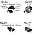
- a whole mount section of human prostate tissue representing a cross section of the entire organ, was placed on top of the substrate and transferred through ten capture layers, and onto a nitrocellulose membrane.
- the membrane was subsequently processed similar to a standard immunoblot using an antibody against cytokeratin, which selectively stains epithelium.
- FIG. 3A cytokeratin antibody transfer layer
- FIG. 3B hematoxylin and eosin stained slide of an adjacent recut from the same tissue block.
- the maintenance of cellular architecture helps determine associations between cellular findings and molecular characteristics determined by the expression scan. For example, the presence of the lymphocytes can be correlated with findings associated with other of the layers. Also, expression of a particular receptor may be correlated or mapped to epithelium. Alternatively, another molecular marker can be associated with areas of metaplasia or capsular invasion.
- Separate analysis of the substrate layers allows one to investigate multiple regions of the molecule of interest, i.e., domains of a protein or exons of a RNA transcript, as described more fully in the Examples.
- the present method can provide a quantitative indication of the relative abundance of the components in the cellular specimen when the identification molecules interact in relative abundance to the quantity of the component of interest in the cellular specimen.
- Mass spectroscopy sequencing can also be performed after separation to characterize a captured amino acid sequence.
- the LES procedure was performed on prostate tumor sections.
- the preliminary experiment used cytokeratin as the protein of interest.
- a whole mount cryostat section of human prostate tissue was prepared by making a thin frozen section of prostate, the section having a thickness of about 10 ⁇ m.
- the section includes multiple cell populations of biological interest including normal epithelium, pre-malignant lesions, high and low grade tumor foci, and significant tumor-host interactions such as lymphocytes interacting with cancer cells.
- This section was placed on an ultrathin 2% agarose gel that had been cast on a glass histology slide. The section was covered with 2 % agarose solution.
- a cover slip was applied on top of the section and the agarose was allowed to polymerize, thus creating a two-layered sample gel with the tissue section in between.
- the agarose sample gel containing the tissue sample was applied to the surface of a single layer substrate made of a 1.75" X 1.75" 0.45 pore size nitrocellulose membrane (Schlieicher and Schuell, Keen, NH).
- the membrane was then probed with an antibody against cytokeratin (Sigma, 1:1000 dilution) overnight at 4° C.
- This membrane was then probed a second time with a biotinylated secondary antibody (Sigma, 1:5000 titer) for 30 minutes at room temperature.
- the membranes were visualized by autoradiography using enhanced chemiluminescence (ECL) as recommended by the manufacturer (Pierce, Rockford, IL).
- ECL enhanced chemiluminescence
- a second experiment to test the specificity of "capture molecules" in the membrane layers was then performed.
- a 20 ⁇ m cryostat section of prostate tissue was prepared within an ultrathin 2% agarose gel as described above. Components of this tissue section as transferred overnight at room temperature through ten contiguous nitrocellulose membranes (0.5" X 0.5," 0.45 pore size, Schliecher and Schuell) by capillary action. Prior to use, each membrane was linked to a different identification molecule, in this case, antibodies, for 1 hour at room temperature. The membranes were washed 3 times for 10 minutes in IX PBS, and treated with a commercial blocking agent (Pierce) for 1 hour at room temperature, followed by a repeat wash.
- a commercial blocking agent Pieris
- nitrocellulose/antibody membranes (illustrated as A-J in Figure 2) were prepared as follows: Layer Identification Molecule Source A Anti-PB39, 644 NCI B Anti-actin Sigma C Anti-tubulin Sigma D Anti-PB39, 655 NCI E Polyclonal anti-PSA Scirpps, San Diego, CA F Anti-CAIR 1 NCI G Anti-PB-39, 656 NCI H Anti-cytokeratin Sigma I Anti-CD-3 NCI J Anti-PB-39, 645 NCI
- Antibodies were linked to the nitrocellulose membranes according well known procedures such as those disclosed in U.S. Pat. No. 4,774,177, issued to Marks on 9/27/88 or U.S. Pat. No. 4,727,037, issued to Ring on Febniary 23, 1988.
- Nitrocellulose layers are examples of porous layers that exert capillary pressure on the specimens on the top surface of the substrate, and conduct components of the specimens through the layers. Such porous layers or membranes allow the movement of liquid from one face to an opposite face of the membrane, and exert capillary action on the specimen to move soluble components of the specimen through the multiple layers.
- nitrocellulose avidly binds biomolecules such as proteins the nitrocellulose can be altered with well known blocking agents to inhibit e.g. protein binding, and promote movement of the protein or other biomolecule through the nitrocellulose layers.
- Blocking agents serve to prevent non-specific interactions between the substrate and the components of the sample as they are transferred through the substrate.
- Blocking agent is a collective term for various additives that prevent non-specific binding, but that have no active part in the specific reaction, such as an immunochemical reaction, between a particular identification molecule and its target. Blocking agents are most commonly concentrated protein solutions. Examples of such solutions include 10-20% fetal calf serum and 5% non-fat dry milk powder dissolved in a buffer such as PBS, TBS, or TBST.
- blocking agents include SuperBlock TM , Blocker TM BLOTTO, Blocker TM BSA, and SeaBlock TM (Pierce Chemical, Rockford lll) as well as NAP-SureBlocker TM , a non-animal protein blocking agent (Geno Technology, Maplewood, MO).
- each membrane was separately placed into 30 ⁇ l of SDS sample buffer (Novex; San Diego, CA) to remove any captured molecules.
- the removed, solubilized molecules were separated by electrophoresis on a 4-20% tris-glycine acrylamide gel (Novex) for 1.5 hr at 110V.
- the proteins were transferred to a 0.2 ⁇ m pore size PVDF membrane for 2 hours at 40V and analyzed by a standard immunoblotting procedure using a 1:1000 titer of monoclonal anti-PSA molecules (Scripps). In each case, the signal obtained was restricted to the appropriately sized molecular weight band for the molecule captured by the antibody.
- each of the ten membranes was probed with a monoclonal antibody against PSA and visualized by enhanced chemiluminescence (ECL) as described above.
- ECL enhanced chemiluminescence
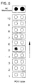
- the first nine membranes A through I did not produce an ECL signal, indicating no capture of PSA had occurred.
- positive staining for PSA was visualized on membrane J in all of the samples containing epithelium (sample numbers 1-10). This result is consistent with the known epithelial localization of PSA. Samples 11-20 did not contain epithelial cells and were appropriately negative for PSA staining.
- cell samples from five separate patients were procured from tissue specimens and solubilized in standard protein extraction buffer.
- the samples included lysates of normal lung, lung cancer, esophageal cancer, normal prostate, and breast cancer tissue.
- Each of the cell lysates was placed within a discrete 4 mm diameter spot on the top layer of a capture membrane set. This was accomplished by punching 4 mm diameter holes ("wells") in a 2 mm thick agarose gel, adding the lysates to 1 % liquid agarose, filling the 4 mm wells with the lysate/agarose solution, and allowing them to solidify.
- the sample gel thus created was placed on the top layer of a capture membrane set.
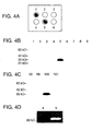
- FIG. 4A shows capture layer number 10 after probing with a monoclonal antibody against PSA and visualization by enhanced chemiluminescence (ECL).
- Samples 1 (purified PSA) and 5 (normal prostate tissue) show a positive signal, which indicates that PSA has been successfully captured.
- Samples 2 (normal lung), 3 (lung tumor), 4 (esophageal tumor), and 6 (breast cancer) do not contain PSA and are appropriately negative.
- a location of each of the samples that was placed on the top layer was substantially preserved and reproduced on the membranes through which the samples were transferred. Their substantial retention of spatial relationship conveniently allows the resulting patterns to be correlated with the original specimens.
- FIG 4B shows the results from each of capture layers one through nine.
- a repeat of the experiment was performed except the tissue was transferred through 101 capture layers with anti-PSA placed on layer number 100.
- Successful and specific capture of PSA is shown in Fig. 4C.
- the remaining capture membranes are negative for PSA.
- the specific and selective capture observed after transfer through this large number of layers indicates that it is possible to utilize layered expression scanning for the simultaneous measurement of hundreds, thousands, or even tens of thousands of molecular species, by providing different capture agents in different layers.
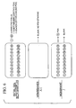
- This example demonstrates the ability of layered expression scanning to analyze nucleic acids.
- 32 P-Iabeled PCR products (200 bp) were amplified from plasmids containing cDNAs of the POV1 (PB39, NCI) and ⁇ -actin (Clonetech, Palo Alto, CA) genes, respectively.
- the radiolabeled PCR products were excised from an agarose gel, and 5 % of each product was placed in discrete 4 mm spots as described for the tissue samples in Example 2.
- the PCR products were transferred through 10 capture layers overnight by capillary transfer using 6X SSC. In this experiment, the capture layers consisted of ultrathin ( ⁇ 50 ⁇ m) 2 % agarose gels.
- Capture layer five contained a plasmid containing the entire cDNA for the POV1 gene.
- the POV1 cDNA-containing plasmid was added to the agarose prior to gel polymerizationat a final concentration of 30 ng/ ⁇ L.
- a nonblocked nitrocellulose membrane was used to bind the noncaptured POV1 and ⁇ -actin PCR products after they traversed the membrane set. After transfer, the layers were separated and visualized by X-OMAT radiography.
- FIG. 5 shows successful and selective capture of POV1 cDNA in layer 5, while the actin PCR product moved through the entire set of layers and was not captured until it reacted the nonblocked nitrocellulose layer.
- layered expression scanning may also be utilized to analyze intact tissue sections. If an intact tissue section is used as the sample, it is possible to correlate the two-dimensional architecture of the tissue section with the two-dimensional pattern of cellular components localized in particular capture layers following transfer.
- tissue section 10 ⁇ m thick whole-mount cryostat sections of human prostate from radical prostatectomy specimens were placed on top of either a ten-layer or a one hundred-layer agarose gel set.
- the intact tissue section was transferred through the layers by capillary fluid movement overnight at room temperature to a 1.75-square inch, 0.45 ⁇ m pore size nitrocellulose membrane (Schleicher and Schuell).
- the nitrocellulose membranes were probed with an antibody against cytokeratin (Sigma 1:1000 dilution) to selectively identify epithelial elements and were visualized by ECL according to the recommendations of the manufacturer (Pierce).
- FIG 3 A-D Retention of the basic organization of the tissue section throughout the transfer process is demonstrated in FIG 3 A-D by comparing the transferred sections (FIG. 3A and FIG 3C) with a hematoxylin and eosin (H&E) stained slide of an adjacent recut section.
- the overall architecture of the transferred sections is highly similar to the corresponding H&E stained slides, and the location of individual glandular epithelial elements within the tissue sections can be determined.
- layered expression scanning can be used for analyzing intact tissue sections while retaining a correspondence between the two-dimensional architecture of the tissue section and the two-dimensional position of components transferred to the capture layers. Single cell-level of resolution will permit individual cells to be analyzed for the presence of particular molecules.
- Membranes and gels useful for creating identification and capture layers as utilized in the Examples may have one or more of the following properties.
- the membranes or gels are able to immobilize individual identification or capture molecules (e.g. antibodies, nucleic acids, and dyes).
- the membranes or gels permit cellular components transferred from a sample to efficiently traverse the set of layers and accumulate or react in the appropriate layer.
- the membranes or gels facilitate transfer with minimal loss of the two-dimensional relationship of the biological sample(s).
- Particular examples of materials appropriate for constructing a set of layers for layered expression scanning include nitrocellulose membranes, derivatized nitrocellulose membranes, high concentration agarose gels, low concentration agarose gels, high concentration polyacrylamide gels, a low concentration polyacrylamide gel, and membranes, such as porous membranes like nitrocellulose paper.
- Low concentration agarose is from about 0.1 to about 3%, while high concentration is above about 3 %.
- Low concentration acrylamide is about 2 % to about 20 %, while high concentration is above about 20 %.
- Individual layers may also be composites of two or more membranes or gels.
- thin polymer membranes such as polar polymer membranes, for instance polyester membranes
- nitrocellulose membranes or agarose or polyacrylamide gels may be combined with nitrocellulose membranes or agarose or polyacrylamide gels to form composite layers for layered expression scanning.
- the composite membrane is formed as follows.
- a thin (10 ⁇ m) polyester membrane is used as a backbone layer.
- the polyester membrane is then coated with a soluble polymer material, such as 2% agarose, to form an ultrathin ( ⁇ 1 ⁇ m) layer covering the polyester backbone.
- a capture molecule e.g., an antibody or nucleic acid
- the polyester backbone/polymer gel composite containing the capture molecule may then be used as a layered expression scanning capture membrane.
- a particular advantage of the composite membranes is that the polymer gel that is coated on the polyester backbone serves as a "contact substance" between each of the layers, thereby permitting efficient transfer of biomolecules with minimal loss of correspondence with the two-dimensional architecture in the sample.
- Different tumor cell populations are separately collected using laser microdissection techniques as described by Emmert-Buck et al. (1997).
- Each different cell population is placed in its own location within a sample gel, as described above in Example 1.
- the sample gel is placed on a multi-layer substrate, containing at least one layer cross-linked with antibodies against one or more known binding partners of the molecule of interest.
- the molecules could be treated with a cross-linking agent, thus binding partners will remain in the state they are in at the time of the preparation of the cryostat during transfer.
- the layers are separated and the molecules of interest are run on a gel and probed by the capture antibody.
- this experiment shows whether or not a molecule of interest is bound or free at various stages of tumor development by determining the molecular weight of the species when the tissue sample is prepared.
- MS-MS sequencing can identify the proteins recovered from relatively few numbers of microdissected cells as described in Huang et al. (1999).
- LES can be used as an "open system" to search for disease associated molecular alterations in tissue samples.
- normal and diseased cell samples are placed within the sample gel as described in Example 1.
- the information molecules cross-linked on the membrane layers can be antibodies, peptides, or DNA sequences for either known proteins, or libraries of ssDNA or mRNA.
- Large numbers of capture molecules are simultaneously used to analyze the comparative expression between normal and diseased cell populations of the targets of the capture molecules.
- the samples tested can be derived from one or multiple patients. Once a protein or nucleic acid is shown to be expressed differently in normal and diseased cells, its identity can be determined by the capture molecule to which it binds. This identity can be confirmed using standard sequencing techniques, or such sequencing techniques can be used initially to determine whether the target of the capture molecule is unknown.
- Different cell populations are separately collected using laser microdissection techniques as described by Emmert-Buck et al. (1996).
- Each cell population is placed in its own location within a sample gel, as described above in Example 1.
- the sample gel is placed on a substrate, containing at least one membrane cross-linked with polyclonal antibody against tumor suppressor protein.
- the membranes are separated and the anti-tumor suppressor protein membrane, with its captured molecules, is probed with two differentially labeled monoclonal antibodies that recognize different regions of the tumor suppressor protein.
- One antibody is specific for the N-terminus of the protein, and the other is specific for the C-terminus of the protein.
- both PSA and PSA-ACT migrate through the membranes and are captured.
- Alteration of experimental conditions to effect molecular migration can allow investigators to customize experiments as needed for particular objectives. For example, study of subcellular molecular profiles may be performed by utilizing transfer buffers with and without detergents to selectively mobilize soluble or membrane-bound proteins.
- the layered expression scanning of the present invention can also be used in association with an automated laboratory instrument capable of multiple applications.
- the capture layers in the present prototype system are replaced by thin transparent membranes such that several thousand stacked layers will cumulatively be only a few millimeters in thickness.
- the total migration distance of the tissue sample during transfer and detection or immobilization is minimal, thereby optimizing the cellular resolution of the system.
- the tissue sample, wash buffers, and fluorescently labeled secondary detection molecules are transferred through the intact membrane set, thus obviating the need to separate and individually process each capture layer.
- the sample, wash buffers and fluorescently labeled secondary detection molecules may be transferred into the stacked layers either in the same direction as the sample components are conducted through the stacked layers or in another direction, such as in the reverse direction or along the direction of the layers themselves.
- the intact membrane set is then analyzed by confocal fluorescence microscopy, and the expression data of each individual layer is determined and overlayed with the high quality histological image of the tissue section.
- the approach was demonstrated in an experiment similar to that shown in FIG 4, in which each of the detection reagents were transferred through the capture membranes while the membranes remained as an intact set. Successful capture and analysis occurred.
- the set of capture layers may be utilized repeatedly to produce expression scans by washing the stacked layers with a denaturing buffer between scans to remove captured molecules.
- Suitable buffers for this purpose include buffers containing denaturants, such as detergents or urea, and salts, such as sodium chloride, at concentrations that are sufficient to remove captured molecules from the stacked layers.
- a particular example of a suitable denaturing buffer is a buffer containing 1 % sodium dodecyl sulfate (SDS) and 500 mM sodium chloride.
- SDS sodium dodecyl sulfate
- Other denaturing buffer systems are known in the art and their suitability for use with automated expression scanning can be determined by analyzing the layers for the continued presence of bound molecules after they are washed with a particular denaturing buffer system.
- the capture membranes will be separable and processed individually after tissue transfer.
- the separated membranes may then be studied beyond measurement of expression levels of individual molecules.
- mass spectrometry can be used to identify binding partners which are "co-captured" along with targeted proteins.
- the layered expression scanning (LES) methods can be used to analyze for individual cloned biomolecules, such as messenger RNAs recovered from a cell population and cloned into bacteria using standard methods.
- the bacteria are plated on media and individual colonies are grown in the presence of a labeled nucleotide. Individual colonies are then placed on top of an LES device and the nucleic acids from each colony are transferred through a set of LES layers such as those described in Example 5 above and where each LES layer contains an individual cDNA clone. The identity of the cDNA in all bacterial colonies is simultaneously determined by analyzing for the presence or absence of hybridization on each capture membrane after the cloned DNA has traversed the LES layer set.
- One application of this particular method is to perform high-throughput gene expression analysis of a given cell population by determining the identity of a large number of bacterial clones derived from a particular cells messenger RNA population.
- the layered expression scanning method may be used to analyze the genomic DNA content of individual cells or cells within a tissue section.
- One example of this application is as follows.
- DNA from a series of cell lines is purified, labeled with a "tagged" (radiolabeled or fluorescently labeled) nucleotide and placed in a grid on a membrane on top of the LES device, such as described above in Example 2.
- each of the LES layers contains a specific genomic DNA clone.
- the DNA samples are transferred through the LES layers such that the DNA gragments from the cell samples specifically hybridize to the LES layer that contains the corresponding genomic clone.
- the LES layers are then analyzed (by radiography or fluorescence) to provide a quantitative measure of the amount of DNA in each cell sample at each genomic locus included in the LES layer set. This application would be useful in determining the specific regions of DNA (and associated genes) that are amplified or deleted in a series of cell lines.
- each layer may be subdivided into multiple regions, each with a different capture molecule, which are capable of producing more complex patterns that can be recognized by the user or image processing software.
- Each of the regions can extend in any desired shape throughout the layer, which can extend in any direction relative to the direction of movement of the sample through the substrate.
- the different regions are transverse to the direction of movement to maintain a spatial correspondence between a surface of the substrate to which the specimen is applied, and the region which captures a molecule of interest.
- any pattern that conveys information about the molecular content of the specimen may be used.
- pattern recognition software is particularly effective to store and compare patterns.
Landscapes
- Health & Medical Sciences (AREA)
- Life Sciences & Earth Sciences (AREA)
- Engineering & Computer Science (AREA)
- Chemical & Material Sciences (AREA)
- Molecular Biology (AREA)
- Immunology (AREA)
- Biomedical Technology (AREA)
- Hematology (AREA)
- Urology & Nephrology (AREA)
- Physics & Mathematics (AREA)
- General Health & Medical Sciences (AREA)
- Biochemistry (AREA)
- Microbiology (AREA)
- Organic Chemistry (AREA)
- Biotechnology (AREA)
- Medicinal Chemistry (AREA)
- Analytical Chemistry (AREA)
- General Physics & Mathematics (AREA)
- Cell Biology (AREA)
- Pathology (AREA)
- Food Science & Technology (AREA)
- Proteomics, Peptides & Aminoacids (AREA)
- Bioinformatics & Cheminformatics (AREA)
- Biophysics (AREA)
- Zoology (AREA)
- Wood Science & Technology (AREA)
- Bioinformatics & Computational Biology (AREA)
- Oncology (AREA)
- Chemical Kinetics & Catalysis (AREA)
- General Chemical & Material Sciences (AREA)
- Hospice & Palliative Care (AREA)
- General Engineering & Computer Science (AREA)
- Genetics & Genomics (AREA)
- Measuring Or Testing Involving Enzymes Or Micro-Organisms (AREA)
- Investigating Or Analysing Biological Materials (AREA)
- Apparatus Associated With Microorganisms And Enzymes (AREA)
- Investigating Or Analysing Materials By Optical Means (AREA)
Applications Claiming Priority (3)
| Application Number | Priority Date | Filing Date | Title |
|---|---|---|---|
| US14561399P | 1999-07-26 | 1999-07-26 | |
| US145613P | 1999-07-26 | ||
| PCT/US2000/020354 WO2001007915A2 (en) | 1999-07-26 | 2000-07-26 | Layered device with capture regions for cellular analysis |
Publications (2)
| Publication Number | Publication Date |
|---|---|
| EP1218743A2 EP1218743A2 (en) | 2002-07-03 |
| EP1218743B1 true EP1218743B1 (en) | 2006-05-31 |
Family
ID=22513857
Family Applications (1)
| Application Number | Title | Priority Date | Filing Date |
|---|---|---|---|
| EP00953685A Expired - Lifetime EP1218743B1 (en) | 1999-07-26 | 2000-07-26 | Layered device with capture regions for cellular analysis |
Country Status (9)
| Country | Link |
|---|---|
| US (2) | US6602661B1 (enExample) |
| EP (1) | EP1218743B1 (enExample) |
| JP (1) | JP4137444B2 (enExample) |
| AT (1) | ATE328277T1 (enExample) |
| AU (1) | AU771499B2 (enExample) |
| CA (1) | CA2375034C (enExample) |
| DE (1) | DE60028393T2 (enExample) |
| ES (1) | ES2269171T3 (enExample) |
| WO (1) | WO2001007915A2 (enExample) |
Families Citing this family (74)
| Publication number | Priority date | Publication date | Assignee | Title |
|---|---|---|---|---|
| US6969614B1 (en) * | 1999-02-16 | 2005-11-29 | The United States Of America As Represented By The Department Of Health And Human Services | Methods for the isolation and analysis of cellular protein content |
| US7214477B1 (en) * | 1999-07-26 | 2007-05-08 | The United States Of America As Represented By The Secretary Of The Department Of Health And Human Services | Layered device with capture regions for cellular analysis |
| US7838222B2 (en) * | 1999-07-26 | 2010-11-23 | United States of America/ NIH | Methods, devices and kits for multiplex blotting of biological samples from multi-well plates |
| US6969615B2 (en) * | 1999-07-26 | 2005-11-29 | 20/20 Genesystems, Inc. | Methods, devices, arrays and kits for detecting and analyzing biomolecules |
| US20060275851A1 (en) * | 2000-07-26 | 2006-12-07 | Emmert-Buck Michael R | Layered peptide/antigen arrays - for high-throughput antibody screening of clinical samples |
| EP1305622A2 (en) * | 2000-07-26 | 2003-05-02 | THE GOVERNMENT OF THE UNITED STATES OF AMERICA, as represented by THE SECRETARY, DEPARTMENT OF HEALTH AND HUMAN SERVICES | Transfer microdissection |
| US20040053326A1 (en) * | 2001-03-14 | 2004-03-18 | Emmert-Buck Michael R. | Transfer microdessection |
| DE10137864B4 (de) * | 2001-08-02 | 2005-05-19 | Picorapid Technologie Gmbh | Substanzträger mit Markierung |
| US20040072274A1 (en) * | 2002-05-09 | 2004-04-15 | Lebrun Stewart J. | System and method for visualization and digital analysis of protein and other macromolecule microarrays |
| SE0202346D0 (sv) * | 2002-07-30 | 2002-07-30 | Astrazeneca Ab | Profiling process |
| WO2004048928A2 (en) * | 2002-11-25 | 2004-06-10 | The United States Of America As Represented By The Secretary Of Health And Human Services, Nih | Method and apparatus for performing multiple simultaneous manipulations of biomolecules in a two-dimensional array |
| US20040171167A1 (en) * | 2002-12-13 | 2004-09-02 | Affymetrix, Inc. | Chip-in-a-well scanning |
| WO2004065543A2 (en) * | 2003-01-16 | 2004-08-05 | Exact Sciences Corporation | Devices and method for recovery of a target molecule from a sample |
| JP2007527523A (ja) * | 2003-07-07 | 2007-09-27 | シンジェンタ パーティシペーションズ アクチェンゲゼルシャフト | 飼料用酵素を検出するための試薬、方法、及びキット |
| US20050121325A1 (en) * | 2003-09-19 | 2005-06-09 | Timothy Updyke | Composite compositions for electrophoresis |
| AU2005211907A1 (en) * | 2004-02-11 | 2005-08-25 | Pamgene B.V. | A device for analysing an interaction between target and probe molecules |
| US8173002B2 (en) | 2005-02-24 | 2012-05-08 | Life Technologies Corporation | Electro-blotting devices, systems, and kits, and methods for their use |
| JP2009531652A (ja) * | 2005-12-08 | 2009-09-03 | プロテイン・デイスカバリー・インコーポレーテツド | 化学分析のための被分析物の濃縮と分画のための方法と装置 |
| JP4915527B2 (ja) * | 2007-09-27 | 2012-04-11 | 凸版印刷株式会社 | 電気泳動兼転写用積層体、電気泳動兼転写用チップ、電気泳動兼転写装置、電気泳動兼転写方法、電気泳動兼転写用積層体の製造方法 |
| US20100044229A1 (en) * | 2008-07-11 | 2010-02-25 | Life Technologies Corporation | Electrophoretically Enhanced Detection of Analytes on a Solid Support |
| WO2010126897A1 (en) * | 2009-04-27 | 2010-11-04 | Protein Discovery, Inc. | Programmable electrophoretic notch filter systems and methods |
| US8524507B2 (en) * | 2009-08-19 | 2013-09-03 | The Cleveland Clinic Foundation | Method for detecting a target molecule in a biological sample |
| CA2775613C (en) | 2009-10-13 | 2018-06-12 | Nanostring Technologies, Inc. | Protein detection via nanoreporters |
| WO2011068088A1 (ja) | 2009-12-04 | 2011-06-09 | 株式会社日立製作所 | 2次元cDNAライブラリを用いた遺伝子発現解析方法 |
| US20190300945A1 (en) | 2010-04-05 | 2019-10-03 | Prognosys Biosciences, Inc. | Spatially Encoded Biological Assays |
| US10787701B2 (en) | 2010-04-05 | 2020-09-29 | Prognosys Biosciences, Inc. | Spatially encoded biological assays |
| RS54482B1 (sr) | 2010-04-05 | 2016-06-30 | Prognosys Biosciences, Inc. | Biološka testiranja sa prostornim kodiranjem |
| GB201106254D0 (en) * | 2011-04-13 | 2011-05-25 | Frisen Jonas | Method and product |
| JP2013178140A (ja) * | 2012-02-28 | 2013-09-09 | Sharp Corp | 核酸−タンパク質複合体の単離装置 |
| EP3901280B1 (en) * | 2012-10-17 | 2025-03-12 | 10x Genomics Sweden AB | Methods and product for optimising localised or spatial detection of gene expression in a tissue sample |
| US9291596B2 (en) | 2013-02-12 | 2016-03-22 | Pierce Biotechnology, Inc. | Electroblot transfer buffer |
| USD738527S1 (en) | 2013-05-28 | 2015-09-08 | Life Technologies Corporation | Electroblotting apparatus |
| LT3013983T (lt) | 2013-06-25 | 2023-05-10 | Prognosys Biosciences, Inc. | Erdviniai koduoti biologiniai tyrimai, naudojant mikrofluidinį įrenginį |
| EP3147659B1 (en) | 2014-05-19 | 2022-03-30 | National University Corporation Nagoya University | Method of analysis of component in sample, method of specific isolation of component in sample, and sample for mass spectrometry |
| HK1250245A1 (zh) | 2015-04-10 | 2018-12-07 | Spatial Transcriptomics Ab | 生物样本的空间区别、多重核酸分析 |
| ES2975362T3 (es) | 2015-07-17 | 2024-07-04 | Nanostring Technologies Inc | Cuantificación simultánea de la expresión génica en una región definida por el usuario de un tejido seccionado transversalmente |
| AU2016297513B2 (en) | 2015-07-17 | 2021-02-25 | Board Of Regents, The University Of Texas System | Simultaneous quantification of a plurality of proteins in a user-defined region of a cross-sectioned tissue |
| EP3463452A1 (en) | 2016-05-24 | 2019-04-10 | Institut National de la Sante et de la Recherche Medicale (INSERM) | Methods and pharmaceutical compositions for the treatment of non small cell lung cancer (nsclc) that coexists with chronic obstructive pulmonary disease (copd) |
| WO2018046736A1 (en) | 2016-09-12 | 2018-03-15 | INSERM (Institut National de la Santé et de la Recherche Médicale) | Methods for predicting the survival time of patients suffering from cancer |
| WO2018055080A1 (en) | 2016-09-22 | 2018-03-29 | INSERM (Institut National de la Santé et de la Recherche Médicale) | Methods and pharmaceutical compositions for reprograming immune environment in a subject in need thereof |
| WO2018122245A1 (en) | 2016-12-28 | 2018-07-05 | INSERM (Institut National de la Santé et de la Recherche Médicale) | Methods of predicting the survival time of patients suffering from cms3 colorectal cancer |
| WO2018122249A1 (en) | 2016-12-28 | 2018-07-05 | INSERM (Institut National de la Santé et de la Recherche Médicale) | Methods for predicting the survival time of patients suffering from a microsatellite stable colorectal cancer |
| US20200150109A1 (en) | 2017-05-05 | 2020-05-14 | INSERM (Institut National de la Santé et de la Recherche Médicale) | Microtubule-targeting drugs as immune checkpoint inhibitors and methods of screening novel immune checkpoint inhibitors for the treatment of cancers and infectious diseases |
| KR102879375B1 (ko) | 2018-02-12 | 2025-10-30 | 브루커 스페이셜 바이올로지, 인크. | 유전자 및 단백질 발현을 검출하는 생체 분자 프로브 및 방법 |
| CN109254346B (zh) * | 2018-07-16 | 2020-09-11 | 哈尔滨工程大学 | 一种基于波分复用技术的单光纤光镊 |
| CN109556939B (zh) * | 2018-12-03 | 2021-05-11 | 中国科学院青藏高原研究所 | 样品裂变径迹原位蚀刻观测方法及设备 |
| EP4567127A3 (en) | 2018-12-10 | 2025-09-03 | 10x Genomics, Inc. | Methods for determining a location of a biological analyte in a biological sample |
| BR112021013157A8 (pt) | 2019-01-03 | 2022-12-06 | Inst Nat Sante Rech Med | Usos de um inibidor de nrp-1, uso de uma combinação, uso de um anticorpo multiespecífico, método ex vivo para predizer, uso de um inibidor, anticorpo multiespecífico, população de células modificadas, método ex vivo de produção e uso de uma população de células t |
| US11926867B2 (en) | 2019-01-06 | 2024-03-12 | 10X Genomics, Inc. | Generating capture probes for spatial analysis |
| US11649485B2 (en) | 2019-01-06 | 2023-05-16 | 10X Genomics, Inc. | Generating capture probes for spatial analysis |
| US20220177978A1 (en) | 2019-04-02 | 2022-06-09 | INSERM (Institut National de la Santé et de la Recherche Médicale) | Methods of predicting and preventing cancer in patients having premalignant lesions |
| WO2020208060A1 (en) | 2019-04-09 | 2020-10-15 | INSERM (Institut National de la Santé et de la Recherche Médicale) | Use of sk2 inhibitors in combination with immune checkpoint blockade therapy for the treatment of cancer |
| BR112021024374A8 (pt) | 2019-06-03 | 2022-12-06 | Inst Nat Sante Rech Med | Método para modular um regime de tratamento |
| CA3157889A1 (en) | 2019-10-17 | 2021-04-22 | Inserm (Institut National De La Sante Et De La Recherche Medicale) | Methods for diagnosing nasal intestinal type adenocarcinomas |
| US11821035B1 (en) | 2020-01-29 | 2023-11-21 | 10X Genomics, Inc. | Compositions and methods of making gene expression libraries |
| US12076701B2 (en) | 2020-01-31 | 2024-09-03 | 10X Genomics, Inc. | Capturing oligonucleotides in spatial transcriptomics |
| US12399123B1 (en) | 2020-02-14 | 2025-08-26 | 10X Genomics, Inc. | Spatial targeting of analytes |
| US12281357B1 (en) | 2020-02-14 | 2025-04-22 | 10X Genomics, Inc. | In situ spatial barcoding |
| EP4110955A1 (en) | 2020-02-28 | 2023-01-04 | INSERM (Institut National de la Santé et de la Recherche Médicale) | Methods for diagnosing, prognosing and managing treatment of breast cancer |
| EP4162074B1 (en) | 2020-06-08 | 2024-04-24 | 10X Genomics, Inc. | Methods of determining a surgical margin and methods of use thereof |
| CN115997123A (zh) | 2020-06-30 | 2023-04-21 | 国家医疗保健研究所 | 用于预测实体癌患者在术前辅助治疗后复发和/或死亡风险的方法 |
| CN115843335A (zh) | 2020-06-30 | 2023-03-24 | 国家医疗保健研究所 | 用于预测患有实体癌的患者在术前辅助治疗和根治性手术后复发和/或死亡风险的方法 |
| JP7475999B2 (ja) * | 2020-07-03 | 2024-04-30 | 旭化成株式会社 | イムノクロマト展開膜及びその製造方法並びに迅速検査キット |
| US11981960B1 (en) | 2020-07-06 | 2024-05-14 | 10X Genomics, Inc. | Spatial analysis utilizing degradable hydrogels |
| EP4313317A1 (en) | 2021-03-23 | 2024-02-07 | INSERM (Institut National de la Santé et de la Recherche Médicale) | Methods for the diagnosis and treatment of t cell-lymphomas |
| WO2022256503A1 (en) | 2021-06-03 | 2022-12-08 | 10X Genomics, Inc. | Methods, compositions, kits, and systems for enhancing analyte capture for spatial analysis |
| US20250067745A1 (en) | 2022-01-31 | 2025-02-27 | Institut National de la Santé et de la Recherche Médicale | Cd38 as a biomarker and biotarget in t-cell lymphomas |
| AU2022447608A1 (en) | 2022-03-17 | 2024-09-26 | Assistance Publique - Hôpitaux De Paris | Methods for predicting response to an immunotherapeutic treatment in a patient with a cancer |
| US20250231193A1 (en) | 2022-04-11 | 2025-07-17 | Institut National de la Santé et de la Recherche Médicale | Methods for the diagnosis and treatment of t-cell malignancies |
| WO2023198874A1 (en) | 2022-04-15 | 2023-10-19 | Institut National de la Santé et de la Recherche Médicale | Methods for the diagnosis and treatment of t cell-lymphomas |
| WO2024003310A1 (en) | 2022-06-30 | 2024-01-04 | Institut National de la Santé et de la Recherche Médicale | Methods for the diagnosis and treatment of acute lymphoblastic leukemia |
| JP2025526336A (ja) | 2022-07-22 | 2025-08-13 | アンスティチュ ナショナル ドゥ ラ サンテ エ ドゥ ラ ルシェルシュ メディカル | T細胞悪性腫瘍におけるバイオマーカー及びバイオターゲットとしてのgarp |
| WO2024023283A1 (en) | 2022-07-29 | 2024-02-01 | Institut National de la Santé et de la Recherche Médicale | Lrrc33 as a biomarker and biotarget in cutaneous t-cell lymphomas |
| WO2024079192A1 (en) | 2022-10-12 | 2024-04-18 | Institut National de la Santé et de la Recherche Médicale | Cd81 as a biomarker and biotarget in t-cell malignancies |
Citations (2)
| Publication number | Priority date | Publication date | Assignee | Title |
|---|---|---|---|---|
| US5057438A (en) * | 1986-09-24 | 1991-10-15 | Agency Of Industrial Science And Technology | Electrophoretic antigen-antibody determination with laminate of multiple membranes |
| WO1999067647A1 (en) * | 1998-06-24 | 1999-12-29 | Chen & Chen, Llc | Multi-layer testing column |
Family Cites Families (51)
| Publication number | Priority date | Publication date | Assignee | Title |
|---|---|---|---|---|
| US3620947A (en) | 1969-04-10 | 1971-11-16 | Ortec Inc | Electrophoretic system |
| DE2622684B2 (de) * | 1976-05-21 | 1979-03-15 | Licentia Patent-Verwaltungs-Gmbh, 6000 Frankfurt | Membranaustauscher |
| US4337131A (en) | 1978-11-13 | 1982-06-29 | C. Desaga Gmbh Nachf. Erich Fecht | Process for electrophoresis |
| US4668619A (en) * | 1980-10-30 | 1987-05-26 | Miles Laboratories, Inc. | Multilayer homogeneous specific binding assay device |
| US4444879A (en) * | 1981-01-29 | 1984-04-24 | Science Research Center, Inc. | Immunoassay with article having support film and immunological counterpart of analyte |
| AR231590A1 (es) * | 1981-04-29 | 1984-12-28 | Ciba Geigy Ag | Dispositivo de analisis inmunologico y procedimiento para obtenerlo |
| JPS5818167A (ja) * | 1981-07-24 | 1983-02-02 | Fuji Photo Film Co Ltd | 分析フイルム及びこれを用いる分析方法 |
| JPS5934154A (ja) * | 1982-08-19 | 1984-02-24 | Konishiroku Photo Ind Co Ltd | 免疫分析素子測定法 |
| US4539294A (en) * | 1982-09-30 | 1985-09-03 | Akzona Incorporated | Immobilization of proteins on polymeric supports |
| EP0139373A1 (en) * | 1983-08-26 | 1985-05-02 | The Regents Of The University Of California | Multiple immunoassay system |
| DE3516579A1 (de) * | 1984-11-19 | 1986-05-22 | Boehringer Mannheim Gmbh, 6800 Mannheim | Gerinnungstest auf teststreifen |
| US4716101A (en) | 1984-12-18 | 1987-12-29 | Calgene, Inc. | Rapid enzyme assay by product selective blot |
| US4795562A (en) | 1985-09-13 | 1989-01-03 | Walsh James W | Membrane batch-processing apparatus |
| US4959305A (en) * | 1986-06-18 | 1990-09-25 | Miles Inc. | Reversible immobilization of assay reagents in a multizone test device |
| DE3638654A1 (de) * | 1986-11-12 | 1988-05-26 | Boehringer Mannheim Gmbh | Testtraeger zur analytischen bestimmung eines bestandteils einer koerperfluessigkeit |
| US4840714A (en) | 1987-05-13 | 1989-06-20 | American Bionetics, Inc. | Electroblotting technique for transferring specimens from a polyacrylamide electrophoresis or like gel onto a membrane |
| US4874691A (en) * | 1987-10-16 | 1989-10-17 | Quadra Logic Technologies Inc. | Membrane-supported immunoassays |
| EP0334615B1 (en) | 1988-03-22 | 1994-11-17 | Erik Nieman | Electrophoresis apparatus |
| US5173159A (en) | 1988-09-06 | 1992-12-22 | Bertin & Cie | Multiple electrophoresis method for the controlled migration of macromolecules through rectangular gel plates |
| GB8905001D0 (en) * | 1989-03-04 | 1989-04-19 | Univ Leicester | Screening for natural products of microbial metabolism |
| US5155049A (en) | 1989-08-22 | 1992-10-13 | Terrapin Technologies, Inc. | Blotting technique for membrane assay |
| US5238651A (en) | 1990-07-23 | 1993-08-24 | New York University | Gel plates, equipment and kits for combined electrophoretic-immunoelectrophoretic analysis |
| CS257291A3 (en) | 1990-12-13 | 1992-06-17 | Jan J Prof Mudr Opplt | Multi-layered, non-sequential discontinuous polyacrylamide gels, forming equilibrium molecular sieves for biomolecules separation |
| US5078853A (en) | 1991-03-22 | 1992-01-07 | Assay Technology, Inc. | Segmented electrophoretic separation system useful for lipid profiling |
| EP0579748A4 (en) * | 1991-04-08 | 1995-05-17 | Environmental Test Systems | MULTI-LAYER STRUCTURE OF TEST DEVICE. |
| US5877028A (en) * | 1991-05-29 | 1999-03-02 | Smithkline Diagnostics, Inc. | Immunochromatographic assay device |
| CA2074752A1 (en) * | 1991-07-29 | 1993-01-30 | Tadakazu Yamauchi | Process and device for specific binding assay |
| DE4132743A1 (de) * | 1991-10-02 | 1993-04-08 | Boehringer Mannheim Gmbh | Testtraeger fuer die analyse von fluessigkeiten |
| US5438128A (en) * | 1992-02-07 | 1995-08-01 | Millipore Corporation | Method for rapid purifiction of nucleic acids using layered ion-exchange membranes |
| WO1994022904A1 (en) | 1993-04-07 | 1994-10-13 | Gradipore Limited | Improvements in protein separation |
| US5384255A (en) * | 1993-06-21 | 1995-01-24 | Rappaport Family Institute For Research In The Medical Sciences | Ubiquitin carrier enzyme E2-F1, purification, production, and use |
| US5427664A (en) | 1993-07-22 | 1995-06-27 | Stoev; Stoyan V. | Free solution electrophoresis-membrane filters trapping assay apparatus and method |
| US5332484A (en) | 1993-08-02 | 1994-07-26 | Hi-Tech Bio-Tech, Inc. | Apparatus and process for the large scale separation and retrieval of proteins from cellular extracts |
| US5716508A (en) | 1993-09-09 | 1998-02-10 | Glyko Incorporated | Utilization of additives and defined storage systems to increase the stability and performance of electrophoresis media |
| US5843657A (en) | 1994-03-01 | 1998-12-01 | The United States Of America As Represented By The Department Of Health And Human Services | Isolation of cellular material under microscopic visualization |
| EP0671626B1 (en) | 1994-03-08 | 2000-01-12 | Zeptosens AG | Device and method for combined bioaffinity assay and electrophoretic separation |
| US5679310A (en) * | 1995-07-11 | 1997-10-21 | Polyfiltronics, Inc. | High surface area multiwell test plate |
| US5665596A (en) * | 1995-07-31 | 1997-09-09 | Becton, Dickinson And Company | Device for cell co-culture and method for its use in culturing cells |
| WO1998020353A1 (en) | 1996-11-01 | 1998-05-14 | Grace Bio-Labs, Inc. | Cytoperfusion of tissue specimens |
| GB9624927D0 (en) | 1996-11-29 | 1997-01-15 | Oxford Glycosciences Uk Ltd | Gels and their use |
| US5804384A (en) * | 1996-12-06 | 1998-09-08 | Vysis, Inc. | Devices and methods for detecting multiple analytes in samples |
| US6087134A (en) | 1997-01-14 | 2000-07-11 | Applied Imaging Corporation | Method for analyzing DNA from a rare cell in a cell population |
| US6168929B1 (en) * | 1997-01-27 | 2001-01-02 | University Of Pittsburgh | Method of identifying agents that disrupt the interaction of SV40 T antigen with Dnak homologues |
| WO1998041863A1 (en) * | 1997-03-14 | 1998-09-24 | Compucyte Corp. | Multiple simultaneous testing media |
| US5993627A (en) | 1997-06-24 | 1999-11-30 | Large Scale Biology Corporation | Automated system for two-dimensional electrophoresis |
| US6013165A (en) | 1998-05-22 | 2000-01-11 | Lynx Therapeutics, Inc. | Electrophoresis apparatus and method |
| US6232067B1 (en) * | 1998-08-17 | 2001-05-15 | The Perkin-Elmer Corporation | Adapter directed expression analysis |
| US6135942A (en) | 1998-11-30 | 2000-10-24 | Leptin; Maria | Nucleic acids proteins of a D. melanogaster insulin-like gene and uses thereof |
| WO2000045168A1 (en) | 1999-01-28 | 2000-08-03 | Universite De Geneve | Method and kit for identifying or characterising polypeptides |
| US6969615B2 (en) * | 1999-07-26 | 2005-11-29 | 20/20 Genesystems, Inc. | Methods, devices, arrays and kits for detecting and analyzing biomolecules |
| US20020012920A1 (en) * | 2000-11-20 | 2002-01-31 | Kevin Gardner | Method and kit for proteomic identification |
-
2000
- 2000-07-26 AU AU66091/00A patent/AU771499B2/en not_active Expired
- 2000-07-26 DE DE60028393T patent/DE60028393T2/de not_active Expired - Lifetime
- 2000-07-26 CA CA2375034A patent/CA2375034C/en not_active Expired - Lifetime
- 2000-07-26 WO PCT/US2000/020354 patent/WO2001007915A2/en not_active Ceased
- 2000-07-26 ES ES00953685T patent/ES2269171T3/es not_active Expired - Lifetime
- 2000-07-26 EP EP00953685A patent/EP1218743B1/en not_active Expired - Lifetime
- 2000-07-26 JP JP2001512292A patent/JP4137444B2/ja not_active Expired - Lifetime
- 2000-07-26 AT AT00953685T patent/ATE328277T1/de not_active IP Right Cessation
- 2000-11-20 US US09/718,990 patent/US6602661B1/en not_active Expired - Lifetime
-
2003
- 2003-07-25 US US10/627,352 patent/US20040081987A1/en not_active Abandoned
Patent Citations (2)
| Publication number | Priority date | Publication date | Assignee | Title |
|---|---|---|---|---|
| US5057438A (en) * | 1986-09-24 | 1991-10-15 | Agency Of Industrial Science And Technology | Electrophoretic antigen-antibody determination with laminate of multiple membranes |
| WO1999067647A1 (en) * | 1998-06-24 | 1999-12-29 | Chen & Chen, Llc | Multi-layer testing column |
Also Published As
| Publication number | Publication date |
|---|---|
| WO2001007915A8 (en) | 2002-03-07 |
| ES2269171T3 (es) | 2007-04-01 |
| CA2375034A1 (en) | 2001-02-01 |
| CA2375034C (en) | 2012-01-03 |
| WO2001007915A2 (en) | 2001-02-01 |
| JP2003505694A (ja) | 2003-02-12 |
| US6602661B1 (en) | 2003-08-05 |
| US20040081987A1 (en) | 2004-04-29 |
| AU6609100A (en) | 2001-02-13 |
| DE60028393T2 (de) | 2007-06-14 |
| DE60028393D1 (de) | 2006-07-06 |
| WO2001007915A3 (en) | 2001-04-05 |
| EP1218743A2 (en) | 2002-07-03 |
| AU771499B2 (en) | 2004-03-25 |
| JP4137444B2 (ja) | 2008-08-20 |
| ATE328277T1 (de) | 2006-06-15 |
Similar Documents
| Publication | Publication Date | Title |
|---|---|---|
| EP1218743B1 (en) | Layered device with capture regions for cellular analysis | |
| US7214477B1 (en) | Layered device with capture regions for cellular analysis | |
| AU772680B2 (en) | LCM (Laser capture microdissection) for cellular protein analysis | |
| Craven et al. | Laser capture microdissection and two-dimensional polyacrylamide gel electrophoresis: evaluation of tissue preparation and sample limitations | |
| Emmert‐Buck et al. | An approach to proteomic analysis of human tumors | |
| Fung et al. | Protein biochips for differential profiling | |
| US6969615B2 (en) | Methods, devices, arrays and kits for detecting and analyzing biomolecules | |
| US6969614B1 (en) | Methods for the isolation and analysis of cellular protein content | |
| Unger et al. | Prediction of individual response to anticancer therapy: historical and future perspectives | |
| CN114480574A (zh) | 代表性诊断 | |
| Cowherd et al. | Proteomic analysis of human breast cancer tissue with laser-capture microdissection and reverse-phase protein microarrays | |
| US20020012920A1 (en) | Method and kit for proteomic identification | |
| CA2428441A1 (en) | Methods, devices, arrays and kits for detecting and analyzing biomolecules | |
| Matsuda et al. | Assessment of tumor characteristics based on glycoform analysis of membrane-tethered MUC1 | |
| WO2002077176A2 (en) | Aberrantly expressed proteins in laser capture microdissected tumors | |
| EP1891433B1 (en) | Method of classifying chemically crosslinked cellular samples using mass spectra | |
| US20090263842A1 (en) | Method of assessing the metastatic status of a primary tumor | |
| Craven et al. | [4] Use of laser capture microdissection to selectively obtain distinct populations of cells for proteomic analysis | |
| Sreekumar et al. | Using protein microarrays to study cancer | |
| Tangrea et al. | Novel proteomic approaches for tissue analysis | |
| Tangrea et al. | Layered expression scanning: multiplex analysis of RNA and protein gels | |
| Burke et al. | Proteomic Techniques for Individualized Cancer Therapy | |
| KR20250162871A (ko) | 다중 응용을 위한 고정 시료의 항원 복원 방법 | |
| Ware | What can proteomic analyses contribute to understanding the molecular biology and clinical behavior of prostate cancer? | |
| Mumenthaler et al. | Tissue microarrays: construction and utilization for biomarker studies |
Legal Events
| Date | Code | Title | Description |
|---|---|---|---|
| PUAI | Public reference made under article 153(3) epc to a published international application that has entered the european phase |
Free format text: ORIGINAL CODE: 0009012 |
|
| 17P | Request for examination filed |
Effective date: 20020222 |
|
| AK | Designated contracting states |
Kind code of ref document: A2 Designated state(s): AT BE CH CY DE DK ES FI FR GB GR IE IT LI LU MC NL PT SE |
|
| AX | Request for extension of the european patent |
Free format text: AL;LT;LV;MK;RO;SI |
|
| 17Q | First examination report despatched |
Effective date: 20030911 |
|
| GRAP | Despatch of communication of intention to grant a patent |
Free format text: ORIGINAL CODE: EPIDOSNIGR1 |
|
| GRAS | Grant fee paid |
Free format text: ORIGINAL CODE: EPIDOSNIGR3 |
|
| GRAA | (expected) grant |
Free format text: ORIGINAL CODE: 0009210 |
|
| AK | Designated contracting states |
Kind code of ref document: B1 Designated state(s): AT BE CH CY DE DK ES FI FR GB GR IE IT LI LU MC NL PT SE |
|
| PG25 | Lapsed in a contracting state [announced via postgrant information from national office to epo] |
Ref country code: FI Free format text: LAPSE BECAUSE OF FAILURE TO SUBMIT A TRANSLATION OF THE DESCRIPTION OR TO PAY THE FEE WITHIN THE PRESCRIBED TIME-LIMIT Effective date: 20060531 Ref country code: AT Free format text: LAPSE BECAUSE OF FAILURE TO SUBMIT A TRANSLATION OF THE DESCRIPTION OR TO PAY THE FEE WITHIN THE PRESCRIBED TIME-LIMIT Effective date: 20060531 Ref country code: NL Free format text: LAPSE BECAUSE OF FAILURE TO SUBMIT A TRANSLATION OF THE DESCRIPTION OR TO PAY THE FEE WITHIN THE PRESCRIBED TIME-LIMIT Effective date: 20060531 |
|
| REG | Reference to a national code |
Ref country code: GB Ref legal event code: FG4D Ref country code: CH Ref legal event code: EP |
|
| REG | Reference to a national code |
Ref country code: IE Ref legal event code: FG4D |
|
| REF | Corresponds to: |
Ref document number: 60028393 Country of ref document: DE Date of ref document: 20060706 Kind code of ref document: P |
|
| PG25 | Lapsed in a contracting state [announced via postgrant information from national office to epo] |
Ref country code: MC Free format text: LAPSE BECAUSE OF NON-PAYMENT OF DUE FEES Effective date: 20060731 |
|
| PG25 | Lapsed in a contracting state [announced via postgrant information from national office to epo] |
Ref country code: DK Free format text: LAPSE BECAUSE OF FAILURE TO SUBMIT A TRANSLATION OF THE DESCRIPTION OR TO PAY THE FEE WITHIN THE PRESCRIBED TIME-LIMIT Effective date: 20060831 Ref country code: SE Free format text: LAPSE BECAUSE OF FAILURE TO SUBMIT A TRANSLATION OF THE DESCRIPTION OR TO PAY THE FEE WITHIN THE PRESCRIBED TIME-LIMIT Effective date: 20060831 |
|
| REG | Reference to a national code |
Ref country code: CH Ref legal event code: NV Representative=s name: ISLER & PEDRAZZINI AG |
|
| PG25 | Lapsed in a contracting state [announced via postgrant information from national office to epo] |
Ref country code: PT Free format text: LAPSE BECAUSE OF FAILURE TO SUBMIT A TRANSLATION OF THE DESCRIPTION OR TO PAY THE FEE WITHIN THE PRESCRIBED TIME-LIMIT Effective date: 20061031 |
|
| NLV1 | Nl: lapsed or annulled due to failure to fulfill the requirements of art. 29p and 29m of the patents act | ||
| ET | Fr: translation filed | ||
| REG | Reference to a national code |
Ref country code: ES Ref legal event code: FG2A Ref document number: 2269171 Country of ref document: ES Kind code of ref document: T3 |
|
| PLBE | No opposition filed within time limit |
Free format text: ORIGINAL CODE: 0009261 |
|
| STAA | Information on the status of an ep patent application or granted ep patent |
Free format text: STATUS: NO OPPOSITION FILED WITHIN TIME LIMIT |
|
| 26N | No opposition filed |
Effective date: 20070301 |
|
| REG | Reference to a national code |
Ref country code: CH Ref legal event code: PCAR Free format text: ISLER & PEDRAZZINI AG;POSTFACH 1772;8027 ZUERICH (CH) |
|
| PG25 | Lapsed in a contracting state [announced via postgrant information from national office to epo] |
Ref country code: GR Free format text: LAPSE BECAUSE OF FAILURE TO SUBMIT A TRANSLATION OF THE DESCRIPTION OR TO PAY THE FEE WITHIN THE PRESCRIBED TIME-LIMIT Effective date: 20060901 |
|
| PG25 | Lapsed in a contracting state [announced via postgrant information from national office to epo] |
Ref country code: LU Free format text: LAPSE BECAUSE OF NON-PAYMENT OF DUE FEES Effective date: 20060726 |
|
| PG25 | Lapsed in a contracting state [announced via postgrant information from national office to epo] |
Ref country code: CY Free format text: LAPSE BECAUSE OF FAILURE TO SUBMIT A TRANSLATION OF THE DESCRIPTION OR TO PAY THE FEE WITHIN THE PRESCRIBED TIME-LIMIT Effective date: 20060531 |
|
| REG | Reference to a national code |
Ref country code: FR Ref legal event code: PLFP Year of fee payment: 17 |
|
| REG | Reference to a national code |
Ref country code: FR Ref legal event code: PLFP Year of fee payment: 18 |
|
| REG | Reference to a national code |
Ref country code: FR Ref legal event code: PLFP Year of fee payment: 19 |
|
| PGFP | Annual fee paid to national office [announced via postgrant information from national office to epo] |
Ref country code: IE Payment date: 20190729 Year of fee payment: 20 Ref country code: ES Payment date: 20190801 Year of fee payment: 20 Ref country code: FR Payment date: 20190726 Year of fee payment: 20 Ref country code: IT Payment date: 20190726 Year of fee payment: 20 Ref country code: DE Payment date: 20190729 Year of fee payment: 20 |
|
| PGFP | Annual fee paid to national office [announced via postgrant information from national office to epo] |
Ref country code: BE Payment date: 20190729 Year of fee payment: 20 |
|
| PGFP | Annual fee paid to national office [announced via postgrant information from national office to epo] |
Ref country code: GB Payment date: 20190729 Year of fee payment: 20 |
|
| PGFP | Annual fee paid to national office [announced via postgrant information from national office to epo] |
Ref country code: CH Payment date: 20190801 Year of fee payment: 20 |
|
| REG | Reference to a national code |
Ref country code: DE Ref legal event code: R071 Ref document number: 60028393 Country of ref document: DE |
|
| REG | Reference to a national code |
Ref country code: CH Ref legal event code: PL |
|
| REG | Reference to a national code |
Ref country code: GB Ref legal event code: PE20 Expiry date: 20200725 |
|
| REG | Reference to a national code |
Ref country code: BE Ref legal event code: MK Effective date: 20200726 |
|
| REG | Reference to a national code |
Ref country code: IE Ref legal event code: MK9A |
|
| PG25 | Lapsed in a contracting state [announced via postgrant information from national office to epo] |
Ref country code: GB Free format text: LAPSE BECAUSE OF EXPIRATION OF PROTECTION Effective date: 20200725 Ref country code: IE Free format text: LAPSE BECAUSE OF EXPIRATION OF PROTECTION Effective date: 20200726 |
|
| REG | Reference to a national code |
Ref country code: ES Ref legal event code: FD2A Effective date: 20201106 |
|
| PG25 | Lapsed in a contracting state [announced via postgrant information from national office to epo] |
Ref country code: ES Free format text: LAPSE BECAUSE OF EXPIRATION OF PROTECTION Effective date: 20200727 |