EP1167382A1 - Sich an das hämagglutinin des influenzavirus bindendes peptid - Google Patents
Sich an das hämagglutinin des influenzavirus bindendes peptid Download PDFInfo
- Publication number
- EP1167382A1 EP1167382A1 EP00911385A EP00911385A EP1167382A1 EP 1167382 A1 EP1167382 A1 EP 1167382A1 EP 00911385 A EP00911385 A EP 00911385A EP 00911385 A EP00911385 A EP 00911385A EP 1167382 A1 EP1167382 A1 EP 1167382A1
- Authority
- EP
- European Patent Office
- Prior art keywords
- influenza virus
- binding
- hemagglutinin
- peptide
- virus hemagglutinin
- Prior art date
- Legal status (The legal status is an assumption and is not a legal conclusion. Google has not performed a legal analysis and makes no representation as to the accuracy of the status listed.)
- Withdrawn
Links
Images


Classifications
-
- C—CHEMISTRY; METALLURGY
- C07—ORGANIC CHEMISTRY
- C07K—PEPTIDES
- C07K7/00—Peptides having 5 to 20 amino acids in a fully defined sequence; Derivatives thereof
- C07K7/04—Linear peptides containing only normal peptide links
- C07K7/08—Linear peptides containing only normal peptide links having 12 to 20 amino acids
-
- A—HUMAN NECESSITIES
- A61—MEDICAL OR VETERINARY SCIENCE; HYGIENE
- A61P—SPECIFIC THERAPEUTIC ACTIVITY OF CHEMICAL COMPOUNDS OR MEDICINAL PREPARATIONS
- A61P31/00—Antiinfectives, i.e. antibiotics, antiseptics, chemotherapeutics
- A61P31/12—Antivirals
- A61P31/14—Antivirals for RNA viruses
- A61P31/16—Antivirals for RNA viruses for influenza or rhinoviruses
-
- A—HUMAN NECESSITIES
- A61—MEDICAL OR VETERINARY SCIENCE; HYGIENE
- A61K—PREPARATIONS FOR MEDICAL, DENTAL OR TOILETRY PURPOSES
- A61K38/00—Medicinal preparations containing peptides
Definitions
- the present invention relates to peptides having specific binding affinities for influenza viruses. More particularly, the invention relates to a peptide specifically binding to the hemagglutinin on the influenza virus envelope.
- Infection of a host with an influenza virus is established as the virus binds to the host cell receptor via the hemagglutinin anchored in the virion envelope.
- the peptide of the invention binds itself to the hemagglutinin to preclude binding of the influenza virus to the host cell receptor and thereby contribute significantly to prevention of an infection with the influenza virus.
- the present invention is directed to use of said peptide as an inhibitor of the hemagglutinin-mediated binding of an influenza virus to the host receptor and to use thereof as an antiinfluenza drug.
- hemagglutinin hemagglutinin
- NA neuraminidase
- neuraminidase is a receptor-degrading enzyme which plays the role of cleaving the sialic acid residue of its own or on the host cell membrane to allow the emergence or release of virus particles from the host cell.
- Influenza viruses are classified, according to the antigenicity of viral neucleoprotein and matrix protein present internally in the virion envelope, into type A, type B or type C. Influenza A viruses, in particular, are responsible for the repeated epidemics on a worldwide scale, which are due to their inter-subtype variations arising from the antigenic drifts by DNA hybridization, point mutation and other causes.
- the hemagglutinin associated with the first step of an influenza virus infection occurs in various subtypes according to the diversity of amino acid sequences of the antigenic determinant regions (A ⁇ E) which are highly mutatable.
- the inter-subtype variation in the amino acid sequence of hemagglutinin is as large as 25 ⁇ 75% but the so-called receptor binding pocket region where the host cell receptor is bound is comparatively mutation-free and its three-dimensional structure is well conserved (Y. Suzuki, Prog. Lipid. Res., 33 , 429 (1994)).
- the principle involved here is that of constructing the amino acid sequence of the supervariable region of an antiideotype antibody which, in spatial configuration, resembles the three-dimentional structure of the sialic acid and sialosugar chain functioning as the hemagglutinin receptor as a substitute for said three-dimensional structure and let it mimic the hemagglutinin receptor of the host cell [Suzuki Yasuo, Viral Infection and Sugar Chain, Nikkei Science supplemental issue "Sugar Chain and Cell", 89 ⁇ 101, October 1994].
- a substance capable of recognizing and binding to the hemagglutinin of influenza virus specifically that is to say a substance capable of acting as a receptor of hemagglutinin in lieu of the host cell receptor, it should be able to competitively suppress or inhibit the binding of an influenza virus to the host receptor through the hemagglutinin, thus making it possible to prevent influenza virus infection.
- the above substance is one which specifically recognizes and binds the "receptor binding pocket" of hemagglutinin which is well conserved among subtypes of influenza virus, it is expected that the substance will be able to prevent infection with influenza viruses in general regardless of subtypes.
- the present invention has for its object providing an influenza virus hemagglutinin-binding peptide having such efficacy.
- the inventors of the present invention did an intensive research to accomplish the above object and confirmed that a peptide specifically recognizing and binding influenza virus hemagglutinin can be selectively obtained by utilizing the phage display technique.
- the present invention has been developed on the basis of the above finding.
- the present invention is directed to the influenza virus hemagglutinin-binding peptides defined in the following paragraphs 1 ⁇ 6:
- peptides may be alkylated or modified with lipids (phospholipids) to give lipopeptides. Therefore, the peptide of the invention includes such lipopeptides.
- the present invention is directed to a liposome containing such a lipopeptide.
- the present invention is directed to an influenza virus hemagglutinin binding inhibitor or antagonist comprising at least one species of the above-described influenza virus hemagglutinin-binding peptide as an active ingredient.
- the present invention is further directed to the following pharmaceutical compositions which are of use as antiinfluenza drugs, specifically as prophylactic or therapeutic drugs for influenza.
- the present invention is further directed to a method for prophylaxis or therapy of influenza which comprises administering any of the above-mentioned pharmaceutical compositions (A) ⁇ (G) to a recipient individual.
- the recipient includes those individuals susceptible to infection with influenza viruses [man and other animals (inclusive of mammals, birds, reptiles, fishes and amphibians) and those infected with an influenza virus.
- influenza viruses man and other animals (inclusive of mammals, birds, reptiles, fishes and amphibians) and those infected with an influenza virus.
- the preferred are individuals in whom the influenza virus involved is a type A virus and the more preferred are those in which the virus involved belongs to type A H1 subtype or H3 subtype.
- the present invention is further directed to use of the above-described influenza virus hemagglutinin-binding peptide or peptide-modified liposome as said antiinfluenza drug (a prophylactic or therapeutic drug for influenza) and further to use of said influenza virus hemagglutinin-binding peptide or peptide-modified liposome in the production of a pharmaceutical composition for use as said antiinfluenza drug (prophylactic or therapeutic drug for influenza).
- influenza virus hemagglutinin-binding peptide of the invention As specific examples of the influenza virus hemagglutinin-binding peptide of the invention, the peptides having the amino acid sequences defined under SEQ ID NOs:1 ⁇ 11 as obtainable by the procedures shown in Examples which appear hereinafter can be mentioned.
- HA-binding peptide hemagglutinin-binding peptide
- a molecular library screening technique can be employed, and as the library, a phage display library, among others, can be used with advantage.
- a commercial library can be utilized.
- a peptide binding to a specific target molecule or cell can be selected using said molecule or cell in vitro and, moreover, the peptide so selected can be expressed. Therefore, the library is useful for the selection and identification of a peptide specifically binding to a target molecule or cell.
- the screening technique using this phage display library is known as the phage display method and has heretofore been used for the selection and identification of ligands specifically binding to various cell surface receptors or of various antibodies.
- the method of constructing a phage display library and the method for in vitro screening reference may be made to the reports of Scott and Smith (Scott, J. M.
- the HA-binding peptide of the invention can be acquired by carrying out the above in vitro screening for hemagglutinin-binding peptides by said phage display method. The following specific procedure may be used.
- the display phage is reacted with the influenza virus hemagglutinin immobilized on the surface of a solid phase such as a microtiter plate and the phage which binds specifically to said hemagglutinin is recovered (biopanning).
- influenza virus hemagglutinin to be immobilized on a microtiter plate is not particularly restricted insofar as it is conservatively possessed of at least the "receptor-binding pocket" which binds to the host cell receptor.
- it may be a very hemagglutinin or an influenza virus as such or even an influenza virus extract prepared by extracting the influenza virus with an organic solvent such as ether.
- the type of influenza virus is not particularly restricted, either, but, depending on the objective, may be any of types A, B and C viruses, viruses isolated from humans, viruses isolated from other mammals such as swine and equine species, and viruses of the avian origin. The preferred are type A viruses and viruses isolated from humans.
- the three-dimensional structure of the "receptor binding pocket" of a hemagglutinin is well conserved regardless of subtypes of the influenza virus. Therefore, the subtype of said influenza virus hemagglutinin to be immobilized on a microtiter plate is not restricted but the Type A H1 subtype and H3 subtype viruses isolated from humans in the most recent decade or so can be mentioned as preferred examples.
- the phage binding specifically to the hemagglutinin can be recovered by permitting an HA receptor substance which competes with the host cell receptor in the binding to the hemagglutinin or an HA inhibitor or receptor antagonist capable of inhibiting the binding of the hemagglutinin to the host cell receptor to act upon the immobilized hemagglutinin.
- the phage bound specifically to the hemagglutinin immobilized on a microtiter plate can be eluted for recovery by reacting said HA receptor substance or HA inhibitor with the phage.
- the HA receptor substance mentioned above is not particularly restricted insofar as it is capable of binding specifically to the hemagglutinin.
- it is recommendable to use an HA receptor substance capable of binding specifically to the receptor-binding pocket of the hemagglutinin.
- Such HA receptor substances are not restricted but ganglioside GM3, ⁇ (2 ⁇ 6) GM3, and sialyl-Lewis X, among others, can be mentioned.
- the HA inhibitor or antagonist is not particularly restricted insofar as it is capable of inhibiting the binding of the hemagglutinin to the host cell receptor.
- the sialic acid derivative 7-F-Neu5Ac2en, 2,7-dideoxy-7-fluoro-2,3-didehydro sialic acid, sialic acid dendrimer, and sialic acid-containing polymers can be mentioned.
- Escherichia coli is infected with the phage thus obtained and cultured on a high production scale, followed by separation and purification to give a phage expressing a peptide binding specifically to the hemagglutinin, preferably a phage expressing a peptide binding specifically to the receptor-binding pocket of the hemagglutinin.
- the phage thus obtained is submitted to the panning procedure for screening for a phage specifically binding to the hemagglutinin of the influenza virus by the reaction with the immobilized hemagglutinin in the same manner as described above.
- a phage capable of expressing peptide binding specifically to the hemagglutinin preferably a phage capable of expressing peptide binding specifically to the receptor binding pocket of the hemagglutinin can be selected.
- DNA is extracted from the selected phage and sequenced to identify the peptide expressed by the phage, that is to say the peptide binding specifically to the influenza virus hemagglutinin (HA-binding peptide), preferably the peptide binding specifically to the receptor binding pocket of said hemagglutinin.
- HA-binding peptide a peptide binding specifically to the influenza virus hemagglutinin
- the above DNA sequencing can be easily carried out by any of the methods known in the art, for example the dideoxy method [Proc. Natl. Acad. Sci. USA., 74 , 5463-5467 (1977)] and the Maxam-Gilbert method [Method in Enzymology, 65 , 499 (1980)].
- the determination of nucleotide sequences can be easily made using a commercial sequencing kit as well.
- any known phage library that has been generally used for the like purposes can be employed.
- a random peptide display phage (filamentous phage) constructed by inserting a random DNA in the coat protein pIII gene of the phage so that a peptide having a random amino acid sequence of 15 residues may be expressed on the surface of the phage capsid can be mentioned as a preferred example [Title of speech: 2C103 "Selection of glycolipid-binding peptides by the phage display technique", The Third Symposium of Biotechnology Group of The Chemical Society of Japan (1998); JP Patent Application H11-000769].
- the affinity (binding affinity) of the thus-identified HA-binding peptide for the hemagglutinin can be verified and evaluated by the same method as the above-described method of screening (panning) for a phage capable of expressing said HA-binding peptide except that said identified HA-binding peptide as the object of determination is used in lieu of the random peptide display phage.
- a protocol for selecting a phage expressing an HA-binding peptide by using an ether extract of A/PR/8/34 (H1N1), which is an influenza A virus (H1 subtype), as the immobilized influenza virus (hemagglutinin) and either an HA receptor substance or an HA inhibitor as the eluent and identifying the expressed peptide is described hereinafter in Example 1.
- the peptide which can be identified by such an embodiment of the invention has any of the amino acid sequences defined under SEQ ID NOs:1 ⁇ 6, and these peptides are invariably characterized in that they have binding affinities for the influenza virus hemagglutinin, particularly the receptor-binding pocket of the hemagglutinin. Furthermore, these peptides are characterized in that they have binding affinities for type A or human virus hemagglutinin among various influenza viruses. These peptides are selected by using hemagglutinin H1 subtype as the target and, therefore, characterized by their specific binding to this subtype. Particularly preferred examples of the peptide binding specifically to H1 subtype are those having the amino acid sequences defined under SEQ ID NO:1 and NO:5.
- a protocol using an ether extract of the type A H1 subtype A/PR/8/34 (H1N1) and that of the H3 subtype A/Wuhan/359/95 (H3N2) as the immobilized influenza viruses (hemagglutinins) and various HA receptor substances (Sialyl-LewisX, ⁇ (2 ⁇ 6) GM3) as the eluents to select phages expressing a peptide which recognizes and binds both of these hemagglutinins and identifying the expressed peptide is described hereinafter in Example 3.
- the peptide identified by this protocol according to the invention has one of the amino acid sequences defined under SEQ ID NO:7 through NO:11. These HA-binding peptides are characterized by the ability to bind both hemagglutinin subtypes H1 and H3. Since it is known that the H1 subtype and H3 subtype of hemagglutinin are 75% dissimilar in amino acid sequence, it appears that the above HA-binding peptides bind these subtypes by recognizing a highly homologous region of H1 and H3 subtypes of hemagglutinin.
- the peptide corresponding to SEQ ID NO:7 in particular can be eluted and isolated with either of the two different eluents (sialyl-LewisX and ⁇ (2 ⁇ 6) GM3) and, therefore, the site of its binding to the hemagglutinin is the same as the site of binding of sialyl-LewisX and ⁇ (2 ⁇ 6) GM3 to the hemagglutinin and the mode of binding involved is also considered to be the same.
- the peptide of the invention includes not only the above peptide having any of the amino acid sequences defined under SEQ ID NO:1 through NO:6 and the peptide having any of the amino acid sequences defined under SEQ ID NO:7 through NO:11 but also any and all peptides, inclusive of proteins, that have modified amino acid sequences derived from said amino acid sequences by substitution, deletion or addition of one or a plurality of amino acids and capable of binding to influenza virus hemagglutinins, particularly influenza A virus hemagglutinins.
- the extent and amino acid positions of said "substitution, deletion or addition" are not particularly restricted insofar as the resulting mutant peptide or protein is equivalent to the peptide having any of the amino acid sequences defined under SEQ ID NO: 1 through NO:6 in the above-mentioned characteristic parameter, namely a binding affinity for an influenza virus hemagglutinin, particularly the receptor-binding pocket of the hemagglutinin.
- the preferred characteristic parameter may be a specific binding affinity for the H1 subtype of hemagglutinin.
- the extent and amino acid positions of said "substitution, deletion or addition" are not particularly restricted insofar as the resulting mutant peptide or protein is equivalent to the peptide having any of the amino acid sequences defined under SEQ ID NO:7 through NO:11 in the above-mentioned characteristic parameter, namely a binding affinity for an influenza virus hemagglutinin, particularly the receptor-binding pocket of the hemagglutinin.
- the preferred equivalent peptide is a peptide which characteristically binds to both the H1 subtype and H3 subtype of hemagglutinin.
- Such modification (mutation) of an amino acid sequence may take place through spontaneous mutation or posttranslational modification but can also be induced artificially.
- the present invention encompasses all modified or mutant peptides having the above characteristic ability regardless of the cause or means of such modification or mutation.
- the HA-binding peptide of the present invention can be produced by the general technology for chemical synthesis based on its amino acid sequence.
- This technology includes the conventional liquid-phase and solid-phase methods. More particularly, the technology of peptide synthesis includes the stepwise elongation method in which the component individual amino acids are serially condensed one after another according to the amino acid sequence information provided by the present invention and the fragment condensation method which comprises synthesizing fragments each consisting of several amino acids in advance and coupling these fragments.
- the synthesis of peptides of the invention can be made by whichever of the above methods.
- the condensation reactions for use in this peptide synthesis may also be carried out by various known methods.
- Specific processes include but are not limited to the azide process, mixed acid anhydride process, DCC process, active ester process, redox process, DPPA (diphenylphosphoryl azide) process, DCC + additive (1-hydroxybenzotriazole, N-hydroxysuccinimide, N-hydroxy-5-norbornene-2,3-dicarboximide, or the like) process, and Woodward process.
- the solvent for use in these processes can also be judiciously selected from among the common solvents which are well known to be of use for peptide condensation reactions of this kind.
- dimethylformamide (DMF), dimethyl sulfoxide (DMSO), hexaphosphoramide, dioxane, tetrahydrofuran (THF), ethyl acetate, and mixtures of such solvents, to mention but a few examples.
- the carboxyl groups of amino acids or fragment peptides which are not to be involved in the reactions can be generally protected by esterification, for example in the form of a lower alkyl ester, e.g. methyl ester, ethyl ester, tert-butyl ester or the like, or an aralkyl ester such as benzyl ester, p-methoxybenzyl ester, p-nitrobenzyl ester or the like.
- esterification for example in the form of a lower alkyl ester, e.g. methyl ester, ethyl ester, tert-butyl ester or the like, or an aralkyl ester such as benzyl ester, p-methoxybenzyl ester, p-nitrobenzyl ester or the like.
- the hydroxyl group of Tyr being an example, such functional group may be protected with a suitable protective group such as acetyl, benzyl, benzyloxycarbonyl, tert-butyl or the like but such protection is not always essential.
- a suitable protective group such as nitro, tosyl, 2-methoxybenzenesulfonyl, methylene-2-sulfonyl, benzyloxycarbonyl, isobornyloxycarbonyl, adamantyloxycarbonyl or the like.
- the reactions for elimination of protective groups from such protected amino acids or peptides or from the end product peptide of the invention can also be carried out by the routine technology, for example by catalytic reduction or by a method using liquid ammonia/sodium, hydrogen fluoride, hydrogen bromide, hydrogen chloride, trifluoroacetic acid, acetic acid, formic acid or methanesulfonic acid, to mention but a few examples.
- the HA-binding polypeptide of the invention can be purified as needed in the routine manner, that is to say by using the methods in common use in the field of peptide chemistry, such as ion exchange resin partition chromatography, gel chromatography, affinity chromatography, high performance liquid chromatography (HPLC), counter-current distribution, and so forth.
- the above HA-binding peptide of the invention can be suitably modified.
- chemical modification such as alkylation or "lipidation” (phospholipidation)
- the cell affinity or tissue affinity of the HA-binding peptide can be enhanced and/or the blood half-life time of the peptide be increased to thereby potentiate its pharmacologic activity.
- Alkylation of the HA-binding peptide can be carried out in the routine manner. For example, this can be easily done by an amide-forming reaction between a fatty acid and the N-terminal amino group of the HA-binding peptide (under the same conditions as already described for peptide synthesis).
- the fatty acid can be liberally selected, without any particular restriction, from a broad range of compounds, regardless of whether it is a straight-chain acid or a branched-chain acid and whether it is saturated or unsaturated.
- fatty acids occurring in the living body can be chosen with advantage.
- fatty acids containing about 12 ⁇ 20 carbon atoms i.e. such saturated fatty acids as lauric acid, myristic acid, palmitic acid, stearic acid, arachic acid, etc. and such unsaturated fatty acids as oleic acid, eraidic acid, rinolic acid, linolenic acid, arachidonic acid, etc. can be mentioned by way of example.
- Said alkylation may also be effected by the amide-forming reaction between an alkylamine and the C-terminal carboxyl group of the HA-binding peptide (under the same conditions as already described for peptide synthesis).
- alkylamine various alkylamines can be used as in the case of physiological fatty acids mentioned above, and generally those having fatty acid chains (about 12 ⁇ 20 carbon atoms ) which exist physiologically can be used with advantage.
- the lipidation of the HA-binding peptide can also be carried out in the conventional manner (New Current, 11(3), 15-20 (2000); Biochemica et Biophysica Acta., 1128, 44-49 (1992); FEBS Letters, 413, 177-180 (1997); J. Biol. Chem., 257, 286-288 (1982), etc.).
- a lipid-modified HA-binding peptide can be constructed through an arbitrary spacer.
- the phospholipid mentioned above is not particularly restricted, either.
- the compounds derived from said various fatty acids such as phosphatidic acid, phosphatidylcholine (lecithin), phosphatidylethanolamine, phosphatidylserine, phosphatidylinositol, phosphatidylglycerol, etc. can be used with success.
- the alkylated or lipidated HA-binding peptide (lipopeptide) functions as a lipid component in the preparation of a liposome as well and, further, when the peptide is presented on the liposome, it can be used with great advantage as the liposome preparation to be described hereinafter.
- the HA-binding peptide of the invention has an amino acid sequence which specifically recognizes and binds the influenza virus hemagglutinin involved in the first step of influenza infection and, by itself, does competitively antagonize the binding of the hemagglutinin-mediated binding of the influenza virus to the host cell receptor in vivo, thus being of use as a hemagglutinin binding inhibitor.
- This hemagglutinin binding inhibitor can be used as a tool for casting more light on the hemagglutinin-mediated influenza virus infection and the associated various cell functions and vital phenomena.
- an antiinfluenza drug for preventing an influenza virus infection arising from the binding of the viral hemagglutinin to the host cell receptor or curing influenza, that is to say as the active ingredient of an antiinfective drug for influenza or a therapeutic drug for influenza.
- influenza virus to which the present invention is directed is not particularly restricted in type or origin as mentioned hereinbefore and may be any of type A, type B and type C viruses or a virus of the human or other animal origin, e.g. swine or equine origin, or even of the avian origin.
- the preferred are influenza A viruses.
- viruses of the human origin or human-infective viruses are also preferred.
- the HA-binding peptide (inclusive of a mutated or modified peptide derived therefrom and having an HA-binding affinity; the same applies hereinafter) according to the invention is administered to a recipient individual, either as it is or in the form of a pharmaceutical composition containing a pharmaceutically acceptable carrier.
- the pharmaceutically acceptable carrier mentioned above can be judiciously selected from among the carriers in routine use in the art according to the specific form of the pharmaceutical composition.
- the pharmaceutical composition when the pharmaceutical composition is to be prepared in the form of an aqueous solution, purified water (sterile water) or a physiological buffer solution can be utilized as said carrier.
- purified water sterile water
- a physiological buffer solution when the composition is to be provided in the form of a suitable solution, a glycol, a glycerol or an injectable organic ester, such as olive oil, can also be used as said carrier.
- the composition may be supplemented with a stabilizer, excipient and other additives which are in routine use in the field of peptide preparations or protein preparations.
- the HA-binding peptide of the invention can also be provided in the form of a liposome preparation.
- the liposome preparation can be produced by causing the HA-binding peptide of the invention to be supported on a liposome comprising an acid phospholipid as membrane constituent or a neutral phospholipid and an acid phospholipid as membrane constituents.
- the acid phospholipid as a membrane constituent is defined here in a narrower sense than the ordinary definition of acid phospholipid.
- PG native or synthetic phosphatidylglycerols
- DLPG dilauroylphosphatidylglycerol
- DMPG dimyristoylphosphatidylglycerol
- DPPG dipalmitoylphosphatidylglycerol
- DSPG distearoylphosphatidylglycerol
- DOPG dioleoylphosphatidylglycerol
- egg yolk PG hydrogenated egg yolk phosphatidylglycerol, etc.
- phosphatidylinositols such as dilauroylphosphatidylinositol (DLPI), dimyristoylphosphatidylinositol (DMPI), dipalmitoylphosphatidylinositol (DPPI), distearoylphosphatidylinositol (DSPI), dioleoylphosphatidylinositol (DOPI), soybean phosphatidylinositol (Soybean PI), hydrogenated soybean phosphatidylinositol, and so forth.
- DLPI dilauroylphosphatidylinositol
- DMPI dimyristoylphosphatidylinositol
- DPPI dipalmitoylphosphatidylinositol
- DSPI distearoylphosphatidylinositol
- DOPI dioleoylphosphatidylinositol
- the neutral phospholipid includes native and synthetic phosphatidylcholines (PC) such as soybean phosphatidylcholine, egg yolk phosphatidylcholine, hydrogenated soybean phosphatidylcholine, hydrogenated egg yolk phosphatidylcholine, dimyristoylphosphatidylcholine (DMPC), dipalmitoylphosphatidylcholine (DPPC), dilauroylphosphatidylcholine (DLPC), distearoylphosphatidylcholine (DSPC), myristoylpalmitoylphosphatidylcholine (MPPC), palmitoylstearoylphosphatidylcholine (PSPC), dioleoylphosphatidylcholine (DOPC), etc, andnative and synthetic phosphatidylethanolamines (PE) such as soybean phosphatidylethanolamine, egg yolk phosphatidylethanolamine, hydrogenated soybean phosphatidylethanolamine, hydrogenated egg yolk phosphatidy
- the liposome membrane mentioned above can be formed in the conventional manner using said acid phospholipid as the sole constituent or said neutral phospholipid and acid phospholipid in combination.
- the proportion of the acid phospholipid relative to the whole composition of the liposome membrane is about 0.1 ⁇ 100 mole %, preferably about 1 ⁇ 90 mole %, more preferably about 10 ⁇ 50 mole %.
- a further component material such as, for example, cholesterol can be added.
- cholesterol the fluidity of the phospholipid can be modulated to facilitate production of the liposome.
- the cholesterol is added at a level up to one equivalent, preferably at a level of 0.5 ⁇ 1 equivalent, relative to the phospholipid.
- the formulating ratio of the acid phospholipid to the active ingredient in a liposome dispersion is about 0.5 ⁇ 100 equivalents, preferably about 1 ⁇ 60 equivalents, more preferably about 1.5 ⁇ 20 equivalents, relative to the active ingredient.
- the amount of the HA-binding peptide to be contained in the liposome may be a few mole % to tens of mole %, preferably about 5 ⁇ 10 mole %, based on the total lipid, although it may generally be about 5 mole %.
- a liposome preparation containing the HA-binding peptide of the invention as the drug various known technologies can be employed.
- the peptide of the invention when it is a lipopeptide, can be processed into the desired liposome by utilizing it as the lipid component in such technologies.
- multi-lamellar liposomes can be produced as follows. First, a lipid is dissolved in an organic solvent (chloroform, ether or the like) and placed in a round-bottom flask and the organic solvent is removed under nitrogen or reduced pressure to leave a thin lipid membrane in the bottom of the flask. In this step, the membrane may be further left standing under reduced pressure in a desiccator so as to completely remove the organic solvent. Then, a drug solution is added onto the thin lipid membrane and the lipid is hydrated to give an opal white liposome suspension.
- organic solvent chloroform, ether or the like
- LUV Large unilamellar liposomes
- procedure which comprises adding Ca 2+ to small unilamellar liposomes of phosphatidylserine to prepare a cylindrical sheet by fusion and adding the chelating agent EDTA so as to remove Ca 2+
- method which comprises pouring an ether solution of a lipid into an aqueous medium at about 60° C and evaporating off the ether (Biochim. Biophys. Acta 443, 629-634, 1976).
- the method of preparing liposomes by reverse-phase technique devised by Szoka et al. can also be employed.
- a drug solution is added to an ether solution of a phospholipid and the mixture is sonicated to give a W/O emulsion.
- This W/O emulsion is subjected to an evaporator treatment under reduced pressure to remove the ether and a buffer solution is then added.
- the resulting mixture is stirred with a vortex mixer, whereupon the W/O emulsion is reversed into an O/W emulsion, and the residual organic solvent is removed to give liposomes.
- liposomes of small vesicle size can be prepared by the French press technique (FEBS Lett. 99 , 210-214, 1979). It is also possible to use freeze-drying (Chem. Pharm. Bull. 32 , 2442-2445, 1984) and freeze-thaw (Chem. Pharm. Bull. 33 , 2916-2923, 1985) techniques which feature high liposome-entrapping efficiencies as reported by Ohsawa et al.
- the liposomes thus prepared can be size-selected by dialysis (J. Pharm. Sci. 71 , 806-812, 1982) or a filtration technique using a polycarbonate membrane (Biochim. Biophys. Acta 557, 9-23, 1979; Biochim. Biophys. Acta 601, 559-571, 1980).
- a dialytic technique, a gel filtration technique or a centrifugal technique can be used for removing the drug not entrapped by the liposomes from the liposome solution (Liposome: "Chemistry of Lipids [Society of Japan(ed.), Seikagaku Jikken Koza (Biochemical Experiment Series) 3], Tokyo Kagaku Dojin, 1974).
- the liposomes can be concentrated by means of a dialysis membrane.
- the liposome dispersion thus produced may be supplemented with various known additives as needed for drug design, such as the antiseptic, isotonizing agent, buffer, stabilizer, solubilizer, adsorption promoter, etc. in suitable amounts, and where necessary, can be diluted with a liquid containing such additives or water.
- additives mentioned above include the following specific substances, among others.
- the antiseptic includes preservatives which are active against fungi and bacteria, such as benzalkonium chloride, benzethonium chloride, chlorhexidine, parabens (methyl paraben, ethyl paraben, etc.), thimerosal, etc.;
- the isotonizing agent includes polyhydric alcohols such as D-mannitol, D-sorbitol, D-xylitol, glycerol, glucose, maltose, sucrose, propylene glycol, etc. and electrolytes such as sodium chloride;
- the stabilizer includes tocopherol, butylhydroxyanisole, butylhydroxytoluene, ethylenediaminetetraacetic acid (EDTA), cysteine, and so forth.
- liposome comprising the HA-binding peptide of the invention may be further incorporated one or more other drugs such as an antiviral agent to provide a liposome preparation in the like manner.
- Liposome preparations can be manufactured by the procedures described in Woodle et al. (Long Circulating Liposomes: old drugs, New therapeutics., M. C. Woodle, G. Storm, Eds: Springer-Verlag Berlin (1998)) and Namba et al. (Liposomal applications to cancer therapy, Y. Namba, N. Oku, J. Bioact. Compat. Polymers, 8 , 158-177(1993)), among other techniques.
- a typical liposome preparation relevant to the present invention is disclosed hereinafter in Example 9.
- the amount of the HA-binding peptide in the pharmaceutical composition inclusive of said liposome preparation according to the invention is not particularly restricted but can be liberally selected from a broad range.
- the amount of the peptide may be about 0.0002 ⁇ 0.2% (w/v %), preferably about 0.001 ⁇ 0.1 (w/v %), based on the whole composition.
- influenza virus hemagglutinin binding inhibitor and pharmaceutical composition of the present invention can each be administered in the form of a pharmaceutical preparation containing the HA-binding peptide as an active ingredient in a pharmaceutically acceptable carrier to individuals either after or before influenza virus infection.
- the method of administering said inhibitor or pharmaceutical composition is not particularly restricted but can be judiciously selected according to the dosage form, patient factors such as age, sex, etc., and severity of illness, among other variables.
- the preferred dosage form includes non-oral dosage forms such as injections, drip injections, nasal drops, and inhalants. Particularly when the dosage form is an injection or a drip injection, it is administered intravenously, optionally in admixture with a standard infusion such as a glucose infusion or an amino acid infusion or administered intramuscularly, intradermally, subcutaneously or intraperitoneally.
- the daily dosage of the influenza virus hemagglutinin antagonist (binding inhibitor) or pharmaceutical composition of the present invention cannot be stated in general terms, for it depends on the recipient's condition, body weight, age, sex and other factors but, in terms of the amount of the HA-binding peptide, the daily dosage can be chosen from the range of about 0.001 ⁇ 100 mg per adult human.
- the preparation of the invention can be administered not only in a single daily dose but also in several divided doses a day.
- a phage display library (2.5 ⁇ 10 8 clones) was constructed by reference to the report of Nishi, Saya, et al. (Nishi T., Saya H., et al., FEBS Lett, 399 , 237-240 (1996)).
- This phage display library is a filamentous phage fd in which a DNA containing a sequence of 15 repeats of NNK (N represents the nucleotide adenine (A), cytosine (C), guanine (G) or thymine (T) and K represents guanine (G) or thymine (T)) has been inserted by a genetic engineering technique and has been so constructed that a DNA coding for a random 15-residue amino acid sequence is inserted in the N-terminal region of the capsid protein pIII gene so that the peptide having a random 15-residue amino acid sequence may be expressed on the phage capsid surface.
- N represents the nucleotide adenine (A), cytosine (C), guanine (G) or thymine (T)
- K represents guanine (G) or thymine (T)
- influenza virus hemagglutinin was immobilized as follows.
- H1N1 hemagglutinin
- H3N2 ether extract of the type A subtype H3
- H3N2 ether extract of the type A subtype H3
- the plate was allowed to sit for 2 hours, after which time the wells were washed with 50 mM TBS buffer (pH 7.6) 6 times, and 100 ⁇ l of 1 mM ethanolamine/H 2 O was then added. The plate was allowed to sit for 10 minutes, after which the wells were washed again with 6 portions of 50 mM TBS buffer (pH 7.6) to complete immobilization of the influenza virus HA on the plate (Fig. 1).
- HA (H1N1) plate prepared in Reference Example 2 as the HA-supporting plate
- 100 ⁇ l of the phage display library solution prepared in Reference Example 1 (6.2 ⁇ 10 10 TU/TBS buffer) was injected and the plate was allowed to sit at room temperature for 4 hours.
- the plate was then washed with good pipetting 5 times using 200 ⁇ l each of an elution buffer (50 mM Tris-HCl, 150 mM NaCl) to remove the unbound non-specific phages.
- 100 ⁇ l of 10 mM sialic acid derivative 7-F-Neu5Ac2en solution or 10 mM ganglioside GM3 solution was added to each well and the plate was allowed to sit at room temperature for 1 hour.
- the sialic acid derivative 7-F-Neu5Ac2en is an HA inhibitor having the ability to inhibit binding of hemagglutinin, particularly binding of the sialic acid-containing sugar chain to H1N1, while the ganglioside GM3 is a specific HA receptor substance having a specific binding affinity for the receptor-binding pocket of hemagglutinin [T. Saito, et al., Chem. Lett., 145 (1999)]. Therefore, by adding said HA inhibitor or HA receptor substance to the wells of a phage-supporting plate, the phage specifically bound to the hemagglutinin receptor binding pocket of the influenza virus can be eluted.
- the HA-binding phage was eluted in the above manner and a solution containing the phage was recovered.
- TC tetracycline
- the mixture was centrifuged at 12,000 rpm for 10 minutes to remove the supernatant and the phage pellet obtained was dissolved thoroughly by adding 1 ml of TBS.
- This phage solution was transferred to a 1.5 ml Eppendorf tube (Ep tube) and centrifuged at 15,000 rpm for 10 minutes to remove the insolubles.
- To this solution was added 150 ⁇ l of PEG/NaCl solution again, and after several cycles of gentle stirring, the mixture was allowed to stand at 4°C for not less than 1 hour. The mixture was then centrifuged at 15,000 rpm for 10 minutes to recover a pellet, which was then dissolved in 0.02% NaN 3 /TBS (200 ⁇ l).
- the HA (H1N1)-supporting plate prepared in Reference Example 2 was injected with 100 ⁇ l of an amplified phage display library solution and the plate was allowed to sit at room temperature for 4 hours. Then, using 200 ⁇ l of an elution buffer (50 mM Tris-HCl, 150 mM NaCl), the wells were thoroughly washed 10 times with good pipetting to remove the unbound non-specific phages. Thereafter, 100 ⁇ l of 10 mM sialic acid derivative 7-F-Neu5Ac2en solution or 10 mM GM3 solution was added to each well and the plate was allowed to sit at room temperature for 1 hour. The phage specifically bound to the influenza HA binding pocket was thus eluted and a solution containing the phage was recovered.
- an elution buffer 50 mM Tris-HCl, 150 mM NaCl
- the HA (H1N1)-supporting plate was injected with 100 ⁇ l of an amplified phage display library solution and the plate was allowed to sit at room temperature for 1 hour. Then, the wells were thoroughly washed with 200 ⁇ l of an elution buffer (50 mM Tris-HCl, 300 mM NaCl) 10 times with good pipetting to remove the unbound non-specific phages . Then, 100 ⁇ l of 10 mM sialic acid derivative 7-F-Neu5Ac2en solution or 10 mM GM3 solution was added to each well and the plate was allowed to sit at room temperature for 1 hour. The phage specifically bound to the receptor-binding pocket of the influenza HA was thus eluted and a solution containing the phage was recovered.
- an elution buffer 50 mM Tris-HCl, 300 mM NaCl
- the HA (H1N1)-supporting plate was injected with 100 ⁇ l of an amplified phage display library solution and the plate was allowed to sit at room temperature for 1 hour. Then, the wells were thoroughly washed with 200 ⁇ l of an elution buffer (50 mM Tris-HCl, 300 mM NaCl) 10 times with good pipetting to remove the unbound non-specific phages . Then, 100 ⁇ l of 10 mM sialic acid derivative 7-F-Neu5Ac2en solution or 10 mM GM3 solution was added to each well and the plate was allowed to sit at room temperature for 30 minutes. The phage weakly bound to the receptor-binding pocket of the influenza HA was thus eluted.
- an elution buffer 50 mM Tris-HCl, 300 mM NaCl
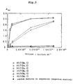
- the phage recovery rate increased from the fourth to the six cycle where the panning was repeated under uniform conditions. It could, thus, be confirmed that phages expressing the peptide specifically binding to the influenza virus hemagglutinin (H1N1) were recovered.
- the phage obtained by the sixth cycle of panning was used to determine the amino acid sequence of the expressed peptide, specifically, two kinds of phages, namely the phage (hereinafter referred to as H1/7 phage: 19 clones) bound to the immobilized hemagglutinin H1N1 and eluted with 7-F-Neu5Ac2en and the phage (hereinafter referred to as H1/3 phage: 20 clones) bound to the immobilized hemagglutinin H1N1 and eluted with ganglioside GM3, were used for the sequencing of the expressed peptides.
- H1/7 phage 19 clones
- H1/3 phage the phage bound to the immobilized hemagglutinin H1N1 and eluted with 7-F-Neu5Ac2en
- H1/3 phage 20 clones
- the supernatant was further transferred to an Oak-Ridge centrifuge tube, in which 3 ml of polyethylene glycol (PEG 6000; product of Nacalai Tesque)/NaCl was added and the mixture was stirred well and then allowed to stand at 4°C for 4 hours. The mixture was centrifuged at 12,000 rpm for 10 minutes to precipitate the phages. The supernatant was discarded and the phage pellet was suspended in 1 ml of TBS (Tris-buffer solution). The suspension was transferred to a 1.5 ml Ep tube and centrifuged at 15,000 rpm for 10 minutes to remove insolubles.
- PEG 6000 polyethylene glycol
- Nacalai Tesque Nacalai Tesque
- the supernatant was transferred to another Ep tube, in which 150 ⁇ l of polyethylene glycol/NaCl was added. The mixture was stirred well and, then, allowed to stand at 4°C for 1 hour. It was then centrifuged at 15,000 rpm for 10 minutes to reprecipitate the phages. The supernatant was discarded and the phage pellet was resuspended in 200 ⁇ l of TBS. The suspension was centrifuged at 15,000 rpm for 10 minutes to precipitate insolubles and the precipitate was transferred to a 0.5 ml Ep tube. The phage clone was stored at 4°C.
- the sequencing of the peptide encoded by phage DNA was determined by the dideoxy method (Proc. Natl. Acad. Sci., USA., 74 , 5463-5467 (1977)) using Amersham's THERMO Sequencing Kit (Amersham life Science, Code; US79765, Lot: 201503) according to the accompanying manual.
- the PCR reaction of the DNA was carried out in 30 cycles of 96°C, 30 sec., 45°C, 15 sec., 60°C, 4 min. and the DNA sequencing was made using ABI's DNA sequencer (ABI PRISMTM377).
- amino acid sequences of the peptides having a binding affinity for the receptor-binding pocket of the hemagglutinin as determined from clones of the two kinds of phages are shown in one-letter expression in Table 3.
- the amino acid sequences indicated in the following table are restated hereinafter under SEQUENCE LISTING as SEQ ID NO:1 (NOs: 1 and 5), 2 (NO:2), 3 (NO:3), 4 (NO:4), 5 (NO:6) and 6 (NO:7), respectively.
- the HA receptor substance ganglioside GM3 is known to have a high affinity for hemagglutinin H1 and the HA inhibitor 7F-Neu5Ac2en is known to have a high antagonist activity against hemagglutinin H1.
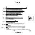
- the phage obtained in Example 1 was cloned and the binding affinity of the expressed peptide for hemagglutinin was evaluated by ELISA.
- H1N1 hemagglutinin
- TBS fetal bovine serum
- BSA Albumin Bovine, Fatty Acid Free; Sigma
- 50 ⁇ l of an amplified phage solution H1/H7 phage: Clone Nos. 1 ⁇ 4; H1/3 phage: Clone Nos. 5 ⁇ 7
- TBST TBS/5% Tween 20
- 150 ⁇ l of TBS/5% BSA was added, and the plate was allowed to sit at room temperature overnight.
- the substrate solution (0.4 mg/ml o-phenylendiamine substrate in citrate-phosphate buffer (pH 5.0)) was added, 100 ⁇ l/well, and the reaction was carried out for 10 minutes. The reaction was stopped by adding 100 ⁇ l/well of 3 N-H 2 SO 4 and the absorbance at 492 nm was measured with a microplate reader.
- H1 phages H1/7 phage and H1/3 phage subjected to panning with hemagglutinin (H1N1) as the target
- H1N1 hemagglutinin
- Biopanning was carried out as in Example 1 using two kinds of influenza virus hemagglutinins H1 (H1N1, A/PR/8/34) and H3 (H3N2, A/Wuhan/359/95) as the target. Specifically, using the H1-supporting plate and H3-supporting plate prepared in accordance with Reference Example 2 as the HA-supporting plate and the HA receptor Sialyl-LewisX and ⁇ (2 ⁇ 6) GM3 as the eluent, biopanning was performed in the same manner as in Example 1 to select phages expressing HA-binding peptides. For representative phage clones obtained by the biopanning, the amino acid sequences of the expressed peptides were determined. The results are shown in Table 4.
- Clone "C-1" was found in one of 9 clones selected by the panning system "H1/GM” and in one of 10 clones selected by the panning system "H3/GM".
- Clone "C-2" was found in one of 9 clones selected by the panning system "H1/GM” and in 4 of 10 clones selected by the panning system "H3/X".
- Clone "A-1” was selected from all the panning systems varying in target and eluent.
- the finding that clones expressing peptides of the same sequence could be obtained by pannings with different eluents suggests that the peptide expressed by this Clone "A-1” has the same hemagglutinin binding sites (NeuAc-associated sites) as the binding sites possessed by the HA receptor substances Sialyl-LewisX and a (2 ⁇ 6) GM3 and that it is a peptide having the same binding mode as the above receptor substances.
- Clones "B-1" and “B-2” were found to have GRxP (x represents V or P) as a common motif and that the peptides expressed by Clones "C-1" and “C-2” were found to have IAFSxyA (x represents S or R; y represents L or A) as a common motif.
- hemagglutinins H1 (H1N1) and H3 (H3N2) and wheat germ agglutinin (WGA), which is a kind of lectin were respectively immobilized on a 96-well carboplate (product of Sumilon) and ELISA was carried out by adding 5 ⁇ 10 10 virions/ml of each phage clone solution.
- hemagglutinins H1 and H3 are different from each other by 75% in amino acid sequence. Therefore, the peptides expressed by clones "A-1", “B-1", “B-2”, “C-1” and “C-2” are considered to recognize a homologous sequence of the two hemagglutinins, i.e. H1 and H3, and be bound specifically to that site.
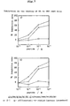
- the binding affinity of each clone for anti-GM3 antibody was evaluated by ELISA as in Example 4. Specifically, for ELISA, the anti-GM3 antibody (M2590, Nippon Biotest Kenkyusho) was immobilized on a 96-well carboplate (product of Sumilon) and 5 ⁇ 10 10 virions/ml of each phage clone solution was added.
- GM3 solution (8.5 ⁇ g/ml methanol) was added to a 96-well microtiter plate, followed by addition of 50 ⁇ l of 0.08% polyisobutyl methacrylate solution. After the solvent was evaporated off with a dryer, 50 ⁇ l of 1% BSA/TBS was added and the plate was allowed to sit at 4° C overnight. After the plate was washed with 3 portions of TBS, 50 ⁇ l of a solution containing a phage clone of predetermined concentration (Table 6) and hemagglutinin (in a concentration such that the system hemagglutinin concentration would be 30 nM) was added and the plate was allowed to sit at 20°C for 2 hours.
- a solution containing a phage clone of predetermined concentration (Table 6) and hemagglutinin (in a concentration such that the system hemagglutinin concentration would be 30 nM) was added and the plate was allowed to sit at 20°C for
- Example 3 To confirm that the phage clones ("A-1”, “B-1”, “B-2”, “C-1”, “C-2”) obtained in Example 3 actually recognize the host receptor recognition sites of hemagglutinins (HA), it was investigated by ELISA as in Example 6 whether the binding of said phage clones to immobilized HA is inhibited by GM3 which is an HA receptor substance. As the hemagglutinin, H1N1 and H3N2 were respectively used. As control, similar experiments were performed using GM1 and GlcCer in lieu of GM3.
- the peptides expressed on the phage clones obtained in Examples 1 and 3 were synthesized in the following manner.
- Each peptide was synthesized by the solid-phase method (Fmoc/NMP, HOBt method) by using a full-automatic peptide synthesizer (ACT357, manufactured by Advanced Chemtech) in accordance with the manufacturer's program [Fmoc: 9-fluorenylmethoxycarbonyl; NMP: N-methylpyrrolidone; HOBt: 1-hydroxybenzotriazole].
- an Fmoc-amino acid corresponding to the C-terminal amino acid was condensed to 0.25 mmol of Fmoc-NH-FAL resin according to a synthesizing program and, thereafter, Fmoc-amino acids corresponding to the second and subsequent amino acids from the C terminus were serially condensed for chain extension.
- the N-terminal Fmoc group was subjected to deprotection reaction according to the program.
- the peptide resin thus obtained was recovered in a polypropylene mini-column (product of Assist Co.), washed with methanol, and dried in vacuo. Thereafter, the peptide was excised off the resin by the following procedure and the side chains were deprotected.
- Reagent K 82.5% TFA, 5% phenol, 5% H 2 O, 5% thioanisole, 2.5% ethanedithiol
- reaction mixture was dripped into 8 ml of cold diethyl ether to stop the reaction and, at the same time, precipitate the peptide. Furthermore, after the mini-column was washed with 2 ml of TFA, 5 ml of cold diethyl ether was further added and the mixture was centrifuged. The pellet was washed 4 times with 10 ml portions of diethyl ether and the peptide was solubilized with about 5 ml of 50% acetonitrile and lyophilized. The solubilization and lyophilization procedure was repeated twice to give the objective crude lyophilizate.
- the synthetic peptides having cysteine residues can be optionally cyclized. Taking the peptide expressed by clone "H1-1" as an example, this peptide was cyclized in the following manner.
- Lipopeptides and peptide-modified liposomes Lipopeptides and peptide-modified liposomes
- the HA-binding peptide of the invention can be introduced onto liposomes.
- the peptide expressed by the Clone "A-1" obtained in Example 3 is taken as an example in the following explanation.
- the peptide expressed by clone "A-1" was synthesized by the cycling in accordance with the procedure of Example 8. Then, according to said synthesizing program, stearic acid was coupled to the N-terminus of the above peptide. The peptide was excised from the resin and deprotected. Thereafter, the procedure described in Example 8 was followed to give the desired stearic acid-coupled peptide ("C 18 A-1 lipopeptide").
- the internal peptide content was determined with a commercial kit (Bio-Rad Protein Assay Kit) to confirm that the lipopeptide prepared as above had been incorporated in the liposomes.
- Each of the above liposome solutions was adjusted to pH 8 with 1 N-NaOH/H 2 O and the peptide of the invention, i.e. H-Gly-Cys-OH added to the C terminus of "A-1" peptide, was added in a predetermined final concentration.
- the mixture was stirred gently at room temperature for 6 hours and subjected to centrifugal ultrafiltration as in Example 9 to give a liposome solution.
- the peptide content of the liposome thus obtained was determined in the same manner as in Example 9.
- a modifier peptide consisting in an HA-binding peptide bound to the SH group of cysteine through the maleimide of DPPE-PEG-Mal was introduced onto the liposome to give a peptide-modified liposome.
- the peptide expressed by clone "A-1" was synthesized by the procedure described in Example 8 and the inhibitory effect of this synthetic peptide on the binding of hemagglutinin to ganglioside GM3 was evaluated. Specifically, it was experimentally explored by ELISA whether the above synthetic peptide inhibits the binding of ganglioside GM3 to immobilized HA (H1N1, H3N2). As control, similar experiments were performed using the N-terminal 15-residue peptide of the phage coat protein pIII and sialyl-lactose in lieu of said synthetic peptide.
- influenza virus infection inhibitory activity of the HA-binding peptide of the invention was evaluated.
- influenza virus-infected cell MDCK (Mardin-Darby canine kidney) cell was used.
- the influenza virus infection of this cell was verified by detecting LDH (lactate dehydrogenase) activity using a commercial kit (LDH-Cytotoxic Test, Wako) (J. Virol. Meth. 51 , 185-192 (1995)).
- LDH-Cytotoxic Test, Wako LDH-Cytotoxic Test, Wako
- H1 A/PR/8/34 (H1N1)
- H3 A/Victoria/395 (H3N2) influenza A viruses were respectively used.
- the infective titer of the influenza virus was determined as follows.
- 3 ⁇ 10 5 cells/ml of the MDCK cell grown in MEM/10% FBS were sown on a 96-well microtiter plate, 100 ml/well, and incubated under 5% CO 2 at 37°C overnight. After confirming that the cells in all wells had formed confluent monolayers, the wells were washed with serum-free MEM (MEM(-)) twice and a serial dilution of the virus solution (MEM(-)) was injected into the wells, 100 ⁇ l/well. The plate was incubated under 5% CO 2 at 37°C for 2 hours, after which the virus solution was discarded. The wells were washed with 2 portions of culture medium and incubated in the same medium for 20 hours.
- MEM(-) serum-free MEM
- MEM(-) serial dilution of the virus solution
- a 15 ⁇ l portion of the supernatant in each well was taken, diluted 4-fold with PBS to make 50 ⁇ l, and transferred to a fresh plate, in which 50 ml/well of a color developer (nitro blue tetrazolium, diaphorase and NDA+ in lithium lactate-PBS) was added.
- the plate was allowed to sit at room temperature for 20 minutes , after which 100 ml of a reaction stopper (0.5 M HCl in PBS) was added to the wells to stop the color reaction and the absorbance at 550 nm was measured with a microplate reader.
- the ID 50 value was calculated from the result obtained.
- the experiment on the inhibition of influenza virus infection by the HA-binding peptide was carried out as follows. The test was performed with an amount of the virus corresponding to 50 times the ID 50 value found above (50 LDH ID 50 ).
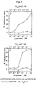
- the results of influenza virus antiinfection experiments performed using the "C 18 A-1 lipopeptide" according to Example 9 and a liposome preparation containing 10% of this lipopeptide as the HA-binding peptide are presented in Figs. 8 and 9.
- Fig. 8 represents the result obtained with "C 18 A-1 lipopeptide”
- Fig. 9 represents the result with the "C 18 A-1 lipopeptide”-modified liposome (10% peptide).
- a) represents the antiinfective effect on influenza A virus H1 (A/PR/8/34 (H1N1))
- b) represents the antiinfective effect on H3 (A/Victoria/359 (H3N2)).
- HA-binding peptide of the invention is of use as a prophylactic (antiinfective ) or therapeutic agent for influenza.
- a peptide capable of binding specifically to influenza virus hemagglutinin capable of binding specifically to influenza virus hemagglutinin.
- the peptide of the invention binds specifically to the hemagglutinin associated with the first step of influenza virus infection to successfully inhibit binding of the virus to the host receptor and, as such, finds application as a prophylactic drug for influenza virus infection or a therapeutic drug for influenza.
- the preferred peptide of the invention has the property to bind specifically to the receptor-binding pocket of hemagglutinin, the three-dimensional structure of which is well conserved among subtypes.
- the peptide of the invention is stable and because of the variegateness of its side chains, the peptide may undergo interactions with the binding sites of hemagglutinins. For this reason, the peptide of the invention is expected to find application as a broad-spectrum antiinfective or therapeutic drug for influenza, particularly for type A viruses in general, regardless of subtypes or over many subtypes.
- the peptide of the invention lends itself well to large-scale synthesis and modification as compared with the sugar chain analogs under development for use as antiinfluenza drugs, thus being useful for drug production and further drug development.
Landscapes
- Health & Medical Sciences (AREA)
- Life Sciences & Earth Sciences (AREA)
- Chemical & Material Sciences (AREA)
- Virology (AREA)
- Organic Chemistry (AREA)
- Medicinal Chemistry (AREA)
- General Health & Medical Sciences (AREA)
- Molecular Biology (AREA)
- Oncology (AREA)
- Pulmonology (AREA)
- Chemical Kinetics & Catalysis (AREA)
- Nuclear Medicine, Radiotherapy & Molecular Imaging (AREA)
- Communicable Diseases (AREA)
- Pharmacology & Pharmacy (AREA)
- Animal Behavior & Ethology (AREA)
- General Chemical & Material Sciences (AREA)
- Public Health (AREA)
- Veterinary Medicine (AREA)
- Biochemistry (AREA)
- Biophysics (AREA)
- Genetics & Genomics (AREA)
- Proteomics, Peptides & Aminoacids (AREA)
- Medicines That Contain Protein Lipid Enzymes And Other Medicines (AREA)
- Peptides Or Proteins (AREA)
Applications Claiming Priority (3)
| Application Number | Priority Date | Filing Date | Title |
|---|---|---|---|
| JP9196299 | 1999-03-31 | ||
| JP9196299 | 1999-03-31 | ||
| PCT/JP2000/001867 WO2000059932A1 (fr) | 1999-03-31 | 2000-03-27 | Peptides se liant a l'hemagglutinine du virus de la grippe |
Publications (1)
| Publication Number | Publication Date |
|---|---|
| EP1167382A1 true EP1167382A1 (de) | 2002-01-02 |
Family
ID=14041194
Family Applications (1)
| Application Number | Title | Priority Date | Filing Date |
|---|---|---|---|
| EP00911385A Withdrawn EP1167382A1 (de) | 1999-03-31 | 2000-03-27 | Sich an das hämagglutinin des influenzavirus bindendes peptid |
Country Status (5)
| Country | Link |
|---|---|
| EP (1) | EP1167382A1 (de) |
| JP (1) | JP4528989B2 (de) |
| AU (1) | AU3328900A (de) |
| CA (1) | CA2365575A1 (de) |
| WO (1) | WO2000059932A1 (de) |
Cited By (10)
| Publication number | Priority date | Publication date | Assignee | Title |
|---|---|---|---|---|
| WO2009091501A1 (en) * | 2008-01-18 | 2009-07-23 | Response Biomedical Corporation | Heated assays for influenza |
| US20100119588A1 (en) * | 2006-03-13 | 2010-05-13 | Keio University | Peptide for inhibition of influenza infection, inhibitor of influenza infection, liposome, and prophylactic/therapeutic agent for influenza |
| EP2300492A1 (de) * | 2008-06-12 | 2011-03-30 | Universiti Putra Malaysia | Neues antivirales peptid gegen das vogelgrippevirus h9n2 |
| CN104797934A (zh) * | 2012-10-05 | 2015-07-22 | 电化生研株式会社 | 流感病毒的血凝素的测定方法 |
| US9683030B2 (en) | 2012-05-10 | 2017-06-20 | Massachusetts Institute Of Technology | Agents for influenza neutralization |
| RU2711932C2 (ru) * | 2014-07-15 | 2020-01-23 | МЕДИММЬЮН, ЭлЭлСи | Нейтрализующие антитела к вирусу гриппа B и пути их применения |
| US10882897B2 (en) | 2015-06-01 | 2021-01-05 | Medimmune, Llc | Neutralizing anti-influenza binding molecules and uses thereof |
| US11186627B2 (en) | 2013-10-02 | 2021-11-30 | Medimmune, Llc | Neutralizing anti-influenza A antibodies and uses thereof |
| RU2784915C2 (ru) * | 2014-07-15 | 2022-12-01 | МЕДИММЬЮН, ЭлЭлСи | Нейтрализующие антитела к вирусу гриппа b и пути их применения |
| US11547756B2 (en) | 2016-01-13 | 2023-01-10 | Medimmune, Llc | Method of treating influenza A |
Families Citing this family (15)
| Publication number | Priority date | Publication date | Assignee | Title |
|---|---|---|---|---|
| JP4651213B2 (ja) * | 2001-03-27 | 2011-03-16 | 株式会社グライコメディクス | インフルエンザウイルス・ヘマグルチニン結合性ペプチド |
| WO2005044992A2 (en) * | 2003-11-04 | 2005-05-19 | The Administrators Of The Tulane Educational Fund | Fusion initiation region in rna virus envelope proteins |
| CA2559067A1 (en) * | 2004-03-09 | 2005-09-15 | Glycomedics, Inc. | Influenza virus infection suppressor |
| US7449481B2 (en) | 2004-04-13 | 2008-11-11 | Cephalon, Inc. | Thio-substituted biaryl-methanesulfinyl derivatives |
| JP2006101709A (ja) * | 2004-09-30 | 2006-04-20 | Glycomedics Inc | ヘマグルチニン結合ペプチド、インフルエンザウイルス感染阻害剤、リポソーム、インフルエンザ治療薬、インフルエンザ予防薬 |
| WO2008021415A2 (en) * | 2006-08-14 | 2008-02-21 | Massachusetts Institute Of Technology | Glycan data mining system |
| EP2327714A4 (de) * | 2008-08-29 | 2012-04-25 | Univ Keio | Prophylaktischer oder therapeutischer wirkstoff gegen grippevirusinfektion |
| JP5396111B2 (ja) * | 2009-03-12 | 2014-01-22 | 学校法人慶應義塾 | インフルエンザ治療/予防薬 |
| US8765686B2 (en) | 2010-08-03 | 2014-07-01 | University Of Washington Through Its Center For Commercialization | Polypeptides for treating and/or limiting influenza infection |
| CN102268072B (zh) * | 2011-07-06 | 2014-11-05 | 中国人民解放军军事医学科学院放射与辐射医学研究所 | 流感病毒血凝素蛋白结合多肽的结构和用途 |
| WO2013138259A2 (en) | 2012-03-13 | 2013-09-19 | University Of Washington Through Its Center For Commercialization | Polypeptides for treating and/or limiting influenza infection |
| US9771395B2 (en) | 2014-03-21 | 2017-09-26 | University Of Washington | Enhanced influenza hemagglutinin binders |
| WO2016063969A1 (ja) * | 2014-10-24 | 2016-04-28 | ペプチドリーム株式会社 | ヘマグルチニン結合ペプチド |
| WO2018038168A1 (ja) * | 2016-08-25 | 2018-03-01 | 学校法人同志社 | ヘマグルチニン結合ペプチド、および、これを含むインフルエンザウイルス感染症の予防・治療薬 |
| TW202042836A (zh) * | 2019-02-18 | 2020-12-01 | 日商肽夢想股份有限公司 | 血球凝集素結合肽 |
Family Cites Families (1)
| Publication number | Priority date | Publication date | Assignee | Title |
|---|---|---|---|---|
| WO1998039469A1 (en) * | 1997-03-04 | 1998-09-11 | Bio-Technology General Corp. | Isolation of tissue specific peptide ligands and their use for targeting pharmaceuticals to organs |
-
2000
- 2000-03-27 CA CA002365575A patent/CA2365575A1/en not_active Abandoned
- 2000-03-27 WO PCT/JP2000/001867 patent/WO2000059932A1/ja not_active Application Discontinuation
- 2000-03-27 AU AU33289/00A patent/AU3328900A/en not_active Abandoned
- 2000-03-27 JP JP2000609442A patent/JP4528989B2/ja not_active Expired - Fee Related
- 2000-03-27 EP EP00911385A patent/EP1167382A1/de not_active Withdrawn
Non-Patent Citations (1)
| Title |
|---|
| See references of WO0059932A1 * |
Cited By (23)
| Publication number | Priority date | Publication date | Assignee | Title |
|---|---|---|---|---|
| US20100119588A1 (en) * | 2006-03-13 | 2010-05-13 | Keio University | Peptide for inhibition of influenza infection, inhibitor of influenza infection, liposome, and prophylactic/therapeutic agent for influenza |
| US8299214B2 (en) | 2006-03-13 | 2012-10-30 | Keio University | Influenza infection-inhibiting peptide, influenza virus infection inhibitor, liposome, and influenza preventive/therapeutic agent |
| WO2009091501A1 (en) * | 2008-01-18 | 2009-07-23 | Response Biomedical Corporation | Heated assays for influenza |
| US8318420B2 (en) | 2008-01-18 | 2012-11-27 | Response Biomedical Corporation | Heated assays for influenza |
| EP2300492A1 (de) * | 2008-06-12 | 2011-03-30 | Universiti Putra Malaysia | Neues antivirales peptid gegen das vogelgrippevirus h9n2 |
| EP2300492A4 (de) * | 2008-06-12 | 2012-03-14 | Univ Putra Malaysia | Neues antivirales peptid gegen das vogelgrippevirus h9n2 |
| US10538578B2 (en) | 2012-05-10 | 2020-01-21 | Massachusetts Institute Of Technology | Agents for influenza neutralization |
| US10947300B2 (en) | 2012-05-10 | 2021-03-16 | Massachusetts Institute Of Technology | Agents for influenza neutralization |
| US9982037B2 (en) | 2012-05-10 | 2018-05-29 | Massachusetts Institute Of Technology | Agents for influenza neutralization |
| US9683030B2 (en) | 2012-05-10 | 2017-06-20 | Massachusetts Institute Of Technology | Agents for influenza neutralization |
| US10365277B2 (en) | 2012-10-05 | 2019-07-30 | Denka Seiken Co., Ltd. | Method for measuring hemagglutinin from influenza virus |
| CN104797934A (zh) * | 2012-10-05 | 2015-07-22 | 电化生研株式会社 | 流感病毒的血凝素的测定方法 |
| US11932682B2 (en) | 2013-10-02 | 2024-03-19 | Medimmune, Llc | Neutralizing anti-influenza A antibodies and uses thereof |
| US11186627B2 (en) | 2013-10-02 | 2021-11-30 | Medimmune, Llc | Neutralizing anti-influenza A antibodies and uses thereof |
| US11174304B2 (en) | 2014-07-15 | 2021-11-16 | Medimmune, Llc | Neutralizing anti-influenza B antibodies and uses thereof |
| RU2739952C2 (ru) * | 2014-07-15 | 2020-12-30 | МЕДИММЬЮН, ЭлЭлСи | Нейтрализующие антитела к вирусу гриппа b и пути их применения |
| RU2784915C2 (ru) * | 2014-07-15 | 2022-12-01 | МЕДИММЬЮН, ЭлЭлСи | Нейтрализующие антитела к вирусу гриппа b и пути их применения |
| US11787853B2 (en) | 2014-07-15 | 2023-10-17 | Medimmune, Llc | Neutralizing anti-influenza b antibodies and uses thereof |
| RU2711932C2 (ru) * | 2014-07-15 | 2020-01-23 | МЕДИММЬЮН, ЭлЭлСи | Нейтрализующие антитела к вирусу гриппа B и пути их применения |
| US10882897B2 (en) | 2015-06-01 | 2021-01-05 | Medimmune, Llc | Neutralizing anti-influenza binding molecules and uses thereof |
| US11524993B2 (en) | 2015-06-01 | 2022-12-13 | Medimmune, Llc | Neutralizing anti-influenza binding molecules and uses thereof |
| US11926657B2 (en) | 2015-06-01 | 2024-03-12 | Medimmune, Llc | Neutralizing anti-influenza binding molecules and uses thereof |
| US11547756B2 (en) | 2016-01-13 | 2023-01-10 | Medimmune, Llc | Method of treating influenza A |
Also Published As
| Publication number | Publication date |
|---|---|
| WO2000059932A1 (fr) | 2000-10-12 |
| JP4528989B2 (ja) | 2010-08-25 |
| CA2365575A1 (en) | 2000-10-12 |
| AU3328900A (en) | 2000-10-23 |
Similar Documents
| Publication | Publication Date | Title |
|---|---|---|
| EP1167382A1 (de) | Sich an das hämagglutinin des influenzavirus bindendes peptid | |
| US8299214B2 (en) | Influenza infection-inhibiting peptide, influenza virus infection inhibitor, liposome, and influenza preventive/therapeutic agent | |
| CA1250100A (en) | Pre-s gene coded peptide hepatitis b immunogens, vaccines, diagnostics, and synthetic lipid vesicle carriers | |
| KR101688547B1 (ko) | 아포리포단백질 a-i 모방체 | |
| US7205273B2 (en) | Fusogenic liposomes | |
| JP4651213B2 (ja) | インフルエンザウイルス・ヘマグルチニン結合性ペプチド | |
| WO1997038010A9 (en) | Fusogenic liposomes | |
| KR102438720B1 (ko) | 안지오텐신 전환효소 2를 포함하는 나노디스크 및 이의 항바이러스 용도 | |
| CA2691358A1 (en) | Influenza inhibiting compositions comprising hemagglutinin 2-derived peptides and use thereof | |
| US11160860B2 (en) | HSV antigenic peptides and HSV protein vaccines | |
| JP2022502392A (ja) | 経口剤形の製造方法 | |
| JP2006101709A (ja) | ヘマグルチニン結合ペプチド、インフルエンザウイルス感染阻害剤、リポソーム、インフルエンザ治療薬、インフルエンザ予防薬 | |
| WO2001012668A1 (en) | Cathelicidin-derived peptides with broad spectrum antimicrobial activity | |
| WO2003020753A1 (en) | Peptidic compounds selectively binding to p-selectin | |
| KR102017973B1 (ko) | 항-b형 간염 바이러스 x 단백질 폴리펩티드 약제 | |
| JPH07509714A (ja) | ペプチド肺表面活性剤及び治療用組合せ | |
| US20090098195A1 (en) | Influenza virus infection suppressor | |
| JP6714260B2 (ja) | インフルエンザウイルス感染阻害剤、リポソーム、インフルエンザ予防・治療剤 | |
| KR0173366B1 (ko) | B형 간염 바이러스의 펩타이드 항원을 함유하는 리포좀 | |
| KR0178422B1 (ko) | 한탄 바이러스의 펩타이드 항원을 함유하는 리포좀 | |
| KR100189701B1 (ko) | B형 간염 바이러스의 x 단밸직 유래의 펩타이드 항원을 함유하는 리포좀 | |
| EP4359009A1 (de) | Griffithsin zur verwendung in einem verfahren zur vorbeugung oder behandlung von infektionen mit respiratorischen viren | |
| JPH0418029A (ja) | Hiv感染細胞を殺傷するリポソーム | |
| EP0547681A2 (de) | Zyklisches HIV PND und Lipopeptid enthaltende synthetische Peptide | |
| AU2002324363A1 (en) | Peptidic compounds selectively binding to p-selectin |
Legal Events
| Date | Code | Title | Description |
|---|---|---|---|
| PUAI | Public reference made under article 153(3) epc to a published international application that has entered the european phase |
Free format text: ORIGINAL CODE: 0009012 |
|
| 17P | Request for examination filed |
Effective date: 20011019 |
|
| AK | Designated contracting states |
Kind code of ref document: A1 Designated state(s): AT BE CH CY DE DK ES FI FR GB GR IE IT LI LU MC NL PT SE |
|
| RIN1 | Information on inventor provided before grant (corrected) |
Inventor name: OGINO, KOICHI Inventor name: ISHIKAWA, DAI Inventor name: TANAKA, MICHINORI Inventor name: TAKI, TAKAO Inventor name: SATO, TOSHINORI |
|
| STAA | Information on the status of an ep patent application or granted ep patent |
Free format text: STATUS: THE APPLICATION HAS BEEN WITHDRAWN |
|
| 18W | Application withdrawn |
Withdrawal date: 20021023 |