EP1120426A1 - G-protein-gekoppelte rezeptoren - Google Patents
G-protein-gekoppelte rezeptoren Download PDFInfo
- Publication number
- EP1120426A1 EP1120426A1 EP99970399A EP99970399A EP1120426A1 EP 1120426 A1 EP1120426 A1 EP 1120426A1 EP 99970399 A EP99970399 A EP 99970399A EP 99970399 A EP99970399 A EP 99970399A EP 1120426 A1 EP1120426 A1 EP 1120426A1
- Authority
- EP
- European Patent Office
- Prior art keywords
- protein
- gtar14
- gtar11
- dna
- peptide
- Prior art date
- Legal status (The legal status is an assumption and is not a legal conclusion. Google has not performed a legal analysis and makes no representation as to the accuracy of the status listed.)
- Withdrawn
Links
Images


Classifications
-
- C—CHEMISTRY; METALLURGY
- C07—ORGANIC CHEMISTRY
- C07K—PEPTIDES
- C07K14/00—Peptides having more than 20 amino acids; Gastrins; Somatostatins; Melanotropins; Derivatives thereof
- C07K14/435—Peptides having more than 20 amino acids; Gastrins; Somatostatins; Melanotropins; Derivatives thereof from animals; from humans
- C07K14/705—Receptors; Cell surface antigens; Cell surface determinants
Definitions
- the present invention relates to novel G protein-coupled receptor proteins and their genes, as well as methods for producing and using same.
- the olfactory receptor (OR) gene family has been known as a multiple gene family encoding odorant receptors that govern the system for olfactory response in the living body.
- a member of the gene family was first reported to be expressed in rat olfactory neurons in 1991 (Buck L. et al. Cell 65:175-187 (1991)), and subsequently, a number of homologous genes were found across species, in canine (Permentier M. et al. Nature 355:453-455 (1992)), mouse (Ressler K.J. et al. Cell 73:597-609 (1993)), human (Selbie L.A. et al. Mol. Brain Res.
- the receptor proteins encoded by the genes of the family have a unique seven transmembrane structure that is similar to other G protein-coupled receptor subfamilies, and their first extracellular domain contains a common [N-x-S/T] motif (x: any amino acid) which is modified by sugar. Their second and third extracellular domains, loop-1 and loop-2, respectively, are cross-linked by a disulfide bond between the conserved cysteine residues of each.
- the [M-A-Y-D-R-Y-L/V-A-I/V-C] sequence within the second intracellular domain is another common motif predicted to be functionally important.
- the [D-R-Y] motif is suggested to be essential for the binding to intracellular G proteins (Rosenthal W. et al. J. Biol. Chem. 268:13030-13033 (1993); Marchese A. et al. Genomics 23:609-618 (1994)).
- the present invention provides novel G protein-coupled receptor proteins and their genes, as well as materials and methods for producing them, molecules used for the method and such, and their use.
- the present inventors performed BLAST search of the GenBank public database and found multiple human genome sequences that encode novel seven transmembrane G protein-coupled receptor genes.
- the inventors carried out RT-PCR using oligonucleotide primers that were designed based on the above genome sequences and mRNA prepared from human tissues as a template, and obtained partial cDNA sequences of the target genes. This RT-PCR was also used to evaluate the expression pattern and profile of the genes in those tissues. Then, the inventors carried out 5'- and 3'- RACE using a cDNA library prepared from human testis as a template, in which the genes were highly expressed based on the result of RT-PCR, and succeeded in isolating the full length cDNA.
- the first three genes isolated, originating from human chromosome 14, are designated as GTAR14-1, GTAR14-3, and GTAR14-5, and the next four genes isolated, originating from human chromosome 11, are designated as GTAR11-1, GTAR11-2, GTAR11-3, and GTAR11-4.
- GTAR14-1 was detected strongly in lymphatic/hematopoietic tissues, including thymus and spleen, testis, and pancreas, and was also expressed in fetal thymus.
- GTAR14-3 was detected strongly in thymus and testis, but was also expressed widely in multiple tissues, including digestive tissues such as small intestine and colon, and hormone secreting tissues such as placenta and prostate.
- GTAR14-5 was detected strongly in lymphatic/hematopoietic tissues, such as thymus, spleen, and peripheral leukocytes, and testis.
- GTAR11-1 was detected strongly in lymphatic/hematopoietic tissues including thymus, spleen, and peripheral lymphocytes, digestive tissues such as small intestine and colon, and testis and placenta.
- GTAR11-2 was detected strongly in lymphatic/hematopoietic tissues including thymus and peripheral lymphocytes, and testis and placenta.
- GTAR11-3 was detected strongly in lymphatic/hematopoietic tissues including thymus and spleen, and genital organs, and was also detected broadly in multiple tissues including digestive tissues such as small intestine and colon, muscle, and neuronal tissues.
- GTAR11-4 was detected strongly in lymphatic/hematopoietic tissues including thymus and spleen, and genital organs, and was also extensively expressed in multiple tissues including digestive tissues such as small intestine and colon, and endocrine tissues.
- proteins encoded by those genes have an important function in lymphatic/hematopoietic and endocrine tissues. Consequently, it was considered that the proteins can be used for screening a novel hematopoietic peptide factor or a novel hormone-like peptide factor.
- the present invention relates to novel G protein-coupled receptor proteins and their genes, and their production and use as well. More specifically, it provides:
- the nucleotide sequences of GTAR14-1, GTAR14-3, and GTAR14-5 cDNAs that were isolated by the present inventors, and the amino acid sequences of the proteins encoded by the cDNAs are shown in SEQ ID NO: 1, 2, 3, 4, 5, and 6, respectively.
- the nucleotide sequences of GTAR11-1, GTAR11-2, GTAR11-3, and GTAR11-4 cDNAs that were also isolated by the present inventors, and the amino acid sequences of the proteins encoded by the cDNAs are shown in SEQ ID NOs: 24, 25, 26, 27, 28, 29, 30, and 31, respectively.
- the receptor proteins of the OR gene family have a seven transmembrane type structure like other G protein-coupled receptors, and their first extracellular domains have a [N-x-S/T] motif (x: any amino acid) in common that is modified by sugar.
- x any amino acid
- cysteine residues in the second (loop-1) and third (loop-2) extracellular domains that form a disulfide bond for cross-linking Furthermore, there is a [M-A-Y-D-R-Y-L/V-A-I/V-C] sequence (especially [D-R-Y] motif) in the second intracellular domain, which is suggested to be essential for binding to the G protein (Rosenthal W. et al. J. Biol. Chem. 268:13030-13033 (1993); Marchese A.
- the seven proteins encoded by the seven genes isolated by the present inventors are cell membrane proteins and are seven transmembrane receptors that contain seven hydrophobic transmembrane domains.
- GTAR proteins like known odorant receptors, have the above described motifs characteristic to G protein-coupled receptors.
- the [N-x-S/T] motif (x: any amino acid) modified by sugar is found within the first extracellular domain as [N-Q-T], [N-S-T], and [N-T-S] in GTAR14-1, GTAR14-3, and GTAR14-5 proteins, respectively, as [N-Y-S] in GTAR11-1 protein, and as [N-S-T] in GTAR11-2, GTAR11-3, and GTAR11-4 proteins.
- GTAR proteins have conserved cysteine residues in the second and third extracellular domains (loop-1 and loop-2, respectively). Furthermore, they have the [M-A-Y-D-R-Y-L/V-A-I/V-C] sequence in the second intracellular domain, which is suggested to be essential for binding to G protein, as well: [V-A-Y-D-R-Y-V-A-I-C] in GTAR14-1 protein, and [M-A-Y-D-R-Y-L-A-I-C] in all the others.
- GTAR proteins of the present invention function through binding to intracellular G proteins in response to the stimuli of extracellular ligands.
- GTAR proteins of the present invention may regulate the differentiation, or proliferation and activation of a particular group of lymphatic cells.
- GTAR proteins could function as a central molecule that controls the activities characteristic to lymphocytes through interaction with G proteins in response to ligand stimuli. As these proteins are expressed in testis, where cells are changing continuously in the cell cycle at a high turn over rate, it is possible that they regulate cell cycle and cell division.
- GTAR14-1 gene was detected in spleen, where hematopoietic cells are expected to exist.
- the GTAR14-5 gene was also expressed in spleen and peripheral leukocytes, where hematopoietic cells are thought to be contained.
- the GTAR11 genes were expressed in peripheral leukocytes, in which hematopoietic cells are considered to be included, and furthermore the genes for GTAR11-1, GTAR11-3, and GTAR11-4 were expressed in spleen, which is expected to have hematopoietic cells.
- the GTAR14-3 gene was expressed in thymus and testis as described above, and also in digestive tissues, including small intestine and colon, and endocrine organs, such as placenta and prostate. Moreover, the result of RT-PCR using mRNA prepared from human fetal tissues showed a broad distribution pattern of GTAR14-3 gene expression, with strong expression in thymus, brain, heart, smooth muscle, and kidney, and weak expression in fetal liver and fetal lung. This not only suggests that GTAR14-3 protein can function as a receptor of a novel hematopoietic factor, but also strongly suggests that the protein could regulate diverse physiological functions as a receptor of a novel hormone-like peptide molecule.
- the GTAR proteins of the present invention which can play a central role in the function of immune responsive cells, may find utility in clinical treatment of the following field of diseases.
- a ligand or agonist derived from the living body that is capable of functionally binding to GTAR proteins or (2) a specific antibody that can activate the function of GTAR proteins.
- Such substances that are capable of functionally binding to GTAR proteins can be clinically applied as a growth promoting factor, a differentiation inducing factor, or an activator of immune cell functions in immune responsive cells, especially in the group of T-cells. This means that they can promote in vivo immune response, including anti tumor immunity and anti infection immunity. More specifically, it is possible to enhance cytotoxic immunity toward a particular type of tumor tissue, and also increase resistance against a variety of viruses, including but not limited to HCV and HIV, by enhancing anti infection immunity in the living body.
- an antagonist or inhibitor that is capable of functionally binding to GTAR proteins or (2) a specific antibody that can inhibit the function of GTAR proteins.
- Such substances that are capable of functionally binding to GTAR proteins can be clinically applied as a growth inhibiting factor, a differentiation inhibiting factor, or a suppressor of immune cell functions of immune responsive cells, especially of the group of T-cells.
- they can suppress autoimmune diseases resulting from self damaging, and rejection reactions accompanying tissue transplantation, a most critical issue in transplantation immunity.
- the inhibitors are expected to be very effective for diseases including, but not limited to, diabetes, liver damage, collagen arthritis, GVHD, and EAE, all of which are induced by abnormal activation of immune response. Immunosuppression by the inhibitors could also be effective for a variety of antigen specific allergies, such as metal allergy and pollen allergy.
- the present invention further provides proteins that are functionally equivalent to the GTAR proteins of the present invention.
- functionally equivalent means that a protein has an equivalent biological activity to that of GTAR proteins.
- An illustrative biological activity is the ability of GTAR to act as a G protein-coupled receptor protein, which is an activity to interact with intracellular G proteins in response to ligand stimuli and transduce signals from outside to inside of cells.
- a functionally equivalent protein may be obtained by introducing a mutation into the amino acid sequence of the protein.
- site-directed mutagenesis using synthetic oligonucleotide primers may be used to introduce a desired mutation (Kramer W. and Fritz H.J. Methods Enzymol. 154:350-367 (1987)).
- PCR mediated site-directed mutagenesis system (GIBCO-BRL) may also be used for introducing mutations into amino acid sequences of proteins.
- GTAR proteins having an amino acid sequence of a GTAR protein (i.e., SEQ ID NO: 4 to 6, and SEQ ID NO: 28 to 31) in which one or more amino acids are deleted, added, and/or substituted by other amino acid residues without affecting their biological activities.
- a functionally equivalent protein to GTAR proteins may have a sequence in which one or more, preferably two or more but not more than 30, more preferably two or more but not more than 10 amino acid residues are deleted from the amino acid sequence of SEQ ID NOs: 4 to 6 or SEQ ID NOs: 28 to 31.
- a functionally equivalent protein to a GTAR protein of the present invention may have a sequence in which one or more, preferably two or more but not more than 30, more preferably two or more but not more than 10 amino acid residues are added to the amino acid sequence of SEQ ID NOs: 4 to 6 or SEQ ID NOs: 28 to 31.
- a functionally equivalent protein to a GTAR protein of the present invention may have a sequence in which one or more, preferably two or more but not more than 30, more preferably two or more but not more than 10 amino acid residues are substituted by other amino acids in the amino acid sequence of SEQ ID NOs: 4 to 6 or SEQ ID NOs: 28 to 31.
- a protein in which one or more amino acid residues are added to GTAR proteins may be a fusion protein comprising GTAR proteins.
- the present invention provides for a fusion protein, wherein one or more GTAR proteins and one or more of another protein or peptide are fused.
- Conventional methods may be used to generate a fusion protein of the present invention.
- a DNA encoding the GTAR protein and a DNA encoding the other protein or peptide are ligated so as to match their ORFs in the same frame, and introduced into an expression vector.
- the resulting fusion protein is then expressed in a host cell.
- the protein or peptide to be fused to the GTAR protein of the present invention is not limited to any specific protein or peptide.
- known peptides including FLAG peptide (Hopp T.P. et al. BioTechnology 6:1204-1210 (1988)), 6xHis that is made up of six histidine residues, 10xHis, influenza hemagglutinin (HA), human c-myc fragment, VSV-GP fragment, p18HIV fragment, T7-tag, HSV-tag, E-tag, SV40 T antigen fragment, lck tag, ⁇ -tubulin fragment, B-tag, and Protein C fragment may be used for fusion.
- FLAG peptide Hopp T.P. et al. BioTechnology 6:1204-1210 (1988)
- 6xHis that is made up of six histidine residues, 10xHis, influenza hemagglutinin (HA), human c-myc fragment, VSV-GP fragment, p18HIV fragment, T7-tag, HSV-tag, E-tag, SV40 T antigen fragment, lck tag,
- GST glutathione-S-transferase
- HA influenza hemagglutinin
- MBP maltose binding protein
- the present invention also provides proteins encoded by DNA that hybridizes with DNA having a nucleotide sequence of SEQ ID NOs: 1 to 3 or 24 to 27 under stringent conditions, and is functionally equivalent to GTAR proteins.
- Stringent conditions may be appropriately chosen by a skilled artisan, and include low stringent conditions.
- Low stringent conditions may be, for example, at 42°C in 2x SSC and 0.1% SDS, preferably at 50°C in 2x SSC and 0.1% SDS. More preferably, high stringent conditions such as at 65°C in 2x SSC and 0.1% SDS may be chosen. DNA with higher homology can be obtained at higher temperature under these conditions.
- the present invention provides proteins that are functionally equivalent to GTAR proteins and homologous to the amino acid sequence of the proteins (SEQ ID NOs: 4 to 6 and 28 to 31).
- a protein is "homologous" provided it has at least 70% homology, preferably 80% or higher, more preferably 90% or higher, and even more preferably 95% or higher homology to an amino acid sequence of SEQ ID NOs: 4 to 6 or 28 to 31.
- the degree of homology between two proteins can be determined by using. an algorithm described in the literature (Wilbur W.J. and Lipman D.J. Proc. Natl. Acad. Sci. U.S.A. 80:726-730 (1983)).
- a protein of the present invention may have variations in amino acid sequence, molecular weight, isoelectric point, the presence or absence of sugar chains, or form, depending on the cell or host used to produce it or the purification method utilized . Nevertheless, as long as it has an activity as a G protein-coupled receptor, it is within the scope of the present invention.
- a protein of this invention may be produced by inserting the obtained DNA into an expression vector and expressing under the regulation of an expression regulating region, such as an enhancer or promoter.
- the expression vector is used to transform a host cell, where the protein is expressed.
- a DNA or a vector in which a standard useful promoter/enhancer, a DNA encoding a protein of the invention, and a polyA signal at the 3' downstream are functionally integrated may be used as a promoter/enhancer.
- virus promoter/enhancers such as those of retrovirus, polyomavirus, adenovirus, and simian virus 40 (SV40), or mammalian promoter/enhancers such as that of human elongation factor 1 ⁇ (HEF1 ⁇ ) may be used as a promoter/enhancer for protein expression.
- retrovirus polyomavirus
- adenovirus adenovirus
- simian virus 40 SV40
- mammalian promoter/enhancers such as that of human elongation factor 1 ⁇ (HEF1 ⁇ ) may be used as a promoter/enhancer for protein expression.
- HEF1 ⁇ human elongation factor 1 ⁇
- the SV40 promoter/enhancer may be readily used according to the method of Mulligan et al. (Nature 277:108 (1979)), and HEF1 ⁇ promoter/enhancer may be readily used following the method of Mizushima et al. (Nucleic Acids Res. 18:5322 (1990)).
- a DNA may be constructed in which a standard useful promoter, a signal sequence for polypeptide secretion, and the gene to be expressed are functionally integrated.
- the promoter may be, for example, the LacZ promoter, the araB promoter, and the like.
- the LacZ promoter may be used according to the method of Ward et al. (Nature 341:544-546 (1998); FASEB J. 6:2422-2427 (1992)).
- the araB promoter may be used following the method of Better et al. (Science 240:1041-1043 (1988)).
- the pelB signal sequence (Lei S.P. et al. J. Bacteriol. 169:4379 (1987)) may be used as a signal sequence.
- the origin used for replication may be those of SV40, polyomavirus, adenovirus, bovine papilloma virus (BPV), and the like.
- the expression vector may include a selection marker gene for amplification of the gene copies in host cells. Examples of such markers include, but are not limited to, the aminoglycoside transferase (APH) gene, the thymidine kinase (TK) gene, the E . coli xanthine-guanine phosphoribosyl transferase (Ecogpt) gene, and the dihydrofolate reductase (dhfr) gene.
- APH aminoglycoside transferase
- TK thymidine kinase
- Ecogpt E . coli xanthine-guanine phosphoribosyl transferase
- dhfr dihydrofolate reductase
- the expression vector for production of the protein of the invention is not limited to any specific one as long as it is suitably used for the purpose.
- the expression vector may be a mammal-derived expression vector (e.g., pEF and pCDM8), an insect cell-derived expression vector (e.g., pBacPAK8)., a plant-derived expression vector (e.g., pMH1 and pMH2), an animal virus-derived expression vector (e.g., pHSV, pMV, and pAdexLcw), a retrovirus-derived expression vector (e.g., pZIpneo), an yeast-derived expression vector (e.g., pNV11 and SP-Q01), a Bacillus subtilis-derived expression vector (e.g., pPL608 and pKTH50), or an E . coli -derived expression vector (e.g., pQE, pGEAPP, pGEMEAPP, and pMALp2).
- the vector of the present invention not only may be used for producing the protein of the invention in vivo or in vitro, but also may be used for gene therapy in mammals including humans.
- Any production system can be used herein for producing proteins.
- the present invention provides methods of producing a protein of the invention both in vitro or in vivo.
- eukaryotic cells or prokaryotic cells can be used.
- Useful eukaryotic cells may be animal, plant, or fungi cells.
- animal cells mammalian cells such as CHO (J. Exp. Med. 108:945 (1995)), COS, myeloma, baby hamster kidney (BHK), HeLa, or Vero cells, amphibian cells such as Xenopus oocytes (Valle et al. Nature 291:340-358 (1981)), or insect cells such as sf9, sf21, or Tn5 cells can be used.
- CHO cells lacking DHFR gene dhfr-CHO) (Proc. Natl. Acad. Sci. U.S.A. 77:4216-4220 (1980)) or CHO K-1 (Proc. Natl. Acad. Sci. U.S.A. 60:1275 (1968)) may also be used.
- plant cells plant cells originating from Nicotiana tabacum are known and may be used as callus cultures.
- fungi cells yeast cells such as Saccharomyces, including Saccharomyces cerevisiae, or filamentous fungi such as Aspergillus, including Aspergillus niger, are known and may be used herein.
- Useful prokaryotic cells include bacterial cells, such as E. coli or Bacillus subtilis.
- Transformants can be cultured using known methods.
- Culture medium such as DMEM, MEM, RPMI1640, or IMDM may be used with or without serum supplement such as fetal calf serum (FCS).
- FCS fetal calf serum
- the pH of the culture medium is preferably between about 6 and 8.
- Such cells are typically cultured at about 30 to 40 °C for about 15 to 200 hr, and the culture medium may be replaced, aerated, or stirred if necessary.
- Animal and plant hosts may be used for in vivo production.
- a desired DNA can be introduced into an animal or plant host.
- Encoded proteins are produced in vivo, and then recovered.
- These animal and plant hosts are included in host cells of the present invention.
- Animals to be used for the production system described above include, but are not limited to, mammals and insects. Mammals such as goat, porcine, sheep, mouse, and bovine may be used (Vicki Glaser, SPECTRUM Biotechnology Applications (1993)). Alternatively, the mammals may be transgenic animals.
- a desired DNA may be prepared as a fusion gene by inserting it into the middle of a gene, such as goat ⁇ casein gene which encodes a protein specifically produced into milk. DNA fragments comprising the fusion gene having the desired DNA are injected into goat embryos, which are then introduced back to female goats. Proteins are recovered from milk produced by the transgenic goats (i.e., those born from the goats that had received the modified embryos) or from their offspring. To increase the amount of milk containing the proteins produced by transgenic goats, appropriate hormones may be administered to them (Ebert K.M. et al. Bio/Technology 12:699-702 (1994)).
- insects such as the silkworm
- a desired DNA inserted into baculovirus can be used to infect silkworms, and the desired protein is recovered from their body fluid (Susumu M. et al. Nature 315:592-594 (1985)).
- tobacco can be used.
- a desired DNA may be inserted into a plant expression vector, such as pMON530, which is introduced into a bacteria, such as Agrobacterium tumefaciens. Then the bacteria is used to infect tobacco, such as Nicotiana tabacum, and a desired polypeptide is recovered from their leaves (Julian K.-C. Ma et al. Eur. J. Immunol. 24:131-138 (1994)).
- a protein of the present invention obtained as above may be isolated from inside or outside of cells or hosts, and purified as a substantially pure homogeneous protein.
- the method for protein isolation and purification is not limited to any specific method; in fact, any standard method may be used. For instance, column chromatography, filter, ultrafiltration, salt precipitation, solvent precipitation, solvent extraction, distillation, immunoprecipitation, SDS-polyacrylamide gel electrophoresis, isoelectric point electrophoresis, dialysis, and recrystallization may be appropriately selected and combined to isolate and purify the protein.
- chromatography for example, affinity chromatography, ion-exchange chromatography, hydrophobic chromatography, gel filtration, reverse phase chromatography, adsorption chromatography, and such may be used (Strategies for Protein Purification and Characterization: A Laboratory Course Manual. Ed. Daniel R. Marshak et al., Cold Spring Harbor Laboratory Press (1996)). These chromatographies may be performed by liquid chromatography such as HPLC and FPLC. Thus, the present invention provides for highly purified proteins, produced by the above methods .
- a protein of the present invention may be optionally modified or partially deleted by treating it with an appropriate protein modification enzyme before or after purification.
- useful protein modification enzymes include, but are not limited to, trypsin, chymotrypsin, lysylendopeptidase, protein kinase, and glucosidase.
- the present invention also provides partial peptides of the GTAR proteins of the present invention (i.e., SEQ ID NOs: 4 to 6 and 28 to 31).
- a partial peptide may be, for example, a peptide which is one of the partial peptide sequences encoded by GTAR genes, and corresponds to the binding domain to an in vivo ligand.
- Such a partial peptide when administered in vivo , may bind to the ligand competitively, and inhibit the binding between the GTAR protein and the ligand.
- a partial peptide corresponding to the G-protein binding domain of a GTAR protein may antagonistically inhibit the binding between GTAR protein and intracellular G protein.
- Such partial peptides may be useful as inhibitors of signal transduction pathways mediated by GTAR proteins.
- a partial peptide of the protein of the invention can be produced by genetic engineering, known methods of peptide synthesis, or by digesting the protein of the invention with an appropriate peptidase.
- peptide synthesis for example, solid phase synthesis or liquid phase synthesis may be used.
- the present invention relates to a DNA encoding the protein of the present invention as described.
- a cDNA encoding the protein may be obtained by, for example, screening a human cDNA library using one or more probes described herein.
- a cDNA may also be obtained from different cells, tissues, organs, or species by further screening a cDNA library using the obtained cDNA or cDNA fragment as a probe.
- a cDNA library may be prepared, for example, by the method described in literature (Sambrook J. et al. Molecular Cloning, Cold Spring Harbor Laboratory Press (1989)), or may be commercially available.
- the nucleotide sequence of the obtained cDNA is determined to find an open reading frame, and thereby the amino acid sequence of the protein of the invention can be obtained.
- the cDNA obtained may also be used as a probe for screening a genomic library to isolate a genomic DNA.
- mRNAs may first be prepared from a cell, tissue, or organ in which the protein of the invention is expressed.
- Known methods can be used to isolate mRNAs; for instance, total RNA is prepared by guanidine ultracentrifugation (Chirgwin J.M. et al. Biochemistry 18:5294-5299 (1979)) or AGPC method (Chomczynski P. and Sacchi N. Anal. Biochem. 162:156-159 (1987)), and mRNA is purified from total RNA using mRNA Purification Kit (Pharmacia) and such. Alternatively, mRNA may be directly purified by QuickPrep mRNA Purification Kit (Pharmacia).
- cDNA is used to synthesize cDNA using reverse transcriptase.
- cDNA may be synthesized by using a kit such as the AMV Reverse Transcriptase First-strand cDNA Synthesis Kit (Seikagaku Kogyo).
- kit such as the AMV Reverse Transcriptase First-strand cDNA Synthesis Kit (Seikagaku Kogyo).
- cDNA may be synthesized and amplified following the 5'-RACE method(Frohman M.A. et al. Proc. Natl. Acad. Sci. U.S.A. 85:8998-9002 (1988); Belyavsky A. et al. Nucleic Acids Res. 17:2919-2932 (1989)) which uses a probe described herein, the 5'-Ampli FINDER RACE Kit (Clontech), and polymerase chain reaction (PCR).
- 5'-RACE method Frohman M.A. et al. Pro
- a desired DNA fragment is prepared from the PCR products and ligated with a vector DNA.
- the recombinant vectors are used to transform E . coli and such, and a desired recombinant vector is prepared from a selected colony.
- the nucleotide sequence of the desired DNA is verified by conventional methods, such as dideoxynucleotide chain termination.
- a DNA of the invention may be designed to have a sequence that is expressed more efficiently by taking into account the frequency of codon usage in the host to be used for expression (Grantham R. et al. Nucleic Acids Res. 9:43-74 (1981)).
- the DNA of the present invention may be altered by a commercially available kit or a conventional method. For instance, the DNA may be altered by digestion with restriction enzymes, insertion of a synthetic oligonucleotide or an appropriate DNA fragment, addition of a linker, or insertion of the initiation codon (ATG) and/or the stop codon (TAA, TGA, or TAG).
- the present invention particularly provides DNA having the following nucleotide sequences: from A at 9 to C at 947 of SEQ ID NO: 1, from A at 13 to T at 951 of SEQ ID NO: 2, from A at 410 to T at 1339 of SEQ ID NO: 3, from A at 17 to G at 892 of SEQ ID NO: 24, from A at 18 to C at 956 of SEQ ID NO: 25, from A at 18 to C at 956 of SEQ ID NO: 26, or from A at 19 to C at 945 of SEQ ID NO: 27.
- the present invention provides DNA that is capable of hybridizing with DNA having a nucleotide sequence of SEQ ID NOs: 1 to 3 or 24 to 27 under stringent conditions, and encoding a protein functionally equivalent to the protein of the invention described above.
- Stringent conditions may be appropriately chosen by one skilled in the art. For instance, low stringent conditions may be 42°C in 2xSSC and 0.1% SDS, and preferably 50°C in 2xSSC and 0.1% SDS. More preferably, high stringent conditions such as 65°C in 2xSSC and 0.1% SDS may be chosen. Under the conditions, DNAs having higher homologies can be obtained with increasing temperature.
- the hybridizing DNA above is preferably a cDNA or a chromosomal DNA.
- the protein of the invention may be used for screening a compound that is capable of binding to the protein, such compound beinguseful as a drug, or the like.
- the protein may be used in a method of screening the compound, such method comprising the steps of exposing the protein of the present invention to a test sample in which a compound binding to the protein is expected to be contained, and selecting the compound having the activity of binding to the protein.
- the proteins of the invention used for screening may be recombinant or wild type proteins, or partial peptides. Alternatively, they may be purified proteins or partial peptides, or in the form expressed on the surface of cells or in the form of a membrane fraction.
- a compound having the activity of binding to the protein of the invention may be a natural or artificially synthesized compound.
- a natural compound may be, for example, a ligand binding to the protein of the invention.
- a ligand herein is defined as a natural compound that binds to a protein of the invention and induces a specific physiological reaction in cells expressing the protein.
- the physiological reaction includes, but is not limited to, biochemical changes such as production of cAMP, change in the concentration of intracellular calcium, or activation of protein kinase C, as described below.
- cDNAs are isolated from cells, tissues, or organs in which the proteins binding to the protein of the present invention is expected to be expressed, and introduced into a phage vector such as ⁇ gt11 or ZAPII to construct a cDNA library.
- the phage vectors of the library are spread on plates containing medium, and proteins are expressed.
- the proteins expressed are transferred to a filter membrane, which is reacted with a labeled protein of the invention and then purified.
- the plaques expressing those proteins that bound to the protein of the invention can be identified by detecting the label.
- the protein of the invention may be labeled by a method utilizing the binding between biotin and avidin, or a method utilizing an antibody that specifically binds to the protein of the present invention, or a peptide or polypeptide that is fused to the protein of the present invention. Methods using radioisotope or fluorescence and such may also be used.
- a two-hybrid system utilizing cells may be used (Fields S. and Sternglanz R. Trends Genet. 10:286-292 (1994)).
- the two-hybrid system can be used as follows.
- a firstexpression vector comprising a DNA encoding a fusion protein of the present invention and a subunit of a transcription factor that forms a heterodimer
- a second expression vector comprising a desired cDNA to be tested ligated to a DNA encoding the other subunit of the transcription factor
- a reporter gene that was integrated into the cells is expressed only if the protein of the invention and the protein encoded by a desired cDNA bind to each other and when the transcription factors form a heterodimer.
- a protein that is capable of binding to a protein of the invention can be selected by detecting or measuring the expression level of the reporter gene.
- a DNA encoding a protein of the invention and a gene encoding the DNA binding domain of LexA are ligated in frame to construct an expression vector.
- the desired cDNA and a gene encoding the GAL4 transcription activation domain are ligated to obtain another expression vector.
- Cells carrying the HIS3 gene under the expressional regulation of a promoter containing a LexA binding motif are transformed with the above two expression plasmids of the two-hybrid system, and incubated in a synthetic medium lacking histidine. Cells can grow only if there is a protein-protein interaction. Thus, the expression level of the reporter gene is examined by the growth rate of transformants.
- the reporter gene may be a luciferase gene, a plasminogen activator inhibitor typel (PAI-1) gene, or else.
- the two-hybrid system may be constructed by a standard method, or a commercial kit such as MATCHMAKER Two-Hybrid System, Mammalian MATCHMAKER Two-Hybrid Assay Kit (both from Clontech), or HybriZAP Two-Hybrid Vector System (Stratagene) may be used.
- MATCHMAKER Two-Hybrid System Mammalian MATCHMAKER Two-Hybrid Assay Kit (both from Clontech), or HybriZAP Two-Hybrid Vector System (Stratagene) may be used.
- the method for screening of the present invention utilizes affinity chromatography.
- the protein of the invention is immobilized on a carrier of an affinity column, and a test sample, in which a protein capable of binding to the protein of the invention is supposed to be expressed, is applied to the column.
- a test sample herein may be cell culture supernatant, cell extracts, cell lysates, etc. After loading the test sample, the column is washed, and proteins bound to the protein of the invention can be obtained.
- a compound that binds to the protein of the invention may be screened by stably expressing the protein of the present invention on the surface of an appropriate host cell, exposing the cell to a sample which is assumed to contain a ligand, and detecting a biochemical change induced in the cell.
- the host cell may be one of a variety of mammalian cells, but preferably it is a cell which naturally express the protein of the invention, for example, the immune cells.
- Different biochemical changes may be used as an index, depending on the type of G proteins that bind to the protein of the invention. Examples include, but are not limited to, cAMP production, change in intracellular calcium concentration, and activation of protein kinase C.
- micro extracellular pH change can be measured by a microphysiometer (for instance, "Cytosensor R ", Molecular Device Inc.).
- Samples to be tested in the method for screening of the present invention include, but are not limited to, peptides, purified or crude preparation of proteins, non-peptide compounds, synthetic compounds, products of microorganism fermentation, cell extracts, animal tissue extracts, marine organism extracts, and plant extracts.
- an agonist or antagonist of a receptor protein of the invention by using as an index the biochemical changes described above or the activity inhibiting the binding between the receptor protein and its ligand.
- an agonist is defined herein as a compound that is not derived from nature but is capable of binding to the protein(s) of the invention and inducing a physiological reaction in cells in a manner analogous to that of a ligand.
- an antagonist is defined herein as a compound that inhibits the function of a ligand or agonist toward the protein(s) of the invention and includes both natural and unnatural compounds.
- the above-described cells stably expressing a protein of the invention may be exposed to a test sample in the presence of a ligand, and the above-described biochemical index, such as cAMP production, change of intracellular calcium concentration, or activation of protein kinase C, may be measured. If inhibition is seen in the biochemical index, the compound contained in the test sample is judged to be an antagonist of the protein of the present invention.
- the above cells stably expressing a protein of the invention may be directly exposed to a test sample, and a biochemical change as above described may be measured to isolate an agonist.
- phage display method may be used to isolate an agonist or antagonist.
- Phage display may be performed using, for example, the pSKAN Phagemid Display System (Catalogue #OB-15 18-00) according to the literature (Nicholas C. et al. Science 273:458 (1996)).
- a compound obtained by the method of screening of the present invention may be applied as a medicine to humans or others mammals, such as mouse, rat, guinea pig, rabbit, chicken, cat, dog, sheep, porcine, bovine, monkey, sacred baboon, or chimpanzee, according to the standard procedures.
- the compound may be administered orally, in the form of tablet, if necessary coated with sugar, capsule, elixir, or microcapsule, or may be parenterally administered, in the form of sterile solution in water or other pharmaceutically acceptable solution, or injection of suspension.
- a medicine may be prepared by, for example, mixing a substance having activity to bind to the protein of the present invention with a physiologically acceptable carrier, flavor, excipient, vehicle, preservative, stabilizer, or binder to a form a unit or dosage that is required by standard pharmaceutical procedures. The amount of active ingredient contained within the medicine may provide an appropriate dose within an indicated range.
- Additives for instance, binders such as gelatin, corn starch, tragacanth gum, or Arabia gum, excipients such as crystalline cellulose, swelling agents such as corn starch, gelatin, or alginic acid, lubricants such as magnesium stearate, sweeteners such as sucrose, lactose, or saccharine, or flavors such as peppermint, Gaultheria adenothrix oil, or cherry may be mixed within the tablet or capsule. If the preparative unit form is a capsule, liquid carriers such as fatty oils may be included as well. Sterile compositions for injection may be prepared by using vehicles such as distilled water for injection according to a standard pharmaceutical procedures.
- Aqueous solutions such as saline, or isotonic solutions containing glucose or other adjuvants such as D-sorbitol, D-mannose, D-mannitol, or sodium chloride may be used for injection.
- alcohols such as ethanol, polyalcohols such as propylene glycol, or polyethylene glycol, or non-ionic detergents such as polysorbate 80TM, or HCO-50 may be appropriately added as an adjuvant.
- Sesame oil or soybean oil may be used as oleaginous solution.
- Solubilizing agents such as benzyl-benzoate or benzyl alcohol may be added to increase solubility.
- Buffers such as phosphate buffer or sodium acetate buffer, pain-killers such as procaine chloride, stabilizers such as benzyl alcohol or phenol, or antioxidants may be mixed as well.
- Prepared solutions for injection may be packed in an appropriate ampoule.
- the dose of the compound having the activity to bind to the protein of the invention may vary depending on symptom, but may be, in general, approximately 0.1 to 100 mg, preferably approximately 1.0 to 50 mg, and more preferably approximately 1.0 to 20 mg administered orally per day for adult (60 kg body weight).
- a dose of, for example, approximately 0.01 to 30 mg, preferably approximately 0.1 to 20 mg, and more preferably approximately 0.1 to 10 mg may be injected intravenously per day for adult (60 kg body weight), although the dose may vary depending on the recipient, target organ, symptom, or method. In case of other animals, a dose normalized by the dose per 60 kg body weight may be applied.
- An antibody of the present invention may be obtained as a monoclonal or polyclonal antibody by conventional methods.
- the protein may be used as an antigen to immunize animals according to a standardmethod. Immune cells are thus obtained, and fused with known parental cells by standard cell fusion methods. Fusion cells are then screened for cells producing antibodies using standard methods.
- a monoclonal or polyclonal antibody that specifically binds to the protein of the invention may be generated as follows:
- a protein of the invention used as an antigen to obtain an antibody may be derived from any animal species, but preferably it is from a mammal such as a human, mouse, or rat, or more preferably from a human.
- a human-derived protein may be obtained from the nucleotide or amino acid sequences disclosed herein.
- a partial peptide may be, for example, an amino (N)-terminal or carboxy (C)-terminal fragment of the protein.
- an antibody is defined as an antibody that specifically reacts with either the full length or a fragment of the protein.
- a gene encoding a protein of the invention or its fragment may be inserted into a known expression vector, which is used to transform a host cell as described herein.
- the desired protein or its fragment may be recovered from the outside or inside of host cells, or from host cells by any standard method, and may be used as an antigen.
- cells expressing the protein or their lysates, or a chemically synthesized protein may be used as an antigen.
- Any mammalian animal may be immunized with the antigen, but preferably the compatibility with parental cells used for cell fusion is taken into account.
- animals of Rodentia, Lagomorpha, or Primates are used.
- Animals of Rodentia include, for example, mouse, rat, and hamster.
- Animals of Lagomorpha include, for example, rabbit.
- Animals of Primates include, for example, amonkey of Catarrhini (old world monkey) such as Macaca fascicularis, rhesus monkey, sacred baboon, or chimpanzee.
- antigens may be diluted and suspended in an appropriate amount with phosphate buffered saline (PBS), physiological saline, etc.
- PBS phosphate buffered saline
- the antigen suspension may be mixed with an appropriate amount of a standard adjuvant, such as Freund's complete adjuvant, made into emulsion, and then administered to mammalian animals.
- a standard adjuvant such as Freund's complete adjuvant
- an appropriately amount of Freund's incomplete adjuvant every 4 to 21 days.
- An appropriate carrier may also be used for immunization.
- serum is examined for increase of the amount of desired antibodies by a standard method.
- Polyclonal antibodies to the proteins of the present invention may be prepared by collecting blood from the immunized mammal examined for the increase of desired antibodies in the serum, and by separating serum from the blood by any conventional method. Polyclonal antibodies may be used as serum containing the polyclonal antibodies, or if necessary, a fraction containing the polyclonal antibodies may be isolated from the serum.
- immune cells are collected from the mammal immunized with the antigen and checked for the increased level of desired antibodies in the serum as described above, and are subjected to cell fusion.
- the immune cells used for cell fusion are preferably obtained from spleen.
- the other parent cell which is fused with the above immune cell is preferably a mammalian myeloma cell, and more preferably a myeloma cell that has acquired a special feature that can be used for selection of fusion cells by drug.
- Cell fusion of the above immune cell and myeloma cell may be performed by any standard method, such as those described in the literature (Galfre G. and Milstein C. Methods Enzymol. 73:3-46 (1981)).
- Resulting hybridomas obtained by the cell fusion may be selected by cultivating them in a standard selection medium, such as HAT medium (hypoxanthine, aminopterin, and thymidine containing medium).
- HAT medium hyperxanthine, aminopterin, and thymidine containing medium.
- the cell culture is typically continued in the HAT medium for several days to several weeks, the time being sufficient to allow all the other cells, except desired hybridoma (non-fused cells), to die. Then, the standard limiting dilution is performed to screen and clone a hybridoma cell producing the desired antibody.
- human lymphocytes such as that infected by EB virus may be immunized with a protein, protein expressing cells, or their lysates in vitro. Then, the immunized lymphocytes are fused with human-derived myeloma cells that is capable of indefinitely dividing, such as U266, to yield a hybridoma producing a desired human antibody, able to bind to the protein can be obtained (Unexamined Published Japanese Patent Application (JP-A) No. Sho 63-17688).
- JP-A Japanese Patent Application
- transgenic animals having a repertory of human antibody genes may be immunized with a protein, protein expressing cells, or their lysates as an antigen.
- Antibody producing cells are collected from the animals, and fused with myeloma cells to obtain hybridoma, from which human antibodies against the protein can be prepared (see WO92-03918, WO93-2227, WO94-02602, WO94-25585, WO96-33735, and WO96-34096)
- an immune cell such as an immunized lymphocyte, producing antibodies may be immortalized by an oncogene and used for preparing monoclonal antibodies.
- Monoclonal antibodies thus obtained can be also be recombinantly prepared using conventional genetic engineering techniques (see, for example, Borrebaeck C.A.K. and Larrick J.W. Therapeutic Monoclonal Antibodies, published in the United Kingdom by MacMillan Publishers LTD (1990)).
- a DNA encoding an antibody may be cloned from an immune cell such as a hybridoma or an immunized lymphocyte producing the antibody, inserted into an appropriate vector, and introduced into host cells to prepare a recombinant antibody.
- the present invention also provides recombinant antibodies prepared as described above.
- an antibody of the present invention may be a fragment of an antibody or modified antibody, solong as it binds to one or more of the proteins of the invention.
- the antibody fragment may be Fab, F(ab') 2 , Fv, or single chain Fv (scFv), in which Fv fragments from H and L chains are ligated by an appropriate linker (Huston J.S. et al. Proc. Natl. Acad. Sci. U.S.A. 85:5879-5883 (1988)).
- an antibody fragment may be generated by treating an antibody with an enzyme such as papain or pepsin.
- a gene encoding the antibody fragment may be constructed, inserted into an expression vector, and expressed in an appropriate host cell (see, for example, Co M.S. et al. J. Immunol. 152:2968-2976 (1994); Better M. and Horwitz A.H. Methods Enzymol. 178:476-496 (1989); Pluckthun A. and Skerra A. Methods Enzymol. 178:497-515 (1989); Lamoyi E. Methods Enzymol. 121:652-663 (1986); Rousseaux J. et al. Methods Enzymol. 121:663-669 (1986); Bird R.E. and Walker B.W. Trends Biotechnol. 9:132-137 (1991)).
- An antibody may be modified by conjugation with a variety of molecules, such as polyethylene glycol (PEG).
- PEG polyethylene glycol
- the present invention provides such modified antibodies.
- the modified antibody can be obtained by chemically modifying an antibody. These modification methods are conventional in the field.
- an antibody of the present invention may be obtained as a chimeric antibody, between a variable region derived from nonhuman antibody and the constant region derived from human antibody, or as a humanized antibody, comprising the complementarity determining region (CDR) derived from nonhuman antibody, the frame work region (FR) derived from human antibody, and the constant region.
- CDR complementarity determining region
- FR frame work region
- Obtained antibodies may be purified to homogeneity.
- the invention is not limited to one particular method for antibody separation and purification. Any standard method of protein separation and purification may be used.
- the concentration of the obtained antibody may be determined by measuring absorbance, by enzyme-linked immunosorbent assay (ELISA), etc.
- ELISA enzyme immunoassay
- RIA radioimmunoassay
- immunofluorescence may be used to measure the antigen binding activity of the antibody of the invention.
- the antibody of the present invention is immobilized on a plate, protein of the invention is applied to the plate, and then a sample containing a desired antibody, such as culture supernatant of antibody producing cells or purified antibodies, is applied. Then, a secondary antibody, that recognizes the primary antibody and is labeled with an enzyme such as alkaline phosphatase, is applied, and the plate is incubated.
- a desired antibody such as culture supernatant of antibody producing cells or purified antibodies
- an enzyme substrate such as p-nitrophenyl phosphate
- the absorbance is measured to evaluate the antigen binding activity of the sample.
- a fragment of the protein such as a C-terminal or N-terminal fragment, may be used as a protein.
- BIAcore Pharmacia
- the above methods allow for the detection or measurement of the protein of the invention, by exposing the antibody of the invention to a sample assumed to contain the protein of the invention, and detecting or measuring the immune complex formed by the antibody and the protein.
- the method of detection or measurement of the protein according to the invention can specifically detect or measure a protein, the method may be useful in a variety of experiments in which the protein is used.
- the present invention also provides DNA of a length of 15 nucleotides or longer that hybridize with DNA having the nucleotide sequence of SEQ ID NOs: 1 to 3 or 24 to 27, or with DNA complementary to the DNA (complementary strand).
- the present invention provides probes that can selectively hybridize with a DNA encoding a protein of the invention, a DNA complementary to such a DNA, or a nucleotide or its derivative, such as antisense oligonucleotide, a ribozyme, etc.
- the present invention comprises, for example, an antisense oligonucleotide that hybridizes with any portion of the nucleotide sequence of SEQ ID. NOs: 1 to 3 and 24 to 27.
- the antisense oligonucleotide is preferably an antisense of a continuous sequence of a length of 15 nucleotides or longer within the nucleotide sequence of any of SEQ ID NOs: 1 to 3 and 24 to 27. More preferably, the above continuous sequence of a length of 15 nucleotides or longer contains the translation initiation codon.
- a derivative or modified form of antisense oligonucleotide may also be used.
- the latter form may modified with lower alkylphosphonate such as methylphosphonate or ethylphosphonate, or with phosphorothioate, or phosphoroamidate.
- an antisense oligonucleotide is not restricted to one in which all nucleotides are complementary to the corresponding nucleotides within a given region of a DNA or mRNA; as long as it can selectively form a stable hybrid with any of the nucleotide sequences of SEQ ID NOs: 1 to 3 or 24 to 27, it may have one or more nucleotide mismatches.
- the phrase "selectively form a stable hybrid” is defined herein as not forming a significant cross hybrid with a DNA encoding other proteins under normal conditions of hybridization, or preferably under stringent conditions.
- DNA include those which have a homology of at least 70%, preferably 80% or higher, more preferably 90% or higher, and even more preferably 95% or higher within a continuous sequence of a length of 15 nucleotide or longer.
- the algorithm described herein may be used. As described in the following Examples, the above DNA is useful as a probe for detection or isolation of a DNA encoding the protein of the invention, or as a primer for its amplification.
- a derivative of an antisense oligonucleotide of a present invention may act on cells producing the protein of the invention and bind to a DNA or mRNA encoding the protein, and then, it may inhibit the expression of the protein of the invention by inhibiting its transcription or translation, or by promoting the degradation of mRNA, and thereby inhibiting the function of the protein of the invention.
- a derivative of an antisense oligonucleotide of the present invention may be mixed with an appropriate base which is inactive against the derivative, and used as a medicine for externally application such as salve or poultice.
- the above may be achieved according to standard methods.
- a derivative of an antisense oligonucleotide of the present invention may be, for example, directly applied to the affected area of a patient, or administered into blood vessels so as to finally reach the affected area.
- the derivative may be encapsulated in antisense-encapsulating materials such as liposome, poly-L-lysine, lipid, cholesterol, lipofectin, or their derivative in order to increase durability and/or membrane permeability.
- Dose of the derivative of the antisense oligonucleotide of the present invention may be appropriately adjusted depending on the patient's conditions, and a favorable amount such as 0.1 to 100 mg/kg, or more preferably 0.1 to 50 mg/kg may be administered.
- antisense oligonucleotides of the present invention inhibit expression of the protein of the invention, it finds utility as an inhibitor of biological activity of the protein of the invention.
- An inhibitor of expression comprising the antisense oligonucleotide of the present invention is useful because it can inhibit the biological activity of the protein of the invention.
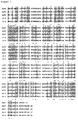
- Figure 1 shows a comparison of the amino acid sequences among known human olfactory receptors and GTAR14-1 and GTAR14-2. Shadowed residues are shared by at least four proteins out of five. Dotted residues are matched in all five proteins. Abbreviations are as follows:
- OLF1 for human olfactory receptor 1 (GenBank Accession# U56420); OLF2 for human olfactory receptor 2 (GenBank Accession# L35475); OLF3 for human olfactory receptor 3 (GenBank Accession# L56421); 14-1 for GTAR14-1; and 14-2 for GTAR14-2 (pseudogene).
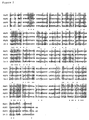
- Figure 2 shows a comparison of the amino acid sequences among known human olfactory receptors and GTAR14-3. Shadowed residues are shared by at least three proteins out of four. Dotted residues are matched in all four proteins. Abbreviations are as follows:
- OLF1 for human olfactory receptor 1 (GenBank Accession# U56420); OLF2 for human olfactory receptor 2 (GenBank Accession# L35475); OLF3 for human olfactory receptor 3 (GenBank Accession# L56421); and 14-3 for GTAR14-3.
- Figure 3 shows a comparison of the amino acid sequences among known human olfactory receptors and GTAR14-4 and GTAR14-5. Shadowed residues are shared by at least three proteins out of five. Dotted residues are matched in all five proteins. Abbreviations are as follows:
- OLF1 for human olfactory receptor 1 (GenBank Accession# U56420); OLF2 for human olfactory receptor 2 (GenBank Accession# L35475); OLF3 for human olfactory receptor 3 (GenBank Accession# L56421); 14-4 for GTAR14-4 (pseudogene); and 14-5 for GTAR14-5.
- Figure 4 shows the nucleotide sequence of GTAR14-1 which was obtained by genomic PCR.
- the positions of the primers used are underlined.
- the right-directed and left-directed arrows indicate the positions of 14-1-S1 primer, and 14-1-A1 primer, respectively.
- Figure 5 shows the nucleotide sequence of GTAR14-3 which was obtained by genomic PCR.
- the positions of the primers used are underlined.
- the right-directed and left-directed arrows indicate the positions of 14-3-S1 primer, and 14-3-A1 primer, respectively.
- Figure 6 shows the nucleotide sequence of GTAR14-5 which was obtained by genomic PCR.
- the positions of the primers used are underlined.
- the right-directed and left-directed arrows indicate the positions of 14-5-S1 primer, and 14-5-A1 primer, respectively.
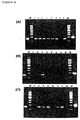
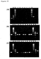
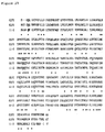
- Figure 7 is a photograph showing the result of RT-PCR analysis of the gene expression pattern of GTAR14-1 in human tissues.
- Lanes 1 fetal brain, 2: fetal heart, 3: fetal kidney, 4: fetal liver, 5: fetal lung, 6: fetal muscle, 7: fetal spleen, 8: fetal thymus, 9: heart, 10:brain, 11: placenta, 12: lung, 13: liver, 14: skeletal muscle, 15: kidney, 16: pancreas, 17: spleen, 18: thymus, 19: prostate, 20: testis, 21: ovary, 22: small intestine, 23: colon, and24: peripheral leukocytes.
- the left lane of each panel shows the result of the positive control in which human genomic DNA was amplified as a template using the same primer set.
- the right lane of each panel shows the product of the control reaction in which G3PDH was amplified from a standard cDNA as a template using a primer set for G3PDH.
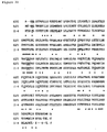
- Figure 8 is a photograph showing the result of RT-PCR analysis of the gene expression pattern of GTAR14-3 in human tissues.
- Lanes 1 fetal brain, 2: fetal heart, 3: fetal kidney, 4: fetal liver, 5: fetal lung, 6: fetal muscle, 7: fetal spleen, 8: fetal thymus, 9: heart, 10:brain, 11: placenta, 12: lung, 13: liver, 14: skeletal muscle, 15: kidney, 16: pancreas, 17: spleen, 18: thymus, 19: prostate, 20: testis, 21: ovary, 22: small intestine, 23: colon, and 24: peripheral leukocytes.
- the left lane of each panel shows the result of the positive control in which human genomic DNA was amplified as a template using the same primer set.
- the right lane of each panel shows the product of the control reaction in which G3PDH was amplified from a standard cDNA as a template using a primer set for G3PDH.
- Figure 9 is a photograph showing the result of RT-PCR analysis of the gene expression pattern of GTAR14-5 in human tissues.
- Lanes 1 fetal brain, 2: fetal heart, 3: fetal kidney, 4: fetal liver, 5: fetal lung, 6: fetal muscle, 7: fetal spleen, 8: fetal thymus, 9: heart, 10:brain, 11: placenta, 12: lung, 13: liver, 14: skeletal muscle, 15: kidney, 16: pancreas, 17: spleen, 18: thymus, 19: prostate, 20: testis, 21: ovary, 22: small intestine, 23: colon, and 24: peripheral leukocytes.
- the left lane of each panel shows the result of the positive control in which human genomic DNA was amplified as a template using the same primer set.
- the right lane of each panel shows the product of the control reaction in which G3PDH was amplified from a standard cDNA as a template using a primer set for G3PDH.
- Figure 10 shows the nucleotide sequence of the full length GTAR14-1 cDNA that was obtained by integrating the products of 5'-RACE and 3'-RACE.
- the amino acid sequence encoded by the GTAR14-1 cDNA is also indicated.
- the amino acid sequences predicted to be a transmembrane domain are underlined (TM-I, -II, -III, -IV, and -V).
- Figure 11 shows the nucleotide sequence of the full length GTAR14-1 cDNA that was obtained by integrating the products of 5'-RACE and 3'-RACE, continued from Figure 10.
- the amino acid sequence encoded by the GTAR14-1 cDNA is also indicated.
- the amino acid sequences predicted to be a transmembrane domain are underlined (TM-VI and -VII).
- Figure 12 shows the nucleotide sequence of the full length GTAR14-3 cDNA that was obtained by integrating the products of 5'-RACE and 3'-RACE.
- the amino acid sequence encoded by the GTAR14-3 cDNA is also indicated.
- Amino acid sequences predicted to be a a transmembrane domain are underlined (TM-I, -II, -III, -IV, and -V) .
- Figure 13 shows the nucleotide sequence of the full length GTAR14-3 cDNA that was obtained by integrating the products of 5'-RACE and 3' -RACE, continued from Figure 12.
- the amino acid sequence encoded by the GTAR14-3 cDNA is also indicated.
- Amino acid sequences predicted to be a transmembrane domain are underlined (TM-VI and -VII).
- Figure 14 shows the nucleotide sequence of the full length GTAR14-5 cDNA that was obtained by integrating the products of 5'-RACE and 3'-RACE.
- the amino acid sequence encoded by the GTAR14-5 cDNA is also indicated.
- Amino acid sequences predicted to be a transmembrane domain are underlined (TM-I, -II, -III, and -IV).
- Figure 15 shows the nucleotide sequence of the full length GTAR14-5 cDNA that was obtained by integrating the products of 5'-RACE and 3'-RACE, continued from Figure 14.
- the amino acid sequence encoded by the GTAR14-5 cDNA is also indicated.
- minAo acid sequences predicted to be a transmembrane domain are underlined (TM-V, -VI, and -VII).
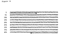
- Figure 16 shows the nucleotide sequence of GTAR11-1 which was obtained by genomic PCR.
- the positions of the primers used are underlined.
- the right-directed and left-directed arrows indicate the positions of 11-1-S2 primer, and 11-1-A3 primer, respectively.
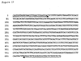
- Figure 17 shows the nucleotide sequence of GTAR11-2 which was obtained by genomic PCR.
- the positions of the primers used are underlined.
- the right-directed and left-directed arrows indicate the positions of 11-2-S2 primer, and 11-2-A2 primer, respectively.
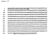
- Figure 18 shows the nucleotide sequence of GTAR11-3 which was obtained by genomic PCR.
- the positions of primers used are underlined.
- the right-directed and left-directed arrows indicate the positions of 11-3-S2 primer, and 11-3-A2 primer, respectively.
- Figure 19 shows the nucleotide sequence of GTAR11-4 which was obtained by genomic PCR.
- the positions of the primers used are underlined.
- the right-directed and left-directed arrows indicate the positions of 11-4-S2 primer, and 11-4-A2 primer, respectively.
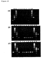
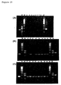
- Figure 20 is a photograph showing the result of RT-PCR analysis of the gene expression pattern of GTAR11-1 in human tissues.
- Lanes 1 fetal brain, 2: fetal heart, 3: fetal kidney, 4: fetal liver, 5: fetal lung, 6: fetalmuscle, 7: fetal spleen, 8: fetal thymus, 9: brain, 10: heart, 11: kidney, 12: liver, 13: lung, 14: pancreas, 15: placenta, 16: skeletal muscle, 17: colon, 18: ovary, 19: peripheral leukocytes, 20: prostate, 21: small intestine, 22: spleen, 23: testis, and 24: thymus.
- the left lane of each panel shows the result of the positive control in which human genomic DNA was amplified as a template using the same primer set.
- the right lane of each panel shows the product of the control reaction in which G3PDH was amplified from a standard cDNA as a template using a primer set for G3PDH.
- Figure 21 is a photograph showing the result of RT-PCR analysis of the gene expression pattern of GTAR11-2 in human tissues.
- Lanes 1 fetal brain, 2: fetal heart, 3: fetal kidney, 4: fetal liver, 5: fetal lung, 6: fetal muscle, 7: fetal spleen, 8: fetal thymus, 9: brain, 10: heart, 11: kidney, 12: liver, 13: lung, 14: pancreas, 15: placenta, 16: skeletal muscle, 17: colon, 18: ovary, 19: peripheral leukocytes, 20: prostate, 21: small intestine, 22: spleen, 23: testis, and 24: thymus.
- the left lane of each panel shows the result of the positive control in which human genomic DNA was amplified as a template using the same primer set.
- the right lane of each panel shows the product of the control reaction in which G3PDH was amplified from a standard cDNA as a template using a primer set for G3PDH.
- Figure 22 is a photograph showing the result of RT-PCR analysis of the gene expression pattern of GTAR11-3 in human tissues.
- Lanes 1 fetal brain, 2: fetal heart, 3: fetal kidney, 4: fetal liver, 5: fetal lung, 6: fetal muscle, 7: fetal spleen, 8: fetal thymus, 9: brain, 10: heart, 11: kidney, 12: liver, 13: lung, 14: pancreas, 15: placenta, 16: skeletal muscle, 17: colon, 18: ovary, 19: peripheral leukocytes, 20: prostate, 21: small intestine, 22: spleen, 23: testis, and 24: thymus.
- the left lane of each panel shows the result of the positive control in which human genomic DNA was amplified as a template using the same primer set.
- the right lane of each panel shows the product of the control reaction in which G3PDH was amplified from a standard cDNA as a template using a primer set for G3PDH.
- Figure 23 is a photograph showing the result of RT-PCR analysis of the gene expression pattern of GTAR11-4 in human tissues.
- Lanes 1 fetal brain, 2: fetal heart, 3: fetal kidney, 4: fetal liver, 5: fetal lung, 6: fetal muscle, 7: fetal spleen, 8: fetal thymus, 9: brain, 10: heart, 11: kidney, 12: liver, 13: lung, 14: pancreas, 15: placenta, 16: skeletal muscle, 17: colon, 18: ovary, 19: peripheral leukocytes, 20: prostate, 21: small intestine, 22: spleen, 23: testis, and 24: thymus.
- the left lane of each panel shows the result of the positive control in which human genomic DNA was amplified as a template using the same primer set.
- the right lane of each panel shows the product of the control reaction in which G3PDH was amplified from a standard cDNA as a template using a primer set for G3PDH.
- Figure 24 shows the nucleotide sequence of the GTAR11-1 cDNA that was obtained by integrating the products of 5'-RACE and 3'-RACE.
- the amino acid sequence encoded by the GTAR11-1 cDNA is also indicated.
- Amino acid sequences predicted to be a transmembrane domain are underlined (TM-I, -II, -III, -IV, -V, -VI, and -VII).
- Figure 25 shows the nucleotide sequence of the GTAR11-2 cDNA that was obtained by integrating the products of 5'-RACE and 3'-RACE.
- the amino acid sequence encoded by the GTAR11-2 cDNA is also indicated.
- Amino acid equences predicted to be a transmembrane domain are underlined (TM-I, -II, -III, -IV, -V, -VI, and -VII).
- Figure 26 shows the nucleotide sequence of the GTAR11-3 cDNA that was obtained by integrating the products of 5'-RACE and 3'-RACE.
- the amino acid sequence encoded by the GTAR11-3 cDNA is also indicated.
- Amino acid sequences predicted to be a transmembrane domain are underlined (TM-I, -II, -III, -IV, -V, -VI, and -VII).
- Figure 27 shows the nucleotide sequence of the GTAR11-4 cDNA that was obtained by integrating the products of 5'-RACE and 3'-RACE.
- the amino acid sequence encoded by the GTAR11-4 cDNA is also indicated.
- Amino acid sequences predicted to be a transmembrane domain are underlined (TM-I, -II, -III, ,-IV, -V, -VI, and -VII).
- Figure 28 shows comparison of the amino acid sequences among known human olfactory receptors and GTAR11-1. Shadowed residues are shared by at least three proteins out of four. Dotted residues are matched in all four proteins.
- the N residue of the [N-x-S/T] motif (x, any amino acid residue) that is modified by sugar, conserved cysteine residues that form a disulfide bond required for cross-linking, and the D-R-Y sequence in the [M-A-Y-D-R-Y-L/V-A-I/V-C] motif in the second intracellular domain, which is suggested to be essential for the binding to intracellular G proteins, are underlined. Abbreviations are as follows:
- OLF1 for human olfactory receptor 1 (GenBankAccession# U56420); OLF2 for human olfactory receptor 2 (GenBank Accession# L35475); OLF3 for human olfactory receptor 3 (GenBank Accession# L56421); and 11-1 for GTAR11-1.
- Figure 29 shows comparison of the amino acid sequences among known human olfactory receptors and GTAR11-2. Dotted residues are matched in all three proteins.
- OLF2 for human olfactory receptor 2 (GenBank Accession# L35475)
- OLF3 for human olfactory receptor 3 (GenBank Accession# L56421)
- 11-2 for GTAR11-2.
- Figure 30 shows comparison of the amino acid sequences among known human olfactory receptors and GTAR11-3. Dotted residues are matched in all three proteins.
- OLF2 for human olfactory receptor 2 (GenBankAccession# L35475)
- OLF3 for human olfactory receptor 3 (GenBank Accession# L56421)
- 11-3 for GTAR11-3.
- Figure 31 shows comparison of the amino acid sequences among known human olfactory receptors and GTAR11-4. Dotted residues are matched in all three proteins.
- OLF2 for human olfactory receptor 2 (GenBank Accession# L35475)
- OLF3 for human olfactory receptor 3 (GenBank Accession# L56421)
- 11-4 for GTAR11-4.
- BLAST search was performed on the Non-redundant Sequence (nr) database in GenBank, and a human genomic sequence that contains a gene encoding a novel seven transmembrane G protein-coupled receptor, which has a high structural homology to known olfactory receptor (OR) gene family, was found.
- the gene is contained in the sequence of BAC129 clone (GenBank Accession# U85195), originating from the vicinity of the ⁇ / ⁇ T-cell receptor locus on human chromosome 14, and shows about 50 to 60% homology to known human olfactory receptors, OLF1, OLF2, and OLF3 at the amino acid sequence level.
- GTAR14-1 G protein-coupled T-cell Activating Receptor 14-1
- GTAR14-3 G protein-coupled T-cell Activating Receptor 14-3
- GTAR14-5 G protein-coupled T-cell Activating Receptor 14-5
- the genomic structures of known OR gene family have a unique feature in that the ORF from the initiation codon to the stop codon is contained in a single exon of the genomic sequence encoding the known OR gene.
- the full length ORFs of GTAR14-1, GTAR14-3, and GTAR14-5 are also contained in a sequence that is supposed to be a single exon.
- GTAR14-2 and GTAR14-4 novel OR-like genes were also found, which were named GTAR14-2 and GTAR14-4.
- the ORF of GTAR14-2 must be split in two by deletion of a single nucleotide in the middle.
- the ORF of GTAR14-4 has to be divided into two portions by an in-frame stop codon in the second extracellular domain (loop-1). Accordingly, these genes were judged to be a typical pseudogene of the OR gene family, and were excluded from further study.
- FIG. 1 Comparison of the amino acid sequences of known human OLF1, OLF2, and OLF3, and proteins of the present invention, GTAR14-1 and GTAR14-2, is shown.
- the sequence of GTAR14-2 is of an artificial translational frame created by inserting a single nucleotide at the position of the missing nucleotide.
- Figure 2 shows comparison of the amino acid sequences among known human OLF1, OLF2, and OLF3, and protein of the present invention, GTAR14-3.
- FIG. 3 shows comparison of the amino acid sequences among known human OLF1, OLF2, and OLF3, and proteins of the present invention, GTAR14-4 and GTAR14-5.
- the amino acid sequence of GTAR14-4 is artificially created by reading through the in frame stop codon.
- BAC129 clone U85195 was analyzed for the presence of an exon that is broadly conserved in known OLF receptors at the amino acid sequence level, and the predicted exon was used to design primers as follows.
- GTAR14-1-S1 For amplification of GTAR14-1, GTAR14-1-S1 (5'-ATG GAC AGT CTA AAC CAA ACA AGA GTG-3'; SEQ ID NO: 7), GTAR14-1-S2 (5'-ATG GCA TTC TCA GCC ATT TAT ATG CTA-3'; SEQ ID NO: 8), and GTAR14-1-S3 (5'-GGG AAC ATT CTC ATC ATC ATT GCC ACA-3'; SEQ ID NO: 9) were used as sense primers, and GTAR14-1-A1 (5'-TTA TGT ATA TGA TTT CGT GAA AAA AAC-3'; SEQ ID NO: 10), GTAR14-1-A2 (5'-TAC CTC CTC ATT CCT CAA GGT GTA AAT-3'; SEQ ID NO: 11), and GTAR14-1-A3 (5'-GGT GAC CAC TGT GTA GAA GAC AGA CAC-3'; SEQ ID NO: 12)
- GTAR14-3-S1 For amplification of GTAR14-3, GTAR14-3-S1 (5'-ATG GAA AGA ATC AAC AGC ACA CTG TTG-3'; SEQ ID NO: 13), and GTAR14-3-S2 (5'-TCT AAT CTA CAT CCT GAC TCA GCT GGG-3'; SEQ ID NO: 14) were used as sense primers, and GTAR14-3-A1 (5'-TTC AAA CCT CAC TCG GAG TTC TTG GGC-3'; SEQ ID NO: 15), and GTAR14-3-A2 (5'-AGC TTC ACC TCT TGG TTC CGC AGA GTG-3'; SEQ ID NO: 16) were used as antisense primers.
- GTAR14-5-S1 For amplification of GTAR14-5, GTAR14-5-S1 (5'-ATG GGA AAG ACC AAA AAC ACA TCG CTG-3'; SEQ ID NO: 17), and GTAR14-5-S2 (5'-CGT GGT GAC AGA TTT CAT TCT TCT GGG-3'; SEQ ID NO: 18) were used as sense primers, and GTAR14-5-A1 (5'-TCA ACC TGC TGT TAT CCT CTT CAG GGC-3'; SEQ ID NO: 19), and GTAR14-5-A2 (5'-CCT GGT TCC TCA GTG TAT AGA TGA GGG-3'; SEQ ID NO: 20) were used as antisense primers.
- Genomic PCR was performed using Human Genomic DNA (Clontech #6550-1) as a template, and primer sets of "14-1-S1 and 14-1-A1," "14-1-S1 and 14-1-A2," “14-1-S2 and 14-1-A1,” and “14-1-S2 and 14-1-A2” for amplification of GTAR14-1, "14-3-S1 and 14-3-A1,” "14-3-S1 and 14-3-A2,” “14-3-S2 and 14-3-A1,” and "14-3-S2 and 14-3-A2” for amplification of GTAR14-3, and "14-5-S1 and 14-5-A1," "14-5-S1 and 14-5-A2,” “14-5-S2 and 14-5-A1,” and "14-5-S2 and 14-5-A2” for amplification of GTAR14-5.
- PCR was performed using the Advantage cDNA Polymerase Mix (Clontech #8417-1) on a thermal cycler (Perkin Elmer Gene Amp PCR System 2400). By performing PCR under the following conditions, a single band was observed with a predicted size for each primer set.
- PCR condition 94°C for 4 min, 5 cycles of 94°C for 20 sec and 72°C for 2 min, 5 cycles of 94°C for 20 sec and 70°C for 2 min, 28 cycles of 94°C for 20 sec and 68°C for 2 min, 72°C for 4 min, and termination at 4°C.
- the obtained products of genomic PCR were subcloned into the pGEM-T Easy vector (Promega #A1360), and the nucleotide sequence was determined.
- the PCR products were cloned into the pGEM-T Easy vector by ligation using T4 DNA ligase (Promega #A1360) at 4°C for 12 hr.
- the sample was used to transform E . coli DH5 ⁇ (Toyobo # DNA-903) to obtain recombinants of the PCR products and the pGEM-T Easy vector. Recombinants were selected by using the Insert Check Ready (Toyobo #PIK-101).
- the nucleotide sequence was determined by using the dRhodamine Terminator Cycle Sequencing kit (ABI/Perkin Elmer #4303141) on a DNA Sequencer (ABI PRISM 377).
- RT-PCR was performed using a primer set of "14-1-S2 and 14-1-A2,” “14-3-S2 and 14-3-A2,” and "14-5-S2 and 14-5-A2,” respectively.
- PCR reactions were performed under conditions with a single cycle of 94°C for 4 min, sequentially followed by 5 cycles of 94°C for 20 sec and 72°C for 2 min, 5 cycles of 94°C for 20 sec and 70°C for 2 min, 38 cycles of 94°C for 20 sec and 68°C for 2 min, a single cycle of 72°C for 4 min, and termination at 4°C.
- GTAR14-3 was strongly amplified in thymus, brain, heart, smooth muscle, and kidney among human fetal tissues, and weakly expressed in liver and lung as well. GTAR14-3 was strongly detected in thymus and testis among adult tissues, and also expressed in digestive organs such as small intestine and colon, and endocrine organs such as placenta and prostate, showing a broad distribution among tissues (Figure 8).
- GTAR14-5 was strongly amplified in thymus, spleen, and lung among human fetal tissues, and weakly expressed in brain and kidney. It was detected strongly in lymphatic/hematopoietic tissues such as thymus, spleen, and peripheral leukocytes, and testis, and pancreas in adult tissues (Figure 9).
- the products of PCR were subcloned into the pGEM-T Easy vector (Promega #A1360), and the nucleotide sequence were determined.
- the PCR products were cloned into the pGEM-T Easy vector by ligation using T4 DNA ligase (Promega #A1360) at 4°C for 12 hr.
- the samples were used to transform E . coli DH5 ⁇ (Toyobo # DNA-903) to obtain recombinants of the PCR products and the pGEM-T Easy vector. Recombinants were selected by using the Insert Check Ready (Toyobo #PIK-101).
- the nucleotide sequence were determined by using the dRhodamine Terminator Cycle Sequencing kit (ABI/Perkin Elmer #4303141) on a DNA Sequencer (ABI PRISM 377). For each gene, ten independent recombinant clones were selected, and the complete nucleotide sequence of the inserts were determined. As a result, all clones showed a single nucleotide sequence.
- the obtained sequences were verified to be a partial sequence of the GTAR14-1, GTAR14-3, or GTAR14-5, and confirmed not to be a non-specific product of RT-PCR.
- 5'-RACE-PCR was performed to isolate full length cDNAs of GTAR14-1, GTAR14-3, and GTAR14-5.
- Primers used for amplification of GTAR14-1, GTAR14-3, and GTAR14-5 genes were "14-1-A1,” "14-3-A1,” and “14-5-A1” for the first PCR, and "14-1-A2, " “14-3-A2, “ and “14-5-A2” for the second PCR, respectively.
- the obtained 5'-RACE-PCR products which showed a single band, were subcloned into the pGEM-T Easy vector as described above.
- the nucleotide sequence of all inserts were determined by using the dRhodamine Terminator Cycle Sequencing kit (ABI/Perkin Elmer #4303141) on a DNA Sequencer (ABI PRISM 377), as described above.
- 3'-RACE-PCR was further performed to isolate full length cDNAs of GTAR14-1, GTAR14-3, and GTAR14-5 genes.
- Primers used for amplification of GTAR14-1, GTAR14-3, and GTAR14-5 genes were "14-1-S1,” "14-3-S1,” and “14-5-S1” for first PCR, and "14-1-S2,” “14-3-S2,” and "14-5-S2" for second PCR, respectively.
- nucleotide sequences obtained by 5'-RACE and 3'-RACE were combined to determine the nucleotide sequence of full length cDNA of GTAR14-1, GTAR14-3, and GTAR14-5.
- the determined nucleotide sequences of full length cDNAs are shown in Figures 10 and 11 (GTAR14-1), Figures 12 and 13 (GTAR14-3), and Figures 14 and 15 (GTAR14-5), and SEQ ID NO: 1 (GTAR14-1), SEQ ID NO: 2 (GTAR14-3), and SEQ ID NO: 3 (GTAR14-5).
- GTAR11-1 G protein-coupled T-cell Activating Receptor 11-1
- GTAR11-2 G protein-coupled T-cell Activating Receptor 11-2
- GTAR11-3 G protein-coupled T-cell Activating Receptor 11-3
- GTAR11-4 G protein-coupled T-cell Activating Receptor 11-4
- the sequence of the BAC clone AC002556 was analyzed for the presence of an exon that is broadly conserved in known OLF receptors at the amino acid sequence level, and the exon predicted was used to design primers as follows.
- GTAR11-1-S1 For amplification of GTAR11-1, GTAR11-1-S1 (5'-GAA GAG CAG TGA GGG TCC ATG TTA AGG-3'; SEQ ID NO: 34), and GTAR11-1-S2 (5'-CAG CAG CTT GTC CTT CGT CGA TTT CTG C-3'; SEQ ID NO: 35) were used as sense primers, and GTAR11-1-A2 (5'-GCT AGG GTG GGC ACC AAG GTG TTA AAC CC-3'; SEQ ID NO: 36), and GTAR11-1-A3 (5'-TGC AAA AGG ACA GTT TCA TCA TGG CAC-3'; SEQ ID NO: 37) were used as antisense primers.
- GTAR11-2-S1 For amplification of GTAR11-2, GTAR11-2-S1 (5'-CAA AGA ACT CAC CCA AAT TCC TAC AGC T-3'; SEQ ID NO: 38), and GTAR11-2-S2 (5'-CAT GGT AGG CAA CCT TGG CTT GAT CAC-3'; SEQ ID NO: 39) were used as sense primers, and GTAR11-2-A1 (5'-GTT TAT TAA ATC ACA CAT AAC ACC ATC TG-3'; SEQ ID NO: 40), and GTAR11-2-A2 (5'-CAG AGA CAG AGC AAT GAC ATG AGA GCT AC-3'; SEQ ID NO: 41) were used as antisense primers.
- GTAR11-3-S1 For amplification of GTAR11-3, GTAR11-3-S1 (5'-CAA AGA ACT CAC CCA AAT TCC TAC AGC C-3'; SEQ ID NO: 42), and GTAR11-3-S2 (5'-CAT GGT AGG CAA CCT TGG CTT GAT CAT-3'; SEQ ID NO: 43) were used as sense primers, and GTAR11-3-A1 (5'-GTT TAT TAA ATC ACA CAT AAC ACC ATC TG-3'; SEQ ID NO: 44), and GTAR11-3-A2 (5'-CAG AGA CAG AGC AAT GAC ATG AGA GCT AC-3'; SEQ ID NO: 45) were used as antisense primers.
- GTAR11-4-S1 For amplification of GTAR11-4, GTAR11-4-S1 (5'-CCA GAC AGC TCG CCA AGA GAG AAT GAC-3'; SEQ ID NO: 46), and GTAR11-4-S2 (5'-CCT TTA TAG ATC TCT GTT ATT CCT GTG TG-3'; SEQ ID NO: 47) were used as sense primers, and GTAR11-4-A1 (5'-TCG GTT GCC AGT GAT ATG AAG AGA CCC-3'; SEQ ID NO: 48), and GTAR11-4-A2 (5'-GGC TTT GGA TCT GCC CTC TGC AGA AGG-3'; SEQ ID NO: 49) were used as antisense primers.
- Genomic PCR was performed using Human Genomic DNA (Clontech #6550-1) as a template, and primer sets of "11-1-S1 and 11-1-A2," “11-1-S1 and 11-1-A3,” “11-1-S2 and 11-1-A2,” and “11-1-S2 and 11-1-A3" for amplification of GTAR11-1, "11-2-S1 and 11-2-A1,” "11-2-S1 and 11-2-A2,” “11-2-52 and 11-2-A1,” and “11-2-S2 and 11-2-A2” for amplification of GTAR11-2, "11-3-S1 and 11-3-A1," "11-3-S1 and 11-3-A2,” “11-3-S2 and 11-3-A1,” and “11-3-S2 and 11-3-A2” for amplification of GTAR11-3, and "11-4-S1 and 11-4-A1," "11-4-S1 and 11-4-A2,” "11-4-S2 and 11-4
- PCR was performed using the Advantage cDNA Polymerase Mix (Clontech #8417-1) on a thermal cycler (Perkin Elmer Gene Amp PCR System 2400). Following conditions were used for PCR; a single cycle of 94°C for 4 min, sequentially followed by 5 cycles of 94°C for 20 sec and 72°C for 2 min, 5 cycles of 94°C for 20 sec and 70°C for 2 min, 28 cycles of 94°C for 20 sec and 68°C for 2 min, and then a single cycle of 72°C for 4 min followed by termination at 4°C. As a result, a single band was detected with predicted size for each primer set.
- the products obtained in the above genomic PCR were subcloned into the pGEM-T Easy vector (Promega #A1360), and the nucleotide sequence were determined.
- PCR products were cloned into the pGEM-T Easy vector by ligation using T4 DNA ligase (Promega #A1360) at 4°C for 12 hr.
- the samples were used to transform E . coli DH5 ⁇ (Toyobo # DNA-903) to obtain recombinants of the PCR products and the pGEM-T Easy vector. Recombinants were selected by using the Insert Check Ready (Toyobo #PIK-101).
- the nucleotide sequences were determined by using the dRhodamine Terminator Cycle Sequencing kit (ABI/Perkin Elmer #4303141) on a DNA Sequencer (ABI PRISM 377).
- ten independent clones of recombinants were selected from the products of genomic PCR that had been performed using a set of primers that are located within the ORF of the target gene fragment; that is the product of the primer set of "11-1-S2 and 11-1-A3" for GTAR11-1 gene, that of "11-2-S2 and 11-2-A2" for GTAR11-2 gene, that of "11-3-S2 and 11-3-A2" for GTAR11-3 gene, and that of "11-4-S2 and 11-4-A2" for GTAR11-4 gene.
- the nucleotide sequence of all the inserts were determined.
- RT-PCR was performed using a primer set of "11-1-S2 and 11-1-A2," “11-2-S2 and 11-2-A2,” “11-3-S2 and 11-3-A2,” and "11-4-S2 and 11-4-A2" for GTAR11-1, GTAR11-2, GTAR11-3, and GTAR11-4 genes, respectively.
- PCR reactions were performed under conditions with a single cycle of 94°C for 4 min, sequentially followed by 5 cycles of 94°C for 20 sec and 72°C for 2 min, 5 cycles of 94°C for 20 sec and 70°C for 2 min, 38 cycles of 94°C for 20 sec and 68°C for 2 min, and then a single cycle of 72°C for 4 min followed by termination at 4°C.
- GTAR11-1 mRNA was also expressed in multiple tissues in adult including lymphatic/hematopoietic tissues such as thymus, spleen, and peripheral lymphocytes, digestive organs such as small intestine and colon, genital organs, neuronal tissues, and endocrine organs (Figure 20).
- GTAR11-2 gene was strongly amplified in thymus, and weakly expressed in spleen, smooth muscle, lung, and kidney among fetal tissues. GTAR11-2 gene was also detected in multiple tissues in adult including lymphatic/hematopoietic tissues such as thymus and peripheral lymphocytes, digestive organs, genital organs, neuronal tissues, and endocrine organs (Figure 21).
- GTAR11-3 gene was strongly amplified in smooth muscle among human fetal tissues, and weakly expressed in brain, lung, and liver as well. The gene was also detected in multiple tissues in adult including lymphatic/hematopoietic tissues such as thymus, spleen, and peripheral lymphocytes, digestive organs such as small intestine and colon, muscle, genital organs, neuronal tissues, and endocrine organs (Figure 22).
- GTAR11-4 gene was strongly amplified only in smooth muscle in fetus. It was also expressed in multiple tissues in adult including lymphatic/hematopoietic tissues such as thymus, spleen, and peripheral lymphocytes, digestive organs such as small intestine and colon, muscle, genital organs, and endocrine organs (Figure 23).
- the obtained PCR products were subcloned into the pGEM-T Easy vector (Promega #A1360), and the nucleotide sequence were determined.
- the PCR products were cloned into the pGEM-T Easy vector by ligation using T4 DNA ligase (Promega #A1360) at 4°C for 12 hr.
- the samples were used to transform E. coli DH5 ⁇ (Toyobo # DNA-903) to obtain recombinants of the PCR products and the pGEM-T Easy vector. Recombinants were selected by using the Insert Check Ready (Toyobo #PIK-101).
- the nucleotide sequence were determined by using the dRhodamine Terminator Cycle Sequencing kit (ABI/Perkin Elmer #4303141) on a DNA Sequencer (ABI PRISM 377). For each gene, ten independent clones of recombinants were selected, and the nucleotide sequence of all the inserts were determined. All clones of each gene showed a single nucleotide sequence.
- the obtained sequences were verified to be a partial sequence of GTAR11-1, GTAR11-2, GTAR11-3, and GTAR11-4 genes, and confirmed not to be a non-specific product of RT-PCR.
- the obtained 5#-RACE-PCR products, detected as a single band, were subcloned into the pGEM-T Easy vector, six independent clones of recombinants were selected for each, and the nucleotide sequence of all inserts were determined by using the dRhodamine Terminator Cycle Sequencing kit (ABI/Perkin Elmer #4303141) on a DNA Sequencer (ABI PRISM 377), as described above.
- 3'-RACE-PCR was further performed to isolate full length cDNAs of GTAR11 genes.
- Primers used for amplification of GTAR11-1, GTAR11-2, GTAR11-3, and GTAR11-4 genes were "11-1-S1,” “11-2-S1,” “11-3-S1,” and “11-4-S1” for first PCR, and "11-1-S2,” “11-2-S2,” “11-3-S2,” and “11-4-S2" for second PCR, respectively.
- the nucleotide sequences obtained by 5'-RACE and 3'-RACE were combined to generate the sequence of full length cDNA of each GTAR11 gene.
- the resulting nucleotide sequences of cDNAs are shown in Figure 24 (GTAR11-1), Figure 25 (GTAR11-2), Figure 26 (GTAR11-3), and Figure 27 (GTAR11-4), and SEQ ID NO: 24 (GTAR11-1), SEQ ID NO: 25 (GTAR11-2), SEQ ID NO: 26 (GTAR11-3), and SEQ ID NO: 27 (GTAR11-4).
- the GTAR11-1 cDNA obtained by the first 3'-RACE-PCR had a deletion at the C-terminus, and thus was not full length (the nucleotide sequence of the cDNA and the amino acid sequence of the peptide encoded by the cDNA are shown in SEQ ID NO: 32, and SEQ ID NO: 33, respectively). Consequently, 3'-RACE-PCR was performed again for GTAR11-1, and the full length cDNA of GTAR11-1 was obtained as described above.
- the genome of known OR gene family shares a unique structural feature that the ORF from the initiation codon to the stop codon is contained in a single exon of the genome sequence encoding known OR genes.
- the isolated full length CDNAS of GTAR11-1, GTAR11-2, GTAR11-3, and GTAR11-4 genes also have their whole ORF within a sequence that is supposed to be a single exon.
- Figure 28 shows comparison of the amino acid sequences of known human OLF1, OLF2, and OLF3, and GTAR11-1.
- Figure 29 shows comparison of the amino acid sequences of known human OLF2 and OLF3, and GTAR11-2.
- Figure 30 shows comparison of the amino acid sequences of known human OLF2 and OLF3, and GTAR11-3.
- Figure 31 shows comparison of the amino acid sequences of known human OLF2 and OLF3, and GTAR11-4.
- the present invention provides novel G protein-coupled receptor proteins and their genes. According to their particular expression patterns and such, the receptor proteins of the present invention appear to be involved in immune response, hematopoiesis, etc. Thus, one can use these proteins in screening assays, for developing drugs that can regulate immune response and/or hematopoietic cells. Using the receptor proteins of the present invention, one can also screen for ligands capable of binding to the receptor proteins of the invention, such ligands also being potentially involved in immune response, hematopoiesis, and so on.
Landscapes
- Health & Medical Sciences (AREA)
- Chemical & Material Sciences (AREA)
- Life Sciences & Earth Sciences (AREA)
- Organic Chemistry (AREA)
- Biochemistry (AREA)
- Genetics & Genomics (AREA)
- Gastroenterology & Hepatology (AREA)
- Toxicology (AREA)
- Immunology (AREA)
- Biophysics (AREA)
- General Health & Medical Sciences (AREA)
- Zoology (AREA)
- Medicinal Chemistry (AREA)
- Molecular Biology (AREA)
- Proteomics, Peptides & Aminoacids (AREA)
- Cell Biology (AREA)
- Peptides Or Proteins (AREA)
- Micro-Organisms Or Cultivation Processes Thereof (AREA)
Applications Claiming Priority (7)
| Application Number | Priority Date | Filing Date | Title |
|---|---|---|---|
| JP28856598 | 1998-10-09 | ||
| JP28856598 | 1998-10-09 | ||
| JP34754698 | 1998-12-07 | ||
| JP34754698 | 1998-12-07 | ||
| JP36353798 | 1998-12-21 | ||
| JP36353798 | 1998-12-21 | ||
| PCT/JP1999/005578 WO2000021999A1 (fr) | 1998-10-09 | 1999-10-08 | Nouveaux recepteurs couples a la proteine g |
Publications (2)
| Publication Number | Publication Date |
|---|---|
| EP1120426A1 true EP1120426A1 (de) | 2001-08-01 |
| EP1120426A4 EP1120426A4 (de) | 2005-08-31 |
Family
ID=27337448
Family Applications (1)
| Application Number | Title | Priority Date | Filing Date |
|---|---|---|---|
| EP99970399A Withdrawn EP1120426A4 (de) | 1998-10-09 | 1999-10-08 | G-protein-gekoppelte rezeptoren |
Country Status (3)
| Country | Link |
|---|---|
| EP (1) | EP1120426A4 (de) |
| AU (1) | AU6006399A (de) |
| WO (1) | WO2000021999A1 (de) |
Cited By (1)
| Publication number | Priority date | Publication date | Assignee | Title |
|---|---|---|---|---|
| WO2002024907A3 (en) * | 2000-09-20 | 2002-10-03 | Merck Patent Gmbh | G-protein coupled receptor hfrbn63 |
Families Citing this family (5)
| Publication number | Priority date | Publication date | Assignee | Title |
|---|---|---|---|---|
| US20040029793A1 (en) * | 2000-06-15 | 2004-02-12 | Takeo Moriya | Novel g protein-coupled receptor protein and dna thereof |
| AU2001273167A1 (en) * | 2000-06-30 | 2002-01-14 | Curagen Corporation | G-protein coupled receptors and nucleic acids encoding same |
| US20040248321A1 (en) * | 2000-07-07 | 2004-12-09 | Takeo Moriya | Novel g protein-coupled receptor protein and dna thereof |
| WO2002061083A1 (fr) * | 2001-01-30 | 2002-08-08 | Takeda Chemical Industries, Ltd. | Nouvelle proteine de recepteur couple aux proteines g et son adn |
| US7033790B2 (en) | 2001-04-03 | 2006-04-25 | Curagen Corporation | Proteins and nucleic acids encoding same |
Family Cites Families (3)
| Publication number | Priority date | Publication date | Assignee | Title |
|---|---|---|---|---|
| JPH09176048A (ja) * | 1995-12-28 | 1997-07-08 | Takeda Chem Ind Ltd | ヒトMIP−1α/RANTES受容体タンパク質の製造法および用途 |
| US6037146A (en) * | 1997-01-28 | 2000-03-14 | Smithkline Beecham Corporation | CDNA clone HE8CH90 that encodes a novel 7-transmembrane receptor |
| WO2001027158A2 (en) * | 1999-10-08 | 2001-04-19 | Digiscents | Olfactory receptor sequences |
-
1999
- 1999-10-08 AU AU60063/99A patent/AU6006399A/en not_active Abandoned
- 1999-10-08 WO PCT/JP1999/005578 patent/WO2000021999A1/ja not_active Application Discontinuation
- 1999-10-08 EP EP99970399A patent/EP1120426A4/de not_active Withdrawn
Non-Patent Citations (2)
| Title |
|---|
| DATABASE Geneseq [Online] 30 July 2001 (2001-07-30), "Human olfactory receptor polypeptide, SEQ ID NO: 1758." XP002322779 retrieved from EBI accession no. GSN:AAG72077 Database accession no. AAG72077 & WO 01/27158 A (DIGISCENTS; YEDA RESEARCH AND DEVELOPMENT CO., LTD; BELLENSON, JOEL; S) 19 April 2001 (2001-04-19) * |
| See also references of WO0021999A1 * |
Cited By (1)
| Publication number | Priority date | Publication date | Assignee | Title |
|---|---|---|---|---|
| WO2002024907A3 (en) * | 2000-09-20 | 2002-10-03 | Merck Patent Gmbh | G-protein coupled receptor hfrbn63 |
Also Published As
| Publication number | Publication date |
|---|---|
| EP1120426A4 (de) | 2005-08-31 |
| WO2000021999A1 (fr) | 2000-04-20 |
| AU6006399A (en) | 2000-05-01 |
Similar Documents
| Publication | Publication Date | Title |
|---|---|---|
| JP5634674B2 (ja) | 新規ヘモポエチン受容体蛋白質、nr10 | |
| WO2001023556A1 (fr) | Nouvelle proteine receptrice d'hemopoietine (nr12) | |
| US7829083B2 (en) | Antibodies to brain specific membrane protein | |
| WO2002077230A1 (fr) | Variants d'epissage nr10 | |
| EP1120426A1 (de) | G-protein-gekoppelte rezeptoren | |
| US20090197317A1 (en) | TSG-Like Gene | |
| US8242245B2 (en) | Fetal polypeptides from human liver | |
| US20090275082A1 (en) | Mast Cell-Derived Membrane Proteins | |
| US20070105143A1 (en) | Novel bHLH type transcription factor genes | |
| JP2008099690A (ja) | 新規ヘモポエチン受容体蛋白質、nr12 | |
| US8058408B2 (en) | Transcription factor having zinc finger domains | |
| EP1260584A1 (de) | Protein mit signalsequenz | |
| JP2004267003A (ja) | ヒト白血球型抗原領域に存在する新規遺伝子 |
Legal Events
| Date | Code | Title | Description |
|---|---|---|---|
| PUAI | Public reference made under article 153(3) epc to a published international application that has entered the european phase |
Free format text: ORIGINAL CODE: 0009012 |
|
| 17P | Request for examination filed |
Effective date: 20010508 |
|
| AK | Designated contracting states |
Kind code of ref document: A1 Designated state(s): AT BE CH CY DE DK ES FI FR GB GR IE IT LI LU MC NL PT SE |
|
| AX | Request for extension of the european patent |
Free format text: AL;LT;LV;MK;RO;SI |
|
| RAP1 | Party data changed (applicant data changed or rights of an application transferred) |
Owner name: CHUGAI SEIYAKU KABUSHIKI KAISHA |
|
| RIC1 | Information provided on ipc code assigned before grant |
Ipc: 7C 12R 1/91 B Ipc: 7G 01N 33/53 B Ipc: 7G 01N 33/68 B Ipc: 7C 07K 16/28 B Ipc: 7C 12P 21/08 B Ipc: 7C 12P 21/02 B Ipc: 7C 12N 5/10 B Ipc: 7C 12N 15/12 B Ipc: 7C 07K 14/705 A |
|
| A4 | Supplementary search report drawn up and despatched |
Effective date: 20050714 |
|
| 17Q | First examination report despatched |
Effective date: 20051017 |
|
| 17Q | First examination report despatched |
Effective date: 20051017 |
|
| STAA | Information on the status of an ep patent application or granted ep patent |
Free format text: STATUS: THE APPLICATION IS DEEMED TO BE WITHDRAWN |
|
| 18D | Application deemed to be withdrawn |
Effective date: 20070605 |