CROSS REFERENCE TO RELATED APPLICATIONS
This application is a divisional application of U.S. patent application Ser. No. 11/800,888, filed on May 8, 2007, now U.S. Pat. No. 7,919,604 which is a divisional application of U.S. patent application Ser. No. 11/182,592 (now U.S. Pat. No. 7,214,774), filed on Jul. 14, 2005, which is a divisional application of U.S. patent application Ser. No. 09/974,143 (now U.S. Pat. No. 7,022,474), filed on Oct. 9, 2001, which is a continuation-in-part of PCT/JP00/02281, filed Apr. 7, 2000, which claims priority from Japanese patent application Ser. No. 11/103,356, filed Apr. 9, 1999, the disclosures of which are incorporated by reference herein in their entireties.
TECHNICAL FIELD
The present invention relates to proteins encoded by fetal genes the expression of which is activated in fetal tissues and tumor cells. Proteins of this invention can be utilized as target molecules for developing medicines for the treatment of cancer.
BACKGROUND
Fetal tissues are comprised of many undifferentiated cells that proliferate actively, highly activated cells, nascent vascular endothelial cells, and so on. Although these are stringently regulated in fetal tissues and inhibited as individuals mature, this can be considered very similar to the state of a solid tumor except that the activity is regulated. Therefore, some of the genes expressed specifically in fetal tissues (fetal genes) can be genes involved in the phenomena characteristic of solid tumors such as abnormal growth, immortalization, infiltration, metastasis, and angiogenesis. In addition, some diseases other than cancers are also supposed to arise because fetal genes, which are repressed in a normal living body, are abnormally activated. Therefore, genes involved in various diseases such as cancers can be screened by isolating and analyzing fetal genes. Furthermore, development of medicines using novel action mechanisms is thought to be possible by designing drugs targeted on the genes.
Recently, as fetal genes which are assumed to be involved in malignant transformation, genes for survivin (Nat. Med., 3:917-921, 1997), aurora kinase (EMBO J., 17:3052-3065, 1998) and LYAR (Genes Dev., 7:735-748, 1993) have been reported. Their expressions are all activated in colon cancer, leukemia cells, and such, and these genes are thought to contribution importantly to the malignant transformation. In fact, it has been demonstrated that survivin has the apoptosis inhibitor activity, and aurora kinase participates in the cell cycle regulation as their physiological functions, indicating that acquiring function of either of them works favorably for cancer cells.
SUMMARY
The present invention provides novel proteins specifically or more strongly expressed in fetal tissues, genes encoding the proteins, and preparation and uses of them.
As a method for isolating a gene involved in the malignant transformation, many strategies have been hitherto tried where cancer cells and normal cells are directly compared so as to identify a common gene the expression level of which is significantly different. However, such a method had a problem that a large number of genes not directly involved in malignant transformation might be isolated as the noise due to irregularity in the gene expression control, which is one of characteristics of cancer cells, leading to no isolation of important genes. Therefore, the present inventors have planned a strategy where genes, the expression of which is physiologically controlled from physiological necessity, are first selected and examined whether the genes are abnormally reactivated under non-physiological conditions such as cancer. Based on this strategy, the inventors have focused on the genes specifically expressed in fetal tissues as the genes the expression of which is physiologically controlled from the physiological necessity, and performed their isolations and analyses.
Specifically, first, by the suppression subtractive hybridization method, the present inventors performed the subtraction method with cDNA derived from fetal liver as the tester and cDNA derived from adult liver as the driver to search for fetal genes expressed specifically or more strongly in the fetal liver.
As a result, they isolated a plurality of genes. Then, they examined these genes specifically expressed in fetal tissues for their expression levels in cancer cells. As a result, they have succeeded in identifying novel genes, “fls353” and “fls485” the expressions of which are activated in cancer cells such as colon cancer and hepatoma and so on. Cloning of these full-length cDNAs followed by their sequencings have revealed that, although amino acid sequences of proteins encoded by these genes comprise several characteristic domain structures, no protein having a particularly significant homology to these gene products could be detected in the database. As a result of further studies on the tissue specificities of these gene expressions in more detail, the present inventors have found that these genes are expressed in a variety of cancer cells, and also specifically in normal tissues, such as fetal tissues and other tissues which are thought to contain many undifferentiated cells and actively differentiating/proliferating cells.
These facts have strongly suggested participation of the novel fetal genes “fls353” and “fls485” in the malignant transformation of cells. Therefore, not only expression inhibitors of these fetal genes are expected to be used as the anticancer agent but also fetal genes and proteins encoded by the genes according to this invention can be used as the tool for developing drugs for the treatment of cancer.
The present invention relates to the novel fetal genes “fls353” and “fls485” which are presumed to be involved in the malignant transformation, and the proteins encoded by these genes as well as their preparations and uses, and more specifically, this invention is to provide the following:
(1) A DNA of any one of (a) to (d) below:
-
- (a) a DNA encoding a protein comprising the amino acid sequence of any one of SEQ ID NOs:2, 4 and 6,
- (b) a DNA comprising a coding region in the nucleotide sequence of any one of SEQ ID NOs:1, 3 and 5,
- (c) a DNA encoding a protein that comprises the amino acid sequence of any one of SEQ ID NOs:2, 4 and 6 in which one or more amino acids are replaced, deleted, inserted and/or added and that is functionally equivalent to the protein comprising the amino acid sequence of any one of SEQ ID NOs:2, 4 and 6, and
- (d) a DNA that hybridizes under stringent conditions with the DNA comprising the nucleotide sequence of any one of SEQ ID NOs:1, 3 and 5 and that codes a protein functionally equivalent to the protein comprising the amino acid sequence of any one of SEQ ID NOs:2, 4 and 6.
(2) A DNA encoding a partial peptide of a protein comprising the amino acid sequence of any one of SEQ ID NOs:2, 4 and 6.
(3) A vector into which the DNA of (1) or (2) is inserted.
(4) A transformant harboring the DNA of (1) or (2) or the vector of (3).
(5) A protein or a peptide encoded by the DNA of (1) or (2).
(6) A method for producing the protein or the peptide of (5), the method comprising the steps of culturing the transformant of (4) and recovering a protein expressed from the transformant or the culture supernatant thereof.
(7) An antibody against the protein of (5).
(8) A polynucleotide that hybridizes with the DNA comprising the nucleotide sequence of any one of SEQ ID NOs:1, 3 and 5 or the complementary strand thereof and that comprises at least 15 nucleotides.
(9) A method for screening a compound binding to the protein of (5), the method comprising the steps of:
-
- (a) contacting a test sample with the protein or a partial peptide thereof,
- (b) detecting a binding activity of the test sample to the protein or the partial peptide thereof, and
- (c) selecting a compound comprising the biding activity to the protein or the partial peptide thereof.
(10) A compound biding to the protein of (5), wherein the compound can be isolated by the method of (9).
(11) A method for screening a compound that suppresses or promotes expression of the DNA of (1), wherein the method comprises the steps of:
-
- (a) contacting a test sample with cells expressing the DNA,
- (b) detecting the expression of the DNA in the cells, and
- (c) selecting a compound that decreases or increases the expression of the DNA compared with that in the case where the test sample is not contacted with the cells (control).
(12) A method for screening a compound that suppresses or promotes expression of the DNA of (1), wherein the method comprises the steps of:
-
- (a) providing cells into which a vector comprising a reporter gene functionally linked downstream of the expression control region of the DNA of (1),
- (b) contacting a test sample with the cells,
- (c) detecting the activity of the reporter gene in the cells, and
- (d) selecting a compound that decreases or increases the activity compared with that in the case where the test sample is not contacted with the cells (control).
(13) A compound that suppresses or promotes expression of the DNA of (1), wherein the compound can be isolated by the method of (11) or (12).
BRIEF DESCRIPTION OF THE DRAWINGS
FIG. 1 is a photograph showing the results of the fls353 expression examined by northern analysis.
FIG. 2 is a photograph showing the results of the fls353 expression in a clinical sample of E. coli, analyzed by RT-PCR, continued to FIG. 3.
FIG. 3 is a photograph of the continuation of FIG. 2.
FIG. 4 shows the nucleotide sequence of fls353 (SEQ ID NO:1) and its putative amino acid sequence (SEQ ID NO:2). Amino acids corresponding to the consensus sequence at the ATP/GTP binding site are shown enclosed by a solid line. Continued to FIG. 5.
FIG. 5 is the continuation of FIG. 4.
FIG. 6 is a photograph showing the results of the fls485 expression examined by northern blot analysis.
FIG. 7 is a photograph showing the results of RT-PCR analysis of the expression of fls485 and a fetoprotein in various cultured cells.
FIG. 8 shows the nucleotide sequence of fls485 L (SEQ ID NO:3) and its putative amino acid sequence (SEQ ID NO:4). Fls485 S (SEQ ID NO: 5 and 6) has the same sequence as fls485 L except that in fls485 S, the nucleotides underlined in FIG. 8 do not exist, and the encircled methionine (M) is assumed to be used as the initiation codon. The nucleotide and amino acid sequences of fls485 S are shown in SEQ ID NOs: 5 and 6, respectively. Sequence surrounded by the solid square is the amino acid sequence coinciding with the sequence comprising Cys-Xaa-Xaa-Cys-Xaa-Gly-Xaa-Gly (SEQ ID NO: 33), and that surrounded by the dotted square is the amino acid sequence coinciding with the sequence comprising Cys-Xaa-Xaa-Cys-Xaa-Gly (SEQ ID NO: 34).
FIG. 9 is a photograph showing the results of RT-PCR using a set of primers sandwiching the entire coding region of fls485 with cDNA derived from the human fetal liver as the template. The shaded box in the above figure represents the coding region of fls485.
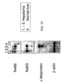
FIG. 10 is a photograph showing the results of northern blot analysis of the expressions of fls353, fls485 and a fetoprotein in a clinical hepatoma sample.
FIG. 11 is a photograph showing the results of northern analysis of the expressions of fls353 and fls485 in a clinical lung cancer sample.
DETAILED DESCRIPTION
This invention relates to the novel proteins “fls353” and “fls485” the expressions of which are activated in fetal tissues and tumor cells. Nucleotide sequence of cDNA for the novel human fetal gene “fls353” is set forth in SEQ ID NO:1, and two types of nucleotide sequences of cDNAs for the novel human fetal gene “fls485” which are included in this invention are set forth in SEQ ID NO:3 (fls485 L) and 5 (fls485 S), respectively. Amino acid sequences encoded by these cDNAs are set forth in SEQ ID NOs:2 (fls353), 4 (fls485 L) and 6 (fls485 S), respectively.
In normal tissues, those genes have been found to be specifically expressed in undifferentiated cells such as in fetal tissues, and in tissues which are thought to contain many actively differentiating and proliferating cells. Examination of their expressions in cancer cells revealed that they were activated in those such as colon cancer and hepatoma and so on.
As to fetal genes, genes for survivin, aurora kinase and LYAR have been assumed to be involved in malignant transformation, and it has been reported that expressions of the genes show a common pattern that their expressions in normal tissues are prominent in fetal tissues, especially in the fetal liver, and that in adult tissues their expressions are abundant in tissues such as testis and thymus containing many actively proliferating undifferentiated cells but not detected in other tissues than those at all (Nat. Med., 3:917-921, 1997; EMBO J., 17: 3052-3065, 1998; and Genes Dev., 7:735-748, 1993). This common expression pattern is shared by nearly all of fetal genes in this invention, indicating that such a common feature may be produced from the necessity for cell proliferation.
Although mechanisms by which cancer cells reactivate such genes favorably acting for their own proliferation have not been elucidated, it has been demonstrated in the case of aurora kinase that the gene duplication occurs in high frequency in cancer cells, and thus, this is expected to be an important cause of its activated expression. From these facts, fetal genes of this invention are thought to be involved in the malignant transformation of cells mediated by their activation due to the gene duplication or methylation of DNAs and so on.
Therefore, fetal genes and proteins encoded by the genes of this invention can be preferably used not only as the tool for purifying and cloning factors to control the cellular differentiation and proliferation but also as the target for screening candidate compounds of therapeutics and preventives for disorders such as tumors. In addition, the “fls353” and/or “fls485” genes can be applied to the medical treatment such as gene therapy in various cancers.
The term “substantially pure” as used herein in reference to a given polypeptide means that the polypeptide is substantially free from other biological macromolecules. For example, the substantially pure polypeptide is at least 75%, 80, 85, 95, or 99% pure by dry weight. Purity can be measured by any appropriate standard method known in the art, for example, by column chromatography, polyacrylamide gel electrophoresis, or HPLC analysis.
Accordingly, the invention includes a polypeptide having a sequence shown as SEQ ID NOs:2, 4 or 6. The invention also includes a polypeptide, or fragment thereof, that differs from the corresponding sequence shown as SEQ ID NOs:2, 4 or 6. The differences are, preferably, differences or changes at a non-essential residue or a conservative substitution. In one embodiment, the polypeptide includes an amino acid sequence at least about 60% identical to a sequence shown as SEQ ID NOs:2, 4 or 6, or a fragment thereof. Preferably, the polypeptide is at least 65%, 70%, 75%, 80%, 85%, 90%, 95%, 98%, 99% or more identical to SEQ ID NOs:2, 4 or 6 and has at least one cancer related or fetal function or activity described herein. Preferred polypeptide fragments of the invention are at least 10%, preferably at least 20%, 30%, 40%, 50%, 60%, 70%, or more, of the length of the sequence shown as SEQ ID NOs:2, 4 or 6 and have at least one cancer related or fetal activity described herein. Or alternatively, the fragment can be merely an immunogenic fragment.
This invention also includes proteins functionally equivalent to the human “fls353” or “fls485” proteins. Such proteins comprise, for example, homologous proteins from other organisms corresponding to the human “fls353” or “fls485” protein, and mutants of the human “fls353” or “fls485” protein.
In this invention, “functionally equivalent” means that a subject protein has functions related to cancer. Whether a protein has functions related to cancer or not can be characterized by, for example, its prominent expression specificity in cancer cell lines, fetal tissues or other tissues containing actively proliferating cells.
As a method well known by a person skilled in the art for preparing a protein functionally equivalent to a given protein, a method for introducing mutation into the protein is known. For example, one skilled in the art can prepare proteins functionally equivalent to the human “fls353” or “fls485” protein by introducing an appropriate mutation in the amino acid sequence of the human “fls353” or “fls485” protein by site-directed mutagenesis (Hashimoto-Gotoh et al., Gene, 152:271-275, 1995; Zoller et al., Methods Enzymol., 100:468-500, 1983; Kramer et al., Nucleic Acids Res., 12:9441-9456, 1984; Kramer et al., Methods. Enzymol., 154:350-367, 1987; Kunkel, Proc. Natl. Acad. Sci. USA, 82:488-492, 1985; Kunkel, Methods Enzymol., 85:2763-2766, 1988). Mutation of amino acids could occur in nature, too. The protein of the present invention includes those comprising amino acid sequences of the human “fls353 or “fls485” protein in which one or more amino acids are mutated and functionally equivalent to the human “fls353” or “fls485” protein. It is considered that the number of amino acids to be mutated in such a mutant, is generally 10 amino acids or less, preferably 5 amino acids or less, and more preferably 3 amino acids or less. Proteins having amino acid sequences modified by deleting, adding and/or replacing one or more amino acid residues of a certain amino acid sequence, have been known to retain the original biological activity (Mark et al., Proc. Natl. Acad. Sci. USA, 81:5662-5666, 1984; Zoller et al., Nucleic Acids Res., 10:6487-6500, 1982; Wang et al., Science, 224:1431-1433; Dalbadie-McFarland et al., Proc. Natl. Acad. Sci. USA, 79:6409-6413, 1982).
As for the amino acid residue to be mutated, it is preferable to be mutated into a different amino acid in which the properties of the amino acid side-chain are conserved. Examples of properties of amino acid side chains are, hydrophobic amino acids (A, I, L, M, F, P, W, Y, V), hydrophilic amino acids (R, D, N, C, E, Q, Q, H, K, S, T), and amino acids comprising the following side chains: an aliphatic side-chain (G, A, V, L, I, P); a hydroxyl group containing side-chain (S, T, Y); a sulfur atom containing side-chain (C, M); a carboxylic acid and amide containing side-chain (D, N, E, Q); a base containing side-chain (R, K, H); and an aromatic containing side-chain (H, F, Y, W). (The parenthetic letters indicate the one-letter codes of amino acids.)
As a protein to which one or more amino acids residues are added to the amino acid sequence of human “fls353” or “fls485” protein (SEQ ID NOs:2, 4 or 6), for example, a fusion protein comprising human “fls353” or “fls485” protein can be used. Fusion proteins are fusions of the human “fls353” or “fls485” protein and other peptides or proteins, and are included in the present invention. Fusion proteins can be made by techniques well known to a person skilled in the art, such as by linking the DNA encoding the human “fls353” or “fls485” protein of the invention with DNA encoding other peptides or proteins, so as the frames match, inserting this into an expression vector and expressing it in a host. There is no restriction as to the peptides or proteins fused to the protein of the present invention.
Known peptides, for example, FLAG (Hopp et al., Biotechnology, 6:1204-1210, 1988), 6×His consisting of six His (histidine) residues, 10×His, Influenza agglutinin (HA), human c-myc fragment, VSV-GP fragment, p18HIV fragment, T7-tag, HSV-tag, E-tag, SV40T antigen fragment, lck tag, α-tubulin fragment, B-tag, Protein C fragment, and such, can be used as peptides that are fused to the protein of the present invention. Examples of proteins that are fused to protein of the present invention are, GST (glutathione-S-transferase), Influenza agglutinin (HA), immunoglobulin constant region, β-galactosidase, MBP (maltose-binding protein), and such.
Fusion proteins can be prepared by fusing commercially available DNA encoding these peptides or proteins with the DNA encoding the protein of the present invention and expressing the fused DNA prepared.
An alternative method known in the art to isolate functionally equivalent proteins is, for example, the method using a hybridization technique (Sambrook et al., Molecular Cloning 2nd ed., 9.47-9.58, Cold Spring Harbor Lab. Press, 1989). One skilled in the art can readily isolate DNA having high homology with a whole or part of the DNA sequence (SEQ ID NOs:1, 3 or 5) encoding the human “fls353” or “fls485” protein, and isolate functionally equivalent proteins to the human “fls353” or “fls485” protein from the isolated DNA. The proteins of the present invention include those encoded by DNA that hybridizes with a whole or part of the DNA sequence encoding human “fls353” or “fls485” protein and functionally equivalent to the human “fls353” or “fls485” protein. These proteins include a homologue of mammals corresponding to the protein derived from human (for example, a protein encoded by a monkey, rat, mouse, rabbit and bovine gene). When a cDNA highly homologous with the DNA encoding human “fls353” or “fls485” protein is isolated from animals, especially fetal tissues, fetal liver or kidney particularly preferable.
As used herein, an “isolated nucleic acid” is a nucleic acid, the structure of which is not identical to that of any naturally occurring nucleic acid or to that of any fragment of a naturally occurring genomic nucleic acid spanning more than three genes. The term therefore covers, for example, (a) a DNA which has the sequence of part of a naturally occurring genomic DNA molecule but is not flanked by both of the coding sequences that flank that part of the molecule in the genome of the organism in which it naturally occurs; (b) a nucleic acid incorporated into a vector or into the genomic DNA of a prokaryote or eukaryote in a manner such that the resulting molecule is not identical to any naturally occurring vector or genomic DNA; (c) a separate molecule such as a cDNA, a genomic fragment, a fragment produced by polymerase chain reaction (PCR), or a restriction fragment; and (d) a recombinant nucleotide sequence that is part of a hybrid gene, i.e., a gene encoding a fusion protein. Specifically excluded from this definition are nucleic acids present in random, uncharacterized mixtures of different DNA molecules, transfected cells, or cell clones, e.g., as these occur in a DNA library such as a cDNA or genomic DNA library.
Accordingly, in one aspect, the invention provides an isolated or purified nucleic acid molecule that encodes a polypeptide described herein or a fragment thereof. Preferably, the isolated nucleic acid molecule includes a nucleotide sequence that is at least 60% identical to the nucleotide sequence shown in SEQ ID NOs:1, 3 or 5. More preferably, the isolated nucleic acid molecule is at least 65%, 70%, 75%, 80%, 85%, 90%, 91%, 92%, 93%, 94%, 95%, 96%, 97%, 98%, 99%, or more, identical to the nucleotide sequence shown in SEQ ID NOs:1, 3 or 5. In the case of an isolated nucleic acid molecule which is longer than or equivalent in length to the reference sequence, e.g., SEQ ID NOs:1, 3 or 5, the comparison is made with the full length of the reference sequence. Where the isolated nucleic acid molecule is shorter that the reference sequence, e.g., shorter than SEQ ID NOs:1, 3 or 5, the comparison is made to a segment of the reference sequence of the same length (excluding any loop required by the homology calculation).
As a condition of hybridization for isolating a DNA encoding a protein functionally equivalent to human “fls353” or “fls485” protein, a person skilled in the art can conveniently select. One example of the hybridization condition for isolating functionally equivalent proteins is as follows. That is, after prehybridization at 55° C. for 30 minutes or more, hybridization is performed by adding labeled probes and incubating at 37 to 55° C. for an hour or more using “ExpressHyb Hybridization Solution” (CLONTECH). After that, the resulting hybridized product is washed three times for 20 minutes each at room temperature in 2×SSC and 0.1% SDS then once at 37° C. in 1×SSC and 0.1% SDS.
More preferably (more stringently), after prehybridization at 60° C. for 30 minutes or more, hybridization is performed by adding labeled probes and incubating at 60° C. for an hour or longer using “ExpressHyb Hybridization Solution” (CLONTECH). Thereafter, the hybridized product is washed three times for 20 minutes each at room temperature in 2×SSC and 0.1% SDS then twice at 50° C. in 1×SSC and 0.1% SDS.
Still more preferably (still more stringently), after prehybridization at 68° C. for 30 minutes or more, hybridization is performed by adding labeled probes and incubating at 68° C. for an hour or more using “ExpressHyb Hybridization Solution” (CLONTECH). Thereafter, the hybridized product is washed three times for 20 minutes each at room temperature in 2×SSC and 0.1% SDS then twice at 50° C. in 0.1×SSC and 0.1% SDS.
However, several factors such as temperature or salt concentration can influence the stringency of hybridization and one skilled in the art can suitably select the factors to accomplish a similar stringency. In place of hybridization, the gene amplification method using a primer synthesized based on the sequence information of the DNA (SEQ ID NOs:1, 3 or 5) encoding the human “fls353” or “fls485” protein, for example, the polymerase chain reaction (PCR) method can be utilized to isolate a DNA encoding the human “fls353” or “fls485” protein.
Proteins that have functionally equivalent as human “fls353” or “fls485” protein encoded by the DNA isolated through the above hybridization technique or gene amplification techniques, usually have a high homology to the amino acid sequence of the human “fls353” or “fls485” protein. “High homology” refers to, usually a homology of 60% or higher, preferably 70% or higher, more preferably 80% or higher, even more preferably 90% or higher. The homology of a protein can be determined by following the algorithm in “Wilbur et al., Proc. Natl. Acad. Sci. USA, 80:726-730, 1983”.
As used herein, “% identity” of two amino acid sequences, or of two nucleic acid sequences, is determined using the algorithm of Karlin and Altschul (Proc. Natl. Acad. Sci. USA, 87:2264-2268, 1990), modified as in Karlin and Altschul, Proc. Natl. Acad. Sci. USA, 90:5873-5877, 1993). Such an algorithm is incorporated into the NBLAST and XBLAST programs of Altschul et al. (J. Mol. Biol., 215:403-410, 1990). BLAST nucleotide searches are performed with the NBLAST program, score=100, wordlength=12. BLAST protein searches are performed with the XBLAST program, score=50, wordlength=3. To obtain gapped alignment for comparison purposes GappedBLAST is utilized as described in Altschul et al. (Nucleic Acids Res., 25:3389-3402, 1997). When utilizing BLAST and GappedBLAST programs the default parameters of the respective programs (e.g., XBLAST and NBLAST) are used to obtain nucleotide sequences homologous to a nucleic acid molecule of the invention.
A protein of the present invention may have variations in amino acid sequence, molecular weight, isoelectric point, the presence or absence of sugar chains, or form, depending on the cell or host used to produce it or the purification method utilized. Nevertheless, as long as it has similar function as human “fls353” or “fls485” protein (SEQ ID NOs:2, 4 or 6) of the present invention, it is within the scope of the present invention.
The proteins of the present invention can be prepared as a recombinant protein or a natural protein by the method well known by the person skilled in the art. A recombinant DNA can be prepared by inserting a DNA (for example, the DNA comprising the nucleotide sequence of SEQ ID NOs:1, 3 or 5) which codes the protein of the present invention into an appropriate vector, collecting a recombinant obtained by introducing the vector into appropriate host cells, obtaining the extract, and purifying by subjecting the extract to chromatography such as ion exchange, reverse, gel filtration, or affinity chromatography in which an antibody against the protein of the present invention is fixed on column or by combining more than one of these columns.
Also when the protein of the present invention is expressed within host cells (for example, animal cells and E. coli) as a fusion protein with glutathione-S-transferase protein or as a recombinant protein supplemented with multiple histidines, the expressed recombinant protein can be purified using a glutathione column or nickel column.
After purifying the fusion protein, it is also possible to exclude regions other than the objective protein by cutting with thrombin or factor-Xa as required. A natural protein can be isolated by the method known by a person skilled in the art, for example, by effecting the affinity column in which the antibody binding to the human “fls353” or “fls485” protein described below is bound against the extract of tissues or cells expressing the protein of the present invention is expressed. An antibody can be a polyclonal or a monoclonal antibody.
The present invention also contains a partial peptide of the protein of the present invention. A partial peptide comprising the amino acid sequence specific to the protein of the present invention comprises at least 7 amino acids, preferably 8 amino acids or more, and more preferably 9 amino acids or more. The partial peptide can be used, for example, for preparing an antibody against the protein of the present invention, screening a compound biding to the protein of the present invention, and for screening accelerators or inhibitors of the protein of the present invention.
A partial peptide of the invention can be produced by genetic engineering, known methods of peptide synthesis, or by digesting the protein of the invention with an appropriate peptidase. For peptide synthesis, for example, solid phase synthesis or liquid phase synthesis may be used.
Furthermore, the present invention relates to a DNA encoding the protein of the present invention as described. The DNA of the present invention can be used for the production of the protein of the present invention in vivo or in vitro as described above as well as for, for example, application to the gene therapy for diseases attributed to genetic abnormality in the gene encoding the protein of the present invention. Any form of the DNA of the present invention can be used as long as it encodes the protein of the present invention. Specifically, cDNA synthesized from the mRNA, genomic DNA, or chemically synthesized DNA can be used. The present invention includes a DNA comprising a given nucleotide sequence based on degeneracy of genetic codons, as long as it encodes the protein of the present invention.
The DNA of the present invention can be prepared by the method known by a person skilled in the art. For example, the DNA of the present invention can be prepared by preparing a cDNA library from cells which express the protein of the present invention, and conducting hybridization by using a partial sequence of the DNA of the present invention (e.g., SEQ ID NOs:1, 3, or 5) as a probe. A cDNA library can be prepared, for example, by the method described in Sambrook et al., Molecular Cloning, Cold Spring Harbor Laboratory Press (1989), or by using a cDNA library in the market. A cDNA library can be also prepared by preparing RNA from cells expressing the protein of the present invention, synthesizing an oligo DNA base on the sequence of the DNA of the present invention (for example, SEQ ID NOs:1, 3 or 5), conducting PCR by using these as primers, and amplifying cDNA encoding the protein of the present invention.
In addition, by sequencing the nucleotides of the obtained cDNA, a translation region encoded by this can be determined, and an amino acid sequence of the protein of the present invention can be obtained. Moreover, by screening the genomic DNA library using the obtained cDNA as a probe, genomic DNA can be isolated.
More specifically, mRNAs may first be prepared from a cell, tissue, or organ (for example, fetal liver or kidney, carcinoma clone and so on) in which the protein of the invention is expressed. Known methods can be used to isolate mRNAs, for instance, total RNA is prepared by guanidine ultracentrifugation (Chirgwin et al., Biochemistry, 18:5294-5299, 1979) or AGPC method (Chomczynski et al., Anal. Biochem., 162:156-159, 1987), and mRNA is purified from total RNA using mRNA Purification Kit (Pharmacia) and such. Alternatively, mRNA may be directly purified by QuickPrep mRNA Purification Kit (Pharmacia).
The obtained mRNA is used to synthesize cDNA using reverse transcriptase. cDNA may be synthesized by using a kit such as the AMV Reverse Transcriptase First-strand cDNA Synthesis Kit (Seikagaku Kogyo). Alternatively, cDNA may be synthesized and amplified following the 5′-RACE method (Frohman et al., Proc. Natl. Acad. Sci. USA, 85: 8998-9002, 1988); Belyaysky et al., Nucleic Acids Res., 17:2919-2932, 1989) which uses a primer and such, described herein, the 5′-Ampli FINDER RACE Kit (CLONTECH), and polymerase chain reaction (PCR).
A desired DNA fragment is prepared from the PCR products and ligated with a vector DNA. The recombinant vectors are used to transform E. coli and such, and a desired recombinant vector is prepared from a selected colony. The nucleotide sequence of the desired DNA is able to verify by conventional methods, such as dideoxynucleotide chain termination.
A DNA of the invention may be designed to have a sequence that is expressed more efficiently by taking into account the frequency of codon usage in the host to be used for expression (Grantham et al., Nucleic Acids Res., 9:43-74, 1981). The DNA of the present invention may be altered by a commercially available kit or a conventional method. For instance, the DNA may be altered by digestion with restriction enzymes, insertion of a synthetic oligonucleotide or an appropriate DNA fragment, addition of a linker, or insertion of the initiation codon (ATG) and/or the stop codon (TAA, TGA, or TAG).
Specifically, DNAs of this invention include DNAs comprising the bases A472 through C2712 in the nucleotide sequence set forth in SEQ ID NO:1, the bases A247 through G1305 in the nucleotide sequence set forth in SEQ ID NO:3, and the bases A254 through G1159 in the nucleotide sequence set forth in SEQ ID NO:5.
Furthermore, the present invention provides DNA that is capable of hybridizing with DNA having a nucleotide sequence of SEQ ID NOs:1, 3 or 5 under stringent conditions, and encoding a protein functionally equivalent to the protein of the invention described above.
Stringent conditions may be appropriately chosen by one skilled in the art, and, for example, low stringent conditions can be used. More preferably, high stringent conditions can be used. These conditions are the same as the above. The above hybridizing DNA is preferably a cDNA or chromosomal DNA.
The present invention also relates to a vector into which the DNA of the present invention is inserted. The vector of the present invention is useful to keep DNA of the present invention in host cell, or to express the protein of the present invention.
When E. coli is a host cell and the vector is amplified and produced in a large amount in E. coli (e.g., JM109, DH5α, HB101, or XL1Blue), the vector should have “ori” to be amplified in E. coli and a marker gene for selecting transformed E. coli (e.g., a drug-resistance gene selected by a drug (e.g., ampicillin, tetracycline, kanamycin, or chloramphenicol)). For example, M13-Series vectors, pUC-series vectors, pBR322, pBluescript, pCR-Script, and so on can be used. For example, pGEM-T, pDIRECT, and pT7 can also be used for subcloning and extracting cDNA as well as the vectors described above. When a vector is used to produce the protein of the present invention, an expression vector is especially useful. For example, an expression vector to be expressed in E. coli should have the above characteristics to be amplified in E. coli. When E. coli, such as JM109, DH5α, HB101, or XL1 Blue, are used as a host cell, the vector should have a promoter as well as the above characters such as the vector is copied in the host, for example, lacZ promotor (Ward et al., Nature, 341:544-546, 1989; FASEB J., 6:2422-2427, 1992), araB promoter (Better et al., Science, 240:1041-1043, 1988), or T7 promoter and such, that can efficiently express the desired gene in E. coli. As such a vector, for example, pGFX-5X-1 (Pharmacia), “QIAexpress system” (Qiagen), pEGFP or pET (in this case, a host is preferably BL21 which expresses T7 RNA polymerase) can be used besides the above vectors.
A vector also may contain a signal sequence for polypeptide secretion. As a signal sequence for protein secretion, pelB signal sequence (Lei et al., J. Bacteriol., 169:4379, 1987) can be used in the case of producing periplasm in E. coli. For introducing a vector into host cells, for example, calcium chloride method, and electroporation method can be used.
Besides E. coli, for example, expression vectors derived from mammals (for example, pcDNA3 (Invitrogen) and pEGF-BOS (Nucleic Acids Res., 18:5322, 1990), pEF, pCDM8), expression vectors derived from insect cells (for example, “Bac-to-Bac baculovairus expression system” (GIBCO BRL), pBacPAK8), expression vectors derived from plants (for example, pMH1, pMH2), expression vectors derived from animal viruses (for example, pHSV, pMV, pAdexLcw), expression vectors derived from retroviruses (for example, pZIPneo), expression vector derived from yeast (for example, “Pichia Expression Kit” (Invitrogen), pNV11, SP-Q01), expression vectors derived from Bacillus subtilis (for example, pPL608, pKTH50) can be used for producing the protein of the present invention.
In order to express the vector in animal cells, such as CHO, COS, or NIH3T3 cells, the vector should have a promoter necessary for expression in such cells, for example, SV40 promoter (Mulligan et al., Nature, 277:108, 1979), MMLV-LTR promoter, EF1α promoter (Mizushima et al., Nucleic Acids Res., 18:5322, 1990), or CMV promoter, and such, and preferably a marker gene for selecting transformants (for example, a drug resistance gene selected by a drug (e.g., neomycin, G418)). Examples of the vectors with these characteristics include pMAM, pDR2, pBK-RSV, pBK-CMV, pOPRSV, and pOp13 and so on.
In addition, for the purpose of expressing a gene stably and amplifying the copy number of the gene in cells, for example, the method for introducing a vector comprising the complementary DHFR gene (for example, pCHOI), into CHO cells in which nuclei acid synthesizing pathway is deleted and amplifying by methotrexate (MTX) can be used and in the case of transient expression of a gene, the method for transforming with a vector (e.g., pcD) comprising replication origin of SV40 using COS cells comprising the SV40 T antigen expressing gene on chromosomes can be used.
On the other hand, the DNA of the present invention can be expressed in vivo in animals, for example, by inserting the DNA of the present invention into an appropriate vector and introducing in vivo by such as retrovirus method, liposome method, cationic liposome method, adenovirus method. By using these, gene therapy against diseases attributed to mutation of “fls353” or “fls485” gene of the present invention can be effected. As a vector to be used, for example, adenovirus vector (for example, pAdexlcw), and retrovirus vector (for example, pZIPneo) can be used, but not restricted thereto. Common gene manipulation, for example, insertion of the DNA of the present invention to a vector, can be performed according to the standard method (Sambrook, J. et al., Molecular Cloning 2nd ed., 5. 61-5. 63, Cold Spring Harbor Lab. Press, 1989). Administration into a living body can be either ex vivo method, or in vivo method.
The present invention relates to a host cell into which the vector of the present invention has been introduced. The host cell into which the vector of the invention is introduced is not particularly limited. E. coli or various animal cells can be used. The host cells of the present invention can be used for, for example, production system for producing or expressing the protein of the present invention. The present invention provides methods of producing a protein of the invention both in vitro or in vivo. For in vitro production, eukaryotic cells or prokaryotic cells can be used as host cells.
Useful eukaryotic cells as host may be animal, plant, or fungi cells. As animal cells, mammalian cells such as CHO (J. Exp. Med., 108:945, 1995), COS, 3T3, myeloma, baby hamster kidney (BHK), HeLa, Vero cells, or amphibian cells such as Xenopus oocytes (Valle, et al., Nature, 291:340-358, 1981), or insect cells such as sf9, sf21, or Tn5 cells can be used. CHO cells lacking DHFR gene (dhfr-CHO) (Proc. Natl. Acad. Sci. USA, 77:4216-4220, 1980) or CHO K-1 (Proc. Natl. Acad. Sci. USA, 60:1275, 1968) may also be used. In animal cells, CHO cells are particularly preferable for the mass expression. A vector can be introduced into host cells by, for example, calcium phosphate method, DEAE dextran method, cationic liposome DOTAP (Boehringer Mannheim), electroporation method, lipofection method. As plant cells, plant cells originating from Nicotiana tabacum are known as protein-production system, and may be used as callus cultures. As fungi cells, yeast cells such as Saccharomyces, including Saccharomyces cerevisiae, or filamentous fungi such as Aspergillus, including Aspergillus niger, are known and may be used herein.
Useful prokaryotic cells include bacterial cells, such as E. coli, for example, JM109, DH5α, HB101 are known. Others, Bacillus subtilis is known.
These cells are transformed by a desired DNA, and the resulting transformants are cultured in vitro to obtain the protein. Transformants can be cultured using known methods. Culture medium for animal cell, for example, DMEM, MEM, RPMI1640, or IMDM may be used with or without serum supplement such as fetal calf serum (FCS). The pH of the culture medium is preferably between about pH 6 to 8. Such cells are typically cultured at about 30 to 40° C. for about 15 to 200 hr, and the culture medium may be replaced, aerated, or stirred if necessary.
Animal and plant hosts may be used for in vivo production. For example, a desired DNA can be introduced into an animal or plant host. Encoded proteins are produced in vivo, and then recovered. These animal and plant hosts are included in host cells of the present invention.
Animals to be used for the production system described above include, but are not limited to, mammals and insects. Mammals such as goat, porcine, sheep, mouse, and bovine may be used (Vicki Glaser, SPECTRUM Biotechnology Applications (1993)). Alternatively, the mammals may be transgenic animals.
For instance, a desired DNA may be prepared as a fusion gene with a gene encoding a protein specifically produced into milk, such as goat β casein. DNA fragments comprising the fusion gene having the desired DNA are injected into goat embryos, which are then introduced back to female goats. Proteins are recovered from milk produced by the transgenic goats (i.e., those born from the goats that had received the modified embryos) or from their offspring. To increase the amount of milk containing the proteins produced by transgenic goats, appropriate hormones may be administered to them (Ebert et al., Bio/Technology, 12:699-702, 1994).
Alternately, insects, such as the silkworm, may be used. A desired DNA inserted into baculovirus can be used to infect silkworms, and the desired protein is recovered from their body fluid (Susumu et al., Nature, 315:592-594, 1985).
As plants, for example, tobacco can be used. In use of tobacco, a desired DNA may be inserted into a plant expression vector, such as pMON530, which is introduced into a bacteria, such as Agrobacterium tumefaciens. Then the bacteria is used to infect tobacco, such as Nicotiana tabacum, and a desired polypeptide is recovered from their leaves (Julian et al., Eur. J. Immunol., 24:131-138, 1994).
A protein of the present invention obtained as above may be isolated from inside or outside (e.g., medium) of cells or hosts, and purified as substantially pure homogeneous protein. The method for protein isolation and purification is not limited to any specific method; in fact, any standard method may be used. For instance, column chromatography, filter, ultrafiltration, salt precipitation, solvent precipitation, solvent extraction, distillation, immunoprecipitation, SDS-polyacrylamide gel electrophoresis, isoelectric point electrophoresis, dialysis, and recrystallization may be appropriately selected and combined to isolate and purify the protein.
For chromatography, for example, affinity chromatography, ion-exchange chromatography, hydrophobic chromatography, gel filtration, reverse phase chromatography, adsorption chromatography, and such may be used (Strategies for Protein Purification and Characterization: A Laboratory Course Manual. Ed, Daniel R. Marshak et al., Cold Spring Harbor Laboratory Press (1996)). These chromatographies may be performed by liquid chromatography such as HPLC and FPLC. Thus, the present invention provides for highly purified proteins, produced by the above methods.
A protein of the present invention may be optionally modified or partially deleted by treating it with an appropriate protein modification enzyme before or after purification. Useful protein modification enzymes include, but are not limited to, trypsin, chymotrypsin, lysylendopeptidase, protein kinase, and glucosidase.
The present invention relates to an antibody that binds to the protein of the invention. The antibody of the invention can be used in any form, such as monoclonal or polyclonal antibodies, and includes antiserum obtained by immunizing a rabbit with the protein of the invention, all classes of polyclonal and monoclonal antibodies, human antibodies, and humanized antibodies produced by genetic recombination.
A protein of the invention used as an antigen to obtain an antibody may be derived from any animal species, but preferably from a mammal such as a human, mouse, or rat, or more preferably from a human. A human-derived protein may be obtained from the nucleotide or amino acid sequences disclosed herein.
In the present invention, a protein to be used as an immunization antigen may be a complete protein or a partial peptide of the protein. A partial peptide may be, for example, an amino (N)-terminal or carboxy (C)-terminal fragment of the protein. Herein, “an antibody” is defined as an antibody that specifically reacts with the full length or a fragment of the protein.
A gene encoding a protein of the invention or its fragment may be inserted into a known expression vector, which is used to transform a host cell as described herein. The desired protein or its fragment may be recovered from the outside or inside of host cells by any standard method, and may be used as an antigen. Alternatively, cells expressing the protein or their lysates, or a chemically synthesized protein may be used as an antigen.
Any mammalian animal may be immunized with the antigen, but preferably the compatibility with parental cells used for cell fusion is taken into account. In general, animals of Rodentia, Lagomorphs, or Primates are used.
Animals of Rodentia include, for example, mouse, rat, and hamster. Animals of Lagomorphs include, for example, rabbit. Animals of Primates include, for example, a monkey of catarrhine (old world monkey) such as Macaca fascicularis, rhesus monkey, sacred baboon, or chimpanzee.
Methods for immunizing animals against antigens are known in the art. Intraperitoneal injection or subcutaneous injection of antigens is used as a standard method for immunization of mammals. More specifically, antigens may be diluted and suspended in an appropriate amount with phosphate buffered saline (PBS), physiological saline, etc. If desired, the antigen suspension may be mixed with an appropriate amount of a standard adjuvant, such as Freund's complete adjuvant, made into emulsion, and then administered to mammalian animals. Preferably, it is followed by several administrations of antigen mixed with an appropriately amount of Freund's incomplete adjuvant every 4 to 21 days. An appropriate carrier may also be used for immunization. After immunization as above, serum is examined for increase of the amount of desired antibodies by a standard method.
Polyclonal antibodies against the proteins of the present invention may be prepared by collecting blood from the immunized mammal examined for the increase of desired antibodies in the serum, and by separating serum from the blood by any conventional method. Polyclonal antibodies may be used as serum containing the polyclonal antibodies, or if necessary, a fraction containing the polyclonal antibodies may be isolated from the serum. Immunoglobulin G or M can be prepared by obtaining a fraction which recognizes only the protein of the present invention using an affinity column coupled with the protein of the present invention and further purifying this fraction by using protein A or protein G column.
To prepare monoclonal antibodies, immune cells are collected from the mammal immunized against the antigen and checked for the increased level of desired antibodies in the serum as described above, and are subjected to cell fusion. The immune cells used for cell fusion are preferably obtained from spleen. As the other parental cells to be fused with the above immunocyte, for example, preferably myeloma cells of mammalians, and more preferably myeloma cells which acquired the property for selecting fused cells by drugs can be used.
The above immunocyte and myeloma cells can be fused by the known method, for example, the method by Milstein et al. (Methods Enzymol., 73:3-46, 1981).
Resulting hybridomas obtained by the cell fusion may be selected by cultivating them in a standard selection medium, such as HAT medium (medium containing hypoxanthine, aminopterin, and thymidine). The cell culture is typically continued in the HAT medium for several days to several weeks, the sufficient time to allow all the other cells, except desired hybridoma (non-fused cells), to die. Then, by the standard limiting dilution method, a hybridoma cell producing the desired antibody is screened and cloned.
Besides the above method, in which a non human animal is immunized against an antigen for preparing hybridoma, human lymphocytes such as that infected by EB virus may be immunized with a protein, protein expressing cells, or their lysates in vitro. Then, the immunized lymphocytes are fused with human-derived myeloma cells capable of indefinitely dividing, such as U266, to yield a hybridoma producing a desired human antibody having binding ability to the protein can be obtained (Unexamined Published Japanese Patent Application (JP-A) No. Sho 63-17688).
Next, the monoclonal antibody obtained by transplanting the obtained hybridomas into the abdominal cavity of a mouse and by extracting ascites can be purified by, for example, ammonium sulfate precipitation, protein A or protein G column, DEAE ion exchange chromatography, or an affinity column to which the protein of the present invention is coupled. The antibody of the present invention can be used not only for purification and detection of the protein of the present invention, but also as a candidate for agonists and antagonists of the protein of the present invention. In addition, this antibody can be applied to the antibody treatment for diseases involved by the protein of the present invention. When the obtained antibody is used for the administration to the human body (antibody treatment), a human antibody or a humanized antibody is preferable for reducing immunogenicity.
For example, transgenic animals having a repertory of human antibody genes may be immunized against a protein, protein expressing cells, or their lysates as an antigen. Antibody producing cells are collected from the animals, and fused with myeloma cells to obtain hybridoma, from which human antibodies against the protein can be prepared (see WO92-03918, WO93-2227, WO94-02602, WO94-25585, WO96-33735, and WO96-34096)
Alternatively, an immune cell, such as an immunized lymphocyte, producing antibodies may be immortalized by an oncogene and used for preparing monoclonal antibodies.
Monoclonal antibodies thus obtained can be also recombinantly prepared using genetic engineering techniques (see, for example, Borrebaeck C. A. K. and Larrick, J. W., THERAPEUTIC MONOCLONAL ANTIBODIES, published in the United Kingdom by MACMILLAN PUBLISHERS LTD (1990)). A DNA encoding an antibody may be cloned from an immune cell such as a hybridoma or an immunized lymphocyte producing the antibody, inserted into an appropriate vector, and introduced into host cells to prepare a recombinant antibody. The present invention also provides recombinant antibodies prepared as described above.
Furthermore, an antibody of the present invention may be a fragment of an antibody or modified antibody, so long as it binds to one or more of the proteins of the invention. For instance, the antibody fragment may be Fab, F (ab′)2, Fv, or single chain Fv (scFv), in which Fv fragments from H and L chains are ligated by an appropriate linker (Huston et al., Proc. Natl. Acad. Sci. USA, 85:5879-5883, 1988). More specifically, an antibody fragment may be generated by treating an antibody with an enzyme such as papain or pepsin. Alternatively, a gene encoding the antibody fragment may be constructed, inserted into an expression vector, and expressed in an appropriate host cell (see, for example, Co et al., J. Immunol., 152:2968-2976, 1994; Better et al., Methods Enzymol., 178:476-496, 1989; Pluckthun et al., Methods Enzymol., 178:497-515, 1989; Lamoyi, E., Methods Enzymol., 121:652-663, 1986; Rousseaux et al., Methods Enzymol., 121:663-669, 1986; Bird et al., Trends Biotechnol., 9:132-137, 1991).
An antibody may be modified by conjugation with a variety of molecules, such as polyethylene glycol (PEG). The present invention provides such modified antibodies. The modified antibody can be obtained by chemically modifying an antibody. These modification methods are conventional in this field.
Alternatively, an antibody of the present invention may be obtained as a chimeric antibody, between a variable region derived from nonhuman antibody and the constant region derived from human antibody, or as a humanized antibody, comprising the complementarity determining region (CDR) derived from nonhuman antibody, the frame work region (FR) derived from human antibody, and the constant region.
Obtained antibodies may be purified into homogeneity. The antibody used in the present invention can be separated and purified by used for separating and purifying usual proteins. For example, the separation and purification of the protein can be performed by the appropriately selected and combined use of column chromatography such as affinity chromatography, filter, ultrafiltration, salting-out, dialysis, SDS polyacrylamide gel electrophoresis, isoelectric focusing, and others (Antibodies: A Laboratory Manual. Ed Harlow and David Lane, Cold Spring Harbor Laboratory, 1988), but the methods are not limited thereto.
Examples of columns used for affinity chromatography include protein A column and protein G column. Examples of columns using protein A column include Hyper D, POROS, Sepharose F. F, (Pharmacia), etc.
Besides affinity chromatography, the chromatography includes, for example, ion-exchange chromatography, hydrophobic chromatography, gel filtration, reverse-phase chromatography, adsorption chromatography, and the like (Strategies for Protein Purification and Characterization: A Laboratory Course Manual. Ed Daniel R. Marshak et al, Cold Spring Harbor Laboratory Press. 1996). The chromatographic procedures can be carried out by liquid-phase chromatography such as HPLC, FPLC, or the like.
For example, measurement of absorbance, enzyme-linked immunosorbent assay (ELISA), enzyme immunoassay (EIA), radioimmunoassay (RIA), and/or immunofluorescence may be used to measure the antigen binding activity of the antibody of the invention. In ELISA, the antibody of the present invention is immobilized on a plate, protein of the invention is applied to the plate, and then a sample containing a desired antibody, such as culture supernatant of antibody producing cells or purified antibodies, is applied. Then, a secondary antibody, that recognizes the primary antibody and is labeled with an enzyme such as alkaline phosphatase, is applied, and the plate is incubated. Next, after washing, an enzyme substrate, such as p-nitrophenyl phosphate, is added to the plate, and the absorbance is measured to evaluate the antigen binding activity of the sample. A fragment of the protein, such as a C-terminal or N-terminal fragment, may be used as a protein. BIAcore (Pharmacia) may be used to evaluate the activity of the antibody according to the present invention.
The above methods allow for the detection or measurement of the protein of the invention, by exposing the antibody of the invention to a sample assumed to contain the protein of the invention, and detecting or measuring the immune complex formed by the antibody and the protein.
Because the method of detection or measurement of proteins according to the invention can specifically detect or measure proteins, the method may be useful in a variety of experiments in which the protein is used.
The present invention relates a nucleotide which hybridizes with the DNA encoding human “fls353” or “fls485” protein (SEQ ID NOs:1, 3 or 5) or the complementary strand, and comprises at least 15 nucleotides. The nucleotide of the present invention is preferably the nucleotide which specifically hybridizes with the DNA encoding the protein of the present invention. The term “specifically hybridize” as used herein, means that cross-hybridization does not occur significantly with DNA encoding other proteins, in the above-mentioned hybridizing conditions, preferably under stringent hybridizing conditions. Such nucleotide includes, probes, primers, nucleotides and nucleotide derivatives (for example, antisense oligonucleotides and ribozymes and so on), which specifically hybridize with DNA encoding the protein of the invention or its complementary strand. Moreover, such nucleotide can utilize as preparation of DNA chip.
The present invention includes an antisense oligonucleotide that hybridizes with any site within the nucleotide sequence any one of SEQ ID NOs:1, 3 and 5. This antisense oligonucleotide is preferably that against the at least 15 continuous nucleotides in the nucleotide sequence any one of SEQ ID NOs: 1, 3 or 5. The above-mentioned antisense oligonucleotide, which contains an initiation codon in the above-mentioned at least 15 continuous nucleotides, is even more preferred.
Derivatives or modified products of antisense oligonucleotides can be used as antisense oligonucleotides. Examples of such modified products are, lower alkyl phosphonate modifications such as methyl-phosphonate-type or ethyl-phosphonate-type, phosphothioate modifications and phosphoamidate modifications.
The term “antisense oligonucleotides” as used herein means, not only those in which the entire nucleotides corresponding to those constituting a specified region of a DNA or mRNA are complementary, but also those having a mismatch of one or more nucleotides, as long as DNA or mRNA and an oligonucleotide can specifically hybridize with the nucleotide sequence of SEQ ID NOs:1, 3 or 5.
Such nucleotides have a homology of at least 70%, preferably at least 80%, more preferably 90% or higher, even more preferably 95% or higher in the at least 15 continuous nucleotide sequence region. The algorithm stated herein can be used to determine homology. Such oligonucleotides are useful as probes for the isolation or detection of DNA encoding the protein of the invention as stated in a later example or as a primer used for amplifications.
The antisense oligonucleotide derivative of the present invention has inhibitory effect on the function of the protein of the present invention as a result that the derivative inhibits the expression of the protein of the invention by acting upon cells producing the protein of the invention and by binding to the DNA or mRNA encoding the protein to inhibit its transcription or translation or to promote the degradation of the mRNA.
The antisense oligonucleotide derivative of the present invention can be made into an external preparation such as a liniment and a poultice by mixing with a suitable base material which is inactive against the derivatives.
Also, as necessary, the derivatives can be formulated into tablets, powders, granules, capsules, liposome capsules, injections, solutions, nose-drops, and freeze-drying agents and such by adding excipients, isotonic agents, solubilizing agents, stabilizers, preservative substance, pain-killers, and such. These can be prepared by following usual methods.
The antisense oligonucleotide derivative is given to the patient by directly applying onto the ailing site or by injecting into a blood vessel so that it will reach the site of ailment. An antisense-mounting medium can also be used to increase durability and membrane-permeability. Examples are, liposome, poly-L-lysine, lipid, cholesterol, lipofectin or derivatives of these.
The dosage of the antisense oligonucleotide derivative of the present invention can be adjusted suitably according to the patient's condition and used in desired amounts. For example, a dose range of 0.1 to 100 mg/kg, preferably 0.1 to 50 mg/kg can be administered.
The antisense oligonucleotide of the invention inhibits the expression of the protein of the invention and thereby useful for suppressing the biological activity of the protein of the invention. Also, expression-inhibitors comprising the antisense oligonucleotide of the invention are useful in that they can inhibit the biological activity of the protein of the invention. It is thought it possible to use antisense oligonucleotides of this invention for the purpose of antineoplastic.
Moreover the present invention relates to a method for screening a compound binding to the protein of the present invention by using the protein of the present invention. This screening method contains the steps of: (a) contacting a test sample with the protein of the present invention or a partial peptide thereof, (b) detecting a binding activity of the test sample to the protein of the present invention or the partial peptide thereof, and (c) selecting a compound which binds to the protein of the present invention or the partial peptide thereof.
The protein of the present invention to be used for screening may be a recombinant protein, a protein derived from the nature, or the partial peptide thereof. Any test sample, for example, cell extracts, cell culture supernatant, products of fermenting microorganism, extracts from marine organism, plant extracts, purified or crude proteins, peptides, non-peptide compounds, synthetic low molecular compounds and natural compounds, can be used. The protein of the present invention to be contacted with a test sample can be contacted, for example, as a purified protein, a soluble protein, a form bound to a carrier, or a fusion protein with another protein.
By using the protein of the present invention, for example, as a method for screening a protein binding to the protein thereof, many methods well known by a person skilled in the art can be used. Such a screening can be conducted by, for example, immunoprecipitation method, specifically, in the following manner. The gene encoding the protein of the present invention is expressed in such as animal cells by inserting the gene to a expression vector for foreign gene, such as pSV2neo, pcDNA I, pCD8. As a promoter to be used for the expression, any promoter which can be used in general can be selected, for example, SV40 early promoter (Rigby in Williamson (ed.), Genetic engineering, vol. 3. Academic Press, London, p. 83-141 (1982)), EF-1α promoter (Kim et al., Gene, 91:217-223, 1990), CAG promoter (Niwa et al., Gene, 108:193-200, 1991), RSV LTR promoter (Cullen, Methods in Enzymology, 152:684-704, 1987), SRα promoter (Takebe et al, Mol. Cell. Boil., 8:466, 1988), CMV immediate early promoter (Seed et al., Proc. Natl. Acad. Sci. USA, 84:3365-3369, 1987), SV40 late promoter (Gheysen et al., J. Mol. Appl. Genet., 1:385-394, 1982), Adenovirus late promoter (Kaufman et al., Mol. Cell. Biol., 9:946, 1989), HSV TK promoter and so on. To express a foreign gene by introducing the gene into animal cells, electroporation method (Chu et al., Nucl. Acid Res., 15:1311-1326, 1987), calcium phosphate method (Chen et al., Mol. Cell. Biol., 7:2745-2752, 1987), DEAE dextran method (Lopata et al., Nucl. Acids Res., 12:5707-5717, 1984; Sussman, et al., Mol. Cell. Biol., 4:1642-1643, 1985), Lipofectin method (Derijard, Cell, 7:1025-1037, 1994; Lamb et al., Nature Genetics, 5:22-30, 1993; Rabindran et al., Science, 259:230-234, 1993), and such can be exemplified, and any method can be used. The protein of the present invention can be expressed as a fusion protein comprising an a recognition site (epitope) of monoclonal antibody by introducing the epitope of the monoclonal antibody which property has been revealed to N or C terminus of the protein of the present invention. An epitope-antibody system in the market can be used (Experimental Medicine, 13:85-90, 1995). Through a multiple cloning site, a vector which can express a fusion protein with, for example, β-galactosidase, maltose binding protein, glutathione S-transferase, green florescence protein (GFP), is available in the market.
Methods have been reported in which a fusion protein is prepared by introducing only small epitopes comprising several to ten and several amino acids so that properties of the protein of the present invention may not change by making the protein a fusion protein. Epitopes, for example, polyhistidine (His-tag), influenza aggregate HA, human c-myc, FLAG, Vesicular stomatitis virus glycoprotein (VSV-GP), T7 gene 10 protein (T7-tag), human simple herpes virus glycoprotein (HSV-tag), epitope such as E-tag (an epitope on monoclonal phage), and monoclonal antibodies recognizing these can be, used as an epitope-antibody system for screening a protein binding to the protein of the present invention (Experimental Medicine, 13:85-90, 1995).
In the immunoprecipitation, an immune complex is formed by adding these antibodies to cell eluate prepared by using an appropriate detergent. This immune complex comprises the protein of the present invention, a protein comprising the binding ability with the protein, and an antibody. Immunoprecipitation can be conducted by an antibody against the protein of the present invention, besides using antibodies against the above epitopes. An antibody against the protein of the present invention can be prepared, for example by introducing a gene encoding the protein of the present invention to an appropriate E. coli expression vector, expressing the gene in E. coli, purifying the expressed protein, and immunizing the protein to for example, rabbits, mice, rats, goats, and domestic fowls and such. The antibody can be prepared also by immunizing the above animals against a synthesized partial peptide of the protein of the present invention.
An immune complex can be precipitated, for example by Protein A Sepharose or Protein G Sepharose when an antibody is mouse IgG antibody. When the protein of the present invention is prepared as a fusion protein with an epitope, for example GST, an immune complex can be formed by using a substance specifically binding to these epitopes, such as glutathione-Sepharose 4B in the same manner as in the use of an antibody of the protein of the present invention.
Popular Immunoprecipitation can be performed by following or according to, for example, the reference (Harlow et al., Antibodies, 511-552, Cold Spring Harbor Laboratory publications, New York (1988)).
SDS-PAGE is commonly used for analysis of immunoprecipitated proteins and the binding protein can be analyzed depending on a molecular weight of the protein by using gel with an appropriate concentration. In general, because it is difficult to detect a protein binding to the protein of the present invention by a common staining method, such as Coomassie staining or silver staining, the detection sensitivity for the protein can be improved by culturing in the culture medium containing radioactive isomer, 35S-methionine or 35S-cystein, labeling proteins in the cells, and detecting the proteins. The target protein can be purified from SDS-polyacrylamide gel and its sequence can be determined directly after a molecular weight of the protein is determined.
As a method for isolating a protein binding to the protein of the present invention by using the protein, for example, West-Western blotting analysis (Skolnik et al., Cell, 65:83-90, 1991) can be used. That is, a protein binding to the protein of the present invention can be obtained by preparing a cDNA library from cells, tissues, organs (for example, fetal liver, fetal kidney or cancer cell strain, and so on) expected to express a binding protein bound to the protein of the present invention by using a phage vector (λgt11, ZAP), expressing the protein on LB-agarose, and fixing the protein expressed on the filter, reacting the purified and labeled protein of the present invention with the above vector, and detecting a plaques expressing proteins bound to the protein of the present invention by the label. As a method to label the protein of the invention is a method utilizing the binding between biotin and avidin, or a method utilizing an antibody that specifically binds to the protein of the present invention, or a peptide or polypeptide (for example, GST) that is fused to the protein of the present invention, and a method using radioisotope or fluorescence and such.
Alternatively, as another embodiment of the method of screening of the present invention is a two-hybrid system utilizing cells (Fields et al., Trends Genet., 10:286-292, 1994).
Proteins binding to the proteins of this invention or genes thereof can be prepared utilizing the “two-hybrid system” (“MATCHMAKER Two-Hybrid System”, “Mammalian MATCHMAKER Two-Hybrid Assay Kit”, “MATCHMAKER One-Hybrid System” (all from Clontech), “HybriZAP Two-Hybrid Vector System (Stratagene), (Dalton, S., and Treisman, R. (1992) Characterization of SAP-1, a protein recruited by serum response factor to the c-fos serum response element. Cell, 68:597-612), wherein the protein of this invention is made to be expressed in yeast cells as a protein fused to the DNA-binding domain of SRF or GAL4; a cDNA library is prepared from cells which are assumed to express proteins binding to the protein of this invention so as to express the protein of this invention in the form fused to the VP16 or GAL4 transcriptional activation domain; the library is transferred to the aforementioned yeast cells to isolate the library-derived cDNA from the positive clones detected, which is then transferred to and expressed in E. coli (when a protein binding to the protein of this invention is expressed in yeast cells, the reporter gene is activated due to the binding of both proteins, enabling the identification of positive clones.)
As a reporter gene, for example, Ade2 gene, lacZ gene, CAT gene, luciferase gene, can be used besides HIS3 gene.
A protein binding to the protein of the present invention can be screened by using an affinity chromatography. For example, the method for screening of the present invention utilizes affinity chromatography. The protein of the invention is immobilized on a carrier of an affinity column, and a test sample, in which a protein capable of binding to the protein of the invention is supposed to be expressed, is applied to the column. A test sample herein may be, for example, cell extracts, cell lysates, etc. After loading the test sample, the column is washed, and proteins bound to the protein of the invention can be prepared.
An amino acid sequence of the obtained protein is analyzed, an oligo DNA was synthesized based on the sequence, and cDNA libraries are screened using the DNA as a probe to obtain a DNA encoding the protein.
A biosensor using the Surface Plasmon Resonance phenomenon may be used as a mean for detecting or quantifying the bound compound in the present invention. When such a biosensor is used, the interaction between the protein of the invention and a test compound can be observed in real-time as a surface plasmon resonance signal, using only a minute amount of proteins without labeling (for example, BIAcore, Pharmacia). Therefore, it is possible to evaluate the binding between the protein of the invention and a test compound using a biosensor such as BIAcore.
The method of screening molecules that bind when the immobilized protein of the present invention is exposed to synthetic chemical compounds, natural substance banks, or a random phage peptide display library, or the method of screening using high-throughput based on combinatorial chemistry techniques (Wrighton et al., “Small peptides as potent mimetics of the protein hormone erythropoietin”, Science, 273:458-64, 1996; Verdine, “The combinatorial chemistry of nature”, Nature, 384:11-13, 1996; Hogan, “Directed combinatorial chemistry”, Nature, 384:17-19, 1996) is well known to one skilled in the art as a method for isolating not only proteins but also chemical compounds that bind to protein of the present invention (including agonist and antagonist).
A compound isolated by the screening becomes a candidate for drugs which promote or inhibit the activity of the protein of the present invention, for treating or preventing of diseases attributed to, for example, functional abnormality of the protein of the present invention, or neoplasm. A substance obtained by converting, using addition, deletion and/or replacement, a part of the structure of a compound being obtained using the screening method of the present invention and having the activity to bind to the protein of the present invention is also included in compounds obtained using the screening method of the present invention.
In addition, using the genes encoding the proteins of this invention or the expression-controlling domains thereof, it is thought it possible to screen compounds which can suppress or promote the expression (including the transcription and translation) of these genes in vivo. This screening method can be utilized, for example, to screen candidate compounds for therapeutic and preventive products for cancer.
This screening can be performed by a method comprising the steps of: (a) contacting a test sample with cells expressing the genes of this invention, (b) detecting the expression of the genes of this invention in the cells, and (c) selecting a compound that decreases or increases the expression of the genes of this invention compared with that in the case where the test sample is not contacted with the cells (control).
Desired compounds can be screened, for example, by culturing a suitable cell strain which expresses the “fls353” or “fls485” gene (such as HeLa S3, Hep G2, etc.) together with a test sample and investigating the expression of these genes (including transcription and translation thereof) by detecting mRNA using northern blot technique or RT-PCR method, by detecting protein using western blotting, or these methods modified, and selecting compounds to increase or decrease the expression of these genes compared with the case where no test sample is added.
It is also possible to screen compounds to suppress or promote the gene expression of this invention in vivo by a method employing the activation or inactivation of the expression-controlling domain for the gene of this invention as an indicator. This screening can be implemented by a method comprising the steps of (a) providing cells into which a vector comprising a reporter gene functionally linked downstream of the expression control region of the gene of this invention, (b) contacting a test sample with the cells, (c) detecting the activity of the reporter gene in the cells, and (d) selecting a compound that decreases or increases the activity compared with that in the case where the test sample is not contacted with the cells (control).
Herein, “functionally linked” means that the expression-controlling domain and a reporter gene are linked in such a way that the reporter gene linked to downstream of the expression-controlling domain can be expressed in response to the activation of the expression-controlling domain.
For example, after cloning the expression-controlling domain (promoter, enhancer, etc.) of the gene of this invention by screening a genomic DNA library with the nucleotide sequence set forth in SEQ ID NOs:1, 3 or 5 or portions thereof as the probe an expression vector is prepared to insert the domain to upstream of an appropriate reporter gene (chloramphenicol acetyltransferase gene, luciferase gene, etc.), and transferred to a mammalian cell line. Next, by contacting test samples with the cell line to detect the reporter activity, and selecting a compound to increase or decrease the reporter activity compared with that in the cells contacted with no test samples, compounds to suppress or promote the expression of the gene of this invention can be screened. Detecting the expression of the gene of this invention with the reporter activity as an index, this screening method is characteristic in its simplicity and easiness compared with the above-described direct detection method such as northern analysis.
These compounds that are isolated by screening and that promote or suppress the expression of the gene of this invention can be candidates of medicines for various disorders caused by the aberrant expression of the gene, and expected to be used as drugs to prevent or treat disorders such as cancer in particular. In addition, substances obtainable by using the above-described screening method, and having the structures of compounds to promote or suppress the gene expression of this invention which are partially modified by addition, deletion and/or substitution are also included in the compounds obtained by using the screening method of this invention.
When the compound obtained by the screening method of the invention is used as a pharmaceutical for humans and other mammals, such as, mice, rats, guinea pigs, rabbits, chicken, cats, dogs, sheep, pigs, bovines, monkeys, baboons, chimpanzees, the isolated compound can be administered not only directly, but also a dosage form using known pharmaceutical preparation methods. For example, according to the need, the drugs can be taken orally as sugarcoated tablets, capsules, elixirs and microcapsules or non-orally in the form of injections of sterile solutions or suspensions with water or any other pharmaceutically acceptable liquid. For example, the compounds can be mixed with pharmacologically acceptable carriers or medium, specifically, sterilized water, physiological saline, plant-oil, emulsifiers, suspending agent, surface-active agent, stabilizers, flavoring agents, excipients, vehicles, preservatives and binders, into a unit dose form required for generally accepted drug implementation. The amount of active ingredients in these preparations makes a suitable dosage within the indicated range acquirable.
Examples for additives which can be mixed to tablets and capsules are, binders such as gelatin, corn starch, tragacanth gum and gum acacia; excipients such as crystalline cellulose; swelling agents such as corn starch, gelatin and alginic acid; lubricants such as magnesium stearate; sweeteners such as sucrose, lactose or saccharin; flavoring agents such as peppermint, Gaultheria adenothrix oil and cherry. When the unit dosage form is a capsule, a liquid carrier such as oil can also be included in the above ingredients. Sterile composites for injections can be formulated following normal drug implementations using vehicles such as distilled water used for injections.
Physiological saline, glucose, and other isotonic liquids including adjuvants, such as D-sorbitol, D-mannose, D-mannitol, and sodium chloride, can be used as aqueous solutions for injections. These can be used in conjunction with suitable solubilizers, such as alcohol, specifically ethanol, polyalcohols such as propylene glycol and polyethylene glycol, and non-ionic surfactants such as Polysorbate 80 (TM) and HCO-50.
Sesame oil or Soy-bean oil can be used as a oleaginous liquid and may be used in conjunction with benzyl benzoate or benzyl alcohol as a solubilizers; may be formulated with a buffer such as phosphate buffer and sodium acetate buffer, a pain-killer such as procaine hydrochloride, a stabilizer such as benzyl alcohol and phenol, and an anti-oxidant. The prepared injection is filled into a suitable ampule.
Methods well known to one skilled in the art may be used to administer a pharmaceutical compound to patients, for example as intraarterial, intravenous, percutaneous injections and also as intranasal, transbronchial, intramuscular or oral administrations. The dosage varies according to the body-weight and age of a patient and the administration method, but one skilled in the art can suitably select them. If the compound can be encoded by a DNA, the DNA can be inserted into a vector for gene therapy and perform the therapy. The dosage and method of administration vary according to the body-weight, age, and symptoms of a patient, but one skilled in the art can select them suitably.
For example, although there are some differences according to the symptoms, the dose of a compound that binds with the transcriptional regulatory factor of the present invention and inhibits its activity is about 0.1 mg to about 100 mg per day, preferably about 1.0 mg to about 50 mg per day and more preferably about 1.0 mg to about 20 mg per day, when administered orally to a normal adult (weight 60 kg).
When administering parenterally in the form of an injection to a normal adult (weight 60 kg), although there are some differences according to the patient, target organ, symptoms and method of administration, it is convenient to intravenously inject a dose of about 0.01 mg to about 30 mg per day, preferably about 0.1 to about 20 mg per day and more preferably about 0.1 to about 10 mg per day. Also, in the case of other animals too, it is possible to administer an amount converted to 60 kgs of body-weight.
All references and patents cited herein are incorporated by reference in their entirety.
EXAMPLES
In the following, the present invention will be more specifically described with reference to examples, but is not be construed as being limited thereto.
Example 1
Preparation of Subtractive Library of Fetal Gene
By the suppression subtractive hybridization method with cDNAs derived from fetal livers as the tester and cDNAs derived from adult livers as the driver, the subtraction method was performed to prepare a subtractive library comprising fetal genes expressed specifically or more prominently in the fetal liver.
A subtraction library was prepared using the PCR-Select™ cDNA Subtraction kit (CLONTECH) basically according to the method described by Luda Diatchenko et al. (Proc. Natl. Acad. Sci. USA, 93:6025-6030, 1996).
First, double-stranded cDNAs were synthesized from polyA+ RNA prepared from human fetal (CLONTECH) and adult livers (CLONTECH) by the standard method using MMLV reverse transcriptase. Next, the respective cDNAs were blunt-ended with T4 DNA polymerase, and then cleaved by RsaI. A part of the cDNA originating from fetal liver (tester) was split in two; one of which was ligated with the adapter-1 and the other with the adapter-2. Each aliquot was mixed with 120-fold amount of the adult liver cDNA (driver), denatured by heat, and subjected to the first hybridization at 68° C. for 8 hours. Aliquots were then combined without heat denaturation, mixed further with an excess amount of heat-denatured driver, and subjected to the second hybridization at 68° C. for about 16 hours. The mixture was diluted with the dilution buffer, incubated at 75° C. for 7 minutes to remove the shorter strands of adapters, and used as a template for PCR. By performing PCR with primers corresponding to the adapters, PCR primers 1 and 2, cDNAs carrying different adapters on their two ends (subtracted cDNAs) were selectively amplified (suppression PCR). To obtain products with further selectivity, a portion of the amplified products was used as a template for PCR with primers Nested PCR primers 1 and 2, which locate further inside of the primers; PCR primers 1 and 2. The products were purified using the “QIAquick PCR Purification kit” (QIAGEN), and cloned into the pT7Blue-T vector (Novagen) by the TA cloning method to create a subtraction library.
Adaptors 1 and 2, primers 1 and 2, nested PCR primers 1 and 2 are shown in Table 1.
| |
5′ CTAATACGACTCACTATAGGGC |
(SEQ ID NO: 7) |
| |
TCGAGCGGCCGCCCGGGCAGGT 3′ |
|
| |
| |
3′ GGCCCGTCCA 5′ |
(SEQ ID NO: 8) |
| |
| |
5′ TGTAGCGTGAAGACGACAGAAA |
(SEQ ID NO: 9) |
| |
GGGCGTGGTGCGGAGGGCGGT 3′ |
|
| |
| |
3′ GCCTCCCGCCA 5′ |
(SEQ ID NO: 10) |
| |
| PCR primer 1 |
5′ CTAATACGACTCACTATAGGGC 3′ |
(SEQ ID NO: 11) |
| |
| PCR primer 2 |
5′ TGTAGCGTGAAGACGACAGAA 3′ |
(SEQ ID NO: 12) |
| |
| Nested PCR primer 1 |
5′ TCGAGCGGCCGCCCGGGCAGGT 3′ |
(SEQ ID NO: 13) |
| |
| Nested PCR primer 2 |
5′ AGGGCGTGGTGCGGAGGGCGGT 3′ |
(SEQ ID NO: 14) |
| |
Example 2
Construction of Fetal Gene EST
By sequencing clones extracted from the library prepared in Example 1 at random, EST comprising fetal genes was constructed.
Plasmid DNA prepared by the alkali SDS method or products of colony PCR were used as a template for sequence reaction. Sequence reaction was performed by the cycle-sequencing method using the ABI PRISM™ Dye Terminator Cycle Sequencing Ready Reaction Kit With AmplyTaq DNA Polymerase, FS (Perkin Elmer), and the result was analyzed by the ABI 377 DNA Sequencer.
Colony PCR was performed as follows. Colonies carrying recombinant vectors were directly suspended into PCR reaction mixtures that contain vector primers, “M13 P4-22 primer” (5′ CCAGGGTTTTCCCAGTCACGAC 3′: SEQ ID NO:15) and “M13 P5-22 primer” (5′ TCACACAGGAAACAGCTATGAC 3′: SEQ ID NO:16). After PCR reaction, amplified insert DNA was separated from unreacted primers and nucleotides and such by gel filtration or the like, and used as a template for sequencing.
Example 3
RT-PCR Assay
As a result of EST (expressed sequence tags) construction by sequencing, primer sets specific for 84 kinds of ESTs having novel sequences were prepared respectively, and screening was performed to examine the expression of each gene in fetal liver, kidney, lung and brain as well as in adult liver, kidney, lung, brain, bone marrow and testis by RT-PCR.
Single-stranded cDNAs were synthesized from polyA+ RNA (CLONTECH) or the total RNA by the standard method using a SUPERSCRIPT™ II RNase H− Reverse Transcriptase (GIBCO BRL), and a portion of the cDNAs thus obtained was used as the template for RT-PCR assay. PCR was carried out principally under the following conditions.
Composition of the Reaction Solution
-
- TakaRa Taq (TaKaRa) 0.24 μl
- TaqStart™ Antibody (CLONTECH) 0.24 μl
- 10×PCR buffer (TaKaRa) 3 μl
- 2.5 mM dNTPs (TaKaRa) 2.4 μl
- 20 μM primer 0.6 μl each
- Template DNA 6 μl
- /total volume 30 μl
Reaction Conditions
1 cycle of 94° C. for 2 min; 28 to 35 cycles of 94° C. for 30 s, 50° C. to 60° C. for 1 min, and 72° C. for 1 min; and 1 cycle of 72° C. for 3 min.
Sixty-three clones that showed expression patterns specific in fetal tissues were selected, and then examined for their expressions in clinical samples of colon cancer and a variety of cancer cell strains. As a result, clones “fls353” and “fls485” were selected as the fetal genes with the expression activated in cancer cells.
Primer sets used in RT-PCR for amplifying “fls353” and “fls485”, and G3PDH used as the control are shown in Table 2.
| FLS353 S1 primer |
5′ GATGCTTTCGTA |
(SEQ ID NO: 17) |
| |
GTTCAGATGTAA 3′ |
|
| |
| FLS353 A1 primer |
5′ AATGCAGCAAGA |
(SEQ ID NO: 18) |
| |
GGTGGTGGAGAT 3′ |
| |
| FLS485 S1 primer |
5′ GAGGGGGATCAG |
(SEQ ID NO: 19) |
| |
CTCAATGGTCTG 3′ |
|
| |
| FLS485 A1 primer |
5′ ACCCCATCGTGG |
(SEQ ID NO: 20) |
| |
ACTTCCCTCTGC 3′ |
| |
| HG3 S1 primer |
5′ TCATCATCTCTG |
(SEQ ID NO: 21) |
| |
CCCCCTCTGCTG 3′ |
|
| |
| HG3 A1 primer |
5′ GACGCCTGCTTC |
(SEQ ID NO: 22) |
| |
ACCACCTTCTTG 3′ |
| |
Example 4
Cloning of Full Length cDNA of fls353
A 5′-stretch plus cDNA library derived from human fetal liver (CLONTECH) was screened by the plaque hybridization method with the fls353 original clone as the probe. Screening of about 5×10⁵ plaques resulted in gaining a few positive clones. DNA inserts of these positive clones were amplified by PCR and directly sequenced to determine their sequences. Public database search using the sequences thus obtained several ESTs which coincided with the 3′-terminus of fls353. Based on these EST sequences, FLS353 E2 primer (5′-CTGGTGCAGTTGGTGAGGTTTTCT 3′/SEQ ID NO:23) and FLS353 R1 primer (5′-CAATCACCGTCCCCAAGTCACCAG 3′/SEQ ID NO:24) were prepared, and the 3′-terminal fragments of fls353 gene were amplified by PCR with cDNA derived from human fetal liver as the template. Amplified fragments were subcloned into the pT7Blue-T vector (Novagen), and the nucleotide sequence of the 3′-terminal fragment containing a stop codon for fls353 gene was determined by sequencing a plurality of subclones. In addition, the 5′-terminus of fls353 gene was cloned by the 5′ RACE (Rapid Amplification of cDNA Ends) method. That is, a 5′-terminal fragment (approximately 1.3 kbp) was amplified by PCR with a Marathon™ Ready cDNA (CLONTECH) as the template using the FLS353 S1 primer and AP1 primer (CLONTECH), and PCR products were subcloned into the pT7Blue-T vector. A plurality of subclones were sequenced to determine the nucleotide sequence of the 5′-terminus of fls353 gene containing the initiation codon.
cDNA containing the entire coding region was amplified from the fetal liver-derived cDNAs and such by PCR using FLS353 WN1 primer (5′-AGTCGCGGCCGCCGGTATGCAGAGAAGAGGACAGAA-3′/SEQ ID NO:25) and WN2 primer (5′-AGTCGCGGCCGCAAAAGGGGTGAAAGAGAAGATTGC-3′/SEQ ID NO:26).
Example 5
Cloning of Full-Length cDNA of fls485
Human testis cDNA library (CLONTECH) was plated on the plates by the standard method, and then transferred onto nylon membrane (Hybond N+). This membrane was cut into fragments presumed to contain about 200 plaques each, and the phage on the membrane was extracted into a lambda dilution buffer (SM). With the cDNA pool thus obtained as a template, screening was performed by PCR using the above-described FLS485 S1 primer and FLS485 A1 primer to identify a pool containing positive clones. With this pool as a template, a fragment (about 800 bp) of this positive clone was amplified using the above-described FLS485 A1 primer and GT10 S1 primer (5′-CTTTTGAGCAAGTTCAGCCT-3′/SEQ ID NO:27) which is a vector-primer of the library (λgt10 vector). After the sequence was confirmed, using this fragment as a probe, a human fetal liver-derived 5′-stretch plus cDNA library (CLONTECH) was screened by the plaque hybridization method. Screening of about 106 plaques yielded 8 positive clones. DNA inserts of these positive clones were amplified by PCR and directly sequenced. Based on the sequence thus obtained, FLS485 A4 primer (5′-TTGAAATGTCCACTCGCTTATCCT-3′/SEQ ID NO:28) was prepared, and the 5′-terminal fragment was cloned by the 5′ RACE (Rapid Amplification of cDNA Ends) method. A 5′-terminal fragment (approximately 600 bp) was amplified by PCR with a human fetal liver-derived Marathon™ Ready cDNA (CLONTECH) as a template using FLS485 A4 primer and AP1 primer (CLONTECH), and PCR-amplified products were subcloned into the pT7Blue-T vector (Novagen). A plurality of subclones was sequenced to determine the nucleotide sequence of the 5′-terminus of fls485 gene containing the initiation codon.
cDNA containing the entire coding region was amplified and cloned from the human fetal liver-derived cDNAs and such by PCR using a combination of the 5′-terminal sense primer and 3′-terminal antisense primer described in Table 3.
| TABLE 3 |
| |
| 5′-terminal sense primer |
| |
| S1 primer |
| |
| 5′ AGTCGCGGCCGCGCTAAG |
(SEQ ID NO: 29) |
| CAGGTGCGGAGGGGAGTC 3′ |
|
| |
| S2 primer |
|
| 5′ AGTCGCGGCCGCCAGATA |
(SEQ ID NO: 30) |
| TTCTTCCCACCTTTGGAG 3′ |
|
| 3′-terminal antisense primer |
| |
| |
|
| 5′ AGTCGCGGCCGCGAGGAG |
(SEQ ID NO: 31) |
| CTGTATAAGGGGTTGGAG 3′ |
|
| |
| A2 primer |
|
| 5′ AGTCGCGGCCGCTGCCAG |
(SEQ ID NO: 32) |
| GGTTTGTATGTGATTGTC 3′ |
| |
Example 6
Analyses of fls353
6-1 Expression Distribution in Normal Tissues
Expression distribution of fls353 in normal tissues was examined by northern analysis. The original clone of fls353 was labeled with [α-32P]dCTP by the random primer method using the Ready-to Go DNA labeling beads (Pharmacia) to be used as a probe. Hybridization was performed at 68° C. in the ExpressHyb Hybridization Solution (CLONTECH) according to the method recommended by the manufacturer using the Multiple Tissue Northern (MTN) Blot—Human, Human II, Human III, Human Fetal II, Human Cancer Cell Line (CLONTECH). Final washing was performed in 0.1×SSC and 0.1% SDS at 50° C.
The results are shown in FIG. 1. Prominent expression of fls353 was detected in fetal liver, kidney, lung and brain, but almost no expression of it was seen in the corresponding adult tissues. Among other adult tissues, relatively strong expression was observed in thymus and testis, and only very weak expression was seen in other tissues.
6-2 Expression in Cancer Cell Strains
First, analyses of the fls353 expression in cancer cell strains similarly performed by the aforementioned northern hybridization detected its strong expression in 7 out of 8 cancer cell strains examined (FIG. 1). Subsequent examination of the fls353 expression in clinical samples of colon cancer by RT-PCR (FIGS. 2 and 3) resulted in the detection of its expression in 10 different types of tumors out of 12 types of samples. As the controls for 6 types out of these tumors, the fls353 expression was simultaneously examined in normal tissues taken from the same patient, but practically no expression was detected, indicating that the activation of fls353 expression is induced by malignant transformation of colon tissue cells.
6-3 Expression of fls353 in Clinical Cancer Samples
Northern analysis of fls353 expression in clinical samples of hepatoma and lung cancer (FIGS. 10 and 11) resulted in a clear demonstration of its activated expression in cancer tissues. These results indicate that the fls353 expression is activated in a variety of cancer types, and involved in the progress of these cancers.
6-4 Structural Analyses
cDNA covering the entire coding sequence of the fls353 gene was cloned by screening the cDNA library using the plaque hybridization method and RACE method and such, and sequenced. As a result, fls353 was proved to encode a protein comprising 747 amino acid residues (FIGS. 4 and 5). Nucleotide sequence of the fls353 cDNA thus determined and amino acid sequence encoded by the cDNA are shown in SEQ ID NO:1 and 2, respectively. Database search using this amino acid sequence detected no protein showing a significant homology to the sequence. The fls353 protein is structurally characteristic in that it has the ATP/GTP binding consensus sequence (aa52-59, [Ala/Gly]-Xaa-Xaa-Xaa-Xaa-Gly-Lys-[Ser/Thr] (SEQ ID NO:35), where Xaa represents any amino acid residue), and that its content percentage of basic amino acid, Lys, is high (13.2%), and assumed to be a basic protein (calculated isoelectric point pI=9.62). However, no motif predicting the function of this protein was detected.
Example 7
Analyses of fls485
7-1 Expression Distribution in Normal Tissues
Expression of fls485 in normal tissues was examined by northern analysis (FIG. 6), which was performed similarly as the above-described fls353 using the original clones of fls485 as a probe.
As a result, it has been proved that fls485 is a gene which is expressed with an extremely high selectivity in organs such as fetal liver and kidney as well as adult testis and small intestine containing many undifferentiated cells, wherein cell differentiation and proliferation are actively in progress.
7-2 Expression in Tumor Cell Strains
Examination of fls485 expression in cultured cells by RT-PCT analysis has revealed that fls485 exists in several hepatoma-derived cell strains—HepG2, HuH7 and PLC/PRF5 (Alexander), though not detected in primary culture of lung fibroblast which are normal cells (FIG. 7). Expression pattern of fls485 in hepatoma-derived cell strains coincided very well with that of a fetoprotein, a hepatoma tumor marker, indicating that a close correlation of fls485 expression with cell strains more reminiscent of the property of cancer cells in adult tissues. No expression of fls485 was detected in the adult liver at all on the analytical level either by northern analysis or even by the more sensitive RT-PCR analysis. These results indicate that fls485 expression is activated accompanied with a progress of malignant transformation of hepatocytes.
Also, the database search detected registration of four ESTs predicted to be derived from fls485 in a public database. Three out of these ESTs are derived from colon cancer cell strains (AA308814 and AA305159 are derived from the large intestine adenoma strain Caco2; AA622964 is derived from the colon cancer strain RER+). These results indicate the fls485 expression is also activated in certain types of colon cancers.
7-3 Expression in Clinical Cancer Samples
Northern analysis of fls485 expression in clinical samples of hepatoma and lung cancer (FIGS. 10 and 11) resulted in a clear demonstration of its activated expression in cancer tissues. These results indicate that the fls353 gene expression is activated in a variety of cancer types, and involved in the progress of these cancers.
7-4 Structural Analyses
Cloning of cDNA covering the entire coding region of fls485 has proved that the gene encodes a protein comprising 353 amino acid residues (FIG. 8). Nucleotide sequence of the full-length fls485 cDNA thus determined and the amino acid sequence encoded by the cDNA are shown in SEQ ID NOs:3 and 4, respectively. This amino acid sequence is characteristic as follows:
1. In the central portion, there exists a sequence predicted to be a zinc finger domain like C4-type. This domain is in the form of two repeats of a structural unit comprising two consecutive sequences each comprising Cys-Xaa-Xaa-Cys-Xaa-Gly-Xaa-Gly (SEQ ID NO:33) followed by one sequence comprising Cys-Xaa-Xaa-Cys-Xaa-Gly (SEQ ID NO:34).
2. The C-terminal sequence fits the consensus sequence of a Cys-Aaa-Aaa-Xaa (SEQ ID NO:36) box (Aaa mainly represents aliphatic amino acid). This motif is often observed in G-protein including ras proteins, and supposed to be a necessary motif for the mechanism to localize Cys to the inner side of cell membrane through its modification with lipids. No other signal for the intracellular localization is detected.
3. Furthermore, the C-terminal sequence also fits the (Ser/Thr)-Xaa-Val motif which is supposed to bind to a characteristic domain structure referred to as PDZ (DHR) domain which has been recently elucidated. This binding motif is observed in the binding of APC, a causative gene of familial polyposis, and human homologue of tumor suppressor gene DLG of Drosophila, which has been recognized as an important and principal binding motif in the intracellular signal transduction.
4. This protein is hydrophilic on the whole, and transmembrane domain, secretory signal, and such are not detected therein.
Homology search detected no protein having a significant homology to fls485 in database, although homology to several proteins was observed in the zinc finger-like domain.
Although the zinc finger domain is a structural domain often found in a large number of transcriptional regulatory factors, a zinc finger-like domain of fls485 is different from those of such a typical transcriptional regulatory factor type, and, furthermore, no sequence thought to be a nuclear transport signal has been detected in the fls485 amino acid sequence.
In addition, analysis by PCR has demonstrated the presence of two types of mRNAs for fls485, one comprising a sequence of 146 bp present in the 5′-terminus (fls485 L/SEQ ID NO:3) and the other skipping the sequence (fls485 S/SEQ ID NO:5) (FIG. 9). Since this region, underlined in FIG. 8, corresponds to the N-terminus of the putative coding region, fls485 L is able to encode a protein longer by 51 amino acids than fls485 S (amino acid sequence of fls485 L protein is shown in SEQ ID NO:4, and that of fls485 S protein in SEQ ID NO:6). Furthermore, northern analysis demonstrated the presence of a longer transcript in testis than those found in other tissues. However, analysis by PCR proved that this transcript has the same coding region as that in other tissues, and this was thought to be due to the difference in length in the 3′-untranslated region (UTR) (possibly due to the difference in the polyA addition site).
Industrial Applicability
The present invention has provided novel proteins which are specifically or more prominently expressed in fetal tissues, and genes encoding the proteins. The genes are strongly expressed in tumor cells, and assumed to participate in malignant transformation of cells. Therefore, expression inhibitors for fetal genes of this invention are expected to be useful as antitumor agents. Furthermore, it is possible to utilize the fetal genes of this invention and proteins encoded by the genes as the tool for purification and cloning of novel factors involved in cell proliferation, immortalization, cellular infiltration, metastasis and angiogenesis, and further as the tool for the development of drugs for various disorders caused by the aberrant activation of gene expression of this invention due to the irregularity of expression control in vivo. It is thought it possible to develop medicines depending on novel action mechanisms by designing drugs targeted on the genes of this invention.