EP1107789B1 - Zusammensetzungen und verfahren zum schutz von organen, gewebe und zellen vor schäden, die durch das immunsystem verursacht werden - Google Patents
Zusammensetzungen und verfahren zum schutz von organen, gewebe und zellen vor schäden, die durch das immunsystem verursacht werden Download PDFInfo
- Publication number
- EP1107789B1 EP1107789B1 EP99946625A EP99946625A EP1107789B1 EP 1107789 B1 EP1107789 B1 EP 1107789B1 EP 99946625 A EP99946625 A EP 99946625A EP 99946625 A EP99946625 A EP 99946625A EP 1107789 B1 EP1107789 B1 EP 1107789B1
- Authority
- EP
- European Patent Office
- Prior art keywords
- cells
- hsp47
- polypeptide
- cik
- nucleic acid
- Prior art date
- Legal status (The legal status is an assumption and is not a legal conclusion. Google has not performed a legal analysis and makes no representation as to the accuracy of the status listed.)
- Expired - Lifetime
Links
Images


Classifications
-
- A—HUMAN NECESSITIES
- A61—MEDICAL OR VETERINARY SCIENCE; HYGIENE
- A61K—PREPARATIONS FOR MEDICAL, DENTAL OR TOILETRY PURPOSES
- A61K38/00—Medicinal preparations containing peptides
- A61K38/16—Peptides having more than 20 amino acids; Gastrins; Somatostatins; Melanotropins; Derivatives thereof
- A61K38/17—Peptides having more than 20 amino acids; Gastrins; Somatostatins; Melanotropins; Derivatives thereof from animals; from humans
- A61K38/1703—Peptides having more than 20 amino acids; Gastrins; Somatostatins; Melanotropins; Derivatives thereof from animals; from humans from vertebrates
- A61K38/1709—Peptides having more than 20 amino acids; Gastrins; Somatostatins; Melanotropins; Derivatives thereof from animals; from humans from vertebrates from mammals
-
- A—HUMAN NECESSITIES
- A61—MEDICAL OR VETERINARY SCIENCE; HYGIENE
- A61K—PREPARATIONS FOR MEDICAL, DENTAL OR TOILETRY PURPOSES
- A61K38/00—Medicinal preparations containing peptides
- A61K38/04—Peptides having up to 20 amino acids in a fully defined sequence; Derivatives thereof
- A61K38/08—Peptides having 5 to 11 amino acids
-
- A—HUMAN NECESSITIES
- A61—MEDICAL OR VETERINARY SCIENCE; HYGIENE
- A61K—PREPARATIONS FOR MEDICAL, DENTAL OR TOILETRY PURPOSES
- A61K38/00—Medicinal preparations containing peptides
- A61K38/16—Peptides having more than 20 amino acids; Gastrins; Somatostatins; Melanotropins; Derivatives thereof
- A61K38/17—Peptides having more than 20 amino acids; Gastrins; Somatostatins; Melanotropins; Derivatives thereof from animals; from humans
-
- A—HUMAN NECESSITIES
- A61—MEDICAL OR VETERINARY SCIENCE; HYGIENE
- A61P—SPECIFIC THERAPEUTIC ACTIVITY OF CHEMICAL COMPOUNDS OR MEDICINAL PREPARATIONS
- A61P35/00—Antineoplastic agents
-
- A—HUMAN NECESSITIES
- A61—MEDICAL OR VETERINARY SCIENCE; HYGIENE
- A61P—SPECIFIC THERAPEUTIC ACTIVITY OF CHEMICAL COMPOUNDS OR MEDICINAL PREPARATIONS
- A61P35/00—Antineoplastic agents
- A61P35/04—Antineoplastic agents specific for metastasis
-
- A—HUMAN NECESSITIES
- A61—MEDICAL OR VETERINARY SCIENCE; HYGIENE
- A61P—SPECIFIC THERAPEUTIC ACTIVITY OF CHEMICAL COMPOUNDS OR MEDICINAL PREPARATIONS
- A61P37/00—Drugs for immunological or allergic disorders
-
- A—HUMAN NECESSITIES
- A61—MEDICAL OR VETERINARY SCIENCE; HYGIENE
- A61P—SPECIFIC THERAPEUTIC ACTIVITY OF CHEMICAL COMPOUNDS OR MEDICINAL PREPARATIONS
- A61P37/00—Drugs for immunological or allergic disorders
- A61P37/02—Immunomodulators
- A61P37/06—Immunosuppressants, e.g. drugs for graft rejection
-
- A—HUMAN NECESSITIES
- A61—MEDICAL OR VETERINARY SCIENCE; HYGIENE
- A61P—SPECIFIC THERAPEUTIC ACTIVITY OF CHEMICAL COMPOUNDS OR MEDICINAL PREPARATIONS
- A61P43/00—Drugs for specific purposes, not provided for in groups A61P1/00-A61P41/00
-
- C—CHEMISTRY; METALLURGY
- C07—ORGANIC CHEMISTRY
- C07K—PEPTIDES
- C07K14/00—Peptides having more than 20 amino acids; Gastrins; Somatostatins; Melanotropins; Derivatives thereof
- C07K14/435—Peptides having more than 20 amino acids; Gastrins; Somatostatins; Melanotropins; Derivatives thereof from animals; from humans
- C07K14/46—Peptides having more than 20 amino acids; Gastrins; Somatostatins; Melanotropins; Derivatives thereof from animals; from humans from vertebrates
- C07K14/47—Peptides having more than 20 amino acids; Gastrins; Somatostatins; Melanotropins; Derivatives thereof from animals; from humans from vertebrates from mammals
-
- C—CHEMISTRY; METALLURGY
- C07—ORGANIC CHEMISTRY
- C07K—PEPTIDES
- C07K16/00—Immunoglobulins [IG], e.g. monoclonal or polyclonal antibodies
- C07K16/18—Immunoglobulins [IG], e.g. monoclonal or polyclonal antibodies against material from animals or humans
Definitions
- the field of the invention is compositions and methods for the protection of tissue, organs and cells from immune system mediated damage.
- Bone marrow transplantation represents one form of curative therapy for patients with malignant diseases.
- the beneficial effects of BMT are not only due to the high dose chemo- and radiation therapy but also to immune mechanisms termed the graft-versus-leukemia effect (GvL) ( Sullivan et al., Blood 73:1720 (1989 ); Weiden et al., N. Engl. J. Med. 300:1068-1073 (1979 )).
- GvL graft-versus-leukemia effect
- T and NK cell populations have been expanded in vitro and utilized in-vivo as effector cells for adoptive immune therapy.
- Lymphokine activated killer (LAK) cells are IL-2 dependent short term culture products with limited proliferative potential derived from NK cells.
- LAK cells recognize a broad array of tumor cell targets and autologous tumor cells in-vitro ; however, their in-vivo efficacy is limited by the requirement for the co-application of IL-2 ( Blaise et al., Eur. Cytokine Netw. 2:121-129 (1991 ); Fortis et al., Cancer Immunol. Immunother 33:128-132 (1991 ); Lee et al., J. Biol. Response Mod. 7:43-53 (1988 ); Nalesnik et al., Transplantation 63:1200-1205 (1997 ); Rosenberg, J. Biol.
- Tumor infiltrating lymphocytes require isolation from a surgical specimen. They usually have a high selectivity for their respective tumor. However, their generation tends to be cumbersome and associated with low yields. Specific tumor antigens are rarely available to ensure sufficient in-vitro expansion for either successive treatments, or dose escalations ( Rosenberg et al., New. Engl. J. of Med. 319:1676-1680 (1988 ); Rosenberg et al., J. Nat. Canc. Inst. 86:1159-1166 (1994 )).
- Cytokine Induced Killer (CIK) cells are generated in the absence of target cells from peripheral blood mononuclear cells (PBMC) by in-vitro culture in presence of IFN- ⁇ , OKT-3, and IL-2.
- CIK cells have superior in-vivo GvL activity as compared to LAK and are capable of purging SCID mice from lethal hematopoietic tumor burden ( Lu et al. J. Immunol. 153:1687-1696 (1994 ); Schmidt-Wolf et al., Ann. Hematol. 74:51-56 (1997 ); Schmidt-Wolf et al., Exp. Hematol. 21:1673-1679 (1993 )).
- EC Cytokine Induced Killer
- Vascular leak syndrome is a significant side effect associated with adoptive immunotherapy for treating diseases such as cancer.
- VLS Vascular leak syndrome
- IL-2 and the various forms of IL-2 activated NK and T-cells can lead to significant toxicity including VLS.
- no agent has been reported which is capable of inhibiting the lysis of endothelial cells by activated, non-classical MHC class I restricted NK- and T-cells ( Blaheta et al., Immunology 94:213-220 (1998 ); Finnegan et al., Cancer 82:186-199 (1998 ); Utoguchi et al., Inflammation 21:223-233 (1997 )).
- Vascular damage by the immune system is one manifestation of an overall activation of the immune system. While activation of the immune response is extremely important to a host's health and proper functioning, there are a number of situations where such activation is undesirable.
- One particular area is associated with transplantation, where one rarely has an identical match between the donor and recipient of the MHC antigens.
- Another clinical situation is autoimmune disease.
- CTLs attack cells where the MHC and associated peptide are both endogenous, as occurs in diseases such as insulin-dependent diabetes mellitus (IDDM).
- IDDM insulin-dependent diabetes mellitus
- Immunosuppression has become a general approach in situations where the inhibition of activation of CTLs, NKs and other NK-like cells is desired.
- immunosuppressants such as corticosteroids, cyclosporine A, FK506, and the like, have numerous undesirable side effects including, for example, a general immunosuppressive effect on the host's entire immune system.
- a general immunosuppressive effect on the host's entire immune system.
- compositions which can specifically protect tissues from damage caused by cells such as T-lymphocytes and other NK-like cells, particularly CTLs, while having less of a universal immunosuppressive effect on the immune system and fewer side effects, so as to leave the host with a substantial proportion of the immune system for protection against adventitious infection.
- Verrico et al, 1996 deals with Hsp47 expression in murine skin fibroblasts after photodynamic injury and provides no indication of its possible function in the human immune system.
- EP 0 813 871 deals with the inhibition of the Hsp47 production by Berberine derivatives in order to treat collagen overproduction diseases. It does not deal with the human immune system.
- XP-00 227 8849 Kureha Chemicals, 1994 deals with the increase of collagen synthesis in rat osteoblasts and provide no basis for Hsp47 testing with respect to the human immune system.
- WO 95/21614 published in 1995 provides compounds structurally related to Brefeldin but gives no indication of any use in upregulating Hsp47 to prevent immune system mediated damage.
- a pharmaceutical composition comprising HuHsp47 polypeptide, compositions and methods for protecting cells, organs and other tissues, such as vascular endothelial cells, from damage caused by lymphocytes, NK cells and NK-like cells are herein provided. These compositions find use either by themselves or in conjunction with other immunosuppressing agents or other therapeutic compounds as a combined therapy.
- the subject compositions may be contacted ex vivo with cells, tissue or an organ. In some embodiments, the then treated material is transplanted into a recipient.
- the subject compositions may also be introduced in vivo by any convenient means, in sufficient amount to substantially protect cells, organs and/or other tissues from immune system-mediated damage.
- the subject compositions are preferably HuHsp47-related immunoprotective polypeptides or nucleic acids encoding such polypeptides.
- cells, tissue or organs are contacted with an immunoprotecting amount of an HuHsp47-related immunoprotective polypeptide.
- diseasesd states which cause the immune mediated damage include various autoimmune disease, graft vs . host disease and host vs . graft disease.
- the invention also includes chimeric molecules of Hsp47-related immunoprotective polypeptides and labelled forms thereof. Methods are also disclosed for adoptive immunotherapy wherein an expanded set of T lymphocytes is returned to a patient with an immunoprotecting amount of brefeldin or an HuHsp47-related immunoprotective polypeptide.
- Methods and composition are provided for protecting cells, organs and tissues, such as vascular endothelial cells, from immune system-mediated damage caused by activated lymphocytes, NK and other NK-like cells.
- the methods and compositions find use for the treatment of a variety of adverse indications that are mediated by activated immune system cells including, for example, septic shock, scleroderma, rheumatoid arthritis, Crohn's disease, inflammatory bowel disease, colitis, asthma, graft versus host disease, coronary artery disease and other cardio myopathies, adult respiratory distress syndrome, and viral liver cirrhosis, and the like.
- the immunoprotecting compositions of the present invention comprise Hsp47-related immunoprotective polypeptides.
- Hsp47-related immunoprotective polypeptides refers to any polypeptide which has immunoprotecting properties which are similar to that of Hsp47 polypeptide as defined herein.
- human Hsp47 polypeptide has been shown to protect endothelial cells from lysis from CIK cells.
- An Hsp47-related immunoprotective polypeptide would be one which shares either qualitatively or quantitatively this or other immunoprotective properties of human Hsp47.
- the Hsp47-related immunoprotective polypeptide additionally is characterized by an amino acid sequence motif which corresponds to the overall consensus sequence as set forth in Figure 1 .
- This can be represented as AX 1 X 2 X 3 AX 4 X 5 X 6 R.
- X 1 is V, L, A or T
- X 2 is L or H
- X 3 is S or V
- X 4 is D or E
- X 5 is Q, K or R
- X 6 is L or V.
- This can alternatively be represented as A(v,l,a,t)(l,h)(s,v)A(d,e)(k,q,r)(l,v)R.
- An Hsp47-related immunoprotective polypeptide can also comprise an IL-12 consensus sequence such as that shown in Figure 2 .
- This can be represented as AX 1 LSAEX 5 X 6 R where X 1 is preferably V, L or T, X 5 is preferably Q, K or R, and X 6 is L or V.
- This aspect of the invention can also be represented as A(v,l,t)LSAE(q,k,r)(l,v)R.
- the Hsp47-related immunoprotective polypeptide comprises the concensus sequence as set forth for HLA-A as set forth in Figure 1 .
- This can be represented as AX 1 X 2 X 3 AEQLR.
- X 1 is preferably V or A
- X 2 is preferably L or H
- X 3 is preferably S or V.
- This aspect of the invention can also be represented as A(v,a)(1,h)(s,v)AEQLR.
- the Hsp47-related polypeptide comprises the consensus sequence for Hsp as set forth in Figure 2 .
- This can be represented as AVLSAX 4 X 5 LR.
- X 4 is preferably D or E
- X 5 is preferably K or Q.
- This aspect of the invention can also be represented as AVLSA(d,e)(k,q)LR.
- the Hsp47-related immunoprotective polypeptide has the sequence AX 1 X 2 X 3 AEQLR, wherein X 1 , X 2 and X 3 can be any amino acid and Hsp47 polypeptides preferably comprising the sequence AVLSAEQLR.
- Figure 2 shows the relationship between the peptide sequence spanning residues 96 through 104 of human Hsp47 as compared to similar motifs found in other specific HLA-A antigen molecules and IL-12. Accordingly, in the method of reducing immune mediated damage, any Hsp47-related immunoprotective polypeptide involving HLA-A, Hsp47 and IL-12 and immunoprotecting fragments or variants thereof.
- Hsp47 polypeptide refers to a polypeptide having the sequence set forth in Figure 1 .
- the term also includes proteins which have at least 70% amino acid sequence identity with the sequence as set forth in Figure 1 , more preferably greater than 90%, most preferably greater than 95% identity with the sequence set forth in Figure 1 .
- the term also refers to a protein encoded by a nucleic acid which is capable of hybridizing with a nucleic acid having the sequence set forth in Figure 1 under moderate or stringent conditions as set forth in more detail hereinafter.
- the definition of Hsp47 polypeptide also includes immunoprotecting fragments and allelic variations as well as modifications introduced by recombinant technique to substitute, insert or delete one or more amino acid residues.
- Hsp47 cDNA sequence is set forth in Figure 1A . It is within the skill level of the art to prepare purified recombinant or synthetic Hsp47 polypeptides. It is also well within the skill level of the art to identify other Hsp47 cDNA and polypeptides from vertebrates such as mammals and employ those non-human cDNAs and Hsp47 polypeptides in the subject compositions and methods. For example, counterparts to human Hsp47 are known in chicken and rat. As such, Hsp47 polypeptides as used herein include Hsp47 polypeptides and immunoprotecting fragments and variants thereof from all vertebrates, preferably mammals and most preferably humans.
- immunoprotecting and grammatical equivalents thereof means that the variant(s) or fragment(s) are capable of reducing or eliminating damage to cells, organs or tissue that is caused by activated lymphocytes, NK cells or NK-like cells as compared to damage to the cells, organs or tissue that would be caused by such cells in the absence of the immunoprotecting agent.
- those amino acid sequence fragments will generally have at least about 8 consecutive amino acids, usually at least about 10 consecutive amino acids, preferably at least about 15 consecutive amino acids and more preferably at least about 20 consecutive amino acids of the Hsp47 protein sequence.
- the active sequence may be bonded or non-covalently linked within a chain or as a side chain of other peptides or proteins, for a variety of purposes.
- Hsp47 immunoprotective polypeptides as well as other Hsp47-related immunoprotective polypeptides that find use herein may comprise one or more amino acid substitutions, deletions or insertions. Typical conservative substitutions are shown in Table 1 under the heading of preferred substitutions. If such substitutions do not result in a change in biological activity, then more substantial changes, denominated exemplary substitutions in Table 1, or as further described below in reference to amino acid classes, are introduced and the products screened.
- Hsp47 polypeptide-encoding nucleic acids that find use in the subject compositions and methods will have at least about 70%, usually at least about 75%, more usually at least about 80%, preferably at least about 85%, more preferably at least about 90% and most preferably at least about 95% nucleotide sequence identity with the corresponding nucleotide sequence shown in Figure 1A .
- Percent (%) amino acid sequence identity with respect to the Hsp47 polypeptide sequences identified herein is defined as the percentage of amino acid residues in a candidate sequence that are identical with the amino acid residues in the wild-type human Hsp47 sequence (see Figure 1A ), after aligning the sequences and introducing gaps, if necessary, to achieve the maximum percent sequence identity, and not considering any conservative substitutions as part of the sequence identity.
- Percent (%) nucleotide sequence identity with respect to the Hsp47 polypeptide-encoding sequences identified herein is defined as the percentage of nucleotides in a candidate sequence that are identical with the nucleotide sequence shown in Figure 1 , after aligning the sequences and introducing gaps, if necessary, to achieve the maximum percent sequence identity.
- the % identity values used herein are generated by WU-BLAST-2 which was obtained from [ Altschul et al., Methods in Enzymology, 266:460-480 (1996); http://blast.wustl/edu/blast/README.html ]. WU-BLAST-2 uses several search parameters, most of which are set to the default values.
- the HSP S and HSP S2 parameters are dynamic values and are established by the program itself depending upon the composition of the particular sequence and composition of the particular database against which the sequence of interest is being searched; however, the values may be adjusted to increase sensitivity.
- a % amino acid sequence identity value is determined by the number of matching identical residues divided by the total number of residues of the "longer" sequence in the aligned region.
- the "longer" sequence is the one having the most actual residues in the aligned region (gaps introduced by WU-Blast-2 to maximize the alignment score are ignored).
- Hsp47 polypeptides or other Hsp47-related immunoprotective polypeptides that find use herein may be fused to heterologous amino acid sequences in order to provide chimeric or fusion proteins that will find use in the subject invention. Additionally, Hsp47 polypeptides and Hsp47-related polypeptides that find use herein may be obtained from recombinant expression of nucleotide sequences that encode the desired polypeptide(s). In this regard, nucleotide sequences that encode Hsp47 polypeptides of the present invention will hybridize to the nucleotide sequence shown in Figure 1 under moderately stringent conditions, preferably under stringent conditions.
- stringent conditions means (1) employing low ionic strength and high temperature for washing, for example, 15 mM sodium chloride/1.5 mM sodium citrate (0.1x SSC)/0.1% sodium dodecyl sulfate at 50°C, or (2) employing during hybridization a denaturing agent, such as formamide, for example, 50% (vol/vol) formamide with 0.1% bovine serum albumin/0.1% Ficoll/0.1% polyvinylpyrrolidone/50 nM sodium phosphate buffer at pH 6.5 with 750 mM sodium chloride, 75 mM sodium citrate (5x SSC) at 42°C.
- a denaturing agent such as formamide, for example, 50% (vol/vol) formamide with 0.1% bovine serum albumin/0.1% Ficoll/0.1% polyvinylpyrrolidone/50 nM sodium phosphate buffer at pH 6.5 with 750 mM sodium chloride, 75 mM sodium citrate (5x SSC) at 42°C.
- Another example is use of 50% formamide, 5 x SSC (750 mM NaCl, 75 mM sodium citrate), 50 mM sodium phosphate (pH 6/8), 0.1% sodium pyrophosphate, 5 x Denhardt's solution, sonicated salmon sperm DNA (50 ⁇ g/ml), 0.1% SDS, and 10% dextran sulfate at 42°C, with washes at 42°C in 0.2 x SSC and 0.1% SDS.
- Yet another example is hybridization using a buffer of 10% dextran sulfate, 2 x SSC and 50% formamide at 55°C, followed by a high-stringency wash consisting of 0.1 x SSC containing EDTA at 55°C.
- Modely stringent conditions are described in Sambrook et al., supra, and include the use of a washing solution and hybridization conditions (e.g. , temperature, ionic strength, and %SDS) less stringent than described above.
- An example of moderately stringent conditions is a condition such as overnight incubation at 37°C in a solution comprising: 20% formamide, 5 x SSC (750 mM NaCl, 75 mM sodium citrate), 50 mM sodium phosphate (pH 7.6), 5 x Denhardt's solution, 10% dextran sulfate, and 20 mg/mL denatured sheared salmon sperm DNA, followed by washing the filters in 1 x SSC at about 37-50°C.
- the skilled artisan will recognize how to adjust the temperature, ionic strength, etc., as necessary to accommodate factors such as probe length and the like.
- isolated when used to describe the various Hsp47 polypeptides or Hsp47-related polypeptides disclosed herein, means polypeptide that has been identified and separated and/or recovered from a component of its natural environment. Contaminant components of its natural environment are materials that would typically interfere with diagnostic or therapeutic uses for the polypeptide, and may include enzymes, hormones, and other proteinaceous or non-proteinaceous solutes.
- the polypeptide will be purified (1) to a degree sufficient to obtain at least 15 residues of N-terminal or internal amino acid sequence by use of a spinning cup sequenator, or (2) to homogeneity by SDS-PAGE under non-reducing or reducing conditions using Coomassie blue or, preferably, silver stain.
- Isolated polypeptide includes polypeptide in situ within recombinant cells, since at least one component of the Hsp47 polypeptide natural environment will not be present. Ordinarily, however, isolated polypeptide will be prepared by at least one purification step. Isolated Hsp47 polypeptides preferably do not contain Hsp47 specific antibodies bound thereto.
- An "isolated" nucleic acid encoding Hsp47 polypeptide or Hsp47-related polypeptides is a nucleic acid molecule that is identified and separated from at least one contaminant nucleic acid molecule with which it is ordinarily associated in the natural source of the Hsp47-related polypeptide-encoding nucleic acid.
- An isolated Hsp47-related polypeptide-encoding nucleic acid molecule is other than in the form or setting in which it is found in nature. Isolated Hsp47-related polypeptide-encoding nucleic acid molecules therefore are distinguished from the Hsp47-related polypeptide-encoding nucleic acid molecule as it exists in natural cells.
- an isolated Hsp47-related polypeptide-encoding nucleic acid molecule includes Hsp47 polypeptide-encoding nucleic acid molecules contained in cells that ordinarily express Hsp47 polypeptide where, for example, the nucleic acid molecule is in a chromosomal location different from that of natural cells.
- control sequences refers to DNA sequences necessary for the expression of an operably linked coding sequence in a particular host organism.
- the control sequences that are suitable for prokaryotes include a promoter, optionally an operator sequence, and a ribosome binding site.
- Eukaryotic cells are known to utilize promoters, polyadenylation signals, and enhancers.
- Nucleic acid is "operably linked" when it is placed into a functional relationship with another nucleic acid sequence.
- DNA for a presequence or secretory leader is operably linked to DNA for a polypeptide if it is expressed as a preprotein that participates in the secretion of the polypeptide;
- a promoter or enhancer is operably linked to a coding sequence if it affects the transcription of the sequence; or
- a ribosome binding site is operably linked to a coding sequence if it is positioned so as to facilitate translation.
- "operably linked” means that the DNA sequences being linked are contiguous, and, in the case of a secretory leader, contiguous and in reading phase. However, enhancers do not have to be contiguous. Linking is accomplished by ligation at convenient restriction sites. If such sites do not exist, the synthetic oligonucleotide adaptors or linkers are used in accordance with conventional practice.
- antibody is used in the broadest sense and specifically covers single anti-Hsp47 polypeptide monoclonal antibodies (including agonist, antagonist, and neutralizing antibodies) and anti-Hsp47 antibody compositions with polyepitopic specificity.
- the antibodies preferably do not bind to the epitopes reactive with MabN6 or Mab SPA470. In one aspect, the antibodies are specific for epitopes defined by the aforementioned consensus sequences.
- monoclonal antibody refers to an antibody obtained from a population of substantially homogeneous antibodies, i . e ., the individual antibodies comprising the population are identical except for possible naturally-occurring mutations that may be present in minor amounts.
- treating refers to curative therapy, prophylactic therapy, and preventative therapy.
- mammal refers to any animal classified as a mammal, including humans, cows, horses, dogs and cats. In a preferred embodiment of the invention, the mammal is a human.
- Hsp47-related polypeptides or the fragments thereof may be labeled using standard techniques to incorporate radio labels, e . g ., 32 P, fluorescent labels, etc. Additional moieties can be added to facilitate purification of the polypeptide either alone or in conjunction with binding to a cell.
- Hsp47 polypeptides including a fluorescent label facilitates fluorescence activated cell sorting.
- moieties which are capable of interacting with a magnetic field can be linked to Hsp47. In the latter case, magnetic beads attached to avidin can be used by combining them with biotin labeled Hsp47 polypeptide.
- Hsp47 was identified as a protein induced in endothelial cells treated with brefeldin A.
- Hsp47 identified as the 465 KDa band in Figure 14A and as p47 in Figure 14B
- other proteins are induced by brefeldin A. These include the protein designated p27 in Figure 14B and a faint band above p47 in Figure 14B having a molecular weight of approximately 60-70 KDa.
- additional proteins may also be immunoprotective polypeptides useful in inhibiting the immune response alone or in combination with all the other proteins induced by brefeldin A.
- the foregoing molecules can be used to identify cells that bind to the Hsp47 polypeptide or fragment thereof. Detecting the presence of such labels can be performed by any conventional technique, such as radiography, fluorescence detection, fluorescence activated cell sorting and divergence in a magnetic field depending upon the particular type of modified Hsp47 polypeptide used.
- the subject compositions find use in a variety of ways. For research purposes, they may be used for analyzing the physiological pathway associated with immunoprotection and/or activation and inactivation of T lymphocytes, NK cells and other NK-like immune system cells as well as immunoprotection of various mammalian tissues, including vascular endothelium. For example, one can combine lymphocytes, particularly CTL cell lines having known peptide targets in conjunction with the subject compositions, in the presence and absence of antigen presenting cells to which the CTLs are restricted. After the lysis by the CTLs, one may then separate the activated CTL cells from quiescent CTL cells by means of the marker CD69, which marker is upregulated in vitro upon activation. Separation can be achieved using a FACS and a fluorescent labeled anti-CD69.
- any other type of label may be employed, normally a small organic molecule, such as biotin, a fluorescer, and the like. Where biotin is used, after separation, avidin may be added, where the avidin is labeled with a label as described previously.
- lymphocytes which have been combined with antigen presenting cells in the presence and the absence of the subject immunoprotecting compositions.
- cDNA libraries may be prepared in each instance and representational differential analysis, subtraction, or the like may be employed to detect the differences in expression between the cells which have been activated in the presence and the absence of the subject compositions.
- One may also determine whether particular subsets of lymphocytes or other immune system cells respond differently from other subsets to the subject immunomodulating compositions by their expression or lack of expression or one or more proteins, particularly surface membrane proteins. In this way, lymphocytes may be identified which may be removed by leukophoresis or the like, in order to diminish an unwanted lymphocyte attack on tissue.
- the Hsp47-related immunoprotective polypeptides may be modified to change their distribution in the blood stream, diminish or enhance binding to blood components, enhancing the lifetime of the polypeptide in the blood stream, and the like.
- Immunoprotective polypeptides may be bound to these other components by linkers which are cleavable or non-cleavable in the physiological environment of the blood.
- Immunoprotective polypeptides may be joined at any point of the polypeptide where a functional group is present, such as hydroxyl, thiol, carboxyl, amino, or the like. Desirably, binding will be at either the N-terminus or the C-terminus.
- the Hsp47-related immunoprotective compositions of the present invention may also be joined to a wide variety of other oligopeptides or proteins for a variety of purposes.
- components of the subject compositions may be covalently linked to an immunogen to produce antibodies to components of the subject compositions, where the antibodies may serve for identification of other peptides having a comparable conformation.
- the antibodies may be used to prepare anti-idiotypic antibodies which may compete with the subject proteins or peptides for binding to a target site. These anti-idiotypic antibodies may then be used for identifying proteins to which the subject proteins and/or peptides bind.
- the immunoprotective polypeptides may be expressed in conjunction with other peptides or proteins, so as to be a portion of the chain, either internal, or at the N- or C- terminus.
- various post-expression modifications may be achieved.
- one may provide for lipidation, e . g ., prenylation or myristoylation.
- the immunoprotective polypeptides will be bound to a lipid group at a terminus, so as to be able to be bound to a lipid membrane, such as a liposome.
- liposomes may be used, where drugs may be introduced into the lumen of the liposome, so as to cooperate with the subject polypeptides in protecting tissues from immune system-mediated damage.
- immunosuppressants may be included in the lumen, so that the subject compositions and immunosuppressant may act in a localized manner.
- Hsp47-related immunoprotective polypeptides such as Hsp47 may be PEGylated, where the polyethyleneoxy group provides for enhanced lifetime in the blood stream.
- the Hsp47-derived polypeptides or other immunoprotective polypeptides may also be combined with other proteins, such as the Fc of an IgG isotype, which may be complement binding or not bind complement, or with a toxin, such as ricin, abrin, diphtheria toxin, or the like, particularly the A chain.
- the Hsp47-related immunoprotective polypeptides may be modified in a wide variety of ways. Sequence analogs may be prepared by oligopeptide synthesis using a stepwise substitution of the amino acids at each position with alanine or valine, particularly alanine. Generally the total number of amino acids substituted will not exceed 3, ranging from 1 to 3, usually 1 to 2. Methods of producing "scanning" mutations are known in the art, and have been successfully used with a number of different peptides. Examples of protocols for scanning mutations may be found in Gustin, et al. Biotechniques 14:22 (1993 ); Barany, Gene 37:111-123 (1985 ); Colicelli, et al. Mol Gen Genet 199:537-539 (1985 ), and Prentki, et al. Gene 29:303-313 (1984 ).
- the gene may be introduced into an appropriate expression vector, there being many expression vectors commercially available, whereby the gene is then expressed in an appropriate host. See, Sambrook et al., Molecular Biology: A Laboratory Manual, Second Edition, Cold Spring Harbor Laboratories, Cold Spring Harbor, NY, 1989.
- the Hsp47-related immunoprotective polypeptides may be prepared by synthesis or by using recombinant techniques, as indicated above.
- Various commercial synthetic apparatuses are available, for example automated synthesizers by Applied Biosystems Inc., Foster City, CA, Beckman, etc.
- synthesizers By using synthesizers, naturally occurring amino acids may be substituted with unnatural amino acids, particularly D-stereoisomers, side chains having different lengths or functionalities, and the like.
- synthesizers By recombinant techniques, one may prepare a nucleic acid sequence which encodes a plurality of the subject polypeptides in tandem, with an intervening amino acid or sequence, which allows for cleavage to the single polypeptide or head to tail dimer.
- methionine is absent, one may have an intervening methionine which allows for single amino acid cleavage.
- compositions which are used will comprise at least 20% by weight of the desired polypeptide product, more usually at least about 75% by weight, preferably at least about 95% by weight, and for therapeutic purposes, usually at least about 99.5% by weight, in relation to contaminants related to the method of preparation of the product and its purification. Usually, the percentages will be based upon total polypeptide.
- brefeldin A is meant the compound having the chemical formula 1,6,7,8,9,11a,12,13,14,14a-decahydro-1,13-dihydroxy-6-methyl-4H-cyclopent[f]oxacyclotridecin-4-one, which is commonly known in the art as brefeldin A, as well as any other members of the brefeldin A drug class or other drugs with the same or similar mechanism of action.
- immunoprotecting amount is meant the amount of brefeldin A or immunoprotecting polypeptide or expressible nucleic acid encoding it that is capable of reducing or eliminating the lymphocyte-, NK cell- or NK-like cell-mediated destruction of a tissue, preferably a vascular endothelial tissue. Immunoprotecting amounts may differ depending upon the indication for which the composition is employed and may be determined empirically and without undue experimentation by those of ordinary skill in the art.
- compositions including HuHsp47-related polypeptides and expressible nucleic acids encoding such immunoprotective polypeptides or combinations thereof can be used in vitro to inhibit lysis by T or NK cells of target antigen presenting cells, particularly vascular endothelial cells.
- target antigen presenting cells particularly vascular endothelial cells.
- lymphocytes would be activated and kill antigen presenting cells, such as macrophages or B-lymphocytes, or other cells which might serve as target cells, e.g. , neoplastic cells, viral infected cells, or the like
- the lysis can be inhibited so that the cellular population may be maintained while under investigation.
- compositions including HuHsp47-related polypeptides and expressible nucleic acids encoding such immunoprotective polypeptides or combinations thereof can be used may also be used ex vivo.
- the donor organ or cells may be bathed in a medium comprising the subject compositions. In this way, lymphocytes present with the implant will be inhibited from participating in graft versus host disease.
- the concentration of the composition will vary in the medium, depending upon the activity of the composition, the level of inhibition desired, the presence of other compounds affecting CTL activation, and the like.
- the concentration will be in the range of about 0.1 to 100 ⁇ g/ml of polypeptide, more usually in the range of about 1 to 10 ⁇ g/ml, although concentrations outside of these ranges may also find use.
- Other components of the bathing medium will generally be constituents normally used in an organ preservation solution, e.g. HBSS.
- the time for the organ to be maintained in the medium will generally be in the range of about 2 to 72 h.
- the subject compositions including HuHsp47-related polypeptides and expressible nucleic acids encoding such immunoprotective polypeptides or combinations thereof may be also employed in vivo, administrating the subject compositions by any convenient means for the treatment of autoimmune diseases or to facilitate organ or cellular (e.g. , bone marrow) transplants.
- the subject compositions may be administered prior to implantation, administration usually beginning not later than about 14 days prior to implantation, there preferably being at least one dosage administered within three days of administration.
- the subject compositions may be administered in the period beginning about 6 h prior to implantation and may be continued on a predetermined schedule thereafter, usually not past 30 days, more usually not past 20 days.
- the subject compositions may be administered as needed, depending upon the response of the recipient to the organ or cells. In some situations, the subject compositions may be administered chronically, as long as the implant is present in the host.
- Other forms of in vivo use include injection of the subject compositions into areas of inflamation, e.g. , joints, ligaments, tendons and the like as well as various organs such as liver.
- immunosuppressants which may be present during in vitro, ex vivo or in vivo treatment include injectable, parenteral or topical forms of cortisone, hydrocortisone, modified corticosteroids, Cyclosporin A, FK506, Imuran, azathioprine, D-pennicillamide, MMF (Mofetyl), Methotrexate, cyclophosphamide, gold preparations, salycilates, sufazalisine, antimalarials and NSAIDS.
- Biologic agents may also be used in conjunction with the subject compositions, including anti-CD5, Campath Mab, anti-CD4, diphtheria-IL-2 fusion toxin, soluble TNF-R and dimer, soluble IL-1-R,chimeric anti-TNF-alpha Mab, anti-ICAM-1 Mab, OKT3, anti-thymocyte serum/A6, anti-IL6, antiIL-2, antiCD7, as well as antibodies to CD4, CD8, CD3, LFA-1 and CD28.
- Subtherapeutic dosages will be employed, generally when present, not less than about 5% of the normal dosage, and not more than about 75%, usually in the range of about 10 to 60%.
- the subject compositions may be administered in combination with various immunostimulants.
- various immunostimulants such as IL-2 and other interleukins, interferons, cytokines or chemokines reduces the side effects associated therewith.
- side effects included the amelioration of vascular leak syndrome which is often associated with treatments with immunostimulants.
- a bolus of the subject composition which is administered will be in the range of about 0.1-50, more usually from about 1-25 mg/kg, of host.
- the host may be any mammal including domestic animals, pets, laboratory animals, primates, particularly humans.
- the amount will generally be adjusted depending upon the half life of the immunoprotecting drug or polypeptide, where the half life will generally be at least one minute, more usually at least about 10 min, desirably in the range of about 10 min to 12 h. Short half-lives are acceptable, so long as efficacy can be achieved with individual dosages or continuous infusion or repetitive dosages.
- Dosages in the lower portion of the range and even lower dosages may be employed, where the drug or polypeptide has an enhanced half life or is provided as a depot, such as a slow release composition comprising particles, introduced in a matrix which maintains the peptide over an extended period of time, e.g., a collagen matrix, use of a pump which continuously infuses the peptide over an extended period of time over a substantially continuous rate, or the like.
- a slow release composition comprising particles, introduced in a matrix which maintains the peptide over an extended period of time, e.g., a collagen matrix, use of a pump which continuously infuses the peptide over an extended period of time over a substantially continuous rate, or the like.
- the transplantation may involve any organ or cells, including organs such as a heart, kidneys, lung, eyes, liver, gut, vascular vessel, or other organ, and cells, such as ⁇ -islet cells, bone marrow cells, or other cells, where the organ or cells are allogeneic or xenogeneic, particularly where one or more of the Class I or II MHC antigens are different in the donor as compared to the recipient.
- organs such as a heart, kidneys, lung, eyes, liver, gut, vascular vessel, or other organ
- cells such as ⁇ -islet cells, bone marrow cells, or other cells, where the organ or cells are allogeneic or xenogeneic, particularly where one or more of the Class I or II MHC antigens are different in the donor as compared to the recipient.
- the subject immunoprotecting compositions including HuHsp47-related immunoprotective polypeptides and nucleic acids encoding them, by themselves as conjugates or as combinations thereof, may be prepared as formulations in pharmaceutically acceptable media, for example, saline, PBS, aqueous ethanol, glucose, propylene glycol, or the like or as solid formulations in appropriate excipients, generally at a pharmacologically effective dose.
- concentrations of the HuHsp47 polypeptide or other related immunoprotective polypeptide will be determined empirically in accordance with conventional procedures for the particular purpose.
- the formulations may include bactericidal agents, stabilizers, buffers, or the like.
- the amount administered to the host will vary depending upon what is being administered, the purpose of the administration, such as prophylaxis or therapy, the state of the host, the manner of administration, the number of administrations and the interval between administrations, and the like.
- the compositions may be encapsulated, introduced into the lumen of liposomes, prepared as a colloid, or other conventional technique may be employed, which provides an extended life time of the compositions ex vivo or in vivo .
- doses of immunoprotective amounts can be packaged in liquid or solid (e . g ., lypphilized) form in appropriate containers such as viles, etc.
- the composition is preferably in a pharmaceutically acceptable medium (carrier).
- carrier e.g., with water or a pharmaceutical carrier
- the examples disclose the effect of brefeldin A and Hsp47 polypeptides including the polypeptide AVLSAEQLR on CIK cell lysis of cultures of endothelial cells derived from human umbilical cord samples. These experiments demonstrated that CIK lysis of the endothelial cells is inhibited by brefeldin A and by at least Hsp47 which is expressed upon contacting the endothelial cells with brefeldin A.
- the following discussion summarizes the examples set forth herein as well as other experimental results. However, such results are merely exemplary of the scope of the invention in that the methods and compositions of the invention can be used to meliorate not only the undesirable lysis of endothelial cells but immune mediated damage to other cells, tissues and organs.
- compositions and methods are not limited to CIK cells but rather to non-MHC I restricted cytotoxic T lymphocytes and natural killer cells in general.
- methods and compositions of the invention inhibit non-MHC I restricted CTLs, other modes of immune response are not significantly affected.
- a BMT experiment was performed on full body irradiated mice treated with or without an Hsp47 polypeptide. Graft vs. host disease appeared in the non-Hsp47 treated group. Of the members of the Hsp47 treated group, none developed graft vs . host disease. Moreover, none developed opportunistic infection over the time period tested which otherwise would have been expected if the entire immune system was suppressed.
- CIK allogeneic and autologous CIK have a high cytotoxicity against a variety of hematopoietic and solid tumor targets including lymphoid, myeloid, and solid tumors as well as against in-vitro cultured EC.
- CIK also lyse additional cancer targets ( Lopez et al., Faseb. J. 9:A1024 (1995 ); Lu et al., J. Immunol. 153:1687-1696 (1994 ); Mehta et al., Blood 86:3493-3499 (1995 ); Schmidt-Wolf et al., Ann. Hematol. 74:51-56 (1997 )). All of the targets studied express normal levels of MHC I.
- Anti-MHC class I Mab W6/32 an antibody with proven capability to prevent or disrupt MHC I/TcR interactions in-vitro ( Shields et al., Tissue Antigens 51:567-570 (1998 )) does not block CIK mediated target lysis.
- the nature of the CIK mediated GvL effect was demonstrated to be a purely autologous process, in 51 Cr release assays using cord-blood derived CIK and human umbilical cord derived endothelial targets from the same donor. This is consistent with the concept that CIK represent non classical MHC I restricted cytotoxic T-cells and excludes the possibility that this observed lysis represents the result of allo-recognition.
- CIK are remarkable for their strong GvL effect without causing measurable GvHD in murine models, a finding also confirmed in man. This finding was documented with the SCID/hu in-vivo tumor purging model for B-cell lymphoma SU-DHL4 and chronic myelogenous leukemia ( Lu et al., J. Immunol. 153:1687-1696 (1994 ); Hoyle et al., Blood 92:3318-3327 (1998 )).
- a murine SCID system with orthotopic full-thickness human skin allografts was used to provide human cell adhesion molecules in the vessel walls of the skin grafts. This demonstrated lack of allorecognition and GvHD potential by CIK.
- the established skin grafts did not show signs of CIK infiltration, inflammation or GvHD. Only the human derived neoangiogenesis of solid tumors becomes the selective target of CIK mediated immune surveillance, but not physiologic normal vascular beds outside of the tumor vicinity.
- the mechanism of T-cell co-stimulation through ICAM-1/LFA-1 ligation was recently discovered to involve myosin motor protein mediated accumulation of co-stimulatory signalling molecules in the T-cell/tumor target interface, leading to an amplified and prolonged signalling process ( Wulfing et al., Science 282:2266-2269 (1998 )). This process can be captured in real time via syn-capping micro-videography ( Wulfing et al., Proc. Natl. Acad.
- ICAM-1/LFA-1 interaction is also crucial for the process of lymph node homing at high endothelial venules ( Lawrence et al., Eur. J. Immunol. 25:1025-1031 (1995 ); Oppenheimer-Marks et al., J. of Immunology 145:140-148 (1990 ); Rosenman et al., J. of Leukocyte Biol. 53:1-10 (1993 )).
- the ICAM-1/LFA-1 interaction seems to be not required for their interaction with non-activated EC, as this process was found to lack capping of LFA-1 and TcR in their interaction with EC ( Kozeny et al., J. Clin. Oncol. 6:1170-1176 (1988 )).
- the presence of a ICAM-1/LFA-1 dependent tumor lysis and an ICAM1/LFA-1 independent EC lysis process suggests the existence of at least two distinct pathways of CIK mediated cytotoxicity.
- Lack of lytic ability by CIK of either the tumor or the EC target can be the result of at least three different clonal constellations: Each target class may require a specific cascade of signalling molecules to recognize it; lack of certain signalling cascade members in a particular CIK clone may lead to ineffective signalling for a given target. There may alternatively be a number of independently inhibitory signalling pathways interfering with the execution of a cytotoxic recognition event; protection from CIK lysis may result from a target cell's ability to express a respective ligand. Finally, sensitivity to a particular CIK killing mechanism may differ between tumor targets and EC; therefore individual clonal expression patterns and levels of alternate target attack mechanisms may account for the observed difference in effective target lysis.
- CIK are anergic limited auto-reactive T-cells ( Schwartz, Curr. Opin. Immunol. 9:351-357 (1997 )). If so, they would represent a pool of T-cells with a heterogenic, differentiated CD complement, which may or may not have lost their function at that time point ( Guidos et al., J. of Exp. Med. 172:835-845 (1990 )). CIK culture like conditions might lead to rescue of anergic T-cells by providing high dose IL-2 stimulation and thus overcoming anergy ( Madrenas et al., Proc. Natl. Acad. Sci. USA 93:9736-9741 (1996 )).
- CIK may therefore simply maintain their previous individual cell surface expression patterns of now non-functional receptors. This model allows for the observed heterogeneity of FACS markers present on equally functional CIK, as well as the loss of function associated with TcR/MHC I, CD4 and CD8 observed in our functional analysis.
- BFA specifically blocked EC lysis in a dose responsive fashion. This is the first report of an agent capable to suppress EC lysis by activated NK or T-cells. BFA induces a resistant EC state which persists for at least six hours after removal of the agent.
- BFA treatment has been reported to lead to a complex series of changes in treated cells, via selective intoxication of a G-protein responsible for protein transport between cis- and medial golgi compartments.
- This transport block results in a backing-up of secretion directed proteins into an enlarging ER system and leads finally to the fusion of the cis-golgi compartments with the ER ( Lippincott-Schwartz et al., Cell 56:801-813 (1989 )).
- the enlargement of the ER compartment increases the synthesis of ER resident proteins.
- ER resident proteins are characterized by a C-terminal four amino acid "KDEL" or "RDEL"-ER retention signal.
- ER-resident proteins examples include Hsp47, calreticulin, Grp78 and Grp94 ( Ferreira et al., Arch. Virol. 138:273-285 (1994 ); Ferreira et al., J. Cell Biochem. 56:518-526 (1994 ); Ferreira et al., Connect Tissue Res. 33:265-273 (1996 ); Smith et al., J. Biol. Chem. 270:18323-18328 (1995 )). These ER-resident proteins are not secreted out of the ER compartment, because they are in a Ca 2+ dependent fashion recognized, bound to and re-cycled back into the ER by the KDEL/RDEL-receptors, Erd2.1 and Erd2.2.
- the ER serves as major intracellular Ca 2+ store. Enlargement of the ER by BFA treatment perturbs the cellular Ca 2+ homeostasis and leads to inactivation of the KDEL/RDEL receptor ( Llewellyn et al., Biochemical and Biophysical Research Communications 240:36-40 (1997 )). ER resident proteins, including Hsp47, thus become freely secreted ( Hu et al., J. Cell Biochem. 59:350-367 (1995 ); Hu et al., J. Cell Biochem. 59:214-234 (1995 )). BFA treatment of EC therefore leads to an increased production of Hsp47 and to the free secretion of this otherwise ER resident protein.
- 35 S* pulse-chase labelled protein extracts of the BFA induced resistant state of EC were compared with the untreated sensitive phenotype.
- the consistently radio-labelled proteins increased in extracts from BFA treated EC was a protein of 46.5 KDa, p46.5.
- phase partitioning at 0-4°C with Triton-X-114 we were able to demonstrate p46.5 to be at least partially localized to the cell membrane of treated EC.
- Biotinoylation of the surface proteins of intact BFA treated EC monolayers with the water soluble, lipid insoluble reagent biotin-SS-NHS also demonskated Hsp47 and a 25-27 KDa associated protein to be upregulated at the cell surface after BFA treatment. See Figures 14A and 14B .
- p46.5 was positively identified to be human Hsp47 on the original membrane of the N6 immunoprecipitation via the specific anti-Hsp47 monoclonal antibody SPA-470.
- PCR based site directed mutagenesis was used to both amplify and ultimately join the two cDNA fragments into one continuous reading frame coding for the human Hsp47 protein. Based on the published sequence data and our choice of cloning sites, two sets of primers were designed for PCR. An artificial BamHI site was introduced flanking the 5' end of the coding sequence. An artificial EcoRI site was introduced at the 3' end of the coding sequence, as well as a MscI site within each side of the area of overlap between the two clones to facilitate the joining of the cDNAs. After PCR amplification of the two plasmids, the smaller fragment was MscI restricted and gel purified.
- This smaller fragment was used as the 5' primer on the 1300 bp fragment as template and thus generated the first reported expressible gene cassette of human hsp 47.
- the PCR product of this reaction was then directionally ligated into BamHI/EcoRI restricted bacterial protein expression vector pGEX-4T1, and in-vivo amplified from single colonies of transformed BL21(DE3) bacterial host cells.
- pGEX-4T bacterial protein expression system was used for IPTG induction to overexpress Hsp47 and to obtain a fusion protein of human Hsp47 and N-terminally tagged gluthathione-S-transferase (GST).
- GST N-terminally tagged gluthathione-S-transferase
- GST itself has been shown to be non-toxic in most cellular assays and did not increase or reduce target cytotoxicity in the 51 Cr release assays. Purified recombinant huHsp47 protects EC in a dose responsive fashion.
- a Kozak initiation sequence and a functional secretion signal for optimal eukaryotic expression was introduced by a combination of PCR and restriction enzyme cloning, using the eukaryotic baculovirus protein expression vector pMel-Bac as template for the Kozak and the mellitin secretion leader sequences. Selection of the strong CMV IE promoter was done to ensure a high level of protein expression.
- the BFA treatment of EC resulted in a marked increase of secreted, extracellular Hsp47, despite the presence of an ER-retention signal "RDEL" on Hsp47.
- Hsp47 As detailed above, BFA treatment leads to Ca 2+ flux pertubation of the ER compartment which results in dysfunctional ER retention of KDEL/RDEL-proteins, including Hsp47.
- Hsp47 is a serine protease inhibitor with theoretical specificity for lysine at the active site of incoming serine proteases.
- Serpins act as "bait" representing cleavable substrates which form a stable instead of a transitory covalent bond with the active center of serine proteases. Those then do not become released any more, thus blocking the active site of the serine protease they reacted with. Thus, serpins inactivate serine proteases in a stoichiometric ratio of 1:1. Despite the sequence relationship, no substrate serine protease has so far been identified for Hsp47.
- the second domain is the short C-terminal ER retention signal "RDEL.” which we discussed above.
- the third domain is the functionally evident collagen/RGD binding domain. So far it has not been directly localized. Truncation of either the C-terminal 32 amino acids or the N-terminal 34 amino acids of murine Hsp47 reportedly reduce the gelatin binding activity of mHsp47 ( Davids et al., Bioorganic Chemistry 23:437-438 (1995 )). It is however not clear at present, whether these effects are secondary to conformational changes in the Hsp47-truncational mutants or whether they indicate the exact location of the functional RGD-binding domain. Significant sequence homology of mHsp47 exists with the crystallized collagen binding human protein C inhibitor (hPCI). This allowed for one attempt to theoretically model the three dimensional structure of Hsp47 and to predict a RGD binding loop at the C-terminal end of the protein ( Davids et al., Bioorganic Chemistry 23:437-438 (1995 ))
- the fourth domain contains an alpha helical stretch closely resembling the peptide binding groove flanking ⁇ 2 -helix of the a 2 -domain of HLA-A2 and related MHC I proteins.
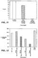
- This HLA-A2 consensus peptide was synthesized. This peptide is capable of protecting EC from CIK mediated killing in a dose responsive fashion ( Figure 12 ).
- An immunoprotective domain of Hsp47 therefore contains a relatively small peptide domain which by itself is capable and sufficient to account for the protection of EC from non-MHC restricted killing.
- Hsp47 is co-secreted as collagen chaperone by all cells actively depositing extracellular matrix containing collagens I or IV ( Brewer et al., Embo J. 16:7207-7216 (1997 )).
- Hsp47 can bind to ubiquitous fibronectin consensus sequences (RGD) on fibronectin, osteonectin heparin and collagen molecules in the vicinity of the cell surface of the secreting cell ( Nakai et al., Biochem Biophys Res Comm 164:259-264 (1989 )). Very recently it was reported that Hsp47 binds to tetraspanin CD9 on the surface of cells ( Hebert et al., J. Cell Biochem 73:248-258 (1999 )). Tetraspanins are important membrane signal transducing molecules involved in the shaping of the T-cell repertoire ( Levy et al., Annu Rev Immunol 16:89-109 (1998 )).
- the physiologically aberrant vascular beds of tumors are characterized by fenestrated basal membranes and lack of normal EM production.
- fenestrated basal membranes In healthy individuals, only the ellipsoid meshwork of the spleen and liver have EC with fenestrated basal membranes. Those EC anchoring on physiologically fenestrated basal membranes are reported to have unusually high Hsp47 amounts ( Kasai et al., Cell Tissue Res 281:135-141 (1995 )).
- Hsp47 mediated non-kill signal theory could provide a mechanism for CIK mediated in-vivo screening of tissue resident cells for extracellular matrix production and collagen production. According to this theory, detection and destruction of metastatic solid tumor cells would be the result from the latter lacking collagen and Hsp47 production.
- Hsp47 is down-regulated in cells undergoing oncogenic transformation ( Clarke et al., J. Cell Biol 121:193-199 (1993 ); Hirayoshi et al., Mol Cell Biol 11:4036-4044 (1991 )).
- in-vivo studies comparing a wide range of solid tumors, it was further found that Hsp47 becomes increasingly less expressed, as those tumors become more malignant and screening for Hsp47 levels was suggested to clinically follow tumor progression in cancer patients ( Morino et al., In Vivo 8:285-288 (1994 )). Metastatic tumor nodules and their Hsp47 deprived vicinity thus could represent prime targets for cellular in-vivo tumor surveillance.
- Blocking antibodies to KiR or p40 do not enhance the killing of tumor or endothelial targets.
- the involvement of other killer inhibitory receptors however remains possible.
- Blocking Mab NK-B 1 and NKp40 were also tested in 51 Cr release cytotoxicity assays. Functional involvement of either molecule in CIK mediated cytotoxicity against tumor or EC targets has not been shown. These receptors were assayed because expression of these NK markers is reported to occur on non-classical MHC I restricted cytotoxic T-cell ( D'Andrea et al., J. Immunol 155:2306-2310 (1995 ); Gumperz et al., J. Exp. Med. 181:1133-1144 (1995 ); Lanier et al., J.
- CIK can be used in vivo without co-injection of IL-2 ( Lu et al., J. Immunol 153:1687-1696 (1994 )). This is a significant difference to past and current treatment of patients with IL-2 activated CD56 + cells, IL-2 alone ( Yang, Cancer 76:687-694 (1995 )) or a combination thereof ( Phillips, Journal of Clinical Oncology 5:1933-194 (1987 )). Whereas the latter approaches reported a significant risk for the development of potentially fatal vascular leak syndrome (VLS), VLS was not observed in the in-vivo tumor purging models with SCID/hu mice. Nor was there significant CIK infiltrates in grafted normal human skin.
- VLS vascular leak syndrome
- the reliable expansion and generation of highly specific anti-tumor activity is a sine qua non for any adoptive immune therapy cell production intended for clinical use.
- High dose Th 1 -type hormonal stimulation of ⁇ / ⁇ T-cells with IFN- ⁇ , OKT-3 and IL-2 results in CIK AI cells with superior cytotoxicity against malignant hematopoietic and solid tumor targets.
- the use of turbine agitated bioreactors proves to be a plus, resulting in rapid, safe and improved selective expansion of the cytolytic CD3 + 56 + subset. Yields of 10 11 cells are routinely obtained from a single buffy coat source. This is a sufficient number of effector cells for high dose and/or repeat treatment schedules in their clinical application.
- CIK can be generated from pure T-cell sources, maintained in long-term culture, cryo-preserved, and applied in-vitro and in-vivo without co-administration of IL-2. These characteristics make CIK superior choices for adoptive immune therapy of cancer as compared to LAK NK derivatives.
- PBL peripheral blood lymphocytes
- Enriched populations of large granular lymphocytes (LGL) and T-cells were obtained by subsequent exclusion of plastic and nylon wool adherent cells.
- Source LGL were cultured in a humidified incubator with 5% carbon dioxide at 37 °C.
- Hormonal stimulation consisted of addition of recombinant human interferon gamma (rhu g-IFN) (a kind gift of Genentech, South San Francisco, CA) at 1000 IU ml-1 at the start of culture day d0, subsequent stimulation on day one (d1) with soluble anti-human CD3 monoclonal antibody OKT-3 (derived from hybridomas by the American Tissue Culture Collection, Rockville, MD) at 50 ng per ml and recombinant human interleukin-2 (rhuIL-2) (a kind gift of Cetus/Chiron, Emeryville, CA) at 300-500 IU per ml.
- rhu g-IFN recombinant human interferon gamma
- T cells were purified from source LGL by FACS sorting for presence of CD3 or CD56 and absence of CD 16 as described below.
- Cells were sorted at a mean density of 0.6 cells per well in a 96 well plate and expanded by co-culture with twice irradiated autologous feeder CIK at 0.5x10 6 cells per ml with identical hormonal stimulation as described for bulk culture. Cells were expanded to more than 2x10 6 cells each and then tested for clonal cytotoxicity as described below.
- LGL For large scale production of single donor CIK, healthy donor apheresis products were treated as above to isolate LGL.
- LGL were cultured at 37 °C with identical hormonal stimulation as above, but at a tenfold density of 5-10 x10 6 cells/ml in turbine/impeller agitated bioreacters of 500 ml, 1,000 ml (Nalgene, Rochester, NY) and pO2 and pH controlled 15,000 ml (Chemap, Völketsvil, Switzerland) capacity.
- Use of 15L bioreactors allowed expansion up to 3x10 11 cells from a single donor with the expansion of the CD3 + CD56 + cells exceeding 600 fold.
- HUVEC human umbilical chord endothelial cells
- the hematopoietic tumor cell lines SU-DHL, OCI-Ly8, K562 and lymphoblastoid cell lines AMK (a kind gift of Dr. A, Krensky, Stanford University, CA) and RDL (a kind gift of Dr. R. Lopez, Stanford University, CA) were cultured in RPMI 1640 medium (GIBCO-BRL/Life Technologies, Grand Island, NY) containing 50 ⁇ m ⁇ -mercapto-ethanol (ME), 100 IU penicillin-G per ml, 100 IU streptomycin per ml and 10% FCS (all: Sigma Chemical Co., St.Louis, MO) at a density of 0.5-2x10 6 cells per ml in a humidified incubator with 5% carbon dioxide at 37 °C.
- RPMI 1640 medium Gibco-BRL/Life Technologies, Grand Island, NY
- FCS all: Sigma Chemical Co., St.Louis, MO
- Melanoma cell lines WM 9 and 1205 LU (a kind gift of Dr. M. Herlyn, Wistar Institute of Anatomy and Biology, Philadelphia, PA) were maintained on tissue culture treated flasks (Beckton Dickinson, San Jose, CA) in MCDB 153 medium (Life Technologies, Grand Island, NY) supplemented with 5 ⁇ g per ml insulin and 2% FCS in a humidified incubator with 5% carbon dioxide at 37 °C.
- the constituently cytoplasmic luciferase expressing cervical carcinoma cell line HeLaluc (a kind gift of C. Contag, Stanford University, Stanford, CA) was cultured in DMEM medium (GIBCO-BRL/Life Technologies, Grand Island, NY) containing 50 ⁇ m ⁇ -mercapto-ethanol (ME), 100 IU penicillin-G per ml, 100 IU streptomycin per ml and 10% FCS (all: Sigma Chemical Co., St.Louis, MO) in a humidified incubator with 5% carbon dioxide at 37 °C.
- DMEM medium Gibco-BRL/Life Technologies, Grand Island, NY
- FCS all: Sigma Chemical Co., St.Louis, MO
- CTL cell-line AJY and specific CTL target cell-line AJ were kindly provided by the laboratory of A. Krensky (Stanford University, Stanford, CA).
- Partial huHsp47 gene fragments derived by RT-PCR were amplified in vitro and artificial cloning sites introduced by PCR. These fragments correspond to nucleotides in Figure 1A .
- the nucleic acid encoding the amino terminal 39 amino acids of Hsp47 (excluding the signal sequence and first amino acid of the mature protein) was amplified with the following primers: 5' primer ACGTTTGGATCCAGGTGAAGA, 3' primer GTCCTTGGCCAT. The 5' primer incorporated a Bam HI site to facilitate cloning into further vectors.
- the 3' primer incorporated an Mlu NI site to facilitate fusing the nucleic acids encoding the amino and the carboxy terminal portion of the protein.
- the nucleic acid encoding the carboxy terminal 360 amino acids was amplified with the following primers: 5' primer GCAATGGCCAAGGACCAGGCAGTGGAG, 3' primer ACGCTCTGCTCAATATCCTTAAGTCTA.
- the 5' primer incorporated an Mlu NI site to facilitate fusing the two portions of the gene into one continuous reading frame and the 3' primer incorporated an Eco RI site to facilitate cloning into further vectors. Standard PCR conditions generally known to those of skill in the art were used to amplify the two fragments from the starting clones (see Figure 8 ).
- the resulting amplification products were digested with Mlu NI, purified and ligated to each other.
- the resulting nucleic acid was further digested with Bam HI and Eco RI and the huHsp47 gene cassette was directionally cloned using standard recombinant DNA techniques into pUC 19 (Pharmacia, Uppsala, Sweden) which was used for construction purposes.
- the resulting plasmid, pUC/huHsp47 was amplified in recA bacterial host strain DH5a (Life Technologies, NY).
- the sequence of the cloned gene cassette was verified via fluorescent sequencing on an ABI sequencer (ABI,) using T7 DNA polymerase (Pharmacia, Uppsala, Sweden).
- the huHsp47 gene cassette was PCR amplified using an alternative set of primers which results in a BamHI-EcoRI expressible gene cassette for bacterial protein expression.
- the expressible gene cassette was cloned into the pGEX 4T 1 vector (Pharmacia, Uppsala, Sweden), which produces a GST-fusion protein under the control of the IPTG inducible laq q promoter, and grown in the bacterial host strain BL21(DE3) (Stratagene, La Jolla, CA).
- cleavage with 10 IU of thrombin (Sigma, St. Louis, MO) per ⁇ g fused GST moiety was performed for 10-12 hours at 37 °C using a thrombin-cleavage site present in the linker amino acids between GST and the recombinant protein of interest.
- Routinely 5-10 mg of recombinant huHsp47 fusion protein were obtained per 4 liters of BL21(DE3) host cells transformed by pGEX-4T-Hsp47 via glutathione affinity FPLC. Purified huHsp47 was used in 51 CR release cytotoxicity assays described above.
- HuHsp 47 has at least four distinct functional domains. Beginning at the amino-terminus of the protein they are: a collagen/RGD binding domain, a domain with homology to the ⁇ -2 domain of human HLA-A2 molecules, a serine protease inhibitor (serpin) domain and an ER retention signal RDEL domain. Three specific deletions were generated.
- Deletion 1 removed the RDEL domain
- deletion 2 removed the RDEL and serpin domains
- deletion 3 removed the carboxy-terminal 150 amino acids including the serpin and RDEL domains.
- the same 5' primer was used in PCR reactions to generate all three deletion mutants.
- the 5' primer sequence is: CGGAATTCTGGCCGAGGTGAAGAAACC.
- the 3' primer used to generate the RDEL deletion mutant was the same primer used to generate the huHsp47-GFP fusion protein and has the sequence: AGTTCCCACTGTTCTACGACCTAGGGC.
- the deletion 2 3' primer is: AACTCAACCTGTGTCTAGACCTATGGGC.
- the deletion 3 3' primer is: ACGCGCTGCTCCTCCACGACCTAGGGC (see Figure 14 ).
- the 5' and 3' primers incorporated BamHI and EcoRI restriction enzyme sites respectively to facilitate subsequent cloning steps.
- the 5' and individual 3' primers were mixed with the pUC/huHsp47 plasmid containing the huHsp47 gene cassette and nucleic acids encoding each deletion mutant were PCR amplified in separate reactions.
- PCR products were purified, digested with BamHI and EcoRI and ligated into either the pGEX-4T 1 vector to create huHsp47deletion-GST fusion proteins for cytotoxicity assays (see below) or pEGFP-NI, which also contained the mel secretion signal described above, to create eGFP-Hsp47deletion fusion proteins for use as FACS probes.
- RNA for each time point was purified, denatured, subjected to formamide flatbed agarose gel electrophoresis, transferred to nitrocellulose, prehybridized, hybridized with the 32 P-labelled probes, washed under stringent conditions and autoradiograped without intensifier screens at -80 °C using techniques well known to those skilled in the art (see Sambrook et al. Molecular Cloning, A Laboratory Manual, second edition, Cold Spring Harbor Laboratory Press, 1989 ). Results indicate that BFA treatment does not affect the expression of the housekeeping gene beta action, but that BFA treatment does upregulate huHsp47 gene expression over the period evaluated. See Figure 10 . These data are consistant with the observed increase in Hsp47 protein seen in BFA treated cells and isolated by immunoprecipitation experiments. See Figure 5 .
- Protease inhibitor free production of rhuHsp47 for granzyme A assays was carried out by growing protease free transformed BL21 (DE3) bacteria in amp selective NZCYM medium using casein acidic hydrolysate in lieu of tryptic casein amino acids as nutritional peptide source for the host bacteria undergoing IPTG induction. To minimize incubation times, protein extracts were batch incubated on glutathione resin and initial removal of non GST proteins was carried out with a modified spin column approach. The remainder of the protocol was unchanged, thrombin was not utilized.
- Target cells were metabolically labelled with 51 Cr (Dupont- New England Nuclear, Boston, MA) by incubating 1x10 6 cells in 300 mCi 51 Cr at 37 ° C for 1-1.5 hours.
- the labelled cells were washed three times with phosphate buffered saline (PBS) containing 0.1% bovine serum albumin.
- PBS phosphate buffered saline
- the labelled cells were distributed in flat-bottomed 96 well microtiter plates at a concentration of 2x10 4 cells/well in triplicate. Effector (CIK) cells were added at the indicated ratios. Mabs were added prior to the addition of effector cells, and incubated for 15-30 minutes at room temperature. The final volume of the assay mixture in each well was 0.2 ml.
- % specific Cr 51 release ( test release ) - spontaneous release ( maximal release ) - spontaneous release ⁇ 100 % Spontaneous release was obtained by incubating the labelled cells in complete medium alone and maximal release by treatment of the cells with 1% NP-40.
- the anti-tumor activity of adult and umbilical cord derived CIK cells were compared in cytotoxicity assays.
- the cytotoxicity results of the CIK cells derived from both sources were superimposable against B-cell lymphomas OCI-Ly8 and SU-DHL4/LAM53 and myeloid leukemia target K562.
- Both sets of CIK cells displayed marginal cytotoxicity against melanoma WM9 in vitro. Both sets of CIK cells were cytotoxic against freshly confluent HUVEC and less so against EC monolayers confluent for longer periods of time (see Figure 3 ).
- Figures 3A, 3B and 3C describe 4 hour 51 Cr release cytotoxicity assays with d 21 CIK and various targets.
- CIK were derived from PBMC by culture in complete RPMI supplemented by the following: At d 0 : IFN -g at 1000 u ml -1 . At d 1 : 5 ng soluble OKT-3 and 500 u IL-2 ml -1 . At d 5 , d 9 , d 13 , d 17 and d 21 : 500 u IL-2 ml -1 . All cells were in a humidified incubator with 5% CO 2 at 37°C.
- the hematopoietic tumor lines K562, OCI-Ly8, and SU-DHL4 were maintained in complete RPMI medium; melanoma line W9 was cultured in MCDB 153 medium supplemented with 5 ⁇ g ml -1 insulin and 2% FCS.
- HUV-EC were obtained from the ATCC. They were grown on 2% gelatin coated 24 well plates in TC-199 or Ham's F12-K medium, each supplemented with 20% FCS, 5 ⁇ g porcine heparin ml -1 and 80 ⁇ g endothelial mitogen ml -1 . Routine tryptic passaging was carried out when HUVEC reached more than 80% confluency; 51 Cr release assays were routinely performed at the time point of fresh confluency.
- Figure 3A is a cytotoxicity profile of adult and cord blood CIK d 21 CIK shows high cytotoxicity against B-cell lymphomas OCI-Ly8 and SU-DHL4 (blue), as well as erythromyeloid leukemia K562 (yellow). Moderate cytotoxicity exists against nodular melanoma line WM9 (red) and freshly confluent HUV-EC (dark green).
- Figure 3B shows the effect of EC culture duration after reaching confluency on the EC sensitivity to d 21 CIK lysis: freshly confluent EC (dark green) are lysed significantly more readily than are EC cultured for 5 additional days after reaching confluency (light green).
- clones of CIK were generated via plating of a mean of "0.6 cells per well" of a 96 well plate; then expanded by co-culture with twice irradiated, autologous feeder cells at 0.5x10 6 cells ml -1 , and with identical hormonal stimulation as described above for bulk cultures. After individual clones expanded to more than 2x10 6 cells each, they were tested for clonal cytotoxicity profile at a E:T ratio of 5:1. Target specificity of CIK is determined at the clonal level.
- Expanded CIK single cell clones were tested to determine if subpopulations of cells exhibited target specificity in cytotoxicity assays performed as described above. Target specificity was observed, was line specific and was directed against tumor cells only, EC cells only or against both targets, the latter being the most common.
- the 2D10 CIK cell line tested displayed cytotoxicity against the OCI-Ly8 tumor cell line but not the HUVEC cell line.
- the 5F7 CIK cell line displayed cytoxicity against HUVEC cells but not OCI-Ly8 cells.
- the 2C10 CIK cell line was cytotoxic against both the OCI-Ly8 and HUVEC cells.
- BFA brefeldin A
- freshly confluent HUVEC monolayers, nonadherent target cells or CIK cells at 0.5-2 x 10 6 cells per ml were incubated with medium supplemented with 0.5-10 ⁇ g of brefeldin A (Bschreibinger Mannheim GmbH, Mannheim, Germany) per ml medium and incubated for 2-6 hours in a humidified incubator with 5% carbon dioxide at 37 °C.
- BFA was dissolved at 100 ⁇ g per ml in 70% ethanol and used fresh or stored up to 4 weeks at -20 °C. Prior to cytotoxicity assay the cells were rinsed three times with PBS, and fresh medium not containing BFA was added for the remainder of the assay ( Figure 4 ).
- BFA treatment interferes with the intracellular vesicle transport from the endoplasmic reticulum to the proximal and intermediate golgi compartments preventing glycosylation of proteins directed for secretion and active vesicle transport of mature proteins to the cell surface (Lippincott-Schwartz et al., 1990; Lippincott-Schwartz et al., 1991; Lippincott-Schwartz et al.,1989; Orci et al., 1991; Vogel et al., 1993). Brefeldin treatment of endothelial cells leads to up regulation and secretion of a 46.5 KDa protein inhibitor presently identified as Hsp47 by immunodetection with MAB SPA470.
- Figure 5A is a silver stained SDS-PAGE, 10% polyacrylamide gel. Specific detection of p46.5 occurs only at a protective BFA dose. Note heavy and light chains of N6 Mab IgG.
- Figure 5B is an auto-radiograph of a 12% polyacrylamide gel SDS-PAGE transferred to an NC membrane. A 46.5 KDa 35 S radio-labeled band is present only at 10 ⁇ M BFA.
- Figure 5C demonstrates the positive identification of p46.5: It is huHsp47, immuno-detection with Hsp47 specific Mab SPA 470.
- Hsp47 migrates in this 12% PAGE with identical M r of 46,500 as the radio-labeled peptide.
- the protein bands of p46.5 and huHsp47 are superimposable. Note, that due to the use of mouse IgG Mab N6 for the IP and detection of the primary mouse anti-Hsp47 Mab SPA470 with secondary goat-anti mouse peroxidase Ab, the heavy and light chains of the N6 Mab are also detected in this ECL assay.
- BFA treatment of targets was able to suppress the CIK mediated lysis of EC in a dose dependent manner but did not affect the ability of CIK cells to lyse tumor targets ( Figure 4 ).
- Both HUVEC and OCI-Ly8 cells were incubated with BFA at concentrations of 0, 2.5, 5, 7.5 and 10 ⁇ M for 6 hours as described above, washed, labeled with 51 Cr and used in a cytotoxicity assay with d 21 CIK cells at an E:T ratio of 20:1.
- Preincubation of EC targets with 2.5 ⁇ M BFA reduced CIK mediated cytotoxicity by 40%. Larger doses reduced cytotoxicity further and preincubation in 10 ⁇ M BFA was sufficient to completely block CIK mediated lysis of HUVECs.
- NaN3 free monoclonal antibody (Mab) was added per ml of medium to the target cells and incubated at room temperature (RT) for 15 minutes. Effector cells were added directly to the treated targets. The remainder of the assay was performed as detailed above, at 37 °C in a humidified atmosphere of 5% CO 2 .
- Mabs to MHC class I W6/32
- T-cell markers CD2 LFA-2
- NK markers CD 16, CD56, NK-B 1 (NKTA255) and Nkp40 (DX 9) do not block the cytotoxicity of CIK cells against either tumor or EC targets.
- Mabs to ICAM-1 (CD54), and LFA-1 CD 18/11 a , LFA-1 ⁇ -chain, LFA-1 ⁇ -chain) do block cytotoxicity of CIK cells against tumor cells but not EC targets.
- Deletion 3 mutant protein lacking the carboxy-terminal 150 amino acids, was as effective as deletion 1 mutant protein in protecting EC from CIK mediated lysis suggesting that the reduced protection seen with the deletion 2 mutant protein is due to an altered conformation of the protein and is not due to the serpin domain participating in the protection.
- the deletion 3 mutant protein retains both the collagen/RGD binding domain and the ⁇ 2-HLA-A2 homology domain which contains the AVLSAEQLR peptide sequence.
- Nucleic acids encoding huHsp47 and fragments were cloned into eucaryotic expression vectors.
- a nucleic acid encoding a fragment of huHsp47, in which the carboxy-terminal RDEL amino acid sequence is deleted, was PCR amplified from the pUC/huHsp47 plasmid using the following primers: 5' primer CGGAATTCTGGCCGAGGTGAAGAAACC, 3' primer AGTTCCCACTGTTCTACGACCTAGGGC.
- the amplified product was ligated to the melittin secretion signal and Kozak sequences derived from pMel-Bac (Invitrogen, San Diego, CA) and the resulting fragment was cloned into the multiple cloning site of pEGFP-N1 (Clontech, Palo Alto, CA) using general techniques well known to those of skill in the art.
- the resulting plasmid, eGFP-Hsp47 was transfected into EC.
- Nucleic acids encoding full length huHsp47 and huHsp47 ⁇ RDEL were cloned into the pBK-CMV (Stratagene, La Jolla, CA) and expressed in EC.
- the pBK vector was digested with Nhe I and EcoRI to remove the Lac promotor, most of the MCS and a portion of the Lac Z gene.
- the mellitin secretion signal peptide and Kozak sequence were PCR amplified from the pMel-Bac vector as described above and ligated into the pBK vector adjacent to the CMV IE promoter.
- EC were also co-transfected with a combination of vectors; eGFP-Hsp47, CMV-neo-mel-huHsp47fullength and pCMV-CD20 (a vector expressing CD-20 under the control of the CMV promoter, a kind gift of Dr. S.vHeuvel, Massachusetts General Hospital, Boston, MA) using the low dose lipofectamine transfection protocol.
- Cells were subjected to FACS analysis 24 hours after co-transfection.
- EC targets were used in cytotoxicity assays 24 hours after lipofectamine mediated transfection of either CMV-neo-mel-huHsp47fullength or CMV-neo-mel-huHsp47 ⁇ RDEL into previously susceptable EC.
- Mature d 21 CIK cells were used at an E:T ratio of 20:1.
- EC over-expressing full length huHsp47 showed a 25% reduction in the CIK mediated lysis.
- EC over-expressing huHsp47 ⁇ RDEL were entirely resistant to CIK mediated lysis (see Figure 11 ).
- RDEL peptide which serves as an endoplasmic reticulum (ER) retention signal, from huHsp47, allows the protein to be freely secreted and protects the EC targets expressing the protein from CIK mediated lysis.
- ER endoplasmic reticulum
- FACS Fluorescence Activated Cell Sorting
- Stained cells were subjected to three subsequent washes with 10 volumes of Ca 2+ , Mg 2+ and 5% FCS containing PBS and taken up in 500 ⁇ l of this buffer for analysis or sort by FACScan/FACStar (Beckton Dickinson, San Jose, CA) with excitation at 488 nm and the appropriate channel filters for detection of emission.
- FACScan/FACStar Beckton Dickinson, San Jose, CA
- huHsp47 ⁇ RDEL-enhanced green fluorescent protein (eGFP) fusion protein expressed under the control of the CMV IE promoter, was isolated as described above and used as a FACS probe to search for huHsp47 cell surface binding proteins.
- eGFP huHsp47 ⁇ RDEL-enhanced green fluorescent protein
- FACS analysis of mature d 21 CIK with eGFP-tagged rhuHsp47 as molecular probe was carried out by first staining with PE-Mab and specifically PE-CD56 if desired, then by suspending the washed CIK in 10 ⁇ g eGFP-huHsp47 per ml of PBS, incubating for 15 minutes at 0-4 °C in the dark, removal of unbound protein by centrifugation in a refrigerated eppendorf centrifuge at 1000 rpm at 0-4 °C.
- Stained cell pellets were either resuspended in 500-1000 ⁇ l of PBS immediately prior to the FACS analysis or suspended in PBS fixans containing 3% (w/v) formaldehyde prior to analysis. After staining, cells were sorted using the FITC channel. 26% of the cells analysed were positive for both eGFP-huHsp47 and PE-CD56 probes indicating that a distinct subset of the CIK cells had receptors for huHsp47.
- EC cells co-transfected with eGFP-Hsp47, CMV-neo-mel-huHsp47fullength and pCMV-CD20 using the low dose lipofectamine transfection protocol were analysed 24 hours after co-transfection.
- Cells were stained with PE-CD20 Mab and analysed for the presence of both the CD-20 antigen and eGFP-Hsp47 using the FITC channel. Over 79% of the cells were positive for both probes indicating that there was an extremely high transfection efficiency.
- targets Prior to cell harvest, targets were cultured or treated BFA as described above, and dead cells removed from EC monolayers by washes with Ca 2+ , Mg 2+ containing PBS, from nonadherent targets via density gradient centrifugation.
- Cell lysis was performed with ultrasonic cell disruption (Branson, New Brunswick, NJ) in PBS buffer containing 1% Triton-X 100 (Tx-100, Sigma, St. Louis, MO) and complete protease inhibitor cocktail (Bschreibinger Mannheim, Mannheim, Germany).
- Pan-anti-heat shock protein Mab N6 hybridoma (a kind gift of Dr. K Nagata, Kyoto, Japan) was injected intraperitoneally into six-week old female Balb/c mice. Ascitis fluid was harvested, shockfrozen in liquid N2 and stored at -20 °C until needed.
- FPLC purification of polyclonal and monoclonal antibodies from serum or ascitis was performed after 7x dilution in PBS and chromatography on Protein A sepharose (Pharmacia, Uppsala, Sweden) with UV260nm control. Bound Ig fractions were eluted with 25 mM glycine buffer at pH 2.5 and rapidly titrated back to pH 7.4 by addition of TRIZMA (Sigma, St. Louis, MO) base. Protein amounts were determined by Bradford assay at 595 nm in a DU-650 spectrophotometer (Beckman, Palo Alto, CA).
- Immunoprecipitation was carried out by first preparing preformed immune complexes as follows: Purified specific mouse monoclonal or rabbit polyclonal antibody were incubated in stoichiometric ratio with purified secondary goat anti-mouse or anti-rabbit antibody at a concentration of 10 ⁇ g ml-1 of 150 mM NaCl, 10 mM Tris-HCl, 1% (w/v) NP40, pH 7.8 with gentle agitation for 3 hours at 4 °C. In a separate reaction their respective isotype matched control antibodies were coupled to protein A sepharose beads (Pharmacia, Uppsala, Sweden).
- the formed immune complexes were harvested by centrifugation at 13,000 rpm in a refrigerated Eppendorf centrifuge (Eppendorf, Hamburg, Germany) for 15 minutes at 4 °C, washed twice with one volume of 500 mM NaCl, 50 mM Tris-HCl, 2% BSA, 0.25% (w/v) SDS, 0.5% (w/v) Tx-100, pH 7.5 and taken up in 120 ⁇ l of 150 mM NaCl, 10 mM Tris-HCl, 1% (w/v) NP40 at pH 7.8.
- Immunoprecipitation with specific antibody was carried out by adding 30 ⁇ l of cleared protein extract to 200 ⁇ l of 150 mM NaCl, 10 mM Tris-HCl, 1% (w/v) NP40, pH 7.8 and incubating with 10 ⁇ l of preformed specific immune complexes per reaction with gentle agitation for 3 hours at 4 °C.
- the specific immune precipitate was recovered by centrifugation at 13,000 rpm for 30 minutes at 4 °C, washed twice with 10 mM Tris-HCl at pH 6.8 and analyzed by SDS-PAGE and subsequent silver stain, transfer to solid support and streptavidin-AP detection and autoradiography.
- AMK targets were first treated for 16 hours with TM at 5 ⁇ g per ml or left untreated and then treated concurrently with BFA and 35 S met/cys (in the presence of TM in the originally treated group) as described above followed by a 2 hour chase.
- Crude cell extracts were immunoprecipitated (IP) either with polyclonal antisera prepared against AMK membrane preparations described above or with pan-anti heat shock Mab N6.
- IP immunoprecipitated
- Autoradiographs of the SDS-PAGE separated IP with either antibody of total cellular protein shows the presence of a 35 S labeled 46.5 KDa protein in CIK resistant untreated lymphoblastoid cells which was absent in the TM treated cells.
- EC targets were cultured in the presence of no BFA, the protective dose of 10 ⁇ m BFA, or the non-protective dose of 30 ⁇ M BFA concurrently with 35 S met/cys as described above. Samples were processed as described for the AMK cells. EC targets cultured in the presence of BFA also contained a 35 S labeled 46.5 KDa protein. This protein was not detectable in IP from EC cultured without BFA or with the non-protective dose of 30 ⁇ M BFA (see Figure 5 ).
- This 46.5 KDa protein was identified as Hsp47 in a Western analysis using the Mab SPA-470 antibody.
- 35 S labeled HUVEC extracts prepared from BFA treated cells described above were immunoprecipitated as described using the N6 pan-heat shock Mab. Precipitates were run on a 12% SDS-PAGE, transferred to NC membrane, blocked and reacted with the SPA-470 antibody using techniques well known to those of skill in the art.
- the presence of the SPA-470 antibody bound to the 46.5 KDa protein and the presence of the N6 heavy and light chains were detected using goat-anti mouse horseradish peroxidase staining assay(ECL).
- Biotinoylation of live adherant EC with water soluble, lipid insoluble Biotin analog NHS-LC-biotin performed as described above followed by IP of cell extracts using either the anti-AMK polyclonal antibody or N6 Mab and subsequent SDS-PAGE, transfer to NC membrane and detection using a streptavidin-Alkaline phospahtase system showed that the Hsp47 protein induced by BFA treatment and a minor protein of 27 KDa are localized on the cell surface of EC targets.
- Native and recombinant protein purification was performed by subjecting protein crude extracts to anion-exchange FPLC chromatography on Q-sepharose® at pH 7.4, and pH 6.8, eluting separated fractions by increasing ionic strength and subsequent size exclusion FPLC chromatography on Superose 12 separating fractions with increasing elution volumes Ve, controlled by a UV-M monitor 214, 260 and 280nm and recorded in analogue mode (all equipment and resins: Pharmacia, Uppsala, Sweden) (see Figure 6 ). Protein amounts were quantitated on-line by integration of the area under the recorded peak of absorbance (AuP) and by Bradford assay of sampled fractions. Qualitative analysis was performed by SDS-PAGE and subsequent Coomassie and silver staining, as well as membrane transfer and western blot, when specific antibodies were available.
- Figure 6A is a UV 260nm profile of native Hsp47 purification from EC on Q-sepharose ff anion exchange resin at pH 8.9 175 flasks of confluent HUVEC were induced with 10 ⁇ M BFA as described.
- Figure 6B is a 12% SDS-PAGE analysis with silver staining of the collected p46.5 containing fractions. Those were pooled and concentrated by YD10 membrane to 1.0 ml.
- Figure 6C is a 12% SDS-PAGE analysis with silver staining of the collected p46.5 containing fraction after size exclusion FPLC chromatography on Superose 12. Yield: 240 ⁇ g.
- Affinity FPLC chromatography was performed by loading protein extracts of wt and BFA treated EC onto gelatin-sepharose (Pharmacia, Uppsala, Sweden) matrix with help of a peristaltic pump and incubating the extract for 30 minutes at 0-4 °C. Unbound and unspecifically bound proteins were removed by washing with 7 column volumes of high salt buffer followed by original protein extraction buffer until the UV260nm detection at AU 0.02 returned to baseline. pH specific elution was carried out with PBS buffer at pH 5.5, alternative eluents included 100 mM ATP in PBS and 100 ⁇ M RGD peptide in PBS, both at pH 7.4.
- Immunoaffinity FPLC chromatography was carried out in a similar manner, with the protein extract loaded onto a specific Mab column: 10 mg of purified Mab N6 was coupled via a covalent thioether bond with a 13 atom spacer arm to activated, crosslinked agarose via the Sulfolink (Pierce, Rockford, IL) protocol. Unspecifically bound proteins were removed from the columns as above, specifically immuno recognized proteins were eluted via pH 2.5 elution in 50 mM Na*citrate buffer.
- Hsp47 may also be gelatin-affinity purification.
- 175 flasks of confluent HUVEC were induced with 10 ⁇ M BFA for 6 h at 37°C, 5% Co 2 , in F12K medium supplemented with 20% FCS and standard EC culture supplements.
- adherent EC were harvested by scraping in detergent buffer (75 mM Tris•Cl, 175 mM NaCl, 2 mM DTT, 0.5 mM EDTA, 0.5% Tx-100, 2 mM PMSF), lysed by sonification and the extract cleared from cell debris by centrifugation at 15,000 rpm in a refrigerated eppendorf centrifuge.
- the cleared extract was loaded onto a 5 ml covalently coupled gelatin 4B Sepharose column via peristaltic pump at 0.5 ml min -1 . Unbound proteins were discarded.
- the column was then washed with detergent free buffer (as above, w/o Tx-100), low salt buffer (no NaCl) and high salt buffer (plus 500 mM NaCl) under UV 260nm control until baseline readings were achieved. Specific elution of Hsp47 was achieved by lowering the pH of the detergent free buffer to pH 4.8. Collected eluate was immediately back-titrated to pH 7.4. The column was immediately regenerated and washed with pH 7.4 buffer.
- Figure 2 shows the results of incubation of EC with gelatin-affinity purified Hsp47 for 30 minutes at 21°C, prior to 51 Cr release CIK cytotoxicity assay. Excess non-bound Hsp47 protein was removed from the wells by three washes prior to the addition of CIK effector cells. As a control, untreated BFA induced HUVEC targets were compared to the protein treatment group. Human Hsp47, the protein of 46.5 KDa purified with pH specific elution from gelatin matrix, protects EC.
- a hydrophilic 9 mer peptide of the sequence AVLSAEQLR which corresponds to the region of shared homology of Hsp47 with HLA-A2 was synthesized by the Stanford PAN facility on a BioApplied ® peptide synthesizer, HPLC purified and verified by amino acid sequencing. Pure peptide was lyophilized and stored at -20 °C. The peptide was soluble in aqueous solution and used in PBS and RPMI 1640 at indicated concentrations.
- 1x10 5 melanoma WN9 or 1205 LU cells in PBS were injected intradermally into a single site of the human full-thickness skin of triplicate animals and tumor growth monitored by observation and palpation. Animals with evident nodular tumor burden exceeding a diameter of 5 mm were injected intraperitoneally with 1x10 7 CIK cells (suspended in RPMI without FCS or IL-2) of proven lytic activity in same day 51 Cr release cytotoxicity assay at CIK culture day d14-21.
- Transfection efficiency was monitored via co-transfection of eGFP and the Hsp47 construct with subsequent fluorescence laser microscopy analysis of transformed HeLaluc monolayers with a Axiophot (Zeiss, Jena, Germany) fluorescence microscope described above. Injected animals were monitored at 0, 2 and 7 days for in-vivo chemo-luminescence of remaining HeLa luciferase post CIK challenge with a quantitative digital imaging process in collaboration with Dr. C. Contag (see Figure 13 ). Animals receiving the CIK injection demonstrated a dramatic decrease in live, light emmitting tumor burden one week after CIK purging. Animals which did not receive the CIK cells demonstrated widespread IP growth of HeLa cells. The CIK cells demonstrated a high and rapid solid tumor purging potential.
- Bone marrow (BM) and spleen derived stem cells were obtained from B10D2 (H-2 d background) mice. 2.5x10 6 BM and 1x10 6 spleen cells were pooled as a graft source for each recipient male Balb/c animal. This non-enriched stem cell population of cells were either suspended in RPMI containing 300 ⁇ g Hsp47-Gst protein in 300 ⁇ l total volume, or, in buffer containing 100 ⁇ g of Gst control protein alone. Cells and protein were given in a single injection on day 0. No other immunosuppressive treatments were given. Animals were monitored for 4 weeks after the bone marrow transplant.
- This Example discloses the use of modified Hsp47 to identify and separate immune cell subsets.
- Hsp47 A variety of immune cells respond to Hsp47 and do not attack endothelial cells.
- fluorescent tagged Hsp47 can be used in a standard cell sorting apparatus to identify and separate such cells. Such use can also serve as a diagnostic tool for immune cell subset analysis in various disease states.
- eGFP-tagged Hsp47 FACS probe was generated through additional genetic engineering ( Figure 8A ). This enabled searching for both potential huHsp47 receptors and for CIK subsets expressing such Hsp47 binding proteins. FACS analysis of mature d 21 CIK with eGFP-tagged HuHsp47 as FACS probe for the FITC channel, with CE56-PE co-staining, is based on differences in the Hsp47 binding abiligy of the analyzed CIK. The eGFP tagged Hsp47 probe stained a quarter of mature CIK (26%).
Landscapes
- Health & Medical Sciences (AREA)
- Life Sciences & Earth Sciences (AREA)
- Chemical & Material Sciences (AREA)
- General Health & Medical Sciences (AREA)
- Medicinal Chemistry (AREA)
- Organic Chemistry (AREA)
- Immunology (AREA)
- Veterinary Medicine (AREA)
- Public Health (AREA)
- Animal Behavior & Ethology (AREA)
- Pharmacology & Pharmacy (AREA)
- Bioinformatics & Cheminformatics (AREA)
- Engineering & Computer Science (AREA)
- Proteomics, Peptides & Aminoacids (AREA)
- Gastroenterology & Hepatology (AREA)
- Zoology (AREA)
- Epidemiology (AREA)
- General Chemical & Material Sciences (AREA)
- Nuclear Medicine, Radiotherapy & Molecular Imaging (AREA)
- Chemical Kinetics & Catalysis (AREA)
- Molecular Biology (AREA)
- Genetics & Genomics (AREA)
- Biophysics (AREA)
- Biochemistry (AREA)
- Toxicology (AREA)
- Marine Sciences & Fisheries (AREA)
- Oncology (AREA)
- Transplantation (AREA)
- Micro-Organisms Or Cultivation Processes Thereof (AREA)
- Medicines Containing Material From Animals Or Micro-Organisms (AREA)
- Peptides Or Proteins (AREA)
- Medicines That Contain Protein Lipid Enzymes And Other Medicines (AREA)
- Pharmaceuticals Containing Other Organic And Inorganic Compounds (AREA)
- Measuring Or Testing Involving Enzymes Or Micro-Organisms (AREA)
- Preparation Of Compounds By Using Micro-Organisms (AREA)
Claims (13)
- Eine pharmazeutische Zusammensetzung mit HuHsp47 Polypeptid.
- Verwendung eines HuHsp47 verwandten immunprotektiven Polypeptids zur Herstellung einer pharmazeutischen Zusammensetzung zur Prävention und/oder Behandlung von Zellschädigungen, die von Wirkungen bzw. Krankheiten des Immunsystems hervorgerufen werden.
- Verwendung eines HuHsp47 verwandten immunprotektiven Polypeptids gemäß Anspruch 2, bei dem die Immunsystem-bedingte Zellschädigung durch eine Autoimmunkrankheit, Graft vs. Host Reaktion oder Host vs. Graft Reaktion ausgelöst wird.
- Ein Polypeptid mit immunprotektiver Aktivität ausgewählt aus:a) HuHsp47b) die Aminosäuresequenz der Fig. 1c) eine Aminosäuresequenz mit immunprotektiver Aktivität mit mindestens 70% Sequenzidentität mit der Aminosäuresequenz von a) oder b)d) eine Aminosäuresequenz mit immunprotektiver Aktivität kodiert durch eine Nukleinsäure, die hybridisiert mit einer Nukleinsäure, die Aminosäuresequenz a) oder b) kodierte) ein Fragment von a), b), c) oder d) mit immunprotektiver Aktivitätf) eine Variante von a), b), c) oder d) mit immunprotektiver Aktivität
- Eine isolierte Nukleinsäure kodierend für ein Polypeptid mit immunprotektiver Aktivität ausgewählt aus der Gruppe bestehend aus:a) Ein Nukleinsäure kodierend für huHsp47b) die Nukleinsäure der Fig. 1c) eine Nukleinsäure mit immunprotektiver Aktivität mit mindestens 70% Sequenzidentität mit der Nukleinsäure von a)d) eine Nukleinsäure mit immunprotektiver Aktivität, die hybridisiert mit einer Nukleinsäure die Aminosäuresequenz der Fig. 1 kodierte) ein Fragment von a), b), c) oder d) kodierend für ein Polypeptid mit immunprotektiver Aktivitätf) eine Variante von a), b), c) oder d) kodierend für ein Polypeptid mit immunprotektiver Aktivität.
- Ein Vektor mit einer Nukleinsäure kodierend für humanes Hsp47 Polypeptid, funktionell an Kontrollsequenzen gekoppelt, die durch eine mit dem Vektor transfizierte Wirtszelle erkannt werden.
- Eine Wirtszelle mit dem Vektor des Anspruchs 6.
- Ein Verfahren zur Verminderung Immunsystem-bedingter Schädigung von Gewebe, Organen oder Zellen mit den Schritten eines Kontaktierens des Gewebes, Organs oder der Zellen mit einem HuHsp47 verwandten immunprotektiven Polypeptid bei dem das Verfahren in vitro oder in vivo durchgeführt wird.
- Verwendung eines immunprotektiven Polypeptids nach Anspruch 4 oder einer Nukleinsäure nach Anspruch 5 zur Herstellung einer pharmazeutischen Zusammensetzung zur Prävention und/oder Behandlung von Zellschädigungen die von Wirkungen bzw. Krankheiten bzw. Immunsystems hervorgerufen werden.
- Verwendung eines Polypeptids nach Anspruch 9, wobei die Immunsystem-bedingte Zellschädigung von einer Autoimmunkrankheit, einer Graft vs. Host Reaktion oder einer Host vs. Graft Reaktion hervorgerufen werden.
- Verwendung eines immunprotektiven Polypeptids nach Anspruch 4 oder einer Nukleinsäure nach Anspruch 5 zur Herstellung einer Zusammensetzung für eine adoptive Immuntherapie zur Behandlung von Krebs, wobei die Zusammensetzung derart ausgebildet ist, dass sie einem Patienten zusammen mit T-Lymphozyten aus einer T-Lymphozytenkultur eines Donors verabreicht werden kann, und bei den T-Lymphozyten bestimmte spezifische Untergruppen expandiert werden.
- Verfahren zur Verminderung Immunsystem-bedingter Schädigung von Gewebe, Organen oder Zellen mit den Schritten Herstellen eines Kontakts zu einem Gewebe, Organ oder Zellen mit einer expressiblen Nukleinsäure kodierend für eine Polypeptid nach Anspruch 4 oder einer Nukleinsäure nach Anspruch 5, wobei das Verfahren in vitro oder ex vivo durchgeführt wird.
- Ein HuHsp47 verwandtes immunprotektives Polypeptid nach einem der vorhergehenden Ansprüche in Kombination mit einer Zelle, einem Gewebe oder einem Organ.
Priority Applications (1)
| Application Number | Priority Date | Filing Date | Title |
|---|---|---|---|
| EP08000330A EP1964572A3 (de) | 1998-08-24 | 1999-08-24 | Zusammensetzungen und Verfahren zum Schutz von Organen, Gewebe und Zellen aus immunsystemvermittelten Schäden |
Applications Claiming Priority (5)
| Application Number | Priority Date | Filing Date | Title |
|---|---|---|---|
| US9764098P | 1998-08-24 | 1998-08-24 | |
| US97640P | 1998-08-24 | ||
| US38208899A | 1999-08-24 | 1999-08-24 | |
| PCT/US1999/019337 WO2000010603A1 (en) | 1998-08-24 | 1999-08-24 | Compositions and methods for protecting organs, tissue and cells from immune system-mediated damage |
| US382088P | 1999-08-24 |
Related Child Applications (1)
| Application Number | Title | Priority Date | Filing Date |
|---|---|---|---|
| EP08000330A Division EP1964572A3 (de) | 1998-08-24 | 1999-08-24 | Zusammensetzungen und Verfahren zum Schutz von Organen, Gewebe und Zellen aus immunsystemvermittelten Schäden |
Publications (3)
| Publication Number | Publication Date |
|---|---|
| EP1107789A1 EP1107789A1 (de) | 2001-06-20 |
| EP1107789A4 EP1107789A4 (de) | 2004-09-01 |
| EP1107789B1 true EP1107789B1 (de) | 2008-06-25 |
Family
ID=26793493
Family Applications (2)
| Application Number | Title | Priority Date | Filing Date |
|---|---|---|---|
| EP08000330A Withdrawn EP1964572A3 (de) | 1998-08-24 | 1999-08-24 | Zusammensetzungen und Verfahren zum Schutz von Organen, Gewebe und Zellen aus immunsystemvermittelten Schäden |
| EP99946625A Expired - Lifetime EP1107789B1 (de) | 1998-08-24 | 1999-08-24 | Zusammensetzungen und verfahren zum schutz von organen, gewebe und zellen vor schäden, die durch das immunsystem verursacht werden |
Family Applications Before (1)
| Application Number | Title | Priority Date | Filing Date |
|---|---|---|---|
| EP08000330A Withdrawn EP1964572A3 (de) | 1998-08-24 | 1999-08-24 | Zusammensetzungen und Verfahren zum Schutz von Organen, Gewebe und Zellen aus immunsystemvermittelten Schäden |
Country Status (10)
| Country | Link |
|---|---|
| US (1) | US20080241194A1 (de) |
| EP (2) | EP1964572A3 (de) |
| JP (1) | JP2004503463A (de) |
| AT (1) | ATE399024T1 (de) |
| CA (1) | CA2341558A1 (de) |
| DE (1) | DE69938969D1 (de) |
| DK (1) | DK1107789T3 (de) |
| ES (1) | ES2310048T3 (de) |
| PT (1) | PT1107789E (de) |
| WO (1) | WO2000010603A1 (de) |
Families Citing this family (10)
| Publication number | Priority date | Publication date | Assignee | Title |
|---|---|---|---|---|
| WO2001037671A1 (en) * | 1999-11-24 | 2001-05-31 | Hope Ernest G | Anti-angiogenic cellular agent for cancer therapy |
| US20060228351A1 (en) * | 2003-03-31 | 2006-10-12 | Junichi Masuyama | Method of inducing differentiation and proliferating regulatory t cell by anti-cd52 antibody and medicinal composition therefor |
| JP2008531631A (ja) * | 2005-03-03 | 2008-08-14 | アンスティテュ ナシオナル ド ラ サント エ ド ラ ルシュルシェ メディカル(アンセルム) | ヒト心臓肥大症を治療するためのepacのアンタゴニストの使用 |
| KR102917220B1 (ko) | 2016-01-11 | 2026-01-22 | 더 보드 어브 트러스티스 어브 더 리랜드 스탠포드 주니어 유니버시티 | 키메라 단백질 및 유전자 발현을 조절하는 방법 |
| EA201891614A1 (ru) | 2016-01-11 | 2019-02-28 | Те Борд Оф Трастиз Оф Те Лилэнд Стэнфорд Джуниор Юниверсити | Химерные белки и способы иммунотерапии |
| CN111544585B (zh) | 2019-02-11 | 2024-12-27 | 北京卡替医疗技术有限公司 | 一种可助推免疫细胞在体内扩增的佐剂 |
| JP7727326B6 (ja) | 2020-01-19 | 2025-09-08 | 北京▲峠▼替医療技術有限公司 | 免疫細胞の機能を向上させる強化受容体 |
| KR20250059384A (ko) | 2022-07-01 | 2025-05-02 | 푼다상 데 아나 지 소메르 샴팔리무 에 도우토르 카를루스 몽테스 샴팔리 무 - 셍트루 지 잉베스티가상 다 푼다상 샴팔리무 | 키메라 폴리펩티드 시스템 및 유전자 조절 방법 |
| WO2024168065A1 (en) | 2023-02-08 | 2024-08-15 | Tambo, Inc. | System comprising modified whole cell payloads for targeted delivery |
| JP2026505560A (ja) | 2023-02-09 | 2026-02-13 | フンダサーオ・デー・アンナ・デ・ソマー・シャンパリマウド・エ・ドクター・カルロス・モンテス・シャンパリマウド・ファウンデーション | ガンマデルタt細胞受容体を発現する細胞における遺伝子発現の調節 |
Family Cites Families (3)
| Publication number | Priority date | Publication date | Assignee | Title |
|---|---|---|---|---|
| WO1995021614A1 (en) * | 1994-02-09 | 1995-08-17 | Ariad Pharmaceuticals, Inc. | New protein trafficking inhibitors |
| JP2707407B2 (ja) * | 1994-02-23 | 1998-01-28 | 呉羽化学工業株式会社 | 骨マトリックス合成促進剤 |
| JPH107569A (ja) * | 1996-06-18 | 1998-01-13 | Kureha Chem Ind Co Ltd | ベルベリン誘導体含有hsp47合成抑制剤 |
-
1999
- 1999-08-24 JP JP2000565923A patent/JP2004503463A/ja active Pending
- 1999-08-24 ES ES99946625T patent/ES2310048T3/es not_active Expired - Lifetime
- 1999-08-24 PT PT99946625T patent/PT1107789E/pt unknown
- 1999-08-24 WO PCT/US1999/019337 patent/WO2000010603A1/en not_active Ceased
- 1999-08-24 CA CA002341558A patent/CA2341558A1/en not_active Abandoned
- 1999-08-24 EP EP08000330A patent/EP1964572A3/de not_active Withdrawn
- 1999-08-24 DK DK99946625T patent/DK1107789T3/da active
- 1999-08-24 EP EP99946625A patent/EP1107789B1/de not_active Expired - Lifetime
- 1999-08-24 AT AT99946625T patent/ATE399024T1/de not_active IP Right Cessation
- 1999-08-24 DE DE69938969T patent/DE69938969D1/de not_active Expired - Lifetime
-
2008
- 2008-03-28 US US12/058,483 patent/US20080241194A1/en not_active Abandoned
Also Published As
| Publication number | Publication date |
|---|---|
| PT1107789E (pt) | 2008-10-08 |
| EP1107789A4 (de) | 2004-09-01 |
| EP1964572A2 (de) | 2008-09-03 |
| DE69938969D1 (de) | 2008-08-07 |
| JP2004503463A (ja) | 2004-02-05 |
| DK1107789T3 (da) | 2008-11-03 |
| WO2000010603A1 (en) | 2000-03-02 |
| EP1107789A1 (de) | 2001-06-20 |
| CA2341558A1 (en) | 2000-03-02 |
| ATE399024T1 (de) | 2008-07-15 |
| US20080241194A1 (en) | 2008-10-02 |
| EP1964572A3 (de) | 2009-03-04 |
| ES2310048T3 (es) | 2008-12-16 |
Similar Documents
| Publication | Publication Date | Title |
|---|---|---|
| US20080241194A1 (en) | Compositions and methods for protecting organs, tissue and cells from immune system-mediated damage | |
| US5695994A (en) | Isolated cytolytic T cells specific for complexes of MAGE related peptides and HLA molecules | |
| AU693664B2 (en) | Isolated nonapeptides presented by HLA molecules, and uses thereof | |
| EP2333065B1 (de) | MHC Klasse II restringierte T-Zellepitope des Krebsantigens NY ESO-1 | |
| US5462871A (en) | Isolated nucleic acid molecules which encode MAGE derived nonapeptides | |
| EP1417227B1 (de) | Peptide für die behandlung von tumoren und anderen zuständen, die das entfernen oder zerstören von zellen erfordern | |
| US6147187A (en) | Isolated tumor rejection antigen precursor mage-2 derived peptides and uses thereof | |
| US20020168364A1 (en) | Methods and compositions for modulating tumor growth | |
| AU751151C (en) | T-cell membrane protein (TIRC7), peptides and antibodies derived therefrom and uses thereof | |
| JPH10513347A (ja) | 組換え型ヒトα−フェトプロテインおよびその利用 | |
| EP1185283B1 (de) | Neue therapeutische verwendung eines eine virale ansteckung modulierendes protein zur verhinderung von abstossung von fremdtransplantaten | |
| JP2003128579A (ja) | 特定種のlfa−3またはcd2結合蛋白質を投与することによる同種移植または異種移植の寛容性を改善するための方法 | |
| US7931901B2 (en) | T-cell membrane protein (TIRC7), peptides and antibodies derived therefrom and uses thereof | |
| US6521448B1 (en) | Porcine MHC class I genes and uses thereof | |
| US6982246B1 (en) | Cytomodulating peptide for inhibiting lymphocyte activity | |
| EP1896055B1 (de) | Medikament gegen immunkrankheiten mit einem modulator der bindung zwischen einer heparin-bindenden domäne von thrombospondin-1 und einem beta1-integrin | |
| WO1998055133A1 (en) | A melanoma associated antigen, t cell epitopes thereof and methods of using same | |
| US20090054330A1 (en) | Novel Therapeutic Use of Viral Inflammation Modulatory Protein in Blocking Xenograft Rejection | |
| KR20160076556A (ko) | 난소 기능 보존용 펩티드 및 이를 포함하는 조성물 | |
| US20060171944A1 (en) | N-formyl peptide receptor mediation of platelet chemotaxis toward injured cells and activation of immune response |
Legal Events
| Date | Code | Title | Description |
|---|---|---|---|
| PUAI | Public reference made under article 153(3) epc to a published international application that has entered the european phase |
Free format text: ORIGINAL CODE: 0009012 |
|
| 17P | Request for examination filed |
Effective date: 20010326 |
|
| AK | Designated contracting states |
Kind code of ref document: A1 Designated state(s): AT BE CH CY DE DK ES FI FR GB GR IE IT LI LU MC NL PT SE |
|
| RIN1 | Information on inventor provided before grant (corrected) |
Inventor name: NEGRIN, ROBERT Inventor name: HOPE, ERNEST, G. |
|
| RIC1 | Information provided on ipc code assigned before grant |
Ipc: 7A 61P 37/06 B Ipc: 7C 12Q 1/68 B Ipc: 7C 12N 15/85 B Ipc: 7C 12N 1/21 B Ipc: 7C 07K 16/18 B Ipc: 7A 61K 38/02 B Ipc: 7A 61K 39/395 A |
|
| A4 | Supplementary search report drawn up and despatched |
Effective date: 20040719 |
|
| 17Q | First examination report despatched |
Effective date: 20041202 |
|
| 17Q | First examination report despatched |
Effective date: 20041202 |
|
| GRAP | Despatch of communication of intention to grant a patent |
Free format text: ORIGINAL CODE: EPIDOSNIGR1 |
|
| GRAS | Grant fee paid |
Free format text: ORIGINAL CODE: EPIDOSNIGR3 |
|
| GRAA | (expected) grant |
Free format text: ORIGINAL CODE: 0009210 |
|
| RIN1 | Information on inventor provided before grant (corrected) |
Inventor name: NEGRIN, ROBERT Inventor name: HOPE, ERNEST, G. |
|
| AK | Designated contracting states |
Kind code of ref document: B1 Designated state(s): AT BE CH CY DE DK ES FI FR GB GR IE IT LI LU MC NL PT SE |
|
| REG | Reference to a national code |
Ref country code: GB Ref legal event code: FG4D |
|
| REG | Reference to a national code |
Ref country code: CH Ref legal event code: EP |
|
| REF | Corresponds to: |
Ref document number: 69938969 Country of ref document: DE Date of ref document: 20080807 Kind code of ref document: P |
|
| REG | Reference to a national code |
Ref country code: IE Ref legal event code: FG4D |
|
| REG | Reference to a national code |
Ref country code: PT Ref legal event code: SC4A Free format text: AVAILABILITY OF NATIONAL TRANSLATION Effective date: 20080925 |
|
| REG | Reference to a national code |
Ref country code: GR Ref legal event code: EP Ref document number: 20080402525 Country of ref document: GR |
|
| REG | Reference to a national code |
Ref country code: SE Ref legal event code: TRGR |
|
| PG25 | Lapsed in a contracting state [announced via postgrant information from national office to epo] |
Ref country code: FI Free format text: LAPSE BECAUSE OF FAILURE TO SUBMIT A TRANSLATION OF THE DESCRIPTION OR TO PAY THE FEE WITHIN THE PRESCRIBED TIME-LIMIT Effective date: 20080625 |
|
| REG | Reference to a national code |
Ref country code: DK Ref legal event code: T3 |
|
| REG | Reference to a national code |
Ref country code: ES Ref legal event code: FG2A Ref document number: 2310048 Country of ref document: ES Kind code of ref document: T3 |
|
| PG25 | Lapsed in a contracting state [announced via postgrant information from national office to epo] |
Ref country code: MC Free format text: LAPSE BECAUSE OF NON-PAYMENT OF DUE FEES Effective date: 20080831 |
|
| EUG | Se: european patent has lapsed | ||
| PLBE | No opposition filed within time limit |
Free format text: ORIGINAL CODE: 0009261 |
|
| STAA | Information on the status of an ep patent application or granted ep patent |
Free format text: STATUS: NO OPPOSITION FILED WITHIN TIME LIMIT |
|
| REG | Reference to a national code |
Ref country code: DK Ref legal event code: EBP |
|
| NLV4 | Nl: lapsed or anulled due to non-payment of the annual fee |
Effective date: 20090301 |
|
| REG | Reference to a national code |
Ref country code: IE Ref legal event code: MM4A |
|
| PG25 | Lapsed in a contracting state [announced via postgrant information from national office to epo] |
Ref country code: NL Free format text: LAPSE BECAUSE OF NON-PAYMENT OF DUE FEES Effective date: 20090301 |
|
| 26N | No opposition filed |
Effective date: 20090326 |
|
| PG25 | Lapsed in a contracting state [announced via postgrant information from national office to epo] |
Ref country code: GR Free format text: LAPSE BECAUSE OF NON-PAYMENT OF DUE FEES Effective date: 20090304 |
|
| PG25 | Lapsed in a contracting state [announced via postgrant information from national office to epo] |
Ref country code: IE Free format text: LAPSE BECAUSE OF NON-PAYMENT OF DUE FEES Effective date: 20080824 Ref country code: BE Free format text: LAPSE BECAUSE OF NON-PAYMENT OF DUE FEES Effective date: 20080831 |
|
| PG25 | Lapsed in a contracting state [announced via postgrant information from national office to epo] |
Ref country code: IT Free format text: LAPSE BECAUSE OF NON-PAYMENT OF DUE FEES Effective date: 20080824 |
|
| PG25 | Lapsed in a contracting state [announced via postgrant information from national office to epo] |
Ref country code: AT Free format text: LAPSE BECAUSE OF NON-PAYMENT OF DUE FEES Effective date: 20080824 |
|
| PG25 | Lapsed in a contracting state [announced via postgrant information from national office to epo] |
Ref country code: DK Free format text: LAPSE BECAUSE OF NON-PAYMENT OF DUE FEES Effective date: 20090331 |
|
| PGFP | Annual fee paid to national office [announced via postgrant information from national office to epo] |
Ref country code: PT Payment date: 20100223 Year of fee payment: 11 Ref country code: ES Payment date: 20100225 Year of fee payment: 11 Ref country code: CH Payment date: 20100225 Year of fee payment: 11 |
|
| PG25 | Lapsed in a contracting state [announced via postgrant information from national office to epo] |
Ref country code: DK Free format text: LAPSE BECAUSE OF NON-PAYMENT OF DUE FEES Effective date: 20080831 Ref country code: CY Free format text: LAPSE BECAUSE OF NON-PAYMENT OF DUE FEES Effective date: 20080824 |
|
| PGFP | Annual fee paid to national office [announced via postgrant information from national office to epo] |
Ref country code: FR Payment date: 20100316 Year of fee payment: 11 |
|
| PG25 | Lapsed in a contracting state [announced via postgrant information from national office to epo] |
Ref country code: LU Free format text: LAPSE BECAUSE OF NON-PAYMENT OF DUE FEES Effective date: 20080824 |
|
| PGFP | Annual fee paid to national office [announced via postgrant information from national office to epo] |
Ref country code: GB Payment date: 20100225 Year of fee payment: 11 Ref country code: DE Payment date: 20100222 Year of fee payment: 11 |
|
| PG25 | Lapsed in a contracting state [announced via postgrant information from national office to epo] |
Ref country code: SE Free format text: LAPSE BECAUSE OF NON-PAYMENT OF DUE FEES Effective date: 20080825 |
|
| REG | Reference to a national code |
Ref country code: PT Ref legal event code: MM4A Free format text: LAPSE DUE TO NON-PAYMENT OF FEES Effective date: 20110224 |
|
| REG | Reference to a national code |
Ref country code: CH Ref legal event code: PL |
|
| GBPC | Gb: european patent ceased through non-payment of renewal fee |
Effective date: 20100824 |
|
| PG25 | Lapsed in a contracting state [announced via postgrant information from national office to epo] |
Ref country code: LI Free format text: LAPSE BECAUSE OF NON-PAYMENT OF DUE FEES Effective date: 20100831 Ref country code: CH Free format text: LAPSE BECAUSE OF NON-PAYMENT OF DUE FEES Effective date: 20100831 |
|
| REG | Reference to a national code |
Ref country code: FR Ref legal event code: ST Effective date: 20110502 |
|
| PG25 | Lapsed in a contracting state [announced via postgrant information from national office to epo] |
Ref country code: PT Free format text: LAPSE BECAUSE OF NON-PAYMENT OF DUE FEES Effective date: 20110224 |
|
| REG | Reference to a national code |
Ref country code: DE Ref legal event code: R119 Ref document number: 69938969 Country of ref document: DE Effective date: 20110301 |
|
| PG25 | Lapsed in a contracting state [announced via postgrant information from national office to epo] |
Ref country code: FR Free format text: LAPSE BECAUSE OF NON-PAYMENT OF DUE FEES Effective date: 20100831 Ref country code: DE Free format text: LAPSE BECAUSE OF NON-PAYMENT OF DUE FEES Effective date: 20110301 |
|
| PG25 | Lapsed in a contracting state [announced via postgrant information from national office to epo] |
Ref country code: GB Free format text: LAPSE BECAUSE OF NON-PAYMENT OF DUE FEES Effective date: 20100824 |
|
| REG | Reference to a national code |
Ref country code: ES Ref legal event code: FD2A Effective date: 20111019 |
|
| PG25 | Lapsed in a contracting state [announced via postgrant information from national office to epo] |
Ref country code: ES Free format text: LAPSE BECAUSE OF NON-PAYMENT OF DUE FEES Effective date: 20100825 |
|
| PG25 | Lapsed in a contracting state [announced via postgrant information from national office to epo] |
Ref country code: CY Free format text: LAPSE BECAUSE OF NON-PAYMENT OF DUE FEES Effective date: 20080924 |