EP1083437B1 - Method and apparatus for MR data acquisition using a notched RF saturation pulse - Google Patents
Method and apparatus for MR data acquisition using a notched RF saturation pulse Download PDFInfo
- Publication number
- EP1083437B1 EP1083437B1 EP00307198A EP00307198A EP1083437B1 EP 1083437 B1 EP1083437 B1 EP 1083437B1 EP 00307198 A EP00307198 A EP 00307198A EP 00307198 A EP00307198 A EP 00307198A EP 1083437 B1 EP1083437 B1 EP 1083437B1
- Authority
- EP
- European Patent Office
- Prior art keywords
- slice
- data
- pulse
- notched
- saturation
- Prior art date
- Legal status (The legal status is an assumption and is not a legal conclusion. Google has not performed a legal analysis and makes no representation as to the accuracy of the status listed.)
- Expired - Lifetime
Links
- 238000000034 method Methods 0.000 title claims description 12
- 230000010412 perfusion Effects 0.000 claims description 31
- 238000002595 magnetic resonance imaging Methods 0.000 claims description 11
- 229920006395 saturated elastomer Polymers 0.000 claims description 6
- 238000003384 imaging method Methods 0.000 description 18
- 238000002360 preparation method Methods 0.000 description 16
- 230000005415 magnetization Effects 0.000 description 13
- 210000001519 tissue Anatomy 0.000 description 11
- 239000008280 blood Substances 0.000 description 10
- 210000004369 blood Anatomy 0.000 description 10
- 230000000747 cardiac effect Effects 0.000 description 10
- 238000001208 nuclear magnetic resonance pulse sequence Methods 0.000 description 10
- 230000002107 myocardial effect Effects 0.000 description 9
- 230000005284 excitation Effects 0.000 description 8
- 230000006793 arrhythmia Effects 0.000 description 7
- 206010003119 arrhythmia Diseases 0.000 description 7
- 230000017531 blood circulation Effects 0.000 description 6
- 230000000694 effects Effects 0.000 description 6
- 238000011084 recovery Methods 0.000 description 6
- 239000002872 contrast media Substances 0.000 description 4
- 230000036039 immunity Effects 0.000 description 4
- 230000001629 suppression Effects 0.000 description 4
- 230000007423 decrease Effects 0.000 description 3
- 238000011161 development Methods 0.000 description 3
- 238000010586 diagram Methods 0.000 description 3
- 230000006872 improvement Effects 0.000 description 3
- 210000004165 myocardium Anatomy 0.000 description 3
- 238000000079 presaturation Methods 0.000 description 3
- 230000000241 respiratory effect Effects 0.000 description 3
- 238000003860 storage Methods 0.000 description 3
- OIRDTQYFTABQOQ-KQYNXXCUSA-N adenosine Chemical compound C1=NC=2C(N)=NC=NC=2N1[C@@H]1O[C@H](CO)[C@@H](O)[C@H]1O OIRDTQYFTABQOQ-KQYNXXCUSA-N 0.000 description 2
- 238000004458 analytical method Methods 0.000 description 2
- 238000002583 angiography Methods 0.000 description 2
- 238000003491 array Methods 0.000 description 2
- 230000005540 biological transmission Effects 0.000 description 2
- 230000008859 change Effects 0.000 description 2
- 238000004590 computer program Methods 0.000 description 2
- 230000007547 defect Effects 0.000 description 2
- 230000006735 deficit Effects 0.000 description 2
- 238000005259 measurement Methods 0.000 description 2
- 239000000126 substance Substances 0.000 description 2
- 230000002123 temporal effect Effects 0.000 description 2
- 230000002861 ventricular Effects 0.000 description 2
- 238000012800 visualization Methods 0.000 description 2
- 238000012935 Averaging Methods 0.000 description 1
- 239000002126 C01EB10 - Adenosine Substances 0.000 description 1
- 230000002159 abnormal effect Effects 0.000 description 1
- 229960005305 adenosine Drugs 0.000 description 1
- 238000010420 art technique Methods 0.000 description 1
- 230000002238 attenuated effect Effects 0.000 description 1
- 230000008081 blood perfusion Effects 0.000 description 1
- 239000003795 chemical substances by application Substances 0.000 description 1
- 230000001010 compromised effect Effects 0.000 description 1
- 238000013211 curve analysis Methods 0.000 description 1
- 230000003111 delayed effect Effects 0.000 description 1
- 238000001514 detection method Methods 0.000 description 1
- 238000009826 distribution Methods 0.000 description 1
- 230000006870 function Effects 0.000 description 1
- 230000002452 interceptive effect Effects 0.000 description 1
- 238000002075 inversion recovery Methods 0.000 description 1
- 230000001788 irregular Effects 0.000 description 1
- 230000004807 localization Effects 0.000 description 1
- 238000004519 manufacturing process Methods 0.000 description 1
- 239000011159 matrix material Substances 0.000 description 1
- 230000004089 microcirculation Effects 0.000 description 1
- 210000003205 muscle Anatomy 0.000 description 1
- 238000011002 quantification Methods 0.000 description 1
- 230000009467 reduction Effects 0.000 description 1
- 230000029058 respiratory gaseous exchange Effects 0.000 description 1
- 230000004044 response Effects 0.000 description 1
- 238000012360 testing method Methods 0.000 description 1
- 230000002792 vascular Effects 0.000 description 1
Images


Classifications
-
- G—PHYSICS
- G01—MEASURING; TESTING
- G01R—MEASURING ELECTRIC VARIABLES; MEASURING MAGNETIC VARIABLES
- G01R33/00—Arrangements or instruments for measuring magnetic variables
- G01R33/20—Arrangements or instruments for measuring magnetic variables involving magnetic resonance
- G01R33/44—Arrangements or instruments for measuring magnetic variables involving magnetic resonance using nuclear magnetic resonance [NMR]
- G01R33/48—NMR imaging systems
- G01R33/54—Signal processing systems, e.g. using pulse sequences ; Generation or control of pulse sequences; Operator console
- G01R33/56—Image enhancement or correction, e.g. subtraction or averaging techniques, e.g. improvement of signal-to-noise ratio and resolution
- G01R33/563—Image enhancement or correction, e.g. subtraction or averaging techniques, e.g. improvement of signal-to-noise ratio and resolution of moving material, e.g. flow contrast angiography
- G01R33/56341—Diffusion imaging
-
- G—PHYSICS
- G01—MEASURING; TESTING
- G01R—MEASURING ELECTRIC VARIABLES; MEASURING MAGNETIC VARIABLES
- G01R33/00—Arrangements or instruments for measuring magnetic variables
- G01R33/20—Arrangements or instruments for measuring magnetic variables involving magnetic resonance
- G01R33/44—Arrangements or instruments for measuring magnetic variables involving magnetic resonance using nuclear magnetic resonance [NMR]
- G01R33/48—NMR imaging systems
- G01R33/483—NMR imaging systems with selection of signals or spectra from particular regions of the volume, e.g. in vivo spectroscopy
- G01R33/4838—NMR imaging systems with selection of signals or spectra from particular regions of the volume, e.g. in vivo spectroscopy using spatially selective suppression or saturation of MR signals
Definitions
- the present invention relates generally to magnetic resonance imaging (MRI), and more particularly to, a method and apparatus to acquire MR images with improved image signal and contrast using an interleaved, notched RF saturation pulse.
- MRI magnetic resonance imaging
- polarizing field B 0 When a substance such as human tissue is subjected to a uniform magnetic field (polarizing field B 0 ), the individual magnetic moments of the spins in the tissue attempt to align with this polarizing field, but precess about it in random order at their characteristic Larmor frequency. If the substance, or tissue, is subjected to a magnetic field (excitation field B 1 ) which is in the x-y plane and which is near the Larmor frequency, the net aligned moment, or "longitudinal magnetization", M z , may be rotated, or "tipped", into the x-y plane to produce a net transverse magnetic moment M t . A signal is emitted by the excited spins after the excitation signal B 1 is terminated and this signal may be received and processed to form an image.
- excitation field B 1 which is in the x-y plane and which is near the Larmor frequency
- magnetic field gradients G x G y and G z
- the region to be imaged is scanned by a sequence of measurement cycles in which these gradients vary according to the particular localization method being used.
- the resulting set of received NMR signals are digitized and processed to reconstruct the image using one of many well known reconstruction techniques.
- Myocardial perfusion imaging is the detection of a contrast agent as it passes through the muscle tissue in the heart to non-invasively study blood flow in the micro-circulation of the heart.
- perfusion imaging consists of using an injected contrast agent (bolus) with rapid imaging during the first pass of the bolus using carefully optimized pulse sequence parameters. Quantification of blood flow from these images is accomplished with a region-of-interest based signal, time-intensity curve analysis.
- the perfusion images are typically acquired with ECG gating to synchronize the repeated acquisition of images at different spatial locations, each to the same relative point in the cardiac cycle (see, e.g., S.D.
- the goal of myocardial perfusion imaging is to detect and characterize the abnormal distribution of myocardial blood flow.
- the ability to extract quantitative perfusion indices such as time-to-peak, contrast enhancement ratio, and the slope from the first-pass contrast-enhanced MR images requires a generation of myocardial and blood-pool time-intensity curves for desired regions-of-interest.
- the computation of these curves is complicated when patients do not suspend respiration adequately, which then results in an image misregistration over time. Mis-registration artifacts occur frequently due to the fact that the breath-hold duration required to capture first-pass kinetics is typically 20-30 seconds.
- An accurate spatial alignment of images over a period of time is necessary for creating representative and accurate time-intensity curves for a given region of the myocardium.
- MR perfusion imaging is performed by injecting the bolus of an MR active contrast agent into the patient during an imaging session. These agents can either decrease the T 1 of blood to enhance the detected MR signal, or decrease the T 2 of blood to attenuate the detected MR signal. As the bolus passes through the body, the enhanced or attenuated signal increases or decreases the signal intensity observed in perfused tissue, but not in non-perfused tissue. The degree of signal change in the observed tissue can be used to determine the degree of tissue perfusion.
- the MR signal strength be made insensitive to other measured variables.
- One such variable is the magnitude of the longitudinal magnetization M z , which is tipped into the transverse plane by the RF excitation pulse in the MR pulse sequence. After each such excitation, the longitudinal magnetization is reduced and then recovers magnitude as a rate determined by the T 1 constant of the particular spins being imaged. If another pulse sequence is performed before the longitudinal magnetization has recovered, the magnitude of the acquired MR signal will be less than the signal produced by a pulse sequence which is delayed long enough to allow full recovery of the longitudinal magnetization. It is therefore important in perfusion imaging that the longitudinal magnetization variable be maintained at a constant level throughout the scan.
- the time interval between acquisitions can vary considerably with a consequent variation in the longitudinal magnetization. This is particularly true if the subject has an irregular heartbeat (arrhythmia) or other variations in the heart rate.
- arrhythmia irregular heartbeat
- One solution to this problem is to apply an RF saturation pulse to the subject just prior to each image acquisition pulse sequence, or imaging pulse sequence segment, for each slice, and allow a fixed recovery time (TI) to occur before performing the pulse sequence.
- the recovery time TI is fairly lengthy, the resulting MR signals will not have significant contrast between tissues of differing T 1 relaxation times, in addition to the MR signal being small, with a consequent reduction in the acquired MR signals, signal-to-noise ratio (SNR), and contrast-to-noise ratio (CNR).
- SNR signal-to-noise ratio
- CNR contrast-to-noise ratio
- One particular method includes preparing a given slice with a non-selective 45-60° pulse which is allowed to recover for a time TI, then acquire data with an echo planar imaging (EPI) readout.
- the 45-60° preparation pulse provides only weak T 1 weighting and can allow signal variation due to variations in the patient's cardiac interval arrhythmias.
- the short TI (approximately 10ms) does not allow development of suffcient image SNR or contrast.
- the low preparation flip angle of 45-60° does not allow for the development of adequate contrast and such a low excitation flip angle does not provide adequate SNR. Replacing the short TI partial-saturation preparation sequence with a long TI saturation-recovery sequence would address this one problem, but causes another by reducing slice coverage.
- the present invention uses a volume-selective RF saturation pulse with a stop-band, or "notched,” sliced profile.
- Notched pulses are known, for instance, from US-A-5 821 752, and from US-A-4 715 382.
- Document US-A-4 715 382 discloses a multi-slice sequence whereby spins on opposite sides of the sequence of slices are saturated.
- the notch is designed to coincide with the slice location that will be imaged by a following data acquisition.
- the width of the notch is user selectable and slightly greater than the imaged slice thickness (to account for cardiac or respiratory motion during the TI time).
- the notched pulse saturates all the spins outside of the notched stop-band. This results in the saturation of the blood in the ventricular chambers and provides a high degree of immunity to the effects caused by arrhythmias or other variations in the patient's heart rate.
- the notched saturation pulse does not affect the slice to be immediately acquired after the transmission of the pulse so longer TI times are attainable while maintaining the ability to obtain at least 3-4 slices per R-R interval.
- the present invention provides substantial improvement in SNR and contrast to provide better visualization of perfusion defects in MR contrast perfusion scans while simultaneously providing blood pool suppression.
- FIG. 1 the major components of a preferred MRI system 10 incorporating the present invention are shown.
- the operation of the system is controlled from an operator console 12 which includes a keyboard or other input device 13, a control panel 14, and a display 16.
- the console 12 communicates through a link 18 with a separate computer system 20 that enables an operator to control the production and display of images on the screen 16.
- the computer system 20 includes a number of modules which communicate with each other through a backplane 20a. These include an image processor module 22, a CPU module 24 and a memory module 26, known in the art as a frame buffer for storing image data arrays.
- the computer system 20 is linked to a disk storage 28 and a tape drive 30 for storage of image data and programs, and it communicates with a separate system control 32 through a high speed serial link 34.
- the input device 13 can include a mouse, joystick, keyboard, track ball, touch screen, light wand, voice control, or similar such device, and may be used for interactive geometry prescription.
- the system control 32 includes a set of modules connected together by a backplane 32a. These include a CPU module 36 and a pulse generator module 38 which connects to the operator console 12 through a serial link 40. It is through link 40 that the system control 32 receives commands from the operator which indicate the scan sequence that is to be performed.
- the pulse generator module 38 operates the system components to carry out the desired scan sequence and produces data which indicates the timing, strength and shape of the RF pulses produced, and the timing and length of the data acquisition window.
- the pulse generator module 38 connects to a set of gradient amplifiers 42, to indicate the timing and shape of the gradient pulses that are produced during the scan.
- the pulse generator module 38 also receives patient data from a physiological acquisition controller 44 that receives signals from a number of different sensors connected to the patient, such as ECG signals from electrodes attached to the patient. And finally, the pulse generator module 38 connects to a scan room interface circuit 46 which receives signals from various sensors associated with the condition of the patient and the magnet system. It is also through the scan room interface circuit 46 that a patient positioning system 48 receives commands to move the patient to the desired position for the scan.
- the gradient waveforms produced by the pulse generator module 38 are applied to the gradient amplifier system 42 having Gx, Gy, and Gz amplifiers.
- Each gradient amplifier excites a corresponding gradient coil in an assembly generally designated 50 to produce the magnetic field gradients used for position encoding acquired signals.
- the gradient coil assembly 50 forms part of a magnet assembly 52 which includes a polarizing magnet 54 and a whole-body RF coil 56.
- a transceiver module 58 in the system control 32 produces pulses which are amplified by an RF amplifier 60 and coupled to the RF coil 56 by a transmit/receive switch 62.
- the resulting signals radiated by the excited nuclei in the patient may be sensed by the same RF coil 56 and coupled through the transmit/receive switch 62 to a preamplifier 64.
- the amplified NMR signals are demodulated, filtered, and digitized in the receiver section of the transceiver 58.
- the transmit/receive switch 62 is controlled by a signal from the pulse generator module 38 to electrically connect the RF amplifier 60 to the coil 56 during the transmit mode and to connect the preamplifier 64 during the receive mode.
- the transmit/receive switch 62 also enables a separate RF coil (for example, a surface coil) to be used in either the transmit or receive mode.
- the NMR signals picked up by the RF coil 56 are digitized by the transceiver module 58 and transferred to a memory module 66 in the system control 32.
- a memory module 66 in the system control 32.
- this raw k-space data is rearranged into separate k-space data arrays for each image to be reconstructed, and each of these is input to an array processor 68 which operates to Fourier transform the data into an array of image data.
- This image data is conveyed through the serial link 34 to the computer system 20 where it is stored in the disk memory 28.
- this image data may be archived on the tape drive 30, or it may be further processed by the image processor 22 and conveyed to the operator console 12 and presented on the display 16.
- the present invention includes a method and system suitable for use with the above-referenced NMR system, or any similar or equivalent system for obtaining MR images.
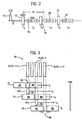
- a pulse/acquisition sequence is shown according to the present invention. As indicated, the sequence is gated to an ECG signal 70.
- the presaturation RF pulses must be slice selective and, preferably, the acquisitions interleaved.
- interleaving There are there are two types of interleaving. The first is interleaving of the preparation pulses and data acquisitions, as demonstrated in Fig. 2 (i.e., prep, acq, prep, acq). The second involves an interleaving of the slice acquisitions, as in acquiring data from every other slice, or every third slice, or some other such variation, as will be further discussed.
- the term is limited to the latter definition.
- each RF (prep) pulse 72, 74, 76 occurring at times t 1 , t 3 and t 5 , respectively, is followed by an MR data acquisition 78, 80, and 82, at times t 2 , t 4 , and t 6 , respectively.
- each prep pulse includes a dephasing or spoiling gradient.
- Each data acquisition 90 follows an RF saturation pulse, but in order to attain a longer TI time, a notched RF saturation pulse is used, as best shown in Fig. 3, wherein the profiles of the saturation pulses 72, 74, and 76 are shown spatially with respect to a plurality of slice locations 84.
- Each of the saturation pulses 72-76 have a stop-band 86 between a pair of pass-bands 88 so that the spins of the next slice location to be scanned are located within the stop-band of the pulse and are unaffected by the RF saturation pulse.
- spins within the pass-bands 88 are effectively saturated, which is the converse of a conventional RF excitation pulse.
- the magnetization of the spins within the immediate slice are not perturbed and only the spins that are outside the slice location experience this saturation pulse.
- Image acquisition of the next slice will not commence until after the next notched RF pulse is transmitted.
- the spins in the next slice will have recovered for a time TI, Fig. 2, equivalent to the time of an image acquisition segment.
- the present invention contemplates interleaving of the presaturation RF pulses, while providing saturation of blood over a large volume outside of a slice thickness defined by the size of the stop-band 86, Fig. 3.
- Such advantages allow for the use a 90° flip angle for the presaturation RF pulse for maximum dynamic range of contrast enhancement and results in minimizing the effects of arrhythmias and other variations in the patient's heart rate because the magnetization is allowed to recover for a fixed period, as defined by the TI time from a state where the longitudinal magnetization is zero.
- the first slice in the sequence represents a special case.
- the first slice of the first phase has no preparation.
- Fig. 3 demonstrates a portion of the sequence in the middle of the sequence, if slice n-1 were the first sequence in the first phase of the imaging, it would have no corresponding preparation pulse.
- Subsequent phases of the first slice are prepared by the saturation pulse that preceded the last slice of the previous phase. That is, slice 1 of phase 2 is saturated by the preparation pulse preceding the last slice of phase 1. Consequently, the time between the preparation and acquisition of the first slice should span an ECG trigger.
- the TI for the first slice is therefore longer than that of the other slices and is also variable, depending on the R-R interval of the particular heart beat.
- slice n is saturated by the preparation pulse 72 at time t 1 that precedes the EPI acquisition 90 at time t 2 of slice n-1.
- Slice n is subsequently unaffected by the preparation pulse 74 at time t 3 that immediately precedes its acquisition 90 at t 4 . Therefore, the time between the preparation of each slice and the acquisition of that slice is the effective TI. In this case, TI is t 4 -t 1 .
- the slices are acquired in an interleaved manner, rather than sequentially, to allow a wider notch, or stop-band 86, to compensate for potential cardiac motion between saturation in readout. The slices are so labeled to reflect the actual temporal order of the interleaved slices.
- the width of notch 86 is a user selectable parameter, which may be input through input device 13, Fig. 1.
- the width of notch 86, Fig. 3, should be greater than the image slice thickness sought.
- the pass-bands 88, on either side of the notch 86, are fixed at approximately five times the width of notch 86 in the preferred embodiment. This is so because when the slices are interleaved (i.e., acquired in the following order: 1,3,5,7,2,4,6), there is a space of 5 slice thicknesses between the acquisitions of slices 7 and 2.
- the width requirement is that the pass-band be wide enough such that it covers the distance between current slice and the next slice.
- the RF pulse is 15 ms. in duration and is played out in the presence of a low slice-select gradient.
- a gradient crusher is then used to dephase the resulting transverse magnetization.
- the total duration of the preparation sequence is about 18 ms., which is approximately the same length as the preparation sequence of comparable conventional inversion recovery pulse sequence segments, which includes a 5 ms. RF pulse followed by a 10 ms. recovery time.
- FASTCARD-ET TM pulse sequence fast segmented k-space gradient echo acquisition with a short echo train length echo planar imaging readout
- ECG-triggering TR 5.6 ms
- flip angle 25° echotrain length (ETL) 4
- FOV 36x27 cm slice thickness 10 mm; 0-2 mm spacing
- 7 slices acquired over 2 heartbeats bandwidth ⁇ 125 kHz
- matrix 128x128 (96 k y lines)
- notch width 15 mm 1.5 times the slice thickness
- overall saturation slab width 165 mm 0.1 mmol/kg Gd contrast.
- FASTCARD-ET TM is a trademark of the General Electric Company.
- Fig. 4 shows the difference in peak enhancements for several patients between current perfusion sequences 100 and those acquired with the modified sequence of the present invention 102.
- the average peak enhancements were 92%, with a ⁇ 27% variance for the prior art perfusion sequences 100, and 235% with a 31% variance for the present inventive sequence 102.
- Fig. 5 shows time-intensity curves for the prior art perfusion sequence 104 and the present invention sequence 106.
- the inventive sequence 106 exhibits a lower baseline signal, as well as a greater dynamic range than the prior art sequence 104, even with the use of a higher excitation flip angle, such as 25° versus 12°.
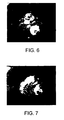
- Figs. 6 and 7 show actual results of a myocardial perfusion MRI.
- Fig. 6 shows a stress image of the same nominal slice location of a patient acquired with the perfusion sequence of the present invention.
- Fig. 7 shows that same nominal slice location in a rest image.
- the images show substantial improvement in SNR and contrast, which permits better visualization of perfusion defects.
- the regions of perfusion deficits are indicated by a dark circular arc on the lower portion of the left ventricular in the myocardium on the stress image and indicate blood flow failure under stress conditions.
- the absence of a similar dark spot in the rest image indicates that the tissue is still alive, and the severe contrast between the two images clearly indicates to the physician the specific locations of the deficits.
- an interleaved notched preparation pulse is ideal to fulfill the requirements of perfusion MRI.
- a 90° saturation angle provides immunity to arrhythmias and allows for the development of better T 1 weighting.
- the effective interleaving of the saturation pulses permits a significantly longer relaxation time TI. These effects combine to substantially improve image SNR and contrast, without sacrificing slice coverage, while providing blood pool suppression. Further improvement in SNR is achieved with the use of a higher excitation flip angle because there is more longitudinal magnetization available due to the longer TI.
- the flip angle can accordingly be increased from approximately 12° to 25° and the contrast concentration can be reduced from 0.15 mmol/kg to 0.1 mmol/kg.
- increasing the flip angle too far can result in an unsteady state having highly variable k-space weighting that can increase the amplitude of side lobes in the acquisition point spread function. This has the effect of increasing ghosting and image blurring artifacts.
- the present invention includes a method of acquiring MR data having the steps of selecting a volume of slice locations (n) in which MR data is to be acquired, and then transmitting a series of notched RF saturation pulses into the selected volume of slice locations.
- the method next includes acquiring MR data for the slice location in the stop-band of the notched RF saturation pulse.
- the step of transmitting is repeated at least for every other slice location such that a series of notched RF saturation pulses are transmitted and wherein the step of acquiring MR data is further defined as interleaving the acquisitions so as to acquire data from every other slice location during each phase, or pass through.
- the notched RF saturation pulses are designed to saturate all slice locations in the volume selected, except a slice location in which MR data is to be acquired immediately after the transmission of an RF saturation pulse.
- the notched RF saturation pulse is 90° to minimize signal intensity variations caused by arrhythmias in variations and heart rate.
- the series of transmitting and acquisition interleaving result in longer TI time while maximizing a number of slice locations.
- a width of the stop-band of the notched RF saturation pulse is greater than that of a slice location thickness and the width of the notched RF saturation pulse is a user selectable parameter.
- the pass-band of the notched RF saturation pulse saturates substantially all the volume of slice locations in the selected volume except the slice location in which MR data is to be immediately acquired next in time.
- the pass-band of the notched RF saturation pulse has a width at least five times that of the stop-band and the series of notched RF saturation pulses substantially saturates all the blood within the volume of slice locations selected for effective myocardial perfusion MR imaging.
- the invention also includes an MRI system (as defined in claim 3) to acquire MR images with increased SNR and contrast in perfusion studies that includes an MRI system having a plurality of gradient coils positioned about a bore of a magnet to impress a polarizing magnetic field and an RF transceiver system, including an RF modulator controlled by a pulse control module to transmit RF signals to an RF coil assembly to acquire MR images.
- the MRI system includes a computer program to periodically activate the MRI system and acquire a time series of MR images.
- the computer is programmed to select a volume of slice locations in which MR data is to be acquired, transmit a notched RF saturation pulse within the selected volume of slice locations, wherein the notched RF saturation pulse has a stop-band between a pair of pass-bands, and to acquire MR data in the stop-band area of the notched RF saturation pulse.
- the system is designed to reconstruct an MR image using the acquired MR data, such that the reconstructed MR image has increased SNR and increased image contrast over those acquired without a notched RF saturation pulse.
- the present invention is implemented via a computer system for use with an MRI apparatus having a computer programmed from a computer readable storage medium having thereon a computer program programmed to select a volume of slice locations in which MR data is to be acquired, transmit a notched RF saturation pulse within the selected volume of slice locations, wherein the notched RF saturation pulse has a stop-band between a pair of pass-bands, and to acquire MR data in the stop-band area of the notched RF saturation pulse.
- the degree of immunity to cardiac motion is determined by the width of the notch in the saturation pulse of the present invention.
- the width should be chosen wide enough to allow for some motion of the desired slice within the stop-band, but not so wide that the subsequent slice falls into the stop-band and avoids full saturation.
- the optimal choice is to have each notch contiguous with the prior one.
Landscapes
- Physics & Mathematics (AREA)
- Health & Medical Sciences (AREA)
- Nuclear Medicine, Radiotherapy & Molecular Imaging (AREA)
- Vascular Medicine (AREA)
- General Health & Medical Sciences (AREA)
- Radiology & Medical Imaging (AREA)
- Engineering & Computer Science (AREA)
- Signal Processing (AREA)
- High Energy & Nuclear Physics (AREA)
- Condensed Matter Physics & Semiconductors (AREA)
- General Physics & Mathematics (AREA)
- Magnetic Resonance Imaging Apparatus (AREA)
Applications Claiming Priority (2)
| Application Number | Priority Date | Filing Date | Title |
|---|---|---|---|
| US09/391,965 US6618605B1 (en) | 1999-09-08 | 1999-09-08 | Method and apparatus for MR perfusion image acquisition using a notched RF saturation pulse |
| US391965 | 1999-09-08 |
Publications (3)
| Publication Number | Publication Date |
|---|---|
| EP1083437A2 EP1083437A2 (en) | 2001-03-14 |
| EP1083437A3 EP1083437A3 (en) | 2003-05-21 |
| EP1083437B1 true EP1083437B1 (en) | 2006-06-21 |
Family
ID=23548704
Family Applications (1)
| Application Number | Title | Priority Date | Filing Date |
|---|---|---|---|
| EP00307198A Expired - Lifetime EP1083437B1 (en) | 1999-09-08 | 2000-08-22 | Method and apparatus for MR data acquisition using a notched RF saturation pulse |
Country Status (4)
| Country | Link |
|---|---|
| US (1) | US6618605B1 (enExample) |
| EP (1) | EP1083437B1 (enExample) |
| JP (1) | JP4785232B2 (enExample) |
| DE (1) | DE60028889T2 (enExample) |
Families Citing this family (22)
| Publication number | Priority date | Publication date | Assignee | Title |
|---|---|---|---|---|
| US7283862B1 (en) * | 2002-05-16 | 2007-10-16 | General Electric Company | Rapid multi-slice MR perfusion imaging with large dynamic range |
| US6836113B2 (en) * | 2003-01-22 | 2004-12-28 | Toshiba America Mri, Inc. | Measurement and correction of gradient-induced cross-term magnetic fields in an EPI sequence |
| US7319784B2 (en) * | 2003-02-05 | 2008-01-15 | National Research Council Of Canada | Magnetic resonance spectroscopy using a conformal voxel |
| US6903548B2 (en) * | 2003-10-21 | 2005-06-07 | General Electric Company | Method and apparatus for MR perfusion image acquisition using non-selective and notched RF saturation pulses |
| US7394920B2 (en) * | 2004-05-19 | 2008-07-01 | Invia, Llc | Automated computer-implemented method and system for reorienting emission computer tomographic myocardial perfusion images |
| US7064545B2 (en) * | 2004-08-30 | 2006-06-20 | General Electric Company | Method and apparatus of background suppression in MR imaging using spin locking |
| DE102004043809B4 (de) * | 2004-09-08 | 2008-01-24 | Charité-Universitätsmedizin Berlin | Verfahren zum Kalibrieren einer kontrastmittelgestützten Perfusionsbildgebung |
| US8406849B2 (en) * | 2006-03-31 | 2013-03-26 | University Of Utah Research Foundation | Systems and methods for magnetic resonance imaging |
| US7642776B2 (en) * | 2006-09-27 | 2010-01-05 | Siemens Aktiengesellschaft | Method to determine an acquisition sequence in an imaging method for generation of 2D slice images |
| US8831703B2 (en) * | 2006-10-23 | 2014-09-09 | The General Hospital Corporation | Selective MR imaging of segmented anatomy |
| JP5121219B2 (ja) * | 2006-12-07 | 2013-01-16 | ジーイー・メディカル・システムズ・グローバル・テクノロジー・カンパニー・エルエルシー | 磁気共鳴イメージング装置および磁気共鳴イメージング方法 |
| JP2009160342A (ja) * | 2008-01-10 | 2009-07-23 | Ge Medical Systems Global Technology Co Llc | 磁気共鳴イメージング装置、rfパルスの送信方法およびプログラム |
| JP5624346B2 (ja) * | 2009-04-03 | 2014-11-12 | 株式会社東芝 | 磁気共鳴イメージング装置 |
| DE102009019592B4 (de) * | 2009-04-30 | 2014-02-20 | Siemens Aktiengesellschaft | Verfahren zur getriggerten Messung an einem Magnetresonanztomograhiegerät sowie ein Magnetresonanztomographiegerät hierfür |
| US8970217B1 (en) | 2010-04-14 | 2015-03-03 | Hypres, Inc. | System and method for noise reduction in magnetic resonance imaging |
| US9151815B2 (en) * | 2010-11-15 | 2015-10-06 | Kabushiki Kaisha Toshiba | Magnetic resonance imaging apparatus and magnetic resonance imaging method |
| GB2494169A (en) * | 2011-09-01 | 2013-03-06 | Univ Sheffield | Nuclear magnetic resonance spectroscopy using an excitation RF spectrum with a spectral gap |
| US8803523B2 (en) * | 2012-02-03 | 2014-08-12 | The Board Of Trustees Of The Leland Stanford Junior University | Flexible ordering for multiple slice MRI |
| JP6580818B2 (ja) * | 2014-09-30 | 2019-09-25 | キヤノンメディカルシステムズ株式会社 | 磁気共鳴イメージング装置 |
| DE102015219932B4 (de) | 2015-09-30 | 2019-06-06 | Siemens Healthcare Gmbh | Beschleunigte Aufnahme von Magnetresonanzdaten |
| KR101820302B1 (ko) | 2015-12-21 | 2018-01-19 | 삼성전자주식회사 | 자기 공명 영상 장치 및 그 제어 방법 |
| CN107536609B (zh) * | 2016-06-28 | 2020-06-19 | 上海联影医疗科技有限公司 | 磁共振成像设备及磁共振成像的扫描方法 |
Family Cites Families (16)
| Publication number | Priority date | Publication date | Assignee | Title |
|---|---|---|---|---|
| US4531094A (en) * | 1982-02-09 | 1985-07-23 | Oxford Research Systems Limited | Methods and apparatus of obtaining NMR spectra |
| US4715383B1 (en) | 1986-11-10 | 1995-10-31 | Mayo Medical Resources | Method for reducing artifacts in NMR images |
| US5054489A (en) * | 1988-10-06 | 1991-10-08 | The Trustees Of The University Of Pennsylvania | Magnetic resonance imaging using spatial modulation of magnetization |
| JPH02255126A (ja) * | 1989-03-29 | 1990-10-15 | Toshiba Corp | 磁気共鳴イメージング装置 |
| JPH0321224A (ja) * | 1989-06-20 | 1991-01-30 | Mitsubishi Electric Corp | 磁気共鳴映像法 |
| JPH03149031A (ja) | 1989-11-06 | 1991-06-25 | Jeol Ltd | 局所磁気共鳴スペクトル測定方法 |
| JP3169400B2 (ja) | 1991-09-24 | 2001-05-21 | ジーイー横河メディカルシステム株式会社 | Mri装置 |
| JPH05277084A (ja) * | 1992-03-31 | 1993-10-26 | Shimadzu Corp | Mrイメージング法 |
| JPH06254076A (ja) * | 1993-03-02 | 1994-09-13 | Toshiba Corp | Mri装置における血流画像撮影方法 |
| DE4327325C1 (de) * | 1993-08-13 | 1995-01-12 | Siemens Ag | Verfahren zur zeitaufgelösten MR-Bildgebung |
| US5492124A (en) * | 1994-03-23 | 1996-02-20 | Siemens Medical Systems, Inc. | Method and apparatus for improved MR angiography for use in regions where bloodflow is regurgitated |
| US5908386A (en) | 1995-12-14 | 1999-06-01 | Regents Of The Universotiy Of Minnesota | Fast MRI for assessment of myocardial perfusion with arrythmia insensitive magnetization preparation |
| US5821752A (en) * | 1996-07-15 | 1998-10-13 | General Electric Company | Real-time RF pulse construction for NMR measurement sequences |
| JP4040742B2 (ja) * | 1997-03-28 | 2008-01-30 | 株式会社東芝 | Mri装置 |
| US6137290A (en) * | 1998-02-19 | 2000-10-24 | General Electric Company | Magnetic resonance spectroscopic imaging having reduced chemical shift error |
| US6078175A (en) * | 1998-10-26 | 2000-06-20 | General Electric Company | Acquistion of segmented cardiac gated MRI perfusion images |
-
1999
- 1999-09-08 US US09/391,965 patent/US6618605B1/en not_active Expired - Lifetime
-
2000
- 2000-08-22 EP EP00307198A patent/EP1083437B1/en not_active Expired - Lifetime
- 2000-08-22 DE DE60028889T patent/DE60028889T2/de not_active Expired - Lifetime
- 2000-09-07 JP JP2000270908A patent/JP4785232B2/ja not_active Expired - Lifetime
Non-Patent Citations (1)
| Title |
|---|
| WOLFF S.D. ET AL: "Assessment of First-Pass Myocardial Perfusion Imaging during Rest and Adenosine Stress: Comparison with Cardiac Catheterization", PROCEEDINGS OF THE SEVENTH SCIENTIFIC MEETING OF THE INTERNATIONAL SOCIETY FOR MAGNETIC RESONANCE IN MEDICINE,, 22 May 1999 (1999-05-22), Philadelphia * |
Also Published As
| Publication number | Publication date |
|---|---|
| EP1083437A3 (en) | 2003-05-21 |
| JP2001120518A (ja) | 2001-05-08 |
| DE60028889T2 (de) | 2007-01-25 |
| EP1083437A2 (en) | 2001-03-14 |
| JP4785232B2 (ja) | 2011-10-05 |
| DE60028889D1 (de) | 2006-08-03 |
| US6618605B1 (en) | 2003-09-09 |
Similar Documents
| Publication | Publication Date | Title |
|---|---|---|
| EP1083437B1 (en) | Method and apparatus for MR data acquisition using a notched RF saturation pulse | |
| JP4536853B2 (ja) | 核磁気共鳴データを取得する方法及び磁気共鳴システム | |
| US6094591A (en) | Measurement of coronary flow reserve with MR oximetry | |
| US7047060B1 (en) | Multiple preparatory excitations and readouts distributed over the cardiac cycle | |
| US6493569B2 (en) | Method and apparatus using post contrast-enhanced steady-state free precession in MR imaging | |
| JP2926110B2 (ja) | 傾斜およびスピンエコー(grase)イメージングを用いた超高速多重セクション全身mri | |
| US6393313B1 (en) | Producing a phase contrast MR image from a partial Fourier data acquisition | |
| US6498946B1 (en) | Efficient multi-slice acquisition with black blood contrast | |
| US5429134A (en) | Multi-phase fat suppressed MRI cardiac imaging | |
| US8588889B2 (en) | Method and apparatus for breath-held MR data acquisition using interleaved acquisition | |
| US5377680A (en) | MRI cardiac image produced by temporal data sharing | |
| US5545992A (en) | Fast cardiac gated NMR acquisition with improved T1 contrast | |
| US7412277B1 (en) | Multi-slice MR data acquisition in successive heartbeats with black blood contrast | |
| US7432706B2 (en) | Magnetic resonance imaging using blood flow navigation | |
| US7064545B2 (en) | Method and apparatus of background suppression in MR imaging using spin locking | |
| US7689263B1 (en) | Method and apparatus for acquiring free-breathing MR images using navigator echo with saturation RF pulse | |
| US7880465B2 (en) | Method and apparatus for contrast inflow dynamic MR angiography | |
| US6889071B2 (en) | Acquisition of high-temporal free-breathing MR images | |
| US6144200A (en) | Acquisition of segmented MRI cardiac data using an EPI pulse sequence | |
| US7546155B2 (en) | Efficient multi-slice acquisition with black blood contrast in fast spin echo imaging | |
| US7283862B1 (en) | Rapid multi-slice MR perfusion imaging with large dynamic range | |
| US7330028B2 (en) | Apparatus and method of simultaneous fat suppression, magnetization transfer contrast, and spatial saturation for 3D time-of-flight imaging | |
| US20090060841A1 (en) | Apparatus and method for combined use of variable flip angles and centric phase encoding in hyperpolarized 13c imaging | |
| US8928317B2 (en) | System and method for controlling apparent timing dependencies for T2-weighted MRI imaging | |
| US6903548B2 (en) | Method and apparatus for MR perfusion image acquisition using non-selective and notched RF saturation pulses |
Legal Events
| Date | Code | Title | Description |
|---|---|---|---|
| PUAI | Public reference made under article 153(3) epc to a published international application that has entered the european phase |
Free format text: ORIGINAL CODE: 0009012 |
|
| AK | Designated contracting states |
Kind code of ref document: A2 Designated state(s): AT BE CH CY DE DK ES FI FR GB GR IE IT LI LU MC NL PT SE |
|
| AX | Request for extension of the european patent |
Free format text: AL;LT;LV;MK;RO;SI |
|
| PUAL | Search report despatched |
Free format text: ORIGINAL CODE: 0009013 |
|
| AK | Designated contracting states |
Designated state(s): AT BE CH CY DE DK ES FI FR GB GR IE IT LI LU MC NL PT SE |
|
| AX | Request for extension of the european patent |
Extension state: AL LT LV MK RO SI |
|
| 17P | Request for examination filed |
Effective date: 20031121 |
|
| AKX | Designation fees paid |
Designated state(s): DE NL |
|
| 17Q | First examination report despatched |
Effective date: 20041007 |
|
| GRAP | Despatch of communication of intention to grant a patent |
Free format text: ORIGINAL CODE: EPIDOSNIGR1 |
|
| RTI1 | Title (correction) |
Free format text: METHOD AND APPARATUS FOR MR DATA ACQUISITION USING A NOTCHED RF SATURATION PULSE |
|
| GRAS | Grant fee paid |
Free format text: ORIGINAL CODE: EPIDOSNIGR3 |
|
| GRAA | (expected) grant |
Free format text: ORIGINAL CODE: 0009210 |
|
| AK | Designated contracting states |
Kind code of ref document: B1 Designated state(s): DE NL |
|
| REF | Corresponds to: |
Ref document number: 60028889 Country of ref document: DE Date of ref document: 20060803 Kind code of ref document: P |
|
| PLBE | No opposition filed within time limit |
Free format text: ORIGINAL CODE: 0009261 |
|
| STAA | Information on the status of an ep patent application or granted ep patent |
Free format text: STATUS: NO OPPOSITION FILED WITHIN TIME LIMIT |
|
| 26N | No opposition filed |
Effective date: 20070322 |
|
| PGFP | Annual fee paid to national office [announced via postgrant information from national office to epo] |
Ref country code: NL Payment date: 20160826 Year of fee payment: 17 |
|
| PGFP | Annual fee paid to national office [announced via postgrant information from national office to epo] |
Ref country code: DE Payment date: 20160826 Year of fee payment: 17 |
|
| REG | Reference to a national code |
Ref country code: DE Ref legal event code: R119 Ref document number: 60028889 Country of ref document: DE |
|
| REG | Reference to a national code |
Ref country code: NL Ref legal event code: MM Effective date: 20170901 |
|
| PG25 | Lapsed in a contracting state [announced via postgrant information from national office to epo] |
Ref country code: NL Free format text: LAPSE BECAUSE OF NON-PAYMENT OF DUE FEES Effective date: 20170901 |
|
| PG25 | Lapsed in a contracting state [announced via postgrant information from national office to epo] |
Ref country code: DE Free format text: LAPSE BECAUSE OF NON-PAYMENT OF DUE FEES Effective date: 20180301 |