EP1042492B1 - Humanized monoclonal antibodies that protect against shiga toxin induced disease - Google Patents
Humanized monoclonal antibodies that protect against shiga toxin induced disease Download PDFInfo
- Publication number
- EP1042492B1 EP1042492B1 EP98965434A EP98965434A EP1042492B1 EP 1042492 B1 EP1042492 B1 EP 1042492B1 EP 98965434 A EP98965434 A EP 98965434A EP 98965434 A EP98965434 A EP 98965434A EP 1042492 B1 EP1042492 B1 EP 1042492B1
- Authority
- EP
- European Patent Office
- Prior art keywords
- antibody
- humanized monoclonal
- monoclonal antibody
- binds
- variable region
- Prior art date
- Legal status (The legal status is an assumption and is not a legal conclusion. Google has not performed a legal analysis and makes no representation as to the accuracy of the status listed.)
- Expired - Lifetime
Links
- 108010079723 Shiga Toxin Proteins 0.000 title claims abstract description 45
- 201000010099 disease Diseases 0.000 title abstract description 13
- 208000037265 diseases, disorders, signs and symptoms Diseases 0.000 title abstract description 13
- 241001529936 Murinae Species 0.000 claims abstract description 20
- 238000011282 treatment Methods 0.000 claims abstract description 14
- 230000002265 prevention Effects 0.000 claims abstract description 7
- 241000588724 Escherichia coli Species 0.000 claims description 31
- 108010090763 Shiga Toxin 2 Proteins 0.000 claims description 27
- 108010091769 Shiga Toxin 1 Proteins 0.000 claims description 25
- 208000015181 infectious disease Diseases 0.000 claims description 17
- 238000004519 manufacturing process Methods 0.000 claims description 17
- 239000008194 pharmaceutical composition Substances 0.000 claims description 14
- 241000894006 Bacteria Species 0.000 claims description 12
- 108090000623 proteins and genes Proteins 0.000 claims description 12
- 102000009786 Immunoglobulin Constant Regions Human genes 0.000 claims description 10
- 108010009817 Immunoglobulin Constant Regions Proteins 0.000 claims description 10
- 102000006496 Immunoglobulin Heavy Chains Human genes 0.000 claims description 10
- 108010019476 Immunoglobulin Heavy Chains Proteins 0.000 claims description 10
- 102000013463 Immunoglobulin Light Chains Human genes 0.000 claims description 10
- 108010065825 Immunoglobulin Light Chains Proteins 0.000 claims description 10
- 239000000203 mixture Substances 0.000 claims description 10
- 208000037157 Azotemia Diseases 0.000 claims description 7
- 239000000427 antigen Substances 0.000 claims description 7
- 108091007433 antigens Proteins 0.000 claims description 7
- 102000036639 antigens Human genes 0.000 claims description 7
- 239000003814 drug Substances 0.000 claims description 7
- 102000004169 proteins and genes Human genes 0.000 claims description 7
- 208000009852 uremia Diseases 0.000 claims description 7
- 239000003085 diluting agent Substances 0.000 claims description 6
- 230000002949 hemolytic effect Effects 0.000 claims description 6
- 208000011580 syndromic disease Diseases 0.000 claims description 6
- 239000003937 drug carrier Substances 0.000 claims description 2
- 108700012359 toxins Proteins 0.000 abstract description 35
- 239000003053 toxin Substances 0.000 abstract description 34
- 231100000765 toxin Toxicity 0.000 abstract description 34
- 239000013598 vector Substances 0.000 abstract description 32
- 239000013604 expression vector Substances 0.000 abstract description 17
- 238000002360 preparation method Methods 0.000 abstract description 5
- 244000052616 bacterial pathogen Species 0.000 abstract 1
- 108020004414 DNA Proteins 0.000 description 59
- 239000013612 plasmid Substances 0.000 description 55
- 239000012634 fragment Substances 0.000 description 50
- 108091034117 Oligonucleotide Proteins 0.000 description 42
- 210000004027 cell Anatomy 0.000 description 40
- 241000699666 Mus <mouse, genus> Species 0.000 description 39
- 241000699670 Mus sp. Species 0.000 description 23
- 239000000047 product Substances 0.000 description 23
- 238000010367 cloning Methods 0.000 description 20
- 238000000034 method Methods 0.000 description 20
- 108010047041 Complementarity Determining Regions Proteins 0.000 description 17
- 238000003556 assay Methods 0.000 description 15
- 239000013615 primer Substances 0.000 description 13
- 208000032759 Hemolytic-Uremic Syndrome Diseases 0.000 description 12
- 108091028043 Nucleic acid sequence Proteins 0.000 description 12
- 241000607764 Shigella dysenteriae Species 0.000 description 11
- FAPWRFPIFSIZLT-UHFFFAOYSA-M Sodium chloride Chemical compound [Na+].[Cl-] FAPWRFPIFSIZLT-UHFFFAOYSA-M 0.000 description 11
- 238000010276 construction Methods 0.000 description 11
- 229940007046 shigella dysenteriae Drugs 0.000 description 11
- 238000001890 transfection Methods 0.000 description 11
- 108010017898 Shiga Toxins Proteins 0.000 description 10
- IAZDPXIOMUYVGZ-UHFFFAOYSA-N Dimethylsulphoxide Chemical compound CS(C)=O IAZDPXIOMUYVGZ-UHFFFAOYSA-N 0.000 description 9
- 238000002965 ELISA Methods 0.000 description 9
- 230000027455 binding Effects 0.000 description 9
- 238000006243 chemical reaction Methods 0.000 description 9
- 210000004408 hybridoma Anatomy 0.000 description 9
- RAXXELZNTBOGNW-UHFFFAOYSA-N imidazole Natural products C1=CNC=N1 RAXXELZNTBOGNW-UHFFFAOYSA-N 0.000 description 9
- 239000002299 complementary DNA Substances 0.000 description 8
- 230000000694 effects Effects 0.000 description 8
- 238000000746 purification Methods 0.000 description 8
- 230000016784 immunoglobulin production Effects 0.000 description 7
- 229940126619 mouse monoclonal antibody Drugs 0.000 description 7
- 108091032973 (ribonucleotides)n+m Proteins 0.000 description 6
- 108020005345 3' Untranslated Regions Proteins 0.000 description 6
- 239000011780 sodium chloride Substances 0.000 description 6
- UCSJYZPVAKXKNQ-HZYVHMACSA-N streptomycin Chemical compound CN[C@H]1[C@H](O)[C@@H](O)[C@H](CO)O[C@H]1O[C@@H]1[C@](C=O)(O)[C@H](C)O[C@H]1O[C@@H]1[C@@H](NC(N)=N)[C@H](O)[C@@H](NC(N)=N)[C@H](O)[C@H]1O UCSJYZPVAKXKNQ-HZYVHMACSA-N 0.000 description 6
- 241000283707 Capra Species 0.000 description 5
- 108091035707 Consensus sequence Proteins 0.000 description 5
- 239000000872 buffer Substances 0.000 description 5
- LOKCTEFSRHRXRJ-UHFFFAOYSA-I dipotassium trisodium dihydrogen phosphate hydrogen phosphate dichloride Chemical compound P(=O)(O)(O)[O-].[K+].P(=O)(O)([O-])[O-].[Na+].[Na+].[Cl-].[K+].[Cl-].[Na+] LOKCTEFSRHRXRJ-UHFFFAOYSA-I 0.000 description 5
- 238000011534 incubation Methods 0.000 description 5
- 210000004962 mammalian cell Anatomy 0.000 description 5
- 238000006386 neutralization reaction Methods 0.000 description 5
- 239000002953 phosphate buffered saline Substances 0.000 description 5
- 239000006228 supernatant Substances 0.000 description 5
- 210000003501 vero cell Anatomy 0.000 description 5
- 108020004635 Complementary DNA Proteins 0.000 description 4
- 241000282412 Homo Species 0.000 description 4
- TWRXJAOTZQYOKJ-UHFFFAOYSA-L Magnesium chloride Chemical compound [Mg+2].[Cl-].[Cl-] TWRXJAOTZQYOKJ-UHFFFAOYSA-L 0.000 description 4
- 238000012408 PCR amplification Methods 0.000 description 4
- 229920001213 Polysorbate 20 Polymers 0.000 description 4
- 239000013599 cloning vector Substances 0.000 description 4
- 239000011248 coating agent Substances 0.000 description 4
- 238000000576 coating method Methods 0.000 description 4
- 230000007423 decrease Effects 0.000 description 4
- 238000010790 dilution Methods 0.000 description 4
- 239000012895 dilution Substances 0.000 description 4
- 230000001900 immune effect Effects 0.000 description 4
- 230000036039 immunity Effects 0.000 description 4
- 239000003550 marker Substances 0.000 description 4
- 235000020030 perry Nutrition 0.000 description 4
- 235000010486 polyoxyethylene sorbitan monolaurate Nutrition 0.000 description 4
- 239000000256 polyoxyethylene sorbitan monolaurate Substances 0.000 description 4
- 230000004044 response Effects 0.000 description 4
- 239000000243 solution Substances 0.000 description 4
- 238000012360 testing method Methods 0.000 description 4
- NFGXHKASABOEEW-UHFFFAOYSA-N 1-methylethyl 11-methoxy-3,7,11-trimethyl-2,4-dodecadienoate Chemical compound COC(C)(C)CCCC(C)CC=CC(C)=CC(=O)OC(C)C NFGXHKASABOEEW-UHFFFAOYSA-N 0.000 description 3
- QKNYBSVHEMOAJP-UHFFFAOYSA-N 2-amino-2-(hydroxymethyl)propane-1,3-diol;hydron;chloride Chemical compound Cl.OCC(N)(CO)CO QKNYBSVHEMOAJP-UHFFFAOYSA-N 0.000 description 3
- 238000011767 DBA/2J (JAX™ mouse strain) Methods 0.000 description 3
- 206010012741 Diarrhoea haemorrhagic Diseases 0.000 description 3
- 206010014896 Enterocolitis haemorrhagic Diseases 0.000 description 3
- 229930193140 Neomycin Natural products 0.000 description 3
- 229930006000 Sucrose Natural products 0.000 description 3
- CZMRCDWAGMRECN-UGDNZRGBSA-N Sucrose Chemical compound O[C@H]1[C@H](O)[C@@H](CO)O[C@@]1(CO)O[C@@H]1[C@H](O)[C@@H](O)[C@H](O)[C@@H](CO)O1 CZMRCDWAGMRECN-UGDNZRGBSA-N 0.000 description 3
- 238000002835 absorbance Methods 0.000 description 3
- OHDRQQURAXLVGJ-HLVWOLMTSA-N azane;(2e)-3-ethyl-2-[(e)-(3-ethyl-6-sulfo-1,3-benzothiazol-2-ylidene)hydrazinylidene]-1,3-benzothiazole-6-sulfonic acid Chemical compound [NH4+].[NH4+].S/1C2=CC(S([O-])(=O)=O)=CC=C2N(CC)C\1=N/N=C1/SC2=CC(S([O-])(=O)=O)=CC=C2N1CC OHDRQQURAXLVGJ-HLVWOLMTSA-N 0.000 description 3
- 230000001580 bacterial effect Effects 0.000 description 3
- 150000001875 compounds Chemical class 0.000 description 3
- 239000013613 expression plasmid Substances 0.000 description 3
- 235000013305 food Nutrition 0.000 description 3
- 235000015220 hamburgers Nutrition 0.000 description 3
- 238000011577 humanized mouse model Methods 0.000 description 3
- 238000002649 immunization Methods 0.000 description 3
- 230000003053 immunization Effects 0.000 description 3
- 238000002347 injection Methods 0.000 description 3
- 239000007924 injection Substances 0.000 description 3
- 229960004927 neomycin Drugs 0.000 description 3
- 108091008146 restriction endonucleases Proteins 0.000 description 3
- 229960005322 streptomycin Drugs 0.000 description 3
- 239000000758 substrate Substances 0.000 description 3
- 239000005720 sucrose Substances 0.000 description 3
- 230000024033 toxin binding Effects 0.000 description 3
- OOIBFPKQHULHSQ-UHFFFAOYSA-N (3-hydroxy-1-adamantyl) 2-methylprop-2-enoate Chemical compound C1C(C2)CC3CC2(O)CC1(OC(=O)C(=C)C)C3 OOIBFPKQHULHSQ-UHFFFAOYSA-N 0.000 description 2
- UZOVYGYOLBIAJR-UHFFFAOYSA-N 4-isocyanato-4'-methyldiphenylmethane Chemical compound C1=CC(C)=CC=C1CC1=CC=C(N=C=O)C=C1 UZOVYGYOLBIAJR-UHFFFAOYSA-N 0.000 description 2
- 108010077805 Bacterial Proteins Proteins 0.000 description 2
- BVKZGUZCCUSVTD-UHFFFAOYSA-M Bicarbonate Chemical compound OC([O-])=O BVKZGUZCCUSVTD-UHFFFAOYSA-M 0.000 description 2
- 239000003155 DNA primer Substances 0.000 description 2
- 206010012735 Diarrhoea Diseases 0.000 description 2
- KCXVZYZYPLLWCC-UHFFFAOYSA-N EDTA Chemical compound OC(=O)CN(CC(O)=O)CCN(CC(O)=O)CC(O)=O KCXVZYZYPLLWCC-UHFFFAOYSA-N 0.000 description 2
- 238000012286 ELISA Assay Methods 0.000 description 2
- 102100034343 Integrase Human genes 0.000 description 2
- JGSARLDLIJGVTE-MBNYWOFBSA-N Penicillin G Chemical compound N([C@H]1[C@H]2SC([C@@H](N2C1=O)C(O)=O)(C)C)C(=O)CC1=CC=CC=C1 JGSARLDLIJGVTE-MBNYWOFBSA-N 0.000 description 2
- 206010035226 Plasma cell myeloma Diseases 0.000 description 2
- 238000010802 RNA extraction kit Methods 0.000 description 2
- 108010092799 RNA-directed DNA polymerase Proteins 0.000 description 2
- PXIPVTKHYLBLMZ-UHFFFAOYSA-N Sodium azide Chemical compound [Na+].[N-]=[N+]=[N-] PXIPVTKHYLBLMZ-UHFFFAOYSA-N 0.000 description 2
- 239000007983 Tris buffer Substances 0.000 description 2
- 150000001413 amino acids Chemical class 0.000 description 2
- 239000003242 anti bacterial agent Substances 0.000 description 2
- 229940088710 antibiotic agent Drugs 0.000 description 2
- 230000037396 body weight Effects 0.000 description 2
- 238000010804 cDNA synthesis Methods 0.000 description 2
- 230000009260 cross reactivity Effects 0.000 description 2
- 239000012228 culture supernatant Substances 0.000 description 2
- 230000001472 cytotoxic effect Effects 0.000 description 2
- 238000002784 cytotoxicity assay Methods 0.000 description 2
- 231100000263 cytotoxicity test Toxicity 0.000 description 2
- 230000034994 death Effects 0.000 description 2
- 231100000517 death Toxicity 0.000 description 2
- 229940079593 drug Drugs 0.000 description 2
- 238000004520 electroporation Methods 0.000 description 2
- 108010030074 endodeoxyribonuclease MluI Proteins 0.000 description 2
- 238000005516 engineering process Methods 0.000 description 2
- 235000013410 fast food Nutrition 0.000 description 2
- 230000036541 health Effects 0.000 description 2
- 238000005304 joining Methods 0.000 description 2
- 231100000518 lethal Toxicity 0.000 description 2
- 230000001665 lethal effect Effects 0.000 description 2
- 239000006166 lysate Substances 0.000 description 2
- 229910001629 magnesium chloride Inorganic materials 0.000 description 2
- 230000001404 mediated effect Effects 0.000 description 2
- 238000002703 mutagenesis Methods 0.000 description 2
- 231100000350 mutagenesis Toxicity 0.000 description 2
- 201000000050 myeloid neoplasm Diseases 0.000 description 2
- 230000001124 posttranscriptional effect Effects 0.000 description 2
- 238000003757 reverse transcription PCR Methods 0.000 description 2
- 239000003161 ribonuclease inhibitor Substances 0.000 description 2
- 238000012163 sequencing technique Methods 0.000 description 2
- 238000002741 site-directed mutagenesis Methods 0.000 description 2
- 210000004988 splenocyte Anatomy 0.000 description 2
- 238000006467 substitution reaction Methods 0.000 description 2
- 238000013518 transcription Methods 0.000 description 2
- 230000035897 transcription Effects 0.000 description 2
- 230000002103 transcriptional effect Effects 0.000 description 2
- 238000011269 treatment regimen Methods 0.000 description 2
- LENZDBCJOHFCAS-UHFFFAOYSA-N tris Chemical compound OCC(N)(CO)CO LENZDBCJOHFCAS-UHFFFAOYSA-N 0.000 description 2
- 238000011144 upstream manufacturing Methods 0.000 description 2
- NGNQZCDZXSOVQU-UHFFFAOYSA-N 8,16,18,26,34,36-hexahydroxyhentetracontane-2,6,10,14,24,28,32-heptone Chemical compound CCCCCC(O)CC(O)CC(=O)CCCC(=O)CC(O)CC(=O)CCCCCC(O)CC(O)CC(=O)CCCC(=O)CC(O)CC(=O)CCCC(C)=O NGNQZCDZXSOVQU-UHFFFAOYSA-N 0.000 description 1
- 206010003445 Ascites Diseases 0.000 description 1
- 241000283690 Bos taurus Species 0.000 description 1
- 108091003079 Bovine Serum Albumin Proteins 0.000 description 1
- 241000282472 Canis lupus familiaris Species 0.000 description 1
- 238000001712 DNA sequencing Methods 0.000 description 1
- 239000006144 Dulbecco’s modified Eagle's medium Substances 0.000 description 1
- 241000283086 Equidae Species 0.000 description 1
- 241000588722 Escherichia Species 0.000 description 1
- 241001646716 Escherichia coli K-12 Species 0.000 description 1
- 208000010201 Exanthema Diseases 0.000 description 1
- 241000282326 Felis catus Species 0.000 description 1
- 108010010803 Gelatin Proteins 0.000 description 1
- 108060003951 Immunoglobulin Proteins 0.000 description 1
- 108010025815 Kanamycin Kinase Proteins 0.000 description 1
- 241000124008 Mammalia Species 0.000 description 1
- 101100370002 Mus musculus Tnfsf14 gene Proteins 0.000 description 1
- 102000005717 Myeloma Proteins Human genes 0.000 description 1
- 108010045503 Myeloma Proteins Proteins 0.000 description 1
- 206010028980 Neoplasm Diseases 0.000 description 1
- 206010030113 Oedema Diseases 0.000 description 1
- 206010048685 Oral infection Diseases 0.000 description 1
- 241000283984 Rodentia Species 0.000 description 1
- 241000607768 Shigella Species 0.000 description 1
- UIIMBOGNXHQVGW-UHFFFAOYSA-M Sodium bicarbonate Chemical compound [Na+].OC([O-])=O UIIMBOGNXHQVGW-UHFFFAOYSA-M 0.000 description 1
- 201000007023 Thrombotic Thrombocytopenic Purpura Diseases 0.000 description 1
- 108060008682 Tumor Necrosis Factor Proteins 0.000 description 1
- 241000251539 Vertebrata <Metazoa> Species 0.000 description 1
- JLCPHMBAVCMARE-UHFFFAOYSA-N [3-[[3-[[3-[[3-[[3-[[3-[[3-[[3-[[3-[[3-[[3-[[5-(2-amino-6-oxo-1H-purin-9-yl)-3-[[3-[[3-[[3-[[3-[[3-[[5-(2-amino-6-oxo-1H-purin-9-yl)-3-[[5-(2-amino-6-oxo-1H-purin-9-yl)-3-hydroxyoxolan-2-yl]methoxy-hydroxyphosphoryl]oxyoxolan-2-yl]methoxy-hydroxyphosphoryl]oxy-5-(5-methyl-2,4-dioxopyrimidin-1-yl)oxolan-2-yl]methoxy-hydroxyphosphoryl]oxy-5-(6-aminopurin-9-yl)oxolan-2-yl]methoxy-hydroxyphosphoryl]oxy-5-(6-aminopurin-9-yl)oxolan-2-yl]methoxy-hydroxyphosphoryl]oxy-5-(6-aminopurin-9-yl)oxolan-2-yl]methoxy-hydroxyphosphoryl]oxy-5-(6-aminopurin-9-yl)oxolan-2-yl]methoxy-hydroxyphosphoryl]oxyoxolan-2-yl]methoxy-hydroxyphosphoryl]oxy-5-(5-methyl-2,4-dioxopyrimidin-1-yl)oxolan-2-yl]methoxy-hydroxyphosphoryl]oxy-5-(4-amino-2-oxopyrimidin-1-yl)oxolan-2-yl]methoxy-hydroxyphosphoryl]oxy-5-(5-methyl-2,4-dioxopyrimidin-1-yl)oxolan-2-yl]methoxy-hydroxyphosphoryl]oxy-5-(5-methyl-2,4-dioxopyrimidin-1-yl)oxolan-2-yl]methoxy-hydroxyphosphoryl]oxy-5-(6-aminopurin-9-yl)oxolan-2-yl]methoxy-hydroxyphosphoryl]oxy-5-(6-aminopurin-9-yl)oxolan-2-yl]methoxy-hydroxyphosphoryl]oxy-5-(4-amino-2-oxopyrimidin-1-yl)oxolan-2-yl]methoxy-hydroxyphosphoryl]oxy-5-(4-amino-2-oxopyrimidin-1-yl)oxolan-2-yl]methoxy-hydroxyphosphoryl]oxy-5-(4-amino-2-oxopyrimidin-1-yl)oxolan-2-yl]methoxy-hydroxyphosphoryl]oxy-5-(6-aminopurin-9-yl)oxolan-2-yl]methoxy-hydroxyphosphoryl]oxy-5-(4-amino-2-oxopyrimidin-1-yl)oxolan-2-yl]methyl [5-(6-aminopurin-9-yl)-2-(hydroxymethyl)oxolan-3-yl] hydrogen phosphate Polymers Cc1cn(C2CC(OP(O)(=O)OCC3OC(CC3OP(O)(=O)OCC3OC(CC3O)n3cnc4c3nc(N)[nH]c4=O)n3cnc4c3nc(N)[nH]c4=O)C(COP(O)(=O)OC3CC(OC3COP(O)(=O)OC3CC(OC3COP(O)(=O)OC3CC(OC3COP(O)(=O)OC3CC(OC3COP(O)(=O)OC3CC(OC3COP(O)(=O)OC3CC(OC3COP(O)(=O)OC3CC(OC3COP(O)(=O)OC3CC(OC3COP(O)(=O)OC3CC(OC3COP(O)(=O)OC3CC(OC3COP(O)(=O)OC3CC(OC3COP(O)(=O)OC3CC(OC3COP(O)(=O)OC3CC(OC3COP(O)(=O)OC3CC(OC3COP(O)(=O)OC3CC(OC3COP(O)(=O)OC3CC(OC3COP(O)(=O)OC3CC(OC3CO)n3cnc4c(N)ncnc34)n3ccc(N)nc3=O)n3cnc4c(N)ncnc34)n3ccc(N)nc3=O)n3ccc(N)nc3=O)n3ccc(N)nc3=O)n3cnc4c(N)ncnc34)n3cnc4c(N)ncnc34)n3cc(C)c(=O)[nH]c3=O)n3cc(C)c(=O)[nH]c3=O)n3ccc(N)nc3=O)n3cc(C)c(=O)[nH]c3=O)n3cnc4c3nc(N)[nH]c4=O)n3cnc4c(N)ncnc34)n3cnc4c(N)ncnc34)n3cnc4c(N)ncnc34)n3cnc4c(N)ncnc34)O2)c(=O)[nH]c1=O JLCPHMBAVCMARE-UHFFFAOYSA-N 0.000 description 1
- 238000007792 addition Methods 0.000 description 1
- 238000013019 agitation Methods 0.000 description 1
- 102000006646 aminoglycoside phosphotransferase Human genes 0.000 description 1
- 230000005875 antibody response Effects 0.000 description 1
- 238000013459 approach Methods 0.000 description 1
- 235000021168 barbecue Nutrition 0.000 description 1
- 235000015278 beef Nutrition 0.000 description 1
- 210000004369 blood Anatomy 0.000 description 1
- 239000008280 blood Substances 0.000 description 1
- 201000011510 cancer Diseases 0.000 description 1
- 239000000969 carrier Substances 0.000 description 1
- 210000003169 central nervous system Anatomy 0.000 description 1
- 238000005119 centrifugation Methods 0.000 description 1
- 239000003153 chemical reaction reagent Substances 0.000 description 1
- 230000001332 colony forming effect Effects 0.000 description 1
- 239000013078 crystal Substances 0.000 description 1
- 238000005520 cutting process Methods 0.000 description 1
- 230000009089 cytolysis Effects 0.000 description 1
- 231100000433 cytotoxic Toxicity 0.000 description 1
- 231100000599 cytotoxic agent Toxicity 0.000 description 1
- 230000003013 cytotoxicity Effects 0.000 description 1
- 231100000135 cytotoxicity Toxicity 0.000 description 1
- 239000002619 cytotoxin Substances 0.000 description 1
- 230000002498 deadly effect Effects 0.000 description 1
- 230000003111 delayed effect Effects 0.000 description 1
- 238000012217 deletion Methods 0.000 description 1
- 230000037430 deletion Effects 0.000 description 1
- 238000001514 detection method Methods 0.000 description 1
- 238000011161 development Methods 0.000 description 1
- 231100000673 dose–response relationship Toxicity 0.000 description 1
- 239000003651 drinking water Substances 0.000 description 1
- 235000020188 drinking water Nutrition 0.000 description 1
- 230000002242 enterotoxic effect Effects 0.000 description 1
- 238000001976 enzyme digestion Methods 0.000 description 1
- 201000005884 exanthem Diseases 0.000 description 1
- 238000002474 experimental method Methods 0.000 description 1
- 239000000284 extract Substances 0.000 description 1
- 239000012894 fetal calf serum Substances 0.000 description 1
- 239000012530 fluid Substances 0.000 description 1
- 230000037406 food intake Effects 0.000 description 1
- 238000009472 formulation Methods 0.000 description 1
- 210000001035 gastrointestinal tract Anatomy 0.000 description 1
- 229920000159 gelatin Polymers 0.000 description 1
- 239000008273 gelatin Substances 0.000 description 1
- 235000019322 gelatine Nutrition 0.000 description 1
- 235000011852 gelatine desserts Nutrition 0.000 description 1
- ZDXPYRJPNDTMRX-UHFFFAOYSA-N glutamine Natural products OC(=O)C(N)CCC(N)=O ZDXPYRJPNDTMRX-UHFFFAOYSA-N 0.000 description 1
- 230000028993 immune response Effects 0.000 description 1
- 102000018358 immunoglobulin Human genes 0.000 description 1
- 230000002458 infectious effect Effects 0.000 description 1
- 230000000968 intestinal effect Effects 0.000 description 1
- 238000010253 intravenous injection Methods 0.000 description 1
- 238000002955 isolation Methods 0.000 description 1
- 210000003734 kidney Anatomy 0.000 description 1
- 231100000225 lethality Toxicity 0.000 description 1
- 238000002483 medication Methods 0.000 description 1
- 239000002609 medium Substances 0.000 description 1
- 239000012528 membrane Substances 0.000 description 1
- 238000012986 modification Methods 0.000 description 1
- 230000004048 modification Effects 0.000 description 1
- 230000002969 morbid Effects 0.000 description 1
- 230000001459 mortal effect Effects 0.000 description 1
- 239000013642 negative control Substances 0.000 description 1
- 230000003472 neutralizing effect Effects 0.000 description 1
- 230000000474 nursing effect Effects 0.000 description 1
- 238000005457 optimization Methods 0.000 description 1
- 230000001717 pathogenic effect Effects 0.000 description 1
- 229940056360 penicillin g Drugs 0.000 description 1
- 230000000144 pharmacologic effect Effects 0.000 description 1
- 238000003752 polymerase chain reaction Methods 0.000 description 1
- 239000013641 positive control Substances 0.000 description 1
- 125000002924 primary amino group Chemical group [H]N([H])* 0.000 description 1
- 230000037452 priming Effects 0.000 description 1
- 230000008569 process Effects 0.000 description 1
- 230000000069 prophylactic effect Effects 0.000 description 1
- 230000001681 protective effect Effects 0.000 description 1
- 238000000159 protein binding assay Methods 0.000 description 1
- 238000010791 quenching Methods 0.000 description 1
- 206010037844 rash Diseases 0.000 description 1
- 239000011535 reaction buffer Substances 0.000 description 1
- 238000011160 research Methods 0.000 description 1
- 238000012552 review Methods 0.000 description 1
- 239000006152 selective media Substances 0.000 description 1
- 238000013207 serial dilution Methods 0.000 description 1
- 239000012679 serum free medium Substances 0.000 description 1
- 239000012536 storage buffer Substances 0.000 description 1
- 208000024891 symptom Diseases 0.000 description 1
- 230000009885 systemic effect Effects 0.000 description 1
- 229940124597 therapeutic agent Drugs 0.000 description 1
- 230000001225 therapeutic effect Effects 0.000 description 1
- 238000002560 therapeutic procedure Methods 0.000 description 1
- 206010043554 thrombocytopenia Diseases 0.000 description 1
- 239000003104 tissue culture media Substances 0.000 description 1
- 231100000419 toxicity Toxicity 0.000 description 1
- 230000001988 toxicity Effects 0.000 description 1
- 102000003390 tumor necrosis factor Human genes 0.000 description 1
- 229960005486 vaccine Drugs 0.000 description 1
- 239000011534 wash buffer Substances 0.000 description 1
- 230000003442 weekly effect Effects 0.000 description 1
Images


Classifications
-
- C—CHEMISTRY; METALLURGY
- C07—ORGANIC CHEMISTRY
- C07K—PEPTIDES
- C07K16/00—Immunoglobulins [IGs], e.g. monoclonal or polyclonal antibodies
- C07K16/12—Immunoglobulins [IGs], e.g. monoclonal or polyclonal antibodies against material from bacteria
- C07K16/1203—Immunoglobulins [IGs], e.g. monoclonal or polyclonal antibodies against material from bacteria from Gram-negative bacteria
- C07K16/1228—Immunoglobulins [IGs], e.g. monoclonal or polyclonal antibodies against material from bacteria from Gram-negative bacteria from Enterobacteriaceae (F), e.g. Citrobacter, Serratia, Proteus, Providencia, Morganella, Yersinia
- C07K16/1232—Immunoglobulins [IGs], e.g. monoclonal or polyclonal antibodies against material from bacteria from Gram-negative bacteria from Enterobacteriaceae (F), e.g. Citrobacter, Serratia, Proteus, Providencia, Morganella, Yersinia from Escherichia (G)
-
- A—HUMAN NECESSITIES
- A61—MEDICAL OR VETERINARY SCIENCE; HYGIENE
- A61P—SPECIFIC THERAPEUTIC ACTIVITY OF CHEMICAL COMPOUNDS OR MEDICINAL PREPARATIONS
- A61P1/00—Drugs for disorders of the alimentary tract or the digestive system
- A61P1/02—Stomatological preparations, e.g. drugs for caries, aphtae, periodontitis
-
- A—HUMAN NECESSITIES
- A61—MEDICAL OR VETERINARY SCIENCE; HYGIENE
- A61P—SPECIFIC THERAPEUTIC ACTIVITY OF CHEMICAL COMPOUNDS OR MEDICINAL PREPARATIONS
- A61P1/00—Drugs for disorders of the alimentary tract or the digestive system
- A61P1/04—Drugs for disorders of the alimentary tract or the digestive system for ulcers, gastritis or reflux esophagitis, e.g. antacids, inhibitors of acid secretion, mucosal protectants
-
- A—HUMAN NECESSITIES
- A61—MEDICAL OR VETERINARY SCIENCE; HYGIENE
- A61P—SPECIFIC THERAPEUTIC ACTIVITY OF CHEMICAL COMPOUNDS OR MEDICINAL PREPARATIONS
- A61P13/00—Drugs for disorders of the urinary system
- A61P13/12—Drugs for disorders of the urinary system of the kidneys
-
- A—HUMAN NECESSITIES
- A61—MEDICAL OR VETERINARY SCIENCE; HYGIENE
- A61P—SPECIFIC THERAPEUTIC ACTIVITY OF CHEMICAL COMPOUNDS OR MEDICINAL PREPARATIONS
- A61P19/00—Drugs for skeletal disorders
- A61P19/02—Drugs for skeletal disorders for joint disorders, e.g. arthritis, arthrosis
-
- A—HUMAN NECESSITIES
- A61—MEDICAL OR VETERINARY SCIENCE; HYGIENE
- A61P—SPECIFIC THERAPEUTIC ACTIVITY OF CHEMICAL COMPOUNDS OR MEDICINAL PREPARATIONS
- A61P31/00—Antiinfectives, i.e. antibiotics, antiseptics, chemotherapeutics
- A61P31/04—Antibacterial agents
-
- A—HUMAN NECESSITIES
- A61—MEDICAL OR VETERINARY SCIENCE; HYGIENE
- A61P—SPECIFIC THERAPEUTIC ACTIVITY OF CHEMICAL COMPOUNDS OR MEDICINAL PREPARATIONS
- A61P37/00—Drugs for immunological or allergic disorders
- A61P37/02—Immunomodulators
-
- A—HUMAN NECESSITIES
- A61—MEDICAL OR VETERINARY SCIENCE; HYGIENE
- A61K—PREPARATIONS FOR MEDICAL, DENTAL OR TOILETRY PURPOSES
- A61K38/00—Medicinal preparations containing peptides
-
- C—CHEMISTRY; METALLURGY
- C07—ORGANIC CHEMISTRY
- C07K—PEPTIDES
- C07K2317/00—Immunoglobulins specific features
- C07K2317/20—Immunoglobulins specific features characterized by taxonomic origin
- C07K2317/24—Immunoglobulins specific features characterized by taxonomic origin containing regions, domains or residues from different species, e.g. chimeric, humanized or veneered
-
- C—CHEMISTRY; METALLURGY
- C07—ORGANIC CHEMISTRY
- C07K—PEPTIDES
- C07K2319/00—Fusion polypeptide
Definitions
- Applicants describe a family of multi-unit bacterial proteins that are associated with hemorrhagic colitis and the life-threatening sequela, hemolytic uremic syndrome. These proteins, defined as members of the "Shiga toxin family", have been utilized in the isolation and identification of murine monoclonal antibodies. Applicants further describe the construction of humanized monoclonal antibodies which incorporate the mouse variable regions. Applicants also describe antibodies to Shiga toxins or toxoids, both monoclonal and polyclonal, and their use in treating, diagnosing, and preventing of disease and infections caused by pathogenic E . coli. The preparation of humanized monoclonal antibodies to proteins of the Shiga toxin family is disclosed.
- EHEC Enterohemorrhagic Escherichia coli
- HC bloody diarrhea
- HUS hemolytic uremic syndrome
- HC and HUS are transmitted by the ingestion of contaminated food, particularly undercooked beef products, such as hamburger.
- undercooked beef products such as hamburger.
- Doyle et al., J. Appl. Environ. Microbiol. 53: 2394 (1987 ); Samadpour et al., J. Appl. Environ. Microbiol. 60: 1038 (1994 ) With the prevalence of EHEC in cattle and the subjective nature of differentiating between cooked and undercooked hamburgers, a stop at a fast food restaurant or a family barbecue can result in tragedy.
- HC and HUS appear to be mediated by the toxin produced by EHEC and Shigella dysenteriae (for a review, see O'Brien and Holmes, Microbiol. Rev., 51: 206-220 (1987 )). These bacteria produce a family of closely related cytotoxins that collectively will be called “Shiga toxins” for the purpose of this application. Shiga toxins (alternatively, “verotoxins”) have cytotoxic, and enterotoxic activity ( Strockbine, N. et al., Infect. Immun., 53: 135-140 (1986 )).
- Shiga toxins Based on the exhibition of immunological non-cross-reactivity, the Shiga toxins have been divided into two groups. (Strockbine et al., supra ). These groups have been designated Shiga toxin type 1 (Stx1) and Shiga toxin type 2 (Stx2).
- the Stx1 group contains the prototype Stx1 toxin from EHEC as well as the Shiga toxin from Shigella dysenteriae type 1.
- other types of toxins have been discovered and considered to be members of the Stx2 group. These are Stx2e, Stx2c, and Stx2d. ( Lindgren et al., Infection and Immunology, 61: 3832 (1993 ); Schmitt, C. et al., Infect. Immun., 59: 1065-1073 (1991 ); Marques, L. et al., FEMS Lett., 44: 33-38 (1987 )).
- Shiga toxin encompasses Shiga toxin and any other toxins in the Stx1 or Stx2 group or their variants.
- Stx will refer to the toxin protein itself.
- Strockbine et al. Monoclonal antibodies against Shiga-like toxins have been described in Strockbine et al., Infection and Immunity, 1985, 50: 695-700 and in Perera, et al J. Clinical Microbiol., 1988,26: 2127-2131 .
- Strockbine et al disclose that the use of the13C4 antibody neutralises the lethality for mice induced with Shiga-like toxin from E.coli H30.
- Strockbine et al further show the use of the 13C4 for the detection of bacterial colonies producing high levels of Shiga-like toxins.
- Perera et al. describes the production of five mouse monoclonal antibodies (including the antibody 11E10) that were capable of neutralising the cytotoxicity of Shiga-like toxin II.
- WO9820903 published on May 22, 1998 describes therapeutic methods for the treatment of hemolytic uremic syndrome, by administering of monoclonal antibodies, chimeric monoclonal antibodies and monospecific polyclonal antibodies specific for Shiga-like toxin.
- Methods to humanize antibodies comprise the exchange of constant regions (see for example Lobuglio et al, supra) or CDR grafting ( EP0239400 )
- the present invention relates to a pharmaceutical composition
- a pharmaceutical composition comprising a first humanized monoclonal antibody that binds to Shiga toxin type 1 (Stx1) protein and a second humanized monoclonal antibody that binds to Shiga toxin type 2 (Stx2) protein, wherein the first antibody comprises a human immunoglobulin constant region and comprises the immunoglobulin heavy chain and light chain variable regions as set forth in SEQ ID NOs: 19 and 21 and wherein the second antibody comprises a human immunoglobulin constant region and comprises the immunoglobulin heavy chain and light chain variable regions as set forth in SEQ ID NOs: 42 and 44.
- the present invention further relates to pharmaceutical composition
- pharmaceutical composition comprising a first humanized monoclonal antibody that binds to Stx1 and a second humanized monoclonal antibody that binds to Stx2 antigen, wherein the variable region of the second antibody comprises the variable region of the murine 11E10 antibody with ATCC Accession No. CRL 1907 and the variable region of the first antibody comprises the variable region of the murine 13C4 antibody with ATCC Accession No. CKL 1794.
- the first or second antibody comprises a human constant region selected from the group consisting of IgG, IgA, and IgM.
- the human constant region of the first or second monoclonal antibody is IgG.
- the human constant region of the first or second monoclonal antibody is IgG1-kappa.
- the above-described pharmaceutical composition further comprises a pharmaceutically acceptable carrier or diluent.
- the present invention further relates to the use of a composition
- a composition comprising a first humanized monoclonal antibody that binds to Stx1 protein and a second humanized monoclonal antibody that binds to Stx2 protein
- the first antibody comprises a human immunoglobulin constant region and comprises the immunoglobulin heavy chain and light chain variable regions as set forth in SEQ ID NOs: 19 and 21
- the second antibody comprises a human immunoglobulin constant region and comprises the immunoglobulin heavy chain and light chain variable regions as set forth in SEQ ID NOs: 42 and 42 in the manufacture of a medicament for the treatment or prevention of an infection resulting in hemolytic uremia syndrome caused by Enterohemorrhagic Escherichia coli or other Shiga toxin producing bacteria.
- the present invention also relates to the use of a composition
- a composition comprising a first humanized monoclonal antibody that binds to Stx1 and a second humanized monoclonal antibody that binds to Stx2 antigen, wherein the variable region of the second antibody comprises the variable region of the murine 11E10 antibody with ATCC Accession No. CRL 1907and the variable region of the first antibody comprises the variable region of the murine 13C4 antibody with ATCC Accession No. CKL 1794 in the manufacture of a medicament for the treatment or prevention of an infection resulting in hemolytic uremia syndrome caused by enterohemorrhagic Escherichia colior other Shiga toxin producing bacteria.
- the first or second antibody comprises a human constant region selected from the group consisting of IgG, IgA, and IgM.
- the human constant region of the first or second monoclonal antibody is IgG, preferably IgG1-kappa.
- mice monoclonal antibodies to Shiga toxin 1 and Shiga toxin 2
- variable region associated with Shiga toxin neutralization as well as the specific complementarity determining regions (CDRs) of the variable region
- CDRs complementarity determining regions
- Antibodies are proteinaceous structures made up of two heavy and two light chains.
- Each class has further subclasses; IgG, for example, may be IgG1, IgG2, IgG3, or IgG4, where the heavy chain is gamma 1, gamma 2, gamma 3 or gamma 4, respectively.
- the pair of light chains in these classes or subclasses may be either kappa or lambda.
- Antibodies are further divided into a constant region and a variable region.
- the carboxy-terminal ends make up the constant sequence region, while the amino terminal ends contain the variable sequence region.
- the complementarity determining regions are located which are primarily responsible for the observe antigen binding which is characteristic of antibodies.
- Humanized monoclonal antibodies mean monoclonal antibodies originally from a non-human source to which human components have been substituted.
- humanized monoclonal antibodies comprise variable regions which derive from non-human sources and constant regions which derive from human sources.
- Shiga toxin proteins refer to the family of multi-unit bacterial proteins produced by EHEC and Shigella dysenteriae which are associated with outbreaks of Shiga toxin induced diseases. Shiga toxin proteins are meant to encompass Stx of Shigella dysenteriae -type 1 and Stx type 1 and type 2, and type 2 variant toxins of E . coli.
- Shiga toxin-induced diseases of humans include bloody diarrhea, hemorrhagic colitis, hemolytic uremic syndrome, and thrombotic thrombocytopenic purpura.
- Applicants further disclose humanized monoclonal antibodies that have the same binding specificity of at least two well characterized murine monoclonal antibodies. These two monoclonal antibodies were developed in Dr. Alison O'Brien's laboratory as set forth in Strockbine, N. A. et al., Infection and Immunity, 1985, 50: 695-700 and Perera, L. P. et al., J. Clinical Microbiol., 1988,26: 2127-2131 , and have been deposited at the ATCC as set forth below.
- binding specificity is meant a level of binding sufficiently detectable in a standard binding assay to distinguish between toxin binding and non-specific background binding as exemplified by appropriate controls (for examples, see Figure 9 and Figure 11 ). Those of ordinary skill in the art can readily test for binding levels using routine skills and techniques.
- the humanized antibodies are characterized by their structural features.
- the constant region is a human constant region and the variable region derives from a rodent, preferably a mouse.
- rodent preferably a mouse.
- the four variable regions set forth in the specification, particularly in Figure 3 and Figure 6 are preferred.
- such variable regions may include modifications (i. e., deletions, additions, and substitutions) that do not appreciably diminish the characteristic binding associated with the exemplified variable regions.
- the CDRs of the variable region are employed.
- CDRs are located in the variable regions of both the light and heavy chains and are responsible for antigen binding.
- the present disclosure is not limited to the CDRs specifically set forth. Indeed, the vectors created by the applicants and described in great detail below are sufficient for use with any CDRs of non-human antibodies to Shiga toxins.
- the humanized monoclonal antibody derives its constant regions from IgG1-kappa and its variable regions from all or part of the sequences as set forth in Figures 3 and 6 .
- the invention comprises pharmaceutical compositions as claimed and described in the "summary of the invention".
- pharmaceutical compositions include but are not limited to diluents and carriers known in the art, such as saline and sucrose solutions appropriate for application to patients.
- patients refers to any susceptible mammal, such as dogs, horses, mice, etc., but is particularly preferred to mean humans.
- Methods of treatment involve the administration of a therapeutically effective mount, as well as a prophylatically effective amount, of the humanized monoclonal antibody.
- a therapeutically effective amount is a dose that ameliorates edema, thrombocytopenia, and uremia associated with EHEC-mediated HUS.
- a prophylactically effective amount is a dose that prevents exposed individuals from developing these symptoms.
- the hybridoma cell producing the"13C4"antibody (Anti-Stx1) was deposited on December 2,1987, at the American Type Culture Collection, Rockville, MD under Accession No. CRL 1794, and can be obtained from the ATCC, or, as here, from Dr. Alison O'Brien (for details of hybridoma preparation, see Strockbine, N. A. et al., Infection and Immunity, 50: 695-700 (1985 )).
- a vial of cells was thawed and resuspended in IMDM (Mediatech) complete media supplemented with 10% FBS (Irvine).
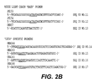
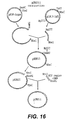
- Figure 1 shows the strategy for cloning the variable region gene fragments and Figure 2 lists the oligonucleotide primers used.
- the "13C4" total RNA (2 ⁇ g) was converted to cDNA by using Superscript 11-MMLV Reverse Transcriptase (Life Technologies) and mouse kappa (oKA57, SEQ ID NO: 57) and mouse CH1 (JS300, SEQ ID NO: 6) specific priming, according to the manufacturer's procedures.
- the first strand cDNA synthesis products were then purified using a membrane concentrator device (Amicon Centricon 30 or Millipore UltraFree 15). Of the cDNA recovered, 3 ⁇ l was used as template DNA for PCR.
- Typical PCR amplification reactions (100 ⁇ l) contained template DNA, 50 pmoles of the appropriate primers (JSS9, JSS10,JSS11,JSS12,JS153 & JSS154-SEQ ID NO: 9-12 for light chains; JSS1, JSS2, JSS3, JSS4, JSS8 and aKA-143SEQ ID NO: 1-5 and SEQ ID NO: 15 for heavy chains), 2.5 units of ExTaq polymerase (PanVera), 1x ExTaq rection buffer, 200 ⁇ M dNTP, and 2mM MgCl 2 . Each of the templates was denatured by an initial incubation at 96 °C for 1 min.
- the heavy chain products were amplified by 40 or 50 thermal cycles of 59 to 72°C for 30 sec., 72°C for 30 sec., then 96°C for I min. and a final extension step at 72°C for 5 min.
- the light chain products were amplified by 6 thermal cycles of 46,48 or 54°C for 30 sec., 72°C for 30 sec., then 96°C for 1 min followed by 35 step cycles of 60°C for 1 min, then 96°C for 1 min. and a final extension step at 72°C for 5 min.
- the PCR products from the successful reactions were purified using the Wizard PCR Purification system (Promega) as per the manufacturer's procedure.
- the heavy chain and light chain PCR products (approximately 400 bp) were then cloned into a bacterial vector for DNA sequence determination. Ligations of the PCR fragments were carried out into the pCR2.1 T/A style cloning vector (Invitrogen) following the manufacturer's procedures using 1: 1, 3: 1 and 5: 1 insert to vector molar ratios. One half of each of the ligation reactions was used to transform competent XL1 Blue cells.
- Heavy chain plasmid clones containing DNA inserts were identified using diagnostic restriction enzyme digests with EcoRI (New England Biolabs). The DNA sequence of plasmids (tKMC217B) containing inserts of the appropriate size (400 bp) were then determined. The final consensus DNA sequence of the heavy chain variable regions is shown in Figure 3 .
- the light chain PCR products were identified differently.
- the hybridoma cell line that expresses the "13C4" antibody was made by fusing mouse splenocytes with the SP2/0 myeloma cell line.
- the SP2/0 cell line transcribes a pseudogene for the kappa light chain.
- the pseudogene transcript when converted to cDNA by RT-PCR, contains an AflIII restriction site.
- Light chain candidate clones (tKMC226 & 227) were digested with AflIII (New England Biolabs) using the manufacturer's procedures to identify clones containing inserts of the appropriate size (403 bp; no AflIII site is present in these products).
- the final consensus DNA sequence of the light chain variable regions is shown in Figure 3 with the CDRs indicated by underlining.
- EXAMPLE 2 Construction of the expression vector tKMC249A.
- the heavy and light chain variable regions were then subcloned into mammalian expression plasmid vectors for the production of recombinant chimeric mouse/human antibody molecules.
- the vectors result in the production of recombinant antibody molecules under the control of CMV transcriptional promoters.
- the heavy chain molecules are direct cDNA constructs that fuse the variable region sequence directly into the human IgG 1 constant domain.
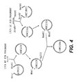
- the light chain molecules on the other hand, have a mouse kappa intron region 3' of the variable region coding fragment. After splicing, the variable region becomes fused to a human kappa constant region exon ( Figure 4 ).
- the selectable marker for the vector in mammalian cells is aminoglycoside phosphotransferase (neo), using the drug G418 (CellTech).
- Neomycin resistance serves as a dominant selectable marker for transfection of mammalian cells.
- these vectors have been designed to allow convenient cloning of any light chain variable region as EcoRV/BstBI fragment, any heavy chain variable region as a NruI/EcoRI fragment, and any heavy chain constant domain as an EcoRI/NotI fragment. These restriction sites were chosen because they occur rarely (if ever) in human and mouse variable regions.
- mice J region/kappa intron fragment fused to a human kappa exon so that after post-transcriptional splicing a mouse human chimeric kappa light chain is produced.
- vectors were designed to facilitate excision (BglII/NheI) of a whole antibody expression cassette from one vector to be ligated into a second vector cut with BamHI/NheI, creating an expression vector with the apparatus for both chains.
- the backbone of the vector was the plasmid pCDNA3 (Invitrogen). This plasmid was cut with HindIII/XhoI and a "light chain polylinker" DNA fragment was inserted to create the starting "light chain vector” pCDNA3.LCPL (see Figure 12 ). This linker contained the restriction sites HindIII, KpnI, ClaI, PmlI, EcoRV, XmaI, BamHI and XhoI to facilitate subsequent cloning steps.
- a SmaII/BclI DNA fragment containing a light chain leader, mouse anti-CKMB kappa light chain genomic fragment, and 3'UTR was cloned into the EcoRV/BamHI sites of pCDNA3.LCPL.
- the mouse kappa intron, exon, and 3'UTR in this fragment was derived from LCPXK2 received from Dr. Richard Near ( Near, R. I. et al., 1990, Mol. Immunol. 27: 901-909 ).
- Mutagenesis was then performed to eliminate an NruI (209), MluI (229), and BstBI (2962) and to introduce an NheI (1229) and a BamHI (1214) site to create the plasmid pCDNA3mut.
- LCPL. LCVK see Figure 12 ).
- a second "heavy chain vector" pCDNA3mut.HCPL was constructed from the pCDNA3mut.LCPL.LCVK plasmid by replacing the light chain expression region (HindIII/XhoI) with a "heavy chain polylinker" consisting of restriction sites HpaI, BspEI, EcoRV, KpnI, and XhoI. This plasmid was digested with EcorRV and KpnI. A SmaI/KpnI digested DNA fragment containing a heavy chain leader and an anti-CKMB IgG2b mouse heavy chain genomic fragment was then ligated into the EcoRV/KpnI digested plasmid.
- a KpnI/SalI oligonucleotide fragment containing a 3'UTR and a NotI upstream of the SalI site was subsequently cloned into the KpnI/XhoI digested plasmid, (knocking out the XhoI site), to create the plasmid pCDNA3mut.HCPL.HCV2b (see Figure 13 ).
- a human kappa light chain constant domain was then cloned into pCDNA3mut.LCPL.LCVK as a EcoNI/XhoI fragment generating the plasmid tKMC 180C2.
- a human IgG1 constant domain was cloned into PSLTN 10 as a EcoRI/NotI fragment creating the plasmid pJRS313.
- the variable regions of 13C4 were cloned into these two vectors (as described above).
- a BglII/NheI fragment from the human heavy chain vector tKMC229C was then cloned into the human light chain vector tKMC231D cut BamHI/NheI to create tKMC249A (see Figure 4 ).
- variable region gene fragments were re-amplified by PCR using primers that adapted the fragments for cloning into the expression vectors (see Figures 2 and 4 ).
- the heavy chain front primer (oKA143, SEQ ID NO: 15) includes a 5' tail that encodes the C-terminus of the heavy chain leader and an NruI restriction site for cloning, while the heavy chain reverse primer (oKA144, SEQ ID NO: 14) adds a 3' EcoRI restriction site for cloning.
- the light chain front primer (oKA145, SEQ ID NO: 16) introduces an EcoRV restriction site at the N-terminus of the light chain variable region for cloning
- the light chain reverse primer (oKA146, SEQ ID NO: 17) adds a 3' DNA sequence for the joining region-kappa exon splice junction followed by a BstBI restriction site for cloning.
- PCR reactions were performed with reagents as described above and with templates of 1-2 ng of PvuI (New England Biolabs) digested plasmid; each of these templates was denatured by an initial one minute incubation at 96°C.
- the heavy chain products were amplified by 35 thermal cycles of 55 or 60°C for 30 sec., 72°C for 30 sec., then 96°C for 1 min and a final extension step at 72°C for 5 min.
- the light chain products were amplified by 8 thermal cycles of 55 or 60°C for 30 sec., 72°C for 30 sec., then 96°C for 1 min followed by 30 step cycles of 60°C for 1 min, then 96°C for 1 min, and a final extension step at 72°C for 5 min.
- the 13C4 heavy chain PCR product (approximately 400 bp) was purified using Qiaquick PCR Purification columns (Qiagen) as described by the manufacturer's instructions and subsequently digested with NruI and EcoRI (New England Biolabs). The digested PCR products were purified using the Wizard PCR Purification system (Promega) as per manufacturer's procedure and ligated into NruI/EcoRI digested and gel-purified pJRS313, resulting in plasmid tKMC229C (see Figure 4 ). The final consensus DNA sequence of the heavy chain variable region and proper splicing of the restriction sites were confirmed in this construct.
- the 13C4 light chain PCR product (approximately 350 bp) was purified using Qiaquick PCR Purification columns (Qiagen) as described by the manufacturer's instructions and subsequently digested with EcoRV and BstBI (New England Biolabs). The digested PCR products were purified using Qiaquick PCR Purification columns (Qiagen) as per manufacturer's procedure and ligated into EcoRV/BstBI digested and gel-purified tKMC 180C2 (as described above), resulting in plasmid tKMC231D (see Figure 4 ). The final consensus DNA sequence of the light chain variable region and proper splicing of the restriction sites were confirmed in this construct.
- EXAMPLE 3 Stable production of recombinant chimeric mouse/human 13C4 antibody.
- the plasmid tKMC249A was transfected into NSO cells (Baxter International, Inc., Durant, CA) using electroporation after linearization with PvuI (New England Biolabs). 40 micrograms of the digested plasmid was mixed with 1x10⁷ cells in a total volume of 800 microliters in a 0.4 centimeter cuvette and subjected to a pulse of 250mA, 960 ⁇ F. The cells were plated out after 24 hours into 96-well tissue culture plates, 6 plates with 200 ⁇ l/well, and incubated at 37°C and 10% CO 2 . As colonies appeared, the supernatants were assayed for the production of "humanized" antibody and for the capability of the expressed antibody to bind to Stx1.
- Antibody production and activity assays for the stable transfectants were performed as described below. These assays demonstrate that the transfection of cells with this plasmid construct can result in the production of a stable cell line that produces a humanized chimeric version of the 13C4 mouse hybridoma antibody (designated H13C4).
- Antibody production ELISA assays were performed in 8-well strips from 96-well microtiter plates (Maxisorp F8; Nunc, Inc.) coated at a 1:500 dilution with Goat anti-Human IgG antibody (Pierce or Biodesign International) using a Tris-HCl coating buffer, pH 8.5. The plates were covered and incubated overnight at 4°C. Plates were then washed once with a rash storage buffer (Tris-HCl/NaCl/0.1% NaN 3 ).
- a sample/conjugate diluent Tris-HCl/NaCl/gelatin/Tween-20.
- the plates were allowed to incubate for 30 to 60 minutes on a rotator at room temperature. They were then washed three times with a wash solution (imidazole/NaCl/Tween-20).
- a goat anti-human kappa-HRP (Southern Biotechnologies) conjugate was diluted 1: 250 in the sample/conjugate diluent and 100 microliters was added to the wells. The plates were incubated on a rotator for 30 to 60 minutes at room temperature.
- the supernatants were then assayed for the ability of the expressed antibodies to bind to Stx1 protein by ELISA.
- the activity assays were preformed in 8-well strips from 96-well microtiter plates (Maxisorp F8; Nunc, Inc.) coated with approximately 0.1 ⁇ g/well purified Stx1 received from Alison O'Brien (or obtained as described in Example 7).
- the plate coating and ELISA procedure was performed in the same manner as the antibody assay above with the substitution of TMB (Kirkgaard & Perry Laboratories) for ABTS as a developing substrate.
- the absorbance value at 450 nm was determined using an automate microtiter plate ELISA reader (Ceres UV900HI, Bioteck, Winooski, Vermont).
- the original mouse monoclonal antibody 13C4 was used, and assayed with a goat anti-mouse IgG conjugate (Jackson Laboratories) at a 1: 2000 dilution.
- This assay (see Figure 9 ) demonstrates that the transfection of cells with this plasmid construct results in cells producing immunoglobulin that binds to Stx1. Neither mouse nor human IgG I K lacking the anti-Stx variable region bound the toxin.
- the hybridoma cell line producing the "11E10" antibody (Anti-Stx2) was deposited on August 1, 1990, at the American Type Culture Collection, Rockville, MD under Accession No. CRL 1907, and can be obtained from the ATCC, or, as here, from Dr. Alison O'Brien (for details of hybridoma preparation, see Perera, L. P. et al., J. Clinical Microbiol., 26: 2127-2131 (1988 )).
- a vial of the cell line was thawed, washed with serum free medium and then resuspended in IMDM (Mediatech) complete media supplemented with 10% FBS (Irvine).
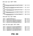
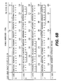
- Figure 1 shows the strategy for cloning the variable region gene fragments and Figure 5 lists the oligonucleotide primers used.
- the "11E10" total RNA (2.5 ⁇ g) was converted to cDNA by using Superscript 11-MMLV Reverse Transcriptase (Life Technologies) according to manufacturer's procedures.
- the mouse light chain (JS153, JS154, SEQ ID NO: 11 and 12) and mouse heavy chain (JS300, SEQ ID NO: 6) were used as specific primers.
- the first strand cDNA synthesis products were then purified using a Centricon-30 concentrator device (Amicon). Of the 70 ⁇ l of cDNA recovered, 3.5 ⁇ l was used as template DNA for PCR.
- Typical PCR amplification reactions (100 ⁇ l) contained template DNA, 50 pmoles of the appropriate primers (JS153, JS154 and JS009, JS010, JS011, JS012, SEQ ID NO: 7-12 for light chains, JS160, JS161, JS162 and JS001, JS002, JS003, JS004, JS008, SEQ ID NO: 28-30, SEQ ID NO: 2-5 for heavy chains), 2.5 units of ExTaq polymerase (PanVera), 1x ExTaq reaction buffer, 200 ⁇ M dNTP, and 2 mM MgCl 2 .
- the template was denatured by an initial five minute incubation at 96°C.
- the products were amplified by 35 thermal cycles of 96°C for 1 min., 55°C for 30 sec., and 72°C for 30 sec, followed by 5 min. at 72°C.
- the PCR products from the successful reactions were purified using the Wizard PCR Purification system (Promega) as per manufacturer's procedure.
- the heavy chain PCR products (approximately 400 bp) and the light chain PCR products (approximately 350 bp) were then cloned into a bacterial vector for DNA sequence determination.
- Ligations of the PCR fragments were carried out into the pCR2.1 T/A style cloning vector (Invitrogen) following the manufacturer's procedures using a 3: 1 insert to vector molar ratio.
- Two ⁇ l of the ligation reactions were used to transform the INV ⁇ F' competent cells (Invitrogen) as per the manufacturer's procedure. Plasmid clones containing DNA inserts were identified using diagnostic restriction enzyme digests, with EcoRI (New England Biolabs).
- the DNA sequence of plasmids containing the heavy chain inserts of the appropriate size (400 bp) was then determined.
- the final consensus DNA sequence of the heavy chain variable region of 11E10 is shown in Figure 6 with the CDRs indicated by underlining.
- the light chain plasmid clones needed to be further characterized because the hybridoma cell line that expresses the "11E10" antibody was made by fusing mouse splenocytes with SP20 myeloma cells.
- the SP20 cell line transcribes a pseudogene for the kappa light chain.
- the pseudogene transcript when converted to cDNA by RT-PCR, contains an AflIII restriction site.
- the plasmid clones for the light chain variable region were digestes with AflIII and those products that did not cut were then submitted for DNA sequencing.
- the final consensus sequence of the light chain variable region is shown in Figure 6 , with the CDRs indicated by underlining.
- variable region gene fragments were re-amplified by PCR using primers that adapted the fragments for cloning into the expression vector (see Figures 5 and 7 ).
- the heavy chain front primer (11E10HF, SEQ ID NO: 37) includes a 5'tail that encodes the C-terminus of the heavy chain leader and an NruI restriction site for cloning, while the heavy chain reverse primer (11E10HB, SEQ ID NO: 38) adds a 3'EcoRI restriction site for cloning.
- the light chain front primer (11E10LF, SEQ ID NO:39) includes a 5'tail that encodes the C-terminus of the light chain leader and an EcoRV restriction site at the N-terminus of the light chain variable region for cloning, while the light chain reverse primer (11E10LB, SEQ ID NO: 40) adds a 3'DNA sequence for the joining region-kappa exon splice junction followed by a BstBI restriction site for cloning.
- PCRs were performed as described above except, following a 5 min. incubation at 96°C, the PCR parameters were 30 thermal cycles of 96°C for 1 min., 62°C for 30 sec., and 70°C for 30 sec., followed by 5 min. at 72°C.
- the heavy chain variable region PCR product was then subcloned into a mammalian expression plasmid vector (pJRS315, produced as set forth below in Example 5) for production of recombinant chimeric mouse/human antibody molecules.
- the resulting vector clone was designated pACE1.
- the resulting plasmid was designated pACELC and digeste, with BstBI/EcoRV, to cut out the light chain variable region.
- the variable region was then subcloned into the mammalian expression vector containing the "11E10" heavy chain variable region (pACE1).
- the final expression vector clone was designated pACE4.
- This vector results in the production of recombinant antibody molecules under the control of the CMV transcriptional promoters.
- the heavy chain molecules are direct cDNA constructs that fuse the variable region sequence directly into the human IgG 1 constant domain.
- the light chain molecules on the other hand, have a mouse kappa intron region 3' of the variable region coding fragment. After splicing, the variable region becomes fused to a human kappa constant region exon (see Figure 7 ).
- the selectable marker for the vector in mammalian cells is neomycin (G418).
- the "1E10" heavy chain PCR product (approximately 400 bp) was digested with NruI and EcoRI (New England Biolabs), purified using a Qiaquick PCR Purification column (Qiagen), as described by the manufacturer, and ligated into NruI/EcoRI digested and gel-purified pJRS315, resulting in plasmid PACTE1 (see Figure 7 ).
- the "11E10" light chain PCR product (approximately 350 bp) was cloned into the T/A cloning vector as per manufacturer's instructions.
- pACELC The resulting clone, pACELC, was digested with EcoRV and BstBI (New England Biolabs) and the light chain fragment was gel-purified. This fragment was then ligated into the EcoRV/BstBI digested and gel-purified pACE1, resulting in plasmid pACE4 (see Figure 7 ). The sequence of the heavy and light chain variable regions was verified prior to mammalian cell transfection.
- EXAMPLE 5 Construction of the expression vector pJRS315.
- the plasmid pJRS315 is the expression plasmid into which the variable regions of the 11E10 antibody were cloned.
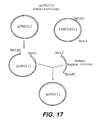
- This plasmid is a derivative of a basic expression vector, plasmid pSUN15, that contains no antibody variable region coding information, pJRS315 was created using DNA fragment ligations and site directed mutagenesis steps. The result was a vector that expresses both antibody chains with CMV promoter driven transcription (see Figure 17 ). Neomycin resistance serves as the dominant selectable marker for transfection of mammalian cells.
- the backbone of the vector was the plasmid pCDNA3 (Invitrogen). This plasmid was cut with HindIII/XhoI and a "light chain polylinker" DNA fragment was inserted to create the starting "light chain vector". This linker contained the restriction sites HindIII, KpnI, ClaI, PmlI, EcoRV, XmaI, BamHI and XhoI to facilitate subsequent cloning steps to create the plasmid pCDNA3.LCPL. A SmaI/BclI DNA fragment containing a light chain leader, anti-CKMB kappa light chain genomic fragment, and 3'UTR was cloned into the EcoRV/BamHI sites of pCDNA3.LCPL.
- the mouse kappa intron, exon and 3'UTR in this fragment was derived from LCPXK2 received from Dr. Richard Near ( Near, R. I. et al., Mol. Immunol. 27: 901-909, (1990 )). Mutagenesis was then performed to eliminate an NruI (209), MluI (229), and BstBI (2962) and to introduce an NheI (1229) and a BamHI (1214) site to create pcDNA3mut.LCPL.LCVK (see Figure 12 ).
- a second "heavy chain vector” was constructed from the pcDNA3mut.LCPL.LCVK plasmid by replacing the light chain expression region (HindIII/XhoI) with a "heavy chain polylinker” consisting of restriction sites HpaI, BspEI, EcoRV, KpnI, and XhoI. This plasmid was digested with EcorRV and KpnI. A SmaI/KpnI digested DNA fragment containing a heavy chain leader and an anti-CKMB IgG2b mouse heavy chain genomic fragment was then ligated into the EcoRV/KpnI digested plasmid.
- a KpnI/SalI oligonucleotide fragment containing a 3'UTR and a NotI upstream of the SalI site was subsequently cloned into the KpnI/XhoI digested plasmid (knocking out the XhoI site), to create the plasmid pCDNA3mut.HCPL.HCV2b (see Figure 13 ).
- a human kappa light chain constant domain was then cloned into pSUN9 as a BstBI/XhoI fragment, and a human IgG1 constant domain was cloned into pSUN10 as a EcoRI/NotI fragment.
- a BglII/NheI fragment from the human heavy chain vector was then cloned into the human light chain vector cut with BamHI/NheI to create pSUN15 (see Figure 16 ).
- the plasmid pJRS315 was then constructed using pSUN15 through the following process.
- a heavy chain variable region from another, unrelated, hybridoma cell line (approximately 400 bp) was digested with NruI and EcoRI (New England Biolabs), purified using a Qiaquick PCR Purification column (Qiagen), as described by the manufacturer, and ligated into NruI/EcoRI digested and gel-purified pSUN15, resulting in plasmid pJRS311 (see Figure 16 ).
- a BstBI/NotI (New England Biolabs) DNA fragment containing a mouse kappa J-kappa intron fragment fused to a human kappa exon fragment was digested and gel-purified from the vector tKMC180C2. This fragment was ligated into the backbone of pJRS311 digested with BstBI/NotI and gel-purified resulting in the plasmid pJRS315 (see Figure 17 ).
- EXAMPLE 6 Stable production of recombinant chimeric mouse/human "11E10" antibody.
- the plasmid pACE4 was transfected into NSO cells using electroporation.
- the plasmid was linearized with a PvuI restriction enzyme digestion.
- 40 ⁇ g of digested plasmid DNA was mixed with 7x10 6 cells in a total volume of 400 ⁇ L and incubated at room temperature with gentle agitation for one minute.
- 10 ⁇ L of DMSO (Sigma) were added to a final concentration of 1.25%.
- the cells/DNA/DMSO mix was transferred to a cold 0.4 centimeter cuvette and subjected to one pulse of 250 volts, 960 ⁇ F.
- the cells were transferred to one well of a six-well plate containing 5 ml of non-selective media supplemented with DMSO (final concentration of 1.25%). After 24 hours at 37°C and 10% CO 2 the cells were plated out into 96-well microtiter plates. As colonies appeared, the supernatants were assayed for the production of "humanized” antibody and for the capability for the expressed antibody to bind to the Stx2 toxin.
- Antibody production and activity assays for the stable transfectants were performed in 8-well strips from 96-well microtiter plates (Maxisorp F8; Nunc, Inc.) coated with a 1: 500 dilution of goat anti-Human F(ab') 2 anti-IgG antibody (Southern Biotechnology) using a bicarbonate coating buffer, pH 8.5.
- the plates were covered with pressure sensitive film (Falcon, Becton Dickinson) and incubated overnight at 4°C. Plates are then washed once with wash solution (imidazole/NaCl/0.4% Tween-20). 100 ⁇ L of culture supernatant was then applied and allowed to incubate for 30 minutes on a plate rotator at room temperature.
- the supernatants were then assayed for the ability of the expressed antibodies to bind to EHEC Stx2 toxin.
- the activity assay was performed, as above, using plates coated at 1 ⁇ g/ml with Stx2 toxin (obtained as in Example 7 from Dr. O'Brien's lab) in a bicarbonate coating buffer, pH 8.5. This assay demonstrates that the transfection of cells with this plasmid construct can result in the production of a humanized chimeric version of the 11E10 mouse monoclonal antibody which effectively binds Shiga toxin type 2 ( Figure 11 ).
- EXAMPLE 7 Verifying Biological and Immunological Efficacy of Humanized Antibodies to Shiga toxin.
- toxin was obtained from cultures of E . coli K-12 strains that contained either plasmid pLPSH3 (encodes Stx; J. Infect. Disease 164: 344-352 (1991 )) or pMJ 100 (encodes Stx2; Inf. and Immunity, 57: 3743-3750 (1989 )). Bacteria were disrupted by sonic lysis and clarified by centrifugation.
- the extracts were serially diluted in tissue culture medium (Dulbecco modified Eagle medium containing 10% fetal calf serum, 0.8 mM glutamine, 500 U of penicillin G per ml, and 500 mg of streptomycin per ml).
- tissue culture medium Dulbecco modified Eagle medium containing 10% fetal calf serum, 0.8 mM glutamine, 500 U of penicillin G per ml, and 500 mg of streptomycin per ml.
- tissue culture medium Dulbecco modified Eagle medium containing 10% fetal calf serum, 0.8 mM glutamine, 500 U of penicillin G per ml, and 500 mg of streptomycin per ml.
- One hundred microliters of 10-fold dilutions of the lysates were added to microtiter plate wells containing about 10 4 Vero cells in 100 ⁇ l of medium.
- the tissue culture cells were incubated at 37°C in 5% CO 2 for 48 hours and then fixed and stained with crystal violet.
- mice were injected intraperitoneally (0.1 ml) with antibody H13C4 (humanized ⁇ -Stx1B), 13C4 (mouse ⁇ -Stx1B), H11E10 (humanized ⁇ -Stx2A), or phosphate buffered saline (PBS). These injections were repeated at day 1. The mice were then challenge by intravenous injection with crude Stx1 toxin (obtained as described in Example 7) at doses of 1.7 x 10 5 or 1.7 x 10 6 CD 50 . These doses of toxin were chosen following preliminary experiments with varying amounts of toxin. Mice were monitored for 21 days.
- mice and bacteria Two different strains of mice and bacteria were used in these studies to test efficacy against both Stx2 and Stx2-variant.
- DBA/2J mice were challenge with EHEC strain 86-24 (0157: H7, Stx2 + ) and CD-1 mice were challenge with strain B2F1 (091: H21, Stx2-variant + ). While E. coli strain B2F1 is normally fatal to both mice trains, E.coli strain 86-24 is fatal to DBA/2J mice, while CD-1 mice will survive 86-24 infection.
- antibody H11E10 humanized ⁇ -Stx2A or 11E10 (mouse ⁇ -Stx2A) was injecte intraperitoneally (0.1 ml) into groups of four or five mice.
- Control groups included mice that had received antibody 13C4 (mouse ⁇ -Stx1B), mice that had received 11E10 ascites fluid (mouse ⁇ -Stx2A), or mice that had received PBS instead of antibody.
- Mice were given streptomycin (5g/L) in their drinking water to decrease normal intestinal flora and their food was removed. Streptomycin resistant derivatives of strains 86-24 (0157: H7, Stx2 + ) and B2F1 (091:H21, Stx2-variant + ) were grown overnight in L broth.
- mice received a second injection of test antibody, control antibody, or PBS.
- the mice were immediately fed 10 10 CFU of 86-24 that had been pelleted and resuspended in 20% sucrose or 10 3 CFU of B2F1 that had been serially diluted in 20% sucrose. Food was returned to the cage and the mice were monitored for 21 days.
- mice immunization of the mice with the either the murine or the humanized anti-Stx2 antibodies resulted in complete protection from a lethal oral dose of EHEC.
- TABLE 3 DBA/2J Mice Fed 101° CFU 0157 (Stx2) Antibody (dose/mouse) Survivors Murine 13C4 (1.4 ⁇ g) 0/5 Murine 11E10 (1.0 ⁇ g) 5/5 Humanized 11E10 (1.0 ⁇ g) 5/5
- EXAMPLE 9 Treatment of disease caused by bacteria producing Shiga toxin.
- Applicants further describe a variety of methods of treating, ameliorating, or preventing, the diseases and effects associated with exposure to Shiga toxin.
- Positive clinical responses in humans have been obtained with monoclonal antibodies, and one skilled in the art would know how to employ anti-Stx humanized monoclonal antibodies in humans. (See Fagerberg et al., Cancer Research, 55: 1824-27 (1995 ); Eur. J. Cancer, 2: 261-267 (1995 )).
- the precise dosage of humanized anti-Shiga toxin antibody administered to a patient for treatment of these diseases will vary in accordance with factors appreciated by the typical clinician. These factors include (but are not limited to) size, age, overall health, the extent of infection, and other medications being administered to said patient.
- Examples of potential patient groups would include (but not be limited to) young children with bloody diarrhea but no white cells in the stool, patients with indications of HUS, patients with positive stool sample tests for Shiga toxin, siblings or daycare cohorts in contact with a case (as a passive preventative measure), and any patient with diarrhea (not necessarily bloody) that has been in contact with an identified case.
- a typical dosage of about 5mg/kg body weight of humanized 13C4 combine with about 10 mg/kg body weight of humanized 11E10 would be contemplated.
- This combined formulation could be administered to the patient twice to ensure effectiveness. Inclusion of both types of humanized antibodies together provides assurance that the patient will be protected against all types of Shiga toxin disclosed herein.
Landscapes
- Health & Medical Sciences (AREA)
- Chemical & Material Sciences (AREA)
- Organic Chemistry (AREA)
- Medicinal Chemistry (AREA)
- General Health & Medical Sciences (AREA)
- Life Sciences & Earth Sciences (AREA)
- Public Health (AREA)
- General Chemical & Material Sciences (AREA)
- Nuclear Medicine, Radiotherapy & Molecular Imaging (AREA)
- Chemical Kinetics & Catalysis (AREA)
- Pharmacology & Pharmacy (AREA)
- Animal Behavior & Ethology (AREA)
- Veterinary Medicine (AREA)
- Immunology (AREA)
- Bioinformatics & Cheminformatics (AREA)
- Engineering & Computer Science (AREA)
- Proteomics, Peptides & Aminoacids (AREA)
- Biochemistry (AREA)
- Molecular Biology (AREA)
- Genetics & Genomics (AREA)
- Biophysics (AREA)
- Physical Education & Sports Medicine (AREA)
- Oncology (AREA)
- Rheumatology (AREA)
- Orthopedic Medicine & Surgery (AREA)
- Communicable Diseases (AREA)
- Urology & Nephrology (AREA)
- Peptides Or Proteins (AREA)
- Preparation Of Compounds By Using Micro-Organisms (AREA)
- Medicines Containing Antibodies Or Antigens For Use As Internal Diagnostic Agents (AREA)
- Micro-Organisms Or Cultivation Processes Thereof (AREA)
Applications Claiming Priority (5)
| Application Number | Priority Date | Filing Date | Title |
|---|---|---|---|
| US6863597P | 1997-12-23 | 1997-12-23 | |
| US68635P | 1997-12-23 | ||
| US09/215,163 US20030170248A1 (en) | 1997-12-23 | 1998-12-18 | Humanized monoclonal antibodies that protect against shiga toxin induced disease |
| US215163 | 1998-12-18 | ||
| PCT/US1998/027267 WO1999032645A1 (en) | 1997-12-23 | 1998-12-22 | Humanized monoclonal antibodies that protect against shiga toxin induced disease |
Publications (2)
| Publication Number | Publication Date |
|---|---|
| EP1042492A1 EP1042492A1 (en) | 2000-10-11 |
| EP1042492B1 true EP1042492B1 (en) | 2009-11-25 |
Family
ID=26749187
Family Applications (1)
| Application Number | Title | Priority Date | Filing Date |
|---|---|---|---|
| EP98965434A Expired - Lifetime EP1042492B1 (en) | 1997-12-23 | 1998-12-22 | Humanized monoclonal antibodies that protect against shiga toxin induced disease |
Country Status (11)
| Country | Link |
|---|---|
| US (6) | US20030170248A1 (enExample) |
| EP (1) | EP1042492B1 (enExample) |
| JP (1) | JP4450505B2 (enExample) |
| AT (1) | ATE449856T1 (enExample) |
| AU (1) | AU764091B2 (enExample) |
| CA (1) | CA2316411C (enExample) |
| DE (1) | DE69841331D1 (enExample) |
| DK (1) | DK1042492T3 (enExample) |
| ES (1) | ES2339618T3 (enExample) |
| PT (1) | PT1042492E (enExample) |
| WO (1) | WO1999032645A1 (enExample) |
Families Citing this family (13)
| Publication number | Priority date | Publication date | Assignee | Title |
|---|---|---|---|---|
| US20030170248A1 (en) * | 1997-12-23 | 2003-09-11 | Jeffrey R. Stinson | Humanized monoclonal antibodies that protect against shiga toxin induced disease |
| RU2217166C2 (ru) | 1998-05-20 | 2003-11-27 | Тейдзин Лимитед | Гуманизированные антитела, которые распознают веротоксин ii, и продуцирующая их линия клеток |
| US6858211B1 (en) | 1998-07-20 | 2005-02-22 | The United States Of America As Represented By The Department Of Health And Human Services | Vaccines against Escherichia coli O157 infection |
| WO2002070751A1 (en) * | 2001-03-02 | 2002-09-12 | University Of Pittsburgh Of The Commonwealth System Of Higher Education | Pcr method |
| US6875433B2 (en) * | 2002-08-23 | 2005-04-05 | The United States Of America As Represented By The Secretary Of The Army | Monoclonal antibodies and complementarity-determining regions binding to Ebola glycoprotein |
| CA2434668A1 (en) | 2003-07-04 | 2005-01-04 | Laurence Mulard | Novel approach to design glycopeptides based on o-specific polysaccharide of shigella flexneri serotype 2a |
| CN100441689C (zh) * | 2006-01-20 | 2008-12-10 | 中国人民解放军第三军医大学 | 抗ehec o157志贺样毒素ⅱa亚单位单克隆抗体5f3轻、重链可变区基因及应用 |
| EP2420246A1 (en) * | 2006-04-20 | 2012-02-22 | The Henry M. Jackson Foundation for the Advancement of Military Medicine, Inc. | Methods and compositions based on shiga toxin type 1 protein |
| US20090258010A1 (en) * | 2006-05-31 | 2009-10-15 | Thallion Pharmaceuticals, Inc. | Methods, compositions, and kits for treating shiga toxin associated conditions |
| AU2007254950A1 (en) * | 2006-05-31 | 2007-12-13 | Thallion Pharmaceuticals Incorporated | Methods, compositions, and kits for treating Shiga toxin associated conditions |
| EP2172481B1 (en) * | 2008-10-06 | 2014-10-29 | Novoplant GmbH | Proteolytically stable antibody formats |
| ES2614803T3 (es) * | 2009-01-23 | 2017-06-02 | The Henry M. Jackson Foundation For The Advancement Of Military Medicine, Inc. | Métodos y composiciones basadas en proteína tipo 2 de la toxina Shiga |
| RU2732155C1 (ru) * | 2019-09-24 | 2020-09-11 | Открытое акционерное общество "Всероссийский научный Центр молекулярной диагностики и лечения" (ОАО ВНЦМДЛ) | Гуманизированное антитело или его антигенсвязывающий фрагмент (Fab) против шига-токсинов первого и/или второго типов (варианты), композиция для лечения токсических состояний, вызванных энтерогеморрагической кишечной палочкой, содержащая указанные антитела и/или Fab |
Family Cites Families (15)
| Publication number | Priority date | Publication date | Assignee | Title |
|---|---|---|---|---|
| GB8607679D0 (en) | 1986-03-27 | 1986-04-30 | Winter G P | Recombinant dna product |
| US5164298A (en) * | 1988-06-24 | 1992-11-17 | Hsc Research Development Corporation | Verocytotoxin receptor assay |
| WO1994004679A1 (en) * | 1991-06-14 | 1994-03-03 | Genentech, Inc. | Method for making humanized antibodies |
| CA2078539C (en) * | 1991-09-18 | 2005-08-02 | Kenya Shitara | Process for producing humanized chimera antibody |
| US5747272A (en) * | 1994-02-14 | 1998-05-05 | Henry M. Jackson Foundation For The Advancement Of Military Medicine | Detection of shiga-like toxins of enterohemoragic Escherichia coli |
| US5968894A (en) * | 1994-02-22 | 1999-10-19 | Lingwood; Clifford A. | Verotoxin pharmaceutical compositions and medical treatments therewith |
| WO1998020903A1 (en) | 1996-11-15 | 1998-05-22 | Trustees Of Tufts College | Humanized neutralizing antibodies against hemolytic uremic syndrome |
| US20020160005A1 (en) * | 1996-11-15 | 2002-10-31 | Trustees Of Tufts College | Human neutralizing antibodies against hemolytic urmec syndrome |
| US7910096B2 (en) * | 1996-11-15 | 2011-03-22 | Trustees Of Tufts College | Human neutralizing antibodies against hemolytic uremic syndrome |
| US20030170248A1 (en) * | 1997-12-23 | 2003-09-11 | Jeffrey R. Stinson | Humanized monoclonal antibodies that protect against shiga toxin induced disease |
| US7807184B2 (en) * | 2003-07-21 | 2010-10-05 | Interuet International B.V. | Hybrid Shiga-like toxin |
| WO2005075647A1 (en) * | 2004-02-06 | 2005-08-18 | Nymox Corporation | Humanized antibody |
| EP1991565B1 (en) * | 2006-02-16 | 2015-01-21 | The Henry M. Jackson Foundation for the Advancement of Military Medicine, Inc. | Shiga toxoid chimeric proteins |
| EP2420246A1 (en) * | 2006-04-20 | 2012-02-22 | The Henry M. Jackson Foundation for the Advancement of Military Medicine, Inc. | Methods and compositions based on shiga toxin type 1 protein |
| US20090258010A1 (en) * | 2006-05-31 | 2009-10-15 | Thallion Pharmaceuticals, Inc. | Methods, compositions, and kits for treating shiga toxin associated conditions |
-
1998
- 1998-12-18 US US09/215,163 patent/US20030170248A1/en not_active Abandoned
- 1998-12-22 AT AT98965434T patent/ATE449856T1/de active
- 1998-12-22 JP JP2000525563A patent/JP4450505B2/ja not_active Expired - Fee Related
- 1998-12-22 DK DK98965434.8T patent/DK1042492T3/da active
- 1998-12-22 AU AU20902/99A patent/AU764091B2/en not_active Expired
- 1998-12-22 DE DE69841331T patent/DE69841331D1/de not_active Expired - Lifetime
- 1998-12-22 ES ES98965434T patent/ES2339618T3/es not_active Expired - Lifetime
- 1998-12-22 CA CA002316411A patent/CA2316411C/en not_active Expired - Lifetime
- 1998-12-22 PT PT98965434T patent/PT1042492E/pt unknown
- 1998-12-22 EP EP98965434A patent/EP1042492B1/en not_active Expired - Lifetime
- 1998-12-22 WO PCT/US1998/027267 patent/WO1999032645A1/en not_active Ceased
-
2006
- 2006-06-20 US US11/471,420 patent/US20070160607A1/en not_active Abandoned
- 2006-06-20 US US11/471,369 patent/US20070003560A1/en not_active Abandoned
-
2009
- 2009-08-06 US US12/536,940 patent/US20100189715A1/en not_active Abandoned
- 2009-12-28 US US12/647,845 patent/US20100166753A1/en not_active Abandoned
-
2013
- 2013-09-25 US US14/036,835 patent/US20140135481A1/en not_active Abandoned
Non-Patent Citations (5)
Also Published As
| Publication number | Publication date |
|---|---|
| DK1042492T3 (da) | 2010-01-18 |
| ES2339618T3 (es) | 2010-05-21 |
| CA2316411C (en) | 2009-02-17 |
| US20070160607A1 (en) | 2007-07-12 |
| JP2003521219A (ja) | 2003-07-15 |
| EP1042492A1 (en) | 2000-10-11 |
| US20070003560A1 (en) | 2007-01-04 |
| US20100166753A1 (en) | 2010-07-01 |
| US20140135481A1 (en) | 2014-05-15 |
| CA2316411A1 (en) | 1999-07-01 |
| ATE449856T1 (de) | 2009-12-15 |
| WO1999032645A1 (en) | 1999-07-01 |
| US20030170248A1 (en) | 2003-09-11 |
| PT1042492E (pt) | 2010-01-08 |
| JP4450505B2 (ja) | 2010-04-14 |
| AU764091B2 (en) | 2003-08-07 |
| AU2090299A (en) | 1999-07-12 |
| DE69841331D1 (de) | 2010-01-07 |
| US20100189715A1 (en) | 2010-07-29 |
Similar Documents
| Publication | Publication Date | Title |
|---|---|---|
| US20100166753A1 (en) | Humanized monoclonal antibodies that protect against shiga toxin induced disease | |
| ES2363689T3 (es) | Anticuerpos monoclonales y quiméricos, opsónicos y protectores, específicos para el ácido lipoteicoico de bacterías gram positivas. | |
| Tzipori et al. | Antibody therapy in the management of Shiga toxin-induced hemolytic uremic syndrome | |
| US7777017B2 (en) | Nucleic acids encoding opsonic monoclonal and chimeric antibodies specific for lipoteichoic acid of gram positive bacteria | |
| AU2018202199A1 (en) | Clostridium difficile antibodies | |
| Henrique et al. | Therapeutic antibodies against Shiga toxins: trends and perspectives | |
| JP2008179634A (ja) | グラム陽性細菌のリポタイコ酸に特異的なオプソニン性モノクローナルおよびキメラ抗体 | |
| KR20120128687A (ko) | 녹농균의 혈청형 g 지질다당류에 대한 항체 | |
| AU2013251165A1 (en) | Cross-reactive Staphylococcus aureus antibody | |
| JP2010013454A (ja) | グラム陽性細菌のペプチドグリカンに対する多機能性モノクローナル抗体 | |
| AU2003298770A2 (en) | Wall teichoic acid as a target for anti-staphylococcal therapies and vaccines | |
| Wei et al. | Blocking HIV-1 infection by chromosomal integrative expression of human CD4 on the surface of Lactobacillus acidophilus ATCC 4356 | |
| JP2005514053A6 (ja) | グラム陽性細菌のリポタイコ酸に特異的なオプソニン性モノクローナルおよびキメラ抗体 | |
| JP2018517670A (ja) | クロストリジウム・ディフィシル(Clostridium difficile)感染および関連疾患の治療方法 | |
| CN119241697B (zh) | 抗金黄色葡萄球菌铁离子调节表面决定簇b蛋白的单克隆抗体及应用 | |
| US7910096B2 (en) | Human neutralizing antibodies against hemolytic uremic syndrome | |
| Li et al. | Preparation and characterization of a neutralizing murine monoclonal antibody against tetanus toxin | |
| WO2014160098A2 (en) | Bordetella specific human recombinant antibodies and uses thereof | |
| Motley | The Role of Anti-Capsular Antibodies in Protection Against Carbapenem-Resistant Klebsiella Pneumoniae | |
| AU2006203322B2 (en) | Opsonic and protective monoclonal and chimeric antibodies specific for lipoteichoic acid of gram positive bacteria | |
| Almdni | Recombinant antibodies against Clostridium difficile toxin A | |
| HK1160872A (en) | Opsonic and protective monoclonal and chimeric antibodies specific for lipoteichoic acid of gram positive bacteria |
Legal Events
| Date | Code | Title | Description |
|---|---|---|---|
| PUAI | Public reference made under article 153(3) epc to a published international application that has entered the european phase |
Free format text: ORIGINAL CODE: 0009012 |
|
| 17P | Request for examination filed |
Effective date: 20000720 |
|
| AK | Designated contracting states |
Kind code of ref document: A1 Designated state(s): AT BE CH CY DE DK ES FI FR GB GR IE IT LI LU MC NL PT SE |
|
| RAP1 | Party data changed (applicant data changed or rights of an application transferred) |
Owner name: HENRY M. JACKSON FOUNDATION FOR THE ADVANCEMENT O Owner name: SUNOL MOLECULAR CORPORATION |
|
| RIN1 | Information on inventor provided before grant (corrected) |
Inventor name: MELTON-CELSA, ANGELA Inventor name: SCHMITT, CLARE K. Inventor name: O'BRIEN, ALISON D. Inventor name: WONG, HING Inventor name: STINSON, JEFFREY L. |
|
| RIN1 | Information on inventor provided before grant (corrected) |
Inventor name: MELTON-CELSA, ANGELA Inventor name: SCHMITT, CLARE K. Inventor name: O'BRIEN, ALISON D. Inventor name: WONG, HING Inventor name: STINSON, JEFFREY R. |
|
| GRAP | Despatch of communication of intention to grant a patent |
Free format text: ORIGINAL CODE: EPIDOSNIGR1 |
|
| GRAS | Grant fee paid |
Free format text: ORIGINAL CODE: EPIDOSNIGR3 |
|
| GRAA | (expected) grant |
Free format text: ORIGINAL CODE: 0009210 |
|
| AK | Designated contracting states |
Kind code of ref document: B1 Designated state(s): AT BE CH CY DE DK ES FI FR GB GR IE IT LI LU MC NL PT SE |
|
| REG | Reference to a national code |
Ref country code: GB Ref legal event code: FG4D |
|
| REG | Reference to a national code |
Ref country code: CH Ref legal event code: EP |
|
| REG | Reference to a national code |
Ref country code: IE Ref legal event code: FG4D |
|
| REF | Corresponds to: |
Ref document number: 69841331 Country of ref document: DE Date of ref document: 20100107 Kind code of ref document: P |
|
| REG | Reference to a national code |
Ref country code: PT Ref legal event code: SC4A Free format text: AVAILABILITY OF NATIONAL TRANSLATION Effective date: 20100104 |
|
| REG | Reference to a national code |
Ref country code: DK Ref legal event code: T3 |
|
| REG | Reference to a national code |
Ref country code: CH Ref legal event code: NV Representative=s name: ROSENICH PAUL; GISLER CHRISTIAN PATENTBUERO PAUL R |
|
| REG | Reference to a national code |
Ref country code: SE Ref legal event code: TRGR |
|
| REG | Reference to a national code |
Ref country code: ES Ref legal event code: FG2A Ref document number: 2339618 Country of ref document: ES Kind code of ref document: T3 |
|
| PG25 | Lapsed in a contracting state [announced via postgrant information from national office to epo] |
Ref country code: CY Free format text: LAPSE BECAUSE OF FAILURE TO SUBMIT A TRANSLATION OF THE DESCRIPTION OR TO PAY THE FEE WITHIN THE PRESCRIBED TIME-LIMIT Effective date: 20091125 |
|
| PG25 | Lapsed in a contracting state [announced via postgrant information from national office to epo] |
Ref country code: MC Free format text: LAPSE BECAUSE OF NON-PAYMENT OF DUE FEES Effective date: 20100701 |
|
| PLBE | No opposition filed within time limit |
Free format text: ORIGINAL CODE: 0009261 |
|
| STAA | Information on the status of an ep patent application or granted ep patent |
Free format text: STATUS: NO OPPOSITION FILED WITHIN TIME LIMIT |
|
| PG25 | Lapsed in a contracting state [announced via postgrant information from national office to epo] |
Ref country code: GR Free format text: LAPSE BECAUSE OF FAILURE TO SUBMIT A TRANSLATION OF THE DESCRIPTION OR TO PAY THE FEE WITHIN THE PRESCRIBED TIME-LIMIT Effective date: 20100226 |
|
| 26N | No opposition filed |
Effective date: 20100826 |
|
| PG25 | Lapsed in a contracting state [announced via postgrant information from national office to epo] |
Ref country code: LU Free format text: LAPSE BECAUSE OF NON-PAYMENT OF DUE FEES Effective date: 20091222 |
|
| PGFP | Annual fee paid to national office [announced via postgrant information from national office to epo] |
Ref country code: DK Payment date: 20121122 Year of fee payment: 15 |
|
| PGFP | Annual fee paid to national office [announced via postgrant information from national office to epo] |
Ref country code: IE Payment date: 20121126 Year of fee payment: 15 Ref country code: FI Payment date: 20121122 Year of fee payment: 15 |
|
| PGFP | Annual fee paid to national office [announced via postgrant information from national office to epo] |
Ref country code: PT Payment date: 20121204 Year of fee payment: 15 Ref country code: SE Payment date: 20121123 Year of fee payment: 15 |
|
| PGFP | Annual fee paid to national office [announced via postgrant information from national office to epo] |
Ref country code: AT Payment date: 20121122 Year of fee payment: 15 Ref country code: NL Payment date: 20121120 Year of fee payment: 15 |
|
| REG | Reference to a national code |
Ref country code: PT Ref legal event code: MM4A Free format text: LAPSE DUE TO NON-PAYMENT OF FEES Effective date: 20140623 |
|
| REG | Reference to a national code |
Ref country code: DK Ref legal event code: EBP Effective date: 20131231 |
|
| REG | Reference to a national code |
Ref country code: NL Ref legal event code: V1 Effective date: 20140701 |
|
| REG | Reference to a national code |
Ref country code: SE Ref legal event code: EUG |
|
| REG | Reference to a national code |
Ref country code: AT Ref legal event code: MM01 Ref document number: 449856 Country of ref document: AT Kind code of ref document: T Effective date: 20131222 |
|
| PG25 | Lapsed in a contracting state [announced via postgrant information from national office to epo] |
Ref country code: FI Free format text: LAPSE BECAUSE OF NON-PAYMENT OF DUE FEES Effective date: 20131222 Ref country code: PT Free format text: LAPSE BECAUSE OF NON-PAYMENT OF DUE FEES Effective date: 20140623 Ref country code: SE Free format text: LAPSE BECAUSE OF NON-PAYMENT OF DUE FEES Effective date: 20131223 |
|
| REG | Reference to a national code |
Ref country code: IE Ref legal event code: MM4A |
|
| PG25 | Lapsed in a contracting state [announced via postgrant information from national office to epo] |
Ref country code: IE Free format text: LAPSE BECAUSE OF NON-PAYMENT OF DUE FEES Effective date: 20131222 Ref country code: NL Free format text: LAPSE BECAUSE OF NON-PAYMENT OF DUE FEES Effective date: 20140701 |
|
| PG25 | Lapsed in a contracting state [announced via postgrant information from national office to epo] |
Ref country code: AT Free format text: LAPSE BECAUSE OF NON-PAYMENT OF DUE FEES Effective date: 20131222 |
|
| PG25 | Lapsed in a contracting state [announced via postgrant information from national office to epo] |
Ref country code: DK Free format text: LAPSE BECAUSE OF NON-PAYMENT OF DUE FEES Effective date: 20131231 |
|
| REG | Reference to a national code |
Ref country code: FR Ref legal event code: PLFP Year of fee payment: 18 |
|
| PGFP | Annual fee paid to national office [announced via postgrant information from national office to epo] |
Ref country code: CH Payment date: 20151228 Year of fee payment: 18 Ref country code: GB Payment date: 20151229 Year of fee payment: 18 |
|
| PGFP | Annual fee paid to national office [announced via postgrant information from national office to epo] |
Ref country code: FR Payment date: 20151217 Year of fee payment: 18 Ref country code: ES Payment date: 20151228 Year of fee payment: 18 |
|
| PGFP | Annual fee paid to national office [announced via postgrant information from national office to epo] |
Ref country code: DE Payment date: 20151229 Year of fee payment: 18 |
|
| PGFP | Annual fee paid to national office [announced via postgrant information from national office to epo] |
Ref country code: BE Payment date: 20151228 Year of fee payment: 18 |
|
| PG25 | Lapsed in a contracting state [announced via postgrant information from national office to epo] |
Ref country code: IT Free format text: LAPSE BECAUSE OF NON-PAYMENT OF DUE FEES Effective date: 20151222 |
|
| PG25 | Lapsed in a contracting state [announced via postgrant information from national office to epo] |
Ref country code: BE Free format text: LAPSE BECAUSE OF NON-PAYMENT OF DUE FEES Effective date: 20161231 |
|
| REG | Reference to a national code |
Ref country code: DE Ref legal event code: R119 Ref document number: 69841331 Country of ref document: DE |
|
| REG | Reference to a national code |
Ref country code: CH Ref legal event code: PL |
|
| GBPC | Gb: european patent ceased through non-payment of renewal fee |
Effective date: 20161222 |
|
| PG25 | Lapsed in a contracting state [announced via postgrant information from national office to epo] |
Ref country code: IT Free format text: LAPSE BECAUSE OF NON-PAYMENT OF DUE FEES Effective date: 20151222 |
|
| PGFP | Annual fee paid to national office [announced via postgrant information from national office to epo] |
Ref country code: IT Payment date: 20151222 Year of fee payment: 18 |
|
| PGRI | Patent reinstated in contracting state [announced from national office to epo] |
Ref country code: IT Effective date: 20170710 |
|
| REG | Reference to a national code |
Ref country code: FR Ref legal event code: ST Effective date: 20170831 |
|
| PG25 | Lapsed in a contracting state [announced via postgrant information from national office to epo] |
Ref country code: FR Free format text: LAPSE BECAUSE OF NON-PAYMENT OF DUE FEES Effective date: 20170102 Ref country code: LI Free format text: LAPSE BECAUSE OF NON-PAYMENT OF DUE FEES Effective date: 20161231 Ref country code: CH Free format text: LAPSE BECAUSE OF NON-PAYMENT OF DUE FEES Effective date: 20161231 Ref country code: IT Free format text: LAPSE BECAUSE OF NON-PAYMENT OF DUE FEES Effective date: 20161222 |
|
| PGRI | Patent reinstated in contracting state [announced from national office to epo] |
Ref country code: IT Effective date: 20170710 |
|
| PG25 | Lapsed in a contracting state [announced via postgrant information from national office to epo] |
Ref country code: DE Free format text: LAPSE BECAUSE OF NON-PAYMENT OF DUE FEES Effective date: 20170701 Ref country code: GB Free format text: LAPSE BECAUSE OF NON-PAYMENT OF DUE FEES Effective date: 20161222 |
|
| REG | Reference to a national code |
Ref country code: BE Ref legal event code: MM Effective date: 20161231 |
|
| PG25 | Lapsed in a contracting state [announced via postgrant information from national office to epo] |
Ref country code: ES Free format text: LAPSE BECAUSE OF FAILURE TO SUBMIT A TRANSLATION OF THE DESCRIPTION OR TO PAY THE FEE WITHIN THE PRESCRIBED TIME-LIMIT Effective date: 20091125 |
|
| REG | Reference to a national code |
Ref country code: ES Ref legal event code: FD2A Effective date: 20181112 |
|
| PG25 | Lapsed in a contracting state [announced via postgrant information from national office to epo] |
Ref country code: ES Free format text: LAPSE BECAUSE OF FAILURE TO SUBMIT A TRANSLATION OF THE DESCRIPTION OR TO PAY THE FEE WITHIN THE PRESCRIBED TIME-LIMIT Effective date: 20161223 |