EP1029241B1 - Verfahren zur parallelen bestimmung der zellvitalität und gentransfer-effizienz - Google Patents
Verfahren zur parallelen bestimmung der zellvitalität und gentransfer-effizienz Download PDFInfo
- Publication number
- EP1029241B1 EP1029241B1 EP98962216A EP98962216A EP1029241B1 EP 1029241 B1 EP1029241 B1 EP 1029241B1 EP 98962216 A EP98962216 A EP 98962216A EP 98962216 A EP98962216 A EP 98962216A EP 1029241 B1 EP1029241 B1 EP 1029241B1
- Authority
- EP
- European Patent Office
- Prior art keywords
- determination
- cells
- efficiency
- galactosidase
- beta
- Prior art date
- Legal status (The legal status is an assumption and is not a legal conclusion. Google has not performed a legal analysis and makes no representation as to the accuracy of the status listed.)
- Expired - Lifetime
Links
- 238000000034 method Methods 0.000 title claims abstract description 34
- 108090000623 proteins and genes Proteins 0.000 title claims abstract description 23
- 238000012546 transfer Methods 0.000 title claims abstract description 15
- 238000012360 testing method Methods 0.000 title claims description 39
- 230000009977 dual effect Effects 0.000 title claims description 7
- 210000004027 cell Anatomy 0.000 title abstract description 68
- 210000003527 eukaryotic cell Anatomy 0.000 title abstract description 6
- 230000000694 effects Effects 0.000 claims abstract description 25
- 108700008625 Reporter Genes Proteins 0.000 claims abstract description 21
- 238000006243 chemical reaction Methods 0.000 claims abstract description 18
- 102000004190 Enzymes Human genes 0.000 claims abstract description 7
- 108090000790 Enzymes Proteins 0.000 claims abstract description 7
- 102000005936 beta-Galactosidase Human genes 0.000 claims description 47
- 108010005774 beta-Galactosidase Proteins 0.000 claims description 47
- 239000000872 buffer Substances 0.000 claims description 44
- 102000013563 Acid Phosphatase Human genes 0.000 claims description 38
- 108010051457 Acid Phosphatase Proteins 0.000 claims description 38
- GZCWLCBFPRFLKL-UHFFFAOYSA-N 1-prop-2-ynoxypropan-2-ol Chemical compound CC(O)COCC#C GZCWLCBFPRFLKL-UHFFFAOYSA-N 0.000 claims description 36
- 239000000758 substrate Substances 0.000 claims description 25
- XZKIHKMTEMTJQX-UHFFFAOYSA-N 4-Nitrophenyl Phosphate Chemical compound OP(O)(=O)OC1=CC=C([N+]([O-])=O)C=C1 XZKIHKMTEMTJQX-UHFFFAOYSA-N 0.000 claims description 20
- 238000003556 assay Methods 0.000 claims description 20
- 230000006037 cell lysis Effects 0.000 claims description 9
- 229910019142 PO4 Inorganic materials 0.000 claims description 6
- -1 naphthyl phosphate Chemical compound 0.000 claims description 6
- 239000010452 phosphate Substances 0.000 claims description 6
- 238000003571 reporter gene assay Methods 0.000 claims description 2
- 230000004075 alteration Effects 0.000 claims 2
- 150000002500 ions Chemical class 0.000 claims 2
- 239000002253 acid Substances 0.000 claims 1
- IAZDPXIOMUYVGZ-UHFFFAOYSA-N Dimethylsulphoxide Chemical compound CS(C)=O IAZDPXIOMUYVGZ-UHFFFAOYSA-N 0.000 description 31
- 239000012981 Hank's balanced salt solution Substances 0.000 description 29
- 230000014509 gene expression Effects 0.000 description 26
- 229920004890 Triton X-100 Polymers 0.000 description 18
- 239000013504 Triton X-100 Substances 0.000 description 18
- BTJIUGUIPKRLHP-UHFFFAOYSA-N 4-nitrophenol Chemical compound OC1=CC=C([N+]([O-])=O)C=C1 BTJIUGUIPKRLHP-UHFFFAOYSA-N 0.000 description 16
- 239000007795 chemical reaction product Substances 0.000 description 16
- UYYRDZGZGNYVBA-VPXCCNNISA-N (2s,3r,4s,5r,6r)-2-[2-chloro-4-[3-(3-chloro-4-hydroxyphenyl)-1,1-dioxo-2,1$l^{6}-benzoxathiol-3-yl]phenoxy]-6-(hydroxymethyl)oxane-3,4,5-triol Chemical compound O[C@@H]1[C@@H](O)[C@@H](O)[C@@H](CO)O[C@H]1OC1=CC=C(C2(C3=CC=CC=C3S(=O)(=O)O2)C=2C=C(Cl)C(O)=CC=2)C=C1Cl UYYRDZGZGNYVBA-VPXCCNNISA-N 0.000 description 13
- KUWPCJHYPSUOFW-YBXAARCKSA-N 2-nitrophenyl beta-D-galactoside Chemical compound O[C@@H]1[C@@H](O)[C@@H](O)[C@@H](CO)O[C@H]1OC1=CC=CC=C1[N+]([O-])=O KUWPCJHYPSUOFW-YBXAARCKSA-N 0.000 description 13
- 230000003287 optical effect Effects 0.000 description 13
- JKMHFZQWWAIEOD-UHFFFAOYSA-N 2-[4-(2-hydroxyethyl)piperazin-1-yl]ethanesulfonic acid Chemical compound OCC[NH+]1CCN(CCS([O-])(=O)=O)CC1 JKMHFZQWWAIEOD-UHFFFAOYSA-N 0.000 description 10
- 239000007995 HEPES buffer Substances 0.000 description 10
- 238000010521 absorption reaction Methods 0.000 description 10
- 238000011534 incubation Methods 0.000 description 9
- LOKCTEFSRHRXRJ-UHFFFAOYSA-I dipotassium trisodium dihydrogen phosphate hydrogen phosphate dichloride Chemical compound P(=O)(O)(O)[O-].[K+].P(=O)(O)([O-])[O-].[Na+].[Na+].[Cl-].[K+].[Cl-].[Na+] LOKCTEFSRHRXRJ-UHFFFAOYSA-I 0.000 description 8
- 238000005259 measurement Methods 0.000 description 8
- 239000002953 phosphate buffered saline Substances 0.000 description 8
- 231100000419 toxicity Toxicity 0.000 description 8
- 230000001988 toxicity Effects 0.000 description 8
- 238000004113 cell culture Methods 0.000 description 7
- 239000002609 medium Substances 0.000 description 7
- IQUPABOKLQSFBK-UHFFFAOYSA-N 2-nitrophenol Chemical compound OC1=CC=CC=C1[N+]([O-])=O IQUPABOKLQSFBK-UHFFFAOYSA-N 0.000 description 6
- VMHLLURERBWHNL-UHFFFAOYSA-M Sodium acetate Chemical compound [Na+].CC([O-])=O VMHLLURERBWHNL-UHFFFAOYSA-M 0.000 description 6
- 101150066555 lacZ gene Proteins 0.000 description 6
- 150000002632 lipids Chemical class 0.000 description 6
- 239000001632 sodium acetate Substances 0.000 description 6
- 235000017281 sodium acetate Nutrition 0.000 description 6
- 238000001890 transfection Methods 0.000 description 6
- 238000005406 washing Methods 0.000 description 6
- 238000011088 calibration curve Methods 0.000 description 5
- WWAABJGNHFGXSJ-UHFFFAOYSA-N chlorophenol red Chemical compound C1=C(Cl)C(O)=CC=C1C1(C=2C=C(Cl)C(O)=CC=2)C2=CC=CC=C2S(=O)(=O)O1 WWAABJGNHFGXSJ-UHFFFAOYSA-N 0.000 description 5
- 239000011550 stock solution Substances 0.000 description 5
- 239000007987 MES buffer Substances 0.000 description 4
- 230000005284 excitation Effects 0.000 description 4
- 239000002502 liposome Substances 0.000 description 4
- 108091003079 Bovine Serum Albumin Proteins 0.000 description 3
- 102000004160 Phosphoric Monoester Hydrolases Human genes 0.000 description 3
- 108090000608 Phosphoric Monoester Hydrolases Proteins 0.000 description 3
- 238000000862 absorption spectrum Methods 0.000 description 3
- 230000002378 acidificating effect Effects 0.000 description 3
- 238000004458 analytical method Methods 0.000 description 3
- 238000010790 dilution Methods 0.000 description 3
- 239000012895 dilution Substances 0.000 description 3
- 239000012894 fetal calf serum Substances 0.000 description 3
- LIIALPBMIOVAHH-UHFFFAOYSA-N herniarin Chemical compound C1=CC(=O)OC2=CC(OC)=CC=C21 LIIALPBMIOVAHH-UHFFFAOYSA-N 0.000 description 3
- JHGVLAHJJNKSAW-UHFFFAOYSA-N herniarin Natural products C1CC(=O)OC2=CC(OC)=CC=C21 JHGVLAHJJNKSAW-UHFFFAOYSA-N 0.000 description 3
- ZLQJVGSVJRBUNL-UHFFFAOYSA-N methylumbelliferone Natural products C1=C(O)C=C2OC(=O)C(C)=CC2=C1 ZLQJVGSVJRBUNL-UHFFFAOYSA-N 0.000 description 3
- 230000035945 sensitivity Effects 0.000 description 3
- 239000000243 solution Substances 0.000 description 3
- 238000001228 spectrum Methods 0.000 description 3
- KJCVRFUGPWSIIH-UHFFFAOYSA-N 1-naphthol Chemical compound C1=CC=C2C(O)=CC=CC2=C1 KJCVRFUGPWSIIH-UHFFFAOYSA-N 0.000 description 2
- QKNYBSVHEMOAJP-UHFFFAOYSA-N 2-amino-2-(hydroxymethyl)propane-1,3-diol;hydron;chloride Chemical compound Cl.OCC(N)(CO)CO QKNYBSVHEMOAJP-UHFFFAOYSA-N 0.000 description 2
- 241000283690 Bos taurus Species 0.000 description 2
- 108010035563 Chloramphenicol O-acetyltransferase Proteins 0.000 description 2
- HEDRZPFGACZZDS-UHFFFAOYSA-N Chloroform Chemical compound ClC(Cl)Cl HEDRZPFGACZZDS-UHFFFAOYSA-N 0.000 description 2
- 239000006144 Dulbecco’s modified Eagle's medium Substances 0.000 description 2
- 241000725303 Human immunodeficiency virus Species 0.000 description 2
- 108060001084 Luciferase Proteins 0.000 description 2
- 231100000002 MTT assay Toxicity 0.000 description 2
- 238000000134 MTT assay Methods 0.000 description 2
- JGSARLDLIJGVTE-MBNYWOFBSA-N Penicillin G Chemical compound N([C@H]1[C@H]2SC([C@@H](N2C1=O)C(O)=O)(C)C)C(=O)CC1=CC=CC=C1 JGSARLDLIJGVTE-MBNYWOFBSA-N 0.000 description 2
- 238000012512 characterization method Methods 0.000 description 2
- 229960001701 chloroform Drugs 0.000 description 2
- 239000003550 marker Substances 0.000 description 2
- 230000001404 mediated effect Effects 0.000 description 2
- 239000000203 mixture Substances 0.000 description 2
- 230000023603 positive regulation of transcription initiation, DNA-dependent Effects 0.000 description 2
- 238000002360 preparation method Methods 0.000 description 2
- 239000000047 product Substances 0.000 description 2
- 239000012679 serum free medium Substances 0.000 description 2
- 239000007974 sodium acetate buffer Substances 0.000 description 2
- 238000010186 staining Methods 0.000 description 2
- MIAKOEWBCMPCQR-YBXAARCKSA-N (2s,3r,4s,5r,6r)-2-(4-aminophenoxy)-6-(hydroxymethyl)oxane-3,4,5-triol Chemical compound C1=CC(N)=CC=C1O[C@H]1[C@H](O)[C@@H](O)[C@@H](O)[C@@H](CO)O1 MIAKOEWBCMPCQR-YBXAARCKSA-N 0.000 description 1
- WEEMDRWIKYCTQM-UHFFFAOYSA-N 2,6-dimethoxybenzenecarbothioamide Chemical compound COC1=CC=CC(OC)=C1C(N)=S WEEMDRWIKYCTQM-UHFFFAOYSA-N 0.000 description 1
- KKFDCBRMNNSAAW-UHFFFAOYSA-N 2-(morpholin-4-yl)ethanol Chemical compound OCCN1CCOCC1 KKFDCBRMNNSAAW-UHFFFAOYSA-N 0.000 description 1
- ISPYQTSUDJAMAB-UHFFFAOYSA-N 2-chlorophenol Chemical compound OC1=CC=CC=C1Cl ISPYQTSUDJAMAB-UHFFFAOYSA-N 0.000 description 1
- XZKIHKMTEMTJQX-UHFFFAOYSA-L 4-nitrophenyl phosphate(2-) Chemical compound [O-][N+](=O)C1=CC=C(OP([O-])([O-])=O)C=C1 XZKIHKMTEMTJQX-UHFFFAOYSA-L 0.000 description 1
- OPIFSICVWOWJMJ-AEOCFKNESA-N 5-bromo-4-chloro-3-indolyl beta-D-galactoside Chemical compound O[C@@H]1[C@@H](O)[C@@H](O)[C@@H](CO)O[C@H]1OC1=CNC2=CC=C(Br)C(Cl)=C12 OPIFSICVWOWJMJ-AEOCFKNESA-N 0.000 description 1
- 208000030507 AIDS Diseases 0.000 description 1
- LSNNMFCWUKXFEE-UHFFFAOYSA-M Bisulfite Chemical compound OS([O-])=O LSNNMFCWUKXFEE-UHFFFAOYSA-M 0.000 description 1
- 241000450599 DNA viruses Species 0.000 description 1
- 108020005199 Dehydrogenases Proteins 0.000 description 1
- 108010001394 Disaccharidases Proteins 0.000 description 1
- 241000598436 Human T-cell lymphotropic virus Species 0.000 description 1
- 241001135569 Human adenovirus 5 Species 0.000 description 1
- 241000700588 Human alphaherpesvirus 1 Species 0.000 description 1
- 108010007403 Immediate-Early Proteins Proteins 0.000 description 1
- 239000005089 Luciferase Substances 0.000 description 1
- AFCARXCZXQIEQB-UHFFFAOYSA-N N-[3-oxo-3-(2,4,6,7-tetrahydrotriazolo[4,5-c]pyridin-5-yl)propyl]-2-[[3-(trifluoromethoxy)phenyl]methylamino]pyrimidine-5-carboxamide Chemical compound O=C(CCNC(=O)C=1C=NC(=NC=1)NCC1=CC(=CC=C1)OC(F)(F)F)N1CC2=C(CC1)NN=N2 AFCARXCZXQIEQB-UHFFFAOYSA-N 0.000 description 1
- 239000002202 Polyethylene glycol Substances 0.000 description 1
- 239000012980 RPMI-1640 medium Substances 0.000 description 1
- 241000700584 Simplexvirus Species 0.000 description 1
- GLNADSQYFUSGOU-GPTZEZBUSA-J Trypan blue Chemical compound [Na+].[Na+].[Na+].[Na+].C1=C(S([O-])(=O)=O)C=C2C=C(S([O-])(=O)=O)C(/N=N/C3=CC=C(C=C3C)C=3C=C(C(=CC=3)\N=N\C=3C(=CC4=CC(=CC(N)=C4C=3O)S([O-])(=O)=O)S([O-])(=O)=O)C)=C(O)C2=C1N GLNADSQYFUSGOU-GPTZEZBUSA-J 0.000 description 1
- 241000700605 Viruses Species 0.000 description 1
- 230000001464 adherent effect Effects 0.000 description 1
- 125000005037 alkyl phenyl group Chemical group 0.000 description 1
- 238000013459 approach Methods 0.000 description 1
- 239000012736 aqueous medium Substances 0.000 description 1
- 239000007853 buffer solution Substances 0.000 description 1
- 239000012928 buffer substance Substances 0.000 description 1
- 239000001506 calcium phosphate Substances 0.000 description 1
- 229910000389 calcium phosphate Inorganic materials 0.000 description 1
- 235000011010 calcium phosphates Nutrition 0.000 description 1
- 125000002091 cationic group Chemical group 0.000 description 1
- 239000006143 cell culture medium Substances 0.000 description 1
- 230000005779 cell damage Effects 0.000 description 1
- 230000010261 cell growth Effects 0.000 description 1
- 208000037887 cell injury Diseases 0.000 description 1
- 238000001516 cell proliferation assay Methods 0.000 description 1
- 230000006364 cellular survival Effects 0.000 description 1
- 238000005119 centrifugation Methods 0.000 description 1
- 239000003153 chemical reaction reagent Substances 0.000 description 1
- 231100000481 chemical toxicant Toxicity 0.000 description 1
- 238000007398 colorimetric assay Methods 0.000 description 1
- 230000009918 complex formation Effects 0.000 description 1
- 150000001875 compounds Chemical class 0.000 description 1
- 210000004087 cornea Anatomy 0.000 description 1
- 210000000399 corneal endothelial cell Anatomy 0.000 description 1
- 239000013078 crystal Substances 0.000 description 1
- 230000001086 cytosolic effect Effects 0.000 description 1
- 238000002784 cytotoxicity assay Methods 0.000 description 1
- 231100000263 cytotoxicity test Toxicity 0.000 description 1
- 239000008367 deionised water Substances 0.000 description 1
- 229910021641 deionized water Inorganic materials 0.000 description 1
- 238000011161 development Methods 0.000 description 1
- 238000000295 emission spectrum Methods 0.000 description 1
- 210000002889 endothelial cell Anatomy 0.000 description 1
- 239000003623 enhancer Substances 0.000 description 1
- 238000006911 enzymatic reaction Methods 0.000 description 1
- 238000001704 evaporation Methods 0.000 description 1
- 230000008020 evaporation Effects 0.000 description 1
- 238000002474 experimental method Methods 0.000 description 1
- 230000002349 favourable effect Effects 0.000 description 1
- 230000001744 histochemical effect Effects 0.000 description 1
- 230000007062 hydrolysis Effects 0.000 description 1
- 238000006460 hydrolysis reaction Methods 0.000 description 1
- 238000000338 in vitro Methods 0.000 description 1
- 230000005764 inhibitory process Effects 0.000 description 1
- 230000000968 intestinal effect Effects 0.000 description 1
- 238000011835 investigation Methods 0.000 description 1
- 230000031700 light absorption Effects 0.000 description 1
- 238000001638 lipofection Methods 0.000 description 1
- 210000004962 mammalian cell Anatomy 0.000 description 1
- 230000002438 mitochondrial effect Effects 0.000 description 1
- VMGAPWLDMVPYIA-HIDZBRGKSA-N n'-amino-n-iminomethanimidamide Chemical compound N\N=C\N=N VMGAPWLDMVPYIA-HIDZBRGKSA-N 0.000 description 1
- 125000000636 p-nitrophenyl group Chemical group [H]C1=C([H])C(=C([H])C([H])=C1*)[N+]([O-])=O 0.000 description 1
- 229940056360 penicillin g Drugs 0.000 description 1
- 238000000053 physical method Methods 0.000 description 1
- 239000013612 plasmid Substances 0.000 description 1
- 229920001223 polyethylene glycol Polymers 0.000 description 1
- 229920000642 polymer Polymers 0.000 description 1
- 238000001556 precipitation Methods 0.000 description 1
- 230000035755 proliferation Effects 0.000 description 1
- 230000002035 prolonged effect Effects 0.000 description 1
- 230000002285 radioactive effect Effects 0.000 description 1
- 239000011541 reaction mixture Substances 0.000 description 1
- 230000001105 regulatory effect Effects 0.000 description 1
- BOLDJAUMGUJJKM-LSDHHAIUSA-N renifolin D Natural products CC(=C)[C@@H]1Cc2c(O)c(O)ccc2[C@H]1CC(=O)c3ccc(O)cc3O BOLDJAUMGUJJKM-LSDHHAIUSA-N 0.000 description 1
- 229960002385 streptomycin sulfate Drugs 0.000 description 1
- 239000000126 substance Substances 0.000 description 1
- 239000006228 supernatant Substances 0.000 description 1
- 239000003440 toxic substance Substances 0.000 description 1
- 238000013518 transcription Methods 0.000 description 1
- 230000035897 transcription Effects 0.000 description 1
- QORWJWZARLRLPR-UHFFFAOYSA-H tricalcium bis(phosphate) Chemical compound [Ca+2].[Ca+2].[Ca+2].[O-]P([O-])([O-])=O.[O-]P([O-])([O-])=O QORWJWZARLRLPR-UHFFFAOYSA-H 0.000 description 1
- 238000001291 vacuum drying Methods 0.000 description 1
- 239000013598 vector Substances 0.000 description 1
- XLYOFNOQVPJJNP-UHFFFAOYSA-N water Chemical compound O XLYOFNOQVPJJNP-UHFFFAOYSA-N 0.000 description 1
Images



Classifications
-
- C—CHEMISTRY; METALLURGY
- C12—BIOCHEMISTRY; BEER; SPIRITS; WINE; VINEGAR; MICROBIOLOGY; ENZYMOLOGY; MUTATION OR GENETIC ENGINEERING
- C12Q—MEASURING OR TESTING PROCESSES INVOLVING ENZYMES, NUCLEIC ACIDS OR MICROORGANISMS; COMPOSITIONS OR TEST PAPERS THEREFOR; PROCESSES OF PREPARING SUCH COMPOSITIONS; CONDITION-RESPONSIVE CONTROL IN MICROBIOLOGICAL OR ENZYMOLOGICAL PROCESSES
- C12Q1/00—Measuring or testing processes involving enzymes, nucleic acids or microorganisms; Compositions therefor; Processes of preparing such compositions
- C12Q1/02—Measuring or testing processes involving enzymes, nucleic acids or microorganisms; Compositions therefor; Processes of preparing such compositions involving viable microorganisms
-
- G—PHYSICS
- G01—MEASURING; TESTING
- G01N—INVESTIGATING OR ANALYSING MATERIALS BY DETERMINING THEIR CHEMICAL OR PHYSICAL PROPERTIES
- G01N33/00—Investigating or analysing materials by specific methods not covered by groups G01N1/00 - G01N31/00
- G01N33/48—Biological material, e.g. blood, urine; Haemocytometers
- G01N33/50—Chemical analysis of biological material, e.g. blood, urine; Testing involving biospecific ligand binding methods; Immunological testing
- G01N33/5005—Chemical analysis of biological material, e.g. blood, urine; Testing involving biospecific ligand binding methods; Immunological testing involving human or animal cells
Definitions
- the invention relates to a method for parallel Determination of cell vitality and efficiency using of a reporter gene after gene transfer to eukaryotic cells in the same culture vessel as well as a dual test system.
- Herpes simplex virus type-1 can reactivate transcription of latent human immunodeficiency virus. Nature. 325: 67-70; Gendelman-HE: Phelps-W; Feigenbaum-L; Ostrove-JM; Adachi-A; Howley PM; Khoury-G; Ginsberg-HS; Martin-MA (1986). Transactivation of the human immunodeficiency virus long terminal repeat sequence by DNA viruses. Proc Natl-Acad-Sci-USA.
- reporter genes such as the LacZ gene (Lim-K; Chae-CB (1989).
- the luciferase gene (Nordeen-SK (1988). Luciferase reporter gene vectors for analysis of promoters and enhancers. Biotechniques. 6: 454-8), the chloramphenicol acetyl transferase gene (Gorman et al (1982), see above) and others related.
- the LacZ gene offers various advantages over other reporter genes.
- a modified acid phosphatase assay with a colorimetric or fluorimetric reporter gene Assay, preferably beta-galactosidase assay to combine.
- the cells are washed with phosphate-buffered saline (PBS) after expression of the reporter gene (24-72 h after transfection). Then the buffer is added to determine the acid phosphatase, which is preferably p-nitrophenyl phosphate (pNPP) as a substrate, 2-morpholinoethanol sulfonic acid (MES) as a buffer and alkylphenyl polyethylene glycol (Triton X-100) or naphthyl phosphate in sodium acetate / Triton X-100 contains for cell lysis.
- PBS phosphate-buffered saline
- MES 2-morpholinoethanol sulfonic acid
- Triton X-100 alkylphenyl polyethylene glycol
- naphthyl phosphate in sodium acetate / Triton X-100 contains for cell lysis.
- reaction is stopped by adding tris (hydroxymethyl) aminomethane-HCl (pH 8.0) (Tris-HCl) or HEPES and the amount of either the reaction product (p-nitrophenol) is spectrophotometrically added 405 nm or determined fluorometrically.
- the level of specific acid phosphatase activity is a measure of the number of living cells.
- a beta-galactosidase assay is then added Buffer in the same cell culture dish.
- This buffer contains CPRG or ONPG in Hank's balanced salt solution (HBSS) or / and MUG in HBSS, extract from MUG stock solution in DMSO and HBSS.
- HBSS Hank's balanced salt solution
- MUG MUG in HBSS
- fluorimetric a measure of the amount of chlorophenol red expression of the reporter gene.
- the test according to the invention allows for the first time Determination of efficiency and toxicity of a Gene transfer method in one, approach and also directly in Cell culture vessel.
- the method used uses the Activity of the enzyme acid phosphatase as a marker for the Vitality of the cells after gene transfer and the activity of the Gene product of the applied lacZ gene, the beta-galactosidase activity, as a marker for the efficiency of Gene application.
- the first step is to determine the Acid phosphatase activity using a modified
- the substrate pNPP which is preferably offered to the acid phosphatase, is converted into p-nitrophenol and the amount of reaction product is determined photometrically by the light absorption at 405 nm.
- the buffer used in this determination contains a lower concentration of buffer substance compared to previous methods and is suitable for determining cell activity. Furthermore, the low ionic strength of the buffer used, after determining the acid phosphatase activity, allows the pH to be changed from 5.5 to about 7.8 by adding an excess of Tris-HCl buffer or HEPES buffer (pH 8.0 ). After the addition of a beta-galactosidase test buffer, this buffer mixture is suitable for the determination of the beta-galactosidase activity in the batch.
- CPRG, ONPG or MUG are used as substrates.
- the absorption maximum of the reaction product of the beta-galactosidase reaction when using CPRG, chlorophenol red, is around 570 nm.
- the spectrum of the reaction product of the acid phosphatase reaction p-nitrophenol which is already in the process of preparation does not therefore interfere with the following determination.
- the reaction product of the acid phosphatase reaction the hydrolysis product o-nitrophenol has an absorption spectrum that is shifted into the longer wavelength range of light. This enables the determination of the beta-galactosidase reaction e.g. B. at 490nm.
- Methylumbelliferone as a reaction product of the beta-galactosidase reaction when using MUG can be determined fluorometrically. It is also possible to combine two fluorescence-active substrates if the emission spectra do not overlap. Naphtyl phosphate can be used as the substrate for the acid phosphatase reaction and MUG as the substrate for the beta-galactosidase reaction.
- the inhibition of the beta-galactosidase determination by the previous activity measurement of the enzyme acid phosphatase is less than a power of ten and is acceptable since the beta-galactosidase enzyme is quite stable under the conditions used and the sensitivity of the test can be further increased by prolonged incubation times.
- the method according to the invention can be used for the characterization of gene transfer methods with regard to toxicity and efficiency.
- the acid phosphatase test described is faster and more accurate than the widely used MTT test when determining cell numbers. Furthermore, the use of toxic chemicals is reduced.
- buffers with low ionic strength are used in the process here. This does not affect the sensitivity of the method, but allows a subsequent increase in the pH value by adding a higher ionic, weakly alkaline buffer system.
- the conditions obtained are favorable for the subsequent determination of the second enzyme in the reaction mixture, the beta-galactosidase enzyme.
- the substrate pNPP is converted into p-nitrophenol in the cell culture.
- the substrate solution 10 mM pNPP in 0.02 M MES (pH 5.5) and 0.1% Triton X-100 is added to an MCF-7 cell culture for cell lysis. After one hour of incubation, the supernatant is removed and the cell debris is removed by centrifugation. The spectrum is then recorded on a spectrophotometer.
- the ONPG and CPRG substrates are fully hydrolyzed by the addition of a beta-galactosidase standard in HBSS.
- the spectrum of the reaction products chlorophenol red and o-nitrophenol in the range of visible light is then also determined (see Figure 1).
- Dilutions of a beta-galactosidase standard are prepared in HBSS and 10 ⁇ l of the standard are added to individual wells of a 96-well plate.
- the activity of the beta-galactosidase enzyme being tested is between 100 and 0.0001 mU / well.
- the following buffers are used: HBSS, HBSS with 0.1% Triton X-100 (HBSS-TX) and 0.02 M MES buffer, pH 5.5 with 0.1% Triton X-100 (MES-TX). 100 ⁇ l HBSS or HBSS-TX or 80 ⁇ l MES-TX are added to each well with beta-galactosidase standard.
- Lipids are dissolved in trichloromethane and after that it becomes Trichloromethane through evaporation and for several hours Vacuum drying removed. The liposomes are then by adding deionized water and strong two minute Shake made on a vortexer.
- MCF-7, MaTu, CC531, 293 and LS174 cells are cultivated in RPMI 1640 medium, F98 cells, Bovine Cornea Endothel cells in Dulbecco's modified Eagle's Medium (DMEM), N64, N39, U343 and U373 cells in modified Eagle's Medium (MEM) , All media contain 10% fetal calf serum, 100 U / ml penicillin G, 100 ⁇ g / ml streptomycin sulfate and 0.25 ⁇ g / ml amphotericine. The cells are cultivated in a cell incubator with 5% CO 2 /95% air at 37 ° C.
- the cells are trypsinized, the number of cells determined using the trypan blue staining and the counting with a microscope. Then between 125 and 30,000 cells per well of a 96-well microtiter plate are sown. The following day, the medium is removed and the adherent cells are washed once with phosphate-buffered saline (PBS). To determine the cell number using the MTT test, 100 ⁇ l medium with 0.5 mg / ml MTT reagent is added. After the cells have been incubated for 3-4 hours at 37 ° C. and 5% CO 2 , the medium is removed and the formazan crystals are dissolved in dimethyl sulfoxide.
- PBS phosphate-buffered saline
- the optical density is then determined on a microtiter plate photometer at 540 nm (reference filter 690nm) (see Figure 3).
- the substrate buffer 10 mM pNPP in 0.02 M MES / 0.1% Triton X-100 is added (for the exact volumes for the various plates examined, see Table 1).
- the reaction is stopped by adding 0.5 M Tris-HCl or 0.5 M HEPES (pH 8.0).
- the p-nitrophenol concentration is then determined by determining the absorption at 405 nm (reference wavelength 690 nm) on the microplate measuring device.
- Acid phosphatase test buffer ( ⁇ l) Stop buffer ( ⁇ l) Beta-galactosidase test buffer (Ul) 96-well dish 80 20 150 24-well dish 240 60 450 6-well dish 800 200 1500
- 96-well flat-bottom cell culture plates are used for the gene transfer studies used. 10,000 to 20,000 cells in 100 ⁇ l medium with 10% fetal calf serum per well seeded. The following day the lipid / DNA complexes are like follows prepared (after Felgner et al., 1994): Das Lipid is in a separate plate in serum-free medium diluted twice from 10 ⁇ l lipid per well starting up to 0.31 ⁇ g lipid per well in a total volume of 50 ⁇ l. The DNA is then separated on another 96-well plate also diluted twice, from 2 to 0.03 ⁇ g in 50 ⁇ l also serum-free medium and then the DNA dilution pipetted onto the cells.
- the acid phosphatase test buffer 10 mM pNPP in 0.02 M MES or 0.02 M sodium acetate / 0.1% Triton X-100 for cell lysis, pH 5.5. After 30 min the acid phosphatase reaction is carried out Add 20 ⁇ l 0.5 M Tris-HCl (or 0.5 M HEPES, pH 8.0) stopped. The cell number is calculated using the p-nitrophenol absorption at 405 to 450 nm. After the measurement The p-nitrophenol concentration becomes the CPRG-beta-galactosidase test buffer (E.g. 50 or 150 ⁇ l 1 mg / ml CPRG in HBSS) added.
- the acid phosphatase test buffer 10 mM pNPP in 0.02 M MES or 0.02 M sodium acetate / 0.1% Triton X-100 for cell lysis, pH 5.5. After 30 min the acid phosphatase reaction is carried out Add 20 ⁇ l 0.5 M Tris-HCl (or
- the total height expression can be based on values of a beta-galactosidase calibration curve related in the same way as that Cells is treated.
- the Total level of expression can be based on values of a beta-galactosidase calibration curve related in the same Way the cells are treated.
- the combination of two substrates enables both very high beta-galactosidase concentrations to be measured using the ONPG substrate and the determination of very low beta-galactosidase concentrations using the MUG substrate. This extends the linearity of the test over a larger area.
- the total level of expression can be related to values of a beta-galactosidase calibration curve which is treated in the same way as the cells.
- HBSS HBSS with 0.1% Triton X-100
- MES 0.02 M MES buffer, pH 5.5 with 0.1% Triton X-100 MES
- 100 ⁇ l HBSS or HBSS-Triton or 80 ⁇ l MES buffer are added to each well with beta-galactosidase standard. This was followed by incubation for 30 min at 37 ° C and 5% CO 2 .
- MCF-7 cells were transfected with DOCSPER liposomes and the plasmid DNA pUT 651 (Cayla, Toulouse, France) as described by Felgner and co-workers (1994).
- the cell vitality and beta-galactosidase expression were determined as in Example 1 (80 ⁇ l 10 mM pNPP in 0.02 M MES; 0.1% Triton X-100 (pH 5.5), 20 ⁇ l Tris HCl (pH 8.0), 150 ⁇ l 1 mg / ml CPRG in HBSS).
- the cell vitality of control cells and the maximum beta-galactosidase expression of the experiment were set to 100%.
Landscapes
- Life Sciences & Earth Sciences (AREA)
- Health & Medical Sciences (AREA)
- Chemical & Material Sciences (AREA)
- Engineering & Computer Science (AREA)
- Immunology (AREA)
- Organic Chemistry (AREA)
- Molecular Biology (AREA)
- Zoology (AREA)
- General Health & Medical Sciences (AREA)
- Proteomics, Peptides & Aminoacids (AREA)
- Microbiology (AREA)
- Urology & Nephrology (AREA)
- Analytical Chemistry (AREA)
- Wood Science & Technology (AREA)
- Physics & Mathematics (AREA)
- Biochemistry (AREA)
- Hematology (AREA)
- Biomedical Technology (AREA)
- Biotechnology (AREA)
- Genetics & Genomics (AREA)
- Tropical Medicine & Parasitology (AREA)
- General Engineering & Computer Science (AREA)
- Cell Biology (AREA)
- Bioinformatics & Cheminformatics (AREA)
- Biophysics (AREA)
- Food Science & Technology (AREA)
- Medicinal Chemistry (AREA)
- General Physics & Mathematics (AREA)
- Pathology (AREA)
- Measuring Or Testing Involving Enzymes Or Micro-Organisms (AREA)
- Investigating Or Analysing Biological Materials (AREA)
Description
Um die Effizienz der Genapplikation bei in vitro Anwendungen zu untersuchen, werden eine Reihe sogenannter Reportergene wie das LacZ-Gen (Lim-K; Chae-CB (1989). A simple assay for DNA transfection by incubation of the cells in culture dishes with substrates for beta-galactosidase. Biotechniques. 7: 576-9), das Luziferase-Gen (Nordeen-SK (1988). Luciferase reporter gene vectors for analysis of promoters and enhancers. Biotechniques. 6: 454-8), das Chloramphenicolacetyltransferase-Gen (Gorman et al (1982), s.o.) und andere verwandt. Das LacZ-Gen bietet gegenüber anderen Reportergenen verschiedene Vorteile. Zum ersten ist es möglich, einzelne, das Reportergen exprimierende Zellen, die transfizierten Zellen, mit Hilfe der sogenannten X-Gal-Färbung (Lojda-Z; Slaby-J; Kraml-J; Kolinska-J (1973). Synthetic substrates in the histochemical demonstration of intestinal disaccharidases. Histochemie. 34: 361-9) anzufärben. Desweiteren ist die Bestimmung der Gesamtgenexpression einer größeren Anzahl von Zellen mit Hilfe eines einfachen Farbtestes (Lim und Chae, 1989, s.o.) bestimmbar. Diese Bestimmungen der Genexpression sind im Gegensatz zu einer Vielzahl anderer Reportergene in jedem Laboratorium durchführbar, da keinerlei radioaktive oder luminometrische Messungen sondern lediglich die Messung der Farbentwicklung notwendig ist.
Da die meisten Techniken, die DNA in eukaryontische Zellen applizieren, von einer gewissen Zellschädigung begleitet sind, ist die Bestimmung der Zellvitalität nach Gentransfer, bzw. die Bestimmung der Toxizität, von Bedeutung für die Charakterisierung der Effizienz und Sicherheit der Methode. Für die Bestimmung der Toxizität von Verbindungen existieren eine Reihe von Verfahren, die zumeist die Messung der Aktivität eines repräsentativen Enzyms der Zellen oder die manuelle Auszählung der lebenden Zellen zum Inhalt haben. Für die Untersuchung einer großen Probenzahl haben sich die enzymatischen Methoden weitestgehend durchgesetzt. Von Bedeutung ist hierbei insbesondere der sogenannte MTT-Test (Mosmann-T (1983). Rapid colorimetric assay for cellular growth and survival: application to proliferation and cytotoxicity assays. J-Immunol-Methods. 65: 55-63) sowie der Saure Phosphatasetest (Connolly-DT; Knight-MB; Harakas-NK; Wittwer-AJ; Feder-J (1986). Determination of the number of endothelial cells in culture using an acid phosphatase assay. Anal-Biochem. 152: 136-40), bei denen die Aktivität mitochondrialer Dehydrogenasen (MTT) bzw. zytosolischer saurer Phosphatasen bestimmt wird. Bis jetzt existieren nur Verfahren, die in separaten Ansätzen entweder die Effizienz der Methode über die Reportergenaktivität bestimmen oder Methoden, die die Toxizität der Methode über eine für die Zellzahl repräsentative Enzymaktivität feststellen.
Dazu werden die Zellen nach Expression des Reportergens (24-72h nach der Transfektion) mit Phosphat gepufferter Salzlösung (PBS) gewaschen. Anschließend erfolgt die Zugabe des Puffers zur Bestimmung der Sauren Phosphatase, welcher vorzugsweise p-Nitrophenylphosphat (pNPP) als Substrat, 2-Morpholinoethanol-sulfonsäure (MES) als Puffer und Alkylphenylpolyethylenglycol (Triton X-100) oder Naphtylphosphat in Natriumacetat/Triton X-100 zur Zellyse enthält. Nach 15 bis 30 min Inkubation bei 37 °C wird die Reaktion durch Zugabe von Tris (hydroxymethyl)aminomethane-HCl (pH 8.0) (Tris-HCl) oder HEPES abgestoppt und die Menge entweder an dem Reaktionsprodukt (p-Nitrophenol)-spektrophotometrisch bei 405 nm oder fluorimetrisch bestimmt. Die Höhe der bestimmten Saure Phosphatase-Aktivität ist ein Maß für die Anzahl lebender Zellen.
einem Substrat zur Bestimmung Saurer Phosphatase und einem Puffer für die Zellyse, wobei als Substrat pNPP oder Naphthylphosphat verwendet wird und der verwendete Puffer eine niedrige Ionenstärke besitzt, die nach Bestimmung der Saure Phosphataseaktivität eine Veränderung des pH-Wertes vom sauren in den basischen pH-Bereich ermöglicht, und
Die Hemmung der Beta-Galaktosidasebestimmung durch die vorhergehende Aktivitätsmessung des Enzyms Saure Phosphatase beträgt weniger als eine Zehnerpotenz und ist akzeptabel, da das Beta-Galaktosidaseenzym bei den verwendeten Bedingungen recht stabil ist und die Sensitivität des Testes durch verlängerte Inkubationszeiten weiter gesteigert werden kann. Das erfindungsgemäße Verfahren kann für die Charakterisierung von Gentransfermethoden hinsichtlich Toxizität und Effizienz verwendet werden. Der beschriebene Saure Phosphatasetest ist bei der Zellzahlbestimmung schneller und genauer als der weithin genutzte MTT-Test. Desweiteren ist der Gebrauch toxischer Chemikalien reduziert. Im Gegensatz zu bisher beschriebenen Methoden werden in dem Verfahren hier Puffer mit niedriger Ionenstärke verwendet. Dies beeinflußt nicht die Sensitivität der Methode, erlaubt aber eine anschließende Erhöhung des pH-Wertes durch Zugabe eines höherionischen schwach alkalischen Puffersystems. Die dabei erhaltenen Bedingungen sind günstig für die nachfolgende Bestimmung des zweiten Enzyms im Reaktionsansatz, des Beta-Galaktosidaseenzyms. Die Aktivität dieses Enzyms ist durch die vorausgehende Bestimmung der Sauren Phosphatase nur geringfügig vermindert und für die Bestimmung der Reportergenexpression in sämtlichen getesteten Zellinien und in primären Cornea Endothel Zellen des Rindes hoch genug. Dieser Test ist sehr geeignet für ein umfassendes schnelles und preiswertes Testen der verschiedenen Methoden bzw. Stoffe für eine Genapplikation.
- MUG:
- Methylumbelliferyl-β-D-Galaktopyranosid
- ONPG:
- o-Nitrophenol-β-D-Galaktopyranosid
- PBS:
- Phosphat gepufferte Saline
- CPRG:
- Chlorphenolrot-β-D-Galaktopyranosid
- pNPP:
- p-Nitrophenylphosphat
- pNP:
- p-Nitrophenol
- DMSO:
- Dimethylsulfoxid
- HBSS:
- Hank's Balancierte Salzlösung
Die Substrate ONPG und CPRG werden durch die Zugabe eines Beta-Galaktosidasestandards in HBSS vollständig hydrolisiert. Anschließend wird ebenfalls das Spektrum der Reaktionsprodukte Chlorphenolrot und o-Nitrophenol im Bereich des sichtbaren Lichtes bestimmt (vergleiche Abbildung 1).
Folgende Puffer kommen zur Anwendung:
HBSS, HBSS mit 0.1 % Triton X-100 (HBSS-TX) sowie 0.02 M MES-Puffer, pH 5.5 mit 0.1 % Triton X-100 (MES-TX).
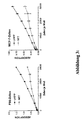
Zu jedem well mit Beta-Galaktosidasestandard werden 100 µl HBSS oder HBSS-TX bzw. 80µl MES-TX gegeben. Anschließend erfolgt eine Inkubation für 30 min bei 37 °C und 5 % CO2. Dann werden zu den Wells mit Beta-Galaktosidasestandard und MES-TX Puffer 20µl 0.5 M Tris-HCl gegeben oder 20 µl 0.5, M HEPES (pH 8.0). Darauf erfolgt die Zugabe von 150 µl CPRG Substratlösung mit 1 mg/ml CPRG gelöst in HBSS. Die optische Dichte bei 540-580 nm wird zu verschiedenen Zeitpunkten an einem Mikrotiterplattenphotometer bestimmt (vergleiche Abbildung 2).
Bei Durchführung des saure Phosphatasetestes erfolgt nach dem Waschen der Zellen mit PBS die Zugabe des Substratpuffers 10 mM pNPP in 0.02 M MES/ 0.1 % Triton X-100 (die genauen Volumina für die verschiedenen untersuchten Platten siehe Tabelle 1). Nach 30 min Inkubation bei 37 °C und 5 % CO2 erfolgt das Abstoppen der Reaktion durch Zugabe von 0.5 M Tris-HCl oder 0.5 M HEPES (pH 8.0). Anschließend erfolgt die Bestimmung der p-Nitrophenolkonzentration durch Bestimmung des Absorption bei 405 nm (Referenzwellenlänge 690 nm) am Mikroplattenmeßgerät.
| Übersicht über Volumina der verschiedenen Testpuffer auf verschiedenen Zellkulturplatten. | |||
| Saure Phosphatasetestpuffer (µl) | Stoppuffer (µl) | Beta-Galaktosidase testpuffer (µl) | |
| 96-Well-Schale | 80 | 20 | 150 |
| 24-Well-Schale | 240 | 60 | 450 |
| 6-Well-Schale | 800 | 200 | 1500 |
- Saure Phosphatasetestpuffer:
- 10 mM pNPP in 0.02M MES/0.1%
Triton X-100
10 mM pNPP in 0.02M Natriumacetat/0.1% Triton X-100 1mM Naphtylphosphat in 0.02M Natriumacetat/0.1% Triton X-100 - Stoppuffer:
- 0.5 M Tris-HCl (pH 8.0)
0.5 M HEPES (pH 8.0) - Beta-Galaktosidasetestpuffer:
- 1 mg/ml CPRG in HBSS (50µl)
2 mg/ml ONPG in HBSS (100µl)
1mM MUG in HBSS (100µl), aus
MUG-Stammlösung 20 mM in DMSO und HBSS
| Vergleich der verschiedenen Verfahren zur dualen Bestimmung von Zellvitalität bzw. Toxizität der Gentransfermethode und Effizienz der Gentransfermethode | |||||
| Beispiel | 1 | 2 | 3 | 4 | 5 |
| Saure Phosphatase-Substrat | pNPP | pNPP | pNPP | Naphtylphosphat | pNPP |
| Saure Phosphatase-Bestimmung | optisch | optisch | optisch | fluorimetrisch | optisch |
| Beta-Galaktosidase-Substrat | CPRG | ONPG | MUG | MUG | MUG/ONPG |
| Beta-Galaktosidase-Bestimmung | optisch | optisch | fluorimetrisch | fluorimetrisch | optisch/ fluorimetrisch |
| Beta-Galaktosidase-Sensitivität | ++ | + | +++ | +++ | +++ |
MCF-7 Zellen wurden mit DOCSPER-Liposomen und der Plasmid-DNA pUT 651 (Cayla, Toulouse, Frankreich) wie bei Felgner und Mitarbeitern (1994) beschrieben transfiziert. 3 Tage nach der Transfektion erfolgte die Bestimmung von Zellvitalität und Beta-Galaktosidaseexpression wie in Beispiel 1 (80 µl 10 mM pNPP in 0,02 M MES; 0,1 % Triton X-100 (pH 5,5), 20 µl Tris-HCl (pH 8,0), 150 µl 1 mg/ml CPRG in HBSS) angegeben. Die Zellvitalität von Kontrollzellen und die maximale Beta-Galaktosidaseexpression des Experiments wurden auf 100 % gesetzt.
Claims (3)
- Verfahren zur parallelen Bestimmung von Zellvitalität und Effizienz nach Gentransfer in eukaryontische Zellen,
dadurch gekennzeichnet, daß man
die Zellzahl der transfizierten Zellen durch eine repräsentative Enzymaktivität mittels Bestimmung der Sauren Phosphatase ermittelt und nachfolgend im selben Reaktionsgefäß die Effizienz des Gentransfers durch colorimetrische oder fluorimetrische Bestimmung der Reportergenaktivität nach Änderung der Ionenkonzentration und des pH-Wertes. - Verfahren nach Anspruch 1,
dadurch gekennzeichnet, daß man
zur Effizienzbestimmung die Pufferstärken erniedrigt. - Duales Testsystem zur Bestimmung von Zellvitalität und Effizienz nach Gentransfer in eukaryontische Zellen umfassenda) einen Vitalitäts-Assay mit
einem Substrat zur Bestimmung Saurer Phosphatase und einem Puffer für die Zellyse, wobei als Substrat pNPP oder Naphthylphosphat verwendet wird und der verwendete Puffer eine niedrige Ionenstärke besitzt, die nach Bestimmung der Saure Phosphataseaktivität eine Veränderung des pH-Wertes vom sauren in den basischen pH-Bereich ermöglicht, undb) einem Substrat zur colorimetrischen oder fluorimetrischen Bestimmung der β-Galaktosidase-Aktivität als Reportengen-Assay zur Effizienzbestimmung.
Applications Claiming Priority (5)
| Application Number | Priority Date | Filing Date | Title |
|---|---|---|---|
| DE19750790 | 1997-11-07 | ||
| DE19750790 | 1997-11-07 | ||
| DE19805818 | 1998-02-13 | ||
| DE19805818A DE19805818C2 (de) | 1997-11-07 | 1998-02-13 | Verfahren zur Bestimmung von Zellvitalität und Effizienz nach Gentransfer in eukaryontische Zellen und duales Testsystem |
| PCT/DE1998/003245 WO1999024602A2 (de) | 1997-11-07 | 1998-11-06 | Verfahren zur parallelen bestimmung der zellvitalität und gentransfer-effizienz |
Publications (2)
| Publication Number | Publication Date |
|---|---|
| EP1029241A2 EP1029241A2 (de) | 2000-08-23 |
| EP1029241B1 true EP1029241B1 (de) | 2004-09-01 |
Family
ID=26041657
Family Applications (1)
| Application Number | Title | Priority Date | Filing Date |
|---|---|---|---|
| EP98962216A Expired - Lifetime EP1029241B1 (de) | 1997-11-07 | 1998-11-06 | Verfahren zur parallelen bestimmung der zellvitalität und gentransfer-effizienz |
Country Status (5)
| Country | Link |
|---|---|
| US (1) | US6692913B1 (de) |
| EP (1) | EP1029241B1 (de) |
| JP (1) | JP2001521762A (de) |
| AT (1) | ATE275267T1 (de) |
| WO (1) | WO1999024602A2 (de) |
-
1998
- 1998-11-06 EP EP98962216A patent/EP1029241B1/de not_active Expired - Lifetime
- 1998-11-06 WO PCT/DE1998/003245 patent/WO1999024602A2/de not_active Ceased
- 1998-11-06 AT AT98962216T patent/ATE275267T1/de not_active IP Right Cessation
- 1998-11-06 JP JP2000519595A patent/JP2001521762A/ja active Pending
-
2000
- 2000-05-08 US US09/571,338 patent/US6692913B1/en not_active Expired - Fee Related
Also Published As
| Publication number | Publication date |
|---|---|
| EP1029241A2 (de) | 2000-08-23 |
| ATE275267T1 (de) | 2004-09-15 |
| WO1999024602A3 (de) | 1999-07-15 |
| WO1999024602A2 (de) | 1999-05-20 |
| JP2001521762A (ja) | 2001-11-13 |
| US6692913B1 (en) | 2004-02-17 |
Similar Documents
| Publication | Publication Date | Title |
|---|---|---|
| CN107843469B (zh) | 一种稳定的生化类复合校准品及其制备方法 | |
| Swift | The desoxyribose nucleic acid content of animal nuclei | |
| Sumner et al. | A difference in dry mass between the heads of X-and Y-bearing human spermatozoa | |
| Uddin et al. | Quality control tests for ophthalmic pharmaceuticals: pharmacopoeial standards and specifications | |
| JP3039594B2 (ja) | 染色試薬およびその使用方法 | |
| DE3034142C2 (de) | ||
| Bemiller et al. | Nucleolar changes in senescing WI-38 cells | |
| DE69433977T2 (de) | Unsterbliche menschliche zellinien, die exogenes cytochrom p-450 gen enthalten | |
| EP0185335B1 (de) | Verfahren zum Nachweis des Vorliegens einer Allergie und zum spezifischen Nachweis des für die Allergie verantwortlichen Allergens | |
| Kraemer et al. | Colony-forming ability of ultraviolet-irradiated xeroderma pigmentosum fibroblasts from different DNA repair complementation groups | |
| US4241174A (en) | Interferon assay | |
| DE19805818C2 (de) | Verfahren zur Bestimmung von Zellvitalität und Effizienz nach Gentransfer in eukaryontische Zellen und duales Testsystem | |
| Maxfield | Acidification of endocytic vesicles and lysosomes | |
| WO1999024602A2 (de) | Verfahren zur parallelen bestimmung der zellvitalität und gentransfer-effizienz | |
| Singh et al. | A novel technique for viable cell determinations | |
| Miike et al. | A fluorescent microscopy study of biopsied muscles from infantile neuromuscular disorders | |
| Zucker et al. | Flow cytometric analysis of the cellular toxicity of tributyltin | |
| DE10235967B4 (de) | Methode zum Identifizieren von Substanzen mit antimikrobieller Wirkung | |
| Ding et al. | The effects of Wubeizi ointment on the proliferation of keloid-derived fibroblasts | |
| EP1356287B1 (de) | Replikationsassay zur auffindung antiviraler substanzen | |
| Givens et al. | Antiproliferative drugs and human ocular fibroblasts: Colorimetric vs. cell counting assays | |
| EP1363129A1 (de) | Kontroll-und Kalibrationsmaterial für Blutgerinnungstests | |
| AT390844B (de) | Reagens zur bestimmung der thrombozyten- und leukozytenzahl | |
| EP0428656B1 (de) | Humane zellinie lc5 sowie deren verwendung | |
| TOMER | STUDIES ON SISTER CHROMATID EXCHANGE AND. CHROMOSOMAL ABERRATIONS IN.· CATTLE IN RELATION TO MASTIT. IS |
Legal Events
| Date | Code | Title | Description |
|---|---|---|---|
| PUAI | Public reference made under article 153(3) epc to a published international application that has entered the european phase |
Free format text: ORIGINAL CODE: 0009012 |
|
| 17P | Request for examination filed |
Effective date: 20000530 |
|
| AK | Designated contracting states |
Kind code of ref document: A2 Designated state(s): AT BE CH DE DK FI FR GB IT LI NL SE |
|
| 17Q | First examination report despatched |
Effective date: 20030603 |
|
| GRAP | Despatch of communication of intention to grant a patent |
Free format text: ORIGINAL CODE: EPIDOSNIGR1 |
|
| GRAS | Grant fee paid |
Free format text: ORIGINAL CODE: EPIDOSNIGR3 |
|
| GRAA | (expected) grant |
Free format text: ORIGINAL CODE: 0009210 |
|
| AK | Designated contracting states |
Kind code of ref document: B1 Designated state(s): AT BE CH DE DK FI FR GB IT LI NL SE |
|
| PG25 | Lapsed in a contracting state [announced via postgrant information from national office to epo] |
Ref country code: NL Free format text: LAPSE BECAUSE OF FAILURE TO SUBMIT A TRANSLATION OF THE DESCRIPTION OR TO PAY THE FEE WITHIN THE PRESCRIBED TIME-LIMIT Effective date: 20040901 Ref country code: IT Free format text: LAPSE BECAUSE OF FAILURE TO SUBMIT A TRANSLATION OF THE DESCRIPTION OR TO PAY THE FEE WITHIN THE PRESCRIBED TIME-LIMIT;WARNING: LAPSES OF ITALIAN PATENTS WITH EFFECTIVE DATE BEFORE 2007 MAY HAVE OCCURRED AT ANY TIME BEFORE 2007. THE CORRECT EFFECTIVE DATE MAY BE DIFFERENT FROM THE ONE RECORDED. Effective date: 20040901 Ref country code: GB Free format text: LAPSE BECAUSE OF FAILURE TO SUBMIT A TRANSLATION OF THE DESCRIPTION OR TO PAY THE FEE WITHIN THE PRESCRIBED TIME-LIMIT Effective date: 20040901 Ref country code: FR Free format text: LAPSE BECAUSE OF FAILURE TO SUBMIT A TRANSLATION OF THE DESCRIPTION OR TO PAY THE FEE WITHIN THE PRESCRIBED TIME-LIMIT Effective date: 20040901 Ref country code: FI Free format text: LAPSE BECAUSE OF FAILURE TO SUBMIT A TRANSLATION OF THE DESCRIPTION OR TO PAY THE FEE WITHIN THE PRESCRIBED TIME-LIMIT Effective date: 20040901 |
|
| REG | Reference to a national code |
Ref country code: GB Ref legal event code: FG4D Free format text: NOT ENGLISH |
|
| REG | Reference to a national code |
Ref country code: CH Ref legal event code: EP |
|
| REF | Corresponds to: |
Ref document number: 59811903 Country of ref document: DE Date of ref document: 20041007 Kind code of ref document: P |
|
| PG25 | Lapsed in a contracting state [announced via postgrant information from national office to epo] |
Ref country code: AT Free format text: LAPSE BECAUSE OF NON-PAYMENT OF DUE FEES Effective date: 20041106 |
|
| PG25 | Lapsed in a contracting state [announced via postgrant information from national office to epo] |
Ref country code: LI Free format text: LAPSE BECAUSE OF NON-PAYMENT OF DUE FEES Effective date: 20041130 Ref country code: CH Free format text: LAPSE BECAUSE OF NON-PAYMENT OF DUE FEES Effective date: 20041130 Ref country code: BE Free format text: LAPSE BECAUSE OF NON-PAYMENT OF DUE FEES Effective date: 20041130 |
|
| PG25 | Lapsed in a contracting state [announced via postgrant information from national office to epo] |
Ref country code: SE Free format text: LAPSE BECAUSE OF FAILURE TO SUBMIT A TRANSLATION OF THE DESCRIPTION OR TO PAY THE FEE WITHIN THE PRESCRIBED TIME-LIMIT Effective date: 20041201 Ref country code: DK Free format text: LAPSE BECAUSE OF FAILURE TO SUBMIT A TRANSLATION OF THE DESCRIPTION OR TO PAY THE FEE WITHIN THE PRESCRIBED TIME-LIMIT Effective date: 20041201 |
|
| NLV1 | Nl: lapsed or annulled due to failure to fulfill the requirements of art. 29p and 29m of the patents act | ||
| GBV | Gb: ep patent (uk) treated as always having been void in accordance with gb section 77(7)/1977 [no translation filed] |
Effective date: 20040901 |
|
| BERE | Be: lapsed |
Owner name: MAX-DELBRUCK-CENTRUM FUR MOLEKULARE MEDIZIN Effective date: 20041130 |
|
| PG25 | Lapsed in a contracting state [announced via postgrant information from national office to epo] |
Ref country code: DE Free format text: LAPSE BECAUSE OF NON-PAYMENT OF DUE FEES Effective date: 20050601 |
|
| PLBE | No opposition filed within time limit |
Free format text: ORIGINAL CODE: 0009261 |
|
| STAA | Information on the status of an ep patent application or granted ep patent |
Free format text: STATUS: NO OPPOSITION FILED WITHIN TIME LIMIT |
|
| REG | Reference to a national code |
Ref country code: CH Ref legal event code: PL |
|
| 26N | No opposition filed |
Effective date: 20050602 |
|
| EN | Fr: translation not filed | ||
| REG | Reference to a national code |
Ref country code: HK Ref legal event code: WD Ref document number: 1032260 Country of ref document: HK |
|
| BERE | Be: lapsed |
Owner name: *MAX-DELBRUCK-CENTRUM FUR MOLEKULARE MEDIZIN Effective date: 20041130 |