EP0951295B1 - Compositions for use in inhibiting of alpha-v-beta3 mediated angiogenesis - Google Patents
Compositions for use in inhibiting of alpha-v-beta3 mediated angiogenesis Download PDFInfo
- Publication number
- EP0951295B1 EP0951295B1 EP97928698A EP97928698A EP0951295B1 EP 0951295 B1 EP0951295 B1 EP 0951295B1 EP 97928698 A EP97928698 A EP 97928698A EP 97928698 A EP97928698 A EP 97928698A EP 0951295 B1 EP0951295 B1 EP 0951295B1
- Authority
- EP
- European Patent Office
- Prior art keywords
- antagonist
- angiogenesis
- tissue
- tumor
- peptide
- Prior art date
- Legal status (The legal status is an assumption and is not a legal conclusion. Google has not performed a legal analysis and makes no representation as to the accuracy of the status listed.)
- Expired - Lifetime
Links
Images


Classifications
-
- C—CHEMISTRY; METALLURGY
- C12—BIOCHEMISTRY; BEER; SPIRITS; WINE; VINEGAR; MICROBIOLOGY; ENZYMOLOGY; MUTATION OR GENETIC ENGINEERING
- C12N—MICROORGANISMS OR ENZYMES; COMPOSITIONS THEREOF; PROPAGATING, PRESERVING, OR MAINTAINING MICROORGANISMS; MUTATION OR GENETIC ENGINEERING; CULTURE MEDIA
- C12N9/00—Enzymes; Proenzymes; Compositions thereof; Processes for preparing, activating, inhibiting, separating or purifying enzymes
- C12N9/14—Hydrolases (3)
- C12N9/48—Hydrolases (3) acting on peptide bonds (3.4)
- C12N9/50—Proteinases, e.g. Endopeptidases (3.4.21-3.4.25)
- C12N9/64—Proteinases, e.g. Endopeptidases (3.4.21-3.4.25) derived from animal tissue
- C12N9/6421—Proteinases, e.g. Endopeptidases (3.4.21-3.4.25) derived from animal tissue from mammals
- C12N9/6489—Metalloendopeptidases (3.4.24)
- C12N9/6491—Matrix metalloproteases [MMP's], e.g. interstitial collagenase (3.4.24.7); Stromelysins (3.4.24.17; 3.2.1.22); Matrilysin (3.4.24.23)
-
- A—HUMAN NECESSITIES
- A61—MEDICAL OR VETERINARY SCIENCE; HYGIENE
- A61K—PREPARATIONS FOR MEDICAL, DENTAL OR TOILETRY PURPOSES
- A61K38/00—Medicinal preparations containing peptides
- A61K38/16—Peptides having more than 20 amino acids; Gastrins; Somatostatins; Melanotropins; Derivatives thereof
- A61K38/43—Enzymes; Proenzymes; Derivatives thereof
- A61K38/46—Hydrolases (3)
- A61K38/48—Hydrolases (3) acting on peptide bonds (3.4)
-
- A—HUMAN NECESSITIES
- A61—MEDICAL OR VETERINARY SCIENCE; HYGIENE
- A61K—PREPARATIONS FOR MEDICAL, DENTAL OR TOILETRY PURPOSES
- A61K38/00—Medicinal preparations containing peptides
- A61K38/04—Peptides having up to 20 amino acids in a fully defined sequence; Derivatives thereof
-
- A—HUMAN NECESSITIES
- A61—MEDICAL OR VETERINARY SCIENCE; HYGIENE
- A61K—PREPARATIONS FOR MEDICAL, DENTAL OR TOILETRY PURPOSES
- A61K38/00—Medicinal preparations containing peptides
- A61K38/16—Peptides having more than 20 amino acids; Gastrins; Somatostatins; Melanotropins; Derivatives thereof
-
- A—HUMAN NECESSITIES
- A61—MEDICAL OR VETERINARY SCIENCE; HYGIENE
- A61K—PREPARATIONS FOR MEDICAL, DENTAL OR TOILETRY PURPOSES
- A61K38/00—Medicinal preparations containing peptides
- A61K38/16—Peptides having more than 20 amino acids; Gastrins; Somatostatins; Melanotropins; Derivatives thereof
- A61K38/43—Enzymes; Proenzymes; Derivatives thereof
- A61K38/46—Hydrolases (3)
- A61K38/48—Hydrolases (3) acting on peptide bonds (3.4)
- A61K38/4886—Metalloendopeptidases (3.4.24), e.g. collagenase
-
- A—HUMAN NECESSITIES
- A61—MEDICAL OR VETERINARY SCIENCE; HYGIENE
- A61P—SPECIFIC THERAPEUTIC ACTIVITY OF CHEMICAL COMPOUNDS OR MEDICINAL PREPARATIONS
- A61P19/00—Drugs for skeletal disorders
- A61P19/02—Drugs for skeletal disorders for joint disorders, e.g. arthritis, arthrosis
-
- A—HUMAN NECESSITIES
- A61—MEDICAL OR VETERINARY SCIENCE; HYGIENE
- A61P—SPECIFIC THERAPEUTIC ACTIVITY OF CHEMICAL COMPOUNDS OR MEDICINAL PREPARATIONS
- A61P27/00—Drugs for disorders of the senses
- A61P27/02—Ophthalmic agents
-
- A—HUMAN NECESSITIES
- A61—MEDICAL OR VETERINARY SCIENCE; HYGIENE
- A61P—SPECIFIC THERAPEUTIC ACTIVITY OF CHEMICAL COMPOUNDS OR MEDICINAL PREPARATIONS
- A61P29/00—Non-central analgesic, antipyretic or antiinflammatory agents, e.g. antirheumatic agents; Non-steroidal antiinflammatory drugs [NSAID]
-
- A—HUMAN NECESSITIES
- A61—MEDICAL OR VETERINARY SCIENCE; HYGIENE
- A61P—SPECIFIC THERAPEUTIC ACTIVITY OF CHEMICAL COMPOUNDS OR MEDICINAL PREPARATIONS
- A61P35/00—Antineoplastic agents
-
- A—HUMAN NECESSITIES
- A61—MEDICAL OR VETERINARY SCIENCE; HYGIENE
- A61P—SPECIFIC THERAPEUTIC ACTIVITY OF CHEMICAL COMPOUNDS OR MEDICINAL PREPARATIONS
- A61P37/00—Drugs for immunological or allergic disorders
- A61P37/08—Antiallergic agents
-
- A—HUMAN NECESSITIES
- A61—MEDICAL OR VETERINARY SCIENCE; HYGIENE
- A61P—SPECIFIC THERAPEUTIC ACTIVITY OF CHEMICAL COMPOUNDS OR MEDICINAL PREPARATIONS
- A61P43/00—Drugs for specific purposes, not provided for in groups A61P1/00-A61P41/00
-
- A—HUMAN NECESSITIES
- A61—MEDICAL OR VETERINARY SCIENCE; HYGIENE
- A61P—SPECIFIC THERAPEUTIC ACTIVITY OF CHEMICAL COMPOUNDS OR MEDICINAL PREPARATIONS
- A61P7/00—Drugs for disorders of the blood or the extracellular fluid
-
- C—CHEMISTRY; METALLURGY
- C07—ORGANIC CHEMISTRY
- C07C—ACYCLIC OR CARBOCYCLIC COMPOUNDS
- C07C279/00—Derivatives of guanidine, i.e. compounds containing the group, the singly-bound nitrogen atoms not being part of nitro or nitroso groups
- C07C279/04—Derivatives of guanidine, i.e. compounds containing the group, the singly-bound nitrogen atoms not being part of nitro or nitroso groups having nitrogen atoms of guanidine groups bound to acyclic carbon atoms of a carbon skeleton
- C07C279/14—Derivatives of guanidine, i.e. compounds containing the group, the singly-bound nitrogen atoms not being part of nitro or nitroso groups having nitrogen atoms of guanidine groups bound to acyclic carbon atoms of a carbon skeleton being further substituted by carboxyl groups
-
- C—CHEMISTRY; METALLURGY
- C07—ORGANIC CHEMISTRY
- C07C—ACYCLIC OR CARBOCYCLIC COMPOUNDS
- C07C311/00—Amides of sulfonic acids, i.e. compounds having singly-bound oxygen atoms of sulfo groups replaced by nitrogen atoms, not being part of nitro or nitroso groups
- C07C311/01—Sulfonamides having sulfur atoms of sulfonamide groups bound to acyclic carbon atoms
- C07C311/02—Sulfonamides having sulfur atoms of sulfonamide groups bound to acyclic carbon atoms of an acyclic saturated carbon skeleton
- C07C311/03—Sulfonamides having sulfur atoms of sulfonamide groups bound to acyclic carbon atoms of an acyclic saturated carbon skeleton having the nitrogen atoms of the sulfonamide groups bound to hydrogen atoms or to acyclic carbon atoms
- C07C311/06—Sulfonamides having sulfur atoms of sulfonamide groups bound to acyclic carbon atoms of an acyclic saturated carbon skeleton having the nitrogen atoms of the sulfonamide groups bound to hydrogen atoms or to acyclic carbon atoms to acyclic carbon atoms of hydrocarbon radicals substituted by carboxyl groups
-
- C—CHEMISTRY; METALLURGY
- C07—ORGANIC CHEMISTRY
- C07C—ACYCLIC OR CARBOCYCLIC COMPOUNDS
- C07C311/00—Amides of sulfonic acids, i.e. compounds having singly-bound oxygen atoms of sulfo groups replaced by nitrogen atoms, not being part of nitro or nitroso groups
- C07C311/01—Sulfonamides having sulfur atoms of sulfonamide groups bound to acyclic carbon atoms
- C07C311/10—Sulfonamides having sulfur atoms of sulfonamide groups bound to acyclic carbon atoms of a saturated carbon skeleton containing rings
-
- C—CHEMISTRY; METALLURGY
- C07—ORGANIC CHEMISTRY
- C07D—HETEROCYCLIC COMPOUNDS
- C07D263/00—Heterocyclic compounds containing 1,3-oxazole or hydrogenated 1,3-oxazole rings
- C07D263/02—Heterocyclic compounds containing 1,3-oxazole or hydrogenated 1,3-oxazole rings not condensed with other rings
- C07D263/30—Heterocyclic compounds containing 1,3-oxazole or hydrogenated 1,3-oxazole rings not condensed with other rings having two or three double bonds between ring members or between ring members and non-ring members
- C07D263/34—Heterocyclic compounds containing 1,3-oxazole or hydrogenated 1,3-oxazole rings not condensed with other rings having two or three double bonds between ring members or between ring members and non-ring members with hetero atoms or with carbon atoms having three bonds to hetero atoms with at the most one bond to halogen, e.g. ester or nitrile radicals, directly attached to ring carbon atoms
- C07D263/36—One oxygen atom
- C07D263/38—One oxygen atom attached in position 2
-
- C—CHEMISTRY; METALLURGY
- C07—ORGANIC CHEMISTRY
- C07K—PEPTIDES
- C07K14/00—Peptides having more than 20 amino acids; Gastrins; Somatostatins; Melanotropins; Derivatives thereof
- C07K14/435—Peptides having more than 20 amino acids; Gastrins; Somatostatins; Melanotropins; Derivatives thereof from animals; from humans
- C07K14/745—Blood coagulation or fibrinolysis factors
- C07K14/75—Fibrinogen
-
- C—CHEMISTRY; METALLURGY
- C07—ORGANIC CHEMISTRY
- C07K—PEPTIDES
- C07K16/00—Immunoglobulins [IGs], e.g. monoclonal or polyclonal antibodies
- C07K16/18—Immunoglobulins [IGs], e.g. monoclonal or polyclonal antibodies against material from animals or humans
- C07K16/28—Immunoglobulins [IGs], e.g. monoclonal or polyclonal antibodies against material from animals or humans against receptors, cell surface antigens or cell surface determinants
- C07K16/2839—Immunoglobulins [IGs], e.g. monoclonal or polyclonal antibodies against material from animals or humans against receptors, cell surface antigens or cell surface determinants against the integrin superfamily
- C07K16/2848—Immunoglobulins [IGs], e.g. monoclonal or polyclonal antibodies against material from animals or humans against receptors, cell surface antigens or cell surface determinants against the integrin superfamily against integrin beta3-subunit-containing molecules, e.g. CD41, CD51, CD61
-
- C—CHEMISTRY; METALLURGY
- C07—ORGANIC CHEMISTRY
- C07C—ACYCLIC OR CARBOCYCLIC COMPOUNDS
- C07C2602/00—Systems containing two condensed rings
- C07C2602/36—Systems containing two condensed rings the rings having more than two atoms in common
- C07C2602/42—Systems containing two condensed rings the rings having more than two atoms in common the bicyclo ring system containing seven carbon atoms
Definitions
- the present invention relates generally to the field of medicine, and relates specifically to compositions for use in inhibiting angiogenesis of tissues using antagonists of the vitronectin receptor ⁇ v ⁇ 3 .
- Integrins are a class of cellular receptors known to bind extracellular matrix proteins, and therefore mediate cell-cell and cell-extracellular matrix interactions, referred generally to as cell adhesion events.
- cell adhesion events Although many integrins and the ligands that bind an integrin are described in the literature, the biological function of many of the integrins remains elusive.
- the integrin receptors constitute a family of proteins with shared structural characteristics of noncovalent heterodimeric glycoprotein complexes formed of ⁇ and ⁇ subunits.
- the vitronectin receptor named for its original characteristic of preferential binding to vitronectin, is now known to refer to three different integrins, designated ⁇ v ⁇ 1 , ⁇ v ⁇ 3 and ⁇ v ⁇ 5 . Horton, Int. J. Exp. Pathol., 71:741-759 (1990 ).
- ⁇ v ⁇ 1 binds fibronectin and vitronectin.
- ⁇ v ⁇ 3 binds a large variety of ligands, including fibrin, fibrinogen, laminin, thrombospondin, vitronectin, von Willebrand's factor, osteospontin and bone sialoprotein I.
- ⁇ v ⁇ 5 binds vitronectin.
- the specific cell adhesion roles these three integrins play in the many cellular interactions in tissues are still under investigation, but it is clear that there are different integrins with different biological functions.
- RGD arginine-glycine-aspartic acid
- Angiogenesis is a process of tissue vascularization that involves the growth of new developing blood vessels into a tissue, and is also referred to as neo-vascularization.
- the process is mediated by the infiltration of endothelial cells and smooth muscle cells.
- the process is believed to proceed in any one of three ways: the vessels can sprout from pre-existing vessels, de-novo development of vessels can arise from precursor cells (vasculogenesis), or existing small vessels can enlarge in diameter. Blood et al., Bioch. Biophys. Acta, 1032:89-118 (1990 ).
- Vascular endothelial cells are known to contain at least five RGD-dependent integrins, including the vitronectin receptor ( ⁇ v ⁇ 3 or ⁇ v ⁇ 5 ), the collagen Types I and IV receptor ( ⁇ 1 ⁇ 1 ), the laminin receptor ( ⁇ 2 ⁇ 1 ), the fibronectin/laminin/collagen receptor ( ⁇ 3 ⁇ 1 ) and the fibronectin receptor ( ⁇ 5 ⁇ 1 ). Davis et al., J. Cell. Biochem., 51:206-218 (1993 ).
- the smooth muscle cell is known to contain at least six RGD-dependent integrins, including ⁇ 5 ⁇ 1 , ⁇ v ⁇ 3 and ⁇ v ⁇ 5 .
- Angiogenesis is an important process in neonatal growth, but is also important in wound healing and in the pathogenesis of a large variety of clinical diseases including tissue inflammation, arthritis, tumor growth, diabetic retinopathy, macular degeneration by neovascularization of retina and the like conditions. These clinical manifestations associated with angiogenesis are referred to as angiogenic diseases. Folkman et al., Science, 235:442-447 (1987 ). Angiogenesis is generally absent in adult or mature tissues, although it does occur in wound healing and in the corpeus leuteum growth cycle. See, for example, Moses et al., Science, 248:1408-1410 (1990 ).
- angiogenesis would be a useful therapy for restricting tumor growth.
- Inhibition of angiogenesis has been proposed by (1) inhibition of release of "angiogenic molecules” such as bFGF (basic fibroblast growth factor), (2) neutralization of angiogenic molecules, such as by use of anti- ⁇ bFGF antibodies, and (3) inhibition of endothelial cell response to angiogenic stimuli.
- angiogenic molecules such as bFGF (basic fibroblast growth factor)
- neutralization of angiogenic molecules such as by use of anti- ⁇ bFGF antibodies
- endothelial cell response inhibitors including collagenase inhibitor, basement membrane turnover inhibitors, angiostatic steroids, fungal-derived angiogenesis inhibitors, platelet factor 4, thrombospondin, arthritis drugs such as D-penicillamine and gold thiomalate, vitamin D 3 analogs, alpha-interferon, and the like that might be used to inhibit angiogenesis.
- endothelial cell response inhibitors including collagenase inhibitor, basement membrane turnover inhibitors, angiostatic steroids, fungal-derived angiogenesis inhibitors, platelet factor 4, thrombospondin, arthritis drugs such as D-penicillamine and gold thiomalate, vitamin D 3 analogs, alpha-interferon, and the like that might be used to inhibit angiogenesis.
- endothelial cell response inhibitors including collagenase inhibitor, basement membrane turnover inhibitors, angiostatic steroids, fungal-derived angiogenesis inhibitors, platelet factor 4, thrombospondin, arthritis drugs such as D-penicillamine and gold thiomalate,
- Invest., 75:563-573 (1996 ) described two particular cyclic methylated RGD-containing peptides that were partially effective at inhibiting retinal neovascularization in the mouse model of oxygen-induced ischemic retinopathy.
- the peptides of the present invention exhibit almost complete inhibition of neovascularization in the model systems described herein.
- microvessels in vitro in collagen gel cultures is not a model for inhibition of angiogenesis in a tissue because it is not shown that the microvessel structures are the same as capillary sprouts or that the formation of the microvessel in collagen gel culture is the same as neovascular growth into an intact tissue, such as arthritic tissue, tumor tissue or disease tissue where inhibition of angiogenesis is desirable.
- endothelial cells For angiogenesis to occur, endothelial cells must first degrade and cross the blood vessel basement membrane in a similar manner used by tumor cells during invasion and metastasis formation.
- angiogenesis depends on the interaction between vascular integrins and extracellular matrix proteins. Brooks et al., Science, 264:569-571 (1994 ). Furthermore, it was reported that programmed cell death (apoptosis) of angiogenic vascular cells is initiated by the interaction, which would be inhibitied by certain antagonists of the vascular integrin ⁇ v ⁇ 3 . Brooks et al., Cell, 79:1157-1164 (1994 ).
- MMP-2 matrix metalloproteinase-2
- vitronectin receptor ⁇ v ⁇ 5 antagonists
- the present invention disclosure demonstrates that angiogenesis in tissues requires integrin ⁇ v ⁇ 3 , and that inhibitors of ⁇ v ⁇ 3 can inhibit angiogenesis.
- the disclosure also demonstrates that antagonists of other integrins, such as ⁇ IIb ⁇ 3 , or ⁇ v ⁇ 1 , do not inhibit angiogenesis, presumably because these other integrins are not essential for angiogenesis to occur.
- the invention therefore describes compounds for use in inhibiting ⁇ v ⁇ 3 mediated angiogenesis in a tissue.
- the tissue to be treated can be any tissue in which inhibition of angiogenesis is desirable, such as diseased tissue where neo-vascularization is occurring.
- exemplary tissues include inflamed tissue, solid tumors, metastases, tissues undergoing restenosis, and the like tissues.
- An ⁇ v ⁇ 3 antagonist for use in the present invention is capable of binding to ⁇ v ⁇ 3 and competitively inhibiting the ability of ⁇ v ⁇ 3 to bind to a natural ligand.
- the ⁇ v ⁇ 3 antagonist is an organic mimetic of ⁇ v ⁇ 3 as defined in claim 1.
- Amino Acid Residue An amino acid formed upon chemical digestion (hydrolysis) of a polypeptide at its peptide linkages.
- the amino acid residues described herein are preferably in the "L” isomeric form. However, residues in the "D" isomeric form can be substituted for any L-amino acid residue, as long as the desired functional property is retained by the polypeptide.
- NH 2 refers to the free amino group present at the amino terminus of a polypeptide.
- COOH refers to the free carboxy group present at the carboxy terminus of a polypeptide.
- amino acid residue sequences are represented herein by formulae whose left and right orientation is in the conventional direction of amino-terminus to carboxy-terminus. Furthermore, it should be noted that a dash at the beginning or end of an amino acid residue sequence indicates a peptide bond to a further sequence of one or more amino acid residues.
- Polypeptide refers to a linear series of amino acid residues connected to one another by peptide bonds between the alpha-amino group and carboxy group of contiguous amino acid residues.
- Peptide refers to a linear series of no more than about 50 amino acid residues connected one to the other as in a polypeptide.
- Cyclic peptide refers to a compound having a heteroatom ring structure that includes several amide bonds as in a typical peptide.
- the cyclic peptide can be a "head to tail" cyclized linear polypeptide in which a linear peptide's n-terminus has formed an amide bond with the terminal carboxylate of the linear peptide, or it can contain a ring structure in which the polymer is homodetic or heterodetic and comprises amide bonds and/or other bonds to close the ring, such as disulfide bridges, thioesters, thioamides, guanidino, and the like linkages.
- Protein refers to a linear series of greater than 50 amino acid residues connected one to the other as in a polypeptide.
- Fusion protein refers to a polypeptide containing at least two different polypeptide domains operatively linked by a typical peptide bond ("fused"), where the two domains correspond to peptides no found fused in nature.
- Synthetic peptide refers to a chemically produced chain of amino acid residues linked together by peptide bonds that is free of naturally occurring proteins and fragments thereof.
- the present invention relates generally to the discovery that angiogenesis is mediated by the specific vitronectin receptor ⁇ v ⁇ 3 , and that inhibition of ⁇ v ⁇ 3 function inhibits angiogenesis. This discovery is important because of the role that angiogenesis plays in a variety of disease processes. By inhibiting angiogenesis, one can intervene in the disease, ameliorate the symptoms, and in some cases cure the disease.
- angiogenesis will reduce the deleterious effects of the disease. Examples include rheumatoid arthritis, diabetic retinopathy, inflammatory diseases, restenosis, and the like. Where the growth of new blood vessels is required to support growth of a deleterious tissue, inhibition of angiogenesis will reduce the blood supply to the tissue and thereby contribute to reduction in tissue mass based on blood supply requirements. Examples include growth of tumors where neovascularization is a continual requirement in order that the tumor grow beyond a few millimeters in thickness, and for the establishment of solid tumor metastases.
- the compounds for use according to the present invention are effective in part because the therapy is highly selective for angiogenesis and not other biological processes. As shown in the Examples, only new vessel growth contains substantial ⁇ v ⁇ 3 , and therefore the therapeutic methods do not adversely effect mature vessels. Furthermore, ⁇ v ⁇ 3 is not widely distributed in normal tissues, but rather is found selectively on new vessels, thereby assuring that the therapy can be selectively targeted to new vessel growth.
- the invention provides compounds, as defined in claim 1, for use in a method for the inhibition of angiogenesis in a tissue, and thereby inhibiting events in the tissue which depend upon angiogenesis.
- the compounds are to be administered to the tissue in a composition comprising an angiogenesis-inhibiting amount of said ⁇ v ⁇ 3 antagonist.
- angiogenesis includes a variety of processes involving neovascularization of a tissue including "sprouting", vasculogenesis, or vessel enlargement, all of which angiogenesis processes are mediated by and dependent upon the expression of ⁇ v ⁇ 3 .
- angiogenesis processes are associated with disease processes and therefore the use of the present therapeutic methods are selective for the disease and do not have deleterious side effects.
- angiogenic diseases including but not limited to, inflammatory disorders such as immune and non-immune inflammation, chronic articular rheumatism and psoriasis, disorders associated with inappropriate or inopportune invasion of vessels such as diabetic retinopathy, neovascular glaucoma, restenosis, capillary proliferation in atherosclerotic plaques and osteoporosis, and cancer associated disorders, such as solid tumors, solid tumor metastases, angiofibromas, retrolental fibroplasia, hemangiomas, Kaposi sarcoma and the like cancers which require neovascularization to support tumor growth.
- inflammatory disorders such as immune and non-immune inflammation, chronic articular rheumatism and psoriasis
- disorders associated with inappropriate or inopportune invasion of vessels such as diabetic retinopathy, neovascular glaucoma, restenosis, capillary proliferation in atherosclerotic plaques and osteoporosis
- the invention contemplates the compounds as defined in claim 1 for use in inhibition of angiogenesis, per se, in a tissue.
- the extent of angiogenesis in a tissue, and therefore the extent of inhibition achieved by the present compounds, can be evaluated by a variety of methods, such as are described in the Examples for detecting ⁇ v ⁇ 3 -immunopositive immature and nascent vessel structures by immunohistochemistry.
- any of a variety of tissues, or organs comprised of organized tissues can support angiogenesis in disease conditions including skin, muscle, gut, connective tissue, joints, bones and the like tissue in which blood vessels can invade upon angiogenic stimuli.
- a tissue to be treated is an inflamed tissue and the angiogenesis to be inhibited is inflamed tissue angiogenesis where there is neovascularization of inflamed tissue.
- the compounds defined in claim 1 are contemplated for use in inhibition of angiogenesis in arthritic tissues, such as in a patient with chronic articular rheumatism, in immune or non-immune inflamed tissues, in psoriatic tissue and the like.
- the patient to be treated according to the present invention in its many embodiments is desirably a human patient, although it is to be understood that the principles of the invention indicate that the invention is effective with respect to all mammals, which are intended to be included in the term "patient".
- a mammal is understood to include any mammalian species in which treatment of diseases associated with angiogenesis is desirable, particularly agricultural and domestic mammalian species.
- a tissue to be treated is a retinal tissue of a patient with a retinal disease such as diabetic retinopathy, macular degeneration or neovascular glaucoma and the angiogenesis to be inhibited is retinal tissue angiogenesis where there is neovascularization of retinal tissue.
- a retinal disease such as diabetic retinopathy, macular degeneration or neovascular glaucoma
- the angiogenesis to be inhibited is retinal tissue angiogenesis where there is neovascularization of retinal tissue.
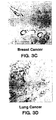
- a tissue to be treated is a tumor tissue of a patient with a solid tumor, a metastases, a skin cancer, a breast cancer, a hemangioma or angiofibroma and the like cancer, and the angiogenesis to be inhibited is tumor tissue angiogenesis where there is neovascularization of a tumor tissue.
- Typical solid tumor tissues treatable by the present methods include lung, pancreas, breast, colon, laryngeal, ovarian, and the like tissues. Exemplary tumor tissue angiogenesis, and inhibition thereof, is described in the Examples.
- Inhibition of tumor tissue angiogenesis is a particularly preferred embodiment because of the important role neovascularization plays in tumor growth. In the absence of neovascularization of tumor tissue, the tumor tissue does not obtain the required nutrients, slows in growth, ceases additional growth, regresses and ultimately becomes necrotic resulting in killing of the tumor.
- the present invention provides compounds for use in a method of inhibiting tumor neovascularization by inhibiting tumor angiogenesis according to the present methods. Similarly, the invention provides angiogenesis-inhibiting compounds, for use in inhibiting tumor growth.
- the compounds are also particularly effective against the formation of metastases because (1) their formation requires vascularization of a primary tumor so that the metastatic cancer cells can exit the primary tumor and (2) their establishment in a secondary site requires neovascularization to support growth of the metastases.
- the invention contemplates the practice of the compounds for use in the method in conjunction with other therapies such as conventional chemotherapy directed against solid tumors and for control of establishment of metastases.
- the angiogenesis inhibitor is typically to be administered during or after chemotherapy, although it is preferable to inhibit angiogenesis after a regimen of chemotherapy at times where the tumor tissue will be responding to the toxic assault by inducing angiogenesis to recover by the provision of a blood supply and nutrients to the tumor tissue.
- the compounds for use in angiogenesis inhibition are to be administered after surgery where solid tumors have been removed as a prophylaxis against metastases.
- the compounds are for use in inhibition of tumor neovascularization, they can also be for use in inhibition of tumor tissue growth, inhibition of tumor metastases formation, and regression of established tumors.
- the Examples demonstrate regression of an established tumor following a single intravenous administration of an ⁇ v ⁇ 3 antagonist according to this invention.
- Restenosis is a process of smooth muscle cell (SMC) migration and proliferation at the site of percutaneous transluminal coronary angioplasty which hampers the success of angioplasty.
- SMC smooth muscle cell
- the migration and proliferation of SMC's during restenosis can be considered a process of angiogenesis which is inhibited by the present compounds. Therefore, the invention also contemplates compounds for use in inhibition of restenosis by inhibiting angiogenesis in a patient following angioplasty procedures.
- the ⁇ v ⁇ 3 antagonist is typically to be administered after the angioplasty procedure for from about 2 to about 28 days, and more typically for about the first 14 days following the procedure.
- the present compounds for use in inhibiting angiogenesis in a tissue, and therefore also for treatment of angiogenesis-related diseases are to be administered to a tissue in which angiogenesis is occurring, or is at risk for occurring, whereby the compounds are in a composition comprising a therapeutically effective amount of an ⁇ v ⁇ 3 antagonist capable of inhibiting ⁇ v ⁇ 3 binding to its natural ligand.
- the compounds for use in the method are to be administered to a patient in a therapeutically effective amount of a physiologically tolerable composition containing an ⁇ v ⁇ 3 antagonist.
- the dosage ranges for the administration of the ⁇ v ⁇ 3 antagonist depend upon the form of the antagonist, and its potency, as described further herein, and are amounts large enough to produce the desired effect in which angiogenesis and the disease symptoms mediated by angiogenesis are ameliorated.
- the dosage should not be so large as to cause adverse side effects, such as hyperviscosity syndromes, pulmonary edema, congestive heart failure, and the like.
- the dosage will vary with the age, condition, sex and extent of the disease in the patient and can be determined by one of skill in the art.
- the dosage can also be adjusted by the individual physician in the event of any complication.
- a therapeutically effective amount is an amount of ⁇ v ⁇ 3 antagonist sufficient to produce a measurable inhibition of angiogenesis in the tissue being treated, ie., an angiogenesis-inhibiting amount.
- Inhibition of angiogenesis can be measured in situ by immunohistochemistry, as described herein, or by other methods known to one skilled in the art.
- an ⁇ v ⁇ 3 antagonist can take the form of any of five different organic mimetics, as defined in claim 1, it is to be appreciated that the potency, and therefore an expression of a "therapeutically effective" amount can vary. However, as shown by the present assay methods, one skilled in the art can readily assess the potency of an ⁇ v ⁇ 3 antagonist for use according to this invention.
- Potency of an ⁇ v ⁇ 3 antagonist can be measured by a variety of means including inhibition of angiogenesis in the CAM assay, in the in vivo rabbit eye assay, in the in vivo chimeric mouse:human assay, and by measuring inhibition of binding of natural ligand to ⁇ v ⁇ 3 , all as described herein, and the like assays.
- a preferred ⁇ v ⁇ 3 antagonist has the ability to substantially inhibit binding of a natural ligand such as fibrinogen or vitronectin to ⁇ v ⁇ 3 in solution at antagonist concentrations of less than 0.5 micromolar (um), preferably less than 0.1 um, and more preferably less than 0.05 um.
- a natural ligand such as fibrinogen or vitronectin
- substantially is meant that at least a 50 percent reduction in binding of fibrinogen is observed by inhibition in the presence of the ⁇ v ⁇ 3 antagonist, and at 50% inhibition is referred to herein as an IC 50 value.
- a more preferred ⁇ v ⁇ 3 antagonist exhibits selectivity for ⁇ v ⁇ 3 over other integrins.
- a preferred ⁇ v ⁇ 3 antagonist substantially inhibits fibrinogen binding to ⁇ v ⁇ 3 but does not substantially inhibit binding of fibrinogen to another integrin, such as ⁇ v ⁇ 1 , ⁇ v ⁇ 5 or ⁇ IIb ⁇ 3 .
- Particularly preferred is an ⁇ v ⁇ 3 antagonist that exhibits a 10-fold to 100-fold lower IC 50 activity at inhibiting fibrinogen binding to ⁇ v ⁇ 3 compared to the IC 50 activity at inhibiting fibrinogen binding to another integrin. Exemplary assays for measuring IC 50 activity at inhibiting fibrinogen binding to an integrin are described in the Examples.
- a therapeutically effective amount of an ⁇ v ⁇ 3 antagonist is described as an amount of polypeptide such that when administered in a physiologically tolerable composition is sufficient to achieve a plasma concentration of from about 0.1 microgram (ug) per milliliter (ml) to about 200 ug/ml, preferably from about 1 ug/ml to about 150 ug/ml.
- the preferred plasma concentration in molarity is from about 2 micromolar (uM) to about 5 millimolar (mM) and preferably about 100 uM to 1 mM polypeptide antagonist.
- the dosage per body weight can vary from about 0.1 mg/kg to about 300 mg/kg, and preferably from about 0.2 mg/kg to about 200 mg/kg, in one or more dose administrations daily, for one or several days.
- unit dose when used in reference to a therapeutic composition of the present invention refers to physically discrete units suitable as unitary dosage for the subject, each unit containing a predetermined quantity of active material calculated to produce the desired therapeutic effect in association with the required diluent; i.e., carrier, or vehicle.
- the ⁇ v ⁇ 3 antagonist is to be administered in a single dosage intravenously.
- compositions are to be administered in a manner compatible with the dosage formulation, and in a therapeutically effective amount.
- quantity to be administered and timing depends on the subject to be treated, capacity of the subject's system to utilize the active ingredient, and degree of therapeutic effect desired. Precise amounts of active ingredient required to be administered depend on the judgement of the practitioner and are peculiar to each individual.
- suitable dosage ranges for systemic application are disclosed herein and depend on the route of administration. Suitable regimes for administration are also variable, but are typified by an initial administration followed by repeated doses at one or more hour intervals by a subsequent injection or other administration. Alternatively, continuous intravenous infusion sufficient to maintain concentrations in the blood in the ranges specified for in vivo therapies are contemplated.
- inhibition of angiogenesis and tumor regression occurs as early as 7 days after the initial contacting with antagonist. Additional or prolonged exposure to antagonist is preferable for 7 days to 6 weeks, preferably about 14 to 28 days.
- the Examples demonstrate the relationship between inhibition of ⁇ v ⁇ 3 and induction of apoptosis in the neovasculature cells bearing ⁇ v ⁇ 3 .
- compounds for use in methods for inhibition of apoptosis in neovasculature of a tissue are also described.
- the compounds are used substantially as described herein for inhibition of angiogenesis in all tissues and conditions described therefor.
- the only noticeable difference is one of timing of effect, which is that apoptosis is manifest quickly, typically about 48 hours after contacting antagonist, whereas inhibition of angiogenesis and tumor regression is manifest more slowly, as described herein.
- This difference affects the therapeutic regimen in terms of time of administration, and effect desired.
- administration for apoptosis of neovasculature can be for 24 hours to about 4 weeks, although 48 hours to 7 days is preferred.
- compositions useful for practicing the therapeutic methods described herein contemplates therapeutic compositions useful for practicing the therapeutic methods described herein.
- Therapeutic compositions for use according to the present invention contain a physiologically tolerable carrier together with an ⁇ v ⁇ 3 antagonist as described herein, dissolved or dispersed therein as an active ingredient.
- the therapeutic ⁇ v ⁇ 3 antagonist composition is not immunogenic when administered to a mammal or human patient for therapeutic purposes.
- compositions, carriers, diluents and reagents are used interchangeably and represent that the materials are capable of administration to or upon a mammal without the production of undesirable physiological effects such as nausea, dizziness, gastric upset and the like.
- compositions that contains active ingredients dissolved or dispersed therein are well understood in the art and need not be limited based on formulation.
- compositions are prepared as injectables either as liquid solutions or suspensions, however, solid forms suitable for solution, or suspensions, in liquid prior to use can also be prepared.
- the preparation can also be emulsified.
- the active ingredient can be mixed with excipients which are pharmaceutically acceptable and compatible with the active ingredient and in amounts suitable for use in the therapeutic methods described herein.
- Suitable excipients are, for example, water, saline, dextrose, glycerol, ethanol or the like and combinations thereof.
- the composition can contain minor amounts of auxiliary substances such as wetting or emulsifying agents, pH buffering agents and the like which enhance the effectiveness of the active ingredient.
- the therapeutic composition of the present invention can include pharmaceutically acceptable salts of the components therein.
- Pharmaceutically acceptable salts include the acid addition salts (formed with the free amino groups of the polypeptide) that are formed with inorganic acids such as, for example, hydrochloric or phosphoric acids, or such organic acids as acetic, tartaric, mandelic and the like. Salts formed with the free carboxyl groups can also be derived from inorganic bases such as, for example, sodium, potassium, ammonium, calcium or ferric hydroxides, and such organic bases as isopropylamine, trimethylamine, 2-ethylamino ethanol, histidine, procaine and the like.
- salts of TFA and HCl when used in the preparation of cyclic polypeptide ⁇ v ⁇ 3 antagonists.
- Representative salts of peptides are described in the Examples.
- Physiologically tolerable carriers are well known in the art.
- Exemplary of liquid carriers are sterile aqueous solutions that contain no materials in addition to the active ingredients and water, or contain a buffer such as sodium phosphate at physiological pH value, physiological saline or both, such as phosphate-buffered saline.
- aqueous carriers can contain more than one buffer salt, as well as salts such as sodium and potassium chlorides, dextrose, polyethylene glycol and other solutes.
- Liquid compositions can also contain liquid phases in addition to and to the exclusion of water.
- additional liquid phases are glycerin, vegetable oils such as cottonseed oil, and water-oil emulsions.
- a therapeutic composition contains an angiogenesis-inhibiting amount of an ⁇ v ⁇ 3 antagonist of the present invention, typically formulated to contain an amount of at least 0.1 weight percent of antagonist per weight of total therapeutic composition.
- a weight percent is a ratio by weight of inhibitor to total composition.
- 0.1 weight percent is 0.1 grams of inhibitor per 100 grams of total composition.
- the present invention relates to certain ⁇ v ⁇ 3 antagonists, designated “mimetics”, which possess the ability to "mimic” a binding domain on the ⁇ v ⁇ 3 ligand involved in the functional interaction of the receptor and ligand, and thereby interfere with (i.e., inhibit) normal function, for use in inhibiting ⁇ v ⁇ 3 -mediated angiogenesis in a tissue.
- a mimetic of this invention is an organic-based molecule and thus is referred to as organic mimetic.
- the organic mimetic molecules that function as ⁇ v ⁇ 3 antagonists by being a mimetic to a ligand of ⁇ v ⁇ 3 are Compounds 7, 9, 10, 12 and 14, as described in Example 10.
- the first assay measures inhibition of direct binding of natural ligand to ⁇ v ⁇ 3 , and a preferred embodiment is described in detail in the Examples.
- the assay typically measures the degree of inhibition of binding of a natural ligand, such as fibrinogen, to isolated ⁇ v ⁇ 3 in the solid phase by ELISA.
- the second assay measures angiogenesis in the chick chorioallantoic membrane (CAM) and is referred to as the CAM assay.
- the CAM assay has been described in detail by others, and further has been used to measure both angiogenesis and neovascularization of tumor tissues. See Ausprunk et al., Am. J. Pathol., 79:597-618 (1975 ) and Ossonski et al., Cancer Res., 40:2300-2309 (1980 ).
- the CAM assay is a well recognized assay model for in vivo angiogenesis because neovascularization of whole tissue is occurring, and actual chick embryo blood vessels are growing into the CAM or into the tissue grown on the CAM.
- the CAM assay illustrates inhibition of neovascularization based on both the amount and extent of new vessel growth. Furthermore, it is easy to monitor the growth of any tissue transplanted upon the CAM, such as a tumor tissue. Finally, the assay is particularly useful because there is an internal control for toxicity in the assay system. The chick embryo is exposed to any test reagent, and therefore the health of the embryo is an indication of toxicity.
- the third assay that measures angiogenesis is the in vivo rabbit eye model and is referred to as the rabbit eye assay.
- the rabbit eye assay has been described in detail by others, and further has been used to measure both angiogenesis and neovascularization in the presence of angiogenic inhibitors such as thalidomide. See D'Amato, et al., Proc. Natl. Acad. Sci., USA, 91:4082-4085 (1994 ).
- the rabbit eye assay is a well recognized assay model for in vivo angiogenesis because the neovascularization process, exemplified by rabbit blood vessels growing from the rim of the cornea into the cornea, is easily visualized through the naturally transparent cornea of the eye. Additionally, both the extent and the amount of stimulation or inhibition of neovascularization or regression of neovascularization can easily be monitored over time.
- the rabbit is exposed to any test reagent, and therefore the health of the rabbit is an indication of toxicity of the test reagent.
- the fourth assay measures angiogenesis in the chimeric mouse:human mouse model and is referred to as the chimeric mouse assay.
- the assay has been described in detail by others, and further has been described herein to measure angiogenesis, neovascularization, and regression of tumor tissues. See Yan, et al., J. Clin. Invest., 91:986-996 (1993 ).
- the chimeric mouse assay is a useful assay model for in vivo angiogenesis because the transplanted skin grafts closely resemble normal human skin histologically and neovascularization of whole tissue is occurring wherein actual human blood vessels are growing from the grafted human skin into the human tumor tissue on the surface of the grafted human skin.
- the origin of the neovascularization into the human graft can be demonstrated by immunohistochemical staining of the neovasculature with human-specific endothelial cell markers.
- the chimeric mouse assay demonstrates regression of neovascularization based on both the amount and extent of regression of new vessel growth. Furthermore, it is easy to monitor effects on the growth of any tissue transplanted upon the grafted skin, such as a tumor tissue. Finally, the assay is useful because there is an internal control for toxicity in the assay system. The chimeric mouse is exposed to any test reagent, and therefore the health of the mouse is an indication of toxicity.
- An article of manufacture which is a labelled container for providing an ⁇ v ⁇ 3 antagonist for use according to the invention.
- An article of manufacture comprises packaging material and a pharmaceutical agent contained within the packaging material.
- the pharmaceutical agent in an article of manufacture is any of the ⁇ v ⁇ 3 antagonists for use according to the present invention, formulated into a pharmaceutically acceptable form as described herein according the the disclosed indications.
- the article of manufacture contains an amount of pharmaceutical agent sufficient for use in treating a condition indicated herein, either in unit or multiple dosages.
- the packaging material comprises a label which indicates the use of the pharmaceutical agent contained therein, e.g., for treating conditions assisted by the inhibition of angiogenesis, and the like conditions disclosed herein.
- the label can further include instructions for use and related information as may be required for marketing.
- the packaging material can include container(s) for storage of the pharmaceutical agent.
- packaging material refers to a material such as glass, plastic, paper, foil, and the like capable of holding within fixed means a pharmaceutical agent.
- the packaging material can be plastic or glass vials, laminated envelopes and the like containers used to contain a pharamaceutical composition including the pharmaceutical agent.
- the packaging material may include a label that is a tangible expression describing the contents of the article of manufacture and the use of the pharmaceutical agent contained therein.
- linear and cyclic polypeptides listed in Table 1 were synthesized using standard solid-phase synthesis techniques as, for example, described by Merrifield, Adv. Enzymol., 32:221-96, (1969 ), and Fields, G.B. and Noble, R.L., Int. J. Peptide Protein Res., 35:161-214, (1990 ).
- a peptide designated as 66203 having an identical sequence to that of peptide 62184, only differed from the latter by containing the salt HCl rather than the TFA salt present in 62184. The same is true for the peptides 69601 and 62185 and for 85189 and 121974.
- Fmoc-Arg(Mtr)-Gly-Asp(OBut)-DPhe-NMeVal-ONa is synthesized using solid-phase Merrifield-type procedures by sequentially adding NMeVal, DPhe, Asp(OBut), Gly and Fmoc-Arg(Mtr) in a step-wise manner to a 4-hydroxymethyl-phenoxymethyl-polystyrene resin (Wang type resin) ( customary Merrifield-type methods of peptide synthesis are applied as described in Houben-Weyl, 1.c., Volume 15/II, Pages 1 to 806 (1974 ).
- the polystyrene resin and amino acid residues precursors are commercially available from Aldrich, Sigma or Fluka chemical companies).
- the resin is then eliminated from the peptide chain using a 1:1 mixture of TFA/dichloromethane which provides the Fmoc-Arg(Mtr)-Gly-Asp(OBut)-DPhe-NMeVal-OH product.
- the Fmoc group is then removed with a 1:1 mixture of piperidine/DMF which provides the crude Arg(Mtr)-Gly-Asp(OBut)-DPhe-NMeVal-OH precursor which is then purified by HPLC in the customary manner.
- TFA salt is removed from the above-produced cyclic peptide by suspending the cyclo-(Arg-Gly-Asp-DPhe-NmeVal) x TFA in water followed by evaporation under vacuum to remove the TFA.
- the cyclic peptide formed is referred to as an "inner salt” and is designated cyclo-(Arg-Gly-Asp-DPhe-NMeVal).
- inner salt is used because the cyclic peptide contains two oppositely charged residues which intra-electronically counterbalance each other to form an overall noncharged molecule. One of the charged residues contains an acid moiety and the other charged residue contains an amino moiety. When the acid moiety and the amino moiety are in close proximity to one another, the acid moiety can be deprotonated by the amino moiety which forms a carboxylate/ammonium salt species with an overall neutral charge.
- Alternative methods of cyclization include derivatizing the side group chains of an acyclic peptide precursor with sulfhydryl moieties, and when exposed to slightly higher than normal physiological pH conditions (pH 7.5), intramolecularly forms disulfide bonds with other sulfhydryl groups present in the molecule to form a cyclic peptide. Additionally, the C-terminus carboxylate moiety of an acyclic peptide precurosor can be reacted with a free sulfhydryl moiety present within the molecule for producing thioester cyclized peptides.
- MMP-2 amino acid residue sequences for synthetic peptides are indicated by the corresponding residue positions shown in Figures 22A and 22B .
- MMP-2 refers to a member of the family of matrix metalloproteinase enzymes.
- the human MMP-2 sequences are listed with the natural cysteine residues but are not listed with engineered cysteine residues as described for the fusion peptides.
- the non-natural cysteine residues were substituted for the natural amino acid residue at the indicated residue positions in order to facilitate solubility of the synthetic as well as expressed fusion proteins and to ensure proper folding for presentation of the binding site.
- the chicken MMP-2 amino acid residue sequences for synthetic peptides are indicated by the corresponding residue positions shown in Figures 22A and 22B .
- the chicken MMP-2 sequences are listed with the natural cysteine residues but not with the engineered cysteine residues as described for the fusion peptides as described above.
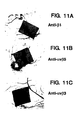
- the monoclonal antibody LM609 secreted by hybridoma ATCC HB 9537 was produced using standard hybridoma methods by immunization with isolated ⁇ v ⁇ 3 adsorbed onto Sepharose-lentil lectin beads.
- the ⁇ v ⁇ 3 had been isolated from human melanoma cells designated M21, and antibody was produced as described by Cheresh et al., J. Biol. Chem., 262:17703-17711 (1987 ).
- M21 cells were provided by Dr. D. L. Morton (University of California at Los Angeles, CA) and grown in suspension cultures in RPMI 1640 culture medium containing 2 mM L-glutamine, 50 mg/ml gentamicin sulfate and 10% fetal calf serum.
- Monoclonal antibody LM609 has been shown to specifically immunoreact with ⁇ v ⁇ 3 complex, and not immunoreact with ⁇ v subunit, with ⁇ 3 subunit, or with other integrins.
- the basement membranes of blood vessels express several adhesive proteins, including von Willebrand factor, fibronectin, and fibrin.
- several members of the integrin family of adhesion receptors are expressed on the surface of cultured smooth muscle and endothelial cells. See, Cheresh, Proc. Natl. Acad. Sci.. USA, 84:6471 (1987 ); Janat et al., J. Cell Physiol., 151:588 (1992 ); and Cheng et al., J. Cell Physiol., 139:275 (1989 ).
- integrins ⁇ v ⁇ 3 , the endothelial cell receptor for von Willebrand factor, fibrinogen (fibrin), and fibronectin as described by Cheresh, Proc. Natl. Acad. Sci.. USA, 84:6471 (1987 ).
- This integrin initiates a calcium-dependent signaling pathway leading to endothelial cell migration, and therefore appears to play a fundamental role in vascular cell biology as described by Leavelsey et al., J. Cell Biol., 121:163 (1993 ).
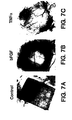
- ⁇ v ⁇ 3 during angiogenesis, human wound granulation tissue or adjacent normal skin was obtained from consenting patients, washed with 1 ml of phosphate buffered saline and embedded in O.T.C medium (Tissue Tek). The embedded tissues were snap frozen in liquid nitrogen for approximately 30 to 45 seconds. Six micron thick sections were cut from the frozen blocks on a cryostat microtome for subsequent immunoperoxidase staining with antibodies specific for either ⁇ 3 integrins ( ⁇ v ⁇ 3 or ⁇ IIb ⁇ 3 ) or the ⁇ 1 subfamily of integrins.
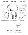
- FIGS 1A-1D The results of the staining of normal human skin and wound granulation tissue are shown in Figures 1A-1D .
- ⁇ v ⁇ 3 integrin was abundantly expressed on blood vessels in granulation tissue ( Figure 1B ) but was not detectable in the dermis and epithelium of normal skin from the same donor ( Figure 1A ).
- ⁇ 1 integrins were abundantly expressed on blood vessels and stromal cells in both normal skin ( Figure 1C ) and granulation tissue ( Figure 1D ) and, as previously shown as described by Adams et al., Cell, 63:425 (1991 ), on the basal cells within the epithelium.
- biopsies of cancer tissue from human patients were also examined for the presence and distribution of ⁇ v ⁇ 3 .
- the tissues were prepared as described in Example 1A with the exception that they were stained with monoclonal antibody LM609 prepared in Example 2 that is specific for the integrin receptor complex, ⁇ v ⁇ 3 .
- tumors were also prepared for microscopic histological analysis by fixing representative examples of tumors in Bulins Fixative for 8 hours and serial sections cut and H&E stained.
- tissue types namely granulated, metastatic tissues and other tissues in which angiogenesis is occurring and not normal tissue where the formation of new blood vessels has stopped. These tissues therefore provide an ideal target for therapeutic aspects of this invention.
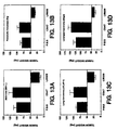
- CS-1 hamster melanoma cells lacking expression of ⁇ v ⁇ 3 and ⁇ v ⁇ 3 were first transfected with an plasmid for expressing the ⁇ 3 subunit as previously described by Filardo et al., J. Cell Biol., 130:441-450 (1995 ). Specificity of potential ⁇ v ⁇ 3 antagonists was determined by the ability to block the binding of ⁇ v ⁇ 3 -expressing CS-1 cells to VN or laminin coated plates. As an example of a typical assay, the wells were first coated with 10 ug/ml substrate overnight.
- peptide 85189 (SEQ ID NO 15) over a concentration range of 0.0001 uM to 100 uM, was separately mixed with CS-1 cells for applying to wells with a cell number of 50,000 cells/well. After a 10-15 minute incubation at 37C, the solution containing the cells and peptides was discarded. The number of attached cells was then determined following staining with 1% crystal violet. Cell associated crystal violet was eluted by the addition of 100 microliters (ul) of 10% acetic acid. Cell adhesion was quantified by measuring the optical density of the eluted crystal violet at a wave length of 600 nm.
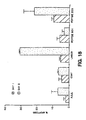
- Figure 21 shows the result of a typical assay with an ⁇ v ⁇ 3 antagonist, here peptide 85189. No inhibition was detected with the peptide on laminin-coated surfaces. In contrast, complete inhibition of binding was obtained on VN-coated surfaces with a peptide concentration of 10 uM or greater, as shown with the dose-response curve.
- MMP-2-derived polypeptides include regions of the C-terminus of MMP-2 active in the binding interaction with ⁇ v ⁇ 3 and thereby capable of inhibiting MMP-2 activation and associated activities.
- These polypeptides are prepared' either as synthetic polypeptides having a sequence derived from the C-terminal domain of MMP-2 as described in Example 1 or as fusion proteins including all or a portion of the C-terminal domain of MMP-2, prepared as described below.
- MMP-2 C-terminal molecules are presented for both chicken and human specific sequences.
- the chicken-derived MMP-2 C-terminal domain also referred to as the hemopexin domain immediately contiguous with the hinge region, comprises the amino acid residues 445-637 of MMP-2.
- the complete nucleotide and encoded amino acid sequence of chicken MMP-2 is described below.
- the human MMP-2 nucleotide and encoded amino acid sequence is also described below.
- the C-terminal domain in the human MMP-2 that corresponds to the chicken region of 445-637 begin at amino acid residue 439 and ends with 631 due to six missing residues from the human sequence as shown in Figures 22A and 22B .
- Both human- and chicken-derived C-terminal MMP-2 synthetic peptides for use in practicing the methods of this invention are listed in Table 1.
- the amino acid residue sequences of the synthetic peptides are the same as those generated by the recombinant fusion protein counterparts but without the GST fusion component.
- the C-terminal MMP-2 fusion proteins derived from both chicken and human are prepared as described
- a MMP-2 fusion protein is a chimeric polypeptide having a sequence of MMP-2 C-terminal domain or a portion thereof fused (operatively linked by covalent peptide bond) to a carrier (fusion) protein, such as glutathione sulfhydryl transferase (GST).
- GST glutathione sulfhydryl transferase
- the amino terminal sequences for the chicken proenzyme and active enzyme are contained with diamonds and single arrowheads.
- the chicken progelatinase nucleotide and amino acid residue sequences are listed together as SEQ ID NO 29 while the encoded amino acid residue sequence is listed separately as SEQ ID NO 30.
- Templates for generating amplified regions of chicken MMP-2 were either a cDNA encoding the full-length mature chicken MMP-2 polypeptide provided by Dr. J. P. Quigley of the State University of New York at Stoney Brook, New York or a cDNA generated from a total cellular RNA template derived by standard techniques from an excised sample of chicken chorioallantoic membrane tissue.
- the cDNA was obtained with MuLV reverse transcriptase and a downstream primer specific for the 3'-terminal nucleotides, 5'ATTGAATTCTTCTACAGTTCA3' (SEQ ID NO 31), the 5' and 3' ends of which was respectively complementary to nucleotides 1932-1912 of the published chick MMP-2 sequence.
- Reverse transcriptase polymerase chain reaction (RT-PCR) was performed according to the specifications of the manufacturer for the GeneAmp RNA PCR Kit (Perkin Elmer). The primer was also engineered to contain an internal EcoRI restriction site.
- Upstream or 5' primers for amplifying each of the nucleotide regions for encoding the above-listed MMP-2 fusion proteins were designed to encode the polypeptide start sites 3' to an engineered, i.e., PCR-introduced, internal BamHI restriction site to allow for directional ligation into either pGEX-1 ⁇ T or pGEX-3X expression vectors.
- the 5' primers included the following sequences, the 5' and 3' ends of which correspond to the indicated 5' and 3' nucleotide positions of the chicken MMP-2 sequence as shown in Figure 22A and 22B (the amino acid residue position start sites are also indicated for each primer): 1) Nucleotides 599-619, encoding a 203 start site 5'ATGGGATCCACTGCAAATTTC3' (SEQ ID NO 32); 2) Nucleotides 809-830, encoding a 274 start site 5'GCCGGATCCATGACCAGTGTA3' (SEQ ID NO 33); 3) Nucleotides 863-883, encoding a 292 start site 5'GTGGGATCCCTGAAGACTATG3' (SEQ ID NO 34); 4) Nucleotides 1217-1237, encoding a 410 start 5'AGGGGATCCTTAAGGGGATTC3' (SEQ ID NO 35); and 5) Nucleotides 1325-1345, encoding a 445
- the indicated nucleotide regions of the template cDNA were subsequently amplified for 35 cycles (annealing temperature 55C) according to the manufacturer's instructions for the Expand High Fidelity PCR System (Boehringer Mannheim).
- the resulting PCR products were gel-purified, digested with BamHI and EcoRI restriction enzymes, and repurified before ligation into either pGEX-1 ⁇ T or pGEX-3X vector (Pharmacia Biotech, Uppsala, Sweden) which had been similarly digested as well as dephosphorylated prior to the ligation reaction.
- the choice of plasmid was based upon the required reading frame of the amplification product. Competent E.
- coli strain BSJ72 or BL21 cells were transformed with the separate constructs by heat shock.
- the resulting colonies were screened for incorporation of the respective MMP-2 fusion protein-encoding plasmid by PCR prior to dideoxy sequencing of positive clones to verify the integrity of the introduced coding sequence.
- verification of incorporation of plasmid was confirmed by expression of the appropriately-sized GST-MMP-2 fusion protein.
- fusion proteins were designed to contain engineered terminal cysteine residues at the amino- or carboxy-terminus of the chicken MMP-2 sequences of interest so as to provide for disulfide-bonding with the naturally occurring cysteine at the other terminus, as required by the construct.
- Oligonucleotide primers were accordingly designed to allow for amplification of chicken MMP-2 C-terminal regions for expression of soluble MMP-2/GST fusion proteins.
- Amplified chicken MMP-2 C-terminal regions included those for encoding amino acid residue positions 445-518, 445-552, 516-637 and 549-637.
- residue 517 the naturally encoded tyrosine residue was substituted for a cysteine to allow for disulfide bonding with either cysteine at residue position 446 or 637.
- residue 551 the naturally encoded tryptophan residue was substituted for a cysteine to allow for disulfide bonding with either naturally encoded cysteine at residue position 446 or 637.
- the pGEX-3X plasmid construct encoding the recombinant GST/MMP-2(410-637) fusion protein prepared above was used as a template for amplification according to the manufacturer's protocol for the Expand High Fidelity PCR Kit (Boehringer Mannheim) utilizing a set of oligonucleotide primers whose design was based on the published chicken MMP-2 sequence (also shown in Figures 22A and 22B .
- One upstream primer designed to encode a chicken MMP-2 protein start site at position 445 after an engineered internal BamHI endonuclease restriction site for insertion into the pGEX-3X GST vector, had the nucleotide sequence (5'CTCGGATCCTCTGCAAGCACG3' (SEQ ID NO 37)). The 5' and 3' ends of the primer respectively corresponded to positions 1325-1345 of the chicken MMP-2 sequence in the figure.
- Another upstream primer designed to encode a chicken MMP-2 protein start site at position 516 after an engineered internal BamHI restriction site for insertion into the pGEX-1 ⁇ T GST vector and to encode a cysteine residue at position 517, had the nucleotide sequence (5'GCAGGATCCGAGTGCTGGGTTTATAC3' (SEQ ID NO 38)).
- the 5' and 3' ends of the primer respectively corresponded to positions 1537-1562 of the chicken MMP-2 sequence.
- a third upstream primer designed to encode a chicken MMP-2 protein start site at position 549 following an engineered internal EcoRI endonuclease restriction site for insertion into the pGEX-1 ⁇ T GST vector and to encode a cysteine residue at position 551, had the nucleotide sequence (5'GCAGAATTCAACTGTGGCAGAAACAAG3' (SEQ ID NO 39)). The 5' and 3' ends of the primer respectively corresponded to positions 1639-1665 of the chicken MMP-2 sequence.
- upstream primers were separately used with one of the following downstream primers listed below to produce the above-described regions from the C-terminal domain of chicken MMP-2.
- a first downstream primer (antisense), designed to encode a chicken MMP-2 protein termination site at position 518, to encode a cysteine residue at position 517, and to contain an internal EcoRI endonuclease restriction site for insertion into a GST vector, had the nucleotide sequence (5'GTAGAATTCCAGCACTCATTTCCTGC3' (SEQ ID NO 40)).
- the 5' and 3' ends of the primer, written in the 5'-3' direction, were respectively complementary in part to positions 1562-1537 of the chicken MMP-2 sequence.
- a second downstream primer designed to encode a chicken MMP-2 protein termination site at position 552, to encode a cysteine residue at position 551, and to contain an internal-EcoRI endonuclease restriction site for insertion into a GST vector, had the nucleotide sequence (5'TCTGAATTCTGCCACAGTTGAAGG3' (SEQ ID NO 41)).
- the 5' and 3' ends of the primer, written in the 5'-3' direction, were respectively complementary in part to positions 1666-1643 of the chicken MMP-2 sequence.
- a third downstream primer designed to encode a chicken MMP-2 protein termination site at position 637 and to contain an internal EcoRI endonuclease restriction site for insertion into a GST vector, had the nucleotide sequence (5'ATTGAATTCTTCTACAGTTCA3' (SEQ ID NO 42)).
- the resulting amplification products were separately purified, digested with BamHI and or EcoRI restriction enzymes as necessary, and repurified before ligation into the appropriate GST fusion protein vector, either pGEX-3X or pGEX-1 ⁇ T, as indicated above by the reading frame of the upstream oligonucleotide primer.
- the vectors were similarly digested as well as dephosphorylated prior to the ligation reaction. Competent E. coli strain BL21 cells were then separately transformed with the resultant MMP-2-containing vector constructs by heat shock. Resulting colonies were then screened for incorporation of the appropriate fusion protein-encoding plasmid by PCR and production of the appropriate sized GST-fusion protein prior to dideoxy sequencing of positive clones to verify the integrity of the introduced coding sequence. Purification of recombinant GST fusion proteins were then performed using IPTG-induced log-phase cultures essentially as described above for producing the other GST-MMP-2 fusion proteins.
- results of inhibition of cell attachment assays with various chicken MMP-2 proteins as well as with other peptides indicate that intact MMP-2, the fusion protein CTMMP-2(2-4) from residues 445-637 and peptide 66203 (SEQ ID NO 5) but not MMP-2 (1-445) and control peptide 69601 inhibited ⁇ 3 -expressing CS-1 cell adhesion to vitronectin but not laminin, and thereby inhibited vitronectin receptor ( ⁇ v ⁇ 3 ) binding to vitronectin by interfering with normal ⁇ v ⁇ 3 binding activity.
- Other tested CTMMP-2 fusion proteins 7-1 from residues 274-637, 10-1 from residues 292-637 and 4-3 from residues 274-400 had less affect on cell adhesion compared to 2-4.
- human MMP-2 GST fusion proteins were produced for expressing amino acid regions 203-631 and 439-631 of the mature human MMP-2 proenzyme polypeptide. The indicated regions correspond respectively to chicken MMP-2 regions 203-637 and 445-637.
- Human MMP-2-GST fusion proteins were produced by PCR as described above for the chicken MMP-2-GST fusion proteins utilizing a cDNA template that encoded the entire human MMP-2 open reading frame provided by Dr. W. G. Stetler-Stevenson at the National Cancer Institute, Bethesda, MD. Upstream 5' primer sequences were designed based upon the previously published sequence of human MMP-2 ( Collier et al., J. Biol. Chem., 263:6579-6587 (1988 ) and to encode an introduced internal EcoRI restriction site to allow for insertion of the amplified products into the appropriate expression vector.
- One upstream primer designed to encode a human MMP-2 protein start site at position 203 after an engineered internal EcoRI endonuclease restriction site for insertion into the pGEX-1 ⁇ T GST vector, had the nucleotide sequence (5'GATGAATTCTACTGCAAGTT3' (SEQ ID NO 43)). The 5' and 3' ends of the primer respectively corresponded to positions 685-704 of the human MMP-2 open reading frame sequence.
- Another upstream primer designed to encode a human MMP-2 protein start site at position 439 after an engineered internal EcoRI restriction site for insertion into the pGEX-1 ⁇ T GST vector, had the nucleotide sequence (5'CACTGAATTCATCTGCAAACA3' (SEQ ID NO 44)). The 5' and 3' ends of the primer respectively corresponded to positions 1392 and 1412 of the human MMP-2 open reading frame sequence.
- Each of the above primers were used separately with a downstream primer, having 5' and 3' ends respectively complementary to bases 1998 and 1978 of the human MMP-2 sequence that ends distal to the MMP-2 open reading frame and directs protein termination after amino acid residue 631.
- the amplified products produced expressed fusion proteins containing human MMP-2 amino acid residues 203-631 (SEQ ID NO 45) and 439-631 (SEQ ID NO 18).
- PCR products were purified, digested with EcoRI and repurified for ligation into a pGEX-1 ⁇ T plasmid that was similarly digested and dephosphorylated prior to the ligation reaction.
- Cells were transformed as described above.
- the synthetic peptides prepared in Example 1 along with the MMP-2 fusion proteins described above were further screened by measuring their ability to antagonize ⁇ v ⁇ 3 and ⁇ IIb ⁇ 3 receptor binding activity in purified ligand-receptor binding assays.
- the method for these binding studies has been described by Barbas et al., Proc. Natl. Acad. Sci.. USA. 90:10003-10007 (1993 ), Smith et al., J. Biol. Chem., 265:11008-11013 (1990 ), and Pfaff et al., J. Biol. Chem., 269:20233-20238 (1994 ), the disclosures of which are hereby incorporated by reference.
- a method of identifying antagonists in a ligand-receptor binding assay in which the receptor is immobilized to a solid support and the ligand and antagonist are soluble. Also described is a ligand-receptor binding assay in which the ligand is immobilized to a solid support and the receptor and antagonists are soluble.
- integrins were separately immobilized in Titertek microtiter wells at a coating concentration of 50 nanograms (ng) per well.
- the purification of the receptors used in the ligand-receptor binding assays are well known in the art and are readily obtainable with methods familiar to one of ordinary skill in the art.
- After incubation for 18 hours at 4C, nonspecific binding sites on the plate were blocked with 10 milligrams/milliliter (mg/ml) of bovine serum albumin (BSA) in Tris-buffered saline.
- BSA bovine serum albumin
- the RGD-containing or RGD-derivatized cyclic peptides 62181, 62184, 62185 and 62187 exhibited preferential inhibition of fibrinogen binding to the ⁇ v ⁇ 3 receptor as measured by the lower concentration of peptide required for half-maximal inhibition as compared to that for the ⁇ IIb ⁇ 3 receptor.
- the other RGD-containing or RGD-derivatized cyclic peptides, 62186, 62175 and 62179 were either not as effective in blocking fibrinogen binding to ⁇ v ⁇ 3 or exhibited preferential inhibition of fibrinogen binding to ⁇ IIb ⁇ 3 as compared to ⁇ v ⁇ 3 .
- ligand receptor binding assays performed as described above with the exception that detection of binding or inhibition thereof was with ELISA and peroxidase-conjugated goat anti-rabbit IgG, the ligands VN, MMP-2 and fibronectin at a range of 5 - 50 ng/well and listed in the order of effectiveness were shown to bind to immobilized ⁇ v ⁇ 3 receptor while collagen did not.
- the ability of peptides to inhibit the binding of either MMP-2 or VN to immobilized ⁇ v ⁇ 3 was assessed with peptides 69601 (SEQ ID NO 6) and 66203 (SEQ ID NO 5). Only peptide 66203 was effective at inhibiting the binding of either substrate to the ⁇ v ⁇ 3 receptor while the control peptide 69601 failed to have an effect with either ligand.
- MMP-2 binding to integrin receptors was confirmed with a solid phase receptor binding assay in which iodinated MMP-2 was shown to bind to ⁇ v ⁇ 3 and not to ⁇ IIb ⁇ 3 that had been immobilized on a solid phase (300 bound cpm versus approximately 10 bound CPM).
- the ability of an MMP-2 derived peptide or fusion protein to inhibit the specific binding of MMP-2 to ⁇ v ⁇ 3 was demonstrated in a comparable assay, the results of which are shown in Figure 23 .
- the GST-CTMMP-2(445-637) (also referred to as CTMMP-2(2-4)) fusion protein prepared as described above, labeled GST-MAID, inhibited the binding of iodinated MMP-2 to ⁇ v ⁇ 3 while GST alone had no effect with levels of bound CPM comparable to wells receiving no inhibitor at all (labeled NT).
- the binding of labeled GST2-4 was competed by unlabeled GST2-4.
- the ligand-receptor assay described herein can be used to screen for both circular or linearized synthetic peptides that exhibit selective specificity for a particular integrin receptor, specifically ⁇ v ⁇ 3 , as used as vitronectin receptor ( ⁇ v ⁇ 3 ) antagonists in practicing this invention.
- Angiogenesis can be induced on the chick chorioallantoic membrane (CAM) after normal embryonic angiogenesis has resulted in the formation of mature blood vessels.
- Angiogenesis has been shown to be induced in response to specific cytokines or tumor fragments as described by Leibovich et al., Nature, 329:630 (1987 ) and Ausprunk et al., Am. J. Pathol., 79:597 (1975 ).
- CAMs were prepared from chick embryos for subsequent induction of angiogenesis and inhibition thereof as described in Examples 6 and 7, respectively. Ten day old chick embryos were obtained from McIntyre Poultry (Lakeside, CA) and incubated at 37C with 60% humidity.
- a small hole was made through the shell at the end of the egg directly over the air sac with the use of a small crafts drill (Dremel, Division of Emerson Electric Co. Racine WI).
- a second hole was drilled on the broad side of the egg in a region devoid of embryonic blood vessels determined previously by candling the egg. Negative pressure was applied to the original hole, which resulted in the CAM (chorioallantoic membrane) pulling away from the shell membrane and creating a false air sac over the CAM.
- a 1.0 centimeter (cm) x 1.0 cm square window was cut through the shell over the dropped CAM with the use of a small model grinding wheel (Dremel). The small window allowed direct access to the underlying CAM.
- the resultant CAM preparation was then either used at 6 days of embryogenesis, a stage marked by active neovascularization, without additional treatment to the CAM reflecting the model used for evaluating effects on embryonic neovascularization or used at 10 days of embryogenesis where angiogenesis has subsided.
- the latter preparation was thus used in this invention for inducing renewed angiogenesis in response to cytokine treatment or tumor contact as described in Example 6.
- the CAMs and tumors were prepared for frozen sectioning as described in Example 3A. Six micron (um) thick sections were cut from the frozen blocks on a cryostat microtome for immunofluorescence analysis.
- Figure 4 shows a typical photomicrograph of an area devoid of blood vessels in an untreated 10 day old CAM.
- angiogenesis in the CAM system is subsiding by this stage of embryogenesis, the system is useful in this invention for stimulating the production of new vasculature from existing vessels from adjacent areas into areas of the CAM currently lacking any vessels.
- mAb CSAT micrograms/milliliter
- mAb CSAT a monoclonal antibody specific for the ⁇ 1 integrin subunit as described by Buck et al., J. Cell Biol., 107:2351 (1988 ) and thus used for controls, or LM609 as prepared in Example 2.
- Primary staining was followed by staining with a 1:250 dilution of goat anti-mouse rhodamine labeled secondary antibody (Tago) to allow for the detection of the primary immunoreaction product.
- the sections were then analyzed with a Zeiss immunofluorescence compound microscope.
- MMP-2 and ⁇ v ⁇ 3 colocalized on endothelial cells undergoing angiogenesis three days following bFGF induction in the 10 day old CAM model.
- MMP-2 was only minimally expressed on vessels that lacked the ⁇ v ⁇ 3 receptor.
- MMP-2 colocalized with ⁇ v ⁇ 3 on angiogenic M21-L tumor-associated blood vessels in vivo (tumors resulting from injection of M21-L human melanoma cells into the dermis of human skin grafts grown on SCID mice as described in Example 11) but not with preexisting non-tumor associated blood vessels.
- Similar results of the selective association of MMP-2 and ⁇ v ⁇ 3 were also obtained with ⁇ v ⁇ 3 bearing CS-1 melanoma tumors in the CAM model but not with CS-1 cells lacking ⁇ v ⁇ 3 .
- Angiogenesis has been shown to be induced by cytokines or growth factors as referenced in Example 5A.
- angiogenesis in the CAM preparation described in Example 5 was induced by growth factors that were topically applied onto the CAM blood vessels as described herein.
- Angiogenesis was induced by placing a 5 millimeter (mm) X 5 mm Whatman filter disk (Whatman Filter paper No.1) saturated with Hanks Balanced Salt Solution (HBSS, GIBCO, Grand Island, NY) or HBSS containing 150 nanograms/milliliter (ng/ml) recombinant basic fibroblast growth factor (bFGF) (Genzyme, Cambridge, MA) on the CAM of a 10-day chick embryo in a region devoid of blood vessels and the windows were latter sealed with tape. In other assays, 125 ng/ml bFGF was also effective at inducing blood vessel growth.
- HBSS Hanks Balanced Salt Solution
- bFGF basic fibroblast growth factor
- angiogenesis was first induced with 1-2 ug/ml bFGF in fibroblast growth medium. Angiogenesis was monitored by photomicroscopy after 72 hours. CAMs were snap frozen, and 6 um cryostat sections were fixed with acetone and stained by immunofluorescence as described in Example 5C with 10 ug/ml of either anti- ⁇ 1 monoclonal antibody CSAT or LM609.
- the immunofluorescence photomicrograph in Figure 5C shows enhanced expression of ⁇ v ⁇ 3 during bFGF-induced angiogenesis on the chick CAM in contrast with the absence of ⁇ v ⁇ 3 expression in an untreated chick CAM as shown in Figure 5B .
- ⁇ v ⁇ 3 was readily detectable on many (75% to 80%) of the vessels on the bFGF-treated CAMs.
- the expression of integrin ⁇ 1 did not change from that seen in an untreated CAM as ⁇ 1 was also readily detectable on stimulated blood vessels.
- ⁇ v ⁇ 3 and ⁇ 1 integrins were then quantified during bFGF-induced angiogenesis by laser confocal image analysis of the CAM cryostat sections.
- the stained sections were then analyzed with a Zeiss laser confocal microscope.
- Twenty-five vessels stained with LM609 and 15 stained with CSAT (average size - 1200 sq mm 2 , range 350 to 3,500 mm 2 ) were selected from random fields and the average rhodamine fluorescence for each vessel per unit area was measured in arbitrary units by laser confocal image analysis. Data are expressed as the mean fluorescence intensity in arbitrary units of vessels ⁇ standard error (SE).
- SE standard error
- the CAM assay was further used to examine the effect of another potent angiogenesis inducer, tumor necrosis factor-alpha (TNF ⁇ ), on the expression of ⁇ 1 and ⁇ 3 integrins.
- TNF ⁇ tumor necrosis factor-alpha
- Filter disks impregnated with either bFGF or TNF ⁇ and placed on CAMs from 10 day embryos were found to promote local angiogenesis after 72 hours.
- the CAM preparation for evaluating the effect of angiogenesis inhibitors on the natural formation of embryonic neovasculature was the 6 day embryonic chick embryo as previously described. At this stage in development, the blood vessels are undergoing de novo growth and thus provides a useful system for determining if ⁇ v ⁇ 3 participates in embryonic angiogenesis.
- the CAM system was prepared as described above with the exception that the assay was performed at embryonic day 6 rather than at day 10.
- the effect on embryonic angiogenesis by treatment with antibodies and peptides of this invention are presented in Example 7C.
- ⁇ v ⁇ 3 tumor-induced angiogenesis
- various ⁇ v ⁇ 3 -negative human melanoma and carcinoma fragments were used in the CAM assay that were previously grown and isolated from the CAM of 17-day chick embryo as described by Brooks et al., J. Cell Biol., 122:1351 (1993 ) and as described herein.
- the fragments induced extensive neovascularization in the presence of buffer alone.
- Angiogenesis was induced in the CAM assay system by direct apposition of a tumor fragment on the CAM.
- Preparation of the chick embryo CAM was identical to the procedure described above. Instead of a filter paper disk, a 50 milligram (mg) to 55 mg in weight fragment of one of human melanoma tumor M21-L, human lung carcinoma tumor UCLAP-3, human pancreatic carcinoma cell line FG ( Cheresh et al., Cell 58:945-953, 1989 ), or human laryngeal carcinoma cell line HEp3, all of which are ⁇ v ⁇ 3 negative tumors, was placed on the CAM in an area originally devoid of blood vessels.
- the tumors were resected from the CAMs and trimmed free of surrounding CAM tissue.
- the tumors were sliced into 50 mg to 55 mg tumor fragments for use in either angiogenesis or tumor growth assays.
- the tumor fragments were placed on a new set of 10 day chick embryo CAMs as described in Example 6A in an area devoid of blood vessels.
- Tumors grown in vivo on the chick embryo CAMs were stained for ⁇ v ⁇ 3 expression with mAb LM609 as described in Example 3A. No specific staining of tumor cells was observed indicating a lack of ⁇ v ⁇ 3 expression.
- CAM tumor preparations were then subsequently treated as described in Examples 7D and 7E for measuring the effects of antibodies and peptides on tumor-induced angiogenesis.
- the CAM tumor preparations were also treated as described in Examples 8, 9, and 12 for measuring the effects of antibodies and peptides on regression of tumors and apoptosis of angiogenic blood vessels and vascular cells.
- ⁇ v ⁇ 3 plays an active role in angiogenesis
- filter disks saturated with bFGF or TNF ⁇ were placed on CAMs then the monoclonal antibodies (also referred to as mAb), LM609 (specific for ⁇ v ⁇ 3 ), CSAT (specific for ⁇ 1 ), or P3G2 or also P1F6 (both specific for ⁇ v ⁇ 5 ) were added to the preparation.
- monoclonal antibodies also referred to as mAb
- LM609 specific for ⁇ v ⁇ 3
- CSAT specific for ⁇ 1
- P3G2 or also P1F6 both specific for ⁇ v ⁇ 5
- Angiogenesis was induced on CAMs from 10 day chick embryos by filter disks saturated with bFGF. Disks were then treated with 50 ml HBSS containing 25 mg of mAb in a total volume of 25 ul of sterile HBSS at 0, 24, and 48 hours. At 72 hours, CAMs were harvested and placed in a 35 mm petri dish and washed once with 1 ml of phosphate buffered saline. The bottom side of the filter paper and CAM tissue was then analyzed under an Olympus stereo microscope, with two observers in a double-blind fashion. Angiogenesis inhibition was considered significant when CAMs exhibited >50% reduction in blood vessel infiltration of the CAM directly under the disk. Experiments were repeated four times per antibody, with 6 to 7 embryos per condition.
- FIGS 8A-8B The results of the effects of mAb treatment on bFGF-induced angiogenesis is shown in Figures 8A-8B .
- An untreated CAM preparation devoid of blood vessels is shown in Figure 8A to provide a comparison with the bFGF-blood vessel induction shows in Figure 8B and effects thereon by the mAbs in Figures 8C-8E .
- About 75% of these CAMs treated with mAb LM609 exhibited >50% inhibition of angiogenesis as shown in Figure 8E , and many of these appeared devoid of vessel infiltration.
- the buffer control ( Figure 8A ) and disks treated with mAbs CSAT ( Figure 8C ) and P3G2 ( Figure 8D ) consistently showed extensive vascularization.
- CAM assays were also performed with the synthetic peptides of this invention to determine the effect of cyclic and linearized peptides on growth factor induced angiogenesis.
- the peptides were prepared as described in Example 1 and 80 ug of peptide were presented in a total volume of 25 ul of sterile HBSS. The peptide solution was applied to the CAM preparation immediately and then again at 24 and 48 hrs. At 72 hours the filter paper and surrounding CAM tissue was dissected and viewed as described above.
- Results from this assay revealed were similar to those shown in Figures 9A-9C as described in Example 7E2) where synthetic peptides were intravenously injected into tumor induced blood vessels.
- the control peptide, 62186 the bFGF-induced blood vessels remained undisturbed as shown in Figure 9A .
- the cyclic RGD peptide, 62814 was applied to the filter, the formation of blood vessels was inhibited leaving the area devoid of new vasculature. This effect was similar in appearance to that shown in Figure 9B as described in Example 7E2) below.
- CAM assays were performed as described above with the exception that angiogenesis was induced with filter discs saturated for 10 minutes with bFGF at a concentration of 1.0 ug/ml in HBS. The discs were then positioned on the CAM in an area that was reduced in the number of preexisting vessels.
- the C-terminal CTMMP-2(410-637) fusion protein, prepared as described above, or control GST receptor associated fusion protein (RAP) was applied then topically to the filter disc once per day for a total of three days.
- angiogenesis was quantified by counting the number of blood vessels branch points that occur within the confines of the filter discs.
- the branched blood vessels are considered to correspond primarily to new angiogenic sprouting blood vessels.
- Angiogenic Index is the number of branch points (bFGF stimulated) minus the number of branch points (control unstimulated) per filter disc. Experiments routinely had 6-10 embryos per condition.
- FIGS 25A-D , 26 and 27 The results of the CAM angiogenesis assay are shown in Figures 25A-D , 26 and 27 .
- Figure 25 a series of photographs divided into four figures, Figures 25A-D , illustrate the comparison of angiogenesis inhibited in the presence of the CTMMP-2 fusion protein (CTMMP-2(410-637) ( Figures 25C-D ) and not inhibited in the presence of control GST fusion protein ( Figures 25A-B ).
- Figures 26 and 27 are bar graphs illustrating the angiogenesis index of CAM angiogenesis assays with CTMMP-2, the same fusion protein as above, compared to controls (bFGF only or GST-RAP fusion protein).
- Figure 27 the results of two separate experiments (#1 & #2) using CTMMP-2(410-637) fusion protein are shown.
- CTMMP-2 fusion protein or polypeptide containing a C-terminal domain of MMP-2 is a useful composition for inhibition of bFGF-mediated angiogenesis by inhibiting ⁇ v ⁇ 3 .
- the preparation of the chick embryo CAMs for intravenous injections were essentially as described in Example 7A with some modifications.
- prominent blood vessels were selected and marks were made on the egg shell to indicate their positions.
- the holes were drilled in the shell and the CAMs were dropped and bFGF saturated filter papers were placed on the CAMs as described above.
- the windows were sealed with sterile tape and the embryos were replaced in the incubator. Twenty four hours later, a second small window was carefully cut on the lateral side of the egg shell directly over prominent blood vessels selected previously.
- the outer egg shell was carefully removed leaving the embryonic membranes intact.
- the shell membrane was made transparent with a small drop of mineral oil (Perkin-Elmer Corp, Norwalk, CT) which allowed the blood vessels to be visualized easily.
- Purified sterile mAbs, or synthetic peptides, the latter of which are described below, were inoculated directly into the blood vessels once with a 30 gauge needle at a dose of 200 ug of IgG per embryo in a total volume of 100 ul of sterile PBS. The windows were sealed with tape and the embryos were allowed to incubate until 72 hours. The filter disks and surrounding CAM tissues were analyzed as described before.
- FIGS. 10A-10C The results of intravenous antibody treatment to the bFGF induced blood vessel CAM preparation are shown in Figures 10A-10C .
- FIG 10A angiogenesis induced as a result of bFGF treatment is shown. No change to the presence of bFGF induced vasculature was seen with intravenous exposure to mAb P3G2, an anti- ⁇ v ⁇ 3 antibody, as shown in Figure 10B .
- treatment of the bFGF induced angiogenesis CAM preparation with LM609, an anti- ⁇ v ⁇ 3 antibody resulted in the complete inhibition of growth of new vessels into the filter area as shown in Figure 10C .
- the inhibitory effect on angiogenesis is thus resulting from the inhibition of ⁇ v ⁇ 3 receptor activity by the LM609 anti- ⁇ v ⁇ 3 -specific antibody. Since the blocking of the ⁇ v ⁇ 5 does not inhibit the formation of neovasculature into the CAMs filter site, ⁇ v ⁇ 5 thus is not essential as compared to ⁇ v ⁇ 3 for growth of new vessels.
- synthetic peptides 69601 (control) and 66203 (SEQ ID NO 5) were separately intravenously injected into CAM preparations 18 hours after bFGF induction of angiogenesis. The preparations were maintained for an additional 36-40 hours after which time the number of branch points were determined as previously described.
- peptide 85189 (SEQ ID NO 15) was evaluated for inhibiting bFGF-induced angiogenesis in the CAM assay over a dosage range of 10 ug/embryo to 300 ug/embryo.
- the assay was performed as previously described. The results are shown in Figure 29 where the lowest effective dose was 30 ug with 100 and 300 ug nearly completely inhibiting angiogenesis.
- peptide 85189 was compared to peptides 69601 and 66203 for anti-angiogenesis activity.
- the assay was performed as described above with the exception that 50 ug peptide were used.
- the results, plotted in Figure 30 showed that peptides 66203 (labeled 203) and 85189 (labeled 189) were effective inhibitors of bFGF-mediated angiogenesis compared to bFGF-treated (labeled bFGF) and 69601-treated (labeled 601) controls.
- peptide 85189 The effectiveness of the different salt formulations of peptide 85189 was also evaluated in similar bFGF-induced CAM assays.
- the peptides were used at 100 ug/embryo.
- Untreated CAMs, labeled as "no cytokine" had approximately half as many branch points as that seen with bFGF treatment, respectively 70 versus 190.
- Treatment with control peptide 69601 had no effect on inhibiting angiogenesis (230 branch points).
- Example 1 The other synthetic peptides prepared in Example 1 are separately intravenously injected into the growth factor induced blood vessels in the CAM preparation as described above. The effect of the peptides on the viability of the vessels is similarly assessed.
- CTMMP-2(2-4), also referred to as CTMMP-2(445-637) and CTMMP-2(10-1), also referred to as CTMMP-2(274-637) was also evaluated.
- the assay was performed as previously described with the exception that 50 ug of fusion protein was administered to the bFGF-treated embryos.
- the effect of fusion protein treatment was assessed at 24 hours, 48 hours and 72 hours.
- MMP-2 compositions whole MMP-2, fragments and fusion proteins, prepared as previously described are also separately intravenously injected into the growth factor induced blood vessels in the CAM preparation as described above.
- the effect of the peptides on the viability of the vessels is similarly assessed.
- Example 5A To determine whether ⁇ v ⁇ 3 participates in embryonic angiogenesis, the effect of LM609 on de novo growth of blood vessels on CAMs was examined in 6 day embryos, a stage marked by active neovascularization as described in Example 5A.
- the CAM assay was prepared as described in Example 6C with the subsequent topical application of disks saturated with mAbs placed on CAMs of 6 day old embryos in the absence of cytokines. After 3 days, CAMS were resected and photographed. Each experiment included 6 embryos per group and was repeated 2 times.
- Antibody LM609 ( Figure 11C ), but not CSAT ( Figure 11A ) or P3G2 ( Figure 11B ), prevented vascular growth under these conditions; this indicates that ⁇ v ⁇ 3 plays a substantial role in embryonic neovascularization that was independent of added growth factors for induction of angiogenesis.
- Example 1 The synthetic peptides prepared in Example 1 are separately added to the embryonic CAM preparation prepared above and as described in Example 5A2) by either topical application to the CAM or intravenous application to blood vessels. The effect of the peptides on the viability of the vessels is similarly assessed.
- mAbs were separately topically applied to the tumor fragments at a concentration of 25 ug in 25 ul of HBSS and the windows were then sealed with tape. The mAbs were added again in the same fashion at 24 hours and 48 hours. At 72 hours, the tumors and surrounding CAM tissues were analyzed as described above in Example 7A1).
- tumors were initially derived by transplanting cultured M21-L cells, which do not to express integrin ⁇ v ⁇ 3 as described by Felding-Habermann et al., J. Clin. Invest. , 89:2018 (1992) onto the CAMs of 10-day old chick embryos.
- These ⁇ v ⁇ 3 -negative fragments induced extensive neovascularization in the presence of buffer alone, or mAbs CSAT (anti- ⁇ 1 ) or P3G2 (anti- ⁇ v ⁇ 3 ).
- mAb LM609 abolished the infiltration of most vessels into the tumor mass and surrounding CAM.
- the synthetic peptides prepared in Example 1, including MMP-2-derived peptides and fusion proteins are topically applied to the tumor-induced angiogenic CAM assay system as described above. The effect of the peptides on the viability of the vessels is similarly assessed.
- Tumor-induced blood vessels prepared as described in Example 7E1) were also treated with mAbs applied by intravenous injection. Tumors were placed on the CAMs as described in Example 7D1) and the windows sealed with tape and 24 hours latter, 200 ug of purified mAbs were inoculated once intravenously in chick embryo blood vessels as described previously. The chick embryos were then allowed to incubate for 7 days. The extent of angiogenesis was then observed as described in above. As described in Example 8 below, after this time period, the tumors were resected and analyzed by their weight to determine the effect of antibody exposure on tumor growth or suppression.
- the effects of peptide exposure to tumor-induced vasculature in the CAM assay system was also assessed.
- the tumor-CAM preparation was used as described above with the exception that instead of intravenous injection of a mAb, synthetic peptides prepared as described in Example 1 and Example 7A2) were separately intravenously injected into visible blood vessels.
- FIGS 9A-9C The results of CAM assays with the cyclic peptide, 66203 containing the HCl salt, and control peptide, 62186, are shown in Figures 9A-9C .
- the treatment with the control peptide did not effect the abundant large blood vessels that were induced by the tumor treatment to grow into an area originally devoid of blood vessels of the CAM.
- the cyclic RGD peptide, 66203, an antagonist to ⁇ v ⁇ 3 was applied to the filter, the formation of blood vessels was inhibited leaving the area devoid of new vasculature as shown in Figure 9B .
- a CS-1 tumor ( ⁇ 3 -negative) was prepared in a CAM as described above. After 24 hours of tumor growth, a composition of MMP-2 fragment, designated CTMMP-2(2-4) and prepared as described in Example 4A, was administered intraveneously at 50 ug fragment in 100 ul of PBS. After 6 days, the tumor was evaluated for mass. Tumors treated with CTMMP-2(2-4) were reduced in growth rate by about 50% when compared to the growth rate of control tumors treated with CTMMP-2(10-1) or with PBS control. Thus, the ⁇ v ⁇ 3 antagonist inhibited tumor growth.
- Example 7E1 in addition to visually assessing the effect of anti- ⁇ v ⁇ 3 antagonists on growth factor or tumor induced angiogenesis, the effect of the antagonists was also assessed by measuring any changes to the tumor mass following exposure.
- the tumor-induced angiogenesis CAM assay system was prepared as described in Example 6C and 7D. At the end of the 7 day incubation period, the resulting tumors were resected from the CAMs and trimmed free of any residual CAM tissue, washed with 1 ml of phosphate buffer saline and wet weights were determined for each tumor.
- preparation of the tumor for microscopic histological analysis included fixing representative examples of tumors in Bulins Fixative for 8 hours and embedding in paraffin.
- Serial sections were cut and stained with hematoxylin and eosin (H&E) for microscopic analysis. Gladson, et al., J. Clin. Invest., 88:1924 (1991 ). Sections were photographed with an Olympus compound microscope at 250x.
- mice The results of typical human melanoma tumor (M21L) weights resulting from topical application of control buffer (HBSS), P3G2 (anti- ⁇ v ⁇ 5 ) or LM609 (anti- ⁇ v ⁇ 3 ) are listed in Table 4.
- HBSS control buffer
- P3G2 anti- ⁇ v ⁇ 5
- LM609 anti- ⁇ v ⁇ 3
- mAb Treatment Tumor Weight (mg) 1 HBSS 108 2 152 3 216 4 270 5 109 6 174 1 P3G2 134 2 144 3 408 4 157 5 198 6 102 7 124 8 99 1 LM609 24 2 135 3 17 4 27 5 35 6 68 7 48 8 59 mAb Treatment Average Tumor Weight (mg) HBSS control 172 ⁇ 26 P3G2 171 ⁇ 36 LM609 52 ⁇ 13
- ⁇ v ⁇ 3 receptor activity is therefore essential for an ⁇ v ⁇ 3 negative tumor to maintain its mass nourished by development of ⁇ v ⁇ 3 -expressing neovasculature.
- the blocking of ⁇ v ⁇ 3 with the ⁇ v ⁇ 3 antagonists of this invention results in the inhibition of angiogenesis into the tumor ultimately resulting in the diminution of tumor mass.
- mAb Treatment Tumor Weight (mg) 1 PBS 101 2 80 3 67 4 90 1 CSAT 151 2 92 3 168 4 61 5 70 1 LM609 16 2 54 3 30 4 20 5 37 6 39 7 12 mAb Treatment Average Tumor Weight (mg) PBS control 85 ⁇ 7 CSAT 108 ⁇ 22 LM609 30 ⁇ 6
- CAM thin semi-transparent membrane
- mAb LM609 inhibited tumor growth significantly (p ⁇ 0.001) as compared to controls in all tumors tested. Tumors treated with PBS or CSAT proliferated in all cases. In contrast, mAb LM609 not only prevented the growth of these tumors but induced extensive regression in most cases. Importantly, these tumor cells do not express integrin ⁇ v ⁇ 3 demonstrating that the inhibition of growth was due to the anti-angiogenic effects of this antibody on neovasculature rather than upon the tumor cells directly.
- Human M21-L melanoma tumor fragments (50 mg) were implanted on the CAMs of 10 day old embryos as described in Example 5A. Twenty four hours later, embryos were injected intravenously with PBS alone or a single dose (300 ug/100 ul) of either mAb LM609 (anti- ⁇ v ⁇ 3 ) or P3G2 (anti- ⁇ v ⁇ 5 ). Tumors were allowed to propagate as described in Example 9A1)a above and were examined morphologically and histologically as herein described.
- M21-L tumors treated with mAbs P3G2 (anti- ⁇ v ⁇ 5 ) or LM609 (anti- ⁇ v ⁇ 3 ) were examined morphologically.
- the P3G2-treated tumors were large (8 mm in diameter) and well vascularized whereas those treated with mAb LM609 were much smaller (3 mm in diameter) and lacked detectable blood vessels.
- tumors were further examined by the preparation of histological sections and staining with hematoxylin and eosin as described in Example 9A1)a.
- mAb P3G2 anti- ⁇ v ⁇ 5
- FIG. 14 lower panel
- tumors treated with mAb P3G2 showed numerous viable and actively dividing tumor cells as indicated by mitotic figures (arrowheads) as well as by multiple blood vessels (arrows) throughout the tumor stroma.
- mAb LM609 anti- ⁇ v ⁇ 3
- Human M21-L melanoma tumor fragments (50 mg) were implanted on the CAMs of 10 day oldembryos as described in Example 5A. Twenty four hours later, embryos received a single intravenous injection of 300 ug/100 ul of either the cyclo-RADfV (69601) and or cyclo-RGDfV (66203). After a total of 72 hours, tumors were removed, examined morphologically, and photographed with a stereo microscope as described in Example 9A1).
- Figure 15A through 15E correspond as follows: Figure 15A , duplicate samples treated with cyclo-RADfV peptide (69601); Figure 15B , duplicate samples treated with cyclo-RGDfV peptide (66203); Figure 15C , adjacent CAM tissue taken from the same embryos treated with cyclo-RGDfV peptide (66203) and Figures 15D and 15E , high magnification (13x) of peptide treated tumors.
- Figure 15D depicts normal blood vessels from control peptide (69601) treated tumor.
- Figure 15E depicts examples of disrupted blood vessels from cyclo-RGDfV peptide (66203) treated tumors (arrows).
- Additional tumor regression assays were performed with the ⁇ v ⁇ 3 -reactive peptide 85189 (SEQ ID NO 15) against 69601 as a control.
- the assays were performed as described above with the exception that 100 ug of peptide was intravenously injected into the CAM at 18 hourst postimplantation. After 48 hours more, the tumors were then resected and wet weights were obtained.
- Figures 32, 33 and 34 respectively show the reduction in tumor weight for UCLAP-3, M21-L and FgM tumors following intravenous exposure to peptide 85189 in contrast to the lack of effect with either PBS or peptide 69601.
- the effect of anti- ⁇ v ⁇ 3 antagonists on growth factor-induced angiogenesis can be observed in naturally transparent structures as exemplified by the cornea of the eye.
- New blood vessels grow from the rim of the cornea, which has a rich blood supply, toward the center of the cornea, which normally does not have a blood supply.
- Stimulators of angiogenesis such as bFGF
- Antagonists of angiogenesis applied to the cornea, inhibit the growth of new blood vessels from the rim of the cornea.
- the cornea undergoes angiogenesis through an invasion of endothelial cells from the rim of the cornea into the tough collagen-packed corneal tissue which is easily visible.
- the rabbit eye model assay therefore provides an in vivo model for the direct observation of stimulation and inhibition of angiogenesis following the implantation of compounds directly into the cornea of the eye.
- Angiogenesis was induced in the in vivo rabbit eye model assay with the growth factor bFGF and is described in the following sections.
- Hydron polymer pellets containing growth factor and mAbs were prepared as described by D'Amato, et al., Proc. Natl. Acid. Sci., USA, 91:4082-4085 (1994 ).
- the individual pellets contained 650 ng of the growth factor bFGF bound to sucralfate (Carafet, Marion Merrell Dow Corporation) to stabilize the bFGF and ensure its slow release into the surrounding tissue.
- hydron pellets were prepared which contained either 40 ug of the mAb LM609 (anti- ⁇ v ⁇ 3 ) or mAb P1F6 (anti- ⁇ v ⁇ 5 ) in PBS.
- the pellets were cast in specially prepared Teflon pegs that have a 2.5 mm core drilled into their surfaces. Approximately 12 ul of casting material was placed into each peg and polymerized overnight in a sterile hood. Pellets were then sterilized by ultraviolet irradiation.
- Each experiment consisted of three rabbits in which one eye received a pellet comprising bFGF and LM609 and the other eye received a pellet comprising bFGF and a mouse mAb P1F6 (anti- ⁇ v ⁇ 5 ).
- the use of paired eye testing to compare LM609 (anti- ⁇ v ⁇ 3 ) to other mAb and PBS controls provides a means for rigorous testing to demonstrate significant differences between the mAbs tested.
- the P1F6 mAb immunoreacts with the integrin ⁇ v ⁇ 5 which is found on the surface of vascular endothelial cells but is presumably not involved in angiogenesis.
- pellets containing only this mAb were prepared and assayed as described below to confirm that the mAb did not induce angiogenesis.
- mAbs tested were purified from ascites fluid using Protein-A Sepharose CL-4B affinity column chromatography according to well-known methods. The eluted immunoglobulin was then dialyzed against PBS and treated with Detoxi-gel (Pierce Chemicals) to remove endotoxin. Endotoxin has been shown to be a potent angiogenic and inflammatory stimulant. mAbs were therefore tested for the presence of endotoxin with the Chromogenic Limulus Amebocyte Lysate Assay (Bio-Whittaker) and only those mAbs without detectable endotoxin were used in the rabbit eye model assay.
- a hydron pellet comprising bFGF and mAb LM609 (anti- ⁇ v ⁇ 3 ) or P1F6 (anti- ⁇ v ⁇ 5 ) was inserted into a corneal pocket formed in the eye of rabbits.
- the hydron pellet also contained sucralfate to stabilize the bFGF during the assay.
- Individual pellets were implanted into surgically created "pockets" formed in the mid-stroma of the cornea of rabbits. The surgical procedure was done under sterile technique using a Wild model M691 operating microscope equipped with a beamsplitter to which was mounted a camera for photographically recording individual corneas.
- a 3 mm by 5 mm "pocket" was created in the corneal stroma by making a 3 mm incision to half the corneal thickness with a 69 Beaver blade.
- the stroma was dissected peripherally using an iris spatula and the pellet was implanted with its peripheral margin 2 mm from the limbus.
- bFGF and mAb diffused from the implanted pellet into the surrounding tissue and thereby effected angiogenesis from the rim of the cornea.
- FIG. 16A through 16E Representative results of each treatment are depicted in Figures 16A through 16E .
- the amount of vessels present are quantitated and described in terms of clock hours which are defined as follows.
- the eye is divided into 12 equal sections in the same manner as a clock is divided into hours.
- "One clock hour of vessels” refers to that amount of vessels which fills an area of the eye equivalent to one hour on a clock.
- the five rabbits which received only bFGF exhibited florid angiogenesis in which new blood vessels had grown from the rim of the cornea toward the center of the cornea, which normally does not have blood vessels.
- One of these rabbits had only 1 clock hour of vessels to the pellet.
- Two of the rabbits which received both bFGF and mAb LM609 had absolutely no detectable angiogenesis up to 14 days following surgery.
- One of these rabbits had 3 foci of hemorrhagic and budding vessels by day 14.
- Two of the rabbits which received bFGF and mAb P3G2 (anti- ⁇ v ⁇ 5 ) showed extensive vascularization in which new blood vessels had grown from the rim of the cornea into the cornea.
- One of these rabbits had only 1 to 2 hours of vessels to the pellet.
- Each experiment consisted of eight rabbits in which one eye received a pellet comprising 100 nanograms (ng) bFGF and the other eye received a pellet comprising 1 microgram (ug) VEGF.
- the pellets were inserted into the corneal pocket as described above, and the cytokines subsequently stimulated the growth of new blood vessels into the cornea.
- Peptides were administered subcutaneously (s.q.) in 1 ml PBS at an initial dosage of 50 ug per kg rabbit the day of pellet insertion, and daily s.q. dosages were given at 20 ug/kg thereafter. After 7 days, the cornea were evaluated as described above.
- Rabbits receiving control peptide 69601 showed substantial corneal blood vessel growth at 7 days, in both vFGF and VEGF stimulated eyes. Rabbits receiving peptide 85189 showed less than 50% of the amount of corneal blood vessel growth compared to controls in vFGF-stimulated eyes and nearly 100% inhibition in VEGF-stimulated eyes.
- An in vivo chimeric mouse:human model was generated by replacing a portion of skin from a SCID mouse with human neonatal foreskin ( Figure 17 ). After the skin graft was established, the human foreskin was inoculated with carcinoma cells. After a measurable tumor was established, either mAb LM609 (anti- ⁇ v ⁇ 3 ) or PBS was injected into the mouse tail vein. Following a 2-3 week period, the tumor was excised and analyzed by weight and histology.
- the in vivo chimeric mouse:human model is prepared essentially as described in Yan, et al., J. Clin. Invest., 91:986-996 (1993 ). Briefly, a 2 cm 2 square area of skin was surgically removed from a SCID mouse (6-8 weeks of age) and replaced with a human foreskin. The mouse was anesthetized and the hair removed from a 5 cm 2 area on each side of the lateral abdominal region by shaving. Two circular graft beds of 2 cm 2 were prepared by removing the full thickness of skin down to the fascia. Full thickness human skin grafts of the same size derived from human neonatal foreskin were placed onto the wound beds and sutured into place. The graft was covered with a Band-Aid which was sutured to the skin. Micropore cloth tape was also applied to cover the wound.
- the M21-L human melanoma cell line or MDA 23.1 breast carcinoma cell line (ATCC HTB 26; ⁇ v ⁇ 3 negative by immunoreactivity of tissue sections with mAb LM609), were used to form the solid human tumors on the human skin grafts on the SCID mice.
- a single cell suspension of 5 x 10 6 M21-L or MDA 23.1 cells was injected intradermally into the human skin graft. The mice were then observed for 2 to 4 weeks to allow growth of measurable human tumors.
- SCID mice which had been injected with M21L tumor cells, were injected intravenously into the tail vein with 250 ⁇ g of either the mAb LM609 (anti- ⁇ v ⁇ 3 ) or PBS twice a week for 2 to 3 weeks. After this time, the tumors were resected from the skin and trimmed free of surrounding tissue. Several mice were evaluated for each treatment with the average tumor weight from each treatment being calculated and shown at the bottom of Table 6.
- M21L tumors treated with the mAb LM609 (anti- ⁇ v ⁇ 3 ) and PBS were examined morphologically.
- the PBS-treated tumors were large (8 to 10 mm in diameter) and well vascularized whereas those treated with mAb LM609 (anti- ⁇ v ⁇ 3 ) were much smaller (3 to 4 mm in diameter) and lacked detectable blood vessels.
- the response with mAB LM609 was compared with the response obtained with the synthetic peptide 85189 (SEQ ID NO 15) as compared to control synthetic peptide 69601 (SEQ ID NO 6).
- the assays were performed as described above.
- the results, shown in Figure 35 demonstrate that the synthetic peptide 85189 reduced tumor volume to below 25 mm 3 as compared to control peptide where the tumor volume was approximately 360 mm 3 .
- the mAB LM609 also significantly reduced tumor volume to approximately 60 mm 3 .
- Tumors formed in skin grafts which had been injected with MDA 23.1 cells were detectable and measurable. Morphological examination of the established tumors revealed that neovascularization from the grafted human tissue into the MDA 23.1 tumor cells had occurred.
- peptide antagonists of ⁇ v ⁇ 3 were injected intravenously into the tail vein of SCID mice having measurable M21-L tumors.
- a dose response curve was performed for peptides 69601 (control) and 85189 (test) injected over a concentration range of 10 to 250 ug/ml.
- the mean volume and weight of resected tumors following treatment were determined with the results respectively shown in Figures 36A and 36B .
- Peptide 85189 was effective at inhibiting M21-L tumor growth over the concentration range tested compared to treatment with control peptide with the most effective dosage being 250 ug/ml.
- peptide 85189 treatment effectiveness For analyzing peptide 85189 treatment effectiveness over a time course, two treatment regimens were evaluated in the same SCID tumor model.
- treatment with either peptide 85189 or 69601 was initiated on day 6, with day 0 being the day of M21-L tumor injection of 3 X 10 6 cells subcutaneously into mouse skin, with intraperitoneal injections of 250 ug/ml peptide 85189 or control 69601 every other day until day 29.
- the other assay was identically performed with the exception that treatment was initiated on day 20.
- the tumors were resected and the mean tumor volume in mm 3 was determined. The data was plotted as this value +/- the standard error of the mean.
- peptide 85189 is an effective ⁇ v ⁇ 3 antagonist of both angiogenesis and thus tumor growth.
- cytokines such as bFGF and VEGF
- Mignatti et al. J. Cell. Biochem., 471:201 (1991 ); Takeshita et al., J. Clin. Invest., 93:662 (1994 ); and Koyama et al., J. Cell. Physiol., 158:1 (1994 ).
- signaling events may regulate the differentiation of these vascular cells into mature blood vessels.
- interfering with signals related to either growth or differentiation of vascular cells undergoing new growth or angiogenesis may result in the perturbation of angiogenesis.
- Integrin ligation events have been shown to participate in both cell proliferation as well as apoptosis or programmed cell death in vitro .
- Schwartz Cancer Res., 51:1503 (1993 ); Meredith et al., Mol. Biol. Cell., 4:953 (1993 ); Frisch et al., J. Cell Biol., 124:619 (1994 ); and Ruoslahti et al., Cell, 77:477 (1994 ).
- Close examination of the effects of ⁇ v ⁇ 3 antagonists on angiogenesis reveals the presence of discontinuous and disrupted tumor-associated blood vessels. Therefore, it is possible that the loss of blood vessel continuity may be due to selective necrosis or apoptosis of vascular cells.
- CAMs were examined after induction of angiogenesis with the growth factor bFGF and treatment with the mAb and cyclic peptides of this invention.
- Apoptosis can be detected by a variety of methods which include direct examination of DNA isolated from tissue to detect fragmentation of the DNA and the detection of 3'OH in intact tissue with an antibody that specifically detects free 3'OH groups of fragmented DNA.
- Angiogenesis was induced by placing filter disks saturated with bFGF on the CAMs of 10-day old embryos as described in Examples 6A.
- Immunohistological analysis of CAMs with LM609 (anti- ⁇ v ⁇ 3 ) revealed peak expression of ⁇ v ⁇ 3 on blood vessels 12 to 24 hours after initiation of angiogenesis with bFGF.
- embryos were inoculated intravenously with 100 ⁇ l of PBS alone or PBS containing 300 ⁇ g of either mAb CSAT (anti- ⁇ 1 ) or LM609 (anti- ⁇ v ⁇ 3 ).
- DNA fragmentation was detected by resecting the CAM tissue directly below bFGF saturated filter disks 24 or 48 hours after intravenous inoculations with mAb LM609 (anti- ⁇ v ⁇ 3 ), CSAT (anti- ⁇ 1 ), or PBS. Resected CAM tissues were washed three times with sterile PBS and finely minced, resuspended in 0.25% bacterial collagenase (Worthington Biochemical; Freehold, NJ) and incubated for 90 minutes at 37C with occasional vortexing. DNA was extracted from equal numbers of CAM cells from single cell suspension as previously described. Bissonette, et al., Nature. 359:552 (1992 ).
- CAM cells were lysed in 10 mM Tris-HCl, pH 8.0, 10 mM EDTA in 0.5% (v/v) Triton X-100 (Sigma, St. Louis, MO). Cell lysates were centrifuged at 16,000x g for 15 minutes at 4C to separate soluble fragmented DNA from the intact chromatin pellet. Fragmented DNA was washed, precipitated, and analyzed on a 1.2% (w/v) agarose gel.
- Soluble fragmented DNA was isolated from an equal number of CAM cells from each treatment, separated electrophoretically on an agarose gel, and visualized by staining with ethidium bromide. No difference was detected in the relative amount of DNA fragmentation resulting from the three different treatments 24 hours after treatment. However, by 48 hours following treatment with mAb LM609 (anti- ⁇ v ⁇ 3 ), a significant increase in DNA fragmentation was observed when compared to embryos treated with either mAb CSAT (anti- ⁇ 1 ) or PBS alone.
- CAMs isolated from embryos 24 and 48 hours after treatment with mAb LM609 (anti- ⁇ v ⁇ 3 ), CSAT (anti- ⁇ 1 ), or PBS were dissociated into single cell suspensions by incubation with bacterial collagenase as described above. Single cells were then permeabilized and stained with Apop Tag Insitu Detection Kit according to the manufacturer's instructions (Oncor, Gaithersburg, MD).
- Apop Tag is an antibody that specifically detects free 3'OH groups of fragmented DNA. Detection of such free 3'OH groups is an established method for the detection of apoptotic cells. Gavrieli et al., J. Cell Biol., 119:493 (1992 ).
- Apop Tag stained cells were then rinsed in 0.1% (v/v) Triton X-100 in PBS and resuspended in FACS buffer containing 0.5% (w/v) BSA, 0.02% (w/v) sodium azide and 200 ug/ml RNase A in PBS. Cells were incubated for 1.5 hrs, washed, and analyzed by fluorescence activated cell sorting. Cell fluorescence was measured using a FACScan flow cytometer and data analyzed as described below.
- the percent of single cells derived from mAb treated CAMs and stained with Apop Tag as determined by FACS analysis is shown in Figure 18 .
- the black bar represents cells from embryos treated 24 hours prior to analysis.
- the stippled bar represents cells from embryos treated 48 hours prior to analysis.
- Each bar represents the mean ⁇ S.E. of three replicates.
- CAMs treated two days prior with mAb LM609 showed a 3 to 4-fold increase in Apop Tag staining as compared to CAMs treated with either PBS alone or CSAT (anti- ⁇ 1 ).
- CAM assays with growth factor-induced angiogenesis were also performed with the synthetic peptides of this invention to determine the effect of cyclic peptides on apoptosis.
- the peptides cyclo-RGDfV (66203) and cyclo-RADfV (69601) were prepared as described in Example 1.
- the peptide solutions or PBS were injected into the CAM preparation at a concentration of 300 ug/ml.
- the filter paper and surrounding CAM tissue was dissected and stained with the Apop Tag to detect apoptosis as described above in Example 12A2).
- CAMs treated two days prior with peptide 69203 showed a 3 to 4-fold increase in Apop Tag staining as compared to CAMs treated with either PBS alone or control cyclic peptide 69601 (cyclo-RADfV).
- Single cell suspensions were also examined for the number of copies of chromosomal DNA by staining with propidium iodide to determine the effect of treatment with monoclonal antibodies on the cell cycle and for apoptosis by staining with the Apop Tag.
- cell suspensions were washed three times with buffer containing 2.5% (w/v) BSA and 0.25% (w/v) sodium azide in PBS. Cells were then fixed in 1% (w/v) paraformaldehyde in PBS for 15 minutes followed by three washes as described above. To prevent nonspecific binding, single cell suspensions were blocked with 5% (w/v) BSA in PBS overnight at 4C. Cells were then washed as before, stained with Apop Tag, and cell fluorescence measured with a FACScan as described above in Example 12A.
- the combined results of single cell suspensions of CAMs from embryos treated with either CSAT (anti- ⁇ 1 ) or LM609 (ant- ⁇ v ⁇ 3 ), stained with Apop Tag and propidium iodide, and analyzed by FACS are given in Figure 19 .
- the Y axis represents Apop Tag staining (apoptosis), the X axis represents propidium iodide staining (DNA content).
- the horizontal line represents the negative gate for Apop Tag staining.
- the left and right panels indicate CAM cells from CSAT and LM609 treated embryos, respectively. Cell cycle analysis was performed by analysis of approximately 8,000 events per condition and data represented in a contour plot.
- CAMs were also examined histologically for expression of ⁇ v ⁇ 3 by immunoreactivity with LM609 and for cells which were undergoing apoptosis by immunoreactivity with Apop Tag.
- CAM sections resected from embryos treated 48 hours prior with LM609 (anti- ⁇ v ⁇ 3 ), CSAT (anti- ⁇ 1 ), or PBS prepared in Example 5A were washed, embedded in OTC (Baxter) and snap frozen in liquid nitrogen. Six micron sections of CAM tissues were cut, fixed in acetone for 30 seconds, and stored at -70C until use. Tissue sections were prepared for staining by a brief rinse in 70% (v/v) ethanol (ETOH) followed by washing three times in PBS.
- panels A through C represent CAM tissue from CSAT (anti- ⁇ 1 ) treated embryos and panels D through F represent CAM tissue from LM609 (anti- ⁇ v ⁇ 3 ) treated embryos.
- Panels A and D depict tissues stained with Apop Tag and visualized by fluorescence (FITC) superimposed on a D.I.C. image.
- Panels B and E depict the same tissues stained with mAb LM609 (anti- ⁇ v ⁇ 3 ) and visualized by fluorescence (rhodamine).
- Panels C and F represent merged images of the same tissues stained with both Apop Tag and LM609 where yellow staining represents colocalization.
- the bar represents 15 and 50 ⁇ m in the left and right panels, respectively.
- vascular cell integrin ⁇ v ⁇ 3 plays a relatively late role in this process.
- immunohistological analysis reveals that expression of ⁇ v ⁇ 3 on vascular cells reached a maximum 12-24 hours after the induction of angiogenesis with bFGF.
- antagonists of ⁇ v ⁇ 3 perturb angiogenesis induced by multiple activators suggesting that this receptor is involved in common pathway downstream from perhaps all primary signaling events leading to angiogenesis.
- mAb LM609 or cyclic peptide treated CAMs did not show a significant increase in apoptosis as measured by DNA laddering until 48 hours post treatment with these antagonists.
- antagonists of ⁇ v ⁇ 3 promote apoptosis of vascular cells that have already been induced to enter the cell cycle.
- antagonists of integrin ⁇ v ⁇ 3 may provide a powerful therapeutic approach for the treatment of neoplasia or other diseases characterized by angiogenesis.
- antagonists of ⁇ v ⁇ 3 disrupt newly forming blood vessels without affecting the pre-existing vasculature.
- these antagonists had no significant effect on chick embryo viability, suggesting they are non-toxic.
- angiogenesis was significantly blocked regardless of the angiogenic stimuli.
- systemic administration of ⁇ v ⁇ 3 antagonists causes dramatic regression of various histologically distinct human tumors.
- the starting material 2-N-BOC-amino-3-(4-hydroxyphenyl)propionate was obtained via esterification of (D or L), N-(tert-butoxycarbonyl)-L(D)-tyrosine (t-Boc-L(D)-tyrosine) (1.0 equivalents; Sigma) in 0.10 M methanol and dilute 1% HCl.
- the reaction mixture was stirred at 25C for 12 hours and then neutralized via potassium carbonate and then diluted with ethyl acetate (0.10 M) and washed 2X with water, 1X with brine and dried over magnesium sulfate.
- the solvent was then removed in vacuo and the crude product was then purified by silica gel column chromatography to obtain 2-N-BOC-amino-3-(4-hydroxyphenyl)propionate.
- the starting material 2-N-butylsulfonylamino-3-(4-hydroxy-phenyl)propionate was obtained via esterification of ((D or L) tyrosine) (1.0 equivalents; Sigma) in 0.10 M methanol and dilute 1% HCl.
- the reaction mixture was stirred at 25C for 12 hours and then neutralized via potassium carbonate and then diluted with ethyl acetate (0.10 M) and washed 2X with water, 1X with brine and dried over magnesium sulfate.
- the solvent was then removed in vacuo and the crude product was then carried on as follows:
- the starting material 2-N-propyl-sulfonylamino-3-(4-hydroxy-phenyl)propionate was obtained via esterification of ((D or L) tyrosine) (1.0 equivalents; Sigma) in 0.10 M methanol and dilute 1% HCl.
- the reaction mixture was stirred at 25C for 12 hours and then neutralized via potassium carbonate and then diluted with ethyl acetate (0.10 M) and washed 2X with water, 1X brine and dried over magnesium sulfate.
- the solvent was then removed in vacuo and the crude product was then carried on as follows:
- the starting material 2-N-ethyl-sulfonylamino-3-(4-hydroxy-phenyl)propionate was obtained via esterification of ((D or L) tyrosine) (1.0 equivalents; Sigma) in 0.10 M methanol and dilute 1% RC1.
- the reaction mixture was stirred at 25C for 12 hours and then neutralized via potassium carbonate and then diluted with ethyl acetate (0.10 M) and washed 2X with water, 1X with brine and dried over magnesium sulfate.
- the solvent was then removed in vacuo and the crude product was then carried on as follows:
- the 10 day old CAM preparation was used as previously described in Example 5A. Twenty-four hours after bFGF-induced angiogenesis was initiated, the organic mimetics referred to as compounds 16 (81218), 17 (87292) and 18 (87293) were separately intravenously injected into the CAM preparation in a volume of 100 ul at a concentration of 1 mg/ml (100 ug/embryo) in 20% tetraglycol-PBS at pH 7.0. In parallel assays, compounds 7 (96112), 9 (99799), 10 (96229), 12 (112854) and 14 (96113) were similarly evaluated. The effects of the organic mimetics were analyzed 48 hours later where quantification was performed by counting the number of blood vessel branch points in the area of the filter disc in a double blind approach.
- organic compounds 7 (96112), 10 (96229) and 14 (96113) were assessed as inhibitors of bFGF-induced angiogenesis along with peptides 69601 and 66203.
- 250 ug/ml of organic compounds were administered 18 hours after bFGF treatment as described in Example 7B.
- the results are shown in Figure 28 where as above, compounds 14 (96113) and 10 (96229) almost completely inhibited the formation of new blood vessels induced by bFGF.
- integrin ⁇ v ⁇ 3 plays a key role in angiogenesis induced by a variety of stimuli and as such ⁇ v ⁇ 3 is a valuable therapeutic target with the ⁇ v ⁇ 3 antagonists of this invention for diseases characterized by neovascularization.
Applications Claiming Priority (5)
| Application Number | Priority Date | Filing Date | Title |
|---|---|---|---|
| US1586996P | 1996-05-31 | 1996-05-31 | |
| US1873396P | 1996-05-31 | 1996-05-31 | |
| US18773P | 1996-05-31 | ||
| US15869P | 1996-05-31 | ||
| PCT/US1997/009158 WO1997045137A1 (en) | 1996-05-31 | 1997-05-30 | Methods and compositions useful for inhibition of angiogenesis |
Publications (3)
| Publication Number | Publication Date |
|---|---|
| EP0951295A1 EP0951295A1 (en) | 1999-10-27 |
| EP0951295A4 EP0951295A4 (en) | 2000-07-26 |
| EP0951295B1 true EP0951295B1 (en) | 2010-06-09 |
Family
ID=26687899
Family Applications (2)
| Application Number | Title | Priority Date | Filing Date |
|---|---|---|---|
| EP97928698A Expired - Lifetime EP0951295B1 (en) | 1996-05-31 | 1997-05-30 | Compositions for use in inhibiting of alpha-v-beta3 mediated angiogenesis |
| EP97927814A Withdrawn EP0907661A4 (en) | 1996-05-31 | 1997-05-30 | METHODS AND COMPOSITIONS FOR INHIBITING ALPHA-V-BETA5 MEDIATED ANGIOGENESIS. |
Family Applications After (1)
| Application Number | Title | Priority Date | Filing Date |
|---|---|---|---|
| EP97927814A Withdrawn EP0907661A4 (en) | 1996-05-31 | 1997-05-30 | METHODS AND COMPOSITIONS FOR INHIBITING ALPHA-V-BETA5 MEDIATED ANGIOGENESIS. |
Country Status (14)
| Country | Link |
|---|---|
| EP (2) | EP0951295B1 (ko) |
| JP (1) | JP2002515036A (ko) |
| KR (2) | KR20000016301A (ko) |
| CN (1) | CN1226254A (ko) |
| AT (1) | ATE470450T1 (ko) |
| BR (1) | BR9709514A (ko) |
| CA (2) | CA2256588A1 (ko) |
| CZ (2) | CZ383498A3 (ko) |
| DE (1) | DE69739907D1 (ko) |
| DK (1) | DK0951295T3 (ko) |
| HU (2) | HUP9901628A3 (ko) |
| NO (2) | NO985574L (ko) |
| PL (2) | PL330241A1 (ko) |
| WO (2) | WO1997045447A1 (ko) |
Families Citing this family (35)
| Publication number | Priority date | Publication date | Assignee | Title |
|---|---|---|---|---|
| US20040063790A1 (en) * | 1996-05-31 | 2004-04-01 | The Scripps Research Institute | Methods for inhibition of angiogenesis |
| US6759047B1 (en) | 1998-06-17 | 2004-07-06 | Beth Israel Deaconess Hospital Corp. | Anti-angiogenic proteins and methods of use thereof |
| US7387779B2 (en) | 1998-06-17 | 2008-06-17 | Beth Israel Deaconess Medical Center | Anti-angiogenic proteins and fragments and methods of use thereof |
| US6962974B1 (en) | 1998-06-17 | 2005-11-08 | Beth Israel Deaconess Medical Center | Anti-angiogenic proteins and fragments and methods of use thereof |
| PL345544A1 (en) * | 1998-07-14 | 2001-12-17 | Upjohn Co | Oxazolidinones to treat eye infections |
| DE19842415A1 (de) | 1998-09-16 | 2000-03-23 | Merck Patent Gmbh | Pharmazeutische Zubereitung |
| US6521593B1 (en) * | 1999-02-01 | 2003-02-18 | Childrens Hospital Los Angeles | Methods for inhibiting brain tumor growth |
| DK1156823T3 (da) * | 1999-02-12 | 2009-01-19 | Scripps Research Inst | Fremgangsmåder til behandling af tumorer og metastaser ved anvendelse af en kombination af anti-angiogene terapier og immunoterapier |
| US6344484B1 (en) | 1999-02-12 | 2002-02-05 | 3-Dimensional Pharmaceuticals, Inc. | Tyrosine alkoxyguanidines as integrin inhibitors |
| UA71608C2 (en) * | 1999-03-11 | 2004-12-15 | Merck Patent Gmbh | A method for producing the cyclic pentapeptide |
| WO2001090186A1 (en) * | 2000-05-26 | 2001-11-29 | Glaxo Group Limited | Methods for identifying modulators of the interaction between lap (latency associated peptide) and intergrin alpha.v.beta.3 and medical use thereof |
| CN1329167A (zh) * | 2000-06-19 | 2002-01-02 | 上海博德基因开发有限公司 | 一种新的多肽——人非整合蛋白-金属蛋白酶10.67和编码这种多肽的多核苷酸 |
| US6486174B2 (en) | 2000-08-07 | 2002-11-26 | 3-Dimensional Pharmaceuticals, Inc. | Tetrahydroisoquinoline-3-carboxylic acid alkoxyguanidines as integrin antagonists |
| JP5054274B2 (ja) | 2000-11-01 | 2012-10-24 | メルク パテント ゲゼルシャフト ミット ベシュレンクテル ハフツング | 眼の疾患を治療するための方法および組成物 |
| KR100854424B1 (ko) | 2001-01-29 | 2008-08-27 | 3-디멘져널 파마슈티칼즈 인코오포레이티드 | 치환된 인돌 및 인테그린 길항제로서의 이들의 용도 |
| CN1247258C (zh) | 2001-04-24 | 2006-03-29 | 默克专利有限公司 | 使用抗血管生成剂和TNFα的联合疗法 |
| US6872730B2 (en) | 2001-04-27 | 2005-03-29 | 3-Dimensional Pharmaceuticals, Inc. | Substituted benzofurans and benzothiophenes, methods of making and methods of use as integrin antagonists |
| US7019108B2 (en) * | 2001-06-01 | 2006-03-28 | University Of Southern California | Method and composition for inhibition of angiogenesis and tumor growth using compounds based on a sequence within MMP-2 |
| EP1411926B1 (en) * | 2001-08-01 | 2005-06-01 | MERCK PATENT GmbH | Integrin inhibitors for the treatment of eye diseases |
| DE10159453A1 (de) * | 2001-12-04 | 2003-06-18 | Merck Patent Gmbh | Verwendung von 1-Phenyl-oxazolidin-2-on-Verbindungen als Protease |
| GB0217017D0 (en) * | 2002-07-23 | 2002-08-28 | Bioacta Ltd | Peptide 2 |
| FR2856598B1 (fr) * | 2003-06-25 | 2005-10-28 | Inst Nat Sante Rech Med | Peptides derives de la proteine mmp-2 et leur utilisation en immunotherapie antitumorale |
| US7196061B2 (en) | 2003-09-10 | 2007-03-27 | Wyeth | Compounds that modulate neuronal growth and their uses |
| EP1973569B1 (en) | 2006-01-18 | 2013-05-22 | Merck Patent GmbH | Specific therapy using integrin ligands for treating cancer |
| EP1862541A1 (en) * | 2006-06-01 | 2007-12-05 | Institut National De La Sante Et De La Recherche Medicale (Inserm) | Polypeptides derived from the hemopexin-like domain of metalloproteinase MMP-2 |
| US20100069302A1 (en) | 2007-07-18 | 2010-03-18 | Stefan Krueger | Specific therapy and medicament using integrin ligands for treating cancer |
| CA2697070A1 (en) * | 2007-08-24 | 2009-03-05 | University Health Network | Methods of inhibiting tumor growth using beta 5 integrin antagonists |
| US8258111B2 (en) | 2008-05-08 | 2012-09-04 | The Johns Hopkins University | Compositions and methods related to miRNA modulation of neovascularization or angiogenesis |
| JP2012528079A (ja) | 2009-05-25 | 2012-11-12 | メルク パテント ゲゼルシャフト ミット ベシュレンクテル ハフツング | 癌を治療するためのインテグリンリガンドの連続投与 |
| US11673914B2 (en) | 2009-11-10 | 2023-06-13 | Allegro Pharmaceuticals, LLC | Peptide therapies for reduction of macular thickening |
| KR102237243B1 (ko) | 2009-11-10 | 2021-04-07 | 알레그로 파마슈티칼스, 인코포레이티드. | Rgd 바인딩 부위에 대한 세포 부착의 저해 또는 진단제 또는 치료제의 유도를 위한 조성물 및 방법 |
| WO2014163156A1 (ja) * | 2013-04-04 | 2014-10-09 | 味の素株式会社 | 脱保護方法 |
| CN107001469A (zh) * | 2014-09-12 | 2017-08-01 | 比奥根Ma公司 | 人源化抗‑αvβ5抗体及其用途 |
| CN111748041B (zh) * | 2019-03-11 | 2021-09-14 | 中国科学院苏州纳米技术与纳米仿生研究所 | 微环境控释型功能化水凝胶、其制备方法及应用 |
| CN116239704B (zh) * | 2023-05-09 | 2023-07-14 | 北京青云智创科技有限公司 | 一种治疗新生血管性眼病的多肽及其制剂 |
Citations (1)
| Publication number | Priority date | Publication date | Assignee | Title |
|---|---|---|---|---|
| WO1997006791A1 (en) * | 1995-08-14 | 1997-02-27 | The Scripps Research Institute | METHODS AND COMPOSITIONS USEFUL FOR INHIBITION OF αvβ5 MEDIATED ANGIOGENESIS |
Family Cites Families (3)
| Publication number | Priority date | Publication date | Assignee | Title |
|---|---|---|---|---|
| US5770565A (en) * | 1994-04-13 | 1998-06-23 | La Jolla Cancer Research Center | Peptides for reducing or inhibiting bone resorption |
| DE19534177A1 (de) * | 1995-09-15 | 1997-03-20 | Merck Patent Gmbh | Cyclische Adhäsionsinhibitoren |
| DE19538741A1 (de) * | 1995-10-18 | 1997-04-24 | Merck Patent Gmbh | Cyclopeptidderivate |
-
1997
- 1997-05-30 PL PL97330241A patent/PL330241A1/xx unknown
- 1997-05-30 HU HU9901628A patent/HUP9901628A3/hu unknown
- 1997-05-30 CA CA002256588A patent/CA2256588A1/en not_active Abandoned
- 1997-05-30 KR KR1019980709874A patent/KR20000016301A/ko not_active Application Discontinuation
- 1997-05-30 AT AT97928698T patent/ATE470450T1/de active
- 1997-05-30 DK DK97928698.6T patent/DK0951295T3/da active
- 1997-05-30 JP JP54291497A patent/JP2002515036A/ja not_active Abandoned
- 1997-05-30 WO PCT/US1997/009099 patent/WO1997045447A1/en not_active Application Discontinuation
- 1997-05-30 DE DE69739907T patent/DE69739907D1/de not_active Expired - Lifetime
- 1997-05-30 CA CA2256543A patent/CA2256543C/en not_active Expired - Fee Related
- 1997-05-30 HU HU9902099A patent/HUP9902099A3/hu unknown
- 1997-05-30 EP EP97928698A patent/EP0951295B1/en not_active Expired - Lifetime
- 1997-05-30 PL PL97330240A patent/PL330240A1/xx unknown
- 1997-05-30 CN CN97196818A patent/CN1226254A/zh active Pending
- 1997-05-30 WO PCT/US1997/009158 patent/WO1997045137A1/en not_active Application Discontinuation
- 1997-05-30 EP EP97927814A patent/EP0907661A4/en not_active Withdrawn
- 1997-05-30 CZ CZ983834A patent/CZ383498A3/cs unknown
- 1997-05-30 KR KR1019980709875A patent/KR20000016302A/ko not_active Application Discontinuation
- 1997-05-30 CZ CZ983800A patent/CZ380098A3/cs unknown
- 1997-05-30 BR BR9709514A patent/BR9709514A/pt not_active IP Right Cessation
-
1998
- 1998-11-27 NO NO985574A patent/NO985574L/no not_active Application Discontinuation
- 1998-11-27 NO NO985575A patent/NO985575L/no not_active Application Discontinuation
Patent Citations (1)
| Publication number | Priority date | Publication date | Assignee | Title |
|---|---|---|---|---|
| WO1997006791A1 (en) * | 1995-08-14 | 1997-02-27 | The Scripps Research Institute | METHODS AND COMPOSITIONS USEFUL FOR INHIBITION OF αvβ5 MEDIATED ANGIOGENESIS |
Non-Patent Citations (1)
| Title |
|---|
| HOOD JD ET AL.: "Differential alpha-v integrin-mediated Ras-ERK signaling during two pathways of angiogenesis", JOURNAL OF CELL BIOLOGY, vol. 162, no. 5, 1 September 2003 (2003-09-01), pages 933 - 943 * |
Also Published As
| Publication number | Publication date |
|---|---|
| EP0907661A1 (en) | 1999-04-14 |
| EP0907661A4 (en) | 2000-07-26 |
| BR9709514A (pt) | 1999-08-10 |
| CZ380098A3 (cs) | 1999-05-12 |
| HUP9902099A3 (en) | 2001-10-29 |
| HUP9901628A2 (hu) | 1999-08-30 |
| CA2256543C (en) | 2010-12-07 |
| NO985574L (no) | 1999-02-01 |
| NO985574D0 (no) | 1998-11-27 |
| JP2002515036A (ja) | 2002-05-21 |
| HUP9901628A3 (en) | 2002-11-28 |
| CZ383498A3 (cs) | 1999-05-12 |
| PL330241A1 (en) | 1999-05-10 |
| ATE470450T1 (de) | 2010-06-15 |
| WO1997045137A1 (en) | 1997-12-04 |
| NO985575L (no) | 1999-02-01 |
| KR20000016302A (ko) | 2000-03-25 |
| WO1997045137A8 (en) | 2001-02-01 |
| PL330240A1 (en) | 1999-05-10 |
| CA2256543A1 (en) | 1997-12-04 |
| EP0951295A1 (en) | 1999-10-27 |
| DE69739907D1 (de) | 2010-07-22 |
| CN1226254A (zh) | 1999-08-18 |
| DK0951295T3 (da) | 2010-09-13 |
| HUP9902099A2 (hu) | 1999-09-28 |
| CA2256588A1 (en) | 1997-12-04 |
| EP0951295A4 (en) | 2000-07-26 |
| NO985575D0 (no) | 1998-11-27 |
| WO1997045447A1 (en) | 1997-12-04 |
| KR20000016301A (ko) | 2000-03-25 |
Similar Documents
| Publication | Publication Date | Title |
|---|---|---|
| EP0951295B1 (en) | Compositions for use in inhibiting of alpha-v-beta3 mediated angiogenesis | |
| AU733303C (en) | Methods and compositions useful for inhibition of angiogenesis | |
| EP1468695B1 (en) | Methods and compositions useful for inhibition of angiogenesis | |
| EP0844874B1 (en) | METHODS AND COMPOSITIONS USEFUL FOR INHIBITION OF ALPHA v BETA 5 MEDIATED ANGIOGENESIS | |
| WO1997045447A9 (en) | METHODS AND COMPOSITIONS USEFUL FOR INHIBITION OF αvβ5 MEDIATED ANGIOGENESIS | |
| US20060165703A1 (en) | Methods and compositions useful for inhibition of alpha v beta 5 mediated angiogenesis | |
| US20060270710A1 (en) | Methods for inhibition of angiogenesis | |
| US20030176334A1 (en) | Methods and compositions useful for inhibition of angiogenesis | |
| ES2348462T3 (es) | Composiciones de uso en la inhibición de la angiogénesis mediada por alfa-v-beta3. | |
| MXPA98009945A (en) | Useful methods and compositions for angiogene inhibition | |
| MXPA98009944A (en) | Methods and useful compositions for medium angiogenesis inhibition by alfavbe |
Legal Events
| Date | Code | Title | Description |
|---|---|---|---|
| PUAI | Public reference made under article 153(3) epc to a published international application that has entered the european phase |
Free format text: ORIGINAL CODE: 0009012 |
|
| 17P | Request for examination filed |
Effective date: 19981117 |
|
| AK | Designated contracting states |
Kind code of ref document: A1 Designated state(s): AT BE CH DE DK ES FI FR GB GR IE IT LI LU MC NL PT SE |
|
| AX | Request for extension of the european patent |
Free format text: AL PAYMENT 19981113;LT PAYMENT 19981113;LV PAYMENT 19981113;RO PAYMENT 19981113;SI PAYMENT 19981113 |
|
| A4 | Supplementary search report drawn up and despatched |
Effective date: 20000613 |
|
| AK | Designated contracting states |
Kind code of ref document: A4 Designated state(s): AT BE CH DE DK ES FI FR GB GR IE IT LI LU MC NL PT SE |
|
| RIN1 | Information on inventor provided before grant (corrected) |
Inventor name: SILLETTI, STEVEN, DR., SCRIPPS RESEARCH INSTITUTE Inventor name: CHERESH, DAVID, A. Inventor name: BROOKS, PETER |
|
| 17Q | First examination report despatched |
Effective date: 20021107 |
|
| RIN1 | Information on inventor provided before grant (corrected) |
Inventor name: CHERESH, DAVID, A. Inventor name: BROOKS, PETER |
|
| APBN | Date of receipt of notice of appeal recorded |
Free format text: ORIGINAL CODE: EPIDOSNNOA2E |
|
| APBR | Date of receipt of statement of grounds of appeal recorded |
Free format text: ORIGINAL CODE: EPIDOSNNOA3E |
|
| APAF | Appeal reference modified |
Free format text: ORIGINAL CODE: EPIDOSCREFNE |
|
| APAF | Appeal reference modified |
Free format text: ORIGINAL CODE: EPIDOSCREFNE |
|
| APBT | Appeal procedure closed |
Free format text: ORIGINAL CODE: EPIDOSNNOA9E |
|
| RIC1 | Information provided on ipc code assigned before grant |
Ipc: A61K 31/12 20060101ALI20081217BHEP Ipc: A61P 35/00 20060101ALI20081217BHEP Ipc: A61P 7/00 20060101ALI20081217BHEP Ipc: A61K 39/00 20060101ALI20081217BHEP Ipc: A61K 38/48 20060101AFI20081217BHEP |
|
| RTI1 | Title (correction) |
Free format text: COMPOSITIONS FOR USE IN INHIBITING OF ALPHA-V-BETA3 MEDIATED ANGIOGENESIS |
|
| GRAP | Despatch of communication of intention to grant a patent |
Free format text: ORIGINAL CODE: EPIDOSNIGR1 |
|
| GRAS | Grant fee paid |
Free format text: ORIGINAL CODE: EPIDOSNIGR3 |
|
| GRAA | (expected) grant |
Free format text: ORIGINAL CODE: 0009210 |
|
| AK | Designated contracting states |
Kind code of ref document: B1 Designated state(s): AT BE CH DE DK ES FI FR GB GR IE IT LI LU MC NL PT SE |
|
| AX | Request for extension of the european patent |
Extension state: AL LT LV RO SI |
|
| REG | Reference to a national code |
Ref country code: CH Ref legal event code: EP |
|
| REG | Reference to a national code |
Ref country code: IE Ref legal event code: FG4D |
|
| REF | Corresponds to: |
Ref document number: 69739907 Country of ref document: DE Date of ref document: 20100722 Kind code of ref document: P |
|
| REG | Reference to a national code |
Ref country code: PT Ref legal event code: SC4A Free format text: AVAILABILITY OF NATIONAL TRANSLATION Effective date: 20100723 |
|
| REG | Reference to a national code |
Ref country code: CH Ref legal event code: NV Representative=s name: E. BLUM & CO. AG PATENT- UND MARKENANWAELTE VSP |
|
| REG | Reference to a national code |
Ref country code: NL Ref legal event code: T3 |
|
| REG | Reference to a national code |
Ref country code: GR Ref legal event code: EP Ref document number: 20100401729 Country of ref document: GR |
|
| REG | Reference to a national code |
Ref country code: DK Ref legal event code: T3 |
|
| REG | Reference to a national code |
Ref country code: SE Ref legal event code: TRGR |
|
| LTIE | Lt: invalidation of european patent or patent extension |
Effective date: 20100609 |
|
| REG | Reference to a national code |
Ref country code: ES Ref legal event code: FG2A Effective date: 20101125 |
|
| PLBE | No opposition filed within time limit |
Free format text: ORIGINAL CODE: 0009261 |
|
| STAA | Information on the status of an ep patent application or granted ep patent |
Free format text: STATUS: NO OPPOSITION FILED WITHIN TIME LIMIT |
|
| REG | Reference to a national code |
Ref country code: DE Ref legal event code: R097 Ref document number: 69739907 Country of ref document: DE Effective date: 20110309 |
|
| PG25 | Lapsed in a contracting state [announced via postgrant information from national office to epo] |
Ref country code: MC Free format text: LAPSE BECAUSE OF NON-PAYMENT OF DUE FEES Effective date: 20110531 |
|
| REG | Reference to a national code |
Ref country code: FR Ref legal event code: PLFP Year of fee payment: 19 |
|
| PGFP | Annual fee paid to national office [announced via postgrant information from national office to epo] |
Ref country code: LU Payment date: 20150529 Year of fee payment: 19 |
|
| PGFP | Annual fee paid to national office [announced via postgrant information from national office to epo] |
Ref country code: CH Payment date: 20150512 Year of fee payment: 19 Ref country code: SE Payment date: 20150512 Year of fee payment: 19 Ref country code: GB Payment date: 20150527 Year of fee payment: 19 Ref country code: ES Payment date: 20150413 Year of fee payment: 19 Ref country code: FI Payment date: 20150512 Year of fee payment: 19 Ref country code: DK Payment date: 20150512 Year of fee payment: 19 Ref country code: PT Payment date: 20150520 Year of fee payment: 19 Ref country code: DE Payment date: 20150527 Year of fee payment: 19 |
|
| PGFP | Annual fee paid to national office [announced via postgrant information from national office to epo] |
Ref country code: NL Payment date: 20150510 Year of fee payment: 19 Ref country code: FR Payment date: 20150508 Year of fee payment: 19 Ref country code: GR Payment date: 20150416 Year of fee payment: 19 Ref country code: IT Payment date: 20150515 Year of fee payment: 19 Ref country code: IE Payment date: 20150512 Year of fee payment: 19 Ref country code: AT Payment date: 20150427 Year of fee payment: 19 Ref country code: BE Payment date: 20150514 Year of fee payment: 19 |
|
| PG25 | Lapsed in a contracting state [announced via postgrant information from national office to epo] |
Ref country code: BE Free format text: LAPSE BECAUSE OF NON-PAYMENT OF DUE FEES Effective date: 20160531 |
|
| REG | Reference to a national code |
Ref country code: DE Ref legal event code: R119 Ref document number: 69739907 Country of ref document: DE |
|
| PG25 | Lapsed in a contracting state [announced via postgrant information from national office to epo] |
Ref country code: LU Free format text: LAPSE BECAUSE OF NON-PAYMENT OF DUE FEES Effective date: 20160530 |
|
| REG | Reference to a national code |
Ref country code: CH Ref legal event code: PL |
|
| REG | Reference to a national code |
Ref country code: DK Ref legal event code: EBP Effective date: 20160531 |
|
| REG | Reference to a national code |
Ref country code: NL Ref legal event code: MM Effective date: 20160601 |
|
| REG | Reference to a national code |
Ref country code: AT Ref legal event code: MM01 Ref document number: 470450 Country of ref document: AT Kind code of ref document: T Effective date: 20160530 |
|
| GBPC | Gb: european patent ceased through non-payment of renewal fee |
Effective date: 20160530 |
|
| PG25 | Lapsed in a contracting state [announced via postgrant information from national office to epo] |
Ref country code: FI Free format text: LAPSE BECAUSE OF NON-PAYMENT OF DUE FEES Effective date: 20160530 Ref country code: CH Free format text: LAPSE BECAUSE OF NON-PAYMENT OF DUE FEES Effective date: 20160531 Ref country code: LI Free format text: LAPSE BECAUSE OF NON-PAYMENT OF DUE FEES Effective date: 20160531 |
|
| REG | Reference to a national code |
Ref country code: GR Ref legal event code: ML Ref document number: 20100401729 Country of ref document: GR Effective date: 20161207 |
|
| REG | Reference to a national code |
Ref country code: IE Ref legal event code: MM4A |
|
| PG25 | Lapsed in a contracting state [announced via postgrant information from national office to epo] |
Ref country code: PT Free format text: LAPSE BECAUSE OF NON-PAYMENT OF DUE FEES Effective date: 20161130 Ref country code: NL Free format text: LAPSE BECAUSE OF NON-PAYMENT OF DUE FEES Effective date: 20160601 Ref country code: AT Free format text: LAPSE BECAUSE OF NON-PAYMENT OF DUE FEES Effective date: 20160530 Ref country code: GR Free format text: LAPSE BECAUSE OF NON-PAYMENT OF DUE FEES Effective date: 20161207 Ref country code: SE Free format text: LAPSE BECAUSE OF NON-PAYMENT OF DUE FEES Effective date: 20160531 Ref country code: IT Free format text: LAPSE BECAUSE OF NON-PAYMENT OF DUE FEES Effective date: 20160530 |
|
| REG | Reference to a national code |
Ref country code: FR Ref legal event code: ST Effective date: 20170131 |
|
| PG25 | Lapsed in a contracting state [announced via postgrant information from national office to epo] |
Ref country code: DE Free format text: LAPSE BECAUSE OF NON-PAYMENT OF DUE FEES Effective date: 20161201 Ref country code: FR Free format text: LAPSE BECAUSE OF NON-PAYMENT OF DUE FEES Effective date: 20160531 |
|
| PG25 | Lapsed in a contracting state [announced via postgrant information from national office to epo] |
Ref country code: GB Free format text: LAPSE BECAUSE OF NON-PAYMENT OF DUE FEES Effective date: 20160530 Ref country code: DK Free format text: LAPSE BECAUSE OF NON-PAYMENT OF DUE FEES Effective date: 20160531 Ref country code: IE Free format text: LAPSE BECAUSE OF NON-PAYMENT OF DUE FEES Effective date: 20160530 |
|
| PG25 | Lapsed in a contracting state [announced via postgrant information from national office to epo] |
Ref country code: PT Free format text: LAPSE BECAUSE OF EXPIRATION OF PROTECTION Effective date: 20170606 |
|
| REG | Reference to a national code |
Ref country code: ES Ref legal event code: FD2A Effective date: 20180507 |
|
| PG25 | Lapsed in a contracting state [announced via postgrant information from national office to epo] |
Ref country code: ES Free format text: LAPSE BECAUSE OF NON-PAYMENT OF DUE FEES Effective date: 20160531 |
|
| PG25 | Lapsed in a contracting state [announced via postgrant information from national office to epo] |
Ref country code: PT Free format text: LAPSE BECAUSE OF EXPIRATION OF PROTECTION Effective date: 20150530 |