EP0918491B1 - Bildanzeigevorrichtung - Google Patents
Bildanzeigevorrichtung Download PDFInfo
- Publication number
- EP0918491B1 EP0918491B1 EP98900026A EP98900026A EP0918491B1 EP 0918491 B1 EP0918491 B1 EP 0918491B1 EP 98900026 A EP98900026 A EP 98900026A EP 98900026 A EP98900026 A EP 98900026A EP 0918491 B1 EP0918491 B1 EP 0918491B1
- Authority
- EP
- European Patent Office
- Prior art keywords
- image
- image display
- display apparatus
- instrument
- patient
- Prior art date
- Legal status (The legal status is an assumption and is not a legal conclusion. Google has not performed a legal analysis and makes no representation as to the accuracy of the status listed.)
- Expired - Lifetime
Links
- 238000002675 image-guided surgery Methods 0.000 claims abstract description 13
- 230000001419 dependent effect Effects 0.000 claims abstract description 6
- 230000005540 biological transmission Effects 0.000 claims description 10
- 238000001514 detection method Methods 0.000 claims description 9
- 239000004973 liquid crystal related substance Substances 0.000 claims description 3
- 238000001454 recorded image Methods 0.000 description 17
- 238000002591 computed tomography Methods 0.000 description 4
- 230000006870 function Effects 0.000 description 4
- 230000005855 radiation Effects 0.000 description 3
- 230000033458 reproduction Effects 0.000 description 3
- 238000010276 construction Methods 0.000 description 2
- 239000011521 glass Substances 0.000 description 2
- 238000001356 surgical procedure Methods 0.000 description 2
- 210000003484 anatomy Anatomy 0.000 description 1
- 238000003745 diagnosis Methods 0.000 description 1
- 210000005069 ears Anatomy 0.000 description 1
- 238000005516 engineering process Methods 0.000 description 1
- 230000002452 interceptive effect Effects 0.000 description 1
- 238000000034 method Methods 0.000 description 1
- 239000000725 suspension Substances 0.000 description 1
Images

Classifications
-
- A—HUMAN NECESSITIES
- A61—MEDICAL OR VETERINARY SCIENCE; HYGIENE
- A61B—DIAGNOSIS; SURGERY; IDENTIFICATION
- A61B90/00—Instruments, implements or accessories specially adapted for surgery or diagnosis and not covered by any of the groups A61B1/00 - A61B50/00, e.g. for luxation treatment or for protecting wound edges
- A61B90/36—Image-producing devices or illumination devices not otherwise provided for
-
- A—HUMAN NECESSITIES
- A61—MEDICAL OR VETERINARY SCIENCE; HYGIENE
- A61B—DIAGNOSIS; SURGERY; IDENTIFICATION
- A61B34/00—Computer-aided surgery; Manipulators or robots specially adapted for use in surgery
- A61B34/20—Surgical navigation systems; Devices for tracking or guiding surgical instruments, e.g. for frameless stereotaxis
-
- A—HUMAN NECESSITIES
- A61—MEDICAL OR VETERINARY SCIENCE; HYGIENE
- A61B—DIAGNOSIS; SURGERY; IDENTIFICATION
- A61B17/00—Surgical instruments, devices or methods, e.g. tourniquets
- A61B17/34—Trocars; Puncturing needles
- A61B17/3403—Needle locating or guiding means
-
- A—HUMAN NECESSITIES
- A61—MEDICAL OR VETERINARY SCIENCE; HYGIENE
- A61B—DIAGNOSIS; SURGERY; IDENTIFICATION
- A61B34/00—Computer-aided surgery; Manipulators or robots specially adapted for use in surgery
- A61B34/20—Surgical navigation systems; Devices for tracking or guiding surgical instruments, e.g. for frameless stereotaxis
- A61B2034/2046—Tracking techniques
- A61B2034/2055—Optical tracking systems
-
- A—HUMAN NECESSITIES
- A61—MEDICAL OR VETERINARY SCIENCE; HYGIENE
- A61B—DIAGNOSIS; SURGERY; IDENTIFICATION
- A61B34/00—Computer-aided surgery; Manipulators or robots specially adapted for use in surgery
- A61B34/20—Surgical navigation systems; Devices for tracking or guiding surgical instruments, e.g. for frameless stereotaxis
- A61B2034/2072—Reference field transducer attached to an instrument or patient
-
- A—HUMAN NECESSITIES
- A61—MEDICAL OR VETERINARY SCIENCE; HYGIENE
- A61B—DIAGNOSIS; SURGERY; IDENTIFICATION
- A61B34/00—Computer-aided surgery; Manipulators or robots specially adapted for use in surgery
- A61B34/25—User interfaces for surgical systems
- A61B2034/254—User interfaces for surgical systems being adapted depending on the stage of the surgical procedure
-
- A—HUMAN NECESSITIES
- A61—MEDICAL OR VETERINARY SCIENCE; HYGIENE
- A61B—DIAGNOSIS; SURGERY; IDENTIFICATION
- A61B90/00—Instruments, implements or accessories specially adapted for surgery or diagnosis and not covered by any of the groups A61B1/00 - A61B50/00, e.g. for luxation treatment or for protecting wound edges
- A61B90/36—Image-producing devices or illumination devices not otherwise provided for
- A61B2090/364—Correlation of different images or relation of image positions in respect to the body
- A61B2090/365—Correlation of different images or relation of image positions in respect to the body augmented reality, i.e. correlating a live optical image with another image
-
- A—HUMAN NECESSITIES
- A61—MEDICAL OR VETERINARY SCIENCE; HYGIENE
- A61B—DIAGNOSIS; SURGERY; IDENTIFICATION
- A61B6/00—Apparatus or devices for radiation diagnosis; Apparatus or devices for radiation diagnosis combined with radiation therapy equipment
- A61B6/46—Arrangements for interfacing with the operator or the patient
- A61B6/461—Displaying means of special interest
- A61B6/462—Displaying means of special interest characterised by constructional features of the display
-
- A—HUMAN NECESSITIES
- A61—MEDICAL OR VETERINARY SCIENCE; HYGIENE
- A61B—DIAGNOSIS; SURGERY; IDENTIFICATION
- A61B6/00—Apparatus or devices for radiation diagnosis; Apparatus or devices for radiation diagnosis combined with radiation therapy equipment
- A61B6/52—Devices using data or image processing specially adapted for radiation diagnosis
- A61B6/5211—Devices using data or image processing specially adapted for radiation diagnosis involving processing of medical diagnostic data
- A61B6/5223—Devices using data or image processing specially adapted for radiation diagnosis involving processing of medical diagnostic data generating planar views from image data, e.g. extracting a coronal view from a 3D image
Definitions
- the invention relates to an image display system, including an image display apparatus for displaying an image, the image being dependent on the position and/or orientation of the image display apparatus.
- the invention also relates to an image guided surgery system.
- An image display system according to the preamble of claim 1 is known from US-A-5 526 812.
- An image display system of this kind is known from the article Virtual reality assisted surgery program by R.A. Robb and B. Cameron in Interactive technology and the new paradigm for healthcare (1995) (pp. 309-320).
- the user of the known image display system wears the image display apparatus on the head.
- the image shown to the user by the image display apparatus is dependent notably on the orientation of the head of the user. It is thus achieved that the user sees previously recorded image information while the illusion is created that the user sees said image information directly.
- the image display system shows previously recorded image information of a patient to be examined and/or treated to the user, in this case being the attending physician or surgeon, during the treatment.
- Such previously recorded image information includes, for example images formed by means of magnetic resonance (MRI) methods or X-ray computer tomography (CT).
- MRI magnetic resonance
- CT X-ray computer tomography
- the user need not observe a separate monitor so as to see the previously recorded image information during the treatment of the patient, but instead of a direct view of the patient the user sees pre-recorded image information which is reproduced in conformity with the viewing direction of the user. It is a drawback of the known image display system that it is not very well possible for the user to see previously recorded image information in combination with a direct view of the patient.
- the image display apparatus displays the previously recorded image information as an image which is dependent on the location and/or the orientation of the image display apparatus.
- the image information is displayed notably as an image which is dependent on the position and/or orientation of the image display apparatus relative to the object, for example a patient to be examined or treated.
- This image reproduces notably image information concerning the interior of the object as it would be directly visible if the object were transparent to some extent.
- the image display apparatus displays an image of a part of the interior of the patient in the vicinity of the relevant position. The user can change the position and/or orientation in order to look past the image display apparatus so as to have a direct view of the object, without the image on the image display apparatus being changed.
- a preferred embodiment of an image display system according to the invention includes a position detection system for measuring a position and/or orientation of the image display apparatus.
- image information is processed by an image processing unit of the image display system in order to derive an image therefrom which corresponds to the measured position and/or orientation of the image display apparatus.
- This image is displayed on the image display apparatus.
- Position data of a number of positions in or on the patient is recorded together with the image information.
- fiducial markers can be used which are also recorded when the image information is recorded; however, clearly recognizable positions in the anatomy can also be used as markers. The positions of these markers are measured and the data processor derives a relation between positions in the patient and corresponding positions in the previously recorded images from the positions of said markers and the positions of the reproductions of these markers in the recorded images.
- the image processing unit derives an image signal from the image information on the basis of the position and/or orientation of the image display apparatus, the position of the patient and the relation between positions in the patient and in the previously recorded images, which image signal represents the image corresponding to the position of the image display apparatus relative to the patient.
- This image signal is applied to the image display apparatus in order to display this image.
- the image display apparatus can thus be used to observe image information concerning the interior of the patient in the same way as the exterior of an object can be studied by means of a magnifying glass.
- a preferred embodiment of an image display system according to the invention is characterized in that the image display apparatus includes a transmission device for transmitting a position signal which represents the position and/or orientation of the image display apparatus.
- the position detection system receives the position signal and derives the position of the image display apparatus therefrom.
- the position detection system notably derives the position of the image display apparatus relative to the patient. It is comparatively simple to measure the position of the image display apparatus because the transmission device reveals the position of the image display apparatus to the position detection system.
- the transmission device includes some light-emitting or infrared emitting diodes (LEDs or IREDs).
- the position detection system comprises one or more CCD sensors and is suitable for receiving the light or infrared radiation transmitted by the transmission device.
- the one or more CCD sensors derive image signals from individual images from different directions of the LEDs or IREDs.
- the position system includes a computer for deriving the positions of the LEDs or IREDs, and hence the position of the image display apparatus, from said image signals.
- a preferred embodiment of an image display system according to the invention is characterized in that the image display apparatus includes a liquid crystal display screen.
- a liquid crystal display screen i.e. a so-called LCD display
- An image display apparatus including such an LCD display may have a very compact, notably flat construction.
- Such an image display apparatus can be readily held in the hand by the user and can hence also be easily moved across the patient by the user. Whenever the image display apparatus is placed in a new position, the image display apparatus will display image information associated with the relevant position.
- the image guided surgery system includes a position measuring system for measuring a position of an instrument and an image display system as claimed in any one of the Claims 1 to 4 for displaying an image which represents image information and the position of the instrument.
- the image guided surgery system shows the user, notably a surgeon, where in the operating zone the instrument is situated during the surgical treatment, for example an operation.
- Image information of the patient such as CT and/or MRI images, is recorded before or during the operation. Markers provided in or on the patient are also reproduced in the images.
- the position measuring system measures positions of the markers in or on the patient.
- the position measuring system includes a computer for deriving a relation between positions in the patient and corresponding positions in the images from the positions of said markers and from the corresponding positions of reproductions of said markers in the images recorded. Using this relation, the computer also calculates the position in one or more of the recorded images which correspond to the measured position of the instrument.
- the image guided surgery system also includes an image processor for deriving an image signal from the recorded image information and the position of the instrument, which image signal represents a recorded image in which the position of the instrument is shown.
- This image displayed by the image display apparatus The image displayed on the image display apparatus shows the user where the instrument in the operating zone within the patient is situated, without the user having a direct view thereof.
- An image guided surgery system is provided with an image display system as claimed in any one of the Claims 1 to 4. It is thus achieved that the user need not or only hardly look away to see at the same time the instrument and to see where the instrument is situated in the patient in the image on the display apparatus, and nevertheless keep a direct view of the patient or even the operating zone.
- the position measuring system of the image guided surgery system is suitable for performing the functions of the position detection system of the image display system.
- the image processor of the image guided surgery system is suitable for performing the function of the image processing unit of the image display system.
- a preferred embodiment of an image display system according to the invention is characterized in that the image display apparatus includes a holder for supporting the instrument.
- the image displayed by the image display apparatus relates to a part of the patient in which the instrument is situated.
- the position of the instrument within the patient can be reproduced in said image.
- the position signal also represents the position of the instrument. Consequently, it will not be necessary to measure the position of the instrument separately. For example, it is not necessary to provide the instrument with a separate transmission device, because use can be made of the transmission device of the image display apparatus.
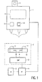
- Fig. 1 shows diagrammatically an image display system according to the invention.
- the image display apparatus 1 includes an LCD screen 4 on which an image can be displayed.
- the image display apparatus also includes a transmission device in the form of three IREDs 3 which emit infrared radiation.
- the position detection system 2 includes a camera unit 10 with two infrared sensitive CCD sensors 11. Each of the CCD sensors 11 picks up images of the IREDs 3 and the image signals supplied by individual CCD sensors represent the position of the image display apparatus. Image signals of this kind are notably electronic video signals.
- the image signals are applied to the computer 12 which derives the position of the image display apparatus from the image signals.
- the computer 12 and the camera unit 10 form part of the position detection system 2. Pre-recorded image information is stored in an image memory 13.
- the image processing unit 14 is controlled so as to form from the image information stored an image signal which represents an image corresponding to the position of the image display apparatus 1.
- the computer 12 derives notably the position of the image display apparatus 1 with respect to an object being observed.
- the image processing unit 14 is controlled on the basis of this position in order to derive from the image information stored an image signal which represents an image in which image information of the object can be seen in the vicinity of the image display apparatus 1. It is notably possible to display image information of a part of the interior of the object in the vicinity of the image display apparatus.
- the image signal of the image processing unit 14 is applied to the image display apparatus 1 in order to display the image on the basis thereof.
- the image signal is applied to the image display apparatus via a wireless connection, so that the image display apparatus 1 can be moved without being impeded by cables.
- the cable is preferably kept out of the operating zone by means of a suspension, thus ensuring that the user is not impeded by the cable.
- the image display apparatus also comprises some controls, such as a push button 21 or a track ball 22, by means of which the image displayed can be adjusted or selected. Such adjustment may concern, for example zooming in or zooming out in relation to important details visible in the image, or adjustment of brightness, contrast and color of the image.
- the selection of the image may relate, for example to the depth within the object wherefrom image information is displayed on the image display apparatus; the user can also recall a previously displayed image.
- the control members may also be software implemented as so-called "icons" in the image. Such control members are activated by pointing out the relevant icon in the image by means of a cursor. It is particularly advantageous to use a pointing member, for example a pen whose position is measured by the position measuring system, in order to point out such an icon. A function indicated by the relevant icon is activated on the basis of the measured position of the pen.
- Fig. 2 shows diagrammatically an image guided surgery system in which the invention is used.
- the image guided surgery system includes a position measuring system which includes a camera unit 10 with two CCD image sensors 11.
- the camera unit picks up images of a surgical instrument 20 from different directions.
- the CCD image sensors supply image signals, notably electronic video signals, which represent the individual images of the instrument 20.
- the position measuring system also includes a computer 12 for deriving the position of the instrument 20 from the image signals.
- Image information of the patient 19 to be examined or treated is stored in an image memory 13.
- the image information comprises, for example MRI and/or CT images recorded before or during the treatment. Markers 23 on or in the patient 19 are also reproduced in the images of the patient.
- the position measuring system measures the positions of the markers 23, for example by pointing out the markers by means of the instrument.
- the computer 12 derives the relationship between positions in or on the patient 19 from the positions of the markers and the positions of the reproductions of the markers in the recorded images.
- the image processor 14 forms an image signal which represents an image showing image information of the patient, together with the instantaneous position of the instrument 20 within the patient.
- the image signal is applied to the image display apparatus 1 by means of a transmitter 18.
- the image display apparatus displays image information of the patient in which the position of the instrument is revealed. The user can thus move the instrument within the patient without having a direct view thereof and without risk of unnecessary damaging of tissues.
- the image display apparatus includes a transmission device 3 in the form of some IREDs which emit infrared radiation.
- the position of the image display apparatus is measured by means of the camera unit 10 and the computer 12.
- the image processor forms an image signal which represents an image which shows, in dependence on the position and/or the orientation of the image display apparatus, image information of the patient together with the instantaneous position of the instrument 20 within the patient. The surgeon can thus observe the image so as to see where within the patient the instrument is situated and can at the same time keep a view of the patient and possibly also of the instrument.
- the image display apparatus includes a holder 5 in which the instrument 20 can be fitted.
- the position of the image display apparatus 1 also determines the position of the instrument 20.
- the image signals of the CCD image sensors 11 can be used to derive the position of the image display apparatus as well as the position of the instrument.
- the image display apparatus provided with an LCD display screen 4 has a flat construction. Such an image display apparatus has a thickness of approximately 2 cm and a surface area of approximately 15 cm x 20 cm.
- the image display apparatus also comprises grips 24. for example in the form of ears.
- Such an image display apparatus can be readily moved across the patient by the surgeon, notably across the operating zone, during which movement image information of the patient is continuously displayed on the display screen 4, together with the position of the instrument, as a function of the position and/or orientation of the image display apparatus relative to the patient.
- the surgeon can use the display apparatus as if it were a magnifying glass in order to see within the patient where the instrument is situated, without having a direct view of the instrument.
- the image display apparatus can also be mounted on a separate stand so that the hands of the surgeon remain free to manipulate the instrument.
Landscapes
- Health & Medical Sciences (AREA)
- Surgery (AREA)
- Life Sciences & Earth Sciences (AREA)
- Engineering & Computer Science (AREA)
- Heart & Thoracic Surgery (AREA)
- Animal Behavior & Ethology (AREA)
- Veterinary Medicine (AREA)
- Biomedical Technology (AREA)
- Nuclear Medicine, Radiotherapy & Molecular Imaging (AREA)
- Medical Informatics (AREA)
- Molecular Biology (AREA)
- Public Health (AREA)
- General Health & Medical Sciences (AREA)
- Pathology (AREA)
- Oral & Maxillofacial Surgery (AREA)
- Robotics (AREA)
- Apparatus For Radiation Diagnosis (AREA)
- Devices For Indicating Variable Information By Combining Individual Elements (AREA)
- Measuring And Recording Apparatus For Diagnosis (AREA)
Claims (5)
- Bildwiedergabesystem, mit einem Bildwiedergabegerät (1), das einen Flüssigkristallanzeigeschirm (4) zum Wiedergeben eines Bildes enthält,
wobei das Bild von der Position und/oder Orientierung des Bildwiedergabegerätes (1) abhängig ist, dadurch gekennzeichnet, dass das Bildwiedergabesystem flach ist und in der Hand gehalten wird und so konfiguriert ist, dass
das Bild bei einer Änderung der Position und/oder der Körperhaltung eines Benutzers des Bildwiedergabegerätes (1) unverändert bleibt. - Bildwiedergabesystem nach Anspruch 1, das auch enthältein Positionsdetektionssystem (2) zum Messen einer Position und/oder Orientierung des Bildwiedergabegerätes (1).
- Bildwiedergabesystem nach Anspruch 2, in dem das Bildwiedergabegerät enthälteine Sendeeinrichtung (3) zum Aussenden eines Positionssignals, das die Position und/oder Orientierung des Bildwiedergabegerätes (1) repräsentiert.
- Bildgeführtes chirurgisches System, miteinem Positionsmesssystem (10, 11,12) zum Messen einer Position eines Instrumentes (20) undeinem Bildwiedergabesystem (1) nach einem der vorhergehenden Ansprüche zum Wiedergeben eines Bildes, das Bildinformation und die Position des Instrumentes repräsentiert.
- Bildgeführite chirurgisches System nach Anspruch 4, in demdas Bildwiedergabegerät (1) einen Halter (5) zum Tragen des Instrumentes (20) enthält.
Priority Applications (1)
| Application Number | Priority Date | Filing Date | Title |
|---|---|---|---|
| EP98900026A EP0918491B1 (de) | 1997-01-24 | 1998-01-12 | Bildanzeigevorrichtung |
Applications Claiming Priority (4)
| Application Number | Priority Date | Filing Date | Title |
|---|---|---|---|
| EP97200196 | 1997-01-24 | ||
| EP97200196 | 1997-01-24 | ||
| PCT/IB1998/000029 WO1998032388A2 (en) | 1997-01-24 | 1998-01-12 | Image display system |
| EP98900026A EP0918491B1 (de) | 1997-01-24 | 1998-01-12 | Bildanzeigevorrichtung |
Publications (2)
| Publication Number | Publication Date |
|---|---|
| EP0918491A2 EP0918491A2 (de) | 1999-06-02 |
| EP0918491B1 true EP0918491B1 (de) | 2003-06-04 |
Family
ID=8227951
Family Applications (1)
| Application Number | Title | Priority Date | Filing Date |
|---|---|---|---|
| EP98900026A Expired - Lifetime EP0918491B1 (de) | 1997-01-24 | 1998-01-12 | Bildanzeigevorrichtung |
Country Status (5)
| Country | Link |
|---|---|
| US (1) | US6038467A (de) |
| EP (1) | EP0918491B1 (de) |
| JP (1) | JP2000509626A (de) |
| DE (1) | DE69815260T2 (de) |
| WO (1) | WO1998032388A2 (de) |
Cited By (1)
| Publication number | Priority date | Publication date | Assignee | Title |
|---|---|---|---|---|
| DE102004022330B3 (de) * | 2004-05-06 | 2005-10-20 | Leica Microsystems Schweiz Ag | Mikroskop |
Families Citing this family (94)
| Publication number | Priority date | Publication date | Assignee | Title |
|---|---|---|---|---|
| US5913820A (en) * | 1992-08-14 | 1999-06-22 | British Telecommunications Public Limited Company | Position location system |
| US6256529B1 (en) * | 1995-07-26 | 2001-07-03 | Burdette Medical Systems, Inc. | Virtual reality 3D visualization for surgical procedures |
| AU4318499A (en) * | 1997-11-24 | 1999-12-13 | Burdette Medical Systems, Inc. | Real time brachytherapy spatial registration and visualization system |
| US6129670A (en) * | 1997-11-24 | 2000-10-10 | Burdette Medical Systems | Real time brachytherapy spatial registration and visualization system |
| US6275721B1 (en) * | 1999-06-10 | 2001-08-14 | General Electriccompany | Interactive MRI scan control using an in-bore scan control device |
| US6314311B1 (en) * | 1999-07-28 | 2001-11-06 | Picker International, Inc. | Movable mirror laser registration system |
| US7635390B1 (en) | 2000-01-14 | 2009-12-22 | Marctec, Llc | Joint replacement component having a modular articulating surface |
| US20030135102A1 (en) * | 2000-05-18 | 2003-07-17 | Burdette Everette C. | Method and system for registration and guidance of intravascular treatment |
| DE10033723C1 (de) * | 2000-07-12 | 2002-02-21 | Siemens Ag | Visualisierung von Positionen und Orientierung von intrakorporal geführten Instrumenten während eines chirurgischen Eingriffs |
| CN100370961C (zh) * | 2001-01-03 | 2008-02-27 | 超形态公司 | 溶解脂肪组织的装置 |
| US7347855B2 (en) * | 2001-10-29 | 2008-03-25 | Ultrashape Ltd. | Non-invasive ultrasonic body contouring |
| US6584339B2 (en) * | 2001-06-27 | 2003-06-24 | Vanderbilt University | Method and apparatus for collecting and processing physical space data for use while performing image-guided surgery |
| US7708741B1 (en) | 2001-08-28 | 2010-05-04 | Marctec, Llc | Method of preparing bones for knee replacement surgery |
| ATE247431T1 (de) * | 2001-12-18 | 2003-09-15 | Brainlab Ag | Projektion von patientenbilddaten aus durchleuchtungs bzw. schichtbilderfassungsverfahren auf oberflächenvideobilder |
| AU2002345319B2 (en) * | 2002-06-25 | 2008-03-06 | Ultrashape Ltd. | Devices and methodologies useful in body aesthetics |
| US7187800B2 (en) * | 2002-08-02 | 2007-03-06 | Computerized Medical Systems, Inc. | Method and apparatus for image segmentation using Jensen-Shannon divergence and Jensen-Renyi divergence |
| EP1542591A2 (de) * | 2002-08-29 | 2005-06-22 | Computerized Medical Systems, Inc. | Verfahren und systeme zur lokalisierung einer medizinischen darstellungssonde und für die räumliche registrierung und verfolgung einer biopsienadel bei einer gewebebiopsie |
| US7426329B2 (en) | 2003-03-06 | 2008-09-16 | Microsoft Corporation | Systems and methods for receiving, storing, and rendering digital video, music, and pictures on a personal media player |
| US20040204645A1 (en) * | 2003-04-10 | 2004-10-14 | Vahid Saadat | Scope position and orientation feedback device |
| US20040201595A1 (en) * | 2003-04-11 | 2004-10-14 | Microsoft Corporation | Self-orienting display |
| EP1470791B1 (de) * | 2003-04-25 | 2007-02-28 | BrainLAB AG | Visualisierungsvorrichtung für kombinierte Patienten- und Objektbilddaten mit einer Eingabevorrichtung und Visualisierungsverfahren |
| US7203277B2 (en) * | 2003-04-25 | 2007-04-10 | Brainlab Ag | Visualization device and method for combined patient and object image data |
| US7463823B2 (en) * | 2003-07-24 | 2008-12-09 | Brainlab Ag | Stereoscopic visualization device for patient image data and video images |
| DE202004014857U1 (de) * | 2003-09-29 | 2005-04-21 | Fraunhofer-Gesellschaft zur Förderung der angewandten Forschung e.V. | Vorrichtung zur virtuellen Lagebetrachtung wenigstens eines in einen Körper intrakorporal eingebrachten medizinischen Instruments |
| US7567833B2 (en) * | 2004-03-08 | 2009-07-28 | Stryker Leibinger Gmbh & Co. Kg | Enhanced illumination device and method |
| US8086332B2 (en) * | 2006-02-27 | 2011-12-27 | Apple Inc. | Media delivery system with improved interaction |
| US8560047B2 (en) | 2006-06-16 | 2013-10-15 | Board Of Regents Of The University Of Nebraska | Method and apparatus for computer aided surgery |
| US7945310B2 (en) * | 2006-09-18 | 2011-05-17 | Stryker Corporation | Surgical instrument path computation and display for endoluminal surgery |
| US7824328B2 (en) * | 2006-09-18 | 2010-11-02 | Stryker Corporation | Method and apparatus for tracking a surgical instrument during surgery |
| US8248414B2 (en) * | 2006-09-18 | 2012-08-21 | Stryker Corporation | Multi-dimensional navigation of endoscopic video |
| US20080071141A1 (en) * | 2006-09-18 | 2008-03-20 | Abhisuek Gattani | Method and apparatus for measuring attributes of an anatomical feature during a medical procedure |
| US8248413B2 (en) * | 2006-09-18 | 2012-08-21 | Stryker Corporation | Visual navigation system for endoscopic surgery |
| JP5135007B2 (ja) * | 2008-03-10 | 2013-01-30 | オリンパスメディカルシステムズ株式会社 | カプセル誘導システム |
| US9687170B2 (en) | 2008-03-11 | 2017-06-27 | General Electric Company | System and method for performing magnetic resonance imaging scan operations from within a scan room |
| TW201004607A (en) * | 2008-07-25 | 2010-02-01 | Been-Der Yang | Image guided navigation system and method thereof |
| EP2455038B1 (de) | 2008-10-21 | 2015-04-01 | Brainlab AG | Integration von chirurgischem Instrument und Anzeigevorrichtung zur Unterstützung der bildgeführten Chirurgie |
| JP5670079B2 (ja) * | 2009-09-30 | 2015-02-18 | 富士フイルム株式会社 | 医用画像表示装置および方法、並びにプログラム |
| WO2011116347A1 (en) * | 2010-03-19 | 2011-09-22 | Quickvein, Inc. | Apparatus and methods for imaging blood vessels |
| US8672837B2 (en) | 2010-06-24 | 2014-03-18 | Hansen Medical, Inc. | Methods and devices for controlling a shapeable medical device |
| US20120330129A1 (en) * | 2011-06-23 | 2012-12-27 | Richard Awdeh | Medical visualization systems and related methods of use |
| US9498231B2 (en) | 2011-06-27 | 2016-11-22 | Board Of Regents Of The University Of Nebraska | On-board tool tracking system and methods of computer assisted surgery |
| CN106913366B (zh) | 2011-06-27 | 2021-02-26 | 内布拉斯加大学评议会 | 工具承载的追踪系统和计算机辅助外科方法 |
| JP6385275B2 (ja) | 2011-09-02 | 2018-09-05 | ストライカー・コーポレイション | ハウジングから延びる切断アクセサリ及びハウジングに対する切断アクセサリの位置を確立するアクチュエータを備える手術器具 |
| US9918681B2 (en) * | 2011-09-16 | 2018-03-20 | Auris Surgical Robotics, Inc. | System and method for virtually tracking a surgical tool on a movable display |
| DE102011083634B4 (de) * | 2011-09-28 | 2021-05-06 | Siemens Healthcare Gmbh | Vorrichtung und Verfahren für eine Bilddarstellung |
| DE102012100504A1 (de) * | 2012-01-23 | 2013-07-25 | Aesculap Ag | Verfahren und Vorrichtung zum Darstellen eines Ultraschallbildes |
| US9566414B2 (en) | 2013-03-13 | 2017-02-14 | Hansen Medical, Inc. | Integrated catheter and guide wire controller |
| US9057600B2 (en) | 2013-03-13 | 2015-06-16 | Hansen Medical, Inc. | Reducing incremental measurement sensor error |
| US9014851B2 (en) | 2013-03-15 | 2015-04-21 | Hansen Medical, Inc. | Systems and methods for tracking robotically controlled medical instruments |
| US10849702B2 (en) | 2013-03-15 | 2020-12-01 | Auris Health, Inc. | User input devices for controlling manipulation of guidewires and catheters |
| US9271663B2 (en) | 2013-03-15 | 2016-03-01 | Hansen Medical, Inc. | Flexible instrument localization from both remote and elongation sensors |
| US9283046B2 (en) | 2013-03-15 | 2016-03-15 | Hansen Medical, Inc. | User interface for active drive apparatus with finite range of motion |
| US10105149B2 (en) | 2013-03-15 | 2018-10-23 | Board Of Regents Of The University Of Nebraska | On-board tool tracking system and methods of computer assisted surgery |
| US9629595B2 (en) | 2013-03-15 | 2017-04-25 | Hansen Medical, Inc. | Systems and methods for localizing, tracking and/or controlling medical instruments |
| US11020016B2 (en) * | 2013-05-30 | 2021-06-01 | Auris Health, Inc. | System and method for displaying anatomy and devices on a movable display |
| EP2923669B1 (de) | 2014-03-24 | 2017-06-28 | Hansen Medical, Inc. | Systeme und vorrichtungen zur instinktiven führung eines katheters |
| GB2536650A (en) | 2015-03-24 | 2016-09-28 | Augmedics Ltd | Method and system for combining video-based and optic-based augmented reality in a near eye display |
| WO2017049163A1 (en) | 2015-09-18 | 2017-03-23 | Auris Surgical Robotics, Inc. | Navigation of tubular networks |
| US10143526B2 (en) | 2015-11-30 | 2018-12-04 | Auris Health, Inc. | Robot-assisted driving systems and methods |
| US11037464B2 (en) | 2016-07-21 | 2021-06-15 | Auris Health, Inc. | System with emulator movement tracking for controlling medical devices |
| CN110248618B (zh) | 2016-09-09 | 2024-01-09 | 莫比乌斯成像公司 | 用于在计算机辅助手术中显示患者数据的方法及系统 |
| US10244926B2 (en) | 2016-12-28 | 2019-04-02 | Auris Health, Inc. | Detecting endolumenal buckling of flexible instruments |
| KR102558061B1 (ko) | 2017-03-31 | 2023-07-25 | 아우리스 헬스, 인코포레이티드 | 생리적 노이즈를 보상하는 관강내 조직망 항행을 위한 로봇 시스템 |
| US10610179B2 (en) * | 2017-06-05 | 2020-04-07 | Biosense Webster (Israel) Ltd. | Augmented reality goggles having X-ray protection |
| US10022192B1 (en) | 2017-06-23 | 2018-07-17 | Auris Health, Inc. | Automatically-initialized robotic systems for navigation of luminal networks |
| CN110809452B (zh) | 2017-06-28 | 2023-05-23 | 奥瑞斯健康公司 | 电磁场发生器对准 |
| JP7330902B2 (ja) | 2017-06-28 | 2023-08-22 | オーリス ヘルス インコーポレイテッド | 電磁歪み検出 |
| US11058493B2 (en) | 2017-10-13 | 2021-07-13 | Auris Health, Inc. | Robotic system configured for navigation path tracing |
| US10555778B2 (en) | 2017-10-13 | 2020-02-11 | Auris Health, Inc. | Image-based branch detection and mapping for navigation |
| AU2018378810B2 (en) | 2017-12-08 | 2024-02-22 | Auris Health, Inc. | System and method for medical instrument navigation and targeting |
| JP7322026B2 (ja) | 2017-12-14 | 2023-08-07 | オーリス ヘルス インコーポレイテッド | 器具の位置推定のシステムおよび方法 |
| WO2019125964A1 (en) | 2017-12-18 | 2019-06-27 | Auris Health, Inc. | Methods and systems for instrument tracking and navigation within luminal networks |
| WO2019191143A1 (en) | 2018-03-28 | 2019-10-03 | Auris Health, Inc. | Systems and methods for displaying estimated location of instrument |
| WO2019191144A1 (en) | 2018-03-28 | 2019-10-03 | Auris Health, Inc. | Systems and methods for registration of location sensors |
| WO2019211741A1 (en) | 2018-05-02 | 2019-11-07 | Augmedics Ltd. | Registration of a fiducial marker for an augmented reality system |
| JP7314175B2 (ja) | 2018-05-18 | 2023-07-25 | オーリス ヘルス インコーポレイテッド | ロボット対応の遠隔操作システムのためのコントローラ |
| WO2019231895A1 (en) | 2018-05-30 | 2019-12-05 | Auris Health, Inc. | Systems and methods for location sensor-based branch prediction |
| EP3801348B1 (de) | 2018-05-31 | 2024-05-01 | Auris Health, Inc. | Bildbasierte atemweganalyse und -kartierung |
| WO2019231990A1 (en) | 2018-05-31 | 2019-12-05 | Auris Health, Inc. | Robotic systems and methods for navigation of luminal network that detect physiological noise |
| CN110831481B (zh) | 2018-05-31 | 2022-08-30 | 奥瑞斯健康公司 | 管状网络的基于路径的导航 |
| US12076100B2 (en) | 2018-09-28 | 2024-09-03 | Auris Health, Inc. | Robotic systems and methods for concomitant endoscopic and percutaneous medical procedures |
| US11766296B2 (en) | 2018-11-26 | 2023-09-26 | Augmedics Ltd. | Tracking system for image-guided surgery |
| EP3692939B1 (de) | 2019-02-07 | 2021-07-14 | Stryker European Operations Limited | Chirurgische systeme zur erleichterung der gewebebehandlung |
| CN114025700A (zh) | 2019-06-28 | 2022-02-08 | 奥瑞斯健康公司 | 控制台叠加以及其使用方法 |
| US11980506B2 (en) | 2019-07-29 | 2024-05-14 | Augmedics Ltd. | Fiducial marker |
| WO2021038495A1 (en) | 2019-08-30 | 2021-03-04 | Auris Health, Inc. | Instrument image reliability systems and methods |
| KR20220058569A (ko) | 2019-08-30 | 2022-05-09 | 아우리스 헬스, 인코포레이티드 | 위치 센서의 가중치-기반 정합을 위한 시스템 및 방법 |
| EP4025921A4 (de) | 2019-09-03 | 2023-09-06 | Auris Health, Inc. | Detektion und kompensation von elektromagnetischer verzerrung |
| US11382712B2 (en) | 2019-12-22 | 2022-07-12 | Augmedics Ltd. | Mirroring in image guided surgery |
| WO2021137072A1 (en) | 2019-12-31 | 2021-07-08 | Auris Health, Inc. | Anatomical feature identification and targeting |
| US11660147B2 (en) | 2019-12-31 | 2023-05-30 | Auris Health, Inc. | Alignment techniques for percutaneous access |
| CN118383870A (zh) | 2019-12-31 | 2024-07-26 | 奥瑞斯健康公司 | 用于经皮进入的对准界面 |
| US11896445B2 (en) | 2021-07-07 | 2024-02-13 | Augmedics Ltd. | Iliac pin and adapter |
| WO2024057210A1 (en) | 2022-09-13 | 2024-03-21 | Augmedics Ltd. | Augmented reality eyewear for image-guided medical intervention |
Family Cites Families (6)
| Publication number | Priority date | Publication date | Assignee | Title |
|---|---|---|---|---|
| JP2612044B2 (ja) * | 1988-07-21 | 1997-05-21 | 株式会社日立製作所 | 電子ファイル装置 |
| US5086401A (en) * | 1990-05-11 | 1992-02-04 | International Business Machines Corporation | Image-directed robotic system for precise robotic surgery including redundant consistency checking |
| US5526812A (en) * | 1993-06-21 | 1996-06-18 | General Electric Company | Display system for enhancing visualization of body structures during medical procedures |
| US5491510A (en) * | 1993-12-03 | 1996-02-13 | Texas Instruments Incorporated | System and method for simultaneously viewing a scene and an obscured object |
| DE69524332T2 (de) * | 1994-09-19 | 2002-06-13 | Matsushita Electric Ind Co Ltd | Vorrichtung zur dreidimensionalen Bildwiedergabe |
| JP3397602B2 (ja) * | 1996-11-11 | 2003-04-21 | 富士通株式会社 | 画像表示装置及び方法 |
-
1998
- 1998-01-12 DE DE69815260T patent/DE69815260T2/de not_active Expired - Lifetime
- 1998-01-12 WO PCT/IB1998/000029 patent/WO1998032388A2/en active IP Right Grant
- 1998-01-12 EP EP98900026A patent/EP0918491B1/de not_active Expired - Lifetime
- 1998-01-12 JP JP10529171A patent/JP2000509626A/ja active Pending
- 1998-01-16 US US09/008,337 patent/US6038467A/en not_active Expired - Fee Related
Cited By (1)
| Publication number | Priority date | Publication date | Assignee | Title |
|---|---|---|---|---|
| DE102004022330B3 (de) * | 2004-05-06 | 2005-10-20 | Leica Microsystems Schweiz Ag | Mikroskop |
Also Published As
| Publication number | Publication date |
|---|---|
| WO1998032388A3 (en) | 1998-11-12 |
| JP2000509626A (ja) | 2000-08-02 |
| US6038467A (en) | 2000-03-14 |
| DE69815260D1 (de) | 2003-07-10 |
| WO1998032388A2 (en) | 1998-07-30 |
| DE69815260T2 (de) | 2004-05-13 |
| EP0918491A2 (de) | 1999-06-02 |
Similar Documents
| Publication | Publication Date | Title |
|---|---|---|
| EP0918491B1 (de) | Bildanzeigevorrichtung | |
| JP2000509626A5 (de) | ||
| US6656110B1 (en) | Endoscopic system | |
| US7885701B2 (en) | Registration pointer and method for registering a bone of a patient to a computer assisted orthopaedic surgery system | |
| US5961456A (en) | System and method for displaying concurrent video and reconstructed surgical views | |
| CA2486525C (en) | A guide system and a probe therefor | |
| US5638819A (en) | Method and apparatus for guiding an instrument to a target | |
| CA2766595C (en) | Surgeon's aid for medical display | |
| EP1395194B1 (de) | Führungssystem | |
| US6135946A (en) | Method and system for image-guided interventional endoscopic procedures | |
| EP0929267B1 (de) | Bildgesteuertes chirurgisches system | |
| DE69112538D1 (de) | Computerunterstützte chirurgische Vorrichtung. | |
| WO2010067267A1 (en) | Head-mounted wireless camera and display unit | |
| ATE188601T1 (de) | Computerunterstützte mikrochirurgieausrüstung sowie diese ausrüstung gebrauchende verfahren | |
| CN102525517A (zh) | 荧光透视数据显示器 | |
| JP2015507493A (ja) | 近赤外光の透過により脈管構造および皮下構造を撮像する装置ならびに方法 | |
| US20210330388A1 (en) | Augmented reality user guidance during examinations or interventional procedures | |
| JP3024162B2 (ja) | 手術用ヘッドアップディスプレイ | |
| US11944508B1 (en) | Augmented reality surgical assistance system | |
| US20050123289A1 (en) | Method and apparatus for observing objects with a microscope | |
| Weber et al. | Application of different visualization concepts in the navigated image viewer | |
| WO2008018025A2 (en) | Apparatus to image blood vessel |
Legal Events
| Date | Code | Title | Description |
|---|---|---|---|
| PUAI | Public reference made under article 153(3) epc to a published international application that has entered the european phase |
Free format text: ORIGINAL CODE: 0009012 |
|
| 17P | Request for examination filed |
Effective date: 19981026 |
|
| AK | Designated contracting states |
Kind code of ref document: A2 Designated state(s): DE FR GB NL SE |
|
| 17Q | First examination report despatched |
Effective date: 20010906 |
|
| GRAH | Despatch of communication of intention to grant a patent |
Free format text: ORIGINAL CODE: EPIDOS IGRA |
|
| GRAH | Despatch of communication of intention to grant a patent |
Free format text: ORIGINAL CODE: EPIDOS IGRA |
|
| GRAA | (expected) grant |
Free format text: ORIGINAL CODE: 0009210 |
|
| AK | Designated contracting states |
Designated state(s): DE FR GB NL SE |
|
| PG25 | Lapsed in a contracting state [announced via postgrant information from national office to epo] |
Ref country code: NL Free format text: LAPSE BECAUSE OF FAILURE TO SUBMIT A TRANSLATION OF THE DESCRIPTION OR TO PAY THE FEE WITHIN THE PRESCRIBED TIME-LIMIT Effective date: 20030604 Ref country code: FR Free format text: LAPSE BECAUSE OF FAILURE TO SUBMIT A TRANSLATION OF THE DESCRIPTION OR TO PAY THE FEE WITHIN THE PRESCRIBED TIME-LIMIT Effective date: 20030604 |
|
| RAP1 | Party data changed (applicant data changed or rights of an application transferred) |
Owner name: PHILIPS AB Owner name: KONINKLIJKE PHILIPS ELECTRONICS N.V. |
|
| REG | Reference to a national code |
Ref country code: GB Ref legal event code: FG4D |
|
| REF | Corresponds to: |
Ref document number: 69815260 Country of ref document: DE Date of ref document: 20030710 Kind code of ref document: P |
|
| PG25 | Lapsed in a contracting state [announced via postgrant information from national office to epo] |
Ref country code: SE Free format text: LAPSE BECAUSE OF FAILURE TO SUBMIT A TRANSLATION OF THE DESCRIPTION OR TO PAY THE FEE WITHIN THE PRESCRIBED TIME-LIMIT Effective date: 20030904 |
|
| NLV1 | Nl: lapsed or annulled due to failure to fulfill the requirements of art. 29p and 29m of the patents act | ||
| REG | Reference to a national code |
Ref country code: GB Ref legal event code: 746 Effective date: 20040108 |
|
| PLBE | No opposition filed within time limit |
Free format text: ORIGINAL CODE: 0009261 |
|
| STAA | Information on the status of an ep patent application or granted ep patent |
Free format text: STATUS: NO OPPOSITION FILED WITHIN TIME LIMIT |
|
| 26N | No opposition filed |
Effective date: 20040305 |
|
| EN | Fr: translation not filed | ||
| PGFP | Annual fee paid to national office [announced via postgrant information from national office to epo] |
Ref country code: GB Payment date: 20100129 Year of fee payment: 13 |
|
| PGFP | Annual fee paid to national office [announced via postgrant information from national office to epo] |
Ref country code: DE Payment date: 20100331 Year of fee payment: 13 |
|
| GBPC | Gb: european patent ceased through non-payment of renewal fee |
Effective date: 20110112 |
|
| PG25 | Lapsed in a contracting state [announced via postgrant information from national office to epo] |
Ref country code: GB Free format text: LAPSE BECAUSE OF NON-PAYMENT OF DUE FEES Effective date: 20110112 |
|
| REG | Reference to a national code |
Ref country code: DE Ref legal event code: R119 Ref document number: 69815260 Country of ref document: DE Effective date: 20110802 |
|
| PG25 | Lapsed in a contracting state [announced via postgrant information from national office to epo] |
Ref country code: DE Free format text: LAPSE BECAUSE OF NON-PAYMENT OF DUE FEES Effective date: 20110802 |