EP0856181B1 - Verfahren und vorrichtung zur auswertung eines narkose- oder intensiv-eeg - Google Patents
Verfahren und vorrichtung zur auswertung eines narkose- oder intensiv-eeg Download PDFInfo
- Publication number
- EP0856181B1 EP0856181B1 EP96945506A EP96945506A EP0856181B1 EP 0856181 B1 EP0856181 B1 EP 0856181B1 EP 96945506 A EP96945506 A EP 96945506A EP 96945506 A EP96945506 A EP 96945506A EP 0856181 B1 EP0856181 B1 EP 0856181B1
- Authority
- EP
- European Patent Office
- Prior art keywords
- eeg
- classification
- parameters
- determined
- narcotic
- Prior art date
- Legal status (The legal status is an assumption and is not a legal conclusion. Google has not performed a legal analysis and makes no representation as to the accuracy of the status listed.)
- Expired - Lifetime
Links
Images


Classifications
-
- A—HUMAN NECESSITIES
- A61—MEDICAL OR VETERINARY SCIENCE; HYGIENE
- A61B—DIAGNOSIS; SURGERY; IDENTIFICATION
- A61B5/00—Measuring for diagnostic purposes; Identification of persons
- A61B5/48—Other medical applications
- A61B5/4821—Determining level or depth of anaesthesia
-
- A—HUMAN NECESSITIES
- A61—MEDICAL OR VETERINARY SCIENCE; HYGIENE
- A61B—DIAGNOSIS; SURGERY; IDENTIFICATION
- A61B5/00—Measuring for diagnostic purposes; Identification of persons
- A61B5/24—Detecting, measuring or recording bioelectric or biomagnetic signals of the body or parts thereof
- A61B5/316—Modalities, i.e. specific diagnostic methods
- A61B5/369—Electroencephalography [EEG]
- A61B5/372—Analysis of electroencephalograms
-
- A—HUMAN NECESSITIES
- A61—MEDICAL OR VETERINARY SCIENCE; HYGIENE
- A61B—DIAGNOSIS; SURGERY; IDENTIFICATION
- A61B5/00—Measuring for diagnostic purposes; Identification of persons
- A61B5/24—Detecting, measuring or recording bioelectric or biomagnetic signals of the body or parts thereof
- A61B5/316—Modalities, i.e. specific diagnostic methods
- A61B5/369—Electroencephalography [EEG]
- A61B5/384—Recording apparatus or displays specially adapted therefor
-
- A—HUMAN NECESSITIES
- A61—MEDICAL OR VETERINARY SCIENCE; HYGIENE
- A61B—DIAGNOSIS; SURGERY; IDENTIFICATION
- A61B5/00—Measuring for diagnostic purposes; Identification of persons
- A61B5/72—Signal processing specially adapted for physiological signals or for diagnostic purposes
- A61B5/7203—Signal processing specially adapted for physiological signals or for diagnostic purposes for noise prevention, reduction or removal
- A61B5/7207—Signal processing specially adapted for physiological signals or for diagnostic purposes for noise prevention, reduction or removal of noise induced by motion artifacts
- A61B5/721—Signal processing specially adapted for physiological signals or for diagnostic purposes for noise prevention, reduction or removal of noise induced by motion artifacts using a separate sensor to detect motion or using motion information derived from signals other than the physiological signal to be measured
-
- A—HUMAN NECESSITIES
- A61—MEDICAL OR VETERINARY SCIENCE; HYGIENE
- A61B—DIAGNOSIS; SURGERY; IDENTIFICATION
- A61B5/00—Measuring for diagnostic purposes; Identification of persons
- A61B5/72—Signal processing specially adapted for physiological signals or for diagnostic purposes
- A61B5/7235—Details of waveform analysis
- A61B5/7264—Classification of physiological signals or data, e.g. using neural networks, statistical classifiers, expert systems or fuzzy systems
-
- A—HUMAN NECESSITIES
- A61—MEDICAL OR VETERINARY SCIENCE; HYGIENE
- A61B—DIAGNOSIS; SURGERY; IDENTIFICATION
- A61B5/00—Measuring for diagnostic purposes; Identification of persons
- A61B5/72—Signal processing specially adapted for physiological signals or for diagnostic purposes
- A61B5/7235—Details of waveform analysis
- A61B5/7253—Details of waveform analysis characterised by using transforms
- A61B5/7257—Details of waveform analysis characterised by using transforms using Fourier transforms
-
- A—HUMAN NECESSITIES
- A61—MEDICAL OR VETERINARY SCIENCE; HYGIENE
- A61B—DIAGNOSIS; SURGERY; IDENTIFICATION
- A61B5/00—Measuring for diagnostic purposes; Identification of persons
- A61B5/72—Signal processing specially adapted for physiological signals or for diagnostic purposes
- A61B5/7235—Details of waveform analysis
- A61B5/7264—Classification of physiological signals or data, e.g. using neural networks, statistical classifiers, expert systems or fuzzy systems
- A61B5/7267—Classification of physiological signals or data, e.g. using neural networks, statistical classifiers, expert systems or fuzzy systems involving training the classification device
-
- A—HUMAN NECESSITIES
- A61—MEDICAL OR VETERINARY SCIENCE; HYGIENE
- A61B—DIAGNOSIS; SURGERY; IDENTIFICATION
- A61B5/00—Measuring for diagnostic purposes; Identification of persons
- A61B5/74—Details of notification to user or communication with user or patient; User input means
- A61B5/742—Details of notification to user or communication with user or patient; User input means using visual displays
-
- G—PHYSICS
- G16—INFORMATION AND COMMUNICATION TECHNOLOGY [ICT] SPECIALLY ADAPTED FOR SPECIFIC APPLICATION FIELDS
- G16H—HEALTHCARE INFORMATICS, i.e. INFORMATION AND COMMUNICATION TECHNOLOGY [ICT] SPECIALLY ADAPTED FOR THE HANDLING OR PROCESSING OF MEDICAL OR HEALTHCARE DATA
- G16H50/00—ICT specially adapted for medical diagnosis, medical simulation or medical data mining; ICT specially adapted for detecting, monitoring or modelling epidemics or pandemics
- G16H50/20—ICT specially adapted for medical diagnosis, medical simulation or medical data mining; ICT specially adapted for detecting, monitoring or modelling epidemics or pandemics for computer-aided diagnosis, e.g. based on medical expert systems
Definitions
- the invention relates to a method for evaluating a Anesthetic or intensive EEG according to the preamble of the claim 1 and a device for evaluating an anesthetic or intensive EEG according to the preamble of the claim 5.
- Electroencephalography is a method of imaging electrical activity generated by the brain.
- the registration of the EEG is carried out in the same way a multi-channel recorder on continuous paper as a rule with a registration speed of 30 mm / s.
- the German EEG society recommends devices with at least to use ten channels.
- the electrodes are in several lead programs interconnected in different ways. The recording is also increasing with the help of computers performed.
- the composition of the waveforms in the electroencephalogram depends on the functional state of the brain.
- the EEG images that are and intensive care areas are diverse and can by a large number of endogenous and exogenous factors to be influenced.
- the following frequency ranges are those occurring in the EEG Waves assigned: Alpha (7.5 - 12.5 Hz), Beta (> 12.5 Hz), theta (3.5 - 7.5 Hz) and delta (0.5 - 3.5 Hz).
- the subdelta ( ⁇ 0.5 Hz) and the gamma band (> 30 Hz) can be delimited.
- the Waves in the frequency ranges with regard to their amplitudes, Frequency, regularity, temporal structure, local distribution and changes in stimuli are described. EEG amplitudes are measured in ⁇ V. Higher frequencies Waves usually have smaller amplitudes during with a slowdown usually an increase in amplitude connected is.
- anesthesia or coma EEG stages Kugler proposes an EEG division in which the waking state with A and EEG images with progressing Damping of the brain function designated with the letters B to F. become.
- the frequency and amplitude of the waves in certain Frequency ranges and typical patterns are used.
- stage A The awake EEG, stage A, is in the majority of adults characterized by waves in the alpha frequency range. Stage B is characterized by waves with fast frequency and low amplitude. In the stadiums C and D occur theta and delta waves. In the stage E determines high-amplitude delta activity the graph.
- Stage F is flatter to isoelectric due to a change Curve sections and groups of higher waves, that Burst suppression pattern, or by continuous very flat activity.
- the invention has for its object a method and a device for evaluating an anesthetic EEG or intensive EEG, to be specified in which the EEG curves all classes of the EEG stage are clearly identified can and influences as far as possible through disturbances be avoided.
- This task is carried out in a method according to the generic term of claim 1 by the in the characterizing part of the claim 1 specified features and in one device according to the preamble of claim 6 by the in the mark of the claims specified 6 solved.
- EEG signals are stochastic, i.e. random, Processes that use mathematical-statistical Have the procedure described approximately. For the further processing of the EEG is done out of practical Desirable, a reduction to a few, to achieve parameters that are as meaningful as possible.
- the parameter formation can either in the time or in the frequency domain be made. Data basis for analyzes in Time domain is the EEG signal, before calculations in the frequency domain the data is transformed.
- One way to analyze EEG signals in the frequency domain is the Fourier analysis. With the help of the Fourier transformation becomes the complex wave pattern of the EEG broken down into the underlying vibration components, there is a conversion from the time domain to the frequency domain. The squared amplitudes of these vibration components form the so-called performance or power spectrum.
- the further processing of the results of the Fourier analysis includes the extraction of so-called spectral parameters as well as further statistical calculations. To the parameters that can be derived from the spectrum, belong e.g. the overall performance as well as absolute and relative powers in different frequency bands. Other commonly used parameters are the median, the Spectral Edge Frequency and the dominant frequency. The Median is the frequency at which the area of the spectrum is divided into two equal parts. The Spectral Edge Frequency is mostly defined as a 95% quantile, i.e. 95% of the total power of the spectrum is below this frequency. The dominant frequency is the frequency with the greatest performance.
- the frequency distribution can be of EEG sections clearly.
- special patterns such as burst suppression phases or seizure potentials, can usually be from the spectrum do not close.
- FFT Fast Fourier Transform
- AR autoregressive
- Y t a 1 * Y t-1 + ... + a p * Y tp + e t .
- Y t denotes the measured value at time t
- the letter p denotes the order of the process, ie the number of historical values that are taken into account.
- the model parameters can be estimated using the Yule-Walker equations. The Box and Jenkins approach is usually used to determine the order of the model and to check the model quality. Kay and Marple provide an overview of other estimation methods and model classes.
- Hjorth A commonly used method to characterize EEG measurements is the calculation of special EEG parameters, proposed by Hjorth and named after him have been. There are three parameters, namely activity, mobility and complexity.
- the Hjorth parameters are derived from the scatter of the EEG signal and their first and second derivatives. Alternatively can also calculate the Hjorth parameters in Frequency range, i.e. with the help of spectral analysis become.
- the activity corresponds to the total power of the signal and is therefore a measure of the amplitude size of the EEG measurement. Mobility can be interpreted as one Measure of the medium frequency and the complexity as a Measure of the variability of the signal.
- Discriminant analytical classification procedures are one of them suitable for objects based on a series of characteristics assign to one of several defined groups.
- the EEG sections the objects to be classified the by spectral parameters and / or AR and / or Hjorth parameters and / or chaos parameters are characterized.
- To calculate suitable classification functions exist a number of methods that involve parametric and nonparametric approaches. Using a sample of objects for which group membership is known, classification functions based on the observed feature values derive.
- the linear one Discriminant analysis sets the equality of the covariance matrices ahead in each group, the quadratic Discriminant analysis enables the consideration of different ones Covariance matrices of the groups.
- the Mahalanobis distance is used that the weighted distance of an observation vector to the Represents group averages.
- An object then becomes one Group assigned to one of the selected Process-dependent function of the Mahalanobis distance on is smallest.
- the k-nearest-neighbor method is an illustrative method.
- the distances of the feature vector to be classified to all others Characteristic vectors of the available sample formed, ordered in size and the observation vectors determined with the k smallest distances, whereby the number k of the considered values is predetermined must become. Then it is determined to which groups it belongs k values and their share in the total number of Measurements determined in the individual groups. The assignment then takes place to the group in which this share of the greatest is.
- This nonparametric method requires an increased one Computational effort compared to parametric methods, because for the classification of an object on the whole Original data record must be used while at parametric methods the characteristic values of an object be used in classification functions.
- error rate means the proportion of misclassifications becomes.
- a way to estimate the error rate consists in the reclassification of the data.
- the error rate determined provides an overly positive one Estimate the true error rate.
- a more realistic one Estimation of the error rate is given when the classifications checked on an independent data set become. This can be done by splitting the given Data record in a training data record for derivation the classification rule and a test data set for Validation of the classification is done. An extreme one The so-called Cross-validation or the leave-one-out procedure. Here, an observation is made from the data record taken out and the classification based on the remaining data calculated discriminant function.
- the stages of anesthesia or intensive EEG can be based on Kugler, who, as mentioned in the introduction, designates the waking state with A and the very low damping of brain function with F.
- the intermediate stages B to E can be further subdivided, as shown in Table 1.
- a 2 Alpha low, sparse, slow theta low B 0 Theta low drowsiness
- B 2 Theta low to medium high Theta medium high very flat anesthesia
- C Theta high 30% of the time light sleep
- stage F While only the stages A until E is reliably recognized, the determination enables additional limit values of the parameters, in particular the Signal power related to a zero line, and a correction the multivariate classification based on the limit values a significant improvement in the visibility of the Stage F of the stage division. Since this stage in Contrary to the other stadiums partly by one Zero line can be characterized, leads the exclusive Evaluation of spectral, AR, Hjorth or Chaos parameters are not always a safe parameter Detection of stage F.
- EEG electroactive polystyrene
- muscle artifacts or interference radiation influenced.
- These disorders can on the one hand Temporarily make evaluation of an EEG completely impossible, but also pretend electrical activity, which does not come from the brain and therefore leads to misinterpretation would lead.
- moving artifacts independent of the EEG recording can be determined based on the movement artifacts corrected or suppressed the staging become.
- a further improvement in the staging is achieved if saved from different age-dependent Classification functions for one Subjects age-specific classification functions to be selected. It was found that a person's EEG age-related characteristics having. In simple terms, e.g. the spectrum in adults with increasing age in Awake at lower frequencies during anesthesia is e.g. the delta power is reduced. By considering age-specific classification functions can make the correct staging more reliably become.
- Coupled motion sensor for evaluation with at least one on the patient 's head or Subjects attachable sensor element may be provided.
- Sensor elements are accelerometers and / or Deformation transducer particularly suitable.
- the sensor elements can also be used with the head of the Patients or subjects coupled electrical lines be formed in a magnetic field and / or electrical field.
- electrical lines be formed in a magnetic field and / or electrical field.
- muscle artifacts from the time and / or frequency range of the EEG curves can be determined and corrected the stage classification based on the muscle artifacts or be suppressed.
- Such muscle artifacts can be caused by the forehead and facial muscles are and are usually recognizable by the fact that in addition to other frequency components also frequency components greater than 30 Hz are present.
- artifacts due to interference radiation can also occur electromedical devices, in particular electrosurgical Devices are determined and based on the Interference radiation artifacts the stage classification of the anesthetic depth corrected or suppressed.
- electromedical devices in particular electrosurgical Devices are determined and based on the Interference radiation artifacts the stage classification of the anesthetic depth corrected or suppressed.
- an interference radiation sensor coupled to the computer provided that detects the interference radiation.
- Artifacts can also be queried through software queries detect.
- the device shown in Fig. 1 consists of a Personal computer with color screen and a measuring unit for two EEG channels. Electrodes are used for EEG registrations glued to the patient's head and to the in preamplifier placed in the immediate vicinity of the patient connected. This is done in a differential amplifier Formation of the difference between the potentials of two Electrodes. The signals are amplified by a factor of 20,000, by a range adjustment to the AD (analog / digital) converter.
- AD analog / digital
- a motion sensor is used to detect motion artifacts attached to the patient's head. Whose signals are also subjected to an AD conversion.
- a 50 Hz notch filter works Net hum artifacts.
- the filtered and amplified Analog signals are digitized at 128 Hz. This is controlled by a microcontroller and this provides the PC with the values for further processing. This takes over the storage and evaluation of the Data.
- the EEG analysis takes place parallel to the AD conversion, ensuring the continuity of data collection.
- the program is operated optionally via an integrated Membrane keyboard, a touch screen or via a external keyboard. Expenses are on the screen and optionally done via a printer.
- the patient's age is entered before each measurement.
- the data can be derived from or two derivatives are evaluated and saved.
- stage classification is based on discriminant analysis Functions and additional limit value queries performed.
- algorithms for improvement Burst suppression phase detection programmed. Two-second epochs that are burdened with artifacts are considered in the stage classification no consideration.
- k stages and a p-dimensional feature vector X (X 1 , .. X p ) (eg p suitable EEG parameters) are assumed. Based on the feature vector X, an assignment to one of the k stages is made.
- the derivation of classification functions with which the assignment is made on the basis of a training data record contains EEG signals, typical of the different stages are.
- discriminant analytics Procedure classification functions calculated for the k stages.
- the EEG section under consideration becomes Parameters (e.g. spectral, AR, Hjorth or Chaos parameters) calculated and in from the training data set determined classification functions used.
- the classification functions provide an assignment probability for each of the k stages, i.e. a measure for the similarity of the EEG section considered the different stages.
- the EEG section is then the stage with the highest probability of assignment assigned.
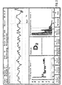
- the EEG sections shown in FIG. 3 are given each lasting 2 seconds using discriminant analysis a stage from A (awake) to E (deep Anesthesia).
- the staging was based on Kugler. Acting in section (a) it is an EEG section from flat anesthesia, in section (b) an EEG from deep anesthesia.
- the first step is to calculate suitable ones Parameters to characterize the respective EEG section.
- five autoregressive parameters were used as well as the logarithmic standard deviation, the corresponds to the power of the EEG signal. It can also others, e.g. Spectral or Hjorth parameters, be used. So an EEG section described by the feature vector X, the above contains six EEG parameters as components.
- the discriminant functions are then calculated on the basis of the training data set, in which the stage affiliation is known on the basis of a visual evaluation.
- linear discriminant analysis was used for 12 stages A, B 0 , B 1 , B 2 , C 0 , C 1 , C 2 , D 0 , D 1 , D 2 , E 0 and E 1 .
- EEG sections (a) and (b) are shown in Table 2.
- the classification is made at the stage to which the EEG section is most similar, ie to the stage with the highest probability of assignment.
- Table 2 shows that EEG section (a) is assigned to stage B 1 and section (b) to stage D 0 .
- Assignment probabilities for the EEG sections from FIG. 3 Allocation probabilities for the stadiums section A B 0 B 1 B 2 C 0 C 1 C 2 D 0 D 1 D 2 E 0 E 1 (a) 0.00 0.25 0.69 0.04 0.02 0.00 0.00 0.00 0.00 0.00 0.00 0.00 0.00 0.00 0.00 0.00 0.00 0.00 0.00 (b) 0.00 0.00 0.00 0.00 0.01 0.11 0.31 0.38 0.19 0.00 0.00 0.00 0.00
Landscapes
- Health & Medical Sciences (AREA)
- Life Sciences & Earth Sciences (AREA)
- Engineering & Computer Science (AREA)
- Physics & Mathematics (AREA)
- Public Health (AREA)
- Molecular Biology (AREA)
- General Health & Medical Sciences (AREA)
- Animal Behavior & Ethology (AREA)
- Surgery (AREA)
- Medical Informatics (AREA)
- Heart & Thoracic Surgery (AREA)
- Veterinary Medicine (AREA)
- Biophysics (AREA)
- Pathology (AREA)
- Biomedical Technology (AREA)
- Psychiatry (AREA)
- Signal Processing (AREA)
- Artificial Intelligence (AREA)
- Physiology (AREA)
- Computer Vision & Pattern Recognition (AREA)
- Psychology (AREA)
- Evolutionary Computation (AREA)
- Mathematical Physics (AREA)
- Anesthesiology (AREA)
- Fuzzy Systems (AREA)
- Measurement And Recording Of Electrical Phenomena And Electrical Characteristics Of The Living Body (AREA)
- Investigating Or Analysing Biological Materials (AREA)
- Saccharide Compounds (AREA)
- Measuring And Recording Apparatus For Diagnosis (AREA)
- Measurement Of The Respiration, Hearing Ability, Form, And Blood Characteristics Of Living Organisms (AREA)
Description
| STADIUM | FÜHRENDE EEG-KENNZEICHEN | VERHALTEN |
| Ao | Alpha okz. Normvariante | Wachheit |
| A1 | Alpha -Diffusion | Subvigilanz |
| A2 | Alpha niedrig, spärlich, langsam Theta niedrig | |
| B0 | Theta niedrig | Schläfrigkeit |
| B1 | Alpha vereinzelt | bzw. |
| B2 | Theta niedrig bis mittelhoch Theta mittelhoch | sehr flache Narkose |
| C0 | Theta hoch 30% der Zeit | leichter Schlaf |
| C1 | Theta hoch 50% der Zeit | bzw. |
| C2 | Theta hoch, langsam, kontinuierlich | flache Narkose |
| D0 | Delta bis 30% der Zeit | mittlerer Schlaf |
| D1 | Delta bis 50% der Zeit | bzw. |
| D2 | Delta bis 80% der Zeit | mittlere Narkose |
| E | Delta kontinuierlich | tiefer Schlaf bzw. tiefe Narkose |
| F | periodisch langsame Gruppen/flache Strecken | Koma bzw. sehr tiefe Narkose |
- Fig. 1
- eine Vorrichtung aus einem Personal Computer und einer Meßeinheit für zwei EEG-Kanäle.
- Fig. 2
- eine Bildschirmdarstellung und
- Fig. 3
- grafische Darstellungen zweier EEG-Abschnitte.
| Zuordnungswahrscheinlichkeiten für die EEG-Abschnitte aus Fig. 3 | ||||||||||||
| Zuordnungswahrscheinlichkeiten für die Stadien | ||||||||||||
| Abschnitt | A | B0 | B1 | B2 | C0 | C1 | C2 | D0 | D1 | D2 | E0 | E1 |
| (a) | 0.00 | 0.25 | 0.69 | 0.04 | 0.02 | 0.00 | 0.00 | 0.00 | 0.00 | 0.00 | 0.00 | 0.00 |
| (b) | 0.00 | 0.00 | 0.00 | 0.00 | 0.01 | 0.11 | 0.31 | 0.38 | 0.19 | 0.00 | 0.00 | 0.00 |
Claims (12)
- Verfahren zur Auswertung eines Narkose- oder Intensiv-EEG, bei dem aus den EEG-Kurven Parameter aus dem Zeit- und/oder Frequenzbereich ermittelt werden, indem die ermittelten Parameter in multivariate Klassifikationsfunktionen eingesetzt werden und daraus automatisch eine Stadieneinteilung des Narkose- oder Intensiv-EEG vorgenommen wird, dadurch gekennzeichnet, daß zusätzlich Grenzwerte der Parameter, insbesondere der Signalleistung bezogen auf eine Null-Linie ermittelt werden und anhand der Grenzwerte eine Korrektur der multivariaten Klassifikation vorgenommen wird und daß Bewegungsartefakte ermittelt werden und anhand der Bewegungsartefakte die Stadieneinteilung des Narkose- oder Intensiv-EEG korrigiert oder unterdrückt wird.
- Verfahren nach Anspruch 1, dadurch gekennzeichnet, daß aus gespeicherten unterschiedlichen altersabhängigen Klassifikationsfunktionen die für einen Probanden altersspezifischen Klassifikationsfunktionen ausgewählt werden.
- Verfahren nach Anspruch 1 oder 2, dadurch gekennzeichnet, daß aus gespeicherten unterschiedlichen medikamentenabhängigen Klassifikationsfunktionen die für ein eingesetztes Medikament spezifischen Klassifikationsfunktionen ausgewählt werden.
- Verfahren nach einem der Ansprüche 1 bis 3, dadurch gekennzeichnet, daß zusätzlich Muskelartefakte aus dem Zeit- und/oder Frequenzbereich der EEG-Kurven ermittelt werden und anhand der Muskelartefakte die Stadieneinteilung des Narkose- oder Intensiv-EEG korrigiert oder unterdrückt wird.
- Verfahren nach einem der Ansprüche 1 bis 4, dadurch gekennzeichnet, daß zusätzlich Artefakte durch Störstrahlung elektromedizinischer Geräte, insbesondere elektro-chirurgischer Geräte ermittelt werden und anhand der Störstrahlungsartefakte die Stadieneinteilung des Narkose- oder Intensiv-EEG korrigiert oder unterdrückt wird.
- Vorrichtung zur Auswertung eines Narkose- oder Intensiv-EEG mittels eines Rechners, der aus den gemessenen EEG-Kurven Parameter aus dem Zeit- und/oder Frequenzbereich ermittelt, die ermittelten Parameter in multivariate Klassifikationsfunktionen einsetzt und daraus automatisch eine Stadieneinteilung des Narkose- oder Intensiv-EEG vornimmt, dadurch gekennzeichnet, daß der Rechner zusätzlich Grenzwerte der Parameter, insbesondere der Signalleistung bezogen auf eine Null-Linie ermittelt und anhand der Grenzwerte eine Korrektur der multivariaten Klassifikation vornimmt, und daß ein mit dem Rechner gekoppelter Bewegungssensor mit wenigstens einem am Kopf des Patienten oder Probanden anbringbaren oder mit diesem koppelbaren Sensorelement vorgesehen ist und daß vom Bewegungssensor ermittelte Bewegungsartefakte vom Rechner in der Weise auswertbar sind, daß die Stadieneinteilung des Narkose- oder Intensiv-EEG korrigiert oder unterdrückt wird.
- Vorrichtung nach Anspruch 6, dadurch gekennzeichnet, daß in einem Speicher des Rechners unterschiedliche altersabhängige Klassifikationsfunktionen gespeichert sind, und daß aus den gespeicherten Klassifikationsfunktionen durch Eingabe einer Altersangabe altersspezifische Klassifikationsfunktionen auswählbar sind.
- Vorrichtung nach Anspruch 6 oder 7, dadurch gekennzeichnet, daß in einem Speicher des Rechners zusätzlich unterschiedliche medikamentenabhängige Klassifikationsfunktionen gespeichert sind, und daß aus den gespeicherten Klassifikationsfunktionen durch Eingabe einer Medikamentenangabe die für ein eingesetztes Medikament spezifischen Klassifikationsfunktionen auswählbar sind.
- Vorrichtung nach einem der Ansprüche 6 bis 8, dadurch gekennzeichnet, daß die Sensorelemente Beschleunigungsaufnehmer und/oder Verformungsaufnehmer sind.
- Vorrichtung nach einem der Ansprüche 6 bis 8, dadurch gekennzeichnet, daß die Sensorelemente als mit dem Kopf des Patienten oder Probanden gekoppelte elektrische Leitungen ausgebildet sind, die sich in einem Magnetfeld und/oder elektrischen Feld befinden und bei Bewegungen im Feld und/oder Änderungen der Feldstärke auswertbare elektrische Ströme und/oder elektrische Spannungen erzeugen.
- Vorrichtung nach einem der Ansprüche 6 bis 10, dadurch gekennzeichnet, daß durch den Rechner Muskelartefakte aus dem Zeit- und/oder Frequenzbereich der EEG-Kurven in der Weise auswertbar sind, daß die Stadieneinteilung des Narkose- oder Intensiv-EEG korrigiert oder unterdrückt wird.
- Vorrichtung nach einem der Ansprüche 6 bis 11, dadurch gekennzeichnet, daß ein mit dem Rechner gekoppelter Störstrahlungssensor vorgesehen ist, und daß vom Störstrahlungssensor ermittelte Störstrahlung elektromedizinischer Geräte, insbesondere elektrochirurgische Geräte, vom Rechner in der Weise auswertbar sind, daß die Stadieneinteilung des Narkose- oder Intensiv-EEG korrigiert oder unterdrückt wird.
Applications Claiming Priority (3)
| Application Number | Priority Date | Filing Date | Title |
|---|---|---|---|
| DE19538925 | 1995-10-19 | ||
| DE19538925A DE19538925C2 (de) | 1995-10-19 | 1995-10-19 | Vorrichtung zur Auswertung eines Narkose- oder Intensiv-EEG |
| PCT/DE1996/001975 WO1997015013A2 (de) | 1995-10-19 | 1996-10-17 | Verfahren und vorrichtung zur auswertung eines narkose- oder intensiv-eeg |
Publications (2)
| Publication Number | Publication Date |
|---|---|
| EP0856181A2 EP0856181A2 (de) | 1998-08-05 |
| EP0856181B1 true EP0856181B1 (de) | 1999-03-10 |
Family
ID=7775255
Family Applications (1)
| Application Number | Title | Priority Date | Filing Date |
|---|---|---|---|
| EP96945506A Expired - Lifetime EP0856181B1 (de) | 1995-10-19 | 1996-10-17 | Verfahren und vorrichtung zur auswertung eines narkose- oder intensiv-eeg |
Country Status (7)
| Country | Link |
|---|---|
| US (1) | US6011990A (de) |
| EP (1) | EP0856181B1 (de) |
| JP (1) | JP4020214B2 (de) |
| AT (1) | ATE177548T1 (de) |
| DE (2) | DE19538925C2 (de) |
| ES (1) | ES2131974T3 (de) |
| WO (1) | WO1997015013A2 (de) |
Cited By (2)
| Publication number | Priority date | Publication date | Assignee | Title |
|---|---|---|---|---|
| WO2010034270A1 (de) * | 2008-09-29 | 2010-04-01 | Narcoscience Gmbh & Co. Kg | Verfahren und vorrichtung zur auswertung eines narkose- oder intensiv-eeg |
| EP2906112B1 (de) | 2012-10-12 | 2023-03-15 | The General Hospital Corporation | System und verfahren zur überwachung und steuerung eines zustandes eines patienten während und nach verabreichung einer narkoseverbindung |
Families Citing this family (50)
| Publication number | Priority date | Publication date | Assignee | Title |
|---|---|---|---|---|
| US6473518B1 (en) | 1997-10-02 | 2002-10-29 | Hitachi, Ltd. | Method of measuring a biomagnetic field, method of analyzing a measured biomagnetic field, method of displaying biomagnetic field data, and apparatus therefor |
| US6032072A (en) * | 1998-01-30 | 2000-02-29 | Aspect Medical Systems, Inc. | Method for enhancing and separating biopotential signals |
| DE19819497B4 (de) * | 1998-04-30 | 2010-09-23 | Reuter, Matthias, Dr.-Ing. | Einrichtung zur Klassifikation und Identifikation von Hirn- und Herzzuständen |
| GB9909300D0 (en) * | 1999-04-22 | 1999-06-16 | Francesco Gino | Angiogenesis detection |
| WO2001030414A2 (en) * | 1999-10-27 | 2001-05-03 | Physiometrix, Inc. | Anesthesia instrumentation user interface |
| GB2359367B (en) | 2000-02-17 | 2003-11-05 | Univ Bristol | Monitoring electrical activity |
| DE20005004U1 (de) | 2000-03-17 | 2000-06-08 | B. Braun Melsungen Ag, 34212 Melsungen | Narkoseregler |
| JP2003310564A (ja) * | 2002-04-22 | 2003-11-05 | Fuji Xerox Co Ltd | 脳波自動解析装置および方法 |
| US7373198B2 (en) * | 2002-07-12 | 2008-05-13 | Bionova Technologies Inc. | Method and apparatus for the estimation of anesthetic depth using wavelet analysis of the electroencephalogram |
| JP2004135829A (ja) * | 2002-10-17 | 2004-05-13 | Fuji Xerox Co Ltd | 脳波診断装置および方法 |
| WO2005036440A2 (en) * | 2003-10-10 | 2005-04-21 | Koninklijke Philips Electronics, N.V. | A system and method to estimate signal artifacts |
| CN100538713C (zh) * | 2003-12-23 | 2009-09-09 | 广州可夫医疗科技有限公司 | 一种脑电涨落信号分析设备 |
| EP1767146A1 (de) * | 2005-09-21 | 2007-03-28 | Max-Planck-Gesellschaft zur Förderung der Wissenschaften e.V. | Überwachung neuronaler Signale |
| DK2076173T3 (da) | 2006-09-29 | 2015-02-09 | Univ California | Overvågning af burst-suppression ved induceret koma |
| US7385443B1 (en) * | 2007-01-31 | 2008-06-10 | Medtronic, Inc. | Chopper-stabilized instrumentation amplifier |
| US8265769B2 (en) * | 2007-01-31 | 2012-09-11 | Medtronic, Inc. | Chopper-stabilized instrumentation amplifier for wireless telemetry |
| US9615744B2 (en) * | 2007-01-31 | 2017-04-11 | Medtronic, Inc. | Chopper-stabilized instrumentation amplifier for impedance measurement |
| US7391257B1 (en) * | 2007-01-31 | 2008-06-24 | Medtronic, Inc. | Chopper-stabilized instrumentation amplifier for impedance measurement |
| US8781595B2 (en) | 2007-04-30 | 2014-07-15 | Medtronic, Inc. | Chopper mixer telemetry circuit |
| US8380314B2 (en) | 2007-09-26 | 2013-02-19 | Medtronic, Inc. | Patient directed therapy control |
| WO2009042170A1 (en) * | 2007-09-26 | 2009-04-02 | Medtronic, Inc. | Therapy program selection |
| WO2009042172A2 (en) * | 2007-09-26 | 2009-04-02 | Medtronic, Inc. | Frequency selective monitoring of physiological signals |
| EP2211986B1 (de) | 2007-10-16 | 2013-11-20 | Medtronic, Inc. | Therapiesteuerung basierend auf patientenbewegungszustand |
| US9072870B2 (en) | 2008-01-25 | 2015-07-07 | Medtronic, Inc. | Sleep stage detection |
| US8478402B2 (en) * | 2008-10-31 | 2013-07-02 | Medtronic, Inc. | Determining intercardiac impedance |
| FR2942388B1 (fr) * | 2009-02-26 | 2012-10-12 | Movea | Systeme et procede de detection de marche d'une personne |
| US9770204B2 (en) | 2009-11-11 | 2017-09-26 | Medtronic, Inc. | Deep brain stimulation for sleep and movement disorders |
| US9211411B2 (en) | 2010-08-26 | 2015-12-15 | Medtronic, Inc. | Therapy for rapid eye movement behavior disorder (RBD) |
| JP2012055588A (ja) * | 2010-09-10 | 2012-03-22 | Konami Digital Entertainment Co Ltd | 生体電気信号検出装置 |
| EP2621333B1 (de) | 2010-09-28 | 2015-07-29 | Masimo Corporation | Bewusstseinstiefenmonitor mit oximeter |
| US9775545B2 (en) | 2010-09-28 | 2017-10-03 | Masimo Corporation | Magnetic electrical connector for patient monitors |
| AU2012208912B2 (en) | 2011-01-18 | 2016-03-03 | Holland Bloorview Kids Rehabilitation Hospital | Method and device for swallowing impairment detection |
| US9439150B2 (en) | 2013-03-15 | 2016-09-06 | Medtronic, Inc. | Control of spectral agressors in a physiological signal montoring device |
| US9521979B2 (en) | 2013-03-15 | 2016-12-20 | Medtronic, Inc. | Control of spectral agressors in a physiological signal monitoring device |
| US10602978B2 (en) * | 2013-09-13 | 2020-03-31 | The General Hospital Corporation | Systems and methods for improved brain monitoring during general anesthesia and sedation |
| DE102013015427A1 (de) * | 2013-09-18 | 2015-03-19 | Universitätsklinikum Erlangen | Verfahren und Vorrichtung zur Messung der zerebralen Perfusion |
| DE102014101814A1 (de) | 2014-02-13 | 2015-08-13 | Arthur Schultz | Verfahren zur automatischen Auswertung eines Absens-EEG, Computerprogramm und Auswertegerät dafür |
| US9924904B2 (en) | 2014-09-02 | 2018-03-27 | Medtronic, Inc. | Power-efficient chopper amplifier |
| US10154815B2 (en) | 2014-10-07 | 2018-12-18 | Masimo Corporation | Modular physiological sensors |
| JP6913478B2 (ja) * | 2017-02-20 | 2021-08-04 | 株式会社Screenホールディングス | 印刷装置及び印刷方法 |
| WO2019060298A1 (en) | 2017-09-19 | 2019-03-28 | Neuroenhancement Lab, LLC | METHOD AND APPARATUS FOR NEURO-ACTIVATION |
| US11717686B2 (en) | 2017-12-04 | 2023-08-08 | Neuroenhancement Lab, LLC | Method and apparatus for neuroenhancement to facilitate learning and performance |
| US12280219B2 (en) | 2017-12-31 | 2025-04-22 | NeuroLight, Inc. | Method and apparatus for neuroenhancement to enhance emotional response |
| US11318277B2 (en) | 2017-12-31 | 2022-05-03 | Neuroenhancement Lab, LLC | Method and apparatus for neuroenhancement to enhance emotional response |
| US11364361B2 (en) | 2018-04-20 | 2022-06-21 | Neuroenhancement Lab, LLC | System and method for inducing sleep by transplanting mental states |
| DE102018110275A1 (de) | 2018-04-27 | 2019-10-31 | Susanne Koch | Verfahren und Vorrichtung zur Bereitstellung eines Parameters, der auf einen Bewusstseinsverlust eines Patienten unter Narkose hinweist |
| CN113382683A (zh) | 2018-09-14 | 2021-09-10 | 纽罗因恒思蒙特实验有限责任公司 | 改善睡眠的系统和方法 |
| US11786694B2 (en) | 2019-05-24 | 2023-10-17 | NeuroLight, Inc. | Device, method, and app for facilitating sleep |
| EP4017345B1 (de) | 2019-08-22 | 2024-10-09 | Advanced Global Clinical Solutions Inc. | Systeme zur detektion von anfällen auf basis von änderungen der nichtlinearität eines elektroenzephalogramms (eeg) |
| WO2021034351A1 (en) * | 2019-08-22 | 2021-02-25 | Hecox Kurt E | Systems and methods for seizure detection based on changes in electroencephalogram (eeg) non-linearities |
Family Cites Families (6)
| Publication number | Priority date | Publication date | Assignee | Title |
|---|---|---|---|---|
| US4557270A (en) * | 1983-08-23 | 1985-12-10 | New York University | Electroencephalographic system for intra-operative open-heart surgery |
| KR940000854B1 (ko) * | 1985-07-30 | 1994-02-03 | 스윈버언 리미티트 | 뇌파전위기록식 의식 및 마취 모니터 |
| US5083571A (en) * | 1988-04-18 | 1992-01-28 | New York University | Use of brain electrophysiological quantitative data to classify and subtype an individual into diagnostic categories by discriminant and cluster analysis |
| US5357976A (en) * | 1989-10-30 | 1994-10-25 | Genquan Feng | Method of and arrangement for diagnosing brain disease |
| US5356368A (en) * | 1991-03-01 | 1994-10-18 | Interstate Industries Inc. | Method of and apparatus for inducing desired states of consciousness |
| US5320109A (en) * | 1991-10-25 | 1994-06-14 | Aspect Medical Systems, Inc. | Cerebral biopotential analysis system and method |
-
1995
- 1995-10-19 DE DE19538925A patent/DE19538925C2/de not_active Expired - Fee Related
-
1996
- 1996-10-17 AT AT96945506T patent/ATE177548T1/de active
- 1996-10-17 ES ES96945506T patent/ES2131974T3/es not_active Expired - Lifetime
- 1996-10-17 JP JP51542197A patent/JP4020214B2/ja not_active Expired - Fee Related
- 1996-10-17 EP EP96945506A patent/EP0856181B1/de not_active Expired - Lifetime
- 1996-10-17 US US09/051,905 patent/US6011990A/en not_active Expired - Lifetime
- 1996-10-17 WO PCT/DE1996/001975 patent/WO1997015013A2/de not_active Ceased
- 1996-10-17 DE DE59601436T patent/DE59601436D1/de not_active Expired - Lifetime
Cited By (6)
| Publication number | Priority date | Publication date | Assignee | Title |
|---|---|---|---|---|
| WO2010034270A1 (de) * | 2008-09-29 | 2010-04-01 | Narcoscience Gmbh & Co. Kg | Verfahren und vorrichtung zur auswertung eines narkose- oder intensiv-eeg |
| WO2010034305A1 (de) * | 2008-09-29 | 2010-04-01 | Arthur Schultz | Verfahren und vorrichtung zur auswertung eines narkose- oder intensiv-eeg |
| CN102215748A (zh) * | 2008-09-29 | 2011-10-12 | 阿瑟·舒尔茨 | 用于分析麻醉时eeg或特护eeg的方法和装置 |
| CN102215748B (zh) * | 2008-09-29 | 2015-05-06 | 阿瑟·舒尔茨 | 用于分析麻醉时eeg或特护eeg的方法和装置 |
| US9254096B2 (en) | 2008-09-29 | 2016-02-09 | Arthur Schultz | Method and device for evaluating an intensive EEG or an EEG during anaesthesia |
| EP2906112B1 (de) | 2012-10-12 | 2023-03-15 | The General Hospital Corporation | System und verfahren zur überwachung und steuerung eines zustandes eines patienten während und nach verabreichung einer narkoseverbindung |
Also Published As
| Publication number | Publication date |
|---|---|
| WO1997015013A3 (de) | 1997-07-24 |
| ATE177548T1 (de) | 1999-03-15 |
| DE59601436D1 (de) | 1999-04-15 |
| DE19538925C2 (de) | 2000-07-27 |
| EP0856181A2 (de) | 1998-08-05 |
| DE19538925A1 (de) | 1997-04-24 |
| JPH11514901A (ja) | 1999-12-21 |
| WO1997015013A2 (de) | 1997-04-24 |
| ES2131974T3 (es) | 1999-08-01 |
| US6011990A (en) | 2000-01-04 |
| JP4020214B2 (ja) | 2007-12-12 |
Similar Documents
| Publication | Publication Date | Title |
|---|---|---|
| DE19538925C2 (de) | Vorrichtung zur Auswertung eines Narkose- oder Intensiv-EEG | |
| EP3019080B1 (de) | Verfahren zur automatischen auswertung eines absens-eeg, computerprogramm und auswertegerät dafür | |
| DE69637337T2 (de) | Verbesserungen in Bezug auf physiologische Überwachung | |
| EP0828225A1 (de) | Verfahren und Vorrichtung zum Auswerten von EEG-Daten | |
| DE69834664T2 (de) | System zum detektieren von aufmerksamkeit und schläfrigkeit | |
| US5083571A (en) | Use of brain electrophysiological quantitative data to classify and subtype an individual into diagnostic categories by discriminant and cluster analysis | |
| DE3854806T2 (de) | Gerät zur Bestimmung der verschiedenen Phasen des Schlafens in Wechselwirkung mit Bedienungspersonen | |
| EP0355506B1 (de) | Anordnung zum Messen lokaler bioelektrischer Ströme in biologischen Gewebekomplexen | |
| DE4091020C2 (de) | Verfahren und Vorrichtung zum Überwachen, Anzeigen und/oder Aufzeichnen von bioelektrischen Spannungssignalen eines menschlichen oder tierischen Körpers | |
| DE69727236T2 (de) | Vorrichtung zum aufzeichnen von galvanischen hautreaktionen | |
| DE69228823T2 (de) | Vorrichtung und verfahren zur untersuchung von zerebralen bio-potentialen | |
| DE69126315T2 (de) | Narkoseüberwachung | |
| EP1059875B1 (de) | Auswertesystem zur gewinnung diagnostischer informationen aus signalen und daten medizinischer sensorsysteme | |
| EP2704629B1 (de) | Verfahren zur bewusstseins- sowie schmerzüberwachung, modul zur analyse von eeg-signalen und eeg-narkosemonitor | |
| US7625340B2 (en) | Identification of a dominant signal component in a biosignal | |
| DE102008003000A1 (de) | Überwachen epileptiformer Aktivität | |
| DE102008008826A1 (de) | Messung der EEG-Reaktivität | |
| DE60123172T2 (de) | Vorrichtung zur Diagnose von Aufmerksamkeitsstörungen | |
| EP0990211B1 (de) | Vorrichtung zum messen und analysieren der hirnströme der beiden hirnhälften eines patienten | |
| Liu et al. | A comparison of five different algorithms for EEG signal analysis in artifacts rejection for monitoring depth of anesthesia | |
| DE4039648A1 (de) | Messwertverarbeitungssystem fuer ein biologisches objekt | |
| Wagner et al. | Statistical non-parametric mapping in sensor space | |
| EP1859733A1 (de) | Verfahren und Vorrichtung zur Korrelation von Signalen der Atmung des Herz-Kreislauf-Systems | |
| Kidwai et al. | A new perspective of detecting and classifying neurological disorders through recurrence and machine learning classifiers | |
| DE19624133A1 (de) | Verfahren und Meßanordnung zum Messen von reizevozierten Potentialen des Gehirns |
Legal Events
| Date | Code | Title | Description |
|---|---|---|---|
| PUAI | Public reference made under article 153(3) epc to a published international application that has entered the european phase |
Free format text: ORIGINAL CODE: 0009012 |
|
| 17P | Request for examination filed |
Effective date: 19980121 |
|
| AK | Designated contracting states |
Kind code of ref document: A2 Designated state(s): AT BE CH DE DK ES FI FR GB GR IE IT LI PT SE |
|
| GRAG | Despatch of communication of intention to grant |
Free format text: ORIGINAL CODE: EPIDOS AGRA |
|
| GRAG | Despatch of communication of intention to grant |
Free format text: ORIGINAL CODE: EPIDOS AGRA |
|
| GRAH | Despatch of communication of intention to grant a patent |
Free format text: ORIGINAL CODE: EPIDOS IGRA |
|
| 17Q | First examination report despatched |
Effective date: 19980828 |
|
| GRAH | Despatch of communication of intention to grant a patent |
Free format text: ORIGINAL CODE: EPIDOS IGRA |
|
| GRAA | (expected) grant |
Free format text: ORIGINAL CODE: 0009210 |
|
| AK | Designated contracting states |
Kind code of ref document: B1 Designated state(s): AT BE CH DE DK ES FI FR GB GR IE IT LI PT SE |
|
| PG25 | Lapsed in a contracting state [announced via postgrant information from national office to epo] |
Ref country code: GR Free format text: LAPSE BECAUSE OF NON-PAYMENT OF DUE FEES Effective date: 19990310 |
|
| REF | Corresponds to: |
Ref document number: 177548 Country of ref document: AT Date of ref document: 19990315 Kind code of ref document: T |
|
| REG | Reference to a national code |
Ref country code: CH Ref legal event code: EP |
|
| REF | Corresponds to: |
Ref document number: 59601436 Country of ref document: DE Date of ref document: 19990415 |
|
| REG | Reference to a national code |
Ref country code: IE Ref legal event code: FG4D Free format text: GERMAN |
|
| ITF | It: translation for a ep patent filed | ||
| GBT | Gb: translation of ep patent filed (gb section 77(6)(a)/1977) |
Effective date: 19990421 |
|
| ET | Fr: translation filed | ||
| PG25 | Lapsed in a contracting state [announced via postgrant information from national office to epo] |
Ref country code: DK Free format text: LAPSE BECAUSE OF FAILURE TO SUBMIT A TRANSLATION OF THE DESCRIPTION OR TO PAY THE FEE WITHIN THE PRESCRIBED TIME-LIMIT Effective date: 19990610 |
|
| PG25 | Lapsed in a contracting state [announced via postgrant information from national office to epo] |
Ref country code: PT Free format text: LAPSE BECAUSE OF FAILURE TO SUBMIT A TRANSLATION OF THE DESCRIPTION OR TO PAY THE FEE WITHIN THE PRESCRIBED TIME-LIMIT Effective date: 19990611 |
|
| REG | Reference to a national code |
Ref country code: ES Ref legal event code: FG2A Ref document number: 2131974 Country of ref document: ES Kind code of ref document: T3 |
|
| PLBE | No opposition filed within time limit |
Free format text: ORIGINAL CODE: 0009261 |
|
| STAA | Information on the status of an ep patent application or granted ep patent |
Free format text: STATUS: NO OPPOSITION FILED WITHIN TIME LIMIT |
|
| 26N | No opposition filed | ||
| REG | Reference to a national code |
Ref country code: GB Ref legal event code: IF02 |
|
| REG | Reference to a national code |
Ref country code: FR Ref legal event code: PLFP Year of fee payment: 20 |
|
| PGFP | Annual fee paid to national office [announced via postgrant information from national office to epo] |
Ref country code: IE Payment date: 20151022 Year of fee payment: 20 Ref country code: CH Payment date: 20151026 Year of fee payment: 20 Ref country code: IT Payment date: 20151026 Year of fee payment: 20 Ref country code: DE Payment date: 20151015 Year of fee payment: 20 Ref country code: FI Payment date: 20151022 Year of fee payment: 20 Ref country code: GB Payment date: 20151026 Year of fee payment: 20 |
|
| PGFP | Annual fee paid to national office [announced via postgrant information from national office to epo] |
Ref country code: ES Payment date: 20151023 Year of fee payment: 20 Ref country code: FR Payment date: 20151026 Year of fee payment: 20 Ref country code: SE Payment date: 20151026 Year of fee payment: 20 Ref country code: BE Payment date: 20151022 Year of fee payment: 20 Ref country code: AT Payment date: 20151022 Year of fee payment: 20 |
|
| REG | Reference to a national code |
Ref country code: DE Ref legal event code: R071 Ref document number: 59601436 Country of ref document: DE |
|
| REG | Reference to a national code |
Ref country code: CH Ref legal event code: PL |
|
| REG | Reference to a national code |
Ref country code: GB Ref legal event code: PE20 Expiry date: 20161016 |
|
| REG | Reference to a national code |
Ref country code: SE Ref legal event code: EUG |
|
| REG | Reference to a national code |
Ref country code: IE Ref legal event code: MK9A |
|
| REG | Reference to a national code |
Ref country code: AT Ref legal event code: MK07 Ref document number: 177548 Country of ref document: AT Kind code of ref document: T Effective date: 20161017 |
|
| REG | Reference to a national code |
Ref country code: ES Ref legal event code: FD2A Effective date: 20170126 |
|
| PG25 | Lapsed in a contracting state [announced via postgrant information from national office to epo] |
Ref country code: GB Free format text: LAPSE BECAUSE OF EXPIRATION OF PROTECTION Effective date: 20161016 Ref country code: IE Free format text: LAPSE BECAUSE OF EXPIRATION OF PROTECTION Effective date: 20161017 |
|
| PG25 | Lapsed in a contracting state [announced via postgrant information from national office to epo] |
Ref country code: ES Free format text: LAPSE BECAUSE OF EXPIRATION OF PROTECTION Effective date: 20161018 |