EP0825442A2 - Procédé de titrage de protéines morphogéniques - Google Patents
Procédé de titrage de protéines morphogéniques Download PDFInfo
- Publication number
- EP0825442A2 EP0825442A2 EP97202681A EP97202681A EP0825442A2 EP 0825442 A2 EP0825442 A2 EP 0825442A2 EP 97202681 A EP97202681 A EP 97202681A EP 97202681 A EP97202681 A EP 97202681A EP 0825442 A2 EP0825442 A2 EP 0825442A2
- Authority
- EP
- European Patent Office
- Prior art keywords
- xaa
- res
- level
- tissue
- morphogen
- Prior art date
- Legal status (The legal status is an assumption and is not a legal conclusion. Google has not performed a legal analysis and makes no representation as to the accuracy of the status listed.)
- Ceased
Links
Images


Classifications
-
- G—PHYSICS
- G01—MEASURING; TESTING
- G01N—INVESTIGATING OR ANALYSING MATERIALS BY DETERMINING THEIR CHEMICAL OR PHYSICAL PROPERTIES
- G01N33/00—Investigating or analysing materials by specific methods not covered by groups G01N1/00 - G01N31/00
- G01N33/48—Biological material, e.g. blood, urine; Haemocytometers
- G01N33/50—Chemical analysis of biological material, e.g. blood, urine; Testing involving biospecific ligand binding methods; Immunological testing
- G01N33/5005—Chemical analysis of biological material, e.g. blood, urine; Testing involving biospecific ligand binding methods; Immunological testing involving human or animal cells
-
- C—CHEMISTRY; METALLURGY
- C07—ORGANIC CHEMISTRY
- C07K—PEPTIDES
- C07K14/00—Peptides having more than 20 amino acids; Gastrins; Somatostatins; Melanotropins; Derivatives thereof
- C07K14/435—Peptides having more than 20 amino acids; Gastrins; Somatostatins; Melanotropins; Derivatives thereof from animals; from humans
- C07K14/475—Growth factors; Growth regulators
- C07K14/51—Bone morphogenetic factor; Osteogenins; Osteogenic factor; Bone-inducing factor
-
- C—CHEMISTRY; METALLURGY
- C07—ORGANIC CHEMISTRY
- C07K—PEPTIDES
- C07K14/00—Peptides having more than 20 amino acids; Gastrins; Somatostatins; Melanotropins; Derivatives thereof
- C07K14/435—Peptides having more than 20 amino acids; Gastrins; Somatostatins; Melanotropins; Derivatives thereof from animals; from humans
- C07K14/575—Hormones
- C07K14/62—Insulins
- C07K14/625—Extraction from natural sources
-
- C—CHEMISTRY; METALLURGY
- C07—ORGANIC CHEMISTRY
- C07K—PEPTIDES
- C07K16/00—Immunoglobulins [IGs], e.g. monoclonal or polyclonal antibodies
- C07K16/18—Immunoglobulins [IGs], e.g. monoclonal or polyclonal antibodies against material from animals or humans
- C07K16/22—Immunoglobulins [IGs], e.g. monoclonal or polyclonal antibodies against material from animals or humans against growth factors ; against growth regulators
-
- C—CHEMISTRY; METALLURGY
- C12—BIOCHEMISTRY; BEER; SPIRITS; WINE; VINEGAR; MICROBIOLOGY; ENZYMOLOGY; MUTATION OR GENETIC ENGINEERING
- C12Q—MEASURING OR TESTING PROCESSES INVOLVING ENZYMES, NUCLEIC ACIDS OR MICROORGANISMS; COMPOSITIONS OR TEST PAPERS THEREFOR; PROCESSES OF PREPARING SUCH COMPOSITIONS; CONDITION-RESPONSIVE CONTROL IN MICROBIOLOGICAL OR ENZYMOLOGICAL PROCESSES
- C12Q1/00—Measuring or testing processes involving enzymes, nucleic acids or microorganisms; Compositions therefor; Processes of preparing such compositions
- C12Q1/68—Measuring or testing processes involving enzymes, nucleic acids or microorganisms; Compositions therefor; Processes of preparing such compositions involving nucleic acids
- C12Q1/6876—Nucleic acid products used in the analysis of nucleic acids, e.g. primers or probes
- C12Q1/6881—Nucleic acid products used in the analysis of nucleic acids, e.g. primers or probes for tissue or cell typing, e.g. human leukocyte antigen [HLA] probes
-
- C—CHEMISTRY; METALLURGY
- C12—BIOCHEMISTRY; BEER; SPIRITS; WINE; VINEGAR; MICROBIOLOGY; ENZYMOLOGY; MUTATION OR GENETIC ENGINEERING
- C12Q—MEASURING OR TESTING PROCESSES INVOLVING ENZYMES, NUCLEIC ACIDS OR MICROORGANISMS; COMPOSITIONS OR TEST PAPERS THEREFOR; PROCESSES OF PREPARING SUCH COMPOSITIONS; CONDITION-RESPONSIVE CONTROL IN MICROBIOLOGICAL OR ENZYMOLOGICAL PROCESSES
- C12Q1/00—Measuring or testing processes involving enzymes, nucleic acids or microorganisms; Compositions therefor; Processes of preparing such compositions
- C12Q1/68—Measuring or testing processes involving enzymes, nucleic acids or microorganisms; Compositions therefor; Processes of preparing such compositions involving nucleic acids
- C12Q1/6876—Nucleic acid products used in the analysis of nucleic acids, e.g. primers or probes
- C12Q1/6883—Nucleic acid products used in the analysis of nucleic acids, e.g. primers or probes for diseases caused by alterations of genetic material
-
- G—PHYSICS
- G01—MEASURING; TESTING
- G01N—INVESTIGATING OR ANALYSING MATERIALS BY DETERMINING THEIR CHEMICAL OR PHYSICAL PROPERTIES
- G01N33/00—Investigating or analysing materials by specific methods not covered by groups G01N1/00 - G01N31/00
- G01N33/48—Biological material, e.g. blood, urine; Haemocytometers
- G01N33/50—Chemical analysis of biological material, e.g. blood, urine; Testing involving biospecific ligand binding methods; Immunological testing
- G01N33/5005—Chemical analysis of biological material, e.g. blood, urine; Testing involving biospecific ligand binding methods; Immunological testing involving human or animal cells
- G01N33/5008—Chemical analysis of biological material, e.g. blood, urine; Testing involving biospecific ligand binding methods; Immunological testing involving human or animal cells for testing or evaluating the effect of chemical or biological compounds, e.g. drugs, cosmetics
- G01N33/502—Chemical analysis of biological material, e.g. blood, urine; Testing involving biospecific ligand binding methods; Immunological testing involving human or animal cells for testing or evaluating the effect of chemical or biological compounds, e.g. drugs, cosmetics for testing non-proliferative effects
-
- G—PHYSICS
- G01—MEASURING; TESTING
- G01N—INVESTIGATING OR ANALYSING MATERIALS BY DETERMINING THEIR CHEMICAL OR PHYSICAL PROPERTIES
- G01N33/00—Investigating or analysing materials by specific methods not covered by groups G01N1/00 - G01N31/00
- G01N33/48—Biological material, e.g. blood, urine; Haemocytometers
- G01N33/50—Chemical analysis of biological material, e.g. blood, urine; Testing involving biospecific ligand binding methods; Immunological testing
- G01N33/5005—Chemical analysis of biological material, e.g. blood, urine; Testing involving biospecific ligand binding methods; Immunological testing involving human or animal cells
- G01N33/5008—Chemical analysis of biological material, e.g. blood, urine; Testing involving biospecific ligand binding methods; Immunological testing involving human or animal cells for testing or evaluating the effect of chemical or biological compounds, e.g. drugs, cosmetics
- G01N33/5044—Chemical analysis of biological material, e.g. blood, urine; Testing involving biospecific ligand binding methods; Immunological testing involving human or animal cells for testing or evaluating the effect of chemical or biological compounds, e.g. drugs, cosmetics involving specific cell types
- G01N33/5058—Neurological cells
-
- G—PHYSICS
- G01—MEASURING; TESTING
- G01N—INVESTIGATING OR ANALYSING MATERIALS BY DETERMINING THEIR CHEMICAL OR PHYSICAL PROPERTIES
- G01N33/00—Investigating or analysing materials by specific methods not covered by groups G01N1/00 - G01N31/00
- G01N33/48—Biological material, e.g. blood, urine; Haemocytometers
- G01N33/50—Chemical analysis of biological material, e.g. blood, urine; Testing involving biospecific ligand binding methods; Immunological testing
- G01N33/68—Chemical analysis of biological material, e.g. blood, urine; Testing involving biospecific ligand binding methods; Immunological testing involving proteins, peptides or amino acids
-
- G—PHYSICS
- G01—MEASURING; TESTING
- G01N—INVESTIGATING OR ANALYSING MATERIALS BY DETERMINING THEIR CHEMICAL OR PHYSICAL PROPERTIES
- G01N33/00—Investigating or analysing materials by specific methods not covered by groups G01N1/00 - G01N31/00
- G01N33/48—Biological material, e.g. blood, urine; Haemocytometers
- G01N33/50—Chemical analysis of biological material, e.g. blood, urine; Testing involving biospecific ligand binding methods; Immunological testing
- G01N33/74—Chemical analysis of biological material, e.g. blood, urine; Testing involving biospecific ligand binding methods; Immunological testing involving hormones or other non-cytokine intercellular protein regulatory factors such as growth factors, including receptors to hormones and growth factors
-
- A—HUMAN NECESSITIES
- A61—MEDICAL OR VETERINARY SCIENCE; HYGIENE
- A61K—PREPARATIONS FOR MEDICAL, DENTAL OR TOILETRY PURPOSES
- A61K38/00—Medicinal preparations containing peptides
-
- C—CHEMISTRY; METALLURGY
- C12—BIOCHEMISTRY; BEER; SPIRITS; WINE; VINEGAR; MICROBIOLOGY; ENZYMOLOGY; MUTATION OR GENETIC ENGINEERING
- C12Q—MEASURING OR TESTING PROCESSES INVOLVING ENZYMES, NUCLEIC ACIDS OR MICROORGANISMS; COMPOSITIONS OR TEST PAPERS THEREFOR; PROCESSES OF PREPARING SUCH COMPOSITIONS; CONDITION-RESPONSIVE CONTROL IN MICROBIOLOGICAL OR ENZYMOLOGICAL PROCESSES
- C12Q2600/00—Oligonucleotides characterized by their use
- C12Q2600/136—Screening for pharmacological compounds
-
- C—CHEMISTRY; METALLURGY
- C12—BIOCHEMISTRY; BEER; SPIRITS; WINE; VINEGAR; MICROBIOLOGY; ENZYMOLOGY; MUTATION OR GENETIC ENGINEERING
- C12Q—MEASURING OR TESTING PROCESSES INVOLVING ENZYMES, NUCLEIC ACIDS OR MICROORGANISMS; COMPOSITIONS OR TEST PAPERS THEREFOR; PROCESSES OF PREPARING SUCH COMPOSITIONS; CONDITION-RESPONSIVE CONTROL IN MICROBIOLOGICAL OR ENZYMOLOGICAL PROCESSES
- C12Q2600/00—Oligonucleotides characterized by their use
- C12Q2600/158—Expression markers
-
- G—PHYSICS
- G01—MEASURING; TESTING
- G01N—INVESTIGATING OR ANALYSING MATERIALS BY DETERMINING THEIR CHEMICAL OR PHYSICAL PROPERTIES
- G01N2333/00—Assays involving biological materials from specific organisms or of a specific nature
- G01N2333/435—Assays involving biological materials from specific organisms or of a specific nature from animals; from humans
- G01N2333/475—Assays involving growth factors
- G01N2333/51—Bone morphogenetic factor; Osteogenins; Osteogenic factor; Bone-inducing factor
-
- G—PHYSICS
- G01—MEASURING; TESTING
- G01N—INVESTIGATING OR ANALYSING MATERIALS BY DETERMINING THEIR CHEMICAL OR PHYSICAL PROPERTIES
- G01N2500/00—Screening for compounds of potential therapeutic value
- G01N2500/10—Screening for compounds of potential therapeutic value involving cells
Definitions
- the invention relates to a method of screening drugs for the ability to modulate the level in mammals of proteins which can induce tissue morphogenesis and to methods of determining which animal tissue(s) and/or cell types within a tissue express a particular morphogenic protein.
- TGF- ⁇ TGF- ⁇ superfamily
- Drosophila decapentaplegic gene product has been implicated in formation of the dorsal-ventral axis in fruit flies; activins induce mesoderm and anterior structure formation in mammals; Müllerian inhibiting substance (MIS) may be required for male sex development in mammals; growth/differentiation factor-1 (GDF-1) has been implicated in nerve development and maintenance; other morphogenic proteins (BMP-2, -3, -4 and OP-1) induce bone formation.
- the development and study of a bone induction model system has identified the developmental cascade of bone differentiation as consisting of chemotaxis of mesenchymal cells, proliferation of these progenitor cells, differentiation of cartilage, ossification and hypertrophy of this cartilaginous tissue, vascular invasion, bone formation, remodeling, and finally, marrow differentiation (Reddi (1981) Collagen Rel. Res. 1:209-206).
- This bone model system which is studied in adult mammals, recapitulates the cascade of bone differentiation events that occur in formation of bone in the developing fetus. In other studies, the epithelium of the urinary bladder has been shown to induce new bone formation. Huggins (1931, Arch. Surg.
- OP-1 Purified human recombinant OP-1, expressed in mammalian cells, has been shown to induce new bone formation in vivo. Like other members of the TGF- ⁇ superfamily, OP-1 is produced as a precursor, glycosylated, processed and secreted as a mature dimer. Mature OP-1 is cleaved at a maturation site following a sequence with the pattern of RXXR (Panganiban et al., Mol. Cell. Biol. 10:2669-2677, 1990).
- tissues can be divided into three broad categories: 1) tissues with static cell populations such as nerve and skeletal muscle where there is little or no cell division and most of the cells formed during development persist throughout adult life and, therefore, possess little or no ability for normal regeneration after injury; 2) tissues containing conditionally renewing populations such as liver where there is generally little cell division but, in response to an appropriate stimulus or injury, cells can divide to produce daughters of the same differentiated cell type; and 3) tissues with permanently renewing populations including blood, bone, testes, and stratified squamous epithelia which are characterized by rapid and continuous cell turnover in the adult.
- the terminally differentiated cells have a short life span and are replaced through proliferation of a distinct subpopulation of cells, known as stem or progenitor cells.
- morphogenic protein morphogen
- Such compounds when administered systemically, will result in altered systemic or local levels of morphogenic activity.
- This morphogenic activity includes the ability to induce proliferation and sequential differentiation of progenitor cells, and the ability to support and maintain the differentiated phenotype or sequence of phenotypes through the progression of events that results in the formation of normal adult tissue (including organ regeneration).
- the invention provides a key to development of additional modalities of therapies involving modulation of morphogenic protein production in animals or adult mammals, e.g., humans, and consequent correction of conditions involving pathologic alteration of the balance of tissue cell turnover.
- Another object of the invention is to provide methodologies for identifying or selecting a combination of compound(s) which may increase a progenitor cell population in a mammal, stimulate progenitor cells to differentiate in vivo or in vitro , maintain the differentiated phenotype or sequence of phenotypes of a tissue, induce tissue-specific growth in vivo, or replace diseased or damaged tissues or organs in vivo.
- Another object of the invention is to determine the tissue(s) or organ(s) of origin of a given morphogen. Another object of the invention is to determine the specific cell type(s) within the tissue(s) or organ(s) of origin, or cell line(s) derived from the tissue(s), or organ(s) of origin, that is responsible for the synthesis and production of a given morphogen.
- the invention features a method of screening candidate compounds for the ability to modulate the effective local or systemic concentration or level of morphogenic protein in an organism.
- the method is practiced by incubating one or more candidate compound(s) with cells from a test tissue type of an organism known to produce a given morphogen for a time sufficient to allow the compound(s) to affect the production, i.e., expression and/or secretion, of morphogen by the cells; and then assaying cells and the medium conditioned by the cells for a change in a parameter indicative of the level of production of the morphogenic protein.
- the procedure may be used to identify compounds showing promise as drugs for human use capable of increasing or decreasing morphogen production in vivo, thereby to correct or alleviate a diseased condition.
- the invention features a method of screening tissue(s) of an organism to assess whether or at what level cells of the tissue(s) produce a particular morphogen, thereby to determine a tissue(s) of origin of the morphogen. This permits selection of the tissue cell type to be used in the screening.
- tissue refers to a group of cells which are naturally found associated, including an organ.
- OP-1 As an example of tissue(s) or organ(s) which produce high levels of morphogen relative to the level produced by other types of tissues, it has been discovered that OP-1, first found in bone tissue is produced at relatively high levels in cells derived from renal, e.g., kidney or bladder, or adrenal tissue; that GDF-1 is produced at relatively high levels in cells derived from nerve, e.g., brain tissue; that DPP is produced at relatively high levels in cells derived from one of the following drosophila tissues: dorsal ectoderm, epithelial imaginal disc, visceral mesoderm, or gut endoderm; that Vgr-1 is produced at relatively high levels in cells derived from mouse lung tissue; and that Vgl is produced at relatively high levels in cells derived from xenopus fetal endoderm tissue.
- BMP3 and CBMP2B transcripts have been identified in abundance in lung tissue.
- derived means the cells are the cultured tissue itself, or are a cell line
- Preferred methods for determining the level of or a change in the level of a morphogen in a cultured cell include using an antibody specific for the morphogen, e.g., in an immunoassay such as an ELISA or radioimmunoassay; and determining the level of nucleic acid, most particularly mRNA, encoding the morphogen using a nucleic acid probe that hybridizes under stringent conditions with the morphogen RNA, such as in an RNA dot blot analysis. Where a change in the presence and/or concentration of morphogen is being determined, it will be necessary to measure and compare the levels of morphogen in the presence and absence of the candidate compound.
- the nucleic acid probe may be a nucleotide sequence encoding the morphogen or a fragment large enough to hybridize specifically only to RNA encoding a specific morphogen under stringent conditions.
- stringent conditions are defined as conditions in which non-specific hybrids will be eluted but at which specific hybrids will be maintained, i.e., incubation at 0.1X SSC (15mM NaCl, 5mM Na citrate) at 50°C for 15 minutes.
- morphogens whose levels may be determined according to the invention include OP-1, OP-2, GDF-1, Vgr-1, DPP, 60A CBMP2A, CBMP2B, BMP 2, 3, 4, 5, 6, or Vgl.
- an immunoassay is used to indicate the presence and/or concentration of a morphogen, an antibody specific for one of these morphogens only, and which will not detect the presence of other morphogens, will be used.
- a morphogen includes an active C-terminal core region, which includes at least six cysteine residues, and a region N-terminal to the C-terminal region that is relatively non-homologous to the equivalent N-terminal regions of other morphogens.
- the 3' noncoding region of the mRNA is unique to each morphogen.
- nucleic acid probe encoding all or a portion of the sequences N-terminal to the C-terminal core region of a morphogen, or encoding all or a portion of the sequences C-terminal to or 3' to the core region of a morphogen may be used as a probe which detects mRNA encoding that morphogen only.
- Morphogenic proteins include naturally-occurring osteogenic proteins capable of inducing the full developmental cascade of bone formation, as well as polypeptide chains not normally associated with bone or bone formation, but sharing substantial sequence homology with osteogenic proteins. Such proteins, as well as DNA sequences encoding them, have been isolated and characterized for a number of different species. See. for example, U.S. Patent No. 4,968,590 and U.S. Patent Number. 5,011,691, U.S. application Serial Number 1989; 422,699, filed October 17, 1989, and 600,024 and 599,543, both filed October 18, 1990; Sampath et al., (1990) J. Biol. Chem.
- morphogens useful in this invention are proteins originally identified as osteogenic proteins, such as the OP-1, OP-2 and CBMP2 proteins, as well as amino acid sequence-related proteins such as DPP (from Drosophila), Vgl (from Xenopus), Vgr-1 (from mouse, see U.S. 5,011,691 to Oppermann et al.), GDF-1 (from mouse, see Lee (1991) PNAS 88 :4250-4254), all of which are presented in Table II and Seq. ID Nos.5-14), and the recently identified 60A protein (from Drosophila, Seq. ID No. 24, see Wharton et al.
- DPP from Drosophila
- Vgl from Xenopus
- Vgr-1 from mouse, see U.S. 5,011,691 to Oppermann et al.
- GDF-1 from mouse, see Lee (1991) PNAS 88 :4250-4254
- 60A protein from Drosophila, Seq. ID No
- the members of this family which include members of the TGF- ⁇ super-family of proteins, share substantial amino acid sequence homology in their C-terminal regions.
- the proteins are translated as a precursor, having an N-terminal signal peptide sequence, typically less than about 30 residues, followed by a "pro" domain that is cleaved to yield the mature sequence.
- the signal peptide is cleaved rapidly upon translation, at a cleavage site that can be predicted in a given sequence using the method of Von Heijne ((1986) Nucleic Acids Research 14 :4683-4691.)
- Table I below, describes the various morphogens identified to date, including their nomenclature as used herein, their Seq. ID references, and publication sources for the amino acid sequences for the full length proteins not included in the Seq. Listing. The disclosure of these publications is incorporated herein by reference.
- OP-1 refers generically to the group of morphogenically active proteins expressed from part or all of a DNA sequence encoding OP-1 protein, including allelic and species variants thereof, e.g., human OP-1 ("hOP-1", Seq. ID No. 5, mature protein amino acid sequence), or mouse OP-1 ("mOP-1", Seq. ID No. 6, mature protein amino acid sequence.)
- human OP-1 hOP-1
- mOP-1 Seq. ID No. 6, mature protein amino acid sequence.
- the conserved seven cysteine skeleton is defined by residues 38 to 139 of Seq. ID Nos. 5 and 6.
- the cDNA sequences and the amino acids encoding the full length proteins are provided in Seq. Id Nos. 16 and 17 (hOP1) and Seq. ID Nos.
- OP-2 refers generically to the group of active proteins expressed from part or all of a DNA sequence encoding OP-2 protein, including allelic and species variants thereof, e.g., human OP-2 ("hOP-2", Seq. ID No. 7, mature protein amino acid sequence) or mouse OP-2 ("mOP-2", Seq. ID No. 8, mature protein amino acid sequence).
- the conserved seven cysteine skeleton is defined by residues 38 to 139 of Seq. ID Nos. 7 and 8.
- the cDNA sequences and the amino acids encoding the full length proteins are provided in Seq. ID Nos. 20 and 21 (hOP2) and Seq. ID Nos. 22 and 23 (mOP2.)
- the mature proteins are defined essentially by residues 264-402 (hOP2) and 261-399 (mOP2).
- the "pro" regions of the proteins, cleaved to yield the mature, morphogenically active proteins likely are defined essentially by residues 18-263 (hOP2) and residues 18-260 (mOP2).
- CBMP2 refers generically to the morphogenically active proteins expressed from a part or all of a DNA sequence encoding the CBMP2 proteins, including allelic and species variants thereof, e.g., human CBMP2A ("CBMP2A(fx)", Seq ID No. 9) or human CBMP2B DNA ("CBMP2B(fx)", Seq. ID No. 10).
- human CBMP2A CBMP2A(fx)
- Seq ID No. 10 human CBMP2B DNA
- the amino acid sequence for the full length proteins referred to in the literature as BMP2A and BMP2B, or BMP2 and BMP4, appear in Wozney, et al. (1988) Science 242 :1528-1534.
- the pro domain for BMP2 (BMP2A) likely includes residues 25-248 or 25-282; the mature protein, residues 249-396 or 283-396.
- the pro domain for BMP4 (BMP2B) likely includes residues 25-256 or 25-292; the mature protein, residues 257-408 or 293-408.
- DPP(fx) refers to protein sequences encoded by the Drosophila DPP gene and defining the conserved seven cysteine skeleton (Seq. ID No. 11). The amino acid sequence for the full length protein appears in Padgett, et al (1987) Nature 325 : 81-84.
- the pro domain likely extends from the signal peptide cleavage site to residue 456; the mature protein likely is defined by residues 457-588.
- Vgl(fx) refers to protein sequences encoded by the Xenopus Vgl gene and defining the conserved seven cysteine skeleton (Seq. ID No. 12). The amino acid sequence for the full length protein appears in Weeks (1987) Cell 51 : 861-867.
- the pro domain likely extends from the signal peptide cleavage site to residue 246; the mature protein likely is defined by residues 247-360.
- Vgr-1(fx) refers to protein sequences encoded by the murine Vgr-1 gene and defining the conserved seven cysteine skeleton (Seq. ID No. 13).
- the amino acid sequence for the full length protein appears in Lyons, et al, (1989) PNAS 86 : 4554-4558.
- the pro domain likely extends from the signal peptide cleavage site to residue 299; the mature protein likely is defined by residues 300-438.
- GDF-1(fx) refers to protein sequences encoded by the human GDF-1 gene and defining the conserved seven cysteine skeleton (Seq. ID No. 14).
- the cDNA and encoded amino sequence for the full length protein is provided in Seq. ID. No. 32.
- the pro domain likely extends from the signal peptide cleavage site to residue 214; the mature protein likely is defined by residues 215-372.
- 60A refers generically to the morphogenically active proteins expressed from part or all of a DNA sequence (from the Drosophila 60A gene) encoding the 60A proteins (see Seq. ID No. 24 wherein the cDNA and encoded amino acid sequence for the full length protein is provided).
- 60A(fx) refers to the protein sequences defining the conserved seven cysteine skeleton (residues 354 to 455 of Seq. ID No.
- the pro domain likely extends from the signal peptide cleavage site to residue 324; the mature protein likely is defined by residues 325-455.
- BMP3(fx) refers to protein sequences encoded by the human BMP3 gene and defining the conserved seven cysteine skeleton (Seq. ID No. 26). The amino acid sequence for the full length protein appears in Wozney et al. (1988) Science 242 : 1528-1534.
- the pro domain likely extends from the signal peptide cleavage site to residue 290; the mature protein likely is defined by residues 291-472.
- BMP5(fx) refers to protein sequences encoded by the human BMP5 gene and defining the conserved seven cysteine skeleton (Seq. ID No. 27).
- the amino acid sequence for the full length protein appears in Celeste, et al. (1991) PNAS 87 : 9843-9847.
- the pro domain likely extends from the signal peptide cleavage site to residue 316; the mature protein likely is defined by residues 317-454.
- BMP6(fx) refers to protein sequences encoded by the human BMP6 gene and defining the conserved seven cysteine skeleton (Seq. ID No. 28).
- the amino acid sequence for the full length protein appear sin Celeste, et al. (1990) PNAS 87: 9843-5847.
- the pro domain likely includes extends from the signal peptide cleavage site to residue 374; the mature sequence likely includes residues 375-513.
- the OP-2 proteins have an additional cysteine residue in this region (e.g., see residue 41 of Seq. ID Nos. 7 and 8), in addition to the conserved cysteine skeleton in common with the other proteins in this family.
- the GDF-1 protein has a four amino acid insert within the conserved skeleton (residues 44-47 of Seq. ID No. 14) but this insert likely does not interfere with the relationship of the cysteines in the folded structure.
- the CBMP2 proteins are missing one amino acid residue within the cysteine skeleton.
- a morphogen is a dimeric protein comprising a pair of polypeptide chains, wherein each polypeptide chain comprises at least the C-terminal six cysteine skeleton defined by residues 43-139 of Seq. ID No.
- the morphogens generally are capable of the following biological functions in a morphogenically permissive environment: stimulating proliferation of progenitor cells; stimulating the differentiation of progenitor cells; stimulating the proliferation of differentiated cells; and supporting the growth and maintenance of differentiated cells, including the "redifferentiation" of transformed cells.
- these morphogens are capable of inducing redifferentiation of committed cells under appropriate environmental conditions.
- Morphogens useful in this invention comprise one of two species of generic amino acid sequences: Generic Sequence 1 (Seq. ID No. 1) or Generic Sequence 2 (Seq. ID No. 2); where each Xaa indicates one of the 20 naturally-occurring L-isomer, ⁇ -amino acids or a derivative thereof.
- Generic Sequence 1 comprises the conserved six cysteine skeleton and Generic Sequence 2 comprises the conserved six cysteine skeleton plus the additional cysteine identified in OP-2 (see residue 36, Seq. ID No. 2).
- these sequences further comprise the following additional sequence at their N-terminus:
- Generic Sequence 3 (Seq. ID No. 3), Generic Sequence 4 (Seq. ID No. 4), Generic Sequence 5 (Seq. ID No. 30) and Generic Sequence 6 (Seq. ID No. 31), listed below.
- Generic Sequences 3 and 4 are composite amino acid sequences of the following proteins presented in Table II and identified in Seq. ID Nos. 5-14: human OP-1 (hOP-1, Seq. ID Nos. 5 and 16-17), mouse OP-1 (mOP-1, Seq.
- Generic Sequence 5 (Seq. ID No. 30) and Generic Sequence 6 (Seq. ID No. 31) accommodate the homologies shared among all the morphogen protein family members identified in Table II.
- Generic Sequences 5 and 6 are composite amino acid sequences of human OP-1 (hOP-1, Seq. ID Nos. 5 and 16-17), mouse OP-1 (mOP-1, Seq. ID Nos. 6 and 18-19), human and mouse OP-2 (Seq. ID Nos. 7, 8, and 20-22), CBMP2A (Seq. ID No. 9), CBMP2B (Seq. ID No. 10), DPP (from Drosophila, Seq. ID No.
- Vgl from Xenopus, Seq. ID No. 12
- Vgr-1 from mouse, Seq. ID No. 13
- GDF-1 from mouse, Seq. ID No. 14
- human BMP3 Seq. ID No. 26
- human BMP5 Seq. ID No. 27
- human BMP6 Seq. ID No. 28
- 60(A) from Drosophila, Seq. ID Nos. 24-25.
- the generic sequences include both the amino acid identity shared by these sequences in the C-terminal domain, defined by the six and seven cysteine skeletons (Generic Sequences 5 and 6, respectively), as well as alternative residues for the variable positions within the sequence.
- Generic Sequences 5 and 6 allow for an additional cysteine at position 41 (Generic Sequence 5) or position 46 (Generic Sequence 6), providing an appropriate cysteine skeleton where inter- or intramolecular disulfide bonds can form, and containing certain critical amino acids which influence the tertiary structure of the proteins.
- Particularly useful sequences for use as morphogens in this invention include the C-terminal domains, e.g., the C-terminal 96-102 amino acid residues of Vgl, Vgr-1, DPP, OP-1, OP-2, CBMP-2A, CBMP-2B, GDF-1 (see Table II, below, and Seq. ID Nos. 5-14), as well as proteins comprising the C-terminal domains of 60A, BMP3, BMP5 and BMP6 (see Seq. ID Nos. 24-28), all of which include at least the conserved six or seven cysteine skeleton.
- sequences include the inhibins/activin proteins (see, for example, U.S. Pat. Nos. 4,968,590 and 5,011,691). Accordingly, other useful sequences are those sharing at least 70% amino acid sequence homology or "similarity", and preferably 80% homology or similarity with any of the sequences above. These are anticipated to include allelic and species variants and mutants, and biosynthetic muteins, as well as novel members of this morphogenic family of proteins.
- amino acid changes from the preferred sequences include conservative changes, e.g., those as defined by Dayoff et al., Atlas of Protein Sequence and Structure; vol. 5, Suppl. 3, pp. 345-362, (M.O. Dayoff, ed., Nat'l BioMed. Research Fdn., Washington, D.C. 1979).
- potentially useful sequences are aligned with a known morphogen sequence using the method of Needleman et al. ((1970) J.Mol.Biol. 48 :443-453) and identities calculated by the Align program (DNAstar, Inc.).
- "Homology” or “similarity” as used herein includes allowed conservative changes as defined by Dayoff et al.
- Morphogen sequences which are detectable according to the methods of the invention include but are not limited to those having greater than 60% identity, preferably greater than 65% identity, with the amino acid sequence defining the conserved six cysteine skeleton of hOP1 (e.g., residues 43-139 of Seq. ID No. 5). These most preferred sequences include both allelic and species variants of the OP-1 and OP-2 proteins, including the Drosophila 60A protein. Accordingly, morphogens which are detectable according to the invention include active proteins comprising species of polypeptide chains having the generic amino acid sequence herein referred to as "OPX", which accommodates the homologies between the various identified species of OP1 and OP2 (Seq. ID No. 29).
- OPX generic amino acid sequence
- the morphogens detectable in the methods of this invention include proteins comprising any of the polypeptide chains described above, whether isolated from naturally-occurring sources, or produced by recombinant DNA or other synthetic techniques, and includes allelic and species variants of these proteins, naturally-occurring or biosynthetic mutants thereof, chimeric variants containing a domain(s) or region(s) of one family member functionally arranged with another domain(s) or regions(s) of a second family member, as well as various truncated and fusion constructs.
- Deletion or insertion or addition mutants also are envisioned to be active, including those which may alter the conserved C-terminal cysteine skeleton, provided that the alteration does not functionally disrupt the relationship of these cysteines in the folded structure. Accordingly, such active forms are considered the equivalent of the specifically described constructs disclosed herein.
- the proteins may include forms having varying glycosylation patterns, varying N-termini, a family of related proteins having regions of amino acid sequence homology, and active truncated or mutated forms of native or biosynthetic proteins, produced by expression of recombinant DNA in host cells.
- the morphogenic proteins can be expressed from intact or truncated cDNA or from synthetic DNAs in procaryotic or eucaryotic host cells, and purified, cleaved, refolded, and dimerized to form morphogenically active compositions.
- Currently preferred host cells include E. coli or mammalian cells, such as CHO, COS or BSC cells.
- a detailed description of the morphogens detectable according to the methods of this invention is disclosed in copending US patent application Serial Nos. 752,764, filed August 30, 1991, and 667,274, filed March 11, 1991, the disclosure of which are incorporated herein by reference.
- the screening method of the invention provides a simple method of determining a change in the level of morphogenic protein as a result of exposure of cultured cells to one or more compound(s).
- the level of a morphogenic protein in a given cell culture, or a change in that level resulting from exposure to one or more compound(s) indicates that direct application of the compound modulates the level of the morphogen expressed by the cultured cells. If, for example, a compound upregulated the production of OP-1 by a kidney cell line, it would then be desirable to test systemic administration of this compound in an animal model to determine if it upregulated the production of OP-1 in vivo.
- the level of morphogen in the body may be a result of a wide range of physical conditions, e.g., tissue degeneration such as occurs in diseases including arthritis, emphysema, osteoporosis, kidney diseases, lung diseases, cardiomyopathy, and cirrhosis of the liver.
- tissue degeneration such as occurs in diseases including arthritis, emphysema, osteoporosis, kidney diseases, lung diseases, cardiomyopathy, and cirrhosis of the liver.
- the level of morphogens in the body may also occur as a result of the normal process of aging.
- a compound selected by the screening method of the invention as, for example, one which increases the level of morphogen in a tissue may be consistent with the administration of the compound systemically or locally to a tissue for the purpose of preventing some form of tissue degeneration or for restoring the degenerated tissue to its normal healthy level.
- advantages of the invention include determining the tissue or tissues of origin of a given morphogen in order to administer a compound aimed at modulating the systemic level of morphogen for treatment of a disease or condition in which the level of morphogen production has become altered.
- Fig. 1 shows the fragments of OP-1, used as probes in Northern hybridizations useful in the processes of the invention.
- Fig. 2 shows results of Northern blot analysis of RNA using different OP-1-specific probes.
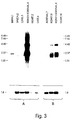
- Fig. 3 shows results of Northern blot analysis of RNA from different cells types probed with an OP-1 probe.
- the invention is based on the discovery of a family of structurally related morphogenic proteins (BMPs), also called osteogenic proteins (OPs), and more particularly that various of these proteins play an important role, not only in embryogenesis, but also in tissue and organ maintenance and repair in juvenile and adult mammals.
- Morphogenic proteins which have been identified include BMP 2, 3, 4, 5, 6, OP-1 and OP-2 (murine and human), Vgr-1, Vgl, DPP, GDF-1, CMBP-2A, CMBP-2B, 60A, and the inhibin/activin class of proteins.
- Other recombinant proteins include COP1, COP3, COP4, COP5, COP7, and COP16.
- variants within the morphogenic protein genes may have specific roles in specific tissue involving for example, stimulation of progenitor cell multiplication, tissue specific or tissue preferred phenotype maintenance, and/or stimulation or modulation of the rate of differentiation, growth or replication of tissue cells characterized by high turnover.
- stimulation of progenitor cell multiplication tissue specific or tissue preferred phenotype maintenance
- stimulation or modulation of the rate of differentiation, growth or replication of tissue cells characterized by high turnover The effect on the long-term physiology, maintenance and repair of particular tissues by particular species of the morphogens is currently unknown in any significant detail.
- This invention provides such methods and, more specifically, two generic processes for obtaining data which ultimately will permit determination of structure/activity relationships of specific naturally occurring mammalian morphogens and drugs capable of modulating their production.
- OP-1 first found in bone and demonstrated to be osteoinductive, is synthesized primarily in kidney, bladder, and adrenal tissue.
- This surprising discovery coupled with the observation that patients with kidney disease often express loss of bone mass, suggests that the bone loss in these patients may be due to pathologic depression of OP-1 synthesis in kidney, and suggests that administration of OP-1 systemically or stimulation of OP-1 expression and secretion by the kidney may arrest bone loss, or effect remineralization through increased bone formation (i.e., osteogenesis).
- One aspect involves an assay to determine tissues and cell types capable of synthesis and secretion of the morphogens; the other involves the use of the identified cell types configured in a screening system to find substances useful therapeutically to modulate, i.e., stimulate or depress, morphogen expression and/or secretion.
- the assay to determine the tissue of origin of a given morphogen involves screening a plurality (i.e., two or more) different tissues by determining a parameter indicative of production of a morphogen in the tissue, and comparing the parameters.
- the tissue(s) of origin will, of course, be the tissue that produces that morphogen.
- the other assay of the invention involves screening candidate compounds for their ability to modulate the effective systemic or local concentration of a morphogen by incubating the compound with a cell culture that produces the morphogen, and assaying the culture for a parameter indicative of a change in the production level of the morphogen.
- Useful candidate compounds then may be tested for in vivo efficacy in a suitable animal model. These compounds then may be used in vivo to modulate effective morphogen concentrating in the disease treatment.
- Morphogens are broadly distributed in developing and adult tissue.
- DPP and 60A are expressed in both embryonic and developing Drosophila tissue.
- Vgl has been identified in Xenopus embryonic tissue.
- Vgr-1 transcripts have been identified in a variety of murine tissues, including embryonic and developing brain, lung, liver, kidney and calvaria (dermal bone) tissue.
- CBMP2B and CBMP3 have been identified in lung tissue.
- Vgr-1 transcripts also have been identified in adult murine lung, kidney, heart, and brain tissue, with particularly high levels in the lung (see infra).
- GDF-1 has been identified in human adult cerebellum and in fetal brain tissue.
- OP-1 is encoded by multiple transcripts in different tissues. This potential alternative splicing is consistent with the hypothesis that the longer transcripts may encoded additional proteins (e.g., bicistronic mRNA) and each form may be tissue or developmentally related.
- additional proteins e.g., bicistronic mRNA
- OP-1 and the CBMP2 proteins both first identified as bone morphogens, have been identified in mouse and human placenta, hippocampus, calvaria and osteosarcoma tissue as determined by identification of OP-1 and CMBP2-specific sequences in cDNA libraries constructed from these tissues (see USSN 422,699, incorporated herein by reference). Additionally, the OP-1 protein is present in a variety of embryonic and developing tissues including kidney, liver, heart and brain as determined by Western blot analysis and immunolocalization (see infra). OP-1-specific transcripts also have been identified in both embryonic and developing tissues, most abundantly in developing kidney, bladder, adrenal and (see infra).
- OP-1 also has been identified as a mesoderm inducing factor present during embryogenesis. Moreover, OP-1 has been shown to be associated with satellite cells in the muscle and associated with potential pluripotential stem cells in bone marrow following damage to adult murine endochondral bone, indicating its morphogenic role in tissue repair and regeneration. In addition, a novel protein GDF-1 comprising a 7 cysteine skeleton, has been identified in neural tissue (Lee, 1991, Proc. Nat. Aca. Sci. 88: 4250-4254).
- tissue distribution of a given morphogen may be useful in choosing a cell type for screening according to the invention, or for targeting that cell type or tissue type for treatment.
- the proteins or their mRNA transcripts
- the proteins are readily identified in different tissues using standard methodologies and minor modifications thereof in tissues where expression may be low.
- protein distribution may be determined using standard Western blot analysis or immunocytochemical techniques, and antibodies specific to the morphogen or morphogens of interest.
- the distribution of morphogen transcripts may be determined using standard Northern hybridization protocols and a transcript-specific probe and hybridization conditions.
- a protein is morphogenic if it is capable of inducing the developmental cascade of cellular and molecular events that culminate in the formation of new, organ-specific tissue and comprises at least the conserved C-terminal six cysteine skeleton or its functional equivalent (see supra).
- the morphogens generally are capable of all of the following biological functions in a morphogenically permissive environment: stimulating proliferation of progenitor cells; stimulating the differentiation of progenitor cells; stimulating the proliferation of differentiated cells; and supporting the growth and maintenance of differentiated cells, including the "redifferentiation" of transformed cells.
- the morphogens detectable according to the methods of this invention first were identified, as well as a description on how to make, use and test them for morphogenic activity are disclosed in USSN 667,274, filed March 11, 1991 and USSN 752,764, filed August 30, 1991, the disclosures of which are hereby incorporated by reference.
- the morphogens may be purified from naturally-sourced material or recombinantly produced from procaryotic or eucaryotic host cells, using the genetic sequences disclosed therein. Alternatively, novel morphogenic sequences may be identified following the procedures disclosed therein.
- Particularly useful proteins include those which comprise the naturally derived sequences disclosed in Table II.
- Other useful sequences include biosynthetic constructs such as those disclosed in U.S. Pat. 5,011,691, the disclosure of which is incorporated herein by reference (e.g., COP-1, COP-3, COP-4, COP-5, COP-7, and COP-16).
- the morphogens detectable according to the methods and compositions of this invention also may be described by morphogenically active proteins having amino acid sequences sharing 70% or, preferably, 80% homology (similarity) with any of the sequences described above, where "homology" is as defined herein above.
- the morphogens detectable according to the method of this invention also can be described by any of the 6 generic sequences described herein (Generic Sequences 1, 2, 3, 4, 5 and 6). Generic sequences 1 and 2 also may, include, at their N-terminus, the sequence
- Table II compares the amino acid sequences of the active regions of native proteins that have been identified as morphogens, including human OP-1 (hOP-1, Seq. ID Nos. 5 and 16-17), mouse OP-1 (mOP-1, Seq. ID Nos. 6 and 18-19), human and mouse OP-2 (Seq. ID Nos. 7, 8, and 20-23), CBMP2A (Seq. ID No. 9), CBMP2B (Seq. ID No. 10), BMP3 (Seq. ID No. 26), DPP (from Drosophila, Seq. ID No. 11), Vgl, (from Xenopus, Seq. ID No. 12), Vgr-1 (from mouse, Seq. ID No.
- amino acid residue 60 of CBMP-2A and CBMP-2B is "missing".
- both these amino acid sequences in this region comprise Asn-Ser (residues 58, 59), with CBMP-2A then comprising Lys and Ile, whereas CBMP-2B comprises Ser and Ile.
- the currently most preferred protein sequences detectable as morphogens in this invention include those having greater than 60% identity, preferably greater than 65% identity, with the amino acid sequence defining the conserved six cysteine skeleton of hOP1 (e.g., residues 43-139 of Seq. ID No. 5). These most preferred sequences include both allelic and species variants of the OP-1 and OP-2 proteins, including the Drosophila 60A protein. Accordingly, in still another preferred aspect, the invention includes detection of morphogens comprising species of polypeptide chains having the generic amino acid sequence referred to herein as "OPX", which defines the seven cysteine skeleton and accommodates the identities between the various identified mouse and human OP1 and OP2 proteins. OPX is presented in Seq.
- each Xaa at a given position independently is selected from the residues occurring at the corresponding position in the C-terminal sequence of mouse or human OP1 or OP2 (see Seq. ID Nos. 5-8 and/or Seq. ID Nos. 16-23).
- a morphogen Once a morphogen is identified in a tissue, its level may be determined either at the protein or nucleic acid level. By comparing the levels of production of a given morphogen among different tissues, it is possible to determine the tissue(s) of origin of that morphogen.
- the level of production of the morphogen OP-1 in different tissues is one example of a morphogen having a tissue of origin, i.e., the kidney, which contains a cell type that can also be used as the cell type which is used to screen, according to the invention, different compounds for their potential effects on morphogen (OP-1) production.
- the level of OP-1 varies among different tissue types. In order to screen compounds for their effect on the production of OP-1 by a given cell type, it may be desirable to determine which tissues produce levels of OP-1 which are sufficiently high to show a potential decrease and sufficiently low to show a potential increase in production. Different tissues may be screened at the RNA level as follows.
- any probe capable of hybridizing specifically to a transcript, and distinguishing the transcript of interest from other, related transcripts may be used. Because the morphogens to be detected in the methods of this invention share such high sequence homology in their C-terminal domain, the tissue distribution of a specific morphogen transcript may best be determined using a probe specific for the "pro" region of the immature protein and/or the N-terminal heterogeneous region of the mature protein. Another useful probe sequence is the 3'non-coding region immediately following the stop codon. These portions of the sequence vary substantially among the morphogens of this invention, and accordingly, are specific for each protein.
- Vgr-1-specific probe sequence is the PvuII-SacI fragment, a 265 bp fragment encoding both a portion of the pro region and the N-terminus of the mature sequence.
- particularly useful mOP-1-specific probe sequences are the BstXI-BglI fragment, a 0.68kb sequence that covers approximately two-thirds of the mOP1 pro region; a StuI-StuI fragment, a 0.2 kb sequence immediately upstream of the 7-cysteine domain, and an EarI-PstI fragment, a 0.3kb fragment containing the 3'untranslated sequence. Similar approaches may be used, for example, with hOP-1 (SEQ. ID NO.16) or human or mouse OP-2 (SEQ. ID NOS.20 and 22).
- morphogen transcripts can be identified in mammalian tissues, using standard methodologies well known to those having ordinary skill in the art. Briefly, total RNA from mouse embryos and organs from post-natal animals is prepared using the acid guanidine thiocyanate-phenolchloroform method (Chomczynski et al., Anal. Biochem. 162:156-159, 1987). The RNA may be dissolved in TES buffer (10 mM Tris-HCl, 1 mM EDTA, 0.1% SDS, pH 7.5) and treated with Proteinase K (approx. 1.5 mg per g tissue sample) at 45°C for 1 hr.
- TES buffer 10 mM Tris-HCl, 1 mM EDTA, 0.1% SDS, pH 7.5
- Poly(A) + RNA selection on oligo(dT)-cellulose may be done in a batch procedure by mixing 0.1 g oligo(dT)-cellulose with 11 ml RNA solution (from 1 g tissue) in TES buffer and 0.5 M NaCl). Thereafter the oligo(dT) cellulose is washed in binding buffer (0.5 M NaCl, 10 mM Tris-HCl, 1 mM EDTA, pH 7.5) and poly(A) + RNA is eluted with water.
- binding buffer 0.5 M NaCl, 10 mM Tris-HCl, 1 mM EDTA, pH 7.5
- Poly(A) + RNA (5 or 15 ⁇ g/lane) is fractionated on 1 or 1.2% agarose-formaldehyde gels (Selden, in Current Protocols in Molecular Biology, Ausubel et al. eds., pp. 1-4, 8, 9, Greene Publishing and Wiley-Interscience, New York, 1991). 1 ⁇ l of 400 pg/ml ethidium bromide is added to each sample prior to heat denaturation (Rosen et al., Focus 12:23-24, 1990). Following electrophoresis, the gels are photographed and the RNA is blotted overnight onto Nytran nitrocellulose membranes (Schleicher & Schuell Inc., Keene, NH) with 10 x SSC.
- the membranes are baked at 80°C for 30-60 min. and irradiated with UV light (1 mW/cm 2 for 25 sec.).
- the Northern hybridization conditions may be as previously described (Ozkaynak et al., EMBO J. 9:2085-2093, 1990).
- the filters may be deprobed in 1 mM Tris-HCl, 1 mM EDTA, 0.1% SDS, pH 7.5, at 90-95°C and exposed to film to assure complete removal of previous hybridization signals.
- One probe which may be used to screen for transcripts encoding a morphogen includes a portion of or the complete OP-1 cDNA, which may be used to detect the presence of OP-1 mRNA or mRNAs of related morphogens.
- the sequence of the murine cDNA gene is set forth in SEQ ID NO:14.
- OP-1 mRNA expression was analyzed in 17 day mouse embryos and 3 day post-natal mice by sequentially hybridizing filters with various probes. Probes from regions other than the highly conserved 7-cysteine domain were selected because this region is highly variable among members of the TGF- ⁇ superfamily.
- Fig. 1 shows the fragments of OP-1, used as probes in the Northern hybridizations. The solid box indicates the putative signal peptide and the hatched box corresponds to the TGF- ⁇ -like domain that contains the seven cysteine residues. Asterisks indicate the potential N-glycosylation sites. The arrow marks the location of the cleavage site for OP-1 maturation.
- Hybridization with a probe that covers approximately two thirds of the pro region reveals a 4 kb message and 3 messages at 1.8 kb, 2.2 kb and 2.4 kb (Fig. 2B and D, and Fig. 3).
- the Northern hybridization of Fig. 2 equal amounts (15 ⁇ g) of poly(A) + RNA were loaded into each lane, electrophoresed on a 1% agarose-formaldehyde gel, blotted and hybridized. A 0.24 - 9.49 kb RNA ladder (Bethesda Research Labs, Inc.) was used as size standard.
- the same filter was used for sequential hybridizations with labeled probes specific for OP-1 (Panels B and D), Vgr-1 (Panel C), and EF-Tu (Panel A).
- Panel A the EF-Tu specific probe (a control) was the 0.4 kb HindIII-SacI fragment (part of the coding region), the SacI site used belonged to the vector;
- Panel B the OP-1 specific probe was the 0.68 kb BstXI-BglI fragment (two thirds of the pro region and upstream sequences of the mature domain, not including any sequences from the 7-cysteine domain);
- Panel C the Vgr-1 specific probe was the 0.26 kb PvuII-SacI fragment (part of the pro region and the amino-terminal sequences of the mature domain, including the first cysteine) (Lyons et al., 1989, Proc.
- Panel D the OP-1 (3' flanking) specific probe was the 0.34 kb EarI-PstI fragment (3' untranslated sequences immediately following the sequences encoding OP-1).
- RNA preparation was obtained from two week old mice (Panel A) or 5 week old mice (Panel B), with the exception of poly A+ RNA which was obtained from kidney adrenal gland of two week old mice (Panel B). Equal amounts of poly A+ RNA (15 ⁇ g for Panel A and 5 ⁇ g for Panel B) were loaded into each well. After electrophoresis (1.2% agarose-formaldehyde gels) and blotting, RNA was hybridized to the OP-1 specific 3' flanking probe described in the legend of Fig. 2 (Panel D). The 0.24-9.5 kb RNA ladder was used as size standard. The arrowheads indicate the OP-1 specific messages. The lower section of Panels A and B show the hybridization pattern obtained with the EF-Tu specific probe (a control).
- the OP-1 4 kb mRNA is somewhat larger.
- the 0.2 kb StuI-StuI fragment which represents the gene specific sequences immediately upstream of those encoding the 7-cysteine domain was used. This probe gave a hybridization pattern similar to the one shown in Fig. 2 Panel B (data not shown).
- the same four OP-1 specific messages were observed with three distinct probes.
- the appearance of a new 4 kb OP-1 mRNA species was initially interpreted as cross hybridization of the OP-1 probe with Vgr-1 mRNA, which is approximately this size (Fig. 2 Panel C).
- the 4 kb message was detected with three different OP-1 specific probes, including one specific to the 3' untranslated region, and moreover it was separated from Vgr-1 message on the basis of size.
- the 4 kb mRNA (and the three species of 1.8 kb, 2.2 kb and 2.4 kb) results from alternative splicing of OP-1 transcripts.
- the 4 kb OP-1 mRNA could also represent a bicistronic mRNA.
- the 4 kb message is a minor species in kidney, while it is more prominent in adrenal tissue.
- RNA was analyzed on Northern blots by hybridization to various probes (Fig. 3. Equal amounts of mRNA, as judged by optical density, were fractionated on agarose formaldehyde gels. Ethidium bromide staining of the gels revealed some residual ribosomal RNA in addition to the mRNA and provided another assurance that the mRNA was not degraded and that there was not significant quantitative or qualitative variation in the preparation.
- EF-Tu translational elongation factor
- OP-1 mRNA patterns display qualitative changes in the various tissues. Of the four messages found in brain, the 2.2 kb message is most abundant whereas in lung and spleen the 1.8 kb message predominates. Levels of the 1.8-2.4 kb in the kidney OP-1 mRNA are approximately two times higher in 3 day post-natal mice than in 17 day embryos, perhaps reflecting phases in bone and/or kidney development. mRNA was also prepared from carefully separated renal and adrenal tissues of 5 week old mice. Northern blot analysis ( Figure 3 Panel B) revealed that the high levels of 2.2 kb mRNA were derived from renal tissue whereas the 4 kb mRNA was more prominent in adrenal tissue.
- tissue-specific expression of a given morphogen is known, cell types known to exist in that tissue or cell lines derived from that tissue can be screened, in a similar manner, to identify the cell type within that tissue that is actually responsible for the tissue specific synthesis and secretion of the morphogen.
- tissue culture assay Once a cell type which produces the morphogen in an amount sufficient to detect increases or decreases in the production level of the morphogen upon exposure to a compound is identified, it may be used in tissue culture assay to rapidly screen for the ability of compound to upregulate or down regulate the synthesis and secretion of the morphogen.
- the level of morphogen production by the chosen cell type is determined with and without incubating the cell in culture with the compound, in order to assess the effects of the compound on the cell's ability to synthesize or secrete the morphogen. This can be accomplished by detection of the level of production of the morphogen either at the protein or mRNA level.
- kidneys may be explanted from neonatal, new born, young or adult rodents (mouse or rat) and used in organ culture as whole or sliced (1-4 mm) tissues.
- Primary tissue cultures and established cell lines, also derived from kidney, adrenals, urinary, bladder, brain, or other tissues may be established in multiwell plates (6 well, 24 well, or 96 well) according to conventional cell culture techniques, and are cultured in the absence or presence of serum for a period of time (1-7 days).
- Cells may be cultured, for example, in Dulbecco's Modified Eagle medium (Gibco, Long Island, NY) containing serum (e.g., fetal calf serum at 1%-10%, Gibco) or in serum-deprived medium, as desired, or in defined medium (e.g., containing insulin, transferrin, glucose, albumin, or other growth factors).
- serum e.g., fetal calf serum at 1%-10%, Gibco
- serum-deprived medium e.g., fetal calf serum at 1%-10%, Gibco
- defined medium e.g., containing insulin, transferrin, glucose, albumin, or other growth factors.
- Samples for testing the level of morphogen production include culture supernatants or cell lysates, collected periodically and evaluated for OP-1 production by immunoblot analysis of a portion of the cell culture itself, collected periodically and used to prepare polyA+ RNA for RNA analysis (Sambrook et al., eds., Molecular Cloning, 1989, Cold Spring Harbor Press, Cold Spring Harbor, NY). To monitor de novo OP-1 synthesis, some cultures are labeled with 3 5 S-methionine/ 3 5 S-cysteine mixture for 6-24 hours and then evaluated for morphogen production by conventional immunoprecipitation methods (Sambrook et al., eds., Molecular Cloning, 1989, Cold Spring Harbor Press, Cold Spring Harbor, NY).
- the production of morphogen or determination of the level of morphogen production may be ascertained using a simple assay for a parameter of cell growth, e.g., cellular proliferation or death.
- a simple assay for a parameter of cell growth e.g., cellular proliferation or death.
- a morphogen is produced by a cultured cell line
- the addition of antibody specific for the morphogen may result in relief from morphogen inhibition of cell growth.
- measurement of cellular proliferation can be used as an indication of morphogen production by a tissue.
- an immunoassay may be performed to detect the morphogen using a polyclonal or monoclonal antibody specific for that morphogen.
- OP-1 may be detected using a polyclonal antibody specific for OP-1 in an ELISA, as follows.
- a 100 ul aliquot of an appropriate dilution of each of the test samples of cell culture supernatant is added to each well in triplicate and incubated at 37°C for 30 min. After incubation, 100 ul biotinylated rabbit anti-OP-1 serum (stock solution is about 1 mg/ml and diluted 1:400 in BSB containing 1% BSA before use) is added to each well and incubated at 37°C for 30 min. The wells are then washed four times with BSB containing 0.1% Tween 20. 100 ul strepavidin-alkaline (Southern Biotechnology Associates, Inc.
- Polyclonal antibody is prepared as follows. Each rabbit is given a primary immunization of 100 ug/500 ul E. coli-produced OP-1 monomer (amino acids 328-431 of SEQ. ID NO: 11) in 0.1% SDS mixed with 500 ul Complete Freund's Adjuvant. The antigen is injected subcutaneously at multiple sites on the back and flanks of the animal. The rabbit is boosted after a month in the same manner using incomplete Freund's Adjuvant. Test bleeds are taken from the ear vein seven days later. Two additional boosts and test bleeds are performed at monthly intervals until antibody against OP-1 is detected in the serum using an ELISA assay. Then, the rabbit is boosted monthly with 100 ug of antigen and bled (15 ml per bleed) at days seven and ten after boosting.
- Monoclonal antibody specific for a given morphogen may be prepared as follows. A mouse is given two injections of E. coli produced OP-1 monomer (amino acids 328-431 in SEQ ID NO:11). The first injection contains 100ug of OP-1 in complete Freund's adjuvant and is given subcutaneously. The second injection contains 50 ug of OP-1 in incomplete adjuvant and is given intraperitoneally. The mouse then receives a total of 230 ug of OP-1 (amino acids 307-431 of SEQ ID NO:11) in four intraperitoneal injections at various times over an eight month period.
- the mouse is boosted intraperitoneally with 100 ug of OP-1 (15-139) and 30 ug of the N-terminal peptide (Ser293-Asn309-Cys) conjugated through the added cys residue to bovine serum albumin with SMCC crosslinking agent. This boost is repeated five days (IP), four days (IP), three days (IP) and one day (IV) prior to fusion.
- the mouse spleen cells are then fused to myeloma (e.g., 653) cells at a ratio of 1:1 using PEG 1500 (Boehringer Mannheim), and the cell fusion is plated and screened for OP-1-specific antibodies using OP-1 (307-431) as antigen.
- the cell fusion and monoclonal screening are according to procedures widely available in the art.
- the neutralizing monoclonal is identified by its ability to block the biological activity of OP-1 when added to a cellular assay which responds biologically to added OP-1.
- a simple medium flux screening assay can be configured in a standard 24 or 96 well microtiter dishe, in which each well contains a constant number of a cell line having the characteristics described above. Increasing concentrations of an OP-1 neutralizing monoclonal antibody is added from left to right across the dish. A constant amount of different test substances is added from top to bottom on the dish. An increase in the synthesis and secretion of OP-1 (over its constitutive (non-induced) level) will be indicated by an increase in the amount of OP-1 neutralizing antibody required to release the cells from the antimitogenic activity of OP-1.
- the assay may be performed as follows. After addition of appropriate concentrations of the OP-1 neutralizing monoclonal antibody and test substances to the wells containing the cells, the dishes are placed in an incubator at 37°C for a period of 1-3 days. After completion of incubation/growth period, the dishes are removed and the cells in the individual wells are washed and stained with a vital stain, such as crystal violet. Washing and staining procedures are well-known in the art. The cells are then lysed and the stain dissolved in a constant amount of a solvent, such as ethanol. Quantitations of the dissolved stain, which is readily performed on an automated plate vendor, allows for direct quantitation of the number of cells in each well.
- a vital stain such as crystal violet
- the above-described assay has the advantages of being rapid and easy to perform becaue it requires few steps. Another advantage is intrinsic to the assay; drugs which are screened according to this procedure that result in cell death (i.e., cytotoxic substances) are immediately, identifiable without the need of operator observation. In addition, although drugs that stop the growth of the cells (i.e., cytostatic substances) are scored as positive due to failure to see increases in cell numbers, they are automatically scored as suspect due to the failure of the highest concentrations of OP-1 neutralizing monoclonal antibody to release the cells from the antimitogenic activity of OP-1.
- the screening methods of the invention is used to test compounds for their effect on the production of morphogenic protein by a given cell type.
- compounds which may be screened include but are not limited to chemicals, biological response modifiers (e.g., lymphokines, cytokines, hormones, or vitamins), plant extracts, microbial broths and extracts medium conditioned by eukaryotic cells, body fluids, or tissue extracts.
- Described is a method of screening candidate compounds for the ability to modulate the effective concentration of a morphogen in an organism, said method comprising
- Said morphogen may be OP-1.
- Said test tissue type may be a human renal-derived tissue.
- said renal-derived tissue is a kidney or bladder-derived tissue.
- said test tissue type is adrenal-derived tissue.
- said morphogen is GDF-1.
- said test tissue type is derived from human nerve tissue.
- said nerve tissue is brain-derived tissue.
- said morphogen is DPP.
- said test tissue type is derived from one of the following drosophila tissues: dorsal ectoderm, epithelial imaginal disc visceral mesoderm, or gut endoderm.
- said morphogen is Vgr-1.
- said test tissue type is mouse lung tissue.
- said morphogen is vgl.
- said test tissue type is xenopus fetal endoderm tissue.
- Described is a method of assessing a tissue of an organism for its level of production of a morphogen and for screening candidate compounds for the ability to modulate the effective concentration of said morphogen produced by cells of said tissue, said method comprising
- Said parameter may be indicative of the level of said morphogen is determined using an antibody specific for said morphogen, or indicative of the level of said morphogen is determined by measuring cellular proliferation in cells which are sensitive to the concentration of secreted OP-1.
- said parameter is indicative of the level of said morphogen is determined using a nucleic acid probe that hybridizes under stringent conditions with nucleic acid encoding said morphogen.
- said morphogen comprises a minimally active core C-terminal region comprising at least six cysteine residues, and said nucleic acid probe hybridizes with an mRNA encoding a region N-terminal to said core region.
- said morphogen comprises a minimally active core C-terminal region comprising at least six cysteine residues, and said nucleic acid probe hybridizes with an mRNA encoding a region 3' to said core region.
Landscapes
- Health & Medical Sciences (AREA)
- Life Sciences & Earth Sciences (AREA)
- Chemical & Material Sciences (AREA)
- Engineering & Computer Science (AREA)
- Immunology (AREA)
- Molecular Biology (AREA)
- Biomedical Technology (AREA)
- Hematology (AREA)
- Urology & Nephrology (AREA)
- General Health & Medical Sciences (AREA)
- Organic Chemistry (AREA)
- Biochemistry (AREA)
- Analytical Chemistry (AREA)
- Cell Biology (AREA)
- Medicinal Chemistry (AREA)
- Proteomics, Peptides & Aminoacids (AREA)
- Physics & Mathematics (AREA)
- Microbiology (AREA)
- Biotechnology (AREA)
- Pathology (AREA)
- General Physics & Mathematics (AREA)
- Food Science & Technology (AREA)
- Bioinformatics & Cheminformatics (AREA)
- Toxicology (AREA)
- Zoology (AREA)
- Genetics & Genomics (AREA)
- Biophysics (AREA)
- Tropical Medicine & Parasitology (AREA)
- Wood Science & Technology (AREA)
- Endocrinology (AREA)
- Gastroenterology & Hepatology (AREA)
- General Engineering & Computer Science (AREA)
- Diabetes (AREA)
- Orthopedic Medicine & Surgery (AREA)
- Neurology (AREA)
- Neurosurgery (AREA)
- Investigating Or Analysing Biological Materials (AREA)
- Peptides Or Proteins (AREA)
- Measuring Or Testing Involving Enzymes Or Micro-Organisms (AREA)
- Preparation Of Compounds By Using Micro-Organisms (AREA)
Applications Claiming Priority (3)
| Application Number | Priority Date | Filing Date | Title |
|---|---|---|---|
| US75286191A | 1991-08-30 | 1991-08-30 | |
| US752861 | 1991-08-30 | ||
| EP92921799A EP0601129B1 (fr) | 1991-08-30 | 1992-08-28 | Procede de triage de proteines morphogeniques |
Related Parent Applications (1)
| Application Number | Title | Priority Date | Filing Date |
|---|---|---|---|
| EP92921799A Division EP0601129B1 (fr) | 1991-08-30 | 1992-08-28 | Procede de triage de proteines morphogeniques |
Publications (2)
| Publication Number | Publication Date |
|---|---|
| EP0825442A2 true EP0825442A2 (fr) | 1998-02-25 |
| EP0825442A3 EP0825442A3 (fr) | 2004-02-11 |
Family
ID=25028180
Family Applications (3)
| Application Number | Title | Priority Date | Filing Date |
|---|---|---|---|
| EP92921799A Expired - Lifetime EP0601129B1 (fr) | 1991-08-30 | 1992-08-28 | Procede de triage de proteines morphogeniques |
| EP00100232A Withdrawn EP1033574A3 (fr) | 1991-08-30 | 1992-08-28 | Procédé de titrage de protéines morphogéniques |
| EP97202681A Ceased EP0825442A3 (fr) | 1991-08-30 | 1992-08-28 | Procédé de titrage de protéines morphogéniques |
Family Applications Before (2)
| Application Number | Title | Priority Date | Filing Date |
|---|---|---|---|
| EP92921799A Expired - Lifetime EP0601129B1 (fr) | 1991-08-30 | 1992-08-28 | Procede de triage de proteines morphogeniques |
| EP00100232A Withdrawn EP1033574A3 (fr) | 1991-08-30 | 1992-08-28 | Procédé de titrage de protéines morphogéniques |
Country Status (8)
| Country | Link |
|---|---|
| EP (3) | EP0601129B1 (fr) |
| JP (3) | JP3616089B2 (fr) |
| AT (1) | ATE197611T1 (fr) |
| AU (1) | AU678345B2 (fr) |
| CA (1) | CA2116560C (fr) |
| DE (1) | DE69231567T2 (fr) |
| ES (1) | ES2156862T3 (fr) |
| WO (1) | WO1993005172A1 (fr) |
Families Citing this family (17)
| Publication number | Priority date | Publication date | Assignee | Title |
|---|---|---|---|---|
| US6071695A (en) * | 1992-02-21 | 2000-06-06 | Creative Biomolecules, Inc. | Methods and products for identification of modulators of osteogenic protein-1 gene expression |
| WO1995014104A1 (fr) * | 1993-11-16 | 1995-05-26 | Children's Medical Center Corporation | Procede d'identification d'une substance apte a induire la formation osseuse |
| DE69535641D1 (de) | 1994-04-29 | 2007-12-27 | Curis Inc | Morphogene protein-spezifische zelloberflächenrezeptoren und ihre verwendungen |
| CA2191583C (fr) * | 1994-06-07 | 2007-03-27 | Engin Ozkaynak | Procedes et compositions de modulation de l'expression de proteines morphogenetiques |
| US20030167492A1 (en) | 1994-07-08 | 2003-09-04 | Johns Hopkins University School Of Medicine | Transgenic non-human animals expressing a gdf-11 dominant negative polypeptide, and methods of making and using same |
| US6090544A (en) * | 1995-07-26 | 2000-07-18 | Creative Biomolecules, Inc. | Methods and compositions for identifying morphogen analogs |
| US7306903B1 (en) | 1995-07-26 | 2007-12-11 | Curis, Inc. | Methods and compositions for identifying morphogen analogs |
| US6103491A (en) * | 1995-07-26 | 2000-08-15 | Creative Biomolecules, Inc. | Methods and compositions for identifying morphogen analogs |
| WO1997007135A2 (fr) * | 1995-08-14 | 1997-02-27 | Creative Biomolecules, Inc. | Liaison de la proteine oestrogene 1 (op-1) et de ses analogues avec le recepteur alk-1 de surface de cellule et ses analogues |
| EP0904093A4 (fr) | 1996-03-22 | 2004-11-17 | Gen Hospital Corp | Administration de facteurs de croissance polypeptidiques apres une ischemie ou un trauma du systeme nerveux central |
| US6498142B1 (en) | 1996-05-06 | 2002-12-24 | Curis, Inc. | Morphogen treatment for chronic renal failure |
| US5928940A (en) * | 1996-09-24 | 1999-07-27 | Creative Biomolecules, Inc. | Morphogen-responsive signal transducer and methods of use thereof |
| JP5033276B2 (ja) | 1997-05-05 | 2012-09-26 | ストライカー コーポレイション | 急性腎不全の治療 |
| JP2001526788A (ja) * | 1997-05-30 | 2001-12-18 | クリエイティブ バイオモレキュールズ,インコーポレイテッド | 組織形態形成および活性を評価するための方法 |
| US7147839B2 (en) | 1998-05-29 | 2006-12-12 | Curis, Inc. | Methods for evaluating tissue morphogenesis and activity |
| US6524797B1 (en) | 1999-05-10 | 2003-02-25 | Bernhard O. Palsson | Methods of identifying therapeutic compounds in a genetically defined setting |
| ES2474194T3 (es) | 2008-02-13 | 2014-07-08 | Keith Hruska | PMO-7 para su uso en el tratamiento de la hiperplasia de la neo�ntima |
Citations (2)
| Publication number | Priority date | Publication date | Assignee | Title |
|---|---|---|---|---|
| WO1990011366A1 (fr) * | 1989-03-28 | 1990-10-04 | Genetics Institute, Inc. | Compositions osteoinductrices |
| WO1992015323A1 (fr) * | 1991-03-11 | 1992-09-17 | Creative Biomolecules, Inc. | Morphogenese induite par des proteines |
Family Cites Families (3)
| Publication number | Priority date | Publication date | Assignee | Title |
|---|---|---|---|---|
| US4980281A (en) * | 1988-02-10 | 1990-12-25 | Housey Gerard M | Method of screening for protein inhibitors and activators |
| JPH03505669A (ja) * | 1988-07-08 | 1991-12-12 | ユニバーシテイ・カレツジ・ロンドン | バイオアツセイにおける改良 |
| CA2064878A1 (fr) * | 1989-08-21 | 1991-02-22 | Hanne Bentz | Proteine specifique des os |
-
1992
- 1992-08-28 ES ES92921799T patent/ES2156862T3/es not_active Expired - Lifetime
- 1992-08-28 CA CA002116560A patent/CA2116560C/fr not_active Expired - Fee Related
- 1992-08-28 WO PCT/US1992/007359 patent/WO1993005172A1/fr active IP Right Grant
- 1992-08-28 JP JP50534593A patent/JP3616089B2/ja not_active Expired - Fee Related
- 1992-08-28 EP EP92921799A patent/EP0601129B1/fr not_active Expired - Lifetime
- 1992-08-28 DE DE69231567T patent/DE69231567T2/de not_active Expired - Fee Related
- 1992-08-28 AU AU28624/92A patent/AU678345B2/en not_active Ceased
- 1992-08-28 EP EP00100232A patent/EP1033574A3/fr not_active Withdrawn
- 1992-08-28 AT AT92921799T patent/ATE197611T1/de not_active IP Right Cessation
- 1992-08-28 EP EP97202681A patent/EP0825442A3/fr not_active Ceased
-
2003
- 2003-01-23 JP JP2003014941A patent/JP2004069670A/ja active Pending
-
2004
- 2004-03-24 JP JP2004087203A patent/JP2004267211A/ja active Pending
Patent Citations (2)
| Publication number | Priority date | Publication date | Assignee | Title |
|---|---|---|---|---|
| WO1990011366A1 (fr) * | 1989-03-28 | 1990-10-04 | Genetics Institute, Inc. | Compositions osteoinductrices |
| WO1992015323A1 (fr) * | 1991-03-11 | 1992-09-17 | Creative Biomolecules, Inc. | Morphogenese induite par des proteines |
Non-Patent Citations (2)
| Title |
|---|
| OZKAYNAK E ET AL: "MURINE OSTEOGENIC PROTEIN (OP-1): HIGH LEVELS OF MRNA IN KIDNEY" BIOCHEMICAL AND BIOPHYSICAL RESEARCH COMMUNICATIONS, ACADEMIC PRESS INC. ORLANDO, FL, US, vol. 179, no. 1, 30 August 1991 (1991-08-30), pages 116-123, XP000561932 ISSN: 0006-291X * |
| OZKAYNAK E ET AL: "OP-1 CDNA ENCODES AN OSTEOGENIC PROTEIN IN THE TGF-BETA FAMILY" EMBO JOURNAL, OXFORD UNIVERSITY PRESS, SURREY, GB, vol. 9, no. 7, 1 July 1990 (1990-07-01), pages 2085-2093, XP000611252 ISSN: 0261-4189 * |
Also Published As
| Publication number | Publication date |
|---|---|
| JP3616089B2 (ja) | 2005-02-02 |
| EP0825442A3 (fr) | 2004-02-11 |
| CA2116560A1 (fr) | 1993-03-18 |
| DE69231567D1 (de) | 2000-12-21 |
| JP2004267211A (ja) | 2004-09-30 |
| DE69231567T2 (de) | 2001-06-28 |
| AU678345B2 (en) | 1997-05-29 |
| JPH06510432A (ja) | 1994-11-24 |
| ATE197611T1 (de) | 2000-12-15 |
| EP0601129B1 (fr) | 2000-11-15 |
| EP0601129A1 (fr) | 1994-06-15 |
| WO1993005172A1 (fr) | 1993-03-18 |
| EP1033574A3 (fr) | 2004-01-28 |
| CA2116560C (fr) | 2002-01-08 |
| EP1033574A2 (fr) | 2000-09-06 |
| JP2004069670A (ja) | 2004-03-04 |
| ES2156862T3 (es) | 2001-08-01 |
| AU2862492A (en) | 1993-04-05 |
Similar Documents
| Publication | Publication Date | Title |
|---|---|---|
| US5650276A (en) | Morphogenic protein screening method | |
| EP0601129B1 (fr) | Procede de triage de proteines morphogeniques | |
| Ozkaynak et al. | OP‐1 cDNA encodes an osteogenic protein in the TGF‐beta family. | |
| Wozney et al. | Novel regulators of bone formation: molecular clones and activities | |
| McGrath et al. | Oocyte-specific expression of growth/differentiation factor-9. | |
| US5674844A (en) | Treatment to prevent loss of and/or increase bone mass in metabolic bone diseases | |
| EP0601135B1 (fr) | Proteines osteogeniques pour le traitement des maladies osseuses metaboliques | |
| EP0575555B1 (fr) | Morphogenese induite par des proteines | |
| US5863738A (en) | Methods of antagonizing OP-1 binding to a cell surface receptor utilizing ALK polypeptides | |
| CA2363965C (fr) | Morphogenese induite par des proteines | |
| EP0653942B1 (fr) | Regeneration et reparation du systeme nerveux induites par morphogene | |
| US6800603B2 (en) | Morphogen-induced neural cell adhesion | |
| EP0601106B2 (fr) | Modulation, induite par morphogene, de reaction inflammatoire | |
| US6949505B1 (en) | Morphogen-induced dendritic growth | |
| US6495513B1 (en) | Morphogen-enhanced survival and repair of neural cells | |
| JPH08503198A (ja) | Op−3誘導形態形成 | |
| US5707810A (en) | Method of diagnosing renal tissue damage or disease | |
| US20050096339A1 (en) | Morphogen-induced modulation of inflammatory response | |
| JPH11510387A (ja) | モルフォゲンアナログを同定するための方法および組成物 | |
| US7056882B2 (en) | Treatment to prevent loss of and/or increase bone mass in metabolic bone diseases | |
| CA2144515A1 (fr) | Traitement des ulceres gastro-intestinaux | |
| AU696364B2 (en) | Morphogenic protein screening method II | |
| US7306903B1 (en) | Methods and compositions for identifying morphogen analogs | |
| CA2291510A1 (fr) | Modulateurs de l'expression de morphogenes et procedes d'identification correspondants |
Legal Events
| Date | Code | Title | Description |
|---|---|---|---|
| PUAI | Public reference made under article 153(3) epc to a published international application that has entered the european phase |
Free format text: ORIGINAL CODE: 0009012 |
|
| AC | Divisional application: reference to earlier application |
Ref document number: 601129 Country of ref document: EP |
|
| AK | Designated contracting states |
Kind code of ref document: A2 Designated state(s): AT BE CH DE DK ES FR GB GR IE IT LI LU MC NL SE |
|
| RIN1 | Information on inventor provided before grant (corrected) |
Inventor name: PANG, ROY HL Inventor name: RUEGER, DAVID C. Inventor name: KUBERASAMPATH, THANGAVEL Inventor name: OZKAYNAK, ENGIN Inventor name: OPPERMANN, HERMANN Inventor name: SMART, JOHN E. |
|
| RIN1 | Information on inventor provided before grant (corrected) |
Inventor name: PANG, ROY HL Inventor name: RUEGER, DAVID C. Inventor name: KUBERASAMPATH, THANGAVEL Inventor name: OZKAYNAK, ENGIN Inventor name: OPPERMANN, HERMANN Inventor name: SMART, JOHN E. |
|
| RIN1 | Information on inventor provided before grant (corrected) |
Inventor name: PANG, ROY HL Inventor name: RUEGER, DAVID C. Inventor name: KUBERASAMPATH, THANGAVEL Inventor name: OZKAYNAK, ENGIN Inventor name: OPPERMANN, HERMANN Inventor name: SMART, JOHN E. |
|
| RAP1 | Party data changed (applicant data changed or rights of an application transferred) |
Owner name: CURIS, INC. |
|
| PUAL | Search report despatched |
Free format text: ORIGINAL CODE: 0009013 |
|
| AK | Designated contracting states |
Kind code of ref document: A3 Designated state(s): AT BE CH DE DK ES FR GB GR IE IT LI LU MC NL SE |
|
| RIC1 | Information provided on ipc code assigned before grant |
Ipc: 7G 01N 33/68 B Ipc: 7G 01N 33/74 B Ipc: 7C 07K 14/51 B Ipc: 7C 07K 16/22 B Ipc: 7C 12Q 1/68 A |
|
| 17P | Request for examination filed |
Effective date: 20040726 |
|
| STAA | Information on the status of an ep patent application or granted ep patent |
Free format text: STATUS: THE APPLICATION HAS BEEN REFUSED |
|
| 18R | Application refused |
Effective date: 20080221 |