EP0767376A2 - Verfahren zur Detektion oder zum Messen einer immunologisch reaktiven Substanz - Google Patents
Verfahren zur Detektion oder zum Messen einer immunologisch reaktiven Substanz Download PDFInfo
- Publication number
- EP0767376A2 EP0767376A2 EP96307270A EP96307270A EP0767376A2 EP 0767376 A2 EP0767376 A2 EP 0767376A2 EP 96307270 A EP96307270 A EP 96307270A EP 96307270 A EP96307270 A EP 96307270A EP 0767376 A2 EP0767376 A2 EP 0767376A2
- Authority
- EP
- European Patent Office
- Prior art keywords
- reagent
- immunologically reactive
- reactive substance
- voltage
- small voltage
- Prior art date
- Legal status (The legal status is an assumption and is not a legal conclusion. Google has not performed a legal analysis and makes no representation as to the accuracy of the status listed.)
- Withdrawn
Links
Images




Classifications
-
- G—PHYSICS
- G01—MEASURING; TESTING
- G01N—INVESTIGATING OR ANALYSING MATERIALS BY DETERMINING THEIR CHEMICAL OR PHYSICAL PROPERTIES
- G01N33/00—Investigating or analysing materials by specific methods not covered by groups G01N1/00 - G01N31/00
- G01N33/48—Biological material, e.g. blood, urine; Haemocytometers
- G01N33/50—Chemical analysis of biological material, e.g. blood, urine; Testing involving biospecific ligand binding methods; Immunological testing
- G01N33/53—Immunoassay; Biospecific binding assay; Materials therefor
- G01N33/543—Immunoassay; Biospecific binding assay; Materials therefor with an insoluble carrier for immobilising immunochemicals
- G01N33/54366—Apparatus specially adapted for solid-phase testing
- G01N33/54373—Apparatus specially adapted for solid-phase testing involving physiochemical end-point determination, e.g. wave-guides, FETS, gratings
- G01N33/5438—Electrodes
Definitions
- This invention relates to a method for detecting or measuring an immunologically reactive substance. More specifically, it relates to a method for carrying out the detection or measurement with high sensitivity and at high speed.
- an immunologically reactive substance forms an insoluble agglomerate through an immunological agglutination reaction
- the immunologically reactive substance can be detected or measured by detecting the insoluble aggregate formed.
- an enzyme immunoassay method or radio-immunoassay method has been widely used as a method for detecting or measuring an immunologically reactive substance, which makes use of this phenomenon.
- these methods feature high sensitivity and high accuracy, they have restrictions on the stability and storage of a reagent because they use radiation and an enzyme and require delicate consideration and high technique. Therefore, a simpler method has been desired.
- Iwata et al. have proposed a method for accelerating an immunological agglutination reaction and increasing the sensitivity of the reaction by applying an AC field of 5 to 50 V/mm for 30 to 60 seconds under the coexistence of 10 mM or more of salt (refer to JP-A 83928/1995 (the term "JP-A” as used herein means an "unexamined published Japanese patent application”)).
- This method makes use of a pearl chain effect.
- the above objects and advantages of the present invention can be attained by a method for detecting or measuring an immunologically reactive substance through an immunological reaction using a reagent selected from the group consisting of an antibody, an antigen, a carrier reagent sensitized by an antigen, and a carrier reagent sensitized by an antibody, wherein an immunological reaction product is produced in an aqueous reaction system by applying a small voltage to the aqueous reaction system in which the reagent and the immunologically reactive substance are present without substantially forming a pearl chain derived from the reagent.
- a reagent selected from the group consisting of an antibody, an antigen, a carrier reagent sensitized by an antigen, and a carrier reagent sensitized by an antibody
- immunological reaction means an immunological agglutination reaction.
- the immunological agglutination reaction refers to a method making use of an antigen-antibody reaction, a reaction with a specific receptor or the like and includes both qualitative and quantitative methods.
- the immunologically reactive substance in the present invention can be selected from substances which can be measured by an agglutination method using latex, liposome or the like as a carrier through the above immunological agglutination reaction.
- Illustrative examples of the immunologically reactive substance as a first group include infectious disease markers such as HBs antigen, HBs antibody, HCV antibody, HIV antibody and TP antibody; thrombosis and fibrinosis markers such as D-dimer, protein C, protein S and AT III; tumor markers such as MP, CEA and CA19-9; hormones such as TSH, prolactin and insulin; tissue elements such as ⁇ 2m, myoglobin and myosin; and nucleic acid such as DNA.
- the detectable concentration of the immunologically reactive substance of the first group is about 0.05 ng/ml to 50 ⁇ g/ml, preferably 0.1 ng/ml to less than 50 ⁇ g/ml.
- Illustrative examples of the immunologically reactive substance as a second group include serum proteins such as CRP, albumin, transferrin, myoglobin, LP(a), C4, C3, IgG, IgM, ⁇ 1-antitrypsin and the like.
- the detectable concentration of the immunologically reactive substance of the second group is about 100 ng/ml to 1 mg/ml and the detection sensitivity is about 100 ng/ml to 10 ⁇ g/ml.
- Illustrative examples of the reagent used in the present invention include an antigen, an antibody, a carrier reagent sensitized by an antigen and a carrier reagent sensitized by an antibody. They may be used alone or in combination of two or more.
- the carrier particle include latex, liposome, gold sol and the like. Latex particles are more preferred. Latex particles which are generally used by an immunological agglutination method can be used in the present invention.
- the average diameter of the carrier particles is preferably 0.01 to 1 ⁇ m, more preferably 0.05 to 0.5 ⁇ m, the most preferably 0.1 to 0.3 ⁇ m. When the average diameter is more than 1 ⁇ m, the Brovmian motion of the particles hardly occurs.
- An immunological agglutination reaction is preferably carried out between the immunologically reactive substance of the first group and a carrier reagent sensitized by an antigen or a carrier reagent sensitized by an antibody as the reagent.
- the reagent is a carrier reagent sensitized by an antigen or a carrier reagent sensitized by an antibody, out of the above reagents, and the carrier itself is latex particles or liposome particles.
- the sensitization of an antigen or antibody to particles can be carried out by adsorbing or bonding an antibody or antigen to particles by a conventionally known method.
- An immunological agglutination reaction is preferably carried out between the immunologically reactive substance of the second group and an antigen or antibody as the reagent.
- an antibody which can immunologically identify the above serum proteins may be used.
- the reagent used in the present invention is generally dispersed in an aqueous medium.
- the salt concentration of the aqueous medium containing the reagent is not particularly limited, it is such that electroconductivity preferably becomes 1 to 100 mS/cm, more preferably 2 to 60 mS/cm.
- Illustrative examples of the salt include potassium chloride, sodium chloride, sodium nitrate and ammonium nitrate, out of which potassium chloride and sodium chloride are particularly preferred.
- the composition of the aqueous medium containing the reagent can be used one which is well known to one of ordinary skill in the art.
- the aqueous medium may contain protein such as albumin, surfactant such as polyethylene glycol, Tween or triton, chloride such as choline chloride, amino acid such as arginine or asparagine, antiseptic such as sodium azide, and the like.
- the electroconductivity of the reaction system is preferably small because the field strength to be applied to the reaction system can be increased when the electroconductivity is small, thereby accelerating an immunological agglutination reaction advantageously.
- a small voltage is applied to the aqueous reaction system in which the reagent and the immunologically reactive substance are present.
- the small voltage may be derived from pulses, DC or AC.
- the voltage is preferably in the range of 0.1 to 10 V/mm, more preferably 0.3 to 5 V/mm.
- the pulse width is preferably in the range of 1 to 100 ⁇ s, more preferably 10 to 50 ⁇ s.
- the frequency of the pulse is preferably in the range of 1 KHz to 1 MHz.
- the voltage is derived from DC, it is preferably in the range of 0.1 to 1 V/mm, more preferably 0.2 to 0.5 V/mm.
- the peak-to-peak voltage is preferably in the range of 0.1 to 20 V/mm, more preferably 0.5 to 10 V/mm.
- the frequency of AC is preferably in the range of 1 KHz to 1 MHz.
- AC voltage may be continuous, pulse or any wave in waveform, it is preferably sine wave, square wave or rectangular wave.
- the small voltage derived from any one of the above is advantageously a voltage which does not substantially cause the electrolysis of the aqueous reaction system.
- the voltage which does not substantially cause the electrolysis of the reaction system is, for example, a pulse voltage having a field strength of 2.4 V/mm (pulse width: 10 ⁇ s, frequency: 10 KHz) or an AC voltage having a field strength of 7.8 V/mm or less (frequency: 10 KHz) when the electroconductivity of the reaction system is 17.4 mS/cm (equivalent to that of physiologic saline).
- a pulse voltage having a field strength of 2.4 V/mm pulse width: 10 ⁇ s, frequency: 10 KHz
- an AC voltage having a field strength of 7.8 V/mm or less frequencies: 10 KHz
- substantially no pearl chain derived from the reagent is formed in the aqueous reaction system by applying a small voltage to the reaction system.
- pearl chain as used herein means three or more particles of the carrier reagent arranged adjacent to one another in the form of a straight chain.
- the method of the present invention can be embodied in various ways as follows depending on the application of a small voltage and timing with detection or measurement.
- the immunologically reactive substance is preferably pretreated in a buffer solution at room temperature to 37°C for 10 seconds to 10 minutes before it is mixed with the reagent.
- the buffer solution preferably contains a substance for eliminating a non-specific agglutination reaction and/or a sensitizing agent.
- Illustrative examples of the substance for eliminating a non-specific agglutination reaction include dithiothreitol, 2-mercaptoethanol and urea.
- Illustrative examples of the sensitizing agent include polyethylene glycol and dextran.
- the immunologically reactive substance can be detected or measured by detecting or measuring an immunological reaction product in the reaction system by a latex photometric immunoassay method or counting immunoassay method, for example.
- a typical apparatus for applying a small voltage to the reagent and a typical measuring instrument for detecting or measuring the immunologically reactive substance in the present invention comprise, as shown in Fig. 1, a power supply unit capable of applying any voltage such as a pulse voltage, AC voltage or DC voltage, a spectrophotometric cell with electrodes, an oscilloscope for monitoring voltage, frequency or pulse width, a spectrophotometer for measuring turbidity and the like, respectively.
- the power supply unit may be used any pulse generator such as a commercial function generator or cell stimulation unit.
- the spectrophotometric cell may be used a commercial glass cell or plastic cell, and as the electrode material may be used platinum or nickel.
- an anti-myoglobin antibody was dissolved in 9.5 ml of a phosphate buffer solution (containing 20 mM phosphate, 50 mM sodium chloride and 0.05 % sodium azide, to be abbreviated as PBS hereinafter) and 0.5 ml of latex particles (particle diameter:0.22 ⁇ m, a suspension having a 10 % solid content, manufactured by Sekisui Chemical Co. Ltd.) was added to the solution and stirred at room temperature for 2 hours. A supernatant was removed by centrifuging the thus sensitized latex.
- PBS phosphate buffer solution
- latex particles particle diameter:0.22 ⁇ m, a suspension having a 10 % solid content, manufactured by Sekisui Chemical Co. Ltd.
- the precipitate was suspended in 10 ml of a glycine buffer solution of 0.2 % bovine serum-albumin (0.1M glycine, 50 mM sodium chloride, 0.05 % sodium azide and 0.2 % BSA, to be abbreviated as 0.2 % BSA-GBS hereinafter) to prepare a latex reagent A sensitized by an anti-myoglobin antibody.
- a glycine buffer solution of 0.2 % bovine serum-albumin 0.1M glycine, 50 mM sodium chloride, 0.05 % sodium azide and 0.2 % BSA, to be abbreviated as 0.2 % BSA-GBS hereinafter
- a myoglobin positive control was diluted to predetermined concentrations (6400, 3200, 1600, 800, 400, 200, 100, 50, 25, 12.5, 6.24, 3.12, 1.56, 0.78 ng/ml) using 0.2 % BSA-GBS to prepare specimens.
- a spectrophotometer was used and electrodes were installed on a glass cell having an optical path of 1 cm to measure at a wavelength of 800 nm.
- FIG. 2 A calibration curve drawn by a fully automatic analyzer (COBAS-MIRA of Nippon Roche K.K.) using the latex reagent A sensitized by an anti-myoglobin antibody and the specimens used in Example 1 is shown in Fig. 2 (+).
- an anti-myoglobin antibody 1.5 mg was dissolved in 9.5 ml of PBS and 0.5 ml of latex having a particle diameter of 0.6 ⁇ m (a suspension having a 10 % solid content, manufactured by Sekisui Chemical Co. Ltd.) was added to the solution to prepare a latex reagent B sensitized by an anti-myoglobin antibody in the same manner as in Example 1.
- a myoglobin positive control was diluted to predetermined concentrations (1600, 800, 400, 200, 100, 50, 25 ng/ml) using 0.2 % BSA-GBS to prepare specimens.
- Example 2 The same measuring instrument as in Example 1 was used to measure at a wavelength of 850 nm.
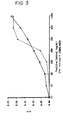
- Example 2 The operation of Example 2 was repeated using the specimens and the latex reagent B sensitized by an anti-myoglobin antibody of Example 2 to obtain A1. Immediately after 5 minutes of incubation at 37°C under this state, absorbance (turbidity) was measured [A2]. Similarly, a change in absorbance (turbidity) [A2-A1] was calculated and a calibration curve shown in Fig. 3 (+) was obtained. This calibration curve is represented by ⁇ OD/min.
- a high-sensitivity reaction can be obtained at a low concentration range of a detected substance and a linear reaction can be obtained at a range from a medium concentration to a high concentration with a latex reagent using relatively large particles. Therefore, linearity can be obtained at a wide concentration range.
- an anti-AFP antibody was dissolved in 9.5 ml of a glycine buffer solution (to be abbreviated as GBS hereinafter) and 0.5 ml of latex particles (particle diameter: 0.2 ⁇ m, a suspension having a 10 % solid content) was added to the solution and stirred at room temperature for 2 hours. A supernatant was removed by centrifuging the sensitized latex suspension. The precipitate was suspended- in 100 ml of 0.2 % BSA-GBS to prepare an AFP latex reagent.
- GBS glycine buffer solution
- An AFP positive control was diluted to predetermined concentrations (2.5, 1.25, 0.625, 0 ng/ml) using 0.2 % BSA-GBS to prepare specimens.
- Example 2 The same measuring instrument as in Example 1 was used to measure at a wavelength of 572 nm.
- a phosphate buffer solution comprising 15 mM phosphoric acid, 0.4 % polyethylene glycol 6000, 2 % sodium chloride and 0.1 % sodium azide and containing 0.2 % bovine serum-albumin, to be abbreviated as 0.2 % BSA-PBS hereinafter
- a DC pulse voltage of 1.8 V/mm pulse width: 20 ⁇ sec, frequency: 10 KHz
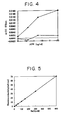
- Example 2 The operation of Example 2 was repeated using the specimens and the latex reagent C sensitized by an anti-AFP antibody of Example 3 to measure A1. Immediately after 5 minutes of incubation at 37°C in this state, turbidity was measured [A2]. A change in turbidity for 5 minutes was obtained from [A2-A1]. The results are shown in Table 1. A calibration curve shown in Fig. 4 (- ⁇ -) was obtained from the results of Table 1. Table 1 AFP ng/ml Comparative Example 3 Usual assay Example 3 Pulse-assay 0 -0.0004 0.0012 0.625 -0.0012 0.0070 1.25 0.0018 0.0135 2.5 0.0015 0.0180
- an anti-AFP antibody was dissolved in 9.5 ml of a glycine buffer solution (to be abbreviated as GBS hereinafter) and 0.5 ml of latex particles (particle diameter: 0.22 ⁇ m, a suspension having a 10 % solid content) was added to the solution and stirred at room temperature for 2 hours.
- GBS glycine buffer solution
- latex particles particle diameter: 0.22 ⁇ m, a suspension having a 10 % solid content
- the experimental apparatus of Fig. 1 in which the pulse generator was replaced by an AC voltage application apparatus was used and AC voltage was applied by setting the condition of a potential to be applied between electrodes by an oscilloscope (distance between electrodes: 3 mm).
- an AFP reference was diluted to prepare specimens having AFP concentrations of 0 to 2.5 ng/ml and use them for measurement.
- the field strength of application voltage was set to 0 V, ⁇ 1 V, ⁇ 3.5 V, ⁇ 4.5 V and ⁇ 7.5 V to measure the specimens having respective concentrations.
- An AFP latex reagent was prepared in the same manner as in Example 6.
- An AFP latex reagent was prepared in the same manner as in Example 6.
- An AFP latex reagent was prepared in the same manner as in Example 6.
- the experimental apparatus of Fig. 1 in which the pulse generator was replaced by a DC voltage application apparatus was used and DC voltage was applied by setting the condition of a potential to be applied between electrodes by an oscilloscope (distance between electrodes: 3 mm).
- N-Assay TIA CRP-S (Nittobo Medical Co.) was used as a reagent for latex photometric immunoassay.
- the reagent consisted of a tris buffer solution and an antiserum solution (20 % anti-CRP caprine serum antibody).
- the experimental apparatus of Fig. 1 was used and pulse voltage was applied by setting the condition of a potential to be applied between electrodes by an oscilloscope (distance between electrodes: 3 mm).
- a commercial CRP reference solution was diluted to prepare specimens having CRP concentrations of 0 to 4.1 mg/dl and use them for measurement.
- Example 10 The same specimens and reagent as in Example 10 were used and the operation of Example 10 was repeated except that pulse voltage to be applied to the reaction system was set at a wave height value of 4.6 V, pulse width of 20 ⁇ s and frequency of 10 KHz and applied for 18 seconds to measure a change in turbidity and draw a calibration curve.
- the results are shown in Table 8 and Fig. 12.
- Table 8 CRP mg/dl Prior art method Pulse B 0 0.0034 0.0112 0.025 0.0040 0.0140 0.05 0.0038 0.0172 0.1 0.0044 0.0230
- Example 10 The same specimens and reagent as in Example 10 were used. The operation of Example 10 was repeated except that pulse voltage was not applied to the reaction system to measure a change in turbidity and draw a calibration curve. The results are shown in Fig. 11 and Fig. 12.
- an immunologically reactive substance can be easily measured with high sensitivity and at high speed, compared with the method of the prior art.
Landscapes
- Health & Medical Sciences (AREA)
- Immunology (AREA)
- Life Sciences & Earth Sciences (AREA)
- Engineering & Computer Science (AREA)
- Molecular Biology (AREA)
- Biomedical Technology (AREA)
- Chemical & Material Sciences (AREA)
- Hematology (AREA)
- Urology & Nephrology (AREA)
- Biotechnology (AREA)
- Biochemistry (AREA)
- Cell Biology (AREA)
- Food Science & Technology (AREA)
- Medicinal Chemistry (AREA)
- Physics & Mathematics (AREA)
- Analytical Chemistry (AREA)
- Microbiology (AREA)
- General Health & Medical Sciences (AREA)
- General Physics & Mathematics (AREA)
- Pathology (AREA)
- Investigating Or Analysing Biological Materials (AREA)
- Medicines Containing Antibodies Or Antigens For Use As Internal Diagnostic Agents (AREA)
- Investigating Or Analysing Materials By The Use Of Chemical Reactions (AREA)
Applications Claiming Priority (2)
| Application Number | Priority Date | Filing Date | Title |
|---|---|---|---|
| JP28444495A JPH09101307A (ja) | 1995-10-06 | 1995-10-06 | 免疫学的反応性物質を検出又は測定する方法 |
| JP284444/95 | 1995-10-06 |
Publications (2)
| Publication Number | Publication Date |
|---|---|
| EP0767376A2 true EP0767376A2 (de) | 1997-04-09 |
| EP0767376A3 EP0767376A3 (de) | 1998-08-12 |
Family
ID=17678629
Family Applications (1)
| Application Number | Title | Priority Date | Filing Date |
|---|---|---|---|
| EP96307270A Withdrawn EP0767376A3 (de) | 1995-10-06 | 1996-10-04 | Verfahren zur Detektion oder zum Messen einer immunologisch reaktiven Substanz |
Country Status (2)
| Country | Link |
|---|---|
| EP (1) | EP0767376A3 (de) |
| JP (1) | JPH09101307A (de) |
Cited By (3)
| Publication number | Priority date | Publication date | Assignee | Title |
|---|---|---|---|---|
| WO2007034508A1 (en) * | 2005-09-19 | 2007-03-29 | Indian Institute Of Science | An ultra-sensitive method for detection and quantification of substance |
| US8637326B2 (en) | 2010-02-25 | 2014-01-28 | Fujifilm Corporation | Biological substance analyzing method, and biological substance analyzing cell, biological substance analyzing chip, and biological substance analyzing apparatus employed in the biological substance analyzing method |
| EP2126576B1 (de) * | 2007-03-14 | 2017-01-04 | Director General, Defence Research & Development Organisation | Reagens zum analytnachweis und verfahren dazu |
Family Cites Families (4)
| Publication number | Priority date | Publication date | Assignee | Title |
|---|---|---|---|---|
| US5380490A (en) * | 1991-01-18 | 1995-01-10 | Canon Kabushiki Kaisha | Apparatus for measuring a test specimen |
| JPH05288752A (ja) * | 1992-04-10 | 1993-11-02 | Canon Inc | 検体測定装置 |
| JP3300493B2 (ja) * | 1993-09-13 | 2002-07-08 | 株式会社 先端科学技術インキュベーションセンター | 生物学的特異的反応性物質の存在を検出又は測定する方法 |
| JPH1073595A (ja) * | 1996-09-02 | 1998-03-17 | Masao Karube | 免疫学的反応性物質の存在を検出又は測定する方法及びそれに用いる試薬 |
-
1995
- 1995-10-06 JP JP28444495A patent/JPH09101307A/ja active Pending
-
1996
- 1996-10-04 EP EP96307270A patent/EP0767376A3/de not_active Withdrawn
Cited By (3)
| Publication number | Priority date | Publication date | Assignee | Title |
|---|---|---|---|---|
| WO2007034508A1 (en) * | 2005-09-19 | 2007-03-29 | Indian Institute Of Science | An ultra-sensitive method for detection and quantification of substance |
| EP2126576B1 (de) * | 2007-03-14 | 2017-01-04 | Director General, Defence Research & Development Organisation | Reagens zum analytnachweis und verfahren dazu |
| US8637326B2 (en) | 2010-02-25 | 2014-01-28 | Fujifilm Corporation | Biological substance analyzing method, and biological substance analyzing cell, biological substance analyzing chip, and biological substance analyzing apparatus employed in the biological substance analyzing method |
Also Published As
| Publication number | Publication date |
|---|---|
| EP0767376A3 (de) | 1998-08-12 |
| JPH09101307A (ja) | 1997-04-15 |
Similar Documents
| Publication | Publication Date | Title |
|---|---|---|
| Viets et al. | Determination of serum protein concentration in nanoliter blood samples using fluorescamine or o-phthalaldehyde | |
| CA1223922A (en) | Methods of assay | |
| US4780423A (en) | Heterogeneous fluorescence assays using controlled pore glass particles | |
| US4302536A (en) | Colorimetric immunoassay process | |
| JP3327484B2 (ja) | 遠心アナライザー中の粒子強化凝集反応を混濁の輝度の測定により分析する方法 | |
| CN103033629B (zh) | 一种脂蛋白磷脂酶a2的测定试剂及试剂的制备方法 | |
| CA1195925A (en) | Method for assaying antigen-antibody reactions and reagent therefor | |
| CN108318680B (zh) | 一种抗药抗体的检测方法及检测试剂盒 | |
| JPS62121361A (ja) | 細胞集団もしくは亜集団を定量するための方法及び標準物並びに試薬キット | |
| CA1146852A (en) | Reagent for latex agglutination | |
| DE2749956A1 (de) | Immunologisches bestimmungsverfahren | |
| US4320194A (en) | Process for the production of carrier particles from yeast cells and for producing diagnostic agents therefrom and test kits containing such agents | |
| EP0722087A1 (de) | Mess-system unter verwendung von vollblut | |
| US5055415A (en) | Method for immunoassay procedure for the detection of antigen by reacting with antibody which increases its electrical charge and electrophoretic mobility | |
| EP2573570B1 (de) | Cortisolimmunoassay mit fluoreszierenden Partikeln | |
| US4851329A (en) | Immunoassay employing optical pulse particle size analysis | |
| EP0566205A1 (de) | Verfahren zur Eliminierung nichtspezifischer Reaktionen in Immuntests | |
| EP0767376A2 (de) | Verfahren zur Detektion oder zum Messen einer immunologisch reaktiven Substanz | |
| SU1431662A3 (ru) | Способ получени реагента дл проведени реакции агглютинации | |
| EP0269526A2 (de) | Verfahren zur quantitativen Bestimmung der Antigene und Antikörper | |
| JP3300493B2 (ja) | 生物学的特異的反応性物質の存在を検出又は測定する方法 | |
| EP1637884A1 (de) | Verfahren zur messung einer affinität aufweisenden substanz | |
| Macart et al. | Optimized microturbidimetric assay for fibrinogen. | |
| CA2088404C (en) | Method for the determination of antigens or antibodies in the presence of an immune complex | |
| EP0184701B1 (de) | Verfahren zur Bestimmung eines Liganden |
Legal Events
| Date | Code | Title | Description |
|---|---|---|---|
| PUAI | Public reference made under article 153(3) epc to a published international application that has entered the european phase |
Free format text: ORIGINAL CODE: 0009012 |
|
| AK | Designated contracting states |
Kind code of ref document: A2 Designated state(s): DE ES FR IT |
|
| PUAL | Search report despatched |
Free format text: ORIGINAL CODE: 0009013 |
|
| AK | Designated contracting states |
Kind code of ref document: A3 Designated state(s): DE ES FR IT |
|
| STAA | Information on the status of an ep patent application or granted ep patent |
Free format text: STATUS: THE APPLICATION IS DEEMED TO BE WITHDRAWN |
|
| 18D | Application deemed to be withdrawn |
Effective date: 19990501 |