EP0658209B1 - Endothelial lineage specific transcriptional regulatory element from tek - Google Patents
Endothelial lineage specific transcriptional regulatory element from tek Download PDFInfo
- Publication number
- EP0658209B1 EP0658209B1 EP93918834A EP93918834A EP0658209B1 EP 0658209 B1 EP0658209 B1 EP 0658209B1 EP 93918834 A EP93918834 A EP 93918834A EP 93918834 A EP93918834 A EP 93918834A EP 0658209 B1 EP0658209 B1 EP 0658209B1
- Authority
- EP
- European Patent Office
- Prior art keywords
- gene
- tek
- cells
- regulatory element
- transcriptional regulatory
- Prior art date
- Legal status (The legal status is an assumption and is not a legal conclusion. Google has not performed a legal analysis and makes no representation as to the accuracy of the status listed.)
- Expired - Lifetime
Links
Images


Classifications
-
- A—HUMAN NECESSITIES
- A01—AGRICULTURE; FORESTRY; ANIMAL HUSBANDRY; HUNTING; TRAPPING; FISHING
- A01K—ANIMAL HUSBANDRY; CARE OF BIRDS, FISHES, INSECTS; FISHING; REARING OR BREEDING ANIMALS, NOT OTHERWISE PROVIDED FOR; NEW BREEDS OF ANIMALS
- A01K67/00—Rearing or breeding animals, not otherwise provided for; New breeds of animals
- A01K67/027—New breeds of vertebrates
- A01K67/0275—Genetically modified vertebrates, e.g. transgenic
-
- A—HUMAN NECESSITIES
- A61—MEDICAL OR VETERINARY SCIENCE; HYGIENE
- A61P—SPECIFIC THERAPEUTIC ACTIVITY OF CHEMICAL COMPOUNDS OR MEDICINAL PREPARATIONS
- A61P35/00—Antineoplastic agents
-
- A—HUMAN NECESSITIES
- A61—MEDICAL OR VETERINARY SCIENCE; HYGIENE
- A61P—SPECIFIC THERAPEUTIC ACTIVITY OF CHEMICAL COMPOUNDS OR MEDICINAL PREPARATIONS
- A61P43/00—Drugs for specific purposes, not provided for in groups A61P1/00-A61P41/00
-
- A—HUMAN NECESSITIES
- A61—MEDICAL OR VETERINARY SCIENCE; HYGIENE
- A61P—SPECIFIC THERAPEUTIC ACTIVITY OF CHEMICAL COMPOUNDS OR MEDICINAL PREPARATIONS
- A61P9/00—Drugs for disorders of the cardiovascular system
- A61P9/08—Vasodilators for multiple indications
-
- A—HUMAN NECESSITIES
- A61—MEDICAL OR VETERINARY SCIENCE; HYGIENE
- A61P—SPECIFIC THERAPEUTIC ACTIVITY OF CHEMICAL COMPOUNDS OR MEDICINAL PREPARATIONS
- A61P9/00—Drugs for disorders of the cardiovascular system
- A61P9/10—Drugs for disorders of the cardiovascular system for treating ischaemic or atherosclerotic diseases, e.g. antianginal drugs, coronary vasodilators, drugs for myocardial infarction, retinopathy, cerebrovascula insufficiency, renal arteriosclerosis
-
- A—HUMAN NECESSITIES
- A61—MEDICAL OR VETERINARY SCIENCE; HYGIENE
- A61P—SPECIFIC THERAPEUTIC ACTIVITY OF CHEMICAL COMPOUNDS OR MEDICINAL PREPARATIONS
- A61P9/00—Drugs for disorders of the cardiovascular system
- A61P9/12—Antihypertensives
-
- C—CHEMISTRY; METALLURGY
- C12—BIOCHEMISTRY; BEER; SPIRITS; WINE; VINEGAR; MICROBIOLOGY; ENZYMOLOGY; MUTATION OR GENETIC ENGINEERING
- C12N—MICROORGANISMS OR ENZYMES; COMPOSITIONS THEREOF; PROPAGATING, PRESERVING, OR MAINTAINING MICROORGANISMS; MUTATION OR GENETIC ENGINEERING; CULTURE MEDIA
- C12N15/00—Mutation or genetic engineering; DNA or RNA concerning genetic engineering, vectors, e.g. plasmids, or their isolation, preparation or purification; Use of hosts therefor
- C12N15/09—Recombinant DNA-technology
- C12N15/63—Introduction of foreign genetic material using vectors; Vectors; Use of hosts therefor; Regulation of expression
- C12N15/79—Vectors or expression systems specially adapted for eukaryotic hosts
- C12N15/85—Vectors or expression systems specially adapted for eukaryotic hosts for animal cells
-
- C—CHEMISTRY; METALLURGY
- C12—BIOCHEMISTRY; BEER; SPIRITS; WINE; VINEGAR; MICROBIOLOGY; ENZYMOLOGY; MUTATION OR GENETIC ENGINEERING
- C12N—MICROORGANISMS OR ENZYMES; COMPOSITIONS THEREOF; PROPAGATING, PRESERVING, OR MAINTAINING MICROORGANISMS; MUTATION OR GENETIC ENGINEERING; CULTURE MEDIA
- C12N9/00—Enzymes; Proenzymes; Compositions thereof; Processes for preparing, activating, inhibiting, separating or purifying enzymes
- C12N9/10—Transferases (2.)
- C12N9/12—Transferases (2.) transferring phosphorus containing groups, e.g. kinases (2.7)
- C12N9/1205—Phosphotransferases with an alcohol group as acceptor (2.7.1), e.g. protein kinases
-
- A—HUMAN NECESSITIES
- A01—AGRICULTURE; FORESTRY; ANIMAL HUSBANDRY; HUNTING; TRAPPING; FISHING
- A01K—ANIMAL HUSBANDRY; CARE OF BIRDS, FISHES, INSECTS; FISHING; REARING OR BREEDING ANIMALS, NOT OTHERWISE PROVIDED FOR; NEW BREEDS OF ANIMALS
- A01K2217/00—Genetically modified animals
- A01K2217/05—Animals comprising random inserted nucleic acids (transgenic)
-
- A—HUMAN NECESSITIES
- A61—MEDICAL OR VETERINARY SCIENCE; HYGIENE
- A61K—PREPARATIONS FOR MEDICAL, DENTAL OR TOILETRY PURPOSES
- A61K48/00—Medicinal preparations containing genetic material which is inserted into cells of the living body to treat genetic diseases; Gene therapy
Definitions
- the invention relates to a novel transcriptional regulatory element which is capable of directing expression of a gene specifically in cells of the endothelial lineage; a recombinant molecule containing the transcriptional regulatory element; a transformant host cell including the recombinant molecule; a DNA construct comprising the transcriptional regulatory element operatively linked to a gene and a reporter gene; and, the use of the transcriptional regulatory element to target expression of a gene in cells of the endothelial lineage.
- Tissue specific transcriptional regulatory elements have been identified which have been used to target expression of exogenous genes in cells and in transgenic animals.
- DNA constructs using an erythroid specific transcriptional element and an oncogene encoding a protein having protein-tyrosine kinase (PTK) activity have been used to produce transgenic animals which have cardiovascular disease ⁇ Yee, S.P. et al. (1989) P.N.A.S., U.S.A. 86, 5873-5877).
- PTK protein-tyrosine kinase
- tek tyrosine kinase
- the present inventors have identified and isolated a transcriptional regulatory element capable of directing expression of a gene in cells of the endothelial lineage.
- the transcriptional element is expressed in cells of the endothelial lineage including mature and progenitor cells.
- the present invention therefore provides an isolated transcriptional regulatory element which is capable of directing expression of a gene specifically in cells of the endothelial lineage.
- the transcriptional regulatory element comprises the initiation codon and the untranslated sequences of tek, a protein tyrosine kinase expressed during marine cardiogenesis.
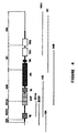
- the transcriptional regulatory element is a 7.2 kb fragment extending from the Bgl II site to the Kpn I site as shown in Figure 3 and comprising the partial nucleotide sequence extending from nucleotide 1 to the Bgl II site as shown in Fig. 11.
- the specification further discloses a method of preparing the transcriptional regulatory element.
- the transcriptional regulatory element may be constructed by synthesis and ligation of DNA oligomers.
- the element may also be isolated by selectively amplifying the region of the transcriptional regulatory element using the polymerase chain reaction method and genomic DNA.
- the specification also permits the construction of nucleotide probes which are unique or substantially homologous to the transcriptional regulatory element of the invention.
- the probe may be labelled, for example, with a radioactive substance and it may be used to select from a mixture of nucleotide sequences a transcriptional regulatory element of the invention or an element homologous thereto.
- the invention also relates to a recombinant molecule adapted for transformation of a host cell comprising a transcriptional regulatory element of the invention and a gene operatively linked to the transcriptional regulatory element.
- a transformant host cell including a recombinant molecule of the invention is also provided.
- this invention provides plasmids which comprise the transcriptional regulatory element of the invention.
- a recombinant molecule comprising the transcriptional regulatory element of the invention operatively linked to a gene and a reporter gene is provided.
- the recombinant molecules of the invention may be used to produce transgenic non-human mammals. Accordingly the specification describes a method of producing a transgenic non-human mammal characterized as having a plurality of cells containing a recombinant molecule of the invention, or an ancestor of the mammal at an embryonic stage, comprising (a) introducing the recombinant molecule into a pronucleus of a mammalian zygote by microinjection, said zygote being capable of development into a mammal, thereby obtaining a genetically transformed zygote; (b) transplanting an embryo derived from the genetically transformed zygote into a pseudo-pregnant female capable of bearing the embryo to term and (c) if desired allowing the embryo to develop to term.
- the invention further relates to a transgenic non-human mammal all of whose germ cells and somatic cells contain a recombinant molecule of the invention as a result of chromosomal incorporation into the non-human mammal genome, or into the genome of an ancestor of said mammal at an embryonic stage.
- the specification further discloses cell cultures of cells of the transgenic mammals.
- the invention also relates to a method of determining the affect of a substance on cells of the endothelial lineage comprising the use of a transgenic non-human mammal of the invention or the ancestor of said mammal at an embryonic stage.
- the description also describes a method of determining the affect of a substance on cells of the endothelial lineage comprising producing a transgenic non-human mammal characterized as having a plurality of cells containing a recombinant molecule comprising the transcriptional regulatory element of the invention operatively linked to a gene and a reporter gene encoding a phenotype which is not displayed by the mammal, or an ancestor of the mammal at an embryonic stage, comprising (a) introducing the recombinant molecule into a pronucleus of a mammalian zygote by microinjection, said zygote being capable of development into a mammal, thereby obtaining a genetically transformed zygote; (b) transplanting an embryo derived from the genetically transformed zygote into a pseudo-pregnant female capable of bearing the embryo to term and (c) isolating the embryo or allowing the embryo to develop to term, (d) assaying for the pheno
- the human homolog of tek has been recently reported by Ziegler, G. F. et al., Oncogene 8:663-670, (1993).
- the tek locus was mapped to chromosome 4, between the brown and pmv-23 loci. This region is syntenic with human chromosomal regions 1p22-32, 9q31-33, and 9p22-13.
- the deduced amino acid sequence of tek predicts that it encodes a putative receptor tyrosine kinase that contains a 21 amino acid kinase insert and which is most closely related in its. catalytic domain to FGFR1 (mouse fibroblast growth factor) and the product of the ret proto-oncogene.
- Figure 1 shows the nucleotide and deduced amino acid sequence of tek. The present inventors have also identified the initiation site of translation of tek.
- tek expression was found to be restricted to cells of the endothelial lineage. Specifically, in situ hybridization analysis of adult tissues, as well as sectioned and whole mount embryos, showed that tek is specifically expressed in the endocardium, the leptomeninges and the endothelial lining of the vasculature from the earliest stages of their development. Moreover, examination of the morphology of tek -expressing cells, and staging of tek expression relative to that of the endothelial cell marker von Willebrand factor, revealed that tek is expressed prior to von Willebrand factor and appears to mark the embryonic progenitors of mature endothelial cells.
- the present inventors have identified a transeriptional regulatory element located upstream of tek which specifically directs expression of a gene in cells of the endothelial lineage.
- the transcriptional regulatory element has been found to direct expression in both mature and progenitor endothelial cells.
- the present inventors isolated a DNA segment from a mouse genomic bacteriophage library using a 5'-prime probe which contained the initiation codon and untranslated sequences of tek using the procedures of Sambrook et al., 1989, Molecular Cloning, A Laboratory Manual. Cold Spring Harbour Lab. Press.
- a 16 kb phage clone was shown by hybridization and sequence analysis to contain a single exon with 175 bp homologous to the cDNA.
- a DNA fragment extending from the Bgl II restriction site located at nucleotide 110 of the cDNA to the nearest Kpn I in the phage was cloned upstream of the bacterial gene lacZ (see Figure 3).
- This reporter construct containing 7.2 kb of the tek gene was microinjected into pronuclei of fertilized randomly bred CD-1 mice using procedures as set out in Hogan et al. (1986, Manipulating the Mouse Embryo, A Laboratory Manual. Cold Spring Harbor Lab. Press). Three transgenic founder embryos were dissected from their foster mothers and yolk sac DNA analyzed for the presence of the transgene. Expression of the transgene was determined by the X-gal staining of whole embryos and subsequent sectioning of the embryos. The 7.2 kb fragment was able to drive lacZ expression in endothelial cells that had previously been shown to express tek RNA thus demonstrating that this DNA contained the tek transcriptional regulatory element.
- the partial nucleotide sequence of the transcriptional regulatory element is shown in Figure 11.
- the description further provides a method of preparing the transeriptional regulatory element.
- the transcriptional regulatory element may be isolated by selectively amplifying the region of the transcriptional regulatory element using the polymerase chain reaction method and genomic DNA. It is possible to design synthetic oligonucleotide primers from the sequence shown in Figure 11 for use in PCR and for screening genomic libraries, in particular human genomic libraries. An amplified fragment can be cloned and characterized by DNA sequence analysis. The nucleotide sequence of the transcriptional regulatory element will also permit the element to be constructed by synthesis and ligation of DNA oligomers.
- the transcriptional regulatory element may be proven functional by assessing the transient expression of a construct bearing a reporter gene. For example, using the reporter gene for B-galactosidase (LacZ) or chloramphenicol acetyltransferase (CAT) after transfection of the DNA into host cells.
- LacZ B-galactosidase
- CAT chloramphenicol ace
- nucleotide sequences can be used which have substantial sequence homology with the nucleotide sequence of the transcriptional regulatory element of the invention.
- sequences having substantial sequence homology means those sequences which have slight or inconsequential sequence variations i.e. the homologous sequences function in substantially the same manner to produce substantially the same result as the actual sequence. The variations may be attributable to local mutations or structural modifications.
- the specification also permits the construction of nucleotide probes which are unique to the transcriptional regulatory element of the invention.
- the specification also relates to a probe comprising a nucleotide sequence substantially homologous to the transcriptional regulatory . element of the invention.
- the probe may be labelled and it may be used to select from a mixture of nucleotide sequences a transcriptional regulatory element of the invention or an element substantially homologous thereto.
- a nucleotide probe may be labelled with a radioactive label which provides for an adequate signal and has sufficient half-life such as 32 P, 3 H, 14 C or the like.
- labels which may be used include antigens that are recognized by a specific labelled antibody, fluorescent compounds, enzymes, antibodies specific for a labelled antigen, and chemiluminescent substances.
- An appropriate label may be selected having regard to the rate of hybridization and binding of the probe to the nucleotide to be detected and the amount of nucleotide available for hybridization.
- the invention also relates to a recombinant molecule adapted for transformation of a host cell comprising a transcriptional regulatory element of the invention and a gene operatively linked thereto.
- the transcriptional regulatory element of the invention operatively linked to a gene may be incorporated in a known manner into a recombinant molecule which ensures good expression of the protein encoded by the gene.
- the transcriptional regulatory element of the invention may be incorporated into a plasmid vector, for example, a retroviral vector, pECE.
- the transcriptional regulatory element of the invention may be operatively linked to a reporter gene or a gene encoding a substance which has toxic or therapeutic activity including a factor which modulates angiogenesis. Examples of reporter genes, factors which modulate angiogenesis, and substances with toxic or therapeutic activity are discussed below.
- a transformant host cell including a recombinant molecule of the invention and a cell line containing such transformant host cells is also provided.
- suitable host cells include human endothelial cells such as umbilical vein endothelial cells and rabbit aortic endothelial cells.
- the invention also relates to a recombinant molecule comprising a transcriptional regulatory element of the invention operatively linked to a gene and a reporter gene.
- the reporter gene may be introduced into the recombinant molecule using conventional methods such as those described in Sambrook et al., 1989, Molecular Cloning, A Laboratory Manual. Cold Spring Harbour Lab. Press.
- the recombinant molecule may also be synthetically produced using conventional methods. Further, the recombinant molecule may be introduced into a host cell using conventional methods.
- the reporter gene should be under the control of the transcriptional regulatory element and the pattern and extent of expression of the gene operatively linked to the transcriptional regulatory element may accordingly be determined in cells of the endothelial lineage.
- the reporter gene codes for a phenotype not displayed by the host cell and the phenotype may be assayed quantitatively.
- reporter genes include lacZ (B-galactosidase), neo (neomycin phosphotransferase), cat (chloramphenicol acetyltransferase) dhfr (dihydrofolate reductase), aphIV (hygromycin phosphotransferase), lux (luciferase), uidA (B-glucuronidase).
- the reporter gene is lacZ which codes for B-galactosidase.
- B-galactosidase may be assayed using the lactose analogue X-gal(5-bromo-4-chloro-3-indolyl-b-D-galactopyranoside) which is broken down by B-galactosidase to a product that is blue in color.
- lactose analogue X-gal(5-bromo-4-chloro-3-indolyl-b-D-galactopyranoside) which is broken down by B-galactosidase to a product that is blue in color.
- the recombinant DNA of the invention may be used to produce transgenic non-human mammals. Accordingly the description also teaches a method of producing a transgenic non-human mammal characterized as having a plurality of cells containing a recombinant molecule of the invention, or an ancestor of the mammal at an embryonic stage, comprising (a) introducing the recombinant molecule into a pronucleus of a mammalian zygote by microinjection, said zygote being capable of development into a mammal, thereby obtaining a genetically transformed zygote; (b) transplanting an embryo derived from the genetically transformed zygote into a pseudo- pregnant female capable of bearing the embryo to term and (c) if desired, allowing the embryo to develop to term.
- the invention further relates to a transgenic non-human mammal all of whose germ cells and somatic cells contain a recombinant molecule of the invention introduced into the animal, or an ancestor of the mammal at an embryonic stage.
- plasmids containing recombinant molecules of the invention are microinjected into mouse embryos.
- the plasmids are injected into the male pronuclei of fertilized one-cell mouse eggs; the injected eggs are transferred to pseudo-pregnant foster females; and, the eggs in the foster females are allowed to develop to term.
- mice Although experimental animals used were mice, the method should not be limited thereto. It may be desirable to use other species such as rats, hamsters and rabbits.
- the invention also relates to a method of determining the affect of a substance on cells of the endothelial lineage comprising the use of a transgenic non-human mammal of the invention characterized as having a plurality of cells containing a recombinant molecule comprising the transcriptional regulatory element of the invention operatively linked to a gene, or an ancestor of the mammal at an embryonic stage, which mammal has been obtained by (a) introducing the recombinant molecule into a pronucleus of a mammalian zygote by microinjection, said zygote being capable of development into a mammal, thereby obtaining a genetically transformed zygote; (b) transplanting an embryo-derived from the genetically transformed zygote into a pseudo-pregnant female capable of bearing the embryo to term and (c) isolating the embryo or allowing the embryo to develop to term, and (d) determining the affect of the substance on cells of the endothelial line
- a method of determining the affect of a substance on cells of the endothelial lineage comprising the use of a transgenic non-human mammal of the invention characterized as having a plurality of cells containing a recombinant molecule comprising a transcriptional regulatory element of the invention linked to a gene encoding the substance, and a reporter gene encoding a phenotype which is not displayed by the mammal, or an ancestor of the mammal at an embryonic stage, which mammal has been obtained by (a) introducing the recombinant molecule into a pronucleus of a mammalian zygote by microinjection, said zygote being capable of development into a mammal, thereby obtaining a genetically transformed zygote; (b) transplanting an embryo derived from the genetically transformed zygote into a pseudo-pregnant female capable of bearing the embryo to term and (c) if desired, allowing the embryo to
- the reporter gene should be under the control of the transcriptional regulatory element and accordingly the pattern and extent of expression of a gene operatively linked to the transcriptional regulatory element may be determined by assaying for the phenotype of the reporter gene.
- the reporter gene codes for a phenotype not displayed by the host cell and the phenotype may be assayed quantitatively.
- reporter genes include lacz ( ⁇ -galactosidase), neo (neomycin phophotransferase), cat (chloramphenicol acetyltransferase) dhfr (dihydrofolate reductase), aphIV (hygromycin phosphotransferase), lux (luciferase), uidA ( ⁇ -glucuronidase).
- the reporter gene is lacZ which codes for ⁇ -galactosidase.
- ⁇ -galactosidase may be assayed using the lactose analogue X-gal (5-bromo-4-chloro-3-indolyl-p-D-galactopyranoside) which is broken down by ⁇ -galactosidase to a product that is blue in color.
- lactose analogue X-gal 5-bromo-4-chloro-3-indolyl-p-D-galactopyranoside
- ⁇ -galactosidase may be assayed using the lactose analogue X-gal (5-bromo-4-chloro-3-indolyl-p-D-galactopyranoside) which is broken down by ⁇ -galactosidase to a product that is blue in color.
- Cells of the transgenic mammals of the invention and produced as described herein may be cultured using standard tissue culture techniques.
- the present invention allows the manipulation of endothelial cell physiology by targeting expression of a substance in cells of the endothelial lineage in a mammal.
- the above described methods, transgenic animals and cell cultures derived therefrom, can therefore be used to assess the role of a substance in the determination, migration, or proliferation of cells of the endothelial lineage.
- the invention provides a mechanism for investigating vascularization of tumors and the control of angiogenesis.
- a transgenic mammal may be produced which expresses a substance exclusively in cells of the endothelial lineage.
- a comparison of endothelial phenotype, morphology, and function using for example immunohistochemical techniques and assays for LDL receptors, and of the pattern and extent of expression of the substance in the animal with a control transgenic animal will provide an indication of the affect of the substance on cells of the endothelial lineage.
- angiogenic factors include substances derived from human and animal tissues which stimulate the proliferation or migration of normally quiescent endothelial cells in culture or promote neovascularization in vivo including factors which are associated with the vascularization that permits tumor growth; substances which are inhibitors of angiogenesis such as transforming growth factor ⁇ , tumor necrosis factor a, human platelet factor 4 (PF4) and a interferon; substances which suppress cell migration, such as proteinase inhibitors which inhibit proteases which may be necessary for penetration of the basement membrane, in particular, tissue inhibitors of metalloproteinase TIMP-1 and TIMP-2; and other proteins such as protamine which has demonstrated angiostatic properties.
- angiogenesis such as transforming growth factor ⁇ , tumor necrosis factor a, human platelet factor 4 (PF4) and a interferon
- substances which suppress cell migration such as proteinase inhibitors which inhibit proteases which may be necessary for penetration of the basement membrane, in particular, tissue inhibitors of metalloproteinase TIMP-1 and T
- the transcriptional regulatory element of the invention may be used in gene therapy to introduce a foreign gene into endothelial cells to correct or prevent vascular disorders.
- the transcriptional regulatory element of the invention may be used to express foreign genes at specific sites in the circulation. Endothelial cells are found at diseased sites and accordingly, the transcriptional regulatory element of the invention may be used to target therapeutic agents including anticoagulants, vasodilator, and angiogenic factors (see above discussion) to endothelial cells found at diseased sites.
- genetic modification of endothelial cells utilizing the transcriptional regulatory element of the invention may be used in the treatment of acquired vascular disorders such as hypertension, atherosclerosis restenosis, arthritis and cancer.
- Endothelial cells line all blood vessels and accordingly the transcriptional regulatory element of the invention may be used to target therapeutic agents into the bloodstream.
- genetic modification of endothelial cells utilizing the transcriptional regulatory element of the invention may also be used in the treatment of systemic or inherited disorders.
- the transcriptional regulatory element of the invention could be operatively linked to the factor VIII gene and introduced into a population of endothelial cells to correct a hemophilia disorder.
- Endothelial cells genetically modified in vitro using the transcriptional regulatory element of the invention i.e transformant host cells or cell lines containing transformant host cells of the invention, may be used to deliver gene products to the vasculature.
- endothelial cells genetically modified in vitro using the transcriptional regulatory element of the invention may be introduced into the vascular wall by catheterization.
- therapeutic proteins may be introduced into diseased arterial segments. The method may be particularly useful for introducing growth inhibitor proteins into an angioplasty site in patients with restenosis who have undergone coronary angioplasty.
- Endothelial cells genetically modified in vitro using the transcriptional regulatory element of the invention may be used to improve the performance of prosthetic vascular grafts.
- Prosthetic vascular grafts may be seeded with endothelial cells genetically modified using the transcriptional regulatory element of the invention, to produce therapeutic proteins which may prevent thrombosis or promote repopulation.
- vascular stents may also be populated with genetically modified endothelial cells to reduce problems such as thrombosis.
- a gene under the control of the transcriptional regulatory element of the invention i.e recombinant molecules of the invention may be directly introduced into endothelial cells in vivo using delivery vehicles such as retroviral vectors, adenoviral vectors and DHA virus vectors. They may also be introduced into endothelial cells in vivo using physical techniques such as microinjection and electroporation or chemical methods such as coprecipitation and incorporation of DNA into liposomes.
- AKR/J, DBA, and AKR/J x DBA recombinant inbred mouse DNAs were obtained from Jackson Labs (Bar Harbor, Maine), digested with Acc I, blotted to Zeta-Probe nylon membrane (Bio-Rad), and probed with the 1.6 kb tek cDNA labelled by random priming (Feinberg, A. P. & Vogelstein, B. (1983) Analyt. Biochem ., 132, 6-13). Hybridization was performed overnight at 65° in 200 mM sodium phosphate pH7.0, 7% sodium dodecyl sulfate (SDS), 1% bovine serum albumin (BSA), and 1 mM EDTA.
- SDS sodium dodecyl sulfate
- BSA bovine serum albumin
- Embryos and adult mouse tissues were obtained from random bred CD-1 stocks (Charles River, Quebec). Embryos were staged as Day 0.5 on the morning of a vaginal plug.
- PBS phosphate buffered saline
- RNA was precipitated with an equal volume of isopropanol, collected by centrifugation, and the pellet resuspended in diethylpyrocarbonate (DEPC)-treated 0.4 M sodium acetate, pH5.2.
- DEPC diethylpyrocarbonate
- the RNA were then precipitated with two volumes of 95% ethanol, washed with 70% and 95% ethanol, dried, and resuspended in DEPC treated 0.3 M sodium acetate, pH5.2.
- the RNA concentration was determined and the RNA stored at -70° until use.
- Poly A - containing RNA was purified from a pool of 100 to 150 Day 12.5 murine embryonic hearts with a QuickPrep mRNA isolation kit (Pharmacia) as outlined by the supplier.
- First strand cDNA was synthesized in a total reaction volume of 20 ⁇ l containing 20 ⁇ g of total RNA, 200 units of Mo-MLV-reverse transcriptase (BRL), either 1 ⁇ g of oligo-d(T) 18 (Day 12.5 RNA) (Boerhinger Mannheim) or 2 ⁇ g of random hexamer primers (Day 9.5 RNA) (Boerhinger Mannheim), 1 x PCR buffer (Cetus), 2.5 mM NgCl 2 , 1 mM of dNTPs (Pharmacia), 40 units of RNAsin (Promega), and 12.5 mM dithiothreitol.
- BTL Mo-MLV-reverse transcriptase
- RNA was heated to 65°C for 10 min and cooled quickly on ice prior to addition to the reaction components. The reaction was allowed to proceed for 1 h at 37° and then terminated by heating for 5 min at 95°.
- the reaction mixture was adjusted to a final volume of 100 ⁇ l containing 1 x PCR buffer, 1.5 mM MgCl 2 , 800 ⁇ M dNTPs, and 1 ⁇ g of each of the two degenerate tyrosine kinase oligonucleotide primers described by Wilks, A.F. (1989) Proc. Natl. Acad. Sci ., 86, 1603-1607.
- Amplification was performed with a Ericomp thermocycler using the following parameters: denaturation for 2 min at 94°, annealing for 2 min at 42°, and extension for 4 min at 63°. After 40 cycles, the reaction products were collected by ethanol precipitation and electrophoresed through at 2% low-melt agarose (Sea Plaque) gel. In most cases a band of approximately 200 bp was visible within a background smear of ethidium bromide staining. This band was excised and recovered by three cycles of freeze-thaw in 100 ul of water. 10 ul of this solution was then subjected to a second round of PCR under the same conditions described above.
- Embryos isolated on Day 12.5 were dissected away from all extraembryonic tissues whereas embryos at earlier time points were recovered in utero . Embryos and adult tissues were fixed overnight in 4% paraformaldehyde, dehydrated with alcohols and xylenes, and embedded in paraffin. Tissues were sectioned at 6 ⁇ m thickness and mounted on 3-aminopropyltriethoxysilane treated slides (Sigma). After removal of paraffin the samples were treated with predigested pronase (Boerhinger Mannheim), acetylated with triethanolamine, dehydrated, and hybridized according to the protocol described by Frohman, N.B., Boyle, M. & Martin, G.R. (1990), Development, 110, 589-607.
- Sections were stained immunohistochemically for von Willebrand factor with a commercially available kit (Biomeda). After color development, slides were counterstained with Harris hematoxylin.
- cDNAs were synthesized from 9.5 and 12.5 day embryonic heart RNA by RT-PCR using degenerate oligonucleotide primers previously demonstrated to amplify tyrosine kinase sequences preferentially (Wilks, A.F. 1989, Proc.Natl.Acad.Sci ., 86, 1603-1607).
- Considerable cellular differentiation and morphogenesis have occurred within the cardiac region of the embryo by Day 9.5.
- the heart has developed from the primordial mesoderm cells of the cardiac plate into a primitive bent tube structure, consisting of two endothelial tubes enclosed within the developing myocardium.
- pdgfrb plays an important role in cardiogenesis, as has been suggested by recent studies demonstrating that the addition of PDGF-BB to explants of axolotol cardiac field mesoderm stimulates the production of beating bodies (Muslin, A.J. & Williams, L.T. (1991). Development, 112, 1095-1101) the fifth cDNA, which was also isolated at high frequency, was novel and for reasons that will become clear below was designated tek .
- the 210 bp RT-PCR-derived tek clone was subsequently used to isolate additional tek cDNA sequences.
- Figure 2 shows the nucleotide sequence of a 1.6 kb tek cDNA isolated from a 13.5 day mouse embryo cDNA library. Translation of this sequence reveals a single large open reading frame that terminates with TAG at nucleotide 907, followed by 696 nucleotides of 3° untranslated sequence.
- 1.6 kb tek cDNA encodes the cytoplasmic portion of a transmembrane RTK, consisting of the catalytic domain followed by a short carboxy-terminal tail of 33 amino acid residues.
- Figure 14 shows a comparison of the deduced amino acid sequence of tek with that of other tyrosine kinases; Identical sequences are denoted by periods. Dashes were added to allow for optimal alignment. The kinase insert and conserved regions of the catalytic domain are indicated beneath the aligned sequences (Hanks, S.K., Quinn, A.M. & Hunter, T. (1988), Scie n ce , 241, 52). Comparative sequences shown are for human Ret (Takahashi, M. & Cooper, G.M. (1987).
- the putative kinase domain contains several sequence motifs conserved among tyrosine kinases, including the tripeptide motif DFG, which is found in almost all known kinases, and the consensus ATP-binding site motifs GXGXXG followed by AXK 16 amino acid residues downstream (Hanks et al ., 1988).
- Transmembrane RTK's possess a methionine residue within the motif WMAIESL of conserved region VIII of the catalytic domain (Hanks et al ., 1988) as does tek , and the catalytic domain is interrupted by a putative 21 amino acid kinase insert, a structural motif not found in cytoplasmic tyrosine kinases (Hanks et al ., 1988).
- Mapping of the tek locus was accomplished by monitoring the strain distribution pattern of an Acc I. restriction site polymorphism in recombinant inbred (RI) mouse strains derived from matings between AKR/J (A) and DBA/2J (D) mice.
- the tek cDNA detects bands of 6.5, 6.1, 1.3 and 6.5, 3.1, 1.3 kb in DNA from the A and D strains, respectively.
- tek expression in embryonic heart was examined by Northern blot hybridization using an antisense probe derived from the 1.6 kb tek cDNA.
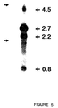
- Figure 5 shows a Northern blot hybridization analysis of tek expression in 12.5 day murine embryonic heart; Arrows on the left denote the position of migration of 28 S and 18 S ribosomal RNAs obtained from adjacent lane loaded with total RNA.
- Figure 5 shows that the tek probe detects 4 transcripts of 4.5, 2.7, 2.2, and 0.8 kb in size in cardiac RNA from 12.5 day mouse embryos. These hybridizing species vary considerably in signal intensity, suggesting that they may differ in relative abundance, with expression of the 2.7 and 2.2 kb transcripts occurring at significantly higher levels than the 4.5 and 0.8 kb RNAs. While the exact relationship among these transcripts is unclear, it is possible that they arise by differential splicing, since the 1.6 kb tek cDNA.detects a single genomic locus in mouse DNA by Southern blot hybridization at the same stringency.
- RNA in situ hybridization analyses were performed on mouse embryos with an antisense riboprobe synthesized from the 1.6 kb tek cDNA.
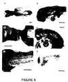
- Figure 6 shows the in situ hybridization analysis of tek expression in the 12.5 day embryo;
- A Dark field illumination of a para-sagittal section. Bar: 600 ⁇ m.
- B. and C Bright and dark field illumination respectively, of the heart region taken from a mid-sagittal section. Bar: 300 ⁇ m.
- IV and VI fourth and sixth aortic arches;
- A atrium; BA, basilar artery; CV, caudal vein;
- E endocardium; L, liver; M, leptomeninges; Ma, mandible; My, myocardium; PC, pericardial cavity;
- RA renal artery;
- Figure 6A shows that in 12.5 day mouse embryos, expression of tek is readily detected in the heart, the leptomeninges lining the brain and spinal cord, and the inner lining of major blood vessels, including the caudal vein and basilar and renal arteries. In addition, thin bands of hybridization are observed in the intersomite regions, corresponding to tek expression in the intersegmental vessels. Close examination of the region of the developing heart ( Figure 6B and 6C) reveals that tek is expressed in the endocardium, as well as in cells lining the lumina of the atria, the IV and VI aortic arches, the sinus venosus, and the sino-auricular septum.
- tek expression is observed in numerous small blood vessels perforating the liver and mandible. These observations, together with the overall pattern of hybridization seen in the 12.5 day embryo, demonstrate that tek is expressed in the endothelial cells of the tunica interna , the innermost lining of the blood vessels; hence the designation tunica interna endothelial cell kinase, tek.
- tek expression was obtained through analysis of sections from earlier developmental stages. Hybridization to 6.5 and 7 day embryos revealed that while tek is expressed strongly in the inner lining of the small blood vessels and capillaries of the maternal decidua, no expression is observed in either the embryo itself or the ectoplacental cone. The absence of tek expression at these stages is consistent with the fact that at 6.5 to 7 days the embryo contains only a small amount of mesoderm from which endothelial cells are known to be derived.
- Figure 7 shows the expression of tek precedes that of von Willebrand factor in 8.5 day embryos; Adjacent transverse sections through an 8.5 day embryo fixed in utero were either hybridized in situ with an [ 35 S]-labelled tek probe or stained immunohistochemically for von Willebrand factor.
- D Adjacent section to A at higher magnification showing absence of expression of von Willebrand factor in the embryo, Bar: 100 ⁇ m.
- H Dark field illumination of G.
- A amnion
- Ag presumptive angioblast
- BI blood island
- D maternal decidua
- DA dorsal aorta
- E endocardium
- Ec ectoplacental cone
- En endothelial cell
- G foregut
- HV head vein
- NF neural fold
- S somite
- Y yolk sac.
- RNA in situ analysis of 8.0 day embryos revealed that tek expression first becomes detectable in the developing yolk sac and a few small clusters of cells in the cephalic mesenchyme. This expression becomes more pronounced by Day 8.5, at which time significant hybridization can be observed in the mesodermal component of the amnion (outer cell layer) and yolk sac (inner cell layer), as well as in the developing endocardium and the inner lining of the head veins and dorsal aortae ( Figure 7A and 7B).
- sagittal sections reveal numerous focal areas of hybridization throughout the cephalic mesenchyme in regions thought to contain developing vasculature, as well as a small number of tek -expressing cells extending beneath the ventral surface of the somites ( Figure 7H and 7J).
- FIG. 8 shows tek expression in whole mount embryos; A., B., C. and D. tek expression in Day 8.0 embryos. E. tek mRNA distribution in a Day 9.5 embryo. F. En2 expression in a Day 8 embryo. I, II, III, first, second and third aortic arches; DA. dorsal aorta; E, endocardium; G, foregut pocket; H, heart; IS, intersegmental vessel; My, myocardium; ; NF, neural fold; OT; otic vesicle; V, vitelline vein; Y, yolk sac. Bars: 250 ⁇ m.
- FIG. 7B and H shows that whereas tek is expressed in both the maternal decidua and the embryo at Day 8.5, expression of von Willebrand factor is observed only in the tek -expressing, vascular endothelial cells of the maternal decidua ( Figure 7D and 7E). Hence tek expression precedes that of von Willebrand factor during embryogenesis. The same scenario is observed at later developmental stages during vascularization of individual organs.
- Figure 9 shows the expression of tek precedes that of von Willebrand factor in the developing leptomeninges;
- A Absence of immunohistochemical staining of von Willebrand factor in Day 12.5 leptomeninges. Arrow denotes a large blood vessel faintly positive for von Willebrand factor.
- B In situ detection of tek expression in Day 12.5 leptomeninges.
- C Staining of von Willebrand factor in Day 14.5 leptomeninges. Day 14.5 leptomeninges were positive for tek expression (not shown).
- M leptomeninges. Bars: 200 ⁇ m.
- Figure 9 shows that in the 12.5 day embryo, the developing leptomeninges hybridizes strongly with tek but fails to stain positive for von Willebrand factor. By Day 14.5, however, expression of von Willebrand factor can be readily detected in the leptomeninges. Assuming that there is not a significant lag between transcription and translation of von Willebrand factor, these observations, together with those on the morphology of tek -expressing cells, suggest that tek is expressed in both mature endothelial cells and their progenitors.
- tek is expressed in adult vasculature
- Figure 10 shows the expression of tek in adult vasculature.
- A Bright field illumination of a section through the upper heart region of a 3 week-old mouse hybridized with an [ 35 S]-labelled tek probe. Bar: 20 ⁇ m.
- B. and C
- the intensity of the hybridization signal observed for these structures is considerably lower than that observed for the endocardium and blood vessels of 12.5 day embryos hybridized and processed in parallel. This could indicate that mature endothelial cells, which are thought to be resting, have a different quantitative or qualitative requirement for expression of tek .
- a DNA segment was isolated from a mouse genomic bacteriophage library using a 5'-prime probe, consisting of nucleotides 0 to 912 of the tek cDNA, which contained the initiation codon and untranslated sequences of tek using the procedures of Sambrook et al., 1989, Molecular Cloning, A Laboratory Manual. Cold Spring Harbour Lab. Press.
- the DNA segment was cloned in the plasmid pGEm72F + and propagated in E. coli K12.
- a 16 kb phage clone was shown by hybridization with this 5'-prime probe and sequence analysis using oligonucleotides specific for the cDNA sequence and the plasmid backbone, to contain a single exon with 175 bp homologous to the cDNA.
- a DNA fragment extending from the Bgl II restriction site located at nucleotide 110 of the cDNA to the nearest Kpn I in the phage was cloned upstream of the bacterial gene lacZ.
- This reporter construct containing 7.2 kb of the tek gene was microinjected into pronuclei of fertilized randomly bred CD-1 mice using procedures as set out in Hogan et al., 1986, Manipulating the Mouse Embryo, A Laboratory Manual. Cold Spring Harbor Lab. Press. Three transgenic founder embryos were dissected from their foster mothers and yolk sac DNA analyzed for the presence of the transgene. Expression of the transgene was determined by the X-gal staining of whole embryos and subsequent sectioning of the embryos. Figure 12 shows expression of LacZ in Day 8.5 embryos and Figure 13 shows mRNA distribution in a Day 8.5 embryo.
Abstract
Description
- The invention relates to a novel transcriptional regulatory element which is capable of directing expression of a gene specifically in cells of the endothelial lineage; a recombinant molecule containing the transcriptional regulatory element; a transformant host cell including the recombinant molecule; a DNA construct comprising the transcriptional regulatory element operatively linked to a gene and a reporter gene; and, the use of the transcriptional regulatory element to target expression of a gene in cells of the endothelial lineage.
- Tissue specific transcriptional regulatory elements have been identified which have been used to target expression of exogenous genes in cells and in transgenic animals. For example, DNA constructs using an erythroid specific transcriptional element and an oncogene encoding a protein having protein-tyrosine kinase (PTK) activity have been used to produce transgenic animals which have cardiovascular disease {Yee, S.P. et al. (1989) P.N.A.S., U.S.A. 86, 5873-5877).
- The ability to introduce into animals exogenous genes which are selectively expressed in a particular cell type provides wide ranging experimental as well as practical opportunities. In particular it permits investigation of the role of a substance in the development, determination, migration, or proliferation of cells of a particular lineage.
- Dumont et al (Oncogene (1992), 7:1471-1480) report the isolation of a novel tyrosine kinase, designated tek which maps to mouse chromosome 4 between the brown and pmv-23 loci. They showed by in situ hybridization that tek is expressed in the endocardium as well as the endothelial lining of the vasculature.
- It was therefore an object of the present invention to provide a transcriptional regulatory element capable of directing expression of a gene in cells of the endothelial lineage.
- The present inventors have identified and isolated a transcriptional regulatory element capable of directing expression of a gene in cells of the endothelial lineage. The transcriptional element is expressed in cells of the endothelial lineage including mature and progenitor cells.
- The present invention therefore provides an isolated transcriptional regulatory element which is capable of directing expression of a gene specifically in cells of the endothelial lineage. The transcriptional regulatory element comprises the initiation codon and the untranslated sequences of tek, a protein tyrosine kinase expressed during marine cardiogenesis. The transcriptional regulatory element is a 7.2 kb fragment extending from the Bgl II site to the Kpn I site as shown in Figure 3 and comprising the partial nucleotide sequence extending from
nucleotide 1 to the Bgl II site as shown in Fig. 11. - The specification further discloses a method of preparing the transcriptional regulatory element. The transcriptional regulatory element may be constructed by synthesis and ligation of DNA oligomers. The element may also be isolated by selectively amplifying the region of the transcriptional regulatory element using the polymerase chain reaction method and genomic DNA.
- The specification also permits the construction of nucleotide probes which are unique or substantially homologous to the transcriptional regulatory element of the invention. The probe may be labelled, for example, with a radioactive substance and it may be used to select from a mixture of nucleotide sequences a transcriptional regulatory element of the invention or an element homologous thereto.
- The invention also relates to a recombinant molecule adapted for transformation of a host cell comprising a transcriptional regulatory element of the invention and a gene operatively linked to the transcriptional regulatory element. A transformant host cell including a recombinant molecule of the invention is also provided. Still further, this invention provides plasmids which comprise the transcriptional regulatory element of the invention.
- In an embodiment of the invention a recombinant molecule comprising the transcriptional regulatory element of the invention operatively linked to a gene and a reporter gene is provided.
- The recombinant molecules of the invention may be used to produce transgenic non-human mammals. Accordingly the specification describes a method of producing a transgenic non-human mammal characterized as having a plurality of cells containing a recombinant molecule of the invention, or an ancestor of the mammal at an embryonic stage, comprising (a) introducing the recombinant molecule into a pronucleus of a mammalian zygote by microinjection, said zygote being capable of development into a mammal, thereby obtaining a genetically transformed zygote; (b) transplanting an embryo derived from the genetically transformed zygote into a pseudo-pregnant female capable of bearing the embryo to term and (c) if desired allowing the embryo to develop to term.
- The invention further relates to a transgenic non-human mammal all of whose germ cells and somatic cells contain a recombinant molecule of the invention as a result of chromosomal incorporation into the non-human mammal genome, or into the genome of an ancestor of said mammal at an embryonic stage. The specification further discloses cell cultures of cells of the transgenic mammals.
- The invention also relates to a method of determining the affect of a substance on cells of the endothelial lineage comprising the use of a transgenic non-human mammal of the invention or the ancestor of said mammal at an embryonic stage.
- The description also describes a method of determining the affect of a substance on cells of the endothelial lineage comprising producing a transgenic non-human mammal characterized as having a plurality of cells containing a recombinant molecule comprising the transcriptional regulatory element of the invention operatively linked to a gene and a reporter gene encoding a phenotype which is not displayed by the mammal, or an ancestor of the mammal at an embryonic stage, comprising (a) introducing the recombinant molecule into a pronucleus of a mammalian zygote by microinjection, said zygote being capable of development into a mammal, thereby obtaining a genetically transformed zygote; (b) transplanting an embryo derived from the genetically transformed zygote into a pseudo-pregnant female capable of bearing the embryo to term and (c) isolating the embryo or allowing the embryo to develop to term, (d) assaying for the phenotype of the reporter gene in the embryo or transgenic non-human mammal to determine the pattern and extent of expression of the gene, and (e) determining the affect of the substance on cells of the endothelial lineage by comparison to a control.
- The invention will be better understood with reference to the drawings in which:
- Figure 1 shows the nucleotide and deduced amino acid sequence of tek;
- Figure 2 shows the nucleotide and deduced amino sequence of a 1601 bp DNA segment of tek;
- Figure 3 is a restriction map showing the transcriptional regulatory element of the invention fused to reporter gene LacZ;
- Figure 4 is a schematic diagram showing the predicted structure of tek;
- Figure 5 shows a Northern blot hybridization analysis of expression of tek in 12.5 day murine embryonic heart;
- Figure 6 shows the in situ hybridization analysis of expression of tek in the 12.5 day embryo;
- Figure 7 shows the expression of tek precedes that of von Willebrand factor in 8.5 day embryos;
- Figure 8 shows expression of tek in whole mount embryos(A., B., and C.); expression in Day 8.0 embryos (D.); mRNA distribution in a Day 9.5 embryo (E.); and En2 expression in a Day 8 embryo (F.);
- Figure 9 shows the expression of tek precedes that of von Willebrand factor in the developing leptomeninges and in particular the absence of immunohistochemical staining of von Willebrand factor in Day 12.5 leptomeninges (A); in situ detection of tek expression in Day 12.5 leptomeninges (B); staining of von Willebrand factor in Day 14.5 leptomeninges (C);
- Figure 10 shows the expression of tek in adult vasculature and in particular bright field illumination of a section through the upper heart region of a 3 week-old mouse hybridized with an [35S]-labelled tek probe (B); bright field illumination showing tek expression in endothelial cells lining the artery and vein respectively (C);
- Figure 11 shows the partial nucleotide sequence of the transcriptional regulatory element of the invention;
- Figure 12 shows expression of LacZ in Day 8.5 embryos produced using a DNA construct comprising the transcriptional regulatory element of the invention and LacZ; and
- Figure 13 shows tek mRNA distribution in a Day 8.5 embryo;
- Figure 14 shows a comparison of a portion of the deduced amino acid sequence of the novel receptor tyrosine kinase protein of the invention with that of other tyrosine kinases.
-
- The human homolog of tek has been recently reported by Ziegler, G. F. et al., Oncogene 8:663-670, (1993). The tek locus was mapped to chromosome 4, between the brown and pmv-23 loci. This region is syntenic with human chromosomal regions 1p22-32, 9q31-33, and 9p22-13. The deduced amino acid sequence of tek predicts that it encodes a putative receptor tyrosine kinase that contains a 21 amino acid kinase insert and which is most closely related in its. catalytic domain to FGFR1 (mouse fibroblast growth factor) and the product of the ret proto-oncogene. Figure 1 shows the nucleotide and deduced amino acid sequence of tek. The present inventors have also identified the initiation site of translation of tek.
- In the adult and all stages of embryonic development examined, tek expression was found to be restricted to cells of the endothelial lineage. Specifically, in situ hybridization analysis of adult tissues, as well as sectioned and whole mount embryos, showed that tek is specifically expressed in the endocardium, the leptomeninges and the endothelial lining of the vasculature from the earliest stages of their development. Moreover, examination of the morphology of tek-expressing cells, and staging of tek expression relative to that of the endothelial cell marker von Willebrand factor, revealed that tek is expressed prior to von Willebrand factor and appears to mark the embryonic progenitors of mature endothelial cells.
- The present inventors have identified a transeriptional regulatory element located upstream of tek which specifically directs expression of a gene in cells of the endothelial lineage. The transcriptional regulatory element has been found to direct expression in both mature and progenitor endothelial cells.
- In particular, the present inventors isolated a DNA segment from a mouse genomic bacteriophage library using a 5'-prime probe which contained the initiation codon and untranslated sequences of tek using the procedures of Sambrook et al., 1989, Molecular Cloning, A Laboratory Manual. Cold Spring Harbour Lab. Press. A 16 kb phage clone was shown by hybridization and sequence analysis to contain a single exon with 175 bp homologous to the cDNA. A DNA fragment extending from the Bgl II restriction site located at nucleotide 110 of the cDNA to the nearest Kpn I in the phage was cloned upstream of the bacterial gene lacZ (see Figure 3). This reporter construct containing 7.2 kb of the tek gene was microinjected into pronuclei of fertilized randomly bred CD-1 mice using procedures as set out in Hogan et al. (1986, Manipulating the Mouse Embryo, A Laboratory Manual. Cold Spring Harbor Lab. Press). Three transgenic founder embryos were dissected from their foster mothers and yolk sac DNA analyzed for the presence of the transgene. Expression of the transgene was determined by the X-gal staining of whole embryos and subsequent sectioning of the embryos. The 7.2 kb fragment was able to drive lacZ expression in endothelial cells that had previously been shown to express tek RNA thus demonstrating that this DNA contained the tek transcriptional regulatory element. The partial nucleotide sequence of the transcriptional regulatory element is shown in Figure 11.
- The description further provides a method of preparing the transeriptional regulatory element. The transcriptional regulatory element may be isolated by selectively amplifying the region of the transcriptional regulatory element using the polymerase chain reaction method and genomic DNA. It is possible to design synthetic oligonucleotide primers from the sequence shown in Figure 11 for use in PCR and for screening genomic libraries, in particular human genomic libraries. An amplified fragment can be cloned and characterized by DNA sequence analysis. The nucleotide sequence of the transcriptional regulatory element will also permit the element to be constructed by synthesis and ligation of DNA oligomers. The transcriptional regulatory element may be proven functional by assessing the transient expression of a construct bearing a reporter gene. For example, using the reporter gene for B-galactosidase (LacZ) or chloramphenicol acetyltransferase (CAT) after transfection of the DNA into host cells.
- It will be appreciated that nucleotide sequences can be used which have substantial sequence homology with the nucleotide sequence of the transcriptional regulatory element of the invention. The term "sequences having substantial sequence homology" means those sequences which have slight or inconsequential sequence variations i.e. the homologous sequences function in substantially the same manner to produce substantially the same result as the actual sequence. The variations may be attributable to local mutations or structural modifications.
- The specification also permits the construction of nucleotide probes which are unique to the transcriptional regulatory element of the invention. Thus, the specification also relates to a probe comprising a nucleotide sequence substantially homologous to the transcriptional regulatory . element of the invention. The probe may be labelled and it may be used to select from a mixture of nucleotide sequences a transcriptional regulatory element of the invention or an element substantially homologous thereto. A nucleotide probe may be labelled with a radioactive label which provides for an adequate signal and has sufficient half-life such as 32P, 3H, 14C or the like. Other labels which may be used include antigens that are recognized by a specific labelled antibody, fluorescent compounds, enzymes, antibodies specific for a labelled antigen, and chemiluminescent substances. An appropriate label may be selected having regard to the rate of hybridization and binding of the probe to the nucleotide to be detected and the amount of nucleotide available for hybridization.
- The invention also relates to a recombinant molecule adapted for transformation of a host cell comprising a transcriptional regulatory element of the invention and a gene operatively linked thereto. The transcriptional regulatory element of the invention operatively linked to a gene may be incorporated in a known manner into a recombinant molecule which ensures good expression of the protein encoded by the gene. The transcriptional regulatory element of the invention may be incorporated into a plasmid vector, for example, a retroviral vector, pECE.
- The transcriptional regulatory element of the invention may be operatively linked to a reporter gene or a gene encoding a substance which has toxic or therapeutic activity including a factor which modulates angiogenesis. Examples of reporter genes, factors which modulate angiogenesis, and substances with toxic or therapeutic activity are discussed below.
- A transformant host cell including a recombinant molecule of the invention and a cell line containing such transformant host cells is also provided. Examples of suitable host cells include human endothelial cells such as umbilical vein endothelial cells and rabbit aortic endothelial cells.
- The invention also relates to a recombinant molecule comprising a transcriptional regulatory element of the invention operatively linked to a gene and a reporter gene. The reporter gene may be introduced into the recombinant molecule using conventional methods such as those described in Sambrook et al., 1989, Molecular Cloning, A Laboratory Manual. Cold Spring Harbour Lab. Press. The recombinant molecule may also be synthetically produced using conventional methods. Further, the recombinant molecule may be introduced into a host cell using conventional methods.
- The reporter gene should be under the control of the transcriptional regulatory element and the pattern and extent of expression of the gene operatively linked to the transcriptional regulatory element may accordingly be determined in cells of the endothelial lineage. Preferably the reporter gene codes for a phenotype not displayed by the host cell and the phenotype may be assayed quantitatively. Examples of suitable reporter genes include lacZ (B-galactosidase), neo (neomycin phosphotransferase), cat (chloramphenicol acetyltransferase) dhfr (dihydrofolate reductase), aphIV (hygromycin phosphotransferase), lux (luciferase), uidA (B-glucuronidase). Preferably, the reporter gene is lacZ which codes for B-galactosidase. B-galactosidase may be assayed using the lactose analogue X-gal(5-bromo-4-chloro-3-indolyl-b-D-galactopyranoside) which is broken down by B-galactosidase to a product that is blue in color. (See for example Old R.W. & Primrose S.B., Principles of Gene Manipulation An Introduction to Genetic Engineering, 4th ed. Oxford University Press at pages 63-66 for a discussion of procedures for screening for recombinants).
- The recombinant DNA of the invention may be used to produce transgenic non-human mammals. Accordingly the description also teaches a method of producing a transgenic non-human mammal characterized as having a plurality of cells containing a recombinant molecule of the invention, or an ancestor of the mammal at an embryonic stage, comprising (a) introducing the recombinant molecule into a pronucleus of a mammalian zygote by microinjection, said zygote being capable of development into a mammal, thereby obtaining a genetically transformed zygote; (b) transplanting an embryo derived from the genetically transformed zygote into a pseudo- pregnant female capable of bearing the embryo to term and (c) if desired, allowing the embryo to develop to term.
- The invention further relates to a transgenic non-human mammal all of whose germ cells and somatic cells contain a recombinant molecule of the invention introduced into the animal, or an ancestor of the mammal at an embryonic stage.
- In the method described herein plasmids containing recombinant molecules of the invention (for example see Figure 3) are microinjected into mouse embryos. In particular, the plasmids are injected into the male pronuclei of fertilized one-cell mouse eggs; the injected eggs are transferred to pseudo-pregnant foster females; and, the eggs in the foster females are allowed to develop to term. (Hogan, B. et al, (1986) A Laboratory Manual, Cold Spring Harbor, New York, Cold Spring Harbor Laboratory).
- It will be realized that methods other than microinjection can be used to generate the transgenic mammals. For instance, retrovirus infection techniques (R. Jaenisch, PNAS U.S.A. 73, p. 1260 (1976); Cell 12, P. 691 (1977); H. Varmus, in RNA Tumor Viruses, R. Weiss et al, Cold Spring Harbor Laboratory, Cold Spring Harbor, NY, (1982) p. 369-512; D. Jahner and R. Scienisch, Nature 287, p. 456 (1980) and R. Jaenisch et al, Cell 24, p. 519 (1981)), direct introduction of DNA into sperm cells followed by in vitro fertilization (Lavitrano, M., et al, Cell. 57, p. 717), and techniques involving the introduction of DNA by viral transduction or transfection into embryonic stem cells which are able to contribute to the germ line when injected into host blastocysts can be employed (A. Bradley et al, Nature 309, p. 255 (1984); A. Gossler et al, AS U.S.A. 83, p. 9065 (1986)).
- Although experimental animals used were mice, the method should not be limited thereto. It may be desirable to use other species such as rats, hamsters and rabbits.
- The invention also relates to a method of determining the affect of a substance on cells of the endothelial lineage comprising the use of a transgenic non-human mammal of the invention characterized as having a plurality of cells containing a recombinant molecule comprising the transcriptional regulatory element of the invention operatively linked to a gene, or an ancestor of the mammal at an embryonic stage, which mammal has been obtained by (a) introducing the recombinant molecule into a pronucleus of a mammalian zygote by microinjection, said zygote being capable of development into a mammal, thereby obtaining a genetically transformed zygote; (b) transplanting an embryo-derived from the genetically transformed zygote into a pseudo-pregnant female capable of bearing the embryo to term and (c) isolating the embryo or allowing the embryo to develop to term, and (d) determining the affect of the substance on cells of the endothelial lineage by comparison to a control.
- In an embodiment of the invention a method of determining the affect of a substance on cells of the endothelial lineage is provided comprising the use of a transgenic non-human mammal of the invention characterized as having a plurality of cells containing a recombinant molecule comprising a transcriptional regulatory element of the invention linked to a gene encoding the substance, and a reporter gene encoding a phenotype which is not displayed by the mammal, or an ancestor of the mammal at an embryonic stage, which mammal has been obtained by (a) introducing the recombinant molecule into a pronucleus of a mammalian zygote by microinjection, said zygote being capable of development into a mammal, thereby obtaining a genetically transformed zygote; (b) transplanting an embryo derived from the genetically transformed zygote into a pseudo-pregnant female capable of bearing the embryo to term and (c) if desired, allowing the embryo to develop to term, (d) assaying for the phenotype of the reporter gene in the embryo or transgenic mammal to determine the pattern and extent of expression of the gene, and (e) determining the affect of the substance on cells of the endothelial lineage by comparison to a standard.
- As discussed above, the reporter gene should be under the control of the transcriptional regulatory element and accordingly the pattern and extent of expression of a gene operatively linked to the transcriptional regulatory element may be determined by assaying for the phenotype of the reporter gene. Preferably the reporter gene codes for a phenotype not displayed by the host cell and the phenotype may be assayed quantitatively. Examples of suitable reporter genes include lacz (β-galactosidase), neo (neomycin phophotransferase), cat (chloramphenicol acetyltransferase) dhfr (dihydrofolate reductase), aphIV (hygromycin phosphotransferase), lux (luciferase), uidA (β-glucuronidase). Preferably, the reporter gene is lacZ which codes for β-galactosidase. β-galactosidase may be assayed using the lactose analogue X-gal (5-bromo-4-chloro-3-indolyl-p-D-galactopyranoside) which is broken down by β-galactosidase to a product that is blue in color. (See for example Old R.W. & Primrose S.B., Principles of Gene Manipulation An Introduction to Genetic Engineering, 4th ed. Oxford University Press at pages 63-66 for a discussion of procedures for screening for recombinants).
- Cells of the transgenic mammals of the invention and produced as described herein may be cultured using standard tissue culture techniques.
- The present invention allows the manipulation of endothelial cell physiology by targeting expression of a substance in cells of the endothelial lineage in a mammal. The above described methods, transgenic animals and cell cultures derived therefrom, can therefore be used to assess the role of a substance in the determination, migration, or proliferation of cells of the endothelial lineage. In particular, the invention provides a mechanism for investigating vascularization of tumors and the control of angiogenesis. A transgenic mammal may be produced which expresses a substance exclusively in cells of the endothelial lineage. A comparison of endothelial phenotype, morphology, and function using for example immunohistochemical techniques and assays for LDL receptors, and of the pattern and extent of expression of the substance in the animal with a control transgenic animal will provide an indication of the affect of the substance on cells of the endothelial lineage.
- Substances which may modulate the angiogenic process (herein also referred to as angiogenic factors) may be tested using the above described method. Examples of such substances include substances derived from human and animal tissues which stimulate the proliferation or migration of normally quiescent endothelial cells in culture or promote neovascularization in vivo including factors which are associated with the vascularization that permits tumor growth; substances which are inhibitors of angiogenesis such as transforming growth factor β, tumor necrosis factor a, human platelet factor 4 (PF4) and a interferon; substances which suppress cell migration, such as proteinase inhibitors which inhibit proteases which may be necessary for penetration of the basement membrane, in particular, tissue inhibitors of metalloproteinase TIMP-1 and TIMP-2; and other proteins such as protamine which has demonstrated angiostatic properties. For a review of factors which play a role in angiogenesis see Maione T.E. and R.J. Sharpe, TIPS, November 1990 Vol. 11 page 457.
- The transcriptional regulatory element of the invention may be used in gene therapy to introduce a foreign gene into endothelial cells to correct or prevent vascular disorders. (See Nabel et al., JACC Vol 17, No.6, page 189B, 1991 for a discussion of gene transfer into vascular cells). For example, the transcriptional regulatory element of the invention may be used to express foreign genes at specific sites in the circulation. Endothelial cells are found at diseased sites and accordingly, the transcriptional regulatory element of the invention may be used to target therapeutic agents including anticoagulants, vasodilator, and angiogenic factors (see above discussion) to endothelial cells found at diseased sites. Thus, genetic modification of endothelial cells utilizing the transcriptional regulatory element of the invention may be used in the treatment of acquired vascular disorders such as hypertension, atherosclerosis restenosis, arthritis and cancer.
- Endothelial cells line all blood vessels and accordingly the transcriptional regulatory element of the invention may be used to target therapeutic agents into the bloodstream. Thus, genetic modification of endothelial cells utilizing the transcriptional regulatory element of the invention may also be used in the treatment of systemic or inherited disorders. For example, the transcriptional regulatory element of the invention could be operatively linked to the factor VIII gene and introduced into a population of endothelial cells to correct a hemophilia disorder.
- Endothelial cells genetically modified in vitro using the transcriptional regulatory element of the invention i.e transformant host cells or cell lines containing transformant host cells of the invention, may be used to deliver gene products to the vasculature. In particular, endothelial cells genetically modified in vitro using the transcriptional regulatory element of the invention may be introduced into the vascular wall by catheterization. Using this method, therapeutic proteins may be introduced into diseased arterial segments. The method may be particularly useful for introducing growth inhibitor proteins into an angioplasty site in patients with restenosis who have undergone coronary angioplasty.
- Endothelial cells genetically modified in vitro using the transcriptional regulatory element of the invention may be used to improve the performance of prosthetic vascular grafts. Prosthetic vascular grafts may be seeded with endothelial cells genetically modified using the transcriptional regulatory element of the invention, to produce therapeutic proteins which may prevent thrombosis or promote repopulation. Vascular stents may also be populated with genetically modified endothelial cells to reduce problems such as thrombosis.
- A gene under the control of the transcriptional regulatory element of the invention i.e recombinant molecules of the invention, may be directly introduced into endothelial cells in vivo using delivery vehicles such as retroviral vectors, adenoviral vectors and DHA virus vectors. They may also be introduced into endothelial cells in vivo using physical techniques such as microinjection and electroporation or chemical methods such as coprecipitation and incorporation of DNA into liposomes.
- The invention will be more fully understood by reference to the following examples. However, these examples are merely intended to illustrate embodiments of the invention and are not to be construed to limit the scope of the invention.
- The following materials and methods were utilized in the investigations outlined in the examples: DNAs
- AKR/J, DBA, and AKR/J x DBA recombinant inbred mouse DNAs were obtained from Jackson Labs (Bar Harbor, Maine), digested with AccI, blotted to Zeta-Probe nylon membrane (Bio-Rad), and probed with the 1.6 kb tek cDNA labelled by random priming (Feinberg, A. P. & Vogelstein, B. (1983) Analyt. Biochem., 132, 6-13). Hybridization was performed overnight at 65° in 200 mM sodium phosphate pH7.0, 7% sodium dodecyl sulfate (SDS), 1% bovine serum albumin (BSA), and 1 mM EDTA. Filters were washed twice at 55° in 2 x SSC (1 x SSC= 0.15 M NaCl, 0.015 M sodium citrate pH7.0) and 0.1% SDS and twice in 0.2 x SSC and 0.1% SDS, and exposed overnight to Kodak XAR-5 film.
- Embryos and adult mouse tissues were obtained from random bred CD-1 stocks (Charles River, Quebec). Embryos were staged as Day 0.5 on the morning of a vaginal plug.
- Total RNA was extracted from pools of 30 to 40 Day 9.5 and 12.5 murine embryonic hearts with RNAzol (CINNA/B10TECX Lab. Int.), with some added modifications. Briefly, tissues were washed with ice cold phosphate buffered saline (PBS) and homogenized in 2.5 ml of RNAzol. Chloroform (250 µl) was.added and the tubes were mixed vigorously and then chilled on ice for 15 min. The suspension was centrifuged for 15 min at 4° after which the aqueous phase was collected and re-extracted twice more with phenol/chloroform/isoamyl alcohol (25:24:1; vol:vol:vol). The RNA was precipitated with an equal volume of isopropanol, collected by centrifugation, and the pellet resuspended in diethylpyrocarbonate (DEPC)-treated 0.4 M sodium acetate, pH5.2. The RNA were then precipitated with two volumes of 95% ethanol, washed with 70% and 95% ethanol, dried, and resuspended in DEPC treated 0.3 M sodium acetate, pH5.2. The RNA concentration was determined and the RNA stored at -70° until use.
- Poly A - containing RNA was purified from a pool of 100 to 150 Day 12.5 murine embryonic hearts with a QuickPrep mRNA isolation kit (Pharmacia) as outlined by the supplier.
- For Northern blot hybridization, 5 µg of poly A - containing RNA from 12.5 day embryonic heart was electrophoresed through a formaldehyde-agarose gel and blotted to a Zeta-Probe nylon membrane (Bio-Rad) according to established protocols (Sambrook et al., 1989, Molecular Cloning. Cold Spring Harbor Laboratory Press). The membrane was hybridized with a [32P]-labelled antisense riboprobe synthesized from the 1.6 kb tek cDNA in run off reactions with SP6 RNA polymerase (Promega).
- First strand cDNA was synthesized in a total reaction volume of 20 µl containing 20 µg of total RNA, 200 units of Mo-MLV-reverse transcriptase (BRL), either 1 µg of oligo-d(T)18 (Day 12.5 RNA) (Boerhinger Mannheim) or 2 µg of random hexamer primers (Day 9.5 RNA) (Boerhinger Mannheim), 1 x PCR buffer (Cetus), 2.5 mM NgCl2, 1 mM of dNTPs (Pharmacia), 40 units of RNAsin (Promega), and 12.5 mM dithiothreitol. The RNA was heated to 65°C for 10 min and cooled quickly on ice prior to addition to the reaction components. The reaction was allowed to proceed for 1 h at 37° and then terminated by heating for 5 min at 95°. For PCR, the reaction mixture was adjusted to a final volume of 100 µl containing 1 x PCR buffer, 1.5 mM MgCl2, 800 µM dNTPs, and 1 µg of each of the two degenerate tyrosine kinase oligonucleotide primers described by Wilks, A.F. (1989) Proc. Natl. Acad. Sci., 86, 1603-1607. Amplification was performed with a Ericomp thermocycler using the following parameters: denaturation for 2 min at 94°, annealing for 2 min at 42°, and extension for 4 min at 63°. After 40 cycles, the reaction products were collected by ethanol precipitation and electrophoresed through at 2% low-melt agarose (Sea Plaque) gel. In most cases a band of approximately 200 bp was visible within a background smear of ethidium bromide staining. This band was excised and recovered by three cycles of freeze-thaw in 100 ul of water. 10 ul of this solution was then subjected to a second round of PCR under the same conditions described above.
- After the second round of amplification, 10 µl of the reaction mixture were analyzed on a gel for successful amplification. The remaining 90 µl were then ethanol precipitated, digested with EcoRI and BamHI, gel purified, and ligated to pGEM7Zf+ (Promega) digested with the same enzymes. The ligation mixture was then transformed into MV1190 competent cells, individual amp r colonies picked, plasmid DNA prepared, and the cDNA inserts analyzed by single track dideoxynucleotide sequencing (Sanger, F., Nicklen, S. & Coulson, A.R. (1977). Proc. Natl. Acad. Sci., 74, 5463-5467). A single representative clone of each multiple isolate was sequenced in its entirety. Of the 58 clones analyzed, roughly 10% showed no sequence identity to tyrosine kinases and were disregarded.
- Approximately 106 plaques from an amplified, random primed 13.5 day murine embryonic λgt10 cDNA library were hybridized with the 210 bp tek PCR product labelled with [32P]-dCTP by PCR. Hybridization was carried out overnight at 55° in 50% formamide, 10% dextran sulfate (Pharmacia), 0.5 % BLOTTO, 4 x SSPE (1 x SSPE= 0.18 M NaCl, 10 mM NaH2PO4, 1 mM EDTA, pH7.4), 100 µg/ml sheared salmon sperm DNA, and 2 x 10⁶ cpm/ml of probe. Filters were washed at 55° twice in 2 x SSC containing 0.1% SDS and twice in 0.2 x SSC containing 0.1% SDS, dried, and exposed overnight to Kodak XAR-5 film. One clone was isolated from this screen and was found to contain a 1.6 kb cDNA. The sequence of the 1.6 kb cDNA was determined by the method of Sanger et al. (1977) from a set of anchored deletions generated with a standardized kit (Erase - A - Base, Promega).
- Embryos isolated on Day 12.5 were dissected away from all extraembryonic tissues whereas embryos at earlier time points were recovered in utero. Embryos and adult tissues were fixed overnight in 4% paraformaldehyde, dehydrated with alcohols and xylenes, and embedded in paraffin. Tissues were sectioned at 6 µm thickness and mounted on 3-aminopropyltriethoxysilane treated slides (Sigma). After removal of paraffin the samples were treated with predigested pronase (Boerhinger Mannheim), acetylated with triethanolamine, dehydrated, and hybridized according to the protocol described by Frohman, N.B., Boyle, M. & Martin, G.R. (1990), Development, 110, 589-607.
- Dark and bright field photomicroscopy was performed with a Leitz Vario Orthomat 2 photomicroscopic system. Adjacent sections probed with a tek sense probe produced no detectable signal above background.
- Whole-mount in situ hybridizations were performed using a modification of existing procedures (Tautz, D. & Pfeifle, C. (1989). Chromosoma, 98,81-85;Hemmati-Brivanlou, A., Franck, D., Bolce, M.E., Brown, B.D., Sive, H.L. & Harland, R.M. (1990). Development, 110, 325-330; Conlon and Rossant, in prep.). The hybridization of single-stranded RNA probes labelled with digoxigenin was detected with antidigoxigenin antibodies coupled to alkaline phosphatase. The En2 cDNA was prepared as set forth in Joyner A.L. & Martin, G.R. (1987). Genes and Dev., 1, 29-38 and expression of En2 is described in Davis, C.A., Holmyard, D.P., Millen, K.J. & Joyner, A.L. (1991) Development, 111:, 287-298.
- Sections were stained immunohistochemically for von Willebrand factor with a commercially available kit (Biomeda). After color development, slides were counterstained with Harris hematoxylin.
- To identify and characterize tyrosine kinases expressed during murine cardiogenesis, cDNAs were synthesized from 9.5 and 12.5 day embryonic heart RNA by RT-PCR using degenerate oligonucleotide primers previously demonstrated to amplify tyrosine kinase sequences preferentially (Wilks, A.F. 1989, Proc.Natl.Acad.Sci., 86, 1603-1607). Considerable cellular differentiation and morphogenesis have occurred within the cardiac region of the embryo by Day 9.5. At this stage the heart has developed from the primordial mesoderm cells of the cardiac plate into a primitive bent tube structure, consisting of two endothelial tubes enclosed within the developing myocardium. Between Day 9.5 and 12.5 the heart undergoes additional complex morphological changes in association with the formation of the four chambers and septa characteristic of the adult heart. Sequence analysis of 58 clones obtained following amplification revealed that whereas roughly 10% did not contain sequence similarities to protein kinases the remainder corresponded to 5 distinct cDNAs (Table 1 - Identity and number of tyrosine kinase cDNA clones recovered from Day 9.5 and 12.5 murine embryonic heart by RT-PCR). Four of these cDNAs represented previously characterized tyrosine kinases including, bmk, c-src, c-abl, and the platelet derived growth factor receptor β-subunit (pdgfrb). The isolation of bmk, c-src, and c-abl is consistent with the broad tissue distribution of these kinases (Wang, J.Y.J. & Baltimore, D. (1983). Mol.Cell.Biol., 3, 773-779; Ben-Neriah et al., (1986). Cell, 44, 577-586; Holtzman, D., Cook, W. & Dunn, A. (1987). Proc.Natl.Acad.Sci., 84, 8325-8329; Renshaw, M.W., Capozza, M.A. & Wang, J.Y.J. (1988). Mol.Cell.Biol., 8, 4547-4551). The recovery from embryonic heart of pdgfrb at a relatively high frequency may indicate that pdgfrb plays an important role in cardiogenesis, as has been suggested by recent studies demonstrating that the addition of PDGF-BB to explants of axolotol cardiac field mesoderm stimulates the production of beating bodies (Muslin, A.J. & Williams, L.T. (1991). Development, 112, 1095-1101) the fifth cDNA, which was also isolated at high frequency, was novel and for reasons that will become clear below was designated tek. The 210 bp RT-PCR-derived tek clone was subsequently used to isolate additional tek cDNA sequences.
- Figure 2 shows the nucleotide sequence of a 1.6 kb tek cDNA isolated from a 13.5 day mouse embryo cDNA library. Translation of this sequence reveals a single large open reading frame that terminates with TAG at nucleotide 907, followed by 696 nucleotides of 3° untranslated sequence. Several features of the deduced amino acid sequence suggest that the 1.6 kb tek cDNA encodes the cytoplasmic portion of a transmembrane RTK, consisting of the catalytic domain followed by a short carboxy-terminal tail of 33 amino acid residues.
- Figure 14 shows a comparison of the deduced amino acid sequence of tek with that of other tyrosine kinases; Identical sequences are denoted by periods. Dashes were added to allow for optimal alignment. The kinase insert and conserved regions of the catalytic domain are indicated beneath the aligned sequences (Hanks, S.K., Quinn, A.M. & Hunter, T. (1988), Science, 241, 52). Comparative sequences shown are for human Ret (Takahashi, M. & Cooper, G.M. (1987). Mol.Cell.Biol., 7, 1378-1385), and Jtk14 (Partanen, J., Mäkelä, T.P., Alitalo, R.,-Lehväslaiho, H. & Alitalo, K. (1990) Proc.Natl.Acad.Sci., 87, 8913-8917) and murine Flg (Reid, H.H., Wilks, A.F. & Bernard, O. (1990) Proc.Natl.Acad.Sci., 87, 1596-1600).
- As shown in Figure 14, the putative kinase domain contains several sequence motifs conserved among tyrosine kinases, including the tripeptide motif DFG, which is found in almost all known kinases, and the consensus ATP-binding site motifs GXGXXG followed by
AXK 16 amino acid residues downstream (Hanks et al., 1988). Transmembrane RTK's possess a methionine residue within the motif WMAIESL of conserved region VIII of the catalytic domain (Hanks et al., 1988) as does tek, and the catalytic domain is interrupted by a putative 21 amino acid kinase insert, a structural motif not found in cytoplasmic tyrosine kinases (Hanks et al., 1988). - Comparison with other tyrosine kinases (Figure 14) reveals that the deduced tek amino acid sequence shows 42% sequence identity to the mouse fibroblast growth factor receptor Flg (Reid et al., 1990; Safran, A., Avivi, A., Orr-Urtereger, A., Neufeld, G., Lonai, P., Givol, D. & Yarden, Y. (1990). oncogene, 5, 635-643, Sambrook, J., Fritsch, E.F. & Maniatis, T. (1989). Molecular Cloning. Cold Spring Bazbor Laboratory Press) and 45% to the transmembrane RTK encoded by the human c-ret proto-oncogene (Takahashi & Cooper, 1987). In addition, striking sequence identity is observed to a 65 amino acid residue sequence encoded by Jtk14, a putative tyrosine kinase cDNA isolated from differentiating human K562 cells by RT-PCR (Partanen et al., 1990). Taken together, the results suggest that tek encodes a novel RTK.
- Mapping of the tek locus was accomplished by monitoring the strain distribution pattern of an AccI. restriction site polymorphism in recombinant inbred (RI) mouse strains derived from matings between AKR/J (A) and DBA/2J (D) mice. The tek cDNA detects bands of 6.5, 6.1, 1.3 and 6.5, 3.1, 1.3 kb in DNA from the A and D strains, respectively. Southern blot hybridization analysis of DNA from 24 RI mice with the 1.6 kb cDNA probe, and comparison of the segregation pattern with the Jackson Laboratory data base, revealed 95.8% cosegregation between tek and both brown and pmv-23, two loci that have previously been localized to mouse chromosome 4 (Lyon & Searle, 1989). Table 2 shows the cosegregation of the tek, brown, and pmv-23 loci in A x D strains. In Table 2 for each RI strain, the symbol shown indicates the presence of an allele characteristic of the progenitor from which the strain was derived (A, AKR/J; D, DBA/2J) These data place tek between the brown and pmv-23 loci within 3.8±1.9 centimorgans of each interval.
- tek expression in embryonic heart was examined by Northern blot hybridization using an antisense probe derived from the 1.6 kb tek cDNA. Figure 5 shows a Northern blot hybridization analysis of tek expression in 12.5 day murine embryonic heart; Arrows on the left denote the position of migration of 28 S and 18 S ribosomal RNAs obtained from adjacent lane loaded with total RNA.
- Figure 5 shows that the tek probe detects 4 transcripts of 4.5, 2.7, 2.2, and 0.8 kb in size in cardiac RNA from 12.5 day mouse embryos. These hybridizing species vary considerably in signal intensity, suggesting that they may differ in relative abundance, with expression of the 2.7 and 2.2 kb transcripts occurring at significantly higher levels than the 4.5 and 0.8 kb RNAs. While the exact relationship among these transcripts is unclear, it is possible that they arise by differential splicing, since the 1.6 kb tek cDNA.detects a single genomic locus in mouse DNA by Southern blot hybridization at the same stringency.
- To determine which cell types express tek during development, RNA in situ hybridization analyses were performed on mouse embryos with an antisense riboprobe synthesized from the 1.6 kb tek cDNA.
- Figure 6 shows the in situ hybridization analysis of tek expression in the 12.5 day embryo; A. Dark field illumination of a para-sagittal section. Bar: 600µm. B. and C. Bright and dark field illumination respectively, of the heart region taken from a mid-sagittal section. Bar: 300 µm. IV and VI, fourth and sixth aortic arches; A, atrium; BA, basilar artery; CV, caudal vein; E, endocardium; L, liver; M, leptomeninges; Ma, mandible; My, myocardium; PC, pericardial cavity; RA, renal artery; SS, sino-auricular septum; SV, sinus venosus; V, ventricle.
- Figure 6A shows that in 12.5 day mouse embryos, expression of tek is readily detected in the heart, the leptomeninges lining the brain and spinal cord, and the inner lining of major blood vessels, including the caudal vein and basilar and renal arteries. In addition, thin bands of hybridization are observed in the intersomite regions, corresponding to tek expression in the intersegmental vessels. Close examination of the region of the developing heart (Figure 6B and 6C) reveals that tek is expressed in the endocardium, as well as in cells lining the lumina of the atria, the IV and VI aortic arches, the sinus venosus, and the sino-auricular septum. In addition, tek expression is observed in numerous small blood vessels perforating the liver and mandible. These observations, together with the overall pattern of hybridization seen in the 12.5 day embryo, demonstrate that tek is expressed in the endothelial cells of the tunica interna, the innermost lining of the blood vessels; hence the designation tunica interna endothelial cell kinase, tek.
- More detailed information on tek expression was obtained through analysis of sections from earlier developmental stages. Hybridization to 6.5 and 7 day embryos revealed that while tek is expressed strongly in the inner lining of the small blood vessels and capillaries of the maternal decidua, no expression is observed in either the embryo itself or the ectoplacental cone. The absence of tek expression at these stages is consistent with the fact that at 6.5 to 7 days the embryo contains only a small amount of mesoderm from which endothelial cells are known to be derived.
- Figure 7 shows the expression of tek precedes that of von Willebrand factor in 8.5 day embryos; Adjacent transverse sections through an 8.5 day embryo fixed in utero were either hybridized in situ with an [35S]-labelled tek probe or stained immunohistochemically for von Willebrand factor. A. Bright field illumination of tek expression, Bar: 300 µm. B. Dark field illumination of section in A. C. High magnification of a blood island, slightly out of the field shown in A, depicting silver grains over flat, elongated cells of endothelial-like morphology, Bar: 50 µm. D. Adjacent section to A at higher magnification showing absence of expression of von Willebrand factor in the embryo, Bar: 100 µm. E. Adjacent section to A at higher magnification showing expression of von Willebrand factor in the endothelial lining of the blood vessels of the maternal decidua. Bar: 200 µm. F. High magnification of cephalic region in A showing silver grains over a large, round cell of angioblast-like morphology (arrow). Bar: 50 µm. G. Bright field illumination of a sagittal section of an 8.5 day embryo hybridized in situ with an [35S]-labelled tek probe. Bar: 300 µm. H. Dark field illumination of G. I. Higher magnification of heart region in A showing silver grains over cells with endothelial- and angioblast-like morphology in the developing endocardium. Bar: 100 µm. J. Higher magnification of somite region in A showing tek-expressing cells extending beneath, and possibly from, the ventral surface of the somites. Bar: 100 µm. A, amnion; Ag, presumptive angioblast; BI, blood island; D, maternal decidua; DA, dorsal aorta; E, endocardium; Ec, ectoplacental cone; En, endothelial cell; G, foregut; HV, head vein; NF, neural fold; S, somite; Y, yolk sac.
- RNA in situ analysis of 8.0 day embryos revealed that tek expression first becomes detectable in the developing yolk sac and a few small clusters of cells in the cephalic mesenchyme. This expression becomes more pronounced by Day 8.5, at which time significant hybridization can be observed in the mesodermal component of the amnion (outer cell layer) and yolk sac (inner cell layer), as well as in the developing endocardium and the inner lining of the head veins and dorsal aortae (Figure 7A and 7B). In addition, sagittal sections reveal numerous focal areas of hybridization throughout the cephalic mesenchyme in regions thought to contain developing vasculature, as well as a small number of tek-expressing cells extending beneath the ventral surface of the somites (Figure 7H and 7J).
- Whole mount in situ hybridization analysis confirmed and extended the above observations, as well as provided a three dimensional perspective on tek expression during embryogenesis. Figure 8 shows tek expression in whole mount embryos; A., B., C. and D. tek expression in Day 8.0 embryos. E. tek mRNA distribution in a Day 9.5 embryo. F. En2 expression in a Day 8 embryo. I, II, III, first, second and third aortic arches; DA. dorsal aorta; E, endocardium; G, foregut pocket; H, heart; IS, intersegmental vessel; My, myocardium; ; NF, neural fold; OT; otic vesicle; V, vitelline vein; Y, yolk sac. Bars: 250 µm.
- Consistent with our observations with sectioned material, localized tek expression was not observed on embryonic Day 7. The first detectable expression was seen about the time of first somite formation when signal was observed in the yolk sac, head mesenchyme, and heart. In Day 8.5 embryos, tek was found to be expressed in these same areas, and in the paired dorsal aortae, the vitelline veins, and in the forming intersegmental vessels (Figure 8). By this time, tek expression was clearly confined to blood vessels within the embryo. On Day 9, tek expression was seen in addition, in the aortic arches and expression was very striking in the endocardium (Figure BE). Control hybridizations with an En-2 probe demonstrated the specificity of tek RNA detection (Figure 8F).
- The observation that tek is expressed between Day 8.0 and 8.5 in focal regions thought to represent developing blood vessels raised the possibility that tek might be expressed in endothelial cell progenitors. Indeed, close inspection of hybridized sections from 8 to 8.5 day embryos revealed that while the expression of tek in the maternal decidua is restricted to cells of an endothelial cell morphology, tek expressing cells in the embryo are of two morphologically distinct cell types. In the developing blood islands of the yolk sac, where tek expression is first detected, silver grains are localized predominantly to elongated cells with characteristic endothelial cell morphology (Figure 7C). In contrast, within the cephalic mesenchyme, silver grains are frequently observed over large, round cells that, on the basis of similar morphology to cells described during avian embryogenesis (Pardanaud et al., 1987; Coffin & Poole, 1988; Noden, 1989; Noden, 1991), correspond to angioblasts, the presumptive progenitor of endothelial cells (Figure 7F). Both cell types are observed in the developing endocardium (Figure 7I) which, at later stages, is known to contain only fully mature endothelial cells.
- To characterize more precisely the staging of tek expression within the endothelial lineage, sections adjacent to those used for in situ hybridization were stained immunohistochemically for von Willebrand factor, a well characterized marker of mature endothelial cells (Jaffe, E.A., Hoyer, L.W. & Nachman, R.L. (1973). J.Clin.Invest., 52, 2757-2764; Hormia, M., Lehto, V.-P. & Virtanen, I. (1984), Eur.J.Cell.Biol., 33, 217-228). Figure 7B and H shows that whereas tek is expressed in both the maternal decidua and the embryo at Day 8.5, expression of von Willebrand factor is observed only in the tek-expressing, vascular endothelial cells of the maternal decidua (Figure 7D and 7E). Hence tek expression precedes that of von Willebrand factor during embryogenesis. The same scenario is observed at later developmental stages during vascularization of individual organs.
- Figure 9 shows the expression of tek precedes that of von Willebrand factor in the developing leptomeninges; A. Absence of immunohistochemical staining of von Willebrand factor in Day 12.5 leptomeninges. Arrow denotes a large blood vessel faintly positive for von Willebrand factor. B. In situ detection of tek expression in Day 12.5 leptomeninges. C. Staining of von Willebrand factor in Day 14.5 leptomeninges. Day 14.5 leptomeninges were positive for tek expression (not shown). M, leptomeninges. Bars: 200 µm.
- Figure 9 shows that in the 12.5 day embryo, the developing leptomeninges hybridizes strongly with tek but fails to stain positive for von Willebrand factor. By Day 14.5, however, expression of von Willebrand factor can be readily detected in the leptomeninges. Assuming that there is not a significant lag between transcription and translation of von Willebrand factor, these observations, together with those on the morphology of tek-expressing cells, suggest that tek is expressed in both mature endothelial cells and their progenitors.
- While the above results establish that tek is expressed during vascularization of the embryo, it was also of interest to determine whether expression of tek is maintained in endothelial cells of the adult. In situ hybridization analysis of a section through the heart region of a 3 week-old mouse revealed that tek is expressed in the endocardium as well as in the endothelial lining of major blood vessels, both arteries and veins, connecting with the adult heart (Figure 10).
- Figure 10 shows the expression of tek in adult vasculature. A. Bright field illumination of a section through the upper heart region of a 3 week-old mouse hybridized with an [35S]-labelled tek probe. Bar: 20 µm. B. and C.
- Bright field illumination showing tek expression in endothelial cells lining the artery and vein respectively. Bar: 1 µm. Immunohistochemical staining of adjacent sections revealed that structures positive for tek expression also stained positive for von Willebrand factor. A, artery; Bl, extravasated blood; T, trachea; V, vein.).
- The intensity of the hybridization signal observed for these structures is considerably lower than that observed for the endocardium and blood vessels of 12.5 day embryos hybridized and processed in parallel. This could indicate that mature endothelial cells, which are thought to be resting, have a different quantitative or qualitative requirement for expression of tek.
- Additional cDNA sequences spanning the entire tek cDNA were obtained by screening cDNA libraries using well established protocols (Sambrook et al, 1989, Molecular Cloning, A Laboratory Manual Cold Spring Harbor Lab. Press). Analysis of the complete cDNA sequence allowed determination of the most probable start of translation for the following reasons: (1) The putative initiation codon (Methionine) is followed by a stretch of 23 amino acids which are of sufficient hydrophobicity that they could serve as a signal peptide. (2) The reading frame does not contain any stop codons for 1118 amino acids and the derived amino acid sequence contains primary sequence motifs that are characteristic of receptor tyrosine kinases. (3) the other two forward reading frames are not open for any significant distance and contain multiple stop codons.
- A DNA segment was isolated from a mouse genomic bacteriophage library using a 5'-prime probe, consisting of nucleotides 0 to 912 of the tek cDNA, which contained the initiation codon and untranslated sequences of tek using the procedures of Sambrook et al., 1989, Molecular Cloning, A Laboratory Manual. Cold Spring Harbour Lab. Press. The DNA segment was cloned in the plasmid pGEm72F+ and propagated in E. coli K12. A 16 kb phage clone was shown by hybridization with this 5'-prime probe and sequence analysis using oligonucleotides specific for the cDNA sequence and the plasmid backbone, to contain a single exon with 175 bp homologous to the cDNA. A DNA fragment extending from the Bgl II restriction site located at nucleotide 110 of the cDNA to the nearest Kpn I in the phage was cloned upstream of the bacterial gene lacZ. This reporter construct containing 7.2 kb of the tek gene was microinjected into pronuclei of fertilized randomly bred CD-1 mice using procedures as set out in Hogan et al., 1986, Manipulating the Mouse Embryo, A Laboratory Manual. Cold Spring Harbor Lab. Press. Three transgenic founder embryos were dissected from their foster mothers and yolk sac DNA analyzed for the presence of the transgene. Expression of the transgene was determined by the X-gal staining of whole embryos and subsequent sectioning of the embryos. Figure 12 shows expression of LacZ in Day 8.5 embryos and Figure 13 shows mRNA distribution in a Day 8.5 embryo. The 7.2 kb fragment was able to drive lacZ expression in endothelial cells that had previously been shown to express tek RNA (Figure 12) thus demonstrating that this DNA contained the tek promoter.
Protein tyrosine kinase cDNAs isolated by RT-PCR Embryonic Age
(Days)cDNA tek pdgfrb c-abl c-src bmk 9.5 26 7 2 1 1 12.5 5 10 - - - Cosegregation of the tek, blown, and pmv-23 loci in A x D strains. A x D strain Locus 1 2 3 6 7 8 9 10 11 12 13 14 15 16 18 20 21 22 23 24 25 26 27 28 tek D D A D D A A A D A D A D D D D A D D A D D D D brown D D A D D A A A D A A A D D D D A D A A D D D D pmv-23 D D A D D A D A D A D D D A D D A D D A D D D A -
- (1) GENERAL INFORMATION:
- (i) APPLICANTS Mount Sinai Hospital Corporation
Breitman, Martin L
Dumont, Daniel J.
Gradwohl, Gerard G - (ii) TITLE OF INVENTION: Tissue Specific Transcriptional Regulatory Element
- (iii) NUMBER OF SEQUENCES: 5
- (iv) CORRESPONDENCE ADDRESS:
- (A) ADDRESSEE: Linda M. Kurdydyk, Bereskin & Parr
- (B) STREET:
Box 401, 40 King Street West - (C) CITY: Toronto
- (D) STATE: Ontario
- (E) COUNTRY: Canada
- (F) ZIP: MSH 3Y2
- (V) COMPUTER READABLE FORM:
- (A) MEDIUM TYPE: Floppy disk
- (B) COMPUTER: IBM PC compatible
- (C) OPERATING SYSTEM: PC-DOS/MS-DOS
- (D) SOFTWARE: PatentIn Release #1.0, Version #1.25
- (vi) CURRENT APPLICATION DATA:
- (A) APPLICATION NUMBER:
- (B) FILING DATES 25-AUG-1993
- (C) CLASSIFICATION:
- (viii) ATTORNEY/AGENT INFORMATION:
- (A) NAME: Kurdydyk, Linda M
- (B) REGISTRATION NUMBER: 34,971
- (C) REFERENCE/DOCKET NUMBER: 3153-086/LMK
- (ix) TELECOMMUNICATION INFORMATION:
- (A) TELEPHONE: (416) 364-7311
- (B) TELEFAX: (416) 361-1398
- (C) TELEX: 06-23115
- (2) INFORMATION FOR SEQ ID NO:1:
- (i) SEQUENCE CHARACTERISTICS:
- (A) LENGTH: 4176 base pairs
- (B) TYPE: nucleic acid
- (C) STRANDEDNESS: single
- (D) TOPOLOGY: linear
- (ii) MOLECULE TYPE: cDNA
- (iii) HYPOTHETICAL: NO
- (iv) ANTI-SENSE: NO
- (v) FRAGMENT TYPE: N-terminal
- (vi) ORIGINAL SOURCE:
- (A) ORGANISM: Mus musculus
- (B) STRAIN: CD-1
- (D) DEVELOPMENTAL STAGE: Embryo
- (F) TISSUE TYPE: Heart
- (vii) IMMEDIATE SOURCE:
- (B) CLONE: tek
- (viii) POSITION IN GENOME:
- (A) CHROMOSOME/SEGMENT: 4
- (B) MAP POSITION: Between the brown and pmv-23 loci
- (ix) FEATURE:
- (A) NAME/KEY: CDS
- (B) LOCATION: 124..3489
- (D) OTHER INFORMATION: /function= "putative transmembrane
receptor"
/product= "tyrosine kinase"
/gene= "tek"
/standard_name= "protein tyrosine kinase receptor" - (xi) SEQUENCE DESCRIPTION: SEQ ID NO:1:


- (2) INFORMATION FOR SEQ ID NO:2:
- (i) SEQUENCE CHARACTERISTICS:
- (A) LENGTH: 1122 amino acids
- (B) TYPE: amino acid
- (D) TOPOLOGY: linear
- (ii) MOLECULE TYPE: protein
- (xi) SEQUENCE DESCRIPTION: SEQ ID NO:2:


- (2) INFORMATION FOR SEQ ID NO:3:
- (i) SEQUENCE CHARACTERISTICS:
- (A) LENGTH: 1590 base pairs
- (B) TYPE: nucleic acid
- (C) STRANDEDNESS: single
- (D) TOPOLOGY: linear
- (ii) MOLECULE TYPE: cDNA
- (vi) ORIGINAL SOURCE:
- (A) ORGANISM: Mus musculus
- (D) DEVELOPMENTAL STAGE: Embryo
- (vii) IMMEDIATE SOURCE:
- (A) LIBRARY: murine embryonic lambda gt10 cDNA library
- (B) CLONE: 1.6kb clone
- (viii) POSITION IN GENOME:
- (A) CHROMOSOME/SEGMENT: 4
- (B) MAP POSITION: Between the brown and pmv-23 loci
- (ix) FEATURE:
- (A) NAME/KEY: CDS
- (B) LOCATION: 1..903
- (xi) SEQUENCE DESCRIPTION: SEQ ID NO:3:


- (2) INFORMATION FOR SEQ ID NO:4:
- (i) SEQUENCE CHARACTERISTICS:
- (A) LENGTH: 301 amino acids
- (B) TYPE: amino acid
- (D) TOPOLOGY: linear
- (ii) MOLECULE TYPE: protein
- (xi) SEQUENCE DESCRIPTION: SEQ ID NO:4:


- (2) INFORMATION FOR SEQ ID NO:5:
- (i) SEQUENCE CHARACTERISTICS:
- (A) LENGTH: 847 base pairs
- (B) TYPE: nucleic acid
- (C) STRANDEDNESS: single
- (D) TOPOLOGY: linear
- (ii) MOLECULE TYPE: DNA (genomic)
- (vi) ORIGINAL SOURCE:
- (A) ORGANISM: Mus musculus
- (vii) IMMEDIATE SOURCE:
- (A) LIBRARY: mouse genomic bacteriophage library
- (viii) POSITION IN GENOME:
- (A) CHROMOSOME/SEGMENT: 4
- (B) MAP POSITION: Between the brown and pmv-23 loci
- (xi) SEQUENCE DESCRIPTION: SEQ ID NO:5:


-
Claims (13)
- An isolated transcriptional regulatory element capable of directing expression of a gene in cells of the endothelial lineage comprising a 7.2 kb fragment which extends from the Bgl II restriction site to the Kpn I restriction site as shown in Figure 3 and comprising the partial nucleotide sequence extending from nucleotide 1 to the Bgl II site as shown in Figure 11.
- A recombinant DNA molecule comprising a transcriptional regulatory element as claimed in claim 1 and a gene operatively linked thereto.
- A recombinant DNA molecule as claimed in claim 2 wherein the gene is a reporter gene.
- A recombinant DNA molecule as claimed in claim 2, wherein the gene encodes a toxic or therapeutic substance or an angiogenic factor.
- A recombinant DNA molecule comprising a transcriptional regulatory element as claimed in claim 1, operatively linked to a gene and a reporter gene.
- A recombinant DNA molecule as claimed in claim 3 or 5 wherein the reporter gene is a lacZ gene which codes for β-galactosidase, a neo gene which codes for neomycin phosphotransferase, a cat gene which codes for chloramphenicol acetyltransferase, a dhfr gene which codes for dihydrofolate reductase, a aphIV gene which codes for hydromycin phosphotransferase, a lux gene which codes for luciferase, or a uidA gene which codes for β-glucuronidase.
- A host cell having introduced therein a recombinant molecule as claimed in any preceding claim 3 to 6.
- A host cell as claimed in claim 7 which is a genetically transformed non-human zygote produced by a method comprising the step of introducing a recombinant molecule as claimed in any one of claims 3 to 6 into a pronucleus of a non-human mammalian zygote by micro-injection and obtaining a non-human zygote being capable of development into a mammal.
- A transgenic non-human embryo all of whose germ cells and somatic cells contain a recombinant molecule as claimed in any one of claims 3 to 6 as a result of chromosomal incorporation into the non-human animal genome.
- A transgenic non-human mammal all of whose germ cells and somatic cells contain a recombinant molecule as claimed in any one of claims 3 to 6, as a result of chromosomal incorporation into the non-human mammal genome, or into the genome of an ancestor of said mammal at an embryonic stage.
- A transgenic non-human mammal as claimed in claim 10, or the ancestor of the non-human mammal of claim 10 at an embryonic stage, wherein the recombinant molecule comprises a reporter gene encoding a phenotype which is not displayed by the non-human mammal, or by the ancestor of the non-human mammal, so that the phenotype of the reporter gene in the transgenic non-human mammal, or the ancestor of the non-human mammal, can be assayed to determine the pattern and extent of expression of the gene.
- A transgenic non-human mammal as claimed in claim 10 wherein the transcriptional regulatory element is operatively linked to a gene encoding a substance to be tested for its ability to affect cells of the endothelial lineage.
- Use of the trangenic non-human mammal of claim 10, 11, or 12, or the ancestor of the mammal of claim 10, 11, or 12 at an embryonic stage, in a method of determining the affect of a substance on cells of the endothelial lineage.
Applications Claiming Priority (3)
| Application Number | Priority Date | Filing Date | Title |
|---|---|---|---|
| US07/934,393 US5466596A (en) | 1992-08-25 | 1992-08-25 | Tissue specific transcriptional regulatory element |
| PCT/CA1993/000352 WO1994004694A1 (en) | 1992-08-25 | 1993-08-25 | Endothelial lineage specific transcriptional regulatory element from tek |
| US934393 | 2007-11-02 |
Publications (2)
| Publication Number | Publication Date |
|---|---|
| EP0658209A1 EP0658209A1 (en) | 1995-06-21 |
| EP0658209B1 true EP0658209B1 (en) | 2003-10-22 |
Family
ID=25465497
Family Applications (1)
| Application Number | Title | Priority Date | Filing Date |
|---|---|---|---|
| EP93918834A Expired - Lifetime EP0658209B1 (en) | 1992-08-25 | 1993-08-25 | Endothelial lineage specific transcriptional regulatory element from tek |
Country Status (7)
| Country | Link |
|---|---|
| US (1) | US5466596A (en) |
| EP (1) | EP0658209B1 (en) |
| JP (1) | JPH08500247A (en) |
| AT (1) | ATE252644T1 (en) |
| CA (1) | CA2143336A1 (en) |
| DE (1) | DE69333260D1 (en) |
| WO (1) | WO1994004694A1 (en) |
Families Citing this family (10)
| Publication number | Priority date | Publication date | Assignee | Title |
|---|---|---|---|---|
| EP0674658A4 (en) * | 1992-10-30 | 1997-12-03 | Ludwig Inst Cancer Res | A novel protein tyrosine kinase. |
| AU701568B2 (en) * | 1994-09-22 | 1999-02-04 | Licentia Ltd | Promoter for the receptor tyrosine kinase, tie |
| IL125756A (en) | 1996-02-15 | 2003-05-29 | Biosense Inc | Catheter for use in surgery |
| US6443974B1 (en) | 1996-07-28 | 2002-09-03 | Biosense, Inc. | Electromagnetic cardiac biostimulation |
| AU719629B2 (en) | 1996-11-08 | 2000-05-11 | Oklahoma Medical Research Foundation | Endothelium specific expression regulated by EPCR control elements |
| DE19704301C1 (en) * | 1997-02-06 | 1998-03-26 | Hoechst Ag | Human endoglin gene promoter |
| US20030129750A1 (en) * | 1998-02-05 | 2003-07-10 | Yitzhack Schwartz | Homing of donor cells to a target zone in tissue using active therapeutics or substances |
| US20030113303A1 (en) * | 1998-02-05 | 2003-06-19 | Yitzhack Schwartz | Homing of embryonic stem cells to a target zone in tissue using active therapeutics or substances |
| ES2293473T3 (en) | 1998-02-05 | 2008-03-16 | Biosense Webster, Inc. | INTRACARDIAC ADMINISTRATION OF FARMACO. |
| WO2005113596A2 (en) * | 2004-05-14 | 2005-12-01 | Receptor Biologix, Inc. | Cell surface receptor isoforms and methods of identifying and using the same |
-
1992
- 1992-08-25 US US07/934,393 patent/US5466596A/en not_active Expired - Lifetime
-
1993
- 1993-08-25 EP EP93918834A patent/EP0658209B1/en not_active Expired - Lifetime
- 1993-08-25 DE DE69333260T patent/DE69333260D1/en not_active Expired - Lifetime
- 1993-08-25 WO PCT/CA1993/000352 patent/WO1994004694A1/en active IP Right Grant
- 1993-08-25 CA CA002143336A patent/CA2143336A1/en not_active Abandoned
- 1993-08-25 JP JP6505719A patent/JPH08500247A/en not_active Ceased
- 1993-08-25 AT AT93918834T patent/ATE252644T1/en not_active IP Right Cessation
Also Published As
| Publication number | Publication date |
|---|---|
| CA2143336A1 (en) | 1994-03-03 |
| ATE252644T1 (en) | 2003-11-15 |
| US5466596A (en) | 1995-11-14 |
| JPH08500247A (en) | 1996-01-16 |
| EP0658209A1 (en) | 1995-06-21 |
| WO1994004694A1 (en) | 1994-03-03 |
| DE69333260D1 (en) | 2003-11-27 |
Similar Documents
| Publication | Publication Date | Title |
|---|---|---|
| Rausa et al. | The cut-homeodomain transcriptional activator HNF-6 is coexpressed with its target gene HNF-3β in the developing murine liver and pancreas | |
| Durkin et al. | DLC-1, a Rho GTPase-activating protein with tumor suppressor function, is essential for embryonic development | |
| Muller et al. | The int‐2 gene product acts as an epithelial growth factor in transgenic mice. | |
| JP4534092B2 (en) | Heart failure treatment | |
| Weiler-Guettler et al. | Developmentally regulated gene expression of thrombomodulin in postimplantation mouse embryos | |
| Lira et al. | An upstream region of the mouse ZP3 gene directs expression of firefly luciferase specifically to growing oocytes in transgenic mice. | |
| EP1133310B1 (en) | Artery- and vein-specific proteins and uses therefor | |
| US20020022259A1 (en) | Cardiac-cell specific enhancer elements and uses thereof | |
| EP0783572B1 (en) | Promoter for the receptor tyrosine kinase, tie | |
| JP2002525064A (en) | A novel recombinant adenovirus expressed tissue-specifically in the heart | |
| EP0658209B1 (en) | Endothelial lineage specific transcriptional regulatory element from tek | |
| Roberts et al. | A vascular gene trap screen defines RasGRP3 as an angiogenesis-regulated gene required for the endothelial response to phorbol esters | |
| US7011973B1 (en) | Regulatory sequences capable of conferring expression of a heterologous DNA sequence in endothelial cells in vivo and uses thereof | |
| Masucci et al. | Identification of two regions from the Drosophila decapentaplegic gene required for embryonic midgut development and larval viability | |
| Minami et al. | Differential regulation of the von Willebrand factor and Flt-1 promoters in the endothelium of hypoxanthine phosphoribosyltransferase–targeted mice | |
| Tsai et al. | The egr‐1 promoter contains information for constitutive and inducible expression in transgenic mice | |
| CA2085291A1 (en) | Novel receptor tyrosine kinase | |
| Sands et al. | Cytoplasmic β‐actin promoter produces germ cell and preimplantation embryonic transgene expression | |
| US7427677B2 (en) | Expression of zebrafish bone morphogenetic protein 4 | |
| Perkinson et al. | Expression of the mouse fibronectin gene and fibronectin‐lacZ transgenes during somitogenesis | |
| Fukamizu et al. | Neuroectodermal tumors expressing c-, L-, and N-myc in transgenic mice that carry the E1A/E1B gene of human adenovirus type 12. | |
| CA2200846C (en) | Promoter for the receptor tyrosine kinase, tie | |
| Zhou | Role of fibroblast growth factor-2 in cardiovascular development and function | |
| Fine | Expression of Escherichia coli beta-galactosidase in transgenic mice: An approach to cell lineage analysis | |
| Jing | Gene Trap Identification of mbrg-1: A Novel Mammalian Gene Encoding a Bromodomain Containing Protein |
Legal Events
| Date | Code | Title | Description |
|---|---|---|---|
| PUAI | Public reference made under article 153(3) epc to a published international application that has entered the european phase |
Free format text: ORIGINAL CODE: 0009012 |
|
| 17P | Request for examination filed |
Effective date: 19950323 |
|
| AK | Designated contracting states |
Kind code of ref document: A1 Designated state(s): AT BE CH DE DK ES FR GB GR IE IT LI LU MC NL PT SE |
|
| 17Q | First examination report despatched |
Effective date: 20000616 |
|
| GRAH | Despatch of communication of intention to grant a patent |
Free format text: ORIGINAL CODE: EPIDOS IGRA |
|
| GRAS | Grant fee paid |
Free format text: ORIGINAL CODE: EPIDOSNIGR3 |
|
| GRAA | (expected) grant |
Free format text: ORIGINAL CODE: 0009210 |
|
| AK | Designated contracting states |
Kind code of ref document: B1 Designated state(s): AT BE CH DE DK ES FR GB GR IE IT LI LU MC NL PT SE |
|
| PG25 | Lapsed in a contracting state [announced via postgrant information from national office to epo] |
Ref country code: NL Free format text: LAPSE BECAUSE OF FAILURE TO SUBMIT A TRANSLATION OF THE DESCRIPTION OR TO PAY THE FEE WITHIN THE PRESCRIBED TIME-LIMIT Effective date: 20031022 Ref country code: LI Free format text: LAPSE BECAUSE OF FAILURE TO SUBMIT A TRANSLATION OF THE DESCRIPTION OR TO PAY THE FEE WITHIN THE PRESCRIBED TIME-LIMIT Effective date: 20031022 Ref country code: IT Free format text: LAPSE BECAUSE OF FAILURE TO SUBMIT A TRANSLATION OF THE DESCRIPTION OR TO PAY THE FEE WITHIN THE PRE;WARNING: LAPSES OF ITALIAN PATENTS WITH EFFECTIVE DATE BEFORE 2007 MAY HAVE OCCURRED AT ANY TIME BEFORE 2007. THE CORRECT EFFECTIVE DATE MAY BE DIFFERENT FROM THE ONE RECORDED.SCRIBED TIME-LIMIT Effective date: 20031022 Ref country code: FR Free format text: LAPSE BECAUSE OF FAILURE TO SUBMIT A TRANSLATION OF THE DESCRIPTION OR TO PAY THE FEE WITHIN THE PRESCRIBED TIME-LIMIT Effective date: 20031022 Ref country code: CH Free format text: LAPSE BECAUSE OF FAILURE TO SUBMIT A TRANSLATION OF THE DESCRIPTION OR TO PAY THE FEE WITHIN THE PRESCRIBED TIME-LIMIT Effective date: 20031022 Ref country code: BE Free format text: LAPSE BECAUSE OF FAILURE TO SUBMIT A TRANSLATION OF THE DESCRIPTION OR TO PAY THE FEE WITHIN THE PRESCRIBED TIME-LIMIT Effective date: 20031022 Ref country code: AT Free format text: LAPSE BECAUSE OF FAILURE TO SUBMIT A TRANSLATION OF THE DESCRIPTION OR TO PAY THE FEE WITHIN THE PRESCRIBED TIME-LIMIT Effective date: 20031022 |
|
| REG | Reference to a national code |
Ref country code: GB Ref legal event code: FG4D |
|
| REG | Reference to a national code |
Ref country code: CH Ref legal event code: EP |
|
| REG | Reference to a national code |
Ref country code: IE Ref legal event code: FG4D |
|
| REF | Corresponds to: |
Ref document number: 69333260 Country of ref document: DE Date of ref document: 20031127 Kind code of ref document: P |
|
| PG25 | Lapsed in a contracting state [announced via postgrant information from national office to epo] |
Ref country code: SE Free format text: LAPSE BECAUSE OF FAILURE TO SUBMIT A TRANSLATION OF THE DESCRIPTION OR TO PAY THE FEE WITHIN THE PRESCRIBED TIME-LIMIT Effective date: 20040122 Ref country code: GR Free format text: LAPSE BECAUSE OF FAILURE TO SUBMIT A TRANSLATION OF THE DESCRIPTION OR TO PAY THE FEE WITHIN THE PRESCRIBED TIME-LIMIT Effective date: 20040122 Ref country code: DK Free format text: LAPSE BECAUSE OF FAILURE TO SUBMIT A TRANSLATION OF THE DESCRIPTION OR TO PAY THE FEE WITHIN THE PRESCRIBED TIME-LIMIT Effective date: 20040122 |
|
| PG25 | Lapsed in a contracting state [announced via postgrant information from national office to epo] |
Ref country code: DE Free format text: LAPSE BECAUSE OF FAILURE TO SUBMIT A TRANSLATION OF THE DESCRIPTION OR TO PAY THE FEE WITHIN THE PRESCRIBED TIME-LIMIT Effective date: 20040123 |
|
| PG25 | Lapsed in a contracting state [announced via postgrant information from national office to epo] |
Ref country code: ES Free format text: LAPSE BECAUSE OF FAILURE TO SUBMIT A TRANSLATION OF THE DESCRIPTION OR TO PAY THE FEE WITHIN THE PRESCRIBED TIME-LIMIT Effective date: 20040202 |
|
| NLV1 | Nl: lapsed or annulled due to failure to fulfill the requirements of art. 29p and 29m of the patents act | ||
| REG | Reference to a national code |
Ref country code: CH Ref legal event code: PL |
|
| PG25 | Lapsed in a contracting state [announced via postgrant information from national office to epo] |
Ref country code: LU Free format text: LAPSE BECAUSE OF NON-PAYMENT OF DUE FEES Effective date: 20040825 Ref country code: IE Free format text: LAPSE BECAUSE OF NON-PAYMENT OF DUE FEES Effective date: 20040825 Ref country code: GB Free format text: LAPSE BECAUSE OF NON-PAYMENT OF DUE FEES Effective date: 20040825 |
|
| PLBE | No opposition filed within time limit |
Free format text: ORIGINAL CODE: 0009261 |
|
| STAA | Information on the status of an ep patent application or granted ep patent |
Free format text: STATUS: NO OPPOSITION FILED WITHIN TIME LIMIT |
|
| PG25 | Lapsed in a contracting state [announced via postgrant information from national office to epo] |
Ref country code: MC Free format text: LAPSE BECAUSE OF NON-PAYMENT OF DUE FEES Effective date: 20040831 |
|
| 26N | No opposition filed |
Effective date: 20040723 |
|
| EN | Fr: translation not filed | ||
| GBPC | Gb: european patent ceased through non-payment of renewal fee |
Effective date: 20040825 |
|
| REG | Reference to a national code |
Ref country code: IE Ref legal event code: MM4A |
|
| PG25 | Lapsed in a contracting state [announced via postgrant information from national office to epo] |
Ref country code: PT Free format text: LAPSE BECAUSE OF NON-PAYMENT OF DUE FEES Effective date: 20040322 |