EP0605828A1 - Essai d'hybridation à écoulement de séquences oligonucléotidiques - Google Patents
Essai d'hybridation à écoulement de séquences oligonucléotidiques Download PDFInfo
- Publication number
- EP0605828A1 EP0605828A1 EP93120394A EP93120394A EP0605828A1 EP 0605828 A1 EP0605828 A1 EP 0605828A1 EP 93120394 A EP93120394 A EP 93120394A EP 93120394 A EP93120394 A EP 93120394A EP 0605828 A1 EP0605828 A1 EP 0605828A1
- Authority
- EP
- European Patent Office
- Prior art keywords
- probe
- membrane
- ligand
- tracer
- sequence
- Prior art date
- Legal status (The legal status is an assumption and is not a legal conclusion. Google has not performed a legal analysis and makes no representation as to the accuracy of the status listed.)
- Withdrawn
Links
Images

Classifications
-
- G—PHYSICS
- G01—MEASURING; TESTING
- G01N—INVESTIGATING OR ANALYSING MATERIALS BY DETERMINING THEIR CHEMICAL OR PHYSICAL PROPERTIES
- G01N33/00—Investigating or analysing materials by specific methods not covered by groups G01N1/00 - G01N31/00
- G01N33/48—Biological material, e.g. blood, urine; Haemocytometers
- G01N33/50—Chemical analysis of biological material, e.g. blood, urine; Testing involving biospecific ligand binding methods; Immunological testing
- G01N33/53—Immunoassay; Biospecific binding assay; Materials therefor
- G01N33/543—Immunoassay; Biospecific binding assay; Materials therefor with an insoluble carrier for immobilising immunochemicals
- G01N33/54366—Apparatus specially adapted for solid-phase testing
- G01N33/54386—Analytical elements
-
- C—CHEMISTRY; METALLURGY
- C12—BIOCHEMISTRY; BEER; SPIRITS; WINE; VINEGAR; MICROBIOLOGY; ENZYMOLOGY; MUTATION OR GENETIC ENGINEERING
- C12Q—MEASURING OR TESTING PROCESSES INVOLVING ENZYMES, NUCLEIC ACIDS OR MICROORGANISMS; COMPOSITIONS OR TEST PAPERS THEREFOR; PROCESSES OF PREPARING SUCH COMPOSITIONS; CONDITION-RESPONSIVE CONTROL IN MICROBIOLOGICAL OR ENZYMOLOGICAL PROCESSES
- C12Q1/00—Measuring or testing processes involving enzymes, nucleic acids or microorganisms; Compositions therefor; Processes of preparing such compositions
- C12Q1/68—Measuring or testing processes involving enzymes, nucleic acids or microorganisms; Compositions therefor; Processes of preparing such compositions involving nucleic acids
- C12Q1/6813—Hybridisation assays
- C12Q1/6834—Enzymatic or biochemical coupling of nucleic acids to a solid phase
Definitions
- the present invention relates to membrane flow-through assays for detecting nucleic acids.
- Complementarity of nucleic acid sequences is a well known means for identifying, detecting and isolating specific nucleic acids and nucleic acid sequences. Such methods form the basis of many current nucleic acid assays, in particular hybridization assays.
- the principle of hybridization assays is to expose two or more single stranded nucleic acids to each other, allow complementary sequences to hybridize, and then measure the amount of double stranded nucleic acid formed.
- successful synthesis of a complementary nucleic acid strand can be used to indicate the presence of the template strand which directed its synthesis.
- the nucleic acid or sequence which is sought to be identified, detected or isolated is customarily called the target nucleic acid or target sequence.
- a single stranded nucleic acid or sequence which is complementary to the target nucleic acid used to isolate, identify or detect the target is customarily called a probe.
- a probe often has a detectable label incorporated into the structure of the probe which facilitates identification of the target nucleic acid when the probe hybridizes to it. The label produces a signal which can be detected either visually or by instrumentation.
- Many of the nucleic acid assays known in the art depend on hybridization of complementary single strands. In solution hybridization procedures, the target nucleic acid and the probe(s) are brought together in solution and allowed to hybridize.
- Hybridization of the probe to the target can be followed by changes in optical density as double stranded nucleic acid is formed.
- double stranded nucleic acid may be separated from unreacted single strands and detected by means of a label present in the hybridized material.
- hydroxyapatite can be used to selectively bind double stranded nucleic acids or S 1 nuclease can be used to specifically degrade unreacted single stranded nucleic acid, which can then be separated from hybridized double stranded nucleic acid by size separation methods known in the art.
- Solution hybridization is rapid and allows for efficient reaction between complementary nucleic acid sequences.
- subsequent separation and detection of the hybridized products is labor intensive, time consuming and difficult to automate.
- solid-surface hybridization methods have been developed in which one of the single stranded components of the hybridization mixture is immobilized on a solid support.
- Hybridization of the immobilized nucleic acid with complementary single stranded sequences in a solution of the remaining assay components results in immobilization of the entire hybridized complex through base pairing.
- the solid support is then easily removed from the solution to separate hybridized from unhybridized components.
- the occurrence of hybridization can be determined directly on the solid surface by detection of the label present in the hybrid.
- Solid surface or solid phase nucleic acid hybridization methods provide rapid and simple separation of hybridized nucleic acids from unhybridized single stranded nucleic acids in the assay. However, they also exhibit several drawbacks. The kinetics of hybridization on a solid surface where not all of the reaction components can diffuse result in significantly slower reaction rates. As a result, hybridization and washing steps take much longer when a solid support is involved and assay sensitivity is often reduced. In addition, binding of nucleic acids to a solid support makes some of the bases unavailable for pairing, further reducing hybridization efficiency.
- WO 89/10979 discloses one such solid phase nucleic acid hybridization assay.
- a sandwich hybridization is performed in solution and the hybrids are captured by a complementary capture ligand on a porous membrane in an assay format similar to the flow through method commonly used for immunoassays.
- the solution is incubated with the membrane for 10 min. prior to allowing the solution to flow through.
- the method also requires two probes for detection of the desired target nucleic acid - one carrying a label and a second carrying a ligand for capture.
- the present invention overcomes several problems in the art of solid-surface nucleic acid hybridization.
- a flow-through membrane hybridization assay is provided which requires no period of incubation of the sample with the membrane.
- the assay therefore allows rapid, simple detection of ligand-containing nucleic acids on a solid surface and is useful for screening nucleic acid amplification reaction products and for a variety of diagnostics based on detection of specific nucleic acid sequences.
- the present invention provides a flow-through assay method in which nucleic acid probes containing a ligand are used to detect the desired target nucleic acid sequence.
- the probes are synthesized with incorporation of the ligand using the target sequence as a template.
- the presence of the synthesized ligand-containing nucleic acid probe is then detected directly as an indication of the presence of the target by binding to an anti-ligand antibody or specific binding pair member for the ligand immobilized on the reaction surface of the flow-through assay device.
- the target sequence is detected by hybridization of the ligand-containing probe to a complementary capture sequence immobilized on the reaction surface.
- the bound ligand-containing nucleic acid may be detected by binding the ligand to a labeled anti-ligand antibody or a labeled specific binding partner for the ligand.
- the target sequence may be detected in a sandwich assay format by hybridization with a ligand-containing probe and an immobilized capture sequence on the reaction surface, both probe and capture sequence being complementary to the target.
- the preferred ligand for incorporation into nucleic acids complementary to the target sequence is biotin.
- nucleic acid probes complementary to a desired target nucleic acid sequence are synthesized with incorporation of a ligand, using the target sequence as a template for synthesis.
- the ligand may serve as a means for capturing the probe on the reaction membrane of a flow-through assay device and/or as a means for detecting the presence of the probe on the reaction membrane.
- the preferred flow-through membrane assay devices which are useful to perform the nucleic acid hybridization assays of the invention are essentially those known in the art for use in flow-through immunoassays. Exemplary devices for flow-through immunoassays suitable for adaptation to the invention are described in U.S. Patent No. 4,366,241, U.S. Patent No. 4,632,901, U.S. Patent No. 4,818,677 and U.S. Patent No. 4,920,046. The principles of operation of these devices remain the same when adapted to the inventive nucleic acid assay procedures, however, some modifications in the reagents and protocols are necessary to achieve specificity and sensitivity, as disclosed below.
- the flow-through assay device includes a porous membrane on which the binding or hybridization reaction takes place as fluids flow through it.
- a porous membrane on which the binding or hybridization reaction takes place as fluids flow through it.
- materials suitable for use as reaction membranes are known for particular assay applications.
- Nylon or nitrocellulose membranes are most often used for nucleic acid studies and are suitable for use in the assay of the invention.
- Nylon membranes such as the BIODYNE membranes available from Pall BioSupport Corporation (Glen Cove, N.Y.) are preferred because the process of immobilizing nucleic acids on the membrane by UV irradiation is simple to perform.
- BIODYNE B membranes in which the nylon membrane surface has been modified with cationic quarternary ammonium groups to allow rapid immobilization of nucleic acids without UV irradiation.
- the target nucleic acid sequence is the oligonucleotide sequence to be detected. It is preferably a DNA oligonucleotide but may also be RNA.
- the target sequence may be any nucleic acid sequence suitable for use as a template for synthesis of a complementary probe as described below. Examples of target sequences include plasmids or portions thereof, genomic DNA sequences, DNA sequences derived from subcellular organelles, messenger RNAs and ribosomal RNAs, all of which may be isolated and prepared for synthesis of the complementary probe using methods known in the art.
- a sample containing or suspected of containing the target nucleic acid sequence is prepared, usually in solution, for synthesis of a ligand-containing probe which is complementary to the target sequence.
- a ligand is a molecule which specifically binds to a second molecule, i.e., it is a member of a specific binding pair.
- Antigens, antibodies, biotin, lectins and haptens are examples of ligands which bind specifically to antigens, avidin or streptavidin, carbohydrates and antibodies, respectively.
- Synthesis of the probe may be accomplished by target-specific cDNA or RNA synthetic methods which allow the incorporation of ligand-derivatized oligonucleotide precursors (deoxynucleoside triphosphates or dNTPs) into the complementary probe, e.g., polymerase chain reaction or other nucleic acid amplification methods which result in a defined probe sequence.
- Polymerase chain reaction (PCR) is the preferred means for synthesizing the probes.
- biotin-derivatized nucleotides are incorporated into the probe, but a variety of hapten ligands may also be incorporated, for example digoxigenin.
- a tracer is used to detect the complementary probe.
- the tracer comprises a specific binding pair member for the ligand conjugated to a label.
- the specific binding, pair member may be an antibody which recognizes the ligand (e.g., anti-biotin) or a specific binding partner for the ligand (e.g., avidin or streptavidin for a biotin ligand).
- the label portion of the tracer is a detectable moiety which facilitates identification of the probe when it is immobilized on the reaction membrane of the flow-through assay device.
- the label may be directly detectable or it may be rendered detectable after further chemical reaction.
- the label may be a radioisotope, a fluorescent dye or a visible colored dye, all of which are directly detectable visually or by instrumentation.
- Enzymes which are capable of reacting with additional chemical components to produce a colored product are examples of indirectly detectable labels, and include alkaline phosphatase and horse radish peroxidase.
- Liposomes encapsulating a visible colored dye and derivatized with the specific binding molecule are preferred labels.
- an anti-ligand antibody preferably anti-biotin
- This embodiment is useful for capture of ligand-containing single stranded or double stranded nucleic acids such as polymerase chain reaction products (single stranded or hybridized to the template nucleic acid) synthesized with a biotinylated deoxynucleoside triphosphate.
- the single stranded target sequence and/or its complementary strand, or a portion thereof may be immobilized on the reaction membrane as a capture sequence and the probe synthesis reaction mixture applied such that the ligand-containing probe hybridizes to the capture sequence as it flows through the membrane.
- the capture sequence may be synthesized, generated by PCR or it may be a restriction fragment which includes the target sequence.
- the capture sequence will be a subset of the target sequence being used for synthesis of the complementary probe to reduce nonspecific hybridization of capture sequence and probe in the absence of the target sequence.
- the capture sequence also will preferably be derived from a portion of the target sequence which does not include sequences complementary to the primers used for amplification so that there will be no hybridization with unextended primers which may be present.
- the reaction membrane may also be blocked with protein, polynucleotides, polysaccharides, etc. to reduce nonspecific binding to the membrane.
- the capture sequence immobilized on the membrane is denatured and nondiffusively bound to the membrane using appropriate materials and methods. These include heat or ultraviolet irradiation for fixation to amino-modified nylon membranes (e.g., BIODYNE B) or application to nitrocellulose.
- the capture sequence may be denatured by heating as well as chemical or enzymatic means. Studies were done to determine the maximum amount of capture sequence which could be retained by the reaction membrane. For this purpose, from 25 ng to 3 mg of a labeled 250 bp capture sequence was spotted on BIODYNE B membranes, followed by addition of all the solutions customarily used for the hybridization assay. The results demonstrated that between 500 ng and 750 ng of capture sequence the loss of capture sequence from the membrane begins to increase to over 25%. This suggests that the membrane becomes saturated with a 250 bp capture sequence at approximately 500 ng.
- the probe synthesis reaction mixture is applied to the immobilized capture sequence on the membrane such that, if a probe has been synthesized, hybridization between the complementary sequences occurs as the mixture flows through the membrane.
- the mixture applied to the membrane includes components which slightly increase the viscosity to slow the flow of fluid through the membrane.
- dextran sulfate up to about 10%
- Denhardt's solution about 1X, containing Ficoll and PVP
- a washing step is usually included after hybridization to remove unbound probe sequences and reduce nonspecific binding to the membrane.
- the hybridized probe may then be detected on the membrane using a tracer as previously described.
- the ligand contained in the probe is biotin and the tracer is an anti-biotin derivatized liposome encapsulating a dye which produces an area of visible color on the membrane if probe has hybridized to the capture sequence. Hybridization of probe on the membrane indicates that the target sequence is present in the sample used for synthesis of the probe.
- the assay may be performed as a flow-through sandwich assay.
- the capture sequence immobilized on the reaction membrane will be complementary to a portion of the target sequence.
- the probe is synthesized to be complementary to a second portion of the target sequence such that hybridization of the probe to the target sequence will not interfere with hybridization of the capture sequence to the target sequence.
- the capture sequence and the probe are hybridized to the target sequence.
- the capture sequence immobilizes the hybridized capture/target/probe complex on the membrane.
- the probe and target sequence may be hybridized in solution prior to application to the capture sequence on the membrane.
- the target sequence be hybridized to the capture sequence, followed by hybridization of the probe to the capture sequence/target sequence hybrid, i.e., in a stepwise hybridization.
- the stepwise method is faster because the slower solution hybridization is eliminated.
- the immobilized probe may then be detected as previously described.
- the devices and/or reagents for the assay may be packaged in the form of a kit.
- a kit may contain several or all of the following components of the assay: 1) a vial of ligand-derivatized nucleic acid precursors, 2) reagents for oligonucleotide probe synthesis appropriate for the selected method, 3) a flow-through assay device having the appropriate ligand binding partner or capture sequence immobilized on the reaction membrane, 4) wash reagents, 5) a tracer and 6) reagents for detecting the label of the tracer if it is not directly detectable.
- presynthesized probes with the incorporated ligand may be provided for use in a particular assay application, e.g., detection of Neisseria.
- Target and probe sequences were generated by PCR.
- Target fragments were amplified using the following conditions: 62.5 ml water, 10.0 ml 10X AMPLITAQ buffer (Perkin-Elmer Cetus, Norwalk, CT), 16 ml 1.25 mM dNTP's, 5.0 ml 20 mM forward primer, 5.0 ml 20 mM reverse primer, 1-20 ng template nucleic acid in 1.0 ml total volume and 0.5 ml AMPLITAQ DNA polymerase (5 units/ml, Perkin-Elmer Cetus).
- AMPLITAQ 10X buffer contained 100 mM Tris-CI pH 8.3, 500 mM KCI, 15 mM MgC1 2 and 0.01% gelatin.
- Probe fragments were amplified using the following conditions: 57.7 ml water, 10.0 ml 10X AMPLITAQ buffer, 16 ml dNTP's (1.25mM A,C,G; 0.94 mM T), 5.0 ml 1 mM biotin-16-dUTP, 5.0 ml 20 mM forward primer, 5.0 ml 20 mM reverse primer, 1-20 ng template nucleic acid in 1.0 ml total volume and 0.5 ml AMPLITAQ DNA polymerase (5 units/ml).
- the template was a BLUESCRIPT plasmid (Stratagene.
- PCR amplification reactions were typically run for about 35 cycles, denaturing at 95°C for 2 min., annealing at 50°C for 2 min. and elongating at 72°C for 3 min. at each cycle.
- 250 bp capture and probe sequences were purified by HPLC on a Perkin-Elmer PE TSK nonporous column to remove primers and unincorporated nucleotides, according to the manufacturer's protocol. This step allowewd the DNA to be precisely quantitated.
- Capture DNA was spotted onto the membranes in 1 ml of 10 mM Tris-Hcl pH 7.5, 1 mM EDTA (TE buffer). Varying amounts of the capture sequence were applied to the membrane to determine the optimal concentration for capture (50 ng, 10 ng and 5 ng).
- Membranes prepared by applying 2 ml of DNA to 3 mm BIODYNE B membranes were wet with 0.2 M NaOH and crosslinked with ultraviolet light using a Stratagene STRATALINKER at a setting of 1200 mjoules. The membranes were then neutralized by washing with 100 mM sodium phosphate, pH 8.0, 10 mM EDTA. The membranes were baked at 45°C for 30 minutes and stored with a desiccant if not immediately used. The assay was performed by laying the membrane containing the capture DNA on an absorbant pad as shown in Fig. 1. The absorbant pad directs the flow of fluids through the nylon membrane.
- the absorbant pad comprises a top polycarbonate layer (1 mm pore size). a polyester layer in contact with the polycarbonate layer and a layer of absorbent cellulose material in contact with the polyester layer. The layers of the absorbant pad may be sewn together.
- Hybridization Buffer I Hybridization Buffer I
- Hyb I Hybridization Buffer I
- the probe solution was heated at 70°C for 5 minutes.
- the heated probe was immediately added dropwise to the nylon membrane. All subsequently added solutions were also added dropwise.
- two wash solutions were applied. The first (150 ml of 2X SSC containing 0.1% SDS) was applied and allowed to flow completely through, then the second wash solution was added (150m!
- the hybridization reaction was also shown to be specific.
- a 500 bp nucleic acid fragment of lambda DNA was amplified and biotin-labeled for use as a nonhomologous control capture sequence (Perkin/Elmer Cetus GENEAMP control fragment). Fifty nanograms were applied to a membrane and tested against the 250 bp target sequence. There was no visible reaction. A strong positive reaction was seen when a flow-through assay was performed using the amplified lambda fragment as both the capture and target sequence, confirming that the fragment would hybridize to itself.
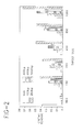
- probe hybridization and the resulting signal could be detected by the reflectometer.
- the largest probe (1,650 bp) gave the most intense signal with all sizes of capture DNAs.
- the other probes in general gave signals of decreasing intensity with decreasing probe sequence size.
- the specificity of the assay using the different size fragments was tested by using nonhomologous capture and probe fragments and in all cases the readings were similar to background levels.
- the nonhomologous probe did produce a very weak positive reaction with the largest capture fragment (1,650 bp). This may be due to a small amount of nonspecific hybridization due to the relatively large size of the capture fragment or to partial pore blockage in the membrane interfering with free flow of the probe through the membrane.
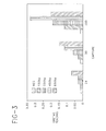
- the capture sequences of 50 bp and 24 bp were found to be capable of capturing the 850 bp, 450 bp, 250 bp and 150 bp probes.
- the intensity of the signal was reduced as compared to the assay using larger capture sequences.
- 100 ng of 24 bp or 50 bp capture DNA was required to detect 200 ng of probe.
- the intensities were less than those seen when smaller amounts of the larger fragments were used (i.e., 50 ng of the 250 bp capture fragment).
- Neisseria gonorrhea was used as a model system to demonstrate the utility of the inventive method for detecting a specific nucleic acid fragment in cells.
- Primers were selected and synthesized to amplify an approximately 281 bp portion of the gonococcal H.8 gene from nucleotides 53 to 320 as reported by Woods et al. (1989) Molecular Microbiology 3 (1): 43-48. This gene is highly conserved among the pathogenic Neisseria species. Amplified probes were synthesized using the polymerase chain reaction as previously described.
- Neisseria gonorrhea strain GC5766 was grown in liquid culture. Ten fold serial dilutions were made in 200 ml of 2 mM Tris-CI, pH 8.5, 2 mM EDTA, 1% Triton X-100 to obtain 10 6 to 10 2 organisms per ml. Each dilution was heated to 95°C for 10 minutes. Twenty five microliters of each dilution were used as templates in PCR reactions. The 281 bp fragment of the H.8 gene was amplified, incorporating biotin-16 UTP as described above.
- the flow-through hybridization assay may also be done in a sandwich assay format.
- a small 150 bp fragment of the rubella E1 gene was used as the capture sequence for a 1,650 bp target sequence.
- a probe was prepared by amplification of a 400 bp portion of the 1,650 bp target sequence which would not hybridize with the capture sequence, incorporating biotin-16-UTP as described above.
- the 150 bp capture sequence was spotted on BIODYNE B membranes at 50 ng and 10 ng in 2 ml.
- the assay was performed essentially as in Example 2. Five microliters of target sequence, purified and diluted in TE buffer, were mixed with 45 ml of Hyb I and heated at 70°C for 5 min. The prepared target DNA was then applied to the nylon membrane and allowed to flow through. The membrane was washed with 45 ml of 0.2X SSC followed by 45 ml of 2X SSC and 150 ml of 100 mM PBS pH 8.0. Probe (92 ng) in 5 ml was added to 45 ml of hybridization buffer, heated at 70°C for 5 minutes and allowed to flow through the membrane.
- the membrane was washed as before with 150 ml of each of the SSC solutions and blocked with 150 ml of Blotto/BSA solution as described above.
- Rabbit anti-biotin liposomes encapsulating sulforhodamine 150 ml of a 1:3 dilution in 1% BLOTTO were used to detect hybridized probe on the membrane, followed by addition of 2 drops of 2 M urea.
Applications Claiming Priority (2)
| Application Number | Priority Date | Filing Date | Title |
|---|---|---|---|
| US42593A | 1993-01-04 | 1993-01-04 | |
| US425 | 1993-01-04 |
Publications (1)
| Publication Number | Publication Date |
|---|---|
| EP0605828A1 true EP0605828A1 (fr) | 1994-07-13 |
Family
ID=21691486
Family Applications (1)
| Application Number | Title | Priority Date | Filing Date |
|---|---|---|---|
| EP93120394A Withdrawn EP0605828A1 (fr) | 1993-01-04 | 1993-12-17 | Essai d'hybridation à écoulement de séquences oligonucléotidiques |
Country Status (4)
| Country | Link |
|---|---|
| EP (1) | EP0605828A1 (fr) |
| JP (1) | JPH0811080B2 (fr) |
| AU (1) | AU675251B2 (fr) |
| CA (1) | CA2112645A1 (fr) |
Cited By (14)
| Publication number | Priority date | Publication date | Assignee | Title |
|---|---|---|---|---|
| WO1996019587A2 (fr) * | 1994-12-22 | 1996-06-27 | Abbott Laboratories | Procedes d'immobilisation d'oligonucleotides sur des materiaux de support et procedes d'utilisation des oligonucleotides fixes auxdits supports |
| US5741647A (en) * | 1996-02-16 | 1998-04-21 | Tam; Joseph Wing On | Flow through nucleic acid hybridisation uses thereof and a device thereof |
| WO1999050665A1 (fr) * | 1998-03-31 | 1999-10-07 | Johnco, Inc. | Dispositif et methodes de microtitrage |
| WO1999064628A1 (fr) * | 1998-06-08 | 1999-12-16 | Acgt Medico, Inc. | Detection d'adn, d'arn et de proteines |
| WO2000031304A1 (fr) * | 1998-11-25 | 2000-06-02 | The Perkin-Elmer Corporation | Procedes et appareil pour hybridation en continu |
| US6174733B1 (en) | 1997-06-09 | 2001-01-16 | Acgt Medico Inc. | Detection of pathogens using a column device with a sample containing a control |
| WO2001061042A2 (fr) * | 2000-02-18 | 2001-08-23 | Pall Corporation | Membranes |
| US6337214B1 (en) | 1997-06-09 | 2002-01-08 | Acgt Medico, Inc. | Detection of DNA, RNA and proteins using a test column with two snares |
| US6734012B2 (en) | 2000-07-05 | 2004-05-11 | Cuno Incorporated | Low fluorescence nylon/glass composites for micro-analytical diagnostic applications |
| WO2004050910A1 (fr) * | 2002-12-04 | 2004-06-17 | Pamgene Bv | Procede d'hybridation d'adn genomique immobilise |
| US6890483B2 (en) | 2000-07-05 | 2005-05-10 | Cuno Incorporated | Non-luminescent substrate |
| EP1845374A1 (fr) * | 2006-04-14 | 2007-10-17 | Koninklijke Philips Electronics N.V. | Membrane sous forme d'inhibition d'une cellule à flux |
| US9568404B2 (en) | 2014-05-16 | 2017-02-14 | Junyu Mai | Method and apparatus for biomolecule analysis |
| CN114350753A (zh) * | 2021-12-20 | 2022-04-15 | 江苏海洋大学 | 自由联结的管式基因芯片及其在疾病检测中的应用 |
Families Citing this family (3)
| Publication number | Priority date | Publication date | Assignee | Title |
|---|---|---|---|---|
| PT1435205E (pt) | 1995-09-19 | 2010-02-04 | Bayer Bioscience Gmbh | Plantas que sintetizam um amido modificado, processo para a sua produção e amido modificado |
| EP1157264B1 (fr) * | 1999-02-26 | 2004-04-21 | EXACT Sciences Corporation | Dispositifs de purification biochimique avec sondes de capture immobilisees et leurs utilisations |
| BR112020021494B1 (pt) | 2018-04-20 | 2023-10-03 | Kordsa Teknik Tekstil Anonim Sirketi | Pré-impregnado adequado para uso a fim de realizar armadura em um concreto ou um material de suporte de carga |
Citations (6)
| Publication number | Priority date | Publication date | Assignee | Title |
|---|---|---|---|---|
| EP0302673A1 (fr) * | 1987-08-06 | 1989-02-08 | Becton, Dickinson and Company | Système et procédé d'essai visuel d'une substance à déterminer |
| EP0335244A2 (fr) * | 1988-03-28 | 1989-10-04 | Abbott Laboratories | Dispositif d'analyse en phase solide et procédé pour son utilisation |
| WO1990004786A1 (fr) * | 1988-10-17 | 1990-05-03 | Molecular Devices Corporation | Membrane de capture derive de haptene et tests de diagnostic utilisant de telles membranes |
| EP0455905A2 (fr) * | 1990-05-11 | 1991-11-13 | Microprobe Corporation | Supports solides pour l'essai d'hybridition des acides nucléiques et procédés pour l'immobilisation covalente d'oligonucléotides |
| EP0458231A1 (fr) * | 1990-05-25 | 1991-11-27 | Becton, Dickinson and Company | Substrat et procédés pour l'essai immunologique et determination d'urée employant l'uréase immobilisée |
| WO1992016659A1 (fr) * | 1991-03-21 | 1992-10-01 | Eastman Kodak Company | Element et procede pour l'amplification et la detection de l'acide nucleique au moyen de sondes collees |
-
1993
- 1993-12-09 AU AU52278/93A patent/AU675251B2/en not_active Ceased
- 1993-12-17 EP EP93120394A patent/EP0605828A1/fr not_active Withdrawn
- 1993-12-22 JP JP5324047A patent/JPH0811080B2/ja not_active Expired - Lifetime
- 1993-12-30 CA CA002112645A patent/CA2112645A1/fr not_active Abandoned
Patent Citations (6)
| Publication number | Priority date | Publication date | Assignee | Title |
|---|---|---|---|---|
| EP0302673A1 (fr) * | 1987-08-06 | 1989-02-08 | Becton, Dickinson and Company | Système et procédé d'essai visuel d'une substance à déterminer |
| EP0335244A2 (fr) * | 1988-03-28 | 1989-10-04 | Abbott Laboratories | Dispositif d'analyse en phase solide et procédé pour son utilisation |
| WO1990004786A1 (fr) * | 1988-10-17 | 1990-05-03 | Molecular Devices Corporation | Membrane de capture derive de haptene et tests de diagnostic utilisant de telles membranes |
| EP0455905A2 (fr) * | 1990-05-11 | 1991-11-13 | Microprobe Corporation | Supports solides pour l'essai d'hybridition des acides nucléiques et procédés pour l'immobilisation covalente d'oligonucléotides |
| EP0458231A1 (fr) * | 1990-05-25 | 1991-11-27 | Becton, Dickinson and Company | Substrat et procédés pour l'essai immunologique et determination d'urée employant l'uréase immobilisée |
| WO1992016659A1 (fr) * | 1991-03-21 | 1992-10-01 | Eastman Kodak Company | Element et procede pour l'amplification et la detection de l'acide nucleique au moyen de sondes collees |
Cited By (21)
| Publication number | Priority date | Publication date | Assignee | Title |
|---|---|---|---|---|
| WO1996019587A3 (fr) * | 1994-12-22 | 1996-09-19 | Abbott Lab | Procedes d'immobilisation d'oligonucleotides sur des materiaux de support et procedes d'utilisation des oligonucleotides fixes auxdits supports |
| WO1996019587A2 (fr) * | 1994-12-22 | 1996-06-27 | Abbott Laboratories | Procedes d'immobilisation d'oligonucleotides sur des materiaux de support et procedes d'utilisation des oligonucleotides fixes auxdits supports |
| US6020187A (en) * | 1996-02-16 | 2000-02-01 | Tam; Joseph Wing On | Flow through nucleic acid hybridisation device |
| US5741647A (en) * | 1996-02-16 | 1998-04-21 | Tam; Joseph Wing On | Flow through nucleic acid hybridisation uses thereof and a device thereof |
| US6337214B1 (en) | 1997-06-09 | 2002-01-08 | Acgt Medico, Inc. | Detection of DNA, RNA and proteins using a test column with two snares |
| US6174733B1 (en) | 1997-06-09 | 2001-01-16 | Acgt Medico Inc. | Detection of pathogens using a column device with a sample containing a control |
| US6224831B1 (en) | 1998-03-31 | 2001-05-01 | John Co., Inc. | Microassay device and methods |
| WO1999050665A1 (fr) * | 1998-03-31 | 1999-10-07 | Johnco, Inc. | Dispositif et methodes de microtitrage |
| WO1999064628A1 (fr) * | 1998-06-08 | 1999-12-16 | Acgt Medico, Inc. | Detection d'adn, d'arn et de proteines |
| AU776749B2 (en) * | 1998-11-25 | 2004-09-23 | Applera Corporation | Methods and apparatus for flow-through hybridization |
| WO2000031304A1 (fr) * | 1998-11-25 | 2000-06-02 | The Perkin-Elmer Corporation | Procedes et appareil pour hybridation en continu |
| US6638760B1 (en) | 1998-11-25 | 2003-10-28 | Pe Corporation (Ny) | Method and apparatus for flow-through hybridization |
| WO2001061042A2 (fr) * | 2000-02-18 | 2001-08-23 | Pall Corporation | Membranes |
| WO2001061042A3 (fr) * | 2000-02-18 | 2002-02-07 | Pall Corp | Membranes |
| US6734012B2 (en) | 2000-07-05 | 2004-05-11 | Cuno Incorporated | Low fluorescence nylon/glass composites for micro-analytical diagnostic applications |
| US6890483B2 (en) | 2000-07-05 | 2005-05-10 | Cuno Incorporated | Non-luminescent substrate |
| WO2004050910A1 (fr) * | 2002-12-04 | 2004-06-17 | Pamgene Bv | Procede d'hybridation d'adn genomique immobilise |
| EP1845374A1 (fr) * | 2006-04-14 | 2007-10-17 | Koninklijke Philips Electronics N.V. | Membrane sous forme d'inhibition d'une cellule à flux |
| US9568404B2 (en) | 2014-05-16 | 2017-02-14 | Junyu Mai | Method and apparatus for biomolecule analysis |
| US9857368B2 (en) | 2014-05-16 | 2018-01-02 | Junyu Mai | Method and apparatus for biomolecule analysis |
| CN114350753A (zh) * | 2021-12-20 | 2022-04-15 | 江苏海洋大学 | 自由联结的管式基因芯片及其在疾病检测中的应用 |
Also Published As
| Publication number | Publication date |
|---|---|
| JPH0811080B2 (ja) | 1996-02-07 |
| CA2112645A1 (fr) | 1994-07-05 |
| AU675251B2 (en) | 1997-01-30 |
| JPH06261797A (ja) | 1994-09-20 |
| AU5227893A (en) | 1994-07-14 |
Similar Documents
| Publication | Publication Date | Title |
|---|---|---|
| EP0605828A1 (fr) | Essai d'hybridation à écoulement de séquences oligonucléotidiques | |
| US6037127A (en) | Method for detection of non-denatured nucleic acid fragments | |
| US5989813A (en) | Detection of amplified nucleic acid sequences using bifunctional haptenization and dyed microparticles | |
| AU629845B2 (en) | Detection and amplification of target nucleic acid sequences | |
| US5627030A (en) | Method of amplification for increasing the sensitivity of detecting nucleic acid-probe target hybrids | |
| US5985548A (en) | Amplification of assay reporters by nucleic acid replication | |
| US6090592A (en) | Method for performing amplification of nucleic acid on supports | |
| EP0374665A2 (fr) | Essai des séquences utilisant des gènes amplifiés | |
| WO1990002205A1 (fr) | Detection de sequences d'acides nucleiques par agglutination de particules | |
| WO1992004469A2 (fr) | Procede de detection d'acide nucleique utilisant l'agglutination de particules | |
| JPH07501933A (ja) | 核酸配列の検出のための方法及び装置 | |
| JP2005507674A (ja) | 迅速乾燥アッセイ法の形で核酸を検出する方法 | |
| WO1993015229A2 (fr) | Amplification de rapporteurs d'analyse par replication d'une sequence d'acide nucleique | |
| WO1995027081A1 (fr) | Methode de detection de fragments d'acide nucleique | |
| WO2013122453A1 (fr) | Dispositif et procédé de détection d'acide(s) nucléique(s) | |
| EP1290228B1 (fr) | Detection inverse pour l'identification et/ou la quantification des sequences nucleotidiques cibles sur des biopuces | |
| JPH06292599A (ja) | フロースルー膜による核酸ハイブリダイゼーションアッセイ | |
| US6485915B1 (en) | Analytical element for species-specific detection of nucleic acids | |
| JP3604758B2 (ja) | 標的核酸の半定量的検出方法、試験要素および試験キット | |
| US6306657B1 (en) | Polynucleotide probe and kit for amplifying signal detection in hybridization assays | |
| WO2004042084A1 (fr) | Bandelette d'essai a reactif sec et detection d'acide nucleique | |
| WO1989009281A1 (fr) | Procede d'amplification et de detection d'acide nucleique dans un liquide test | |
| JP2003199600A (ja) | 標的核酸の検出方法 | |
| KR20230038926A (ko) | 염 매개의 핵산 고정화 방법, 상기 방법에 따라 고정화된 포획 핵산을 포함하는 고형 지지체, 상기 고형 지지체를 포함하는 측방 유동 분석 스트립, 및 표적 핵산 검출 방법 | |
| WO2002053768A2 (fr) | Procedes et appareil de detection immune rapide d'adn |
Legal Events
| Date | Code | Title | Description |
|---|---|---|---|
| PUAI | Public reference made under article 153(3) epc to a published international application that has entered the european phase |
Free format text: ORIGINAL CODE: 0009012 |
|
| AK | Designated contracting states |
Kind code of ref document: A1 Designated state(s): DE FR GB IT |
|
| 17P | Request for examination filed |
Effective date: 19941126 |
|
| 17Q | First examination report despatched |
Effective date: 19950228 |
|
| STAA | Information on the status of an ep patent application or granted ep patent |
Free format text: STATUS: THE APPLICATION IS DEEMED TO BE WITHDRAWN |
|
| 18D | Application deemed to be withdrawn |
Effective date: 19990608 |