EP0556243B1 - Automatic biopsy instrument - Google Patents
Automatic biopsy instrument Download PDFInfo
- Publication number
- EP0556243B1 EP0556243B1 EP91919513A EP91919513A EP0556243B1 EP 0556243 B1 EP0556243 B1 EP 0556243B1 EP 91919513 A EP91919513 A EP 91919513A EP 91919513 A EP91919513 A EP 91919513A EP 0556243 B1 EP0556243 B1 EP 0556243B1
- Authority
- EP
- European Patent Office
- Prior art keywords
- cannula
- tissue
- stylet
- instrument
- cavity
- Prior art date
- Legal status (The legal status is an assumption and is not a legal conclusion. Google has not performed a legal analysis and makes no representation as to the accuracy of the status listed.)
- Expired - Lifetime
Links
- 238000001574 biopsy Methods 0.000 title claims description 40
- 210000001519 tissue Anatomy 0.000 description 80
- 206010028980 Neoplasm Diseases 0.000 description 12
- 238000000034 method Methods 0.000 description 11
- 238000013461 design Methods 0.000 description 5
- 210000001185 bone marrow Anatomy 0.000 description 4
- 230000000994 depressogenic effect Effects 0.000 description 4
- 238000005070 sampling Methods 0.000 description 4
- 201000011510 cancer Diseases 0.000 description 3
- 238000003745 diagnosis Methods 0.000 description 3
- 230000007246 mechanism Effects 0.000 description 3
- 210000003813 thumb Anatomy 0.000 description 3
- 241001465754 Metazoa Species 0.000 description 2
- 208000037265 diseases, disorders, signs and symptoms Diseases 0.000 description 2
- 230000002962 histologic effect Effects 0.000 description 2
- 238000003384 imaging method Methods 0.000 description 2
- 238000012986 modification Methods 0.000 description 2
- 230000004048 modification Effects 0.000 description 2
- 239000007787 solid Substances 0.000 description 2
- 238000012360 testing method Methods 0.000 description 2
- 208000006994 Precancerous Conditions Diseases 0.000 description 1
- 230000009471 action Effects 0.000 description 1
- 230000004075 alteration Effects 0.000 description 1
- 238000013459 approach Methods 0.000 description 1
- 210000000988 bone and bone Anatomy 0.000 description 1
- 230000006835 compression Effects 0.000 description 1
- 238000007906 compression Methods 0.000 description 1
- 230000000881 depressing effect Effects 0.000 description 1
- 201000010099 disease Diseases 0.000 description 1
- 208000035475 disorder Diseases 0.000 description 1
- 230000009977 dual effect Effects 0.000 description 1
- 238000013399 early diagnosis Methods 0.000 description 1
- 230000000694 effects Effects 0.000 description 1
- 238000005516 engineering process Methods 0.000 description 1
- 238000000605 extraction Methods 0.000 description 1
- 230000006870 function Effects 0.000 description 1
- 238000003306 harvesting Methods 0.000 description 1
- 208000014674 injury Diseases 0.000 description 1
- 238000003780 insertion Methods 0.000 description 1
- 230000037431 insertion Effects 0.000 description 1
- 239000007788 liquid Substances 0.000 description 1
- 238000002559 palpation Methods 0.000 description 1
- 239000012188 paraffin wax Substances 0.000 description 1
- 230000035515 penetration Effects 0.000 description 1
- 230000008569 process Effects 0.000 description 1
- 230000001737 promoting effect Effects 0.000 description 1
- 230000000717 retained effect Effects 0.000 description 1
- 238000007789 sealing Methods 0.000 description 1
- 238000004448 titration Methods 0.000 description 1
- 230000008733 trauma Effects 0.000 description 1
- 238000002604 ultrasonography Methods 0.000 description 1
- 238000012285 ultrasound imaging Methods 0.000 description 1
Images

Classifications
-
- A—HUMAN NECESSITIES
- A61—MEDICAL OR VETERINARY SCIENCE; HYGIENE
- A61B—DIAGNOSIS; SURGERY; IDENTIFICATION
- A61B10/00—Instruments for taking body samples for diagnostic purposes; Other methods or instruments for diagnosis, e.g. for vaccination diagnosis, sex determination or ovulation-period determination; Throat striking implements
- A61B10/02—Instruments for taking cell samples or for biopsy
- A61B10/0233—Pointed or sharp biopsy instruments
- A61B10/0283—Pointed or sharp biopsy instruments with vacuum aspiration, e.g. caused by retractable plunger or by connected syringe
-
- A—HUMAN NECESSITIES
- A61—MEDICAL OR VETERINARY SCIENCE; HYGIENE
- A61B—DIAGNOSIS; SURGERY; IDENTIFICATION
- A61B10/00—Instruments for taking body samples for diagnostic purposes; Other methods or instruments for diagnosis, e.g. for vaccination diagnosis, sex determination or ovulation-period determination; Throat striking implements
- A61B10/02—Instruments for taking cell samples or for biopsy
- A61B10/0233—Pointed or sharp biopsy instruments
- A61B10/0266—Pointed or sharp biopsy instruments means for severing sample
- A61B10/0275—Pointed or sharp biopsy instruments means for severing sample with sample notch, e.g. on the side of inner stylet
Definitions
- This invention relates to an automated mechanism for collecting a tissue sample from humans or animals by a procedure referred to as tissue biopsy, and more particularly to an instrument for automatically performing the tissue extraction from a tissue mass in a precise and rapid manner with minimum patient discomfort.
- Tumors are first noted in a patient by one of three ways palpation, X-ray imaging or ultrasound imaging.
- tissue biopsy is performed to establish whether cells are cancerous.
- Biopsy may be done by an open or closed technique. Open biopsy removes the entire tissue mass or a part of the tissue mass. Closed biopsy on the other hand is usually performed with a needle-like instrument and may be either an aspiration biopsy (hollow needle on a syringe) or a cored biopsy (special tissue cutting needle design). In needle aspiration biopsy, individual cells or clusters of cells are obtained for cytologic examination. In core biopsy, a segment of tissue is obtained for histologic examination which may be done as a frozen section or paraffin section.
- tissue samples for cytologic or histologic examination have been performed historically by manual insertion and manipulation of the needle. These procedures are performed blind by the physician and guided by feel and known anatomic landmarks.
- One prior art manual biopsy device includes a syringe arrangement including a stylet surrounded by a cannula.
- the stylet has a pointed tip and behind the tip a reduced diameter shank.
- the diameter of the pointed tip is slightly less than the internal diameter of the cannula such that the tip prevents tissue from entering the cannula as the cannula is passed through surrounding tissue to the point of intended biopsy.
- An O-ring is placed in sealing relationship between the reduced diameter shank and the internal diameter of the cannula.
- the cannula is urged forward past the tip of the stylet in order to collect a tissue sample. As this occurs, a vacuum is formed in the cannula between the O-ring and the tissue sample. This vacuum tends to draw the tissue sample into the cannula.
- tissue harvesting devices have been described in United States Patent Nos. 4,651,752; 4,702,260; and 4,243,048.
- the ACBD Automatic Core Biopsy Device
- the ACBD is an instrument which propels a needle set with considerable force and speed in order to pierce the tumor mass and collect the tissue sample. This ACBD has allowed physicians to test tissue masses in the early stages of growth and has contributed to the medical trend of early diagnosis and successful treatment of cancer.
- the Automated Core Biopsy Device allows a biopsy to be performed on tumor masses as small as two millimeters in diameter. This procedure is performed under ultrasound or X-ray guidance. Tumors of this size cannot be biopsied reliably by hand since the tumor is about the same size as the biopsy needle. Manual attempts at biopsy pushes the tumor away without piercing the mass. Automatic puncture devices accelerate the needle at such a velocity that even a small tumor can be pierced.
- the True Cut needle is comprised of an inner notched stylet with an outer cannula.
- the stylet is advanced into the tissue under spring power followed by the cannula which cuts and traps the tissue sample in the notch of the stylet.
- the True Cut needle yields a core sample which is semi-circular in cross-section with a length determined by the stroke of the ACBD.
- the stylet is a needle with a notched cut-out at the distal end.
- the cannula is a hollow needle with an angled cutting surface at the distal end which slides over the stylet. When the stylet is pushed into the tissue, the tissue is pierced and relaxes into the notched cut-out. When the cannula is slid forward, the tissue in the notch of the stylet is sliced off and retained in the notch until the cannula is drawn back.
- the most common True Cut needle size used by ACBD's is 18 gage (1.2mm diameter).
- 18 gage needles is a compromise between the physician's desire to use the smallest, least invasive, needle gage and the pathologist's needs for as large a tissue sample as possible to minimize false-positive diagnosis. This compromise in needle size leads the physician to obtain multiple core samples from the biopsy site to allow the pathologist sufficient tissue for an accurate diagnosis.
- the ideal product would allow the use of smaller needle gages and/or lessen the need for multiple samples to be taken from a given biopsy site.
- US-A-4,747,414 discloses an instrument for bone marrow puncture comprising a sampling needle disposed in a cannula and fused to a piston which can be displaced in a piston barrel.
- a sampling needle disposed in a cannula and fused to a piston which can be displaced in a piston barrel.
- the needle is made to pierce the bone and reach the region of bone marrow.
- the posterior part of the piston barrel defines a closed chamber for the collection of semi-liquid bone marrow aspirated into this chamber by retracting the piston and thereby forming a negative pressure in the chamber.
- EP-A-0,390,528, which forms the preamble of claim 1, discloses an automatic biopsy instrument for obtaining a sample of tissue including a body, a cavity in the body, a piston mounted in the cavity, an elongate cannula with an elongate inner bore, the cannula being secured to the piston, a stylet secured to the body and being positioned in the inner bore of the cannula, and means for driving the cannula out beyond the stylet into the tissue to be sampled.
- the biopsy instrument of EP-A-0,390,528 can be provided with stylets of known configuration; in connection with the preferred embodiment reference is made to a stylet of the TRU-CUTTM type.
- the present invention is characterised in that the instrument is provided with means for communicating the inner bore of the cannula with the cavity of the body; and in that said means for driving the cannula out beyond the stylet into the tissue to be sampled act so as to create a vacuum in said cavity which vacuum is communicated with the inner bore of the cannula by said communicating means thereby to assist in breaking off a tissue sample and retaining it in the cannula.
- a preferred embodiment of the invention is an instrument for removing cylindrically shaped tissue samples of pre-determined size from a tissue mass with an instrument that automatically penetrates, captures and removes the tissue sample for examination.
- the instrument is a spring powered mechanical design.
- the needle set is integral with the housing and consists of an outer hollow cannula and an inner pointed tipped stylet. The stylet is stationary and the cannula is driven forward under spring force.
- the housing is comprised of a cavity which guides a spring backed piston to which the cannula is attached. Once the spring is released, the piston and cannula are driven forward. As the piston advances, a vacuum is created behind the piston, with the only outlet for this vacuum being down the bore of the advancing cannula. As the tissue is penetrated by the cannula, a vacuum force is exerted on the captured tissue and this vacuum force pulls the tissues down the bore of the cannula. The vacuum holds the tissue and allows the tissue to break off at the tip of the distal end as the needle is withdrawn.
- the stylet is a stationary needle which has a reduced diameter beyond the distal pointed tip to allow for the creation of the maximum vacuum force in the piston and cannula.
- a solid pointed tip facilitates the introduction of the needles into the tissue mass.
- the stylet is positioned flush with the end of the cannula in the cocked position. In the cocked position, the stylet prevents tissue from entering the cannula s the needle set is introduced into the body. As the device is fired, the cannula advances while the stylet remains stationary, thus allowing space for the penetrated tissue to enter the cannula.
- the cannula is moved backwards over the stationary stylet, pushing the tissue sample out of the cannula. This action removes the tissue sample and cocks the gun in one motion.
- a volumetric analysis of a cross-section of tissue area collected with the True Cut needle set vs. the cylindrical core samples of this invention follows: Stylet Diameter Gage X-Section Area Percentage True Cut 1.2mm 18 ga. .0007 sq/in. (.0045cm 2 ) Cylindrical 1.2mm 18 ga. .0012 sq/in. (.0077cm 2 ) 72% larger sample Cylindrical 0.9mm 20 ga. .0005 sq/in. (.0032cm 2 ) 71% of True Cut 18 ga.
- the cylindrical core volume of this invention is 72% larger than that provided by the True Cut needle set.
- a 20 gage (0.9mm diameter) cylindrical needle in accordance with this invention will yield 71% of the tissue yielded by an 18 gage (1.2mm diameter) True Cut needle set.
- tissue sampling device for obtaining tissue samples which have a circular cross-section.
- Such a cross-section provides for more tissue mass for a given needle gage, provides for a less invasive procedure with reduced tissue trauma and allows for the maximum tissue to be harvested with the minimum number of samples taken.
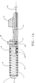
- Fig. 1A illustrates a side elevation view of the embodiment of the inventive biopsy instrument which is shown to depict the main components of said embodiment with the main body shown generally at 1 and the tissue piercing and removal device, cannula 13.
- the main housing 1 extends from end cap 7 to the thumb knob 4.
- a plunger 2 Within said housing 1 is a plunger 2 with annular grooves that capture two "O"-ring seals 10.
- Plunger rod 2 is depressed by exerting pressure on thumb knob 4 which compresses the main spring 5. Plunger 2 and main spring 5 are held in the compressed state by latch pin 12 which resides on actuator button 8 (Fig. 1B). Spring 9 pushes actuator button 8 and latch pin 12 up against plunger rod 3. As plunger rod 3 is depressed latch pin 12 slides along plunger rod 3 until latch pin 12 detents into the annular groove in plunger rod 3. Latch pin 12 holds plunger rod 3 in place and thus holds plunger 2 and main spring 5 in compression until such time that actuator button 8 is depressed releasing plunger rod 3. As plunger rod 3 is released, main spring 5 pushes plunger 2 forward with the associated component cannula 13 which is attached to plunger 2.
- Cannula 13 is fixed to plunger 2 through a hole in plunger 2 and sealed such that plunger 2 with the associated "O"-rings 10 and end cap 7 with associated "O"-ring 6 create an air tight chamber or cavity in main housing 1, between the end cap 7 and the plunger 2, with the hollow body of cannula 13 the only avenue of air passage.
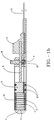
- the diameter of the tip 14 of the stylet 11 is, however, small enough relative to the bore diameter to allow the passage of air between the inner diameter of the cannula 13 and the outer diameter of the tip 14 of the stylet 11 (Fig. 2).
- the vacuum in main housing 1 creates a suction force down the bore of cannula 13 and is attached to main housing 1 at the point where end cap 7 is attached.
- Stylet 11 remains stationary in respect to main housing 1 as cannula 13 slides over stylet 11 and pierces the tissue.
- the reduced diameter shank of stylet 11 allows the suction generated in main housing 1 to provide maximum effect on pierced tissue captured in cannula 13.
- the captured tissue in cannula 13 acts as a seal thus maintaining a vacuum on the captive tissue in cannula 13 allowing the tissue to break off at the distal end of the cannula 13 and be extracted from the tissue mass.
- Fig. 2 illustrates a cross-sectional side elevation view of the distal ends of cannula 13 and stylet 11.
- Stylet 11 is a is a dual diameter solid rod with a pointed tip 14 at the distal end. The tip 14 prevents tissue from entering cannula 13 as cannula 13 is passed through surrounding tissue to the point of intended biopsy.
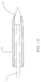
- Figs. 3A, 3B and 3C illustrate the preferred embodiment of the distal needle end of this invention. Three stages of motion are depicted.
- Fig. 3A shows cannula 13 and stylet 11 in the cocked position;
- Fig. 3B shows cannula 13 and stylet 11 in the fired position.
- Fig. 3C shows cannula 13 and stylet 11 in the act of cocking the mechanism and the subsequent expulsion of the tissue sample.
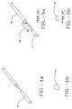
- Figs. 4A, 4B, 5A and 5B illustrate a comparison of the tissue sample obtained at the distal needle ends of the preferred embodiment of this invention and tissue obtained by the True Cut needle design used in prior art biopsy instruments.
- Fig. 4A illustrates the preferred embodiment of the distal needle end with the expulsed tissue sample 15.
- Fig. 4B illustrates the cross-section of tissue 15 obtained from the distal end of the preferred embodiment of this invention.
- Fig. 5A illustrates the distal needle end 17 extending from cannula 16 of the prior art device with the expulsed tissue sample 18.
- Fig. 5B illustrates the cross-section of tissue 18 obtained from the distal needle end of the prior art device.
Landscapes
- Health & Medical Sciences (AREA)
- Life Sciences & Earth Sciences (AREA)
- Medical Informatics (AREA)
- Engineering & Computer Science (AREA)
- Biomedical Technology (AREA)
- Heart & Thoracic Surgery (AREA)
- Pathology (AREA)
- Molecular Biology (AREA)
- Surgery (AREA)
- Animal Behavior & Ethology (AREA)
- General Health & Medical Sciences (AREA)
- Public Health (AREA)
- Veterinary Medicine (AREA)
- Surgical Instruments (AREA)
- Infusion, Injection, And Reservoir Apparatuses (AREA)
Applications Claiming Priority (3)
| Application Number | Priority Date | Filing Date | Title |
|---|---|---|---|
| US07/610,006 US5183052A (en) | 1990-11-07 | 1990-11-07 | Automatic biopsy instrument with cutting cannula |
| US610006 | 1990-11-07 | ||
| PCT/US1991/007639 WO1992008409A1 (en) | 1990-11-07 | 1991-10-18 | Automatic biopsy instrument |
Publications (3)
| Publication Number | Publication Date |
|---|---|
| EP0556243A1 EP0556243A1 (en) | 1993-08-25 |
| EP0556243A4 EP0556243A4 (enExample) | 1994-03-16 |
| EP0556243B1 true EP0556243B1 (en) | 1998-09-09 |
Family
ID=24443223
Family Applications (1)
| Application Number | Title | Priority Date | Filing Date |
|---|---|---|---|
| EP91919513A Expired - Lifetime EP0556243B1 (en) | 1990-11-07 | 1991-10-18 | Automatic biopsy instrument |
Country Status (5)
| Country | Link |
|---|---|
| US (1) | US5183052A (enExample) |
| EP (1) | EP0556243B1 (enExample) |
| JP (1) | JPH06501408A (enExample) |
| DE (1) | DE69130174D1 (enExample) |
| WO (1) | WO1992008409A1 (enExample) |
Families Citing this family (108)
| Publication number | Priority date | Publication date | Assignee | Title |
|---|---|---|---|---|
| US5400798A (en) * | 1989-03-29 | 1995-03-28 | Baran; Gregory W. | Automated biopsy instrument |
| US5617874A (en) * | 1989-03-29 | 1997-04-08 | Baran; Gregory W. | Automated biopsy instrument |
| GB2256369B (en) * | 1991-06-04 | 1995-10-25 | Chiou Rei Kwen | Improved biopsy device |
| DE9414727U1 (de) * | 1993-09-09 | 1994-12-08 | Heske, Norbert, 82299 Türkenfeld | Biopsiesystem |
| US5379773A (en) * | 1993-09-17 | 1995-01-10 | Hornsby; James J. | Echographic suction cannula and electronics therefor |
| US5649547A (en) | 1994-03-24 | 1997-07-22 | Biopsys Medical, Inc. | Methods and devices for automated biopsy and collection of soft tissue |
| US5526822A (en) * | 1994-03-24 | 1996-06-18 | Biopsys Medical, Inc. | Method and apparatus for automated biopsy and collection of soft tissue |
| US5817033A (en) * | 1994-04-11 | 1998-10-06 | Desantis; Stephen A. | Needle core biopsy device |
| US5560373A (en) * | 1994-04-11 | 1996-10-01 | De Santis; Stephen A. | Needle core biopsy instrument with durable or disposable cannula assembly |
| US5511556A (en) * | 1994-04-11 | 1996-04-30 | Desantis; Stephen A. | Needle core biopsy instrument |
| US5449001A (en) * | 1994-04-14 | 1995-09-12 | Terwilliger; Richard A. | Biopsy needle |
| US5954670A (en) * | 1994-10-05 | 1999-09-21 | Baker; Gary H. | Mandrel-guided tandem multiple channel biopsy guide device and method of use |
| WO1996027329A1 (en) | 1995-03-08 | 1996-09-12 | Terwilliger Richard A | Echogenic needle |
| US5779647A (en) | 1995-06-07 | 1998-07-14 | Chau; Sonny | Automated biopsy instruments |
| US6017316A (en) * | 1997-06-18 | 2000-01-25 | Biopsys Medical | Vacuum control system and method for automated biopsy device |
| US6080113A (en) | 1998-09-11 | 2000-06-27 | Imagyn Medical Technologies California, Inc. | Incisional breast biopsy device |
| US6551253B2 (en) | 1997-09-12 | 2003-04-22 | Imagyn Medical Technologies | Incisional breast biopsy device |
| US6383145B1 (en) | 1997-09-12 | 2002-05-07 | Imagyn Medical Technologies California, Inc. | Incisional breast biopsy device |
| US6142955A (en) | 1997-09-19 | 2000-11-07 | United States Surgical Corporation | Biopsy apparatus and method |
| US6019733A (en) * | 1997-09-19 | 2000-02-01 | United States Surgical Corporation | Biopsy apparatus and method |
| US6050955A (en) * | 1997-09-19 | 2000-04-18 | United States Surgical Corporation | Biopsy apparatus and method |
| US6007495A (en) * | 1998-01-22 | 1999-12-28 | United States Surgical Corporation | Biopsy apparatus and method |
| US6193673B1 (en) | 1998-02-20 | 2001-02-27 | United States Surgical Corporation | Biopsy instrument driver apparatus |
| US6086543A (en) * | 1998-06-24 | 2000-07-11 | Rubicor Medical, Inc. | Fine needle and core biopsy devices and methods |
| CA2351331C (en) | 1998-11-25 | 2010-07-20 | United States Surgical Corporation | Biopsy system |
| ITCE990004A1 (it) * | 1999-10-25 | 2000-01-25 | Mario Immacolato Paternuosto | Valve per pinza da biopsia in endoscopia digestiva |
| DE10026303A1 (de) * | 2000-05-26 | 2002-02-07 | Pajunk Gmbh | Biopsienadel |
| US6712773B1 (en) | 2000-09-11 | 2004-03-30 | Tyco Healthcare Group Lp | Biopsy system |
| US6602203B2 (en) * | 2000-10-13 | 2003-08-05 | Ethicon Endo-Surgery, Inc. | Remote thumbwheel for a surgical biopsy device |
| US7458940B2 (en) | 2000-11-06 | 2008-12-02 | Suros Surgical Systems, Inc. | Biopsy apparatus |
| US6758824B1 (en) * | 2000-11-06 | 2004-07-06 | Suros Surgical Systems, Inc. | Biopsy apparatus |
| GB2376633B (en) * | 2000-11-06 | 2004-11-10 | Suros Surgical Systems Inc | Biopsy apparatus |
| EP1339326B1 (en) | 2000-11-27 | 2013-03-06 | Covidien LP | Tissue sampling and removal apparatus |
| DE50301103D1 (de) | 2002-03-19 | 2005-10-06 | Bard Dublin Itc Ltd | Vakuum-biopsievorrichtung |
| US8002713B2 (en) | 2002-03-19 | 2011-08-23 | C. R. Bard, Inc. | Biopsy device and insertable biopsy needle module |
| US7740597B2 (en) * | 2002-12-11 | 2010-06-22 | Ethicon Endo-Surgery, Inc. | Biopsy device with sample tube |
| MXPA05008653A (es) | 2003-02-14 | 2006-04-27 | Depuy Spine Inc | Dispositivo de fusion intervertebral formado in situ. |
| US7156815B2 (en) * | 2003-03-19 | 2007-01-02 | Biomedical Resources, Inc. | Soft tissue biopsy instrument |
| US7588545B2 (en) * | 2003-09-10 | 2009-09-15 | Boston Scientific Scimed, Inc. | Forceps and collection assembly with accompanying mechanisms and related methods of use |
| US8357103B2 (en) | 2003-10-14 | 2013-01-22 | Suros Surgical Systems, Inc. | Vacuum assisted biopsy needle set |
| WO2005037106A2 (en) * | 2003-10-14 | 2005-04-28 | Suros Surgical Systems, Inc. | Vacuum assisted biopsy needle set |
| US7988642B2 (en) * | 2003-10-14 | 2011-08-02 | Suros Surgical Systems, Inc. | Vacuum assisted biopsy device |
| US8048003B2 (en) | 2003-10-14 | 2011-11-01 | Suros Surgical Systems, Inc. | Vacuum assisted biopsy device |
| US7942896B2 (en) | 2003-11-25 | 2011-05-17 | Scimed Life Systems, Inc. | Forceps and collection assembly and related methods of use and manufacture |
| US20050256425A1 (en) * | 2004-05-11 | 2005-11-17 | Inpro Biotechnology, Inc. | Device, system and method for extracting and preparing brain tissue |
| US7708751B2 (en) | 2004-05-21 | 2010-05-04 | Ethicon Endo-Surgery, Inc. | MRI biopsy device |
| US9638770B2 (en) | 2004-05-21 | 2017-05-02 | Devicor Medical Products, Inc. | MRI biopsy apparatus incorporating an imageable penetrating portion |
| US8932233B2 (en) | 2004-05-21 | 2015-01-13 | Devicor Medical Products, Inc. | MRI biopsy device |
| WO2006005342A1 (en) | 2004-07-09 | 2006-01-19 | Sonion Roskilde A/S | Firing system for biopsy device |
| US8062230B1 (en) * | 2004-10-14 | 2011-11-22 | Suros Surgical Systems, Inc. | Surgical site marker delivery system |
| US7517321B2 (en) | 2005-01-31 | 2009-04-14 | C. R. Bard, Inc. | Quick cycle biopsy system |
| US7762960B2 (en) * | 2005-05-13 | 2010-07-27 | Boston Scientific Scimed, Inc. | Biopsy forceps assemblies |
| US7556622B2 (en) * | 2005-05-18 | 2009-07-07 | Suros Surgical Systems, Inc. | Selectively openable tissue filter |
| EP1921998B8 (en) | 2005-08-10 | 2021-07-07 | C.R.Bard, Inc. | Single-insertion, multiple sampling biopsy device with linear drive |
| ATE541517T1 (de) | 2005-08-10 | 2012-02-15 | Bard Inc C R | Transportsystem für biopsievorrichtung mit mehrfache probennahme durch einzeleinführung |
| US7666226B2 (en) * | 2005-08-16 | 2010-02-23 | Benvenue Medical, Inc. | Spinal tissue distraction devices |
| US8454617B2 (en) | 2005-08-16 | 2013-06-04 | Benvenue Medical, Inc. | Devices for treating the spine |
| US8366773B2 (en) | 2005-08-16 | 2013-02-05 | Benvenue Medical, Inc. | Apparatus and method for treating bone |
| US8591583B2 (en) | 2005-08-16 | 2013-11-26 | Benvenue Medical, Inc. | Devices for treating the spine |
| US8251917B2 (en) | 2006-08-21 | 2012-08-28 | C. R. Bard, Inc. | Self-contained handheld biopsy needle |
| ES2663296T3 (es) | 2006-10-06 | 2018-04-11 | Bard Peripheral Vascular, Inc. | Sistema de manipulación de tejidos con exposición reducida del operador |
| WO2008051987A2 (en) | 2006-10-24 | 2008-05-02 | C.R. Bard Inc. | Large sample low aspect ratio biopsy needle |
| WO2008070863A2 (en) | 2006-12-07 | 2008-06-12 | Interventional Spine, Inc. | Intervertebral implant |
| US8900307B2 (en) | 2007-06-26 | 2014-12-02 | DePuy Synthes Products, LLC | Highly lordosed fusion cage |
| US8808200B2 (en) | 2007-10-01 | 2014-08-19 | Suros Surgical Systems, Inc. | Surgical device and method of using same |
| US8202229B2 (en) | 2007-10-01 | 2012-06-19 | Suros Surgical Systems, Inc. | Surgical device |
| US8241225B2 (en) | 2007-12-20 | 2012-08-14 | C. R. Bard, Inc. | Biopsy device |
| KR101552476B1 (ko) | 2008-01-17 | 2015-09-11 | 신세스 게엠바하 | 팽창가능한 추간 임플란트 및 관련된 그 제조 방법 |
| CA2720580A1 (en) | 2008-04-05 | 2009-10-08 | Synthes Usa, Llc | Expandable intervertebral implant |
| US8449478B2 (en) * | 2008-05-16 | 2013-05-28 | Conquest Medical Technologies | Biopsy device |
| US20100160777A1 (en) * | 2008-12-22 | 2010-06-24 | Hardin Terry D | Reverse deployment device |
| US8535327B2 (en) | 2009-03-17 | 2013-09-17 | Benvenue Medical, Inc. | Delivery apparatus for use with implantable medical devices |
| US9526620B2 (en) | 2009-03-30 | 2016-12-27 | DePuy Synthes Products, Inc. | Zero profile spinal fusion cage |
| US8529468B2 (en) * | 2009-07-01 | 2013-09-10 | Suros Surgical Systems, Inc. | Surgical system |
| US8348929B2 (en) | 2009-08-05 | 2013-01-08 | Rocin Laboratories, Inc. | Endoscopically-guided tissue aspiration system for safely removing fat tissue from a patient |
| US8465471B2 (en) | 2009-08-05 | 2013-06-18 | Rocin Laboratories, Inc. | Endoscopically-guided electro-cauterizing power-assisted fat aspiration system for aspirating visceral fat tissue within the abdomen of a patient |
| WO2011019343A1 (en) | 2009-08-12 | 2011-02-17 | C.R. Bard, Inc. | Biopsy appaparatus having integrated thumbwheel mechanism for manual rotation of biopsy cannula |
| US8430824B2 (en) | 2009-10-29 | 2013-04-30 | Bard Peripheral Vascular, Inc. | Biopsy driver assembly having a control circuit for conserving battery power |
| US8283890B2 (en) | 2009-09-25 | 2012-10-09 | Bard Peripheral Vascular, Inc. | Charging station for battery powered biopsy apparatus |
| US9393129B2 (en) | 2009-12-10 | 2016-07-19 | DePuy Synthes Products, Inc. | Bellows-like expandable interbody fusion cage |
| US8845733B2 (en) | 2010-06-24 | 2014-09-30 | DePuy Synthes Products, LLC | Lateral spondylolisthesis reduction cage |
| US8979860B2 (en) | 2010-06-24 | 2015-03-17 | DePuy Synthes Products. LLC | Enhanced cage insertion device |
| US8623091B2 (en) | 2010-06-29 | 2014-01-07 | DePuy Synthes Products, LLC | Distractible intervertebral implant |
| US9402732B2 (en) | 2010-10-11 | 2016-08-02 | DePuy Synthes Products, Inc. | Expandable interspinous process spacer implant |
| WO2012178018A2 (en) | 2011-06-24 | 2012-12-27 | Benvenue Medical, Inc. | Devices and methods for treating bone tissue |
| US9717601B2 (en) | 2013-02-28 | 2017-08-01 | DePuy Synthes Products, Inc. | Expandable intervertebral implant, system, kit and method |
| US9522070B2 (en) | 2013-03-07 | 2016-12-20 | Interventional Spine, Inc. | Intervertebral implant |
| US10085783B2 (en) | 2013-03-14 | 2018-10-02 | Izi Medical Products, Llc | Devices and methods for treating bone tissue |
| BR112015023708B1 (pt) | 2013-03-20 | 2021-10-26 | Bard Peripheral Vascular, Inc. | Dispositivo de biópsia |
| NZ719786A (en) | 2013-11-05 | 2019-06-28 | Bard Inc C R | Biopsy device having integrated vacuum |
| US10390806B2 (en) | 2014-03-28 | 2019-08-27 | Covidien Lp | Devices, systems, and methods for obtaining a tissue sample using a biopsy tool |
| US11426290B2 (en) | 2015-03-06 | 2022-08-30 | DePuy Synthes Products, Inc. | Expandable intervertebral implant, system, kit and method |
| US10695038B2 (en) | 2015-04-20 | 2020-06-30 | Covidien Lp | Devices, systems, and methods for obtaining a tissue sample |
| DK3288467T3 (da) | 2015-05-01 | 2022-01-31 | Bard Inc C R | Biopsiindretning |
| US20180153528A1 (en) * | 2015-06-04 | 2018-06-07 | University Of Florida Research Foundation, Inc. | Coaxial biopsy needles |
| JP2018530409A (ja) | 2015-08-24 | 2018-10-18 | ユニバーシティー オブ ルイヴィル リサーチ ファウンデーション, インコーポレーテッドUniversity Of Louisville Research Foundation, Inc. | 生体組織サンプリング装置 |
| JP6995789B2 (ja) | 2016-06-28 | 2022-01-17 | イーアイティー・エマージング・インプラント・テクノロジーズ・ゲーエムベーハー | 拡張可能かつ角度調節可能な椎間ケージ |
| US11596522B2 (en) | 2016-06-28 | 2023-03-07 | Eit Emerging Implant Technologies Gmbh | Expandable and angularly adjustable intervertebral cages with articulating joint |
| US10888433B2 (en) | 2016-12-14 | 2021-01-12 | DePuy Synthes Products, Inc. | Intervertebral implant inserter and related methods |
| US10398563B2 (en) | 2017-05-08 | 2019-09-03 | Medos International Sarl | Expandable cage |
| US11344424B2 (en) | 2017-06-14 | 2022-05-31 | Medos International Sarl | Expandable intervertebral implant and related methods |
| US10940016B2 (en) | 2017-07-05 | 2021-03-09 | Medos International Sarl | Expandable intervertebral fusion cage |
| US11446156B2 (en) | 2018-10-25 | 2022-09-20 | Medos International Sarl | Expandable intervertebral implant, inserter instrument, and related methods |
| US11426286B2 (en) | 2020-03-06 | 2022-08-30 | Eit Emerging Implant Technologies Gmbh | Expandable intervertebral implant |
| US11805998B2 (en) | 2020-04-20 | 2023-11-07 | Covidien Lp | Devices and methods for obtaining adenomyosis and other biopsy samples |
| US11850160B2 (en) | 2021-03-26 | 2023-12-26 | Medos International Sarl | Expandable lordotic intervertebral fusion cage |
| US11752009B2 (en) | 2021-04-06 | 2023-09-12 | Medos International Sarl | Expandable intervertebral fusion cage |
| US12090064B2 (en) | 2022-03-01 | 2024-09-17 | Medos International Sarl | Stabilization members for expandable intervertebral implants, and related systems and methods |
Family Cites Families (13)
| Publication number | Priority date | Publication date | Assignee | Title |
|---|---|---|---|---|
| DE1160573B (de) * | 1961-09-08 | 1964-01-02 | Dr Med Paul Ludwig | Probeexcisionsinstrument |
| EP0010321A1 (de) * | 1978-10-19 | 1980-04-30 | Renzo Dr. Brun Del Re | Vorrichtung zur einhändigen Bedienung eines Biopsiegerätes |
| DD141108A1 (de) * | 1978-12-28 | 1980-04-16 | Heinz Kuhn | Exzisionsinstrument |
| US4480184A (en) * | 1982-03-16 | 1984-10-30 | Burroughs Corporation | Molded optical waveguide switching apparatus |
| US4476864A (en) * | 1982-09-29 | 1984-10-16 | Jirayr Tezel | Combined multiple punch and single punch hair transplant cutting device |
| US4600014A (en) * | 1984-02-10 | 1986-07-15 | Dan Beraha | Transrectal prostate biopsy device and method |
| US4570632A (en) * | 1984-03-16 | 1986-02-18 | Woods Randall L | Cystotome for eye surgery and method of opening lens capsule |
| FR2577412B1 (fr) * | 1985-02-20 | 1989-04-07 | Biolog Ind Sarl | Dispositif de ponction de moelle |
| DE3518547C2 (de) * | 1985-05-23 | 1994-04-14 | Angiomed Ag | Hohlnadel eines Biopsiebestecks |
| SE456886B (sv) * | 1986-02-19 | 1988-11-14 | Radiplast Ab | Anordning foer vaevnadsprovtagning med hjaelp av ett naalaggregat |
| SU1551362A1 (ru) * | 1987-01-08 | 1990-03-23 | Институт металлофизики АН УССР | Устройство дл пункционной биопсии |
| IL88947A (en) * | 1989-01-13 | 1993-02-21 | Mordechai Ravid Tel Aviv Uzi K | Biopsy syringe device and method of using same |
| US5025797A (en) * | 1989-03-29 | 1991-06-25 | Baran Gregory W | Automated biopsy instrument |
-
1990
- 1990-11-07 US US07/610,006 patent/US5183052A/en not_active Expired - Fee Related
-
1991
- 1991-10-18 JP JP3517595A patent/JPH06501408A/ja active Pending
- 1991-10-18 DE DE69130174T patent/DE69130174D1/de not_active Expired - Lifetime
- 1991-10-18 WO PCT/US1991/007639 patent/WO1992008409A1/en not_active Ceased
- 1991-10-18 EP EP91919513A patent/EP0556243B1/en not_active Expired - Lifetime
Also Published As
| Publication number | Publication date |
|---|---|
| US5183052A (en) | 1993-02-02 |
| EP0556243A1 (en) | 1993-08-25 |
| JPH06501408A (ja) | 1994-02-17 |
| DE69130174D1 (de) | 1998-10-15 |
| EP0556243A4 (enExample) | 1994-03-16 |
| WO1992008409A1 (en) | 1992-05-29 |
Similar Documents
| Publication | Publication Date | Title |
|---|---|---|
| EP0556243B1 (en) | Automatic biopsy instrument | |
| US5188118A (en) | Automatic biopsy instrument with independently actuated stylet and cannula | |
| US5282476A (en) | Biopsy apparatus with tapered vacuum chamber | |
| US5823970A (en) | Biopsy needle set | |
| US5980469A (en) | Method and apparatus for automated biopsy and collection of soft tissue | |
| US7794411B2 (en) | Methods and devices for automated biopsy and collection of soft tissue | |
| US5752923A (en) | Biopsy instrument with handle and needle set | |
| US5449001A (en) | Biopsy needle | |
| CA2331450C (en) | Biopsy needle handle | |
| US6083176A (en) | Automated biopsy needle handle | |
| US20050256426A1 (en) | Apparatus and method for collecting tissue samples | |
| US20100113972A1 (en) | Biopsy needle device and method for using same | |
| WO1993004629A1 (en) | Biopsy instrument with radius ground cutting edge |
Legal Events
| Date | Code | Title | Description |
|---|---|---|---|
| PUAI | Public reference made under article 153(3) epc to a published international application that has entered the european phase |
Free format text: ORIGINAL CODE: 0009012 |
|
| 17P | Request for examination filed |
Effective date: 19930602 |
|
| AK | Designated contracting states |
Kind code of ref document: A1 Designated state(s): AT BE CH DE DK ES FR GB GR IT LI LU NL SE |
|
| A4 | Supplementary search report drawn up and despatched |
Effective date: 19940126 |
|
| AK | Designated contracting states |
Kind code of ref document: A4 Designated state(s): AT BE CH DE DK ES FR GB GR IT LI LU NL SE |
|
| RAP1 | Party data changed (applicant data changed or rights of an application transferred) |
Owner name: ARLANDA MEDICINSKA INSTRUMENT COMPANY AB |
|
| RIN1 | Information on inventor provided before grant (corrected) |
Inventor name: TERWILLIGER, RICHARD A. |
|
| 17Q | First examination report despatched |
Effective date: 19950803 |
|
| GRAG | Despatch of communication of intention to grant |
Free format text: ORIGINAL CODE: EPIDOS AGRA |
|
| GRAG | Despatch of communication of intention to grant |
Free format text: ORIGINAL CODE: EPIDOS AGRA |
|
| GRAH | Despatch of communication of intention to grant a patent |
Free format text: ORIGINAL CODE: EPIDOS IGRA |
|
| GRAH | Despatch of communication of intention to grant a patent |
Free format text: ORIGINAL CODE: EPIDOS IGRA |
|
| RBV | Designated contracting states (corrected) |
Designated state(s): DE ES FR GB IT SE |
|
| GRAA | (expected) grant |
Free format text: ORIGINAL CODE: 0009210 |
|
| AK | Designated contracting states |
Kind code of ref document: B1 Designated state(s): DE ES FR GB IT SE |
|
| PG25 | Lapsed in a contracting state [announced via postgrant information from national office to epo] |
Ref country code: IT Free format text: LAPSE BECAUSE OF FAILURE TO SUBMIT A TRANSLATION OF THE DESCRIPTION OR TO PAY THE FEE WITHIN THE PRESCRIBED TIME-LIMIT;WARNING: LAPSES OF ITALIAN PATENTS WITH EFFECTIVE DATE BEFORE 2007 MAY HAVE OCCURRED AT ANY TIME BEFORE 2007. THE CORRECT EFFECTIVE DATE MAY BE DIFFERENT FROM THE ONE RECORDED. Effective date: 19980909 Ref country code: ES Free format text: THE PATENT HAS BEEN ANNULLED BY A DECISION OF A NATIONAL AUTHORITY Effective date: 19980909 Ref country code: FR Free format text: LAPSE BECAUSE OF FAILURE TO SUBMIT A TRANSLATION OF THE DESCRIPTION OR TO PAY THE FEE WITHIN THE PRESCRIBED TIME-LIMIT Effective date: 19980909 |
|
| REF | Corresponds to: |
Ref document number: 69130174 Country of ref document: DE Date of ref document: 19981015 |
|
| PG25 | Lapsed in a contracting state [announced via postgrant information from national office to epo] |
Ref country code: GB Free format text: LAPSE BECAUSE OF NON-PAYMENT OF DUE FEES Effective date: 19981209 Ref country code: SE Free format text: LAPSE BECAUSE OF FAILURE TO SUBMIT A TRANSLATION OF THE DESCRIPTION OR TO PAY THE FEE WITHIN THE PRESCRIBED TIME-LIMIT Effective date: 19981209 |
|
| PG25 | Lapsed in a contracting state [announced via postgrant information from national office to epo] |
Ref country code: DE Free format text: LAPSE BECAUSE OF FAILURE TO SUBMIT A TRANSLATION OF THE DESCRIPTION OR TO PAY THE FEE WITHIN THE PRESCRIBED TIME-LIMIT Effective date: 19981210 |
|
| EN | Fr: translation not filed | ||
| PLBE | No opposition filed within time limit |
Free format text: ORIGINAL CODE: 0009261 |
|
| STAA | Information on the status of an ep patent application or granted ep patent |
Free format text: STATUS: NO OPPOSITION FILED WITHIN TIME LIMIT |
|
| GBPC | Gb: european patent ceased through non-payment of renewal fee |
Effective date: 19981209 |
|
| 26N | No opposition filed |