EP0549858A2 - Verfahren zur quantitativen Stoff-Analyse - Google Patents
Verfahren zur quantitativen Stoff-Analyse Download PDFInfo
- Publication number
- EP0549858A2 EP0549858A2 EP92117069A EP92117069A EP0549858A2 EP 0549858 A2 EP0549858 A2 EP 0549858A2 EP 92117069 A EP92117069 A EP 92117069A EP 92117069 A EP92117069 A EP 92117069A EP 0549858 A2 EP0549858 A2 EP 0549858A2
- Authority
- EP
- European Patent Office
- Prior art keywords
- substance
- radiation
- substances
- energy
- quantitative analysis
- Prior art date
- Legal status (The legal status is an assumption and is not a legal conclusion. Google has not performed a legal analysis and makes no representation as to the accuracy of the status listed.)
- Granted
Links
- 239000000126 substance Substances 0.000 title claims abstract description 127
- 238000000034 method Methods 0.000 title claims abstract description 44
- 238000004445 quantitative analysis Methods 0.000 title claims description 18
- 230000005855 radiation Effects 0.000 claims abstract description 46
- 230000005540 biological transmission Effects 0.000 claims abstract description 37
- 239000000470 constituent Substances 0.000 claims abstract description 8
- 210000000988 bone and bone Anatomy 0.000 claims description 28
- 238000005259 measurement Methods 0.000 claims description 27
- 210000003205 muscle Anatomy 0.000 claims description 23
- 238000011002 quantification Methods 0.000 claims description 4
- 230000000694 effects Effects 0.000 abstract description 2
- OMOVVBIIQSXZSZ-UHFFFAOYSA-N [6-(4-acetyloxy-5,9a-dimethyl-2,7-dioxo-4,5a,6,9-tetrahydro-3h-pyrano[3,4-b]oxepin-5-yl)-5-formyloxy-3-(furan-3-yl)-3a-methyl-7-methylidene-1a,2,3,4,5,6-hexahydroindeno[1,7a-b]oxiren-4-yl] 2-hydroxy-3-methylpentanoate Chemical compound CC12C(OC(=O)C(O)C(C)CC)C(OC=O)C(C3(C)C(CC(=O)OC4(C)COC(=O)CC43)OC(C)=O)C(=C)C32OC3CC1C=1C=COC=1 OMOVVBIIQSXZSZ-UHFFFAOYSA-N 0.000 description 15
- 101000911772 Homo sapiens Hsc70-interacting protein Proteins 0.000 description 6
- 238000011410 subtraction method Methods 0.000 description 6
- 238000000926 separation method Methods 0.000 description 3
- 238000010521 absorption reaction Methods 0.000 description 2
- 238000004458 analytical method Methods 0.000 description 2
- 238000007796 conventional method Methods 0.000 description 2
- 238000010586 diagram Methods 0.000 description 2
- 229910052688 Gadolinium Inorganic materials 0.000 description 1
- 210000000577 adipose tissue Anatomy 0.000 description 1
- 238000010276 construction Methods 0.000 description 1
- UIWYJDYFSGRHKR-UHFFFAOYSA-N gadolinium atom Chemical compound [Gd] UIWYJDYFSGRHKR-UHFFFAOYSA-N 0.000 description 1
- 229910052500 inorganic mineral Inorganic materials 0.000 description 1
- 230000001678 irradiating effect Effects 0.000 description 1
- 239000011707 mineral Substances 0.000 description 1
- 238000012986 modification Methods 0.000 description 1
- 230000004048 modification Effects 0.000 description 1
- 238000012545 processing Methods 0.000 description 1
- 239000013558 reference substance Substances 0.000 description 1
- 230000001360 synchronised effect Effects 0.000 description 1
- 239000013076 target substance Substances 0.000 description 1
- 210000001519 tissue Anatomy 0.000 description 1
Images


Classifications
-
- A—HUMAN NECESSITIES
- A61—MEDICAL OR VETERINARY SCIENCE; HYGIENE
- A61B—DIAGNOSIS; SURGERY; IDENTIFICATION
- A61B6/00—Apparatus or devices for radiation diagnosis; Apparatus or devices for radiation diagnosis combined with radiation therapy equipment
- A61B6/50—Apparatus or devices for radiation diagnosis; Apparatus or devices for radiation diagnosis combined with radiation therapy equipment specially adapted for specific body parts; specially adapted for specific clinical applications
- A61B6/505—Apparatus or devices for radiation diagnosis; Apparatus or devices for radiation diagnosis combined with radiation therapy equipment specially adapted for specific body parts; specially adapted for specific clinical applications for diagnosis of bone
-
- A—HUMAN NECESSITIES
- A61—MEDICAL OR VETERINARY SCIENCE; HYGIENE
- A61B—DIAGNOSIS; SURGERY; IDENTIFICATION
- A61B6/00—Apparatus or devices for radiation diagnosis; Apparatus or devices for radiation diagnosis combined with radiation therapy equipment
- A61B6/40—Arrangements for generating radiation specially adapted for radiation diagnosis
- A61B6/4035—Arrangements for generating radiation specially adapted for radiation diagnosis the source being combined with a filter or grating
-
- A—HUMAN NECESSITIES
- A61—MEDICAL OR VETERINARY SCIENCE; HYGIENE
- A61B—DIAGNOSIS; SURGERY; IDENTIFICATION
- A61B6/00—Apparatus or devices for radiation diagnosis; Apparatus or devices for radiation diagnosis combined with radiation therapy equipment
- A61B6/40—Arrangements for generating radiation specially adapted for radiation diagnosis
- A61B6/405—Source units specially adapted to modify characteristics of the beam during the data acquisition process
-
- A—HUMAN NECESSITIES
- A61—MEDICAL OR VETERINARY SCIENCE; HYGIENE
- A61B—DIAGNOSIS; SURGERY; IDENTIFICATION
- A61B6/00—Apparatus or devices for radiation diagnosis; Apparatus or devices for radiation diagnosis combined with radiation therapy equipment
- A61B6/42—Arrangements for detecting radiation specially adapted for radiation diagnosis
- A61B6/4208—Arrangements for detecting radiation specially adapted for radiation diagnosis characterised by using a particular type of detector
- A61B6/4241—Arrangements for detecting radiation specially adapted for radiation diagnosis characterised by using a particular type of detector using energy resolving detectors, e.g. photon counting
-
- A—HUMAN NECESSITIES
- A61—MEDICAL OR VETERINARY SCIENCE; HYGIENE
- A61B—DIAGNOSIS; SURGERY; IDENTIFICATION
- A61B6/00—Apparatus or devices for radiation diagnosis; Apparatus or devices for radiation diagnosis combined with radiation therapy equipment
- A61B6/48—Diagnostic techniques
- A61B6/482—Diagnostic techniques involving multiple energy imaging
-
- G—PHYSICS
- G01—MEASURING; TESTING
- G01N—INVESTIGATING OR ANALYSING MATERIALS BY DETERMINING THEIR CHEMICAL OR PHYSICAL PROPERTIES
- G01N23/00—Investigating or analysing materials by the use of wave or particle radiation, e.g. X-rays or neutrons, not covered by groups G01N3/00 – G01N17/00, G01N21/00 or G01N22/00
- G01N23/02—Investigating or analysing materials by the use of wave or particle radiation, e.g. X-rays or neutrons, not covered by groups G01N3/00 – G01N17/00, G01N21/00 or G01N22/00 by transmitting the radiation through the material
-
- H—ELECTRICITY
- H05—ELECTRIC TECHNIQUES NOT OTHERWISE PROVIDED FOR
- H05G—X-RAY TECHNIQUE
- H05G1/00—X-ray apparatus involving X-ray tubes; Circuits therefor
- H05G1/08—Electrical details
- H05G1/26—Measuring, controlling or protecting
Definitions
- the present invention relates to a method for substance quantitative analysis using radiation, and particularly to a body composition analysis method using a bone densitometory.
- the energy subtraction method which uses the transmission intensity of a radiation source containing plural energy levels or energy bands is a conventional method of determining the quantity of specific substances in a physical object.
- the quantity of a specific constituent substance can be determined as follows.
- an object 80 composed of substances identified simply as substance A and substance C as shown in Fig. 12 is to be analyzed.
- radiation 4 comprising two different energy levels E1 and E2 is irradiated to the object 80 being analyzed, and the corresponding transmission intensities values I1 and I2 are measured at predetermined points.
- the transmission intensities I X1 and I X2 of the radiation 4 comprising two different energies E1 and E2 passing through the object 80 at point X in Fig. 12 can be expressed by equations 1 and 2 below.
- I X1 I01exp(- ⁇ A1 ⁇ A T A - ⁇ C1 ⁇ C T C ') [1]
- I X2 I02exp(- ⁇ A2 ⁇ A T A - ⁇ C2 ⁇ C T C ') [2]
- the transmission intensities I Y1 and I Y2 after passing through the object 80 at point Y in Fig. 12 can be expressed by equations 3 and 4 below.
- I Y1 I01exp(- ⁇ C1 ⁇ C T C ) [3]
- I Y2 I02exp(- ⁇ C2 ⁇ C T C ) [4]
- ⁇ A1 , ⁇ A2 , ⁇ C1 , and ⁇ C2 are the mass attenuation coefficients of substances A and C at energy levels E1 and E2;
- I O1 and I O2 are the radiation intensities of the energy levels E1 and E2 irradiated to the object 80, respectively;
- ⁇ A and ⁇ C are the density of substances A and C, respectively;
- T A and T C are the thicknesses of substances A and C, respectively.
- T C ' is the thickness of substance C at X as defined by equation [5].
- T C ' T C - T A [5]
- the density of substance A is obtained based on the energy subtraction calculation equation [6], which eliminates the effect of T C .
- the object being analyzed is composed of three types of substances in a layered construction whereby the radiation passes through all three substances, it is necessary to use a radiation source with three energy bands.
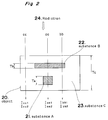
- a radiation 4 comprising three different energy bands E1, E2, and E3 is irradiated to the object 20 being analyzed.
- the transmission intensities values I aa1 , I aa2 , and I aa3 of the radiation 4 comprising three different energy bands E1, E2, and E3 passing through the object 80 at point aa in Fig. 2 can be expressed by equations 9, 10, and 11 below.
- I aa1 I01exp(- ⁇ A1 ⁇ A T A - ⁇ B1 ⁇ B T B - ⁇ C1 ⁇ C T C ') [9]
- I aa2 I02exp(- ⁇ A2 ⁇ A T A - ⁇ B2 ⁇ B T B - ⁇ C2 ⁇ C T C ') [10]
- I aa3 I03exp(- ⁇ A3 ⁇ A T A - ⁇ B3 ⁇ B T B - ⁇ C3 ⁇ C T C ') [11]
- I cc1 I01exp(- ⁇ C1 ⁇ C T C ) [12]
- I cc2 I02exp(- ⁇ C2 ⁇ C T C ) [13]
- I cc3 I03exp(- ⁇ C3 ⁇ C T C ) [14]
- the transmission intensities I bb1 , I bb2 and I bb3 after passing through the object at point bb in Fig. 2 can be expressed by equations 15, 16 and 17 below.
- I bb1 I01exp(- ⁇ B1 ⁇ B T B - ⁇ C1 ⁇ C T C '') [15]
- I bb2 I02exp(- ⁇ B2 ⁇ B T B - ⁇ C2 ⁇ C T C '') [16]
- I bb3 I03exp(- ⁇ B3 ⁇ B T B - ⁇ C3 ⁇ C T C '') [17]
- ⁇ A1 , ⁇ A2 , ⁇ A3 , ⁇ B1 , ⁇ B2 , ⁇ B3 , ⁇ C1 , ⁇ C2 and ⁇ C3 are the mass attenuation coefficients of substances A, B, and C at energy bands E1, E2 and E3, respectively;
- I O1 , I O2 and I O3 are the radiation intensities of the energy bands E1, E2, E3, respectively, irradiated to the object;
- T C ' and T C '' are defined by equations 18 and 19 below.
- T C ' T C - T A - T B [18]
- T C '' T C - T B [19]
- the logarithm of both sides of equations 9 - 11 is obtained and expressed as follows.
- L aa1 - ⁇ A1 ⁇ A T A - ⁇ B1 ⁇ B T B - ⁇ C1 ⁇ C T C ' [20]
- L aa2 - ⁇ A2 ⁇ A T A - ⁇ B2 ⁇ B T B - ⁇ C2 ⁇ C T C ' [21]
- L aa3 - ⁇ A3 ⁇ A T A - ⁇ B3 ⁇ B T B - ⁇ C3 ⁇ C T C ' [22]
- Substance A 21 is not in the radiation transmission path at points cc and bb, and the transmission intensity of the radiation is expressed by equations [12] - [17]. Applying these values in equation [23], m A is 0 at points cc and bb.
- the above method has thus been used to determine the quantities of specific substances in the object.
- an object of the present invention is to provide a method whereby measurement of plural substances in a single analysis sample is possible using a radiation source with fewer energy bands than substances in the sample, and quantitative analysis of plural substances is possible using a single calculation equation.
- the present invention uses the negative of the result calculated by the energy subtraction method to quantify the constituent substances.
- the calculated result of the second substance is defined as the new base, and a third overlapping substance is quantified by obtaining the difference between this base value and the third substance.
- the result of the energy subtraction calculation in equation [7] will be a negative value for that part containing substance B and a positive value for that part containing substance A.
- the third substance can be quantified even when overlapping the first and second substances by evaluating radiation with two energy levels or energy bands at a measurement point containing substances 1 and 2 in the direction of radiation transmission and at a measurement point containing substances 1, 2 and 3. Then, after eliminating the first substance using the energy subtraction method at the transmission intensity of each measurement point, the difference between the calculated result for the third substance and the calculated result for the second substance is obtained to quantify the third substance. In other words, it is possible to quantify another substance in the overlapping area by using as a new calculation base the result of a first energy difference operation at the same point.
- the radiation intensity I1 and I2 of the transmitted energy bands E1 and E2 is calculated by applying the subtraction equation (equation [7]) for each part of the object. More specifically, the transmission intensity of the energy bands is calculated using the difference equation [7] at each part of the object believed to contain substance B 12.
- the transmitted radiation intensity at measurement points X and Y can be expressed by equations 1 - 4.
- I Z2 I02exp(- ⁇ B2 ⁇ B T B - ⁇ C2 ⁇ C T C ') [27]
- the values 1n(I Z1 /I01) and 1n(I Z2 /I02) derived from equations [26] and [27] can be substituted for the values 1n(I X1 /I01) and 1n(I X2 /I02) in equation [7], resulting in equation [28].
- the calculated energy difference result based on equation [7] is a negative value for those areas containing substance B 12 and a positive value for those areas containing substance A 11.
- the substance contained in a given area of the analyzed object can therefore easily be determined by evaluating whether the calculated result of equation [7] is positive or negative.
- the ratio between the low energy attenuation coefficient and the high energy attenuation coefficient increases with the increase in the effective atomic number in an energy area not containing a k absorption edge. It can therefore be concluded that a substance with an atomic number or effective atomic number greater than that of the eliminated substance (substance C 13 in this example) is found in that part of the object for which the calculated energy difference is positive, and a substance with a lower atomic number or effective atomic number is found in that part for which the calculated energy subtraction is negative.
- the density m B per unit area of the substance B 12 is obtained from equation [29]. As these equations show, it is therefore possible to obtain the density of plural substances from a single equation [7]. A practical application for this is in quantifying the amount of bone mineral content and body fat in a given part of the human body. This specific application is described next.
- Fig. 3 is a conceptual cross section of the human hip containing bone 31, muscle 32, and fat 33. The quantities of these tissues are measured using the X-ray transmission and measurement system shown in Fig. 4.
- the primary components of this measurement system are the X-ray source 41, k-edge filter 44, X-ray image sensor 42, calculation unit 43, and image display device 47.
- the X-ray source 41, k-edge filter 44, and X-ray image sensor 42 are held by the support member 48, which is moved by the drive unit 49. The operation of these components is synchronized to scan the subject 45 and measure the two-dimensional transmitted radiation intensity.
- the k-edge filter 44 is made from gadolinium, and is provided directly below the X-ray source 41.
- the fan beam X-ray 46 emitted by the X-ray source 41 is separated into two energy bands by the k-edge filter 44 before irradiating the subject 45. As shown in Fig. 5, when the X-ray tube voltage is 100 kV, the effective energy is divided into two energy bands of 45 keV and 75 keV.
- the X-ray image sensor 42 comprises plural detecting elements, and each detecting element can distinguish the X-ray photon energy bands.
- the X-ray intensity of each energy band is measured by the X-ray image sensor 42 using a photon counting method.
- Fig. 6 shows the results of X-ray photon counting at each part of the subject at the two energy levels.
- the results obtained from the three measurement points shown in Fig. 3 are shown in Table 1.
- X-ray intensity was measured after passing through muscle and bone (point X), muscle only (point Y), and muscle and fat (point Z).
- Table 1 45 keV 75 keV X (bone) 1370 2770 Y (muscle) 3262 4100 Z (fat) 4192 4830
- the density per unit area of bone is the positive value 1.143 g/cm2, and the subtraction calculation results in a muscle value of essentially zero. Note that the value for point Z is negative. It is therefore known that a substance with an attenuation coefficient ratio less than muscle, i.e., a lower effective atomic number, is present in this area.
- the numerical values of the calculated result of m A for the two-dimensional area measured by the X-ray image sensor 42 are colored for presentation on the image display device 47. Negative value areas can be readily identified by the color appearing on the image display, and the observer can immediately determine where the fat is.
- the attenuation coefficient and attenuation coefficient ratio R of fat are shown in Table 4, and the effective atomic numbers of muscle, bone, and fat are shown in Table 5.
- Table 4 45 keV 75 keV ⁇ (fat) 0.2135 0.1768 R 1.2075
- Table 5 Effective atomic number Bone 13.8 Fat 5.92 Muscle 7.42
- both the attenuation coefficient ratio and effective atomic number of fat are lower than those of muscle, the reference substance. This makes it possible to determine that fat is present in those areas for which the result of the subtraction calculation is a negative value.
- the fat density per unit area is calculated by applying equation [29] to those areas for which a negative value is obtained, i.e., those areas now known to contain fat.
- the resulting density through point Z is 4.5529 g/cm2, and the amount of fat is quantified.
- the fat density in the areas of interest can also be calculated.
- the method of the present invention can simultaneously detect and quantify the amount of bone and fat in the body by using the negative values obtained by the subtraction calculation process.
- the object 20 being analyzed comprises three substances A 21, B 22 and C 23, that substances A 21 and B 22 are inside substance C 23, and that substance A 21 partially overlaps substance B 22.
- the objective is to quantify substance A 21. This is possible as follows.
- I aa1 I01exp(- ⁇ A1 ⁇ A T A - ⁇ B1 ⁇ B T B - ⁇ C1 ⁇ C T C '') [30]
- I cc2 I02
- the result at point aa where all three substances, A 21, B 22, and C 23, are present is equal to the sum of the density per unit area of substance A 21 added to the difference result at point bb where both substances B 22 and C 23 are present.
- ⁇ A T A can be obtained by using k as the reference value in Fig. 8.
- the density of substance B can be obtained at point bb using the method described above.
- Fig. 9 is another conceptual cross section of the human hip.
- the body 60 contains bone 61, muscle 62, and fat 63.
- the apparatus shown in Fig. 4 is again used to measure X-ray transmissions through the hip using 45-keV and 75-keV effective energy levels as described above.
- the X-ray intensity of these two energy bands is scanned and measured by the X-ray image sensor 42 using the same photon counting method as above.
- substance A is the bone
- substance B is fat
- substance C is muscle.
- the density per unit area of bone can be obtained as the difference between the value k' at point cc (where both fat 13 and muscle 12 are present) and the measurement obtained from the bone area.
- the X-ray energy band separation method of the invention is not limited to separation using a k-edge filter as described above. It is also possible to change the X-ray tube voltage to generate X-rays at different energy levels, and measure the transmission intensity of the energy bands using an appropriate detector. It is thus possible to perform the same measurements and obtain the density of bone and fat using the same method of calculation.
- the method of the invention provides a substance quantitative analysis method that, by using the negative component in the result of a subtraction calculation process, can simultaneously distinguish and quantify plural substances in a given object using a single equation.
Landscapes
- Health & Medical Sciences (AREA)
- Life Sciences & Earth Sciences (AREA)
- Medical Informatics (AREA)
- Engineering & Computer Science (AREA)
- General Health & Medical Sciences (AREA)
- Pathology (AREA)
- Physics & Mathematics (AREA)
- Animal Behavior & Ethology (AREA)
- Veterinary Medicine (AREA)
- Public Health (AREA)
- Surgery (AREA)
- Biophysics (AREA)
- High Energy & Nuclear Physics (AREA)
- Molecular Biology (AREA)
- Nuclear Medicine, Radiotherapy & Molecular Imaging (AREA)
- Optics & Photonics (AREA)
- Radiology & Medical Imaging (AREA)
- Biomedical Technology (AREA)
- Heart & Thoracic Surgery (AREA)
- Analytical Chemistry (AREA)
- Biochemistry (AREA)
- Chemical & Material Sciences (AREA)
- General Physics & Mathematics (AREA)
- Immunology (AREA)
- Toxicology (AREA)
- Orthopedic Medicine & Surgery (AREA)
- Dentistry (AREA)
- Oral & Maxillofacial Surgery (AREA)
- Apparatus For Radiation Diagnosis (AREA)
- Analysing Materials By The Use Of Radiation (AREA)
Applications Claiming Priority (4)
| Application Number | Priority Date | Filing Date | Title |
|---|---|---|---|
| JP257396/91 | 1991-10-04 | ||
| JP3257396A JP2973643B2 (ja) | 1991-10-04 | 1991-10-04 | 物質の定量測定法 |
| JP333794/91 | 1991-12-18 | ||
| JP33379491 | 1991-12-18 |
Publications (3)
| Publication Number | Publication Date |
|---|---|
| EP0549858A2 true EP0549858A2 (de) | 1993-07-07 |
| EP0549858A3 EP0549858A3 (de) | 1995-01-18 |
| EP0549858B1 EP0549858B1 (de) | 1998-03-25 |
Family
ID=26543196
Family Applications (1)
| Application Number | Title | Priority Date | Filing Date |
|---|---|---|---|
| EP92117069A Expired - Lifetime EP0549858B1 (de) | 1991-10-04 | 1992-10-06 | Verfahren zur quantitativen Stoff-Analyse |
Country Status (3)
| Country | Link |
|---|---|
| US (1) | US5247559A (de) |
| EP (1) | EP0549858B1 (de) |
| DE (1) | DE69224890T2 (de) |
Cited By (5)
| Publication number | Priority date | Publication date | Assignee | Title |
|---|---|---|---|---|
| US5809104A (en) * | 1995-01-12 | 1998-09-15 | Kullenberg; Ragnar | Method and device for measuring the content of bone mineral in the skeleton |
| EP2335057A4 (de) * | 2008-09-08 | 2011-09-28 | Tech Resources Pty Ltd | Verfahren und vorrichtung zur analyse eines materials |
| EP2462431A4 (de) * | 2009-08-04 | 2015-03-18 | Rapiscan Lab Inc | Verfahren und system zur extraktion stereoskopischer informationen aus bildern und kurven |
| CN109047026A (zh) * | 2018-08-02 | 2018-12-21 | 重庆科技学院 | 一种矿石筛选系统及方法 |
| EP3858240A4 (de) * | 2018-11-09 | 2022-06-08 | Canon Kabushiki Kaisha | Informationsverarbeitungsvorrichtung und -verfahren und radiografiesystem |
Families Citing this family (29)
| Publication number | Priority date | Publication date | Assignee | Title |
|---|---|---|---|---|
| GB2316167B (en) * | 1996-08-05 | 2000-06-14 | Framo Eng As | Detection of water constituents |
| AU2887499A (en) * | 1998-03-02 | 1999-09-20 | Image Anaylsis, Inc. | Automated x-ray bone densitometer |
| US6097786A (en) * | 1998-05-18 | 2000-08-01 | Schlumberger Technology Corporation | Method and apparatus for measuring multiphase flows |
| US6510197B1 (en) | 2000-01-11 | 2003-01-21 | Alara, Inc. | Method and apparatus for osteoporosis screening |
| US6449334B1 (en) * | 2000-09-29 | 2002-09-10 | Lunar Corporation | Industrial inspection method and apparatus using dual energy x-ray attenuation |
| WO2002065915A1 (en) * | 2001-02-23 | 2002-08-29 | Koninklijke Philips Electronics N.V. | Method and system for determining a density of a volume in an image data set |
| DE10143131B4 (de) * | 2001-09-03 | 2006-03-09 | Siemens Ag | Verfahren zur Ermittlung von Dichte- und Ordnungszahlverteilungen bei radiographischen Untersuchungsverfahren |
| US6990222B2 (en) * | 2001-11-21 | 2006-01-24 | Arnold Ben A | Calibration of tissue densities in computerized tomography |
| US7295691B2 (en) * | 2002-05-15 | 2007-11-13 | Ge Medical Systems Global Technology Company, Llc | Computer aided diagnosis of an image set |
| FR2841987B1 (fr) * | 2002-07-05 | 2004-08-13 | Commissariat Energie Atomique | Procede d'examen radiologique multi-energie d'un objet |
| US7963695B2 (en) | 2002-07-23 | 2011-06-21 | Rapiscan Systems, Inc. | Rotatable boom cargo scanning system |
| US6922462B2 (en) * | 2002-07-31 | 2005-07-26 | Ge Medical Systems Global Technology Company, Llc | Method, system and computer product for plaque characterization |
| US6891918B2 (en) * | 2002-11-27 | 2005-05-10 | Ge Medical Systems Global Technology Company, Llc | Methods and apparatus for acquiring perfusion data |
| US6987833B2 (en) * | 2003-10-16 | 2006-01-17 | General Electric Company | Methods and apparatus for identification and imaging of specific materials |
| US7684540B2 (en) * | 2006-06-20 | 2010-03-23 | Schlumberger Technology Corporation | Apparatus and method for fluid phase fraction determination using x-rays |
| US7542543B2 (en) * | 2006-09-15 | 2009-06-02 | Schlumberger Technology Corporation | Apparatus and method for well services fluid evaluation using x-rays |
| JP2008229122A (ja) * | 2007-03-22 | 2008-10-02 | Fujifilm Corp | 画像成分分離装置、方法、およびプログラム |
| CN101647706B (zh) * | 2008-08-13 | 2012-05-30 | 清华大学 | 高能双能ct系统的图象重建方法 |
| KR101180698B1 (ko) * | 2010-04-22 | 2012-09-07 | 국방과학연구소 | 탄소/탄소 재료의 내부 밀도 분포 측정 방법 및 그의 표준밀도시편 제작방법 |
| KR101689866B1 (ko) * | 2010-07-29 | 2016-12-27 | 삼성전자주식회사 | 영상 처리 방법 및 장치와 이를 채용한 의료영상시스템 |
| US9218933B2 (en) | 2011-06-09 | 2015-12-22 | Rapidscan Systems, Inc. | Low-dose radiographic imaging system |
| US9521982B2 (en) * | 2011-06-17 | 2016-12-20 | The Board Of Trustees Of The Leland Stanford Junior University | Computed tomography system with dynamic bowtie filter |
| US9414792B2 (en) | 2011-06-17 | 2016-08-16 | The Board Of Trustees Of The Leland Stanford Junior University | Computed tomography system with dynamic bowtie filter |
| US9044186B2 (en) | 2012-06-25 | 2015-06-02 | George W. Ma | Portable dual-energy radiographic X-ray perihpheral bone density and imaging systems and methods |
| JP6305692B2 (ja) * | 2013-05-28 | 2018-04-04 | キヤノンメディカルシステムズ株式会社 | X線診断装置 |
| JP2015180859A (ja) * | 2014-03-05 | 2015-10-15 | 株式会社東芝 | フォトンカウンティングct装置 |
| KR101725099B1 (ko) * | 2014-12-05 | 2017-04-26 | 삼성전자주식회사 | 컴퓨터 단층 촬영장치 및 그 제어방법 |
| JP7054329B2 (ja) | 2017-10-06 | 2022-04-13 | キヤノン株式会社 | 画像処理装置、画像処理方法及びプログラム |
| DE102019111567A1 (de) * | 2019-05-03 | 2020-11-05 | Wipotec Gmbh | Verfahren und Vorrichtung zur Röntgeninspektion von Produkten, insbesondere von Lebensmitteln |
Citations (4)
| Publication number | Priority date | Publication date | Assignee | Title |
|---|---|---|---|---|
| US4662379A (en) * | 1984-12-20 | 1987-05-05 | Stanford University | Coronary artery imaging system using gated tomosynthesis |
| US4686695A (en) * | 1979-02-05 | 1987-08-11 | Board Of Trustees Of The Leland Stanford Junior University | Scanned x-ray selective imaging system |
| EP0385505A2 (de) * | 1989-03-03 | 1990-09-05 | Matsushita Electric Industrial Co., Ltd. | Verfahren zur röntgenologischen Bildverarbeitung und photographisches Bildverarbeitungsgerät dafür |
| US5049746A (en) * | 1989-10-19 | 1991-09-17 | Fuji Photo Film Co., Ltd. | Method and apparatus for displaying energy subtraction images |
Family Cites Families (16)
| Publication number | Priority date | Publication date | Assignee | Title |
|---|---|---|---|---|
| US3848130A (en) * | 1973-06-25 | 1974-11-12 | A Macovski | Selective material x-ray imaging system |
| US4029963A (en) * | 1976-07-30 | 1977-06-14 | The Board Of Trustees Of Leland Stanford Junior University | X-ray spectral decomposition imaging system |
| DE3008261C2 (de) * | 1980-03-04 | 1988-05-05 | Siemens AG, 1000 Berlin und 8000 München | Röntgendiagnostikeinrichtung mit Mitteln zur Bildung eines Transparenzsignals |
| DE3035929C2 (de) * | 1980-09-24 | 1983-08-25 | Gkss - Forschungszentrum Geesthacht Gmbh, 2000 Hamburg | Vorrichtung zur Ermittlung der Volumenanteile eines Mehrkomponentengemisches durch Transmission mehrerer Gammalinien |
| GB2088050A (en) * | 1980-11-25 | 1982-06-03 | Kendall Ernest John Michael | Gamma Ray Analysis of Multi- component Material |
| US4626688A (en) * | 1982-11-26 | 1986-12-02 | Barnes Gary T | Split energy level radiation detection |
| NL8401946A (nl) * | 1984-06-19 | 1986-01-16 | Optische Ind De Oude Delft Nv | Stelsel voor het detecteren van twee roentgenstralingsenergieen. |
| US4980904A (en) * | 1985-11-15 | 1990-12-25 | Picker International, Inc. | Radiation imaging calibration |
| US4953192A (en) * | 1986-04-14 | 1990-08-28 | The University Of Rochester | Scanning equalization radiography |
| US4947414A (en) * | 1986-07-14 | 1990-08-07 | Hologic, Inc. | Bone densitometer |
| US4811373A (en) * | 1986-07-14 | 1989-03-07 | Hologic, Inc. | Bone densitometer |
| US5040199A (en) * | 1986-07-14 | 1991-08-13 | Hologic, Inc. | Apparatus and method for analysis using x-rays |
| SU1385049A1 (ru) * | 1986-08-08 | 1988-03-30 | Всесоюзный научно-исследовательский и конструкторско-технологический институт природных алмазов и инструмента | Способ определени количественного состава композиционных материалов |
| US5033075A (en) * | 1988-05-18 | 1991-07-16 | Rad/Red Laboratories Inc. | Radiation reduction filter for use in medical diagnosis |
| EP0449113B1 (de) * | 1990-03-22 | 1994-10-26 | Matsushita Electric Industrial Co., Ltd. | Verfahren zur Feststellung des Massenanteils eines Target- materials mit Hilfe eines mehrkanaligen Röntgen-Bildsensors |
| JP2827425B2 (ja) * | 1990-03-31 | 1998-11-25 | 株式会社島津製作所 | 骨塩定量装置 |
-
1992
- 1992-10-05 US US07/956,257 patent/US5247559A/en not_active Expired - Lifetime
- 1992-10-06 DE DE69224890T patent/DE69224890T2/de not_active Expired - Fee Related
- 1992-10-06 EP EP92117069A patent/EP0549858B1/de not_active Expired - Lifetime
Patent Citations (4)
| Publication number | Priority date | Publication date | Assignee | Title |
|---|---|---|---|---|
| US4686695A (en) * | 1979-02-05 | 1987-08-11 | Board Of Trustees Of The Leland Stanford Junior University | Scanned x-ray selective imaging system |
| US4662379A (en) * | 1984-12-20 | 1987-05-05 | Stanford University | Coronary artery imaging system using gated tomosynthesis |
| EP0385505A2 (de) * | 1989-03-03 | 1990-09-05 | Matsushita Electric Industrial Co., Ltd. | Verfahren zur röntgenologischen Bildverarbeitung und photographisches Bildverarbeitungsgerät dafür |
| US5049746A (en) * | 1989-10-19 | 1991-09-17 | Fuji Photo Film Co., Ltd. | Method and apparatus for displaying energy subtraction images |
Cited By (7)
| Publication number | Priority date | Publication date | Assignee | Title |
|---|---|---|---|---|
| US5809104A (en) * | 1995-01-12 | 1998-09-15 | Kullenberg; Ragnar | Method and device for measuring the content of bone mineral in the skeleton |
| EP2335057A4 (de) * | 2008-09-08 | 2011-09-28 | Tech Resources Pty Ltd | Verfahren und vorrichtung zur analyse eines materials |
| EP2462431A4 (de) * | 2009-08-04 | 2015-03-18 | Rapiscan Lab Inc | Verfahren und system zur extraktion stereoskopischer informationen aus bildern und kurven |
| US9404875B2 (en) | 2009-08-04 | 2016-08-02 | Rapiscan Systems, Inc. | Method and system for extracting spectroscopic information from images and waveforms |
| CN109047026A (zh) * | 2018-08-02 | 2018-12-21 | 重庆科技学院 | 一种矿石筛选系统及方法 |
| EP3858240A4 (de) * | 2018-11-09 | 2022-06-08 | Canon Kabushiki Kaisha | Informationsverarbeitungsvorrichtung und -verfahren und radiografiesystem |
| US11832983B2 (en) | 2018-11-09 | 2023-12-05 | Canon Kabushiki Kaisha | Information processing apparatus and method, and radiography system |
Also Published As
| Publication number | Publication date |
|---|---|
| EP0549858B1 (de) | 1998-03-25 |
| EP0549858A3 (de) | 1995-01-18 |
| DE69224890T2 (de) | 1998-07-23 |
| US5247559A (en) | 1993-09-21 |
| DE69224890D1 (de) | 1998-04-30 |
Similar Documents
| Publication | Publication Date | Title |
|---|---|---|
| EP0549858A2 (de) | Verfahren zur quantitativen Stoff-Analyse | |
| Dunn et al. | Measurement of bone mineral content in human vertebrae and hip by dual photon absorptiometry. | |
| EP0385505B1 (de) | Verfahren zur röntgenologischen Bildverarbeitung und photographisches Bildverarbeitungsgerät dafür | |
| US7158611B2 (en) | Method for determining density distributions and atomic number distributions during radiographic examination methods | |
| CN101313332B (zh) | 具有多个能窗的计算机断层摄影中的数据处理和分析 | |
| Colbert et al. | Radiographic absorptiometry (photodensitometry) | |
| US7431500B2 (en) | Dynamic exposure control in radiography | |
| Ostlere et al. | Osteoporosis and bone density measurement methods | |
| US5122664A (en) | Method and apparatus for quantitatively analyzing bone calcium | |
| EP0321289A1 (de) | Verfahren zur Messung der Röntgenenergie | |
| EP0137487B1 (de) | Gerät und Strahlungsabbildung mit Zählung von in Energieklassen getrennten Impulsen | |
| JPH08503637A (ja) | カルシウム密度を定量化する装置および方法 | |
| KR20170133450A (ko) | X선 장치, 데이터 처리 장치 및 데이터 처리 방법 | |
| WO1990010859A1 (en) | Apparatus and method for analysis using x-rays | |
| JPH06121791A (ja) | X線定量装置およびx線定量方法 | |
| Soares et al. | Measurement of the linear attenuation coefficient of breast tissues using polienergetic x-ray for energies from 12 to 50 keV and a silicon dispersive detector | |
| US11517278B2 (en) | System and method for basis material decomposition with general physical constraint for multi-energy computed tomography | |
| Strid et al. | Bone mass determination from microradiographs by computer-assisted videodensitometry: I. Methodology | |
| JP2973643B2 (ja) | 物質の定量測定法 | |
| EP0211956A1 (de) | Tomographie-einheit mit rechner | |
| JPH05237081A (ja) | 物質の定量測定装置 | |
| Wanner et al. | Methods and application of bone densitometry in clinical diagnosis | |
| US5809104A (en) | Method and device for measuring the content of bone mineral in the skeleton | |
| JP3030950B2 (ja) | 校正用ファントムおよびデータ校正法 | |
| JP2627098B2 (ja) | 骨塩定量分析方法および装置 |
Legal Events
| Date | Code | Title | Description |
|---|---|---|---|
| PUAI | Public reference made under article 153(3) epc to a published international application that has entered the european phase |
Free format text: ORIGINAL CODE: 0009012 |
|
| 17P | Request for examination filed |
Effective date: 19921006 |
|
| AK | Designated contracting states |
Kind code of ref document: A2 Designated state(s): DE FR GB |
|
| PUAL | Search report despatched |
Free format text: ORIGINAL CODE: 0009013 |
|
| AK | Designated contracting states |
Kind code of ref document: A3 Designated state(s): DE FR GB |
|
| GRAG | Despatch of communication of intention to grant |
Free format text: ORIGINAL CODE: EPIDOS AGRA |
|
| RAP1 | Party data changed (applicant data changed or rights of an application transferred) |
Owner name: MATSUSHITA ELECTRIC INDUSTRIAL CO., LTD. |
|
| 17Q | First examination report despatched |
Effective date: 19970325 |
|
| GRAG | Despatch of communication of intention to grant |
Free format text: ORIGINAL CODE: EPIDOS AGRA |
|
| GRAG | Despatch of communication of intention to grant |
Free format text: ORIGINAL CODE: EPIDOS AGRA |
|
| GRAH | Despatch of communication of intention to grant a patent |
Free format text: ORIGINAL CODE: EPIDOS IGRA |
|
| GRAH | Despatch of communication of intention to grant a patent |
Free format text: ORIGINAL CODE: EPIDOS IGRA |
|
| GRAA | (expected) grant |
Free format text: ORIGINAL CODE: 0009210 |
|
| AK | Designated contracting states |
Kind code of ref document: B1 Designated state(s): DE FR GB |
|
| ET | Fr: translation filed | ||
| REF | Corresponds to: |
Ref document number: 69224890 Country of ref document: DE Date of ref document: 19980430 |
|
| PLBE | No opposition filed within time limit |
Free format text: ORIGINAL CODE: 0009261 |
|
| STAA | Information on the status of an ep patent application or granted ep patent |
Free format text: STATUS: NO OPPOSITION FILED WITHIN TIME LIMIT |
|
| 26N | No opposition filed | ||
| REG | Reference to a national code |
Ref country code: GB Ref legal event code: IF02 |
|
| PGFP | Annual fee paid to national office [announced via postgrant information from national office to epo] |
Ref country code: DE Payment date: 20060928 Year of fee payment: 15 |
|
| PGFP | Annual fee paid to national office [announced via postgrant information from national office to epo] |
Ref country code: GB Payment date: 20061004 Year of fee payment: 15 |
|
| GBPC | Gb: european patent ceased through non-payment of renewal fee |
Effective date: 20071006 |
|
| PG25 | Lapsed in a contracting state [announced via postgrant information from national office to epo] |
Ref country code: DE Free format text: LAPSE BECAUSE OF NON-PAYMENT OF DUE FEES Effective date: 20080501 |
|
| REG | Reference to a national code |
Ref country code: FR Ref legal event code: ST Effective date: 20080630 |
|
| PGFP | Annual fee paid to national office [announced via postgrant information from national office to epo] |
Ref country code: FR Payment date: 20061010 Year of fee payment: 15 |
|
| PG25 | Lapsed in a contracting state [announced via postgrant information from national office to epo] |
Ref country code: GB Free format text: LAPSE BECAUSE OF NON-PAYMENT OF DUE FEES Effective date: 20071006 |
|
| PG25 | Lapsed in a contracting state [announced via postgrant information from national office to epo] |
Ref country code: FR Free format text: LAPSE BECAUSE OF NON-PAYMENT OF DUE FEES Effective date: 20071031 |