EP0474313B1 - Nucleotide sequence coding for an outer membrane protein from Neisseria meningitidis and use of said protein in vaccine preparations - Google Patents
Nucleotide sequence coding for an outer membrane protein from Neisseria meningitidis and use of said protein in vaccine preparations Download PDFInfo
- Publication number
- EP0474313B1 EP0474313B1 EP91202291A EP91202291A EP0474313B1 EP 0474313 B1 EP0474313 B1 EP 0474313B1 EP 91202291 A EP91202291 A EP 91202291A EP 91202291 A EP91202291 A EP 91202291A EP 0474313 B1 EP0474313 B1 EP 0474313B1
- Authority
- EP
- European Patent Office
- Prior art keywords
- protein
- meningitidis
- nucleotide sequence
- amino acid
- acid sequence
- Prior art date
- Legal status (The legal status is an assumption and is not a legal conclusion. Google has not performed a legal analysis and makes no representation as to the accuracy of the status listed.)
- Expired - Lifetime
Links
- 108090000623 proteins and genes Proteins 0.000 title claims description 147
- 102000004169 proteins and genes Human genes 0.000 title claims description 120
- 241000588650 Neisseria meningitidis Species 0.000 title claims description 65
- 229960005486 vaccine Drugs 0.000 title claims description 28
- 239000002773 nucleotide Substances 0.000 title claims description 25
- 125000003729 nucleotide group Chemical group 0.000 title claims description 25
- 238000002360 preparation method Methods 0.000 title description 21
- 101710116435 Outer membrane protein Proteins 0.000 title description 3
- 241000588724 Escherichia coli Species 0.000 claims description 21
- 244000005700 microbiome Species 0.000 claims description 19
- 229920001282 polysaccharide Polymers 0.000 claims description 18
- 239000005017 polysaccharide Substances 0.000 claims description 18
- 150000004676 glycans Chemical class 0.000 claims description 16
- 108020001507 fusion proteins Proteins 0.000 claims description 14
- 102000037865 fusion proteins Human genes 0.000 claims description 14
- 239000000203 mixture Substances 0.000 claims description 11
- 239000013604 expression vector Substances 0.000 claims description 8
- 108091033319 polynucleotide Proteins 0.000 claims description 7
- 102000040430 polynucleotide Human genes 0.000 claims description 7
- 239000002157 polynucleotide Substances 0.000 claims description 7
- 239000000126 substance Substances 0.000 claims description 7
- 108010008281 Recombinant Fusion Proteins Proteins 0.000 claims description 4
- 102000007056 Recombinant Fusion Proteins Human genes 0.000 claims description 4
- 238000010367 cloning Methods 0.000 claims description 3
- 239000002671 adjuvant Substances 0.000 claims description 2
- 239000013599 cloning vector Substances 0.000 claims description 2
- 238000012258 culturing Methods 0.000 claims description 2
- 239000003085 diluting agent Substances 0.000 claims description 2
- 238000004519 manufacturing process Methods 0.000 claims description 2
- 230000001131 transforming effect Effects 0.000 claims description 2
- 125000003275 alpha amino acid group Chemical group 0.000 claims 6
- 235000018102 proteins Nutrition 0.000 description 104
- 150000001413 amino acids Chemical group 0.000 description 30
- 235000001014 amino acid Nutrition 0.000 description 23
- 241000588653 Neisseria Species 0.000 description 18
- 230000000844 anti-bacterial effect Effects 0.000 description 18
- 238000000034 method Methods 0.000 description 15
- 230000001717 pathogenic effect Effects 0.000 description 15
- 239000000427 antigen Substances 0.000 description 12
- 102000036639 antigens Human genes 0.000 description 12
- 108091007433 antigens Proteins 0.000 description 12
- 238000010276 construction Methods 0.000 description 12
- 230000000875 corresponding effect Effects 0.000 description 12
- 210000004379 membrane Anatomy 0.000 description 11
- 239000012528 membrane Substances 0.000 description 11
- 239000012634 fragment Substances 0.000 description 10
- 239000013612 plasmid Substances 0.000 description 9
- 108020004414 DNA Proteins 0.000 description 8
- 241000894006 Bacteria Species 0.000 description 7
- 241000282412 Homo Species 0.000 description 7
- 210000004027 cell Anatomy 0.000 description 7
- 230000004927 fusion Effects 0.000 description 7
- 210000002966 serum Anatomy 0.000 description 7
- 238000001262 western blot Methods 0.000 description 7
- 102000005421 acetyltransferase Human genes 0.000 description 6
- 108020002494 acetyltransferase Proteins 0.000 description 6
- 239000002158 endotoxin Substances 0.000 description 6
- 230000005847 immunogenicity Effects 0.000 description 6
- 230000004044 response Effects 0.000 description 6
- 102000028526 Dihydrolipoamide Dehydrogenase Human genes 0.000 description 5
- 108010028127 Dihydrolipoamide Dehydrogenase Proteins 0.000 description 5
- 241001131785 Escherichia coli HB101 Species 0.000 description 5
- 241000699670 Mus sp. Species 0.000 description 5
- 241000283973 Oryctolagus cuniculus Species 0.000 description 5
- TXWRERCHRDBNLG-UHFFFAOYSA-N cubane Chemical compound C12C3C4C1C1C4C3C12 TXWRERCHRDBNLG-UHFFFAOYSA-N 0.000 description 5
- 238000010790 dilution Methods 0.000 description 5
- 239000012895 dilution Substances 0.000 description 5
- 201000010099 disease Diseases 0.000 description 5
- 208000037265 diseases, disorders, signs and symptoms Diseases 0.000 description 5
- 239000000047 product Substances 0.000 description 5
- 238000002965 ELISA Methods 0.000 description 4
- 102000004190 Enzymes Human genes 0.000 description 4
- 108090000790 Enzymes Proteins 0.000 description 4
- XEEYBQQBJWHFJM-UHFFFAOYSA-N Iron Chemical compound [Fe] XEEYBQQBJWHFJM-UHFFFAOYSA-N 0.000 description 4
- 101710107904 NADH-ubiquinone oxidoreductase subunit 9 Proteins 0.000 description 4
- 241000588652 Neisseria gonorrhoeae Species 0.000 description 4
- 101710132845 Protein P1 Proteins 0.000 description 4
- 230000000890 antigenic effect Effects 0.000 description 4
- 229940031416 bivalent vaccine Drugs 0.000 description 4
- 229940088598 enzyme Drugs 0.000 description 4
- 230000002068 genetic effect Effects 0.000 description 4
- 230000003053 immunization Effects 0.000 description 4
- 238000002649 immunization Methods 0.000 description 4
- 229920006008 lipopolysaccharide Polymers 0.000 description 4
- 238000012360 testing method Methods 0.000 description 4
- WEVYAHXRMPXWCK-UHFFFAOYSA-N Acetonitrile Chemical compound CC#N WEVYAHXRMPXWCK-UHFFFAOYSA-N 0.000 description 3
- QIVBCDIJIAJPQS-VIFPVBQESA-N L-tryptophane Chemical compound C1=CC=C2C(C[C@H](N)C(O)=O)=CNC2=C1 QIVBCDIJIAJPQS-VIFPVBQESA-N 0.000 description 3
- OKKJLVBELUTLKV-UHFFFAOYSA-N Methanol Chemical compound OC OKKJLVBELUTLKV-UHFFFAOYSA-N 0.000 description 3
- 241000831652 Salinivibrio sharmensis Species 0.000 description 3
- QIVBCDIJIAJPQS-UHFFFAOYSA-N Tryptophan Natural products C1=CC=C2C(CC(N)C(O)=O)=CNC2=C1 QIVBCDIJIAJPQS-UHFFFAOYSA-N 0.000 description 3
- XAGFODPZIPBFFR-UHFFFAOYSA-N aluminium Chemical compound [Al] XAGFODPZIPBFFR-UHFFFAOYSA-N 0.000 description 3
- 229910052782 aluminium Inorganic materials 0.000 description 3
- BTANRVKWQNVYAZ-UHFFFAOYSA-N butan-2-ol Chemical compound CCC(C)O BTANRVKWQNVYAZ-UHFFFAOYSA-N 0.000 description 3
- 230000015556 catabolic process Effects 0.000 description 3
- 230000009260 cross reactivity Effects 0.000 description 3
- 238000006731 degradation reaction Methods 0.000 description 3
- 238000002474 experimental method Methods 0.000 description 3
- 239000000499 gel Substances 0.000 description 3
- 208000002672 hepatitis B Diseases 0.000 description 3
- 238000004128 high performance liquid chromatography Methods 0.000 description 3
- 238000011534 incubation Methods 0.000 description 3
- 125000003977 lipoyl group Chemical group S1SC(C([H])([H])C(C(C(C(=O)[*])([H])[H])([H])[H])([H])[H])([H])C([H])([H])C1([H])[H] 0.000 description 3
- 238000003752 polymerase chain reaction Methods 0.000 description 3
- 238000000926 separation method Methods 0.000 description 3
- 239000000243 solution Substances 0.000 description 3
- 239000006228 supernatant Substances 0.000 description 3
- 229960004799 tryptophan Drugs 0.000 description 3
- 239000013598 vector Substances 0.000 description 3
- 102000004594 DNA Polymerase I Human genes 0.000 description 2
- 108010017826 DNA Polymerase I Proteins 0.000 description 2
- KCXVZYZYPLLWCC-UHFFFAOYSA-N EDTA Chemical compound OC(=O)CN(CC(O)=O)CCN(CC(O)=O)CC(O)=O KCXVZYZYPLLWCC-UHFFFAOYSA-N 0.000 description 2
- 101001121580 Enterobacteria phage PRD1 Adsorption protein P2 Proteins 0.000 description 2
- 241000606768 Haemophilus influenzae Species 0.000 description 2
- 241000282414 Homo sapiens Species 0.000 description 2
- 241001465754 Metazoa Species 0.000 description 2
- 241000588677 Neisseria meningitidis serogroup B Species 0.000 description 2
- 101001125164 Parietaria judaica Probable non-specific lipid-transfer protein 2 Proteins 0.000 description 2
- ISWSIDIOOBJBQZ-UHFFFAOYSA-N Phenol Chemical compound OC1=CC=CC=C1 ISWSIDIOOBJBQZ-UHFFFAOYSA-N 0.000 description 2
- 101000580771 Pseudomonas phage phi6 RNA-directed RNA polymerase Proteins 0.000 description 2
- 101001121571 Rice tungro bacilliform virus (isolate Philippines) Protein P2 Proteins 0.000 description 2
- VMHLLURERBWHNL-UHFFFAOYSA-M Sodium acetate Chemical compound [Na+].CC([O-])=O VMHLLURERBWHNL-UHFFFAOYSA-M 0.000 description 2
- BFNBIHQBYMNNAN-UHFFFAOYSA-N ammonium sulfate Chemical compound N.N.OS(O)(=O)=O BFNBIHQBYMNNAN-UHFFFAOYSA-N 0.000 description 2
- 229910052921 ammonium sulfate Inorganic materials 0.000 description 2
- 239000001166 ammonium sulphate Substances 0.000 description 2
- 235000011130 ammonium sulphate Nutrition 0.000 description 2
- 230000001580 bacterial effect Effects 0.000 description 2
- 239000000872 buffer Substances 0.000 description 2
- 210000004899 c-terminal region Anatomy 0.000 description 2
- 239000000356 contaminant Substances 0.000 description 2
- 238000011156 evaluation Methods 0.000 description 2
- 229940047650 haemophilus influenzae Drugs 0.000 description 2
- 210000004408 hybridoma Anatomy 0.000 description 2
- 230000001900 immune effect Effects 0.000 description 2
- 230000002163 immunogen Effects 0.000 description 2
- 239000000411 inducer Substances 0.000 description 2
- 208000015181 infectious disease Diseases 0.000 description 2
- 229910052742 iron Inorganic materials 0.000 description 2
- 238000010369 molecular cloning Methods 0.000 description 2
- 244000052769 pathogen Species 0.000 description 2
- 229920002981 polyvinylidene fluoride Polymers 0.000 description 2
- 239000002244 precipitate Substances 0.000 description 2
- 230000008569 process Effects 0.000 description 2
- 238000012545 processing Methods 0.000 description 2
- 230000004224 protection Effects 0.000 description 2
- 239000002510 pyrogen Substances 0.000 description 2
- 230000003248 secreting effect Effects 0.000 description 2
- 239000001632 sodium acetate Substances 0.000 description 2
- 235000017281 sodium acetate Nutrition 0.000 description 2
- JQWHASGSAFIOCM-UHFFFAOYSA-M sodium periodate Chemical compound [Na+].[O-]I(=O)(=O)=O JQWHASGSAFIOCM-UHFFFAOYSA-M 0.000 description 2
- 238000001228 spectrum Methods 0.000 description 2
- 238000013518 transcription Methods 0.000 description 2
- 230000035897 transcription Effects 0.000 description 2
- 238000002255 vaccination Methods 0.000 description 2
- AZXKALLRCOCGBV-UHFFFAOYSA-N 1-phenyl-2-(propan-2-ylamino)hexan-1-one Chemical compound CCCCC(NC(C)C)C(=O)C1=CC=CC=C1 AZXKALLRCOCGBV-UHFFFAOYSA-N 0.000 description 1
- PQMRRAQXKWFYQN-UHFFFAOYSA-N 1-phenyl-2-sulfanylideneimidazolidin-4-one Chemical class S=C1NC(=O)CN1C1=CC=CC=C1 PQMRRAQXKWFYQN-UHFFFAOYSA-N 0.000 description 1
- HZAXFHJVJLSVMW-UHFFFAOYSA-N 2-Aminoethan-1-ol Chemical compound NCCO HZAXFHJVJLSVMW-UHFFFAOYSA-N 0.000 description 1
- 229930024421 Adenine Natural products 0.000 description 1
- GFFGJBXGBJISGV-UHFFFAOYSA-N Adenine Chemical compound NC1=NC=NC2=C1N=CN2 GFFGJBXGBJISGV-UHFFFAOYSA-N 0.000 description 1
- 244000063299 Bacillus subtilis Species 0.000 description 1
- 235000014469 Bacillus subtilis Nutrition 0.000 description 1
- 239000002028 Biomass Substances 0.000 description 1
- 108091026890 Coding region Proteins 0.000 description 1
- VJDOAZKNBQCAGE-LMVFSUKVSA-N D-ribitol 5-phosphate Chemical compound OC[C@H](O)[C@H](O)[C@H](O)COP(O)(O)=O VJDOAZKNBQCAGE-LMVFSUKVSA-N 0.000 description 1
- 101710108485 Envelope phospholipase F13 Proteins 0.000 description 1
- 241000701959 Escherichia virus Lambda Species 0.000 description 1
- LFQSCWFLJHTTHZ-UHFFFAOYSA-N Ethanol Chemical compound CCO LFQSCWFLJHTTHZ-UHFFFAOYSA-N 0.000 description 1
- 241000976806 Genea <ascomycete fungus> Species 0.000 description 1
- 241000700721 Hepatitis B virus Species 0.000 description 1
- 201000009906 Meningitis Diseases 0.000 description 1
- 206010058858 Meningococcal bacteraemia Diseases 0.000 description 1
- 102000007474 Multiprotein Complexes Human genes 0.000 description 1
- 108010085220 Multiprotein Complexes Proteins 0.000 description 1
- 102000016943 Muramidase Human genes 0.000 description 1
- 108010014251 Muramidase Proteins 0.000 description 1
- 108010062010 N-Acetylmuramoyl-L-alanine Amidase Proteins 0.000 description 1
- 241000588654 Neisseria cinerea Species 0.000 description 1
- 241000588651 Neisseria flavescens Species 0.000 description 1
- 241000588649 Neisseria lactamica Species 0.000 description 1
- 241000947238 Neisseria meningitidis serogroup C Species 0.000 description 1
- 241000588659 Neisseria mucosa Species 0.000 description 1
- 241000588645 Neisseria sicca Species 0.000 description 1
- 241001136170 Neisseria subflava Species 0.000 description 1
- 101710163270 Nuclease Proteins 0.000 description 1
- 108091028043 Nucleic acid sequence Proteins 0.000 description 1
- 108010079246 OMPA outer membrane proteins Proteins 0.000 description 1
- 239000002033 PVDF binder Substances 0.000 description 1
- 108020002230 Pancreatic Ribonuclease Proteins 0.000 description 1
- 102000005891 Pancreatic ribonuclease Human genes 0.000 description 1
- 108091005804 Peptidases Proteins 0.000 description 1
- 239000004793 Polystyrene Substances 0.000 description 1
- 108010059712 Pronase Proteins 0.000 description 1
- 239000004365 Protease Substances 0.000 description 1
- 102000012751 Pyruvate Dehydrogenase Complex Human genes 0.000 description 1
- 108010090051 Pyruvate Dehydrogenase Complex Proteins 0.000 description 1
- 108020004511 Recombinant DNA Proteins 0.000 description 1
- 102100037486 Reverse transcriptase/ribonuclease H Human genes 0.000 description 1
- JVWLUVNSQYXYBE-UHFFFAOYSA-N Ribitol Natural products OCC(C)C(O)C(O)CO JVWLUVNSQYXYBE-UHFFFAOYSA-N 0.000 description 1
- 238000002105 Southern blotting Methods 0.000 description 1
- 108010008038 Synthetic Vaccines Proteins 0.000 description 1
- 239000007983 Tris buffer Substances 0.000 description 1
- 239000006035 Tryptophane Substances 0.000 description 1
- 229960000643 adenine Drugs 0.000 description 1
- 230000002411 adverse Effects 0.000 description 1
- 239000011543 agarose gel Substances 0.000 description 1
- 125000003172 aldehyde group Chemical group 0.000 description 1
- 239000004411 aluminium Substances 0.000 description 1
- 229910021502 aluminium hydroxide Inorganic materials 0.000 description 1
- WNROFYMDJYEPJX-UHFFFAOYSA-K aluminium hydroxide Chemical compound [OH-].[OH-].[OH-].[Al+3] WNROFYMDJYEPJX-UHFFFAOYSA-K 0.000 description 1
- 238000004458 analytical method Methods 0.000 description 1
- 230000005875 antibody response Effects 0.000 description 1
- 244000052616 bacterial pathogen Species 0.000 description 1
- 229960001212 bacterial vaccine Drugs 0.000 description 1
- 239000003855 balanced salt solution Substances 0.000 description 1
- 210000004369 blood Anatomy 0.000 description 1
- 239000008280 blood Substances 0.000 description 1
- 230000037396 body weight Effects 0.000 description 1
- 210000004556 brain Anatomy 0.000 description 1
- 244000309466 calf Species 0.000 description 1
- 238000007385 chemical modification Methods 0.000 description 1
- 238000006243 chemical reaction Methods 0.000 description 1
- YTRQFSDWAXHJCC-UHFFFAOYSA-N chloroform;phenol Chemical compound ClC(Cl)Cl.OC1=CC=CC=C1 YTRQFSDWAXHJCC-UHFFFAOYSA-N 0.000 description 1
- 230000001332 colony forming effect Effects 0.000 description 1
- 230000000295 complement effect Effects 0.000 description 1
- 230000021615 conjugation Effects 0.000 description 1
- 239000000470 constituent Substances 0.000 description 1
- 238000011109 contamination Methods 0.000 description 1
- 229940028617 conventional vaccine Drugs 0.000 description 1
- 230000002596 correlated effect Effects 0.000 description 1
- 238000005520 cutting process Methods 0.000 description 1
- 238000012217 deletion Methods 0.000 description 1
- 230000037430 deletion Effects 0.000 description 1
- 239000003599 detergent Substances 0.000 description 1
- 238000002405 diagnostic procedure Methods 0.000 description 1
- 238000000502 dialysis Methods 0.000 description 1
- 230000004069 differentiation Effects 0.000 description 1
- 230000029087 digestion Effects 0.000 description 1
- 230000003292 diminished effect Effects 0.000 description 1
- 230000000694 effects Effects 0.000 description 1
- 238000001493 electron microscopy Methods 0.000 description 1
- 108010052305 exodeoxyribonuclease III Proteins 0.000 description 1
- 230000002349 favourable effect Effects 0.000 description 1
- 238000000855 fermentation Methods 0.000 description 1
- 230000004151 fermentation Effects 0.000 description 1
- 230000001605 fetal effect Effects 0.000 description 1
- 238000001641 gel filtration chromatography Methods 0.000 description 1
- 238000010353 genetic engineering Methods 0.000 description 1
- 229910001679 gibbsite Inorganic materials 0.000 description 1
- 239000001963 growth medium Substances 0.000 description 1
- 108010046119 hemolin Proteins 0.000 description 1
- HNDVDQJCIGZPNO-UHFFFAOYSA-N histidine Natural products OC(=O)C(N)CC1=CN=CN1 HNDVDQJCIGZPNO-UHFFFAOYSA-N 0.000 description 1
- 125000000487 histidyl group Chemical group [H]N([H])C(C(=O)O*)C([H])([H])C1=C([H])N([H])C([H])=N1 0.000 description 1
- 238000009396 hybridization Methods 0.000 description 1
- 230000000521 hyperimmunizing effect Effects 0.000 description 1
- 230000028993 immune response Effects 0.000 description 1
- 230000002480 immunoprotective effect Effects 0.000 description 1
- 230000006872 improvement Effects 0.000 description 1
- 230000005764 inhibitory process Effects 0.000 description 1
- 230000003834 intracellular effect Effects 0.000 description 1
- 238000002955 isolation Methods 0.000 description 1
- 230000002045 lasting effect Effects 0.000 description 1
- 230000000670 limiting effect Effects 0.000 description 1
- GZQKNULLWNGMCW-PWQABINMSA-N lipid A (E. coli) Chemical compound O1[C@H](CO)[C@@H](OP(O)(O)=O)[C@H](OC(=O)C[C@@H](CCCCCCCCCCC)OC(=O)CCCCCCCCCCCCC)[C@@H](NC(=O)C[C@@H](CCCCCCCCCCC)OC(=O)CCCCCCCCCCC)[C@@H]1OC[C@@H]1[C@@H](O)[C@H](OC(=O)C[C@H](O)CCCCCCCCCCC)[C@@H](NC(=O)C[C@H](O)CCCCCCCCCCC)[C@@H](OP(O)(O)=O)O1 GZQKNULLWNGMCW-PWQABINMSA-N 0.000 description 1
- 230000004807 localization Effects 0.000 description 1
- 125000003588 lysine group Chemical group [H]N([H])C([H])([H])C([H])([H])C([H])([H])C([H])([H])C([H])(N([H])[H])C(*)=O 0.000 description 1
- 239000004325 lysozyme Substances 0.000 description 1
- 229960000274 lysozyme Drugs 0.000 description 1
- 235000010335 lysozyme Nutrition 0.000 description 1
- 239000002609 medium Substances 0.000 description 1
- 208000022089 meningococcemia Diseases 0.000 description 1
- HEBKCHPVOIAQTA-UHFFFAOYSA-N meso ribitol Natural products OCC(O)C(O)C(O)CO HEBKCHPVOIAQTA-UHFFFAOYSA-N 0.000 description 1
- 230000004060 metabolic process Effects 0.000 description 1
- 235000013336 milk Nutrition 0.000 description 1
- 239000008267 milk Substances 0.000 description 1
- 210000004080 milk Anatomy 0.000 description 1
- 238000002156 mixing Methods 0.000 description 1
- 238000012986 modification Methods 0.000 description 1
- 230000004048 modification Effects 0.000 description 1
- 230000002969 morbid Effects 0.000 description 1
- 230000001459 mortal effect Effects 0.000 description 1
- 229940126619 mouse monoclonal antibody Drugs 0.000 description 1
- 210000004400 mucous membrane Anatomy 0.000 description 1
- 210000004897 n-terminal region Anatomy 0.000 description 1
- 239000013642 negative control Substances 0.000 description 1
- 231100000252 nontoxic Toxicity 0.000 description 1
- 230000003000 nontoxic effect Effects 0.000 description 1
- 238000007899 nucleic acid hybridization Methods 0.000 description 1
- 108020004707 nucleic acids Proteins 0.000 description 1
- 102000039446 nucleic acids Human genes 0.000 description 1
- 150000007523 nucleic acids Chemical class 0.000 description 1
- 229920001542 oligosaccharide Polymers 0.000 description 1
- 150000002482 oligosaccharides Chemical class 0.000 description 1
- 230000036961 partial effect Effects 0.000 description 1
- KHIWWQKSHDUIBK-UHFFFAOYSA-N periodic acid Chemical compound OI(=O)(=O)=O KHIWWQKSHDUIBK-UHFFFAOYSA-N 0.000 description 1
- 239000012071 phase Substances 0.000 description 1
- 150000003904 phospholipids Chemical class 0.000 description 1
- 239000000902 placebo Substances 0.000 description 1
- 229940068196 placebo Drugs 0.000 description 1
- 239000013600 plasmid vector Substances 0.000 description 1
- 229920002223 polystyrene Polymers 0.000 description 1
- 239000013641 positive control Substances 0.000 description 1
- 150000003141 primary amines Chemical class 0.000 description 1
- 108090000765 processed proteins & peptides Proteins 0.000 description 1
- 230000001681 protective effect Effects 0.000 description 1
- 238000000746 purification Methods 0.000 description 1
- 229940124551 recombinant vaccine Drugs 0.000 description 1
- 238000006268 reductive amination reaction Methods 0.000 description 1
- 108091008146 restriction endonucleases Proteins 0.000 description 1
- 230000002441 reversible effect Effects 0.000 description 1
- HEBKCHPVOIAQTA-ZXFHETKHSA-N ribitol Chemical compound OC[C@H](O)[C@H](O)[C@H](O)CO HEBKCHPVOIAQTA-ZXFHETKHSA-N 0.000 description 1
- 238000013341 scale-up Methods 0.000 description 1
- 230000009291 secondary effect Effects 0.000 description 1
- 230000035945 sensitivity Effects 0.000 description 1
- 238000012163 sequencing technique Methods 0.000 description 1
- 230000000405 serological effect Effects 0.000 description 1
- 150000003384 small molecules Chemical class 0.000 description 1
- BEOOHQFXGBMRKU-UHFFFAOYSA-N sodium cyanoborohydride Chemical compound [Na+].[B-]C#N BEOOHQFXGBMRKU-UHFFFAOYSA-N 0.000 description 1
- 239000007790 solid phase Substances 0.000 description 1
- 241000894007 species Species 0.000 description 1
- 230000000087 stabilizing effect Effects 0.000 description 1
- 238000010561 standard procedure Methods 0.000 description 1
- 238000003756 stirring Methods 0.000 description 1
- 230000001629 suppression Effects 0.000 description 1
- 239000000725 suspension Substances 0.000 description 1
- 229940031351 tetravalent vaccine Drugs 0.000 description 1
- 230000009466 transformation Effects 0.000 description 1
- 238000013519 translation Methods 0.000 description 1
- LENZDBCJOHFCAS-UHFFFAOYSA-N tris Chemical compound OCC(N)(CO)CO LENZDBCJOHFCAS-UHFFFAOYSA-N 0.000 description 1
- 229940125575 vaccine candidate Drugs 0.000 description 1
- 230000035899 viability Effects 0.000 description 1
- XLYOFNOQVPJJNP-UHFFFAOYSA-N water Substances O XLYOFNOQVPJJNP-UHFFFAOYSA-N 0.000 description 1
- 230000003442 weekly effect Effects 0.000 description 1
Images


Classifications
-
- C—CHEMISTRY; METALLURGY
- C12—BIOCHEMISTRY; BEER; SPIRITS; WINE; VINEGAR; MICROBIOLOGY; ENZYMOLOGY; MUTATION OR GENETIC ENGINEERING
- C12Y—ENZYMES
- C12Y108/00—Oxidoreductases acting on sulfur groups as donors (1.8)
- C12Y108/01—Oxidoreductases acting on sulfur groups as donors (1.8) with NAD+ or NADP+ as acceptor (1.8.1)
- C12Y108/01004—Dihydrolipoyl dehydrogenase (1.8.1.4), i.e. lipoamide-dehydrogenase
-
- A—HUMAN NECESSITIES
- A61—MEDICAL OR VETERINARY SCIENCE; HYGIENE
- A61P—SPECIFIC THERAPEUTIC ACTIVITY OF CHEMICAL COMPOUNDS OR MEDICINAL PREPARATIONS
- A61P31/00—Antiinfectives, i.e. antibiotics, antiseptics, chemotherapeutics
- A61P31/04—Antibacterial agents
-
- C—CHEMISTRY; METALLURGY
- C07—ORGANIC CHEMISTRY
- C07K—PEPTIDES
- C07K14/00—Peptides having more than 20 amino acids; Gastrins; Somatostatins; Melanotropins; Derivatives thereof
- C07K14/195—Peptides having more than 20 amino acids; Gastrins; Somatostatins; Melanotropins; Derivatives thereof from bacteria
- C07K14/22—Peptides having more than 20 amino acids; Gastrins; Somatostatins; Melanotropins; Derivatives thereof from bacteria from Neisseriaceae (F)
-
- C—CHEMISTRY; METALLURGY
- C07—ORGANIC CHEMISTRY
- C07K—PEPTIDES
- C07K16/00—Immunoglobulins [IGs], e.g. monoclonal or polyclonal antibodies
- C07K16/12—Immunoglobulins [IGs], e.g. monoclonal or polyclonal antibodies against material from bacteria
- C07K16/1203—Immunoglobulins [IGs], e.g. monoclonal or polyclonal antibodies against material from bacteria from Gram-negative bacteria
- C07K16/1217—Immunoglobulins [IGs], e.g. monoclonal or polyclonal antibodies against material from bacteria from Gram-negative bacteria from Neisseriaceae (F)
-
- C—CHEMISTRY; METALLURGY
- C07—ORGANIC CHEMISTRY
- C07K—PEPTIDES
- C07K16/00—Immunoglobulins [IGs], e.g. monoclonal or polyclonal antibodies
- C07K16/40—Immunoglobulins [IGs], e.g. monoclonal or polyclonal antibodies against enzymes
-
- C—CHEMISTRY; METALLURGY
- C12—BIOCHEMISTRY; BEER; SPIRITS; WINE; VINEGAR; MICROBIOLOGY; ENZYMOLOGY; MUTATION OR GENETIC ENGINEERING
- C12N—MICROORGANISMS OR ENZYMES; COMPOSITIONS THEREOF; PROPAGATING, PRESERVING, OR MAINTAINING MICROORGANISMS; MUTATION OR GENETIC ENGINEERING; CULTURE MEDIA
- C12N9/00—Enzymes; Proenzymes; Compositions thereof; Processes for preparing, activating, inhibiting, separating or purifying enzymes
- C12N9/0004—Oxidoreductases (1.)
- C12N9/0051—Oxidoreductases (1.) acting on a sulfur group of donors (1.8)
-
- C—CHEMISTRY; METALLURGY
- C12—BIOCHEMISTRY; BEER; SPIRITS; WINE; VINEGAR; MICROBIOLOGY; ENZYMOLOGY; MUTATION OR GENETIC ENGINEERING
- C12N—MICROORGANISMS OR ENZYMES; COMPOSITIONS THEREOF; PROPAGATING, PRESERVING, OR MAINTAINING MICROORGANISMS; MUTATION OR GENETIC ENGINEERING; CULTURE MEDIA
- C12N9/00—Enzymes; Proenzymes; Compositions thereof; Processes for preparing, activating, inhibiting, separating or purifying enzymes
- C12N9/10—Transferases (2.)
- C12N9/1025—Acyltransferases (2.3)
- C12N9/1029—Acyltransferases (2.3) transferring groups other than amino-acyl groups (2.3.1)
-
- A—HUMAN NECESSITIES
- A61—MEDICAL OR VETERINARY SCIENCE; HYGIENE
- A61K—PREPARATIONS FOR MEDICAL, DENTAL OR TOILETRY PURPOSES
- A61K39/00—Medicinal preparations containing antigens or antibodies
-
- C—CHEMISTRY; METALLURGY
- C07—ORGANIC CHEMISTRY
- C07K—PEPTIDES
- C07K2319/00—Fusion polypeptide
-
- C—CHEMISTRY; METALLURGY
- C07—ORGANIC CHEMISTRY
- C07K—PEPTIDES
- C07K2319/00—Fusion polypeptide
- C07K2319/01—Fusion polypeptide containing a localisation/targetting motif
- C07K2319/035—Fusion polypeptide containing a localisation/targetting motif containing a signal for targeting to the external surface of a cell, e.g. to the outer membrane of Gram negative bacteria, GPI- anchored eukaryote proteins
-
- C—CHEMISTRY; METALLURGY
- C07—ORGANIC CHEMISTRY
- C07K—PEPTIDES
- C07K2319/00—Fusion polypeptide
- C07K2319/40—Fusion polypeptide containing a tag for immunodetection, or an epitope for immunisation
Definitions
- the present invention is in the field of Genetic Engineering and Biotechnology. More in particular, the invention is related to a nucleotide sequence obtained from the pathogenic bacterium Neisseria meningitidis, which nucleotide sequence codes for a protein belonging to the outer membrane of said bacterium. Said protein is cloned and expressed in the host Escherichia coli. The characteristics of this protein as well as its capacity to induce immunologically active antibodies (bactericidal antibodies) in its natural host, allow its use in vaccine preparations against pathogenic strains of this microorganism.
- N. meningitidis The gram-negative bacterium N. meningitidis is responsible for one of every three cases of bacterial meningitidis in the world. It was described for the first time by Anton Weichselbaum in 1887 (I. DeVoe, 1982, Microbiol. Revs. 46: 162-190), and man (i.e. human beings) is its only natural host up to date.
- the polysaccharide corresponding to N. meningitidis serogroup B is poorly immunogenic (E. Gotschlich et al., 1969, J. Exp. Med. 129: 1349-1365) and induces a poor response of IgM of low specificity (W. Zollinger et al., 1979, J. Clin. Invest. 63: 836-848).
- N. meningitidis include phospholipids, lipopolysaccharides (LPS or endotoxins), pili proteins and others.
- LPS lipopolysaccharides
- Different immunotypes of LPS have been described for N. meningitidis (W. Zollinger and R. Mandrell, 1977, Infect. Immun. 18: 424-433; C.M. Tsai et al., 1983, J. Bacteriol. 155: 498-504) and immunogenicity using non-toxic derivatives was assayed (H. Jennings et al., 1984, Infect. Immun. 43: 407-412) but their variability (H. Schneider et al., 1984, Infect. Immun. 45: 544-549) and pyrogenicity (when it is conjugated to lipid A) are limiting factors up to now.
- strains of N. meningitidis are subdivided in serotypes according to the presence of specific epitopes in the majoritary protein P1/P2 and in subtypes according to other epitopes in protein P1 (C. Frasch et al., 1985, Rev. Infect. Dis. 7: 504-510).
- this type of vaccine starts with the multiplication in an appropiate culture of a microorganism which is highly pathogenic, with the associated biological risk of handling directly the bacteria.
- this kind of preparation contains lipopolysaccharides, a contaminant that, although it may increase the product's effectiveness, shows at the same time undesirable secondary effects because of its powerful pyrogenicity.
- its variation in minor antigenic components, which form part of the preparation cannot be controlled in the different batches, which makes it difficult to follow important parameters related to the reactogenicity and immunogenicity.
- the protein P1 located in the outer membrane of N. meningitidis is one of the best characterized and studied antigens. This protein presents no variability within the same strain. However, there are more than 17 types of proteins P1 in Neisseria which have differences in three variable regions, this being the basis of the classification of N. in different subtypes.
- This protein is very immunogenic in humans (W.D. Zollinger and R.E. Mandrell, 1983, Med. Trop. 43:143-147), eliciting protective antibodies (E. Wedege and L.O. Froholm, 1986, Infect. Immun. 51: 571-578; K. Saukkonen et al, 1987, Microb. Pathogen. 3:261-267), that give it a special importance in vaccine preparations.
- Some subtypes of proteins P1 have been cloned in E. coli, starting from genomic libraries (A.K. Barlow et al., 1989, Molec. Microb. 3:131-139) or using the PCR technique (S. Butcher et al., VIIth International Congress of Neisseria, R.C. Seid, Patent Application WO 90/06696; Brian Mc Guinness et al., 1990, J. Exp. Med. 171:1871-1882, M.C.J. Maiden et al., VIIth International Conference of Neisseria, Berlin, Sept. 9-14, 1990, and 1991, Molec. Microb. 3:727; J.
- This invention is related to a nucleotide sequence coding for a protein having a molecular weight of about 64 kilodaltons. This sequence has been found in all N. meningitidis serotypes and serogroups tested, as verified by nucleic acid hybridization, Western-bloting, Dot-blot and ELISA.
- a technical object of this invention is the identification of a nucleotide sequence which codes for a highly conserved protein and is common to the majority of pathogenic strains of Neisseria (named P64k), in order to obtain the protein by a recombinant way with a high grade of purity and in commercially useful quantities, so that it can be employed in diagnostic methods and as an integrating part of a vaccine preparation of broad spectrum of protection.
- the invention provides a recombinant polynucleotide, comprising a nucleotide sequence coding for a protein P64k of Neisseria meningitidis, said protein P64k essentially having the amino acid sequence shown in SEQ ID NO:1.
- said nucleotide sequence coding for the protein P64k of N. meningitidis essentially consists of the nucleotide sequence shown in SEQ ID NO:1.
- the recombinant polynucleotide may further comprise a nucleotide sequence of a cloning or expression vector.
- the invention also provides a transformed microorganism containing a recombinant polynucleotide as defined above, preferably a transformed microorganism which is capable of expressing the protein P64k of N. meningitidis.
- the transformed microorganism is an Escherichia coli strain, e.g. E. coli strain HB101, transformed with an expression vector containing a nucleotide sequence coding for the protein P64k of N. meningitidis, e.g. the expression vector pM-6.
- the invention also provides a recombinant proteinaceous substance, comprising an amino acid sequence corresponding to the amino acid sequence of a protein P64k of N. meningitidis, said protein P64k essentially having the amino acid sequence shown in SEQ ID NO:1.
- Said recombinant proteinaceous substance may essentially consist of protein P64k, or be a fusion protein or a protein/polysaccharide conjugate comprising the amino acid sequence of protein P64k of N. meningitidis.
- the invention further provides a vaccine composition, comprising a recombinant protein as defined above, together with a suitable carrier, diluent or adjuvant.
- the invention provides a monoclonal antibody, raised against a recombinant proteinaceous substance as defined above and capable of binding a protein P64k of N. meningitidis.
- the invention also provides a process for preparing a protein P64k of Neisseria meningitidis, or a fusion protein comprising protein P64k, said protein P64k essentially having the amino acid sequence shown in SEQ ID NO:1, comprising the steps of transforming a microorganism with an expression vector containing a nucleotide sequence coding for said protein P64k, or said fusion protein, culturing the transformed microorganism to obtain expression of said protein P64k, or said fusion protein, and isolating said expression product.
- One novel aspect of this invention is the gene isolated from the N. meningitidis strain B:4:P1.15, which was named M-6 and has as a principal characteristic its stability in E. coli vectors. This gene does not produce adverse effects on the host, allowing to obtain yields of over 25 % of total protein (ratio of P64k to total protein from host strain). On the other hand, it has been demonstrated by Southern and Western Blot hybridizations (E. Southern, 1975, J. Mol. Biol. 98: 503-527 and W. Burnette, 1981, Anal. Biochem. 12: 195-203) that protein P64k is present in all the following studied strains of N. meningitidis:
- the protein is not present in N. cinerea, N. lactamica, N. sicca and N. flavescens, but these are not of interest because they are not pathogenic.
- the protein having a molecular weight of about 64 kDa can be localized by electron microscopy in the outer membrane of N. meningitidis. Therefore, this antigen is an exposed antigen which is favorable for use in a vaccine preparation.
- the protein was recognized in Western blot immunoidentification experiments with sera from convalescents and individuals vaccinated with the conventional Cuban vaccine Va-Mengoc-BC (Centro Nacional de Biopreparados, Havana, Cuba). This aspect guarantees the immunogenicity of the antigen and at the same time confirms its presence within the high molecular weight protein fraction constituent of this vaccine, which is responsible of the lasting immune response to the disease.
- the protein which is an object of this invention, produces antibodies with a broad bactericidal spectrum (different serogroupes, serotypes and subtypes), a characteristic which has not been reported previously for any protein from N. meningitidis.
- This protein obtained in high levels in E. coli becomes an important candidate for the improvement of immunogenicity when expressed as a fusion protein with other proteins. It could also increase the expression by conferring enhanced stability and suitability in the molecular structure during transcription and translation processes. Belonging to Neisseria, this protein can also be fused to other proteins from Neisseria in order to obtain vaccine preparations against this microorganism with increased immunogenicity. These fusion proteins are also objects of this invention.
- the gene M-6 obtained from a genomic library of the strain N. meningitidis B:4:P1.15 showed a great homology with sequences of lipoamide-dehydrogenases and acetyl-transferases from other microorganisms and higher organisms.
- An important object of this invention is the nucleotide sequence which codes for the M-6 gene (SEQ ID NO:1 of the Sequence Listing) whose product is the protein P64k.
- This gene was derived from the genome of the strain B385 isolated in Cuba (N. meningitidis B:4:P1.15), by the construction of a genomic library in the phage EMBL 3.
- the recombinant DNA including the gene M-6 constitutes another object of this invention, which includes the phage lambda, the plasmid pM-3 and the expression vector pM-6 for expression in bacteria.
- the M-6 gene was cloned under the tryptophane promotor and using its own termination signal of transcription and a linkage fragment between M-6 and the cloning site NcoI which adds the following nucleotide sequence at the 5' end:
- N-terminal of the protein P64k encoded by the M-6 gene inserted in plasmid pM-6 which adds 5 aminoacids to the N-terminal of the original protein corresponds to:
- Another object of this invention are the microorganisms resulting from the transformation of E. coli strain HB 101 with the pM-6 vector, which are characterized by the expression of high levels of protein P64k, good viability and great stability.
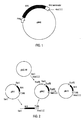
- the transformed clone of E. coli was denominated HBM64 (Fig. 2), and presents levels of expression of P64k higher than 25 % in relation to the total protein of the cell (Fig. 6).
- the antigen obtained from the isolated sequence was very useful in the preparation of different types of potential vaccine preparations, like bivalent vaccines with a broad immunoprotective spectrum, e.g., protein-polysaccharide conjugates, fusion proteins, etc.
- the DNA of about 60 kb was subjected to a partial digestion with the enzyme Sau 3A, obtaining a population of fragments of about 15 kb.
- This majoritary fraction was isolated and purified by separation in agarose gel (T. Maniatis et al., 1982, Molecular Cloning: A Laboratory Manual. Cold Spring Harbor Lab.: Cold Spring Harbor NY).
- the library was screened by immunoidentification (R. Young and R. Davis, 1983, PNAS USA 80: 1194-1198) using rabbit serum obtained against a preparation of proteins belonging to the outer membrane of the strain N. meningitidis B:4:P1.15.
- the clones were analyzed by Western-blot (Burnette, 1981) and the expression of the P64k protein with a molecular weight of about 70 kDa was detected.
- the resulting recombinant phage was named 31.
- the Western blot was also made using a mixture of sera from convalescents of meningococcemia, free of antibodies from E. coli, obtaining the same result as that using hyperimmunized rabbit sera.
- the 17 kb insert corresponding to the phage isolated from the library was cloned in the plasmid pUC18 after separation from the phage's arms using the enzyme SalI. This resulted in the construction pM-1 (Fig. 2), that was subjected to restriction analysis (Fig. 3).
- the sequence of the insert in pM-3 was determined by the method of Sanger (F. Sanger et al., 1977, PNAS USA 74: 5463-5467).
- the plasmid pM-3 (Fig. 2) was linearized with the enzyme EcoRI and successive suppressions of the gene were carried out, incubating the sample with the nucleases ExoIII and S1.
- the resulting fragments were separated from the rest of the vector pUC18 by cutting with the restriction enzyme HindIII and were cloned fused to a stabilizator fragment (European patent application EP-A-0 416 673), using an Xba-blunt adapter to conserve the XbaI site of the stabilizator gene:
- the insert sequences were established using Sanger's Method (F. Sanger et al., 1977, PNAS USA 74: 5463-5467). From the obtained sequences the approximate molecular weight of the protein encoded by this gene was deduced.
- the fusion region between the proteins was localized in the gene sequences.
- NcoI-XbaI fragment corresponding to the stabilizing peptide coding sequence, was deleted from pILM-25, obtaining a non-fused protein expressed under the tryptophan promoter with it's original terminator from the N. Meningitidis B:4:P1.15, according to the pM-6 construction ( Figure 1).
- the pM-6 plasmid was transformed in different strains of E. coli like W3110, JA-221, HB-101, LE-392 and MC-1061, and the expression of P64k was compared. The best results were obtained in W3110, JA-221 and HB-101. These strains were chosen to scale up fermentation, and expression levels up to 25 % of total cell proteins were obtained.
- the P64k protein was desalted by gel filtration chromatography (PD-10, Pharmacia), eluted with water and monitored at 280 nm. The protein fraction was concentrated to 0.5 nM/ ⁇ l. One ⁇ l of this solution was applied to a PVDF (polyvinylidene difluoride, Millipore) filter, previously activated with methanol.
- PD-10 gel filtration chromatography
- PVDF polyvinylidene difluoride, Millipore
- the Edman degradation was made using the Knauer's Automatic Sequencer, model 810, connected to a HPLC (High Performance Liquid Chromatography) system, so as to detect the phenylthiohydantoin derivatives of the aminoacids (PTH-aminoacids).
- the standard procedure of sequencing as recommended by the manufacturer of the equipment was followed.
- the separation of the PTH-aminoacids was performed in a reverse phase column C-18 (5 ⁇ m), 250 mm x 2 mm (Merck), eluted with an acetonitrile gradient (B buffer) in sodium acetate (A buffer), prepared according to the manufacturers, with a 200 ⁇ l/minute flow and at 42°C.
- the PTH-aminoacids were detected at 269 nm.
- the bactericidal test against B385 (B:4:P1.15) was made according to the procedure described by Larrick et al. (Scand. J. Immunol. 32, 1990, 121-128) with modifications. With this objective, a mixture was made of a) a suspension of bacteria, cultured under special conditions (1-5 colony forming units/1), b) Gey's balanced salt solution, c) rabbit sera (3 to 4 weeks) as a source of complement and d) pooled sera from mice, immunized against protein P64k in Aluminium Hydroxyde Gel, and inactivated at 56°C for 30 minutes. The immunization of mice was carried out according to an immunization scheme of 3 doses of 20 ⁇ g each.
- the proportions used in the aforementioned mixture were 1:2:1:1 in a total volume of 125 ⁇ l.
- the mixture was incubated at 37°C during 1 hour and plated in fresh Mueller Hinton Agar (Oxoid, London) supplemented with 5 % calf serum (CubaVet, Habana). The counting of surviving colonies was done after 18 hours of incubation of the plates in an atmosphere of 5 % CO 2 at 37°C.
- the bactericidal titer was considered as the maximum serum dilution necessary to render a 50 % inhibition of bacterial growth, with respect to the same mixture without the test serum. It was found that 1:20 serum dilution still maintains its bactericidal activity. As negative controls (non bactericidal at 1:2 dilution) pooled sera from mice immunized with Aluminum Hydroxyde Gel, and pooled sera from mice immunized with cuban Hepatitis B recombinant vaccine, were used. The bactericidal effect was specific to the anti-P64k antibodies.
- the bactericidal test against different strains of N. meningitidis was made using:
- the anti-P64k monoclonal antibodies have significant bactericidal titers against different serogroups (A, B and C), serotypes (4, 14, 13, 15 and NT) and subtypes (7, 15, 16 and NT) of bacteria.
- Fusion protein M-14 (P64k and P1.15)
- the gene coding for P1.15 protein was cloned using the Polymerase Chain Reaction (PCR).
- PCR Polymerase Chain Reaction
- the sites for gene fusion of P1.15 with M-6 are the following: *: N does not belong to any of the fusion proteins and was created by the genetic construction.
- the resulting fusion protein (M-14) was expressed in E. coli using a plasmid vector under the tryptophan promoter, to levels higher than 10 % of total cell protein.
- the protein was recognized by bactericidal monoclonal antibodies, and anti-P1.15 and P64k polyclonal antibodies, in Western-Blot.
- the protein P64k was conjugated with the polysaccharide from Haemophilus influenzae using the reductive amination method.
- the Haemophilus influenzae polysaccharide (Polyribosyl ribitol phosphate, PRP) was purified by the cold phenol method described by Frasch, 1990 (in: Bacterial Vaccines, 1990, Alan R. Liss, Inc., pp. 123-145). The final contamination of PRP with proteins or nucleic acids was less than 1 %.
- This polysaccharide was degraded using the method of Parikh et al. 1974 (Methods in Enzymol.
- the conjugate is obtained by mixing protein and polyssacharide in a 1 : 1 ratio (w/w), adding sodium cyanoborohydride and subjecting the mixture to an incubation, first for 48 hours at 4°C and later at 37°C for 24 hours.
- the high molecular weight complex which contains the resulting conjugate with protein-polysaccharide in a 1 : 2.3 ratio, can be separated from the non reactive contaminants by HPLC.
- Antibody titers against P64k protein were measured in solid phase Enzyme Linked Immunosorbent Assay (ELISA), using P64k at 5 mg/ml to coat the polystyrene plate.
- the antibody titers against HBsAg were determined by a Commercial ELISA (Organon Teknika, Boxtel).
- Figure 5 shows the dynamics of antibody response against protein P64k, using sera diluted 1/10 000. The response against P64k is not interfered by the presence of the other antigen.
- Figure 6 shows the titers against HBsAg after each dosis. The titers against this protein are not diminished by the presence of P64k in the preparation. High titers are obtained against both antigens in the same vaccine preparation.
Description
- The present invention is in the field of Genetic Engineering and Biotechnology. More in particular, the invention is related to a nucleotide sequence obtained from the pathogenic bacterium Neisseria meningitidis, which nucleotide sequence codes for a protein belonging to the outer membrane of said bacterium. Said protein is cloned and expressed in the host Escherichia coli. The characteristics of this protein as well as its capacity to induce immunologically active antibodies (bactericidal antibodies) in its natural host, allow its use in vaccine preparations against pathogenic strains of this microorganism.
- The gram-negative bacterium N. meningitidis is responsable for one of every three cases of bacterial meningitidis in the world. It was described for the first time by Anton Weichselbaum in 1887 (I. DeVoe, 1982, Microbiol. Revs. 46: 162-190), and man (i.e. human beings) is its only natural host up to date.
- In the first half of this century some essential aspects were found in relation to the metabolism and serological differentiation of this microorganism. The first unsuccessful attempts to obtain vaccine preparations were based on its capsular polysaccharide (E. Kabat et al., 1945, J. Exp. Med. 80: 299-307). According to the chemical composition of this capsular polysaccharide, the bacterium N. meningitidis is serogrouped in A, B, C, 29-E, H, I, K, L, W-135, X, Y or Z, and the major percentage of illness is caused by A, C, Y, W-135 and B. Non-encapsulated strains are not associated with the invasive disease.
- Using different methods of purification of these polysaccharides (E. Gotschlich et al., 1969, J. Exp. Med. 129: 1349-1365) the four first polysaccharides (PS) showed to be good immunogens and inducers of bactericidal antibodies in humans (E. Gotschlich et al., 1969, J. Exp. Med. 129: 1367-1384). The presence of this kind of antibodies has been correlated previously with non-susceptibility to the infection (I. Goldschneider et al., 1969, J. Exp. Med. 129: 1307-1326). As of today mono-, bi- or tetravalent vaccines have been well studied for serotypes A, C and W-135 (F. Ambrosch et al., 1983, Bulletin of the WHO 61: 317-323; I. Vodopija et al., 1983, Infect. Immunol. 42: 599-604; M. Cadoz et al., 1985, Vaccine 3: 340-342; H. Peltola et al., 1985, Pediatrics 76: 91-96).
- These vaccines have been licensed for their use in humans in different countries (Centers for Disease Control, 1985, Morbid. Mortal. Weekly Report 34: 255-259) and some of them are commercially available from different firms and producers (Connaught Laboratories, USA; Smith Kline-RIT, Belgium; Institute Merieux, France; Behringwerke Aktiengesellschaft, Germany; Istituto Sieroterapico e Vaccino genea Toscano "Sclavo", Italy; Swiss Serum and Vaccine Institute, Berne, Switzerland; among others).
- However, the conventional vaccine against N. meningitidis serogroup C does not induce sufficient levels of bactericidal antibodies in children under 2 years old, which are the principal victims of this disease. It has been demonstrated that the titer of specific antibodies against N. meningitidis in children under four years of age, after three years of vaccination, is similar in vaccinated and in non-vaccinated ones (H. Kayhty et al., 1980, J. of Infect. Dis. 142: 861-868). Also, no memory response was found against N. meningitidis after 8 years of vaccination in young adults (N. Rautonen et al., 1986, J. of Immunol. 137: 2670-2675).
- The polysaccharide corresponding to N. meningitidis serogroup B is poorly immunogenic (E. Gotschlich et al., 1969, J. Exp. Med. 129: 1349-1365) and induces a poor response of IgM of low specificity (W. Zollinger et al., 1979, J. Clin. Invest. 63: 836-848). There are different theories related to this problem, such as cross-reactivity between B polysaccharide and fetal brain structures, antigenic structures modified in solution and sensitivity to neuroaminidases (C. Moreno et al., 1985, Infect. Immun. 47: 527-533). Recently, a chemical modification of PS B was achieved, which induced a response in the host (H. Jennings et al., 1988, US
Patent 4 727 136; F. Ashton et al., 1989, Microb. Pathogen. 6: 455-458), but safety of this vaccine in humans has not been demonstrated. - Due to the lack of an effective vaccine against N. meningitidis B, and because the risk of endemic infection is low and mainly restricted to children, a routine immunization with polysaccharides is not recommended (C. Frasch, 1989, Clin. Microbiol. Revs. 2: S134-S138) except in the case of an epidemic.
- Since after the Second World War the disease was caused in most of the cases by N. meningitidis B, vaccines against serogroup B gained special significance.
- Other outer membrane components of N. meningitidis include phospholipids, lipopolysaccharides (LPS or endotoxins), pili proteins and others. Different immunotypes of LPS have been described for N. meningitidis (W. Zollinger and R. Mandrell, 1977, Infect. Immun. 18: 424-433; C.M. Tsai et al., 1983, J. Bacteriol. 155: 498-504) and immunogenicity using non-toxic derivatives was assayed (H. Jennings et al., 1984, Infect. Immun. 43: 407-412) but their variability (H. Schneider et al., 1984, Infect. Immun. 45: 544-549) and pyrogenicity (when it is conjugated to lipid A) are limiting factors up to now.
- The pili, structures needed to fix cells to nasopharingeal mucous membrane (D. Stephens et al., 1983, The J. Infect. Dis. 148: 369-376) have antigenic diversity among different strains (J. Greenblatt et al., 1988, Infect. Immun. 56: 2356-2362) with some common epitopes (D. Stephens et al., 1988, The J. Infect. Dis. 158: 332-342). Presently there are some doubts in relation to the effectiveness of a vaccine based on these structures. However some of these types of vaccine have been obtained, without known results related to their use in humans (C. Brinton, 1988, US
Patent 4 769 240). - Recently, the attention has switched to the other proteins of the outer membrane of this bacterium. There are many immunological types of these protein complexes.
- The strains of N. meningitidis are subdivided in serotypes according to the presence of specific epitopes in the majoritary protein P1/P2 and in subtypes according to other epitopes in protein P1 (C. Frasch et al., 1985, Rev. Infect. Dis. 7: 504-510).
- There are several published articles and patent applications concerning vaccines based on cocktails of these proteins, with previous selective removal of endotoxins using biocompatible detergents. The immunogenicity of these cocktails in animals and humans has been demonstrated (W. Zollinger et al., 1979, J. Clin. Invest. 63: 836-848; C. Frasch and M. Peppler, 1982, Infect. Immun. 37: 271-280; E. Beuvery et al., 1983, Infect. Immun. 40: 369-380; E. Rosenqvist et al., 1983, NIPH Annals 6: 139-149; L. Wang and C. Frasch, 1984, Infect. Immun. 46: 408-414; C. Moreno et al., 1985, Infect. Immun. 47: 527-533; E. Wedege and L. Froholm, 1986, Infect. Immun. 51: 571-578; C. Frasch et al., 1988, The J. Infect. Dis. 158: 710-718; M. Lifely. and Z. Wang, 1988, Infect. Immun. 56: 3221-3227; J. Poolman et al., 1988, In J. Poolman et al (Eds), Gonococci and Meningococci, Kluwer Academic Publishers, Dordrecht, The Netherlands, pp 159-165; E. Rosenqvist et al., 1988, J. Clin. Microbiol. 26: 1543-1548), including results in massive field trials e.g. Capetown, South Africa in 1981 (C. Frasch, 1985, Eur. J. Clin. Microbiol. 4: 533-536); Iquique, Chile, 1987 (W. Zollinger, 1988, Proceedings of the Sixth International Pathogenic Neisseria Conference. Callaway Gardens Conference Center) and Cuba 1986 and 1988 (G. Sierra, 1988, Proceedings of the Sixth International Pathogenic Neisseria Conference. Callaway Gardens Conference Center). However, with the exception of the last case, the bactericidal antibodies induced by these preparations were restricted to the same serotype strains or related ones.
- One of these vaccines is referred to in US patent 4,601,903 which is restricted to one of the Neisseria types producing meningitis (serotype 2), with a high incidence, but also other serotypes have been isolated with high frequency from patients, such as serotypes 4 (Cuba from 1981 to 1983, H. Abdillahi et al., 1988, Eur. J. Clin. Microbiol. Infect. Dis. 7: 293-296; Finland from 1976 to 1987, H. Kayhty et al., 1989, Scand. J. Infect. Dis. 21: 527-535); 8 (Australia from 1971 to 1980, F. Ashton et al., 1984, Can. J. Med. Biol. 30: 1289-1291) and 15 (Norway from 1982 to 1984, L. Froholm et al., 1985, Proceedings of the Fourth International Symposium on Pathogenic Neisseria. American Society for Microbiology; Chile from 1985 to 1987, S. Ruiz et al., 1988, Proceedings of the Sixth International Pathogenic Neisseria Conference. Callaway Gardens Conference Center) as well as strains of undefined serotype (F. Ashton et al., 1980, Can. J. Microbiol. 26: 1480-1488; Australia from 1971 to 1980, F. Ashton et al., 1984, Can. J. Med. Biol. 30: 1289-1291; Finland from 1976 to 1987, H. Kayhty et al., 1989, Scan. J. Infect. Dis. 21: 527-535).
- The Cuban vaccine achieved in 1988 by the Centro Nacional de Biopreparados (European Patent Application. No. 301 992) has proven to be very effective. It is based on a high molecular weight antigenic complex. It possesses a broad range of cross-reactivity with other strains and produces and maintains bactericidal antibodies in the immunized host.
- However, the methods employed to obtain this type of vaccine start with the multiplication in an appropiate culture of a microorganism which is highly pathogenic, with the associated biological risk of handling directly the bacteria. Moreover, this kind of preparation contains lipopolysaccharides, a contaminant that, although it may increase the product's effectiveness, shows at the same time undesirable secondary effects because of its powerful pyrogenicity. Also, its variation in minor antigenic components, which form part of the preparation, cannot be controlled in the different batches, which makes it difficult to follow important parameters related to the reactogenicity and immunogenicity.
- For this reason, there is increasing interest in the identification of nucleotide sequences coding for highly conserved proteins in all strains, and even more so the identification of inducer proteins of bactericidal antibodies common to the majority of pathogenic Neisseria, in order to obtain vaccine preparations with a broad spectrum of protection.
- There are different proteins with high molecular weight which are present in low amounts in the outer membrane of N. meningitidis when this microorganism is grown in conventional culture media but have a strong response in affected individuals (J. Black et al., 1986, Infect. Immun. 54: 710-713; L. Aoun et al., 1988, Ann. Inst. Pasteur/ Microbiol. 139: 203-212) and/or increase their response under special culture conditions (J. van Putten et al., 1987, Antoine van Leeuwenhoek 53: 557-5564; A. Schryvers and L. Morris, 1988, Molecular Microbiol. 2: 281-288 and Infect. Immun. 56: 1144-1149). Some of these proteins are highly conserved among the different strains, in particular those related to the acquisition of iron by the microorganism, that have become interesting vaccine candidates (L. Mocca et al., 1988, Proceedings of the Sixth International Pathogenic Neisseria Conference. Callaway Gardens Conference Center; C. Frasch, 1989, Clin. Microbiol. Revs. 2: S134-S138).
- In addition to pure proteins obtained from the micro-organism or strains of related species (e.g. 37 kD protein, T. Mietzner and S. Morse, 1987,
US Patent 4 681 761), several related genes have been cloned and expressed. Among these proteins are the following: - protease IgAl (J. Koomey and S. Falkow, 1984, Infect. Immun. 43: 101-107);
- protein P1 (A. Barlow et al., 1987, Infect. Immun. 55: 2734-2740, and 1989, Molec. Microbiol. 3: 131-139);
- protein P5a (T. Kawula et al., 1988, Infect. Immun. 56: 380-386);
- protein P5c (T. Olyhoek and M. Achtman, 1988, Proceedings of the Sixth International Pathogenic N. Conference. Callaway Gardens Conference Center);
- protein P4 (K. Klugman et al., 1989, Infect. Immun. 57: 2066-2071);
- protein P2 (K. Murakami et al., 1989, Infect. Immun. 57: 2318-2323);
- antigen H.8 (W. Black and J.G. Cannon, 1985, Infect. Immun. 47: 322-325);
- macromolecular complex (W. Tsai and C. Wilde, 1988, Proceedings of the Sixth International Pathogenic Neisseria Conference. Callaway Gardens Conference Center);
- 37 kDa protein, repressed in the presence of iron (S. Berish et al., 1988, Proceedings of the Sixth International Pathogenic Neisseria Conference. Callaway Gardens Conference Center).
- The use of these proteins as active vaccine preparation has not been reported or the bactericidal tests of antibodies induced against them were negative, such as in the case of mouse monoclonal antibodies against H.8 (J. Woods et al., 1987, Infect. Immun. 55: 1927-1928).
- Up to the moment, the protein P1 located in the outer membrane of N. meningitidis is one of the best characterized and studied antigens. This protein presents no variability within the same strain. However, there are more than 17 types of proteins P1 in Neisseria which have differences in three variable regions, this being the basis of the classification of N. in different subtypes. This protein is very immunogenic in humans (W.D. Zollinger and R.E. Mandrell, 1983, Med. Trop. 43:143-147), eliciting protective antibodies (E. Wedege and L.O. Froholm, 1986, Infect. Immun. 51: 571-578; K. Saukkonen et al, 1987, Microb. Pathogen. 3:261-267), that give it a special importance in vaccine preparations.
- Some subtypes of proteins P1 have been cloned in E. coli, starting from genomic libraries (A.K. Barlow et al., 1989, Molec. Microb. 3:131-139) or using the PCR technique (S. Butcher et al., VIIth International Congress of Neisseria, R.C. Seid, Patent Application WO 90/06696; Brian Mc Guinness et al., 1990, J. Exp. Med. 171:1871-1882, M.C.J. Maiden et al., VIIth International Conference of Neisseria, Berlin, Sept. 9-14, 1990, and 1991, Molec. Microb. 3:727; J. Suker et al., VIIth International Conference of Neisseria, Berlin, Sept. 9-14, 1990). However, up to now, there is no genetic construction able to produce this protein with high levels of expression. Only low levels of expression (D.A. White et al., 1990, Molec. Microb. 4:769:776) or its expression in Bacillus subtilis fused to the outer membrane protein A of E. coli (omp A) (E. Wahlstrom et al., VIIth International Congress of Neisseria, September 9-14, 1990, Berlin) have been reported.
- It can be affirmed that up to the moment no antigen has been isolated which is common to all types and serogroups of N. meningitidis and is able to produce bactericidal antibodies. For this reason, an antigen of this kind, conjugated or fused to other proteins or polysaccharides of immunological interest, would be relevant as a candidate for bivalent vaccine preparations.
- This invention is related to a nucleotide sequence coding for a protein having a molecular weight of about 64 kilodaltons. This sequence has been found in all N. meningitidis serotypes and serogroups tested, as verified by nucleic acid hybridization, Western-bloting, Dot-blot and ELISA.
- A technical object of this invention is the identification of a nucleotide sequence which codes for a highly conserved protein and is common to the majority of pathogenic strains of Neisseria (named P64k), in order to obtain the protein by a recombinant way with a high grade of purity and in commercially useful quantities, so that it can be employed in diagnostic methods and as an integrating part of a vaccine preparation of broad spectrum of protection.
- On the level of genetic information (DNA and RNA), the invention provides a recombinant polynucleotide, comprising a nucleotide sequence coding for a protein P64k of Neisseria meningitidis, said protein P64k essentially having the amino acid sequence shown in SEQ ID NO:1. In a preferred embodiment said nucleotide sequence coding for the protein P64k of N. meningitidis essentially consists of the nucleotide sequence shown in SEQ ID NO:1. The recombinant polynucleotide may further comprise a nucleotide sequence of a cloning or expression vector.
- The invention also provides a transformed microorganism containing a recombinant polynucleotide as defined above, preferably a transformed microorganism which is capable of expressing the protein P64k of N. meningitidis. In a particularly preferred embodiment of the invention, the transformed microorganism is an Escherichia coli strain, e.g. E. coli strain HB101, transformed with an expression vector containing a nucleotide sequence coding for the protein P64k of N. meningitidis, e.g. the expression vector pM-6.
- The invention also provides a recombinant proteinaceous substance, comprising an amino acid sequence corresponding to the amino acid sequence of a protein P64k of N. meningitidis, said protein P64k essentially having the amino acid sequence shown in SEQ ID NO:1. Said recombinant proteinaceous substance may essentially consist of protein P64k, or be a fusion protein or a protein/polysaccharide conjugate comprising the amino acid sequence of protein P64k of N. meningitidis.
- The invention further provides a vaccine composition, comprising a recombinant protein as defined above, together with a suitable carrier, diluent or adjuvant.
- In addition, the invention provides a monoclonal antibody, raised against a recombinant proteinaceous substance as defined above and capable of binding a protein P64k of N. meningitidis.
- The invention also provides a process for preparing a protein P64k of Neisseria meningitidis, or a fusion protein comprising protein P64k, said protein P64k essentially having the amino acid sequence shown in SEQ ID NO:1, comprising the steps of transforming a microorganism with an expression vector containing a nucleotide sequence coding for said protein P64k, or said fusion protein, culturing the transformed microorganism to obtain expression of said protein P64k, or said fusion protein, and isolating said expression product.
- One novel aspect of this invention is the gene isolated from the N. meningitidis strain B:4:P1.15, which was named M-6 and has as a principal characteristic its stability in E. coli vectors. This gene does not produce adverse effects on the host, allowing to obtain yields of over 25 % of total protein (ratio of P64k to total protein from host strain). On the other hand, it has been demonstrated by Southern and Western Blot hybridizations (E. Southern, 1975, J. Mol. Biol. 98: 503-527 and W. Burnette, 1981, Anal. Biochem. 12: 195-203) that protein P64k is present in all the following studied strains of N. meningitidis:
- N. meningitidis A
- N. meningitidis B:1
- N. meningitidis B:2
- N. meningitidis B:4
- N. meningitidis B:5
- N. meningitidis B:8
- N. meningitidis B:9
- N. meningitidis B:11
- N. meningitidis B:15
- N. meningitidis B:4:P1.15
- N. meningitidis C
- N. meningitidis B:15:P1.16
- N. meningitidis B:15:P1.16 (H 44/76)
- N. meningitidis B:NT (121/85)
- N. meningitidis B:NT (71/86)
- N. meningitidis B:NT (210/86)
- The protein is not present in N. cinerea, N. lactamica, N. sicca and N. flavescens, but these are not of interest because they are not pathogenic.
- The protein having a molecular weight of about 64 kDa can be localized by electron microscopy in the outer membrane of N. meningitidis. Therefore, this antigen is an exposed antigen which is favorable for use in a vaccine preparation. The protein was recognized in Western blot immunoidentification experiments with sera from convalescents and individuals vaccinated with the conventional Cuban vaccine Va-Mengoc-BC (Centro Nacional de Biopreparados, Havana, Cuba). This aspect guarantees the immunogenicity of the antigen and at the same time confirms its presence within the high molecular weight protein fraction constituent of this vaccine, which is responsable of the lasting immune response to the disease.
- Another novel aspect is that the protein, which is an object of this invention, produces antibodies with a broad bactericidal spectrum (different serogroupes, serotypes and subtypes), a characteristic which has not been reported previously for any protein from N. meningitidis.
- This protein obtained in high levels in E. coli becomes an important candidate for the improvement of immunogenicity when expressed as a fusion protein with other proteins. It could also increase the expression by conferring enhanced stability and suitability in the molecular structure during transcription and translation processes. Belonging to Neisseria, this protein can also be fused to other proteins from Neisseria in order to obtain vaccine preparations against this microorganism with increased immunogenicity. These fusion proteins are also objects of this invention.
- On the other hand, surprisingly, it was found that the gene M-6 obtained from a genomic library of the strain N. meningitidis B:4:P1.15 showed a great homology with sequences of lipoamide-dehydrogenases and acetyl-transferases from other microorganisms and higher organisms.
- An important object of this invention is the nucleotide sequence which codes for the M-6 gene (SEQ ID NO:1 of the Sequence Listing) whose product is the protein P64k.
- This gene was derived from the genome of the strain B385 isolated in Cuba (N. meningitidis B:4:P1.15), by the construction of a genomic library in the
phage EMBL 3. - The recombinant DNA including the gene M-6 constitutes another object of this invention, which includes the phage lambda, the plasmid pM-3 and the expression vector pM-6 for expression in bacteria.
- In particular, for the intracellular expression in E. coli, the M-6 gene was cloned under the tryptophane promotor and using its own termination signal of transcription and a linkage fragment between M-6 and the cloning site NcoI which adds the following nucleotide sequence at the 5' end:

- The N-terminal of the protein P64k encoded by the M-6 gene inserted in plasmid pM-6 which adds 5 aminoacids to the N-terminal of the original protein corresponds to:

- Another object of this invention are the microorganisms resulting from the transformation of E. coli strain HB 101 with the pM-6 vector, which are characterized by the expression of high levels of protein P64k, good viability and great stability.
- The transformed clone of E. coli was denominated HBM64 (Fig. 2), and presents levels of expression of P64k higher than 25 % in relation to the total protein of the cell (Fig. 6).
- The procedure described in the present invention, due to the levels of expression achieved for this product, allows to reach an optimal purity for use of this protein in humans.
- On the other hand, the antigen obtained from the isolated sequence was very useful in the preparation of different types of potential vaccine preparations, like bivalent vaccines with a broad immunoprotective spectrum, e.g., protein-polysaccharide conjugates, fusion proteins, etc.
- For the isolation of genomic DNA from N. meningitidis B:4:P1.15, the cells were grown in Mueller-Hinton medium (OXOID, London). The biomass from a culture of 100 ml was resuspended in 8 ml of Tris (hydroxymethyl-aminomethane) 100 mM, EDTA (ethylenediamine tetraacetic acid) 1mM,
pH 8. The cells were subjected to a treatment with lysozyme (10 mg/ml), followed by 200 µl of self-digested pronase (20 mg/ml) and 1.1 ml of 10% SDS. The mixture was incubated at 37°C during 1 hour, then it was treated with phenolchloroform (v/v) and the remains of phenol were eliminated using 2-butanol. Finally, the DNA was precipitated with absolute ethanol and RNA was eliminated with ribonuclease A (Sigma, London). - The DNA of about 60 kb was subjected to a partial digestion with the enzyme Sau 3A, obtaining a population of fragments of about 15 kb. This majoritary fraction was isolated and purified by separation in agarose gel (T. Maniatis et al., 1982, Molecular Cloning: A Laboratory Manual. Cold Spring Harbor Lab.: Cold Spring Harbor NY).
- For the construction of the genomic library, the process described by Maniatis was essentially followed (T. Maniatis et al., 1982, Molecular Cloning: A Laboratory Manual. Cold Spring Harbor Lab.: Cold Spring Harbor NY). Four µg of purified DNA were ligated with 8 µg of BamHI-digested EMBL-3. The ligation product was packed and the phages were finally plated on the E. coli strain C66P2.
- The library was screened by immunoidentification (R. Young and R. Davis, 1983, PNAS USA 80: 1194-1198) using rabbit serum obtained against a preparation of proteins belonging to the outer membrane of the strain N. meningitidis B:4:P1.15. The clones were analyzed by Western-blot (Burnette, 1981) and the expression of the P64k protein with a molecular weight of about 70 kDa was detected. The resulting recombinant phage was named 31. The Western blot was also made using a mixture of sera from convalescents of meningococcemia, free of antibodies from E. coli, obtaining the same result as that using hyperimmunized rabbit sera.
- This experiment was repeated using sera from several healthy individuals, and the signal obtained was negative against the recombinant protein P64k.
- For subcloning in bacteria, the 17 kb insert corresponding to the phage isolated from the library was cloned in the plasmid pUC18 after separation from the phage's arms using the enzyme SalI. This resulted in the construction pM-1 (Fig. 2), that was subjected to restriction analysis (Fig. 3).
- The fragment SalI-HindIII of about 6 kb was recloned in the plasmid pUC18 and the construction pM-2 was obtained (Fig. 2). In order to obtain a more exact localization of the gene coding for protein P64k deletions were carried out with the enzymes ClaI, EcoRI and HincII. The complete fragment of the gene M-6 was finally localized as an EcoRI-HindIII insert corresponding to the construction pM-3 (Fig. 2).
- In all constructions, the presence of the gene was confirmed by recognizing the protein by colony immunoidentification and Western Blot using hyperimmunized rabbit sera.
- The sequence of the insert in pM-3 was determined by the method of Sanger (F. Sanger et al., 1977, PNAS USA 74: 5463-5467).
- From the obtained sequence, the approximate molecular weight of the protein encoded by the gene was deduced.
- In order to obtain a construction for high expression of the protein P64k, the plasmid pM-3 (Fig. 2) was linearized with the enzyme EcoRI and successive suppressions of the gene were carried out, incubating the sample with the nucleases ExoIII and S1.
- The resulting fragments were separated from the rest of the vector pUC18 by cutting with the restriction enzyme HindIII and were cloned fused to a stabilizator fragment (European patent application EP-A-0 416 673), using an Xba-blunt adapter to conserve the XbaI site of the stabilizator gene:


- The constructions in which the fused fragment coincided with the reading frame were selected by immunoidentification using hyperimmune rabbit sera.
- The insert sequences were established using Sanger's Method (F. Sanger et al., 1977, PNAS USA 74: 5463-5467). From the obtained sequences the approximate molecular weight of the protein encoded by this gene was deduced.
- The fusion region between the proteins was localized in the gene sequences. In the clone pILM-25 (Figure 4) the ATG of the gene predetermined by the sequence of the DNA insert isolated from the library, coincided with the fusion site.
- The NcoI-XbaI fragment, corresponding to the stabilizing peptide coding sequence, was deleted from pILM-25, obtaining a non-fused protein expressed under the tryptophan promoter with it's original terminator from the N. Meningitidis B:4:P1.15, according to the pM-6 construction (Figure 1).
- The pM-6 plasmid was transformed in different strains of E. coli like W3110, JA-221, HB-101, LE-392 and MC-1061, and the expression of P64k was compared. The best results were obtained in W3110, JA-221 and HB-101. These strains were chosen to scale up fermentation, and expression levels up to 25 % of total cell proteins were obtained.
- To confirm the correct expression of the cloned gene the N-terminal region of the intact protein was subjected to the Edman degradation method (P. Edman, 1950, Acta Chem. Scand. 4: 283-293). This technique elucidates the sequence (primary structure) of this region in the molecule.
- The P64k protein was desalted by gel filtration chromatography (PD-10, Pharmacia), eluted with water and monitored at 280 nm. The protein fraction was concentrated to 0.5 nM/µl. One µl of this solution was applied to a PVDF (polyvinylidene difluoride, Millipore) filter, previously activated with methanol.
- The Edman degradation was made using the Knauer's Automatic Sequencer, model 810, connected to a HPLC (High Performance Liquid Chromatography) system, so as to detect the phenylthiohydantoin derivatives of the aminoacids (PTH-aminoacids). The standard procedure of sequencing as recommended by the manufacturer of the equipment was followed. The separation of the PTH-aminoacids was performed in a reverse phase column C-18 (5 µm), 250 mm x 2 mm (Merck), eluted with an acetonitrile gradient (B buffer) in sodium acetate (A buffer), prepared according to the manufacturers, with a 200 µl/minute flow and at 42°C. The PTH-aminoacids were detected at 269 nm.
- Data processing and registration were made in a Shimadzu model CR-6a automatic integrator, using a program for data processing by subtraction of two consecutive chromatograms, to facilitate the evaluation of the Edman degradation cycles. Sequence identification is obtained by the chromatographic evaluation of the corresponding analyzed cycle and confirmed by the chromatogram obtained by subtraction, allowing to determine 25 residues.
- To demonstrate that the protein P64k is recognized by the sera of individuals vaccinated with the Cuban Va-Mengoc-BC preparation (Centro Nacional de Biopreparados, Havana, Cuba.), a Western-Blot was made, with a mixture of 12 sera from adults (immunized with two doses of the Cuban vaccine) diluted in a solution containing defatted milk (Oxoid, London). The experiment included:
- recombinant protein P64k, purified from E. coli HB-101 transformed with the pM-6 plasmid;
- supernatant of the ultrasonic cell rupture of untransformed E. coli HB-101;
- the reaction was revealed with a protein A-colloidal gold conjugate.
- The bactericidal test against B385 (B:4:P1.15) was made according to the procedure described by Larrick et al. (Scand. J. Immunol. 32, 1990, 121-128) with modifications. With this objective, a mixture was made of a) a suspension of bacteria, cultured under special conditions (1-5 colony forming units/1), b) Gey's balanced salt solution, c) rabbit sera (3 to 4 weeks) as a source of complement and d) pooled sera from mice, immunized against protein P64k in Aluminium Hydroxyde Gel, and inactivated at 56°C for 30 minutes. The immunization of mice was carried out according to an immunization scheme of 3 doses of 20 µg each. The proportions used in the aforementioned mixture were 1:2:1:1 in a total volume of 125 µl. The mixture was incubated at 37°C during 1 hour and plated in fresh Mueller Hinton Agar (Oxoid, London) supplemented with 5 % calf serum (CubaVet, Habana). The counting of surviving colonies was done after 18 hours of incubation of the plates in an atmosphere of 5 % CO2 at 37°C.
- The bactericidal titer was considered as the maximum serum dilution necessary to render a 50 % inhibition of bacterial growth, with respect to the same mixture without the test serum. It was found that 1:20 serum dilution still maintains its bactericidal activity. As negative controls (non bactericidal at 1:2 dilution) pooled sera from mice immunized with Aluminum Hydroxyde Gel, and pooled sera from mice immunized with cuban Hepatitis B recombinant vaccine, were used. The bactericidal effect was specific to the anti-P64k antibodies.
- The bactericidal test against different strains of N. meningitidis was made using:
- 1. An ammonium sulphate precipitate of the supernatant harvested from a culture of hybridoma cells secreting monoclonal antibodies specific against P64k (anti P64k)/Sample to analyze.
- 2.An ammonium sulphate precipitate of the supernatant harvested from hybridoma cells secreting monoclonal antibodies specific against the P1.15 protein present in N. meningitidis strain B385 (anti P1.15)/Positive control of the system.
- The maximum dilutions tested were always 1:16. The maximum dilutions tested which had a bactericidal effect, according to the EXAMPLE 5, are indicated:
Strain anti-P64k anti-P1.15 B385 1:16 1:16 B:4:P1.15 1:16 1:16 B:14:P1.7 1:16 - B:NT:NT 1:16 - B:15:P1.15 1:8 - B:15:P1.16 1:8 - B:13 1:8 - C 1:16 - A 1:16 - - As seen, the anti-P64k monoclonal antibodies have significant bactericidal titers against different serogroups (A, B and C), serotypes (4, 14, 13, 15 and NT) and subtypes (7, 15, 16 and NT) of bacteria.
- In order to obtain a genetic construction for high expression that contained the variable epitopes of the P1.15 protein (Outer membrane protein from N. meninigitidis B:4:P1.15) fused to the P64k protein, the gene coding for P1.15 protein was cloned using the Polymerase Chain Reaction (PCR). The following region containing the variable immunodeterminants of P1.15:was inserted in the Mlu I site of the gene M-6, encoding for P64k, after having been made blunt with the klenow fragment from DNA polymerase I. The sites for gene fusion of P1.15 with M-6 are the following:


 *: N does not belong to any of the fusion proteins and was created by the genetic construction.
- The resulting fusion protein (M-14) was expressed in E. coli using a plasmid vector under the tryptophan promoter, to levels higher than 10 % of total cell protein. The protein was recognized by bactericidal monoclonal antibodies, and anti-P1.15 and P64k polyclonal antibodies, in Western-Blot.
- The protein P64k was conjugated with the polysaccharide from Haemophilus influenzae using the reductive amination method. The Haemophilus influenzae polysaccharide (Polyribosyl ribitol phosphate, PRP) was purified by the cold phenol method described by Frasch, 1990 (in: Bacterial Vaccines, 1990, Alan R. Liss, Inc., pp. 123-145). The final contamination of PRP with proteins or nucleic acids was less than 1 %. This polysaccharide was degraded using the method of Parikh et al. 1974 (Methods in Enzymol. 34B: 77-102) with sodium periodate in PRP (ratio 1 : 5 w/w) dissolved in 0.1 M sodium acetate (pH 4.5). The incubation was carried out in the dark during 30 minutes with stirring. The periodate excess was eliminated by addition of ribitol. Very low molecular weight compounds were eliminated by dialysis (Medicell International Ltd. Membrane, London). The resulting oligosaccharide had free aldehyde groups able to react with primary amines (e.g. lysine residues in proteins). The conjugate is obtained by mixing protein and polyssacharide in a 1 : 1 ratio (w/w), adding sodium cyanoborohydride and subjecting the mixture to an incubation, first for 48 hours at 4°C and later at 37°C for 24 hours. The high molecular weight complex which contains the resulting conjugate with protein-polysaccharide in a 1 : 2.3 ratio, can be separated from the non reactive contaminants by HPLC.
- Bivalent vaccine preparation against Hepatitis B virus and N. meningitidis.
- In order to obtain a bivalent vaccine preparation, different quantities of protein P64k and Hepatitis B Surface Antigen (Vacuna Recombinante contra la Hepatitis B, Heber Biotec, Havana, Cuba) were mixed. The antigens were adjuvated with Aluminum Hydroxyde Gel, at 2mg/dose and inoculated in Balb/c mice having a body weight of 20 g in 3 dosis of 0.5 ml each. Different variants were assayed:
- 1. P64k 20 µg (P20)
- 2. HBsAg 20 µg (H20)
- 3. P64k 10 µg +
HBsAg 10 µg (P10:H10) - 4.
P64K 15 µg +HBsAg 5 µg (P15:H5) - 5. Placebo (Al(OH)3)
- Seven days after the immunization with the first doses, the second doses were applied. The third dose was given 14 days after the second. Seven days later, blood was taken and the serum of each immunized animal was separated. Antibody titers against P64k protein were measured in solid phase Enzyme Linked Immunosorbent Assay (ELISA), using P64k at 5 mg/ml to coat the polystyrene plate. The antibody titers against HBsAg were determined by a Commercial ELISA (Organon Teknika, Boxtel). Figure 5 shows the dynamics of antibody response against protein P64k, using sera diluted 1/10 000. The response against P64k is not interfered by the presence of the other antigen. Figure 6 shows the titers against HBsAg after each dosis. The titers against this protein are not diminished by the presence of P64k in the preparation. High titers are obtained against both antigens in the same vaccine preparation.
- A software was created to search the EMBL (European Molecular Biology Laboratoty) Data Base and detect the homology between P64k and other proteins. As result of the search it was found that there is homology of one segment in the sequence of P64k with segments in the sequences of N. gonorrhoeae. This sequence was found as characteristic in both N. gonorrhoeae and N. meningitidis (F.F. Correia, S. Inouye and M. Inouye, 1988, J. Biol. Chem. 263, No. 25, 12194-12198).
- Another region with high homology was found in two proteins of the Pyruvate Dehydrogenase Complex from E.coli K12:
- Homology exists between a segment comprising 100 amino acids, repeated at the beginning of the amino acid sequence of the Acetyltransferase ("Lipoyl Domain", including the "Lipoyl Binding Site" (P.E. Stephens et al., 1983,. Eur. J. Biochem. 133, 481-489)) and a region located in the first 111 amino acids of the P64k:


 where (∗) indicates positions with the same amino acids and (-) indicates positions of conservative amino acid changes.
- Homology exists between the Lipoamide Dehydrogenase from E. coli (a protein having 473 amino acids, P.E. Stephens et al., 1983, Eur. J. Biochem. 133, 481-489) and the protein P64k, specifically in a segment which represents almost the total protein, except the region with homology with the "lipoyl domain" from Acetyltransferase.


 Where:
- |-1-|: Adenine binding site (FAD)
- |-2-|: Redox active disulphide region
- |-3-|: Active site histidine
- An E. coli HB-101 clone containing the plasmid pM-3 (a pUC18 plasmid containing the 4.1 kb DNA fragment from Neisseria meninigitidis, strain B:4:P1.15, cloned between the EcoRI and HindIII restriction sites), was deposited on August 30, 1991, with the Centraalbureau voor Schimmelcultures (CBS), Baarn, The Netherlands, and obtained deposit number CBS 485 .91.
-
- SEQ ID NO:1
- SEQUENCE TYPE : Nucleotide with corresponding amino acid
- SEQUENCE LENGTH: 1830 bp
- STRANDEDNESS: Single
- TOPOLOGY: Linear
- MOLECULE TYPE: genomic DNA
- ORIGINAL SOURCE ORGANISM: N. meningitidis group B
- IMMEDIATE EXPERIMENTAL SOURCE: Strain B:4:P1:15 isolated in Cuba
- FEATURES: From 1 to 1830 bp mature protein
- PROPERTIES: Gene coding for P64k protein from outer membrane of N. meningitidis


- SEQ ID NO: 2
- SEQUENCE TYPE : Nucleotide
- SEQUENCE LENGTH: 15 bases

- SEQ ID NO: 3
- SEQUENCE TYPE : Amino acid
- SEQUENCE LENGTH: 25 amino acids
- MOLECULE TYPE: Sequence N-terminal of P64k protein from outer membrane of N. meningitidis

- SEQ ID NO: 4
- SEQUENCE TYPE : Nucleotide
- SEQUENCE LENGTH: 12 bases

- SEQ ID NO: 5
- SEQUENCE TYPE : Nucleotide
- SEQUENCE LENGTH: 8 bases

- SEQ ID NO: 6
- SEQUENCE TYPE : Amino acid
- SEQUENCE LENGTH: 221 amino acids
- MOLECULE TYPE: Sequence which includes variable regions of P1.15 protein


- SEQ ID NO: 7
- SEQUENCE TYPE : Amino acid
- SEQUENCE LENGTH: 6 amino acids
- MOLECULE TYPE: Sequence corresponding to the fusion site between N-terminal of P64k and P1.15

- SEQ ID NO: 8
- SEQUENCE TYPE : Nucleotide
- SEQUENCE LENGTH: 19 bases
- MOLECULE TYPE: Sequence corresponding to the fusion of N-terminal from gene M-6 and from gene P1.15

- SEQ ID NO: 9
- SEQUENCE TYPE : Amino acid
- SEQUENCE LENGTH: 6 amino acids
- MOLECULE TYPE: Sequence corresponding to the fusion site between C-terminal of P64k and P1.15

- SEQ ID NO: 10
- SEQUENCE TYPE : Nucleotide
- SEQUENCE LENGTH: 18 bases
- MOLECULE TYPE: Sequence corresponding to the fusion of C-terminal from gene M-6 and from gene P1.15

- SEQ ID NO: 11
- SEQUENCE TYPE : Amino acid
- SEQUENCE LENGTH: 111 amino acids
- MOLECULE TYPE: p64k N-terminal comprising homology region with "lypoil binding site" from E. coli Acetyl transferase


- SEQ ID NO: 12
- SEQUENCE TYPE : Amino acid
- SEQUENCE LENGTH: 112 amino acids
- MOLECULE TYPE: "lypoil binding site" from E. coli Acetyltransferase

- SEQ ID NO: 13
- SEQUENCE TYPE : Amino acid
- SEQUENCE LENGTH: 481 amino acids
- MOLECULE TYPE: P64k fragment comprising the homology region with Lipoamide Dehydrogenase from E. coli


- SEQ ID NO: 14
- SEQUENCE TYPE : Amino acid
- SEQUENCE LENGTH: 472 bases
- MOLECULE TYPE: Segment of E. coli Lipoamide Dehydrogenase with homology to P64k protein


Claims (11)
- A recombinant polynucleotide, comprising a nucleotide sequence coding for a protein P64k of Neisseria meningitidis, said protein P64k essentially having the amino acid sequence shown in SEQ ID NO:1.
- A recombinant polynucleotide according to claim 1, wherein said nucleotide sequence coding for the protein P64k of N. meningitidis essentially consists of the nucleotide sequence shown in SEQ ID NO:1.
- A recombinant polynucleotide according to claim 1 or 2, further comprising a nucleotide sequence of a cloning or expression vector.
- A transformed microorganism, containing a recombinant polynucleotide according to any one of claims 1 to 3.
- A transformed microorganism according to claim 4, which is capable of expressing the protein P64k of N. meningitidis.
- A transformed microorganism according to claim 5, which is an Escherichia coli strain, transformed with an expression vector containing a nucleotide sequence coding for the protein P64k of N. meningitidis.
- A recombinant proteinaceous substance, comprising an amino acid sequence corresponding to the amino acid sequence of a protein P64k of N. meningitidis, said protein P64k essentially having the amino acid sequence shown in SEQ ID NO:1.
- A recombinant proteinaceous substance according to claim 7, which is a fusion protein or a protein/polysaccharide conjugate comprising the amino acid sequence of protein P64k of N. meningitidis.
- A vaccine composition, comprising a recombinant protein according to claim 7 or 8, together with a suitable carrier, diluent or adjuvant.
- A monoclonal antibody, raised against a recombinant proteinaceous substance according to claim 7 or 8 and capable of binding a protein P64k of N. meningitidis.
- A process for preparing a protein P64k of Neisseria meningitidis, or a fusion protein comprising protein P64k, said protein P64k essentially having the amino acid sequence shown in SEQ ID NO:1, comprising the steps of transforming a microorganism with an expression vector containing a nucleotide sequence coding for said protein P64k, or said fusion protein, culturing the transformed microorganism to obtain expression of said protein P64k, or said fusion protein, and isolating said expression product.
Applications Claiming Priority (2)
| Application Number | Priority Date | Filing Date | Title |
|---|---|---|---|
| CU14590 | 1990-09-07 | ||
| CU90145A CU22302A1 (en) | 1990-09-07 | 1990-09-07 | Codifying nucleotidic sequence for a protein of the external membrane of neisseria meningitidis and the use of that protein in preparing vaccines. |
Publications (3)
| Publication Number | Publication Date |
|---|---|
| EP0474313A2 EP0474313A2 (en) | 1992-03-11 |
| EP0474313A3 EP0474313A3 (en) | 1993-02-24 |
| EP0474313B1 true EP0474313B1 (en) | 1997-04-23 |
Family
ID=5459451
Family Applications (1)
| Application Number | Title | Priority Date | Filing Date |
|---|---|---|---|
| EP91202291A Expired - Lifetime EP0474313B1 (en) | 1990-09-07 | 1991-09-06 | Nucleotide sequence coding for an outer membrane protein from Neisseria meningitidis and use of said protein in vaccine preparations |
Country Status (15)
| Country | Link |
|---|---|
| US (1) | US5286484A (en) |
| EP (1) | EP0474313B1 (en) |
| JP (1) | JP3253327B2 (en) |
| AR (1) | AR245778A1 (en) |
| AT (1) | ATE152175T1 (en) |
| AU (1) | AU657487B2 (en) |
| CA (1) | CA2050749C (en) |
| CU (1) | CU22302A1 (en) |
| DE (1) | DE69125769T2 (en) |
| ES (1) | ES2103295T3 (en) |
| FI (1) | FI103511B1 (en) |
| GR (1) | GR3024127T3 (en) |
| IL (1) | IL99401A (en) |
| NO (1) | NO913524D0 (en) |
| RU (1) | RU2132383C1 (en) |
Cited By (13)
| Publication number | Priority date | Publication date | Assignee | Title |
|---|---|---|---|---|
| US7273611B1 (en) | 1995-03-17 | 2007-09-25 | Id Biomedical Corporation | Proteinase K resistant surface protein of neisseria meningitidis |
| US7795210B2 (en) | 2001-10-10 | 2010-09-14 | Novo Nordisk A/S | Protein remodeling methods and proteins/peptides produced by the methods |
| US7842661B2 (en) | 2003-11-24 | 2010-11-30 | Novo Nordisk A/S | Glycopegylated erythropoietin formulations |
| US7932364B2 (en) | 2003-05-09 | 2011-04-26 | Novo Nordisk A/S | Compositions and methods for the preparation of human growth hormone glycosylation mutants |
| US7956032B2 (en) | 2003-12-03 | 2011-06-07 | Novo Nordisk A/S | Glycopegylated granulocyte colony stimulating factor |
| US8008252B2 (en) | 2001-10-10 | 2011-08-30 | Novo Nordisk A/S | Factor VII: remodeling and glycoconjugation of Factor VII |
| US8053410B2 (en) | 2002-06-21 | 2011-11-08 | Novo Nordisk Health Care A/G | Pegylated factor VII glycoforms |
| US8207112B2 (en) | 2007-08-29 | 2012-06-26 | Biogenerix Ag | Liquid formulation of G-CSF conjugate |
| US8633157B2 (en) | 2003-11-24 | 2014-01-21 | Novo Nordisk A/S | Glycopegylated erythropoietin |
| US8969532B2 (en) | 2006-10-03 | 2015-03-03 | Novo Nordisk A/S | Methods for the purification of polypeptide conjugates comprising polyalkylene oxide using hydrophobic interaction chromatography |
| US9029331B2 (en) | 2005-01-10 | 2015-05-12 | Novo Nordisk A/S | Glycopegylated granulocyte colony stimulating factor |
| US9187546B2 (en) | 2005-04-08 | 2015-11-17 | Novo Nordisk A/S | Compositions and methods for the preparation of protease resistant human growth hormone glycosylation mutants |
| US9187532B2 (en) | 2006-07-21 | 2015-11-17 | Novo Nordisk A/S | Glycosylation of peptides via O-linked glycosylation sequences |
Families Citing this family (46)
| Publication number | Priority date | Publication date | Assignee | Title |
|---|---|---|---|---|
| US7118757B1 (en) * | 1988-12-19 | 2006-10-10 | Wyeth Holdings Corporation | Meningococcal class 1 outer-membrane protein vaccine |
| AT402367B (en) * | 1990-10-11 | 1997-04-25 | Immuno Ag | PHARMACEUTICAL PREPARATION BASED ON LYS PLASMINOGEN |
| US5439808A (en) * | 1993-07-23 | 1995-08-08 | North American Vaccine, Inc. | Method for the high level expression, purification and refolding of the outer membrane group B porin proteins from Neisseria meningitidis |
| AU2816095A (en) * | 1994-06-24 | 1996-01-19 | Children's Hospital Medical Center | Escherichia coli o157:h7 epithelial adhesin |
| US5545553A (en) * | 1994-09-26 | 1996-08-13 | The Rockefeller University | Glycosyltransferases for biosynthesis of oligosaccharides, and genes encoding them |
| US5747287A (en) * | 1995-04-28 | 1998-05-05 | North American Vaccine, Inc. | Method for the high level expression, purification and refolding of the outer membrane group B porin proteins from Neisseria meningitidis |
| CU22559A1 (en) * | 1996-01-17 | 1999-05-03 | Ct Ingenieria Genetica Biotech | EXPRESSION SYSTEM OF HETEROLOGICAL ANTIGENS IN E. COLI AS FUSION PROTEINS |
| CA2264496C (en) * | 1996-09-04 | 2008-10-07 | Takara Shuzo Co., Ltd. | Fungal antigens and process for producing the same |
| WO1999036544A2 (en) * | 1998-01-14 | 1999-07-22 | Chiron S.P.A. | Neisseria meningitidis antigens |
| BR9910005A (en) * | 1998-04-29 | 2001-01-16 | American Cyanamid Co | Vaccine composition, processes for immunization against neisseria gonorrhoeae and to prepare a vaccine composition, isolated and purified DNA sequence, plasmid, host cell, process for producing a chimeric recombinant pilin protein from neisseria gonorrhoeae and neisseria meningitidis, chimeric recombinant protein isolated and purified from neisseria gonorhoeae and neisseria meningitidis, and, process for immunization against neisseria muningitidis |
| EP2261349A3 (en) * | 1998-05-01 | 2012-01-11 | Novartis Vaccines and Diagnostics, Inc. | Neisseria meningitidis antigens and compositions |
| US6756493B1 (en) * | 1998-09-01 | 2004-06-29 | Antex Biologics, Inc. | Nucleic acid sequence and uses thereof |
| US6693186B2 (en) * | 1998-09-01 | 2004-02-17 | Antex Biologics Inc | Neisseria meningitidis polypeptide, nucleic acid sequence and uses thereof |
| EP1144998A3 (en) * | 1998-10-09 | 2002-08-07 | Chiron Corporation | Neisseria genomic sequences and methods of their use |
| DE69839740D1 (en) | 1998-10-22 | 2008-08-28 | Univ Montana | Vaccine containing Omp85 proteins of Neisseria gonorrhoeae and Neisseria meningitidis |
| EP1535928B1 (en) * | 1998-10-22 | 2008-07-16 | The University of Montana | Vaccine compositions comprising Omp85 proteins of Neisseria gonorrhoeae and Neisseria meningitidis |
| US6610306B2 (en) | 1998-10-22 | 2003-08-26 | The University Of Montana | OMP85 protein of neisseria meningitidis, compositions containing the same and methods of use thereof |
| EP1123403A1 (en) * | 1998-10-22 | 2001-08-16 | The University Of Montana | OMP85 PROTEINS OF $i(NEISSERIA GONORRHOEAE) AND $i(NEISSERIA MENINGITIDIS), COMPOSITIONS CONTAINING SAME AND METHODS OF USE THEREOF |
| ES2281409T3 (en) | 2000-02-28 | 2007-10-01 | Novartis Vaccines And Diagnostics S.R.L. | NEISSERIA PROTEIN HETEROLOGY EXPRESSION. |
| AU2001267358A1 (en) * | 2000-04-20 | 2001-11-07 | Max-Planck-Gesellschaft Zur Forderung Der Wissenschaften E.V. | Secretory tyrosine phosphatases from mycobacteria |
| CU23077A1 (en) * | 2000-12-06 | 2005-08-17 | Centro Inmunologia Molecular | VACCINE COMPOSITION CONTAINING TRANSFORMING GROWTH FACTOR (TGF-ALFA). ITS USE IN MALIGNAL DISEASE THERAPY |
| CU23245A1 (en) | 2001-07-16 | 2007-10-17 | Inst De Medicina Tropical Pedr | CHEMICAL CODING CHAINS FOR INDUCTIVE PROTEINS OF VIRUS EFFECTS. PREPARED USING CHEMICAL PROTEINS |
| GB0121591D0 (en) | 2001-09-06 | 2001-10-24 | Chiron Spa | Hybrid and tandem expression of neisserial proteins |
| US7214660B2 (en) | 2001-10-10 | 2007-05-08 | Neose Technologies, Inc. | Erythropoietin: remodeling and glycoconjugation of erythropoietin |
| US7696163B2 (en) | 2001-10-10 | 2010-04-13 | Novo Nordisk A/S | Erythropoietin: remodeling and glycoconjugation of erythropoietin |
| US7173003B2 (en) | 2001-10-10 | 2007-02-06 | Neose Technologies, Inc. | Granulocyte colony stimulating factor: remodeling and glycoconjugation of G-CSF |
| AU2004236174B2 (en) | 2001-10-10 | 2011-06-02 | Novo Nordisk A/S | Glycopegylation methods and proteins/peptides produced by the methods |
| WO2004014419A1 (en) * | 2002-08-02 | 2004-02-19 | Glaxosmithkline Biologicals S.A. | Vaccine composition comprising transferrin binding protein and hsf from gram negative bacteria |
| DK2351579T3 (en) * | 2002-10-11 | 2017-01-09 | Novartis Vaccines And Diagnostics S R L | Polypeptide vaccines for broad protection against hypervirulent meningococcal lineages. |
| WO2004043489A1 (en) * | 2002-11-14 | 2004-05-27 | Instituto Finlay. Centro De Investigacion-Produccion De Vacunas Y Sueros. | Method of obtaining conjugate vaccines and vaccine compositions containing same |
| CA2519092C (en) | 2003-03-14 | 2014-08-05 | Neose Technologies, Inc. | Branched water-soluble polymers and their conjugates |
| EP1613261A4 (en) | 2003-04-09 | 2011-01-26 | Novo Nordisk As | Intracellular formation of peptide conjugates |
| WO2005012484A2 (en) | 2003-07-25 | 2005-02-10 | Neose Technologies, Inc. | Antibody-toxin conjugates |
| US20080305992A1 (en) | 2003-11-24 | 2008-12-11 | Neose Technologies, Inc. | Glycopegylated erythropoietin |
| CN101072789B (en) | 2004-01-08 | 2013-05-15 | 生物种属学股份公司 | O-linked glycosylation of peptides |
| WO2006010143A2 (en) | 2004-07-13 | 2006-01-26 | Neose Technologies, Inc. | Branched peg remodeling and glycosylation of glucagon-like peptide-1 [glp-1] |
| EP1799249A2 (en) | 2004-09-10 | 2007-06-27 | Neose Technologies, Inc. | Glycopegylated interferon alpha |
| EP1814573B1 (en) | 2004-10-29 | 2016-03-09 | ratiopharm GmbH | Remodeling and glycopegylation of fibroblast growth factor (fgf) |
| CU23297A1 (en) * | 2004-11-16 | 2008-07-24 | Ct De Inmunologa A Molecular | IMMUNOTHERAPY / 00UTICAL FORMULATIONS FOR THE INDUCTION OF BLOCKING AUTHORTIC BODIES OF THE INTERLEUCINE-2 UNION TO ITS RECEIVER. ITS USE IN THE TREATMENT OF CÃ NCER |
| US20070105755A1 (en) | 2005-10-26 | 2007-05-10 | Neose Technologies, Inc. | One pot desialylation and glycopegylation of therapeutic peptides |
| CU23578A1 (en) | 2005-09-16 | 2010-09-30 | Ct Ingenieria Genetica Biotech | VIRUS CAPSID PROTEIN DENGES INDUCTIVE PROTECTION RESPONSE AND VACCINE COMPOSITION |
| US20090048440A1 (en) | 2005-11-03 | 2009-02-19 | Neose Technologies, Inc. | Nucleotide Sugar Purification Using Membranes |
| CU23630A1 (en) | 2006-10-30 | 2011-02-24 | Ct Ingenieria Genetica Biotech | CHEMERIC PEPTIDE MOLECULES WITH ANTIVIRAL PROPERTIES AGAINST VIRUSES OF THE FLAVIVIRIDAE FAMILY |
| KR20150064246A (en) | 2007-04-03 | 2015-06-10 | 바이오제너릭스 게엠베하 | Methods of treatment using glycopegylated g―csf |
| JP5876649B2 (en) | 2007-06-12 | 2016-03-02 | ラツィオファルム ゲーエムベーハーratiopharm GmbH | Improved process for producing nucleotide sugars |
| CN103497246B (en) | 2008-02-27 | 2016-08-10 | 诺沃—诺迪斯克有限公司 | The Factor VlII molecule puted together |
Family Cites Families (2)
| Publication number | Priority date | Publication date | Assignee | Title |
|---|---|---|---|---|
| US4459286A (en) * | 1983-01-31 | 1984-07-10 | Merck & Co., Inc. | Coupled H. influenzae type B vaccine |
| RU2023448C1 (en) * | 1987-07-30 | 1994-11-30 | Сентро Насьональ Де Биопрепарадос | Method for manufacturing vaccine against various pathogenic serotypes of group b neisser's meningitis |
-
1990
- 1990-09-07 CU CU90145A patent/CU22302A1/en unknown
-
1991
- 1991-09-03 FI FI914129A patent/FI103511B1/en active
- 1991-09-04 IL IL9940191A patent/IL99401A/en not_active IP Right Cessation
- 1991-09-05 AU AU83683/91A patent/AU657487B2/en not_active Ceased
- 1991-09-05 CA CA002050749A patent/CA2050749C/en not_active Expired - Fee Related
- 1991-09-05 US US07/754,918 patent/US5286484A/en not_active Expired - Lifetime
- 1991-09-06 AT AT91202291T patent/ATE152175T1/en active
- 1991-09-06 AR AR91320611A patent/AR245778A1/en active
- 1991-09-06 DE DE69125769T patent/DE69125769T2/en not_active Expired - Fee Related
- 1991-09-06 RU SU5001752A patent/RU2132383C1/en not_active IP Right Cessation
- 1991-09-06 ES ES91202291T patent/ES2103295T3/en not_active Expired - Lifetime
- 1991-09-06 EP EP91202291A patent/EP0474313B1/en not_active Expired - Lifetime
- 1991-09-06 NO NO913524A patent/NO913524D0/en unknown
- 1991-09-07 JP JP25587291A patent/JP3253327B2/en not_active Expired - Fee Related
-
1997
- 1997-07-16 GR GR970401777T patent/GR3024127T3/en unknown
Cited By (17)
| Publication number | Priority date | Publication date | Assignee | Title |
|---|---|---|---|---|
| US7749499B2 (en) | 1995-03-17 | 2010-07-06 | Id Biomedical Corporation | Proteinase K resistant surface protein of Neisseria meningitidis |
| US7776335B2 (en) | 1995-03-17 | 2010-08-17 | Id Biomedical Corporation | Proteinase K resistant surface protein of Neisseria meningitidis |
| US7786286B2 (en) | 1995-03-17 | 2010-08-31 | Id Biomedical Corporation | Proteinase K resistant surface protein of Neisseria meningitidis |
| US7833717B2 (en) | 1995-03-17 | 2010-11-16 | Id Biomedical Corporation | Proteinase K resistant surface protein of Neisseria meningitidis |
| US7273611B1 (en) | 1995-03-17 | 2007-09-25 | Id Biomedical Corporation | Proteinase K resistant surface protein of neisseria meningitidis |
| US8008252B2 (en) | 2001-10-10 | 2011-08-30 | Novo Nordisk A/S | Factor VII: remodeling and glycoconjugation of Factor VII |
| US7795210B2 (en) | 2001-10-10 | 2010-09-14 | Novo Nordisk A/S | Protein remodeling methods and proteins/peptides produced by the methods |
| US8053410B2 (en) | 2002-06-21 | 2011-11-08 | Novo Nordisk Health Care A/G | Pegylated factor VII glycoforms |
| US7932364B2 (en) | 2003-05-09 | 2011-04-26 | Novo Nordisk A/S | Compositions and methods for the preparation of human growth hormone glycosylation mutants |
| US8633157B2 (en) | 2003-11-24 | 2014-01-21 | Novo Nordisk A/S | Glycopegylated erythropoietin |
| US7842661B2 (en) | 2003-11-24 | 2010-11-30 | Novo Nordisk A/S | Glycopegylated erythropoietin formulations |
| US7956032B2 (en) | 2003-12-03 | 2011-06-07 | Novo Nordisk A/S | Glycopegylated granulocyte colony stimulating factor |
| US9029331B2 (en) | 2005-01-10 | 2015-05-12 | Novo Nordisk A/S | Glycopegylated granulocyte colony stimulating factor |
| US9187546B2 (en) | 2005-04-08 | 2015-11-17 | Novo Nordisk A/S | Compositions and methods for the preparation of protease resistant human growth hormone glycosylation mutants |
| US9187532B2 (en) | 2006-07-21 | 2015-11-17 | Novo Nordisk A/S | Glycosylation of peptides via O-linked glycosylation sequences |
| US8969532B2 (en) | 2006-10-03 | 2015-03-03 | Novo Nordisk A/S | Methods for the purification of polypeptide conjugates comprising polyalkylene oxide using hydrophobic interaction chromatography |
| US8207112B2 (en) | 2007-08-29 | 2012-06-26 | Biogenerix Ag | Liquid formulation of G-CSF conjugate |
Also Published As
| Publication number | Publication date |
|---|---|
| JPH06169779A (en) | 1994-06-21 |
| US5286484A (en) | 1994-02-15 |
| IL99401A (en) | 2003-12-10 |
| AR245778A1 (en) | 1994-02-28 |
| FI103511B (en) | 1999-07-15 |
| RU2132383C1 (en) | 1999-06-27 |
| IL99401A0 (en) | 1992-08-18 |
| AU657487B2 (en) | 1995-03-16 |
| ATE152175T1 (en) | 1997-05-15 |
| FI914129A (en) | 1992-03-08 |
| CU22302A1 (en) | 1995-01-31 |
| EP0474313A3 (en) | 1993-02-24 |
| GR3024127T3 (en) | 1997-10-31 |
| DE69125769T2 (en) | 1997-11-27 |
| NO913524D0 (en) | 1991-09-06 |
| CA2050749C (en) | 2002-07-02 |
| AU8368391A (en) | 1992-03-12 |
| JP3253327B2 (en) | 2002-02-04 |
| FI914129A0 (en) | 1991-09-03 |
| EP0474313A2 (en) | 1992-03-11 |
| FI103511B1 (en) | 1999-07-15 |
| DE69125769D1 (en) | 1997-05-28 |
| CA2050749A1 (en) | 1992-03-08 |
| ES2103295T3 (en) | 1997-09-16 |
Similar Documents
| Publication | Publication Date | Title |
|---|---|---|
| EP0474313B1 (en) | Nucleotide sequence coding for an outer membrane protein from Neisseria meningitidis and use of said protein in vaccine preparations | |
| KR100361562B1 (en) | Proteinase K resistant surface protein of Neisseria meningitidis | |
| EP0649435B1 (en) | Membrane-associated immunogens of mycobacteria | |
| Heckels | Structure and function of pili of pathogenic Neisseria species | |
| US8163295B2 (en) | Outer membrane vesicles and uses thereof | |
| Hadi et al. | Identification and characterization of App: an immunogenic autotransporter protein of Neisseria meningitidis | |
| JP2001204482A (en) | Structural gene of pneumococcal protein | |
| CA2167677A1 (en) | Method for the high level expression, purification and refolding of the outer membrane group b porin proteins from neisseria meningitidis | |
| WO2006024954A9 (en) | Domains and epitopes of meningococcal protein nmb1870 | |
| JP2001508758A (en) | Group B Expression of Neisseria meningitidis outer membrane (MB3) protein from yeast and vaccine | |
| JPH0866198A (en) | Epitope region of surface protein a of pneumococcus | |
| US8114971B2 (en) | Nucleic acid molecules encoding proteins which impart the adhesion of Neisseria cells to human cells | |
| US6994854B1 (en) | Protective epitopes of adenyl cyclase-haemolysin (AC-Hly), their application to the treatment or to the prevention of bordetella infections | |
| EP0734451B1 (en) | ENTEROTOXINS OF SHIGELLA FLEXNERI 2a | |
| JPH11501003A (en) | High molecular weight surface protein of non-typeable hemophilus | |
| WO1994008013A1 (en) | Pilin variants and uses thereof | |
| NO304188B1 (en) | Nucleotide sequence encoding an outer membrane protein from Neisseria meningitis | |
| KR20060124625A (en) | Protein nmb0928 and use thereof in pharmaceutical formulations | |
| US5468639A (en) | Isolated DNA molecule encoding ShET2 of Shigella flexneri 2a | |
| KR101713635B1 (en) | Recombinant Protein Derived Cysteine Protease Domain Derived from Vibrio vulnificus and Use Thereof | |
| KR20080081198A (en) | Pharmaceutical compositions containing protein nma0939 | |
| AU762316B2 (en) | Proteinase K resistant surface protein of neisseria meningitidis | |
| KR20080108576A (en) | Pharmaceutical composition containing the nmb0606 protein | |
| US20070218000A1 (en) | Protein Nmb1125 and Use Thereof in Pharmaceutical Formulations |
Legal Events
| Date | Code | Title | Description |
|---|---|---|---|
| PUAI | Public reference made under article 153(3) epc to a published international application that has entered the european phase |
Free format text: ORIGINAL CODE: 0009012 |
|
| AK | Designated contracting states |
Kind code of ref document: A2 Designated state(s): AT BE CH DE DK ES FR GB GR IT LI LU NL SE |
|
| RIN1 | Information on inventor provided before grant (corrected) |
Inventor name: HOUSSEIN SOSA, MANUEL SELMAN Inventor name: HERRERA MARTINEZ, LUIS S. CENTRO INGENIERIA Inventor name: MORERA CORDOVA, VIVIAN Inventor name: GRILLO, JUAN MORALES Inventor name: FERNANDEZ MASO, JULIO RAUL Inventor name: NIETO, GERARDO GUILLEN Inventor name: RODRIGUEZ, RICARDO SILVA Inventor name: NOVOA PEREZ, LIDIA INES |
|
| PUAL | Search report despatched |
Free format text: ORIGINAL CODE: 0009013 |
|
| AK | Designated contracting states |
Kind code of ref document: A3 Designated state(s): AT BE CH DE DK ES FR GB GR IT LI LU NL SE |
|
| 17P | Request for examination filed |
Effective date: 19930322 |
|
| 17Q | First examination report despatched |
Effective date: 19940715 |
|
| GRAG | Despatch of communication of intention to grant |
Free format text: ORIGINAL CODE: EPIDOS AGRA |
|
| GRAH | Despatch of communication of intention to grant a patent |
Free format text: ORIGINAL CODE: EPIDOS IGRA |
|
| GRAH | Despatch of communication of intention to grant a patent |
Free format text: ORIGINAL CODE: EPIDOS IGRA |
|
| GRAA | (expected) grant |
Free format text: ORIGINAL CODE: 0009210 |
|
| AK | Designated contracting states |
Kind code of ref document: B1 Designated state(s): AT BE CH DE DK ES FR GB GR IT LI LU NL SE |
|
| PG25 | Lapsed in a contracting state [announced via postgrant information from national office to epo] |
Ref country code: LI Effective date: 19970423 Ref country code: AT Effective date: 19970423 Ref country code: CH Effective date: 19970423 Ref country code: DK Effective date: 19970423 |
|
| REF | Corresponds to: |
Ref document number: 152175 Country of ref document: AT Date of ref document: 19970515 Kind code of ref document: T |
|
| REG | Reference to a national code |
Ref country code: CH Ref legal event code: EP |
|
| REF | Corresponds to: |
Ref document number: 69125769 Country of ref document: DE Date of ref document: 19970528 |
|
| ET | Fr: translation filed | ||
| PG25 | Lapsed in a contracting state [announced via postgrant information from national office to epo] |
Ref country code: LU Free format text: LAPSE BECAUSE OF NON-PAYMENT OF DUE FEES Effective date: 19970906 |
|
| REG | Reference to a national code |
Ref country code: ES Ref legal event code: FG2A Ref document number: 2103295 Country of ref document: ES Kind code of ref document: T3 |
|
| PG25 | Lapsed in a contracting state [announced via postgrant information from national office to epo] |
Ref country code: BE Free format text: LAPSE BECAUSE OF NON-PAYMENT OF DUE FEES Effective date: 19970930 |
|
| REG | Reference to a national code |
Ref country code: GR Ref legal event code: FG4A Free format text: 3024127 |
|
| REG | Reference to a national code |
Ref country code: CH Ref legal event code: PL |
|
| PLBE | No opposition filed within time limit |
Free format text: ORIGINAL CODE: 0009261 |
|
| STAA | Information on the status of an ep patent application or granted ep patent |
Free format text: STATUS: NO OPPOSITION FILED WITHIN TIME LIMIT |
|
| BERE | Be: lapsed |
Owner name: CENTRO DE INGENIERIA GENETICA Y BIOTECNOLOGIA Effective date: 19970930 |
|
| 26N | No opposition filed | ||
| REG | Reference to a national code |
Ref country code: GB Ref legal event code: IF02 |
|
| PGFP | Annual fee paid to national office [announced via postgrant information from national office to epo] |
Ref country code: NL Payment date: 20080915 Year of fee payment: 18 Ref country code: FR Payment date: 20080731 Year of fee payment: 18 Ref country code: IT Payment date: 20080819 Year of fee payment: 18 |
|
| PGFP | Annual fee paid to national office [announced via postgrant information from national office to epo] |
Ref country code: GB Payment date: 20080910 Year of fee payment: 18 |
|
| PGFP | Annual fee paid to national office [announced via postgrant information from national office to epo] |
Ref country code: DE Payment date: 20081002 Year of fee payment: 18 |
|
| PGFP | Annual fee paid to national office [announced via postgrant information from national office to epo] |
Ref country code: SE Payment date: 20080909 Year of fee payment: 18 Ref country code: ES Payment date: 20081028 Year of fee payment: 18 |
|
| PGFP | Annual fee paid to national office [announced via postgrant information from national office to epo] |
Ref country code: GR Payment date: 20080904 Year of fee payment: 18 |
|
| REG | Reference to a national code |
Ref country code: NL Ref legal event code: V1 Effective date: 20100401 |
|
| EUG | Se: european patent has lapsed | ||
| GBPC | Gb: european patent ceased through non-payment of renewal fee |
Effective date: 20090906 |
|
| REG | Reference to a national code |
Ref country code: FR Ref legal event code: ST Effective date: 20100531 |
|
| PG25 | Lapsed in a contracting state [announced via postgrant information from national office to epo] |
Ref country code: NL Free format text: LAPSE BECAUSE OF NON-PAYMENT OF DUE FEES Effective date: 20100401 Ref country code: DE Free format text: LAPSE BECAUSE OF NON-PAYMENT OF DUE FEES Effective date: 20100401 Ref country code: FR Free format text: LAPSE BECAUSE OF NON-PAYMENT OF DUE FEES Effective date: 20090930 |
|
| PG25 | Lapsed in a contracting state [announced via postgrant information from national office to epo] |
Ref country code: GR Free format text: LAPSE BECAUSE OF NON-PAYMENT OF DUE FEES Effective date: 20100406 |
|
| PG25 | Lapsed in a contracting state [announced via postgrant information from national office to epo] |
Ref country code: GB Free format text: LAPSE BECAUSE OF NON-PAYMENT OF DUE FEES Effective date: 20090906 |
|
| PG25 | Lapsed in a contracting state [announced via postgrant information from national office to epo] |
Ref country code: IT Free format text: LAPSE BECAUSE OF NON-PAYMENT OF DUE FEES Effective date: 20090906 |
|
| PG25 | Lapsed in a contracting state [announced via postgrant information from national office to epo] |
Ref country code: SE Free format text: LAPSE BECAUSE OF NON-PAYMENT OF DUE FEES Effective date: 20090907 |
|
| REG | Reference to a national code |
Ref country code: ES Ref legal event code: FD2A Effective date: 20110708 |
|
| PG25 | Lapsed in a contracting state [announced via postgrant information from national office to epo] |
Ref country code: ES Free format text: LAPSE BECAUSE OF NON-PAYMENT OF DUE FEES Effective date: 20110628 |
|
| PG25 | Lapsed in a contracting state [announced via postgrant information from national office to epo] |
Ref country code: ES Free format text: LAPSE BECAUSE OF NON-PAYMENT OF DUE FEES Effective date: 20090907 |