EP0440744B1 - Products and methods for controlling the suppression of the neoplastic phenotype - Google Patents
Products and methods for controlling the suppression of the neoplastic phenotype Download PDFInfo
- Publication number
- EP0440744B1 EP0440744B1 EP89912909A EP89912909A EP0440744B1 EP 0440744 B1 EP0440744 B1 EP 0440744B1 EP 89912909 A EP89912909 A EP 89912909A EP 89912909 A EP89912909 A EP 89912909A EP 0440744 B1 EP0440744 B1 EP 0440744B1
- Authority
- EP
- European Patent Office
- Prior art keywords
- gene
- cancer
- cells
- protein
- retinoblastoma
- Prior art date
- Legal status (The legal status is an assumption and is not a legal conclusion. Google has not performed a legal analysis and makes no representation as to the accuracy of the status listed.)
- Revoked
Links
- 230000001629 suppression Effects 0.000 title claims description 31
- 230000001613 neoplastic effect Effects 0.000 title claims description 12
- 238000000034 method Methods 0.000 title abstract description 73
- 108090000623 proteins and genes Proteins 0.000 claims abstract description 313
- 206010028980 Neoplasm Diseases 0.000 claims abstract description 285
- 201000011510 cancer Diseases 0.000 claims abstract description 215
- 102000004169 proteins and genes Human genes 0.000 claims description 87
- 108700025701 Retinoblastoma Genes Proteins 0.000 claims description 50
- 239000012634 fragment Substances 0.000 claims description 37
- 239000003814 drug Substances 0.000 claims description 22
- 239000013598 vector Substances 0.000 claims description 22
- 238000004519 manufacturing process Methods 0.000 claims description 21
- 230000001177 retroviral effect Effects 0.000 claims description 3
- 210000000349 chromosome Anatomy 0.000 abstract description 58
- 230000002950 deficient Effects 0.000 abstract description 28
- 230000002759 chromosomal effect Effects 0.000 abstract description 27
- 238000012360 testing method Methods 0.000 abstract description 15
- 238000010171 animal model Methods 0.000 abstract description 12
- 206010007269 Carcinogenicity Diseases 0.000 abstract description 10
- 231100000260 carcinogenicity Toxicity 0.000 abstract description 10
- 230000007670 carcinogenicity Effects 0.000 abstract description 10
- 208000034826 Genetic Predisposition to Disease Diseases 0.000 abstract description 8
- 239000003256 environmental substance Substances 0.000 abstract description 6
- 238000001415 gene therapy Methods 0.000 abstract description 6
- 238000002560 therapeutic procedure Methods 0.000 abstract description 4
- 210000004027 cell Anatomy 0.000 description 236
- 201000000582 Retinoblastoma Diseases 0.000 description 206
- 101150020201 RB gene Proteins 0.000 description 170
- 108010093322 s-formylglutathione hydrolase Proteins 0.000 description 73
- 108050002653 Retinoblastoma protein Proteins 0.000 description 66
- 239000002299 complementary DNA Substances 0.000 description 66
- 102000028528 s-formylglutathione hydrolase Human genes 0.000 description 56
- 230000014509 gene expression Effects 0.000 description 55
- 235000018102 proteins Nutrition 0.000 description 53
- 241000700605 Viruses Species 0.000 description 50
- 108020004414 DNA Proteins 0.000 description 48
- 108020004999 messenger RNA Proteins 0.000 description 47
- 239000000047 product Substances 0.000 description 43
- 230000000694 effects Effects 0.000 description 37
- 239000000523 sample Substances 0.000 description 35
- 238000012217 deletion Methods 0.000 description 33
- 230000037430 deletion Effects 0.000 description 33
- 201000008968 osteosarcoma Diseases 0.000 description 32
- 210000001519 tissue Anatomy 0.000 description 32
- 210000004881 tumor cell Anatomy 0.000 description 29
- 230000036952 cancer formation Effects 0.000 description 28
- 230000006870 function Effects 0.000 description 28
- 208000005623 Carcinogenesis Diseases 0.000 description 27
- 231100000504 carcinogenesis Toxicity 0.000 description 27
- 230000002068 genetic effect Effects 0.000 description 27
- 239000003550 marker Substances 0.000 description 27
- 230000002779 inactivation Effects 0.000 description 26
- 150000001413 amino acids Chemical group 0.000 description 24
- 238000010367 cloning Methods 0.000 description 23
- 241000699666 Mus <mouse, genus> Species 0.000 description 22
- 238000011282 treatment Methods 0.000 description 21
- 108700028369 Alleles Proteins 0.000 description 20
- 241000699670 Mus sp. Species 0.000 description 18
- 238000011161 development Methods 0.000 description 18
- 230000018109 developmental process Effects 0.000 description 18
- 108010089430 Phosphoproteins Proteins 0.000 description 16
- 102000007982 Phosphoproteins Human genes 0.000 description 16
- 230000035772 mutation Effects 0.000 description 16
- 238000004458 analytical method Methods 0.000 description 15
- 208000015181 infectious disease Diseases 0.000 description 15
- 239000002773 nucleotide Substances 0.000 description 15
- 125000003729 nucleotide group Chemical group 0.000 description 15
- 230000000069 prophylactic effect Effects 0.000 description 15
- 230000001225 therapeutic effect Effects 0.000 description 15
- 101150017041 ESD gene Proteins 0.000 description 13
- 101001010890 Homo sapiens S-formylglutathione hydrolase Proteins 0.000 description 13
- 102000043541 human ESD Human genes 0.000 description 13
- 230000001605 fetal effect Effects 0.000 description 12
- 230000012010 growth Effects 0.000 description 12
- 206010006187 Breast cancer Diseases 0.000 description 11
- 208000026310 Breast neoplasm Diseases 0.000 description 11
- 230000004568 DNA-binding Effects 0.000 description 11
- 241001465754 Metazoa Species 0.000 description 11
- 230000009385 viral infection Effects 0.000 description 11
- 208000002972 Hepatolenticular Degeneration Diseases 0.000 description 10
- 206010029260 Neuroblastoma Diseases 0.000 description 10
- 208000008383 Wilms tumor Diseases 0.000 description 10
- 230000004075 alteration Effects 0.000 description 10
- 230000001413 cellular effect Effects 0.000 description 10
- 238000003745 diagnosis Methods 0.000 description 10
- 238000011580 nude mouse model Methods 0.000 description 10
- 230000003612 virological effect Effects 0.000 description 10
- 208000016096 Hereditary retinoblastoma Diseases 0.000 description 9
- 208000018839 Wilson disease Diseases 0.000 description 9
- 238000001514 detection method Methods 0.000 description 9
- 201000008949 familial retinoblastoma Diseases 0.000 description 9
- 210000002950 fibroblast Anatomy 0.000 description 9
- 238000009396 hybridization Methods 0.000 description 9
- 238000002703 mutagenesis Methods 0.000 description 9
- 231100000350 mutagenesis Toxicity 0.000 description 9
- 238000012216 screening Methods 0.000 description 9
- 108090000371 Esterases Proteins 0.000 description 8
- 108700024394 Exon Proteins 0.000 description 8
- 108700020796 Oncogene Proteins 0.000 description 8
- 102000043276 Oncogene Human genes 0.000 description 8
- 241000283973 Oryctolagus cuniculus Species 0.000 description 8
- 235000001014 amino acid Nutrition 0.000 description 8
- 229940024606 amino acid Drugs 0.000 description 8
- 238000003556 assay Methods 0.000 description 8
- 238000002405 diagnostic procedure Methods 0.000 description 8
- 238000001114 immunoprecipitation Methods 0.000 description 8
- 238000002955 isolation Methods 0.000 description 8
- 230000002246 oncogenic effect Effects 0.000 description 8
- 101150108812 proC gene Proteins 0.000 description 8
- 210000001525 retina Anatomy 0.000 description 8
- 239000000126 substance Substances 0.000 description 8
- 241000699660 Mus musculus Species 0.000 description 7
- 229930193140 Neomycin Natural products 0.000 description 7
- 230000004077 genetic alteration Effects 0.000 description 7
- 231100000118 genetic alteration Toxicity 0.000 description 7
- 238000002744 homologous recombination Methods 0.000 description 7
- 230000006801 homologous recombination Effects 0.000 description 7
- 238000000338 in vitro Methods 0.000 description 7
- 238000013507 mapping Methods 0.000 description 7
- 229930182817 methionine Natural products 0.000 description 7
- 229960004927 neomycin Drugs 0.000 description 7
- 238000011321 prophylaxis Methods 0.000 description 7
- 230000001105 regulatory effect Effects 0.000 description 7
- 238000012163 sequencing technique Methods 0.000 description 7
- 230000005748 tumor development Effects 0.000 description 7
- 102000004190 Enzymes Human genes 0.000 description 6
- 108090000790 Enzymes Proteins 0.000 description 6
- 201000008808 Fibrosarcoma Diseases 0.000 description 6
- 238000000636 Northern blotting Methods 0.000 description 6
- 238000013459 approach Methods 0.000 description 6
- 201000010099 disease Diseases 0.000 description 6
- 208000037265 diseases, disorders, signs and symptoms Diseases 0.000 description 6
- 238000002474 experimental method Methods 0.000 description 6
- 230000003211 malignant effect Effects 0.000 description 6
- 239000002609 medium Substances 0.000 description 6
- 238000010172 mouse model Methods 0.000 description 6
- 210000004940 nucleus Anatomy 0.000 description 6
- 230000008569 process Effects 0.000 description 6
- 230000009711 regulatory function Effects 0.000 description 6
- 238000007894 restriction fragment length polymorphism technique Methods 0.000 description 6
- 229920001817 Agar Polymers 0.000 description 5
- FFEARJCKVFRZRR-BYPYZUCNSA-N L-methionine Chemical compound CSCC[C@H](N)C(O)=O FFEARJCKVFRZRR-BYPYZUCNSA-N 0.000 description 5
- 108060001084 Luciferase Proteins 0.000 description 5
- 208000000172 Medulloblastoma Diseases 0.000 description 5
- 208000003837 Second Primary Neoplasms Diseases 0.000 description 5
- 101150003725 TK gene Proteins 0.000 description 5
- 239000008272 agar Substances 0.000 description 5
- 230000015572 biosynthetic process Effects 0.000 description 5
- 210000003855 cell nucleus Anatomy 0.000 description 5
- 238000006243 chemical reaction Methods 0.000 description 5
- 238000011156 evaluation Methods 0.000 description 5
- 210000005260 human cell Anatomy 0.000 description 5
- 230000002458 infectious effect Effects 0.000 description 5
- 201000008026 nephroblastoma Diseases 0.000 description 5
- 102000044158 nucleic acid binding protein Human genes 0.000 description 5
- 108700020942 nucleic acid binding protein Proteins 0.000 description 5
- 230000036961 partial effect Effects 0.000 description 5
- 230000009467 reduction Effects 0.000 description 5
- 230000002207 retinal effect Effects 0.000 description 5
- 210000001082 somatic cell Anatomy 0.000 description 5
- 238000012546 transfer Methods 0.000 description 5
- 230000001131 transforming effect Effects 0.000 description 5
- 230000005740 tumor formation Effects 0.000 description 5
- 108091032973 (ribonucleotides)n+m Proteins 0.000 description 4
- 108020004705 Codon Proteins 0.000 description 4
- 239000003298 DNA probe Substances 0.000 description 4
- 241000714474 Rous sarcoma virus Species 0.000 description 4
- 238000012300 Sequence Analysis Methods 0.000 description 4
- 230000002159 abnormal effect Effects 0.000 description 4
- 229910000147 aluminium phosphate Inorganic materials 0.000 description 4
- 239000000427 antigen Substances 0.000 description 4
- 108091007433 antigens Proteins 0.000 description 4
- 102000036639 antigens Human genes 0.000 description 4
- 230000000711 cancerogenic effect Effects 0.000 description 4
- 230000005757 colony formation Effects 0.000 description 4
- 238000010276 construction Methods 0.000 description 4
- 230000007547 defect Effects 0.000 description 4
- 230000007613 environmental effect Effects 0.000 description 4
- 230000000977 initiatory effect Effects 0.000 description 4
- 238000011835 investigation Methods 0.000 description 4
- 230000004807 localization Effects 0.000 description 4
- 210000005170 neoplastic cell Anatomy 0.000 description 4
- 238000004806 packaging method and process Methods 0.000 description 4
- NBIIXXVUZAFLBC-UHFFFAOYSA-N phosphoric acid Substances OP(O)(O)=O NBIIXXVUZAFLBC-UHFFFAOYSA-N 0.000 description 4
- 210000002826 placenta Anatomy 0.000 description 4
- 208000011571 secondary malignant neoplasm Diseases 0.000 description 4
- 239000006228 supernatant Substances 0.000 description 4
- 241001430294 unidentified retrovirus Species 0.000 description 4
- 241000283707 Capra Species 0.000 description 3
- 241000282693 Cercopithecidae Species 0.000 description 3
- 108010075016 Ceruloplasmin Proteins 0.000 description 3
- 102100023321 Ceruloplasmin Human genes 0.000 description 3
- RYGMFSIKBFXOCR-UHFFFAOYSA-N Copper Chemical compound [Cu] RYGMFSIKBFXOCR-UHFFFAOYSA-N 0.000 description 3
- WSFSSNUMVMOOMR-UHFFFAOYSA-N Formaldehyde Chemical compound O=C WSFSSNUMVMOOMR-UHFFFAOYSA-N 0.000 description 3
- 206010064571 Gene mutation Diseases 0.000 description 3
- 101000742859 Homo sapiens Retinoblastoma-associated protein Proteins 0.000 description 3
- 108700026244 Open Reading Frames Proteins 0.000 description 3
- 208000006265 Renal cell carcinoma Diseases 0.000 description 3
- 108091081024 Start codon Proteins 0.000 description 3
- 108700025716 Tumor Suppressor Genes Proteins 0.000 description 3
- 102000044209 Tumor Suppressor Genes Human genes 0.000 description 3
- 230000005856 abnormality Effects 0.000 description 3
- 239000004480 active ingredient Substances 0.000 description 3
- 230000002411 adverse Effects 0.000 description 3
- 108010005774 beta-Galactosidase Proteins 0.000 description 3
- 230000010261 cell growth Effects 0.000 description 3
- 210000000170 cell membrane Anatomy 0.000 description 3
- 230000000295 complement effect Effects 0.000 description 3
- 230000001276 controlling effect Effects 0.000 description 3
- 229910052802 copper Inorganic materials 0.000 description 3
- 239000010949 copper Substances 0.000 description 3
- 230000003247 decreasing effect Effects 0.000 description 3
- 239000013604 expression vector Substances 0.000 description 3
- 210000004602 germ cell Anatomy 0.000 description 3
- 208000005017 glioblastoma Diseases 0.000 description 3
- 238000001727 in vivo Methods 0.000 description 3
- 238000003780 insertion Methods 0.000 description 3
- 230000037431 insertion Effects 0.000 description 3
- 208000022013 kidney Wilms tumor Diseases 0.000 description 3
- 239000006166 lysate Substances 0.000 description 3
- 230000001404 mediated effect Effects 0.000 description 3
- 231100000590 oncogenic Toxicity 0.000 description 3
- 238000011275 oncology therapy Methods 0.000 description 3
- 239000008194 pharmaceutical composition Substances 0.000 description 3
- 239000013612 plasmid Substances 0.000 description 3
- 239000013641 positive control Substances 0.000 description 3
- 239000002243 precursor Substances 0.000 description 3
- 238000002360 preparation method Methods 0.000 description 3
- 230000002035 prolonged effect Effects 0.000 description 3
- 238000000746 purification Methods 0.000 description 3
- 238000011160 research Methods 0.000 description 3
- 108091008146 restriction endonucleases Proteins 0.000 description 3
- 210000002966 serum Anatomy 0.000 description 3
- 208000000587 small cell lung carcinoma Diseases 0.000 description 3
- 230000004960 subcellular localization Effects 0.000 description 3
- 239000000758 substrate Substances 0.000 description 3
- 206010042863 synovial sarcoma Diseases 0.000 description 3
- 238000013518 transcription Methods 0.000 description 3
- 230000035897 transcription Effects 0.000 description 3
- 230000014616 translation Effects 0.000 description 3
- 230000000381 tumorigenic effect Effects 0.000 description 3
- 206010069754 Acquired gene mutation Diseases 0.000 description 2
- 201000009030 Carcinoma Diseases 0.000 description 2
- 208000001333 Colorectal Neoplasms Diseases 0.000 description 2
- DHMQDGOQFOQNFH-UHFFFAOYSA-N Glycine Chemical compound NCC(O)=O DHMQDGOQFOQNFH-UHFFFAOYSA-N 0.000 description 2
- ONIBWKKTOPOVIA-BYPYZUCNSA-N L-Proline Chemical compound OC(=O)[C@@H]1CCCN1 ONIBWKKTOPOVIA-BYPYZUCNSA-N 0.000 description 2
- QNAYBMKLOCPYGJ-REOHCLBHSA-N L-alanine Chemical compound C[C@H](N)C(O)=O QNAYBMKLOCPYGJ-REOHCLBHSA-N 0.000 description 2
- 208000009625 Lesch-Nyhan syndrome Diseases 0.000 description 2
- 239000005089 Luciferase Substances 0.000 description 2
- 241000124008 Mammalia Species 0.000 description 2
- 241000713869 Moloney murine leukemia virus Species 0.000 description 2
- 101000742870 Mus musculus Retinoblastoma-associated protein Proteins 0.000 description 2
- 241000286209 Phasianidae Species 0.000 description 2
- ONIBWKKTOPOVIA-UHFFFAOYSA-N Proline Natural products OC(=O)C1CCCN1 ONIBWKKTOPOVIA-UHFFFAOYSA-N 0.000 description 2
- 108700020978 Proto-Oncogene Proteins 0.000 description 2
- 102000052575 Proto-Oncogene Human genes 0.000 description 2
- 101000702488 Rattus norvegicus High affinity cationic amino acid transporter 1 Proteins 0.000 description 2
- 208000016624 Retinal neoplasm Diseases 0.000 description 2
- 241000283984 Rodentia Species 0.000 description 2
- 206010039491 Sarcoma Diseases 0.000 description 2
- 101000895926 Streptomyces plicatus Endo-beta-N-acetylglucosaminidase H Proteins 0.000 description 2
- 208000035317 Total hypoxanthine-guanine phosphoribosyl transferase deficiency Diseases 0.000 description 2
- 208000014070 Vestibular schwannoma Diseases 0.000 description 2
- 208000004064 acoustic neuroma Diseases 0.000 description 2
- 208000037919 acquired disease Diseases 0.000 description 2
- 230000009471 action Effects 0.000 description 2
- 230000004913 activation Effects 0.000 description 2
- 238000001994 activation Methods 0.000 description 2
- 235000004279 alanine Nutrition 0.000 description 2
- 230000002457 bidirectional effect Effects 0.000 description 2
- 230000002146 bilateral effect Effects 0.000 description 2
- 230000027455 binding Effects 0.000 description 2
- 230000005540 biological transmission Effects 0.000 description 2
- 238000001574 biopsy Methods 0.000 description 2
- 206010005084 bladder transitional cell carcinoma Diseases 0.000 description 2
- 201000001528 bladder urothelial carcinoma Diseases 0.000 description 2
- 210000002459 blastocyst Anatomy 0.000 description 2
- 239000001506 calcium phosphate Substances 0.000 description 2
- 229960001714 calcium phosphate Drugs 0.000 description 2
- 229910000389 calcium phosphate Inorganic materials 0.000 description 2
- 231100000357 carcinogen Toxicity 0.000 description 2
- 231100000315 carcinogenic Toxicity 0.000 description 2
- 239000003183 carcinogenic agent Substances 0.000 description 2
- 239000000969 carrier Substances 0.000 description 2
- 230000030570 cellular localization Effects 0.000 description 2
- 238000012512 characterization method Methods 0.000 description 2
- 238000002512 chemotherapy Methods 0.000 description 2
- 208000011654 childhood malignant neoplasm Diseases 0.000 description 2
- 208000012191 childhood neoplasm Diseases 0.000 description 2
- 239000013611 chromosomal DNA Substances 0.000 description 2
- 238000003200 chromosome mapping Methods 0.000 description 2
- 235000019504 cigarettes Nutrition 0.000 description 2
- 238000012937 correction Methods 0.000 description 2
- 210000000448 cultured tumor cell Anatomy 0.000 description 2
- 235000018417 cysteine Nutrition 0.000 description 2
- XUJNEKJLAYXESH-UHFFFAOYSA-N cysteine Natural products SCC(N)C(O)=O XUJNEKJLAYXESH-UHFFFAOYSA-N 0.000 description 2
- 230000001086 cytosolic effect Effects 0.000 description 2
- 230000006378 damage Effects 0.000 description 2
- 230000034994 death Effects 0.000 description 2
- 230000006735 deficit Effects 0.000 description 2
- 238000009795 derivation Methods 0.000 description 2
- 238000009826 distribution Methods 0.000 description 2
- 238000013399 early diagnosis Methods 0.000 description 2
- 150000002148 esters Chemical class 0.000 description 2
- 239000000284 extract Substances 0.000 description 2
- 210000003754 fetus Anatomy 0.000 description 2
- 230000004927 fusion Effects 0.000 description 2
- 108020001507 fusion proteins Proteins 0.000 description 2
- 102000037865 fusion proteins Human genes 0.000 description 2
- 229960002963 ganciclovir Drugs 0.000 description 2
- IRSCQMHQWWYFCW-UHFFFAOYSA-N ganciclovir Chemical compound O=C1NC(N)=NC2=C1N=CN2COC(CO)CO IRSCQMHQWWYFCW-UHFFFAOYSA-N 0.000 description 2
- 239000000499 gel Substances 0.000 description 2
- 238000010353 genetic engineering Methods 0.000 description 2
- 208000006359 hepatoblastoma Diseases 0.000 description 2
- 206010073071 hepatocellular carcinoma Diseases 0.000 description 2
- 210000003917 human chromosome Anatomy 0.000 description 2
- 238000002169 hydrotherapy Methods 0.000 description 2
- 238000012744 immunostaining Methods 0.000 description 2
- 230000000415 inactivating effect Effects 0.000 description 2
- 238000010348 incorporation Methods 0.000 description 2
- 230000006698 induction Effects 0.000 description 2
- 230000002401 inhibitory effect Effects 0.000 description 2
- 238000002347 injection Methods 0.000 description 2
- 239000007924 injection Substances 0.000 description 2
- 230000010354 integration Effects 0.000 description 2
- 230000007257 malfunction Effects 0.000 description 2
- 230000036210 malignancy Effects 0.000 description 2
- 210000001161 mammalian embryo Anatomy 0.000 description 2
- 230000007246 mechanism Effects 0.000 description 2
- 239000002184 metal Substances 0.000 description 2
- 229910052751 metal Inorganic materials 0.000 description 2
- 239000000203 mixture Substances 0.000 description 2
- 230000004048 modification Effects 0.000 description 2
- 238000012986 modification Methods 0.000 description 2
- 238000010369 molecular cloning Methods 0.000 description 2
- 238000004264 monolayer culture Methods 0.000 description 2
- 230000000877 morphologic effect Effects 0.000 description 2
- 239000008363 phosphate buffer Substances 0.000 description 2
- 208000024361 placenta neoplasm Diseases 0.000 description 2
- 229920001184 polypeptide Polymers 0.000 description 2
- 230000001566 pro-viral effect Effects 0.000 description 2
- 108090000765 processed proteins & peptides Proteins 0.000 description 2
- 102000004196 processed proteins & peptides Human genes 0.000 description 2
- 238000012545 processing Methods 0.000 description 2
- 125000001500 prolyl group Chemical group [H]N1C([H])(C(=O)[*])C([H])([H])C([H])([H])C1([H])[H] 0.000 description 2
- 238000001959 radiotherapy Methods 0.000 description 2
- 230000008707 rearrangement Effects 0.000 description 2
- 230000006798 recombination Effects 0.000 description 2
- 238000005215 recombination Methods 0.000 description 2
- 230000002829 reductive effect Effects 0.000 description 2
- 239000002356 single layer Substances 0.000 description 2
- 239000000779 smoke Substances 0.000 description 2
- 238000002415 sodium dodecyl sulfate polyacrylamide gel electrophoresis Methods 0.000 description 2
- 230000037439 somatic mutation Effects 0.000 description 2
- 241000894007 species Species 0.000 description 2
- 238000004114 suspension culture Methods 0.000 description 2
- 238000003786 synthesis reaction Methods 0.000 description 2
- 230000009466 transformation Effects 0.000 description 2
- 238000013519 translation Methods 0.000 description 2
- 231100000588 tumorigenic Toxicity 0.000 description 2
- 241000701161 unidentified adenovirus Species 0.000 description 2
- 238000005406 washing Methods 0.000 description 2
- 238000001262 western blot Methods 0.000 description 2
- 102000040650 (ribonucleotides)n+m Human genes 0.000 description 1
- QKNYBSVHEMOAJP-UHFFFAOYSA-N 2-amino-2-(hydroxymethyl)propane-1,3-diol;hydron;chloride Chemical compound Cl.OCC(N)(CO)CO QKNYBSVHEMOAJP-UHFFFAOYSA-N 0.000 description 1
- HSTOKWSFWGCZMH-UHFFFAOYSA-N 3,3'-diaminobenzidine Chemical compound C1=C(N)C(N)=CC=C1C1=CC=C(N)C(N)=C1 HSTOKWSFWGCZMH-UHFFFAOYSA-N 0.000 description 1
- 239000004475 Arginine Substances 0.000 description 1
- DCXYFEDJOCDNAF-UHFFFAOYSA-N Asparagine Natural products OC(=O)C(N)CC(N)=O DCXYFEDJOCDNAF-UHFFFAOYSA-N 0.000 description 1
- 201000000046 Beckwith-Wiedemann syndrome Diseases 0.000 description 1
- 208000008027 Bone Tissue Neoplasms Diseases 0.000 description 1
- 206010061764 Chromosomal deletion Diseases 0.000 description 1
- 108091026890 Coding region Proteins 0.000 description 1
- 206010009944 Colon cancer Diseases 0.000 description 1
- 108020004635 Complementary DNA Proteins 0.000 description 1
- 208000032170 Congenital Abnormalities Diseases 0.000 description 1
- 206010010356 Congenital anomaly Diseases 0.000 description 1
- 241000699800 Cricetinae Species 0.000 description 1
- 102000004163 DNA-directed RNA polymerases Human genes 0.000 description 1
- 108090000626 DNA-directed RNA polymerases Proteins 0.000 description 1
- 108010042407 Endonucleases Proteins 0.000 description 1
- 102000004533 Endonucleases Human genes 0.000 description 1
- 101710121417 Envelope glycoprotein Proteins 0.000 description 1
- 241001524679 Escherichia virus M13 Species 0.000 description 1
- 108700039691 Genetic Promoter Regions Proteins 0.000 description 1
- WHUUTDBJXJRKMK-UHFFFAOYSA-N Glutamic acid Natural products OC(=O)C(N)CCC(O)=O WHUUTDBJXJRKMK-UHFFFAOYSA-N 0.000 description 1
- 239000004471 Glycine Substances 0.000 description 1
- 208000028782 Hereditary disease Diseases 0.000 description 1
- 208000009889 Herpes Simplex Diseases 0.000 description 1
- 241000282412 Homo Species 0.000 description 1
- 108010001336 Horseradish Peroxidase Proteins 0.000 description 1
- 208000026350 Inborn Genetic disease Diseases 0.000 description 1
- 102100034349 Integrase Human genes 0.000 description 1
- 108091092195 Intron Proteins 0.000 description 1
- 108010025815 Kanamycin Kinase Proteins 0.000 description 1
- XUJNEKJLAYXESH-REOHCLBHSA-N L-Cysteine Chemical compound SC[C@H](N)C(O)=O XUJNEKJLAYXESH-REOHCLBHSA-N 0.000 description 1
- DCXYFEDJOCDNAF-REOHCLBHSA-N L-asparagine Chemical compound OC(=O)[C@@H](N)CC(N)=O DCXYFEDJOCDNAF-REOHCLBHSA-N 0.000 description 1
- CKLJMWTZIZZHCS-REOHCLBHSA-N L-aspartic acid Chemical compound OC(=O)[C@@H](N)CC(O)=O CKLJMWTZIZZHCS-REOHCLBHSA-N 0.000 description 1
- AGPKZVBTJJNPAG-WHFBIAKZSA-N L-isoleucine Chemical compound CC[C@H](C)[C@H](N)C(O)=O AGPKZVBTJJNPAG-WHFBIAKZSA-N 0.000 description 1
- ROHFNLRQFUQHCH-YFKPBYRVSA-N L-leucine Chemical compound CC(C)C[C@H](N)C(O)=O ROHFNLRQFUQHCH-YFKPBYRVSA-N 0.000 description 1
- COLNVLDHVKWLRT-QMMMGPOBSA-N L-phenylalanine Chemical compound OC(=O)[C@@H](N)CC1=CC=CC=C1 COLNVLDHVKWLRT-QMMMGPOBSA-N 0.000 description 1
- 101710128836 Large T antigen Proteins 0.000 description 1
- ROHFNLRQFUQHCH-UHFFFAOYSA-N Leucine Natural products CC(C)CC(N)C(O)=O ROHFNLRQFUQHCH-UHFFFAOYSA-N 0.000 description 1
- 208000035752 Live birth Diseases 0.000 description 1
- 239000004472 Lysine Substances 0.000 description 1
- KDXKERNSBIXSRK-UHFFFAOYSA-N Lysine Natural products NCCCCC(N)C(O)=O KDXKERNSBIXSRK-UHFFFAOYSA-N 0.000 description 1
- 206010064912 Malignant transformation Diseases 0.000 description 1
- 208000024556 Mendelian disease Diseases 0.000 description 1
- 241001529936 Murinae Species 0.000 description 1
- 101100245127 Mus musculus Proc gene Proteins 0.000 description 1
- 101710135898 Myc proto-oncogene protein Proteins 0.000 description 1
- 102100038895 Myc proto-oncogene protein Human genes 0.000 description 1
- 108010084498 Myosin Heavy Chains Proteins 0.000 description 1
- 230000004988 N-glycosylation Effects 0.000 description 1
- 241001045988 Neogene Species 0.000 description 1
- 102000048850 Neoplasm Genes Human genes 0.000 description 1
- 108700019961 Neoplasm Genes Proteins 0.000 description 1
- 239000000020 Nitrocellulose Substances 0.000 description 1
- 208000016107 Non-hereditary retinoblastoma Diseases 0.000 description 1
- 108091028043 Nucleic acid sequence Proteins 0.000 description 1
- 108020005187 Oligonucleotide Probes Proteins 0.000 description 1
- 208000012868 Overgrowth Diseases 0.000 description 1
- 102000007568 Proto-Oncogene Proteins c-fos Human genes 0.000 description 1
- 108010071563 Proto-Oncogene Proteins c-fos Proteins 0.000 description 1
- 108010087776 Proto-Oncogene Proteins c-myb Proteins 0.000 description 1
- 102000009096 Proto-Oncogene Proteins c-myb Human genes 0.000 description 1
- 108091034057 RNA (poly(A)) Proteins 0.000 description 1
- 241000700159 Rattus Species 0.000 description 1
- 108700005079 Recessive Genes Proteins 0.000 description 1
- 102000052708 Recessive Genes Human genes 0.000 description 1
- 240000004808 Saccharomyces cerevisiae Species 0.000 description 1
- MTCFGRXMJLQNBG-UHFFFAOYSA-N Serine Natural products OCC(N)C(O)=O MTCFGRXMJLQNBG-UHFFFAOYSA-N 0.000 description 1
- 206010068771 Soft tissue neoplasm Diseases 0.000 description 1
- AYFVYJQAPQTCCC-UHFFFAOYSA-N Threonine Natural products CC(O)C(N)C(O)=O AYFVYJQAPQTCCC-UHFFFAOYSA-N 0.000 description 1
- 239000004473 Threonine Substances 0.000 description 1
- 101710150448 Transcriptional regulator Myc Proteins 0.000 description 1
- 229920004890 Triton X-100 Polymers 0.000 description 1
- 239000013504 Triton X-100 Substances 0.000 description 1
- GLNADSQYFUSGOU-GPTZEZBUSA-J Trypan blue Chemical compound [Na+].[Na+].[Na+].[Na+].C1=C(S([O-])(=O)=O)C=C2C=C(S([O-])(=O)=O)C(/N=N/C3=CC=C(C=C3C)C=3C=C(C(=CC=3)\N=N\C=3C(=CC4=CC(=CC(N)=C4C=3O)S([O-])(=O)=O)S([O-])(=O)=O)C)=C(O)C2=C1N GLNADSQYFUSGOU-GPTZEZBUSA-J 0.000 description 1
- QIVBCDIJIAJPQS-UHFFFAOYSA-N Tryptophan Natural products C1=CC=C2C(CC(N)C(O)=O)=CNC2=C1 QIVBCDIJIAJPQS-UHFFFAOYSA-N 0.000 description 1
- 239000006035 Tryptophane Substances 0.000 description 1
- 241000251539 Vertebrata <Metazoa> Species 0.000 description 1
- 108010049024 Viral Oncogene Proteins Proteins 0.000 description 1
- 108010067390 Viral Proteins Proteins 0.000 description 1
- 208000036142 Viral infection Diseases 0.000 description 1
- 108700025700 Wilms Tumor Genes Proteins 0.000 description 1
- 229960004150 aciclovir Drugs 0.000 description 1
- MKUXAQIIEYXACX-UHFFFAOYSA-N aciclovir Chemical compound N1C(N)=NC(=O)C2=C1N(COCCO)C=N2 MKUXAQIIEYXACX-UHFFFAOYSA-N 0.000 description 1
- 239000002253 acid Substances 0.000 description 1
- 125000000539 amino acid group Chemical group 0.000 description 1
- 229960000723 ampicillin Drugs 0.000 description 1
- AVKUERGKIZMTKX-NJBDSQKTSA-N ampicillin Chemical compound C1([C@@H](N)C(=O)N[C@H]2[C@H]3SC([C@@H](N3C2=O)C(O)=O)(C)C)=CC=CC=C1 AVKUERGKIZMTKX-NJBDSQKTSA-N 0.000 description 1
- ODKSFYDXXFIFQN-UHFFFAOYSA-N arginine Natural products OC(=O)C(N)CCCNC(N)=N ODKSFYDXXFIFQN-UHFFFAOYSA-N 0.000 description 1
- 125000000637 arginyl group Chemical group N[C@@H](CCCNC(N)=N)C(=O)* 0.000 description 1
- 239000008122 artificial sweetener Substances 0.000 description 1
- 235000021311 artificial sweeteners Nutrition 0.000 description 1
- 235000009582 asparagine Nutrition 0.000 description 1
- 229960001230 asparagine Drugs 0.000 description 1
- 235000003704 aspartic acid Nutrition 0.000 description 1
- 238000000376 autoradiography Methods 0.000 description 1
- 230000009286 beneficial effect Effects 0.000 description 1
- 230000008901 benefit Effects 0.000 description 1
- MSWZFWKMSRAUBD-QZABAPFNSA-N beta-D-glucosamine Chemical compound N[C@H]1[C@H](O)O[C@H](CO)[C@@H](O)[C@@H]1O MSWZFWKMSRAUBD-QZABAPFNSA-N 0.000 description 1
- 102000005936 beta-Galactosidase Human genes 0.000 description 1
- OQFSQFPPLPISGP-UHFFFAOYSA-N beta-carboxyaspartic acid Natural products OC(=O)C(N)C(C(O)=O)C(O)=O OQFSQFPPLPISGP-UHFFFAOYSA-N 0.000 description 1
- 230000003851 biochemical process Effects 0.000 description 1
- 230000008827 biological function Effects 0.000 description 1
- 230000033228 biological regulation Effects 0.000 description 1
- 230000007698 birth defect Effects 0.000 description 1
- 210000004556 brain Anatomy 0.000 description 1
- 201000008275 breast carcinoma Diseases 0.000 description 1
- 238000010804 cDNA synthesis Methods 0.000 description 1
- 235000011010 calcium phosphates Nutrition 0.000 description 1
- 244000309466 calf Species 0.000 description 1
- 230000005907 cancer growth Effects 0.000 description 1
- 125000003178 carboxy group Chemical group [H]OC(*)=O 0.000 description 1
- 230000032823 cell division Effects 0.000 description 1
- 230000003833 cell viability Effects 0.000 description 1
- 239000001913 cellulose Substances 0.000 description 1
- 229920002678 cellulose Polymers 0.000 description 1
- 238000003759 clinical diagnosis Methods 0.000 description 1
- 238000000975 co-precipitation Methods 0.000 description 1
- 201000010989 colorectal carcinoma Diseases 0.000 description 1
- 238000004440 column chromatography Methods 0.000 description 1
- 150000001875 compounds Chemical class 0.000 description 1
- 238000007796 conventional method Methods 0.000 description 1
- 210000004748 cultured cell Anatomy 0.000 description 1
- 230000002559 cytogenic effect Effects 0.000 description 1
- 231100000433 cytotoxic Toxicity 0.000 description 1
- 230000001472 cytotoxic effect Effects 0.000 description 1
- 230000007812 deficiency Effects 0.000 description 1
- 230000003831 deregulation Effects 0.000 description 1
- 230000001066 destructive effect Effects 0.000 description 1
- 238000012631 diagnostic technique Methods 0.000 description 1
- 230000029087 digestion Effects 0.000 description 1
- 238000010790 dilution Methods 0.000 description 1
- 239000012895 dilution Substances 0.000 description 1
- 210000001840 diploid cell Anatomy 0.000 description 1
- 238000002224 dissection Methods 0.000 description 1
- 230000013020 embryo development Effects 0.000 description 1
- 201000009409 embryonal rhabdomyosarcoma Diseases 0.000 description 1
- 210000001671 embryonic stem cell Anatomy 0.000 description 1
- 239000002375 environmental carcinogen Substances 0.000 description 1
- 238000009585 enzyme analysis Methods 0.000 description 1
- 230000001973 epigenetic effect Effects 0.000 description 1
- 230000008029 eradication Effects 0.000 description 1
- 210000003527 eukaryotic cell Anatomy 0.000 description 1
- 230000007717 exclusion Effects 0.000 description 1
- 230000029142 excretion Effects 0.000 description 1
- 208000024519 eye neoplasm Diseases 0.000 description 1
- 238000005194 fractionation Methods 0.000 description 1
- 238000012224 gene deletion Methods 0.000 description 1
- 230000004545 gene duplication Effects 0.000 description 1
- 108091006104 gene-regulatory proteins Proteins 0.000 description 1
- 102000034356 gene-regulatory proteins Human genes 0.000 description 1
- 230000008303 genetic mechanism Effects 0.000 description 1
- 230000001295 genetical effect Effects 0.000 description 1
- 235000013922 glutamic acid Nutrition 0.000 description 1
- 239000004220 glutamic acid Substances 0.000 description 1
- 125000000291 glutamic acid group Chemical group N[C@@H](CCC(O)=O)C(=O)* 0.000 description 1
- 230000013595 glycosylation Effects 0.000 description 1
- 238000006206 glycosylation reaction Methods 0.000 description 1
- 239000001963 growth medium Substances 0.000 description 1
- 230000036541 health Effects 0.000 description 1
- HNDVDQJCIGZPNO-UHFFFAOYSA-N histidine Natural products OC(=O)C(N)CC1=CN=CN1 HNDVDQJCIGZPNO-UHFFFAOYSA-N 0.000 description 1
- 125000000487 histidyl group Chemical group [H]N([H])C(C(=O)O*)C([H])([H])C1=C([H])N([H])C([H])=N1 0.000 description 1
- 230000002962 histologic effect Effects 0.000 description 1
- 210000004754 hybrid cell Anatomy 0.000 description 1
- 230000007062 hydrolysis Effects 0.000 description 1
- 238000006460 hydrolysis reaction Methods 0.000 description 1
- 230000002209 hydrophobic effect Effects 0.000 description 1
- 125000001165 hydrophobic group Chemical group 0.000 description 1
- 230000001900 immune effect Effects 0.000 description 1
- 238000003018 immunoassay Methods 0.000 description 1
- 238000010166 immunofluorescence Methods 0.000 description 1
- 238000011532 immunohistochemical staining Methods 0.000 description 1
- 239000012133 immunoprecipitate Substances 0.000 description 1
- 238000005462 in vivo assay Methods 0.000 description 1
- 230000001939 inductive effect Effects 0.000 description 1
- AGPKZVBTJJNPAG-UHFFFAOYSA-N isoleucine Natural products CCC(C)C(N)C(O)=O AGPKZVBTJJNPAG-UHFFFAOYSA-N 0.000 description 1
- 229960000310 isoleucine Drugs 0.000 description 1
- 210000002510 keratinocyte Anatomy 0.000 description 1
- 210000003734 kidney Anatomy 0.000 description 1
- 210000004072 lung Anatomy 0.000 description 1
- 210000004698 lymphocyte Anatomy 0.000 description 1
- 125000003588 lysine group Chemical group [H]N([H])C([H])([H])C([H])([H])C([H])([H])C([H])([H])C([H])(N([H])[H])C(*)=O 0.000 description 1
- 230000002934 lysing effect Effects 0.000 description 1
- 230000036212 malign transformation Effects 0.000 description 1
- 201000001441 melanoma Diseases 0.000 description 1
- 125000001360 methionine group Chemical group N[C@@H](CCSC)C(=O)* 0.000 description 1
- 150000002742 methionines Chemical class 0.000 description 1
- 238000000520 microinjection Methods 0.000 description 1
- 230000000394 mitotic effect Effects 0.000 description 1
- 230000000869 mutational effect Effects 0.000 description 1
- 108091008800 n-Myc Proteins 0.000 description 1
- 239000013642 negative control Substances 0.000 description 1
- 208000029522 neoplastic syndrome Diseases 0.000 description 1
- 229920001220 nitrocellulos Polymers 0.000 description 1
- 231100000957 no side effect Toxicity 0.000 description 1
- 210000000633 nuclear envelope Anatomy 0.000 description 1
- 201000008106 ocular cancer Diseases 0.000 description 1
- 239000002751 oligonucleotide probe Substances 0.000 description 1
- 230000005853 oncogenic activation Effects 0.000 description 1
- 230000008266 oncogenic mechanism Effects 0.000 description 1
- 230000000771 oncological effect Effects 0.000 description 1
- 239000003960 organic solvent Substances 0.000 description 1
- 230000008520 organization Effects 0.000 description 1
- 239000002245 particle Substances 0.000 description 1
- 230000001575 pathological effect Effects 0.000 description 1
- 230000004526 pharmaceutical effect Effects 0.000 description 1
- 239000000825 pharmaceutical preparation Substances 0.000 description 1
- 229940127557 pharmaceutical product Drugs 0.000 description 1
- COLNVLDHVKWLRT-UHFFFAOYSA-N phenylalanine Natural products OC(=O)C(N)CC1=CC=CC=C1 COLNVLDHVKWLRT-UHFFFAOYSA-N 0.000 description 1
- 230000026731 phosphorylation Effects 0.000 description 1
- 238000006366 phosphorylation reaction Methods 0.000 description 1
- 230000035790 physiological processes and functions Effects 0.000 description 1
- 230000003169 placental effect Effects 0.000 description 1
- 210000005059 placental tissue Anatomy 0.000 description 1
- 229920002401 polyacrylamide Polymers 0.000 description 1
- 238000002264 polyacrylamide gel electrophoresis Methods 0.000 description 1
- 238000001556 precipitation Methods 0.000 description 1
- 125000002924 primary amino group Chemical group [H]N([H])* 0.000 description 1
- 230000000750 progressive effect Effects 0.000 description 1
- 230000035755 proliferation Effects 0.000 description 1
- 230000009145 protein modification Effects 0.000 description 1
- 230000005855 radiation Effects 0.000 description 1
- 238000003127 radioimmunoassay Methods 0.000 description 1
- 230000036647 reaction Effects 0.000 description 1
- 230000021014 regulation of cell growth Effects 0.000 description 1
- 230000000452 restraining effect Effects 0.000 description 1
- 230000000717 retained effect Effects 0.000 description 1
- 208000024725 retina neoplasm Diseases 0.000 description 1
- 230000002441 reversible effect Effects 0.000 description 1
- 201000009410 rhabdomyosarcoma Diseases 0.000 description 1
- PYWVYCXTNDRMGF-UHFFFAOYSA-N rhodamine B Chemical compound [Cl-].C=12C=CC(=[N+](CC)CC)C=C2OC2=CC(N(CC)CC)=CC=C2C=1C1=CC=CC=C1C(O)=O PYWVYCXTNDRMGF-UHFFFAOYSA-N 0.000 description 1
- 229920006395 saturated elastomer Polymers 0.000 description 1
- 208000011581 secondary neoplasm Diseases 0.000 description 1
- 208000000649 small cell carcinoma Diseases 0.000 description 1
- 230000000392 somatic effect Effects 0.000 description 1
- 238000010186 staining Methods 0.000 description 1
- 238000001356 surgical procedure Methods 0.000 description 1
- 231100000606 suspected carcinogen Toxicity 0.000 description 1
- 239000000725 suspension Substances 0.000 description 1
- 208000024891 symptom Diseases 0.000 description 1
- 208000011580 syndromic disease Diseases 0.000 description 1
- 125000000341 threoninyl group Chemical group [H]OC([H])(C([H])([H])[H])C([H])(N([H])[H])C(*)=O 0.000 description 1
- 210000001541 thymus gland Anatomy 0.000 description 1
- 230000000699 topical effect Effects 0.000 description 1
- 230000005945 translocation Effects 0.000 description 1
- QORWJWZARLRLPR-UHFFFAOYSA-H tricalcium bis(phosphate) Chemical compound [Ca+2].[Ca+2].[Ca+2].[O-]P([O-])([O-])=O.[O-]P([O-])([O-])=O QORWJWZARLRLPR-UHFFFAOYSA-H 0.000 description 1
- 229960001814 trypan blue Drugs 0.000 description 1
- 229960004799 tryptophan Drugs 0.000 description 1
- 125000000430 tryptophan group Chemical group [H]N([H])C(C(=O)O*)C([H])([H])C1=C([H])N([H])C2=C([H])C([H])=C([H])C([H])=C12 0.000 description 1
- 230000004614 tumor growth Effects 0.000 description 1
- OUYCCCASQSFEME-UHFFFAOYSA-N tyrosine Natural products OC(=O)C(N)CC1=CC=C(O)C=C1 OUYCCCASQSFEME-UHFFFAOYSA-N 0.000 description 1
- 125000001493 tyrosinyl group Chemical group [H]OC1=C([H])C([H])=C(C([H])=C1[H])C([H])([H])C([H])(N([H])[H])C(*)=O 0.000 description 1
- 201000008903 unilateral retinoblastoma Diseases 0.000 description 1
- 210000002700 urine Anatomy 0.000 description 1
- 210000004291 uterus Anatomy 0.000 description 1
- 238000010200 validation analysis Methods 0.000 description 1
- 239000013603 viral vector Substances 0.000 description 1
Images


Classifications
-
- C—CHEMISTRY; METALLURGY
- C12—BIOCHEMISTRY; BEER; SPIRITS; WINE; VINEGAR; MICROBIOLOGY; ENZYMOLOGY; MUTATION OR GENETIC ENGINEERING
- C12N—MICROORGANISMS OR ENZYMES; COMPOSITIONS THEREOF; PROPAGATING, PRESERVING, OR MAINTAINING MICROORGANISMS; MUTATION OR GENETIC ENGINEERING; CULTURE MEDIA
- C12N15/00—Mutation or genetic engineering; DNA or RNA concerning genetic engineering, vectors, e.g. plasmids, or their isolation, preparation or purification; Use of hosts therefor
- C12N15/09—Recombinant DNA-technology
- C12N15/63—Introduction of foreign genetic material using vectors; Vectors; Use of hosts therefor; Regulation of expression
- C12N15/79—Vectors or expression systems specially adapted for eukaryotic hosts
- C12N15/85—Vectors or expression systems specially adapted for eukaryotic hosts for animal cells
- C12N15/8509—Vectors or expression systems specially adapted for eukaryotic hosts for animal cells for producing genetically modified animals, e.g. transgenic
-
- A—HUMAN NECESSITIES
- A61—MEDICAL OR VETERINARY SCIENCE; HYGIENE
- A61P—SPECIFIC THERAPEUTIC ACTIVITY OF CHEMICAL COMPOUNDS OR MEDICINAL PREPARATIONS
- A61P35/00—Antineoplastic agents
-
- C—CHEMISTRY; METALLURGY
- C07—ORGANIC CHEMISTRY
- C07K—PEPTIDES
- C07K14/00—Peptides having more than 20 amino acids; Gastrins; Somatostatins; Melanotropins; Derivatives thereof
- C07K14/435—Peptides having more than 20 amino acids; Gastrins; Somatostatins; Melanotropins; Derivatives thereof from animals; from humans
- C07K14/46—Peptides having more than 20 amino acids; Gastrins; Somatostatins; Melanotropins; Derivatives thereof from animals; from humans from vertebrates
- C07K14/47—Peptides having more than 20 amino acids; Gastrins; Somatostatins; Melanotropins; Derivatives thereof from animals; from humans from vertebrates from mammals
- C07K14/4701—Peptides having more than 20 amino acids; Gastrins; Somatostatins; Melanotropins; Derivatives thereof from animals; from humans from vertebrates from mammals not used
- C07K14/4736—Retinoblastoma protein
-
- C—CHEMISTRY; METALLURGY
- C07—ORGANIC CHEMISTRY
- C07K—PEPTIDES
- C07K16/00—Immunoglobulins [IGs], e.g. monoclonal or polyclonal antibodies
- C07K16/18—Immunoglobulins [IGs], e.g. monoclonal or polyclonal antibodies against material from animals or humans
-
- C—CHEMISTRY; METALLURGY
- C12—BIOCHEMISTRY; BEER; SPIRITS; WINE; VINEGAR; MICROBIOLOGY; ENZYMOLOGY; MUTATION OR GENETIC ENGINEERING
- C12N—MICROORGANISMS OR ENZYMES; COMPOSITIONS THEREOF; PROPAGATING, PRESERVING, OR MAINTAINING MICROORGANISMS; MUTATION OR GENETIC ENGINEERING; CULTURE MEDIA
- C12N9/00—Enzymes; Proenzymes; Compositions thereof; Processes for preparing, activating, inhibiting, separating or purifying enzymes
- C12N9/14—Hydrolases (3)
- C12N9/16—Hydrolases (3) acting on ester bonds (3.1)
-
- G—PHYSICS
- G01—MEASURING; TESTING
- G01N—INVESTIGATING OR ANALYSING MATERIALS BY DETERMINING THEIR CHEMICAL OR PHYSICAL PROPERTIES
- G01N33/00—Investigating or analysing materials by specific methods not covered by groups G01N1/00 - G01N31/00
- G01N33/48—Biological material, e.g. blood, urine; Haemocytometers
- G01N33/50—Chemical analysis of biological material, e.g. blood, urine; Testing involving biospecific ligand binding methods; Immunological testing
- G01N33/53—Immunoassay; Biospecific binding assay; Materials therefor
- G01N33/574—Immunoassay; Biospecific binding assay; Materials therefor for cancer
-
- A—HUMAN NECESSITIES
- A01—AGRICULTURE; FORESTRY; ANIMAL HUSBANDRY; HUNTING; TRAPPING; FISHING
- A01K—ANIMAL HUSBANDRY; AVICULTURE; APICULTURE; PISCICULTURE; FISHING; REARING OR BREEDING ANIMALS, NOT OTHERWISE PROVIDED FOR; NEW BREEDS OF ANIMALS
- A01K2217/00—Genetically modified animals
- A01K2217/05—Animals comprising random inserted nucleic acids (transgenic)
-
- A—HUMAN NECESSITIES
- A01—AGRICULTURE; FORESTRY; ANIMAL HUSBANDRY; HUNTING; TRAPPING; FISHING
- A01K—ANIMAL HUSBANDRY; AVICULTURE; APICULTURE; PISCICULTURE; FISHING; REARING OR BREEDING ANIMALS, NOT OTHERWISE PROVIDED FOR; NEW BREEDS OF ANIMALS
- A01K2267/00—Animals characterised by purpose
- A01K2267/03—Animal model, e.g. for test or diseases
- A01K2267/0331—Animal model for proliferative diseases
-
- A—HUMAN NECESSITIES
- A61—MEDICAL OR VETERINARY SCIENCE; HYGIENE
- A61K—PREPARATIONS FOR MEDICAL, DENTAL OR TOILETRY PURPOSES
- A61K38/00—Medicinal preparations containing peptides
-
- A—HUMAN NECESSITIES
- A61—MEDICAL OR VETERINARY SCIENCE; HYGIENE
- A61K—PREPARATIONS FOR MEDICAL, DENTAL OR TOILETRY PURPOSES
- A61K48/00—Medicinal preparations containing genetic material which is inserted into cells of the living body to treat genetic diseases; Gene therapy
-
- C—CHEMISTRY; METALLURGY
- C12—BIOCHEMISTRY; BEER; SPIRITS; WINE; VINEGAR; MICROBIOLOGY; ENZYMOLOGY; MUTATION OR GENETIC ENGINEERING
- C12N—MICROORGANISMS OR ENZYMES; COMPOSITIONS THEREOF; PROPAGATING, PRESERVING, OR MAINTAINING MICROORGANISMS; MUTATION OR GENETIC ENGINEERING; CULTURE MEDIA
- C12N2799/00—Uses of viruses
- C12N2799/02—Uses of viruses as vector
- C12N2799/021—Uses of viruses as vector for the expression of a heterologous nucleic acid
- C12N2799/026—Uses of viruses as vector for the expression of a heterologous nucleic acid where the vector is derived from a baculovirus
Definitions
- the presently claimed invention relates in general to products for the therapeutic and prophylactic treatment of mammals, to control the phenotypic expression of cancer, and to the production of medicaments for use in the treatment of cancer in mammals.
- cancerous conditions if diagnosed and treated in a timely fashion, may be arrested with the life of the patient thereby prolonged. It is the hope of this outcome that motivates cancer patients to spend substantial amounts of money on varying forms of cancer treatment. Because cancer attacks the organism at the elementary, cellular level, through the uncontrolled proliferation of cells, the treatment of cancer has been, historically, dramatic and often destructive of the organism.
- cancer suppression was originally defined by a loss of tumorigenicity observed in fusion cells made with tumor cells and normal fibroblasts, lymphocytes or keratinocytes. The effect was presumed to be mediated by dominant suppressive factors in normal cells, Nature 223:363 (1969) and J. Cell Sci. 8:659 (1974).
- Retinoblastoma a childhood eye cancer, provides the prototypic example, J. Cell. Biochem. in press (1988). Refined cytogenetic analyses, Am. J. Dis. Child. 132:161 (1978); Science 208:1042 (1980); J. Med. Genet. 21:92 (1984) and study of restriction fragment length polymorphisms (RFLPs), Nature 305:779 (1983) suggested that retinoblastoma may result from loss of a gene locus, called RB or RB-1, located in chromosome band 13q14.
- RB or RB-1 a gene locus
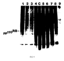
- a protein product of the RB gene was previously identified as a nuclear phosphoprotein of about 110 kd (pp110 RB ) using antibodies generated against selected epitomes predicted from the RB cDNA sequence, Nature 329:642 (1987).
- pp110 RB has been shown to form a tight association with large T antigen and E1A, the transforming proteins of DNA tumor viruses SV40 and adenovirus respectively, Nature 334:124 (1988); Cell 54:275 (1988).
- the RB gene product, or a complex containing it, has been found to have DNA binding activity, Nature 329:642 (1987).
- the prognostic tool is extremely useful in screening a population, to determine which persons may have a predisposition toward cancer.
- the person can be monitored at short intervals for the early signs and symptoms of cancer. If such is found, appropriate procedures, such as surgery, can be undertaken at an early date.
- biotechnical techniques it would be highly desirable to have prophylactic and/or therapeutic treatments for cancer, by utilizing biotechnical techniques. Moreover, it would be important to have such biotechnical modalities, which are effective for many different forms of cancer, with little or no side effects. It would also be desirable to have techniques for proving that certain environmental substances, such as cigarette smoke, cause cancer. Having this type of information could also be used to help people avoid coming into contact with cancer causing substances, since these substances would be proven, rather than merely suspected, of playing a role in oncogenesis.
- the presently claimed invention comprises the use of a cancer suppressing gene or fragment thereof encoding functional cancer suppressor protein for the manufacture of a medicament comprising said cancer suppressing gene or fragment thereof encoding functional cancer suppressor protein to suppress the neoplastic phenotype of a mammalian cancer cell lacking endogenous wild-type cancer suppressing protein encoded by said cancer suppressing gene.
- the presently claimed invention comprises a medicament for use in the suppression of the neoplastic phenotype of mammalian cancer cells lacking endogenous wild-type cancer suppressing protein, comprising a wild-type cancer suppressing gene or fragment thereof encoding functional cancer suppressor protein which gene or fragment encodes said endogenous wild-type cancer suppressing protein.
- the present invention provides a means for prophylactically treating individuals having a genetic predisposition to cancer.
- the presently claimed invention provides the means and methods of producing a medicament for treating cancer which reduces the need for radiation and/or chemotherapy. In addition, it may be employed at a very early stage, after a genetic predisposition to cancer has been discovered, but before the onset of tumorigenesis.
- a further advantage of the presently claimed invention is that it utilizes genetic materials which are smaller than entire chromosomes and are generally more stable and more easily cloned.
- the presently claimed invention relates to the diagnosis and production of medicaments for the treatment of cancer as well as to medicaments themselves.
- One possible way in which we envisage using the presently claimed invention is to identify a suitable patient by determining the susceptibility of a patient to cancer, in some cases before a cancer phenotype is expressed.
- the function of the gene can be determined by observation, under controlled circumstances, of the phenotypic expression of the gene.
- One technique for evaluating phenotypic expression is to test for the presence of the cancer suppressing gene's product.
- a preferred technique is to develop an antibody for the gene product which is capable of forming an immunocomplex with the product thereby providing an accurate test for the presence and amount of protein of gene product in a tissue sample.
- the gene itself can be cloned and subsequently used as a pharmaceutical product according to the therapeutic methods of the present invention.
- the cloned cancer suppressing gene has a utility in addition to its therapeutic applications since it may also be utilized prophylactically.
- cancer suppressing gene function may be determined prior to any expression of the cancer phenotype in the individual and, if gene deficit or absence is determined, a healthy cloned cancer suppressing gene can be utilized prophylactically in the place of the absent or defective gene.
- one aspect of the invention is the use of a mouse model which has been genetically altered so that the model can be utilized as a reliable method for evaluating the therapeutic and prophylactic use of cancer suppressor genes.
- the presently claimed invention is based on the substantial body of scientific evidence which indicates that carcinogenesis is associated with genetic alterations in tumor cells. These alterations may be due to mutation, gene inactivation, suppression, deletion or other causes.
- esterase D is important because its genetic locus coincides with the location of the retinoblastoma gene (RB).
- RB retinoblastoma gene
- cloned esterase cDNA is useful as a probe to clone the RB, and as a prognostic tool for retinoblastoma, Wilson's disease and other diseases controlled by genes located at the 13 chromosome 13q14:11 region.
- esterase D and the retinoblastoma gene are located in the same chromosomal region, evaluation of RB gene function, and its chromosomal patterns, was possible.
- chromosomal walking using the esterase D cDNA clone as the starting point, the RB gene was isolated and its nucleotide sequence was determined.
- the RB gene has a regulatory function and that its presence and normal function prevents the development of retinoblastoma.
- absence, malfunction or inactivation of the RB gene causes the development of, or genetic predisposition and susceptibility to, retinoblastoma.
- RB gene absence, malfunction or inactivation is the primary cause, not only for hereditary and acquired retinoblastoma but for other cancers as well.
- identification of the exact RB gene location and isolation, identification, sequencing and cloning of the RB gene provided a capability for diagnosis and treatment of retinoblastomas and their secondary tumors. It also provided a method for diagnosis and treatment of other cancers related to RB gene function.
- an advance in the diagnosis of retinoblastoma, and a predisposition toward retinoblastoma and other cancers occurred with the identification of a phosphoprotein.
- the phosphoprotein is associated with DNA binding activity located in the nucleus.
- the phosphoprotein, identified as ppRB 110 plays a role in inhibiting the oncogenic activity of genes other than the RB gene and, in addition, it restrains malignant cell growth.
- the identification, isolation, and determination of the nucleotide sequence and cloning of the RB gene, together with the identification of its phosphoprotein product has many uses.
- RB gene protein product which can then be used as an antigen in obtaining specific anti-protein antibody.
- This antibody can be used as a diagnostic immunomarker for the investigation of the presence or absence of the RB gene protein in examined tissue. If the protein is present, the RB gene is intact and retinoblastoma is not present. If, on the other hand, the protein is absent or altered, the deficient RB gene is indicated and resulting retinoblastoma or other cancers or susceptibility to them is diagnosed.
- the RB cDNA or genomic DNA is used as a probe to determine whether a deficit exists in the chromosomal locus of the RB gene.
- immunoscreening of tissue biopsy with specific anti-ppRB 110 antibody is also practical. Both diagnostic methods have application for screening families with a history of hereditary retinoblastoma and for screening of their children.
- the methods may be used for prediction of development of secondary cancer, such as osteosarcoma, fibrosarcoma, glioblastoma, breast cancer and others, whether or not connected with retinoblastoma.
- tumorigenesis can be suppressed by providing cloned cancer suppressing genes, such as RB genes, or the cancer suppressing gene protein product, such as ppRB 110 , after a defective, inactive or absent cancer suppression gene has been diagnosed.
- cloned cancer suppressing genes such as RB genes
- the cancer suppressing gene protein product such as ppRB 110
- ppRB 110 the cancer suppressing gene protein product
- these substances can be provided through molecular induction or gene transplanting or RB cDNA to the individual in need.
- these methods in addition to their prophylactic value, have application to a method of arrest of tumor development in the individual.
- mutant mice become useful models in testing for carcinogenicity of environmental substances.
- such mice could be exposed to cigarette smoke, artificial sweeteners or a myriad of other suspected carcinogens.
- Tumor development in the mice would be a positive indication of the carcinogenicity of the substance tested.
- the availability of the mutant mouse models thus provides a means for determining which environmental substances should be avoided.
- mutant mouse models In addition to their value as models for testing environmental substances, the mutant mouse models have value in cancer therapy studies.
- a known chromosome associated with a particular cancer is identified.
- the chromosome carrying the defective cancer suppressing gene may be determined by examination of phenotypic expression in the absence of the chromosome. Further, such examination can be performed after a portion of the suspected chromosome has been altered by chemical or other techniques or by excision of portions of it.
- understanding of the control of genetic expression has been based largely on the introduction of genes or other defined segments of DNA into cells and the assessment of the genes' ability to function normally.
- the locus is selected on the chromosome for the cancer suppressing gene to be determined. This is accomplished, for example, by the use of probes inserted at random at suspected location of the chromosome in order to establish the location of the gene. This technique is limited because of the large number of genes associated with a typical eukaryotic cell chromosome.
- the chromosomal DNA containing the suspected gene may be cut into fragments, for example, by mechanical excision, or by use of suitable restriction enzymes, or other means. This technique may be employed to establish gene location by, once again, ultimately evaluating the phenotypic expression of the DNA fragments.
- a preferred method of identifying the location of the cancer suppressing gene is through the utilization of a marker gene.
- the marker gene is located in close proximity to the locus of the cancer suppressing gene and, in addition, the marker gene is a readily observable phenotypic expression.
- chromosomal walking techniques are employed in order to analyze portions of the chromosomal DNA in order to locate the cancer suppressing gene. Chromosomal walking depends on isolating a small segment of DNA from one end of a first recombinant and using this piece of DNA as a probe to rescreen the phage or cosmit library in order to obtain a recombinant containing that piece of DNA and the net portion of the genome.
- the second recombinant is then used to obtain a third, and so in, to yield a set of overlapping cloned segments.
- the chromosome walking technique may be utilized bidirectionally along the chromosome, starting with the marker gene.
- esterase D gene Since it was known that the esterase D gene was located in close proximity to the RB gene, it was used as a starting point for identifying the RB gene by chromosomal walking. Because of its proximity to the RB gene, the esterase D gene was ideal as a marker gene. Not only did its proximity reduce the chromosomal walking necessary to locate the cancer suppressing gene, genetic alterations of the esterase D gene would result in, not only an altered esterase D gene phenotype but, in addition, an altered cancer suppressing gene phenotype.
- esterase D cDNA The amino acid sequence of human esterase D enzyme was identified as was the nucleotide sequence of esterase D cDNA. The chromosomal location of the gene was located and cloned esterase D cDNA was utilized as a genetic marker.
- Esterases belong to the family of nonspecific enzymes that catalyze the hydrolysis of esters.
- Human esterase D (ESD) is one member of the esterase family distinguishable by its electrophoretic mobility and its relative specificity for methylumbelliferyl esters as substrate.
- Human ESD is a dimeric enzyme in that it displays several phenotypes as a result of the expression of codominant autosomal alleles, primarily allele ESD 1 and ESD 2.
- the polymorphic nature of human ESD has been of value in the use of human ESD as a marker in studies of population genetics Nature , 304:451-453 (1983); and Am. Hum. Genet., 39:1-20 (1975).
- ESD enzyme depends on the normal function of the ESD gene. Consequently, absence, complete or partial inactivation, deletion of one ESD allele, mutation or other alterations in ESD sequences will result in decreasing ESD activity. For example, the tissues of individuals with a deletion of one chromosome 13 show only 50% of the ESD activity of that found in the healthy individuals possessing a normal set of two chromosomes 13, Science, 219:973-975 (1983).
- the genetic locus of ESD was mapped to the chromosome 13q14:11 region by correlating the loss of enzyme activity with deletions of chromosome 13, Science, 208:1042-1044 (1980).
- the regional assignment of ESD to 13q14:11 region coincides with the location of the retinoblastoma (RB) gene, shown to be involved in the tumorigenesis of retinoblastoma, Am. J. Dis. Child., 132:161-163 (1978); Science, 219:971-973; Science, 213:1501-1503 (1981).
- the localization of the ESD and RB genes in the same chromosomal region provides an advantageous approach for evaluation of the RB gene functioning, for discovery of RB chromosomal patterns, for cloning of the RB gene, for isolation of the RB gene and for identifying the RB gene sequence by chromosomal walking, using ESD as the starting point Science , 235:1394-1399 (1987).
- the tight linkage between these two genes allows the ESD gene to serve as a crucial marker in elucidating the behavior of the RB gene, Science, 219:973-975 (1982); and Nature, 304:451-453 (1983).
- Wilson's disease also known as hepatolenticular degeneration, is a hereditary disease of ceruloplasmin formation transmitted as an autosomal recessive. It is characterized by gross reduction in the incorporation of copper in ceruloplasmin resulting in decreased serum ceruloplasmin and copper values, and increased excretion of copper in the urine.
- the identified and cloned ESD cDNA thus would provide a valuable marker in the identification and sequencing of both the RB gene and the Wilson's disease gene would lead, eventually, to diagnosis and treatment of these disease.
- the purified human ESD obtained was subsequently used in the preparation of specific rabbit anti-esterase antibody.
- the antibody was utilized to identify and isolate ESD cDNA clones through a technique for cloning genes by using specific antibody as probes and for isolating unknown proteins encoded by cloned DNA.
- the method used an expression vector ⁇ gt11 (lac5 nin5 cI857 S 100_ that permitted insertion of foreign DNA into ⁇ -galactosidase structural gene lac Z and promoted synthesis of hybrid fusion proteins, DNA, 3:437-447 (1984).
- Chromosome mapping of the ESD gene to the chromosome 13q14.11 region was accomplished by correlating loss of the ESD enzyme activity with known deletions on chromosome 13 of various mutant cells.
- esterase D cDNA As probe, it was found that (i) the size of the esterase D mRNA is 1.3-1.4 kb; (ii) the gene is around 20-35 kb, indicating the presence of large introns scattered over this genome; and (iii) the esterase D gene is indeed located at 30 chromosome 13q14 region. Also, the deduced amino acid of the esterase D gene was unique when compared to 4000 other well-characterized proteins.
- mapping data shows that the ESD gene is located at chromosome 13q14.11 region with no meiotic recombination observed with the RB gene.
- the human esterase D was successfully purified.
- the polyclonial anti-esterase D antibody was prepared, and oligonucleotide probes complementary to certain ESD polypeptides were constructed.
- the complete amino acid sequence of ESD protein was determined, and ESD cDNA was cloned.
- the complete nucleotide sequence of both ESD cDNA and the ESD gene were identified and the ESD gene was localized.
- the above described immunologic reaction was then used in identification and the isolation of the ESD cDNA clones from two ⁇ gt11 cDNA libraries.
- the technique of cloning genes by using specific antibody as probes and for isolating unknown proteins encoded by cloned DNA is well known.
- the method uses an expression vector, ⁇ gt11 (lac5 nin5 cI857 S 100) that permits insertion of foreign DNA into B-galactosidase structural gene lac Z and promotes synthesis of hybrid fusion proteins, DNA, 3:437-447 (1984).
- ESD ESD was shown to be present in many bodily tissues
- the mRNA coding for ESD would be readily present in tissue extracts and/or in certain tissue tumors.
- Human hepatoma and human placenta tumors both have a relatively high level of expression of ESD mRNA and were, therefore, particularly suitable for the detection of specific ESD cDNA clones in the library. Therefore, these two tumors were chosen for construction of the ESD cDNA libraries in ⁇ gt11 vector.
- RNA blotting analysis was performed and the size of mRNA of two cell lines was found to be ⁇ 14,5S(1.3-1.4 kb).
- a distribution of the ESD gene in the human genome was determined by Southern genomic blotting analysis using 32 P-labeled EL22 clone. It was found that the esterase D gene was distributed over 20-40 kb in the human genome. The combined size of the DNA fragments with positive hybridization was 20-40 kb, indicated that there are large intron sequences in the ESD genome. This was subsequently confirmed by characterizing the complete genomic esterase D clone.
- the marker gene After identifying and cloning the marker gene, its associated cancer suppressing gene is then identified and cloned. Chromosomal walking techniques are utilized to locate the cancer suppressing gene to be found. While the examples herein disclosed relate to use of the esterase D gene as a marker and of the evaluation of the RB gene in oncogenicity, it is understood that the examples disclosed herein will equally apply to the marker genes and the respective cancer suppressing genes as breast cancer suppressing genes, Wilm's tumor suppressing genes, Beckwith-Wiedemann syndrome suppressing gene, bladder transitional cell carcinoma suppressing gene, neuroblastoma suppressing gene, small cell lung carcinoma suppressing gene, renal cell carcinoma suppressing gene, acoustic neuroma suppressing gene, colorectal carcinoma suppressing gene, and others.
- the chromosome walking technique performed as previously described can be utilized for location identification, purification and cloning of the above cancer suppressing genes.
- analysis of the cancer suppressing gene activity can be determined by observation of the gene's phenotypic expression.
- the RB gene role in suppressing retinoblastoma has been determined.
- the genetic control of retinoblastoma and suppression of carcinogenicity were evaluated by means of the cloning, isolation, identification and sequencing of the retinoblastoma gene.
- the cloned retinoblastoma gene cDNA was prepared and used as a tool for diagnosing retinoblastoma, osteosarcoma and fibrosarcoma.
- Development of the retinoblastoma gene led to the therapeutic application whereby a defective gene in cancer cells may be replaced with a normal gene thereby suppressing cancer formation.
- carcinogenesis is associated with genetic alterations in tumor cells. Some of these alterations may occur in precursor somatic cells during the life of an individual, while other mutations might be inherited from a parental germline. The latter type of inheritance would explain cases of familial cancer and inherited cancer predisposition. Cancers with known familial occurrence include retinoblastoma, nephroblastoma (Wilm's tumor), neuroblastoma, osteosarcoma, renal cell carcinoma, melanoma, and breast cancer.
- Oncogenes were initially defined in tumor-inducing retroviruses and tumor DNA capable of transforming non-neoplastic cells in culture. Most oncogenes are activated homologues of protooncogenes that exist in normal cells.
- Retinoblastoma is an intraocular cancer of early childhood that arises from the developing retina. It has been reported that its incidence is about 1 in 20,000 live births and it is the most common intraocular tumor of the pediatric age group. Two forms of retinoblastoma are distinguished on a genetic basis. The hereditary form is an autosomal-dominant cancer susceptibility trait: each offspring of a carrier parent has a 50% chance of inheriting the trait, and 90% of carriers will develop retinoblastoma. Multiple or bilateral retinal tumors are indicative of, and typical for, hereditary retinoblastoma.
- retinoblastoma has been a prototypic model for the study of genetic determination in cancer.
- retinoblastoma could result from as few as two "hits", or mutational events and it has been hypothesized that two hits served to inactivate both alleles of a singe gene (RB) that essentially functioned to suppress retinoblastoma formation.
- RB singe gene
- An individual inheriting a mutant RB allele in all somatic cells would be predisposed to getting retinoblastoma by an additional mutation of the other RB allele in one precursor cell (retinoblast).
- retinoblast retinoblast
- both RB alleles would have to be inactivated by two independent somatic mutations in a single retinoblast.
- This model could explain both the earlier onset and multiplicity of tumors in predisposed individuals.
- the validity of this hypothesis remains to be demonstrated at the molecular level.
- the RB gene has a regulatory function and that its presence and normal function prevent the development of the retinoblastoma.
- absence, malfunctioning or inactivation of the RB gene causes the development of, or genetical predisposition and susceptibility to, the retinoblastoma and is believed to be the primary cause for both hereditary and acquired retinoblastoma, and for the secondary malignancies often recurring in retinoblastoma patients such as osteosarcoma, and fibroblastoma.
- esterase D cDNA fragments By generating specific antisera and partially sequencing the protein, esterase D cDNA fragments have been identified. Also available were anonymous DNA probes mapping to 13q14, such as H3-8, H2-42 and 7D2 that were isolated by random selection from chromosome 13-specific libraries.
- the esterase D gene has been located, identified, sequenced and cloned. Further, it has been located in close vicinity to the RB gene. Therefore, it is useful as the starting point for identifying the RB gene by chromosomal walking. DNA fragments isolated from this process can then be used as probes to examine qualitative or quantitative differences in mRNA from fetal retinal cells and retinoblastoma cells. Detection of such differences would provide evidence that somatic mutations occur in the RB gene of tumor cells. The DNA fragments corresponding to the defective mRNA are the best candidates for the RB gene. Moreover, the availability of mutant cells with known deletion in 13q13.1-14.11 and 13q14.11-q22, respectively, enables determination of the correct direction of walking toward the RB gene.
- Candidate RB genes were used as probes in RNA blotting to detect mRNA transcripts and clones RB-1 and RB-5 were hybridized with a 4.6 kb mRNA fragment obtained from the normal retinal and placental tissues. Hybridization under the same conditions with mRNA obtained from the retinoblastoma mRNA transcripts was not observed at all.
- tumors not related to retinoblastoma namely neuroblastoma and medulloblastoma, displayed normal 4.6 kb mRNA transcripts.
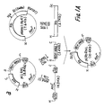
- phage clones were isolated from a human genomic DNA library with RB-1 and RB-5 as probes and were subsequently characterized by restriction mapping and hybridization to subfragment cDNA probes.
- a restriction map of the RB gene was then constructed showing that the RB gene consists of at least 12 exons scattered over more than 100 kb of DNA.
- RNA messenger RNA
- kb 4.6 kilobases
- esterase D esterase D
- mRNA messenger RNA
- kb 4.6 kilobases
- tumor-specific alterations in expression Transcription of this gene was abnormal in six of six retinoblastomas examined.
- full-length RB mRNA was present, in human fetal retina and placenta tumors, and in other tumors such as neuroblastoma and medulloblastoma.
- DNA from retinoblastoma cells had a homozygous gene deletion in one case and hemizygous deletion in another case, while the remainder were not grossly different from normal human control DNA.
- the RB gene contains 27 exons distributed in a region of over 200 kb. Sequence analysis of complementary DNA clones yielded a single long open reading frame that could encode a hypothetical protein of 928 amino acids.
- a computer assisted search of a protein sequence database revealed no closely related proteins.
- Features of the predicted amino acid sequence included potential metal-binding domains similar to those found in nucleic acid-binding proteins. These results suggested involvement of the RB gene as a recessive genetic mechanism for regulation of human cancers.
- esterase D which is linked to the retinoblastoma susceptibility locus in band 13q14.11 within an estimated 1500 kilobase range.
- the esterase D cDNA clone EL-22 was used as a probe to isolate its genomic DNA clones. Distal DNA segments of these genomic clones were used to isolate additional genomic clones. At 20 kb intervals in walking regions, unique sequences were identified that were used as probes to isolate cDNA clones from fetal retina and placenta libraries. A similar strategy using H3-8 as probes was performed to isolate the candidate genes. A bidirectional chromosome walk covering hundreds of kb has been established by alternately screening genomic and cDNA libraries.
- Candidate RB genes were used as probes in RNA blotting analysis to detect relevant messenger RNA (mRNA) transcripts
- polyadenylated RNA was prepared from human fetal retinas (obtained by dissection of about 25 first- or second- transfer fetal eyes) and from portions of normal human placentas. Since the primary retinoblastoma tumor samples large enough to yield sufficient mRNA for analysis are not usually available, polyadenylated RNA was isolated from cultured cells of six retinoblastomas. Additionally, polyadenylated RNA was isolated from three neuroblastomas and one medulloblastoma and from a primary medulloblastoma specimen.
- mRNA messenger RNA
- Esterase D transcripts (1.4kb) were detected in all tumor and tissue samples. This finding was consistent with the known "constitutive" expression of esterase D. Esterase D hybridization was subsequently used as a positive control.
- the numerous large Hind III bands seen on DNA blotting analysis suggested that the RB locus was spread over a rather large genomic region.
- more than 20 phage clones were isolated from the human genomic DNA library with RB-1 and RB-5 as probes. These clones were characterized by restriction mapping and hybridization to subfragment cDNA probes.
- the Hind III restriction map of the RB gene was constructed.
- the RB gene consists of 27 exons scattered over more than 200 kb of DNA.
- One large intron of at least 50 kb is located between exons from the RB-1/RB-5 overlap region.
- the RB gene expression while specifically altered in retinoblastoma, was not confined to normal fetal retina but was also seen in at least one other normal unrelated tissue, placenta.
- mRNA from fetal and adult rat tissues were prepared and analyzed by RNA blotting.
- a 4.6-kb mRNA transcript (presumably the normal size for rat) was detected in all tissues, though quantity varied markedly.
- the revised RB cDNA sequence had an additional methionine codon at nucleotide 139.
- the predicted RB protein had 928 amino acids and a molecular weight of 110 kD.
- the second in-frame methionine was at base 346. Since the nucleotide sequence at the first ATG is not typical of other known mRNAs, the start codon assignment should be regarded as tentative. Later, the in vitro translation results favored the first methionine as the authentic initiation site.
- the predicted protein sequence included ten potential glycosylation sites but a candidate transmembrane domain (at least 20 consecutive hydrophobic residues) was not found.
- the amino acid hydropathy plot showed a mildly hydrophobic region near the putative amino terminus and a hydrophilic region at the carboxyl terminus.
- Two pairs of short amino acid sequences that were bracketed by cysteine and histidine residues in the manner of metal-binding domains found in nucleic acid-binding proteins were identified.
- a region of 54 amino acids from position 663 to 716 contains 14 proline residues (26%).
- proline-rich regions have also been observed in nuclear oncogene proteins myc and myb. While the significance of these observations is not well established, they suggested that the RB gene product may be a nucleic acid-binding protein.
- the RB protein ppRB 110 as been found to be primarily located in the cell nucleus.
- the retinoblastoma gene has been previously found to be of a recessive nature. Tumor-specific alterations in gene expression provide the best evidence for identifying this gene as RB, and the examples of homozygous deletion and absence of mRNA expression support its postulated recessive nature.
- Hereditary retinoblastoma patients have a high incidence of osteosarcoma as a second primary cancer.
- Inactivation of the retinoblastoma susceptibility (RB) gene has been implicated in the genesis of osteosarcoma by chromosomal homozygosity of the 13q14 region found in several osteosarcoma patients with or without retinoblastoma history.
- RB retinoblastoma susceptibility
- nephroblastoma Wilm's tumor
- hepatoblastoma hepatoblastoma
- embryonal rhabdomyosarcoma hepatoblastoma
- neuroblastoma Like retinoblastoma, all of these tumors resemble massive overgrowth of tissues found in normal embryogenesis. Nephroblastoma has been associated with deletions of chromosome region 11p13, and adjacent polymorphic markers become homozygous in these tumors.
- retinoblastoma By analogy to retinoblastoma, a class of "cancer suppressing genes" has been postulated to explain other types of inherited cancers, and tumor-specific chromosomal deletions. Retinoblastomas and osteosarcomas often occurred in the same patients, and several osteosarcomas (from patients both with and without retinoblastoma history) demonstrated a loss of heterozygosity of chromosome 13 markers. A few breast cancers also showed specific loss of chromosome 13 heterozygosity.
- Loss of heterozygosity has been found at chromosome llp in Wilm's tumor (consistent with known deletions), tumors of the Beckwith-Wiedmann syndrome, transitional cell carcinoma of bladder, and, again, breast cancer.
- other suppressing loci have been implicated in neuroblastoma, small cell lung carcinoma, renal cell carcinoma, acoustic neuroma, and colorectal carcinomas.
- the RB gene was initially identified on the basis of altered RB gene expression in retinoblastoma tumors compared to that of normal tissues. Many additional tumors and neoplastic cell lines have been examined, and several with either DNA rearrangements, altered RB mRNA, or absent RB protein have been found. For example, of eight osteosarcoma cells lines tested, three (G292, SAOS2 and OHS) showed abnormalities at the protein level. G292 expressed RB protein of increased molecular mass, whereas SAOS2 and OHS completely lacked the RB protein. Both osteosarcoma cell lines without RB protein had shortened RB mRNA transcripts, and G292 had both shortened and normal-sized RB mRNA transcripts.
- Cell line SAOS2 had homozygous deletion of the 3' end of the RB gene, involving exon-containing Hind III fragments of exons in an internal 7.5 kb Hind III fragment. However, G292 had a grossly normal RB gene structure.
- RB protein was also absent from one synovial sarcoma cell line (GHM) in which norma-sized RB mRNA was present.
- GHM DNA was also grossly normal by DNA blotting analysis with RB cDNA probes. This cell line constitutes the first known example of RB gene inactivation apparently solely at the protein level.
- SDSY1 synovial sarcoma tumor
- Both normal-sized and lengthened RB MRNA transcripts (6.5 kb) were observed by RNA blotting and is apparent that normal-sized transcripts may be derived from non-neoplastic cells intermixed in whole tumors.
- the RB gene inactivation in two of nine human breast cancer cell lines was found.
- the RB gene of one cell line had a homozygous internal duplication of 5 kilobase (kb) region containing exons 5 and 6.
- the RB mRNA transcript was correspondingly lengthened, and it was inferred that its translation was terminated prematurely due to a shifted reading frame.
- the other cell line had a homozygous deletion of the RB gene that removed the entire gene beyond exon 2.
- the RB gene product, pp110 RB was not detectable in either cell line by immunoprecipitation with specific antibodies. Also reported recently was loss of RB gene expression in some cell lines derived from small cell carcinomas of the lung.
- the identification, isolation, determination of the exact nucleotide sequence and cloning of the RB gene has a multiple utility.
- the primary utility lies in the sequence transcription into its corresponding mRNA which is in turn translated into RB gene protein product.
- Protein product can then be used as an antigen in obtaining the specific anti-RB protein antibody.
- Antibody are then used as a diagnostic immunomarker for the investigation of the presence or absence os the RB gene protein in the examined tissue. If the RB protein is present, the RB gene is intact and retinoblastoma not present. If, on the other hand, the protein is absent or altered, the deficient RB gene is indicated and resulting retinoblastoma or other cancers or susceptibility thereto is indicated.
- the sequence of the RB gene can be further utilized in producing the specific RB gene cDNA clones which can be used as the genetic markers and probes in isolation, identification and sequencing of other related genes or genes located in the proximity of the RB gene the function of which is as yet unknown.
- the control and regulatory function of the RB gene is exerted through the RB protein which is instrumental in inhibiting other gene's oncogenic activity and restraining the malignant cell growth.
- the hypothetical protein predicted from the nucleotide sequence was expected to have MW about 106 kD.
- the immunoprecipitated protein has a MW about 110-114 kD.
- the complete RB protein amino acid sequence is illustrated in Table 1. This complete sequence obtained from the newly reconstructed clone which contains the most 5' end stretch missing in the original cDNA clone Science, 235:1394-1399 (1987).
- RB cDNA sequence Science, 235:1394-1399 (1987) contained a long open reading frame from nucleotide 1 through 2688, which when translated from the first methionine codon yielded a hypothetical protein of 816 amino acids and molecular weight 98 kD.
- Recently another isolated RB cDNA clone contained an additional 234 base pairs on the 5'end.
- the revised RB cDNA sequence still maintained the same open reading frame as in the original clones, and an additional methionine codon was found at nucleotide 139. When this codon was used as an initiation codon, the predicted RB protein had 928 amino acids and a molecular weight of 110 kD - identical to the apparent M.W. determined by SDS-PAGE.
- the additional 5' sequence contains a GC-rich region that translates into an unusual cluster of alanine and proline residues.
- the cloned gene can be used to obtain a gene product useful in diagnosing for cancer related to the cancer suppressing gene.
- the technique includes initially developing a product which can react with the cancer suppressing gene product of a patient's tissue sample.
- the reaction between the product is observed to determine the presence or absence of the normal phenotypical expression of the cancer suppressing gene of the tissue sample.
- an antibody is developed.
- the antibody which is specific for the cancer suppressing gene protein product, is used to react with the cancer suppressing gene protein product in a patient's tissue sample for determining whether or not the cancer suppressing gene is functioning properly.
- proper cancer suppressing gene function is demonstrated when an immunocomplex between the antibody and the cancer suppressing gene protein product is formed. Failure of the immunocomplex is indicative of defective, abnormal or absent cancer suppressing gene protein product.
- failure of the antibody to form an immunocomplex with the patient tissue sample substrate is indicative of a defective or absent cancer suppressing gene in the patient.
- the specific antibody is produced, in general, by utilizing the normal cancer suppressing gene protein product as an antigen according to known techniques. Other conventional techniques may also be employed for producing the desired antibodies.
- the protein product of the RB gene has been identified.
- immunoprecipitation of the phosphoprotein was accomplished utilizing preimmune rabbit anti-sera and, as disclosed in said application, a protein with MW 110-114kD was immunoprecipitated with anti-ppRB 110 IgG.
- the clone is useful diagnostically.
- patients can be screened for their predisposition toward cancer, by observing the phenotypical expression of a cancer suppressing gene in a tissue sample taken from a patient.
- phosphoprotein product of the retinoblastoma susceptibility gene has been located primarily in the cell nucleus having DNA binding activity. Its amino acid sequence has been identified.
- the phosphoprotein, identified as ppRB 110 is utilized in diagnosing retinoblastoma and other cancers involving the retinoblastoma gene, and, to some extent, in the treatment of these cancers and in evaluating the oncogenicity of other genes.
- amino acid sequence of a protein can be determined by the genetic code of the particular gene responsible for that particular protein. Therefore, in order to isolate the protein, to determine its exact amino acid sequence and to determine its physiological function in the body, it is necessary to isolate and localize the responsible gene, to clone it and to sequence the cDNA which are useful in identification of the gene's specific protein product.
- retinoblastoma gene and encoding of the amino acid sequence was identified at 13 chromosome, 13 q14:11 region.
- the RB-1 clone was hybridized with 4.8 kb mRNA transcript in human fetal retina and placenta. In retinoblastoma samples, RB-1 clone either detected an abnormal mRNA transcript or the mRNA transcripts were not observed at all. Subsequently identified RB-5 clone, with a 3.5 kb insert, gave identical results as RB-1 in mRNA hybridization. Restriction enzyme analysis suggested that RB-5 and RB-1 clones overlapped in a 0.4 kb region and both together defined a DNA segment of about 4.6 kb, a size close to that of the normal RB mRNA transcript.
- Nucleotide sequence analysis of clones RB-1 and RB-5 was performed by the dideoxy-terminator method described in Proc. Natl. Acad. Sci., 74:5463-5467 (1977) and yielded the reconstructed complete cDNA sequence.
- Different deletion templates were generated by the "cyclone" method in single stranded M13 phage clones, which yielded greater than 95% of the sequence. The remaining gaps were sequenced by primer extension in both strands. The complete sequence identified in this way contained 4,523 nucleotides.
- nucleotide sequence analysis of RB cDNA clones demonstrated a long-open reading frame encoding a hypothetical protein with features suggestive of a DNA binding function. It was identified and used as an antigen for obtaining specific antibody and to determine its predicted DNA binding.
- the purified anti-RB IgG antibody was used for immunoprecipitation, immunostaining and localization of RB protein. It was believed that the antibody will be useful for diagnostic identification of RB protein in human tissue samples.
- LAN-1 neuroblastoma cell line normal human fibroblasts, human hepatoma Alexander cell line and osteosarcoma U20S cell line were used as positive controls containing normal RB mRNA. All these cells were obtained from the American Type Culture Collection (ATCC), Inc. depository. Cell lines with expected shortened or absent RB mRNA, such as retinoblastomas cell lines Y79 (ATCC), RB355 (Gifted from Robert Philips, Toronto, Canada), WERI-1, WERI-24, and WERI-27 (Gifted from T. Sery Wills' eye hospital, Philadelphia) were used as negative controls.
- Cells from all human cell lines were metabolically labeled with 35 S-methionine according to procedure described in J.Virol, 38:1064-1076 (1981).
- Labeled cell mixtures were immunoprecipitated with 1-20 ul, preferably 10 ul, of from 50 ug/ml - 200 ug/ml, preferably 100 ug/ml of anti-RB antibody IgG using the procedure described in J. Virol, 38:1064-1076 (1981).
- the RB proteins immunoprecipitated with rabbit anti-RB IgG were analyzed by SDS/polyacrylamide gel electrophoresis and auto-radiographed. The results indicated immunoprecipitation of the normal positive, i.e. RB protein containing cell lines labeled with 35 S-methionine.
- the cells from several vertebrate species such as QT6 (quail), NIH/3T3 (mouse), Rat-2 (Rat) and cos (monkey) were labeled with 32 P-phosphoric acid and proteins were immunoprecipitated with anti-RB IgG. Antigenically related proteins were detected in all cells with apparent similar molecular weights of 108 kD in quail, 120 kD in mouse, 128 kD in rat and 108-110 kD in monkey, as compared to 110-114 kD in human cells.
- the predicted whole amino acid sequence of the ppRB 110 protein has several characteristics similar to those appearing in other oncogenes. Therefore, the subcellular localization of the ppRB 110 was investigated by cellular fractionization to identify the distribution of ppRB 110 among the nuclear, cytoplasmic, or cell membrane fractions.
- the osteosarcoma cell lines U20S known to have an advantageous cell meorphology for immunohistochemical staining were used.
- the U20S cells were immunoprecipitated with anti-ppRB 110 IgG.
- the U20S cells were immunoprecipitated with preimmune IgG. Both groups were then incubated with rhodamine conjugated goat anti-rabbit IgG obtained commercially from Sigma. Immunofluorescence was observed in cells reacted with anti-ppRB 110 IgG, namely in the cell nucleus. Cells reacted with preimmune control did not show any fluorescence.
- ppRB 110 plays an important regulatory function in regulating other genes and has a DNA binding activity.
- Certain cell lines, particularly those from tumors others than retinoblastome, such as neuroblastome LAN-1 cells were radioactively labeled with 32 P-phosphoric acid. Cellular lysates of these labeled cell mixtures were separated by single or double stranded calf thymus DNA-cellulose columns according to the method described in Mol.Cell. Biol., 6:4450-4457 (1986).
- ppRB 110 binds only to a limited number of DNA sites that are easily saturated. It has been previously shown that other protooncogens such as c-myc, n-myc, c-myb and c-fos are nuclear phosphoproteins with DNA binding activity Mol. Cell. Bill., 6:4450-4457 (1986), Nature, 296:262-266 (1982). Oncogenic activation of these proto-oncogenes occurs by deregulation of gene expression or by structural modification, and the gene product is essential for oncogenicity.
- protooncogens such as c-myc, n-myc, c-myb and c-fos are nuclear phosphoproteins with DNA binding activity Mol. Cell. Bill., 6:4450-4457 (1986), Nature, 296:262-266 (1982). Oncogenic activation of these proto-oncogenes occurs by deregulation of gene expression or by structural modification, and the gene product is essential for oncogenicity.
- radioimmunoassay techniques were utilized, it is to be understood that the methods, such as enzyme immunoassay (Western blotting analysis), immunocytochemical assay, and others, are also suitable.
- ppRB 110 It is the ppRB 110 absence, and not its presence, that appears to be oncogenic due to the partial or complete inactivation of the RB gene. Therefore, the presence of the ppRB 110 somehow suppresses the oncogenic activity of other genes and disallows malignant cell growth.
- the ppRB 110 is thus an important regulatory protein which may prevent and inhibit, by its presence, and trigger, by its absence, the malignant growth. Thus, the importance of ppRB 110 is in regulating other genes. The absence or loss of ppRB 110 mediates oncogenicity.
- ppRB 110 The role of ppRB 110 as a mediator of oncogenicity leads to several practical applications.
- the presence or absence of the ppRB 110 serves as a diagnostic tool in determination of the presence or predisposition to the retinoblastoma and other RB genes involved tumors of the animal fetus or embryo.
- early diagnosis allows an early warning and treatment of retinoblastoma and other tumors with the possibility of preventing development of secondary tumorigenesis.
- ppRB 110 to diagnose the presence or predisposition to retinoblastoma is through immunoscreening of the tissue biopsy with specific anti-ppRB 110 antibody.
- the bioptic tissue is either radioactively labeled and immunoscreened or the proteins extracted from bioptic tissue may be blotted on nitrocellulose filter and probed with labeled antibody according to methods known in the art as Western blotting.
- diagnostic methods will be used particularly for screening families with a history of hereditary retinoblastoma.
- the diagnostic method is useful also for prediction of the development of secondary cancer, such as for example osteosarcoma, fibrosarcoma, glioblastoma, breast cancer, whether or not connected with retinoblastoma.
- ppRB 110 Another use is for tumorigenesis suppression where the absent ppRB 110 will be provided through molecular induction and gene transplanting of the RB cDNA to the individual in need of ppRB 110 .
- Still another application is the suppression of cancerogenous growth by providing the intact RB gene directly to the tumor cells.
- the cells in turn produce ppRB 110 which then effects the other tumorous cells.
- diagnostic techniques are useful in determining the susceptibility of individuals to cancer.
- the cloned cancer suppressing gene itself is delivered to the organism for controlling the suppression of cancer.
- the delivery of the cloned gene is achieved by conventional vectors, and in some applications, the cloned gene is appropriate for topical application.
- the preferred delivery techniques include utilizing viruses liposimes, and other vectors.
- Delivery of cloned cancer suppressing genes, by means of suitable vectors has prophylactic and/or therapeutic effects on patients having a defective or absent cancer suppressing gene while no adverse side effects would be experienced by the individual having the normal genetic complement.
- cloned cancer suppressing genes are delivered to the patient. As indicated above, it is not expected that the administration of such cloned genes will adversely effect the individual with the ormal genetic complement.
- the RB gene was tested therapeutically and prophylatically.
- an assay system for RB gene function was developed by introducing the gene into cultured tumor cells containing inactivated endogenous RB genes. Failure to find an effect in this simple assay would not necessarily invalidate the notion of genetic suppression of cancer. However, a positive result (suppression of the neoplastic phenotype) was realized to strongly reinforce this concept.
- RB gene mutations Direct detection of RB gene mutations would have clinical utility for the following reasons: 1) sporadic unilateral hereditary and nonhereditary cases could be distinguished by examining patients' fibroblasts, allowing accurate assessment of risk for second primary cancers and for transmission to offspring; 2) genetic diagnosis would be possible without informative RFLPs or without examining other family members.
- the intron 1 probe may be useful for this purpose depending on what fraction of mutant RB genes have rearrangements in this region.
- Other common sites of mutation in the RB gene might be identified, and probes designed specifically for their detection. It is possible that the RB gene promoter may be one such common site.
- Antibodies to the RB protein may have diagnostic and/or prognostic application in clinical medicine. For example, mutations in the RB gene could be inferred by absence of immuniperoxidase staining of tumor sections, with nonneoplastic stroma providing an internal positive control. RB antibodies might be used to resolve anbiguities in tissue diagnosis of bone or soft-tissue neoplasms; Perhaps breast cancers could be usefully subclassified on the basis of RB gene involvement.
- cloned RB genes were utilized in vivo and invitro studies.
- cloned RB genes were introduced into tissue cultures.
- cloned RB genes were delivered prophylactically to one side of a nude mouse. Subsequently, carcinogens were introduced into both sides of the nude mouse. Tumor development was observed on one side while the side which had been prophylactically treated with cloned RB genes showed no tumor development.
- Rb virus Two different anphotropic retroviruses were constructed as shown in FIG. 1.
- Rb virus consisted of MuLV LTRs (Moloney murine RB cDNA, and neo fused to the RSV (Rous sarcoma virus) promoter (LTR-RB-RSV-Nne-LTR) Proc. Natl. Acad Sci. 80:4709 (1983).
- Neo encodes Tn5 neomycin phosphotransferase that confers resistance to the neomycin analog G418, and was used to select against noninfected cells J.Mol. Appl. Genet. 1:327 (1982).
- the other, Lux virus was identical except that RB was replaced by the luciferase gene Mol. Cell. Biol. 7:725 (1987).
- the luciferase gene served not only as a control for specific effects of the RB gene, but also as a means to examine expression efficiency of the viral construct in different cells types.
- These two plasmids containing proviral DNA were then transfected into PA12 cells, which carry a packaging-deficient provirus and express all the necessary components for virus production including amphotropic envelope glycoproteins Mol. Cell. Biol. 5:431 (1985) (FIG. 1B).
- Retinoblastoma cell line WERI-Rb27 and osteosarcoma cell line Saos-2 carry inactivated RB genes, as shown by the absence of normal RB protein Nature 329:642 (1987); J-Y Shew et al. unpublished. Both cell lines had partial deletions of the RB gene Proc. Natl. Acad. Sci. 85:2210 (1988); ibid 85:6017 (1988) and are referred to as RB-cells.
- Another osteosarcoma cell line, U-20S expressed normal-sized RB protein and had apparently normal RB alleles Nature 329:642 (1987). These cell lines were used as recipients for infection by RB and Lux viruses.
- FIG. 1A discloses construction of Rb virus. Plasmids p LLRNL and pGeml:Rb4.7 were digested with restriction endonucleases as shown, and appropriate fragments were ligated to form pLRbRNL. Only selected restriction sites are shown (H, Hind III; C, Cla I; S, Sma I; Sc, Sca I; R, EcoR I).
- LTR long terminal repeat of moloney murine leukemia virus
- Lux luciferase gene
- RSV Rous sarcoma virus promoter
- Neo Tn5 neomycin-resistance gene
- Am r ampicillin resistance gene
- FIG. 1B relates to production of amphotropic Rb and Lux viruses.
- Plasmids pLRbRNL or pLLRNL were transfected into amphotropic packaging cell line PA12 by calcium-phosphate precipitation. Viral supernatants were harvested after 48 hours and used to infect ecotropic packaging cell line psi-2. After 3-4 weeks of G418 selection, resistant colonies were assayed for virus production by infecting 208F rat fibroblasts. RB or Lux gene expression in G418-resistant 208F colonies was analyzed by immunoprecipitation with anti-FRB antibody for the Rb virus, or by detection of luciferase activity for the Lux virus. Virus + and Rb + or lux + clones were selected as ecotropic stock lines. Viral supernatants were used to infect PA12 cells and amphotropic stock lines were isolated by repeating the process of G418 selection and analysis of neo r colonies for virus production and RB or Lux gene expression.
- FIGS. 2Ba and 2Bb depict expression of RB protein in Rb virus-infected retinoblastoma and osteosarcoma cell lines.
- WERI Rb27 (lanes 1 & 2)
- Saos-2 (lanes 3 & 4)
- U-20S (lanes 5 & 6) cells were infected with Rb virus (lanes 2,4, & 6) or Lux virus (lanes 1,3,& 5) and grown in the presence of G418 (800 ug/ml for Saos-2 and U-20S and 1.0 mg/ml for WERI-Rb27) for 2 weeks (Saos-2 and U-20S) or 4 weeks (WERI-Rb27).
- Cells were then labeled with 32 P-phosophoric acid (0.25 mCi/ml) for three hours.
- Cellular lysates were immunoprecipitated with rabbi anti-fRB IgG (14) and analyzed on 7.5% SDS-polyacrylamide gels as described.
- RB protein (pp110 RB ) is indicated.
- FIGS. 2Ba and 2Bb Saos-2 cells were infected with Lux virus (panel a) or Rb virus (panel b) and grown in G418-containing medium for 3 weeks in 60-mm dishes. Dishes were washed with PBS between each of the following steps: cells were fixed with 4% formaldehyde in 0.04M phosphate buffer (pH 7.4) for 20 minutes, and immersed in 1% H 2 O 2 in 0.04M phosphate buffer for 10 minutes. Fixed cells were preincubated with normal goat serum for 10 minutes and then incubated with rabbit anit-fRB IgG diluted in 0.02% Triton X-100 overnight.
- biotinylated goat anti-rabbi IgG (TAGO, Burlingame, CA) was added to dishes.
- TAGO horse radish peroxidase
- substrate 0.05% 3,3'-diaminobenzidine tetrahydrocholoride and 0.01% H 2 O 2 in 0.05 M Tris-HCl, pH7.6 (Polysciences, Inc.). Reactions were stopped 3-5 minutes later by washing cells with PBS> Cells were photographed with a Nikon diaphotomicroscope.
- FIGS. 3A-3F depicts morphological effects of Rb or Lux virus infection in retinoblastoma and osteosarcoma cell lines.
- WERI-Rb27 (a & d), Saos-2 ( b & e), and U20S (c & f) cells were infected with Lux virus (a-c) or Rb virus (d-f) and cultured in G418-containing media (concentrations as in FIG. 2A) for 8 weeks (WERI-Rb27) for 4 weeks (Saos-2 and U-20S).
- Cells were photographed in phase contrast with a Nikon diaphotomicroscope (magnification x 320 in all panels).
- the arrow points to an enlarged WERI-Rb27 cell while the arrowhead points to a normal-sized WERI-Rb27 cell.
- FIGS. 4A and 4B graphically illustrates the growth effects of Rb and Lux virus infection on retinoblastoma and osteosarcoma cells.
- Suspension cultures of WERI-Rb27 cells (A) were infected with Rb virus (squares) or Lux virus (circles) for 2 days and grown in the presence of G418 for 8 weeks. 1 x 10 5 cells were then seeded in 200 ul of culture medium in individual wells of 96-well microtiter plates (day 0). Three wells were harvested on each of day 1 to day 5 and counted in a hemacytometer. Average cell number/well (+ 1 S.D.) is shown.
- FIG. 5 RB gene expression in fast-growing Saos-2 colonies infected with Rb virus is depicted. Individual fast-growing Saos-2 colonies (lanes 1-9) were isolated and grown into mass cultures in G418-containing medium. These cells and U-20S cells (lane C) were labelled with 32 P-phosphoric acid and cellular lysates were immunoprecipitated as described in relation to FIG. 2A above. RB protein (ppRB 110 ) is indicated.
- osteosarcoma cells infected with either Rb or Lux virus for their ability to grow in soft agar were assayed J.Virol. 38:1064 (1981). Different densities of cells were initially seeded and the numbers of resulting colonies were scored. As shown in Table 2, colony formation by Rb virus-infected Saos-2 cells was markedly reduced compared to uninfected or Lux virus-infected cells. In contrast, colony formation by U-20S cells did not vary significantly with regard to type of infection (Table 2). These data indicate that Saos-2 colonies formed in soft-agar could be derived from fast-growing cells.
- exogenous RB protein demonstrably suppressed tumor formation in one highly tumorigenic retinoblastoma cell line.
- Rb virus infection severely retarded the growth rate of Saos-2 cultures, so it was impossible to accumulate sufficient numbers of infected cells for our assay.
- the cloned retinoblastoma gene indeed satisfied one definition of a human cancer suppressing gene: it functioned to suppress neoplastic behavior of certain tumor cells. As expected, no suppression effects of exogenous RB protein were observed in tumor cells that contained an intact endogenous RB gene.
- the RB gene has a role in the suppressing of multiple types of human cancer.
- the RB gene was initially defined as the genetic element conferring susceptibility to hereditary retinoblastoma. Children surviving this form of retinoblastoma were highly predisposed to second primary tumors later in life, suggesting a broader role for the kB gene in other cancers J.Cell. Biochem. in press (1988). Mutations of the RB gene are often detected in retinoblastoma tumors; the most sensitive indicator is absence of RB protein, which has been observed in all retinoblastomas tested to date Nature 329:642 (1987).
- a normal RB gene has cancer suppression activity not only in retinoblastoma cells but also in RB-osteosarcoma cells; similar effects have been observed in RB-breast cancer cells (E.Y.-H.P. Lee & W.-H. Lee, unpublished).
- loss of this gene is a critical step, perhaps the initial step, in the genesis of several kinds of human cancer.
- the RB gene may be used for prophylactic prenatal and postnatal screening, and for prediction of the development of secondary cancer, such as osteosarcoma, fibrosarcoma, glioblastoma, breast cancer, and others, whether or not connected with retinoblastoma.
- the presently claimed invention is intended to cover the use of any replacement gene as in the preparation of a pharmaceutical or medicament to treat animals, including humans, who have a cancerous condition or who have inactive, mutated or defective cancer suppressing genes, and to cover the medicament itself.
- injecting, treating, or by other appropriate means, the animal having a defective gene with its identical or nearly identical natural or cloned non-defective gene or gene fragment encoding a functional cancer suppressor protein to treat and/or prevent cancer is how the presently claimed invention is to be used.
- the cancer suppressing gene of an animal model is controlled by genetic manipulation to render it susceptible to carcinogenic influences.
- one of a pair of cancer suppressing genes of the animal is rendered inactive, so that the offspring of the animal may be exposed to the expected environmental carcinogen for testing purposes.
- mice As an example of production of a mouse model for evaluation of carcinogenicity of suspected environmental influences, a strain of mice had been developed having heterogenicity of the RB gene. Exposure of the mouse to a carcinogen results in alteration of the dominant RB gene thereby resulting in production of the homozygous, recessive condition with attendant tumor development.
- a gene on chromosome 13 has been identified as the RB gene Science , 235:1394, (1987); Nature, 323:643, (1986). It encoded a 4.7 kb mRNA transcript, expressed in most normal cells including fetal retina. Partial or complete deletion of this gene were detected in 2 of 6 retinoblastomas examined while 4 had apparently intact genes. However, the RB mRNA was altered in 6 of 6 retinoblastomas examined and the changes included either expression of shortened mRNA message (4.0 kb instead of 4.7 kb) or absent expression. Complete loss of RB gene function in retinoblastomas was further confirmed at the level of protein translation Nature, 329:642, (1987).
- Antibody raised against a segment of the RB gene product can immunoprecipitate a protein of 110 kd in normal cells and many non-retinoblastoma tumor cells.
- this 110 kd protein was absent in 5 of 5 retinoblastomas examined. This suggests its importance in the oncogenesis of retinoblastoma.
- this protein was found to be a phosphoprotein with DNA binding activity and located in the nucleus thereby suggesting its regulatory roles in the expression of genes. As discussed above, the phosphoprotein has been identified as ppRB 110 .
- RB gene Further genetic identification of the RB gene can be confirmed by its expected biological functions. Two basic questions can be addressed: 1) Whether oncogenicity of retinoblastoma cells is suppressed by the presence of functional RB gene products; and 2) Whether the absence of the functional RB gene results in the malignant phenotype. Introduction of chromosome 11 into Wilm's tumor cells resulted in the suppression of tumorigenicity in nude mice although these cells remained morphologically transformed in tissue culture Science, 236:175, (1987).
- the single RB gene can suppress the tumorigenicity of retinoblastoma cells.
- Targeted mutagenesis by homologous recombination has been used successfully in inactivation of the myosin heavy chain gene in Dictyostelivm discoideum Science, 236:1086, (1987).
- Targeted mutagenesis with homologous recombination has also been shown in diploid cells as well as mouse embryonic stem cells Cell, 44:419, (1986); Cell, 51:503, (1987). Therefore, inactivation of the RB gene by targeted mutagenesis provides important information about its role in the regulation of cell growth and its oncogenic potential.
- the roles of the RB gene in the oncogenesis of second primary tumors in retinoblastoma patients can be studied by these methods.
- mice were derived by construction of chimeric mice with HPRT-mutagenized mouse embryonic carcinoma (EC) or embryonic stem (ES) cells which can give rise to germ cells in chimeras. These chimeras allow the derivation of strains of mutant mice having the same biochemical defect as Lesch-Nyhan patients.
- the objective was to utilize either mouse ES or EC cells to establish chimeric mice with one inactive allele of the RB gene.
- Either mouse ES or EC cells were used to establish chimeric mice with one inactive allele of RB gene for studying its biological significance.
- One allele of the RB gene of the mouse ES cell (rb/rb) was inactivated by targeted mutagenesis with a viral vector containing defective RB sequences.
- neomycin-resistant ES cell were further examined for the rb/-) genotype by DNA hybridization with RB probe and were injected into mouse blastocyctes to construct chimeric animals Proc. Natl. Acad. Sci. U.S.A., 83:9065, (1986).
- mice are bred to maintain a colony before sacrificing and examination for the (rb/-) phenotype of the primary fibroblast.
- mice Two potential animal models for Lesch-Nyhan syndrome were developed. Both mouse model systems were derived by construction of chimeric mice with HPRT-mutagenized mouse embryonic carcinoma (EC) or embryonic stem (ES) cells which can give rise to germ cells in chimeras. These allow the derivation of strains of mutant mice having the same biochemical defect as seen in Lesch-Nyhan patients.
- Mouse ES cells were used as the target for RB gene inactivation via homologous recombination.
- the expressions of the RB gene in different tissues of the mouse were examined by either Northern blotting of the poly(A) RNAs with mouse DNA probes or immunoprecipitation of mouse RB proteins with RB specific antibody.
- the expression of RB gene in ES cells as well as fetal retinal cells were the main targets of investigation.
- mouse RB cDNA has been isolated using the human RB cDNA as probe.
- mouse genomic DNA clones were isolated from a mouse genomic library by screening with mouse cDNA probe. These mouse genomic clones were used to construct the DNA fragment containing an inactivated RB coding sequence.
- a neomycin resistent gene derived from Tn5 was inserted into mouse genomic DNA fragments of 5.6 and 10Kb with neomycin and/or G418 used for selection.
- two types of vectors may be used for the targeted mutagenesis, based on the mode of action of homologous recombination.
- the first is a replacement vector carrying homologous sequences of the same orientation at each end of the vector and which results in an exact copy of the gene with internal neo r gene.
- the other is an insertional vector carrying homologous sequences of opposite orientation at each ends of the vector. However, the original DNA reading frame will be continued if this DNA fragment is circlized.
- herpes simplex viral tk gene into the targeted mutagenesis vector described above was beneficial. Basically, the HSV tk gene was linked to the end of a linealized vector. If the random integration of the tk-vector occurred, the tk gene would be carried over into the integration site. However, site specific homologous recombination can only occur after loss of the terminal tk sequence.
- RB(+/-) ES cells obtained were reintroduced by microinjection into blastocysts (early embryo) obtained from superovulated female mice.
- the blastocysts carrying the RB (+/-) ES cells were then transplanted back to the uterus of a pseudopregnant female mouse Proc. Natl. Acad. Sci. U.S.A. 83:9065, (1986). After the foster mother mouse gave birth, the offspring were examined for the hereditary transmission of the mutationally inactivated RB allele.
- the animal models are useful for the evaluation of suspected carcinogenic substances and the therapeutic and prophylactic use of cancer suppressing genes. Because tumorigenicity is observed only when at least one of the cancer suppressing genes at a given locus is defective, mutated or missing, tumor development in the animal after exposure to the environmental substance is a positive indication of carcinogenicity for the substance.
- the chimeric animal is useful for evaluating the pharmaceutical effect of cancer suppressing genes such as the RB gene and RB gene products. It has been found that the administration of the RB gene and/or RB gene products to the healthy cell has no discernible adverse side effects. Thus, the RB gene and/or its products can be administered to organisms to correct a genetic deficiency while the risk of undesirable reactions has been reduced.
- the terms "defective” and “inactive” when referring to a gene are intended to mean the condition wherein the gene expresses an abnormal phenotype or no phenotype.
Landscapes
- Health & Medical Sciences (AREA)
- Chemical & Material Sciences (AREA)
- Life Sciences & Earth Sciences (AREA)
- Organic Chemistry (AREA)
- Genetics & Genomics (AREA)
- Engineering & Computer Science (AREA)
- Molecular Biology (AREA)
- General Health & Medical Sciences (AREA)
- Medicinal Chemistry (AREA)
- Zoology (AREA)
- Biochemistry (AREA)
- Biomedical Technology (AREA)
- Biotechnology (AREA)
- Bioinformatics & Cheminformatics (AREA)
- Immunology (AREA)
- Biophysics (AREA)
- Wood Science & Technology (AREA)
- General Engineering & Computer Science (AREA)
- Proteomics, Peptides & Aminoacids (AREA)
- Microbiology (AREA)
- Toxicology (AREA)
- Veterinary Medicine (AREA)
- Gastroenterology & Hepatology (AREA)
- Physics & Mathematics (AREA)
- Urology & Nephrology (AREA)
- Hematology (AREA)
- Nuclear Medicine, Radiotherapy & Molecular Imaging (AREA)
- Pathology (AREA)
- General Chemical & Material Sciences (AREA)
- Pharmacology & Pharmacy (AREA)
- Chemical Kinetics & Catalysis (AREA)
- Hospice & Palliative Care (AREA)
- Oncology (AREA)
- Cell Biology (AREA)
- Animal Behavior & Ethology (AREA)
- Public Health (AREA)
- Food Science & Technology (AREA)
- Analytical Chemistry (AREA)
- General Physics & Mathematics (AREA)
- Plant Pathology (AREA)
Priority Applications (1)
| Application Number | Priority Date | Filing Date | Title |
|---|---|---|---|
| EP97105585A EP0792931A3 (en) | 1988-10-31 | 1989-10-30 | Products and methods for controlling the suppression of the neoplastic phenotype |
Applications Claiming Priority (3)
| Application Number | Priority Date | Filing Date | Title |
|---|---|---|---|
| US26582988A | 1988-10-31 | 1988-10-31 | |
| US265829 | 1988-10-31 | ||
| PCT/US1989/004808 WO1990005180A1 (en) | 1988-10-31 | 1989-10-30 | Products and methods for controlling the suppression of the neoplastic phenotype |
Related Child Applications (2)
| Application Number | Title | Priority Date | Filing Date |
|---|---|---|---|
| EP97105585A Division EP0792931A3 (en) | 1988-10-31 | 1989-10-30 | Products and methods for controlling the suppression of the neoplastic phenotype |
| EP97105585.0 Division-Into | 1997-04-04 |
Publications (3)
| Publication Number | Publication Date |
|---|---|
| EP0440744A1 EP0440744A1 (en) | 1991-08-14 |
| EP0440744A4 EP0440744A4 (en) | 1992-07-01 |
| EP0440744B1 true EP0440744B1 (en) | 1997-12-10 |
Family
ID=23012037
Family Applications (2)
| Application Number | Title | Priority Date | Filing Date |
|---|---|---|---|
| EP97105585A Withdrawn EP0792931A3 (en) | 1988-10-31 | 1989-10-30 | Products and methods for controlling the suppression of the neoplastic phenotype |
| EP89912909A Revoked EP0440744B1 (en) | 1988-10-31 | 1989-10-30 | Products and methods for controlling the suppression of the neoplastic phenotype |
Family Applications Before (1)
| Application Number | Title | Priority Date | Filing Date |
|---|---|---|---|
| EP97105585A Withdrawn EP0792931A3 (en) | 1988-10-31 | 1989-10-30 | Products and methods for controlling the suppression of the neoplastic phenotype |
Country Status (9)
| Country | Link |
|---|---|
| EP (2) | EP0792931A3 (ja) |
| JP (1) | JPH03505675A (ja) |
| AT (1) | ATE161039T1 (ja) |
| AU (3) | AU638954B2 (ja) |
| CA (1) | CA2001815C (ja) |
| DE (1) | DE68928491T2 (ja) |
| FI (1) | FI113377B (ja) |
| NO (1) | NO314222B1 (ja) |
| WO (1) | WO1990005180A1 (ja) |
Families Citing this family (24)
| Publication number | Priority date | Publication date | Assignee | Title |
|---|---|---|---|---|
| US7384735B1 (en) | 1986-08-11 | 2008-06-10 | Massachusetts Eye And Ear Infirmary | Retinoblastoma nucleic acids |
| US7223842B1 (en) | 1986-08-11 | 2007-05-29 | Massachusetts Eye And Ear Infirmary | Detection of proteins whose absence is associated with a neoplasm |
| US5851991A (en) * | 1987-08-31 | 1998-12-22 | The Regents Of The University Of California | Therapeutic use of the retinoblastoma susceptibility gene product |
| US7105156B1 (en) | 1987-09-17 | 2006-09-12 | The Regents Of The University Of California | Method of using an adenoviral vector encoding a retinoblastoma protein to treat hyperproliferating cells |
| KR970005049B1 (ko) * | 1990-07-16 | 1997-04-11 | 더 리젠츠 오브 더 유니벌시티 오브 캘리포니아 | 바쿨로비루스 발현 시스템, 이 벡터 시스템을 포함하는 이종성 무척추동물 숙주세포, 상기 시스템에 의해 생산되는 가용성의 정제된 야생형 망막아세포종 폴리펩티드 및 이 폴리펩티드의 생산 방법 |
| US5569824A (en) * | 1991-01-04 | 1996-10-29 | Baylor College Of Medicine | Transgenic mice containing a disrupted p53 gene |
| ES2158847T3 (es) * | 1991-01-16 | 2001-09-16 | Univ Johns Hopkins | Mutaciones heredadas y somaticas del gen apc en el cancer colorectal del hombre. |
| US5747469A (en) | 1991-03-06 | 1998-05-05 | Board Of Regents, The University Of Texas System | Methods and compositions comprising DNA damaging agents and p53 |
| WO1992015680A1 (en) | 1991-03-06 | 1992-09-17 | Board Of Regents, The University Of Texas System | Methods and compositions for the selective inhibition of gene expression |
| US6410010B1 (en) | 1992-10-13 | 2002-06-25 | Board Of Regents, The University Of Texas System | Recombinant P53 adenovirus compositions |
| US5330892A (en) * | 1991-03-13 | 1994-07-19 | The Johns Hopkins University | MCC gene (mutated in colorectal cancer) used for diagnosis of cancer in humans |
| JP3151054B2 (ja) * | 1992-06-25 | 2001-04-03 | 協和醗酵工業株式会社 | 抗dcc遺伝子産物モノクローナル抗体 |
| US5496731A (en) * | 1993-03-25 | 1996-03-05 | Xu; Hong-Ji | Broad-spectrum tumor suppressor genes, gene products and methods for tumor suppressor gene therapy |
| US5756668A (en) * | 1994-11-15 | 1998-05-26 | The Johns Hopkins University School Of Medicine | Hypermethylated in cancer polypeptide, HIC-1 |
| JPH10509879A (ja) * | 1994-11-28 | 1998-09-29 | ジェネティック セラピー, インコーポレイテッド | 複製欠陥ベクターを用いる、組織特異的処置、診断方法、および組成物 |
| US5998205A (en) | 1994-11-28 | 1999-12-07 | Genetic Therapy, Inc. | Vectors for tissue-specific replication |
| US6638762B1 (en) | 1994-11-28 | 2003-10-28 | Genetic Therapy, Inc. | Tissue-vectors specific replication and gene expression |
| US6054467A (en) * | 1996-07-05 | 2000-04-25 | Sidney Kimmel Cancer Center | Down-regulation of DNA repair to enhance sensitivity to P53-mediated apoptosis |
| US5958892A (en) | 1996-07-30 | 1999-09-28 | Board Of Regents, The University Of Texas System | 2-methoxyestradiol-induced apoptosis in cancer cells |
| HUP0004240A2 (en) * | 1997-10-25 | 2001-03-28 | Roche Diagnostics Gmbh | Suspension packing cell lines for retrovirus vestors |
| FR2843526A1 (fr) * | 2002-08-16 | 2004-02-20 | Inst Nat Sante Rech Med | Utilisation d'animaux heterozygotes cdx2+/-pour tester des factures cancerogenes ou anti-cancereux |
| US8409861B2 (en) | 2003-08-08 | 2013-04-02 | Sangamo Biosciences, Inc. | Targeted deletion of cellular DNA sequences |
| US7888121B2 (en) | 2003-08-08 | 2011-02-15 | Sangamo Biosciences, Inc. | Methods and compositions for targeted cleavage and recombination |
| US11311574B2 (en) | 2003-08-08 | 2022-04-26 | Sangamo Therapeutics, Inc. | Methods and compositions for targeted cleavage and recombination |
Family Cites Families (2)
| Publication number | Priority date | Publication date | Assignee | Title |
|---|---|---|---|---|
| US4736866A (en) * | 1984-06-22 | 1988-04-12 | President And Fellows Of Harvard College | Transgenic non-human mammals |
| US4942123A (en) * | 1987-09-17 | 1990-07-17 | The Regents Of The University Of California | ppRB110 -phosphoprotein the retinoblastoma susceptibility gene product |
-
1989
- 1989-10-30 JP JP2500324A patent/JPH03505675A/ja active Pending
- 1989-10-30 EP EP97105585A patent/EP0792931A3/en not_active Withdrawn
- 1989-10-30 AT AT89912909T patent/ATE161039T1/de not_active IP Right Cessation
- 1989-10-30 AU AU46350/89A patent/AU638954B2/en not_active Ceased
- 1989-10-30 WO PCT/US1989/004808 patent/WO1990005180A1/en not_active Application Discontinuation
- 1989-10-30 EP EP89912909A patent/EP0440744B1/en not_active Revoked
- 1989-10-30 DE DE68928491T patent/DE68928491T2/de not_active Revoked
- 1989-10-30 CA CA002001815A patent/CA2001815C/en not_active Expired - Lifetime
-
1991
- 1991-04-25 FI FI911997A patent/FI113377B/fi active
- 1991-04-29 NO NO19911690A patent/NO314222B1/no unknown
-
1993
- 1993-10-15 AU AU49155/93A patent/AU670624B2/en not_active Ceased
-
1996
- 1996-10-25 AU AU70421/96A patent/AU7042196A/en not_active Abandoned
Non-Patent Citations (10)
| Title |
|---|
| CANCER RESEARCH, vol. 51, 1991; MADREPERIA et al., pp. 6381-6384; and ISAACS et al., pp. 4716-4720 * |
| CANCER RESEARCH, vol. 52, 1992; TAKAHASHI et al., pp. 2340-2343; and Cheng et al., pp. 222-226 * |
| NATURE, vol. 326, March 1987, London (GB); KUEHN et al., pp. 295-298 * |
| NATURE, vol. 329, October 1987, London (GB); LEE et al., pp. 642-645 * |
| ONCOGENE, vol. 6, 1991; CASEY et al., pp. 1791-1797 * |
| ONCOGENE, vol. 8, 1993; OOKAWA et al., pp. 2175-2181 * |
| PROCEEDINGS OF THE NATL. ACADEMY OF SCIENCES USA, vol. 88, 1991, Washington, DC (US); TAKAHASHI et al., pp. 5257-5261 * |
| PROCEEDINGS OF THE NATL. ACADEMY OF SCIENCES USA, vol. 89, 1992, Washington, DC (US); Shaw et al., pp. 4495-4499 * |
| SCIENCE, vol. 244, June 1989, Washington, DC (US); CAPECCHI, pp. 1288-1292 * |
| SCIENCE, vol. 247, 1990, Washington, DC (US); BOOKSTEIN et al., pp. 712-715 * |
Also Published As
| Publication number | Publication date |
|---|---|
| FI113377B (fi) | 2004-04-15 |
| NO314222B1 (no) | 2003-02-17 |
| EP0440744A1 (en) | 1991-08-14 |
| DE68928491D1 (de) | 1998-01-22 |
| DE68928491T2 (de) | 1998-06-04 |
| WO1990005180A1 (en) | 1990-05-17 |
| NO911690D0 (no) | 1991-04-29 |
| EP0792931A3 (en) | 1999-05-19 |
| AU4635089A (en) | 1990-05-28 |
| CA2001815A1 (en) | 1990-04-30 |
| AU638954B2 (en) | 1993-07-15 |
| AU4915593A (en) | 1994-01-13 |
| CA2001815C (en) | 2002-09-03 |
| EP0440744A4 (en) | 1992-07-01 |
| ATE161039T1 (de) | 1997-12-15 |
| EP0792931A2 (en) | 1997-09-03 |
| AU670624B2 (en) | 1996-07-25 |
| AU7042196A (en) | 1997-02-20 |
| FI911997A0 (fi) | 1991-04-25 |
| NO911690L (no) | 1991-06-25 |
| JPH03505675A (ja) | 1991-12-12 |
Similar Documents
| Publication | Publication Date | Title |
|---|---|---|
| EP0440744B1 (en) | Products and methods for controlling the suppression of the neoplastic phenotype | |
| US5776677A (en) | Methods of detecting cystic fibrosis gene by nucleic acid hybridization | |
| Malkin | Germline p53 mutations and heritable cancer | |
| EP1617875B1 (en) | Novel use of aim3 acting as a tumor suppressor | |
| CA2066204C (en) | Cystic fibrosis gene | |
| AU707636C (en) | Materials and methods relating to the identification and sequencing of the BRCA2 cancer susceptibility gene and uses thereof | |
| JP3715314B2 (ja) | 結節硬化症2遺伝子及びその利用 | |
| IE83911B1 (en) | Cystic fibrosis gene | |
| JPH11500614A (ja) | 多重腫瘍異常生長遺伝子 | |
| EP0808321A1 (en) | InK4c-p18 AND InK4D-p19, INHIBITORS OF CYCLIN-DEPEDENT KINASES CDK4 AND CDK6, AND USES THEREOF | |
| EP2298878A2 (en) | Compositions and methods for the diagnosis of cancer | |
| JP3779989B2 (ja) | リンパ抗原cd30 | |
| PT613945E (pt) | Gene merlin supressor de tumores e suas utilizacoes | |
| US5858771A (en) | Products and methods for controlling the suppression of the neoplastic phenotype | |
| US7060688B2 (en) | Products and methods for controlling the suppression of the neoplastic phenotype | |
| NZ507787A (en) | Novel mutations in the FREAC3 gene for diagnosis and prognosis of glaucoma and anterior segment dysgenesis | |
| US6335169B1 (en) | Nucleic acids encoding hBub1, a cell cycle checkpoint gene | |
| JP2000512133A (ja) | 多発性骨髄腫で変化した遺伝子の同定 | |
| AU2008201172B2 (en) | Methods and Compositions for the Diagnosis of Cancer Susceptibilites and Defective DNA Repair Mechanisms and Treatment Thereof | |
| AU647408C (en) | Cystic fibrosis gene |
Legal Events
| Date | Code | Title | Description |
|---|---|---|---|
| PUAI | Public reference made under article 153(3) epc to a published international application that has entered the european phase |
Free format text: ORIGINAL CODE: 0009012 |
|
| 17P | Request for examination filed |
Effective date: 19910423 |
|
| AK | Designated contracting states |
Kind code of ref document: A1 Designated state(s): AT BE CH DE FR GB IT LI LU NL SE |
|
| A4 | Supplementary search report drawn up and despatched |
Effective date: 19920512 |
|
| AK | Designated contracting states |
Kind code of ref document: A4 Designated state(s): AT BE CH DE FR GB IT LI LU NL SE |
|
| 17Q | First examination report despatched |
Effective date: 19930929 |
|
| GRAG | Despatch of communication of intention to grant |
Free format text: ORIGINAL CODE: EPIDOS AGRA |
|
| GRAG | Despatch of communication of intention to grant |
Free format text: ORIGINAL CODE: EPIDOS AGRA |
|
| GRAH | Despatch of communication of intention to grant a patent |
Free format text: ORIGINAL CODE: EPIDOS IGRA |
|
| GRAH | Despatch of communication of intention to grant a patent |
Free format text: ORIGINAL CODE: EPIDOS IGRA |
|
| GRAA | (expected) grant |
Free format text: ORIGINAL CODE: 0009210 |
|
| AK | Designated contracting states |
Kind code of ref document: B1 Designated state(s): AT BE CH DE FR GB IT LI LU NL SE |
|
| DX | Miscellaneous (deleted) | ||
| REF | Corresponds to: |
Ref document number: 161039 Country of ref document: AT Date of ref document: 19971215 Kind code of ref document: T |
|
| REG | Reference to a national code |
Ref country code: CH Ref legal event code: NV Representative=s name: E. BLUM & CO. PATENTANWAELTE Ref country code: CH Ref legal event code: EP Ref country code: CH Ref legal event code: AEN Free format text: REAKTIVIERUNG NACH IRRTUEMLICHER LOESCHUNG |
|
| ITF | It: translation for a ep patent filed |
Owner name: ING. A. GIAMBROCONO & C. S.R.L. |
|
| REF | Corresponds to: |
Ref document number: 68928491 Country of ref document: DE Date of ref document: 19980122 |
|
| ET | Fr: translation filed | ||
| PLBQ | Unpublished change to opponent data |
Free format text: ORIGINAL CODE: EPIDOS OPPO |
|
| PLBQ | Unpublished change to opponent data |
Free format text: ORIGINAL CODE: EPIDOS OPPO |
|
| PLBI | Opposition filed |
Free format text: ORIGINAL CODE: 0009260 |
|
| PLBF | Reply of patent proprietor to notice(s) of opposition |
Free format text: ORIGINAL CODE: EPIDOS OBSO |
|
| 26 | Opposition filed |
Opponent name: INTROGEN THERAPEUTICS , INC. Effective date: 19980910 Opponent name: RHONE-POULENC RORER Effective date: 19980910 |
|
| NLR1 | Nl: opposition has been filed with the epo |
Opponent name: INTROGEN THERAPEUTICS , INC. Opponent name: RHONE-POULENC RORER |
|
| PLBF | Reply of patent proprietor to notice(s) of opposition |
Free format text: ORIGINAL CODE: EPIDOS OBSO |
|
| PLBF | Reply of patent proprietor to notice(s) of opposition |
Free format text: ORIGINAL CODE: EPIDOS OBSO |
|
| PLBF | Reply of patent proprietor to notice(s) of opposition |
Free format text: ORIGINAL CODE: EPIDOS OBSO |
|
| PLBQ | Unpublished change to opponent data |
Free format text: ORIGINAL CODE: EPIDOS OPPO |
|
| PLAB | Opposition data, opponent's data or that of the opponent's representative modified |
Free format text: ORIGINAL CODE: 0009299OPPO |
|
| R26 | Opposition filed (corrected) |
Opponent name: RHONE-POULENC RORER * 19980910 INTROGEN THERAPEUTI Effective date: 19980910 |
|
| NLR1 | Nl: opposition has been filed with the epo |
Opponent name: INTROGEN THERAPEUTICS , INC. Opponent name: RHONE-POULENC RORER |
|
| PLBQ | Unpublished change to opponent data |
Free format text: ORIGINAL CODE: EPIDOS OPPO |
|
| PLAB | Opposition data, opponent's data or that of the opponent's representative modified |
Free format text: ORIGINAL CODE: 0009299OPPO |
|
| PLBQ | Unpublished change to opponent data |
Free format text: ORIGINAL CODE: EPIDOS OPPO |
|
| PLAB | Opposition data, opponent's data or that of the opponent's representative modified |
Free format text: ORIGINAL CODE: 0009299OPPO |
|
| R26 | Opposition filed (corrected) |
Opponent name: RHONE-POULENC RORER * 19980910 INTROGEN THERAPEUTI Effective date: 19980910 |
|
| R26 | Opposition filed (corrected) |
Opponent name: RHONE-POULENC RORER * 19980910 INTROGEN THERAPEUTI Effective date: 19980910 |
|
| NLR1 | Nl: opposition has been filed with the epo |
Opponent name: INTROGEN THERAPEUTICS , INC. Opponent name: RHONE-POULENC RORER |
|
| REG | Reference to a national code |
Ref country code: GB Ref legal event code: IF02 |
|
| RDAH | Patent revoked |
Free format text: ORIGINAL CODE: EPIDOS REVO |
|
| APAC | Appeal dossier modified |
Free format text: ORIGINAL CODE: EPIDOS NOAPO |
|
| APAC | Appeal dossier modified |
Free format text: ORIGINAL CODE: EPIDOS NOAPO |
|
| PLBQ | Unpublished change to opponent data |
Free format text: ORIGINAL CODE: EPIDOS OPPO |
|
| PLAB | Opposition data, opponent's data or that of the opponent's representative modified |
Free format text: ORIGINAL CODE: 0009299OPPO |
|
| R26 | Opposition filed (corrected) |
Opponent name: INTROGEN THERAPEUTICS , INC. Effective date: 19980910 Opponent name: RHOENE-POULENC RORER Effective date: 19980910 |
|
| NLR1 | Nl: opposition has been filed with the epo |
Opponent name: INTROGEN THERAPEUTICS , INC. Opponent name: RHOENE-POULENC RORER |
|
| PGFP | Annual fee paid to national office [announced via postgrant information from national office to epo] |
Ref country code: NL Payment date: 20031002 Year of fee payment: 15 Ref country code: AT Payment date: 20031002 Year of fee payment: 15 |
|
| PGFP | Annual fee paid to national office [announced via postgrant information from national office to epo] |
Ref country code: FR Payment date: 20031020 Year of fee payment: 15 |
|
| PGFP | Annual fee paid to national office [announced via postgrant information from national office to epo] |
Ref country code: SE Payment date: 20031021 Year of fee payment: 15 |
|
| PGFP | Annual fee paid to national office [announced via postgrant information from national office to epo] |
Ref country code: GB Payment date: 20031022 Year of fee payment: 15 Ref country code: CH Payment date: 20031022 Year of fee payment: 15 |
|
| PGFP | Annual fee paid to national office [announced via postgrant information from national office to epo] |
Ref country code: LU Payment date: 20031031 Year of fee payment: 15 |
|
| PGFP | Annual fee paid to national office [announced via postgrant information from national office to epo] |
Ref country code: BE Payment date: 20031125 Year of fee payment: 15 |
|
| PGFP | Annual fee paid to national office [announced via postgrant information from national office to epo] |
Ref country code: DE Payment date: 20031201 Year of fee payment: 15 |
|
| APBU | Appeal procedure closed |
Free format text: ORIGINAL CODE: EPIDOSNNOA9O |
|
| RDAG | Patent revoked |
Free format text: ORIGINAL CODE: 0009271 |
|
| STAA | Information on the status of an ep patent application or granted ep patent |
Free format text: STATUS: PATENT REVOKED |
|
| REG | Reference to a national code |
Ref country code: CH Ref legal event code: PL |
|
| 27W | Patent revoked |
Effective date: 20040421 |
|
| GBPR | Gb: patent revoked under art. 102 of the ep convention designating the uk as contracting state |
Free format text: 20040421 |
|
| REG | Reference to a national code |
Ref country code: SE Ref legal event code: ECNC |
|
| NLR2 | Nl: decision of opposition |
Effective date: 20040421 |
|
| APAA | Appeal reference recorded |
Free format text: ORIGINAL CODE: EPIDOS REFN |
|
| APAH | Appeal reference modified |
Free format text: ORIGINAL CODE: EPIDOSCREFNO |
|
| PLAB | Opposition data, opponent's data or that of the opponent's representative modified |
Free format text: ORIGINAL CODE: 0009299OPPO |
|
| PLAB | Opposition data, opponent's data or that of the opponent's representative modified |
Free format text: ORIGINAL CODE: 0009299OPPO |